EP0093920A1 - Bioindikator - Google Patents
Bioindikator Download PDFInfo
- Publication number
- EP0093920A1 EP0093920A1 EP83103955A EP83103955A EP0093920A1 EP 0093920 A1 EP0093920 A1 EP 0093920A1 EP 83103955 A EP83103955 A EP 83103955A EP 83103955 A EP83103955 A EP 83103955A EP 0093920 A1 EP0093920 A1 EP 0093920A1
- Authority
- EP
- European Patent Office
- Prior art keywords
- spore
- nutrient medium
- container
- gas
- film
- Prior art date
- Legal status (The legal status is an assumption and is not a legal conclusion. Google has not performed a legal analysis and makes no representation as to the accuracy of the status listed.)
- Granted
Links
Images

Classifications
-
- C—CHEMISTRY; METALLURGY
- C12—BIOCHEMISTRY; BEER; SPIRITS; WINE; VINEGAR; MICROBIOLOGY; ENZYMOLOGY; MUTATION OR GENETIC ENGINEERING
- C12M—APPARATUS FOR ENZYMOLOGY OR MICROBIOLOGY; APPARATUS FOR CULTURING MICROORGANISMS FOR PRODUCING BIOMASS, FOR GROWING CELLS OR FOR OBTAINING FERMENTATION OR METABOLIC PRODUCTS, i.e. BIOREACTORS OR FERMENTERS
- C12M37/00—Means for sterilizing, maintaining sterile conditions or avoiding chemical or biological contamination
- C12M37/06—Means for testing the completeness of the sterilization
-
- C—CHEMISTRY; METALLURGY
- C12—BIOCHEMISTRY; BEER; SPIRITS; WINE; VINEGAR; MICROBIOLOGY; ENZYMOLOGY; MUTATION OR GENETIC ENGINEERING
- C12Q—MEASURING OR TESTING PROCESSES INVOLVING ENZYMES, NUCLEIC ACIDS OR MICROORGANISMS; COMPOSITIONS OR TEST PAPERS THEREFOR; PROCESSES OF PREPARING SUCH COMPOSITIONS; CONDITION-RESPONSIVE CONTROL IN MICROBIOLOGICAL OR ENZYMOLOGICAL PROCESSES
- C12Q1/00—Measuring or testing processes involving enzymes, nucleic acids or microorganisms; Compositions therefor; Processes of preparing such compositions
- C12Q1/02—Measuring or testing processes involving enzymes, nucleic acids or microorganisms; Compositions therefor; Processes of preparing such compositions involving viable microorganisms
- C12Q1/22—Testing for sterility conditions
Definitions
- the invention relates to bioindicators for the microbiological control of gas sterilization processes.
- the bioindicators are test systems that contain living microorganisms as indicators.
- the bioindicator is exposed to the sterilization conditions together with the goods to be sterilized. After the sterilization process has been completed, the indicator microorganisms are killed.
- Microbiological sterilization indicators are used increasingly in the monitoring of sterilization processes. While the control of sterilization processes previously focused on the sterility test of the sterilized goods, the scope of the sterility test can now be reduced if the sterilization process is checked using microbiological indicators and sufficient sterilization security is demonstrated.
- Gas sterilization processes are based on the use of gaseous, bactericidal and sporicidal chemical agents such as ethylene oxide and formaldehyde.
- the killing effect of chemical agents is essentially determined by their concentration, exposure time and temperature as well as the type and condition of the microorganisms.
- the "decimal killing time” (D value) and the “killing time” serve as a quantitative measure of the resistance of microorganisms to sterilization conditions.
- the “decimal killing time” indicates the time in minutes within which the number of germs capable of reproduction decreases to 1/10.
- the “killing time” indicates how long the sterilization conditions must last until microorganisms that are capable of survival can no longer be detected on the contaminated object or on the bio-indicator.
- the killing time of the bioindicator test germs should be at least as long as the killing time of the germs on the contaminated sample material, i.e. the resistance of the microorganisms of the bioindicator should be similar to the resistance of the germs on the contaminated sample material.
- bioindicators to control gas sterilization processes.
- the most important natural bio-indicator is spore soil; these are mostly compost, which is packed in 0.2 - 2.0 g in special packages.
- the spore soil packs are opened under aseptic conditions, after which the spore soil is transferred to culture tubes with a nutrient medium.
- the cultures are incubated and checked for turbidity as a result of microorganism growth, for example after 1, 3, 7 and 14 days. Cloudy tubes are often additionally examined by inoculating a sample on a nutrient medium.
- spore earth as a bioindicator is not without problems, since the composition of the microorganism flora of the earth and the resistance of the microorganisms to the sterilization conditions can vary widely.
- the evaluation of the sterilization efficiency is cumbersome and lengthy since the samples have to be incubated for several days. Often it is only possible to make a reliable statement about microorganism growth after a sample has been inoculated onto a nutrient medium, since the spore soil itself leads to turbidity of the nutrient medium.
- bioindicators are also used that contain pure cultures of certain microorganisms.
- the most suitable test organism for ethylene oxide sterilization is Bacillus subtilis var. Niger in the form of its spore.
- the spores are quite resistant to ethylene oxide, the microorganism is apothogenic and, thanks to its orange pigment formation, e.g. easily recognizable on casein peptone soy flour peptone agar.
- Bioindicators which contain spores of pure cultures of the indicator organisms are known in the most varied of embodiments, for example on carrier materials such as paper, plastic, glass, sand.
- spore strips are filter paper strips that are loaded with a defined number of spores. Spore strips are usually used with a spore concentration of about 10 6 spores per strip. These strips are individually packaged in small envelope-like packaging made of gas and water vapor permeable material. This bioindicator is placed in the sterilization chamber with the items to be sterilized. To check the efficiency of the sterilization process, the spore strip is used after sterilization removed from the envelope-like packaging with sterile tweezers and placed in a culture tube with sterile nutrient medium. The culture tubes are incubated. The growth of surviving germs is indicated by turbidity in the nutrient medium and / or by the color change of a pH indicator.
- the culture tubes are usually read after 1, 2, 3, 4 and 7 days of incubation.
- the spore strip is removed from the envelope-like packaging for testing for viable germs, there is a risk that the spore strip will be contaminated with living microorganisms and thus an incorrect result will be achieved.
- bioindicators In addition to bioindicators, in which the spore carriers have to be transferred into a separate nutrient medium after sterilization, complete sets of bioindicators are known which contain spore carriers and nutrient medium. These bioindicators consist of plastic tubes that contain a paper strip with a defined number of spores and a thin-walled glass ampoule with nutrient medium. The plastic tube is closed with a gas-permeable cap. To check for viable germs, the glass ampoule is broken by squeezing the plastic tube, causing the nutrient medium to come into contact with the spore carrier. The bio-indicator is then incubated. A growth of germs that have survived the sterilization can be recognized by a color change of the pH indicator in the culture medium. The reading is made after 24 and 48 hours.
- the storage of the nutrient medium in a glass ampoule guarantees water-vapor-tight packaging of the nutrient medium, but a risk of injury from glass fragments when the ampoule breaks is not excluded.
- the known bioindicators are often expensive to produce, the standardization of the sensitivity of the indicator microorganisms to the sterilization conditions is difficult, the handling is unsafe and cumbersome and / or the evaluation of the sterilization process is very time-consuming.
- the invention had for its object to provide a bioindicator for the microbiological control of gas sterilization processes, which does not have the disadvantages of the previously known bioindicators, which is inexpensive to manufacture, easy to standardize, simple and safe to use and quick and easy to evaluate.
- the invention relates to bioindicators for the microbiological control of gas sterilization processes, consisting essentially of a spore carrier (1) in a container (2), which are characterized in that the container (2) is designed as a thermoformed pack and covered with a gas and water vapor permeable film ( 3, 9) which is designed to be removable at least if the container (2) does not additionally contain a closed nutrient medium container (6).
- the container (2) additionally contains a nutrient medium container (6), this is arranged in such a way that the spore carrier (1) can be pressed into the nutrient medium container (6) closed with a press-through film (7).
- the bio-indicator can also be designed in this way be that a sleeve (10) serving as a container for the spore carrier (1) is arranged on the nutrient medium container (6) which is closed with an insertable film (7) and which is closed with a gas and water vapor permeable film (9).
- a plurality of spore carriers (1) with possibly different spore contents or in pairs spore carriers (1) and nutrient medium containers (6) can also be arranged in containers (2) closed with a gas and water vapor permeable film (3, 9).
- the bioindicators contain one or more spore carriers (1), which consist of etched or ground glass balls or rings or of corresponding molded parts made of ceramic or plastic with a rough surface.
- the glass balls expediently have a diameter of approximately 2-8 mm, preferably approximately 5 mm.
- Ceramic rings should have a diameter of about 4-8 mm, preferably about 6 mm.
- the spore carriers are loaded with 10 3 , 10 5 or 10 7 spores / carrier, for example.
- spores of Bacillus subtilis var. Niger are used as indicator microorganisms for bioindicators for the microbiological control of gas sterilization processes. In the same sense, spores of other spore-forming bacteria can be used.
- the spores are applied to the molded parts from ethanolic or aqueous suspensions, and the suspensions can contain salts, indicator dyes, gelatin, various cellulose derivatives, polyvinyl alcohols or polyvinylpyrrolidones.
- all film materials or nonwovens can be used which have good permeability for gaseous chemical sterilization agents which are water vapor permeable and at least in the dry state are bacteria-proof and can be sealed onto plastics such as polyethylene, polypropylene, polyvinyl chloride or corresponding copolymers.
- Sterilization paper according to DIN 58953 or special nonwovens made of polyethylene fibers are particularly suitable.
- the transparent nutrient medium container (6) consists of glass or plastics such as polyethylene or polypropylene, onto which a thin film (7) that is easy to penetrate and has an extremely low gas and water vapor permeability is sealed.
- a thin film (7) that is easy to penetrate and has an extremely low gas and water vapor permeability is sealed.
- About 10 - 15 ⁇ m thick aluminum foil, which is coated with a sealing wax, is preferably suitable as an easily penetrable film (7) for closing a nutrient medium container made of polypropylene or polyethylene.
- the deep-drawn part contains, for example, three containers (2) for spore carriers with different spore concentrations (eg 10 3 , 10 5 , 10 spores / carrier).
- a non-destructive removable gas and water vapor permeable film (3) is sealed onto the deep-drawn part.
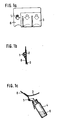
- 1 a shows a top view
- FIG. 1 b shows a cross section.
- FIG. 1 c shows how a spore carrier (1) after partial removal of the film (3) into a separate container (4) with nutrient medium (5) is transferred.
- the bioindicator according to FIGS. 2 and 3 contains both the spore carrier (1) and a container with nutrient medium (6) for testing the vitality of the indicator microorganisms after exposure in the sterilization chamber.
- the nutrient medium container (6) with the nutrient medium (5) is made of transparent material so that the bacterial growth can be monitored from the outside by turbidity and / or color changes, for example by pH indicators.
- the nutrient medium container (6) is sealed gas-tight with an insertable film (7).
- FIG. 2 a shows an embodiment of the bioindicator in section, in which several spore carriers (1) and nutrient medium containers (6) are arranged in pairs in depressions (2) of a deep-drawn part (8). A gas and water vapor permeable film (9) is sealed onto the deep-drawn part (8).
- the spore carrier (1) is located above the transparent nutrient medium container (6), which is sealed gas and water vapor-tight with a thin, easily penetrable film (7). This can, for example, be held centrally above the nutrient medium container by means of a flexible plastic ring (11).
- the thin sealing film (7) of the culture medium container is pressed against the spore carrier (1) by pressing in the flexible deep-drawn part bottom in the area of the culture medium container (6), so that the film (7), as shown in Fig. 2 b, tears and the spore carrier gets into the nutrient medium (5).
- Fig. 2 c shows the bio-indicator set in elevation.
- Fig. 3 shows an embodiment of the bioindicator in which a sleeve (10) which is used as a container for the spore carrier (1) and which is attached to a nutrient medium container (6) closed with a gas and water vapor-tight compressible film (7) one gas and water vapor permeable film (9) is closed.
- the spore carrier (1) can be transported into the nutrient medium (5) by pushing together the nutrient medium container (6) and the attached sleeve (10) and thus tearing the thin film (7).
- the film (9) must be tear-resistant.
- the bioindicator with the goods to be sterilized is placed in the sterilization chamber.
- the nutrient medium containers with the spore carriers are e.g. Incubated for 24 h at 35 ° C.
- a nutrient medium containing glucose is acidified by the breakdown of glucose, which causes the color change of a pH indicator.
- the growth can be recognized by the turbidity of the nutrient medium.
- a longer incubation period allows this statement to be made somewhat more precise, but a incubation period of 24 hours is fully sufficient for routine examinations if the germ content / test area on the material to be sterilized is significantly lower than on the spore carrier with the average spore concentration of 10 5 spores / Carrier so that there is sufficient sterilization security.
- the incubation is usually at 35 ° C.
- a nutrient medium with peptone, glucose and a pH indicator is preferably used as the nutrient medium.
- the containers with the nutrient medium are sterilized before the bio-indicator sets are manufactured, for example by y-radiation.
- Roughly ground glass balls with a diameter of 5.0 ⁇ 0.1 mm are used as spore carriers.
- Bacillus subtilis var. Niger spores are placed on the carrier from an ethanolic suspension.
- the spore carriers loaded with 10 3 , 10 5 and 10 spores per carrier are individually placed in containers of a deep-drawn part made of a 250 ⁇ m thick polyethylene film, approximately 7 mm wide and 7 mm deep.
- a peelable sterilization paper according to DIN 58953 (100 g / m 2 ) is sealed onto the deep-drawn part.
- the removable cover film is removed over the spore carrier container and the spore carrier is placed in a container with nutrient medium consisting of 5.0 g peptone, 5.0 g glucose, 30 mg / l bromothymol blue (pH 7.3 ⁇ 0.05) transferred. After 24 hours of incubation at 35 ° C, the nutrient medium is examined for germ growth, which can be recognized by a change in the pH indicator color from blue to yellow.
- Roughly ground glass balls with a diameter of 5.0 ⁇ 0.1 mm are used as spore carriers.
- the spores of Bacillus subtilis var. Niger are applied to the supports from a suspension in 1 molar sodium chloride solution which contains 5 g of bromothymol blue per liter.
- 3 spore carriers with 10 3 , 105 and 10 7 spores / carriers are used for a bioindicator set.
- the culture medium containers made of a polyethylene injection molded part with a diameter of 1.5 cm and a height of 1.2 cm are 2/3 filled with culture medium.
- On the culture medium container is one. 10 ⁇ m thick aluminum foil, which is coated with a sealing wax, sealed.
- the container is y-sterilized at 28 K Gray ( ⁇ 10%).
- the sterile culture medium containers are inserted into the recesses of a deep-drawn part made of a 300 ⁇ m thick polyethylene film.
- a foam ring with an inner diameter of approximately 7 mm and a height of approximately 5 mm is placed on the nutrient medium container lid.
- One spore holder is placed in the ring opening.
- a sterilization paper according to DIN 58953 (100 g / m 2 ) is sealed onto the deep-drawn part with the nutrient medium container and spore carrier.
- the nutrient medium container After exposure of the bioindicator in the sterilization chamber, the nutrient medium container is pressed against the spore carrier by pressure on the easily deformable deep-drawn part such that the aluminum foil of the nutrient medium container tears and the spore carrier enters the nutrient medium.
- the bioindicator set is incubated at 35 ° C for 24 hours. A discoloration of the nutrient medium to yellow indicates that not all spores of Bacillus subtilis var. Niger have been killed under the chosen sterilization conditions.
- Ceramic rings with a rough surface of 6.0 ⁇ 0.1 mm outer diameter, 3.0 ⁇ 0.1 mm inner diameter and 5.0 ⁇ 0.1 mm height are used as spore carriers.
- the carriers are loaded with 1 0 3 , 105 or 1 0 spores.
- Cylindrical transparent plastic containers injection molded parts made of polypropylene with a diameter of 1.2 cm and a height of 3 cm are 2/3 filled with nutrient medium, sealed with a 15 ⁇ m thick sealing lacquer-coated aluminum foil and sterilized with Y radiation at 28 K Gray ( ⁇ 10%).
- the nutrient medium container is inserted into a polypropylene sleeve with a spore carrier up to a stop approximately in the middle of the sleeve.
- a gas and water vapor permeable nonwoven made of polyethylene fibers (Tyvek) 70 g / m 2 ) is sealed onto the top of the sleeve.
- the spore carrier After exposure of the bioindicator in the sterilization chamber, the spore carrier is pushed into the nutrient medium through the aluminum foil that is torn open by pushing the nutrient medium container and sleeve together. After 24 hours of incubation at 35 ° C, it is determined whether growth of Bacillus subtilis var. Niger can be recognized by a change in the pH indicator from blue to yellow.
Landscapes
- Chemical & Material Sciences (AREA)
- Health & Medical Sciences (AREA)
- Life Sciences & Earth Sciences (AREA)
- Organic Chemistry (AREA)
- Zoology (AREA)
- Wood Science & Technology (AREA)
- Engineering & Computer Science (AREA)
- Bioinformatics & Cheminformatics (AREA)
- Genetics & Genomics (AREA)
- General Health & Medical Sciences (AREA)
- Biotechnology (AREA)
- Microbiology (AREA)
- Proteomics, Peptides & Aminoacids (AREA)
- Molecular Biology (AREA)
- Public Health (AREA)
- Biochemistry (AREA)
- General Engineering & Computer Science (AREA)
- Analytical Chemistry (AREA)
- Epidemiology (AREA)
- Biomedical Technology (AREA)
- Physics & Mathematics (AREA)
- Biophysics (AREA)
- Sustainable Development (AREA)
- Immunology (AREA)
- Apparatus For Disinfection Or Sterilisation (AREA)
- Apparatus Associated With Microorganisms And Enzymes (AREA)
- Measuring Or Testing Involving Enzymes Or Micro-Organisms (AREA)
Abstract
Description
- Die Erfindung betrifft Bioindikatoren zur mikrobiologischen Kontrolle von Gassterilisationsverfahren.
- Bei den Bioindikatoren handelt es sich um Testsysteme, die lebende Mikroorganismen als Indikatoren enthalten. Der Bioindikator wird zusammen mit dem zu sterilisierenden Gut den Sterilisationsbedingungen ausgesetzt. Nach Abschluß des Sterilisiervorganges erfolgt eine Kontrolle der Abtötung der Indikator-Mikroorganismen.
- Mikrobiologische Sterilisationsindikatoren werden in zunehmendem Maße bei der Überwachung von Sterilisationsverfahren verwendet. Während bei der Kontrolle von Sterilisationsprozessen das Schwergewicht früher auf der Sterilitätsprüfung des sterilisierten Gutes lag, kann heute der Umfang der Sterilitätsprüfung verringert werden, wenn das Sterilisationsverfahren mittels mikrobiologischer Indikatoren kontrolliert und eine ausreichende Sterilisationssicherheit nachgewiesen wird.
- Gassterilisationsverfahren beruhen auf der Verwendung von gasförmigen, bakterizid und sporizid wirkenden chemischen Agenzien wie beispielsweise Ethylenoxid und Formaldehyd. Die Abtötungswirkung von chemischen Agenzien wird im wesentlichen durch deren Konzentration, Einwirkungszeit und Temperatur sowie durch die Art und den Zustand der Mikroorganismen bestimmt.
- Als quantitatives Maß für die Widerstandsfähigkeit von Mikroorganismen gegenüber Sterilisationsbedingungen dienen die "Dezimale Abtötungszeit" (D-Wert) sowie die "Abtötungszeit". Die "Dezimale Abtötungszeit" gibt die Zeit in Minuten an, innerhalb der die Zahl der vermehrungsfähigen Keime auf 1/10 abnimmt. Die "Abtötungszeit" gibt an, wie lange die Sterilisationsbedingungen andauern müssen, bis am kontaminierten Objekt oder am Bioindikator keine überlebensfähigen Mikroorganismen mehr nachgewiesen werden können.
- Die Abtötungszeit der Bioindikator-Testkeime sollte mindestens so lang sein wie die Abtötungszeit der Keime auf dem kontaminierten Probenmaterial, d.h. die Resistenz der Mikroorganismen des Bioindikators soll der Resistenz der Keime am kontaminierten Probenmaterial ähnlich sein.
- Die Verwendung von Bioindikatoren zur Kontrolle von Gassterilisationsverfahren ist bekannt. Der wichtigste natürliche Bioindikator ist Sporenerde; dabei handelt es sich meist um Komposterde, die zu 0,2 - 2,0 g in speziellen Päckchen abgepackt ist. Nach der Exposition in der Sterilisationskammer werden die Sporenerde-Päckchen unter aseptischen Bedingungen geöffnet, wonach die Sporenerde in Kulturröhrchen mit einem Nährmedium übergeführt wird. Die Kulturen werden bebrütet und z.B. nach 1, 3, 7 und 14 Tagen auf Trübung infolge Mikroorganismenwachstum überprüft. Trübe Röhrchen werden oft zusätzlich durch Überimpfen einer Probe auf einen Nährboden untersucht.
- Die Verwendung von Sporenerde als Bioindikator ist nicht unproblematisch, da die Zusammensetzung der Mikroorganismenflora der Erde und die Resistenz der Mikroorganismen gegenüber den Sterilisationsbedingungen sehr unterschiedlich sein kann. Die Beurteilung der Sterilisationseffizienz ist umständlich und langwierig, da die Proben mehrere Tage bebrütet werden müssen. Oft kann erst nach Überimpfung einer Probe auf einen Nährboden eine sichere Aussage über ein Mikroorganismenwachstum gemacht werden, da die Sporenerde selbst bereits zu einer Trübung des Nährmediums führt.
- Neben Bioindikatoren mit natürlicher Keimpopulation wie Sporenerde sind auch Bioindikatoren in Gebrauch, die Reinkulturen von bestimmten Mikroorganismen enthalten. Der geeignetste Testorganismus für die Ethylenoxid-Sterilisation ist Bacillus subtilis var. niger in Form seiner Spore. Die Sporen besitzen eine recht große Resistenz gegenüber Ethylenoxid, der Mikroorganismus ist apothogen und dank seiner orangen Pigmentbildung z.B. auf Caseinpepton-Sojamehlpepton-Agar leicht zu erkennen. Bioindikatoren, die Sporen von Reinkulturen der Indikatororganismen enthalten, sind in den unterschiedlichsten Ausführungsformen bekannt beispielsweise auf Trägermaterialien wie Papier, Kunststoff, Glas, Sand.
- Bei den sogenannten Sporenstreifen handelt es sich um Filterpapierstreifen, die mit einer definierten Sporenzahl beladen sind. Sporenstreifen werden gewöhnlich mit einer Sporenkonzentration von etwa 106 Sporen pro Streifen verwendet. Diese Streifen sind einzeln in kleine kuvertartige Verpackungen aus gas- und wasserdampfdurchlässigem Material verpackt. Dieser Bioindikator wird mit dem zu sterilisierenden Gut in die Sterilisationskammer gegeben. Zur Überprüfung der Effizienz des Sterilisationsverfahrens wird der Sporenstr.eifen nach der Sterilisation mit einer sterilen Pinzette aus der kuvertartigen Verpackung entnommen und in ein Kulturröhrchen mit sterilem Nährmedium gegeben. Die Kulturröhrchen werden bebrütet. Das Wachstum überlebender Keime wird durch Trübung des Nährmediums und/oder durch Farbumschlag eines pH-Indikators angezeigt. Die Ablesung der Kulturröhrchen erfolgt meist nach 1, 2, 3, 4 und 7-tägiger Bebrütung. Bei der Entnahme des Sporenstreifens aus der kuvertartigen Verpackung zum Test auf lebensfähige Keime besteht die Gefahr, daß der Sporenstreifen mit lebenden Mikroorganismen kontaminiert wird und somit ein falsches Ergebnis erzielt wird.
- Neben Bioindikatoren, bei denen die Sporenträger nach der Sterilisation in ein separates Nährmedium übertragen werden müssen, sind auch komplette Bioindikatorsätze bekannt, die Sporenträger und Nährmedium enthalten. Diese Bioindikatoren bestehen aus Kunststoffröhrchen, die einen mit einer definierten Sporenzahl belegten Papierstreifen und eine dünnwandige Glasampulle mit Nährmedium enthalten. Das Kunststoffröhrchen ist mit einer gasdurchlässigen Kappe verschlossen. Zur Überprüfung auf lebensfähige Keime wird durch Zusammendrücken des Kunststoffröhrchens die Glasampulle zerbrochen, wodurch das Nährmedium mit dem Sporenträger in Kontakt tritt. Anschließend wird der Bioindikator bebrütet. Ein Wachstum von Keimen, die die Sterilisation überlebt haben, ist an einem Farbumschlag des pH-Indikators im Kulturmedium zu erkennen. Die Ablesung erfolgt nach 24 und 48 Stunden.
- Die Aufbewahrung des Nährmediums in einer Glasampulle gewährleistet zwar eine wasserdampfdichte Verpackung des Nährmediums, jedoch ist eine Verletzungsgefahr durch Glassplitter beim Zerbrechen der Ampulle nicht auszuschließen.
- Die bekannten Bioindikatoren sind oft kostspielig in der Produktion, die Standardisierung der Empfindlichkeit der Indikator-Mikroorganismen gegenüber den Sterilisationsbedingungen ist schwierig, die Handhabung unsicher und umständlich und/oder die Bewertung des Sterilisationsverfahrens sehr zeitaufwendig.
- Der Erfindung lag die Aufgabe zugrunde, einen Bioindikator zur mikrobiologischen Kontrolle von Gassterilisationsverfahren zu schaffen, der die Nachteile der bisher bekannten Bioindikatoren nicht aufweist, der preisgünstig herzustellen, gut zu standardisieren, einfach und sicher anzuwenden sowie schnell und problemlos auszuwerten ist.
- Gelöst wurde diese Aufgabe durch einen Bioindikator zur mikrobiologischen Kontrolle von Gassterilisationsverfahren, der Sporenträger und ggf. Nährmediumbehälter in einer speziellen Tiefziehpackung enthält.
- Gegenstand der Erfindung sind Bioindikatoren zur mikrobiologischen Kontrolle von Gassterilisationsverfahren, bestehend im wesentlichen aus einem Sporenträger (1) in einem Behälter (2), die dadurch gekennzeichnet sind, daß der Behälter (2) als Tiefziehpackung gestaltet und mit einer gas- und wasserdampfdurchlässigen Folie (3, 9) verschlossen ist, die mindestens dann abziehbar gestaltet ist, wenn der Behälter (2) nicht zusätzlich einen abgeschlossenen Nährmediumbehälter (6) enthält.
- Enthält der Behälter (2) zusätzlich einen Nährmediumbehälter (6), so ist dieser derart angeordnet, daß der Sporenträger (1) in den mit einer durchdrückbaren Folie (7) verschlossenen Nährmediumbehälter (6) gedrückt werden kann. Der Bioindikator kann auch so gestaltet sein, daß auf dem mit einer eindrückbaren Folie (7) verschlossenen Nährmediumbehälter (6) eine als Behälter für den Sporenträger (1) dienende Hülse (10) angeordnet ist, die mit einer gas- und wasserdampfdurchlässigen Folie (9) verschlossen ist. Es können auch mehrere Sporenträger (1) mit gegebenenfalls unterschiedlichem Sporengehalt bzw. paarweise Sporenträger (1) und Nährmediumbehälter (6) - in mit einer gas- und wässerdampfdurchlässigen Folie (3, 9) verschlossenen Behältern (2) angeordnet sein.
- Die Bioindikatoren enthalten einen oder mehrere Sporenträger (1), die aus angeätzten oder angeschliffenen Glaskugeln oder -ringen oder aus entsprechenden Formteilen aus Keramik oder Kunststoff mit rauher Oberfläche bestehen. Die Glaskugeln weisen zweckmäßigerweise einen Durchmesser von etwa 2 - 8 mm, vorzugsweise etwa 5 mm auf. Keramikringe sollten einen Durchmesser von etwa 4 - 8 mm, vorzugsweise etwa 6 mm, besitzen. Die Sporenträger werden z.B. mit 103, 105 bzw. 107 Sporen/Träger beladen. Als Indikatormikroorganismen für Bioindikatoren zur mikrobiologischen Kontrolle von Gassterilisationsverfahren werden beispielsweise Sporen von Bacillus subtilis var. niger verwendet. Im gleichen Sinne können Sporen anderer sporenbildender Bakterien eingesetzt werden. Die Sporen werden aus ethanolischen oder wäßrigen Suspensionen auf die Formteile aufgebracht, wobei die Suspensionen Salze, Indikatorfarbstoffe, Gelatine, verschiedene Cellulosederivate, Polyvinylalkohole oder Polyvinylpyrrolidone enthalten können.
- Als gas- und wasserdampfdurchlässige Folien (3, 9) zum Verschluß des Behälters mit dem Sporenträger (1.) können alle Folienmaterialien oder Vliese verwendet werden, die eine gute Durchlässigkeit für gasförmige chemische Sterilisationsagenzien besitzen, die wasserdampfdurchlässig und zumindest im trockenen Zustand bakteriendicht sind sowie auf Kunststoffe wie Polyethylen, Polypropylen, Polyvinylchlorid oder entsprechende Mischpolymerisate aufgesiegelt werden können. Gut geeignet sind z.B. Sterilisationspapier nach DIN 58953 oder spezielle Vliese aus Polyethylenfasern.
- Der transparente Nährmediumbehälter (6) besteht aus Glas oder Kunststoffen wie Polyethylen oder Polypropylen, auf den eine leicht zu durchstoßende dünne Folie (7) mit äußerst geringer Gas- und Wasserdampfdurchlässigkeit aufgesiegelt ist. Etwa 10 - 15 µm dicke Aluminiumfolie, die mit einem Siegellack beschichtet ist, eignet sich vorzugsweise als leicht durchstoßbare Folie (7) zum Verschluß eines Nährmediumbehälters aus Polypropylen oder Polyethylen.
- Die Erfindung wird anhand der Abbildungen 1 bis 3 näher erläutert:
- Beim Bioindikator nach Fig. 1 befinden sich einzelne Sporenträger (1) mit unterschiedlichem Sporengehalt in den Vertiefungen eines Tiefziehteils (8). Das Tiefziehteil enthält z.B. drei Behälter (2) für Sporenträger mit verschiedenen Sporenkonzentrationen (z.B. 103, 10 5, 10 Sporen/Träger). Auf das Tiefziehteil ist eine zerstörungsfrei abziehbare gas- und wasserdampfdurchlässige Folie (3) aufgesiegelt. Fig. 1 a stellt eine Aufsicht, Fig. 1 b einen Querschnitt dar. In Fig. 1 c ist dargestellt, wie ein Sporenträger (1) nach partiellem Abziehen der Folie (3) in ein separates Behältnis (4) mit Nährmedium (5) überführt wird.
- Der Bioindikator nach Fig. 2 und 3 enthält sowohl den Sporenträger (1) als auch ein Behältnis mit Nährmedium (6) zum Test der Vitalität der Indikator-Mikroorganismen nach der Exposition in der Sterilisationskammer. Der Nährmediumbehälter (6) mit dem Nährmedium (5) ist aus transparentem Material gefertigt, damit von außen das Bakterienwachstum durch Trübung und/oder Farbveränderung etwa von pH-Indikatoren verfolgt werden kann. Der Nährmediumbehälter (6) ist mit einer eindrückbaren Folie (7) gasdicht verschlossen.
- Fig. 2 a zeigt eine Ausführungsform des Bioindikators im Schnitt, bei der mehrere Sporenträger (1) und Nährmediumbehälter (6) paarweise in Vertiefungen (2) eines Tiefziehteiles (8) angeordnet sind. Auf das Tiefziehteil (8) ist eine gas- und wasserdampfdurchlässige Folie (9) aufgesiegelt.
- Über dem transparenten Nährmediumbehälter (6), der mit einer dünnen, leicht zu durchstoßenden Folie (7) gas-und wasserdampfdicht verschlossen ist, befindet sich der Sporenträger (1). Dieser kann beispielsweise durch einen flexiblen Kunststoffring (11) zentral über dem Nährmediumbehälter gehalten werden. Zur Beurteilung des Sterilisationserfolges wird durch Eindrücken des flexiblen Tiefziehteilbodens im Bereich des Nährmediumbehälters (6) die dünne Verschlußfolie (7) des Nährmediumbehälters gegen den Sporenträger (1) gepreßt, so daß die Folie (7), wie in Fig. 2 b dargestellt, einreißt und der Sporenträger in das Nährmedium (5) gelangt.
- Fig. 2 c zeigt das Bioindikator-Set im Aufriß.
- Fig. 3 zeigt eine Ausführungsform des Bioindikators, bei der auf einen mit einer gas- und wasserdampfdichtem eindrückbaren Folie (7) verschlossenen Nährmediumbehälter (6) eine Hülse (10) aufgesteckt wird, die als Behälter für den Sporenträger (1) dient und die mit einer gas- und wasserdampfdurchlässigen Folie (9) verschlossen ist. Durch Zusammenschieben von Nährmediumbehälter (6) und aufgesteckter Hülse (10) und somit Zerreißen der dünnen Folie (7) kann der Sporenträger (1) in das Nährmedium (5) befördert werden. Die Folie (9) muß reißfest sein.
- Zur mikrobiologischen Kontrolle von Gassterilisationsverfahren wird der Bioindikator mit dem zu sterilisierenden Gut in die Sterilisationskammer gegeben. Nach der Exposition der Bioindikatoren in der Sterilisationskammer und Überführung der Sporenträger in ein Nährmedium werden die Nährmediumbehälter mit den Sporenträgern z.B. 24 h bei 35 °C bebrütet. Bei einem Wachstum der Testkeime wird z.B. ein Glucose enthaltendes Nährmedium durch Abbau von Glukose angesäuert, was den Farbumschlag eines pH-Indikators bewirkt. Das Wachstum ist an der.Trübung des Nährmediums zu erkennen.
- Da bei chemischen Sterilisationsverfahren einzelne Keime innerhalb einer großen Population häufig unter den normalen Sterilisationsbedingungen nicht abgetötet, sondern nur stark geschädigt werden, erscheint eine generelle Bewertung des Steriliationsverfahrens nach der Endpunkt-Methode, d.h. einer Überprüfung auf Sterilität "Ja/Nein", infolge einer zu langen Analysenzeit für die Routine nicht empfehlenswert.
- Bei einer Bewertung des Sterilisationsverfahrens durch Auswertung des Keimwachstums von Trägern mit z.B. 103, 105 bzw. 107 Sporen nach einer Bebrütungszeit von 24 Stunden gewinnt man sichere Anhaltspunkte dafür, wie effizient das Sterilisationsverfahren war. Zeigen alle 3 Trägertypen nach 24 Stunden Inkubation in einem Testnährmedium kein Wachstum, so kann man davon ausgehen, daß unter den gewählten Sterilisationsbedingungen mindestens 106 Keime pro Träger abgetötet wurden.
- Durch eine längere Bebrütungszeit läßt sich diese Aussage zwar noch etwas präzisieren, jedoch ist für Routineuntersuchungen eine Bebrütungszeit von 24 Stunden voll ausreichend, wenn der Keimgehalt/Testfläche auf dem zu sterilisierenden Gut deutlich geringer ist als auf dem Sporenträger mit der mittleren Sporenkonzentration von 105 Sporen/Träger, so daß eine ausreichende Sterilisationssicherheit vorhanden ist. Die Bebrütung erfolgt in der Regel bei 35 °C. Als Nährmedium wird vorzugsweise ein Nährmedium mit Pepton, Glucose und einem pH-Indikator verwendet. Die Behälter mit dem Nährmedium werden jeweils vor der Fertigung der Bioindikatoren-Sets, z.B. durch y-Bestrahlung, sterilisiert.
- Als Sporenträger werden oberflächlich rauh geschliffene Glaskugeln vom Durchmesser 5,0 ± 0,1 mm verwendet. Die Sporen von Bacillus subtilis var. niger werden aus einer ethanolischen Suspension auf die Träger gebracht. Die mit 103, 105 und 10 Sporen pro Träger beladenen Sporenträger werden einzeln in etwa 7 mm breite und 7 mm tiefe Behälter eines Tiefziehteils aus einer 250 µm dicken Polyethylenfolie gegeben. Auf das Tiefziehteil wird ein abziehbares Sterilisationspapier nach DIN 58953 (100 g/m2) aufgesiegelt. Nach der Exposition der Bioindikatoren in der Sterilisationskammer wird die abziehbare Deckfolie über dem Sporenträger-Behälter abgezogen und der Sporenträger in einen Behälter mit Nährmedium aus 5,0 g Pepton, 5,0 g Glucose, 30 mg/l Bromthymolblau (pH 7,3 ± 0,05) überführt. Nach 24 Stunden Inkubation bei 35 °C wird das Nährmedium auf Keimwachstum untersucht, das an einem Umschlag der pH-Indikatorfarbe von Blau nach Gelb zu erkennen ist.
- Als Sporenträger werden oberflächlich rauh geschliffene Glaskugeln vom Durchmesser 5,0 ± 0,1 mm verwendet. Die Sporen von Bacillus subtilis var. niger werden aus einer Suspension in 1-molarer Natriumchloridlösung, die 5 g Bromthymolblau pro Liter enthält, auf die Träger aufgebracht. Jeweils 3 Sporenträger mit 103, 105 und 107 Sporen/Träger werden für ein Bioindikator-Set verwendet.
- Die Nährmediumbehälter aus einem Polyethylen-Spritzgußteil mit 1,5 cm Durchmesser und 1,2 cm Höhe sind zu 2/3 mit Nährmedium gefüllt. Auf die Nährmediumbehälter ist eine. 10 µm dicke Aluminiumfolie, die mit einem Siegellack beschichtet ist, aufgesiegelt. Der Behälter wird bei 28 K Gray (± 10 %) y-strahlensterilisiert. Die sterilen Nährmediumbehälter werden in Vertiefungen eines Tiefziehteils aus einer 300 µm dicken Polyethylenfolie eingesetzt. Auf die Nährmediumbehälterdeckel wird ein Schaumstoffring mit einem Innendurchmesser von etwa 7 mm und einer Höhe von etwa 5 mm aufgesetzt. Jeweils ein Sporenträger wird in die Ringöffnung gesetzt. Auf das Tiefziehteil mit Nährmediumbehälter und Sporenträger wird ein Sterilisationspapier nach DIN 58953 (100 g/m2) aufgesiegelt.
- Nach der Exposition des Bioindikators in der Sterilisationskammer wird der Nährmediumbehälter durch Druck auf das leicht verformbare Tiefziehteil so gegen den Sporenträger gepreßt, daß die Aluminiumfolie des Nährmediumbehälters reißt und der Sporenträger in das Nährmedium gelangt. Das Bioindikator-Set wird 24 Stunden bei 35 °C bebrütet. Eine Verfärbung des Nährmediums nach Gelb zeigt an, daß unter den gewählten Sterilisationsbedingungen nicht alle Sporen von Bacillus subtilis var. niger abgetötet wurden.
- Als Sporenträger werden Keramikringe mit rauher Oberfläche von 6,0 ± 0,1 mm Außendurchmesser, 3,0 ± 0,1 mm Innendurchmesser und 5,0 ± 0,1 mm Höhe verwendet. Die Träger werden mit 103, 105 bzw. 10 Sporen beladen.
- Zylinderförmige durchsichtige Kunststoffbehälter (Spritzgußteile aus Polypropylen) von 1,2 cm Durchmesser und 3 cm Höhe werden zu 2/3 mit Nährmedium gefüllt, mit einer 15 µm dicken siegellackbeschichteten Aluminiumfolie dicht verschlossen und bei 28 K Gray (± 10 %) y-strahlensterilisiert. Der Nährmediumbehälter wird in eine Polypropylenhülse mit einem Sporenträger bis zu einem Anschlag etwa in der Mitte der Hülse gesteckt. Auf die Hülse ist an ihrer Oberseite ein gas- und wasserdampfdurchlässiges Vlies aus Polyethylenfasern (Tyvek ) (70 g/m2) aufgesiegelt.
- Nach der Exposition des Bioindikators in der Sterilisationskammer wird durch ein Zusammenschieben von Nährmediumbehälter und Hülse der Sporenträger durch die dabei aufreißende Aluminiumfolie in das Nährmedium befördert. Nach 24 Stunden Inkubation bei 35 °C wird festgestellt, ob Wachstum von Bacillus subtilis var. niger an einem Umschlag des pH-Indikators von Blau nach Gelb zu erkennen ist.
Claims (4)
Applications Claiming Priority (2)
| Application Number | Priority Date | Filing Date | Title |
|---|---|---|---|
| DE3217002A DE3217002A1 (de) | 1982-05-06 | 1982-05-06 | Bioindikator |
| DE3217002 | 1982-05-06 |
Publications (2)
| Publication Number | Publication Date |
|---|---|
| EP0093920A1 true EP0093920A1 (de) | 1983-11-16 |
| EP0093920B1 EP0093920B1 (de) | 1987-03-18 |
Family
ID=6162908
Family Applications (1)
| Application Number | Title | Priority Date | Filing Date |
|---|---|---|---|
| EP83103955A Expired EP0093920B1 (de) | 1982-05-06 | 1983-04-22 | Bioindikator |
Country Status (3)
| Country | Link |
|---|---|
| EP (1) | EP0093920B1 (de) |
| JP (1) | JPS58212775A (de) |
| DE (2) | DE3217002A1 (de) |
Cited By (8)
| Publication number | Priority date | Publication date | Assignee | Title |
|---|---|---|---|---|
| EP0202724A1 (de) * | 1985-05-16 | 1986-11-26 | Pymah Corporation | Wegwerfbare Testpackung zum Nachweis biologischer Sterilität |
| WO2000050634A1 (en) * | 1999-02-22 | 2000-08-31 | 3M Innovative Properties Company | Sterilization indicator for liquid peracetic acid procedures |
| WO2001013964A1 (en) * | 1999-08-24 | 2001-03-01 | Process Challenge Devices | Device to assess efficacy of sterilization procedure |
| US6653096B1 (en) | 1996-07-29 | 2003-11-25 | Process Challenge Devices | Process challenge device and method |
| DE10213066B4 (de) * | 2002-02-11 | 2012-07-12 | Melag Ohg | Prüfvorrichtung für einen Sterilisationsvorgang |
| WO2012092684A1 (de) | 2011-01-05 | 2012-07-12 | Skan Ag | Bioindikator |
| WO2020112651A1 (en) * | 2018-11-28 | 2020-06-04 | American Sterilizer Company | Biological sterilization indicator |
| WO2022119972A3 (en) * | 2020-12-02 | 2022-11-24 | Steritec Products Mfg. Co., Inc. | Biological indicators, and systems and methods for determining efficacy of sterilization |
Families Citing this family (4)
| Publication number | Priority date | Publication date | Assignee | Title |
|---|---|---|---|---|
| DE3640114A1 (de) * | 1986-11-24 | 1988-06-01 | Volker Barkey | Vorrichtung zum nachweis der wirksamkeit eines desinfektionsprozesses bei anwendung von desinfektionsmitteln in einem waschvorgang |
| DE3718553A1 (de) * | 1987-06-03 | 1987-12-17 | Raimund Dr Mildner | Geschlossener keimtraeger |
| DE9003257U1 (de) * | 1990-03-20 | 1990-06-07 | Simon, Paul Gerhard, 8000 Muenchen, De | |
| CN110468041B (zh) * | 2019-09-25 | 2024-04-30 | 福建省农业科学院植物保护研究所 | 一种植物病菌孢子监测装置及监测方法 |
Citations (3)
| Publication number | Priority date | Publication date | Assignee | Title |
|---|---|---|---|---|
| US3440144A (en) * | 1965-05-21 | 1969-04-22 | Andersen Prod H W | Method and apparatus for checking and testing the effectiveness of sterilization |
| DE1492498A1 (de) * | 1963-04-10 | 1969-12-18 | Ritter Pfaudler Corp | Biologische Sterilitaets-Anzeigevorrichtung und Verfahren zu ihrer Anwendung |
| US4291122A (en) * | 1980-08-14 | 1981-09-22 | American Sterilizer Company | Biological indicator for sterilization processes |
-
1982
- 1982-05-06 DE DE3217002A patent/DE3217002A1/de not_active Withdrawn
-
1983
- 1983-04-22 EP EP83103955A patent/EP0093920B1/de not_active Expired
- 1983-04-22 DE DE8383103955T patent/DE3370357D1/de not_active Expired
- 1983-05-04 JP JP58077698A patent/JPS58212775A/ja active Pending
Patent Citations (3)
| Publication number | Priority date | Publication date | Assignee | Title |
|---|---|---|---|---|
| DE1492498A1 (de) * | 1963-04-10 | 1969-12-18 | Ritter Pfaudler Corp | Biologische Sterilitaets-Anzeigevorrichtung und Verfahren zu ihrer Anwendung |
| US3440144A (en) * | 1965-05-21 | 1969-04-22 | Andersen Prod H W | Method and apparatus for checking and testing the effectiveness of sterilization |
| US4291122A (en) * | 1980-08-14 | 1981-09-22 | American Sterilizer Company | Biological indicator for sterilization processes |
Cited By (11)
| Publication number | Priority date | Publication date | Assignee | Title |
|---|---|---|---|---|
| EP0202724A1 (de) * | 1985-05-16 | 1986-11-26 | Pymah Corporation | Wegwerfbare Testpackung zum Nachweis biologischer Sterilität |
| US6653096B1 (en) | 1996-07-29 | 2003-11-25 | Process Challenge Devices | Process challenge device and method |
| WO2000050634A1 (en) * | 1999-02-22 | 2000-08-31 | 3M Innovative Properties Company | Sterilization indicator for liquid peracetic acid procedures |
| WO2001013964A1 (en) * | 1999-08-24 | 2001-03-01 | Process Challenge Devices | Device to assess efficacy of sterilization procedure |
| DE10213066B4 (de) * | 2002-02-11 | 2012-07-12 | Melag Ohg | Prüfvorrichtung für einen Sterilisationsvorgang |
| WO2012092684A1 (de) | 2011-01-05 | 2012-07-12 | Skan Ag | Bioindikator |
| CH704312A1 (de) * | 2011-01-05 | 2012-07-13 | Skan Ag | Bioindikator. |
| WO2020112651A1 (en) * | 2018-11-28 | 2020-06-04 | American Sterilizer Company | Biological sterilization indicator |
| US11248251B2 (en) | 2018-11-28 | 2022-02-15 | American Sterilizer Company | Biological sterilization indicator |
| WO2022119972A3 (en) * | 2020-12-02 | 2022-11-24 | Steritec Products Mfg. Co., Inc. | Biological indicators, and systems and methods for determining efficacy of sterilization |
| US11603551B2 (en) | 2020-12-02 | 2023-03-14 | Steritec Products Mfg. Co., Inc. | Biological indicators, and systems and methods for determining efficacy of sterilization |
Also Published As
| Publication number | Publication date |
|---|---|
| JPS58212775A (ja) | 1983-12-10 |
| EP0093920B1 (de) | 1987-03-18 |
| DE3370357D1 (en) | 1987-04-23 |
| DE3217002A1 (de) | 1983-11-10 |
Similar Documents
| Publication | Publication Date | Title |
|---|---|---|
| DE69727069T2 (de) | Mikrobiologisches schnellbestimmungsverfahren und vorrichtung, die die detektion des sauerstoffgradienten benutzt | |
| US4416984A (en) | Sterilization indicator | |
| CA1290229C (en) | Biological indicator for sterilization processes | |
| US3661717A (en) | Unitary sterility indicator and method | |
| Hite et al. | The effect of pressure on certain micro-organisms encountered in the preservation of fruits and vegetables | |
| US4717661A (en) | Biological indicator for sterilization processes | |
| US4596773A (en) | Sterilization indicator | |
| DE69637354T2 (de) | Vorrichtung und verfahren zum detektieren von mikroorganismen | |
| EP0093920B1 (de) | Bioindikator | |
| US4087326A (en) | Microbiological scaled sterility test strips and method | |
| CH629255A5 (de) | Verfahren und testanordnung zur bestimmung des ansprechens eines mikroorganismus auf ein antibiotikum. | |
| US3846242A (en) | Biological sterility indicator and method for using same | |
| DE2152068B2 (de) | ||
| US4311793A (en) | Sterilization indicator | |
| DE60111764T2 (de) | Indikatorsysteme zur bestimmung der wirksamkeit eines sterilisationsverfahrens | |
| DE2027604A1 (de) | Einheitliche Stenlisationsanzeige vorrichtung und Verfahren zur Anwendung der selben | |
| DE1492498B2 (de) | Desinfektion^-Anzeige vorrichtung | |
| EP0346732A1 (de) | Biologische Sammel- und Transportvorrichtung | |
| Kearney et al. | Evaluation of ethylene oxide sterilization of tissue implants | |
| US3313712A (en) | Apparatus for the detection of living microbial contaminants in petroleum products | |
| DE8213134U1 (de) | Bioindikator | |
| EP1485135B1 (de) | Verfahren zur prüfung einer sterilisierverpackungseinheit auf wirksamkeit gegen rekontamination | |
| Spicher | Biological indicators and monitoring systems for validation and cycle control of sterilization processes | |
| Hodges | Reproducibility and performance of endospores as biological indicators | |
| EP1624903B1 (de) | Testeinheit und verfahren zur prüfung einer sterilisierverpackungseinheit auf wirksamkeit gegen rekontamination und eine zu dem verfahren geeignete containerverpackung |
Legal Events
| Date | Code | Title | Description |
|---|---|---|---|
| PUAI | Public reference made under article 153(3) epc to a published international application that has entered the european phase |
Free format text: ORIGINAL CODE: 0009012 |
|
| AK | Designated contracting states |
Designated state(s): BE CH DE FR GB IT LI NL |
|
| 17P | Request for examination filed |
Effective date: 19840316 |
|
| GRAA | (expected) grant |
Free format text: ORIGINAL CODE: 0009210 |
|
| AK | Designated contracting states |
Kind code of ref document: B1 Designated state(s): BE CH DE FR GB IT LI NL |
|
| PG25 | Lapsed in a contracting state [announced via postgrant information from national office to epo] |
Ref country code: NL Effective date: 19870318 Ref country code: IT Free format text: LAPSE BECAUSE OF FAILURE TO SUBMIT A TRANSLATION OF THE DESCRIPTION OR TO PAY THE FEE WITHIN THE PRESCRIBED TIME-LIMIT;WARNING: LAPSES OF ITALIAN PATENTS WITH EFFECTIVE DATE BEFORE 2007 MAY HAVE OCCURRED AT ANY TIME BEFORE 2007. THE CORRECT EFFECTIVE DATE MAY BE DIFFERENT FROM THE ONE RECORDED. Effective date: 19870318 Ref country code: FR Free format text: THE PATENT HAS BEEN ANNULLED BY A DECISION OF A NATIONAL AUTHORITY Effective date: 19870318 Ref country code: BE Effective date: 19870318 |
|
| REF | Corresponds to: |
Ref document number: 3370357 Country of ref document: DE Date of ref document: 19870423 |
|
| PG25 | Lapsed in a contracting state [announced via postgrant information from national office to epo] |
Ref country code: LI Effective date: 19870430 Ref country code: CH Effective date: 19870430 |
|
| EN | Fr: translation not filed | ||
| NLV1 | Nl: lapsed or annulled due to failure to fulfill the requirements of art. 29p and 29m of the patents act | ||
| REG | Reference to a national code |
Ref country code: CH Ref legal event code: PL |
|
| PG25 | Lapsed in a contracting state [announced via postgrant information from national office to epo] |
Ref country code: DE Effective date: 19880101 |
|
| PLBE | No opposition filed within time limit |
Free format text: ORIGINAL CODE: 0009261 |
|
| STAA | Information on the status of an ep patent application or granted ep patent |
Free format text: STATUS: NO OPPOSITION FILED WITHIN TIME LIMIT |
|
| GBPC | Gb: european patent ceased through non-payment of renewal fee | ||
| 26N | No opposition filed | ||
| PG25 | Lapsed in a contracting state [announced via postgrant information from national office to epo] |
Ref country code: GB Free format text: LAPSE BECAUSE OF NON-PAYMENT OF DUE FEES Effective date: 19881122 |