WO2024034580A1 - Ceacam1を検出する方法 - Google Patents
Ceacam1を検出する方法 Download PDFInfo
- Publication number
- WO2024034580A1 WO2024034580A1 PCT/JP2023/028802 JP2023028802W WO2024034580A1 WO 2024034580 A1 WO2024034580 A1 WO 2024034580A1 JP 2023028802 W JP2023028802 W JP 2023028802W WO 2024034580 A1 WO2024034580 A1 WO 2024034580A1
- Authority
- WO
- WIPO (PCT)
- Prior art keywords
- ceacam1
- antibody
- cancer
- ability
- recognize
- Prior art date
- Legal status (The legal status is an assumption and is not a legal conclusion. Google has not performed a legal analysis and makes no representation as to the accuracy of the status listed.)
- Ceased
Links
Images




Classifications
-
- G—PHYSICS
- G01—MEASURING; TESTING
- G01N—INVESTIGATING OR ANALYSING MATERIALS BY DETERMINING THEIR CHEMICAL OR PHYSICAL PROPERTIES
- G01N33/00—Investigating or analysing materials by specific methods not covered by groups G01N1/00 - G01N31/00
- G01N33/48—Biological material, e.g. blood, urine; Haemocytometers
- G01N33/50—Chemical analysis of biological material, e.g. blood, urine; Testing involving biospecific ligand binding methods; Immunological testing
- G01N33/53—Immunoassay; Biospecific binding assay; Materials therefor
- G01N33/575—Immunoassay; Biospecific binding assay; Materials therefor for cancer
- G01N33/57525—Immunoassay; Biospecific binding assay; Materials therefor for cancer of the liver or pancreas
-
- A—HUMAN NECESSITIES
- A61—MEDICAL OR VETERINARY SCIENCE; HYGIENE
- A61K—PREPARATIONS FOR MEDICAL, DENTAL OR TOILETRY PURPOSES
- A61K45/00—Medicinal preparations containing active ingredients not provided for in groups A61K31/00 - A61K41/00
-
- A—HUMAN NECESSITIES
- A61—MEDICAL OR VETERINARY SCIENCE; HYGIENE
- A61P—SPECIFIC THERAPEUTIC ACTIVITY OF CHEMICAL COMPOUNDS OR MEDICINAL PREPARATIONS
- A61P35/00—Antineoplastic agents
-
- C—CHEMISTRY; METALLURGY
- C07—ORGANIC CHEMISTRY
- C07K—PEPTIDES
- C07K16/00—Immunoglobulins [IG], e.g. monoclonal or polyclonal antibodies
-
- C—CHEMISTRY; METALLURGY
- C07—ORGANIC CHEMISTRY
- C07K—PEPTIDES
- C07K16/00—Immunoglobulins [IG], e.g. monoclonal or polyclonal antibodies
- C07K16/18—Immunoglobulins [IG], e.g. monoclonal or polyclonal antibodies against material from animals or humans
- C07K16/28—Immunoglobulins [IG], e.g. monoclonal or polyclonal antibodies against material from animals or humans against receptors, cell surface antigens or cell surface determinants
-
- G—PHYSICS
- G01—MEASURING; TESTING
- G01N—INVESTIGATING OR ANALYSING MATERIALS BY DETERMINING THEIR CHEMICAL OR PHYSICAL PROPERTIES
- G01N33/00—Investigating or analysing materials by specific methods not covered by groups G01N1/00 - G01N31/00
- G01N33/48—Biological material, e.g. blood, urine; Haemocytometers
- G01N33/50—Chemical analysis of biological material, e.g. blood, urine; Testing involving biospecific ligand binding methods; Immunological testing
- G01N33/53—Immunoassay; Biospecific binding assay; Materials therefor
-
- G—PHYSICS
- G01—MEASURING; TESTING
- G01N—INVESTIGATING OR ANALYSING MATERIALS BY DETERMINING THEIR CHEMICAL OR PHYSICAL PROPERTIES
- G01N33/00—Investigating or analysing materials by specific methods not covered by groups G01N1/00 - G01N31/00
- G01N33/48—Biological material, e.g. blood, urine; Haemocytometers
- G01N33/50—Chemical analysis of biological material, e.g. blood, urine; Testing involving biospecific ligand binding methods; Immunological testing
- G01N33/53—Immunoassay; Biospecific binding assay; Materials therefor
- G01N33/543—Immunoassay; Biospecific binding assay; Materials therefor with an insoluble carrier for immobilising immunochemicals
-
- G—PHYSICS
- G01—MEASURING; TESTING
- G01N—INVESTIGATING OR ANALYSING MATERIALS BY DETERMINING THEIR CHEMICAL OR PHYSICAL PROPERTIES
- G01N33/00—Investigating or analysing materials by specific methods not covered by groups G01N1/00 - G01N31/00
- G01N33/48—Biological material, e.g. blood, urine; Haemocytometers
- G01N33/50—Chemical analysis of biological material, e.g. blood, urine; Testing involving biospecific ligand binding methods; Immunological testing
- G01N33/53—Immunoassay; Biospecific binding assay; Materials therefor
- G01N33/575—Immunoassay; Biospecific binding assay; Materials therefor for cancer
-
- G—PHYSICS
- G01—MEASURING; TESTING
- G01N—INVESTIGATING OR ANALYSING MATERIALS BY DETERMINING THEIR CHEMICAL OR PHYSICAL PROPERTIES
- G01N33/00—Investigating or analysing materials by specific methods not covered by groups G01N1/00 - G01N31/00
- G01N33/48—Biological material, e.g. blood, urine; Haemocytometers
- G01N33/50—Chemical analysis of biological material, e.g. blood, urine; Testing involving biospecific ligand binding methods; Immunological testing
- G01N33/53—Immunoassay; Biospecific binding assay; Materials therefor
- G01N33/575—Immunoassay; Biospecific binding assay; Materials therefor for cancer
- G01N33/57535—Immunoassay; Biospecific binding assay; Materials therefor for cancer of the large intestine, e.g. colon, rectum or anus
-
- G—PHYSICS
- G01—MEASURING; TESTING
- G01N—INVESTIGATING OR ANALYSING MATERIALS BY DETERMINING THEIR CHEMICAL OR PHYSICAL PROPERTIES
- G01N33/00—Investigating or analysing materials by specific methods not covered by groups G01N1/00 - G01N31/00
- G01N33/48—Biological material, e.g. blood, urine; Haemocytometers
- G01N33/50—Chemical analysis of biological material, e.g. blood, urine; Testing involving biospecific ligand binding methods; Immunological testing
- G01N33/68—Chemical analysis of biological material, e.g. blood, urine; Testing involving biospecific ligand binding methods; Immunological testing involving proteins, peptides or amino acids
- G01N33/6878—Chemical analysis of biological material, e.g. blood, urine; Testing involving biospecific ligand binding methods; Immunological testing involving proteins, peptides or amino acids in epitope analysis
-
- G—PHYSICS
- G01—MEASURING; TESTING
- G01N—INVESTIGATING OR ANALYSING MATERIALS BY DETERMINING THEIR CHEMICAL OR PHYSICAL PROPERTIES
- G01N2333/00—Assays involving biological materials from specific organisms or of a specific nature
- G01N2333/435—Assays involving biological materials from specific organisms or of a specific nature from animals; from humans
- G01N2333/705—Assays involving receptors, cell surface antigens or cell surface determinants
- G01N2333/70503—Immunoglobulin superfamily, e.g. VCAMs, PECAM, LFA-3
-
- G—PHYSICS
- G01—MEASURING; TESTING
- G01N—INVESTIGATING OR ANALYSING MATERIALS BY DETERMINING THEIR CHEMICAL OR PHYSICAL PROPERTIES
- G01N2458/00—Labels used in chemical analysis of biological material
-
- G—PHYSICS
- G01—MEASURING; TESTING
- G01N—INVESTIGATING OR ANALYSING MATERIALS BY DETERMINING THEIR CHEMICAL OR PHYSICAL PROPERTIES
- G01N2470/00—Immunochemical assays or immunoassays characterised by the reaction format or reaction type
- G01N2470/04—Sandwich assay format
Definitions
- the present invention relates to a method of detecting CEACAM1, etc.
- CEACAM1 is a protein belonging to the carcinoembryonic antigen (CEA) family. There are 12 isoforms of CEACAM1, three of which are secreted and are known to exist in body fluids such as serum. It is also known that secreted CEACAM1 can be used as a marker for cancers such as melanoma, pancreatic cancer, and urothelial bladder cancer. In pancreatic cancer, CEACAM1 has been confirmed to exhibit higher diagnostic sensitivity and specificity than the general cancer marker CA19-9. Therefore, CEACAM1 is expected to be a cancer marker.
- Patent Document 1 discloses an antibody capable of recognizing a CEACAM1-specific epitope (optionally an antibody capable of binding to other subtypes of the CEACAM protein family).
- Patent Document 2 discloses a method for diagnosing the presence or absence of extrahepatic cholangiocarcinoma, intrahepatic cholangiocarcinoma, or gallbladder cancer using an antibody that has the ability to specifically bind to CEACAM1.
- Patent Document 3 discloses a method of heating human serum or plasma under acidic conditions to separate unnecessary substances, and then detecting carcinoembryonic antigen (CEA) by immunoassay.
- CEA carcinoembryonic antigen
- the purpose of the present invention is to detect with high sensitivity a specific protein that can be used as a cancer marker.
- Another object of the present invention is to sensitively and specifically detect a specific protein that can be used as a cancer marker.
- CEACAM1 As a result of extensive studies, the present inventors selected CEACAM1 as a specific protein that can be used as a cancer marker, and after denaturing CEACAM1, used both a capture antibody and a labeled antibody that have the ability to recognize denatured CEACAM1.
- the present inventors have discovered that CEACAM1, which can be used as a cancer marker, can be detected with high sensitivity (and specificity) by using a sandwich assay, and have completed the present invention.
- the prior art does not describe or suggest the use of such a sandwich assay after denaturation of CEACAM1.
- a method for detecting CEACAM1, comprising: (1) denaturing CEACAM1 in a CEACAM1-containing sample to produce denatured CEACAM1; and (2) detecting CEACAM1 by a sandwich assay using both a capture antibody and a labeled antibody that have the ability to recognize denatured CEACAM1.
- process including methods.
- the method of [1], wherein the modification is selected from the group consisting of a modifying agent, heating, and a combination thereof.
- the modifier is selected from the group consisting of alkaline substances, surfactants, reducing agents, and combinations of two or more thereof.
- [4] As one of the capture antibody and the labeled antibody (a) an antibody having the ability to recognize the epitope represented by the amino acid sequence of PANSGRETIY (SEQ ID NO: 1) in CEACAM1 is used, [1] to [3] Any one of these methods. [5] The method of [4], further comprising using, as one of the capture antibody and the labeled antibody, (b) an antibody capable of recognizing the epitope represented by the amino acid sequence of TESMP (SEQ ID NO: 2) in CEACAM1.
- a method for detecting cancer comprising: (1) a step of measuring the amount of CEACAM1 in a specimen collected from a subject by any of the methods [1] to [7]; and (2) a step of comparing the measured amount of CEACAM1 with a reference value; including methods.
- the method of [8], wherein the cancer is selected from the group consisting of colon cancer, liver cancer, pancreatic cancer, and combinations of two or more thereof.
- a method for treating cancer comprising: (1) Measuring the amount of CEACAM1 in the specimen collected from the subject by any of the methods [1] to [7]; (2) a step of comparing the measured amount of CEACAM1 with a reference value; (3) A method comprising the steps of selecting a subject in which an amount of CEACAM1 has been measured higher than a reference value, and (4) administering an anticancer drug to the selected subject.
- a CEACAM1 detection kit comprising: (1) Modifier, (2) a capture antibody that has the ability to recognize denatured CEACAM1, and (3) a labeled antibody that has the ability to recognize denatured CEACAM1, Including the kit.
- the method of detecting CEACAM1 of the present invention is useful, for example, for highly sensitive and specific detection of CEACAM1.
- the method for detecting cancer of the present invention can measure CEACAM1, which is a cancer marker, with high sensitivity and specificity, and is therefore useful for detecting cancer with high diagnostic sensitivity and high diagnostic specificity.
- the therapeutic regimen of the present invention is useful for effective treatment with anticancer drugs. Furthermore, according to these methods of the present invention, the influence of autoantibodies can be reduced or avoided.
- the kit of the present invention is useful, for example, for convenient implementation of the method of the present invention.
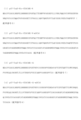
- FIG. 2 is a diagram showing the amino acid sequence of m3C3d).
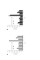
- FIG. 2 is a diagram showing the cross-reactivity of anti-CEACAM1 monoclonal antibodies.
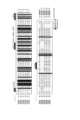
- FIG. 3 is a diagram showing an amino acid sequence alignment of the CEACAM family. Amino acid sequence alignment of N domain and A1 domain including signal sequence of CEACAM family searched by UniProt database.
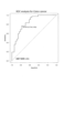
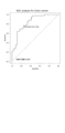
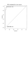
- FIG. 5A is a diagram showing ROC (Receiver Operating Characteristic) analysis of a colorectal cancer specimen.
- FIG. 5B is a diagram showing ROC analysis of liver cancer samples.
- FIG. 5C is a diagram showing ROC analysis of pancreatic cancer samples.
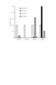
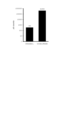
- FIG. 6 is a diagram showing the difference in effects depending on the presence or absence of SDS reduction heat treatment. The results are shown in which CEACAM1 antigen (1 ⁇ g/mL) without denaturation treatment (SDS reduction heat treatment) or under SDS reduction heat treatment conditions was reacted on a co-solid phase plate of A3054 and A3073 and detected with ALP-labeled A7004 antibody.
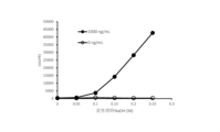
- FIG. 7 is a diagram showing the effect (part 1) of specimen denaturation treatment by alkali treatment (NaOH).
- FIG. 8 is a diagram showing the effect (part 2) of the calibration process using alkali treatment (NaOH).
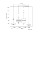
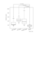
- FIG. 9 is a diagram showing the blood concentration distribution of various cancer samples.
- FIG. 10A is a diagram showing ROC analysis of a colon cancer sample with respect to a healthy human sample.
- FIG. 10B is a diagram showing ROC analysis of a liver cancer sample with respect to a healthy human sample.
- FIG. 10C is a diagram showing ROC analysis of pancreatic cancer samples with respect to healthy human samples.
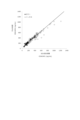
- FIG. 11 is a diagram showing the correlation between SDS reduction heat treatment and alkali treatment.
- FIG. 12 is a diagram showing the effect of denaturation treatment on an autoantibody model specimen.
- the present invention is a method for detecting CEACAM1, comprising: (1) denaturing CEACAM1 in a CEACAM1-containing sample to produce denatured CEACAM1; and (2) detecting CEACAM1 by a sandwich assay using both a capture antibody and a labeled antibody that have the ability to recognize denatured CEACAM1.
- process Provide a method, including.
- CEACAM1 is denatured in the CEACAM1-containing sample, thereby generating denatured CEACAM1.
- denaturation is performed to expose linear epitopes of CEACAM1 that cannot be exposed in the native state (ie, non-denatured state) in order to improve recognition by antibodies.
- the native state ie, non-denatured state
- the linear epitope is covered and cannot be sufficiently exposed.
- Modification can be performed using a denaturing agent, heating, or a combination thereof.
- the modifier include alkaline substances, surfactants, chaotropic modifiers (eg, urea, guanidine), reducing agents, and combinations of two or more thereof.
- alkaline substances include inorganic substances (eg, metal ions) and organic substances.
- inorganic substances eg, metal ions
- organic substances there are many alkaline substances, among which hydroxides of alkali metals (eg, sodium, potassium) or alkaline earth metals (eg, magnesium, calcium) are commonly used. Therefore, such hydroxides can be suitably used.
- the concentration of the alkaline substance is not particularly limited as long as it can denature CEACAM1, and is, for example, 0.1M or higher, preferably 0.2M or higher, more preferably 0.3M or higher, even more preferably 0.35M or higher. It's okay.
- the concentration of the alkaline substance may also be a concentration of 5M or less, 2M or less, or 1M or less.
- the alkaline substance may be used together with a surfactant.
- a surfactant nonionic surfactants, anionic surfactants, cationic surfactants, and amphoteric surfactants are preferred. Particularly preferred are nonionic surfactants.
- nonionic surfactants include ether type surfactants such as polyoxyethylene (23) lauryl ether (Brij 35) and polyethylene glycol hexadecyl ether (Brij 58), polyoxyethylene sorbitan monolaurate (Tween 20), and polyoxyethylene sorbitan monolaurate (Tween 20).
- Polyoxyethylene sorbitan fatty acid esters such as ethylene sorbitan monopalmitate (Tween 40), polyoxyethylene sorbitan monooleate (Tween 80) (e.g. Tween (trade name/registered trademark) series), polyoxy esters such as polyoxyethylene octylphenyl ether, etc.
- Secondary alcohol polyoxyethylene ethers such as ethylene alkylphenyl ethers (e.g., Triton (trade name/registered trademark) series), Tergitol (registered trademark) 15-S-7 (e.g., TERGITOL (trade name/registered trademark) series) , poly(ethylene glycol)-block-poly(propylene glycol)-block-poly(ethylene glycol) (Pluronic F108), copolymers of ethylene oxide and propylene oxide (for example, Pluronic (trade name/registered trademark) series), etc. Can be mentioned.
- the concentration of the surfactant is not particularly limited and can be set as appropriate.
- a denaturation treatment solution containing an alkaline substance may further include a step of adding a neutralization solution containing an acidifying agent or a buffer solution having buffering capacity to neutralize or adjust the pH after the denaturation treatment.
- acidifying agents include hydrochloric acid, sulfuric acid, and acetic acid.
- the step of adding a neutralizing solution is not included, for example, in the reaction step below, by adjusting the buffer capacity and pH of the buffer solution to be mixed, the alkaline substances in the denaturation treatment solution are neutralized and the reaction is performed. The influence on the process may be alleviated.
- the pH in the reaction process can be set appropriately depending on the components included in the reaction process, but for example, the pH in the reaction process may be adjusted using an acidifying agent or a buffer solution contained in the reaction process solution to adjust the pH to 5.5 to 9.5. The amount added can be set appropriately.
- the surfactant examples include anionic surfactants, cationic surfactants, amphoteric surfactants, and nonionic surfactants.
- the surfactant one or more types of surfactants can be used.
- an anionic surfactant may be used alone, or a combination of an anionic surfactant and another surfactant may be used.
- anionic surfactants include sulfate ester type surfactants (e.g., sodium dodecyl sulfate (SDS)), carboxylic acid type surfactants (e.g., sodium N-decanoylsarcosinate (NDS), N-lauroyl sodium sarcosine hydrate (NLS)), sulfonic acid type surfactants (e.g., sodium 1-nonanesulfonate (NSS), sodium dodecylbenzenesulfonate (SDBS)), and carboxylic acid type - sulfonic acid type surfactants (eg, chondroitin sodium sulfate (CSSS)).
- SDS sodium dodecyl sulfate
- carboxylic acid type surfactants e.g., sodium N-decanoylsarcosinate (NDS), N-lauroyl sodium sarcosine hydrate (NLS)
- amphoteric surfactants examples include C10APS (N-decyl-N,N-dimethyl-3-ammonio-1-propanesulfonate) and C12APS (N-dodecyl-N,N-dimethyl-3-ammonio-1). -propanesulfonate), C14APS (N-tetradecyl-N,N-dimethyl-3-ammonio-1-propanesulfonate), C16APS (N-hexadecyl-N,N-dimethyl-3-ammonio-1-propanesulfonate), etc. Can be mentioned.
- Examples of the cationic surfactant include decyltrimethylammonium chloride, dodecyltrimethylammonium chloride, tetradecyltrimethylammonium chloride, hexadecyltrimethylammonium chloride (C16TAC), decyltrimethylammonium bromide, dodecyltrimethylammonium bromide, and tetradecyltrimethylammonium bromide.
- C16TAC hexadecyltrimethylammonium chloride
- C16TAC hexadecyltrimethylammonium chloride
- decyltrimethylammonium bromide dodecyltrimethylammonium bromide
- tetradecyltrimethylammonium bromide tetradecyltrimethylammonium bromide.
- Bromide, hexadecyltrimethylammonium bromide (CTAB) laurylpyridinium chloride,
- the concentration of the surfactant is not particularly limited as long as it can modify CEACAM1, for example, 0.5% by weight or more, preferably 1% by weight or more, more preferably 2% by weight or more, even more preferably 5% by weight or more. It may also be a concentration. The concentration of surfactant may also be 20% by weight or less, or 15% by weight or less.
- a reagent that can cleave the disulfide bonds present in CEACAM1 can be used.
- reducing agents include tricarboxylethylphosphine (TCEP), cysteine, dithiothreitol, reduced glutathione, and ⁇ -mercaptoethanol.
- proteases include trypsin, chymotrypsin, Lys-C, Asp-N, Glu-C, Arg-C, asparaginyl endopeptidase, arginyl endopeptidase, and V8 protease.
- the concentration of the reducing agent is not particularly limited as long as it can cleave the disulfide bonds present in CEACAM1, for example, at a concentration of 0.5 mM or higher, preferably 1 mM or higher, more preferably 2 mM or higher, and even more preferably 5 mM or higher. There may be.
- the concentration of the reducing agent may also be less than or equal to 100mM, or less than or equal to 50mM.
- the concentration values of the above-mentioned denaturing agents are values based on the weight or volume of the denaturing solution containing the denaturing agent.
- the modification treatment liquid contains a solvent or dispersion medium (eg, water), and additives as necessary.
- additives include chelating agents such as EDTA.
- Heating may be, for example, 35°C or higher, 40°C or higher, 45°C or higher, 50°C or higher, 55°C or higher, 60°C or higher, 65°C or higher, 70°C or higher, 75°C or higher, 80°C or higher, 85°C or higher, 90°C or higher. °C or higher, 95°C or higher, or about 100°C (ie, boiling). Heating may also be below 100°C.
- modification may be performed in combination with a surfactant and a reducing agent. Modification may also be performed using a combination of a surfactant, a reducing agent, and heat (eg, SDS reduction heat treatment).
- the treatment time for denaturing CEACAM1 depends on factors such as the type and degree of denaturation treatment (e.g., concentration, heating temperature), etc.
- the time may be 1 second or more, 5 seconds or more, 10 seconds or more, preferably 30 seconds or more, more preferably 1 minute or more, even more preferably 2 minutes or more, 3 minutes or more, or 5 minutes or more. Good too.
- Such time may also be less than 1 hour, less than 30 minutes, or less than 20 minutes.
- the CEACAM1-containing specimen is any specimen containing CEACAM1.
- the CEACAM1-containing sample is a biological fluid sample.
- the CEACAM1-containing specimen may be subjected to other treatments before being used in the method of the present invention. Such treatments include, for example, centrifugation, extraction, filtration, precipitation, heating, freezing, refrigeration, and stirring.
- the CEACAM1-containing sample may be an animal-derived liquid sample.
- animals from which the liquid specimen is derived include mammals (e.g., primates such as humans and monkeys; rodents such as mice, rats, and rabbits; ungulates such as cows, pigs, goats, horses, and sheep; Meat animals such as dogs and cats; and animals such as birds (eg, chickens) are exemplified.
- the animal is a mammal, preferably a human.
- the animal-derived fluid specimen may be a body fluid specimen derived from an animal as described above.
- body fluid samples include blood samples (e.g., whole blood, serum, and plasma), urine, saliva, lymph fluid, tissue fluid, cerebrospinal fluid, ascites, sweat, semen, tears, mucus, milk, pleural fluid, and bronchopulmonary fluid.
- blood samples e.g., whole blood, serum, and plasma
- urine saliva, lymph fluid, tissue fluid, cerebrospinal fluid, ascites, sweat, semen, tears, mucus, milk, pleural fluid, and bronchopulmonary fluid.
- cyst lavage fluid and amniotic fluid.
- the body fluid is a blood sample, urine, or saliva.
- the CEACAM1-containing sample may be a culture supernatant sample.
- the culture supernatant specimen may be a cell culture supernatant specimen or a tissue culture supernatant specimen. Examples and preferred examples of animals from which cells or tissues to be cultured are derived are as described above.
- CEACAM1 can be specified by the type of animal from which it is derived. Examples and preferred examples of animals from which CEACAM1 is derived are the same as those mentioned above. Therefore, CEACAM1 is mammalian CEACAM1, preferably human CEACAM1. CEACAM1 also exists in 12 isoforms, three of which are secreted (CEACAM1-4C1, CEACAM1-3, CEACAM1-3C2). Therefore, when a body fluid specimen is used, CEACAM1 may include such secreted CEACAM1.
- the sample containing denatured CEACAM1 may be diluted with a solution (eg, buffer).
- a solution eg, buffer
- the concentration of the denaturant can be diluted, and as a result, the sandwich assay in step (2) above can be performed successfully. can.
- CEACAM1 is detected by sandwich assay.
- the sandwich assay uses both a capture antibody and a labeled antibody that have the ability to recognize denatured CEACAM1.
- Sandwich assays can be performed in any manner using such antibodies.
- Such formats include, for example, chemiluminescent immunoassays (CLIA) [e.g., chemiluminescent enzyme immunoassays (CLEIA)], enzyme immunoassays (EIA), radioimmunoassays (RIA), fluorescent immunoassays (FIA), and Examples include immunochromatography methods.
- the capture antibody may be immobilized on a solid phase.
- the antibody against CEACAM1 may be an antibody that can be immobilized on a solid phase in the reaction step.
- an antibody immobilized on a solid phase and an antibody that can be immobilized on a solid phase in a reaction step are sometimes simply referred to as immobilized antibodies.
- the capture antibody is an antibody for capturing the target antigen (denatured CEACAM1 in the case of the present invention) on a solid phase.
- the solid phase include particles (eg, Sepharose beads, agarose beads, magnetic particles), supports (eg, membranes), and containers (eg, plates such as plastic plates, tubes, microchannels).
- a previously known method can be used to immobilize the antibody on the solid phase.
- the capture antibody may be an antibody in which one type of capture antibody is immobilized on one type of solid phase (e.g., particles such as magnetic particles), or an antibody in which two or more types of capture antibodies are immobilized on one type of solid phase (e.g., particles such as magnetic particles).
- the antibody may be an antibody immobilized on a solid phase (e.g., a particle such as a magnetic particle), or an antibody in which two or more types of capture antibodies are each immobilized on two or more types of solid phase (e.g., a particle such as a magnetic particle). Good too.
- a solid phase e.g., a particle such as a magnetic particle
- two or more types of capture antibodies are each immobilized on two or more types of solid phase
- a labeled antibody is an antibody labeled with a labeling substance.
- labeling substances include enzymes (e.g., peroxidase, alkaline phosphatase, luciferase, ⁇ -galactosidase), affinity substances (e.g., streptavidin, biotin), fluorescent substances or fluorescent proteins (e.g., fluorescein, fluorescein isothiocyanate, rhodamine, green fluorescent protein, red fluorescent protein), luminescent or light-absorbing substances (eg, luciferin, acridinium, ruthenium), and radioactive substances (eg, 3 H, 14 C, 32 P, 35 S, 125 I). Labeling of an antibody with a labeling substance can be performed covalently or non-covalently.
- an antibody is a protein that includes a portion (eg, a variable region or a portion corresponding thereto) that has the ability to bind to a target antigen. Therefore, antibodies in the capture antibody and labeled antibody include, for example, full-length antibodies (e.g., IgG, IgM, IgA, IgD, IgE, IgY) and antigen-binding fragments of antibodies (e.g., F(ab') 2 , Fab ', Fab, Fv), single chain antibodies, and other modified antibodies. Antibodies may also be polyclonal or monoclonal antibodies.
- antibodies capable of recognizing denatured CEACAM1 can be used as capture antibodies and labeled antibodies.
- the antibody that has the ability to recognize denatured CEACAM1 may be an antibody that has the ability to specifically recognize denatured CEACAM1, or an antibody that has the ability to non-specifically recognize denatured CEACAM1.
- Examples of antibodies that have the ability to nonspecifically recognize denatured CEACAM1 include (i) antibodies that have the ability to recognize not only denatured CEACAM1 but also native (i.e., non-denatured) CEACAM1; (ii) antibodies that have the ability to recognize only denatured CEACAM1; (iii) antibodies that have the ability to recognize other CEACAM1 (e.g., CEACAM3); and (iii) antibodies that have the ability to recognize not only denatured CEACAM1 but also native (i.e., non-denatured) CEACAM1 and other CEACAM1 (e.g., CEACAM3).
- an antibody capable of recognizing the epitope represented by the amino acid sequence of PANSGRETIY (SEQ ID NO: 1) in CEACAM1 (b) TESMP (sequence An antibody capable of recognizing the epitope represented by the amino acid sequence No. 2), (c) an antibody capable of recognizing the epitope represented by the amino acid sequence DTTYLWWINN (SEQ ID No. 3) in CEACAM1, or (d) EATGQFHVYP (SEQ ID NO: 4) in CEACAM1 may be used.
- both of these epitopes are particularly exposed upon denaturation as described above, and thus are epitopes that react better with denatured CEACAM1 than with non-denatured CEACAM1.
- the antibody (b) above was found to be effective against not only CEACAM1 but also CEACAM1. It is not a CEACAM1-specific antibody because it also cross-reacts with AM3, CEACAM5, and/or CEACAM6 (see Examples and FIG. 3).
- the present inventors found that the detection sensitivity of CEACAM1 was improved by performing a sandwich assay using the antibody (b) above as an auxiliary. Furthermore, it has been confirmed that detection of CEACAM3, CEACAM5, and/or CEACAM6 can be avoided by using other antibodies capable of recognizing CEACAM1 in addition to the antibody in (b) above (see Examples). ).
- one of the capture antibody or the labeled antibody is the antibody of (a) above, or a combination of the antibody of (a) above and the antibody of (b) above, and the other is the antibody of (c) above.
- the antibody of (d) above, or a combination of the antibody of (c) above and the antibody of (d) above may be used.
- CEACAM1 can be detected specifically and with high sensitivity.
- the capture antibody the antibody of the above (a) or a combination of the antibody of the above (a) and the antibody of the above (b) is used, and as the labeled antibody, the antibody of the above (c) or the above (d) is used. or a combination of the antibody of (c) above and the antibody of (d) above may be used.
- the weight ratio of (a):(b) is, for example, 1:9 to 9:1, preferably 2:8 to 8:2, or more.
- the ratio may be 3:7 to 7:3, more preferably 4:6 to 6:4, and most preferably 4.5:5.5 to 5.5:4.5.
- An antibody capable of recognizing the epitope as described above can be produced by using CEACAM1 or a partial protein thereof as an antigen.
- the hybridomas producing antibodies (a), (b), (c), and (d) above are 2/500 (0.4%), 4/500 (0.8%), and 1 It has been successfully produced with a probability of /800 (0.1%) and 1/120 (0.8%).
- antibodies capable of recognizing the epitope can be efficiently obtained by selecting from an antibody library using the epitope.
- the present invention also provides a method for detecting cancer, comprising: (1) a step of measuring the amount of CEACAM1 in a specimen collected from a subject by the above-described method of detecting CEACAM1; and (2) a step of comparing the measured amount of CEACAM1 with a reference value.
- a method including.
- the step (1) above in the method for detecting cancer can be performed in the same manner as the method for detecting CEACAM1 described above.
- the subject from which the specimen is collected is the same as the above-mentioned animals, preferably a mammal, and more preferably a human.
- the specimen is the same as the animal-derived liquid specimen (eg, body fluid specimen) described above.
- the measured amount of CEACAM1 is compared with a reference value. If the measured amount of CEACAM1 is higher than the reference value, this can be used as an indicator that the subject may be suffering from cancer.
- CEACAM1 such as secretory CEACAM1 can be used as a marker for various cancers such as melanoma, pancreatic cancer, urothelial bladder cancer, lung cancer, gastric cancer, colorectal cancer, pancreatic cancer, prostate cancer, bladder cancer, and melanoma. It has been known. Therefore, the cancer detection method of the present invention can be used to detect various cancers such as these (for example, colon cancer, liver cancer, and pancreatic cancer as shown in the Examples, as well as combinations of two or more of these cancers). (see ).
- the present invention further provides a method for treating cancer, comprising: (1) Measuring the amount of CEACAM1 in a specimen collected from a subject by the method for detecting CEACAM1 described above; (2) a step of comparing the measured amount of CEACAM1 with a reference value; A method is provided that includes (3) selecting a subject in which an amount of CEACAM1 is measured higher than a reference value, and (4) administering an anticancer drug to the selected subject.
- the steps (1) and (2) above in the method for treating cancer can be performed in the same manner as the steps (1) and (2) above in the method for detecting cancer.
- the step (3) above in the cancer treatment method can be performed by selecting a subject in which a CEACAM1 amount higher than the standard value was measured based on the comparison result of (2) above.
- an anticancer drug is administered to the selected subject.
- the anticancer agent to be administered can be appropriately determined depending on the type of cancer.
- other treatment methods such as radiation therapy may be used in combination.
- the present invention further provides a CEACAM1 detection kit, comprising: (1) Modifier, (2) a capture antibody that has the ability to recognize denatured CEACAM1, and (3) a labeled antibody that has the ability to recognize denatured CEACAM1, Provide a kit including:
- the denaturing agent, the capture antibody that has the ability to recognize denatured CEACAM1, and the labeled antibody that has the ability to recognize denatured CEACAM1 are the same as those described above.
- a capture antibody capable of recognizing denatured CEACAM1 and a labeled antibody capable of recognizing denatured CEACAM1 may be provided in the form of a reagent for a sandwich assay using these antibodies.
- the kit of the present invention is useful, for example, for carrying out the method of the present invention simply and quickly.
- Example 1 Expression and purification of immunization antigen and screening antigen
- A Construction of antigen expression plasmids TrpE-His-CEACAM1-N-m3C3d and TrpE-His-CEACAM1-A1-m3C3d as immunization antigens and screening antigens
- Antigen expression plasmids were constructed to prepare TrpE-His-CEACAM1-N and TrpE-His-CEACAM1-A1.
- PCR was performed using (SEQ ID NO: 23). PCR was performed using a kit from KAPA Taq EXtra (Nippon Genetics), followed by DNA denaturation at 95°C for 15 seconds and annealing at 55°C. DNA synthesis was carried out for 25 cycles at 72°C for 45 seconds, and the obtained DNA fragments were separated by 1.2% agarose gel electrophoresis, using Freeze 'N Squeeze TM DNA Gel Extraction Spin Columns (BIO-RAD). ).
- TrpE-His-CEACAM1-N or TrpE-His-CEACAM1 -10 units of BsiWI and 10 units of SalI for the construction of A1, 10 units of BsiWI and 10 units of XhoI for the construction of pAT-TrpE-His-CEACAM1-N-m3C3d or pAT-TrpE-His-CEACAM1-A1-m3C3d.
- DNA of the expression vector pAT-trpE-His or pAT-trpE-His-m3C3d was added to 20 ⁇ l of restriction enzyme reaction solution [100 mM NaCl, 50 mM Tris-HCl, 10 mM MgCl 2 , 1 mM DTT, pH 7.9].
- the susceptible E. coli strain used for transformation was Mix & Go E. It is made by E.coli Transformation Kit & Buffer Set.
- the transformed E. coli was spread on an LB plate (1% tryptone, 0.5% NaCl, 1.5% agar) containing 100 ⁇ g/ml ampicillin and kept at 37° C. overnight. Colonies of bacteria generated on the plate were picked with a sterile toothpick, transferred to 2YT medium containing 100 ⁇ g/ml ampicillin, and cultured overnight at 37°C.
- TrpE-His-CEACAM1-N or A1 Bacteria were collected by centrifuging 1.5 ml of the bacterial culture, and minipreparation of plasmid DNA was performed using Wizard(R) Plus SV Minipreps DNA Purification Systems (Promega).
- TrpE-His-CEACAM1-N or A1 add 1 ⁇ g of the obtained plasmid DNA to 20 ⁇ l of restriction enzyme reaction solution [100 mM NaCl, 50 mM Tris-HCl, 10 mM MgCl 2 , 1 mM DTT, pH 7.9] and add 10 units of BsiWI and
- digestion was performed for 1 hour at 37°C in a solution containing 10 units of BsiWI and 10 units of XhoI, followed by 1.2% agarose.
- the bacterial cells were collected by centrifugation, resuspended in 50 ml of Lysis solution [50 mM Tris-HCl (pH 8.5), 30 mM NaCl, 5 mM EDTA], and 1 ml of lysozyme solution (10 mg/ml Lysozyme) was added. It was treated for 1 hour at 37°C. The cells of this suspension were disrupted by ultrasonication (150 W, 90 seconds twice). The insoluble fraction was collected by centrifugation at 15,000 rpm for 30 minutes at 4°C.
- Lysis solution 50 mM Tris-HCl (pH 8.5), 30 mM NaCl, 5 mM EDTA]
- lysozyme solution 10 mg/ml Lysozyme
- the insoluble fraction was resuspended in 50 ml of A solution containing 1% NP40 [50 mM Tris-HCl (pH 8.5)]. The suspension was centrifuged at 15,000 rpm for 30 minutes at 4°C to collect the insoluble fraction. The insoluble fraction was resuspended in 50 ml of A solution containing 2M urea. The suspension was centrifuged at 15,000 rpm at 4°C for 30 minutes to collect the insoluble fraction. Transfer the insoluble fraction to 50 ml of 6 M Resuspend in solution A containing urea. The suspension was centrifuged at 15,000 rpm for 30 minutes at 4°C to collect the soluble fraction.
- the antigen protein TrpE- His-CEACAM1-N, TrpE-His-CEACAM1-N-m3C3d, TrpE-His-CEACAM1-A1, and TrpE-His-CEACAM1-A1-m3C3d were purified.
- Example 2 Obtaining anti-CEACAM1 monoclonal antibody (A) Immunization
- the antigen protein TrpE-His-CEACAM1-N-m3C3d or TrpE-His-CEACAM1-A1-m3C3d prepared by the above method was dissolved in 6M urea and then dissolved in 0.15M NaCl. diluted to a final concentration of 1.0 mg/ml in 10 mM phosphate buffer (pH 7.3) (PBS) containing 4- to 6-week-old BALB/c mice. 50-100 ⁇ g was administered subcutaneously. A similar booster immunization was performed every two weeks, and 100 ⁇ g of antigen protein dissolved in PBS was intraperitoneally administered as a final immunization.
- PBS phosphate buffer
- Myeloma cells (mouse myeloma cell line Sp2/0 Ag14) in the logarithmic growth phase were collected from the culture flask, precipitated by centrifugation (1000 rpm, 5 min, room temperature), and suspended in RPMI 1640 medium (no serum added). Myeloma cells and spleen cells were mixed at a cell number ratio of 1:1 and dispensed so that the total number of cells was 3.4 x 10 7 . After centrifuging this cell mixture (1000 rpm, 5 min, room temperature), the supernatant was removed and washed twice with ECF buffer (0.3 M mannitol, 0.1 mM calcium chloride, 0.1 mM magnesium chloride solution).
- ECF buffer 0.3 M mannitol, 0.1 mM calcium chloride, 0.1 mM magnesium chloride solution.
- the mixed cell solution suspended in 0.35 ml of ECF buffer was mixed using a cell fusion device ECFG21 (Neppa Gene) and an MS stand type chamber platinum electrode CUY497P2 (0.8 ml) at a liquid volume of 0.35 ml and an AC voltage.
- the cells were suspended in RPMI1640 medium containing serum and allowed to stand for 10 minutes to recover the cell membrane.
- the cells were suspended in RPMI-1640 medium containing 10% fetal bovine serum and hypoxanthine, aminopterin, and thymidine (HAT), and seeded in a 96-well cell culture plate. After culturing for about 10 days to allow only hybridomas to proliferate, the culture supernatant was collected and used for the screening described below.
- reaction solution (0.1% casein, 1 mM EDTA, PBS) was added to each well, and the mixture was allowed to stand at room temperature for 1 hour. After washing the wells with PBS containing 0.05% Tween 20, 50 ⁇ L of horseradish peroxidase (HRP)-labeled anti-mouse IgG Fc-specific antibody was added and left at room temperature for 1 hour. After washing the wells with PBS containing 0.05% Tween 20, 50 ⁇ L of TMB solution was added to each well to induce color development. Thereafter, 2M H 2 SO 4 was added to each well to stop the reaction, and the absorbance was measured at a wavelength of 450 nm.
- HRP horseradish peroxidase
- Hybridomas obtained from the spleen of mice immunized with the antigen protein TrpE-His-CEACAM1-N-m3C3d were immunized with A3029, A3048, A3054, A3057, A3067, A3073, A6056, and the antigen protein TrpE-His-CEACAM1-A1-m3C3d.
- the hybridoma obtained from mouse spleen was named A7004.
- Hybridomas A3054 and A3067 which produce antibodies with the same epitope, are two clones obtained from 500 positive hybridoma wells, and which produce antibodies with the same epitope.
- Hybridomas A3029, A3048, A3057, and A3073 were obtained from 4 clones obtained from 500 hybridoma-positive wells, A7004 was obtained from 1 clone obtained from 800 hybridoma-positive wells, and A6056 was obtained from 120 hybridoma-positive wells. 1 clone.
- Example 3 Epitope analysis of anti-CEACAM1 monoclonal antibody
- deletion proteins in which 10 or 20 amino acids were deleted from the C-terminus of the N domain protein or A1 domain protein of CEACAM1.
- N-domain protein a deleted protein in which three amino acids were deleted from the N-terminus was expressed in E. coli, and analyzed by dot blotting using a disrupted E. coli solution.
- the deleted protein expression plasmid was constructed using the KOD-Plus-Mutagenesis Kit (TOYOBO). As a template, pAT-GST-His-CEACAM1-N (Nfull) was used to construct the N domain deleted protein expression plasmid, and pAT-TrpE-His-CEACAM1-N- was used to construct the A1 domain deleted protein expression plasmid. A1 (A1full) was used. The primers used were set outside each region to be deleted. Table 1 shows the amino acid sequences corresponding to the various deleted proteins.
- Example 4 Cross-reactivity evaluation using recombinant antigens Obtained using commercially available recombinant antigens CEACAM1 (R&D Systems), CEACAM3 (R&D Systems), CEACAM5 (R&D Systems), and CEACAM6 (R&D Systems) The cross-reactivity of anti-CEACAM1 monoclonal antibodies (one type was selected for antibodies with the same epitope) was evaluated.
- the blocking solution was removed, and 50 ⁇ L of each antibody diluted with a reaction solution (0.1% sodium caseinate, 150 mM NaCl, 1 mM EDTA, PBS) was added to each well and left at room temperature for 1 hour. After washing the wells with PBS containing 0.05% Tween 20, 50 ⁇ L of horseradish peroxidase (HRP)-labeled anti-mouse IgG Fc-specific antibody was added and left at room temperature for 1 hour. After washing the wells with PBS containing 0.05% Tween 20, 50 ⁇ L of TMB solution was added to each well to induce color development. Thereafter, 2M H 2 SO 4 was added to each well to stop the reaction, and the absorbance was measured at a wavelength of 450 nm.
- HRP horseradish peroxidase
- Example 5 Sequence comparison with the CEACAM family Comparing the epitope of each antibody and the amino acid sequence alignment of the CEACAM family (Fig. 3), antibodies A3054 and A3073 have amino acids unique to CEACAM1 (threonine in TESMP (SEQ ID NO: 2), respectively). It can be seen that the sequence containing the residue (T) and the asparagine residue (N) in PANSGRETIY (SEQ ID NO: 1) is recognized (indicated by a black frame in FIG. 3). Based on the cross-reactivity evaluation results, these two antibodies have low cross-reactivity with CEACAM5 and CEACAM6, and therefore these CEACAM1-specific amino acids are considered to be epitope hot spots.
- the A6056 antibody showed moderate cross-reactivity with CEACAM6 and no cross-reactivity with CEACAM5 in the cross-reactivity evaluation results, indicating that the histidine residue (H) in EATGQFHVYP (SEQ ID NO: 4) is an epitope hotspot. It is thought that there is (indicated by a black frame in Figure 3). This sequence is also present in CEACAM3 and CEACAM4, so the A6056 antibody may cross-react with them.
- Example 6 Evaluation of cross-reactivity using peptides
- Cross-reactivity evaluation using synthetic peptides was performed for antibodies A3054 and A3067 and antibodies A3029, A3048, A3067, and A3073, which are thought to recognize CEACAM1-specific sequences.
- the blocking solution was removed, and 50 ⁇ L of each antibody diluted with a reaction solution (0.1% sodium caseinate, 150 mM NaCl, 1 mM EDTA, PBS) was added to each well and left at room temperature for 1 hour. After washing the wells with PBS containing 0.05% Tween 20, 50 ⁇ L of horseradish peroxidase (HRP)-labeled anti-mouse IgG Fc-specific antibody was added and left at room temperature for 1 hour. After washing the wells with PBS containing 0.05% Tween 20, 50 ⁇ L of TMB solution was added to each well to induce color development. Thereafter, 2M H 2 SO 4 was added to each well to stop the reaction, and the absorbance was measured at a wavelength of 450 nm.
- HRP horseradish peroxidase
- Example 7 Construction of CEACAM1 sandwich measurement system
- A Preparation of biotin-labeled antibody
- the buffer of the antibody solution was replaced with PBS using a desalting column, and the antibody concentration was adjusted to 0.5 mg/mL.
- Sulfo-NHS-LC-Biotin (Thermo Scientific) was added to this antibody solution at a concentration of about 70 ⁇ M, and reacted at room temperature for 90 minutes.
- 1/20 volume of 1M Tris (pH 7) was added thereto to stop the reaction, resulting in a biotin-labeled product.
- the labeled body diluent 50mM MES, 100mM NaCl, 0.3mM ZnCl 2 , 1mM MgCl 2 , 2% BSA, 0.10% NaN 3 , pH 6.8 .
- a biotin-labeled antibody diluted to 1 ⁇ g/mL was added to the well and left at room temperature for 1 hour.
- alkaline phosphatase (ALP)-labeled streptavidin diluted with a label diluent was added to the wells and left at room temperature for 30 minutes.
- Lumipulse substrate solution Flujirebio
- Table 4 shows the results of the combination study of solid-phase antibodies and labeled antibodies for sandwich ELISA.
- the ⁇ 0 count value is the difference between the measured value of CEACAM1 antigen 1 ⁇ g/mL minus the measured value of 0 ⁇ g/mL. High reactivity was shown when A3073 antibody, which has the highest CEACAM1 specificity, was used as the solid phase and A7004 antibody or A6056 antibody was used as the label.
- (C) Solid-phase antibody combination The effect of combining A3054 antibody (or A3067 antibody) and A3073 antibody (or A3029, A3048, A3057) as solid-phase antibodies was investigated. The measurement method followed the above-mentioned "(B) Sandwich ELISA antibody combination".
- the solid phase conditions were as follows: using PBS, the single solid phase was diluted to 5 or 10 ⁇ g/mL, and the co-solid phase was mixed to 5 ⁇ g/mL.
- a commercially available CEACAM1 antigen R&D Systems
- Table 5 shows the results of solid phase antibody combinations.
- the ⁇ 0 count value is the difference obtained by subtracting the measured value of 0 ⁇ g/mL from each measured value of 1 ⁇ g/mL of CEACAM1 antigen.
- the co-solid phase of A3054 antibody (or A3067 antibody) and A3073 antibody (or A3029, A3048, A3057) showed higher reactivity than the single solid phase. Since this value is larger than the sum of the individual solid phases, it is considered that a synergistic effect appeared on the CEACAM1 binding strength.
- CEACAM1 antigen R&D Systems
- CEACAM3 antigen R&D Systems
- CEACAM5 antigen R&D Systems
- CEACAM6 antigen R&D Systems
- Table 6 shows the results of cross-reactivity evaluation using a sandwich measurement system.
- the ⁇ 0 count value is the difference between the measured value of CEACAM1 antigen 1 ⁇ g/mL minus the measured value of 0 ⁇ g/mL.
- No significant cross-reactivity was shown for CEACAM3, CEACAM5 and CEACAM6 in any of the solid phase antibody combinations. It has been shown that the cross-reactive A3054 antibody (or A3067 antibody) does not affect the CEACAM1 specificity of the A3073 antibody (or A3029, A3048, A3057).
- Example 8 Evaluation of CEACAM1 assay system using fully automated chemiluminescent enzyme immunoassay system
- A Preparation of anti-CEACAM1 antibody-bound ferrite particles
- Anti-CEACAM1 antibody (A3054) was attached to magnetic particles in 10 mM MES buffer (pH 5.0). and A3073) to obtain a suspension containing 0.04 mg/mL anti-CEACAM1 antibodies (A3054 and A3073) and 5 mg/mL magnetic particles. This suspension was incubated at 25° C. for 1 hour with gentle stirring to immobilize the anti-CEACAM1 antibodies (A3054 and A3073) on the magnetic particles.
- the magnetic particles were collected with a magnet, washed with a washing solution (50mM Tris buffer, 150mM NaCl, 2.0% BSA, pH 7.2), and immobilized with anti-CEACAM1 antibodies (A3054 and A3073). Particles were obtained.
- the reacted antibody and ALP mixture was concentrated and concentrated using ALP buffer (1mM MgCl 2 , 0.1mM ZnCl 2 , 0.1% NaN 3 , 0.15M NaCl, 0.1M MES, pH 6.8).
- the product was purified by gel filtration to obtain an ALP-labeled anti-CEACAM1 antibody.
- CEACAM1 measurement method CEACAM1 was measured using a fully automated chemiluminescent enzyme immunoassay system according to the following method. 50 ⁇ L of the sample and 50 ⁇ L of the denaturation treatment solution (10% SDS, 3.75% EDTA2Na, 10mM TCEP) were mixed, shaken at 1000 rpm, and heated at 80° C. for 10 minutes to prepare a denaturation treatment sample.
- the denaturation treatment solution (10% SDS, 3.75% EDTA2Na, 10mM TCEP
- the magnetic particles were collected and washed to remove components that were not bound to the magnetic particles, and then mixed with a labeled body dilution solution (50mM MES, 1 Add 50 ⁇ L of ALP-labeled A7004 antibody diluted to 0.2 ⁇ g/mL with 00 mM NaCl, 0.3 mM ZnCl 2 , 1 mM MgCl 2 , 2% BSA, 0.10% NaN3, pH 6.8) and incubate for 8 minutes. Made it react.
- the magnetic particles were collected and washed to remove components not bound to the magnetic particles, and 200 ⁇ L of a substrate solution containing AMPDD (Lumipulse substrate solution, manufactured by Fujirebio) was added.
- AMPDD Lipulse substrate solution, manufactured by Fujirebio
- the amount of light emitted by the enzyme reaction was counted, and the CEACAM1 value in the sample was calculated from the count using a calibration curve.
- the calibration curve is based on CEACAM1 (R&D Systems, Inc.) standard solutions were measured in the same manner as the specimen, and created based on the amount of luminescence obtained for each standard solution. All steps after the sample denaturation treatment in this example were performed using an automatic analyzer Lumipulse L2400 (registered trademark, manufactured by Fujirebio Co., Ltd.).
- the average concentrations of CEACAM1 in colorectal cancer samples, liver cancer samples, and pancreatic cancer samples were 216.1 ⁇ 108.4 ng/mL, 399.9 ⁇ 119.3 ng/mL, and 430.9 ⁇ 259.4 ng/mL, respectively. Yes, the value was significantly higher than that of healthy human samples.
- the area under the curve was 0.836 and the cutoff value was 154.4 ng/mL
- the specificity was 0.778 and the sensitivity was 0.762 for colorectal cancer samples.
- the area under the curve was 0.996, the specificity was 0.994, and the sensitivity was 1.0 when the cutoff value was 201.6 ng/mL.
- pancreatic cancer samples the area under the curve was 0.969, the specificity was 0.917, and the sensitivity was 0.925 when the cutoff value was 181.9 ng/mL.
- CEA-absorbed specimen [absorption (+)] was prepared by collecting the magnetic particles and collecting the supernatant. Further, 50 ⁇ L of the particle dilution solution containing no particles and 50 ⁇ L of the sample were mixed, and the mixture was shaken at 1000 rpm and reacted at 37° C. for 30 minutes to prepare an untreated sample [absorption (-)].
- CEACAM1 measurement and CEA measurement were performed using these specimens.
- CEACAM1 measurement follows the CEACAM1 measurement method using the above-mentioned fully automated chemiluminescent enzyme immunoassay system, and CEA measurement follows the package insert of Lumipulse Presto CEA measurement reagent (manufactured by Fujirebio) using the automatic analyzer Lumipulse L2400 (manufactured by Fujirebio). ) was used.
- Table 7 shows the evaluation results of CEACAM1 specificity by CEA absorption.
- the CEA measurement value exceeded 100 ng/mL
- the absorption (+) was a condition in which approximately 90% of CEA could be absorbed. This showed that the constructed CEACAM1 measurement system specifically detected CEACAM1 without being affected by CEA.
- Example 9 Examination of epitope exposure method of anti-CEACAM1 antibody Since the anti-CEACAM1 antibody obtained by the present invention recognizes a linear epitope, an antigen denaturation treatment step is essential to enhance the antigen-antibody reaction.
- SDS reduction heat treatment was used as the antigen denaturation treatment method. As shown in FIG. 6, by subjecting the antigen to SDS reduction heat treatment, the epitope is exposed and the CEACAM1-specific signal increases approximately 1000 times.
- modification treatment with alkali was investigated.
- A3054 and A3073 were each diluted with PBS to a concentration of 5 ⁇ g/mL. 50 ⁇ L of the diluted antibody was added per well to a Nunc multimodule plate, and the plate was left standing at 4-8°C overnight. After removing the antibody solution in the wells and washing with PBS, add 100 ⁇ L of blocking solution (0.1% sodium caseinate, 150 mM NaCl, 1 mM EDTA, PBS) per well and leave at room temperature for 1 hour to remove the antibody solid phase plate. Prepared.
- CEACAM1 antigen R&D Systems
- Lumipulse sample diluent Flujirebio
- 50 ⁇ L of the sample was mixed with 50 ⁇ L of denaturation treatment solution (0-0.5M NaOH).
- a denatured sample was prepared by incubating at 37°C for 7 minutes.
- 50 ⁇ L of the denatured sample with 50 ⁇ L of reaction solution (1.5 M Tris-HCl, 100 mM NaCl, 10 mM EDTA 3Na, 0.1% ProClin 300, 5% BSA, pH 7.5), and add 50 ⁇ L of it to the above blocking solution.
- the solution was added to the wells of the antibody solid-phase plate from which the solution had been removed, and the mixture was allowed to stand at room temperature for 1 hour.
- the labeled body diluent 50mM MES, 100mM NaCl, 0.3mM ZnCl 2 , 1mM MgCl 2 , 2% BSA, 0.10% NaN 3 , pH 6.8
- ALP-labeled A7004 antibody diluted to 1 ⁇ g/mL was added to the wells and allowed to stand at room temperature for 1 hour.
- Lumipulse substrate solution Flujirebio
- Figure 7 shows the results of the study on denaturation treatment with alkali.
- the CEACAM1-specific signal increases in a concentration-dependent manner. This result showed that not only SDS reduction heat treatment but also alkali treatment was effective for exposing the epitope.
- Example 10 Measurement of CEACAM1 by alkaline denaturation treatment
- A Measurement of CEACAM1 using a fully automatic chemiluminescent enzyme immunoassay system 25 ⁇ L of sample and denaturation treatment solution (0.8M NaOH, 0.5% Brij35 (polyoxyethylene 25 ⁇ L of lauryl ether) was mixed and denatured at 37° C. for 6.5 minutes.
- the magnetic particles were collected and washed to remove components that were not bound to the magnetic particles, and then mixed with a labeled body dilution solution (50mM MES, 100mM NaCl, 0.3mM ZnCl 2 , 1mM MgCl 2 , 2% BSA, 0.10% 50 ⁇ L of ALP-labeled A7004 antibody diluted to 0.2 ⁇ g/mL with NaN3, pH 6.8) was added and reacted for 8 minutes.
- the magnetic particles were collected and washed to remove components not bound to the magnetic particles, and 200 ⁇ L of a substrate solution containing AMPDD (Lumipulse substrate solution, manufactured by Fujirebio) was added.
- AMPDD Lipulse substrate solution, manufactured by Fujirebio
- the amount of light emitted by the enzyme reaction was counted, and the CEACAM1 value in the sample was calculated from the count using a calibration curve.
- the calibration curve was created by measuring CEACAM1 (R&D Systems) standard solutions corresponding to CEACAM1 amounts of 0, 8, 40, 200, and 1000 ng/mL in the same manner as the specimen, and based on the luminescence amount obtained for each standard solution. Created. All steps in this example were performed using an automatic analyzer Lumipulse L2400 (manufactured by Fujirebio).
- the average concentrations of CEACAM1 in colorectal cancer samples, liver cancer samples, and pancreatic cancer samples were 276.7 ⁇ 124.7 ng/mL, 458.716 ⁇ 111.2 ng/mL, and 424.3 ⁇ 237.7 ng/mL, respectively. Yes, the value was significantly higher than that of healthy human samples.
- the area under the curve was 0.838, the specificity was 0.583, and the sensitivity was 0.952 when the cutoff value was 166.2.
- the area under the curve was 0.994, the specificity was 0.944, and the sensitivity was 1.0 when the cutoff value was 262.9.
- pancreatic cancer samples the area under the curve was 0.944, and when the cutoff value was 262.2, the specificity was 0.944 and the sensitivity was 0.756.
- Example 11 Effect of denaturation treatment using autoantibody model specimen
- denaturation treatment was performed using the antibody used in the ELISA kit of R&D Systems.
- the magnetic particles were collected and washed to remove components that were not bound to the magnetic particles, and then mixed with a labeled body dilution solution (50mM MES, 100mM NaCl, 0.3mM ZnCl 2 , 1mM MgCl 2 , 2% BSA, 0.10% Add 50 ⁇ L of a mixture of 0.1 ⁇ g/mL biotin-labeled anti-CEACAM1 antibody BAF2244 (R&D Systems) and 0.2 ⁇ g/mL alkaline phosphatase (ALP)-labeled streptavidin diluted with NaN3, pH 6.8), and react for 8 minutes. I let it happen.
- a labeled body dilution solution 50mM MES, 100mM NaCl, 0.3mM ZnCl 2 , 1mM MgCl 2 , 2% BSA, 0.10%
- the magnetic particles were collected and washed to remove components not bound to the magnetic particles, and 200 ⁇ L of a substrate solution containing AMPDD (Lumipulse substrate solution, manufactured by Fujirebio) was added.
- the amount of light emitted by the enzyme reaction was counted, and the CEACAM1 value in the sample was calculated from the count using a calibration curve.
- the calibration curve was created by measuring CEACAM1 (R&D Systems) standard solutions corresponding to CEACAM1 amounts of 0, 8, 40, 200, and 1000 ng/mL in the same manner as the specimen, and based on the luminescence amount obtained for each standard solution. Created. All steps in this example were performed using an automatic analyzer Lumipulse L2400 (manufactured by Fujirebio).
- Table 8 shows the binding rate of various antibodies when anti-CEACAM1 polyclonal antibody was added.
- the binding rate of R&D Systems antibodies BAF2244 and 283324 decreased, and the binding rate of antibody A3054 of the present invention slightly increased, but the binding rate of A3073 and A7004 decreased.
- CEACAM1 belongs to the CEA family, it is considered that autoantibodies are likely to appear. Furthermore, since the CEA family including CEACAM1 has high sequence and structural homology, autoantibodies against CEA may affect the CEACAM1 measurement system. From this point of view, the CEACAM1 measurement system including the denaturation treatment of the present invention can avoid the influence of interfering substances such as autoantibodies, and thus enables highly accurate diagnostic measurements.
Landscapes
- Health & Medical Sciences (AREA)
- Life Sciences & Earth Sciences (AREA)
- Immunology (AREA)
- Engineering & Computer Science (AREA)
- Chemical & Material Sciences (AREA)
- Molecular Biology (AREA)
- Biomedical Technology (AREA)
- Hematology (AREA)
- Urology & Nephrology (AREA)
- Medicinal Chemistry (AREA)
- General Health & Medical Sciences (AREA)
- Biochemistry (AREA)
- Microbiology (AREA)
- Food Science & Technology (AREA)
- Physics & Mathematics (AREA)
- Analytical Chemistry (AREA)
- Cell Biology (AREA)
- Biotechnology (AREA)
- General Physics & Mathematics (AREA)
- Pathology (AREA)
- Organic Chemistry (AREA)
- Proteomics, Peptides & Aminoacids (AREA)
- Animal Behavior & Ethology (AREA)
- Public Health (AREA)
- Veterinary Medicine (AREA)
- Pharmacology & Pharmacy (AREA)
- Biophysics (AREA)
- Genetics & Genomics (AREA)
- Epidemiology (AREA)
- Chemical Kinetics & Catalysis (AREA)
- General Chemical & Material Sciences (AREA)
- Nuclear Medicine, Radiotherapy & Molecular Imaging (AREA)
- Peptides Or Proteins (AREA)
Priority Applications (3)
| Application Number | Priority Date | Filing Date | Title |
|---|---|---|---|
| US19/101,477 US20260194525A1 (en) | 2022-08-08 | 2023-08-07 | Method for detecting ceacam1 |
| JP2024540473A JPWO2024034580A1 (cg-RX-API-DMAC7.html) | 2022-08-08 | 2023-08-07 | |
| CN202380058220.7A CN119654556A (zh) | 2022-08-08 | 2023-08-07 | 检测ceacam1的方法 |
Applications Claiming Priority (2)
| Application Number | Priority Date | Filing Date | Title |
|---|---|---|---|
| JP2022126644 | 2022-08-08 | ||
| JP2022-126644 | 2022-08-08 |
Publications (1)
| Publication Number | Publication Date |
|---|---|
| WO2024034580A1 true WO2024034580A1 (ja) | 2024-02-15 |
Family
ID=89851807
Family Applications (1)
| Application Number | Title | Priority Date | Filing Date |
|---|---|---|---|
| PCT/JP2023/028802 Ceased WO2024034580A1 (ja) | 2022-08-08 | 2023-08-07 | Ceacam1を検出する方法 |
Country Status (4)
| Country | Link |
|---|---|
| US (1) | US20260194525A1 (cg-RX-API-DMAC7.html) |
| JP (1) | JPWO2024034580A1 (cg-RX-API-DMAC7.html) |
| CN (1) | CN119654556A (cg-RX-API-DMAC7.html) |
| WO (1) | WO2024034580A1 (cg-RX-API-DMAC7.html) |
Cited By (1)
| Publication number | Priority date | Publication date | Assignee | Title |
|---|---|---|---|---|
| CN122193579A (zh) * | 2026-05-15 | 2026-06-12 | 杭州市第一人民医院(西湖大学附属杭州市第一人民医院) | 一种用于胰腺癌筛查和诊断的联合检测血清标志物、试剂盒及检测方法 |
Citations (11)
| Publication number | Priority date | Publication date | Assignee | Title |
|---|---|---|---|---|
| JPH02503716A (ja) * | 1987-06-01 | 1990-11-01 | イムノメディックス・インコーポレイテッド | ヒト抗マウス抗体偽陽性を起こさないcea免疫検定法 |
| JPH09166594A (ja) * | 1995-12-14 | 1997-06-24 | Fuji Photo Film Co Ltd | ヘモグロビン誘導体の免疫学的測定方法及びその方法に用いる処理試薬 |
| JPH09178752A (ja) * | 1995-12-26 | 1997-07-11 | Lion Corp | 微生物の検出方法及び検出用検査セット |
| JP2703116B2 (ja) * | 1993-07-28 | 1998-01-26 | ベーリンガー マンハイム ゲーエムベーハー | コラーゲンまたはコラーゲン断片を検出するためのイムノアッセイ |
| JP2000241429A (ja) * | 1998-07-31 | 2000-09-08 | Mitsubishi Chemicals Corp | 生理活性成分の測定法 |
| US20120122122A1 (en) * | 2009-07-21 | 2012-05-17 | Tel Hashomer Medical Research Infrastructure And Services Ltd. | Method of diagnosing cancer |
| JP2012251789A (ja) * | 2011-05-31 | 2012-12-20 | Sekisui Medical Co Ltd | HbA1cの免疫学的測定方法 |
| CN103060361A (zh) * | 2013-01-25 | 2013-04-24 | 昆明医科大学第一附属医院 | CEACAMl蛋白磁粒的制备方法和应用 |
| JP2015502138A (ja) * | 2011-10-11 | 2015-01-22 | テル ハショメール メディカル リサーチ インフラストラクチャー アンド サービシズ リミテッド | 癌胎児抗原関連細胞接着分子(ceacam)に対する抗体 |
| JP2018017723A (ja) * | 2016-07-25 | 2018-02-01 | 静岡県 | 肝外胆管癌、肝内胆管癌、又は胆嚢癌の診断用バイオマーカー |
| JP2019196917A (ja) * | 2018-05-07 | 2019-11-14 | 東洋紡株式会社 | 測定試料希釈液 |
-
2023
- 2023-08-07 JP JP2024540473A patent/JPWO2024034580A1/ja active Pending
- 2023-08-07 US US19/101,477 patent/US20260194525A1/en active Pending
- 2023-08-07 WO PCT/JP2023/028802 patent/WO2024034580A1/ja not_active Ceased
- 2023-08-07 CN CN202380058220.7A patent/CN119654556A/zh active Pending
Patent Citations (11)
| Publication number | Priority date | Publication date | Assignee | Title |
|---|---|---|---|---|
| JPH02503716A (ja) * | 1987-06-01 | 1990-11-01 | イムノメディックス・インコーポレイテッド | ヒト抗マウス抗体偽陽性を起こさないcea免疫検定法 |
| JP2703116B2 (ja) * | 1993-07-28 | 1998-01-26 | ベーリンガー マンハイム ゲーエムベーハー | コラーゲンまたはコラーゲン断片を検出するためのイムノアッセイ |
| JPH09166594A (ja) * | 1995-12-14 | 1997-06-24 | Fuji Photo Film Co Ltd | ヘモグロビン誘導体の免疫学的測定方法及びその方法に用いる処理試薬 |
| JPH09178752A (ja) * | 1995-12-26 | 1997-07-11 | Lion Corp | 微生物の検出方法及び検出用検査セット |
| JP2000241429A (ja) * | 1998-07-31 | 2000-09-08 | Mitsubishi Chemicals Corp | 生理活性成分の測定法 |
| US20120122122A1 (en) * | 2009-07-21 | 2012-05-17 | Tel Hashomer Medical Research Infrastructure And Services Ltd. | Method of diagnosing cancer |
| JP2012251789A (ja) * | 2011-05-31 | 2012-12-20 | Sekisui Medical Co Ltd | HbA1cの免疫学的測定方法 |
| JP2015502138A (ja) * | 2011-10-11 | 2015-01-22 | テル ハショメール メディカル リサーチ インフラストラクチャー アンド サービシズ リミテッド | 癌胎児抗原関連細胞接着分子(ceacam)に対する抗体 |
| CN103060361A (zh) * | 2013-01-25 | 2013-04-24 | 昆明医科大学第一附属医院 | CEACAMl蛋白磁粒的制备方法和应用 |
| JP2018017723A (ja) * | 2016-07-25 | 2018-02-01 | 静岡県 | 肝外胆管癌、肝内胆管癌、又は胆嚢癌の診断用バイオマーカー |
| JP2019196917A (ja) * | 2018-05-07 | 2019-11-14 | 東洋紡株式会社 | 測定試料希釈液 |
Non-Patent Citations (1)
| Title |
|---|
| OE, TOMOYUKI.: "Sandwich-style EIA of estrogen sandwiched between the same antibodies", FARUMASHIA, THE PHARMACEUTICAL SOCIETY OF JAPAN, JP, vol. 35, no. 12, 1 December 1999 (1999-12-01), JP , pages 1257 - 1258, XP009552480, ISSN: 0014-8601, DOI: 10.14894/faruawpsj.35.12_1257 * |
Cited By (1)
| Publication number | Priority date | Publication date | Assignee | Title |
|---|---|---|---|---|
| CN122193579A (zh) * | 2026-05-15 | 2026-06-12 | 杭州市第一人民医院(西湖大学附属杭州市第一人民医院) | 一种用于胰腺癌筛查和诊断的联合检测血清标志物、试剂盒及检测方法 |
Also Published As
| Publication number | Publication date |
|---|---|
| US20260194525A1 (en) | 2026-07-09 |
| JPWO2024034580A1 (cg-RX-API-DMAC7.html) | 2024-02-15 |
| CN119654556A (zh) | 2025-03-18 |
Similar Documents
| Publication | Publication Date | Title |
|---|---|---|
| CN109564223B (zh) | 肿瘤标记物的测定方法及测定试剂 | |
| JP7216948B1 (ja) | SARS-CoV-2の免疫測定方法及び免疫測定キット、並びにモノクローナル抗体又はその抗体断片 | |
| JP3626177B2 (ja) | 尿路上皮癌腫瘍マーカー | |
| WO2022265066A1 (ja) | SARS-CoV-2の免疫測定方法及び免疫測定キット | |
| US11802867B2 (en) | Cardiac troponin assay method and assay reagent | |
| EP3919509B1 (en) | Method for immunological analysis of free aim in biological sample | |
| JP7546255B2 (ja) | 癌を検出する方法および検出試薬 | |
| CN104718455A (zh) | 肾癌血液生物标志物(Biomarker)的检测方法及试剂盒 | |
| JP7449530B2 (ja) | 腎癌の検出方法及び検査薬 | |
| EP4189396A1 (en) | Improved epitope for detecting and/or quantifying autoantibodies against alpha-fetoprotein | |
| CN119654556A (zh) | 检测ceacam1的方法 | |
| US20120309034A1 (en) | METHOD FOR MEASURING IMMUNITY OF COMPLEX OF Ku86 AND AUTOANTIBODY THEREOF, KIT USED THEREFOR, AND METHOD FOR DETERMINING CANCER USING SAME | |
| JP7709703B2 (ja) | 膵臓がんの検出方法及び検出試薬 | |
| WO2023013725A1 (ja) | サイログロブリンのイムノアッセイ及びそのためのキット | |
| RU2678135C1 (ru) | Композиция и способ для детектирования злокачественного неопластического заболевания | |
| CN115280146A (zh) | 包含人iv型胶原7s结构域的片段的测定方法以及用于该测定方法的试剂盒 | |
| US20250172570A1 (en) | Immunoassay method for cross-linked n-telopeptide of type i collagen, immunoassay kit, and antibody or antibody fragment thereof | |
| EP4317172A1 (en) | Immunological analysis method for type-i collagen c-terminal telopeptide | |
| CN103261221B (zh) | 肌动蛋白丝切蛋白1蛋白质的免疫学测定方法 | |
| WO2025182761A1 (ja) | Gfapの検出方法、アルツハイマー病の診断を補助する方法、及びそれらに用いるキット | |
| WO2025100378A1 (ja) | Gpc3の測定方法及びそのためのキット | |
| JP2024058961A (ja) | 肝細胞がんの検出を補助する方法 | |
| CN102597772B (zh) | 5.9kDa肽的免疫学测定方法 |
Legal Events
| Date | Code | Title | Description |
|---|---|---|---|
| 121 | Ep: the epo has been informed by wipo that ep was designated in this application |
Ref document number: 23852542 Country of ref document: EP Kind code of ref document: A1 |
|
| WWE | Wipo information: entry into national phase |
Ref document number: 2024540473 Country of ref document: JP |
|
| WWE | Wipo information: entry into national phase |
Ref document number: 202380058220.7 Country of ref document: CN |
|
| NENP | Non-entry into the national phase |
Ref country code: DE |
|
| WWP | Wipo information: published in national office |
Ref document number: 202380058220.7 Country of ref document: CN |
|
| 122 | Ep: pct application non-entry in european phase |
Ref document number: 23852542 Country of ref document: EP Kind code of ref document: A1 |