WO2023204290A1 - Nanoparticule multispécifique - Google Patents
Nanoparticule multispécifique Download PDFInfo
- Publication number
- WO2023204290A1 WO2023204290A1 PCT/JP2023/015857 JP2023015857W WO2023204290A1 WO 2023204290 A1 WO2023204290 A1 WO 2023204290A1 JP 2023015857 W JP2023015857 W JP 2023015857W WO 2023204290 A1 WO2023204290 A1 WO 2023204290A1
- Authority
- WO
- WIPO (PCT)
- Prior art keywords
- chain variable
- variable region
- amino acid
- seq
- cdr2
- Prior art date
Links
- 239000002105 nanoparticle Substances 0.000 title claims abstract description 495
- 210000004027 cell Anatomy 0.000 claims abstract description 436
- 230000027455 binding Effects 0.000 claims abstract description 215
- 108020001507 fusion proteins Proteins 0.000 claims abstract description 149
- 102000037865 fusion proteins Human genes 0.000 claims abstract description 149
- 239000012528 membrane Substances 0.000 claims abstract description 131
- 150000002632 lipids Chemical class 0.000 claims abstract description 76
- 239000002344 surface layer Substances 0.000 claims abstract description 42
- 125000003275 alpha amino acid group Chemical group 0.000 claims description 624
- 239000012634 fragment Substances 0.000 claims description 206
- 108090000623 proteins and genes Proteins 0.000 claims description 169
- 102000004169 proteins and genes Human genes 0.000 claims description 117
- 206010028980 Neoplasm Diseases 0.000 claims description 108
- 239000000427 antigen Substances 0.000 claims description 104
- 108091007433 antigens Proteins 0.000 claims description 104
- 102000036639 antigens Human genes 0.000 claims description 104
- 238000000034 method Methods 0.000 claims description 103
- 201000011510 cancer Diseases 0.000 claims description 93
- 101000914484 Homo sapiens T-lymphocyte activation antigen CD80 Proteins 0.000 claims description 90
- 102100027222 T-lymphocyte activation antigen CD80 Human genes 0.000 claims description 89
- 230000014509 gene expression Effects 0.000 claims description 87
- 210000000170 cell membrane Anatomy 0.000 claims description 83
- 108091008874 T cell receptors Proteins 0.000 claims description 63
- 102000016266 T-Cell Antigen Receptors Human genes 0.000 claims description 63
- 102100024222 B-lymphocyte antigen CD19 Human genes 0.000 claims description 60
- 101000980825 Homo sapiens B-lymphocyte antigen CD19 Proteins 0.000 claims description 60
- 241000282414 Homo sapiens Species 0.000 claims description 53
- 108090000765 processed proteins & peptides Proteins 0.000 claims description 53
- 239000002245 particle Substances 0.000 claims description 50
- 102000004127 Cytokines Human genes 0.000 claims description 46
- 108090000695 Cytokines Proteins 0.000 claims description 46
- 108090000172 Interleukin-15 Proteins 0.000 claims description 38
- 102000003812 Interleukin-15 Human genes 0.000 claims description 38
- 108010002586 Interleukin-7 Proteins 0.000 claims description 36
- 239000003112 inhibitor Substances 0.000 claims description 36
- 239000013604 expression vector Substances 0.000 claims description 34
- 210000000822 natural killer cell Anatomy 0.000 claims description 34
- 210000002540 macrophage Anatomy 0.000 claims description 33
- 230000004927 fusion Effects 0.000 claims description 28
- 239000008194 pharmaceutical composition Substances 0.000 claims description 26
- 239000002502 liposome Substances 0.000 claims description 25
- 108090000015 Mesothelin Proteins 0.000 claims description 24
- 230000000139 costimulatory effect Effects 0.000 claims description 24
- 102000039446 nucleic acids Human genes 0.000 claims description 24
- 108020004707 nucleic acids Proteins 0.000 claims description 24
- 150000007523 nucleic acids Chemical class 0.000 claims description 24
- 102000003735 Mesothelin Human genes 0.000 claims description 23
- 108010008014 B-Cell Maturation Antigen Proteins 0.000 claims description 21
- 102000006942 B-Cell Maturation Antigen Human genes 0.000 claims description 21
- 102000037982 Immune checkpoint proteins Human genes 0.000 claims description 21
- 108091008036 Immune checkpoint proteins Proteins 0.000 claims description 21
- 101710160107 Outer membrane protein A Proteins 0.000 claims description 19
- -1 migration molecules Proteins 0.000 claims description 18
- 102000005962 receptors Human genes 0.000 claims description 17
- 108020003175 receptors Proteins 0.000 claims description 17
- 101000922348 Homo sapiens C-X-C chemokine receptor type 4 Proteins 0.000 claims description 16
- 102100031650 C-X-C chemokine receptor type 4 Human genes 0.000 claims description 15
- 102000013462 Interleukin-12 Human genes 0.000 claims description 15
- 108010065805 Interleukin-12 Proteins 0.000 claims description 15
- 108010002350 Interleukin-2 Proteins 0.000 claims description 15
- 102000000588 Interleukin-2 Human genes 0.000 claims description 15
- 229940126546 immune checkpoint molecule Drugs 0.000 claims description 15
- 238000004519 manufacturing process Methods 0.000 claims description 15
- 229940076838 Immune checkpoint inhibitor Drugs 0.000 claims description 14
- 102000037984 Inhibitory immune checkpoint proteins Human genes 0.000 claims description 14
- 108091008026 Inhibitory immune checkpoint proteins Proteins 0.000 claims description 14
- 230000015572 biosynthetic process Effects 0.000 claims description 14
- 239000012274 immune-checkpoint protein inhibitor Substances 0.000 claims description 14
- 101000935043 Homo sapiens Integrin beta-1 Proteins 0.000 claims description 13
- 102000003810 Interleukin-18 Human genes 0.000 claims description 13
- 108090000171 Interleukin-18 Proteins 0.000 claims description 13
- 230000005012 migration Effects 0.000 claims description 13
- 238000013508 migration Methods 0.000 claims description 13
- 101000994375 Homo sapiens Integrin alpha-4 Proteins 0.000 claims description 12
- 101000868279 Homo sapiens Leukocyte surface antigen CD47 Proteins 0.000 claims description 12
- 102100032818 Integrin alpha-4 Human genes 0.000 claims description 12
- 102100025304 Integrin beta-1 Human genes 0.000 claims description 12
- 102000003675 cytokine receptors Human genes 0.000 claims description 12
- 108010057085 cytokine receptors Proteins 0.000 claims description 12
- 239000003814 drug Substances 0.000 claims description 12
- 238000013467 fragmentation Methods 0.000 claims description 12
- 238000006062 fragmentation reaction Methods 0.000 claims description 12
- 229920000642 polymer Polymers 0.000 claims description 12
- 102100032913 Leukocyte surface antigen CD47 Human genes 0.000 claims description 11
- 239000000412 dendrimer Substances 0.000 claims description 11
- 229920000736 dendritic polymer Polymers 0.000 claims description 11
- 239000003446 ligand Substances 0.000 claims description 11
- 108060008682 Tumor Necrosis Factor Proteins 0.000 claims description 10
- 229940079593 drug Drugs 0.000 claims description 10
- 101000864344 Homo sapiens B- and T-lymphocyte attenuator Proteins 0.000 claims description 9
- 101001068133 Homo sapiens Hepatitis A virus cellular receptor 2 Proteins 0.000 claims description 9
- 102100029822 B- and T-lymphocyte attenuator Human genes 0.000 claims description 8
- 102100025475 Carcinoembryonic antigen-related cell adhesion molecule 5 Human genes 0.000 claims description 8
- 102100034458 Hepatitis A virus cellular receptor 2 Human genes 0.000 claims description 8
- 101000914324 Homo sapiens Carcinoembryonic antigen-related cell adhesion molecule 5 Proteins 0.000 claims description 8
- 101000831007 Homo sapiens T-cell immunoreceptor with Ig and ITIM domains Proteins 0.000 claims description 8
- 102100029193 Low affinity immunoglobulin gamma Fc region receptor III-A Human genes 0.000 claims description 8
- 230000003399 chemotactic effect Effects 0.000 claims description 8
- 101100510617 Caenorhabditis elegans sel-8 gene Proteins 0.000 claims description 7
- 102100026122 High affinity immunoglobulin gamma Fc receptor I Human genes 0.000 claims description 7
- 101000913074 Homo sapiens High affinity immunoglobulin gamma Fc receptor I Proteins 0.000 claims description 7
- 102100024834 T-cell immunoreceptor with Ig and ITIM domains Human genes 0.000 claims description 7
- 108090001012 Transforming Growth Factor beta Proteins 0.000 claims description 7
- 102000004887 Transforming Growth Factor beta Human genes 0.000 claims description 7
- 210000004899 c-terminal region Anatomy 0.000 claims description 7
- 101150013553 CD40 gene Proteins 0.000 claims description 6
- 229940045513 CTLA4 antagonist Drugs 0.000 claims description 6
- 108020003285 Isocitrate lyase Proteins 0.000 claims description 6
- 102100029204 Low affinity immunoglobulin gamma Fc region receptor II-a Human genes 0.000 claims description 6
- 102100030301 MHC class I polypeptide-related sequence A Human genes 0.000 claims description 6
- 102100028198 Macrophage colony-stimulating factor 1 receptor Human genes 0.000 claims description 6
- 102100040247 Tumor necrosis factor Human genes 0.000 claims description 6
- 102100032101 Tumor necrosis factor ligand superfamily member 9 Human genes 0.000 claims description 6
- 102100040245 Tumor necrosis factor receptor superfamily member 5 Human genes 0.000 claims description 6
- 230000002401 inhibitory effect Effects 0.000 claims description 6
- 239000002082 metal nanoparticle Substances 0.000 claims description 6
- 108010082808 4-1BB Ligand Proteins 0.000 claims description 5
- 101710154606 Hemagglutinin Proteins 0.000 claims description 5
- 101000934356 Homo sapiens CD70 antigen Proteins 0.000 claims description 5
- 101000917826 Homo sapiens Low affinity immunoglobulin gamma Fc region receptor II-a Proteins 0.000 claims description 5
- 101000611936 Homo sapiens Programmed cell death protein 1 Proteins 0.000 claims description 5
- 101001012157 Homo sapiens Receptor tyrosine-protein kinase erbB-2 Proteins 0.000 claims description 5
- 102100025243 Myeloid cell surface antigen CD33 Human genes 0.000 claims description 5
- 101710093908 Outer capsid protein VP4 Proteins 0.000 claims description 5
- 101710135467 Outer capsid protein sigma-1 Proteins 0.000 claims description 5
- 101710176177 Protein A56 Proteins 0.000 claims description 5
- 102100030086 Receptor tyrosine-protein kinase erbB-2 Human genes 0.000 claims description 5
- 108091005735 TGF-beta receptors Proteins 0.000 claims description 5
- 102000016715 Transforming Growth Factor beta Receptors Human genes 0.000 claims description 5
- 239000000556 agonist Substances 0.000 claims description 5
- 102000052116 epidermal growth factor receptor activity proteins Human genes 0.000 claims description 5
- 108700015053 epidermal growth factor receptor activity proteins Proteins 0.000 claims description 5
- 239000000185 hemagglutinin Substances 0.000 claims description 5
- YOHYSYJDKVYCJI-UHFFFAOYSA-N n-[3-[[6-[3-(trifluoromethyl)anilino]pyrimidin-4-yl]amino]phenyl]cyclopropanecarboxamide Chemical compound FC(F)(F)C1=CC=CC(NC=2N=CN=C(NC=3C=C(NC(=O)C4CC4)C=CC=3)C=2)=C1 YOHYSYJDKVYCJI-UHFFFAOYSA-N 0.000 claims description 5
- 238000011084 recovery Methods 0.000 claims description 5
- 238000002525 ultrasonication Methods 0.000 claims description 5
- 241000712461 unidentified influenza virus Species 0.000 claims description 5
- 102100038080 B-cell receptor CD22 Human genes 0.000 claims description 4
- 102100022005 B-lymphocyte antigen CD20 Human genes 0.000 claims description 4
- 108010029697 CD40 Ligand Proteins 0.000 claims description 4
- 102100032937 CD40 ligand Human genes 0.000 claims description 4
- 102100025221 CD70 antigen Human genes 0.000 claims description 4
- 239000012275 CTLA-4 inhibitor Substances 0.000 claims description 4
- 102100041003 Glutamate carboxypeptidase 2 Human genes 0.000 claims description 4
- 108010017213 Granulocyte-Macrophage Colony-Stimulating Factor Proteins 0.000 claims description 4
- 102100039620 Granulocyte-macrophage colony-stimulating factor Human genes 0.000 claims description 4
- 101000884305 Homo sapiens B-cell receptor CD22 Proteins 0.000 claims description 4
- 101000897405 Homo sapiens B-lymphocyte antigen CD20 Proteins 0.000 claims description 4
- 101000914321 Homo sapiens Carcinoembryonic antigen-related cell adhesion molecule 7 Proteins 0.000 claims description 4
- 101000892862 Homo sapiens Glutamate carboxypeptidase 2 Proteins 0.000 claims description 4
- 101000998120 Homo sapiens Interleukin-3 receptor subunit alpha Proteins 0.000 claims description 4
- 101000917824 Homo sapiens Low affinity immunoglobulin gamma Fc region receptor II-b Proteins 0.000 claims description 4
- 101001063392 Homo sapiens Lymphocyte function-associated antigen 3 Proteins 0.000 claims description 4
- 101000991061 Homo sapiens MHC class I polypeptide-related sequence B Proteins 0.000 claims description 4
- 101001133056 Homo sapiens Mucin-1 Proteins 0.000 claims description 4
- 101000934338 Homo sapiens Myeloid cell surface antigen CD33 Proteins 0.000 claims description 4
- 101100460850 Homo sapiens NCR3LG1 gene Proteins 0.000 claims description 4
- 101000617725 Homo sapiens Pregnancy-specific beta-1-glycoprotein 2 Proteins 0.000 claims description 4
- 101000610551 Homo sapiens Prominin-1 Proteins 0.000 claims description 4
- 101710093458 ICOS ligand Proteins 0.000 claims description 4
- 102100034980 ICOS ligand Human genes 0.000 claims description 4
- 108010064593 Intercellular Adhesion Molecule-1 Proteins 0.000 claims description 4
- 102100037850 Interferon gamma Human genes 0.000 claims description 4
- 108010074328 Interferon-gamma Proteins 0.000 claims description 4
- 108090000174 Interleukin-10 Proteins 0.000 claims description 4
- 102000003814 Interleukin-10 Human genes 0.000 claims description 4
- 102000004551 Interleukin-10 Receptors Human genes 0.000 claims description 4
- 108010017550 Interleukin-10 Receptors Proteins 0.000 claims description 4
- 102000004559 Interleukin-13 Receptors Human genes 0.000 claims description 4
- 108010017511 Interleukin-13 Receptors Proteins 0.000 claims description 4
- 102100033493 Interleukin-3 receptor subunit alpha Human genes 0.000 claims description 4
- 108090000978 Interleukin-4 Proteins 0.000 claims description 4
- 102000004388 Interleukin-4 Human genes 0.000 claims description 4
- 108010038486 Interleukin-4 Receptors Proteins 0.000 claims description 4
- 102000010787 Interleukin-4 Receptors Human genes 0.000 claims description 4
- 102100033627 Killer cell immunoglobulin-like receptor 3DL1 Human genes 0.000 claims description 4
- 102100030984 Lymphocyte function-associated antigen 3 Human genes 0.000 claims description 4
- 102100030300 MHC class I polypeptide-related sequence B Human genes 0.000 claims description 4
- 108010058398 Macrophage Colony-Stimulating Factor Receptor Proteins 0.000 claims description 4
- 102100034256 Mucin-1 Human genes 0.000 claims description 4
- 101000597780 Mus musculus Tumor necrosis factor ligand superfamily member 18 Proteins 0.000 claims description 4
- 102100029527 Natural cytotoxicity triggering receptor 3 ligand 1 Human genes 0.000 claims description 4
- 102000004473 OX40 Ligand Human genes 0.000 claims description 4
- 108010042215 OX40 Ligand Proteins 0.000 claims description 4
- 239000012270 PD-1 inhibitor Substances 0.000 claims description 4
- 239000012668 PD-1-inhibitor Substances 0.000 claims description 4
- 239000012271 PD-L1 inhibitor Substances 0.000 claims description 4
- 102100040678 Programmed cell death protein 1 Human genes 0.000 claims description 4
- 102100040120 Prominin-1 Human genes 0.000 claims description 4
- 102000012883 Tumor Necrosis Factor Ligand Superfamily Member 14 Human genes 0.000 claims description 4
- 108010065158 Tumor Necrosis Factor Ligand Superfamily Member 14 Proteins 0.000 claims description 4
- 108060008683 Tumor Necrosis Factor Receptor Proteins 0.000 claims description 4
- 108010000134 Vascular Cell Adhesion Molecule-1 Proteins 0.000 claims description 4
- 102100023543 Vascular cell adhesion protein 1 Human genes 0.000 claims description 4
- 108010074108 interleukin-21 Proteins 0.000 claims description 4
- 229940121655 pd-1 inhibitor Drugs 0.000 claims description 4
- 229940121656 pd-l1 inhibitor Drugs 0.000 claims description 4
- 229920001481 poly(stearyl methacrylate) Polymers 0.000 claims description 4
- 102000003298 tumor necrosis factor receptor Human genes 0.000 claims description 4
- 102100038078 CD276 antigen Human genes 0.000 claims description 3
- 102100029382 CMRF35-like molecule 6 Human genes 0.000 claims description 3
- 101000990034 Homo sapiens CMRF35-like molecule 6 Proteins 0.000 claims description 3
- 101100334515 Homo sapiens FCGR3A gene Proteins 0.000 claims description 3
- 101000971513 Homo sapiens Natural killer cells antigen CD94 Proteins 0.000 claims description 3
- 101000666896 Homo sapiens V-type immunoglobulin domain-containing suppressor of T-cell activation Proteins 0.000 claims description 3
- 102000019223 Interleukin-1 receptor Human genes 0.000 claims description 3
- 108050006617 Interleukin-1 receptor Proteins 0.000 claims description 3
- 102000010781 Interleukin-6 Receptors Human genes 0.000 claims description 3
- 108010038501 Interleukin-6 Receptors Proteins 0.000 claims description 3
- 101150069255 KLRC1 gene Proteins 0.000 claims description 3
- 101100404845 Macaca mulatta NKG2A gene Proteins 0.000 claims description 3
- 102100022682 NKG2-A/NKG2-B type II integral membrane protein Human genes 0.000 claims description 3
- 102100021462 Natural killer cells antigen CD94 Human genes 0.000 claims description 3
- 102100038282 V-type immunoglobulin domain-containing suppressor of T-cell activation Human genes 0.000 claims description 3
- IJJVMEJXYNJXOJ-UHFFFAOYSA-N fluquinconazole Chemical compound C=1C=C(Cl)C=C(Cl)C=1N1C(=O)C2=CC(F)=CC=C2N=C1N1C=NC=N1 IJJVMEJXYNJXOJ-UHFFFAOYSA-N 0.000 claims description 3
- 229940125563 LAG3 inhibitor Drugs 0.000 claims description 2
- 229940125555 TIGIT inhibitor Drugs 0.000 claims description 2
- BGFTWECWAICPDG-UHFFFAOYSA-N 2-[bis(4-chlorophenyl)methyl]-4-n-[3-[bis(4-chlorophenyl)methyl]-4-(dimethylamino)phenyl]-1-n,1-n-dimethylbenzene-1,4-diamine Chemical compound C1=C(C(C=2C=CC(Cl)=CC=2)C=2C=CC(Cl)=CC=2)C(N(C)C)=CC=C1NC(C=1)=CC=C(N(C)C)C=1C(C=1C=CC(Cl)=CC=1)C1=CC=C(Cl)C=C1 BGFTWECWAICPDG-UHFFFAOYSA-N 0.000 claims 1
- 102100037877 Intercellular adhesion molecule 1 Human genes 0.000 claims 1
- 102100021592 Interleukin-7 Human genes 0.000 claims 1
- 230000001568 sexual effect Effects 0.000 claims 1
- 210000004881 tumor cell Anatomy 0.000 abstract description 32
- 238000002360 preparation method Methods 0.000 abstract description 27
- 230000002147 killing effect Effects 0.000 abstract description 3
- 210000001744 T-lymphocyte Anatomy 0.000 description 182
- 102000014914 Carrier Proteins Human genes 0.000 description 68
- 108091008324 binding proteins Proteins 0.000 description 68
- 230000000694 effects Effects 0.000 description 62
- 102000017420 CD3 protein, epsilon/gamma/delta subunit Human genes 0.000 description 58
- 108050005493 CD3 protein, epsilon/gamma/delta subunit Proteins 0.000 description 58
- 238000000684 flow cytometry Methods 0.000 description 51
- 229920001606 poly(lactic acid-co-glycolic acid) Polymers 0.000 description 44
- 102000000704 Interleukin-7 Human genes 0.000 description 35
- 239000013598 vector Substances 0.000 description 32
- 101000946843 Homo sapiens T-cell surface glycoprotein CD8 alpha chain Proteins 0.000 description 28
- 102100034922 T-cell surface glycoprotein CD8 alpha chain Human genes 0.000 description 28
- 208000032839 leukemia Diseases 0.000 description 25
- 238000010586 diagram Methods 0.000 description 24
- 230000004083 survival effect Effects 0.000 description 22
- 239000012636 effector Substances 0.000 description 21
- 241000699666 Mus <mouse, genus> Species 0.000 description 20
- 150000001413 amino acids Chemical class 0.000 description 18
- 238000012360 testing method Methods 0.000 description 18
- 101001057504 Homo sapiens Interferon-stimulated gene 20 kDa protein Proteins 0.000 description 17
- 101001055144 Homo sapiens Interleukin-2 receptor subunit alpha Proteins 0.000 description 17
- 102100026878 Interleukin-2 receptor subunit alpha Human genes 0.000 description 17
- 230000006044 T cell activation Effects 0.000 description 17
- 238000001543 one-way ANOVA Methods 0.000 description 17
- 241000699670 Mus sp. Species 0.000 description 16
- CSCPPACGZOOCGX-UHFFFAOYSA-N Acetone Chemical compound CC(C)=O CSCPPACGZOOCGX-UHFFFAOYSA-N 0.000 description 15
- 108010076504 Protein Sorting Signals Proteins 0.000 description 15
- 230000001472 cytotoxic effect Effects 0.000 description 15
- 108010019670 Chimeric Antigen Receptors Proteins 0.000 description 14
- 108010052285 Membrane Proteins Proteins 0.000 description 14
- 239000004480 active ingredient Substances 0.000 description 14
- 230000006052 T cell proliferation Effects 0.000 description 13
- 238000001514 detection method Methods 0.000 description 13
- 239000004631 polybutylene succinate Substances 0.000 description 13
- 229920002961 polybutylene succinate Polymers 0.000 description 13
- 210000001185 bone marrow Anatomy 0.000 description 12
- 238000003501 co-culture Methods 0.000 description 12
- NFGXHKASABOEEW-UHFFFAOYSA-N 1-methylethyl 11-methoxy-3,7,11-trimethyl-2,4-dodecadienoate Chemical compound COC(C)(C)CCCC(C)CC=CC(C)=CC(=O)OC(C)C NFGXHKASABOEEW-UHFFFAOYSA-N 0.000 description 11
- 101000716102 Homo sapiens T-cell surface glycoprotein CD4 Proteins 0.000 description 11
- 102100036011 T-cell surface glycoprotein CD4 Human genes 0.000 description 11
- 238000012258 culturing Methods 0.000 description 11
- 201000010099 disease Diseases 0.000 description 11
- 208000037265 diseases, disorders, signs and symptoms Diseases 0.000 description 11
- 210000002865 immune cell Anatomy 0.000 description 11
- 239000003550 marker Substances 0.000 description 11
- 102000004196 processed proteins & peptides Human genes 0.000 description 11
- 210000004962 mammalian cell Anatomy 0.000 description 10
- 239000000243 solution Substances 0.000 description 10
- 239000002904 solvent Substances 0.000 description 10
- 230000000638 stimulation Effects 0.000 description 10
- 239000013603 viral vector Substances 0.000 description 10
- 238000011357 CAR T-cell therapy Methods 0.000 description 9
- YMWUJEATGCHHMB-UHFFFAOYSA-N Dichloromethane Chemical compound ClCCl YMWUJEATGCHHMB-UHFFFAOYSA-N 0.000 description 9
- 238000004458 analytical method Methods 0.000 description 9
- 230000001086 cytosolic effect Effects 0.000 description 9
- 239000006228 supernatant Substances 0.000 description 9
- 206010035226 Plasma cell myeloma Diseases 0.000 description 8
- 102000001712 STAT5 Transcription Factor Human genes 0.000 description 8
- 108010029477 STAT5 Transcription Factor Proteins 0.000 description 8
- 125000000539 amino acid group Chemical group 0.000 description 8
- 239000007789 gas Substances 0.000 description 8
- 238000002156 mixing Methods 0.000 description 8
- 230000004048 modification Effects 0.000 description 8
- 238000012986 modification Methods 0.000 description 8
- 230000008569 process Effects 0.000 description 8
- 230000035755 proliferation Effects 0.000 description 8
- 230000001105 regulatory effect Effects 0.000 description 8
- 101000914514 Homo sapiens T-cell-specific surface glycoprotein CD28 Proteins 0.000 description 7
- 108060003951 Immunoglobulin Proteins 0.000 description 7
- 102000018697 Membrane Proteins Human genes 0.000 description 7
- 102100027213 T-cell-specific surface glycoprotein CD28 Human genes 0.000 description 7
- 239000007864 aqueous solution Substances 0.000 description 7
- 102000015736 beta 2-Microglobulin Human genes 0.000 description 7
- 108010081355 beta 2-Microglobulin Proteins 0.000 description 7
- 230000003247 decreasing effect Effects 0.000 description 7
- 239000002552 dosage form Substances 0.000 description 7
- 230000006870 function Effects 0.000 description 7
- 102000018358 immunoglobulin Human genes 0.000 description 7
- 238000004806 packaging method and process Methods 0.000 description 7
- 210000000130 stem cell Anatomy 0.000 description 7
- 238000013268 sustained release Methods 0.000 description 7
- 239000012730 sustained-release form Substances 0.000 description 7
- LFQSCWFLJHTTHZ-UHFFFAOYSA-N Ethanol Chemical compound CCO LFQSCWFLJHTTHZ-UHFFFAOYSA-N 0.000 description 6
- DHMQDGOQFOQNFH-UHFFFAOYSA-N Glycine Chemical compound NCC(O)=O DHMQDGOQFOQNFH-UHFFFAOYSA-N 0.000 description 6
- 102100039078 Interleukin-4 receptor subunit alpha Human genes 0.000 description 6
- 208000034578 Multiple myelomas Diseases 0.000 description 6
- 230000001965 increasing effect Effects 0.000 description 6
- 230000006698 induction Effects 0.000 description 6
- 230000001939 inductive effect Effects 0.000 description 6
- 210000004698 lymphocyte Anatomy 0.000 description 6
- 238000007911 parenteral administration Methods 0.000 description 6
- 210000005259 peripheral blood Anatomy 0.000 description 6
- 210000001519 tissue Anatomy 0.000 description 6
- 102000035160 transmembrane proteins Human genes 0.000 description 6
- 108091005703 transmembrane proteins Proteins 0.000 description 6
- DLZKEQQWXODGGZ-KCJUWKMLSA-N 2-[[(2r)-2-[[(2s)-2-amino-3-(4-hydroxyphenyl)propanoyl]amino]propanoyl]amino]acetic acid Chemical compound OC(=O)CNC(=O)[C@@H](C)NC(=O)[C@@H](N)CC1=CC=C(O)C=C1 DLZKEQQWXODGGZ-KCJUWKMLSA-N 0.000 description 5
- 102100036301 C-C chemokine receptor type 7 Human genes 0.000 description 5
- 101000716065 Homo sapiens C-C chemokine receptor type 7 Proteins 0.000 description 5
- 101001018097 Homo sapiens L-selectin Proteins 0.000 description 5
- 102100033467 L-selectin Human genes 0.000 description 5
- 206010025323 Lymphomas Diseases 0.000 description 5
- 238000000692 Student's t-test Methods 0.000 description 5
- 230000004913 activation Effects 0.000 description 5
- 230000005540 biological transmission Effects 0.000 description 5
- 238000005119 centrifugation Methods 0.000 description 5
- 230000016396 cytokine production Effects 0.000 description 5
- 238000011161 development Methods 0.000 description 5
- 230000018109 developmental process Effects 0.000 description 5
- 239000006185 dispersion Substances 0.000 description 5
- 238000011156 evaluation Methods 0.000 description 5
- 230000013595 glycosylation Effects 0.000 description 5
- 238000006206 glycosylation reaction Methods 0.000 description 5
- 238000005259 measurement Methods 0.000 description 5
- 239000003960 organic solvent Substances 0.000 description 5
- 239000011886 peripheral blood Substances 0.000 description 5
- 210000003819 peripheral blood mononuclear cell Anatomy 0.000 description 5
- 229920001184 polypeptide Polymers 0.000 description 5
- 239000002244 precipitate Substances 0.000 description 5
- 230000001177 retroviral effect Effects 0.000 description 5
- 241000894007 species Species 0.000 description 5
- 238000003756 stirring Methods 0.000 description 5
- 239000000126 substance Substances 0.000 description 5
- 238000006467 substitution reaction Methods 0.000 description 5
- 238000002054 transplantation Methods 0.000 description 5
- 241001430294 unidentified retrovirus Species 0.000 description 5
- XLYOFNOQVPJJNP-UHFFFAOYSA-N water Substances O XLYOFNOQVPJJNP-UHFFFAOYSA-N 0.000 description 5
- 102100027207 CD27 antigen Human genes 0.000 description 4
- 102000019034 Chemokines Human genes 0.000 description 4
- 108010012236 Chemokines Proteins 0.000 description 4
- 101000914511 Homo sapiens CD27 antigen Proteins 0.000 description 4
- 101000917858 Homo sapiens Low affinity immunoglobulin gamma Fc region receptor III-A Proteins 0.000 description 4
- 101000917839 Homo sapiens Low affinity immunoglobulin gamma Fc region receptor III-B Proteins 0.000 description 4
- 101000738771 Homo sapiens Receptor-type tyrosine-protein phosphatase C Proteins 0.000 description 4
- 108010021625 Immunoglobulin Fragments Proteins 0.000 description 4
- 102000008394 Immunoglobulin Fragments Human genes 0.000 description 4
- 102100030236 Interleukin-10 receptor subunit alpha Human genes 0.000 description 4
- 102100020791 Interleukin-13 receptor subunit alpha-1 Human genes 0.000 description 4
- 239000000232 Lipid Bilayer Substances 0.000 description 4
- 102100037422 Receptor-type tyrosine-protein phosphatase C Human genes 0.000 description 4
- 241000700605 Viruses Species 0.000 description 4
- 102000035181 adaptor proteins Human genes 0.000 description 4
- 108091005764 adaptor proteins Proteins 0.000 description 4
- 102000006707 alpha-beta T-Cell Antigen Receptors Human genes 0.000 description 4
- 108010087408 alpha-beta T-Cell Antigen Receptors Proteins 0.000 description 4
- 230000008859 change Effects 0.000 description 4
- 239000000470 constituent Substances 0.000 description 4
- 239000000833 heterodimer Substances 0.000 description 4
- 230000002209 hydrophobic effect Effects 0.000 description 4
- 238000001802 infusion Methods 0.000 description 4
- 210000003734 kidney Anatomy 0.000 description 4
- 238000004020 luminiscence type Methods 0.000 description 4
- 239000002609 medium Substances 0.000 description 4
- 239000000203 mixture Substances 0.000 description 4
- 210000000056 organ Anatomy 0.000 description 4
- 239000000546 pharmaceutical excipient Substances 0.000 description 4
- 239000013600 plasmid vector Substances 0.000 description 4
- 108010056030 retronectin Proteins 0.000 description 4
- 230000009385 viral infection Effects 0.000 description 4
- 241000271566 Aves Species 0.000 description 3
- 101150018757 CD19 gene Proteins 0.000 description 3
- 102100026234 Cytokine receptor common subunit gamma Human genes 0.000 description 3
- 241000588724 Escherichia coli Species 0.000 description 3
- 102100037362 Fibronectin Human genes 0.000 description 3
- 108010067306 Fibronectins Proteins 0.000 description 3
- 241000287828 Gallus gallus Species 0.000 description 3
- 241000238631 Hexapoda Species 0.000 description 3
- 101001055227 Homo sapiens Cytokine receptor common subunit gamma Proteins 0.000 description 3
- 102000015271 Intercellular Adhesion Molecule-1 Human genes 0.000 description 3
- 102100020788 Interleukin-10 receptor subunit beta Human genes 0.000 description 3
- 102000015696 Interleukins Human genes 0.000 description 3
- 108010063738 Interleukins Proteins 0.000 description 3
- 101100497626 Mus musculus Cxcr4 gene Proteins 0.000 description 3
- 108010033276 Peptide Fragments Proteins 0.000 description 3
- 102000007079 Peptide Fragments Human genes 0.000 description 3
- 229920000954 Polyglycolide Polymers 0.000 description 3
- 239000004372 Polyvinyl alcohol Substances 0.000 description 3
- 108020004511 Recombinant DNA Proteins 0.000 description 3
- 108010003723 Single-Domain Antibodies Proteins 0.000 description 3
- FAPWRFPIFSIZLT-UHFFFAOYSA-M Sodium chloride Chemical compound [Na+].[Cl-] FAPWRFPIFSIZLT-UHFFFAOYSA-M 0.000 description 3
- 102000000852 Tumor Necrosis Factor-alpha Human genes 0.000 description 3
- 230000000890 antigenic effect Effects 0.000 description 3
- 230000008901 benefit Effects 0.000 description 3
- 238000004113 cell culture Methods 0.000 description 3
- 230000010261 cell growth Effects 0.000 description 3
- 230000004663 cell proliferation Effects 0.000 description 3
- 239000003795 chemical substances by application Substances 0.000 description 3
- KRKNYBCHXYNGOX-UHFFFAOYSA-N citric acid Chemical compound OC(=O)CC(O)(C(O)=O)CC(O)=O KRKNYBCHXYNGOX-UHFFFAOYSA-N 0.000 description 3
- 239000007771 core particle Substances 0.000 description 3
- 231100000433 cytotoxic Toxicity 0.000 description 3
- 230000001419 dependent effect Effects 0.000 description 3
- 238000000502 dialysis Methods 0.000 description 3
- 235000014113 dietary fatty acids Nutrition 0.000 description 3
- 239000000539 dimer Substances 0.000 description 3
- 210000002889 endothelial cell Anatomy 0.000 description 3
- 238000005516 engineering process Methods 0.000 description 3
- 239000003623 enhancer Substances 0.000 description 3
- 239000000194 fatty acid Substances 0.000 description 3
- 229930195729 fatty acid Natural products 0.000 description 3
- 238000001914 filtration Methods 0.000 description 3
- 210000003714 granulocyte Anatomy 0.000 description 3
- 230000003394 haemopoietic effect Effects 0.000 description 3
- 238000003384 imaging method Methods 0.000 description 3
- 229940047122 interleukins Drugs 0.000 description 3
- 238000001990 intravenous administration Methods 0.000 description 3
- 239000010410 layer Substances 0.000 description 3
- 239000000463 material Substances 0.000 description 3
- 230000001617 migratory effect Effects 0.000 description 3
- 230000035772 mutation Effects 0.000 description 3
- 239000013612 plasmid Substances 0.000 description 3
- 239000004633 polyglycolic acid Substances 0.000 description 3
- 229920002451 polyvinyl alcohol Polymers 0.000 description 3
- 230000002829 reductive effect Effects 0.000 description 3
- 150000003839 salts Chemical class 0.000 description 3
- 210000000952 spleen Anatomy 0.000 description 3
- 235000000346 sugar Nutrition 0.000 description 3
- 239000004094 surface-active agent Substances 0.000 description 3
- 230000008685 targeting Effects 0.000 description 3
- 238000010257 thawing Methods 0.000 description 3
- YBJHBAHKTGYVGT-ZKWXMUAHSA-N (+)-Biotin Chemical compound N1C(=O)N[C@@H]2[C@H](CCCCC(=O)O)SC[C@@H]21 YBJHBAHKTGYVGT-ZKWXMUAHSA-N 0.000 description 2
- QKNYBSVHEMOAJP-UHFFFAOYSA-N 2-amino-2-(hydroxymethyl)propane-1,3-diol;hydron;chloride Chemical compound Cl.OCC(N)(CO)CO QKNYBSVHEMOAJP-UHFFFAOYSA-N 0.000 description 2
- 241000251468 Actinopterygii Species 0.000 description 2
- 208000004736 B-Cell Leukemia Diseases 0.000 description 2
- 102100038077 CD226 antigen Human genes 0.000 description 2
- 210000001266 CD8-positive T-lymphocyte Anatomy 0.000 description 2
- 102100024423 Carbonic anhydrase 9 Human genes 0.000 description 2
- 102000009410 Chemokine receptor Human genes 0.000 description 2
- 108050000299 Chemokine receptor Proteins 0.000 description 2
- 206010009944 Colon cancer Diseases 0.000 description 2
- 208000035473 Communicable disease Diseases 0.000 description 2
- 241000195493 Cryptophyta Species 0.000 description 2
- 101150091887 Ctla4 gene Proteins 0.000 description 2
- 102100039498 Cytotoxic T-lymphocyte protein 4 Human genes 0.000 description 2
- FBPFZTCFMRRESA-KVTDHHQDSA-N D-Mannitol Chemical compound OC[C@@H](O)[C@@H](O)[C@H](O)[C@H](O)CO FBPFZTCFMRRESA-KVTDHHQDSA-N 0.000 description 2
- 229920002307 Dextran Polymers 0.000 description 2
- 206010014733 Endometrial cancer Diseases 0.000 description 2
- 206010014759 Endometrial neoplasm Diseases 0.000 description 2
- LYCAIKOWRPUZTN-UHFFFAOYSA-N Ethylene glycol Chemical compound OCCO LYCAIKOWRPUZTN-UHFFFAOYSA-N 0.000 description 2
- 239000004471 Glycine Substances 0.000 description 2
- AEMRFAOFKBGASW-UHFFFAOYSA-N Glycolic acid Chemical compound OCC(O)=O AEMRFAOFKBGASW-UHFFFAOYSA-N 0.000 description 2
- 102100028972 HLA class I histocompatibility antigen, A alpha chain Human genes 0.000 description 2
- 108010075704 HLA-A Antigens Proteins 0.000 description 2
- HTTJABKRGRZYRN-UHFFFAOYSA-N Heparin Chemical compound OC1C(NC(=O)C)C(O)OC(COS(O)(=O)=O)C1OC1C(OS(O)(=O)=O)C(O)C(OC2C(C(OS(O)(=O)=O)C(OC3C(C(O)C(O)C(O3)C(O)=O)OS(O)(=O)=O)C(CO)O2)NS(O)(=O)=O)C(C(O)=O)O1 HTTJABKRGRZYRN-UHFFFAOYSA-N 0.000 description 2
- 101000884298 Homo sapiens CD226 antigen Proteins 0.000 description 2
- 101000889276 Homo sapiens Cytotoxic T-lymphocyte protein 4 Proteins 0.000 description 2
- 101001003149 Homo sapiens Interleukin-10 receptor subunit beta Proteins 0.000 description 2
- 101000916644 Homo sapiens Macrophage colony-stimulating factor 1 receptor Proteins 0.000 description 2
- 101001109501 Homo sapiens NKG2-D type II integral membrane protein Proteins 0.000 description 2
- 101001117317 Homo sapiens Programmed cell death 1 ligand 1 Proteins 0.000 description 2
- 101000633786 Homo sapiens SLAM family member 6 Proteins 0.000 description 2
- 101000633784 Homo sapiens SLAM family member 7 Proteins 0.000 description 2
- 101000946860 Homo sapiens T-cell surface glycoprotein CD3 epsilon chain Proteins 0.000 description 2
- 101000638251 Homo sapiens Tumor necrosis factor ligand superfamily member 9 Proteins 0.000 description 2
- 241000701044 Human gammaherpesvirus 4 Species 0.000 description 2
- 108010067060 Immunoglobulin Variable Region Proteins 0.000 description 2
- 102000017727 Immunoglobulin Variable Region Human genes 0.000 description 2
- 108010050904 Interferons Proteins 0.000 description 2
- 102000014150 Interferons Human genes 0.000 description 2
- 101710169536 Interleukin-4 receptor subunit alpha Proteins 0.000 description 2
- XEEYBQQBJWHFJM-UHFFFAOYSA-N Iron Chemical compound [Fe] XEEYBQQBJWHFJM-UHFFFAOYSA-N 0.000 description 2
- 102100023678 Killer cell lectin-like receptor subfamily B member 1 Human genes 0.000 description 2
- 108010017736 Leukocyte Immunoglobulin-like Receptor B1 Proteins 0.000 description 2
- 102100025584 Leukocyte immunoglobulin-like receptor subfamily B member 1 Human genes 0.000 description 2
- 102000001845 Lipid-Linked Proteins Human genes 0.000 description 2
- 108060001084 Luciferase Proteins 0.000 description 2
- 239000005089 Luciferase Substances 0.000 description 2
- 210000004322 M2 macrophage Anatomy 0.000 description 2
- 108700018351 Major Histocompatibility Complex Proteins 0.000 description 2
- 241000124008 Mammalia Species 0.000 description 2
- 101100397498 Mus musculus Itga4 gene Proteins 0.000 description 2
- 101100019404 Mus musculus Itgb1 gene Proteins 0.000 description 2
- 102100022680 NKG2-D type II integral membrane protein Human genes 0.000 description 2
- 241000244206 Nematoda Species 0.000 description 2
- 206010033128 Ovarian cancer Diseases 0.000 description 2
- 206010061535 Ovarian neoplasm Diseases 0.000 description 2
- 241000282577 Pan troglodytes Species 0.000 description 2
- 206010057249 Phagocytosis Diseases 0.000 description 2
- 102100024216 Programmed cell death 1 ligand 1 Human genes 0.000 description 2
- 229940124158 Protease/peptidase inhibitor Drugs 0.000 description 2
- 102100029197 SLAM family member 6 Human genes 0.000 description 2
- 102100029198 SLAM family member 7 Human genes 0.000 description 2
- 102100027744 Semaphorin-4D Human genes 0.000 description 2
- 208000005718 Stomach Neoplasms Diseases 0.000 description 2
- 102100035794 T-cell surface glycoprotein CD3 epsilon chain Human genes 0.000 description 2
- 208000036142 Viral infection Diseases 0.000 description 2
- 239000002671 adjuvant Substances 0.000 description 2
- 229960003852 atezolizumab Drugs 0.000 description 2
- 229950002916 avelumab Drugs 0.000 description 2
- 210000003719 b-lymphocyte Anatomy 0.000 description 2
- 230000001580 bacterial effect Effects 0.000 description 2
- 210000003651 basophil Anatomy 0.000 description 2
- 239000011324 bead Substances 0.000 description 2
- 229920000249 biocompatible polymer Polymers 0.000 description 2
- 229920002988 biodegradable polymer Polymers 0.000 description 2
- 239000004621 biodegradable polymer Substances 0.000 description 2
- 239000000872 buffer Substances 0.000 description 2
- 101150058049 car gene Proteins 0.000 description 2
- 230000032823 cell division Effects 0.000 description 2
- 238000010908 decantation Methods 0.000 description 2
- 238000010494 dissociation reaction Methods 0.000 description 2
- 230000005593 dissociations Effects 0.000 description 2
- 239000003937 drug carrier Substances 0.000 description 2
- 210000003162 effector t lymphocyte Anatomy 0.000 description 2
- 210000003979 eosinophil Anatomy 0.000 description 2
- 210000002919 epithelial cell Anatomy 0.000 description 2
- 210000003527 eukaryotic cell Anatomy 0.000 description 2
- 238000002474 experimental method Methods 0.000 description 2
- 238000002189 fluorescence spectrum Methods 0.000 description 2
- 238000009472 formulation Methods 0.000 description 2
- 238000007710 freezing Methods 0.000 description 2
- 230000008014 freezing Effects 0.000 description 2
- 230000002538 fungal effect Effects 0.000 description 2
- 108010062214 gamma-delta T-Cell Antigen Receptors Proteins 0.000 description 2
- 102000011778 gamma-delta T-Cell Antigen Receptors Human genes 0.000 description 2
- 206010017758 gastric cancer Diseases 0.000 description 2
- 150000004676 glycans Chemical class 0.000 description 2
- 229930004094 glycosylphosphatidylinositol Natural products 0.000 description 2
- 239000003102 growth factor Substances 0.000 description 2
- 201000005787 hematologic cancer Diseases 0.000 description 2
- 208000024200 hematopoietic and lymphoid system neoplasm Diseases 0.000 description 2
- 210000003958 hematopoietic stem cell Anatomy 0.000 description 2
- 229920000669 heparin Polymers 0.000 description 2
- 229960002897 heparin Drugs 0.000 description 2
- 210000005260 human cell Anatomy 0.000 description 2
- 230000028993 immune response Effects 0.000 description 2
- 210000000987 immune system Anatomy 0.000 description 2
- 238000001727 in vivo Methods 0.000 description 2
- 230000005917 in vivo anti-tumor Effects 0.000 description 2
- 102000006495 integrins Human genes 0.000 description 2
- 108010044426 integrins Proteins 0.000 description 2
- 229960005386 ipilimumab Drugs 0.000 description 2
- JVTAAEKCZFNVCJ-UHFFFAOYSA-N lactic acid Chemical compound CC(O)C(O)=O JVTAAEKCZFNVCJ-UHFFFAOYSA-N 0.000 description 2
- 108091005630 lipid-anchored proteins Proteins 0.000 description 2
- 239000007788 liquid Substances 0.000 description 2
- 230000007774 longterm Effects 0.000 description 2
- 108010026228 mRNA guanylyltransferase Proteins 0.000 description 2
- 238000007885 magnetic separation Methods 0.000 description 2
- 230000007246 mechanism Effects 0.000 description 2
- 210000003071 memory t lymphocyte Anatomy 0.000 description 2
- 239000010445 mica Substances 0.000 description 2
- 229910052618 mica group Inorganic materials 0.000 description 2
- 210000001616 monocyte Anatomy 0.000 description 2
- 239000000178 monomer Substances 0.000 description 2
- 210000005087 mononuclear cell Anatomy 0.000 description 2
- 201000000050 myeloid neoplasm Diseases 0.000 description 2
- 239000013642 negative control Substances 0.000 description 2
- 210000002569 neuron Anatomy 0.000 description 2
- 230000007935 neutral effect Effects 0.000 description 2
- 210000000440 neutrophil Anatomy 0.000 description 2
- 229960003301 nivolumab Drugs 0.000 description 2
- 230000003204 osmotic effect Effects 0.000 description 2
- 101150054448 pdl-1 gene Proteins 0.000 description 2
- 229960002621 pembrolizumab Drugs 0.000 description 2
- 239000000137 peptide hydrolase inhibitor Substances 0.000 description 2
- 230000008782 phagocytosis Effects 0.000 description 2
- 239000008363 phosphate buffer Substances 0.000 description 2
- 150000003904 phospholipids Chemical class 0.000 description 2
- 239000002504 physiological saline solution Substances 0.000 description 2
- BASFCYQUMIYNBI-UHFFFAOYSA-N platinum Chemical compound [Pt] BASFCYQUMIYNBI-UHFFFAOYSA-N 0.000 description 2
- 229920001282 polysaccharide Polymers 0.000 description 2
- 239000005017 polysaccharide Substances 0.000 description 2
- 239000003755 preservative agent Substances 0.000 description 2
- 210000001236 prokaryotic cell Anatomy 0.000 description 2
- 102000037983 regulatory factors Human genes 0.000 description 2
- 108091008025 regulatory factors Proteins 0.000 description 2
- 230000010076 replication Effects 0.000 description 2
- 238000011160 research Methods 0.000 description 2
- 238000004062 sedimentation Methods 0.000 description 2
- 230000019491 signal transduction Effects 0.000 description 2
- 239000011780 sodium chloride Substances 0.000 description 2
- 239000003381 stabilizer Substances 0.000 description 2
- 201000011549 stomach cancer Diseases 0.000 description 2
- UCSJYZPVAKXKNQ-HZYVHMACSA-N streptomycin Chemical compound CN[C@H]1[C@H](O)[C@@H](O)[C@H](CO)O[C@H]1O[C@@H]1[C@](C=O)(O)[C@H](C)O[C@H]1O[C@@H]1[C@@H](NC(N)=N)[C@H](O)[C@@H](NC(N)=N)[C@H](O)[C@H]1O UCSJYZPVAKXKNQ-HZYVHMACSA-N 0.000 description 2
- 230000020382 suppression by virus of host antigen processing and presentation of peptide antigen via MHC class I Effects 0.000 description 2
- 239000000375 suspending agent Substances 0.000 description 2
- 238000003786 synthesis reaction Methods 0.000 description 2
- 238000007910 systemic administration Methods 0.000 description 2
- 230000001225 therapeutic effect Effects 0.000 description 2
- 238000002560 therapeutic procedure Methods 0.000 description 2
- 229950007217 tremelimumab Drugs 0.000 description 2
- 238000002604 ultrasonography Methods 0.000 description 2
- 230000035899 viability Effects 0.000 description 2
- 239000007762 w/o emulsion Substances 0.000 description 2
- 238000001262 western blot Methods 0.000 description 2
- 210000005253 yeast cell Anatomy 0.000 description 2
- MTCFGRXMJLQNBG-REOHCLBHSA-N (2S)-2-Amino-3-hydroxypropansäure Chemical compound OC[C@H](N)C(O)=O MTCFGRXMJLQNBG-REOHCLBHSA-N 0.000 description 1
- KIUKXJAPPMFGSW-DNGZLQJQSA-N (2S,3S,4S,5R,6R)-6-[(2S,3R,4R,5S,6R)-3-Acetamido-2-[(2S,3S,4R,5R,6R)-6-[(2R,3R,4R,5S,6R)-3-acetamido-2,5-dihydroxy-6-(hydroxymethyl)oxan-4-yl]oxy-2-carboxy-4,5-dihydroxyoxan-3-yl]oxy-5-hydroxy-6-(hydroxymethyl)oxan-4-yl]oxy-3,4,5-trihydroxyoxane-2-carboxylic acid Chemical compound CC(=O)N[C@H]1[C@H](O)O[C@H](CO)[C@@H](O)[C@@H]1O[C@H]1[C@H](O)[C@@H](O)[C@H](O[C@H]2[C@@H]([C@@H](O[C@H]3[C@@H]([C@@H](O)[C@H](O)[C@H](O3)C(O)=O)O)[C@H](O)[C@@H](CO)O2)NC(C)=O)[C@@H](C(O)=O)O1 KIUKXJAPPMFGSW-DNGZLQJQSA-N 0.000 description 1
- MZOFCQQQCNRIBI-VMXHOPILSA-N (3s)-4-[[(2s)-1-[[(2s)-1-[[(1s)-1-carboxy-2-hydroxyethyl]amino]-4-methyl-1-oxopentan-2-yl]amino]-5-(diaminomethylideneamino)-1-oxopentan-2-yl]amino]-3-[[2-[[(2s)-2,6-diaminohexanoyl]amino]acetyl]amino]-4-oxobutanoic acid Chemical compound OC[C@@H](C(O)=O)NC(=O)[C@H](CC(C)C)NC(=O)[C@H](CCCN=C(N)N)NC(=O)[C@H](CC(O)=O)NC(=O)CNC(=O)[C@@H](N)CCCCN MZOFCQQQCNRIBI-VMXHOPILSA-N 0.000 description 1
- FHVDTGUDJYJELY-UHFFFAOYSA-N 6-{[2-carboxy-4,5-dihydroxy-6-(phosphanyloxy)oxan-3-yl]oxy}-4,5-dihydroxy-3-phosphanyloxane-2-carboxylic acid Chemical compound O1C(C(O)=O)C(P)C(O)C(O)C1OC1C(C(O)=O)OC(OP)C(O)C1O FHVDTGUDJYJELY-UHFFFAOYSA-N 0.000 description 1
- 102000007469 Actins Human genes 0.000 description 1
- 108010085238 Actins Proteins 0.000 description 1
- 208000024893 Acute lymphoblastic leukemia Diseases 0.000 description 1
- 208000014697 Acute lymphocytic leukaemia Diseases 0.000 description 1
- 239000005995 Aluminium silicate Substances 0.000 description 1
- 229920000856 Amylose Polymers 0.000 description 1
- 102000006306 Antigen Receptors Human genes 0.000 description 1
- 108010083359 Antigen Receptors Proteins 0.000 description 1
- 102100021569 Apoptosis regulator Bcl-2 Human genes 0.000 description 1
- 108091023037 Aptamer Proteins 0.000 description 1
- 101100504181 Arabidopsis thaliana GCS1 gene Proteins 0.000 description 1
- 102100035526 B melanoma antigen 1 Human genes 0.000 description 1
- 208000010839 B-cell chronic lymphocytic leukemia Diseases 0.000 description 1
- 208000003950 B-cell lymphoma Diseases 0.000 description 1
- 241000894006 Bacteria Species 0.000 description 1
- 208000035143 Bacterial infection Diseases 0.000 description 1
- 102100021663 Baculoviral IAP repeat-containing protein 5 Human genes 0.000 description 1
- 108060000903 Beta-catenin Proteins 0.000 description 1
- 102000015735 Beta-catenin Human genes 0.000 description 1
- 206010004593 Bile duct cancer Diseases 0.000 description 1
- 206010005003 Bladder cancer Diseases 0.000 description 1
- 108091003079 Bovine Serum Albumin Proteins 0.000 description 1
- 208000003174 Brain Neoplasms Diseases 0.000 description 1
- 206010006187 Breast cancer Diseases 0.000 description 1
- 208000026310 Breast neoplasm Diseases 0.000 description 1
- 101710149863 C-C chemokine receptor type 4 Proteins 0.000 description 1
- 102100035875 C-C chemokine receptor type 5 Human genes 0.000 description 1
- 101710149870 C-C chemokine receptor type 5 Proteins 0.000 description 1
- 108700012439 CA9 Proteins 0.000 description 1
- 102100032976 CCR4-NOT transcription complex subunit 6 Human genes 0.000 description 1
- 108010056102 CD100 antigen Proteins 0.000 description 1
- 102100024263 CD160 antigen Human genes 0.000 description 1
- 108010059108 CD18 Antigens Proteins 0.000 description 1
- 210000001239 CD8-positive, alpha-beta cytotoxic T lymphocyte Anatomy 0.000 description 1
- 108090000835 CX3C Chemokine Receptor 1 Proteins 0.000 description 1
- 102100039196 CX3C chemokine receptor 1 Human genes 0.000 description 1
- 108050007957 Cadherin Proteins 0.000 description 1
- 102000000905 Cadherin Human genes 0.000 description 1
- 101001039256 Caenorhabditis elegans Low-density lipoprotein receptor-related protein Proteins 0.000 description 1
- 241000282836 Camelus dromedarius Species 0.000 description 1
- 241000283707 Capra Species 0.000 description 1
- 241000700199 Cavia porcellus Species 0.000 description 1
- 108010067225 Cell Adhesion Molecules Proteins 0.000 description 1
- 102100025064 Cellular tumor antigen p53 Human genes 0.000 description 1
- 206010008342 Cervix carcinoma Diseases 0.000 description 1
- 229920002101 Chitin Polymers 0.000 description 1
- 229920001661 Chitosan Polymers 0.000 description 1
- 102000008186 Collagen Human genes 0.000 description 1
- 108010035532 Collagen Proteins 0.000 description 1
- 208000001333 Colorectal Neoplasms Diseases 0.000 description 1
- 102100032768 Complement receptor type 2 Human genes 0.000 description 1
- 108010047041 Complementarity Determining Regions Proteins 0.000 description 1
- 108010060385 Cyclin B1 Proteins 0.000 description 1
- 229920000858 Cyclodextrin Polymers 0.000 description 1
- 102100027816 Cytotoxic and regulatory T-cell molecule Human genes 0.000 description 1
- FBPFZTCFMRRESA-FSIIMWSLSA-N D-Glucitol Natural products OC[C@H](O)[C@H](O)[C@@H](O)[C@H](O)CO FBPFZTCFMRRESA-FSIIMWSLSA-N 0.000 description 1
- FBPFZTCFMRRESA-JGWLITMVSA-N D-glucitol Chemical compound OC[C@H](O)[C@@H](O)[C@H](O)[C@H](O)CO FBPFZTCFMRRESA-JGWLITMVSA-N 0.000 description 1
- WQZGKKKJIJFFOK-QTVWNMPRSA-N D-mannopyranose Chemical compound OC[C@H]1OC(O)[C@@H](O)[C@@H](O)[C@@H]1O WQZGKKKJIJFFOK-QTVWNMPRSA-N 0.000 description 1
- 241000702421 Dependoparvovirus Species 0.000 description 1
- FEWJPZIEWOKRBE-JCYAYHJZSA-N Dextrotartaric acid Chemical compound OC(=O)[C@H](O)[C@@H](O)C(O)=O FEWJPZIEWOKRBE-JCYAYHJZSA-N 0.000 description 1
- BWGNESOTFCXPMA-UHFFFAOYSA-N Dihydrogen disulfide Chemical compound SS BWGNESOTFCXPMA-UHFFFAOYSA-N 0.000 description 1
- KCXVZYZYPLLWCC-UHFFFAOYSA-N EDTA Chemical compound OC(=O)CN(CC(O)=O)CCN(CC(O)=O)CC(O)=O KCXVZYZYPLLWCC-UHFFFAOYSA-N 0.000 description 1
- 238000002965 ELISA Methods 0.000 description 1
- 102100025137 Early activation antigen CD69 Human genes 0.000 description 1
- 102000004190 Enzymes Human genes 0.000 description 1
- 108090000790 Enzymes Proteins 0.000 description 1
- 108010066687 Epithelial Cell Adhesion Molecule Proteins 0.000 description 1
- 102000018651 Epithelial Cell Adhesion Molecule Human genes 0.000 description 1
- 241000283074 Equus asinus Species 0.000 description 1
- 241000283073 Equus caballus Species 0.000 description 1
- 101001065501 Escherichia phage MS2 Lysis protein Proteins 0.000 description 1
- 208000000461 Esophageal Neoplasms Diseases 0.000 description 1
- 102100038595 Estrogen receptor Human genes 0.000 description 1
- 239000001856 Ethyl cellulose Substances 0.000 description 1
- ZZSNKZQZMQGXPY-UHFFFAOYSA-N Ethyl cellulose Chemical compound CCOCC1OC(OC)C(OCC)C(OCC)C1OC1C(O)C(O)C(OC)C(CO)O1 ZZSNKZQZMQGXPY-UHFFFAOYSA-N 0.000 description 1
- 108010037362 Extracellular Matrix Proteins Proteins 0.000 description 1
- 102000010834 Extracellular Matrix Proteins Human genes 0.000 description 1
- 108010021468 Fc gamma receptor IIA Proteins 0.000 description 1
- 108010073385 Fibrin Proteins 0.000 description 1
- 102000009123 Fibrin Human genes 0.000 description 1
- BWGVNKXGVNDBDI-UHFFFAOYSA-N Fibrin monomer Chemical compound CNC(=O)CNC(=O)CN BWGVNKXGVNDBDI-UHFFFAOYSA-N 0.000 description 1
- 206010017533 Fungal infection Diseases 0.000 description 1
- 102000003688 G-Protein-Coupled Receptors Human genes 0.000 description 1
- 108090000045 G-Protein-Coupled Receptors Proteins 0.000 description 1
- 102100032340 G2/mitotic-specific cyclin-B1 Human genes 0.000 description 1
- 208000022072 Gallbladder Neoplasms Diseases 0.000 description 1
- 108010010803 Gelatin Proteins 0.000 description 1
- 208000032612 Glial tumor Diseases 0.000 description 1
- 206010018338 Glioma Diseases 0.000 description 1
- 101710088083 Glomulin Proteins 0.000 description 1
- WQZGKKKJIJFFOK-GASJEMHNSA-N Glucose Natural products OC[C@H]1OC(O)[C@H](O)[C@@H](O)[C@@H]1O WQZGKKKJIJFFOK-GASJEMHNSA-N 0.000 description 1
- PEDCQBHIVMGVHV-UHFFFAOYSA-N Glycerol Natural products OCC(O)CO PEDCQBHIVMGVHV-UHFFFAOYSA-N 0.000 description 1
- 229930186217 Glycolipid Natural products 0.000 description 1
- 102000009465 Growth Factor Receptors Human genes 0.000 description 1
- 108010009202 Growth Factor Receptors Proteins 0.000 description 1
- 102100020948 Growth hormone receptor Human genes 0.000 description 1
- 102100028976 HLA class I histocompatibility antigen, B alpha chain Human genes 0.000 description 1
- 102100028971 HLA class I histocompatibility antigen, C alpha chain Human genes 0.000 description 1
- 102100040505 HLA class II histocompatibility antigen, DR alpha chain Human genes 0.000 description 1
- 108010058607 HLA-B Antigens Proteins 0.000 description 1
- 108010052199 HLA-C Antigens Proteins 0.000 description 1
- 108010067802 HLA-DR alpha-Chains Proteins 0.000 description 1
- 102000003834 Histamine H1 Receptors Human genes 0.000 description 1
- 108090000110 Histamine H1 Receptors Proteins 0.000 description 1
- 101000694288 Homo sapiens 40S ribosomal protein SA Proteins 0.000 description 1
- 101000971171 Homo sapiens Apoptosis regulator Bcl-2 Proteins 0.000 description 1
- 101000874316 Homo sapiens B melanoma antigen 1 Proteins 0.000 description 1
- 101000937544 Homo sapiens Beta-2-microglobulin Proteins 0.000 description 1
- 101000761938 Homo sapiens CD160 antigen Proteins 0.000 description 1
- 101100166600 Homo sapiens CD28 gene Proteins 0.000 description 1
- 101100099884 Homo sapiens CD40 gene Proteins 0.000 description 1
- 101100059511 Homo sapiens CD40LG gene Proteins 0.000 description 1
- 101000721661 Homo sapiens Cellular tumor antigen p53 Proteins 0.000 description 1
- 101000941929 Homo sapiens Complement receptor type 2 Proteins 0.000 description 1
- 101001033280 Homo sapiens Cytokine receptor common subunit beta Proteins 0.000 description 1
- 101000934374 Homo sapiens Early activation antigen CD69 Proteins 0.000 description 1
- 101000746373 Homo sapiens Granulocyte-macrophage colony-stimulating factor Proteins 0.000 description 1
- 101001019455 Homo sapiens ICOS ligand Proteins 0.000 description 1
- 101100397497 Homo sapiens ITGA4 gene Proteins 0.000 description 1
- 101001042104 Homo sapiens Inducible T-cell costimulator Proteins 0.000 description 1
- 101001034652 Homo sapiens Insulin-like growth factor 1 receptor Proteins 0.000 description 1
- 101000599852 Homo sapiens Intercellular adhesion molecule 1 Proteins 0.000 description 1
- 101000599940 Homo sapiens Interferon gamma Proteins 0.000 description 1
- 101001033233 Homo sapiens Interleukin-10 Proteins 0.000 description 1
- 101001010600 Homo sapiens Interleukin-12 subunit alpha Proteins 0.000 description 1
- 101000852992 Homo sapiens Interleukin-12 subunit beta Proteins 0.000 description 1
- 101001055157 Homo sapiens Interleukin-15 Proteins 0.000 description 1
- 101000960954 Homo sapiens Interleukin-18 Proteins 0.000 description 1
- 101001002657 Homo sapiens Interleukin-2 Proteins 0.000 description 1
- 101001002709 Homo sapiens Interleukin-4 Proteins 0.000 description 1
- 101001043807 Homo sapiens Interleukin-7 Proteins 0.000 description 1
- 101001049181 Homo sapiens Killer cell lectin-like receptor subfamily B member 1 Proteins 0.000 description 1
- 101000971533 Homo sapiens Killer cell lectin-like receptor subfamily G member 1 Proteins 0.000 description 1
- 101000777628 Homo sapiens Leukocyte antigen CD37 Proteins 0.000 description 1
- 101001023379 Homo sapiens Lysosome-associated membrane glycoprotein 1 Proteins 0.000 description 1
- 101001030069 Homo sapiens Major vault protein Proteins 0.000 description 1
- 101001133081 Homo sapiens Mucin-2 Proteins 0.000 description 1
- 101000589305 Homo sapiens Natural cytotoxicity triggering receptor 2 Proteins 0.000 description 1
- 101000633520 Homo sapiens Natural cytotoxicity triggering receptor 3 ligand 1 Proteins 0.000 description 1
- 101000992170 Homo sapiens Oncostatin-M Proteins 0.000 description 1
- 101001043564 Homo sapiens Prolow-density lipoprotein receptor-related protein 1 Proteins 0.000 description 1
- 101001136592 Homo sapiens Prostate stem cell antigen Proteins 0.000 description 1
- 101000738769 Homo sapiens Receptor-type tyrosine-protein phosphatase alpha Proteins 0.000 description 1
- 101000650817 Homo sapiens Semaphorin-4D Proteins 0.000 description 1
- 101000654734 Homo sapiens Septin-4 Proteins 0.000 description 1
- 101000863882 Homo sapiens Sialic acid-binding Ig-like lectin 7 Proteins 0.000 description 1
- 101000863883 Homo sapiens Sialic acid-binding Ig-like lectin 9 Proteins 0.000 description 1
- 101000884271 Homo sapiens Signal transducer CD24 Proteins 0.000 description 1
- 101000685323 Homo sapiens Succinate dehydrogenase [ubiquinone] flavoprotein subunit, mitochondrial Proteins 0.000 description 1
- 101000934341 Homo sapiens T-cell surface glycoprotein CD5 Proteins 0.000 description 1
- 101000596234 Homo sapiens T-cell surface protein tactile Proteins 0.000 description 1
- 101000946850 Homo sapiens T-lymphocyte activation antigen CD86 Proteins 0.000 description 1
- 101000611183 Homo sapiens Tumor necrosis factor Proteins 0.000 description 1
- 101000830594 Homo sapiens Tumor necrosis factor ligand superfamily member 14 Proteins 0.000 description 1
- 101000764263 Homo sapiens Tumor necrosis factor ligand superfamily member 4 Proteins 0.000 description 1
- 101000801234 Homo sapiens Tumor necrosis factor receptor superfamily member 18 Proteins 0.000 description 1
- 101000622304 Homo sapiens Vascular cell adhesion protein 1 Proteins 0.000 description 1
- 108091006905 Human Serum Albumin Proteins 0.000 description 1
- 102000008100 Human Serum Albumin Human genes 0.000 description 1
- CPELXLSAUQHCOX-UHFFFAOYSA-N Hydrogen bromide Chemical class Br CPELXLSAUQHCOX-UHFFFAOYSA-N 0.000 description 1
- 102000006496 Immunoglobulin Heavy Chains Human genes 0.000 description 1
- 108010019476 Immunoglobulin Heavy Chains Proteins 0.000 description 1
- 102100039688 Insulin-like growth factor 1 receptor Human genes 0.000 description 1
- 102000001617 Interferon Receptors Human genes 0.000 description 1
- 108010054267 Interferon Receptors Proteins 0.000 description 1
- 108010066719 Interleukin Receptor Common gamma Subunit Proteins 0.000 description 1
- 102000018682 Interleukin Receptor Common gamma Subunit Human genes 0.000 description 1
- 102000000589 Interleukin-1 Human genes 0.000 description 1
- 108010002352 Interleukin-1 Proteins 0.000 description 1
- 101710146672 Interleukin-10 receptor subunit alpha Proteins 0.000 description 1
- 101710199214 Interleukin-10 receptor subunit beta Proteins 0.000 description 1
- 102000003816 Interleukin-13 Human genes 0.000 description 1
- 108090000176 Interleukin-13 Proteins 0.000 description 1
- 101710112663 Interleukin-13 receptor subunit alpha-1 Proteins 0.000 description 1
- 108010038453 Interleukin-2 Receptors Proteins 0.000 description 1
- 102000010789 Interleukin-2 Receptors Human genes 0.000 description 1
- 102000004889 Interleukin-6 Human genes 0.000 description 1
- 108090001005 Interleukin-6 Proteins 0.000 description 1
- 102000002791 Interleukin-8B Receptors Human genes 0.000 description 1
- 108010018951 Interleukin-8B Receptors Proteins 0.000 description 1
- 208000008839 Kidney Neoplasms Diseases 0.000 description 1
- 101710131918 Killer cell lectin-like receptor subfamily B member 1A Proteins 0.000 description 1
- 102100021457 Killer cell lectin-like receptor subfamily G member 1 Human genes 0.000 description 1
- 206010023825 Laryngeal cancer Diseases 0.000 description 1
- 241000713666 Lentivirus Species 0.000 description 1
- 102100031586 Leukocyte antigen CD37 Human genes 0.000 description 1
- 102100020943 Leukocyte-associated immunoglobulin-like receptor 1 Human genes 0.000 description 1
- 101710099301 Low affinity immunoglobulin gamma Fc region receptor III-A Proteins 0.000 description 1
- 108010047357 Luminescent Proteins Proteins 0.000 description 1
- 102000006830 Luminescent Proteins Human genes 0.000 description 1
- 206010058467 Lung neoplasm malignant Diseases 0.000 description 1
- 208000031422 Lymphocytic Chronic B-Cell Leukemia Diseases 0.000 description 1
- 102100035133 Lysosome-associated membrane glycoprotein 1 Human genes 0.000 description 1
- 108010031034 MHC class I-related chain A Proteins 0.000 description 1
- 108010086911 MICB antigen Proteins 0.000 description 1
- OFOBLEOULBTSOW-UHFFFAOYSA-L Malonate Chemical class [O-]C(=O)CC([O-])=O OFOBLEOULBTSOW-UHFFFAOYSA-L 0.000 description 1
- 108050008953 Melanoma-associated antigen Proteins 0.000 description 1
- 206010027406 Mesothelioma Diseases 0.000 description 1
- 241001465754 Metazoa Species 0.000 description 1
- 229930192392 Mitomycin Natural products 0.000 description 1
- 102100034263 Mucin-2 Human genes 0.000 description 1
- 241000711408 Murine respirovirus Species 0.000 description 1
- 101001016849 Mus musculus Heat shock protein HSP 90-alpha Proteins 0.000 description 1
- 101000985444 Mus musculus Heat shock protein HSP 90-beta Proteins 0.000 description 1
- 101100181099 Mus musculus Klra1 gene Proteins 0.000 description 1
- 108010021466 Mutant Proteins Proteins 0.000 description 1
- 102000008300 Mutant Proteins Human genes 0.000 description 1
- 208000031888 Mycoses Diseases 0.000 description 1
- NWIBSHFKIJFRCO-WUDYKRTCSA-N Mytomycin Chemical compound C1N2C(C(C(C)=C(N)C3=O)=O)=C3[C@@H](COC(N)=O)[C@@]2(OC)[C@@H]2[C@H]1N2 NWIBSHFKIJFRCO-WUDYKRTCSA-N 0.000 description 1
- 230000006051 NK cell activation Effects 0.000 description 1
- 108010004217 Natural Cytotoxicity Triggering Receptor 1 Proteins 0.000 description 1
- 102100032870 Natural cytotoxicity triggering receptor 1 Human genes 0.000 description 1
- 102100032851 Natural cytotoxicity triggering receptor 2 Human genes 0.000 description 1
- 206010029260 Neuroblastoma Diseases 0.000 description 1
- 208000015914 Non-Hodgkin lymphomas Diseases 0.000 description 1
- 206010030155 Oesophageal carcinoma Diseases 0.000 description 1
- 241000283973 Oryctolagus cuniculus Species 0.000 description 1
- 229920005641 PLGA-ethylene oxide fumarate Polymers 0.000 description 1
- 229910019142 PO4 Inorganic materials 0.000 description 1
- 102000036673 PRAME Human genes 0.000 description 1
- 108060006580 PRAME Proteins 0.000 description 1
- 206010061902 Pancreatic neoplasm Diseases 0.000 description 1
- 108090000526 Papain Proteins 0.000 description 1
- 241001494479 Pecora Species 0.000 description 1
- 229930182555 Penicillin Natural products 0.000 description 1
- JGSARLDLIJGVTE-MBNYWOFBSA-N Penicillin G Chemical compound N([C@H]1[C@H]2SC([C@@H](N2C1=O)C(O)=O)(C)C)C(=O)CC1=CC=CC=C1 JGSARLDLIJGVTE-MBNYWOFBSA-N 0.000 description 1
- 102000057297 Pepsin A Human genes 0.000 description 1
- 108090000284 Pepsin A Proteins 0.000 description 1
- 208000009565 Pharyngeal Neoplasms Diseases 0.000 description 1
- 206010034811 Pharyngeal cancer Diseases 0.000 description 1
- 102100021768 Phosphoserine aminotransferase Human genes 0.000 description 1
- 229920000805 Polyaspartic acid Polymers 0.000 description 1
- 239000004698 Polyethylene Substances 0.000 description 1
- 239000002202 Polyethylene glycol Substances 0.000 description 1
- 108010020346 Polyglutamic Acid Proteins 0.000 description 1
- 229920000331 Polyhydroxybutyrate Polymers 0.000 description 1
- 229920001214 Polysorbate 60 Polymers 0.000 description 1
- 208000006664 Precursor Cell Lymphoblastic Leukemia-Lymphoma Diseases 0.000 description 1
- 102100025803 Progesterone receptor Human genes 0.000 description 1
- 206010060862 Prostate cancer Diseases 0.000 description 1
- 102100036735 Prostate stem cell antigen Human genes 0.000 description 1
- 108010072866 Prostate-Specific Antigen Proteins 0.000 description 1
- 208000000236 Prostatic Neoplasms Diseases 0.000 description 1
- 239000004365 Protease Substances 0.000 description 1
- 239000012980 RPMI-1640 medium Substances 0.000 description 1
- 241000700159 Rattus Species 0.000 description 1
- 101001039269 Rattus norvegicus Glycine N-methyltransferase Proteins 0.000 description 1
- 208000015634 Rectal Neoplasms Diseases 0.000 description 1
- 206010038389 Renal cancer Diseases 0.000 description 1
- 108091006275 SLC5A7 Proteins 0.000 description 1
- 206010039491 Sarcoma Diseases 0.000 description 1
- MTCFGRXMJLQNBG-UHFFFAOYSA-N Serine Natural products OCC(N)C(O)=O MTCFGRXMJLQNBG-UHFFFAOYSA-N 0.000 description 1
- 108010029180 Sialic Acid Binding Ig-like Lectin 3 Proteins 0.000 description 1
- 108010047827 Sialic Acid Binding Immunoglobulin-like Lectins Proteins 0.000 description 1
- 102000007073 Sialic Acid Binding Immunoglobulin-like Lectins Human genes 0.000 description 1
- 102100029946 Sialic acid-binding Ig-like lectin 7 Human genes 0.000 description 1
- 102100029965 Sialic acid-binding Ig-like lectin 9 Human genes 0.000 description 1
- 102100038081 Signal transducer CD24 Human genes 0.000 description 1
- 108010062314 Signaling Lymphocytic Activation Molecule Family Proteins 0.000 description 1
- 102000010841 Signaling Lymphocytic Activation Molecule Family Human genes 0.000 description 1
- BQCADISMDOOEFD-UHFFFAOYSA-N Silver Chemical compound [Ag] BQCADISMDOOEFD-UHFFFAOYSA-N 0.000 description 1
- 208000021712 Soft tissue sarcoma Diseases 0.000 description 1
- 108010068542 Somatotropin Receptors Proteins 0.000 description 1
- 108091081024 Start codon Proteins 0.000 description 1
- 229930182558 Sterol Natural products 0.000 description 1
- 241000272534 Struthio camelus Species 0.000 description 1
- 102100023155 Succinate dehydrogenase [ubiquinone] flavoprotein subunit, mitochondrial Human genes 0.000 description 1
- 108010002687 Survivin Proteins 0.000 description 1
- 102100025244 T-cell surface glycoprotein CD5 Human genes 0.000 description 1
- 102100035268 T-cell surface protein tactile Human genes 0.000 description 1
- 238000003917 TEM image Methods 0.000 description 1
- 101001051488 Takifugu rubripes Neural cell adhesion molecule L1 Proteins 0.000 description 1
- FEWJPZIEWOKRBE-UHFFFAOYSA-N Tartaric acid Natural products [H+].[H+].[O-]C(=O)C(O)C(O)C([O-])=O FEWJPZIEWOKRBE-UHFFFAOYSA-N 0.000 description 1
- 108010008125 Tenascin Proteins 0.000 description 1
- 102100038126 Tenascin Human genes 0.000 description 1
- 208000024313 Testicular Neoplasms Diseases 0.000 description 1
- 206010057644 Testis cancer Diseases 0.000 description 1
- 208000024770 Thyroid neoplasm Diseases 0.000 description 1
- 102000040945 Transcription factor Human genes 0.000 description 1
- 108091023040 Transcription factor Proteins 0.000 description 1
- 102100039094 Tyrosinase Human genes 0.000 description 1
- 108060008724 Tyrosinase Proteins 0.000 description 1
- 208000007097 Urinary Bladder Neoplasms Diseases 0.000 description 1
- 208000006105 Uterine Cervical Neoplasms Diseases 0.000 description 1
- 241000700618 Vaccinia virus Species 0.000 description 1
- 108010019530 Vascular Endothelial Growth Factors Proteins 0.000 description 1
- 102000005789 Vascular Endothelial Growth Factors Human genes 0.000 description 1
- 102000040856 WT1 Human genes 0.000 description 1
- 108700020467 WT1 Proteins 0.000 description 1
- 101150084041 WT1 gene Proteins 0.000 description 1
- ABUBSBSOTTXVPV-UHFFFAOYSA-H [U+6].CC([O-])=O.CC([O-])=O.CC([O-])=O.CC([O-])=O.CC([O-])=O.CC([O-])=O Chemical compound [U+6].CC([O-])=O.CC([O-])=O.CC([O-])=O.CC([O-])=O.CC([O-])=O.CC([O-])=O ABUBSBSOTTXVPV-UHFFFAOYSA-H 0.000 description 1
- 238000010521 absorption reaction Methods 0.000 description 1
- 150000001242 acetic acid derivatives Chemical class 0.000 description 1
- 239000002253 acid Substances 0.000 description 1
- 230000002378 acidificating effect Effects 0.000 description 1
- 230000003213 activating effect Effects 0.000 description 1
- 208000009956 adenocarcinoma Diseases 0.000 description 1
- 230000001464 adherent effect Effects 0.000 description 1
- 210000001789 adipocyte Anatomy 0.000 description 1
- 230000032683 aging Effects 0.000 description 1
- 229940072056 alginate Drugs 0.000 description 1
- 235000010443 alginic acid Nutrition 0.000 description 1
- 229920000615 alginic acid Polymers 0.000 description 1
- 235000012211 aluminium silicate Nutrition 0.000 description 1
- 230000002494 anti-cea effect Effects 0.000 description 1
- 230000000259 anti-tumor effect Effects 0.000 description 1
- 210000000612 antigen-presenting cell Anatomy 0.000 description 1
- 239000003963 antioxidant agent Substances 0.000 description 1
- 239000012736 aqueous medium Substances 0.000 description 1
- 125000003118 aryl group Chemical group 0.000 description 1
- 208000022362 bacterial infectious disease Diseases 0.000 description 1
- 230000003542 behavioural effect Effects 0.000 description 1
- 150000001558 benzoic acid derivatives Chemical class 0.000 description 1
- 208000026900 bile duct neoplasm Diseases 0.000 description 1
- 201000009036 biliary tract cancer Diseases 0.000 description 1
- 208000020790 biliary tract neoplasm Diseases 0.000 description 1
- 229960002685 biotin Drugs 0.000 description 1
- 235000020958 biotin Nutrition 0.000 description 1
- 239000011616 biotin Substances 0.000 description 1
- 210000000601 blood cell Anatomy 0.000 description 1
- 210000002449 bone cell Anatomy 0.000 description 1
- 210000002798 bone marrow cell Anatomy 0.000 description 1
- 229940098773 bovine serum albumin Drugs 0.000 description 1
- 150000005693 branched-chain amino acids Chemical class 0.000 description 1
- 239000007853 buffer solution Substances 0.000 description 1
- 239000004067 bulking agent Substances 0.000 description 1
- 239000001506 calcium phosphate Substances 0.000 description 1
- 229910000389 calcium phosphate Inorganic materials 0.000 description 1
- 235000011010 calcium phosphates Nutrition 0.000 description 1
- 230000020411 cell activation Effects 0.000 description 1
- 102000008395 cell adhesion mediator activity proteins Human genes 0.000 description 1
- 230000003833 cell viability Effects 0.000 description 1
- 230000001413 cellular effect Effects 0.000 description 1
- 239000001913 cellulose Substances 0.000 description 1
- 229920002678 cellulose Polymers 0.000 description 1
- 201000010881 cervical cancer Diseases 0.000 description 1
- 238000002512 chemotherapy Methods 0.000 description 1
- 208000012191 childhood neoplasm Diseases 0.000 description 1
- 210000004978 chinese hamster ovary cell Anatomy 0.000 description 1
- 208000006990 cholangiocarcinoma Diseases 0.000 description 1
- 210000001612 chondrocyte Anatomy 0.000 description 1
- 208000032852 chronic lymphocytic leukemia Diseases 0.000 description 1
- 108010072917 class-I restricted T cell-associated molecule Proteins 0.000 description 1
- 239000011248 coating agent Substances 0.000 description 1
- 238000000576 coating method Methods 0.000 description 1
- 229920001436 collagen Polymers 0.000 description 1
- 208000029742 colonic neoplasm Diseases 0.000 description 1
- 238000007796 conventional method Methods 0.000 description 1
- 238000001816 cooling Methods 0.000 description 1
- 238000005138 cryopreservation Methods 0.000 description 1
- 208000035250 cutaneous malignant susceptibility to 1 melanoma Diseases 0.000 description 1
- 229940097362 cyclodextrins Drugs 0.000 description 1
- 230000009089 cytolysis Effects 0.000 description 1
- 238000004163 cytometry Methods 0.000 description 1
- 210000001151 cytotoxic T lymphocyte Anatomy 0.000 description 1
- 238000000354 decomposition reaction Methods 0.000 description 1
- 230000007423 decrease Effects 0.000 description 1
- 230000002950 deficient Effects 0.000 description 1
- 238000004925 denaturation Methods 0.000 description 1
- 230000036425 denaturation Effects 0.000 description 1
- 210000004443 dendritic cell Anatomy 0.000 description 1
- 238000013461 design Methods 0.000 description 1
- 230000014155 detection of activity Effects 0.000 description 1
- 239000008121 dextrose Substances 0.000 description 1
- 230000004069 differentiation Effects 0.000 description 1
- 239000003085 diluting agent Substances 0.000 description 1
- LOKCTEFSRHRXRJ-UHFFFAOYSA-I dipotassium trisodium dihydrogen phosphate hydrogen phosphate dichloride Chemical compound P(=O)(O)(O)[O-].[K+].P(=O)(O)([O-])[O-].[Na+].[Na+].[Cl-].[K+].[Cl-].[Na+] LOKCTEFSRHRXRJ-UHFFFAOYSA-I 0.000 description 1
- 150000002016 disaccharides Chemical class 0.000 description 1
- 239000002270 dispersing agent Substances 0.000 description 1
- 238000009826 distribution Methods 0.000 description 1
- 239000000975 dye Substances 0.000 description 1
- 238000004520 electroporation Methods 0.000 description 1
- 230000008030 elimination Effects 0.000 description 1
- 238000003379 elimination reaction Methods 0.000 description 1
- 238000005538 encapsulation Methods 0.000 description 1
- 238000006911 enzymatic reaction Methods 0.000 description 1
- 229940088598 enzyme Drugs 0.000 description 1
- 201000004101 esophageal cancer Diseases 0.000 description 1
- 108010038795 estrogen receptors Proteins 0.000 description 1
- 235000019325 ethyl cellulose Nutrition 0.000 description 1
- 229920001249 ethyl cellulose Polymers 0.000 description 1
- 230000029142 excretion Effects 0.000 description 1
- 210000002744 extracellular matrix Anatomy 0.000 description 1
- 150000004665 fatty acids Chemical class 0.000 description 1
- 150000002191 fatty alcohols Chemical class 0.000 description 1
- 210000004700 fetal blood Anatomy 0.000 description 1
- 229950003499 fibrin Drugs 0.000 description 1
- 210000002950 fibroblast Anatomy 0.000 description 1
- 238000007667 floating Methods 0.000 description 1
- MHMNJMPURVTYEJ-UHFFFAOYSA-N fluorescein-5-isothiocyanate Chemical group O1C(=O)C2=CC(N=C=S)=CC=C2C21C1=CC=C(O)C=C1OC1=CC(O)=CC=C21 MHMNJMPURVTYEJ-UHFFFAOYSA-N 0.000 description 1
- 238000001943 fluorescence-activated cell sorting Methods 0.000 description 1
- 108091006047 fluorescent proteins Proteins 0.000 description 1
- 102000034287 fluorescent proteins Human genes 0.000 description 1
- 239000003517 fume Substances 0.000 description 1
- 201000010175 gallbladder cancer Diseases 0.000 description 1
- 229920000159 gelatin Polymers 0.000 description 1
- 239000008273 gelatin Substances 0.000 description 1
- 235000019322 gelatine Nutrition 0.000 description 1
- 235000011852 gelatine desserts Nutrition 0.000 description 1
- 239000011521 glass Substances 0.000 description 1
- 235000011187 glycerol Nutrition 0.000 description 1
- PCHJSUWPFVWCPO-UHFFFAOYSA-N gold Chemical compound [Au] PCHJSUWPFVWCPO-UHFFFAOYSA-N 0.000 description 1
- 229910052737 gold Inorganic materials 0.000 description 1
- 239000010931 gold Substances 0.000 description 1
- 201000010536 head and neck cancer Diseases 0.000 description 1
- 208000014829 head and neck neoplasm Diseases 0.000 description 1
- 230000036541 health Effects 0.000 description 1
- 210000002443 helper t lymphocyte Anatomy 0.000 description 1
- 208000006454 hepatitis Diseases 0.000 description 1
- 231100000283 hepatitis Toxicity 0.000 description 1
- 230000005745 host immune response Effects 0.000 description 1
- 230000007236 host immunity Effects 0.000 description 1
- 102000047279 human B2M Human genes 0.000 description 1
- 102000053464 human BTLA Human genes 0.000 description 1
- 102000044459 human CD47 Human genes 0.000 description 1
- 102000053826 human CD70 Human genes 0.000 description 1
- 102000054189 human CD80 Human genes 0.000 description 1
- 102000049849 human CD86 Human genes 0.000 description 1
- 102000046157 human CSF2 Human genes 0.000 description 1
- 102000055647 human CSF2RB Human genes 0.000 description 1
- 102000053523 human CXCR4 Human genes 0.000 description 1
- 102000049109 human HAVCR2 Human genes 0.000 description 1
- 102000043559 human ICAM1 Human genes 0.000 description 1
- 102000043396 human ICOS Human genes 0.000 description 1
- 102000046492 human ICOSLG Human genes 0.000 description 1
- 102000052620 human IL10 Human genes 0.000 description 1
- 102000055062 human IL12A Human genes 0.000 description 1
- 102000050932 human IL12B Human genes 0.000 description 1
- 102000056003 human IL15 Human genes 0.000 description 1
- 102000043959 human IL18 Human genes 0.000 description 1
- 102000055277 human IL2 Human genes 0.000 description 1
- 102000055229 human IL4 Human genes 0.000 description 1
- 102000052622 human IL7 Human genes 0.000 description 1
- 102000055480 human Itgb1 Human genes 0.000 description 1
- 102000048679 human NCR3LG1 Human genes 0.000 description 1
- 102000043703 human OSM Human genes 0.000 description 1
- 102000048362 human PDCD1 Human genes 0.000 description 1
- 102000049823 human TIGIT Human genes 0.000 description 1
- 102000057041 human TNF Human genes 0.000 description 1
- 102000047758 human TNFRSF18 Human genes 0.000 description 1
- 102000051198 human TNFSF14 Human genes 0.000 description 1
- 102000051450 human TNFSF4 Human genes 0.000 description 1
- 229920002674 hyaluronan Polymers 0.000 description 1
- 229960003160 hyaluronic acid Drugs 0.000 description 1
- 150000003840 hydrochlorides Chemical class 0.000 description 1
- WGCNASOHLSPBMP-UHFFFAOYSA-N hydroxyacetaldehyde Natural products OCC=O WGCNASOHLSPBMP-UHFFFAOYSA-N 0.000 description 1
- 239000000815 hypotonic solution Substances 0.000 description 1
- 230000005931 immune cell recruitment Effects 0.000 description 1
- 230000005746 immune checkpoint blockade Effects 0.000 description 1
- 230000008105 immune reaction Effects 0.000 description 1
- 229940124644 immune regulator Drugs 0.000 description 1
- 229940072221 immunoglobulins Drugs 0.000 description 1
- 230000004957 immunoregulator effect Effects 0.000 description 1
- 230000001976 improved effect Effects 0.000 description 1
- 238000011503 in vivo imaging Methods 0.000 description 1
- 206010022000 influenza Diseases 0.000 description 1
- 238000002347 injection Methods 0.000 description 1
- 239000007924 injection Substances 0.000 description 1
- 230000003993 interaction Effects 0.000 description 1
- 229940079322 interferon Drugs 0.000 description 1
- 229940047124 interferons Drugs 0.000 description 1
- 102000002467 interleukin receptors Human genes 0.000 description 1
- 108010093036 interleukin receptors Proteins 0.000 description 1
- 238000001361 intraarterial administration Methods 0.000 description 1
- 230000003834 intracellular effect Effects 0.000 description 1
- 238000007918 intramuscular administration Methods 0.000 description 1
- 238000007912 intraperitoneal administration Methods 0.000 description 1
- 230000002601 intratumoral effect Effects 0.000 description 1
- 238000010253 intravenous injection Methods 0.000 description 1
- 229910052742 iron Inorganic materials 0.000 description 1
- 230000001678 irradiating effect Effects 0.000 description 1
- 239000000644 isotonic solution Substances 0.000 description 1
- NLYAJNPCOHFWQQ-UHFFFAOYSA-N kaolin Chemical compound O.O.O=[Al]O[Si](=O)O[Si](=O)O[Al]=O NLYAJNPCOHFWQQ-UHFFFAOYSA-N 0.000 description 1
- 210000002510 keratinocyte Anatomy 0.000 description 1
- 201000010982 kidney cancer Diseases 0.000 description 1
- 210000001039 kidney glomerulus Anatomy 0.000 description 1
- 229940045426 kymriah Drugs 0.000 description 1
- 239000004310 lactic acid Substances 0.000 description 1
- 235000014655 lactic acid Nutrition 0.000 description 1
- 208000003849 large cell carcinoma Diseases 0.000 description 1
- 206010023841 laryngeal neoplasm Diseases 0.000 description 1
- 210000000265 leukocyte Anatomy 0.000 description 1
- 108010025001 leukocyte-associated immunoglobulin-like receptor 1 Proteins 0.000 description 1
- 238000001638 lipofection Methods 0.000 description 1
- 210000004185 liver Anatomy 0.000 description 1
- 201000007270 liver cancer Diseases 0.000 description 1
- 208000014018 liver neoplasm Diseases 0.000 description 1
- 238000001325 log-rank test Methods 0.000 description 1
- 150000004668 long chain fatty acids Chemical class 0.000 description 1
- 201000005202 lung cancer Diseases 0.000 description 1
- 208000020816 lung neoplasm Diseases 0.000 description 1
- 239000006166 lysate Substances 0.000 description 1
- 208000015486 malignant pancreatic neoplasm Diseases 0.000 description 1
- 235000010355 mannitol Nutrition 0.000 description 1
- 201000001441 melanoma Diseases 0.000 description 1
- 210000002901 mesenchymal stem cell Anatomy 0.000 description 1
- 239000002207 metabolite Substances 0.000 description 1
- 229910052751 metal Inorganic materials 0.000 description 1
- 239000002184 metal Chemical class 0.000 description 1
- 238000012737 microarray-based gene expression Methods 0.000 description 1
- 238000000520 microinjection Methods 0.000 description 1
- 210000003470 mitochondria Anatomy 0.000 description 1
- 229960004857 mitomycin Drugs 0.000 description 1
- 238000010369 molecular cloning Methods 0.000 description 1
- 150000002772 monosaccharides Chemical class 0.000 description 1
- 238000012243 multiplex automated genomic engineering Methods 0.000 description 1
- 210000000663 muscle cell Anatomy 0.000 description 1
- AEMBWNDIEFEPTH-UHFFFAOYSA-N n-tert-butyl-n-ethylnitrous amide Chemical compound CCN(N=O)C(C)(C)C AEMBWNDIEFEPTH-UHFFFAOYSA-N 0.000 description 1
- 229920005615 natural polymer Polymers 0.000 description 1
- 210000001178 neural stem cell Anatomy 0.000 description 1
- 239000002736 nonionic surfactant Substances 0.000 description 1
- 239000007764 o/w emulsion Substances 0.000 description 1
- 229940068798 ociperlimab Drugs 0.000 description 1
- 201000005443 oral cavity cancer Diseases 0.000 description 1
- 150000007524 organic acids Chemical class 0.000 description 1
- 235000005985 organic acids Nutrition 0.000 description 1
- 201000008968 osteosarcoma Diseases 0.000 description 1
- 230000002018 overexpression Effects 0.000 description 1
- 238000007427 paired t-test Methods 0.000 description 1
- 201000002528 pancreatic cancer Diseases 0.000 description 1
- 208000008443 pancreatic carcinoma Diseases 0.000 description 1
- 235000019834 papain Nutrition 0.000 description 1
- 229940055729 papain Drugs 0.000 description 1
- 229920001277 pectin Polymers 0.000 description 1
- 239000001814 pectin Substances 0.000 description 1
- 235000010987 pectin Nutrition 0.000 description 1
- 239000008188 pellet Substances 0.000 description 1
- 229940049954 penicillin Drugs 0.000 description 1
- 229940111202 pepsin Drugs 0.000 description 1
- 229940021222 peritoneal dialysis isotonic solution Drugs 0.000 description 1
- 230000000144 pharmacologic effect Effects 0.000 description 1
- 235000021317 phosphate Nutrition 0.000 description 1
- 239000002953 phosphate buffered saline Substances 0.000 description 1
- 150000003013 phosphoric acid derivatives Chemical class 0.000 description 1
- 238000000053 physical method Methods 0.000 description 1
- 229910052697 platinum Inorganic materials 0.000 description 1
- 229920001562 poly(N-(2-hydroxypropyl)methacrylamide) Polymers 0.000 description 1
- 229920000962 poly(amidoamine) Polymers 0.000 description 1
- 229920001553 poly(ethylene glycol)-block-polylactide methyl ether Polymers 0.000 description 1
- 229920005642 poly(hydroxbutyric acid-cohydroxyvaleric acid) Polymers 0.000 description 1
- 108010064470 polyaspartate Proteins 0.000 description 1
- 229920001610 polycaprolactone Polymers 0.000 description 1
- 229920000728 polyester Polymers 0.000 description 1
- 229920001223 polyethylene glycol Polymers 0.000 description 1
- 229920002643 polyglutamic acid Polymers 0.000 description 1
- 229920000903 polyhydroxyalkanoate Polymers 0.000 description 1
- 229920005643 polyisobutyl cyanoacrylate Polymers 0.000 description 1
- 239000004626 polylactic acid Substances 0.000 description 1
- 229940117828 polylactic acid-polyglycolic acid copolymer Drugs 0.000 description 1
- 108010050934 polyleucine Proteins 0.000 description 1
- 239000011148 porous material Substances 0.000 description 1
- 108091005629 prenylated proteins Proteins 0.000 description 1
- 230000002265 prevention Effects 0.000 description 1
- 230000037452 priming Effects 0.000 description 1
- 108090000468 progesterone receptors Proteins 0.000 description 1
- 238000002331 protein detection Methods 0.000 description 1
- 238000001742 protein purification Methods 0.000 description 1
- 230000002685 pulmonary effect Effects 0.000 description 1
- 239000008213 purified water Substances 0.000 description 1
- 238000011002 quantification Methods 0.000 description 1
- 102000016914 ras Proteins Human genes 0.000 description 1
- 108010014186 ras Proteins Proteins 0.000 description 1
- 206010038038 rectal cancer Diseases 0.000 description 1
- 201000001275 rectum cancer Diseases 0.000 description 1
- 230000009467 reduction Effects 0.000 description 1
- 210000003289 regulatory T cell Anatomy 0.000 description 1
- 229940121484 relatlimab Drugs 0.000 description 1
- 230000004044 response Effects 0.000 description 1
- 230000028327 secretion Effects 0.000 description 1
- 230000035945 sensitivity Effects 0.000 description 1
- 230000001953 sensory effect Effects 0.000 description 1
- 239000013605 shuttle vector Substances 0.000 description 1
- 230000011664 signaling Effects 0.000 description 1
- RMAQACBXLXPBSY-UHFFFAOYSA-N silicic acid Chemical compound O[Si](O)(O)O RMAQACBXLXPBSY-UHFFFAOYSA-N 0.000 description 1
- 235000012239 silicon dioxide Nutrition 0.000 description 1
- 108010064995 silkworm fibroin Proteins 0.000 description 1
- 229910052709 silver Inorganic materials 0.000 description 1
- 239000004332 silver Substances 0.000 description 1
- 208000000649 small cell carcinoma Diseases 0.000 description 1
- 201000002314 small intestine cancer Diseases 0.000 description 1
- 239000007974 sodium acetate buffer Substances 0.000 description 1
- 238000000527 sonication Methods 0.000 description 1
- 229960002920 sorbitol Drugs 0.000 description 1
- 230000009870 specific binding Effects 0.000 description 1
- 238000001228 spectrum Methods 0.000 description 1
- 210000004989 spleen cell Anatomy 0.000 description 1
- 206010041823 squamous cell carcinoma Diseases 0.000 description 1
- 230000006641 stabilisation Effects 0.000 description 1
- 238000011105 stabilization Methods 0.000 description 1
- 238000010186 staining Methods 0.000 description 1
- 150000003432 sterols Chemical class 0.000 description 1
- 235000003702 sterols Nutrition 0.000 description 1
- 229960005322 streptomycin Drugs 0.000 description 1
- 238000007920 subcutaneous administration Methods 0.000 description 1
- 150000008163 sugars Chemical class 0.000 description 1
- 150000003467 sulfuric acid derivatives Chemical class 0.000 description 1
- 230000001629 suppression Effects 0.000 description 1
- 208000024891 symptom Diseases 0.000 description 1
- 229920001059 synthetic polymer Polymers 0.000 description 1
- 230000009885 systemic effect Effects 0.000 description 1
- 239000011975 tartaric acid Substances 0.000 description 1
- 235000002906 tartaric acid Nutrition 0.000 description 1
- 201000003120 testicular cancer Diseases 0.000 description 1
- 238000009210 therapy by ultrasound Methods 0.000 description 1
- 201000002510 thyroid cancer Diseases 0.000 description 1
- 108010078373 tisagenlecleucel Proteins 0.000 description 1
- 230000001988 toxicity Effects 0.000 description 1
- 231100000419 toxicity Toxicity 0.000 description 1
- 238000013518 transcription Methods 0.000 description 1
- 230000035897 transcription Effects 0.000 description 1
- 230000002463 transducing effect Effects 0.000 description 1
- 238000001890 transfection Methods 0.000 description 1
- 238000012546 transfer Methods 0.000 description 1
- 238000011426 transformation method Methods 0.000 description 1
- 238000003146 transient transfection Methods 0.000 description 1
- 238000013519 translation Methods 0.000 description 1
- QORWJWZARLRLPR-UHFFFAOYSA-H tricalcium bis(phosphate) Chemical compound [Ca+2].[Ca+2].[Ca+2].[O-]P([O-])([O-])=O.[O-]P([O-])([O-])=O QORWJWZARLRLPR-UHFFFAOYSA-H 0.000 description 1
- 230000010304 tumor cell viability Effects 0.000 description 1
- 102000003390 tumor necrosis factor Human genes 0.000 description 1
- 210000003171 tumor-infiltrating lymphocyte Anatomy 0.000 description 1
- 108010087967 type I signal peptidase Proteins 0.000 description 1
- 238000005199 ultracentrifugation Methods 0.000 description 1
- 230000009452 underexpressoin Effects 0.000 description 1
- 241000701161 unidentified adenovirus Species 0.000 description 1
- 201000005112 urinary bladder cancer Diseases 0.000 description 1
- 239000003981 vehicle Substances 0.000 description 1
- 210000003462 vein Anatomy 0.000 description 1
- 210000003501 vero cell Anatomy 0.000 description 1
- 239000011534 wash buffer Substances 0.000 description 1
- 238000005406 washing Methods 0.000 description 1
- 229940045208 yescarta Drugs 0.000 description 1
Images


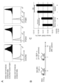
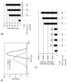
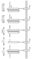
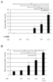


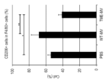
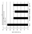


Classifications
-
- A—HUMAN NECESSITIES
- A61—MEDICAL OR VETERINARY SCIENCE; HYGIENE
- A61K—PREPARATIONS FOR MEDICAL, DENTAL OR TOILETRY PURPOSES
- A61K39/00—Medicinal preparations containing antigens or antibodies
- A61K39/395—Antibodies; Immunoglobulins; Immune serum, e.g. antilymphocytic serum
-
- A—HUMAN NECESSITIES
- A61—MEDICAL OR VETERINARY SCIENCE; HYGIENE
- A61K—PREPARATIONS FOR MEDICAL, DENTAL OR TOILETRY PURPOSES
- A61K45/00—Medicinal preparations containing active ingredients not provided for in groups A61K31/00 - A61K41/00
-
- A—HUMAN NECESSITIES
- A61—MEDICAL OR VETERINARY SCIENCE; HYGIENE
- A61K—PREPARATIONS FOR MEDICAL, DENTAL OR TOILETRY PURPOSES
- A61K47/00—Medicinal preparations characterised by the non-active ingredients used, e.g. carriers or inert additives; Targeting or modifying agents chemically bound to the active ingredient
- A61K47/30—Macromolecular organic or inorganic compounds, e.g. inorganic polyphosphates
- A61K47/34—Macromolecular compounds obtained otherwise than by reactions only involving carbon-to-carbon unsaturated bonds, e.g. polyesters, polyamino acids, polysiloxanes, polyphosphazines, copolymers of polyalkylene glycol or poloxamers
-
- A—HUMAN NECESSITIES
- A61—MEDICAL OR VETERINARY SCIENCE; HYGIENE
- A61K—PREPARATIONS FOR MEDICAL, DENTAL OR TOILETRY PURPOSES
- A61K47/00—Medicinal preparations characterised by the non-active ingredients used, e.g. carriers or inert additives; Targeting or modifying agents chemically bound to the active ingredient
- A61K47/50—Medicinal preparations characterised by the non-active ingredients used, e.g. carriers or inert additives; Targeting or modifying agents chemically bound to the active ingredient the non-active ingredient being chemically bound to the active ingredient, e.g. polymer-drug conjugates
- A61K47/69—Medicinal preparations characterised by the non-active ingredients used, e.g. carriers or inert additives; Targeting or modifying agents chemically bound to the active ingredient the non-active ingredient being chemically bound to the active ingredient, e.g. polymer-drug conjugates the conjugate being characterised by physical or galenical forms, e.g. emulsion, particle, inclusion complex, stent or kit
-
- A—HUMAN NECESSITIES
- A61—MEDICAL OR VETERINARY SCIENCE; HYGIENE
- A61K—PREPARATIONS FOR MEDICAL, DENTAL OR TOILETRY PURPOSES
- A61K9/00—Medicinal preparations characterised by special physical form
- A61K9/14—Particulate form, e.g. powders, Processes for size reducing of pure drugs or the resulting products, Pure drug nanoparticles
-
- A—HUMAN NECESSITIES
- A61—MEDICAL OR VETERINARY SCIENCE; HYGIENE
- A61P—SPECIFIC THERAPEUTIC ACTIVITY OF CHEMICAL COMPOUNDS OR MEDICINAL PREPARATIONS
- A61P35/00—Antineoplastic agents
-
- C—CHEMISTRY; METALLURGY
- C07—ORGANIC CHEMISTRY
- C07K—PEPTIDES
- C07K14/00—Peptides having more than 20 amino acids; Gastrins; Somatostatins; Melanotropins; Derivatives thereof
- C07K14/005—Peptides having more than 20 amino acids; Gastrins; Somatostatins; Melanotropins; Derivatives thereof from viruses
- C07K14/08—RNA viruses
- C07K14/11—Orthomyxoviridae, e.g. influenza virus
-
- C—CHEMISTRY; METALLURGY
- C07—ORGANIC CHEMISTRY
- C07K—PEPTIDES
- C07K14/00—Peptides having more than 20 amino acids; Gastrins; Somatostatins; Melanotropins; Derivatives thereof
- C07K14/435—Peptides having more than 20 amino acids; Gastrins; Somatostatins; Melanotropins; Derivatives thereof from animals; from humans
- C07K14/705—Receptors; Cell surface antigens; Cell surface determinants
-
- C—CHEMISTRY; METALLURGY
- C07—ORGANIC CHEMISTRY
- C07K—PEPTIDES
- C07K16/00—Immunoglobulins [IGs], e.g. monoclonal or polyclonal antibodies
-
- C—CHEMISTRY; METALLURGY
- C07—ORGANIC CHEMISTRY
- C07K—PEPTIDES
- C07K16/00—Immunoglobulins [IGs], e.g. monoclonal or polyclonal antibodies
- C07K16/18—Immunoglobulins [IGs], e.g. monoclonal or polyclonal antibodies against material from animals or humans
- C07K16/28—Immunoglobulins [IGs], e.g. monoclonal or polyclonal antibodies against material from animals or humans against receptors, cell surface antigens or cell surface determinants
- C07K16/30—Immunoglobulins [IGs], e.g. monoclonal or polyclonal antibodies against material from animals or humans against receptors, cell surface antigens or cell surface determinants from tumour cells
-
- C—CHEMISTRY; METALLURGY
- C07—ORGANIC CHEMISTRY
- C07K—PEPTIDES
- C07K19/00—Hybrid peptides, i.e. peptides covalently bound to nucleic acids, or non-covalently bound protein-protein complexes
-
- C—CHEMISTRY; METALLURGY
- C12—BIOCHEMISTRY; BEER; SPIRITS; WINE; VINEGAR; MICROBIOLOGY; ENZYMOLOGY; MUTATION OR GENETIC ENGINEERING
- C12N—MICROORGANISMS OR ENZYMES; COMPOSITIONS THEREOF; PROPAGATING, PRESERVING, OR MAINTAINING MICROORGANISMS; MUTATION OR GENETIC ENGINEERING; CULTURE MEDIA
- C12N15/00—Mutation or genetic engineering; DNA or RNA concerning genetic engineering, vectors, e.g. plasmids, or their isolation, preparation or purification; Use of hosts therefor
- C12N15/09—Recombinant DNA-technology
- C12N15/11—DNA or RNA fragments; Modified forms thereof; Non-coding nucleic acids having a biological activity
- C12N15/62—DNA sequences coding for fusion proteins
-
- C—CHEMISTRY; METALLURGY
- C12—BIOCHEMISTRY; BEER; SPIRITS; WINE; VINEGAR; MICROBIOLOGY; ENZYMOLOGY; MUTATION OR GENETIC ENGINEERING
- C12N—MICROORGANISMS OR ENZYMES; COMPOSITIONS THEREOF; PROPAGATING, PRESERVING, OR MAINTAINING MICROORGANISMS; MUTATION OR GENETIC ENGINEERING; CULTURE MEDIA
- C12N15/00—Mutation or genetic engineering; DNA or RNA concerning genetic engineering, vectors, e.g. plasmids, or their isolation, preparation or purification; Use of hosts therefor
- C12N15/09—Recombinant DNA-technology
- C12N15/63—Introduction of foreign genetic material using vectors; Vectors; Use of hosts therefor; Regulation of expression
- C12N15/74—Vectors or expression systems specially adapted for prokaryotic hosts other than E. coli, e.g. Lactobacillus, Micromonospora
-
- C—CHEMISTRY; METALLURGY
- C12—BIOCHEMISTRY; BEER; SPIRITS; WINE; VINEGAR; MICROBIOLOGY; ENZYMOLOGY; MUTATION OR GENETIC ENGINEERING
- C12N—MICROORGANISMS OR ENZYMES; COMPOSITIONS THEREOF; PROPAGATING, PRESERVING, OR MAINTAINING MICROORGANISMS; MUTATION OR GENETIC ENGINEERING; CULTURE MEDIA
- C12N15/00—Mutation or genetic engineering; DNA or RNA concerning genetic engineering, vectors, e.g. plasmids, or their isolation, preparation or purification; Use of hosts therefor
- C12N15/09—Recombinant DNA-technology
- C12N15/63—Introduction of foreign genetic material using vectors; Vectors; Use of hosts therefor; Regulation of expression
- C12N15/79—Vectors or expression systems specially adapted for eukaryotic hosts
- C12N15/80—Vectors or expression systems specially adapted for eukaryotic hosts for fungi
- C12N15/81—Vectors or expression systems specially adapted for eukaryotic hosts for fungi for yeasts
-
- C—CHEMISTRY; METALLURGY
- C12—BIOCHEMISTRY; BEER; SPIRITS; WINE; VINEGAR; MICROBIOLOGY; ENZYMOLOGY; MUTATION OR GENETIC ENGINEERING
- C12N—MICROORGANISMS OR ENZYMES; COMPOSITIONS THEREOF; PROPAGATING, PRESERVING, OR MAINTAINING MICROORGANISMS; MUTATION OR GENETIC ENGINEERING; CULTURE MEDIA
- C12N5/00—Undifferentiated human, animal or plant cells, e.g. cell lines; Tissues; Cultivation or maintenance thereof; Culture media therefor
- C12N5/10—Cells modified by introduction of foreign genetic material
-
- C—CHEMISTRY; METALLURGY
- C12—BIOCHEMISTRY; BEER; SPIRITS; WINE; VINEGAR; MICROBIOLOGY; ENZYMOLOGY; MUTATION OR GENETIC ENGINEERING
- C12P—FERMENTATION OR ENZYME-USING PROCESSES TO SYNTHESISE A DESIRED CHEMICAL COMPOUND OR COMPOSITION OR TO SEPARATE OPTICAL ISOMERS FROM A RACEMIC MIXTURE
- C12P21/00—Preparation of peptides or proteins
- C12P21/02—Preparation of peptides or proteins having a known sequence of two or more amino acids, e.g. glutathione
Definitions
- the present invention relates to multispecific nanoparticles, methods for producing multispecific nanoparticles, and the like.
- CAR-T cell therapy involves introducing chimeric antigen receptors (CARs) into T cells collected from peripheral blood to create CAR-T cells that can attack cancer cells, and use them for treatment. This is the method used (Non-Patent Document 1).
- CARs chimeric antigen receptors
- Chimeric antigen receptors are artificial antigen receptors that combine an extracellular domain containing an antibody variable region that recognizes cancer antigens and a signaling domain of costimulatory molecules involved in T cell activation. be.
- CARs Chimeric antigen receptors
- first generation CAR whose signal transduction domain consists only of CD3 ⁇
- second and third generation CARs containing different structures and types of signal transduction domains have been developed.
- Ta. CAR-T cell therapy is a technological technology that recognizes tumor cells and makes it possible to efficiently kill tumor cells by activating CAR-expressing T cells (CAR-T cells) themselves. It is a cancer treatment.
- CAR-T cell therapy Treatment methods that target the CD19 molecule have been approved in Europe, the United States, and Japan for B-cell leukemia and lymphoma. The response rate is extremely high, and it has been reported that over 80% of remissions can be achieved even in refractory cases. Furthermore, CAR-T cell therapy targeting other antigens and cancer types is being developed, and is expected to be a breakthrough cancer treatment technology following immune checkpoint inhibition therapy.
- CAR-T cell therapy Another problem with CAR-T cell therapy is that the therapeutic effect cannot be stably obtained.
- CAR-T cell therapy CAR-T cells are generated using T cells collected from a patient. Therefore, it is known that the quality of CAR-T cells can vary widely depending on the patient's condition. In particular, it has been reported that in patients who have undergone repeated chemotherapy, etc., the quality of collected T cells decreases during the culture stage, and CAR-T cells disappear early after infusion (Non-patent Document 2). ).
- An object of the present invention is to provide a new non-cell preparation that does not need to be individually prepared for each patient and can stably exhibit a high killing effect on tumor cells at low cost.
- CAR-T cell therapy requires the step of collecting the subject's own T cells, introducing the CAR gene, and then returning them to the patient.
- a series of operations to individually prepare CAR-T cells is a necessary step to create cancer cell-specific cells, but it is a factor that requires high treatment costs and This is also a factor that can cause the quality of treatment to vary widely from patient to patient.
- the present inventors developed nanoparticles with multiple immunoregulatory molecules mounted on the surface layer.
- T cells existing in the living body can be activated "on the spot.”
- this activation is specifically induced in the presence of tumor cells. Therefore, specificity equivalent to that of CAR-T cells was ensured, and cost reduction and stabilization of efficacy were simultaneously achieved.
- the nanoparticles of the present invention can be additionally loaded with immune regulating molecules such as costimulatory molecules and cytokine molecules, thereby making it possible to freely adjust and enhance the effects.
- immune regulating molecules such as costimulatory molecules and cytokine molecules
- the present invention is based on the above research results and provides the following.
- the target binding region is The multispecific nanoparticle according to (1), comprising (a) a T cell receptor complex binding region, a NK cell surface antigen binding region, or a macrophage surface antigen binding region, and (b) a cancer antigen binding region.
- the T cell receptor complex binding region is selected from the group consisting of an anti-CD3 antibody or a fragment thereof, an anti-T cell receptor (TCR) antibody or a fragment thereof, and an HLA/peptide fusion molecule; ) Multispecific nanoparticles as described in ).
- the anti-CD3 antibody is [1] A heavy chain variable region including CDR1, CDR2, and CDR3 consisting of the amino acid sequences shown in SEQ ID NOs: 72 to 74, respectively, and a light chain variable region including CDR1, CDR2, and CDR3 consisting of the amino acid sequences shown in SEQ ID NOs: 75 to 77, respectively.
- chain variable region [2] A heavy chain variable region including CDR1, CDR2, and CDR3 consisting of the amino acid sequences shown in SEQ ID NOs: 80 to 82, respectively, and a light chain variable region including CDR1, CDR2, and CDR3 consisting of the amino acid sequences shown in SEQ ID NOs: 83 to 85, respectively.
- chain variable region [3] A heavy chain variable region including CDR1, CDR2, and CDR3 consisting of the amino acid sequences shown in SEQ ID NOs: 88 to 90, respectively, and a light chain variable region including CDR1, CDR2, and CDR3 consisting of the amino acid sequences shown in SEQ ID NOs: 91 to 93, respectively.
- chain variable region [4] A heavy chain variable region including CDR1, CDR2, and CDR3 consisting of the amino acid sequences shown in SEQ ID NOs: 96 to 98, respectively, and a light chain variable region including CDR1, CDR2, and CDR3 consisting of the amino acid sequences shown in SEQ ID NOs: 99 to 101, respectively.
- chain variable region [5] A heavy chain variable region including CDR1, CDR2, and CDR3 consisting of the amino acid sequences shown in SEQ ID NOs: 104 to 106, respectively, and a light chain variable region including CDR1, CDR2, and CDR3 consisting of the amino acid sequences shown in SEQ ID NOs: 107 to 109, respectively.
- chain variable region [6] A heavy chain variable region including CDR1, CDR2, and CDR3 consisting of the amino acid sequences shown in SEQ ID NOs: 112 to 114, respectively, and a light chain variable region including CDR1, CDR2, and CDR3 consisting of the amino acid sequences shown in SEQ ID NOs: 115 to 117, respectively.
- chain variable region [7] A heavy chain variable region including CDR1, CDR2, and CDR3 consisting of the amino acid sequences shown in SEQ ID NOs: 120 to 122, respectively, and a light chain variable region including CDR1, CDR2, and CDR3 consisting of the amino acid sequences shown in SEQ ID NOs: 123 to 125, respectively.
- chain variable region [8] A heavy chain variable region including CDR1, CDR2, and CDR3 consisting of the amino acid sequences shown in SEQ ID NOs: 128 to 130, respectively, and a light chain variable region including CDR1, CDR2, and CDR3 consisting of the amino acid sequences shown in SEQ ID NOs: 131 to 133, respectively.
- chain variable region [9] A heavy chain variable region including CDR1, CDR2, and CDR3 consisting of the amino acid sequences shown in SEQ ID NOs: 136 to 138, respectively, and a light chain variable region including CDR1, CDR2, and CDR3 consisting of the amino acid sequences shown in SEQ ID NOs: 139 to 141, respectively.
- chain variable region [10] A heavy chain variable region including CDR1, CDR2, and CDR3 consisting of the amino acid sequences shown in SEQ ID NOs: 144 to 146, respectively, and a light chain variable region including CDR1, CDR2, and CDR3 consisting of the amino acid sequences shown in SEQ ID NOs: 147 to 149, respectively.
- chain variable region [11] A heavy chain variable region including CDR1, CDR2, and CDR3 consisting of the amino acid sequences shown in SEQ ID NOs: 152 to 153, respectively, and a light chain variable region including CDR1, CDR2, and CDR3 consisting of the amino acid sequences shown in SEQ ID NOs: 155 to 157, respectively.
- chain variable region [12] A heavy chain variable region including CDR1, CDR2, and CDR3 consisting of the amino acid sequences shown in SEQ ID NOs: 160 to 162, respectively, and a light chain variable region including CDR1, CDR2, and CDR3 consisting of the amino acid sequences shown in SEQ ID NOs: 163 to 165, respectively.
- chain variable region [13] A heavy chain variable region including CDR1, CDR2, and CDR3 consisting of the amino acid sequences shown in SEQ ID NOs: 168 to 170, respectively, and a light chain variable region including CDR1, CDR2, and CDR3 consisting of the amino acid sequences shown in SEQ ID NOs: 171 to 173, respectively.
- a heavy chain variable region comprising CDR1, CDR2, and CDR3 consisting of the amino acid sequences shown in SEQ ID NO: 345 to 347, respectively, CDR1 consisting of the amino acid sequence shown in SEQ ID NO: 348, and consisting of the amino acid sequence GAS (SEQ ID NO: 349)
- a light chain variable region comprising CDR2 and CDR3 consisting of the amino acid sequence shown in SEQ ID NO: 350
- a heavy chain variable region including CDR1, CDR2, and CDR3 consisting of the amino acid sequences shown in SEQ ID NOs: 353 to 355, respectively, and a light chain variable region including CDR1, CDR2, and CDR3 consisting of the amino acid sequences shown in SEQ ID NOs: 356 to 358, respectively.
- chain variable region [17] A heavy chain variable region including CDR1, CDR2, and CDR3 consisting of the amino acid sequences shown in SEQ ID NOs: 359 to 361, respectively, and a light chain variable region including CDR1, CDR2, and CDR3 consisting of the amino acid sequences shown in SEQ ID NOs: 362 to 364, respectively.
- chain variable region [18] A heavy chain variable region including CDR1, CDR2, and CDR3 consisting of the amino acid sequences shown in SEQ ID NOs: 367 to 369, respectively, and a light chain variable region including CDR1, CDR2, and CDR3 consisting of the amino acid sequences shown in SEQ ID NOs: 370 to 372, respectively.
- chain variable region [19] A heavy chain variable region including CDR1, CDR2, and CDR3 consisting of the amino acid sequences shown in SEQ ID NOs: 375 to 377, respectively, and a light chain variable region including CDR1, CDR2, and CDR3 consisting of the amino acid sequences shown in SEQ ID NOs: 378 to 380, respectively.
- chain variable region [20] A heavy chain variable region including CDR1, CDR2, and CDR3 consisting of the amino acid sequences shown in SEQ ID NOs: 383 to 385, respectively, and a light chain variable region including CDR1, CDR2, and CDR3 consisting of the amino acid sequences shown in SEQ ID NOs: 386 to 388, respectively.
- chain variable region [21] A heavy chain variable region including CDR1, CDR2, and CDR3 consisting of the amino acid sequences shown in SEQ ID NOs: 391 to 393, respectively, and a light chain variable region including CDR1, CDR2, and CDR3 consisting of the amino acid sequences shown in SEQ ID NOs: 394 to 396, respectively.
- chain variable region [22] A heavy chain variable region including CDR1, CDR2, and CDR3 consisting of the amino acid sequences shown in SEQ ID NOs: 399 to 401, respectively, and a light chain variable region including CDR1, CDR2, and CDR3 consisting of the amino acid sequences shown in SEQ ID NOs: 402 to 404, respectively.
- a heavy chain variable region consisting of an amino acid sequence selected from the group consisting of SEQ ID NOs: 407 to 426, and a light chain variable region consisting of an amino acid sequence selected from the group consisting of SEQ ID NOs: 427 to 434
- a heavy chain variable region including CDR1, CDR2, and CDR3 consisting of the amino acid sequences shown in SEQ ID NOs: 435 to 437, respectively, and a light chain variable region including CDR1, CDR2, and CDR3 consisting of the amino acid sequences shown in SEQ ID NOs: 438 to 440, respectively.
- chain variable region [25] A heavy chain variable region including CDR1, CDR2, and CDR3 consisting of the amino acid sequences shown in SEQ ID NOs: 443 to 445, respectively, and a light chain variable region including CDR1, CDR2, and CDR3 consisting of the amino acid sequences shown in SEQ ID NOs: 446 to 448, respectively.
- chain variable region [26] A heavy chain variable region comprising CDR1, CDR2, and CDR3 each consisting of the amino acid sequence shown in SEQ ID NO: 451 to 453, and CDR1 consisting of the amino acid sequence shown in SEQ ID NO: 454, and consisting of the amino acid sequence AAS (SEQ ID NO: 455).
- a light chain variable region comprising CDR2 and CDR3 consisting of the amino acid sequence shown in SEQ ID NO: 456, [27] A heavy chain variable region comprising CDR1, CDR2, and CDR3 each consisting of the amino acid sequence shown in SEQ ID NO: 459 to 461, CDR1 consisting of the amino acid sequence shown in SEQ ID NO: 462, and consisting of the amino acid sequence AAS (SEQ ID NO: 463) A light chain variable region comprising CDR2 and CDR3 consisting of the amino acid sequence shown in SEQ ID NO: 464, [28] A heavy chain variable region consisting of an amino acid sequence selected from the group consisting of SEQ ID NOs: 467 to 492, and a light chain variable region consisting of an amino acid sequence selected from the group consisting of SEQ ID NOs: 494 to 518, [29] A heavy chain variable region comprising CDR1, CDR2, and CDR3 each consisting of the amino acid sequence shown in SEQ ID NO: 519 to 521, and CDR1 consisting of the amino
- a light chain variable region comprising CDR2 and CDR3 consisting of the amino acid sequence shown in SEQ ID NO: 524, [30] A heavy chain variable region including CDR1, CDR2, and CDR3 consisting of the amino acid sequences shown in SEQ ID NOs: 527 to 529, respectively, and a light chain variable region including CDR1, CDR2, and CDR3 consisting of the amino acid sequences shown in SEQ ID NOs: 530 to 532, respectively.
- a heavy chain variable region comprising CDR1, CDR2, and CDR3 consisting of the amino acid sequences shown in SEQ ID NOs: 527 to 529, respectively, and CDR1, CDR2, and CDR3 consisting of the amino acid sequences shown in SEQ ID NOs: 530 to 531 and 533, respectively.
- a heavy chain variable region comprising CDR1, CDR2, and CDR3 consisting of the amino acid sequences shown in SEQ ID NOs: 566 to 568, respectively, and CDR1 consisting of the amino acid sequences shown in SEQ ID NOs: 569 to 571, respectively;
- the anti-TCR antibody comprises a light chain variable region comprising CDR2 and CDR3, or
- the multispecific nanoparticle according to (3) comprising a chain variable region.
- the NK cell surface antigen binding region is an anti-CD16 antibody or a fragment thereof, an anti-NKG2D antibody or a fragment thereof, an anti-Nkp30 antibody or a fragment thereof, an anti-Nkp44 antibody or a fragment thereof, an anti-Nkp46 antibody or a fragment thereof, an anti-2B4 antibody or a fragment thereof, an IgG Fc region or a fragment thereof, a MICA protein or a fragment thereof, a MICB protein or a fragment thereof, a B7H6 protein or a fragment thereof, and an influenza virus-derived hemagglutinin or a fragment thereof, according to (2). multispecific nanoparticles.
- the anti-CD16 antibody is [34] A heavy chain variable region including CDR1, CDR2, and CDR3 consisting of the amino acid sequences shown in SEQ ID NOs: 300 to 302, respectively, and a light chain variable region including CDR1, CDR2, and CDR3 consisting of the amino acid sequences shown in SEQ ID NOs: 303 to 305, respectively.
- the anti-NKG2D antibody is [35] A heavy chain variable region including CDR1, CDR2, and CDR3 consisting of the amino acid sequences shown in SEQ ID NOs: 308 to 310, respectively, and a light chain variable region including CDR1, CDR2, and CDR3 consisting of the amino acid sequences shown in SEQ ID NOs: 311 to 313, respectively.
- the anti-NKp46 antibody comprises a chain variable region, or [36] A heavy chain variable region including CDR1, CDR2, and CDR3 consisting of the amino acid sequences shown in SEQ ID NOs: 316 to 318, respectively, and a light chain variable region including CDR1, CDR2, and CDR3 consisting of the amino acid sequences shown in SEQ ID NOs: 319 to 321, respectively.
- the IgG Fc region comprises a chain variable region, [37]
- the macrophage surface antigen-binding region is an anti-FC ⁇ RI (CD64) antibody or a fragment thereof, an anti-FC ⁇ RIIA (CD32) antibody or a fragment thereof, an anti-FC ⁇ RIIIA (CD16A) antibody or a fragment thereof, an anti-CD40 antibody or a fragment thereof, an IgG Fc
- the multispecific nanoparticle according to (2) selected from the group consisting of a region or a fragment thereof, a CD40L protein or a fragment thereof, an anti-CD47 antibody or a fragment thereof, and a TLR4a ligand.
- the anti-CD40 antibody is [39] A heavy chain variable region including CDR1, CDR2, and CDR3 consisting of the amino acid sequences shown in SEQ ID NOs: 324 to 326, respectively, and a light chain variable region including CDR1, CDR2, and CDR3 consisting of the amino acid sequences shown in SEQ ID NOs: 327 to 329, respectively.
- a heavy chain variable region comprising CDR1, CDR2, and CDR3 consisting of the amino acid sequences shown in SEQ ID NOs: 332 to 334, respectively, and CDR1, CDR2 consisting of the amino acid sequences shown in SEQ ID NOs: 335 to 337, respectively; and a light chain variable region comprising CDR3;
- the IgG Fc region is [41] A human IgG1 Fc region consisting of the amino acid sequence shown in SEQ ID NO: 30, or [42] A human IgG3 Fc region consisting of the amino acid sequence shown in SEQ ID NO: 61,
- the anti-CD47 antibody or fragment thereof is [43] A heavy chain variable region comprising CDR1 to CDR3 consisting of the amino acid sequences shown in SEQ ID NOs: 590 to 592, respectively, and a light chain variable region comprising CDR1 to CDR3 consisting of the amino acid sequences shown in SEQ ID NOs: 593 to 595, respectively, or [ 44] comprises
- the cancer antigen is selected from the group consisting of CD19, BCMA, mesothelin, GD2, CD20, CD22, EGFR, CD33, CD123, ERBB2, CD133, CEA, CEACAM5, MUC1, and PSMA; (2) The multispecific nanoparticle according to any one of (8) to (8). (10) One or more selected from the group consisting of costimulatory molecules, cytokine molecules, cytokine receptors, migration molecules, immune checkpoint inhibitory molecules, and immune checkpoint molecules on the surface and/or inside of the nanoparticles.
- the costimulatory molecule is selected from the group consisting of CD80 protein, CD86 protein, 4-1BB ligand protein, ICOS ligand protein, CD40 protein, CD70 protein, OX40 ligand protein, GITR ligand protein, and LIGHT protein.
- the cytokine molecule is IL-7 protein, IL-15 protein, IL-21 protein, IL-2 protein, IL-12 protein, IL-4 protein, IL-10 protein, IL-18 protein, GM-CSF protein, one or more selected from the group consisting of IFN- ⁇ protein, TNF- ⁇ protein, and TGF- ⁇ protein
- the cytokine receptor is a TGF- ⁇ receptor, an IL-6 receptor, an IL-1 receptor, a TNF receptor, an IL-4 receptor, an IL-10 receptor, an IL-13 receptor, and a CSF-1 receptor.
- the migration molecule is one or more selected from the group consisting of CXCR4 protein, ITGA4 protein, ITGB1 protein, ICAM-1 protein, VCAM-1 protein, and LFA3 protein
- the immune checkpoint inhibitor molecule may be a CTLA-4 inhibitor, a PD-1 inhibitor, a PD-L1 inhibitor, a Lag-3 inhibitor, a Tim-3 inhibitor, a TIGIT inhibitor, a B7-H3 inhibitor, or a VISTA inhibitor.
- the immune checkpoint molecule is one or more selected from the group consisting of PD1 protein, Lag-3 protein, Tim-3 protein, TIGIT protein, ICOS protein, CD47 protein, and BTLA protein.
- Specificity nanoparticles (12) The multispecific nanoparticle according to any one of (1) to (11), wherein the lipid membrane is composed of a cell membrane, or a cell membrane and a liposome.
- a pharmaceutical composition comprising the multispecific nanoparticle according to any one of (1) to (14).
- a fusion protein comprising a membrane binding region, and a T cell receptor complex binding region and a cancer antigen binding region on its N-terminal side or C-terminal side, The fusion protein, wherein the T cell receptor complex binding region is selected from the group consisting of an anti-CD3 antibody or a fragment thereof, an anti-T cell receptor (TCR) antibody or a fragment thereof, and an HLA/peptide fusion molecule.
- the anti-CD3 antibody is [1] A heavy chain variable region including CDR1, CDR2, and CDR3 consisting of the amino acid sequences shown in SEQ ID NOs: 72 to 74, respectively, and a light chain variable region including CDR1, CDR2, and CDR3 consisting of the amino acid sequences shown in SEQ ID NOs: 75 to 77, respectively. chain variable region, [2] A heavy chain variable region including CDR1, CDR2, and CDR3 consisting of the amino acid sequences shown in SEQ ID NOs: 80 to 82, respectively, and a light chain variable region including CDR1, CDR2, and CDR3 consisting of the amino acid sequences shown in SEQ ID NOs: 83 to 85, respectively.
- chain variable region [3] A heavy chain variable region including CDR1, CDR2, and CDR3 consisting of the amino acid sequences shown in SEQ ID NOs: 88 to 90, respectively, and a light chain variable region including CDR1, CDR2, and CDR3 consisting of the amino acid sequences shown in SEQ ID NOs: 91 to 93, respectively.
- chain variable region [4] A heavy chain variable region including CDR1, CDR2, and CDR3 consisting of the amino acid sequences shown in SEQ ID NOs: 96 to 98, respectively, and a light chain variable region including CDR1, CDR2, and CDR3 consisting of the amino acid sequences shown in SEQ ID NOs: 99 to 101, respectively.
- chain variable region [5] A heavy chain variable region including CDR1, CDR2, and CDR3 consisting of the amino acid sequences shown in SEQ ID NOs: 104 to 106, respectively, and a light chain variable region including CDR1, CDR2, and CDR3 consisting of the amino acid sequences shown in SEQ ID NOs: 107 to 109, respectively.
- chain variable region [6] A heavy chain variable region including CDR1, CDR2, and CDR3 consisting of the amino acid sequences shown in SEQ ID NOs: 112 to 114, respectively, and a light chain variable region including CDR1, CDR2, and CDR3 consisting of the amino acid sequences shown in SEQ ID NOs: 115 to 117, respectively.
- chain variable region [7] A heavy chain variable region including CDR1, CDR2, and CDR3 consisting of the amino acid sequences shown in SEQ ID NOs: 120 to 122, respectively, and a light chain variable region including CDR1, CDR2, and CDR3 consisting of the amino acid sequences shown in SEQ ID NOs: 123 to 125, respectively.
- chain variable region [8] A heavy chain variable region including CDR1, CDR2, and CDR3 consisting of the amino acid sequences shown in SEQ ID NOs: 128 to 130, respectively, and a light chain variable region including CDR1, CDR2, and CDR3 consisting of the amino acid sequences shown in SEQ ID NOs: 131 to 133, respectively.
- chain variable region [9] A heavy chain variable region including CDR1, CDR2, and CDR3 consisting of the amino acid sequences shown in SEQ ID NOs: 136 to 138, respectively, and a light chain variable region including CDR1, CDR2, and CDR3 consisting of the amino acid sequences shown in SEQ ID NOs: 139 to 141, respectively.
- chain variable region [10] A heavy chain variable region including CDR1, CDR2, and CDR3 consisting of the amino acid sequences shown in SEQ ID NOs: 144 to 146, respectively, and a light chain variable region including CDR1, CDR2, and CDR3 consisting of the amino acid sequences shown in SEQ ID NOs: 147 to 149, respectively.
- chain variable region [11] A heavy chain variable region including CDR1, CDR2, and CDR3 consisting of the amino acid sequences shown in SEQ ID NOs: 152 to 153, respectively, and a light chain variable region including CDR1, CDR2, and CDR3 consisting of the amino acid sequences shown in SEQ ID NOs: 155 to 157, respectively.
- chain variable region [12] A heavy chain variable region including CDR1, CDR2, and CDR3 consisting of the amino acid sequences shown in SEQ ID NOs: 160 to 162, respectively, and a light chain variable region including CDR1, CDR2, and CDR3 consisting of the amino acid sequences shown in SEQ ID NOs: 163 to 165, respectively.
- chain variable region [13] A heavy chain variable region including CDR1, CDR2, and CDR3 consisting of the amino acid sequences shown in SEQ ID NOs: 168 to 170, respectively, and a light chain variable region including CDR1, CDR2, and CDR3 consisting of the amino acid sequences shown in SEQ ID NOs: 171 to 173, respectively.
- chain variable region [14] A heavy chain variable region including CDR1, CDR2, and CDR3 consisting of the amino acid sequences shown in SEQ ID NOs: 176 to 178, respectively, and a light chain variable region including CDR1, CDR2, and CDR3 consisting of the amino acid sequences shown in SEQ ID NOs: 179 to 181, respectively.
- a heavy chain variable region comprising CDR1, CDR2, and CDR3 consisting of the amino acid sequences shown in SEQ ID NO: 345 to 347, respectively, CDR1 consisting of the amino acid sequence shown in SEQ ID NO: 348, and consisting of the amino acid sequence GAS (SEQ ID NO: 349)
- a light chain variable region comprising CDR2 and CDR3 consisting of the amino acid sequence shown in SEQ ID NO: 350
- a heavy chain variable region including CDR1, CDR2, and CDR3 consisting of the amino acid sequences shown in SEQ ID NOs: 353 to 355, respectively, and a light chain variable region including CDR1, CDR2, and CDR3 consisting of the amino acid sequences shown in SEQ ID NOs: 356 to 358, respectively.
- chain variable region [17] A heavy chain variable region including CDR1, CDR2, and CDR3 consisting of the amino acid sequences shown in SEQ ID NOs: 359 to 361, respectively, and a light chain variable region including CDR1, CDR2, and CDR3 consisting of the amino acid sequences shown in SEQ ID NOs: 362 to 364, respectively.
- chain variable region [18] A heavy chain variable region including CDR1, CDR2, and CDR3 consisting of the amino acid sequences shown in SEQ ID NOs: 367 to 369, respectively, and a light chain variable region including CDR1, CDR2, and CDR3 consisting of the amino acid sequences shown in SEQ ID NOs: 370 to 372, respectively.
- chain variable region [19] A heavy chain variable region including CDR1, CDR2, and CDR3 consisting of the amino acid sequences shown in SEQ ID NOs: 375 to 377, respectively, and a light chain variable region including CDR1, CDR2, and CDR3 consisting of the amino acid sequences shown in SEQ ID NOs: 378 to 380, respectively.
- chain variable region [20] A heavy chain variable region including CDR1, CDR2, and CDR3 consisting of the amino acid sequences shown in SEQ ID NOs: 383 to 385, respectively, and a light chain variable region including CDR1, CDR2, and CDR3 consisting of the amino acid sequences shown in SEQ ID NOs: 386 to 388, respectively.
- chain variable region [21] A heavy chain variable region including CDR1, CDR2, and CDR3 consisting of the amino acid sequences shown in SEQ ID NOs: 391 to 393, respectively, and a light chain variable region including CDR1, CDR2, and CDR3 consisting of the amino acid sequences shown in SEQ ID NOs: 394 to 396, respectively.
- chain variable region [22] A heavy chain variable region including CDR1, CDR2, and CDR3 consisting of the amino acid sequences shown in SEQ ID NOs: 399 to 401, respectively, and a light chain variable region including CDR1, CDR2, and CDR3 consisting of the amino acid sequences shown in SEQ ID NOs: 402 to 404, respectively.
- a heavy chain variable region consisting of an amino acid sequence selected from the group consisting of SEQ ID NOs: 407 to 426, and a light chain variable region consisting of an amino acid sequence selected from the group consisting of SEQ ID NOs: 427 to 434
- a heavy chain variable region including CDR1, CDR2, and CDR3 consisting of the amino acid sequences shown in SEQ ID NOs: 435 to 437, respectively, and a light chain variable region including CDR1, CDR2, and CDR3 consisting of the amino acid sequences shown in SEQ ID NOs: 438 to 440, respectively.
- chain variable region [25] A heavy chain variable region including CDR1, CDR2, and CDR3 consisting of the amino acid sequences shown in SEQ ID NOs: 443 to 445, respectively, and a light chain variable region including CDR1, CDR2, and CDR3 consisting of the amino acid sequences shown in SEQ ID NOs: 446 to 448, respectively.
- chain variable region [26] A heavy chain variable region comprising CDR1, CDR2, and CDR3 each consisting of the amino acid sequence shown in SEQ ID NO: 451 to 453, and CDR1 consisting of the amino acid sequence shown in SEQ ID NO: 454, and consisting of the amino acid sequence AAS (SEQ ID NO: 455).
- a light chain variable region comprising CDR2 and CDR3 consisting of the amino acid sequence shown in SEQ ID NO: 456, [27] A heavy chain variable region comprising CDR1, CDR2, and CDR3 each consisting of the amino acid sequence shown in SEQ ID NO: 459 to 461, CDR1 consisting of the amino acid sequence shown in SEQ ID NO: 462, and consisting of the amino acid sequence AAS (SEQ ID NO: 463) A light chain variable region comprising CDR2 and CDR3 consisting of the amino acid sequence shown in SEQ ID NO: 464, [28] A heavy chain variable region consisting of an amino acid sequence selected from the group consisting of SEQ ID NOs: 467 to 492, and a light chain variable region consisting of an amino acid sequence selected from the group consisting of SEQ ID NOs: 494 to 518, [29] A heavy chain variable region comprising CDR1, CDR2, and CDR3 each consisting of the amino acid sequence shown in SEQ ID NO: 519 to 521, and CDR1 consisting of the amino
- a light chain variable region comprising CDR2 and CDR3 consisting of the amino acid sequence shown in SEQ ID NO: 524, [30] A heavy chain variable region including CDR1, CDR2, and CDR3 consisting of the amino acid sequences shown in SEQ ID NOs: 527 to 529, respectively, and a light chain variable region including CDR1, CDR2, and CDR3 consisting of the amino acid sequences shown in SEQ ID NOs: 530 to 532, respectively.
- a heavy chain variable region comprising CDR1, CDR2, and CDR3 consisting of the amino acid sequences shown in SEQ ID NOs: 527 to 529, respectively, and CDR1, CDR2, and CDR3 consisting of the amino acid sequences shown in SEQ ID NOs: 530 to 531 and 533, respectively.
- a heavy chain variable region comprising CDR1, CDR2, and CDR3 consisting of the amino acid sequences shown in SEQ ID NOs: 566 to 568, respectively, and CDR1 consisting of the amino acid sequences shown in SEQ ID NOs: 569 to 571, respectively; comprising a light chain variable region including CDR2 and CDR3;
- the anti-TCR antibody is [33] A heavy chain variable region including CDR1, CDR2, and CDR3 consisting of the amino acid sequences shown in SEQ ID NOs: 184 to 186, respectively, and a light chain variable region including CDR1, CDR2, and CDR3 consisting of the amino acid sequences shown in SEQ ID NOs: 187 to 189, respectively.
- the anti-CD3 antibody is [1] A heavy chain variable region including CDR1, CDR2, and CDR3 consisting of the amino acid sequences shown in SEQ ID NOs: 72 to 74, respectively, and a light chain variable region including CDR1, CDR2, and CDR3 consisting of the amino acid sequences shown in SEQ ID NOs: 75 to 77, respectively. chain variable region, [2] A heavy chain variable region including CDR1, CDR2, and CDR3 consisting of the amino acid sequences shown in SEQ ID NOs: 80 to 82, respectively, and a light chain variable region including CDR1, CDR2, and CDR3 consisting of the amino acid sequences shown in SEQ ID NOs: 83 to 85, respectively.
- chain variable region [3] A heavy chain variable region including CDR1, CDR2, and CDR3 consisting of the amino acid sequences shown in SEQ ID NOs: 88 to 90, respectively, and a light chain variable region including CDR1, CDR2, and CDR3 consisting of the amino acid sequences shown in SEQ ID NOs: 91 to 93, respectively.
- chain variable region [4] A heavy chain variable region including CDR1, CDR2, and CDR3 consisting of the amino acid sequences shown in SEQ ID NOs: 96 to 98, respectively, and a light chain variable region including CDR1, CDR2, and CDR3 consisting of the amino acid sequences shown in SEQ ID NOs: 99 to 101, respectively.
- chain variable region [5] A heavy chain variable region including CDR1, CDR2, and CDR3 consisting of the amino acid sequences shown in SEQ ID NOs: 104 to 106, respectively, and a light chain variable region including CDR1, CDR2, and CDR3 consisting of the amino acid sequences shown in SEQ ID NOs: 107 to 109, respectively.
- chain variable region [6] A heavy chain variable region including CDR1, CDR2, and CDR3 consisting of the amino acid sequences shown in SEQ ID NOs: 112 to 114, respectively, and a light chain variable region including CDR1, CDR2, and CDR3 consisting of the amino acid sequences shown in SEQ ID NOs: 115 to 117, respectively.
- chain variable region [7] A heavy chain variable region including CDR1, CDR2, and CDR3 consisting of the amino acid sequences shown in SEQ ID NOs: 120 to 122, respectively, and a light chain variable region including CDR1, CDR2, and CDR3 consisting of the amino acid sequences shown in SEQ ID NOs: 123 to 125, respectively.
- chain variable region [8] A heavy chain variable region including CDR1, CDR2, and CDR3 consisting of the amino acid sequences shown in SEQ ID NOs: 128 to 130, respectively, and a light chain variable region including CDR1, CDR2, and CDR3 consisting of the amino acid sequences shown in SEQ ID NOs: 131 to 133, respectively.
- chain variable region [9] A heavy chain variable region including CDR1, CDR2, and CDR3 consisting of the amino acid sequences shown in SEQ ID NOs: 136 to 138, respectively, and a light chain variable region including CDR1, CDR2, and CDR3 consisting of the amino acid sequences shown in SEQ ID NOs: 139 to 141, respectively.
- chain variable region [10] A heavy chain variable region including CDR1, CDR2, and CDR3 consisting of the amino acid sequences shown in SEQ ID NOs: 144 to 146, respectively, and a light chain variable region including CDR1, CDR2, and CDR3 consisting of the amino acid sequences shown in SEQ ID NOs: 147 to 149, respectively.
- chain variable region [11] A heavy chain variable region including CDR1, CDR2, and CDR3 consisting of the amino acid sequences shown in SEQ ID NOs: 152 to 153, respectively, and a light chain variable region including CDR1, CDR2, and CDR3 consisting of the amino acid sequences shown in SEQ ID NOs: 155 to 157, respectively.
- chain variable region [12] A heavy chain variable region including CDR1, CDR2, and CDR3 consisting of the amino acid sequences shown in SEQ ID NOs: 160 to 162, respectively, and a light chain variable region including CDR1, CDR2, and CDR3 consisting of the amino acid sequences shown in SEQ ID NOs: 163 to 165, respectively.
- chain variable region [13] A heavy chain variable region including CDR1, CDR2, and CDR3 consisting of the amino acid sequences shown in SEQ ID NOs: 168 to 170, respectively, and a light chain variable region including CDR1, CDR2, and CDR3 consisting of the amino acid sequences shown in SEQ ID NOs: 171 to 173, respectively.
- chain variable region [14] A heavy chain variable region including CDR1, CDR2, and CDR3 consisting of the amino acid sequences shown in SEQ ID NOs: 176 to 178, respectively, and a light chain variable region including CDR1, CDR2, and CDR3 consisting of the amino acid sequences shown in SEQ ID NOs: 179 to 181, respectively.
- a heavy chain variable region comprising CDR1, CDR2, and CDR3 consisting of the amino acid sequences shown in SEQ ID NO: 345 to 347, respectively, CDR1 consisting of the amino acid sequence shown in SEQ ID NO: 348, and consisting of the amino acid sequence GAS (SEQ ID NO: 349)
- a light chain variable region comprising CDR2 and CDR3 consisting of the amino acid sequence shown in SEQ ID NO: 350
- a heavy chain variable region including CDR1, CDR2, and CDR3 consisting of the amino acid sequences shown in SEQ ID NOs: 353 to 355, respectively, and a light chain variable region including CDR1, CDR2, and CDR3 consisting of the amino acid sequences shown in SEQ ID NOs: 356 to 358, respectively.
- chain variable region [17] A heavy chain variable region including CDR1, CDR2, and CDR3 consisting of the amino acid sequences shown in SEQ ID NOs: 359 to 361, respectively, and a light chain variable region including CDR1, CDR2, and CDR3 consisting of the amino acid sequences shown in SEQ ID NOs: 362 to 364, respectively.
- chain variable region [18] A heavy chain variable region including CDR1, CDR2, and CDR3 consisting of the amino acid sequences shown in SEQ ID NOs: 367 to 369, respectively, and a light chain variable region including CDR1, CDR2, and CDR3 consisting of the amino acid sequences shown in SEQ ID NOs: 370 to 372, respectively.
- chain variable region [19] A heavy chain variable region including CDR1, CDR2, and CDR3 consisting of the amino acid sequences shown in SEQ ID NOs: 375 to 377, respectively, and a light chain variable region including CDR1, CDR2, and CDR3 consisting of the amino acid sequences shown in SEQ ID NOs: 378 to 380, respectively.
- chain variable region [20] A heavy chain variable region including CDR1, CDR2, and CDR3 consisting of the amino acid sequences shown in SEQ ID NOs: 383 to 385, respectively, and a light chain variable region including CDR1, CDR2, and CDR3 consisting of the amino acid sequences shown in SEQ ID NOs: 386 to 388, respectively.
- chain variable region [21] A heavy chain variable region including CDR1, CDR2, and CDR3 consisting of the amino acid sequences shown in SEQ ID NOs: 391 to 393, respectively, and a light chain variable region including CDR1, CDR2, and CDR3 consisting of the amino acid sequences shown in SEQ ID NOs: 394 to 396, respectively.
- chain variable region [22] A heavy chain variable region including CDR1, CDR2, and CDR3 consisting of the amino acid sequences shown in SEQ ID NOs: 399 to 401, respectively, and a light chain variable region including CDR1, CDR2, and CDR3 consisting of the amino acid sequences shown in SEQ ID NOs: 402 to 404, respectively.
- a heavy chain variable region consisting of an amino acid sequence selected from the group consisting of SEQ ID NOs: 407 to 426, and a light chain variable region consisting of an amino acid sequence selected from the group consisting of SEQ ID NOs: 427 to 434
- a heavy chain variable region including CDR1, CDR2, and CDR3 consisting of the amino acid sequences shown in SEQ ID NOs: 435 to 437, respectively, and a light chain variable region including CDR1, CDR2, and CDR3 consisting of the amino acid sequences shown in SEQ ID NOs: 438 to 440, respectively.
- chain variable region [25] A heavy chain variable region including CDR1, CDR2, and CDR3 consisting of the amino acid sequences shown in SEQ ID NOs: 443 to 445, respectively, and a light chain variable region including CDR1, CDR2, and CDR3 consisting of the amino acid sequences shown in SEQ ID NOs: 446 to 448, respectively.
- chain variable region [26] A heavy chain variable region comprising CDR1, CDR2, and CDR3 each consisting of the amino acid sequence shown in SEQ ID NO: 451 to 453, and CDR1 consisting of the amino acid sequence shown in SEQ ID NO: 454, and consisting of the amino acid sequence AAS (SEQ ID NO: 455).
- a light chain variable region comprising CDR2 and CDR3 consisting of the amino acid sequence shown in SEQ ID NO: 456, [27] A heavy chain variable region comprising CDR1, CDR2, and CDR3 each consisting of the amino acid sequence shown in SEQ ID NO: 459 to 461, CDR1 consisting of the amino acid sequence shown in SEQ ID NO: 462, and consisting of the amino acid sequence AAS (SEQ ID NO: 463) A light chain variable region comprising CDR2 and CDR3 consisting of the amino acid sequence shown in SEQ ID NO: 464, [28] A heavy chain variable region consisting of an amino acid sequence selected from the group consisting of SEQ ID NOs: 467 to 492, and a light chain variable region consisting of an amino acid sequence selected from the group consisting of SEQ ID NOs: 494 to 518, [29] A heavy chain variable region comprising CDR1, CDR2, and CDR3 each consisting of the amino acid sequence shown in SEQ ID NO: 519 to 521, and CDR1 consisting of the amino
- a light chain variable region comprising CDR2 and CDR3 consisting of the amino acid sequence shown in SEQ ID NO: 524, [30] A heavy chain variable region including CDR1, CDR2, and CDR3 consisting of the amino acid sequences shown in SEQ ID NOs: 527 to 529, respectively, and a light chain variable region including CDR1, CDR2, and CDR3 consisting of the amino acid sequences shown in SEQ ID NOs: 530 to 532, respectively.
- a heavy chain variable region comprising CDR1, CDR2, and CDR3 consisting of the amino acid sequences shown in SEQ ID NOs: 527 to 529, respectively, and CDR1, CDR2, and CDR3 consisting of the amino acid sequences shown in SEQ ID NOs: 530 to 531 and 533, respectively.
- a heavy chain variable region comprising CDR1, CDR2, and CDR3 consisting of the amino acid sequences shown in SEQ ID NOs: 566 to 568, respectively, and CDR1 consisting of the amino acid sequences shown in SEQ ID NOs: 569 to 571, respectively;
- the anti-TCR antibody comprises a light chain variable region comprising CDR2 and CDR3, or
- a method for producing multispecific nanoparticles capable of binding to two or more target cells comprising: (i) two or more fusion proteins containing a membrane-binding region and a target-binding region; or (ii) a cell expressing on its cell membrane a fusion protein containing a membrane-binding region and two or more target-binding regions; a cell membrane fragmentation step that fragments the
- the method includes a nanoparticle forming step of forming nanoparticles by reconstituting the cell membrane fragments after the fragmentation step, and a recovery step of collecting the nanoparticles.
- the target binding region is The method according to (24), comprising (a) a T cell receptor complex binding region, a NK cell surface antigen binding region, or a macrophage surface antigen binding region, and (b) a cancer antigen binding region.
- the reconstitution in the nanoparticle forming step mixes the cell membrane fragment and the liposome and/or the nanoparticle core, and the lipid membrane in which the cell membrane fragment and the liposome are fused forms a nanoparticle that forms the surface layer of the lipid membrane.
- a fusion protein comprising a membrane-binding region, and an NK cell surface antigen-binding region and a cancer antigen-binding region on its N-terminal side or C-terminal side
- the NK cell surface antigen-binding region is an anti-CD16 antibody or a fragment thereof, an anti-NKG2D antibody or a fragment thereof, an anti-Nkp30 antibody or a fragment thereof, an anti-Nkp44 antibody or a fragment thereof, an anti-Nkp46 antibody or a fragment thereof, an anti-2B4 antibody or a fragment thereof , IgG Fc region or a fragment thereof, MICA protein or a fragment thereof, MICB protein or a fragment thereof, B7H6 protein or a fragment thereof, and influenza virus-derived hemagglutinin or a fragment thereof.
- a fusion protein comprising a membrane-binding region, and a macrophage surface antigen-binding region and a cancer antigen-binding region on its N-terminal side or C-terminal side
- the macrophage surface antigen-binding region is an anti-FC ⁇ RI (CD64) antibody or a fragment thereof, an anti-FC ⁇ RIIA (CD32) antibody or a fragment thereof, an anti-FC ⁇ RIIIA (CD16A) antibody or a fragment thereof, an anti-CD40 antibody or a fragment thereof, an IgG Fc region or its fragment.
- CD40L protein or fragment thereof, anti-CD47 antibody or fragment thereof, and TLR4a ligand is an anti-FC ⁇ RI (CD64) antibody or a fragment thereof, an anti-FC ⁇ RIIA (CD32) antibody or a fragment thereof, an anti-FC ⁇ RIIIA (CD16A) antibody or a fragment thereof, an anti-CD40 antibody or a fragment thereof, an IgG Fc region or its fragment.
- a new non-cell preparation that does not need to be individually prepared for each patient and can stably exhibit a high killing effect on tumor cells at low cost.
- FIG. 2 is a diagram showing nanoparticles in one embodiment of the present invention.
- Figure 1A schematically shows the effects of the present invention on two target cells (T cells, NK cells, or macrophages, and cancer cells) based on nanoparticles having a fusion protein containing two or more target binding regions on the surface layer. show.
- FIG. 1B shows the effect of the present invention based on nanoparticles having two or more fusion proteins containing target binding regions on the surface layer.
- 1 is a diagram illustrating an overview of a method for producing nanoparticles in an embodiment of the present invention.
- FIG. FIG. 2 is a diagram showing the CD3/CD19 binding protein and K562-mOKT3/FMC63 cells produced in Example 1.
- Figure 3A shows the structure of the CD3/CD19 binding protein.
- FIG. 3 is a diagram showing a nanoparticle core produced in Example 2.
- Figure 4A shows the appearance of the PLGA nanoparticle core dispersion.
- Figure 4B shows the results of observing the spherical shape of the PLGA nanoparticle core using a transmission electron microscope.
- Figure 4C (left) shows the results of measuring the particle size and concentration of PLGA nanoparticle cores immediately after synthesis.
- FIG. 4C shows the results of measuring the particle size and concentration of PLGA nanoparticle cores after cryopreservation/thawing.
- FIG. 3 is a diagram showing the process of producing nanoparticles in Example 2.
- FIG. 5A shows the results of detecting the expression of CD80 protein and 41BBL protein on the cell surface by flow cytometry.
- Figure 5B shows each step of preparing nanoparticles.
- 3 is a diagram showing the results of analyzing nanoparticles produced in Example 2.
- FIG. FIG. 6A shows the results of observing nanoparticles using a transmission electron microscope (TEM). PLGA nanoparticle cores are shown before (-) and after (+) cell membrane reconstitution.
- TEM transmission electron microscope
- FIG. 6B shows the results of Western blotting analysis of components contained in the protein fractions obtained in each step of nanoparticle preparation.
- FIG. 7 is a diagram showing the effector effect of nanoparticles in Example 3. The figure shows the survival rate of tumor cells after co-culture. “NP/T cell” indicates the ratio of the number of mOKT3/FMC63 nanoparticles (NP) to the number of T cells.
- FIG. 4 is a diagram showing the results of measuring T cell activation based on nanoparticles in Example 4.
- Figure 8A shows the results of detecting CD25 protein expression on the cell surface by flow cytometry.
- FIG. 8B shows the measurement results of CD25 expression level under each condition.
- FIG. 4 is a diagram showing the results of measuring T cell activation based on nanoparticles in Example 4.
- FIG. 7 is a diagram showing the effect of nanoparticles into which costimulatory molecules CD80 protein and 41BBL protein were additionally introduced in Example 5.
- Figure 10A shows the cell doubling rate of T cells.
- “NP/T cell” indicates the ratio of the number of nanoparticles (NP) to the number of T cells.
- Figure 10B shows the survival rate of tumor cells after co-culture. The results for mOKT3/FMC63 nanoparticles (CD80/41BBL (-)) and mOKT3/FMC63/CD80/41BBL nanoparticles (CD80/41BBL (+)) are shown. The results of measuring the percentage of cytokine-producing cells in CD8-positive T cells in Example 5 are shown.
- FIG. 7 is a diagram showing the results of comparing tandem nanoparticles and separated nanoparticles in Example 6.
- FIG. 13A schematically shows the configurations of tandem nanoparticles and separated nanoparticles.
- FIG. 13B shows the results of measuring T cell activation based on tandem nanoparticles or isolated nanoparticles.
- Figure 13C shows the survival rate of tumor cells after co-culture with tandem nanoparticles and isolated nanoparticles and T cells.
- FIG. 7 is a diagram showing the effect of nanoparticles into which cytokine molecules IL7 protein and IL15 protein were additionally introduced in Example 7.
- FIG. 14A shows the results of detecting the expression of each protein by flow cytometry in K562 cells additionally introduced with IL7 protein and IL15 protein.
- FIG. 14B shows the cell doubling rate of T cells by mOKT3/FMC63/CD80/41BBL nanoparticles (-IL7/IL15) and mOKT3/FMC63/CD80/41BBL/IL7/IL15 nanoparticles (+IL7/IL15).
- NP/T cell indicates the ratio of the number of nanoparticles (NP) to the number of T cells.
- FIG. 7 is a diagram showing the results of evaluating T cell memory traits in Example 7.
- FIG. 15A shows the results of evaluating the expression levels of marker proteins CCR7, CD45RA, CD62L, CD28, FSC, and CD27 by flow cytometry.
- FIG. 15B shows undifferentiated memory traits when mOKT3/FMC63/CD80/41BBL nanoparticles (-IL7/IL15) and mOKT3/FMC63/CD80/41BBL/IL7/IL15 nanoparticles (+IL7/IL15) were administered.
- “NP/T cell” indicates the ratio of the number of nanoparticles (NP) to the number of T cells.
- FIG. 7 is a diagram showing the results of evaluating phosphorylated STAT5 in Example 7.
- Figure 16A shows the results of measuring phosphorylated STAT5 (pSTAT5) levels by flow cytometry.
- FIG. 16B shows the results of quantifying phosphorylated STAT5 (pSTAT5) fluorescence intensity.
- FIG. 3 is a diagram showing the CD3/BCMA binding protein and mOKT3/BCMA98/CD80/41BBL nanoparticles produced in Example 8.
- Figure 17A shows the structure of CD3/BCMA binding protein. In the figure, SP indicates a signal peptide.
- Figure 17B shows the results of detecting CD25 protein expression on the cell surface by flow cytometry.
- FIG. 7 is a diagram showing the results of evaluating T cell proliferation and effector effect induction in Example 8.
- Figure 18A shows the cell doubling rate of T cells.
- Figure 18B shows the survival rate of tumor cells after co-culture. The results of measuring the percentage of cytokine-producing cells in Example 8 are shown.
- Figure 19A shows a representative flow cytometry plot.
- FIG. 19B shows the IL2 positive rate (CD4 + IL2 + ) in CD4 positive T cells and the IFN ⁇ positive rate (CD8 + IFN ⁇ + ) in CD8 positive T cells.
- FIG. 7 is a diagram showing the results of evaluating the effect of mOKT3/ss1/CD80/41BBL/IL7/IL15 nanoparticles containing mesothelin binding protein in Example 9.
- FIG. 20A shows the results of evaluating the expression level of CD25 protein by flow cytometry. T cell activation by mOKT3/ss1/CD80/41BBL/IL7/IL15 nanoparticles (ss1) and mOKT3/FMC63/CD80/41BBL/IL7/IL15 nanoparticles (FMC63) is shown.
- FIG. 20B shows the results of quantifying the expression level of CD25 protein.
- Figure 20C shows the survival rate of K562-CD19/Mesothelin cells after co-culture.
- FIG. 7 is a diagram showing the sustained release effect of molecules encapsulated in PLGA nanoparticles in Example 10.
- Figure 21A shows the fluorescence spectrum of FITC-BSA encapsulated PLGA nanoparticles.
- FIG. 21B shows the results of measuring the time change in the concentration of FITC-BSA released to the outside of the dialysis membrane using a fluorometer.
- FIG. 7 is a diagram showing the in vivo antitumor effect of nanoparticles in Example 11.
- FIG. 22A shows the dosing schedule for NALM6-GL cells, T cells, and nanoparticles.
- FIG. 22B shows the imaging results of NALM6-GL cells at 2 to 5 weeks after NALM6-GL cell transplantation in the No NP group, NP ⁇ 3 group, and NP ⁇ 6 group.
- FIG. 22C shows logarithmically transformed luminescence intensity.
- FIG. 7 is a diagram showing the analysis results of mice to which nanoparticles were administered in Example 11.
- Figure 23A shows the results of measuring the number of CD8-positive T cells in peripheral blood.
- Figure 23B shows the results of survival analysis by Kaplan-Meier method. The results of evaluating T cell memory traits in Example 11 are shown.
- Figure 24A shows a representative flow cytometry diagram.
- FIG. 24B shows the percentage of memory stem cell (TSCM) and effector memory (TEM) fractions.
- FIG. 7 is a diagram showing the effect of nanoparticles into which bone marrow homing molecules have been introduced in Example 12.
- FIG. 25A shows the results of flow cytometry detection of the expression of CXCR4 protein, ITGA4 protein, and ITGB1 protein that were additionally introduced into K562 cells.
- Figure 25B shows the dosing schedule for NALM6 cells, T cells, and nanoparticles.
- FIG. 25C shows mOKT3/FMC63/CD80/41BBL/CXCR4/ITGA4/ITGB1 nanoparticles (“CXCR4+VLA4”, (-)) or mOKT3/FMC63/CD80/41BBL nanoparticles (“CXCR4+VLA4”) containing bone marrow homing molecules. ”, (+)) shows the percentage of T cells in the bone marrow or spleen of mice administered.
- FIG. 7 is a diagram showing the results of producing nanoparticles with different particle diameters and testing their effects in Example 13.
- Figure 26A shows the particle size distribution of PLGA nanoparticle cores made from 1 mg/mL or 20 mg/mL PLGA.
- Figure 26B shows the cell doubling rate of T cells.
- NP size indicates the particle size of nanoparticles.
- FIG. 26C shows the results of quantifying the expression level of CD25 protein. Five fusion proteins designed as immune regulatory molecules are shown. The effect of membrane vesicles (MVs) loaded with cytokine molecules on their surface layer is shown.
- Figure 28A shows the absence of membrane vesicles (MV:-) under conditions of not co-culturing with A20 cells (in the figure, A20 cells:-) or under conditions of co-culturing with A20 cells (in the figure, A20 cells:+).
- MV:WT mouse CD8 cultured in the presence of MV (2C11/1D3/41BBL)
- MV:IL12 IL12 fusion protein introduced into the surface layer
- Figure 28B shows the results in the absence of membrane vesicles (MV:-), in the presence of MV(2C11/1D3/41BBL) (MV:WT), and in the presence of MV(2C11/1D3/41BBL/IL12) (MV:IL12).
- mice CD8 cultured in the presence of MV (2C11/1D3/41BBL/IL18) (MV:IL18), or in the presence of MV (2C11/1D3/41BBL/IL12/IL18) (MV:IL12+IL18)
- MV:IL18 mouse CD8 cultured in the presence of MV (2C11/1D3/41BBL/IL12/IL18)
- MV:IL12+IL18 mouse CD8 cultured in the presence of MV (2C11/1D3/41BBL/IL12/IL18)
- MV:IL12+IL18 MV 2C11/1D3/41BBL/IL12/IL18
- TGF ⁇ receptor anti-PDL1 scFV
- anti-CTLA4 scFV mounted on membrane vesicles
- Figure 29A shows conditions in which membrane vesicles are not added to the medium containing TGF ⁇ (MV:-), conditions in which MV (2C11/1D3/41BBL) as a comparison control is added (MV:WT), and MV (2C11/1D3/ 41BBL/TGFBR) was added (MV:TGFBRI/II), and the TGF ⁇ concentration in the supernatant was measured by ELISA.
- Figure 29B shows the results of measuring the PDL1 expression level on A20 cells by flow cytometry after adding MV (2C11/1D3/41BBL/anti-PDL1-scFV) to the PDL1-positive mouse lymphoma cell line A20. .
- WT is the result of A20 cells in which the CTLA4 gene has not been introduced
- MV- is the result when membrane vesicles are not added
- WT-MV is the result of the comparison control MV (2C11/1D3 /41BBL) is shown.
- Tumor volume mm 3
- Figure 30A shows the results of PBS administration.
- Figure 30B shows the results of WT-MV administration.
- Figure 30C shows the results of TME-MV administration.
- FIG. 31A shows the average tumor volume at 13, 15, and 17 days after transplantation of CT26-CD19-luc cells.
- Ctrl indicates the PBS administration group.
- Figure 31B shows the results of mouse survival analysis.
- Ctrl indicates the PBS administration group.
- FIG. 31C shows the results of time-lapse observation of tumor cells in mouse bodies in a representative example of the TME-MV administration group. The results of analyzing cells infiltrating into tumors in Balb/c mice in which PBS, WT-MV, or TME-MV was intratumorally administered after subcutaneously transplanting CT26-CD19-luc cells are shown.
- Figure 32A shows the ratio of CD8-positive T cells to CD45-positive blood cells in infiltrating cells.
- Figure 32B shows the percentage of CD8 positive T cells (CD45+CD8+ cells) among the total cells within the tumor.
- Representative flow cytometry plots for the PBS administration group, WT-MV administration group, and TME-MV administration group are shown.
- the percentage of M2 macrophages among macrophages infiltrating into the tumor is shown.
- Representative flow cytometry plots for the PBS administration group, WT-MV administration group, and TME-MV administration group are shown.
- the results of analyzing the expression levels of MICA/B and HLA class I molecules on leukemia cell line K562 are shown.
- Figure 36A shows that the NK cell activation ligand MICA/B is expressed on leukemia cell line K562.
- Figure 36B shows that HLA class I molecules that act as NK cell suppressors are not expressed on leukemia cell line K562.
- Expression levels of CD69, an activation marker of NK cells, and CD107a, a degranulation marker, were determined in NK cells cultured alone and in NK cells co-cultured with MM.1S cells to which various MVs were added and the CD19 gene was introduced. The results of analysis by flow cytometry are shown. The results of evaluating the cytotoxic effect of NK cells on multiple myeloma cell line MM.1S in the presence of various MVs are shown.
- Figure 39A shows the results of evaluating the cytotoxic effect on the CD19-negative multiple myeloma cell line MM.1S.
- Figure 39B shows the results of evaluating the cytotoxic effect on CD19-positive multiple myeloma cell line MM.1S.
- MV-absent conditions MV:-
- conditions in which membrane vesicles derived from K562 without gene introduction were added MV:+, FMC63:-
- scFV derived from anti-CD19 antibody clone FMC63
- the first aspect of the invention is a multispecific nanoparticle.
- the multispecific nanoparticles of the present invention include a lipid membrane and a fusion protein that constitute the surface layer of the nanoparticles, and are capable of binding to two or more target cells.
- the target cells include T cells and cancer cells
- the multispecific nanoparticles of the present invention can bind to the T cells and cancer cells, activate the T cells, and induce cytotoxic activity against the cancer cells. can.
- cytotoxic activity against cancer cells can also be induced against NK cells and macrophages.
- Nanoparticles refer to particles having a particle diameter on the order of nanometers (nm). Nanoparticles generally mean particles having a particle size of 1 nm to several hundred nm. The particle size of the nanoparticles is preferably large enough to prevent rapid renal excretion. Specifically, the gap between endothelial cells in the kidney glomerulus is 43 nm, and the upper limit of the size that can pass through the multilayered structure formed by endothelial cells and be naturally excreted is approximately 5 to 10 nm.
- the particle diameter of the nanoparticles is preferably 5 nm or more, 10 nm or more, 15 nm or more, 20 nm or more, 30 nm or more, 40 nm or more, or 50 nm or more.
- the particle size of the nanoparticles is preferably small enough not to form emboli in pulmonary capillaries and/or not to be trapped in the liver, for example, 400 nm or less, 300 nm or less, or 250 nm or less.
- Preferred particle size ranges include 50 nm to 250 nm, 60 nm to 200 ⁇ m, 70 nm to 180 nm, 80 to 160 nm, 90 nm to 150 nm, or 100 nm to 140 nm.
- multispecificity refers to specificity for two or more biomolecules or cells. Multispecificity need only be specific to two or more, for example, specific to two or more proteins or two or more cells. Multispecificity includes, for example, bispecificity, trispecificity, quadruplespecificity, quintuspecificity, or more specificity.
- target cells refer to cells targeted by the multispecific nanoparticles of the present invention.
- Target cells may be either prokaryotic or eukaryotic.
- prokaryotic cells include bacterial cells such as E. coli cells.
- eukaryotic cells include fungal cells (e.g. yeast cells), algae cells, plant cells, protozoan cells, insect cells, nematode cells, fish cells, avian cells (e.g. chicken cells), and mammalian cells (e.g. , mouse cells, chimpanzee cells, and human cells). Preferably they are mammalian cells.
- mammalian cells include immune cells, peripheral blood mononuclear cells (PBMCs), cord blood mononuclear cells, skin keratinocytes, epithelial cells, mesenchymal stem cells, and mesenchymal cells (bone cells, fat cells, muscle cells). , chondrocytes, nerve cells, or fibroblasts), hematopoietic stem cells, various cancer cell lines, neural stem cells, nerve cells, iPS cells, and ES cells.
- PBMCs peripheral blood mononuclear cells
- keratinocytes epithelial cells
- mesenchymal stem cells mesenchymal stem cells
- mesenchymal cells mesenchymal cells (bone cells, fat cells, muscle cells).
- chondrocytes, nerve cells, or fibroblasts) hematopoietic stem cells
- various cancer cell lines various cancer cell lines, neural stem cells, nerve cells, iPS cells, and ES cells.
- immune cells include cell types that can function as part of the immune system, as well as undifferentiated cells and progenitor cells (e.g., immune progenitor cells) that can differentiate into such cell types.
- specific examples of immune cells include lymphocytes, granulocytes, dendritic cells, macrophages, and monocytes.
- lymphocytes include T cells, B cells, and natural killer cells (NK cells).
- the lymphocytes may be tumor-infiltrating lymphocytes.
- T cells include killer T cells (cytotoxic T cells), helper T cells, and regulatory T cells.
- the T cells may be either CD8-positive T cells or CD4-positive T cells.
- T cells can be classified into na ⁇ ve T cells, memory T cells, and effector T cells, but they may be any of them. It is known that while na ⁇ ve T cells and memory T cells are abundantly contained in peripheral blood, effector T cells are hardly contained in peripheral blood. Examples of granulocytes include neutrophils, eosinophils, and basophils. Although immune cells may not include stem cells in a narrow sense, in this specification they also include stem cells that can differentiate into lymphocytes (eg, hematopoietic stem cells, etc.).
- two or more target cells is exemplified by any combination of cells including the above target cells.
- the two or more target cells may be a combination of different cell types or a combination of the same cell types.
- combinations of different cell types include, but are not limited to, combinations of T cells and cancer cells, NK cells and cancer cells, and macrophages and cancer cells.
- An example of a combination of the same cell type is a combination of epithelial cells.
- lipid membrane means a membrane whose main constituent is lipid.
- lipid membranes include a lipid bilayer in which lipid molecules are arranged in two layers by associating their hydrophobic parts with each other, and a multilayer structure in which lipid bilayers are the basic structure and are stacked several times.
- the lipid membrane includes a lipid bilayer.
- the type of lipid molecules constituting the lipid membrane is not particularly limited, and may be, for example, a simple lipid, a complex lipid, or a derived lipid.
- lipid molecules include phospholipids, glycolipids, acylglycerols, sterols, long chain fatty acids, long chain fatty alcohols, glycerin fatty acid esters, and the like.
- the lipid membrane can include cell membrane fragments derived from disrupted cells and lipid membranes derived from liposomes.
- T cell receptor is a receptor present on the surface of T cells that recognizes antigens bound to major histocompatibility complex (MHC) molecules.
- MHC major histocompatibility complex
- T cell receptors a dimer consisting of an ⁇ chain and a ⁇ chain (referred to as ⁇ TCR) and a dimer consisting of a ⁇ chain and a ⁇ chain (referred to as ⁇ TCR) are known.
- ⁇ TCR binds to other adapter proteins such as CD3 protein to form a complex.
- TCR complex a complex containing a T cell receptor composed of an ⁇ chain and a ⁇ chain, and an adapter protein is referred to as a “T cell receptor complex (TCR complex)”.
- Adapter proteins that constitute the TCR complex together with ⁇ TCR include CD3 ⁇ protein, CD3 ⁇ protein, CD3 ⁇ protein, and CD3 ⁇ protein.
- cancer antigen refers to an antigen that can distinguish cancer cells from normal cells. Cancer antigens are also called tumor antigens or tumor-associated antigens, and usually refer to biomolecules that are specific and/or overexpressed in cancer cells. Cancer antigens targeted by the multispecific nanoparticles of the present invention are, in principle, biomolecules such as proteins, sugar chains, or lipids displayed on the surface of cancer cells. Proteins that can be presented on the surface of cancer cells are not limited to full-length proteins consisting of wild-type amino acid sequences, but also include peptide fragments and neoantigens.
- peptide fragments include peptide fragments that are degraded by proteasomes within cancer cells and then presented on the surface of cancer cells together with HLA molecules.
- "neoantigen” refers to a cancer antigen containing a mutant amino acid sequence expressed from a mutated gene in cancer cells. Neoantigens are antigens that are not present in normal cells and are therefore highly specific to cancer cells.
- cancer antigens include, but are not limited to, A33, BAGE, B cell maturation antigen (BCMA), Bcl-2, ⁇ -catenin, CA19-9, CA125, carboxy-anhydrase-IX ( CAIX), CCR4, CD5, CD19, CD20, CD21, CD22, CD24, CD33, CD37, CD45, CD123, CD133, CEA, CEACAM5, c-Met, CS-1, Cyclin B1, DAGE, EBNA, EGFR, EpCAM, EphrinB2, estrogen receptor, FAP, folate binding protein, GAGE, G250, disialoganglioside (GD2), GM2, gp75, gp100 (Pmel 17), ERBB2 (HER-2/neu), HPV E6, HPV E7, IGF1R , L1-CAM, LRP, MAGE, MART, mesothelin, MUC (MUC1, MUC2, etc.), MUM-1-B, myc, NY
- membrane proteins include integral membrane proteins, superficial membrane proteins, and lipid-anchored proteins.
- integral membrane protein refers to a protein, at least a portion of which can be embedded in a membrane, and includes integral monotopic proteins and transmembrane proteins. Integral monotopic proteins are membrane proteins that do not completely penetrate the membrane, but only project on one side of the membrane. Transmembrane proteins are membrane proteins that completely span the membrane. Transmembrane proteins are divided into single transmembrane proteins having one transmembrane domain and multitransmembrane proteins having two or more transmembrane domains. Specific examples of single-transmembrane proteins include immunoglobulin heavy chains. Specific examples of multiple transmembrane proteins include choline transporters, histamine H1 receptors, and G protein-coupled receptors.
- a “superficial protein” is a protein that is not embedded in the membrane itself, but is fixed to the membrane by binding to lipids or integral membrane proteins.
- lipid-anchored protein is a protein that is fixed to a membrane by a lipid added by lipid modification.
- GPI glycosylphosphatidylinositol
- prenylated proteins prenylated proteins
- cholesterolylated proteins cholesterolylated proteins
- fatty acid acylated eg, S-palmitoylated and N-myristoylated
- membrane protein fragments include fragments of any of the above membrane proteins that can be fixed to the membrane. Examples include fragments containing one or more transmembrane domains in transmembrane proteins. Examples include CD3 protein, CD4 protein, CD8 protein, CD28 protein, and the transmembrane domain of the IL-2 receptor.
- co-stimulatory molecule refers to a molecule that mediates auxiliary signals necessary for antigen-presenting cells to activate T cells. Also called co-stimulatory factor. Co-stimulatory molecules include CD28 protein, CD40 protein, CD70 protein, CD80 protein, CD86 protein, ICOS ligand protein, OX40 ligand protein, 4-1BB ligand (also called 4-1BBL or CD137L) protein, GITR ligand protein, and LIGHT protein. etc.
- cytokine is a general term for proteins with a relatively small molecular weight that are secreted from cells. Cytokines are mainly secreted by immune cells and are responsible for information transmission between cells. Examples of cytokines include interleukins, interferons, chemokines, hematopoietic factors, cell growth factors, tumor necrosis factors, and the like. Specific examples of interleukins include IL-1, IL-2, IL-4, IL-6, IL-7, IL-10, IL-12, IL-13, IL-15, IL-18, and IL- 21 are listed. A specific example of a hematopoietic factor is GM-CSF. A specific example of interferon is IFN- ⁇ protein. A specific example of a cell growth factor is TGF- ⁇ protein. A specific example of tumor necrosis factor is TNF- ⁇ protein.
- cytokine receptor refers to a protein that functions as a receptor for the cytokine.
- cytokine receptors include interleukin receptors, interferon receptors, chemokine receptors, hematopoietic factor receptors, cell growth factor receptors, and tumor necrosis factor receptors.
- Cytokine receptors include any of the cytokine receptors listed above (e.g., TGF- ⁇ receptor, IL-6 receptor, IL-1 receptor, TNF receptor, IL-4 receptor, IL-10 receptor, IL-13 receptor, and CSF-1 receptor).
- the IL-4 receptor may be a heterodimer composed of IL-4 receptor subunit ⁇ (IL4RA) and IL-2 receptor common ⁇ chain (IL2RG).
- the IL-10 receptor may be a heterodimer composed of IL-10 receptor subunit ⁇ (IL10RA) and IL-10 receptor subunit ⁇ .
- the IL-13 receptor may be a heterodimer composed of IL-13 receptor subunit ⁇ 1 (IL13RA1) and IL-4 receptor subunit ⁇ (IL4RA).
- An example of IL10RA is human-derived IL10RA consisting of the amino acid sequence shown in SEQ ID NO: 597.
- An example of IL10RB includes human-derived IL10RB consisting of the amino acid sequence shown in SEQ ID NO: 598.
- An example of IL4RA includes human-derived IL4RA consisting of the amino acid sequence shown in SEQ ID NO: 599.
- IL2RG human-derived IL2RG consisting of the amino acid sequence shown in SEQ ID NO: 600.
- IL13RA1 human-derived IL13RA1 consisting of the amino acid sequence shown in SEQ ID NO: 601.
- CSF-1 receptor human-derived colony stimulating factor 1 receptor (CSF1R), which has the amino acid sequence shown in SEQ ID NO: 602.
- migrating molecule refers to a molecule that induces migration of migratory cells.
- migrating cells include leukocytes such as granulocytes (eg, neutrophils, eosinophils, or basophils) or mononuclear cells (eg, monocytes or lymphocytes).
- leukocytes such as granulocytes (eg, neutrophils, eosinophils, or basophils) or mononuclear cells (eg, monocytes or lymphocytes).
- chemotactic molecules include interleukins, cytokines, chemokines, homing molecules, adhesion molecules, and the like.
- a "homing molecule” is a surface molecule that is expressed when lymphocytes home to a specific organ.
- homing molecules include chemokine receptors, integrin receptors, interleukin receptors, growth factor receptors, and hormone receptors.
- Specific examples of homing molecules include CX3CR1 protein, CXCR2 protein, CXCR4 protein, ITGA4 protein, ITGB1 protein, ICAM-1 protein, VCAM-1 protein, CCR5 protein, IGFR2 protein, ⁇ 2-integrin, and the like.
- adhesion molecule means a molecule that mediates the bond between cells or the bond between cells and extracellular matrix.
- adhesion molecules include proteins belonging to the cadherin superfamily, proteins belonging to the immunoglobulin superfamily, and proteins belonging to the integrin family.
- immune checkpoint inhibitory molecule refers to a molecule that can inhibit evasion of host immune responses by cancer cells.
- immune checkpoint inhibitor molecules are known in the art, such as CTLA-4 inhibitors, PD-1 inhibitors, PD-L1 inhibitors, Lag-3 inhibitors, Tim-3 inhibitors, TIGIT inhibitors. agents, and BTLA inhibitors.
- Specific examples include antibodies such as pembrolizumab, nivolumab, atezolizumab, durvalmumab, avelumab, tremelimumab, and ipilimumab, and fragments thereof.
- immune checkpoint molecule refers to a molecule that can regulate the activation of immune cells such as T cells, NK cells, and macrophages. Cancer cells can evade attack from the immune system by suppressing immune cell activation via immune checkpoint molecules.
- immune checkpoint molecules include PD1 protein, Lag-3 protein, Tim-3 protein, TIGIT protein, ICOS protein, or BTLA protein.
- a “signal peptide” is an extracellular transfer signal that is necessary when a protein biosynthesized by gene expression is secreted out of the cell, and is also called a signal peptide.
- the signal sequence may include a region composed of hydrophobic amino acids. After translation, the signal peptide is cleaved and removed by a signal peptidase before being exported to the outside of the cell. Signal peptide sequences are present at the N-terminus of many secreted and membrane proteins and are, for example, 15-30 amino acids long.
- the signal peptide may be derived from any species, human or non-human, such as an insect cell or a virus, but is preferably human. A specific example of the signal peptide is the signal peptide derived from human Oncostatin M.
- linker peptide is a peptide that can be inserted between each part of the fusion protein, such as the chimeric cytokine receptor of the present invention, in order for each part to be fused to perform a desired function.
- the length of the linker peptide is not limited, but is typically 3 to 100 amino acids long, preferably 5 to 50 amino acids long. Peptides containing many amino acids with relatively small side chains, such as serine and glycine, are often used.
- a "tag peptide” is a short peptide consisting of a dozen to several dozen amino acids that can label a protein, and is used for protein detection and purification. Usually, a base sequence encoding a tag peptide is linked to the 5' or 3' end of a gene encoding the protein to be labeled, and the protein is labeled by expressing it as a fusion protein with the tag peptide.
- tag peptides have been developed in the art, and any tag peptide may be used. Specific examples of tag peptides include FLAG, HA, His, PA, myc, and the like.
- antibody refers to a protein that exhibits immunoreactivity to an antigen.
- monoclonal antibodies are referred to.
- the biological species from which the antibody is derived is not particularly limited. Antibodies derived from birds and mammals are preferred. Examples include chicken, ostrich, mouse, rat, guinea pig, rabbit, goat, donkey, sheep, camel, horse, or human.
- a “monoclonal antibody” includes a framework region (hereinafter referred to as "FR”) and a complementarity determining region (hereinafter referred to as "CDR"). , a single type of immunoglobulin that can specifically bind to and recognize an antigen, or at least one set of a light chain variable region (V L region) and a heavy chain variable region (V H A recombinant or synthetic antibody that encompasses a region).
- FR framework region
- CDR complementarity determining region
- immunoglobulins can be of any class (e.g., IgG, IgE, IgM, IgA, IgD, and IgY) or of any subclass (e.g., IgG1, IgG2, IgG3, IgG4, IgA1). , and IgA2).
- Recombinant antibody refers to a chimeric antibody or a humanized antibody.
- a “chimeric antibody” is an antibody produced by combining the amino acid sequences of antibodies derived from different animals, and is an antibody in which the constant region (C region) of one antibody is replaced with the C region of another antibody.
- C region constant region
- an antibody in which the C region of a rat monoclonal antibody is replaced with the C region of a human antibody is applicable.
- the heavy chain variable region of a human antibody against any antigen is replaced with the heavy chain variable region of an antibody against the antigen of interest, and the light chain variable region of a human antibody is replaced with the light chain variable region of an antibody against the antigen of interest.
- a "humanized antibody” is a mosaic antibody in which the CDRs in a human antibody are replaced with the CDRs in an antibody derived from a non-human mammal.
- the variable region (V region) of an immunoglobulin molecule consists of four FRs (FR1, FR2, FR3, and FR4) and three CDRs (CDR1, CDR2, and CDR3) starting from the N-terminus: FR1-CDR1-FR2-CDR2-FR3- It is composed of CDR3-FR4 concatenated in the order.
- a humanized antibody is, for example, a set of CDR1, CDR2, and CDR3 in the light chain or heavy chain of an antibody against an antigen of interest, and a set of CDR1, CDR2, and CDR3 in the light chain or heavy chain of a human antibody against any antigen. By substituting each of these with , it is possible to construct a human antibody that inherits the antigen-binding specificity of the antibody for the target antigen. Since such humanized antibodies are derived from human antibodies except for CDRs, they can reduce the immune reaction against the antibodies in the human body more than chimeric antibodies.
- “Synthetic antibody” refers to an antibody that is synthesized chemically or by using recombinant DNA methods. Examples include antibodies newly synthesized using recombinant DNA methods. Specific examples include scFv (single chain fragment of variable region: single chain antibody). In immunoglobulin molecules, a pair of variable regions (light chain variable region V L and heavy chain variable region V H ) that form a functional antigen-binding site are located on separate polypeptide chains, the light chain and the heavy chain. do. scFv is a synthetic antibody with a molecular weight of about 35 kDa or less that has a structure in which V L and V H are linked by a sufficiently long flexible linker and included in one polypeptide chain in an immunoglobulin molecule. Within a scFv, a set of variable regions can self-assemble together to form a functional antigen binding site. scFv can be obtained by incorporating recombinant DNA encoding it into a vector using known techniques and expressing it
- Antibodies can also be modified.
- modification includes functional modifications necessary for antigen-specific binding activity such as glycosylation, and modifications on labels necessary for antibody detection.
- Glycosylation modifications on antibodies are performed to adjust the affinity of the antibody for its target antigen. Specifically, for example, in the FR of an antibody, a modification may be made in which glycosylation at that site is lost by introducing substitutions into amino acid residues constituting glycosylation and removing the glycosylation site.
- the antibody preferably has a dissociation constant with the antigen of 10 -7 M or less, for example, preferably has a high affinity of 10 -8 M or less, more preferably 10 -9 M or less, particularly preferably 10 -10 M or less.
- the above dissociation constant can be measured using techniques known in the art. For example, it may be measured using the Biacore system (GE Healthcare) using speed evaluation kit software.
- fragment of an antibody refers to an antibody fragment that consists of a part of an antibody and exhibits immunoreactivity to an antigen like an antibody, and is an antigen-binding fragment.
- Fab fragment fragment
- Fv fragment fragment stabilized by disulfide bonds
- dsFv disulfide bonds
- dsFv-dsFv' bispecific dsFv
- heavy-duty diabodies ds diabodies
- scFv single chain antibody molecules
- dimeric scFv bivalent diabodies
- multispecific antibodies camelized single domain antibodies (camelized antibodies; VHH antibodies), etc.
- Fab is an antibody fragment produced when an IgG molecule is cleaved by papain on the N-terminal side of the disulfide bond in the hinge region, and constitutes the H chain constant region (heavy chain constant region: hereinafter referred to as C H ). It consists of three domains (C H 1, C H 2, and C H 3), which are adjacent to V H , and the full-length L chain.
- C H H chain constant region
- Fab' has a slightly longer H chain than Fab, including the hinge region, but has substantially the same structure as Fab.
- Fab' is produced by reducing the Fab' dimer (F(ab') 2 ), which is produced by pepsin cleaving the IgG molecule at the C-terminal side of the disulfide bond in the hinge region, under mild conditions. It can be obtained by cleaving disulfide linkages. Since all of these antibody fragments contain an antigen-binding site, they have the ability to specifically bind to an antigen epitope.
- nanoparticle core means a core particle that constitutes the inside of a nanoparticle.
- the multispecific nanoparticle of the present invention corresponds to a core particle encapsulated in a lipid membrane that constitutes the surface layer of the nanoparticle.
- the material of the core particles is not particularly limited, and may be polymer nanoparticles, metal nanoparticles, or dendrimers. Note that in this specification, the multispecific nanoparticles may or may not include a nanoparticle core.
- the polymer constituting the polymer nanoparticles is preferably a biocompatible and biodegradable polymer, such as gelatin, collagen, fibrin, polyaspartic acid, polyglutamic acid, polyleucine, polysaccharide, cellulose, alginate, dextran, amylose, pectin, Natural polymers such as chitosan, chitin, heparin, and hyaluronic acid, as well as polylactic acid (PLA), polyglycolic acid (PGA), polylactic-polyglycolic acid copolymer (PLGA), PLGA-ethylene oxide fumarate, PLGA-TGPS, pCPH , PHB-PVA, PEG-PLA, PCL, PAC, PEC, polyisobutylcyanoacrylate, poly(HPMA), PHB, PHA, poly- ⁇ -R-malic acid, ethylcellulose, and polybutylene succinate (PBS), etc.
- Synthetic polymers include: Polylactic acid-polyglycolic acid copolymer (PLGA) is a polyester containing glycolic acid monomer and lactic acid monomer, and is used in tissue engineering and pharmaceutical delivery systems approved by the US Food and Drug Administration (FDA). Well known biocompatible and biodegradable polymers are used.
- PLGA Polylactic acid-polyglycolic acid copolymer
- FDA US Food and Drug Administration
- the material of the metal nanoparticles is not particularly limited, and may be, for example, gold, platinum, silver, or iron.
- the dendrimer is not particularly limited as long as it is a pharmaceutically acceptable dendrimer.
- examples include PAMAM dendrimers, G5 dendrimers, acylated dendrimers, PEGylated dendrimers, and acetylated dendrimers.
- liposome refers to a vesicle containing a lipid membrane and an aqueous medium encapsulated in the lipid membrane.
- the lipid membrane of a liposome is composed of one or more lipid layers. For example, it is composed of a lipid bilayer containing phospholipids.
- sustained release refers to a substance being gradually released into space. In this specification, this refers to the gradual release of components inside the lipid membrane from the multispecific nanoparticles of the present invention to the outside.
- the period during which the component continues to be diffused is, for example, 1 hour or more, 2 hours or more, 3 hours or more, 6 hours or more, half a day or more, 1 day or more, 2 days or more, 3 days or more, 1 week or more, 2 weeks or more, or This applies to periods of one month or more.
- a protein when specified by a specific amino acid sequence, the corresponding wild-type and mutant proteins derived from any biological species are also referred to.
- amino acid sequence described herein one or more amino acids are deleted, substituted, or added, or 60% or more, 70% or more, 80% or more, 85% or more of the amino acid sequence. It is intended to include proteins consisting of amino acid sequences having % or more, 90% or more, 95% or more, 96% or more, 97% or more, 98% or more, or 99% or more. The same applies to base sequences.
- amino acid identity refers to the amino acid sequences of two polypeptides to be compared, so that the number of amino acid residues that match is maximized, as necessary for one or both polypeptides. It refers to the ratio (%) of the number of identical amino acid residues to the total number of amino acid residues when aligned with appropriate gaps inserted.
- Base identity base sequence identity
- amino acid substitution refers to substitution between the 20 types of amino acids that make up natural proteins. Amino acid substitutions are preferably within a group of conservative amino acids with similar properties such as charge, side chain, polarity, aromaticity, etc.
- uncharged polar amino acids with low polar side chains (Gly, Asn, Gln, Ser, Thr, Cys, Tyr), branched chain amino acids (Leu, Val, Ile), neutral amino acids (Gly, Ile), , Val, Leu, Ala, Met, Pro), neutral amino acids with hydrophilic side chains (Asn, Gln, Thr, Ser, Tyr, Cys), acidic amino acids (Asp, Glu), and basic amino acids (Asp, Glu).
- Examples include substitutions within aromatic amino acid groups (Phe, Tyr, Trp).
- the multispecific nanoparticle of the present invention comprises a lipid membrane constituting the surface layer of the nanoparticle, (i) a membrane binding region bound to the lipid membrane, and two or more target binding regions located outside the lipid membrane. (ii) a membrane binding region associated with a lipid membrane, and a target binding region located external to the lipid membrane.
- the multispecific nanoparticle of the present invention comprises the fusion protein of (i) above.
- a fusion protein having the structure (i) above specifically a fusion protein comprising a membrane binding region bound to a lipid membrane and two or more target binding regions located outside the lipid membrane.
- the protein is called a "type I fusion protein.”
- Type I fusion proteins contain two or more target-binding regions at the N-terminus and a membrane-binding region at the C-terminus, or a membrane-binding region at the N-terminus and two or more target-binding regions at the C-terminus. can be included.
- multispecific nanoparticles containing a type I fusion protein are referred to as "type I multispecific nanoparticles.”
- Type I multispecific nanoparticles are characterized in that at least one fusion protein contains two or more target binding regions.
- the multispecific nanoparticle of the present invention comprises the fusion protein (ii) above.
- each of the two or more fusion proteins includes a membrane binding region bound to a lipid membrane and one or more target binding regions located outside the lipid membrane.
- the protein is called a "type II fusion protein.”
- the type II fusion protein contains a target binding region at the N-terminus and a membrane-binding region at the C-terminus, or a membrane-binding region at the N-terminus and a target-binding region at the C-terminus. be able to.
- type II multispecific nanoparticles containing a type II fusion protein are referred to as "type II multispecific nanoparticles.”
- Type II multispecific nanoparticles are characterized by comprising two or more fusion proteins containing at least one target binding region. It is therefore understood that a type II multispecific nanoparticle includes a total of two or more target binding regions.
- the multispecific nanoparticles of the invention can include both the type I and type II fusion proteins described above. It is understood that such embodiments are encompassed by, and not excluded from, either the Type I and Type II multispecific nanoparticles described above.
- fusion protein shall mean either the type I fusion protein or the type II fusion protein described above, unless otherwise specified.
- Type I fusion proteins and type II fusion proteins may contain a cytoplasmic domain, a signal peptide, a linker peptide, and/or a tag peptide, etc., as necessary, in addition to the membrane binding region and target binding region specifically explained below. I can do it.
- a “membrane binding region” is a region that stably incorporates or attaches a fusion protein to a lipid membrane.
- the term "membrane-binding region bound to a lipid membrane” means that the membrane-binding region contacts, adheres to, or binds to a lipid membrane through hydrophobic interaction.
- membrane-binding regions include the above-mentioned membrane proteins or fragments thereof, and artificial transmembrane domains containing mainly hydrophobic amino acid residues.
- the membrane binding region may be derived from the same biological species and/or the same protein as the signal sequence.
- target binding region refers to a region capable of binding to a target molecule or cell in the above type I fusion protein or type II fusion protein. More specifically, it is a region that can target and bind to a surface antigen expressed on the surface of a target cell.
- the two or more target molecules to which the two or more target binding regions bind are not limited.
- the two or more target molecules may be a combination of different molecules or a combination of the same molecules. However, it is preferable that the same molecules bind to different epitopes.
- Examples of two or more target molecules include a combination of (a) a T cell receptor complex, a NK cell surface antigen, or a macrophage surface antigen, and (b) a cancer antigen.
- the multispecific nanoparticles of the invention target T cells and cancer cells, and the target binding region includes a T cell receptor complex binding region and a cancer antigen binding region.
- the target binding region includes a T cell receptor complex binding region and a cancer antigen binding region means that in the type I multispecific nanoparticle described above, two or more targets contained in the type I fusion protein the binding region comprises a T cell receptor complex binding region and a cancer antigen binding region, or in two or more type II fusion proteins contained in the type II multispecific nanoparticles described above, separate type II fusion proteins; includes a T cell receptor complex binding region and a cancer antigen binding region.
- the cancer antigen binding region is located on the N-terminal side of the T cell receptor complex binding region, regardless of the positional relationship between the T cell receptor complex binding region and the cancer antigen binding region. or the cancer antigen binding region may be located on the C-terminal side of the T cell receptor complex binding region.
- T cell receptor complex binding region refers to a region that can bind to the T cell receptor complex on the cell surface of a T cell.
- the T cell receptor complex binding region is preferably a region that can activate T cells by binding to the T cell receptor complex and induce effector activity such as cytotoxic activity against cancer cells.
- the protein bound by the T cell receptor complex binding region is not particularly limited as long as it is a component of the T cell receptor complex.
- T cell receptor complex binding region examples include any component of the T cell receptor (e.g., T cell receptor alpha chain, T cell receptor beta chain, T cell receptor gamma chain, or T cell receptor ⁇ chain), and any adapter protein included in the T cell receptor complex, such as CD3 ⁇ protein, CD3 ⁇ protein, CD3 ⁇ protein, and CD3 ⁇ protein.
- T cell receptor complex binding regions include anti-T cell receptor (TCR) antibodies (e.g., anti-T cell receptor ⁇ chain antibody, anti-T cell receptor ⁇ chain antibody, anti-T cell receptor gamma and anti-CD3 antibodies (eg, anti-CD3 ⁇ antibodies, anti-CD3 ⁇ antibodies, anti-CD3 ⁇ antibodies, and anti-CD3 ⁇ antibodies).
- T cell receptor complex binding regions that bind to T cell receptors include HLA/peptide fusion molecules.
- HLA/peptide fusion molecule refers to a fusion molecule containing an HLA class I molecule or HLA class II molecule and a peptide.
- the HLA class I molecule has a structure in which ⁇ chain and ⁇ 2 microglobulin are associated.
- HLA class II molecules have a structure in which ⁇ chains and ⁇ chains are associated.
- HLA class I molecules and HLA class II molecules are not particularly limited to HLA antigens.
- the peptide contained in the HLA/peptide fusion molecule is a peptide consisting of several to several tens of amino acid residues, for example, 5 to 20 amino acid residues, that binds to the antigen-presenting groove of HLA class I molecules or HLA class II molecules. It is.
- the HLA/peptide fusion molecule can include a linker peptide, such as a flexible linker, that connects the two. Note that HLA class I molecules and HLA class II molecules are highly polymorphic. Three subclasses of HLA class I molecules, HLA-A, HLA-B, and HLA-C, are known, but they are not particularly limited.
- An example of the ⁇ chain of an HLA class I molecule is the human ⁇ chain, which consists of the amino acid sequence shown in SEQ ID NO: 26 and belongs to HLA-A:0201.
- An example of ⁇ 2 microglobulin is human ⁇ 2 microglobulin consisting of the amino acid sequence shown in SEQ ID NO: 27.
- An example of the ⁇ chain of an HLA class II molecule is the human ⁇ chain, which consists of the amino acid sequence shown in SEQ ID NO: 28 and belongs to HLA-DRA*01:01.
- An example of the ⁇ chain of an HLA class II molecule is the human ⁇ chain consisting of the amino acid sequence shown in SEQ ID NO: 29 and belonging to HLA-DRB*01:01.
- the HLA/peptide fusion molecule comprises the ⁇ chain of an HLA class I molecule, ⁇ 2 microglobulin, and an antigenic peptide, and optionally a linker peptide.
- a linker peptide in the literature (Honda, T., et al., Protein Eng Des Sel., 2015, 28(2):53-8.), in order from the N-terminus, antigen peptide, peptide linker, ⁇ 2 microglobulin, and HLA
- a fusion molecule containing an ⁇ chain of a class I molecule, and a fusion molecule containing, in order from the N-terminus, ⁇ 2 microglobulin, an ⁇ chain of an HLA class I molecule, a peptide linker, and an antigen peptide are disclosed.
- the HLA/peptide fusion molecule comprises ⁇ 2 microglobulin and an antigenic peptide, and optionally a linker peptide.
- a linker peptide In the literature (Tafuro, S., et al., Eur J Immunol., 2001, 31(2):440-9.), fusions containing an antigen peptide, a peptide linker, and ⁇ 2 microglobulin in order from the N-terminus are described.
- a molecule is disclosed that functions as an HLA molecule together with the alpha chain of an HLA class I molecule.
- the HLA/peptide fusion molecule comprises the alpha chain, beta chain of an HLA class II molecule, and an antigenic peptide, and optionally a linker peptide.
- HLA/peptide fusion molecules can activate T cells containing T cell receptors that recognize peptides as antigens, such as CD8-positive T cells and CD4-positive T cells.
- HLA/peptide fusion molecules have the advantage of being able to specifically activate only T cells that contain T cell receptors for specific antigens, unlike anti-CD3 antibodies that can activate T cells non-specifically regardless of the antigen. has.
- the T cell receptor complex binding region is an anti-CD3 antibody or fragment thereof.
- anti-CD3 antibodies include anti-CD3 ⁇ antibodies, anti-CD3 ⁇ antibodies, anti-CD3 ⁇ antibodies, and anti-CD3 ⁇ antibodies.
- the sequences of the heavy and light chain variable regions and CDRs of exemplary anti-CD3 antibodies are shown in Table 1 below. Note that in Table 1 below, anti-CD3 ⁇ antibodies are exemplified. CDRs shall follow the Kabat antibody numbering system.
- anti-CD3 antibodies include anti-CD3 ⁇ antibodies disclosed in WO 2021/224499 and containing the same set of CDRs as the hXR32 antibody described above, i.e., consisting of the amino acid sequences shown in SEQ ID NOs: 104 to 106, respectively.
- An anti-CD3 ⁇ antibody containing a heavy chain variable region comprising CDR1 to CDR3 and a light chain variable region comprising CDR1 to CDR3 comprising the amino acid sequence shown in SEQ ID NO: 107 to 109, respectively for example, a heavy chain variable region comprising the amino acid sequence shown in SEQ ID NO: 343
- Anti-CD3 ⁇ antibody comprising a chain variable region and a light chain variable region consisting of the amino acid sequence shown in SEQ ID NO: 344; disclosed in International Publication No.
- Anti-CD3 ⁇ antibody comprising a region; disclosed in WO 2017/210485, and a heavy chain variable region comprising CDR1 to CDR3 consisting of the amino acid sequences shown in SEQ ID NOs: 359 to 361, respectively, and SEQ ID NOs: 362 to 364, respectively.
- An anti-CD3 ⁇ antibody comprising a light chain variable region comprising CDR1 to CDR3 consisting of the amino acid sequence shown in (for example, a heavy chain variable region consisting of the amino acid sequence shown in SEQ ID NO: 365, and a light chain variable region consisting of the amino acid sequence shown in SEQ ID NO: 366) anti-CD3 ⁇ antibody); disclosed in International Publication No.
- 2016/020309 includes a heavy chain variable region comprising CDR1 to CDR3 consisting of the amino acid sequences shown in SEQ ID NOs: 367 to 369, respectively, and SEQ ID NOs: 370 to 372, respectively.
- An anti-CD3 ⁇ antibody comprising a light chain variable region comprising CDR1 to CDR3 consisting of the amino acid sequence shown in (for example, a heavy chain variable region consisting of the amino acid sequence shown in SEQ ID NO: 373, and a light chain variable region consisting of the amino acid sequence shown in SEQ ID NO: 374) anti-CD3 ⁇ antibody); disclosed in International Publication No.
- An anti-CD3 ⁇ antibody comprising a light chain variable region comprising CDR1 to CDR3 consisting of the amino acid sequence shown in (for example, a heavy chain variable region consisting of the amino acid sequence shown in SEQ ID NO: 381, and a light chain variable region consisting of the amino acid sequence shown in SEQ ID NO: 382) anti-CD3 ⁇ antibody); disclosed in International Publication No.
- An anti-CD3 ⁇ antibody comprising a light chain variable region comprising CDR1 to CDR3 consisting of the amino acid sequence shown in (for example, a heavy chain variable region consisting of the amino acid sequence shown in SEQ ID NO: 389, and a light chain variable region consisting of the amino acid sequence shown in SEQ ID NO: 390) anti-CD3 ⁇ antibody); disclosed in US Patent Application Publication No.
- Anti-CD3 ⁇ antibody comprising a light chain variable region comprising a light chain variable region (for example, an anti-CD3 ⁇ antibody comprising a heavy chain variable region consisting of the amino acid sequence shown in SEQ ID NO: 405 and a light chain variable region consisting of the amino acid sequence shown in SEQ ID NO: 406); International publication No. 2013/072415, the heavy chain variable region consists of an amino acid sequence selected from the group consisting of SEQ ID NOs: 407 to 426, and the light chain variable region consists of an amino acid sequence selected from the group consisting of SEQ ID NOs: 427 to 434.
- Anti-CD3 ⁇ antibody comprising chain variable region disclosed in International Publication No.
- An anti-CD3 ⁇ antibody comprising a light chain variable region comprising CDR1 to CDR3 consisting of the amino acid sequence shown in SEQ ID NO: 440 (for example, a heavy chain variable region consisting of the amino acid sequence shown in SEQ ID NO: 441, and a light chain consisting of the amino acid sequence shown in SEQ ID NO: 442)
- Anti-CD3 ⁇ antibody containing a variable region disclosed in International Publication No. 2021/228783 and International Publication No.
- an anti-CD3 ⁇ antibody for example, a heavy chain variable region consisting of the amino acid sequence shown in SEQ ID NO: 449, and a light chain variable region consisting of the amino acid sequence shown in SEQ ID NO: 449, and SEQ ID NO: 450
- anti-CD3 ⁇ antibody comprising a light chain variable region consisting of the amino acid sequence shown in WO 2021/113701
- an anti-CD3 ⁇ antibody for example, an anti-CD3 ⁇ antibody comprising a heavy chain variable region consisting of the amino acid sequence shown in SEQ ID NO: 457 and a light chain variable region consisting of the amino acid sequence shown in SEQ ID NO: 458), and a CDR1 consisting of the amino acid sequences shown in SEQ ID NOs: 459 to 461, respectively.
- an anti-CD3 ⁇ antibody e.g., an anti-CD3 ⁇ antibody comprising a heavy chain variable region consisting of the amino acid sequence shown in SEQ ID NO: 465 and a light chain variable region consisting of the amino acid sequence shown in SEQ ID NO: 466; disclosed in US Patent No.
- an anti-CD3 ⁇ comprising a heavy chain variable region consisting of an amino acid sequence selected from the group consisting of SEQ ID NOs: 467 to 492, and a light chain variable region consisting of an amino acid sequence selected from the group consisting of SEQ ID NOs: 494 to 518.
- Antibody disclosed in US Patent Application Publication No. 2022/0025047, and includes a heavy chain variable region comprising CDR1 to CDR3 consisting of the amino acid sequences shown in SEQ ID NO: 519 to 521, respectively, and CDR1 consisting of the amino acid sequence shown in SEQ ID NO: 522.
- an anti-CD3 ⁇ antibody for example, a heavy chain variable region consisting of the amino acid sequence shown in SEQ ID NO: 525) comprising a light chain variable region including CDR2 consisting of the amino acid sequence GAS (SEQ ID NO: 523), and CDR3 consisting of the amino acid sequence shown in SEQ ID NO: 524.
- anti-CD3 ⁇ antibody comprising a light chain variable region consisting of the amino acid sequence shown in SEQ ID NO: 526); disclosed in WO 2016/205520;
- An anti-CD3 ⁇ antibody comprising a heavy chain variable region comprising CDR3 and a light chain variable region comprising CDR1 to CDR3 consisting of the amino acid sequences shown in SEQ ID NOs: 530 to 532 or the amino acid sequences shown in SEQ ID NOs: 530 to 531 and 533, respectively; and
- a heavy chain variable region including CDR1, CDR2, and CDR3 consisting of the amino acid sequences shown in SEQ ID NOs: 566 to 568, respectively, and a light chain variable region including CDR1, CDR2, and CDR3 consisting of the amino acid sequences shown in SEQ ID NOs: 569 to 571, respectively.
- an anti-CD3 ⁇ antibody for example, an anti-CD3 ⁇ antibody comprising a heavy chain variable region consisting of the amino acid sequence shown in SEQ ID NO: 572 and a light chain variable region consisting of the amino acid sequence shown in SEQ ID NO: 573; or anti-CD3 ⁇ antibody clone 2C11
- an anti-CD3 ⁇ antibody for example, an anti-CD3 ⁇ antibody comprising a heavy chain variable region consisting of the amino acid sequence shown in SEQ ID NO: 572 and a light chain variable region consisting of the amino acid sequence shown in SEQ ID NO: 573; or anti-CD3 ⁇ antibody clone 2C11
- the T cell receptor complex binding region is an anti-T cell receptor antibody (anti-TCR antibody) or a fragment thereof.
- the anti-TCR antibody may be an antibody against the T cell receptor ⁇ chain, the T cell receptor ⁇ chain, or both. It is known that anti-TCR monoclonal antibodies such as WT31 and BMA031 can activate T cells similarly to anti-CD3 antibodies (Gupta, S., et al., Cell Immunol., 1991, 132(1): 26-44.; Knobloch, C., et al., Cell Immunol., 1991, 138(1):150-64.). The sequences of each heavy chain and light chain variable region and each CDR of an exemplary anti-TCR antibody are shown in Table 2 below.
- CDRs shall follow the Kabat antibody numbering system.
- Table 2 the heavy chain variable region and light chain variable region of WT31, as well as heavy chain CDR1, heavy chain CDR2, heavy chain CDR3, light chain CDR1, chain CDR2, and light chain CDR3 are also illustrated.
- cancer antigen-binding region refers to a region that can bind to a cancer antigen on the cell surface of a cancer cell.
- the type of cancer antigen is not particularly limited, and can be arbitrarily selected from the cancer antigens mentioned above, for example.
- the cancer antigen is CD19, BCMA, mesothelin, GD2, CD20, CD22, EGFR, CD33, CD123, ERBB2, CD133, CEA, CEACAM5, MUC1, or PSMA. All of these have a track record of development as CARs and are undergoing clinical trials.
- the cancer antigen binding region is an anti-CD19 antibody or fragment thereof, an anti-mesothelin antibody or fragment thereof, an anti-BCMA antibody or fragment thereof, an anti-GD2 antibody or fragment thereof, an anti-CD20 antibody or fragment thereof, an anti-CD22 antibody or its fragment, anti-EGFR antibody or its fragment, anti-CD33 antibody or its fragment, anti-CD123 antibody or its fragment, anti-ERBB2 antibody or its fragment, anti-CD133 antibody or its fragment, anti-CEA antibody or its fragment, anti-CEACAM5 antibody or its fragment, anti-MUC1 antibody or fragment thereof, or anti-PSM antibody or fragment thereof.
- the cancer antigen binding region is an anti-CD19 antibody or a fragment thereof.
- the sequences of each heavy chain and light chain variable region and each CDR of an exemplary anti-CD19 antibody are shown in Table 3 below. CDRs shall follow the Kabat antibody numbering system.
- anti-CD19 antibodies or fragments thereof include a heavy chain variable region comprising CDR1, CDR2, and CDR3 contained in the heavy chain variable region consisting of the amino acid sequence shown in SEQ ID NO: 580, and a heavy chain variable region consisting of the amino acid sequence shown in SEQ ID NO: 581.
- a light chain variable region containing CDR1, CDR2, and CDR3 contained in the light chain variable region for example, a heavy chain variable region consisting of the amino acid sequence shown in SEQ ID NO: 580, and a light chain variable region consisting of the amino acid sequence shown in SEQ ID NO: 581) or anti-CD19 antibody clone 1D3).
- the cancer antigen binding region is an anti-mesothelin antibody or a fragment thereof.
- the sequences of each heavy chain and light chain variable region and each CDR of an exemplary anti-mesothelin antibody are shown in Table 4 below. CDRs shall follow the Kabat antibody numbering system.
- the cancer antigen binding region is an anti-BCMA antibody or a fragment thereof.
- the sequences of each heavy chain and light chain variable region and each CDR of an exemplary anti-BCMA antibody are shown in Table 5 below. CDRs shall follow the Kabat antibody numbering system.
- the cancer antigen binding region is an anti-EGFR antibody or a fragment thereof.
- the sequences of each heavy chain and light chain variable region and each CDR of an exemplary anti-EGFR antibody are shown in Table 6 below. CDRs shall follow the Kabat antibody numbering system.
- the cancer antigen binding region is an anti-GD2 antibody or a fragment thereof.
- the sequences of each heavy chain and light chain variable region and each CDR of an exemplary anti-GD2 antibody are shown in Table 7 below. CDRs shall follow the Kabat antibody numbering system.
- a multispecific nanoparticle of the invention comprises a type I fusion protein comprising a T cell receptor complex binding region and a cancer antigen binding region, wherein the T cell receptor complex binding region is an anti-CD3 antibody. or a fragment thereof, and the cancer antigen-binding region consists of an anti-CD19 antibody or a fragment thereof.
- a type I fusion protein is the amino acid sequence shown in SEQ ID NO:5.
- a multispecific nanoparticle of the invention comprises a type II fusion protein comprising a T cell receptor complex binding region and a type II fusion protein comprising a cancer antigen binding region;
- the body-binding region consists of an anti-CD3 antibody or a fragment thereof, and the cancer antigen-binding region consists of an anti-CD19 antibody or a fragment thereof.
- Examples of such a type II fusion protein include a type II fusion protein consisting of the amino acid sequence shown in SEQ ID NO: 9, and a type II fusion protein consisting of the amino acid sequence shown in SEQ ID NO: 11.
- a multispecific nanoparticle of the invention comprises a type II fusion protein comprising a T cell receptor complex binding region and a cancer antigen binding region, wherein the T cell receptor complex binding region is an anti-CD3 antibody binding region. or a fragment thereof, and the cancer antigen-binding region consists of an anti-mesothelin antibody or a fragment thereof.
- a type II fusion protein include a type II fusion protein consisting of the amino acid sequence shown in SEQ ID NO: 9, and a type II fusion protein consisting of the amino acid sequence shown in SEQ ID NO: 21.
- a multispecific nanoparticle of the invention comprises a type I fusion protein comprising a T cell receptor complex binding region and a cancer antigen binding region, wherein the T cell receptor complex binding region is an anti-CD3 antibody. or a fragment thereof, and the cancer antigen-binding region consists of an anti-BCMA antibody or a fragment thereof.
- a type I fusion protein is the amino acid sequence shown in SEQ ID NO: 17.
- the multispecific nanoparticles of the present invention target NK cells and cancer cells, and the target binding region includes a NK cell surface antigen binding region and a cancer antigen binding region.
- the cancer antigen binding region shall be as described above.
- NK cell surface antigen-binding region refers to a region on the cell surface of a NK cell that can bind to any surface antigen thereof.
- NK cell surface antigens include, but are not limited to, CD16, NKG2D, Nkp30, Nkp44, Nkp46, and 2B4. Examples of these are disclosed in the literature (Gauthier, L., et al., Cell, 2019, 177(7):1701-1713.e16.).
- KIR family molecules Ly49 family molecules, CD94-NKG2C/E/H heterodimeric receptors, DNAM-1/CD226, CRTAM, CD94-NKG2A, TIGIT, CD96, SLAM family receptors, 2B4/ CD244, CRACC/SLAMF7, NTB-A/SLAMF6, CD27, CD100/Semaphorin 4D, CD160, Siglec, Siglec-3, Siglec-7, Siglec-9, ILT2/LILRB1, KLRG1, LAIR-1, CD161/NKR-P1A , and CEACAM-1.
- the NK cell surface antigen binding region is an anti-CD16 antibody or fragment thereof, an anti-NKG2D antibody or fragment thereof, an anti-Nkp30 antibody or fragment thereof, an anti-Nkp44 antibody or fragment thereof, an anti-Nkp46 antibody or fragment thereof, or an 2B4 antibody or its fragment.
- a ligand molecule that binds to an NK cell surface antigen or a binding fragment thereof can also be used as the NK cell surface antigen-binding region.
- Specific examples include IgG Fc regions or fragments thereof that bind to CD16, MICA proteins or fragments thereof and MICB proteins or fragments thereof that bind to NKG2D, B7H6 proteins or fragments thereof that bind to Nkp30, and influenza virus that binds to NKp44 or NKp46. derived hemagglutinin or a fragment thereof.
- the IgG Fc region may be of any subclass (IgG1, IgG2, IgG3, and IgG4), including but not limited to IgG1 and IgG1, which can send strong activation signals to NK cells and macrophages.
- the Fc region of IgG3 is preferred. Specific examples of the above examples include human IgG1 Fc region (SEQ ID NO: 30), human IgG3 Fc region (SEQ ID NO: 61), human MICA protein (SEQ ID NO: 31), human MICB protein (SEQ ID NO: 32), and human B7H6 protein (SEQ ID NO: 32). No. 33), and influenza virus-derived hemagglutinin (SEQ ID No. 34).
- the NK cell surface antigen binding region is an anti-CD16 antibody or a fragment thereof.
- the sequences of each heavy and light chain variable region and each CDR of an exemplary anti-CD16 antibody are shown in Table 8 below. CDRs shall follow the Kabat antibody numbering system.
- the NK cell surface antigen binding region is an anti-NKG2D antibody or a fragment thereof.
- the sequences of each heavy chain and light chain variable region and each CDR of an exemplary anti-NKG2D antibody are shown in Table 9 below. CDRs shall follow the Kabat antibody numbering system.
- the NK cell surface antigen binding region is an anti-NKp46 antibody or a fragment thereof.
- the sequences of each heavy chain and light chain variable region and each CDR of an exemplary anti-NKp46 antibody are shown in Table 10 below. CDRs shall follow the Kabat antibody numbering system.
- the multispecific nanoparticles of the present invention target macrophage cells and cancer cells, and the target binding region includes a macrophage cell surface antigen binding region and a cancer antigen binding region.
- the cancer antigen binding region shall be as described above.
- macrophage surface antigen-binding region refers to a region on the cell surface of a macrophage that can bind to any surface antigen thereof.
- macrophage surface antigens include, but are not limited to, FC ⁇ RI (CD64), FC ⁇ RIIA (CD32), FC ⁇ RIIIA (CD16), CD40, CD47, and TLR4a.
- the macrophage surface antigen binding region comprises an anti-FC ⁇ RI (CD64) antibody or fragment thereof, an anti-FC ⁇ RIIA (CD32A) antibody or fragment thereof, an anti-FC ⁇ RIIIA (CD16) antibody or fragment thereof, an anti-CD40 antibody or fragment thereof, an anti-FC ⁇ RIIA (CD32A) antibody or fragment thereof, CD47 antibody or fragment thereof, or anti-TLR4a antibody or fragment thereof.
- anti-CD47 antibodies include a heavy chain variable region comprising CDR1 to CDR3 consisting of the amino acid sequences shown in SEQ ID NOs: 590 to 592, respectively, and a light chain comprising CDR1 to CDR3 consisting of the amino acid sequences shown in SEQ ID NOs: 593 to 595, respectively.
- an anti-CD47 antibody comprising a variable region e.g., an anti-CD47 antibody comprising a heavy chain variable region consisting of the amino acid sequence shown in SEQ ID NO: 589 and a light chain variable region consisting of the amino acid sequence shown in SEQ ID NO: 588
- each SEQ ID NO: Examples include anti-CD47 VHH containing a variable region comprising CDR1 to CDR3 consisting of the amino acid sequence shown by 583 to 585 (for example, anti-CD47 VHH including a variable region shown by SEQ ID NO: 586).
- a ligand molecule that binds to a macrophage surface antigen or a binding fragment thereof can also be used as the macrophage surface antigen-binding region.
- an IgG Fc region that binds to FC ⁇ RI (CD64) (e.g., an IgG1, IgG2, IgG3, and IgG4 Fc region, preferably an IgG1 and IgG3 Fc region; for example, a human IgG1 having the amino acid sequence shown in SEQ ID NO: 30) Fc region) or a fragment thereof, CD40L protein or a fragment thereof that binds to CD40, and TLR4a ligand that binds to TLR4a.
- FC ⁇ RI CD64
- IgG1, IgG2, IgG3, and IgG4 Fc region e.g., an IgG1, IgG2, IgG3, and IgG4 Fc region, preferably an IgG1 and IgG3 Fc region; for example,
- the IgG Fc region is not normally expressed on the cell membrane, it can be expressed on the cell membrane as a fusion polypeptide in which the C-terminal side is linked to a type II membrane protein or a portion thereof located outside the cell.
- a signal that strongly activates macrophages via FC ⁇ RI CD64
- Specific examples of these include the human IgG Fc region described above and the human CD40L protein (SEQ ID NO: 35).
- Specific examples of TLR4a ligands include TLR4a agonists having the amino acid sequence shown by SEQ ID NO: 582 or SEQ ID NO: 596.
- the macrophage surface antigen binding region is an anti-CD40 antibody or a fragment thereof.
- the sequences of the heavy and light chain variable regions and CDRs of exemplary anti-CD40 antibodies are shown in Table 11 below. CDRs shall follow the Kabat antibody numbering system.
- the multispecific nanoparticle of the present invention comprises a co-stimulatory molecule, a cytokine molecule, a chemotactic molecule, an immune checkpoint inhibitory molecule, and an immune checkpoint molecule on the surface and/or interior of the nanoparticle. It may further include one or more of the selected ones.
- a chemotactic molecule may be a homing molecule, a chemokine molecule, or an adhesion molecule.
- the multispecific nanoparticles of the invention include costimulatory molecules. T cell receptor signals, costimulatory molecule signals, and cytokine signals are referred to as signals 1, 2, and 3, and their combination results in strong T cell activation.
- the multispecific nanoparticles of the invention preferably include co-stimulatory molecules and cytokine molecules.
- a cytokine molecule, migration molecule, or homing molecule can be introduced into the surface layer of the nanoparticle as a fusion protein in which any of these molecules is linked to a membrane binding region.
- immune checkpoint inhibitor molecules can be introduced onto the surface layer of nanoparticles as a fusion protein in which a corresponding antibody or antibody fragment such as scFV is linked to a membrane binding region.
- the nanoparticle contains the above molecule, it can be encapsulated inside the lipid membrane without being linked to the membrane binding region.
- Immune checkpoint inhibitor molecules can similarly be encapsulated inside lipid membranes.
- the costimulatory molecule is CD80 protein, CD86 protein, 4-1BB ligand protein, ICOS ligand protein, CD40 protein, CD70 protein, OX40 ligand protein, GITR ligand protein, and/or LIGHT protein, or any of these.
- costimulatory molecules include human CD80 protein (SEQ ID NO: 7), human CD86 protein (SEQ ID NO: 36), human 4-1BB ligand protein (SEQ ID NO: 8), human ICOS ligand protein (SEQ ID NO: 37), Mentioning human CD40 protein (SEQ ID NO: 38), human CD70 protein (SEQ ID NO: 39), human OX40 ligand protein (SEQ ID NO: 40), human GITR ligand protein (SEQ ID NO: 41), and human LIGHT protein (SEQ ID NO: 42) I can do it.
- human CD80 protein SEQ ID NO: 7
- human CD86 protein SEQ ID NO: 36
- human 4-1BB ligand protein SEQ ID NO: 8
- human ICOS ligand protein SEQ ID NO: 37
- Mentioning human CD40 protein SEQ ID NO: 38
- human CD70 protein SEQ ID NO: 39
- human OX40 ligand protein SEQ ID NO: 40
- human GITR ligand protein SEQ ID NO: 41
- the cytokine molecule is IL-7 protein, IL-15 protein, IL-21 protein, IL-2 protein, IL-12 protein, IL-4 protein, IL-10 protein, IL-18 protein, GM - A fusion molecule comprising CSF protein, IFN- ⁇ protein, TNF- ⁇ protein, and/or TGF- ⁇ protein, or any of these.
- cytokine molecules include human IL-7 protein (SEQ ID NO: 43), human IL-15 protein (SEQ ID NO: 44), human IL-21 protein (SEQ ID NO: 45), human IL-2 protein (SEQ ID NO: 46), human IL-12A protein (SEQ ID NO: 47), human IL-12B protein (SEQ ID NO: 62), human IL-4 protein (SEQ ID NO: 48), human IL-10 protein (SEQ ID NO: 49), human IL- 18 protein (SEQ ID NO: 50), human GM-CSF protein (SEQ ID NO: 51), human IFN- ⁇ protein (SEQ ID NO: 52), human TNF- ⁇ protein (SEQ ID NO: 53), and human TGF- ⁇ protein (SEQ ID NO: 53). 54). Particularly in embodiments involving macrophage surface antigen binding regions, IFN ⁇ and GM-CSF are preferred as cytokine molecules.
- the chemotactic molecule is a CXCR4 protein, an ITGA4 protein, an ITGB1 protein, an ICAM-1 protein, a VCAM-1 protein, and/or an LFA3 protein, or a fusion molecule comprising any of these.
- Specific examples of these chemotactic molecules include mouse CXCR4 protein (SEQ ID NO: 23), human CXCR4 protein (SEQ ID NO: 58), mouse ITGA4 protein (SEQ ID NO: 24), human ITGA4 protein (SEQ ID NO: 59), mouse ITGB1 protein (SEQ ID NO: 59), and mouse CXCR4 protein (SEQ ID NO: 58). No. 25), human ITGB1 protein (SEQ ID No. 60), human ICAM-1 protein (SEQ ID No. 55), human VCAM-1 protein (SEQ ID No. 56), and human LFA3 protein (SEQ ID No. 57).
- the immune checkpoint inhibitory molecule is a CTLA-4 inhibitor (e.g., tremelimumab, ipilimumab; for example, a heavy chain variable region comprising CDR1, CDR2, and CDR3 consisting of the amino acid sequences shown in SEQ ID NOs: 558-560, respectively; and an anti-CTLA-4 antibody comprising a light chain variable region comprising CDR1, CDR2, and CDR3 consisting of the amino acid sequences shown in SEQ ID NO: 561 to 563, respectively; or a heavy chain variable region consisting of the amino acid sequence shown in SEQ ID NO: 564, and anti-CTLA-4 antibody containing a light chain variable region consisting of the amino acid sequence shown in SEQ ID NO: 565), PD-1 inhibitors (e.g., pembrolizumab, nivolumab, durvalumumab), PD-L1 inhibitors (e.g., atezolizumab, durvalmumab, Avelum), PD
- Lag-3 inhibitors e.g. Relatlimab
- Tim-3 inhibitors e.g. TIGIT inhibitors
- B7-H3 inhibitors VISTA inhibitors
- ICOS inhibitors ICOS stimulators
- BTLA inhibitors CD47 It may be an inhibitor, a KIR inhibitor, a LIR inhibitor, a CD94 inhibitor, or an NKG2A inhibitor.
- the immune checkpoint molecule may be a PD1 protein, Lag-3 protein, Tim-3 protein, TIGIT protein, ICOS protein, or BTLA protein.
- These immune checkpoint molecules are normally expressed on immune cells, but when included in the surface layer of the multispecific nanoparticles of the present invention, they can It may function similarly to the immune checkpoint inhibitor molecules described above, as its effects on immune cells can be suppressed.
- Specific examples of these immune checkpoint molecules include human PD1 protein (SEQ ID NO: 64), human Lag-3 protein (SEQ ID NO: 65), human Tim-3 protein (SEQ ID NO: 66), and human TIGIT protein (SEQ ID NO: 67).
- human ICOS protein SEQ ID NO: 68
- human BTLA protein SEQ ID NO: 69.
- immune checkpoint molecules include CD47 protein or fusion molecules containing it.
- the CD47 protein is known to function as an immune checkpoint molecule for macrophages. Therefore, when the multispecific nanoparticle of the present invention contains CD47 protein or a fusion molecule containing it on the surface layer of the nanoparticle, phagocytosis of the nanoparticle by macrophages can be avoided. Avoidance of phagocytosis of nanoparticles by macrophages is preferred when the target binding region comprises a T cell receptor complex binding region or a NK cell surface antigen binding region.
- An example of CD47 protein is human CD47 protein (SEQ ID NO: 63).
- the lipid membrane constituting the surface layer of the multispecific nanoparticle of the present invention is composed of a cell membrane, or a cell membrane and a liposome.
- the multispecific nanoparticle of the present invention can contain a nanoparticle core and/or a medium such as water or physiological saline inside the lipid membrane that constitutes its surface layer.
- Nanoparticle cores and vehicles can include cytokines, chemotactic molecules, and the like. Molecules contained within the lipid membrane can be slowly released to the exterior of the lipid membrane.
- the lipid membrane that constitutes the surface layer of the multispecific nanoparticle of the present invention can encapsulate the nanoparticle core.
- nanoparticle cores include polymeric nanoparticles, metal nanoparticles, and dendrimers.
- the lipid membrane constituting the surface layer of the multispecific nanoparticle of the present invention contains an aqueous solution or a buffer solution such as water or physiological saline, and does not encapsulate the nanoparticle core.
- the lipid membrane constituting the surface layer of the multispecific nanoparticle of the invention constitutes a liposome, and the liposome can encapsulate the nanoparticle core.
- Nanoparticles in which a liposome encapsulates a nanoparticle core such as a PLGA polymer are disclosed in the literature (Mandal, B., Nanomedicine, 2013, 9(4):474-91.).
- the multispecific nanoparticles of the present invention can induce effector activity against cancer cells in T cells existing in vivo. Therefore, unlike CAR-T cell therapy, there is no need to individually prepare cells for each patient, making it possible to provide an inexpensive and off-the-shelf treatment method.
- the multispecific nanoparticles of the present invention are surprisingly able to activate T cells only in the presence of tumor cells. Therefore, non-specific toxicity in normal tissues is reduced and safety is high.
- multispecific nanoparticles of the present invention can be added at will.
- T cell proliferation and long-term survival can be improved by adding immune regulatory molecules such as costimulatory molecules and cytokine molecules.
- immune checkpoint inhibitor molecules it is also possible to suppress evasion of host immunity by cancer cells.
- migration molecules such as homing molecules or adhesion molecules, it is also possible to enhance the migration ability to the tumor site.
- the multispecific nanoparticles of the present invention can provide a "field" for highly efficiently inducing an immune response.
- the multispecific nanoparticles of the present invention are large in size relative to the spaces between endothelial cells present in the glomerulus of the kidney, and are difficult to be excreted by the kidneys. Therefore, it is possible to maintain high drug efficacy over a long period of time, which is advantageous compared to protein preparations whose active ingredients are excreted by the kidneys.
- composition 2-1. Summary The second aspect of the invention is a pharmaceutical composition.
- the pharmaceutical composition of this embodiment contains the multispecific nanoparticles of the first embodiment as an active ingredient, and can be used for cancer treatment and the like.
- the pharmaceutical composition of this aspect is a pharmaceutical composition for treating cancer. Additionally, in one embodiment, the pharmaceutical composition of this aspect is a non-cellular preparation.
- the term "subject” refers to a subject to which the pharmaceutical composition of this embodiment is applied.
- a tissue for example, a tissue, an organ, or an individual.
- an individual it is, for example, a mammal, preferably a human individual.
- the human individual may be a patient, such as a cancer patient.
- subject information refers to various information regarding the characteristics and condition of the subject. For example, if the subject is a human individual, age, weight, gender, general health condition, presence or absence of disease, degree of progression and severity of disease, drug sensitivity, presence or absence of concomitant drugs, resistance to treatment, etc. .
- treatment refers to the alleviation or elimination of symptoms associated with a disease, the prevention or suppression of the progression of a disease, and the cure of a disease.
- disease is not limited. Examples of diseases include cancer, hepatitis, and infectious diseases. Infectious diseases include viral infections such as influenza and HIV, bacterial infections, and fungal infections. The disease is preferably cancer.
- cancer is not limited, but includes, for example, adenocarcinoma, squamous cell carcinoma, small cell carcinoma, and large cell carcinoma.
- Specific cancer types include, for example, malignant melanoma, oral cavity cancer, laryngeal cancer, pharyngeal cancer, thyroid cancer, lung cancer, breast cancer, esophageal cancer, stomach cancer, colorectal cancer (colon cancer and (including rectal cancer), small intestine cancer, bladder cancer, prostate cancer, testicular cancer, endometrial cancer, cervical cancer, endometrial cancer, ovarian cancer, stomach cancer, kidney cancer, liver cancer Cancer, pancreatic cancer, biliary tract cancer (including gallbladder cancer and bile duct cancer), brain tumor, head and neck cancer, mesothelioma, osteosarcoma, soft tissue sarcoma, glioma, neuroblastoma, etc.
- Examples include pediatric tumors, blood cancers, lymphoma, myeloma, etc.
- blood cancers include leukemia (e.g., B-cell leukemia, acute lymphoblastic leukemia, chronic lymphocytic leukemia), lymphoma (e.g., non-Hodgkin's lymphoma), and myeloma (e.g., multiple myeloma). Can be mentioned.
- leukemia e.g., B-cell leukemia, acute lymphoblastic leukemia, chronic lymphocytic leukemia
- lymphoma e.g., non-Hodgkin's lymphoma
- myeloma e.g., multiple myeloma
- composition 2-3 Configuration 2-3-1.
- Components The components of the pharmaceutical composition of this embodiment will be explained.
- the pharmaceutical composition of this embodiment includes one or more active ingredients and a solvent and/or carrier as essential components. Each component will be specifically explained below.
- the pharmaceutical composition of this embodiment includes the multispecific nanoparticles described in the first embodiment as an essential active ingredient.
- Pharmaceutical compositions of this embodiment can include one or more multispecific nanoparticles.
- the content of the active ingredient contained in the pharmaceutical composition of the present invention is not particularly limited. In general, the content varies depending on the type of active ingredient, the dosage form, and the type of solvent and carrier that are other constituents described below. Therefore, it may be determined as appropriate in consideration of each condition. It is sufficient that a single dose of the pharmaceutical composition contains an effective amount of the active ingredient. However, if it is necessary to administer a large amount of the pharmaceutical composition to the subject in order to obtain the pharmacological effects of the active ingredient, the administration may be divided into several doses to reduce the burden on the subject. In this case, the amount of the active ingredient may be such that the total amount includes an effective amount.
- Effective amount refers to the amount necessary to exert its function as an active ingredient, and the amount that causes little or no harmful side effects to the subject to whom it is applied. This effective amount may vary depending on various conditions such as subject information, route of application, and number of applications. Therefore, when the pharmaceutical composition of this embodiment is used as a medicine, the content of the active ingredient is ultimately determined by the judgment of a doctor, pharmacist, or the like.
- the amount of multispecific nanoparticles included in the pharmaceutical composition of this embodiment may be, for example, from 10 4 particles to 10 16 particles, from 10 6 particles to 10 15 particles, or from 10 9 particles to 10 14 particles, preferably from 10 11 particles to 10 13 particles, for example 1 ⁇ 10 12 particles to 5 ⁇ 10 12 particles.
- the pharmaceutical composition of the present invention can contain a pharmaceutically acceptable solvent as necessary.
- “Pharmaceutically acceptable solvent” refers to a solvent commonly used in the pharmaceutical art. Examples include water or an aqueous solution, or an organic solvent. Aqueous solutions include, for example, saline, isotonic solutions containing dextrose or other adjuvants, phosphate buffers, phosphate buffered saline, sodium acetate buffers, glycol or ethanol solutions. Examples of the adjuvant include D-sorbitol, D-mannose, D-mannitol, sodium chloride, and other low-concentration nonionic surfactants, polyoxyethylene sorbitan fatty acid esters, and the like. Organic solvents include ethanol.
- composition of the present invention can optionally contain a pharmaceutically acceptable carrier.
- pharmaceutically acceptable carrier refers to excipients commonly used in the pharmaceutical art. Examples include excipients, human serum albumin, and the like.
- Excipients include, for example, sugars such as monosaccharides, disaccharides, cyclodextrins, and polysaccharides, inorganic acid salts such as hydrochlorides, hydrobromides, phosphates, or sulfates, acetates, propions, etc. Salts of organic acids such as acid salts, malonate or benzoate salts, metal salts, citric acid, tartaric acid, glycine, polyethylene glycol, kaolin, silicic acid, or combinations thereof.
- sugars such as monosaccharides, disaccharides, cyclodextrins, and polysaccharides
- inorganic acid salts such as hydrochlorides, hydrobromides, phosphates, or sulfates, acetates, propions, etc.
- Salts of organic acids such as acid salts, malonate or benzoate salts, metal salts, citric acid, tartaric acid, glycine, polyethylene
- solubilizers suspending agents, diluents, dispersants, surfactants, soothing agents, stabilizers, absorption enhancers, bulking agents, etc. commonly used in pharmaceutical compositions, etc.
- Preservatives, preservatives, antioxidants, buffers, tonicity agents, etc. may also be included as appropriate.
- the carrier is used to avoid or suppress the decomposition of the active ingredient by enzymes, etc. in the subject's body, to facilitate formulation and administration methods, and to maintain the dosage form and drug efficacy. It may be used as appropriate.
- Dosage Form The dosage form of the pharmaceutical composition of the present invention is not particularly limited. It may be in any form as long as it can be delivered to the target site without deactivating the active ingredient within the body of the subject.
- the specific dosage form varies depending on the application method described below. Application methods can be broadly classified into parenteral administration and oral administration, with parenteral administration being preferred.
- the preferred dosage form is a liquid preparation that can be administered directly to the target site or systemically via the circulatory system.
- a liquid preparation is an injection.
- injectables are prepared by combining appropriate solvents, excipients, suspending agents, surfactants, stabilizers, pH regulators, etc., and mixing them in a unit dosage form required for generally accepted pharmaceutical practice. It can be formulated into a formulation.
- the method of applying the pharmaceutical composition of the present invention is not particularly limited, and the route of administration is not limited, but parenteral administration may be used, for example.
- Parenteral administration methods can be further subdivided into systemic administration and local administration.
- Local administration includes, for example, intradermal administration, intramuscular administration, subcutaneous administration, intraperitoneal administration, intranasal administration, intratumoral administration, tissue administration, and organ administration
- systemic administration of parenteral administration includes:
- Intracirculatory administration includes, for example, intravenous administration (intravenous injection), intraarterial administration, and intralymphatic administration.
- the preferred route of administration is intravenous administration, and may also be an infusion administered by drip (eg, a single intravenous infusion).
- a method for treating and/or preventing a disease, a method for inducing effector activity in T cells, and a method for inducing effector activity in T cells which includes the step of administering the pharmaceutical composition of the present invention to a subject.
- a method of inducing NK cells and/or macrophages to have cytotoxic activity against cancer cells is provided.
- the disease may be cancer, for example, and the subject may be a cancer patient.
- multispecific nanoparticles or pharmaceutical compositions of the invention in the manufacture of medicaments for treating and/or preventing diseases such as cancer.
- the third aspect of the invention is a method of making multispecific nanoparticles that are capable of binding to two or more target cells.
- the production method of this embodiment includes a cell membrane fragmentation step, a nanoparticle formation step, and a recovery step as essential steps, and a nanoparticle core preparation step and a vector introduction step as selection steps.
- gene expression vectors type I fusion protein expression vector and type II fusion protein expression vector described below
- host cells which will be explained below in the production method of this embodiment, are also provided as a further embodiment of the present invention.
- nanoparticle core preparation process is a step of preparing nanoparticle cores to be mixed with cell membrane fragments in the nanoparticle formation step described below.
- nanoparticle cores may be prepared by a method known in the art depending on the material of the polymer nanoparticle, metal nanoparticle, dendrimer, or the like.
- nanoparticle cores can be obtained by contacting or mixing an aqueous solution with an organic solvent such as acetone in which a polymer such as PLGA is dissolved by dropping or the like, and then removing the organic solvent. Formation of nanoparticle cores can also be promoted by performing ultrasonic treatment or stirring as necessary.
- each molecule can be encapsulated in the nanoparticle core by adding cytokine molecules, chemotactic molecules, and/or immune checkpoint inhibitor molecules to the solvent used for nanoparticle core preparation.
- cytokine molecules such as PLGA
- cytokine molecules, etc. are added to an organic solvent such as acetone or an aqueous solution in which the polymer is dissolved, and then the cytokine molecules, etc. are encapsulated by contacting or mixing such as dropping.
- a nanoparticle core can be obtained.
- Vector introduction step refers to the step of introducing a gene expression vector containing a nucleic acid encoding a type I fusion protein (hereinafter referred to as "type I fusion protein expression vector") into a host cell, or the step of introducing a gene expression vector containing a nucleic acid encoding a type I fusion protein into a host cell, or This is a step of introducing two or more gene expression vectors (hereinafter referred to as "type II fusion protein expression vectors”) containing the encoding nucleic acids into host cells.
- the nucleic acid encoding the type I fusion protein or type II fusion protein may be any nucleic acid encoding the type I fusion protein or type II fusion protein described in the first aspect.
- the base sequence of such a nucleic acid is not limited.
- a nucleic acid containing in frame a nucleic acid encoding a target binding region at the 5' end and a nucleic acid encoding a membrane binding region at the 3' end can be mentioned.
- Further examples include a codon-optimized base sequence and a base sequence with an initiation codon (ATG) added to the 5' end.
- the type I fusion protein expression vector or the type II fusion protein expression vector contains a nucleic acid encoding the above type I fusion protein or type II fusion protein and a promoter, and is capable of expressing the type I fusion protein or type II fusion protein in cells. It is a gene expression vector.
- the gene expression vector may contain constituent elements such as a marker gene (selection marker), an enhancer, a terminator, an origin of replication, and a polyA signal, as necessary. .
- gene expression vector refers to a vector that contains a gene or gene fragment (hereinafter referred to as “gene, etc.”) in an expressible state and includes an expression unit that can control the expression of the gene, etc. .
- Gene expression vectors may be plasmid vectors or viral vectors.
- the term "expressible state” refers to a gene to be expressed, etc. placed in the downstream region of the promoter under the control of the promoter.
- Known vectors include plasmid vectors and virus vectors, and any of these vectors can be used. Generally, any plasmid vector that can be easily genetically recombined or a viral vector that can easily introduce genes into immune cells may be used.
- the plasmid vector may be a commercially available expression vector for mammalian cells, such as Promega's pCI vector or pSI vector, or a shuttle vector that can replicate between mammalian cells and bacteria such as Escherichia coli.
- Viral vectors include, for example, retrovirus vectors (including oncoretrovirus vectors, lentivirus vectors, and pseudotyped vectors), adenovirus vectors, adeno-associated virus (AAV) vectors, simian virus vectors, vaccinia virus vectors, Sendai virus vectors, Viral vectors such as Epstein-Barr virus (EBV) vectors and HSV vectors can be used. Replication-deficient viral vectors may also be used so that they do not replicate autonomously within infected cells.
- retrovirus vectors including oncoretrovirus vectors, lentivirus vectors, and pseudotyped vectors
- AAV adeno-associated virus
- simian virus vectors simian virus vectors
- vaccinia virus vectors Sendai virus vectors
- Sendai virus vectors Sendai virus vectors
- Viral vectors such as Epstein-Barr virus (EBV) vectors and HSV vectors can be used.
- a suitable packaging cell and packaging signal sequence can be selected based on the LTR sequence to produce retrovirus particles.
- packaging cells include PG13 (ATCC® CRL-10686(TM)), PA317 (ATCC® CRL-9078(TM)), GP+E-86 and GP+envAm-12( No. 5,278,056), and Psi-Crip (Proceedings of the National Academy of Sciences of the United States of America, vol. 85, pp. 6460-6464 (1988)).
- Retroviral particles can also be produced using 293 cells or 293T cells with high transfection efficiency. Many types of viral vectors produced based on retroviruses and packaging cells that can be used to package retroviral vectors are commercially available from many companies.
- promoter refers to a gene expression regulatory region that can control the expression of genes located downstream (3' end side) in cells into which a gene expression vector has been introduced. Promoters can be classified into ubiquitous promoters (systemic promoters) and site-specific promoters based on the location where genes under expression control are expressed.
- a ubiquitous promoter is a promoter that controls the expression of a target gene (target gene, etc.) in all cells, that is, in the entire host individual.
- a site-specific promoter is a promoter that controls the expression of a target gene, etc. only in a specific cell or tissue.
- the promoter contained in the gene expression vector of the present invention may be either a ubiquitous promoter or a site-specific promoter, but is preferably capable of inducing expression in immune cells.
- promoters are classified into constitutively active promoters, expression-induced promoters, and time-specific active promoters based on the timing of expression.
- a constitutively active promoter can constantly express a target gene, etc. within cells.
- An expression-inducible promoter can induce expression of a target gene or the like at any time within a cell.
- a stage-specific active promoter can induce expression of a target gene, etc. within cells only at a specific stage of development. Any promoter can be considered an overexpression type promoter since it can lead to excessive expression of the target gene within the host cell.
- the promoter contained in the gene expression vector of the present invention is preferably a constitutively active promoter that enables long-term sustainability of therapeutic effects.
- the promoter is a promoter that can induce expression of a nucleic acid encoding a type I fusion protein or a type II fusion protein in host cells.
- the host cells into which the gene expression vector in this step is introduced are, in principle, mammalian cells, especially human-derived cells, such as human-derived immune cells, so any promoter that can express the downstream gene in these cells may be used.
- Examples include CMV promoter (CMV-IE promoter), SV40 early promoter, RSV promoter, EF1 ⁇ promoter, Ub promoter, 5' LTR promoter, and the like.
- a nucleic acid encoding a chimeric cytokine receptor can be placed downstream of the 5' LTR promoter to drive its gene expression.
- a "marker gene” is a gene that encodes a mark protein, also called a selectable marker or reporter protein.
- a "labeled protein” refers to a peptide that can determine whether a labeled gene is expressed or not based on its activity. Detection of activity may be performed by directly detecting the activity of the labeled protein itself, or indirectly through metabolites generated by the activity of the labeled protein, such as dyes. good. Detection may include biological detection (including detection by binding of peptides and nucleic acids such as antibodies and aptamers), chemical detection (including enzyme reaction detection), physical detection (including behavioral analysis detection), or detection. It may be any sensory detection (including detection by sight, touch, smell, hearing, and taste) of the person.
- labeled protein encoded by the labeled gene is not particularly limited as long as its activity can be detected by methods known in the art.
- a labeled protein that is less invasive to transformants upon detection is preferred. Examples include tag peptides, drug-resistant proteins, chromoproteins, fluorescent proteins, luminescent proteins, and the like.
- the term “enhancer” is not particularly limited as long as it can enhance the expression efficiency of a gene or a fragment thereof within a vector.
- a "terminator” is a sequence capable of terminating the transcription of a gene, etc. expressed by the activity of the promoter.
- the type of terminator is not particularly limited.
- the terminator is derived from the same species as the promoter.
- a terminator paired with the promoter on the genome in a single gene expression control system is particularly preferred.
- this step comprises, in addition to the type I fusion protein expression vector or type II fusion protein expression vector, costimulatory molecules, cytokine molecules, migration molecules, homing molecules, adhesion molecules, and immune checkpoint inhibitor molecules.
- a gene expression vector encoding one or more selected from the group can be additionally introduced.
- the structure of each of these molecules shall be as described in the first embodiment, but cytokine molecules, migration molecules, homing molecules, immune checkpoint inhibitor molecules, etc. are preferably introduced as fusion molecules containing a membrane binding region. .
- the structure of the gene expression vector shall be similar to the above-mentioned structure. There are no particular limitations on the method of introducing each of the above-mentioned vectors into host cells.
- the type of host cell in this step is not limited.
- the host cell may be any cell in which the type I fusion protein or type II fusion protein can be expressed on its membrane, and is therefore not limited to mammalian cells.
- the host cell may be either prokaryotic or eukaryotic.
- prokaryotic cells include bacterial cells such as E. coli cells.
- eukaryotic cells include fungal cells (e.g. yeast cells), algae cells, plant cells, protozoan cells, insect cells, nematode cells, fish cells, avian cells (e.g. chicken cells), and mammalian cells (e.g. , mouse cells, chimpanzee cells, and human cells).
- mammalian cells e.g. , mouse cells, chimpanzee cells, and human cells.
- mammalian cells e.g. , mouse cells, chimpanzee cells, and human cells.
- mammalian cells e.g. , mouse cells, chimpanzee
- mammalian cells include, but are not limited to, CHO cells, COS cells, Vero cells, HEK293 cells and their derivative cells (e.g., 293F cells, 293T cells, Freestyle 293 cells, etc.), HeLa cells, NIH3T3 cells, and K562 cells. etc.
- Host cells may be adherent cells or floating cells. Further, the host cell may be a cell (autologous cell) that has been isolated in advance from a subject to whom the multispecific nanoparticles are administered.
- each vector is a viral vector
- methods of viral infection of cells are known in the art.
- a functional substance that improves virus infection efficiency such as fibronectin or a fibronectin fragment (for example, Retronectin (registered trademark) or Vecofusin-1 (registered trademark), which is a fibronectin fragment having a heparin binding site), etc.
- fibronectin or a fibronectin fragment for example, Retronectin (registered trademark) or Vecofusin-1 (registered trademark), which is a fibronectin fragment having a heparin binding site
- Functional substances may be used.
- virus infection methods using retronectin include the following methods. After treating the cell culture plate with RetroNectin, block the bottom of the plate with 2% BSA/PBS solution for 30 minutes. Thereafter, after washing with PBS, a retrovirus solution derived from PG13 packaging cells is loaded, and the culture plate is centrifuged at 32°C and 2000g for 2 hours. After centrifugation, the virus solution is removed and the cells are plated.
- each vector is a non-viral vector such as a plasmid
- plasmid a non-viral vector
- examples include lipofection method, electroporation method, microinjection method, calcium phosphate method, DEAE-Dextran method, particle bombardment, and the like.
- the host cells into which the vector has been introduced in this step can usually be used as is, but if necessary, only the cells into which the vector has been introduced may be separated.
- the host cell into which the vector has been introduced in this step is capable of transducing (i) a fusion protein containing a membrane-binding region and two or more target-binding regions, or (ii) a fusion protein containing two or more membrane-binding regions and a target-binding region into a cell membrane. It is expressed above. Note that both extracellular and intracellular cell membrane fragments can be placed on the surface of nanoparticles formed in the nanoparticle formation step described below, so in host cells, the target binding region of the fusion protein is, in principle, located outside the cell. The target binding region may be located either on the cytoplasmic side or on the cytoplasmic side, but it is preferable that the target binding region be located on the outside of the cell. This is because proteins on the outside of the cell membrane are modified with sugar chains, making it easier for the outside of the cell membrane fragment to be placed on the surface side of the nanoparticle (Luk et al., Nanoscale, 2014, 6(5): 2730-7.).
- Cell membrane fragmentation step refers to disrupting cells expressing two or more fusion proteins containing a membrane-binding region and a target-binding region, or a fusion protein containing a membrane-binding region and two or more target-binding regions on their cell membranes. , a step of fragmenting the cell membrane. This step aims to obtain fragmented cell membranes containing type I or type II fusion proteins.
- the method of fragmenting the cell membrane in this step is not limited.
- cells can be disrupted by a physical method or a method using a lysis solution to fragment the cell membrane.
- physical fragmentation methods include a method of disrupting cells by osmotic pressure using a hypotonic solution, a method of mechanically disrupting cells using glass beads, etc., ultrasonication, and freezing and thawing. It will be done.
- the fragmentation method using a lysate include a method in which cells are disrupted using a surfactant.
- the mechanical crushing method it is preferable to crush the protein while cooling it to prevent thermal denaturation of the protein.
- fractions other than cell membrane fragments and/or unbroken cells can be removed. Removal of fractions other than cell membrane fragments and/or unbroken cells can be performed by centrifugation, filtration, sedimentation, decantation, magnetic separation, or a combination thereof. Magnetic separation may be performed by collecting cell membrane fragments using antibody-conjugated magnetic beads or the like, or by removing unbroken cells, if necessary.
- the “nanoparticle forming step” is a step of forming nanoparticles by reconstituting the cell membrane fragments after the fragmentation step.
- the specific method for reconstituting the cell membrane fragments in this step is not limited, and for example, by applying physical stimulation (e.g., stirring treatment, shaking treatment, and/or ultrasonication treatment, etc.) to the cell membrane fragments, the cell membrane fragments can be reconstituted. Fragments can be reassembled.
- Nanoparticles whose surface layer is composed of a lipid membrane in which cell membrane fragments and liposomes are fused, or Nanoparticles can also be formed in which a lipid membrane containing cell membrane fragments encapsulates a nanoparticle core.
- Such nanoparticles can be produced by mixing cell membrane fragments with liposomes such that the liposomes fuse with the cell membrane fragments, or by mixing cell membrane fragments and nanoparticle cores so that the cell membrane fragments aggregate with each other so as to encapsulate the nanoparticle cores. It is formed by this. This forms nanoparticles with a specific range of particle sizes.
- Particle formation can be promoted by physical and/or chemical methods.
- a simple method is to release the bonds between cell membrane fragments and/or the bonds between liposomes or nanoparticle cores by physical stimulation, thereby increasing the efficiency of contact between the two.
- Physical stimulation can be provided, for example, by stirring, shaking, or ultrasonication.
- this step includes sonication of cell membrane fragments and liposomes and/or nanoparticle cores.
- the step includes mixing the cell membrane fragment with both the liposome and the nanoparticle core, such that the lipid membrane in which the cell membrane fragment and the liposome are fused constitutes its surface layer, and the lipid membrane encapsulates the nanoparticle core.
- Nanoparticles can also be formed. Nanoparticles in which a liposome encapsulates a nanoparticle core such as a PLGA polymer can be produced with reference to literature (Mandal, B., Nanomedicine, 2013, 9(4):474-91.).
- the solvent used to fuse the liposome and the cell membrane fragment contains a cytokine molecule, a migration molecule, and/or an immune checkpoint inhibitor molecule.
- the nanoparticles after fusion can encapsulate these molecules.
- the “recovery process” is a process of recovering the nanoparticles formed in the nanoparticle formation process.
- the recovery method in this step is not limited as long as it is a method of separating cell membrane fragments and cell membrane vesicles that did not form nanoparticles from the nanoparticles formed in the nanoparticle formation step.
- the desired nanoparticles can be obtained by centrifugation, filtration, sedimentation, decantation, or a combination thereof. All of these steps can basically be carried out in accordance with conventional methods in the field.
- centrifugation may be performed under conditions in which the target nanoparticles precipitate and cell membrane vesicles that have not formed nanoparticles do not precipitate.
- a filter may be used that has a pore size through which the target nanoparticles can pass, but through which cell membrane vesicles that have not formed nanoparticles cannot pass.
- nanoparticle cores that were not encapsulated in the lipid membrane composed of cell membrane fragments and liposomes that were not induced with cell membrane fragments can also be removed, if necessary.
- the collected nanoparticles of interest can be stored refrigerated or frozen until their use.
- the surface layer of the nanoparticles contains type I fusion protein or type II fusion protein derived from the cell membrane of the host cell used for nanoparticle production.
- nanoparticles can be produced in which two or more target binding regions are displayed on the surface.
- costimulatory molecules, cytokine molecules, etc. are additionally introduced into the surface layer and/or inside.
- Example 1 Design and cell surface expression of CD3/CD19 binding protein> (the purpose) A fusion protein containing two scFVs derived from an anti-CD3 ⁇ antibody and an anti-CD19 antibody (hereinafter referred to as "CD3/CD19 binding protein") is produced, and the gene is introduced into leukemia cell line K562. Furthermore, the expression of CD3/CD19 binding protein on the cell surface is detected. In addition, cytotoxic activity against the CD19-positive leukemia cell line NALM6 will be verified.
- the CD3/CD19 binding protein consists of a signal peptide (hereinafter referred to as "SP"), a single chain variable region fragment derived from mouse anti-CD3 ⁇ antibody clone OKT3 (from the N-terminus), single chain variable fragment; scFV) (hereinafter referred to as "mOKT3 scFV”), linker peptide, scFV derived from anti-CD19 antibody clone FMC63 (hereinafter referred to as "FMC63 scFV”), and CD8 ⁇ protein-derived fragment. ( Figure 3A).
- SP signal peptide
- mOKT3 scFV single chain variable fragment derived from mouse anti-CD3 ⁇ antibody clone OKT3 (from the N-terminus)
- mOKT3 scFV single chain variable fragment
- linker peptide linker peptide
- FMC63 scFV anti-CD19 antibody clone FMC63
- SP is a peptide for transporting CD3/CD19 binding protein to the cell surface, and a signal peptide derived from silkworm Fibroin L protein (SEQ ID NO: 1) was used.
- mOKT3 scFV is an scFV derived from the mouse anti-CD3 ⁇ antibody clone OKT3 and contains a sequence in which two amino acid mutations have been introduced. :445-53.). In order to indicate that a mutation has been introduced in OKT3, it is referred to herein as "mOKT3".
- mOKT3 scFV has a heavy chain variable region including CDR1, CDR2, and CDR3 consisting of the amino acid sequences shown in SEQ ID NOs: 80 to 82, and CDR1, CDR2, and CDR3 consisting of the amino acid sequences shown in SEQ ID NOs: 83 to 85. It contains a light chain variable region including CDR3, and its entire length consists of the amino acid sequence shown in SEQ ID NO:2.
- FMC63 scFV is an scFV derived from anti-CD19 antibody clone FMC63, and is based on the literature (Nicholson et al., Mol Immunol. 1997, 34(16-17):1157-65.). Specifically, FMC63 scFV has a heavy chain variable region including CDR1, CDR2, and CDR3 consisting of the amino acid sequences shown in SEQ ID NOs: 192 to 194, and CDR1, CDR2, and CDR3 consisting of the amino acid sequences shown in SEQ ID NOs: 195 to 197. It contains a light chain variable region including CDR3, and its entire length consists of the amino acid sequence shown in SEQ ID NO:3.
- the CD8 ⁇ protein-derived fragment is included in the CD3/CD19 binding protein for the purpose of retaining it on the cell membrane, and includes the region from the hinge containing the cell membrane-spanning domain to the cytoplasmic domain in the human CD8 ⁇ protein. Specifically, it consists of the amino acid sequence shown in SEQ ID NO:4.
- the full-length amino acid sequence of the CD3/CD19 binding protein is shown in SEQ ID NO: 5. Furthermore, the base sequence of the gene encoding the CD3/CD19 binding protein is shown as SEQ ID NO: 6.
- Figure 3B shows flow cytometry plots of cells transfected with CD3/CD19 binding protein (Figure 3B, "K562-mOKT3/FMC63") and control cells without it ( Figure 3B, "K562"). . Most of the cells into which the CD3/CD19 binding protein was introduced were positive, indicating that the CD3/CD19 binding protein was expressed on the cell surface.
- K562 cell line (hereinafter referred to as "K562-mOKT3/FMC63 cell line") into which CD3/CD19 binding protein has been introduced has cytotoxic activity against tumor cells. We verified that it could be induced into cells.
- human cultured T cells, NALM6 cell line, and K562-mOKT3/FMC63 cell line were co-cultured at a ratio of 3:3:1 (K562-OKT3/FMC63 cell line is 3.3 ⁇ 10 4 cells/well). did. After overnight culture, the percentage of surviving NALM6 cells was measured by flow cytometry. The survival rate was calculated by setting the number of tumor cells in wells in which only NALM6 cells were cultured as 100%.
- the cell line introduced with the CD3/CD19 binding protein was able to induce T cells to have cytotoxic activity against tumor cells.
- Nanoparticles are produced from the cell membrane of a cell line into which the CD3/CD19 binding protein has been introduced. Specifically, by reconstituting cell membrane fragments obtained by disrupting cell lines onto PLGA nanoparticle cores, nanoparticles containing CD3/CD19 binding proteins on the surface layer are prepared ( Figure 2).
- PLGA nanoparticle core was prepared based on the nanoprecipitation method.
- the nanoprecipitation method was performed by partially modifying the method described in the literature (Barichello et al., Drug Dev Ind Pharm, 1999; Hu et al., Nature, 2015).
- PLGA Resomer® RG 502H, poly(D,L-lactide-co-glycolide), Sigma-Aldrich
- PVA polyvinyl alcohol
- PLGA nanoparticle cores sterile spherical nanoparticle cores
- FIG. 4A shows the appearance of the PLGA nanoparticle core dispersion. Observation using a transmission electron microscope confirmed the spherical shape of the PLGA nanoparticle core ( Figure 4B). As a result of measuring the particle size and concentration of the PLGA nanoparticle core using NanoSight (Malvern), the particle size immediately after synthesis was approximately 117 nm ( Figure 4C, left). Even after freezing/thawing, no change in particle size was observed and the particles remained stable ( Figure 4C, right).
- the process of preparing nanoparticles using cell lines is shown in Figure 5B.
- the cell line was suspended in a low osmotic buffer (composition: 20mM Tris-HCl pH 7.5, 10mM KCl, 2mM MgCl 2 , EDTA-free protease inhibitor (Fang et al. Nano Lett. 2014)). (1 ⁇ 10 8 cells/2 mL).
- the cells were crushed using a homogenizer, and it was confirmed using a microscope that almost all the cells were crushed.
- the cell disruption solution was centrifuged at 3,200g for 5 minutes at 4°C, and the supernatant was collected.
- the supernatant was centrifuged at 10,000g and 4°C for 10 minutes, and then the supernatant was ultracentrifuged at 100,000g and 4°C for 60 minutes. After removing the supernatant containing cytoplasmic proteins, suspend the precipitate containing cell membrane fragments in washing buffer (10mM Tris-HCl pH 7.5, 2mM EDTA), repeat this operation, and then add the precipitate containing cell membrane fragments to pure water. Suspended.
- Coating of PLGA nanoparticle cores with cell membrane fragments was performed by the following method.
- the PLGA nanoparticle core dispersion obtained in (1) above and the cell membrane fragments obtained above were mixed at a mass ratio of 2:1, and suspended using a micropipette for about 2 minutes while irradiating with ultrasound.
- the mixture was then subjected to ultracentrifugation at 20,000 rpm for 30 minutes at 4°C, and the resulting pellets were suspended in PBS under ultrasonic irradiation.
- Nanoparticles were obtained by further performing ultrasonic irradiation.
- the obtained nanoparticles were stored at -80°C.
- Figure 6B shows the results of western blotting analysis of the components contained in the protein fractions obtained in each step of nanoparticle preparation. From the results shown in Figure 6B, CD80 protein and 41BBL protein, which are cell membrane proteins, are contained in the protein fraction used for reconstitution, while SDHA protein contained in mitochondria and ⁇ - actin protein was not included, indicating that cell membranes containing cell membrane proteins were selectively used for nanoparticle preparation.
- Figure 6A shows the results of observing the obtained nanoparticles with a transmission electron microscope (TEM). After the nanoparticle dispersion was dropped onto a TEM grid, dried and fixed, it was stained with uranium acetate and TEM observation was performed. The TEM image of the obtained nanoparticles ( Figure 6A, (+)) confirmed the formation of a new layer on the particle surface compared to the PLGA nanoparticle core before cell membrane reconstitution ( Figure 6A, (-)). , it was shown that the cell membrane was reconstituted on the particle surface layer.
- TEM transmission electron microscope
- Nanoparticles produced from the K562 cell line into which CD3/CD19 binding protein, CD80 protein, and 41BBL protein were introduced by the above method are referred to as “mOKT3/FMC63/CD80/41BBL nanoparticles” in the following examples.
- nanoparticles produced from the K562 cell line into which CD3/CD19 binding protein has been introduced by the same method are referred to as “mOKT3/FMC63 nanoparticles” in the following examples.
- Example 3 Induction of effector effect by nanoparticles> (the purpose) We will verify that mOKT3/FMC63 nanoparticles can induce T cells to have cytotoxic activity against tumor cells.
- T cells were cryopreserved peripheral blood mononuclear cells cultured for about one week after initial stimulation. Cultivation was performed in RPMI 1640 medium containing 10% FBS, penicillin (100 units/mL), and streptomycin (100 ⁇ g/mL), and recombinant IL2 (100 IU/mL). Similar media were used in the following Examples unless otherwise specified. On the first day of culture, peripheral blood mononuclear cells were stimulated with anti-CD3 antibody (priming).
- K562 cell line that expressed a single chain variable region fragment (scFV) derived from an anti-CD3 antibody (mOKT3, which is an antibody obtained by introducing amino acid mutations into clone OKT3) and the costimulatory molecule CD80 on the cell surface.
- scFV single chain variable region fragment
- mOKT3 anti-CD3 antibody
- CD80 costimulatory molecule
- CAR-T cells were used as a control.
- the CAR gene was introduced on the second day of T cell culture.
- the pMX plasmid (Kitamura T. et al., Exp Hematol., 2003, 31:1007-1014.) as a retroviral plasmid was introduced into Plat-E packaging cells by transient transfection using TranslT293 (Mirus Bio).
- the resulting ecotropic retroviral vector was stably introduced into PG13 packaging cells.
- the obtained PG13 cell-derived viral vector was introduced into T cells using RetroNectin (Takara Bio).
- nanoparticles were added to each culture plate at 10 3 for the number of T cells.
- the particles were administered at ⁇ 10 7 times the number of particles and co-cultured.
- the percentage of surviving NALM6 cells was measured by flow cytometry. The survival rate was calculated by setting the number of tumor cells in wells in which only NALM6 cells were cultured as 100%.
- Figure 7 shows the results.
- mOKT3/FMC63 nanoparticles NPs
- NPs mOKT3/FMC63 nanoparticles
- FIG. 7 “CART” indicates the survival rate of tumor cells when CAR-T cells and NALM6 cells were mixed at a ratio of 1:1 as a control.
- the CAR-T cells used here were second-generation CARs, which were already approved for B-cell tumors.
- FMC63 scFV a CD28 protein-derived fragment (including the region from the hinge to the cytoplasmic domain containing the transmembrane domain in the human CD28 protein), and a chimeric antigen receptor (CAR) containing the cytoplasmic domain of CD3 ⁇ were introduced.
- CD28 protein-derived fragment including the region from the hinge to the cytoplasmic domain containing the transmembrane domain in the human CD28 protein
- CAR chimeric antigen receptor
- Example 4 Mechanism of T cell activation by nanoparticles> (the purpose) The mechanism of T cell activation by mOKT3/FMC63 nanoparticles was investigated using the expression of CD25 protein, a marker of T cell activation, as an indicator.
- CD25 is regulated at the gene expression level by NFAT, a transcription factor activated by T cell receptor stimulation. It is commonly used as a marker to measure the intensity of T cell receptor stimulation because its expression level can change sensitively depending on the intensity of T cell receptor stimulation (Schuh et al. J Exp Med. 1998;188(7):1369-73.)
- NFAT a transcription factor activated by T cell receptor stimulation
- the CD19-positive leukemia cell line NALM6 or the leukemia cell line K562 into which CD19 protein was introduced (hereinafter referred to as "K562-CD19 cells") was used.
- T cells were cultured with mOKT3/FMC63 nanoparticles in the presence or absence of any of these target cells, and 24 hours later, the expression of CD25 protein in the CD8-positive T cell fraction was analyzed by flow cytometry. Note that the number of mOKT3/FMC63 nanoparticles was 1 ⁇ 10 6 times or 5 ⁇ 10 6 times the number of T cells.
- NALM6 cells were co-cultured at the same number of T cells, and K562-CD19 cells were co-cultured at one-third the number of T cells.
- Figure 8A shows representative flow cytometry results when T cells were cultured with mOKT3/FMC63 nanoparticles at 5x10 6 times the number of particles.
- mOKT3/FMC63 nanoparticles activated T cells.
- mOKT3/FMC63 nanoparticles did not activate T cells in the absence of target cells. This result indicated that T cells could be activated by mOKT3/FMC63 nanoparticles only in the presence of target cells.
- expression of CD25 protein when CAR-T cells and NALM6 cells were co-cultured at 1:1 for CD19 protein and when CAR-T cells and K562-CD19 cells were co-cultured at 3:1 for CD19 protein. It also indicates the amount.
- the same CAR-T cells as described in Example 3 were used as CAR-T cells.
- T cells were activated by mOKT3/FMC63 nanoparticles in the presence of NALM6 cells or K562-CD19 cells. In contrast, in the absence of target cells, mOKT3/FMC63 nanoparticles did not activate T cells.
- mOKT3/FMC63 nanoparticles can activate T cells only in the presence of tumor cells. Conversely, T cells were not activated in the absence of tumor cells. Therefore, mOKT3/FMC63 nanoparticles are an extremely safe technology that does not activate T cells in normal tissues and can activate T cells only in the presence of tumor cells. It was revealed. Without being bound by theory, it is possible that mOKT3/FMC63 nanoparticles intensively bind between tumor cells and T cells by providing a scaffold for tumor cells to present mOKT3/FMC63 nanoparticles to T cells. This is thought to cause activation signals to be input to T cells above the threshold.
- CD80 protein and 41BBL protein are additionally introduced into the nanoparticles, which are costimulatory molecules, mOKT3/FMC63 nanoparticles. Examine the effects on T cell proliferation, effector effects, and cytokine production.
- T cells were cultured with nanoparticles in the presence or absence of the CD19-positive leukemia cell line NALM6.
- the number of nanoparticles was 1 ⁇ 10 6 times or 5 ⁇ 10 6 times the number of T cells.
- NALM6 cells were co-cultured at the same number of T cells.
- Three days after the start of culture cytokine IL2 was added at a concentration of 100 IU/mL.
- the number of T cells was measured and the cell doubling rate was calculated.
- Example 6 Comparison of tandem nanoparticles and separated nanoparticles> (the purpose)
- the mOKT3/FMC63 nanoparticle developed in the above example contains a CD3/CD19 binding protein containing two scFVs in tandem on its surface layer ( Figure 13A, "tandem expression”; hereinafter referred to as “tandem nanoparticle”).
- Figure 13A "tandem expression”; hereinafter referred to as “tandem nanoparticle”
- two fusion proteins containing each of the two scFVs were simultaneously introduced into the nanoparticle surface layer (Fig. 13A, "separate expression”; hereinafter referred to as “separate nanoparticles”), and tandem nanoparticles and Compare the effects of separated nanoparticles.
- tandem nanoparticles are mOKT3/FMC63/CD80/41BBL nanoparticles prepared by the same method as in Examples 1 and 2. Therefore, the tandem nanoparticle contains CD3/CD19 binding protein, CD80 protein, and 41BBL protein on its surface layer (FIG. 13A, "tandem expression").
- the CD3/CD19 binding protein has a structure in which SP, mOKT3 scFV, linker peptide, FMC63 scFV, and CD8 ⁇ protein-derived fragment are linked in order from the N-terminus.
- Isolated nanoparticles consist of a fusion protein (hereinafter referred to as "CD3 binding protein") having a structure in which SP, mOKT3 scFV, and CD8 ⁇ protein-derived fragment are linked in order from the N-terminus. and a fusion protein having a structure in which SP, FMC63 scFV, and a CD8 ⁇ protein-derived fragment are linked in order from the N-terminal side (hereinafter referred to as "CD19 binding protein”), and the surface layer contains CD80 protein and 41BBL protein ( Figure 13A , "separate expression").
- CD3 binding protein a fusion protein having a structure in which SP, mOKT3 scFV, and CD8 ⁇ protein-derived fragment are linked in order from the N-terminus.
- CD19 binding protein a fusion protein having a structure in which SP, FMC63 scFV, and a CD8 ⁇ protein-derived fragment are linked in order from the N-terminal side
- the full-length amino acid sequence of the CD3 binding protein is shown in SEQ ID NO: 9.
- the base sequence of the gene encoding the CD3 binding protein is shown in SEQ ID NO: 10.
- the full-length amino acid sequence of the CD19 binding protein is shown as SEQ ID NO: 11.
- the base sequence of the gene encoding the CD19 binding protein is shown as SEQ ID NO: 12.
- Isolated nanoparticles were prepared using a leukemia cell line in which a gene encoding a CD3 binding protein, a gene encoding a CD19 binding protein, a gene encoding a CD80 protein, and a gene encoding a 41BBL protein were obtained by the same method as in Examples 1 and 2. It was introduced into K562 cells and produced from the cells in the same manner as in Example 2.
- T cell activation using tandem nanoparticles and separated nanoparticles using the CD19-positive leukemia cell line NALM6 as the target cell, T cells, NALM6 cells, and nanoparticles were added at a ratio of 1:1:5 ⁇ 10 6 cells. After overnight co-culture, the expression of CD25 protein in CD8-positive T cells was analyzed by flow cytometry.
- Example 7 Introduction of cytokine molecules into nanoparticles and its effects> (the purpose) Cytokine molecules IL7 and IL15 proteins are additionally introduced into the nanoparticles. Examine the effects on T cell proliferation, T cell memory traits, and phosphorylated STAT5.
- IL7 fusion protein having a structure in which SP, IL7 protein, and CD8 ⁇ protein-derived fragments were linked in order from the N-terminus was used.
- the full-length amino acid sequence of the IL7 fusion protein is shown in SEQ ID NO: 13.
- the base sequence of the gene encoding the IL7 fusion protein is shown as SEQ ID NO: 14.
- IL15 fusion protein For the introduction of IL15 protein into the cell membrane, a fusion protein (hereinafter referred to as "IL15 fusion protein") having a structure in which SP, IL15 protein, and CD8 ⁇ protein-derived fragments were linked in order from the N-terminus was used.
- the full-length amino acid sequence of the IL15 fusion protein is shown in SEQ ID NO: 15.
- the base sequence of the gene encoding the IL15 fusion protein is shown in SEQ ID NO: 16.
- mOKT3/FMC63/CD80/41BBL nanoparticles produced in Example 2 were used as a control (referred to as "mOKT3/FMC63/CD80/41BBL nanoparticles" in this example).
- T cells and CD19-positive leukemia cell line NALM6 were co-cultured in the presence of nanoparticles.
- the number of nanoparticles was 2 ⁇ 10 6 times or 1 ⁇ 10 7 times the number of T cells.
- NALM6 cells were co-cultured at the same number of T cells. After culturing for one week in a medium without adding cytokines, the number of T cells was measured and the cell doubling rate was calculated.
- T cell memory traits It is known that T cells normally lose their undifferentiated memory traits (CD45RA +/- CCR7 + CD28 + CD62L + CD27 + ) as they proliferate. Therefore, the memory traits of the T cells proliferated in (1) above after culturing in (1) above were analyzed by flow cytometry. Specifically, the expression levels of each marker protein of CCR7, CD45RA, CD62L, CD28, FSC, and CD27 were evaluated by flow cytometry.
- FIG. 15A A representative FACS plot when nanoparticles were administered at 2 ⁇ 10 6 times is shown in FIG. 15A.
- mOKT3/FMC63/CD80/41BBL/IL7/IL15 nanoparticles compared to T cells proliferated in the presence of mOKT3/FMC63/CD80/41BBL nanoparticles.
- Figure 15B shows the measurement results of the cell doubling rate of T cells with undifferentiated memory traits.
- FIG. 16A A representative flow cytometry plot is shown in Figure 16A.
- Example 8 Development of nanoparticles targeting BCMA protein> (the purpose) A fusion protein (hereinafter referred to as "CD3/BCMA binding protein") containing two scFVs derived from an anti-CD3 ⁇ antibody and an anti-BCMA (B Cell Maturation Antigen) antibody is produced. Furthermore, we will create nanoparticles containing CD3/BCMA binding protein on their surface layer and verify their effect on RPMI8226 cells, a BCMA-positive multiple myeloma cell line.
- CD3/BCMA binding protein A fusion protein (hereinafter referred to as "CD3/BCMA binding protein") containing two scFVs derived from an anti-CD3 ⁇ antibody and an anti-BCMA (B Cell Maturation Antigen) antibody is produced. Furthermore, we will create nanoparticles containing CD3/BCMA binding protein on their surface layer and verify their effect on RPMI8226 cells, a BCMA-positive multiple myeloma cell line.
- CD3/BCMA binding protein consists of SP, mOKT3 scFV, linker peptide, and scFV derived from anti-BCMA antibody clone BCMA98 (hereinafter referred to as "BCMA98 scFV") in order from the N-terminal side. , and a structure in which CD8 ⁇ protein-derived fragments are linked (FIG. 17A). Components other than BCMA98 scFV are as described in Example 1.
- BCMA98 scFV is an scFV derived from anti-BCMA antibody clone BCMA98.
- BCMA98 scFV has a heavy chain variable region including CDR1, CDR2, and CDR3 consisting of the amino acid sequences shown in SEQ ID NOs: 240 to 242, and CDR1, CDR2, and CDR3 consisting of the amino acid sequences shown in SEQ ID NOs: 243 to 245. It contains a light chain variable region including CDR3, and its entire length consists of the amino acid sequence shown in SEQ ID NO: 17.
- the full-length amino acid sequence of the CD3/BCMA binding protein is shown in SEQ ID NO: 18. Furthermore, the base sequence of the gene encoding the CD3/BCMA binding protein is shown as SEQ ID NO: 19.
- Nanoparticles containing CD3/BCMA binding protein, CD80 protein, and 41BBL protein on the surface layer were prepared. Specifically, each gene encoding the CD3/BCMA binding protein, CD80 protein, and 41BBL protein was introduced into the leukemia cell line K562 by the same method as in Examples 1 and 2. Nanoparticles containing these proteins on their surface layer were produced from these cells in the same manner as in Example 2 (hereinafter referred to as "mOKT3/BCMA98/CD80/41BBL nanoparticles").
- RPMI8226 cells BCMA-positive multiple myeloma cell line; JCRB, JCRB0034
- T cells and RPMI8226 cells were co-cultured in the presence of mOKT3/BCMA98/CD80/41BBL nanoparticles. The number of nanoparticles was 5 ⁇ 10 6 times the number of T cells.
- RPMI8226 cells were co-cultured at the same number of T cells. Co-culture was performed overnight, and the expression of CD25 protein in the CD8-positive T cell fraction was analyzed by flow cytometry.
- Figure 17B shows representative flow cytometry results from three experiments. mOKT3/BCMA98/CD80/41BBL nanoparticles were shown to induce T cell activation in the presence of target cells.
- T cells and RPMI8226 cells were co-cultured in the presence of mOKT3/BCMA98/CD80/41BBL nanoparticles by the same method as in (3) above. One week after the start of culture, the number of T cells was measured and the cell doubling rate was calculated. As a control, we also analyzed cell proliferation when CAR-T cells and RPMI8226 cells were co-cultured against BCMA.
- the CAR-T cells used here were T cells transfected with mOKT3 scFV, BCMA98 scFV, the same CD28 protein-derived fragment as above, and a chimeric antigen receptor (CAR) containing the cytoplasmic domain of CD3 ⁇ .
- FIG. 19A shows the IL2 positive rate in CD4-positive T cells (FIG. 19B, "CD4 + IL2 + ”) and the IFN ⁇ -positive rate in CD8-positive T cells (FIG. 19B, "CD8 + IFN ⁇ + ").
- Nanoparticles targeting mesothelin> (the purpose) Nanoparticles will be created in which two fusion proteins containing each of two scFVs derived from an anti-CD3 ⁇ antibody and an anti-mesothelin antibody are simultaneously introduced into the nanoparticle surface layer, and the effect on K562-CD19/Mesothelin cells will be verified. Note that the nanoparticles of this example have the configuration shown in "separate expression" in FIG. 13A.
- a fusion protein having a structure in which SP, mOKT3 scFV, and CD8 ⁇ protein-derived fragment are linked in order from the N-terminus (hereinafter referred to as "CD3-binding protein"), and the N-terminus.
- ss1 scFV is an scFV derived from anti-mesothelin antibody clone ss1.
- ss1 scFV has a heavy chain variable region including CDR1, CDR2, and CDR3 consisting of the amino acid sequences shown in SEQ ID NOs: 224 to 226, and CDR1, CDR2, and CDR3 consisting of the amino acid sequences shown in SEQ ID NOs: 227 to 229. It contains a light chain variable region including CDR3, and its entire length consists of the amino acid sequence shown in SEQ ID NO: 20.
- the full-length amino acid sequence of mesothelin binding protein is shown in SEQ ID NO: 21. Furthermore, the base sequence of the gene encoding mesothelin binding protein is shown as SEQ ID NO: 22. Note that the structure of the CD3 binding protein and the gene encoding it is as described in Example 6.
- Nanoparticles containing CD3 binding protein, mesothelin binding protein, CD80 protein, 41BBL protein, IL7 protein, and IL15 protein on the surface layer were prepared. Specifically, each gene encoding a CD3 binding protein, mesothelin binding protein, CD80 protein, 41BBL protein, IL7 protein, and IL15 protein was introduced into the leukemia cell line K562 by the same method as in Examples 1 and 2. Nanoparticles containing these proteins on their surface layers were produced from these cells in the same manner as in Example 2 (hereinafter referred to as "mOKT3/ss1/CD80/41BBL/IL7/IL15 nanoparticles").
- K562-CD19/Mesothelin cells Effect on T cell activation K562 cells into which CD19 gene and mesothelin gene were each stably introduced were used as target cells (hereinafter referred to as "K562-CD19/Mesothelin cells"). T cells and K562-CD19/Mesothelin cells were co-cultured in the presence of mOKT3/ss1/CD80/41BBL/IL7/IL15 nanoparticles. The number of nanoparticles was 5 ⁇ 10 6 times the number of T cells. In addition, K562-CD19/Mesothelin cells were co-cultured at a number 0.33 times the number of T cells. Co-culture was performed overnight, and the expression of CD25 protein in the CD8-positive T cell fraction was analyzed by flow cytometry.
- FIG. 20A shows representative flow cytometry results from four experiments.
- mOKT3/ss1/CD80/41BBL/IL7/IL15 nanoparticles ( Figure 20, "ss1") showed similar T It has been shown to induce cell activation.
- Example 10 Sustained release effect of molecules encapsulated in PLGA nanoparticles> (the purpose) We will verify that molecules encapsulated within PLGA nanoparticles are slowly released from the PLGA nanoparticles to the outside. We also examine the effect of the molecular weight of PLGA on the sustained release rate.
- the obtained w/o emulsion was dropped into a PVA aqueous solution under ultrasound irradiation, and then DCM was evaporated and separated and washed using an ultracentrifuge to obtain FITC-BSA-encapsulated PLGA nanoparticles. .
- the fluorescence spectrum of FITC-BSA encapsulated PLGA nanoparticles is shown in Figure 21A.
- FITC-BSA-derived fluorescence was observed around 530 nm, confirming that FITC-BSA was encapsulated within the PLGA nanoparticles.
- Example 11 In vivo antitumor effect of nanoparticles> (the purpose) The tandem mOKT3/FMC63/CD80/41BBL nanoparticles produced in Example 2 are administered to mice, and the antitumor effect on the CD19-positive leukemia cell line NALM6 in the mouse body is examined.
- Nanoparticles 1 ⁇ 10 6 CD19-positive leukemia cell line NALM6 (denoted as “NALM6-GL cells”) into which the EGFP-P2A-Luc2 gene has been introduced and 4 ⁇ 10 6 cultured human T cells were NSGed. It was administered to mice (Charles River Japan, JAX Mice Stock No: 005557) through the tail vein. Since T cells do not exist in NSG mice, human T cells were introduced externally by infusion to create T cells that can be activated by nanoparticles within the mouse body.
- NP x 3 the group administered the first three times
- NP x 6 the group administered six times in total, including the first three times and the latter three times
- No NP a group to which no nanoparticles were administered
- FIG. 22B Imaging results at 2 to 5 weeks after NALM6-GL cell transplantation in the No NP group, NP ⁇ 3 group, and NP ⁇ 6 group are shown in FIG. 22B. Furthermore, the results of measuring the amount of NALM6 tumor as the amount of luciferase luminescence using IVIS Imaging are shown in FIG. 22C.
- the amount of NALM6-GL cells was significantly decreased in the NP ⁇ 3 group and the NP ⁇ 6 group compared to the No NP group (indicated as “No treatment” in the figure).
- the amount of NALM6-GL cells was significantly decreased in the NP ⁇ 6 group compared to the NP ⁇ 3 group.
- Figure 24A shows a representative flow cytometry diagram.
- TSCM memory stem cell
- TEM effector memory
- Nanoparticles containing a bone marrow homing molecule additionally introduced as a migration molecule will be administered to mice, and the effect on the proliferation of T cells in the bone marrow will be examined.
- bone marrow homing molecules CXCR4 protein and VLA4 (a heterodimer of ITGA4 protein and ITGB1 protein) are introduced into nanoparticles.
- Nanoparticles containing these proteins on their surface layer were produced from these cells in the same manner as in Example 2 (hereinafter referred to as "mOKT3/FMC63/CD80/41BBL/CXCR4/ITGA4/ITGB1 nanoparticles").
- mOKT3/FMC63/CD80/41BBL/CXCR4/ITGA4/ITGB1 nanoparticles as a control, we used nanoparticles that did not contain CXCR4 protein, ITGA4 protein, and ITGB1 protein among the above proteins, but contained CD3/CD19 binding protein, CD80 protein, and 41BBL protein (in this example, (referred to as “mOKT3/FMC63/CD80/41BBL nanoparticles”).
- Nanoparticles were produced from the same method as in Example 2.
- T cells and the NALM6 cell line were co-cultured in the presence of nanoparticles with different particle sizes. Note that T cells and NALM6 cells were co-cultured with the same number of cells, and nanoparticles were used in a number 5 ⁇ 10 6 times as large as the number of T cells. After co-culturing for one week, the number of T cells was measured by flow cytometry, and the proliferation of T cells was analyzed.
- Example 14 Modification of tumor microenvironment based on immune regulatory factors> (the purpose) Membrane vesicles carrying cytokine molecules or immune checkpoint inhibitor molecules on their surface as immune regulators are prepared.
- cytokine molecules IL12 and IL18, TGF- ⁇ receptor, immune checkpoint inhibitor molecules anti-PDL1 antibody and antibody CTLA4 antibody are introduced into membrane vesicles, and created a tumor microenvironment based on these immune regulatory factors. Evaluate the modification effect.
- MVs membrane vesicles
- fusion proteins (a) to (e) as immune regulatory molecules to modify the tumor microenvironment ( Figure 27):
- (a) IL12 fusion protein In order from the N-terminus, SP, mouse-derived IL12-p40 protein and IL12-p35 protein, and the transmembrane domain and cytoplasmic domain derived from mouse-derived CD8 ⁇ protein (hereinafter referred to as “CD8 ⁇ protein-derived fragment”)
- CD8 ⁇ protein-derived fragment mouse-derived CD8 ⁇ protein-derived fragment
- a fusion protein (hereinafter referred to as "IL12 fusion protein") was designed.
- the full-length amino acid sequence of the IL12 fusion protein is shown as SEQ ID NO: 534.
- IL18 fusion protein A fusion protein (hereinafter referred to as "IL18 fusion protein") was designed in which SP, mouse-derived IL18 protein, and human-derived CD8 ⁇ protein-derived fragment were linked in order from the N-terminal side.
- the full-length amino acid sequence of the IL18 fusion protein is shown as SEQ ID NO: 535.
- TGFBRI/II fusion protein A fusion protein (hereinafter referred to as “TGFBRI/II fusion protein) was designed.
- the full-length amino acid sequence of the TGFBRI/II fusion protein is shown as SEQ ID NO: 536.
- Anti-PDL1-scFV fusion protein A fusion protein (hereinafter referred to as , referred to as “anti-PDL1-scFV fusion protein”).
- the full-length amino acid sequence of the anti-PDL1-scFV fusion protein is shown as SEQ ID NO: 537.
- Anti-CTLA4-scFV fusion protein A fusion protein (hereinafter referred to as ⁇ anti- CTLA4-scFV fusion protein was designed.
- the full-length amino acid sequence of the anti-CTLA4-scFV fusion protein is shown as SEQ ID NO: 538.
- fusion protein in which SP, 2C11 scFV (single chain variable region fragment derived from mouse anti-CD3 ⁇ antibody clone 2C11), and human CD8 ⁇ protein-derived fragment are linked in order from the N-terminus (hereinafter referred to as "CD3 binding protein").
- CD19 binding protein 1D3 A fusion protein (hereinafter referred to as “CD19 binding protein 1D3), whose amino acid sequence is shown as SEQ ID NO: 540); and (iii) a mouse in which the CD19 gene and PDL1 gene are knocked out in each gene encoding the mouse-derived 41BBL protein consisting of the amino acid sequence shown in SEQ ID NO: 587. It was introduced into lymphoma cell line A20.
- the cells were suspended (1 ⁇ 10 8 cells/2mL). The cells were crushed using a homogenizer, and it was confirmed using a microscope that almost all the cells were crushed.
- the cell disruption solution was centrifuged at 3,200g for 5 minutes at 4°C, and the supernatant was collected. The supernatant was centrifuged at 10,000g and 4°C for 10 minutes, and then the supernatant was ultracentrifuged at 100,000g and 4°C for 60 minutes.
- the precipitate containing cell membrane fragments excluding the supernatant containing cytoplasmic proteins, is suspended in washing buffer (10mM Tris-HCl pH 7.5, 2mM EDTA), and after repeating this operation, the precipitate containing cell membrane fragments is supernatant in PBS.
- membrane vesicles By thoroughly suspending the cells under sonic irradiation, membrane vesicles (the surface layer of which is composed of a lipid bilayer membrane embedded with proteins derived from cell membrane fragments) are produced, which are equivalent to 5 ⁇ 10 4 to 3 ⁇ 10 5 cells per 1 ⁇ L.
- a MV solution containing vesicles with a diameter of about 50 nm to about 200 nm and a bag-like structure was prepared and used in the following examples.
- MV membrane vesicle that does not contain a PLGA nanoparticle core
- the MV containing the above (a) and (i) to (iii) in its surface layer is referred to as "MV(2C11/1D3/41BBL/IL12)", and the above (b) and (i) to (iii) are referred to as "MV(2C11/1D3/41BBL/IL12)".
- the MV that contains the above is called “MV(2C11/1D3/41BBL/IL18)”
- the MV that contains the above (a), (b), and (i) to (iii) on its surface layer is called “MV(2C11/1D3/41BBL/IL18)".
- MV(2C11/1D3/41BBL/TGFBR) MV containing (c) and (i) to (iii) above in its surface layer, "MV(2C11/1D3/41BBL/TGFBR)", and (d) and (i) to (iii) above
- the MV containing in its surface layer is called "MV(2C11/1D3/41BBL/anti-PDL1-scFV)”
- the MV containing the above (e) and (i) to (iii) in its surface layer is called "MV(2C11/1D3/anti-PDL1-scFV)". 41BBL/anti-CTLA4-scFV).
- an MV containing only (i) to (iii) in its surface layer without the above (a) was produced by the same method as above and referred to as "MV(2C11/1D3/41BBL)".
- mice spleen-derived T cells were treated with the CD3 binding protein (2C11) in (i) above and the 41BBL protein in (iii) above.
- IFN ⁇ production of CD8-positive T cells was measured using a flow cytometer. Evaluation was made using a meter.
- TGF ⁇ receptor/adsorption of TGF ⁇ MV containing MV equivalent to 5 ⁇ 10 4 cells per 1 ⁇ L prepared in (1) above in 200 ⁇ L of medium containing 10 ng/mL mouse TGF ⁇ (Biolegend, #763102) 2C11/1D3/41BBL/TGFBR) solution was added. After 1 hour, the TGF ⁇ concentration in the supernatant was measured by ELISA (mouse TGF-beta 1 DuoSet, R&D systems #DY1679).
- Example 15 In vivo antitumor effect of MV> (the purpose) The MV produced in Example 14 will be administered to mice, and the antitumor effect on the colon cancer cell line CT26 in the mice will be examined.
- TBE-MV MV containing all of (a) to (e) and (i) to (iii) described in Example 14 on its surface layer was used as the MV described in Example 14. It was prepared in the same manner as described above and administered to mice.
- TME-MV contains IL12 fusion protein, IL18 fusion protein, TGFBRI/II fusion protein, Anti-PDL1-scFV fusion protein, and Anti-CTLA4-scFV fusion protein, as well as CD3 binding protein (2C11), CD19 binding protein ( 1D3) and 41BBL protein were introduced, and the MV was prepared in the same manner as in Example 14 from the mouse lymphoma cell line A20 in which the CD19 gene and PDL1 gene were knocked out.
- CD3 binding protein 2C11
- CD19 binding protein 1D3
- 41BBL protein 41BBL protein
- the MV (2C11/1D3/41BBL) produced in Example 14 was used as a control MV (hereinafter referred to as "WT-MV" in this example).
- Colon cancer cell line CT26 (hereinafter referred to as "CT26-CD19-luc cells") into which mouse CD19 gene and luciferase gene were introduced was subcutaneously transplanted into Balb/c mice.
- PBS or the above WT-MV or TME-MV was intratumorally administered 8, 15, and 22 days after transplantation, and the therapeutic effect was evaluated by measuring the tumor diameter (tumor volume) and survival rate over time. evaluated.
- mice received 50 ⁇ L of a 500 ⁇ L dose of MV solution containing MV equivalent to 1.5 ⁇ 10 8 cells.
- the tumor diameter was observed over time based on the detection of luciferase luminescence and direct measurement of the subcutaneous tumor diameter.
- mice survival analysis was performed by the Kaplan-Meier method, with the end point being when the tumor volume continuously exceeded 400 mm 3 .
- Figure 30 shows the results of measuring the tumor diameter over time in the groups administered with PBS, WT-MV, or TME-MV.
- Figure 31 shows the mean values of tumor volume in each group 13, 15, and 17 days after CT26 cell transplantation (mean ⁇ standard deviation; p value by one way ANOVA with multiple comparison test * P ⁇ 0.05, ** Showed in the figure as P ⁇ 0.01, ns indicates that no significant difference was detected).
- the WT-MV administration group the tumor volume was suppressed lower than in the PBS administration group, and in the TME-MV administration group, the increase in tumor volume was further effectively suppressed compared to the WT-MV administration group.
- FIG 31B The results of survival analysis in each group of mice are shown in Figure 31B.
- the survival rate was increased in the WT-MV-treated group compared to the PBS-treated group, and the survival rate was further increased in the TME-MV-treated group compared to the WT-MV-treated group (p-value by log-rank test * P ⁇ 0.05, ** Shown as P ⁇ 0.01 in the figure).
- Figure 31C shows a typical example in which the amount/localization of tumor cells in the mouse body in the TME-MV administration group was analyzed over time using an in vivo imaging system, and the tumor disappeared after 29 days.
- CT26-CD19-luc cells were subcutaneously transplanted into Balb/c mice, and 8 days after transplantation, PBS, WT-MV, or TME-MV was intratumorally administered, and 13 days after transplantation, the cells infiltrated into the tumor.
- Figures 32 to 35 show the ratio of CD8-positive T cells to CD45-positive blood cells in infiltrating cells (Figure 32A) and the ratio of CD8-positive T cells (CD45+CD8+ cells) to all cells in the tumor ( Figure 32B).
- Example 16 Induction of effector function on NK cells> (the purpose)
- anti-NKp46 antibody, anti-NKG2D antibody, and IgG1 Fc region are used as NK cell activation ligands.
- a gene encoding the following (a) and a gene encoding any of the following (b) to (d) were introduced into the leukemia cell line K562: (a) A fusion protein in which SP, FMC63 scFV (single chain variable region fragment derived from mouse anti-CD19 antibody clone FMC63), and human-derived CD8 ⁇ protein-derived fragment are linked in order from the N-terminus (as described in Example 6).
- CD19 binding protein consisting of the amino acid sequence shown in SEQ ID NO: 11);
- a fusion protein hereinafter referred to as “NKp46 binding protein, whose amino acid sequence is shown as SEQ ID NO: 541);
- a fusion protein hereinafter referred to as “NKG2D binding protein ” and its amino acid sequence is shown as SEQ ID NO: 542);
- a fusion protein hereinafter referred to as "IgG1 Fc fusion protein, whose amino acid sequence is shown as SEQ ID NO: 543).
- MV was produced by the same method as in Example 14 from the leukemia cell line K562 into which the gene encoding the above (a) and the gene encoding any of the following (b) to (d) were introduced.
- the MV containing the above (a) and (b) on its surface layer is called “Nkp46Ab-MV”
- the MV containing the above (a) and (c) on its surface layer is called “NKG2DAb-MV”
- IgG1Fc-MV the above ( MV containing a) and (d) on its surface layer
- MV was produced from leukemia cell line K562 into which only (a) above was introduced, and used as a control in the following examples (hereinafter referred to as "WT-MV" in this example).
- MV was produced by the same method as in Example 14 from the leukemia cell line K562 into which all of the genes encoding (a) to (d) above were introduced. This MV is called "IgG1Fc/NKG2DAb/Nkp46Ab-MV.”
- NK cells (1 x 10 5 cells ) and CD19 in a culture medium added with 10 ⁇ L of the MV solution containing MV equivalent to 3 x 10 5 cells per 1 ⁇ L prepared in (1) above.
- the multiple myeloma cell line MM.1S (1 ⁇ 10 5 cells) into which the gene had been introduced was co-cultured, and 3 hours later, the expression levels of activation markers CD69 and CD107a were analyzed by flow cytometry.
- NK cells co-cultured with MM.1S-CD19 cells (MM.1S cells transfected with the CD19 gene) in the presence of Nkp46Ab-MV, NKG2DAb-MV, or IgG1Fc-MV compared to NK cells cultured alone.
- the levels of activation markers increased, indicating activation.
- NK cells showed activation even in the presence of WT-MV, and IgG1Fc/NKG2DAb/Nkp46Ab-MV loaded with multiple antibodies against activating ligands induced stronger effector functions compared to MV loaded with each antibody. It became clear that it could be done.
- NK cells (1 ⁇ 10 5 cells) and CD19 gene-transfected multiple myeloma were cultured in a culture medium to which 10 ⁇ L of the MV solution containing MV equivalent to 3 ⁇ 10 5 cells per 1 ⁇ L prepared in (1) above was added.
- Cell line MM.1S (1 ⁇ 10 5 cells) was co-cultured, and 24 hours later, the cytotoxic effect on MM.1S cells was analyzed by flow cytometry. The survival rate was significantly decreased in MM.1S cells after co-culture in the presence of Nkp46Ab-MV, NKG2DAb-MV, or IgG1Fc-MV, indicating that MV enhances the antitumor effect of NK cells.
- MM.1S CD19-negative multiple myeloma cell line MM.1S
- the cytotoxic effect was evaluated using MM.1S (hereinafter referred to as "MM.1S (CD19 negative)") and MM.1S into which the CD19 gene was stably introduced (hereinafter referred to as "MM.1S (CD19 positive)").
- the MV used was the FMC63-CD8 ⁇ -introduced MV prepared in (1) above, and the MV isolated from K562 cells to which no gene had been introduced.
- NK cells (1 ⁇ 10 5 cells) were added to a culture solution containing 10 ⁇ L of FMC63-CD8 ⁇ -transfected MVs or non-transfected MVs (10 ⁇ L of 500 ⁇ L containing MVs equivalent to 1 ⁇ 10 8 cells). ) and 1 ⁇ 10 5 MM.1S (CD19 negative) or MM.1S (CD19 positive) were co-cultured. After 24 hours of co-culture, the cytotoxic effect on MM.1S cells was analyzed by flow cytometry.
- Example 17 Induction of phagocytosis to macrophages> (the purpose) We will create nanoparticles loaded with activating ligands for macrophages and verify their ability to induce phagocytosis in human and mouse macrophages.
- CD19 binding protein consisting of the amino acid sequence shown in SEQ ID NO: 11);
- a fusion protein hereinafter referred to as "IgG1 Fc fusion protein, whose amino acid sequence is shown as SEQ ID NO: 543).
- a gene encoding the following (c), or a gene encoding the following (c) and a gene encoding the following (d) or (e) was introduced into the mouse lymphoma cell line A20: (c) A fusion protein in which SP, 1D3 scFV (single chain variable region fragment derived from mouse anti-CD19 antibody clone 1D3), and human-derived CD8 ⁇ protein-derived fragment are linked in order from the N-terminus (as described in Example 14).
- SP single chain variable region fragment derived from mouse anti-CD19 antibody clone 1D3
- human-derived CD8 ⁇ protein-derived fragment are linked in order from the N-terminus (as described in Example 14).
- CD19 binding protein (1D3) consisting of the amino acid sequence shown in SEQ ID NO: 540;
- SP anti-CD47 VHH (camelized antibody derived from anti-CD47 antibody clone A4, containing a variable region consisting of the amino acid sequence shown in SEQ ID NO: 586; Sockolosky et al. Proc Natl Acad) Sci U S A.
- anti-CD47 VHH fusion protein a fusion protein in which human-derived CD8 ⁇ protein-derived fragments are linked
- anti-CD47 VHH fusion protein the amino acid sequence of which is shown as SEQ ID NO: 545
- Mouse CD40L consisting of the amino acid sequence shown in SEQ ID NO: 546.
- nanoparticles (NP) containing a PLGA nanoparticle core and having a reconstituted cell membrane on the surface layer were produced using the same method as in Example 2.
- NPs containing the above (a) on their surface layer are called “NP(FMC63)”
- NPs containing the above (a) and (b) on their surface layer are called “NP(FMC63/IgG1 Fc)”
- NP containing the above (c) in its surface layer is "NP(1D3)”
- NP containing the above (c) and (d) in its surface layer is "NP(1D3/CD40L)”
- the above (c) and (e) NP containing in its surface layer is called “NP(1D3/anti-CD47 VHH)”.
- Monocytes were isolated from human peripheral blood mononuclear cells using anti-CD14 microbeads (Miltenyi Biotec). The isolated monocytes were then cultured for 6 days in a medium supplemented with M-CSF (Peprotech, #300-25) to differentiate into macrophages. Thereafter, the macrophages were differentiated into M2 type macrophages by culturing for 2 days in a medium supplemented with 20 ng/mL IL-4 (Peprotech, #200-04).
- Human-derived M2 type macrophages (3 x 10 4 cells) and the CD19 gene were introduced into the culture solution containing the nanoparticles (5 x 10 11 particles) prepared in (1) above, and labeled with CFSE.
- K562 cells (1 ⁇ 10 5 cells) were co-cultured. After 4 hours of co-culture, the percentage of macrophages (CD14+CFSE+) that phagocytosed K562 cells and became CFSE positive was analyzed by flow cytometry.
- mouse-derived M2 type macrophages (3 x 10 4 ) and CFSE-labeled CD19-positive lymphoma cell line A20 were incubated. cells (1 ⁇ 10 5 cells) were co-cultured. After 4 hours of co-culture, the percentage of macrophage cells (F4/80+CFSE+) that phagocytosed the CFSE-labeled A20 cells and became CFSE-positive was analyzed by flow cytometry.
- Example 18 Evaluation of sequences derived from various anti-CD3 antibody clones> (the purpose) Membrane vesicles carrying scFV derived from anti-CD3 antibody clones OKT3, L2K, or UCHT1 on the surface layer will be prepared, and the effector function induction effect on T cells will be evaluated.
- MV membrane vesicles
- CD3 binding protein is identical and consists of the amino acid sequence shown in SEQ ID NO: 9, and is referred to as "CD3 binding protein (OKT3)" in this example);
- a fusion protein hereinafter referred to as “CD3 binding protein L2K), whose amino acid sequence is shown as SEQ ID NO: 547);
- a fusion protein hereinafter referred to as “CD3 binding protein UCHT1, whose amino acid sequence is shown as SEQ ID NO: 548);
- a fusion protein (“mesothelin binding protein, consisting of the amino acid sequence shown in SEQ ID NO: 21),
- e A fusion protein in which SP, AbA scFV (single chain variable region fragment derived from anti-EGFR antibody clone AbA), and human-derived CD8 ⁇ protein-derived fragment are linked in order from the N-terminus (hereinafter referred to as "EGFR binding protein”).
- SEQ ID NO: 549 CD80 protein consisting of the amino acid sequence shown in SEQ ID NO: 7;
- CD80 protein consisting of the amino acid sequence shown in SEQ ID NO: 7;
- 41BBL protein consisting of the amino acid sequence shown in SEQ ID NO: 8;
- IL-7 protein consisting of the amino acid sequence shown in SEQ ID NO: 43;
- IL-15 protein consisting of the amino acid sequence shown in SEQ ID NO: 44.
- MV was produced by the same method as in Example 14 from the leukemia cell line K562 into which the above gene had been introduced.
- the MV containing the above (a), (d), and (f) to (i) on its surface layer is called “ss1-OKT3-MV”; the above (a), (e), and (f ) to (i) in its surface layer is "AbA-OKT3-MV”; MV containing the above (b), (d), and (f) to (i) in its surface layer is "ss1-L2K-MV".
- MV containing the above (b), (e), and (f) to (i) on its surface layer is "AbA-L2K-MV”; the above (c), (d), and (f) to (i) ) on its surface layer is called “ss1-UCHT1-MV”; MV containing the above (c), (e), and (f) to (i) on its surface layer is called “AbA-UCHT1-MV”.
- MV was produced from the leukemia cell line K562 into which none of the above (a) to (i) had been introduced, and was used as a control in the following examples (hereinafter referred to as "MV (No Ab)").
- TOV21G cells (1 x 10 5 cells) and T cells (1 x 10 5 cells) were co-cultured in a culture medium to which 10 ⁇ L of the MV solution containing 5 x 10 5 cells per 1 ⁇ L of MV prepared in (1) above was added. Cultured. After 24 hours, cytotoxic activity against TOV21G cells was analyzed by flow cytometry.
Landscapes
- Health & Medical Sciences (AREA)
- Life Sciences & Earth Sciences (AREA)
- Chemical & Material Sciences (AREA)
- Genetics & Genomics (AREA)
- Organic Chemistry (AREA)
- Engineering & Computer Science (AREA)
- General Health & Medical Sciences (AREA)
- Bioinformatics & Cheminformatics (AREA)
- Biochemistry (AREA)
- Zoology (AREA)
- Medicinal Chemistry (AREA)
- Molecular Biology (AREA)
- Wood Science & Technology (AREA)
- Biophysics (AREA)
- Biomedical Technology (AREA)
- Biotechnology (AREA)
- General Engineering & Computer Science (AREA)
- Proteomics, Peptides & Aminoacids (AREA)
- Immunology (AREA)
- Microbiology (AREA)
- Veterinary Medicine (AREA)
- Public Health (AREA)
- Pharmacology & Pharmacy (AREA)
- Animal Behavior & Ethology (AREA)
- Epidemiology (AREA)
- Physics & Mathematics (AREA)
- Plant Pathology (AREA)
- Mycology (AREA)
- Virology (AREA)
- Gastroenterology & Hepatology (AREA)
- Cell Biology (AREA)
- Toxicology (AREA)
- General Chemical & Material Sciences (AREA)
- Chemical Kinetics & Catalysis (AREA)
- Pulmonology (AREA)
- Communicable Diseases (AREA)
- Nuclear Medicine, Radiotherapy & Molecular Imaging (AREA)
- Inorganic Chemistry (AREA)
- Peptides Or Proteins (AREA)
Abstract
La présente invention a pour but de procurer une nouvelle préparation acellulaire pouvant être préparée individuellement pour chaque patient et permettant d'obtenir, de manière stable et à peu de frais, un effet de destruction élevé sur les cellules tumorales. L'invention concerne une nanoparticule multispécifique capable de se lier à au moins deux cellules cibles, la nanoparticule multispécifique contenant : (1) une membrane lipidique constituant la couche de surface de la nanoparticule; et (2) (i) une protéine de fusion comprenant une région de liaison à la membrane liée à la membrane lipidique et au moins deux régions de liaison cibles situées à l'extérieur de la membrane lipidique, ou (ii) au moins deux protéines de fusion comprenant une région de liaison à la membrane liée à la membrane lipidique et une région de liaison cible située à l'extérieur de la membrane lipidique.
Applications Claiming Priority (2)
| Application Number | Priority Date | Filing Date | Title |
|---|---|---|---|
| JP2022069831 | 2022-04-21 | ||
| JP2022-069831 | 2022-04-21 |
Publications (1)
| Publication Number | Publication Date |
|---|---|
| WO2023204290A1 true WO2023204290A1 (fr) | 2023-10-26 |
Family
ID=88419941
Family Applications (1)
| Application Number | Title | Priority Date | Filing Date |
|---|---|---|---|
| PCT/JP2023/015857 WO2023204290A1 (fr) | 2022-04-21 | 2023-04-21 | Nanoparticule multispécifique |
Country Status (1)
| Country | Link |
|---|---|
| WO (1) | WO2023204290A1 (fr) |
Citations (9)
| Publication number | Priority date | Publication date | Assignee | Title |
|---|---|---|---|---|
| WO2009020093A1 (fr) * | 2007-08-09 | 2009-02-12 | Daiichi Sankyo Company, Limited | Immunoliposome induisant l'apoptose dans une cellule exprimant un récepteur comportant le domaine apoptotique |
| JP2016514676A (ja) * | 2013-03-15 | 2016-05-23 | メルク パテント ゲゼルシャフト ミット ベシュレンクテル ハフツングMerck Patent Gesellschaft mit beschraenkter Haftung | 四価二重特異性抗体 |
| JP2018522825A (ja) * | 2015-05-26 | 2018-08-16 | ザ ジェネラル ホスピタル コーポレイション | リポソームナノ構造並びにそれを製造及び使用する方法 |
| JP2019512272A (ja) * | 2016-03-21 | 2019-05-16 | エルスター セラピューティクス, インコーポレイテッド | 多重特異性および多機能性分子ならびにその使用 |
| WO2020071554A1 (fr) * | 2018-10-05 | 2020-04-09 | 北海道公立大学法人 札幌医科大学 | Anticorps spécifique de cellules souches cancéreuses |
| JP2020533360A (ja) * | 2017-09-14 | 2020-11-19 | 上海交通大学Shanghai Jiao Tong University | 複数細胞標的化リポソーム |
| WO2021155380A1 (fr) * | 2020-01-31 | 2021-08-05 | Gensun Biopharma Inc. | Agents de mise en contact de lymphocytes t bispécifiques |
| WO2021224499A1 (fr) * | 2020-05-08 | 2021-11-11 | Genmab A/S | Anticorps bispécifiques dirigés contre cd3 et cd20 |
| JP2022511382A (ja) * | 2018-10-01 | 2022-01-31 | エフ・ホフマン-ラ・ロシュ・アクチェンゲゼルシャフト | 抗fapクローン212を含む二重特異性抗原結合分子 |
-
2023
- 2023-04-21 WO PCT/JP2023/015857 patent/WO2023204290A1/fr unknown
Patent Citations (9)
| Publication number | Priority date | Publication date | Assignee | Title |
|---|---|---|---|---|
| WO2009020093A1 (fr) * | 2007-08-09 | 2009-02-12 | Daiichi Sankyo Company, Limited | Immunoliposome induisant l'apoptose dans une cellule exprimant un récepteur comportant le domaine apoptotique |
| JP2016514676A (ja) * | 2013-03-15 | 2016-05-23 | メルク パテント ゲゼルシャフト ミット ベシュレンクテル ハフツングMerck Patent Gesellschaft mit beschraenkter Haftung | 四価二重特異性抗体 |
| JP2018522825A (ja) * | 2015-05-26 | 2018-08-16 | ザ ジェネラル ホスピタル コーポレイション | リポソームナノ構造並びにそれを製造及び使用する方法 |
| JP2019512272A (ja) * | 2016-03-21 | 2019-05-16 | エルスター セラピューティクス, インコーポレイテッド | 多重特異性および多機能性分子ならびにその使用 |
| JP2020533360A (ja) * | 2017-09-14 | 2020-11-19 | 上海交通大学Shanghai Jiao Tong University | 複数細胞標的化リポソーム |
| JP2022511382A (ja) * | 2018-10-01 | 2022-01-31 | エフ・ホフマン-ラ・ロシュ・アクチェンゲゼルシャフト | 抗fapクローン212を含む二重特異性抗原結合分子 |
| WO2020071554A1 (fr) * | 2018-10-05 | 2020-04-09 | 北海道公立大学法人 札幌医科大学 | Anticorps spécifique de cellules souches cancéreuses |
| WO2021155380A1 (fr) * | 2020-01-31 | 2021-08-05 | Gensun Biopharma Inc. | Agents de mise en contact de lymphocytes t bispécifiques |
| WO2021224499A1 (fr) * | 2020-05-08 | 2021-11-11 | Genmab A/S | Anticorps bispécifiques dirigés contre cd3 et cd20 |
Similar Documents
| Publication | Publication Date | Title |
|---|---|---|
| JP7262535B2 (ja) | 融合タンパク質を用いたtcrの再プログラミングのための組成物及び方法 | |
| Bielamowicz et al. | Trivalent CAR T cells overcome interpatient antigenic variability in glioblastoma | |
| JP7291396B2 (ja) | 融合タンパク質を用いたtcrの再プログラミングのための組成物及び方法 | |
| JP7217970B2 (ja) | 融合タンパク質を用いてt細胞受容体をリプログラミングするための組成物及び方法 | |
| ES2963718T3 (es) | Capacidad presentadora de antígenos de células CAR-T potenciada mediante introducción conjunta de moléculas co-estimuladoras | |
| US20220025001A1 (en) | Nucleic acid constructs for co-expression of chimeric antigen receptor and transcription factor, cells containing and therapeutic use thereof | |
| US20200115461A1 (en) | Compositions and methods for adoptive cell therapies | |
| US20190367621A1 (en) | Chimeric antigen receptors against axl or ror2 and methods of use thereof | |
| JP2019531056A (ja) | 融合タンパク質を使用したtcrの再プログラム化のための組成物及び方法 | |
| KR20140135715A (ko) | 항종양 활성 및 car 지속을 증진시키기 위한 icos 기반 car의 용도 | |
| US20210187022A1 (en) | Engineered t cells for the treatment of cancer | |
| EP3833682B1 (fr) | Compositions et procédés de module suicide | |
| JP2024502157A (ja) | 樹状細胞活性化キメラ抗原受容体及びその使用 | |
| KR20180021683A (ko) | 생체내 지속성 및 치료학적 활성 및 이의 증식을 위한 nkt-세포 서브세트 | |
| WO2021244486A1 (fr) | Récepteur de conversion de signal et son utilisation | |
| JP2022526856A (ja) | 臨床的に意義のあるegfr変異型タンパク質との交差反応性を有する高親和性キメラ抗原受容体(car)を含む、組成物および方法 | |
| WO2020140973A1 (fr) | Cellules immunitaires modifiées exprimant un polypeptide de flagelline | |
| AU2020373899A1 (en) | Drug for treating cancer, combination drug, drug composition, immune responsive cell, nucleic acid delivery vehicle, and product | |
| CN118541160A (zh) | 通过促进优越的适应性免疫细胞群体增强过继细胞转移 | |
| WO2023204290A1 (fr) | Nanoparticule multispécifique | |
| EP3752601A1 (fr) | Compositions d'agent ciblant le foxp3 et méthodes d'utilisation pour une thérapie cellulaire adoptive | |
| WO2018110374A1 (fr) | Lymphocyte génétiquement modifié de récepteur d'antigène chimérique possédant un effet de cytotoxicité | |
| WO2023280307A1 (fr) | Compositions de il-15 mutant et procédés associés | |
| CN115960257B (zh) | 靶向IL13Rα2的经优化的嵌合抗原受体及其用途 | |
| EA043737B1 (ru) | Композиции и способы репрограммирования т-клеточных рецепторов с помощью гибридных белков |
Legal Events
| Date | Code | Title | Description |
|---|---|---|---|
| 121 | Ep: the epo has been informed by wipo that ep was designated in this application |
Ref document number: 23791938 Country of ref document: EP Kind code of ref document: A1 |