WO2023074723A1 - 腎機能障害診断マーカーの検出方法、腎予後の判定方法、腎機能障害診断マーカーの検出キット及び腎機能障害診断マーカー - Google Patents
腎機能障害診断マーカーの検出方法、腎予後の判定方法、腎機能障害診断マーカーの検出キット及び腎機能障害診断マーカー Download PDFInfo
- Publication number
- WO2023074723A1 WO2023074723A1 PCT/JP2022/039851 JP2022039851W WO2023074723A1 WO 2023074723 A1 WO2023074723 A1 WO 2023074723A1 JP 2022039851 W JP2022039851 W JP 2022039851W WO 2023074723 A1 WO2023074723 A1 WO 2023074723A1
- Authority
- WO
- WIPO (PCT)
- Prior art keywords
- muc1
- mgam
- diagnostic marker
- urinary
- renal
- Prior art date
- Legal status (The legal status is an assumption and is not a legal conclusion. Google has not performed a legal analysis and makes no representation as to the accuracy of the status listed.)
- Ceased
Links
Images














Classifications
-
- G—PHYSICS
- G01—MEASURING; TESTING
- G01N—INVESTIGATING OR ANALYSING MATERIALS BY DETERMINING THEIR CHEMICAL OR PHYSICAL PROPERTIES
- G01N33/00—Investigating or analysing materials by specific methods not covered by groups G01N1/00 - G01N31/00
- G01N33/48—Biological material, e.g. blood, urine; Haemocytometers
- G01N33/50—Chemical analysis of biological material, e.g. blood, urine; Testing involving biospecific ligand binding methods; Immunological testing
- G01N33/68—Chemical analysis of biological material, e.g. blood, urine; Testing involving biospecific ligand binding methods; Immunological testing involving proteins, peptides or amino acids
- G01N33/6893—Chemical analysis of biological material, e.g. blood, urine; Testing involving biospecific ligand binding methods; Immunological testing involving proteins, peptides or amino acids related to diseases not provided for elsewhere
-
- G—PHYSICS
- G01—MEASURING; TESTING
- G01N—INVESTIGATING OR ANALYSING MATERIALS BY DETERMINING THEIR CHEMICAL OR PHYSICAL PROPERTIES
- G01N2333/00—Assays involving biological materials from specific organisms or of a specific nature
- G01N2333/435—Assays involving biological materials from specific organisms or of a specific nature from animals; from humans
- G01N2333/46—Assays involving biological materials from specific organisms or of a specific nature from animals; from humans from vertebrates
- G01N2333/47—Assays involving proteins of known structure or function as defined in the subgroups
- G01N2333/4701—Details
- G01N2333/4725—Mucins, e.g. human intestinal mucin
-
- G—PHYSICS
- G01—MEASURING; TESTING
- G01N—INVESTIGATING OR ANALYSING MATERIALS BY DETERMINING THEIR CHEMICAL OR PHYSICAL PROPERTIES
- G01N2333/00—Assays involving biological materials from specific organisms or of a specific nature
- G01N2333/435—Assays involving biological materials from specific organisms or of a specific nature from animals; from humans
- G01N2333/705—Assays involving receptors, cell surface antigens or cell surface determinants
- G01N2333/70596—Molecules with a "CD"-designation not provided for elsewhere in G01N2333/705
-
- G—PHYSICS
- G01—MEASURING; TESTING
- G01N—INVESTIGATING OR ANALYSING MATERIALS BY DETERMINING THEIR CHEMICAL OR PHYSICAL PROPERTIES
- G01N2333/00—Assays involving biological materials from specific organisms or of a specific nature
- G01N2333/90—Enzymes; Proenzymes
- G01N2333/914—Hydrolases (3)
- G01N2333/924—Hydrolases (3) acting on glycosyl compounds (3.2)
- G01N2333/926—Hydrolases (3) acting on glycosyl compounds (3.2) acting on alpha -1, 4-glucosidic bonds, e.g. hyaluronidase, invertase, amylase
- G01N2333/934—Glucoamylase
-
- G—PHYSICS
- G01—MEASURING; TESTING
- G01N—INVESTIGATING OR ANALYSING MATERIALS BY DETERMINING THEIR CHEMICAL OR PHYSICAL PROPERTIES
- G01N2800/00—Detection or diagnosis of diseases
- G01N2800/34—Genitourinary disorders
- G01N2800/347—Renal failures; Glomerular diseases; Tubulointerstitial diseases, e.g. nephritic syndrome, glomerulonephritis; Renovascular diseases, e.g. renal artery occlusion, nephropathy
Definitions
- the present invention relates to a method for detecting a diagnostic marker for renal dysfunction, a method for determining renal prognosis, a detection kit for a diagnostic marker for renal dysfunction, and a diagnostic marker for renal dysfunction.
- CKD chronic kidney disease
- CAKUT congenital anomalies of the kidney and urinary tract
- Extracellular vesicles are particles that are naturally released from cells separated by lipid bilayers.
- Urinary EVs (uEVs) or urinary exosomes contain specific proteins from cells of all parts of the nephron.
- uEVs can be a resource of urinary biomarkers that reflect molecular expression changes in renal physiologic and pathological conditions.
- uEV-based studies of biomarkers for various diseases such as acute kidney injury, glomerular disease, renal tubular disorder, polycystic kidney disease, and kidney transplant have been reported (see, for example, Non-Patent Documents 1 and 2).
- Urinary extracellular vesicles A position paper by the Urine Task Force of the International Society for Extracellular Vesicles. Erdbrugger U, et al., J Extracell Vesicles. 2093. Epub 2021 May 21.
- the present invention has been made in view of the above circumstances, and includes a method for detecting renal dysfunction diagnostic markers capable of diagnosing renal dysfunction with high accuracy and non-invasively, a method for determining renal prognosis, and a renal dysfunction diagnostic marker.
- a detection kit and a diagnostic marker for renal dysfunction are provided.
- the present invention includes the following aspects.
- a method for detecting a renal dysfunction diagnostic marker comprising measuring the expression level of MUC1 and/or MGAM in urinary extracellular vesicles derived from a subject.
- [4] A method for determining renal prognosis, wherein the ratio of the expression level of MGAM and the expression level of MUC1 in urinary extracellular vesicles derived from a subject is calculated, and the ratio is a threshold value or more A method for determining that the prognosis is poor.
- a kit for detecting a diagnostic marker for renal dysfunction comprising an anti-MUC1 antibody and/or an anti-MGAM antibody.
- the kit of [6] further comprising an affinity substance for urinary extracellular vesicles.
- the affinity substance is Tim4.
- a marker for diagnosing renal dysfunction comprising MUC1 and/or MGAM in urinary extracellular vesicles derived from a subject.
- the marker of [9] wherein the urinary extracellular vesicles are phosphatidylserine-positive.
- the detection method of the renal dysfunction diagnostic marker which can diagnose renal dysfunction highly accurately and noninvasively, the renal prognosis determination method, the detection kit of a renal dysfunction diagnostic marker, and a renal dysfunction diagnostic marker are provided. can.
- FIG. 4 shows a scheme of uEV isolation from urine samples using Tim4 affinity beads.
- Urine samples were centrifuged at 1,200 ⁇ g for 20 min at 4° C. to remove cell debris and urine salts, and then at 10,000 ⁇ g for 30 min at 4° C. to detect large EVs such as apoptotic bodies. removed.
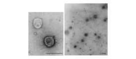
- Streptavidin magnetic beads coupled with biotinylated Tim4 were added to the supernatant and the mixture was rotated at 20-25°C for 1 hour. The beads were washed three times with wash buffer and bound uEVs were eluted with elution buffer. It is a negative Tein transmission electron microscope image of uEV. Scale bar indicates 200 nm.
- Nanoparticle tracking analysis of uEVs isolated from healthy controls Venn diagram of total protein detected in uEVs isolated from healthy controls. Bar graphs representing the abundance of common classical exosome markers (CD63, CD9) and classical microvesicle markers (Annexin A1) and ARRDC1-mediated microvesicle markers (TSG101) in healthy controls. The y-axis represents the log10 relative abundance.
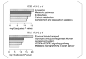
- KEGG/Wiki pathway analysis results of 1,298 common proteins contained in uEV from healthy controls (g: Profiler). The top 5 terms with the lowest adjusted P-values were extracted. Kidney cell explorer visualization of the relative abundance in nephron fractions of the top 50 molecules in uEV from healthy controls and immunofluorescence images of 5 of the 50 molecules in human kidney specimens.
- Glutathione hydrolase 1 proenzyme (GGT1), phosphoglycerate kinase 1 (PGK1), uromodulin (UMOD), annexin A11 (ANXA11), and keratin 14 (KRT14) regulate proximal, Henle's loop, and distal tubules, respectively. , collecting ducts, and deep medullary pelvic epithelium.
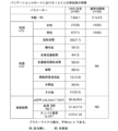
- the numbers above the figure represent each nephron segment. 1 podocyte; 2 apical cortex; 3 proximal tubules; 4 Henle's loops; 5 distal tubules; Scale bar represents 200 ⁇ m. Table showing baseline characteristics of Discovery cohort participants.
- FIG. 3 is a correlation plot of particle count and urinary creatinine;
- B Particle size versus particle correlation plot.
- C Correlation plot of particle size and urinary creatinine. Correlation coefficients are expressed as Pearson's R.
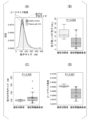
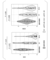
- A Density plot showing particle size distribution in healthy controls and CKD patients. Dotted lines indicate the average size of particles in each group.
- B Boxplot comparing particle counts between healthy control and CKD patient samples.
- C Boxplot comparing mean particle size between healthy control and CKD patient samples.
- D Boxplot comparing peak size densities between healthy control and CKD patient samples. Data were compared using a two-tailed Welch's t-test.
- FIG. 1 shows the selection process of candidate molecules for ELISA.
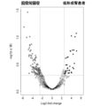
- MGAM was the only transmembrane protein among 35 molecules that were elevated in bilateral hypoplastic kidney or CKD uEV.
- MUC1 was particularly suitable for evaluation by Tim4-ELISA.
- Standard curves for MUC1, MGAM, and exosome markers (CD9 and CD63) are obtained by plotting absorbance at 450 nm (y-axis) against values of standard concentration (x-axis).
- R2 is the correlation coefficient of the standard curve.
- A Schematic of sandwich ELISA using Tim4.
- B Immunostaining image showing expression of MGAM, MUC1, and CD9 in nephron segments.
- FIG. 10 is a graph comparing MUC1 and MGAM levels (absorbance at 450 nm) measured by customized ELISA in samples from healthy controls and CKD patients.
- G1 indicates CKD patients with eGFR ⁇ 90 and G2-5 indicates CKD patients with eGFR ⁇ 90 mL/min/1.73 m 2 .
- A ROC curve for distinguishing patients with reduced renal function (eGFR ⁇ 60) from healthy controls by logistic regression.
- B ROC curve for distinguishing patients with reduced renal function (eGFR ⁇ 90) from healthy controls by logistic regression.
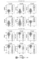
- C ROC curve for distinguishing CKD patients with normal eGFR ⁇ 90 from healthy controls by logistic regression. Box and beeswarm plots of MGAM/MUC1 assay values (MGAM expression divided by MUC1 expression) in CKD patients and healthy controls with each renal function in the discovery and validation cohorts.
- FIG. 10 is a graph examining the expression of exosome markers (CD9 and CD63) in uEVs of the validation cohort.
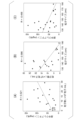
- FIG. 10 is a graph examining the correlation between urinary albumin and ELISA expression levels of MGAM, MUC1, CD9, and CD63.
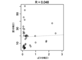
- FIG. 10 is a graph examining the correlation between uEV and urinary MUC1 expression levels.
- FIG. Correlation plots using 40 randomly selected cases from the validation cohort are shown. The correlation coefficient is expressed as Pearson's R. Values on the x-axis indicate absorbance at 450 nm by ELISA, and values on the y-axis represent urinary concentrations of MUC1 (U/mL).
- ⁇ Diagnostic marker for renal dysfunction ⁇ This embodiment provides a renal dysfunction diagnostic marker comprising MUC1 and/or MGAM in subject-derived urinary extracellular vesicles.
- Diseases that can be tested as renal dysfunction include chronic kidney disease, congenital renal and urinary tract abnormalities, hypoplastic kidney, dysplastic kidney, renal tubular dysgenesis, hydronephrosis, single kidney, and polycystic dysplastic kidney. , nephron aplasia, ciliopathy, autosomal recessive polycystic kidney disease, autosomal dominant polycystic kidney disease, renal scar, vesicoureteral reflux disease, megaureter, fused kidney, horseshoe kidney, and the like.
- the present inventors comprehensively analyzed proteins in uEVs isolated from patients with hypoplastic kidney, and discovered markers for diagnosing renal dysfunction.
- Subjects are not particularly limited, and include pediatric renal disease patients, adult renal disease patients, and the like.
- MUC1 (Mucin1) is a type I transmembrane glycoprotein belonging to the family of mucin proteins, a protein consisting of an extracellular domain, a transmembrane domain and a short cytoplasmic domain. The expression level was decreased in uEV derived from patients with hypoplastic kidney.
- MGAM (Maltase-Glucoamylase) is an ⁇ -glucosidase digestive enzyme and a transmembrane protein. The expression level was increased in uEV derived from patients with hypoplastic kidney.
- the urinary extracellular vesicles are preferably phosphatidylserine-positive, and the renal dysfunction diagnostic marker of the present embodiment preferably further includes CD9.
- CD9 is a cell surface glycoprotein found on the surface of exosomes. MGAM; MUC1 and MGAM; MUC1 and CD9; MGAM and CD9; MUC1, MGAM and CD9; The greater the number of marker molecules, the more accurate the diagnosis.
- kits for detecting a diagnostic marker for renal dysfunction comprising an anti-MUC1 antibody and/or an anti-MGAM antibody.
- Antibodies are not limited as long as they recognize antigens, and include monoclonal antibodies, polyclonal antibodies, multispecific antibodies (eg, bispecific antibodies), antibody fragments, and the like.
- the kit of this embodiment preferably further contains a substance with an affinity for urinary extracellular vesicles.
- Substances with affinity for urinary extracellular vesicles include antibodies against molecules expressed on urinary extracellular vesicle membranes such as CD9, and phospholipid affinity substances. Phosphatidylserine affinity substances are preferred, and Tim4 is more preferred.
- the kit of this embodiment preferably further contains a solid phase or carrier bound to a substance with an affinity for urinary extracellular vesicles.
- the solid phase include glass substrates, silicon substrates, plastic substrates, metal substrates, etc. Plastic substrates used for ELISA and the like are preferred.
- Carriers include beads of silicon, titanium dioxide, aluminum oxide, glass, polystyrene, cellulose, polyamides, and the like.
- the kit of this embodiment preferably contains a plastic substrate on which Tim4 is immobilized and an antibody against MUC1 and/or MGAM.
- a method for using the kit of the present embodiment urinary extracellular vesicles of a subject are captured on a Tim4-immobilized plastic substrate, and then labeled antibodies against MUC1 and/or MGAM are used to extract urinary extracellular vesicles.
- a method for detecting MUC1 and/or MGAM expressed on the vesicle outer surface is included.
- Substances used for labeling antibodies against MUC1 and/or MGAM include labeling enzymes such as horseradish peroxidase and alkaline phosphatase.
- the present embodiment provides a method for detecting a diagnostic marker for renal dysfunction, comprising measuring the expression levels of MUC1 and/or MGAM in urinary extracellular vesicles derived from a subject.
- MUC1 protein expression in the specimen If the amount is less than the control expression level of the poor prognosis control, it can be predicted that the subject has impaired renal function.
- the MGAM protein expression level in urinary extracellular vesicles derived from the subject and the control expression level of a control subject with a known poor prognosis were compared to compare both expression levels, and MGAM protein expression in the sample If the amount is greater than the control expression level in poor prognosis controls, it can be predicted that the subject has impaired renal function.
- the urinary extracellular vesicles are preferably positive for phosphatidylserine, and the method for detecting a marker for diagnosing renal dysfunction of the present embodiment further comprises measuring the expression level of CD9 in the urinary extracellular vesicles. is preferred. Furthermore, the accuracy of diagnosis is improved by measuring the expression level of CD9.
- This embodiment is a method for determining renal prognosis, in which the ratio of the expression level of MGAM and the expression level of MUC1 in urinary extracellular vesicles derived from a subject is calculated. Provided is a determination method for determining that a patient has a poor prognosis in some cases.
- the average eGFR at onset of patients with MGAM/MUC1 of 0.35 or more was reduced to It was confirmed that the eGFR decreased significantly in some patients, lower than the group.
- the urinary extracellular vesicles are preferably phosphatidylserine positive.
- the protein composition of uEVs separated from control samples was analyzed using liquid chromatography-tandem mass spectrometry (LC-MS/MS). Purified uEVs were individually analyzed using an LC-MS/MS system.
- a subsequent Sequest database search identified 1,298 non-redundant proteins in all three control samples (see Figure 5). This list included most non-tissue specific EV proteins (category 1a) and EV-recovered cytosolic proteins (category 2).
- common exosome markers CD63 and CD9
- ANXA1 classical microvesicle markers
- ARMM ARRDC1-mediated microvesicle
- uEVs from all patients with hypoplastic kidney contained marker proteins for classical exosomes (CD63 and CD9), classical microvesicles (ANXA1), and ARMM (TSG101) (see Figure 10). .).
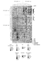
- Multidimensional scaling (MDS) analysis using the expression levels of these 135 proteins in uEV differentiated between healthy controls and patients with hypoplastic kidneys see Figure 12).
- the mean eGFR for patients in cluster 1 was 99.2 mL/min/1.73 m 2 and the mean eGFR for patients in cluster 2 was significantly reduced to 46.0 mL/min/1.73 m 2 .
- the quantities are shown in FIG. MDS analysis using quantitative proteomic data showed that the uEV proteome could distinguish patients with renal impairment (see Figure 16). This result suggests that the expression of these proteins may be used to screen urine from patients with impaired renal function.
- MUC1 is a transmembrane glycoprotein whose expression is restricted to the apical surface of distal tubules and collecting ducts.
- MGAM which is an ⁇ -glucosidase, is expressed only in kidney proximal tubular cells (see FIG. 22(B)).
- CD9 and CD63 are markers of these small EVs ( See Figure 21).
- CD9 is mainly expressed on the basement membrane side of distal tubules and collecting ducts (see Figure 22B).
- MUC1 concentrations were examined by ELISA.
- the sample size required to discriminate renal hypofunction eGFR ⁇ 60 or ⁇ 90 by MUC1 was calculated to be 10.8 or 23.9, respectively.
- Urine samples were collected from 26 controls and 94 pediatric patients with CKD (validation cohort; see Figure 23). Reflecting the etiology of pediatric CKD, patients predominantly suffer from CAKUT and none with diabetes or diabetic kidney disease.
- MUC1, MGAM, CD9, and CD63 levels in uEVs were quantified by ELISA (see Figures 24 and 28).
- the MGAM/MUC1 value yielded an AUC of 0.922 for segregating patients with renal impairment (eGFR ⁇ 90) (see FIG. 27(A)).
- the sensitivity and specificity of MGAM/MUC1 for diagnosing eGFR depression were 88.5% and 87.5%, respectively, based on optimal cut-off values derived from ROC curves. It achieves significantly higher accuracy than urinary creatinine, albumin, or L-FABP, suggesting that the new method of the present application captures changes that cannot be detected by existing urinary biomarkers (see Figure 27 (A)). .
- renal dysfunction can be diagnosed with high precision and non-invasiveness.
Landscapes
- Life Sciences & Earth Sciences (AREA)
- Health & Medical Sciences (AREA)
- Engineering & Computer Science (AREA)
- Molecular Biology (AREA)
- Chemical & Material Sciences (AREA)
- Biomedical Technology (AREA)
- Urology & Nephrology (AREA)
- Hematology (AREA)
- Immunology (AREA)
- Biotechnology (AREA)
- Microbiology (AREA)
- Cell Biology (AREA)
- Proteomics, Peptides & Aminoacids (AREA)
- Food Science & Technology (AREA)
- Medicinal Chemistry (AREA)
- Physics & Mathematics (AREA)
- Analytical Chemistry (AREA)
- Biochemistry (AREA)
- General Health & Medical Sciences (AREA)
- General Physics & Mathematics (AREA)
- Pathology (AREA)
- Investigating Or Analysing Biological Materials (AREA)
Priority Applications (3)
| Application Number | Priority Date | Filing Date | Title |
|---|---|---|---|
| US18/703,972 US20240418733A1 (en) | 2015-12-29 | 2022-10-26 | Method for detecting kidney dysfunction diagnostic marker, method for determining renal prognosis, detection kit for kidney dysfunction diagnostic marker, and kidney dysfunction diagnostic marker |
| EP22887048.1A EP4425182A4 (en) | 2021-10-27 | 2022-10-26 | METHOD FOR DETECTING A DIAGNOSTIC MARKER FOR RENAL DYSFUNCTION, METHOD FOR DETERMINING THE PROGNOSIS OF NEPHROPATHY, KIT FOR DETECTING A DIAGNOSTIC MARKER FOR RENAL DYSFUNCTION, AND DIAGNOSTIC MARKER FOR RENAL DYSFUNCTION |
| JP2023556592A JPWO2023074723A1 (https=) | 2021-10-27 | 2022-10-26 |
Applications Claiming Priority (2)
| Application Number | Priority Date | Filing Date | Title |
|---|---|---|---|
| US202163272499P | 2021-10-27 | 2021-10-27 | |
| US63/272,499 | 2021-10-27 |
Publications (1)
| Publication Number | Publication Date |
|---|---|
| WO2023074723A1 true WO2023074723A1 (ja) | 2023-05-04 |
Family
ID=86157933
Family Applications (1)
| Application Number | Title | Priority Date | Filing Date |
|---|---|---|---|
| PCT/JP2022/039851 Ceased WO2023074723A1 (ja) | 2015-12-29 | 2022-10-26 | 腎機能障害診断マーカーの検出方法、腎予後の判定方法、腎機能障害診断マーカーの検出キット及び腎機能障害診断マーカー |
Country Status (3)
| Country | Link |
|---|---|
| EP (1) | EP4425182A4 (https=) |
| JP (1) | JPWO2023074723A1 (https=) |
| WO (1) | WO2023074723A1 (https=) |
Citations (4)
| Publication number | Priority date | Publication date | Assignee | Title |
|---|---|---|---|---|
| US20150252423A1 (en) * | 2012-08-10 | 2015-09-10 | The Broad Institute, Inc. | Methods of diagnosing and treating medullary cystic kidney disease |
| JP2018191636A (ja) * | 2017-05-12 | 2018-12-06 | 国立大学法人広島大学 | 癌の診断デバイス |
| WO2021106312A1 (ja) * | 2019-11-27 | 2021-06-03 | 国立大学法人岡山大学 | 腫瘍融解ウイルスを含有する生体由来の細胞外小胞 |
| US20210169827A1 (en) * | 2019-06-21 | 2021-06-10 | The Broad Institute, Inc. | Agents for reversing toxic proteinopathies |
-
2022
- 2022-10-26 JP JP2023556592A patent/JPWO2023074723A1/ja active Pending
- 2022-10-26 WO PCT/JP2022/039851 patent/WO2023074723A1/ja not_active Ceased
- 2022-10-26 EP EP22887048.1A patent/EP4425182A4/en active Pending
Patent Citations (4)
| Publication number | Priority date | Publication date | Assignee | Title |
|---|---|---|---|---|
| US20150252423A1 (en) * | 2012-08-10 | 2015-09-10 | The Broad Institute, Inc. | Methods of diagnosing and treating medullary cystic kidney disease |
| JP2018191636A (ja) * | 2017-05-12 | 2018-12-06 | 国立大学法人広島大学 | 癌の診断デバイス |
| US20210169827A1 (en) * | 2019-06-21 | 2021-06-10 | The Broad Institute, Inc. | Agents for reversing toxic proteinopathies |
| WO2021106312A1 (ja) * | 2019-11-27 | 2021-06-03 | 国立大学法人岡山大学 | 腫瘍融解ウイルスを含有する生体由来の細胞外小胞 |
Non-Patent Citations (7)
| Title |
|---|
| AWDISHU LINDA, TSUNODA SHIRLEY, PEARLMAN MICHELLE, KOKOY-MONDRAGON CHANTHEL, GHASSEMIAN MAJID, NAVIAUX ROBERT K., PATTON HEATHER M: "Identification of Maltase Glucoamylase as a Biomarker of Acute Kidney Injury in Patients with Cirrhosis", CRITICAL CARE RESEARCH AND PRACTICE, vol. 2019, 16 April 2019 (2019-04-16), pages 1 - 8, XP093062051, ISSN: 2090-1305, DOI: 10.1155/2019/5912804 * |
| BOURDERIOUX, M. ET AL.: "IDENTIFICATION OF URINARY EXOSOMAL PROTEINS BY MASS SPECTROMETRY", NEPHROLOGY DIALYSIS TRANSPLANTATION, vol. 27, no. suppl. 2, 2012, pages ii76, XP009545337 * |
| ERDBRUGGER U ET AL.: "Urinary extracellular vesicles: A position paper by the Urine Task Force of the International Society for Extracellular Vesicles", J EXTRACELL VESICLES, vol. 10, no. 7, May 2021 (2021-05-01), pages 2093 |
| ERDBRUGGER ULE TH.: "Extracellular Vesicles in Renal Diseases: More than Novel Biomarkers?", J AM SOC NEPHROL, vol. 27, no. 1, 6 August 2015 (2015-08-06), pages 12 - 26, XP055607375, DOI: 10.1681/ASN.2015010074 |
| See also references of EP4425182A4 |
| STAUBACH SIMON, WENZEL ANDREA, BECK BODO B., RINSCHEN MARKUS M., MÜLLER STEFAN, HANISCH FRANZ-GEORG: "Autosomal Tubulointerstitial Kidney Disease-MUC1 Type: Differential Proteomics Suggests that Mutated MUC1 (insC) Affects Vesicular Transport in Renal Epithelial Cells", PROTEOMICS, vol. 18, no. 7, 1 April 2018 (2018-04-01), DE , pages 1700456, XP093062055, ISSN: 1615-9853, DOI: 10.1002/pmic.201700456 * |
| YAMAMOTO SATOKO, KAIMORI JUN-YA, YOSHIMURA TAKUJI, NAMBA TOMOKO, IMAI ATSUKO, KOBAYASHI KAORI, IMAMURA RYOICHI, ICHIMARU NAOTSUGU,: "Analysis of an ADTKD family with a novel frameshift mutation in MUC1 reveals characteristic features of mutant MUC1 protein", NEPHROLOGY DIALYSIS TRANSPLANTATION, vol. 32, no. 12, 1 December 2017 (2017-12-01), GB , pages 2010 - 2017, XP093062053, ISSN: 0931-0509, DOI: 10.1093/ndt/gfx083 * |
Also Published As
| Publication number | Publication date |
|---|---|
| JPWO2023074723A1 (https=) | 2023-05-04 |
| EP4425182A4 (en) | 2025-09-24 |
| EP4425182A1 (en) | 2024-09-04 |
Similar Documents
| Publication | Publication Date | Title |
|---|---|---|
| US20240418733A1 (en) | Method for detecting kidney dysfunction diagnostic marker, method for determining renal prognosis, detection kit for kidney dysfunction diagnostic marker, and kidney dysfunction diagnostic marker | |
| CN110108889B (zh) | 一种用于诊断IgA肾病的试剂盒及其应用 | |
| Urrego et al. | Utility of urinary transferrin and ceruloplasmin in patients with systemic lupus erythematosus for differentiating patients with lupus nephritis | |
| JP2026067922A (ja) | 補体の活性化を検出するための細胞外小胞の使用方法、並びに補体性疾患の処置を評価及び/又は監視するためのその使用 | |
| WO2009090882A1 (ja) | 非アルコール性脂肪性肝疾患の診断方法 | |
| US20130210667A1 (en) | Biomarkers for Predicting Kidney and Glomerular Pathologies | |
| Yoshimura et al. | Galectin-10 in serum extracellular vesicles reflects asthma pathophysiology | |
| JP2024059621A (ja) | がんの診断及び治療のための組成物及び方法 | |
| JP2022000635A (ja) | 新型コロナウイルス感染者の重症化リスクの検査方法、その検査キット、コンパニオン診断薬及びその重症化リスクマーカー | |
| CN114441760A (zh) | 一种用于肝癌诊断的生物标志物和试剂盒及检测方法 | |
| TWI822802B (zh) | 腎病變蛋白生物標記及其應用 | |
| CN116298323B (zh) | 一种用于诊断狼疮性肾炎的生物标志物及应用 | |
| WO2023074723A1 (ja) | 腎機能障害診断マーカーの検出方法、腎予後の判定方法、腎機能障害診断マーカーの検出キット及び腎機能障害診断マーカー | |
| Zhang et al. | Liquid Biopsy in Lung Cancer: Nano–Flow Cytometry Detection of Non–Small Cell Lung Cancer in Blood | |
| US20100047830A1 (en) | Compositions and methods for detecting cancers in a subject | |
| US20240036050A1 (en) | Methods Of Detecting And Treating Hepatocellular Carcinoma | |
| WO2024067470A1 (zh) | 一种用于检测早期肝癌的试剂盒 | |
| Huang et al. | Untargeted metabolomic analysis in endolymphatic sac luminal fluid from patients with Meniere’s Disease | |
| WO2021162030A1 (ja) | 薬剤性間質性肺炎の代謝物診断バイオマーカー | |
| CN119881329B (zh) | 携带Tau的星形胶质细胞来源细胞外囊泡在辅助诊断进行性核上性麻痹中的应用 | |
| US20180275135A1 (en) | Biomarker panel to identify steroid resistance in childhood idiopathic nephrotic syndrome | |
| CN113884684B (zh) | 一种活动性结核病多组学整合标志物、试剂盒及检测模型的构建方法 | |
| CN116087519B (zh) | 一种用于高危人群贲门腺癌早期筛查的标志物及其应用 | |
| JPWO2019098328A1 (ja) | IgA腎症診断用キット | |
| CN121978343A (en) | Application of urine TRPV5 as biomarker in chronic kidney disease diagnosis and risk assessment |
Legal Events
| Date | Code | Title | Description |
|---|---|---|---|
| 121 | Ep: the epo has been informed by wipo that ep was designated in this application |
Ref document number: 22887048 Country of ref document: EP Kind code of ref document: A1 |
|
| WWE | Wipo information: entry into national phase |
Ref document number: 18703972 Country of ref document: US |
|
| ENP | Entry into the national phase |
Ref document number: 2023556592 Country of ref document: JP Kind code of ref document: A |
|
| WWE | Wipo information: entry into national phase |
Ref document number: 2022887048 Country of ref document: EP |
|
| NENP | Non-entry into the national phase |
Ref country code: DE |
|
| ENP | Entry into the national phase |
Ref document number: 2022887048 Country of ref document: EP Effective date: 20240527 |