WO2022075477A1 - 乾燥脱細胞化組織の製造方法、脱細胞化組織の保存前処理方法、及び、乾燥脱細胞化組織 - Google Patents
乾燥脱細胞化組織の製造方法、脱細胞化組織の保存前処理方法、及び、乾燥脱細胞化組織 Download PDFInfo
- Publication number
- WO2022075477A1 WO2022075477A1 PCT/JP2021/037486 JP2021037486W WO2022075477A1 WO 2022075477 A1 WO2022075477 A1 WO 2022075477A1 JP 2021037486 W JP2021037486 W JP 2021037486W WO 2022075477 A1 WO2022075477 A1 WO 2022075477A1
- Authority
- WO
- WIPO (PCT)
- Prior art keywords
- decellularized tissue
- decellularized
- tissue
- sucrose
- dry
- Prior art date
- Legal status (The legal status is an assumption and is not a legal conclusion. Google has not performed a legal analysis and makes no representation as to the accuracy of the status listed.)
- Ceased
Links
Classifications
-
- A—HUMAN NECESSITIES
- A61—MEDICAL OR VETERINARY SCIENCE; HYGIENE
- A61K—PREPARATIONS FOR MEDICAL, DENTAL OR TOILETRY PURPOSES
- A61K35/00—Medicinal preparations containing materials or reaction products thereof with undetermined constitution
- A61K35/12—Materials from mammals; Compositions comprising non-specified tissues or cells; Compositions comprising non-embryonic stem cells; Genetically modified cells
- A61K35/30—Nerves; Brain; Eyes; Corneal cells; Cerebrospinal fluid; Neuronal stem cells; Neuronal precursor cells; Glial cells; Oligodendrocytes; Schwann cells; Astroglia; Astrocytes; Choroid plexus; Spinal cord tissue
-
- A—HUMAN NECESSITIES
- A61—MEDICAL OR VETERINARY SCIENCE; HYGIENE
- A61L—METHODS OR APPARATUS FOR STERILISING MATERIALS OR OBJECTS IN GENERAL; DISINFECTION, STERILISATION OR DEODORISATION OF AIR; CHEMICAL ASPECTS OF BANDAGES, DRESSINGS, ABSORBENT PADS OR SURGICAL ARTICLES; MATERIALS FOR BANDAGES, DRESSINGS, ABSORBENT PADS OR SURGICAL ARTICLES
- A61L27/00—Materials for grafts or prostheses or for coating grafts or prostheses
- A61L27/36—Materials for grafts or prostheses or for coating grafts or prostheses containing ingredients of undetermined constitution or reaction products thereof, e.g. transplant tissue, natural bone, extracellular matrix
-
- A—HUMAN NECESSITIES
- A61—MEDICAL OR VETERINARY SCIENCE; HYGIENE
- A61P—SPECIFIC THERAPEUTIC ACTIVITY OF CHEMICAL COMPOUNDS OR MEDICINAL PREPARATIONS
- A61P27/00—Drugs for disorders of the senses
- A61P27/02—Ophthalmic agents
-
- C—CHEMISTRY; METALLURGY
- C12—BIOCHEMISTRY; BEER; SPIRITS; WINE; VINEGAR; MICROBIOLOGY; ENZYMOLOGY; MUTATION OR GENETIC ENGINEERING
- C12N—MICROORGANISMS OR ENZYMES; COMPOSITIONS THEREOF; PROPAGATING, PRESERVING, OR MAINTAINING MICROORGANISMS; MUTATION OR GENETIC ENGINEERING; CULTURE MEDIA
- C12N5/00—Undifferentiated human, animal or plant cells, e.g. cell lines; Tissues; Cultivation or maintenance thereof; Culture media therefor
- C12N5/06—Animal cells or tissues; Human cells or tissues
Definitions
- the present invention relates to a method for producing dried decellularized tissue, a method for preserving decellularized tissue, and a method for preserving dry decellularized tissue.
- an artificial cornea is expected as a means for solving such a problem.
- the material of the artificial cornea is required to have high compatibility with the living tissue in order to prevent dropping and infectious diseases at the junction between the living tissue and the artificial cornea.
- Patent Document 1 proposes a decellularized treatment solution containing a predetermined compound.
- the decellularized tissue may be stored in a solution such as a preservative solution until it is used for transplantation after its production.
- the decellularized tissue becomes cloudy when it is stored in the solution for a long period of time.
- a commercially available storage solution preservation solution according to the type of tissue such as corneal preservation solution
- cloudiness was observed in about one week.
- the inventors have found that it is difficult to restore decellularized tissue without making it cloudy even if it is stored dry instead of stored in solution.
- the present invention has been made in view of the above circumstances, and an object of the present invention is to provide a technique capable of suppressing white turbidity of decellularized tissue after storage (particularly after restoration).
- the present inventors have described the above problems according to a method for producing a dry decellularized tissue, which comprises performing a dipping treatment of the decellularized tissue in a predetermined medium and a vacuum drying treatment in this order.
- a method for producing a dry decellularized tissue which comprises performing a dipping treatment of the decellularized tissue in a predetermined medium and a vacuum drying treatment in this order.
- a dipping step of immersing the decellularized tissue in a sucrose-containing medium and After the dipping step, a vacuum drying step of drying the decellularized tissue under reduced pressure and a vacuum drying step of drying the decellularized tissue under reduced pressure Including
- the sucrose concentration in the sucrose-containing medium is 5% by mass or more and 55% by mass or less.
- a dipping step of immersing the decellularized tissue in a sucrose-containing medium and After the dipping step, a vacuum drying step of drying the decellularized tissue under reduced pressure and a vacuum drying step of drying the decellularized tissue under reduced pressure Obtained from methods including
- the sucrose concentration in the sucrose-containing medium is 5% by mass or more and 55% by mass or less. Dry decellularized tissue.
- the method for producing a dry decellularized tissue of the present invention includes a dipping step of immersing the decellularized tissue in a sucrose-containing medium, and after the dipping step, the decellularized tissue.
- the vacuum drying step of drying the chemical tissue under reduced pressure is included, and the sucrose concentration in the sucrose-containing medium is 5% by mass or more and 55% by mass or less.
- the production method of the present invention is a method for obtaining a dry decellularized tissue by subjecting the decellularized tissue to a predetermined treatment.
- the dried decellularized tissue dehydrated with a sucrose-containing medium and then dried under reduced pressure can be stored not only in a dry state but also restored using a medium or the like. A surprising finding was found that the white turbidity was suppressed.
- the tissue from which the decellularized tissue is derived (cornea, etc.) usually swells in a solution (for example, washing solution, medium, etc.), and the transparency decreases. This is a state in which the order of the collagen fibers that make up the tissue (corneal tissue, etc.) is disturbed (however, basically, the gap between the collagen fibers is only increased and the structure itself is not broken). This is because it scatters light. Therefore, when the decellularized tissue is stored in a conventional storage solution, it is extremely difficult to suppress the white turbidity of the tissue.
- sucrose has a dehydrating effect on tissues and an anti-icing effect on tissues when frozen. Due to these actions by sucrose, the tissue swollen in the process of producing decellularized tissue can be dehydrated by sucrose to arrange the disorder of the ordered arrangement of collagen fibers, and as a result, the scattering of light is suppressed. Can improve transparency. Furthermore, by performing vacuum drying, the decellularized tissue whose transparency has been improved by sucrose can be maintained as it is. In such a case, since there is almost no free water in the tissue, swelling is unlikely to occur. In addition, since sugar (sucrose, etc.) remains in the tissue, the stability is improved. As a result, even if it is stored for a long period of time, it can be restored in a state where the white turbidity of the decellularized tissue is suppressed.
- the present inventors have confirmed that when the decellularized tissue is frozen without being immersed in sucrose, ice crystals are formed, the structure of the tissue itself is destroyed, and even if it is restored, remarkable cloudiness occurs. bottom.
- preservation of decellularized tissue is an arbitrary means under the condition that the dry decellularized tissue obtained by the production method of the present invention is difficult to increase or does not increase the water content in the tissue. And it means to save under the conditions.
- Examples of the storage means include vacuum storage, storage with an inert gas, and the like.
- Examples of the storage temperature include -80 to 30 ° C.
- the storage humidity (relative humidity) is, for example, 0 to 30%.
- white turbidity of decellularized tissue after storage is suppressed means that the white turbidity of the dry decellularized tissue obtained by the production method of the present invention is suppressed at any timing after storage. It means that it is.
- whitening of decellularized tissue after restoration is suppressed means that the dry decellularized tissue obtained by the production method of the present invention is decellularized under the condition of increasing the amount of water in the tissue. It means that the white turbidity when the decellularized tissue is restored is suppressed.
- the "decellularized tissue after restoration” may have transparency closer to the tissue from which the decellularized tissue is derived (the one just collected).
- "restoration of decellularized tissue” is to bring the dried decellularized tissue closer to the state before drying, for example, by supplying an appropriate amount of water to the dried decellularized tissue and using it for various purposes. It is possible to make it a form applicable to a transplant piece, etc.).
- the transparency of the decellularized cornea is evaluated by the method shown in the examples.
- Decellularized tissue In the production method of the present invention, decellularized tissue produced by a conventionally known means using any biological tissue can be used.
- the biological tissue from which the decellularized tissue is derived is not particularly limited as long as it is a tissue in which cloudiness can be a problem, and examples thereof include corneal tissue and submucosal tissue of the small intestine.
- the cornea is preferable as the biological tissue from which the decellularized tissue is derived from the viewpoint that the effect of the present invention is particularly likely to be exerted.
- the decellularized tissue in the present invention is preferably a decellularized cornea.
- the animal from which the biological tissue is derived is not particularly limited, but mammals such as pigs, cows, horses, goats, sheep, rabbits, kangaroos, monkeys, and humans can be mentioned.
- the method for decellularizing living tissue is not particularly limited, but may include, for example, a pressure application step, a cell residue removal step, a washing step, and the like.
- Examples of the application step include applying ultra-high hydrostatic pressure to the living tissue in an arbitrary decellularization treatment solution. By this step, cells in living tissue can be destroyed.
- Examples of the decellularization treatment solution used in the application step include a medium (M199 medium, MEM ⁇ medium, a mixed medium thereof, etc.).
- Ultra-high hydrostatic pressure means hydrostatic pressure that is said to be able to destroy indigenous bacteria, bacteria and viral cells present in soft tissues.
- the lower limit of the ultra-high hydrostatic pressure is preferably 2000 atm or more.
- the upper limit of the ultra-high hydrostatic pressure is preferably 10,000 atm or less.
- an ultra-high hydrostatic pressure treatment device for example, "Dr. CHEF (model)” (manufactured by Kobe Steel, Ltd.)
- Dr. CHEF model
- the like can be used.
- the application time in the application step is not particularly limited as long as the cells are satisfactorily destroyed, and is usually about 10 to 40 minutes in total (for example, pressurization 5 minutes, maintenance 10 minutes, decompression 5 minutes), and decellularization at the time of application.
- the temperature of the cell treatment solution is usually 5 to 40 ° C, preferably 5 to 15 ° C.
- Examples of the removal step include accelerating the removal of cell residues by immersing, convection, or circulating the biological tissue together with the washing solution after the application step.
- the cleaning liquid used in the removal step is not particularly limited, and examples thereof include any cleaning liquid capable of suppressing deterioration of the structure and characteristics of the tissue.
- the cleaning solution examples include known cell culture solutions (for example, decellularization treatment solutions similar to those in the application step), PBS aqueous solutions, HEPES buffer solutions, MES buffer solutions and the like.
- the washing solution may contain a DNA degrading enzyme (DNase).
- DNase DNA degrading enzyme
- the temperature condition in the removal step is preferably 20 to 37 ° C, more preferably 22 to 25 ° C.
- the treatment time in the removal step is preferably about 24 to 72 hours, more preferably about 48 to 72 hours.
- the cleaning solution may be changed as appropriate (for example, once).
- the decellularized tissue obtained from the living tissue may be directly subjected to a dipping step or the like, or may be stored in a refrigerator or a freezer and then subjected to a dipping step or the like. From the viewpoint that the effect of the present invention can be easily exerted, the decellularized tissue obtained from the living tissue can be directly subjected to a dipping step or the like (that is, after preparing the decellularized tissue without going through other steps). preferable.
- the dipping step is a step of immersing the decellularized tissue in a medium having a sucrose concentration of 5% by mass or more and 55% by mass or less.
- sucrose concentration When the sucrose concentration is less than 5% by mass, swelling is likely to occur. When the sucrose concentration is more than 55% by mass, the decellularized tissue contracts, and the function as the decellularized tissue is impaired.
- the lower limit of the sucrose concentration of the sucrose-containing medium used in the dipping step is preferably 10% by mass or more, more preferably 20% by mass or more, still more preferably 30% by mass or more, and most preferably 35% by mass or more.
- the upper limit of the sucrose concentration of the sucrose-containing medium used in the dipping step is preferably 53% by mass or less, more preferably 52% by mass or less, still more preferably 50% by mass or less.
- the medium to which sucrose used in the dipping step is to be blended is not particularly limited as long as it does not affect the structure of decellularized tissue, but M199 medium, MEM ⁇ medium, a mixed medium thereof, physiological saline, and the like.
- a buffer solution and the like can be mentioned.
- the time for immersing the decellularized tissue in the sucrose-containing medium is not particularly limited as long as the water in the decellularized tissue can be sufficiently dehydrated and the water in the decellularized tissue can be sufficiently replaced with sucrose. However, it is preferably 30 minutes or more and 24 hours or less, and more preferably 1 hour or more and 6 hours or less.
- the temperature condition for immersing the decellularized tissue in the sucrose-containing medium is not particularly limited, but the immersing step is usually performed in a room temperature environment (for example, 4 to 37 ° C.). In such cases, the medium temperature of the sucrose-containing medium is usually 4 to 25 ° C.
- the sucrose-containing medium may or may not contain sucrose, the medium, and other components as long as the action of sucrose is not inhibited.
- the component that can be contained in the sucrose-containing medium include tocopherol, ascorbic acid and the like.
- the blending amount of these components can be appropriately set according to the effect to be obtained and the like.
- the vacuum drying step is a step of drying the decellularized tissue under reduced pressure after the dipping step.
- the conditions for vacuum drying in the vacuum drying step are not particularly limited, and any conditions that can reduce the water content of the decellularized tissue under reduced pressure can be adopted.
- vacuum drying may be performed using a program freezer.
- the dry decellularized tissue obtained by the production method of the present invention can be stored under any conditions.
- the dried decellularized tissue is preferably stored immediately after the vacuum drying step (ie, it does not include any other steps between the vacuum drying and storage steps).
- an embodiment including another step between the vacuum drying step and the storage step is not excluded.
- it is desirable that such other steps do not include a step (washing, operation under high humidity, etc.) in which the water content of the dried decellularized tissue is excessively increased.
- dry means that the water content in the decellularized tissue is sufficiently low (for example, the water content with respect to the mass of the decellularized tissue is 5% by mass or less).
- the water content in the decellularized tissue is specified by an infrared / halogen moisture meter.
- the storage method is not particularly limited, but the dried decellularized tissue may be placed in a container (vial bottle, plastic container, etc.) and stored.
- the storage temperature is not particularly limited, but is usually 4 to 10 ° C. from the viewpoint of not affecting the structure of the decellularized tissue.
- the storage time is not particularly limited, and according to the present invention, the white turbidity of the decellularized tissue after restoration is suppressed even if the storage time is long.
- the storage time may be, for example, 1 day or more and 2 years or less.
- the decellularized tissue can be restored by any method, so that it can be used for desired purposes (transplantation, etc.) according to the type of tissue from which the decellularized tissue is derived.
- the method for restoring the decellularized tissue is not particularly limited, but any method (simple immersion, vacuum impregnation, vacuum pressure impregnation, etc.) capable of supplying water or the like into the decellularized tissue can be used.
- the solution used to restore decellularized tissue is not particularly limited, but is limited to a sugar (Dextran 70, etc.) solution, any buffer, saline, and any preservation solution known as tissue preservation solution (eg, corneal tissue preservation). Examples thereof include "Optisol-GS" known as a liquid.
- the temperature condition for restoration of decellularized tissue is not particularly limited, but room temperature (for example, 4 ° C or higher and 25 ° C or lower) is preferable.
- the time for restoring the decellularized tissue (time for immersing the decellularized tissue in the solution) is not particularly limited, but is preferably 10 minutes or more and 120 minutes or less from the viewpoint of suppressing the swelling of the tissue and the resulting white turbidity.
- ⁇ Preservation method for decellularized tissue> By treating the decellularized tissue by combining the dipping step and the vacuum drying step in the present invention, a dried decellularized tissue in which white turbidity after restoration is suppressed can be obtained. Therefore, the method including the dipping step and the vacuum drying step in combination in the present invention is useful as a pretreatment method performed before storage of the decellularized tissue.
- the dry decellularized tissue in the present invention is useful as a graft or a portion of the graft to be transplanted into an animal.
- the decellularized tissue when it is a decellularized cornea, it may be applied as a graft having the following composition.
- Decellularized cornea and epithelium Partial corneal graft
- Decellularized cornea and endothelium Partial corneal graft
- Decellularized cornea, endothelium, and epithelium full-thickness graft
- a decellularized cornea was prepared using a pig's cornea by the following method.
- a piece of strong cornea was collected from the eyeball of an adult pig using an ophthalmic scalpel.
- M ⁇ solution mixed medium of M199 medium and MEM ⁇ medium
- the washed strong corneal piece was placed in a polyethylene bag in a state of being immersed in a glycerol-added M ⁇ solution, and heat-sealed to prepare a sampling bag.
- a cold isotropic pressurizing device (trade name "Dr. CHEF", manufactured by Kobe Steel, Ltd.) was used to apply a high hydrostatic pressure at 600 MPa to the sample bag at 10 ° C. for 10 minutes. ..
- the pressurization / depressurization rate was set to 120 MPa / min.
- transfer the strong corneal piece to a polycarbonate storage bottle (capacity 150 mL) in a clean environment, add 100 mL of M ⁇ solution for washing, and shake and wash (60 rpm) at 25 ° C. for 48 hours to remove cell residues. It was removed to obtain a decellularized cornea.
- the "M ⁇ solution for cleaning” is a solution obtained by adding the following to the M ⁇ solution: 3.5 mass% dextran 70, 200 ⁇ g / mL DNase I, 10 ⁇ g / mL gentamicin sulfate, 500 ⁇ g / mL L-ascorbic acid. 2-Phosphate ester trisodium.
- the decellularized cornea obtained above was subjected to a dipping treatment and a vacuum drying treatment by the following methods.
- the decellularized cornea was immersed in an M ⁇ solution for immersion (10 ml). This step corresponds to "immersion treatment”.
- the "M ⁇ solution for immersion” is a solution obtained by adding the following to the M ⁇ solution: 0, 20, 35, 40, or 50% by mass of sucrose.
- the treatment time in the dipping treatment was set to 60 minutes.
- the treatment temperature in the dipping treatment is as shown in the section of "treatment temperature” in Table 1.
- the decellularized cornea was continuously shaken at 60 rpm.
- sucrose-substituted decellularized cornea was transferred to a sterile vial (capacity: 5 mL) and quickly frozen in liquid nitrogen.
- the temperature is raised to 10 ° C. after primary drying at ⁇ 40 ° C. for 24 hours and then at ⁇ 15 ° C. for 72 hours under reduced pressure ( ⁇ 3Pa) using a program freezer. Then, secondary drying was carried out for 1 hour or more. This step corresponds to "vacuum drying process”.
- the obtained dried decellularized tissue was placed in a vial under vacuum, sealed, and stored at 4 ° C. for 6 months.
- ⁇ Reference test Preservation of decellularized tissue using preservative solution>
- the preservation solution the following was added to the M ⁇ solution: 3.5% by mass dextran 70, 10 ⁇ g / mL gentamicin sulfate, 500 ⁇ g / mL L-ascorbic acid 2-phosphate ester trisodium.
- Each sample was placed in a 24-well plate, and the absorbance at the center of the cornea at 400 to 700 nm was measured in 1 nm increments. Then, the transmittance was calculated based on the Lambert-Beer law, and the average transmittance at 400 to 700 nm was specified.
- the average transmittance at 400 to 700 nm was also specified for the following untreated cornea or each decellularized cornea.
- Untreated Freshly collected cornea that has not been decellularized.
- HHP The cornea immediately after the application of high hydrostatic pressure is completed in ⁇ Preparation of decellularized cornea>.
- the dry decellularized tissue obtained from the production method of the present invention had good transparency after restoration. Specifically, it had the same or higher transparency as "immediately after decellularization".
- the dipping step immersion treatment with a sucrose-containing medium
- the decellularized tissue was cracked during the vacuum drying step, and remarkable cloudiness was observed after restoration.
- the decellularized cornea obtained by the production method of the present invention and restored after storage for 6 months has high transparency.
- this decellularized cornea was transplanted into a rabbit and followed up for 3 months, no adverse events (angiogenesis, serious inflammatory reaction, etc.) were observed.
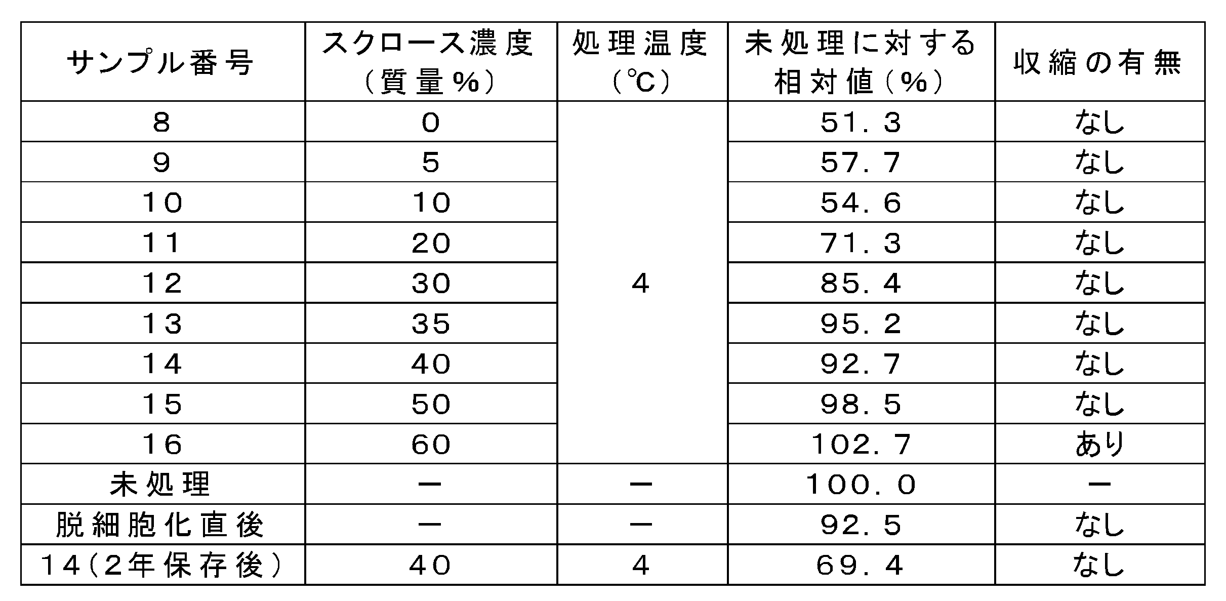
- Test 2 Preparation and evaluation of decellularized cornea-2 >> Same as "Test 1" above, except that the sucrose concentration and temperature conditions in the immersion treatment of the decellularized cornea were set to the conditions shown in Table 2 and the transparency was evaluated by the following method. Decellularized corneas were prepared and evaluated by the method.
- the dry decellularized tissue obtained from the production method of the present invention had good transparency after restoration.
- sucrose concentration was 30 to 50% by mass, a particularly good effect was obtained.
- sucrose concentration was lower than the lower limit in the present invention, swelling was not sufficiently suppressed.
- the sucrose concentration in the sucrose-containing medium was higher than the upper limit in the present invention, the decellularized tissue was significantly contracted and did not function as the decellularized tissue.
Landscapes
- Health & Medical Sciences (AREA)
- Life Sciences & Earth Sciences (AREA)
- Engineering & Computer Science (AREA)
- Biomedical Technology (AREA)
- Chemical & Material Sciences (AREA)
- General Health & Medical Sciences (AREA)
- Biotechnology (AREA)
- Medicinal Chemistry (AREA)
- Veterinary Medicine (AREA)
- Public Health (AREA)
- Cell Biology (AREA)
- Animal Behavior & Ethology (AREA)
- Zoology (AREA)
- Pharmacology & Pharmacy (AREA)
- Developmental Biology & Embryology (AREA)
- Bioinformatics & Cheminformatics (AREA)
- Epidemiology (AREA)
- Organic Chemistry (AREA)
- Virology (AREA)
- Immunology (AREA)
- Ophthalmology & Optometry (AREA)
- Wood Science & Technology (AREA)
- Chemical Kinetics & Catalysis (AREA)
- Genetics & Genomics (AREA)
- Botany (AREA)
- Transplantation (AREA)
- Oral & Maxillofacial Surgery (AREA)
- Dermatology (AREA)
- Microbiology (AREA)
- Neurosurgery (AREA)
- Biochemistry (AREA)
- General Engineering & Computer Science (AREA)
- Neurology (AREA)
- General Chemical & Material Sciences (AREA)
- Nuclear Medicine, Radiotherapy & Molecular Imaging (AREA)
- Agricultural Chemicals And Associated Chemicals (AREA)
Abstract
Description
前記浸漬工程後、前記脱細胞化組織を減圧乾燥する減圧乾燥工程と、
を含み、
前記スクロース含有培地中のスクロース濃度が、5質量%以上55質量%以下である、
乾燥脱細胞化組織の製造方法。
前記浸漬工程後、前記脱細胞化組織を減圧乾燥する減圧乾燥工程と、
を含み、
前記スクロース含有培地中のスクロース濃度が、5質量%以上55質量%以下である、
脱細胞化組織の保存前処理方法。
前記浸漬工程後、前記脱細胞化組織を減圧乾燥する減圧乾燥工程と、
を含む方法から得られ、
前記スクロース含有培地中のスクロース濃度が、5質量%以上55質量%以下である、
乾燥脱細胞化組織。
本発明の乾燥脱細胞化組織の製造方法(以下、「本発明の製造方法」ともいう。)は、脱細胞化組織をスクロース含有培地に浸漬する浸漬工程と、該浸漬工程後、前記脱細胞化組織を減圧乾燥する減圧乾燥工程と、を含み、かつ、スクロース含有培地中のスクロース濃度が、5質量%以上55質量%以下である。
本発明者らの検討の結果、脱細胞化組織をスクロース含有培地によって脱水させ、次いで減圧乾燥させた乾燥脱細胞化組織は、ドライな状態で保存できるだけではなく、培地等を用いて復元させた際の白濁が抑制されているという意外な知見が見出された。
まず、脱細胞化組織の由来となる組織(角膜等)は、通常、溶液(例えば、洗浄液、培地等)中において膨潤し、透明度が低下する。これは、組織(角膜組織等)を構築しているコラーゲン線維の秩序配列が乱れた状態(ただし、基本的にはコラーゲン線維間の間隙が増加するだけで構造自体は壊れていない状態)となり、光を散乱するようになるためである。
したがって、従来のような保存液中で脱細胞化組織を保存した場合、組織の白濁を抑制することは極めて難しい。
他方で、本発明者らは、スクロースが、組織に対する脱水作用や、組織の凍結時の氷晶形成防止作用を有することを見出した。
スクロースによるこれらの作用により、脱細胞化組織の製造工程において膨潤した組織を、スクロースにより脱水することで、コラーゲン線維の秩序配列の乱れを整えることができ、その結果、光の散乱が抑制され、透明性を改善し得る。
さらに、減圧乾燥を行うことで、スクロースによって透明性が改善した脱細胞化組織をそのままの状態で維持することができる。かかる場合、組織中に自由水がほぼないため、膨潤が生じにくい。また、組織中に糖(スクロース等)が残存していることから復元性が高くなる。
その結果、長期保存を行ったとしても、脱細胞化組織の白濁が抑制された状態で復元が可能になる。
例えば、本発明において「復元後の脱細胞化組織」は、該脱細胞化組織の由来となる組織(採取したばかりのもの)により近い透明性を有し得る。
本発明の製造方法では、任意の生体組織を用いて従来知られる手段で製造された脱細胞化組織を用いることができる。
これらのうち、本発明の効果が特に奏されやすいという観点から、脱細胞化組織の由来となる生体組織は角膜が好ましい。換言すれば、本発明における脱細胞化組織は、脱細胞化角膜が好ましい。
印加工程としては、生体組織を任意の脱細胞処理液の中で超高静水圧を印加することが挙げられる。この工程により、生体組織内の細胞を破壊できる。
超高静水圧の下限値は、好ましくは2000気圧以上である。
超高静水圧の上限値は、好ましくは10000気圧以下である。
除去工程としては、印加工程後に、生体組織を洗浄液とともに、浸漬させたり、対流又は循環させたりすることによって、細胞残渣の除去を促進することが挙げられる。
破壊された細胞中の核酸の残渣による痛風等の誘発を防止するため、洗浄液にはDNA分解酵素(DNase)が含まれていてもよい。
本発明の効果が奏されやすいという観点から、生体組織から得た脱細胞化組織は、そのまま(すなわち、脱細胞化組織の調製後、その他の工程を経ることなく)浸漬工程等に供することが好ましい。
浸漬工程は、脱細胞化組織を、スクロース濃度が5質量%以上55質量%以下の培地に浸漬する工程である。
スクロース濃度が55質量%超であると、脱細胞化組織に収縮が生じ、脱細胞化組織としての機能が損なわれる。
浸漬工程において用いるスクロース含有培地のスクロース濃度の上限値は、好ましくは53質量%以下、より好ましくは52質量%以下、さらに好ましくは50質量%以下である。
スクロース含有培地に含まれ得る成分としては、トコフェロール、アスコルビン酸等が挙げられる。これらの成分の配合量は、得ようとする効果等に応じて適宜設定できる。
減圧乾燥工程は、浸漬工程後、脱細胞化組織を減圧乾燥する工程である。
本発明の製造方法によって得られた乾燥脱細胞化組織は、任意の条件下で保存することができる。
ただし、本発明において、減圧乾燥工程と保存工程との間に他の工程を含む態様は排除されない。かかる場合、このような他の工程としては、乾燥脱細胞化組織の水分含量が過度に増加する工程(洗浄、高湿度下の操作等)を含まないことが望ましい。
脱細胞化組織中の水分含量は、赤外線/ハロゲン水分計によって特定される。
保存時間は、例えば、1日以上2年以下であってもよい。
保存期間完了後、脱細胞化組織を任意の方法で復元することで、脱細胞化組織の由来となる組織の種類に応じた所望の用途(移植等)に利用できる。
本発明における浸漬工程及び減圧乾燥工程を組み合わせて脱細胞化組織を処理することにより、復元後の白濁が抑制された乾燥脱細胞化組織が得られる。
したがって、本発明における浸漬工程及び減圧乾燥工程を組み合わせて含む方法は、脱細胞化組織の保存前に行われる前処理方法として有用である。
本発明における乾燥脱細胞化組織は、動物に移植される移植片、又は移植片の部分として有用である。
例えば、脱細胞化組織が脱細胞化角膜である場合、以下の構成を有する移植片としての応用があり得る。
脱細胞化角膜及び上皮(部分角膜移植片)
脱細胞化角膜及び内皮(部分角膜移植片)
脱細胞化角膜、内皮、及び上皮(全層移植片)
以下の方法で脱細胞化角膜を作製し、乾燥処理を行った後に復元し、復元後の透明性を評価した。
以下の方法で、ブタの角膜を用いて脱細胞化角膜を作製した。
(1)成体ブタの眼球から眼科用メスを用いて強角膜片を採取した。
(2)得られた強角膜片を、M199培地及びMEMα培地の等量混合培地(以下、該混合培地を「Mα溶液」ともいう。)に10質量%グリセロール添加した溶液(グリセロール添加Mα溶液)を用いて洗浄した。
(3)洗浄後の強角膜片をグリセロール添加Mα溶液に浸した状態でポリエチレン製バッグ入れ、ヒートシールし、サンプリングバッグを作製した。
(4)サンプルバッグに対し、冷間等方加圧装置(商品名「Dr.CHEF」、株式会社神戸製鋼所製)を用いて、600MPaでの高静水圧印加を10℃で10分間行った。
加圧/減圧速度は、120MPa/minに設定した。
(5)印加後、強角膜片をポリカーボネート製ストレージボトル(容量150mL)に清潔環境下で移し、洗浄用Mα溶液を100mL加え、25℃で48時間振盪洗浄(60rpm)することで、細胞残渣を除去し、脱細胞化角膜を得た。
なお、「洗浄用Mα溶液」とは、Mα溶液に下記を加えたものである:3.5質量% デキストラン 70、200μg/mL DNase I、10μg/mL ゲンタマイシン硫酸塩、500μg/mL L-アスコルビン酸2-リン酸エステル三ナトリウム。
上記で得られた脱細胞化角膜に対し、以下の方法で浸漬処理及び減圧乾燥処理を行った。
(1)脱細胞化角膜を、浸漬用Mα溶液(10ml)に浸漬した。この工程は「浸漬処理」に相当する。
なお、「浸漬用Mα溶液」とは、Mα溶液に下記を加えたものである:0、20、35、40、又は50質量%のスクロース。
また、浸漬処理における処理時間は60分に設定した。浸漬処理における処理温度は、表1の「処理温度」の項に示すとおりである。浸漬処理中、脱細胞化角膜を60rpmで振盪し続けた。
(2)スクロース置換した脱細胞化角膜を、滅菌バイアル瓶(容量5mL)に移し、液体窒素により急速凍結した。
(3)凍結後、プログラムフリーザーを用いて、減圧下(<3Pa)にて、-40℃で24時間、次いで-15℃で72時間の一次乾燥後を行った後、10℃に温度を上昇させ1時間以上の2次乾燥を行った。この工程は「減圧乾燥処理」に相当する。
(4)減圧乾燥後、得られた乾燥脱細胞化組織を、真空下にてバイアル瓶に入れて密栓し、4℃で、6ヶ月保存した。
6ヶ月保存後の各乾燥脱細胞化角膜の全体を、室温下で、生理食塩水(5ml)に1時間浸漬し、脱細胞化組織を復元した。
参考として、上記<脱細胞化角膜への浸漬処理及び減圧乾燥処理>に示した方法の代わりに、脱細胞化角膜を保存液中に浸漬して、4℃で1週間又は2週間保存した組織も準備した。
保存液としては、Mα溶液に下記を加えたものを用いた:3.5質量% デキストラン 70、10μg/mL ゲンタマイシン硫酸塩、500μg/mL L-アスコルビン酸2-リン酸エステル三ナトリウム。
復元後の各乾燥脱細胞化角膜について、その透明性を以下の方法で評価した。
(1)未処理:脱細胞化処理を施していない、採取したばかりの角膜。
(2)HHP直後:<脱細胞化角膜の作製>において高静水圧印加を完了した直後の角膜。
(3)脱細胞化直後:<脱細胞化角膜の作製>の全操作を終えた直後の脱細胞化角膜
(4)4(2年保存後):<脱細胞化角膜への浸漬処理及び減圧乾燥処理>において保存を2年行った点以外は、サンプル番号4と同様に得られた乾燥脱細胞化角膜
(5)溶液保存(1週間):<参考試験:保存液を用いた脱細胞化組織の保存>において1週間の保存を行った脱細胞化角膜
(6)溶液保存(2週間):<参考試験:保存液を用いた脱細胞化組織の保存>において2週間の保存を行った脱細胞化角膜

具体的には、「脱細胞化直後」と同等か、それ以上の透明性を有していた。
この脱細胞化角膜をウサギに移植して3ヶ月経過観察したところ、有害事象(血管新生、重篤な炎症反応等)は認められなかった。
脱細胞化角膜の浸漬処理におけるスクロース濃度及び温度条件を表2に示した条件に設定した点、及び、透明性の評価を以下の方法で行った点以外は、上記「試験1」と同様の方法で脱細胞化角膜を作製及び評価した。
Claims (5)
- 脱細胞化組織をスクロース含有培地に浸漬する浸漬工程と、
前記浸漬工程後、前記脱細胞化組織を減圧乾燥する減圧乾燥工程と、
を含み、
前記スクロース含有培地中のスクロース濃度が、5質量%以上55質量%以下である、
乾燥脱細胞化組織の製造方法。 - 前記浸漬工程における浸漬時間が、30分間以上24時間以下である、請求項1に記載の製造方法。
- 前記脱細胞化組織が脱細胞化角膜である、請求項1又は2に記載の製造方法。
- 脱細胞化組織をスクロース含有培地に浸漬する浸漬工程と、
前記浸漬工程後、前記脱細胞化組織を減圧乾燥する減圧乾燥工程と、
を含み、
前記スクロース含有培地中のスクロース濃度が、5質量%以上55質量%以下である、
脱細胞化組織の保存前処理方法。 - 脱細胞化組織をスクロース含有培地に浸漬する浸漬工程と、
前記浸漬工程後、前記脱細胞化組織を減圧乾燥する減圧乾燥工程と、
を含む方法から得られ、
前記スクロース含有培地中のスクロース濃度が、5質量%以上55質量%以下である、
乾燥脱細胞化組織。
Priority Applications (2)
| Application Number | Priority Date | Filing Date | Title |
|---|---|---|---|
| CN202180066986.0A CN116456994A (zh) | 2020-10-08 | 2021-10-08 | 干燥脱细胞化组织的制造方法、脱细胞化组织的保存预处理方法、以及干燥脱细胞化组织 |
| JP2022555607A JPWO2022075477A1 (ja) | 2020-10-08 | 2021-10-08 |
Applications Claiming Priority (2)
| Application Number | Priority Date | Filing Date | Title |
|---|---|---|---|
| JP2020170530 | 2020-10-08 | ||
| JP2020-170530 | 2020-10-08 |
Publications (1)
| Publication Number | Publication Date |
|---|---|
| WO2022075477A1 true WO2022075477A1 (ja) | 2022-04-14 |
Family
ID=81126532
Family Applications (1)
| Application Number | Title | Priority Date | Filing Date |
|---|---|---|---|
| PCT/JP2021/037486 Ceased WO2022075477A1 (ja) | 2020-10-08 | 2021-10-08 | 乾燥脱細胞化組織の製造方法、脱細胞化組織の保存前処理方法、及び、乾燥脱細胞化組織 |
Country Status (3)
| Country | Link |
|---|---|
| JP (1) | JPWO2022075477A1 (ja) |
| CN (1) | CN116456994A (ja) |
| WO (1) | WO2022075477A1 (ja) |
Citations (3)
| Publication number | Priority date | Publication date | Assignee | Title |
|---|---|---|---|---|
| JP2013042677A (ja) * | 2011-08-22 | 2013-03-04 | Tokyo Medical & Dental Univ | 脱細胞処理液、脱細胞化角膜の調製方法、及び脱細胞化角膜を備える移植片 |
| WO2013032009A1 (ja) * | 2011-09-02 | 2013-03-07 | 株式会社エフコム | 脱細胞化組織製品の調製方法、及び脱細胞化組織製品を備える移植片 |
| CN105343934A (zh) * | 2015-05-15 | 2016-02-24 | 广州悦清再生医学科技有限公司 | 一种人工角膜及其制备方法 |
Family Cites Families (3)
| Publication number | Priority date | Publication date | Assignee | Title |
|---|---|---|---|---|
| KR101089614B1 (ko) * | 2010-02-26 | 2011-12-05 | (주)시지바이오 | 무세포 진피 기질의 제조 방법 및 그로부터 제조된 무세포 진피 기질 |
| KR20190036882A (ko) * | 2017-09-28 | 2019-04-05 | 사회복지법인 삼성생명공익재단 | 생체 이식 또는 삽입용 무세포 진피의 제조방법 |
| CN111035807B (zh) * | 2020-01-02 | 2022-02-11 | 华中科技大学同济医学院附属协和医院 | 一种超薄apcs植片的制备方法 |
-
2021
- 2021-10-08 CN CN202180066986.0A patent/CN116456994A/zh active Pending
- 2021-10-08 WO PCT/JP2021/037486 patent/WO2022075477A1/ja not_active Ceased
- 2021-10-08 JP JP2022555607A patent/JPWO2022075477A1/ja active Pending
Patent Citations (3)
| Publication number | Priority date | Publication date | Assignee | Title |
|---|---|---|---|---|
| JP2013042677A (ja) * | 2011-08-22 | 2013-03-04 | Tokyo Medical & Dental Univ | 脱細胞処理液、脱細胞化角膜の調製方法、及び脱細胞化角膜を備える移植片 |
| WO2013032009A1 (ja) * | 2011-09-02 | 2013-03-07 | 株式会社エフコム | 脱細胞化組織製品の調製方法、及び脱細胞化組織製品を備える移植片 |
| CN105343934A (zh) * | 2015-05-15 | 2016-02-24 | 广州悦清再生医学科技有限公司 | 一种人工角膜及其制备方法 |
Non-Patent Citations (2)
| Title |
|---|
| HUANG YAN-YAN, XU XIAO-LU, HUANG XI-JUN, LIU JIANG-HUI, QI JIAN, ZHU SHUANG, ZHU ZHAO-WEI, HE BO, ZHU QING-TANG, XU YANG-BIN, GU L: "Various changes in cryopreserved acellular nerve allografts at −80°C", NEURAL REGENERATION RESEARCH, MEDKNOW PUBLICATIONS AND MEDIA PVT. LTD., CN, vol. 13, no. 9, 1 January 2018 (2018-01-01), CN , pages 1643, XP055919735, ISSN: 1673-5374, DOI: 10.4103/1673-5374.237138 * |
| KIMURA T, FUNAMOTO S, HASHIMOTO Y, ET AL: "Preparation of decellularized cornea as artificial cornea", ANNUAL REPORTS OF THE INSTITUTE OF BIOMATERIALS AND BIOENGINEERING, vol. 40, 3 March 2007 (2007-03-03), JP , pages 16 - 19, XP008134290, ISSN: 1345-2886 * |
Also Published As
| Publication number | Publication date |
|---|---|
| CN116456994A (zh) | 2023-07-18 |
| JPWO2022075477A1 (ja) | 2022-04-14 |
Similar Documents
| Publication | Publication Date | Title |
|---|---|---|
| DE69901612T2 (de) | Verfahren zur herstellung von elastischem dehydriertem implantatgewebe | |
| US12403218B2 (en) | Biological valve material, preparation method, and cross-linking agent | |
| JP6244031B2 (ja) | 無細胞コラーゲン組織及び無細胞コラーゲン組織を含む人工弁膜の処理方法 | |
| US20050186286A1 (en) | Skin decellularization method, acellular dermal matrix and production method therefore employing said decellularization method, and composite cultured skin employing said matrix | |
| JP6091414B2 (ja) | 脱細胞化組織製品の調製方法、及び脱細胞化組織製品を備える移植片 | |
| US11318168B2 (en) | Human tissue derived compositions and uses thereof | |
| JPH06261933A (ja) | 移植用にコラーゲンベース組織を加工処理、保存するための方法 | |
| JPWO2008111530A1 (ja) | 脱細胞化軟組織の調製方法、移植片、及び培養部材 | |
| van Steenberghe et al. | Enhanced vascular biocompatibility of decellularized xeno-/allogeneic matrices in a rodent model | |
| JP2013042677A (ja) | 脱細胞処理液、脱細胞化角膜の調製方法、及び脱細胞化角膜を備える移植片 | |
| JP3658385B2 (ja) | 皮膚の無細胞化方法、該方法による無細胞化真皮マトリックス及びその製造方法並びに該マトリックスを用いた複合培養皮膚 | |
| WO2004024170A1 (ja) | 超高静水圧印加による移植用生体組織の処理方法 | |
| CN109069263B (zh) | 猪角膜脱细胞方法及其脱细胞角膜以及板层干燥角膜使用方法 | |
| JP5526573B2 (ja) | 脱細胞化生体組織の調製方法 | |
| KR20190036882A (ko) | 생체 이식 또는 삽입용 무세포 진피의 제조방법 | |
| CA2755907A1 (en) | Collagen implant | |
| AU752457B2 (en) | Method of processing and preserving collagen based tissues | |
| CN109475663B (zh) | 一种脱细胞猪角膜的制备方法及其脱细胞板层角膜和用法 | |
| JP5610268B2 (ja) | 高張電解質溶液による生体組織の脱細胞化処理方法 | |
| CN116456994A (zh) | 干燥脱细胞化组织的制造方法、脱细胞化组织的保存预处理方法、以及干燥脱细胞化组织 | |
| JP2024515581A (ja) | 多層羊膜組織移植片及びその使用 | |
| Szaflik et al. | Corneal storage methods | |
| RU2780475C1 (ru) | Способ консервации амниотической мембраны | |
| US9555162B2 (en) | Phospholipid reduction in biological tissue | |
| Gurin et al. | Preparation of a Potential Material for Cardiac Surgery from the Swim Bladder of the Silver Carp |
Legal Events
| Date | Code | Title | Description |
|---|---|---|---|
| 121 | Ep: the epo has been informed by wipo that ep was designated in this application |
Ref document number: 21877776 Country of ref document: EP Kind code of ref document: A1 |
|
| ENP | Entry into the national phase |
Ref document number: 2022555607 Country of ref document: JP Kind code of ref document: A |
|
| WWE | Wipo information: entry into national phase |
Ref document number: 202180066986.0 Country of ref document: CN |
|
| NENP | Non-entry into the national phase |
Ref country code: DE |
|
| 122 | Ep: pct application non-entry in european phase |
Ref document number: 21877776 Country of ref document: EP Kind code of ref document: A1 |