WO2022050423A1 - スタビロン改変体に結合する抗体又はその抗原結合フラグメント - Google Patents
スタビロン改変体に結合する抗体又はその抗原結合フラグメント Download PDFInfo
- Publication number
- WO2022050423A1 WO2022050423A1 PCT/JP2021/032827 JP2021032827W WO2022050423A1 WO 2022050423 A1 WO2022050423 A1 WO 2022050423A1 JP 2021032827 W JP2021032827 W JP 2021032827W WO 2022050423 A1 WO2022050423 A1 WO 2022050423A1
- Authority
- WO
- WIPO (PCT)
- Prior art keywords
- amino acid
- antibody
- acid sequence
- stabilon
- seq
- Prior art date
Links
Images


Classifications
-
- C—CHEMISTRY; METALLURGY
- C07—ORGANIC CHEMISTRY
- C07K—PEPTIDES
- C07K16/00—Immunoglobulins [IGs], e.g. monoclonal or polyclonal antibodies
-
- C—CHEMISTRY; METALLURGY
- C07—ORGANIC CHEMISTRY
- C07K—PEPTIDES
- C07K16/00—Immunoglobulins [IGs], e.g. monoclonal or polyclonal antibodies
- C07K16/44—Immunoglobulins [IGs], e.g. monoclonal or polyclonal antibodies against material not provided for elsewhere, e.g. haptens, metals, DNA, RNA, amino acids
-
- C—CHEMISTRY; METALLURGY
- C12—BIOCHEMISTRY; BEER; SPIRITS; WINE; VINEGAR; MICROBIOLOGY; ENZYMOLOGY; MUTATION OR GENETIC ENGINEERING
- C12N—MICROORGANISMS OR ENZYMES; COMPOSITIONS THEREOF; PROPAGATING, PRESERVING, OR MAINTAINING MICROORGANISMS; MUTATION OR GENETIC ENGINEERING; CULTURE MEDIA
- C12N15/00—Mutation or genetic engineering; DNA or RNA concerning genetic engineering, vectors, e.g. plasmids, or their isolation, preparation or purification; Use of hosts therefor
- C12N15/09—Recombinant DNA-technology
- C12N15/63—Introduction of foreign genetic material using vectors; Vectors; Use of hosts therefor; Regulation of expression
- C12N15/74—Vectors or expression systems specially adapted for prokaryotic hosts other than E. coli, e.g. Lactobacillus, Micromonospora
-
- C—CHEMISTRY; METALLURGY
- C12—BIOCHEMISTRY; BEER; SPIRITS; WINE; VINEGAR; MICROBIOLOGY; ENZYMOLOGY; MUTATION OR GENETIC ENGINEERING
- C12N—MICROORGANISMS OR ENZYMES; COMPOSITIONS THEREOF; PROPAGATING, PRESERVING, OR MAINTAINING MICROORGANISMS; MUTATION OR GENETIC ENGINEERING; CULTURE MEDIA
- C12N15/00—Mutation or genetic engineering; DNA or RNA concerning genetic engineering, vectors, e.g. plasmids, or their isolation, preparation or purification; Use of hosts therefor
- C12N15/09—Recombinant DNA-technology
- C12N15/63—Introduction of foreign genetic material using vectors; Vectors; Use of hosts therefor; Regulation of expression
- C12N15/79—Vectors or expression systems specially adapted for eukaryotic hosts
- C12N15/80—Vectors or expression systems specially adapted for eukaryotic hosts for fungi
- C12N15/81—Vectors or expression systems specially adapted for eukaryotic hosts for fungi for yeasts
-
- C—CHEMISTRY; METALLURGY
- C12—BIOCHEMISTRY; BEER; SPIRITS; WINE; VINEGAR; MICROBIOLOGY; ENZYMOLOGY; MUTATION OR GENETIC ENGINEERING
- C12N—MICROORGANISMS OR ENZYMES; COMPOSITIONS THEREOF; PROPAGATING, PRESERVING, OR MAINTAINING MICROORGANISMS; MUTATION OR GENETIC ENGINEERING; CULTURE MEDIA
- C12N5/00—Undifferentiated human, animal or plant cells, e.g. cell lines; Tissues; Cultivation or maintenance thereof; Culture media therefor
- C12N5/10—Cells modified by introduction of foreign genetic material
-
- G—PHYSICS
- G01—MEASURING; TESTING
- G01N—INVESTIGATING OR ANALYSING MATERIALS BY DETERMINING THEIR CHEMICAL OR PHYSICAL PROPERTIES
- G01N33/00—Investigating or analysing materials by specific methods not covered by groups G01N1/00 - G01N31/00
- G01N33/48—Biological material, e.g. blood, urine; Haemocytometers
- G01N33/50—Chemical analysis of biological material, e.g. blood, urine; Testing involving biospecific ligand binding methods; Immunological testing
- G01N33/53—Immunoassay; Biospecific binding assay; Materials therefor
Definitions
- the present invention relates to an antibody that binds to a Stabilon variant or an antigen-binding fragment thereof.
- a nucleic acid or vector encoding the antibody or its antigen-binding fragment, a transformant into which the nucleic acid or vector has been introduced, a peptide capable of dissociating a Stabilon variant tag fusion protein from the antibody or antigen-binding fragment, and the Stabilon.
- the present invention relates to a kit containing an antibody that binds to a variant or an antigen-binding fragment thereof.
- FLAG (registered trademark) tags are widely used as epitope tags (for example, Patent Document 1).
- An epitope tag refers to a tag sequence that serves as an epitope of an antibody (anti-tag antibody) that recognizes a tag.
- Epitope tags such as FLAG tags can be used together with anti-tag antibodies to investigate the localization and expression level of the protein of interest, and to purify them.
- a Stabilon sequence which is a motif that strongly prevents proteolysis of other molecules, has been known (for example, Patent Document 2).
- the stabilon sequence is a naturally occurring amino acid sequence.
- Patent Document 3 As a tag for detecting or purifying a protein, it is preferable to have an amino acid sequence that does not exist in nature. Therefore, a Stabilon variant has been proposed (for example, Patent Document 3).
- the Stabilon variant described in Patent Document 3 has an amino acid sequence that does not exist in nature.
- an antibody that specifically binds to the Stabilon variant is required.
- an antibody that specifically binds to a stabilon variant production on an industrial scale is required, so a monoclonal antibody is desirable. In recent years, monoclonal antibodies are desirable from the viewpoint of animal welfare.
- an object of the present invention to provide an antibody that specifically binds to a Stabilon variant or an antigen-binding fragment thereof.
- a nucleic acid or vector encoding the antibody or its antigen-binding fragment, a transformant into which the nucleic acid or vector has been introduced, a peptide capable of dissociating a Stabilon variant tag fusion protein from the antibody or antigen-binding fragment, and the antibody is an object of the present invention to provide a kit containing an antigen-binding fragment.
- the present invention includes the following aspects. [1] (a) CDR-H1 containing an amino acid sequence in which one to several amino acids are deleted, substituted or added in the amino acid sequence set forth in SEQ ID NO: 1 or the amino acid sequence set forth in SEQ ID NO: 1. And (b) CDR-H2 containing an amino acid sequence in which one to several amino acids are deleted, substituted or added in the amino acid sequence set forth in SEQ ID NO: 2 or the amino acid sequence set forth in SEQ ID NO: 2. , (C) CDR-H3 containing the amino acid sequence set forth in SEQ ID NO: 3 or the amino acid sequence in which one to several amino acids are deleted, substituted or added in the amino acid sequence set forth in SEQ ID NO: 3.
- Amino acid sequence in which one to several amino acids are deleted, substituted or added in (e) the amino acid sequence set forth in SEQ ID NO: 6 or the amino acid sequence set forth in SEQ ID NO: 6 with CDR-L1 containing Containing CDR-L2 and (f) the amino acid sequence set forth in SEQ ID NO: 7, or the amino acid sequence set forth in SEQ ID NO: 7, in which one to several amino acids are deleted, substituted or added.
- An antibody or an antigen-binding fragment thereof comprising CDR-L3 and a light chain variable region, which specifically binds to a stabilon variant.
- CDR-H1 Containing CDR-H1 containing the amino acid sequence set forth in SEQ ID NO: 1, CDR-H2 containing the amino acid sequence set forth in SEQ ID NO: 2, and CDR-H3 containing the amino acid sequence set forth in SEQ ID NO: 3.
- the heavy chain variable region and CDR-L1 containing the amino acid sequence set forth in SEQ ID NO: 5 CDR-L2 containing the amino acid sequence set forth in SEQ ID NO: 6, and CDR-L3 containing the amino acid sequence set forth in SEQ ID NO: 7.
- [3] The antibody according to [1] or [2], which specifically binds to the peptide consisting of the amino acid sequence set forth in SEQ ID NO: 9 or 10 and does not bind to the peptide consisting of the amino acid sequence set forth in SEQ ID NO: 11. , Or its antigen-binding fragment.
- [4] The nucleic acid encoding the antibody according to any one of [1] to [3] or an antigen-binding fragment thereof.
- [5] A vector containing the nucleic acid according to [4].
- [6] A transformant containing the nucleic acid according to [4].
- [7] A peptide containing the amino acid sequence set forth in SEQ ID NO: 20 and having 15 or less amino acids.
- [8] A kit containing the antibody according to any one of [1] to [3] or an antigen-binding fragment thereof.
- an antibody that specifically binds to a Stabilon variant or an antigen-binding fragment thereof.
- a nucleic acid or vector encoding the antibody or its antigen-binding fragment, a transformant into which the nucleic acid or vector has been introduced, a peptide capable of dissociating a Stabilon variant tag fusion protein from the antibody or antigen-binding fragment, and the antibody.
- a kit containing an antigen-binding fragment can be provided.
- the same sample as in FIG. 2 was used.
- IP; Prog indicates a sample (without antibody added) obtained by adding only Protein G sepharose and reacting.
- Flag-hRAR ⁇ -No The results of immunostaining of HEK293T cells introduced with 9-Stabilon / pcDNA3 using the antibody "24-8G-4G” are shown.
- the results of the elution test of the Stabilon variant tag fusion protein by peptide A (“A” in the figure) or peptide B (“B” in the figure) are shown.
- a Stabilon variant tag fusion protein was eluted from the antibody "24-8G-4G" bound Protein G sepharose, and Western blot analysis of the eluate was performed.
- “3.0” indicates the result of elution using 0.1 M Glycine Buffer (pH 3.0).
- IPTG (+) indicates the result of using an extract of cells whose expression was induced by IPTG in Escherichia coli expressing the Stabilon variant tag fusion protein under the control of the lac promoter.
- IPTG (-) shows the results of using an extract of cells in which expression was not induced by IPTG in Escherichia coli expressing the Stabilon variant tag fusion protein under the control of the lac promoter.
- the results of examining the availability of the stabilon variant tag and the anti-stabilon variant antibody in the plant expression system and the insect expression system by Western blot analysis are shown.
- “(+)” indicates the result of using the biological extract to which the Stabilon variant tag fusion protein was added.
- (-) indicates the result of using the biological extract to which the Stabilon variant tag fusion protein was not added.
- the term “comprise” means that components other than the target component may be included.
- the term “consist of” means that it does not include any component other than the target component.
- the term “consentually of” does not include components other than the target component in a mode that exerts a special function (such as a mode in which the effect of the invention is completely lost). means.
- the term “comprise” includes a "consist of" mode and a “consentially of” mode.
- Proteins, peptides, nucleic acids (DNA, RNA), vectors, and cells can be isolated. "Isolated” means a state isolated from the natural state.
- the proteins, peptides, polynucleotides (DNA, RNA), vectors, and cells described herein are isolated proteins, isolated peptides, isolated polynucleotides (isolated DNA,). It can be an isolated RNA), an isolated vector, and an isolated cell.
- sequence identity (or homology) between base sequences or amino acid sequences corresponds to insertion and deletion of two base sequences or amino acid sequences so that the corresponding bases or amino acids match most. It is juxtaposed with gaps in the portions, and is determined as the ratio of matching bases or amino acids to the entire base sequence or the entire amino acid sequence, excluding the gaps in the obtained alignment.
- Sequence identity between base sequences or amino acid sequences can be determined using various homology search software known in the art. For example, the sequence identity value of a base sequence can be obtained by calculation based on the alignment obtained by the known homology search software BLASTN, and the sequence identity value of an amino acid sequence can be obtained by a known homology search. It can be obtained by calculation based on the alignment obtained by the software BLASTP.
- the invention provides an antibody that specifically binds to a Stabilon variant, or an antigen-binding fragment thereof.
- the antibody is (A) CDR-H1 containing an amino acid sequence in which one to several amino acids are deleted, substituted or added in the amino acid sequence set forth in SEQ ID NO: 1 or the amino acid sequence set forth in SEQ ID NO: 1. (B) CDR-H2 containing an amino acid sequence in which one to several amino acids are deleted, substituted or added in the amino acid sequence set forth in SEQ ID NO: 2 or the amino acid sequence set forth in SEQ ID NO: 2.
- C CDR-H3 containing an amino acid sequence in which one to several amino acids are deleted, substituted or added in the amino acid sequence set forth in SEQ ID NO: 3 or the amino acid sequence set forth in SEQ ID NO: 3.
- Heavy chain variable region including (D) CDR-L1 containing an amino acid sequence in which one to several amino acids are deleted, substituted or added in the amino acid sequence set forth in SEQ ID NO: 5 or the amino acid sequence set forth in SEQ ID NO: 5.
- E CDR-L2 containing the amino acid sequence set forth in SEQ ID NO: 6 or the amino acid sequence in which one to several amino acids are deleted, substituted or added in the amino acid sequence set forth in SEQ ID NO: 6.
- Light chain variable region including Includes and specifically binds to the Stabilon variant.
- Stabilon is a proteolysis-inhibiting motif having an amino acid sequence consisting mainly of acidic amino acids.
- Stabilon preferably comprises or contains the amino acid sequence of all or part of the C-terminal acidic amino acid region of the DP-1 protein (eg, GenBank No. NM_007111.5). It is a proteolysis inhibitory motif.
- the stabilon sequence is an amino acid sequence possessed by stabilon.
- stabilon is a degradation-inhibiting motif consisting of the amino acid sequence region at positions 395 to 410, which is the region on the C-terminal side of the DP-1 protein.
- the stabilon comprises the following amino acid sequence: EDDEEDDDDFNENDEDD (SEQ ID NO: 11)
- E Glutamic acid
- D Aspartic acid
- N Asparagine
- F Phenylalanine
- the term "stabilon variant” means a variant of the above stabilon sequence. Specifically, the Stabilon variant has an amino acid sequence in which one to several amino acids are deleted, substituted or added in the amino acid sequence represented by SEQ ID NO: 11.
- the Stabilon variant preferably contains an amino acid sequence represented by the following formula (I).
- X p (XFDXN) m X q (XFDXN) n X r (I) (X represents E or D; p represents an integer from 0 to 10; q represents an integer from 0 to 20; r represents an integer from 0 to 10; m represents an integer from 0 to 3; n Represents an integer from 0 to 3.
- the Stabilon variant preferably consists of an amino acid sequence that does not exist in nature.
- the stabilon variant preferably comprises the amino acid sequence set forth in SEQ ID NO: 9 or 10.
- non-naturally occurring amino acid sequence means an amino acid sequence that does not exist within a naturally occurring protein.
- a protein database is searched using the target amino acid sequence as a query, and a highly homologous (for example, sequence identity of 60% or more, 70% or more, or 80% or more) is a natural protein. Is an amino acid sequence that does not hit.
- the protein database include a Non-redundant protein sequences (nr) database provided by NCBI.
- Non-naturally occurring amino acid sequences can be described, for example, in the nr database (http://ftp.ncbi.nlm.nih.gov/blast/db/) and blastp (http://blast.ncbi.nlm.nih.gov/).
- nr database http://ftp.ncbi.nlm.nih.gov/blast/db/
- blastp http://blast.ncbi.nlm.nih.gov/.
- PAGE Proteins
- antibodies include all classes and subclasses of immunoglobulins.
- the term "monoclonal antibody” means an antibody obtained from a substantially homogeneous population of antibodies.
- the antibody of this embodiment is preferably a monoclonal antibody.
- the "antigen-binding fragment" of an antibody means a part (partial fragment) of an antibody that specifically binds to a target protein (antigen) to which the antibody specifically binds.
- the antigen-binding fragment usually comprises any of the six CDRs of the antibody (CDR-H1-3, CDR-L1-3).
- the antigen binding fragment preferably contains all six CDRs.
- Examples of the antigen-binding fragment include Fab, Fab', F (ab') 2, variable region fragment (Fv), disulfide bond Fv, single-chain Fv (scFv), sc (Fv) 2, diabody, and polyspecific. Examples thereof include sex antibodies and polymers thereof.
- binding property of an antibody to an antigen can be evaluated, for example, by quantifying the binding of the antibody to an antigen in an in vitro assay.
- the in vitro assay include a plasmon resonance assay using a purified antigen (for example, BIAcore, GE-Healthcare Uppsala, Sweden, etc.) and the like.
- the binding affinity of an antibody for an antigen can be defined by ka (binding rate constant: rate constant for antibody binding from antibody-antigen complex), kd (dissociation rate constant), and dissociation constant KD (kd / ka). can.
- the dissociation constant (KD) when the antibody is specifically bound to the antigen is preferably 10-8 mol / L or less, and is 10-13 mol / L or more and 10-9 mol / L or less. Is more preferable.
- the side chain of the original amino acid and the side chain of the substituted amino acid have similar chemical properties.
- Groups of amino acid residues having amino acid side chains with similar chemical properties are well known in the art to which the present invention belongs.
- amino acids include acidic amino acids (aspartic acid and glutamic acid), basic amino acids (lysine, arginine, histidine), and neutral amino acids (amino acids having a hydrocarbon chain (glycine, alanine, valine, leucine, etc.), depending on the type of side chain.
- Isoleucine / proline amino acid with hydroxy group (serine / threonine), amino acid containing sulfur (cysteine / methionine), amino acid with amide group (asparagin / glutamine), amino acid with imino group (proline), aromatic group It can be classified into amino acids (phenylalanine, tyrosine, tryptophan), etc.
- amino acid added means that an amino acid is added to either or both of the N-terminal and C-terminal of the amino acid sequence, or the amino acid is inserted at any position of the peptide. Means that.
- the number of amino acids deleted, substituted or added in the above (a) to (f) is not particularly limited, but is, for example, 1 to 4, 1 to 3, 1 to 2, or 1 amino acid. Is preferable.
- the antibody of the present embodiment or an antigen-binding fragment thereof is described in SEQ ID NO: 1, among them, CDR-H1 containing the amino acid sequence set forth in SEQ ID NO: 1, CDR-H2 containing the amino acid sequence set forth in SEQ ID NO: 2, and SEQ ID NO: 3.
- the antibody of the present embodiment or an antigen-binding fragment thereof has CDR-H1 consisting of the amino acid sequence set forth in SEQ ID NO: 1, CDR-H2 consisting of the amino acid sequence set forth in SEQ ID NO: 2, and the amino acid sequence set forth in SEQ ID NO: 3.
- CDR-H3 consisting of the amino acid sequence set forth in SEQ ID NO: 1
- CDR-H2 consisting of the amino acid sequence set forth in SEQ ID NO: 2
- the amino acid sequence set forth in SEQ ID NO: 3 In the heavy chain variable region containing CDR-H3, CDR-L1 consisting of the amino acid sequence set forth in SEQ ID NO: 5, CDR-L2 consisting of the amino acid sequence set forth in SEQ ID NO: 6, and SEQ ID NO: 7. It preferably has a light chain variable region comprising CDR-L3 having the described amino acid sequence.
- CDR means complementarity-determining regions.
- the three CDRs contained in the heavy chain variable region of the antibody are described as CDR-H1, CDR-H2, and CDR-H3 in order from the N-terminal side.
- the three CDRs contained in the light chain variable region of the antibody are described as CDR-L1, CDR-L2, and CDR-L3 in order from the N-terminal side.
- the antibody of the present embodiment or an antigen-binding fragment thereof specifically binds to a stabilon variant and does not bind to stabilon. That is, the antibody of the present embodiment or an antigen-binding fragment thereof specifically binds to the peptide consisting of the amino acid sequence set forth in SEQ ID NO: 9 or 10, and does not bind to the peptide consisting of the amino acid sequence set forth in SEQ ID NO: 11. Is more preferable.
- the method for producing the antibody or the antigen-binding fragment thereof of the present embodiment is not particularly limited, and a known antibody production method can be used.
- known antibody production methods include a hybridoma method, a recombinant DNA method, and the like.
- hybridoma method examples include a method developed by Kohler & Milstein (see, for example, Kohler & Milstein, Nature, 256: 495, 1975.).
- the antibody-producing cells used in the cell fusion step in this method include, for example, spleen cells, lymph node cells, and peripheral blood leukocytes of an antigen-immunized animal (eg, mouse, rat, hamster, rabbit, monkey, goat, etc.). And so on. Further, antibody-producing cells obtained by allowing an antigen to act in a medium against the above-mentioned cells or lymphocytes isolated in advance from a non-immune animal can also be used.
- myeloma cells various known cell lines can be used.
- the antibody-producing cells and myeloma cells may be of different animal species origin as long as they can be fused, but are preferably of the same animal species origin.
- a method for obtaining a hybridoma for example, a monoclonal antibody specific to a target protein is obtained by cell fusion between spleen cells obtained from an antigen-immunized mouse and mouse myeloma cells and then screened. Examples thereof include a method for obtaining a hybridoma to be produced.
- Examples of the method for obtaining the monoclonal antibody produced by the hybridoma include a method for obtaining the monoclonal antibody from the culture medium of the hybridoma, a method for obtaining the monoclonal antibody from the ascites of the mammal transplanted with the hybridoma, and the like.
- a nucleic acid (DNA) encoding the antibody or antigen-binding fragment of the present embodiment is incorporated into an appropriate vector, and this is incorporated into a host cell (for example, mammalian cell line, Escherichia coli, yeast cell, insect cell, etc.).
- a host cell for example, mammalian cell line, Escherichia coli, yeast cell, insect cell, etc.
- Examples thereof include a method of introducing into a plant cell or the like to produce the antibody or antigen-binding fragment of the present embodiment as a recombinant antibody (for example, "PJ Delves, Antibody Production: Essential Technologies, 1997 WILEY", ". See P. Shepherd and C. Dean Monoclonal Antibodies, 2000 OXFORD UNIVERSITY PRESS, "Vandamme A. M.
- the DNA encoding the antibody or antigen-binding fragment of the present embodiment may be cloned from a hybridoma or B cell producing these, or may be chemically synthesized by a phosphoramidite method or the like.
- the DNA encoding the antibody of the present embodiment the DNA encoding the heavy chain and the light chain of the antibody may be incorporated into different expression vectors to transform the host cell.
- the DNA encoding the heavy and light chains of the antibody may be integrated into a single expression vector to transform the host cell (see, eg, International Patent Application No. 94/11523).
- the antibody or antigen-binding fragment of the present embodiment can be produced by a host cell by culturing the host cell transformed as described above.
- the antibody or antigen-binding fragment produced can be isolated and purified in the host cell or from the culture medium and obtained in a substantially pure and uniform form.
- the methods used in the purification of conventional polypeptides can be used.
- a transgenic animal in which the antibody gene is integrated for example, cow, goat, sheep, pig, etc.
- a monoclonal derived from the antibody gene is derived from the milk of the transgenic animal. Examples thereof include a method of obtaining a large amount of antibody.
- the antibody of this embodiment can be produced, for example, by the iliac lymphatic method.
- the mouse iliac lymph node method the method described in Japanese Patent No. 4098796 can be used.
- the mouse iliac lymph node method uses the iliac lymph nodes of immunized mice. Antigen immunization should be performed only once on the ridge of the mouse, and cell fusion should be performed 2-3 weeks after antibody immunization. Then, ELISA screening is performed and the hybridoma is cloned.
- the antibody or antigen-binding fragment of the present embodiment preferably contains a heavy chain variable region containing the amino acid sequence set forth in SEQ ID NO: 4 and a light chain variable region containing the amino acid sequence set forth in SEQ ID NO: 8.
- the antibody of the present embodiment or an antigen-binding fragment thereof may be an amino acid sequence variant as long as it can specifically bind to the stabilon variant.
- the antibody of the present embodiment or an antigen-binding fragment thereof is a heavy chain variable containing an amino acid sequence in which one to several amino acids are deleted, substituted or added in the amino acid sequence set forth in SEQ ID NO: 4. It may include a region and a light chain variable region containing an amino acid sequence in which one to several amino acids are deleted, substituted or added in the amino acid sequence represented by SEQ ID NO: 8.
- the number of amino acids deleted, substituted or added in the heavy chain variable region or the light chain variable region is, for example, 1 to 20, preferably 1 to 10, more preferably 1 to 5, and 1 to 1.
- the antibody of the present embodiment or an antigen-binding fragment thereof has a heavy chain variable region having 90% or more sequence identity with the amino acid sequence set forth in SEQ ID NO: 4, and 90% or more with the amino acid sequence set forth in SEQ ID NO: 8. It may contain a light chain variable region having the sequence identity of.
- the sequence identity is preferably 95% or more, more preferably 98% or more, still more preferably 99% or more.
- Amino acid sequence variants can be produced by introducing a mutation into the DNA encoding the antibody chain or by peptide synthesis.
- the site to be modified in the amino acid sequence of the antibody is not particularly limited as long as it has the same antigen-binding activity as the antibody before modification.
- the modification site may be a constant region of the heavy or light chain of the antibody, or may be a variable region (framework region and CDR).
- a method for screening an antibody having an enhanced affinity for an antigen by modifying the amino acid of the CDR may be used (for example, "PNAS, 102: 8466-8471 (2005)", “Protein Engineering, Design & Selection, 21". : 485-493 (2008) ", International Publication No. 2002/051870," J. Biol. Chem., 280: 24880-24887 (2005) ",” Protein Engineering, Design & Selection, 21 “(2008) reference).
- the amino acid sequence of CDR may be modified in the heavy chain variable region having the amino acid sequence set forth in SEQ ID NO: 4 or the light chain variable region having the amino acid sequence set forth in SEQ ID NO: 8, and the frame may be modified.
- the amino acid sequence of the work region may be modified.
- the rate of modification in the heavy chain variable region is preferably 15% or less, more preferably 12% or less, and 10% or less, with the entire amino acid sequence of the heavy chain variable region (that is, four framework regions) other than the CDR as 100%. Is more preferable, and 5% or less, 3% or less, 2% or less, or 1% or less is particularly preferable.
- the rate of modification in the light chain variable region is preferably 15% or less, more preferably 12% or less, and 10% or less, with the entire amino acid sequence of the light chain variable region (that is, the four framework regions) other than the CDR as 100%. Is more preferable, and 5% or less, 3% or less, 2% or less, or 1% or less is particularly preferable.
- the modification is preferably performed in a region other than the CDR (framework region).
- the binding activity of the antibody of the present embodiment or its antigen-binding fragment (including the above-mentioned amino acid sequence variant and the like) to the Stabilon variant is, for example, an ELISA method, a Western blotting method, a flow cytometry method, a Western blot method, a dot. It can be evaluated by blotting, radioimmunoassay, immunoprecipitation, immunostaining, plasmon resonance assay and the like.
- the antibody of this embodiment or an antigen-binding fragment thereof can be used to detect a peptide or protein containing a stabilon variant.
- the antibody of the present embodiment or an antigen-binding fragment thereof can be used for isolating and / or purifying a peptide or protein containing a stabilizeron variant.
- a peptide or protein containing a Stabilon variant may be referred to as a "Stabilon variant tag fusion protein" or a "Stabilon variant tag fusion peptide".
- nucleic acid encoding an antibody or its antigen-binding fragment
- the nucleic acid according to one embodiment of the present invention encodes the above-mentioned antibody or an antigen-binding fragment thereof (hereinafter collectively referred to as “anti-stabilon variant antibody”).
- anti-stabilon variant antibody By introducing the nucleic acid of the present embodiment into an appropriate host cell and expressing it, an anti-stabilon modified antibody can be produced.
- nucleic acid of the present embodiment examples include a gene encoding a light chain containing the light chain variable region of the above-mentioned antibody, a gene encoding a heavy chain containing the heavy chain variable region of the above-mentioned antibody, and the like.
- nucleic acid of the present embodiment examples include a gene encoding an antigen-binding fragment having the same antigen-binding property as the antibody.
- a preferred example of the nucleic acid of the present embodiment is an antibody or antibody fragment thereof comprising a heavy chain variable region containing the amino acid sequence set forth in SEQ ID NO: 4 and a light chain variable region containing the amino acid sequence set forth in SEQ ID NO: 8. Nucleic acid encoding.
- nucleic acid examples include, for example, the base sequence set forth in SEQ ID NO: 12 as a base sequence encoding a heavy chain variable region, and the base sequence set forth in SEQ ID NO: 13 as a base sequence encoding a light chain variable region. Nucleic acid containing.
- the vector according to one embodiment of the present invention contains the above-mentioned nucleic acid.
- the vector of this embodiment may be an expression vector. By introducing the vector of this embodiment into an appropriate host, an anti-stabilon modified antibody can be produced.
- the expression vector is not particularly limited as long as it can express the antibody or antigen-binding fragment encoded by the above-mentioned nucleic acid contained in the vector in the cell to be introduced.
- the expression vector may be a plasmid vector or a viral vector.
- examples of the expression vector include vectors derived from Escherichia coli such as pBR322, pBR325, pUC12 and pUC13; vectors derived from bacillus such as pUB110, pTP5 and pC194; vectors derived from yeast such as pSH19 and pSH15; bacteriophage vectors such as ⁇ phage. ; Virus vectors such as adenovirus, adeno-associated virus, lentivirus, vaccinia virus, bacteriophage, etc .; and vectors modified thereof.
- the term "expression vector” means a vector containing a target nucleic acid and comprising a system for making the target nucleic acid expressable in the cell into which the vector has been introduced.
- the "expressible state” means that the target nucleic acid can be transcribed in the cell into which the target nucleic acid has been introduced.
- the expression vector may contain other base sequences in addition to the above-mentioned nucleic acids. Examples of other base sequences include promoters, enhancers, marker genes, origins of replication, and the like.
- the expression vector preferably contains a promoter, and it is preferable that the above-mentioned nucleic acid (gene) is functionally linked to the promoter. When a nucleic acid (gene) is functionally linked to a promoter, it means that the nucleic acid is linked so as to be expressed under the control of the promoter.
- the transformant according to one embodiment of the present invention contains the above-mentioned nucleic acid or vector.
- an anti-stabilon variant antibody that specifically binds to the stabilon variant can be produced by culturing in an appropriate medium.
- the anti-stabilon variant antibody is produced by growing the plant or animal and extracting and purifying the anti-stabilon variant antibody from the plant or animal. can do.
- transformant examples include cultured cells such as Escherichia coli, yeast, plant cells, insect cells, and animal cells into which the above-mentioned nucleic acid or vector has been introduced; and insect organisms such as silkworm into which the above-mentioned nucleic acid or vector has been introduced.
- Plants such as tobacco into which the above-mentioned nucleic acid or vector has been introduced; animals such as goats, sheep, cows and chickens genetically modified to express the above-mentioned anti-stabilon variant antibody in milk and eggs. And so on.
- the peptide according to one embodiment of the present invention contains the amino acid sequence (EFNDNDE) set forth in SEQ ID NO: 20 and has 15 or less amino acids.
- the peptide of this embodiment contains a part (EFNDNDE: SEQ ID NO: 20) of the amino acid sequence (eg, SEQ ID NO: 9 or 10) of the Stabilon variant.
- the peptide of this embodiment contains an epitope of the anti-stabilon variant antibody described above.
- the peptide of the present embodiment can dissociate the Stabilon variant tag fusion protein from the antigen-antibody complex by contacting it with the antigen-antibody complex of the Stabilon variant tag fusion protein and the anti-Stabilon variant antibody. ..
- the number of amino acids contained in the peptide of this embodiment is 7 to 15.
- the number of amino acids in the peptide of the present embodiment is preferably 14 or less, 13 or less, 12 or less, 11 or less, 10 or less, 9 or less, 8 or less, or 7 amino acids.
- the peptide having 7 amino acids is a peptide having the amino acid sequence shown in SEQ ID NO: 20.
- the type of amino acid added to the amino acid sequence shown in SEQ ID NO: 20 is not particularly limited.
- the amino acid addition may be on the N-terminal side, the C-terminal side, or both the N-terminal side and the C-terminal side of the amino acid sequence set forth in SEQ ID NO: 20.
- the peptide of this embodiment can be used, for example, to elute a Stabilon variant tag fusion protein captured via an anti-Stabilon variant antibody into an anti-Stabilon variant antibody-bound resin.
- a cell extract or the like containing a stabilon-modified tag fusion protein is brought into contact with an anti-stabilon-modified antibody-binding carrier, and the stabilon-modified tag fusion protein is captured by the anti-stabilon-modified antibody.
- the peptide of the present embodiment is brought into contact with the anti-stabilon modified antibody-binding carrier.
- This allows the Stabilon variant tag fusion protein to be eluted from the anti-Stabilon variant antibody binding carrier. Therefore, by using the peptide of this embodiment together with an anti-stabilon variant antibody, a stabilon variant tag fusion protein can be isolated and purified from a protein mixture such as a cell extract.
- the carrier to which the anti-stabilon modified antibody is bound examples include a carrier to which an immunoglobulin binding protein such as Protein G is bound.
- An anti-stabilon-modified antibody-binding carrier can be prepared by contacting an anti-stabilon-modified antibody with a carrier to which an immunoglobulin-binding protein is bound.
- the carrier is not particularly limited, and examples thereof include, but are not limited to, resin beads (Sepharose beads, agarose beads, etc.), magnetic beads, and the like.
- the stabilon variant detection kit contains the above-mentioned antibody or an antigen-binding fragment thereof (anti-stabilon variant antibody).
- the stabilon variant detection kit according to the present embodiment may contain other configurations in addition to the anti-stabilon variant antibody. Other configurations include, for example, the above-mentioned eluted peptide, a vector containing a base sequence encoding a Stabilon variant, and the like.
- the kit of the present embodiment may further include an anti-stabilon-modified antibody-binding carrier, various buffers, cytolytic solution, instructions for use, and the like.
- the anti-stabilon variant antibody may be bound to a carrier. Examples of the carrier include the same carriers as those mentioned in the above ⁇ eluted peptide>.
- the kit of the present embodiment may include a vector containing a base sequence encoding a stabilon variant (hereinafter, also referred to as “stabilon variant gene”) in addition to the anti-stabilon variant antibody.
- the base sequence encoding the Stabilon variant is not particularly limited as long as it encodes the Stabilon variant.
- the Stabilon variant preferably comprises the amino acid sequence set forth in SEQ ID NO: 9 or 10. Examples of the base sequence encoding the amino acid sequence set forth in SEQ ID NO: 9 include the base sequence set forth in SEQ ID NO: 22. Examples of the base sequence encoding the amino acid sequence set forth in SEQ ID NO: 10 include the base sequence set forth in SEQ ID NO: 23.
- the vector preferably contains an insertion site for a nucleic acid (hereinafter, also referred to as "test peptide gene”) containing a base sequence encoding an arbitrary peptide or protein (hereinafter, also referred to as "test peptide”).
- the vector preferably has a structure capable of expressing a fusion protein of a stabilon variant and a test peptide by inserting the test peptide gene into the insertion site.
- the vector may have, for example, the insertion site upstream of the Stabilon variant gene. It is preferable that the stabilon variant gene and the test peptide gene are linked in-frame by inserting the test peptide gene into the insertion site.
- the test peptide in which the Stabilon variant tag is fused to the N-terminal side can be expressed.
- the vector may have, for example, the insertion site downstream of the Stabilon variant gene.
- the test peptide gene and the stabilon variant gene are linked in-frame.
- the test peptide in which the Stabilon variant tag is fused to the C-terminal side can be expressed.
- the vector may contain a base sequence encoding an arbitrary protease recognition sequence between the stabilon variant gene and the insertion site.
- a protease recognition sequence is an amino acid sequence that is recognized and cleaved by a protease. Examples of the protease include, but are not limited to, thrombin, TEV protease and the like.
- the vector contains a base sequence encoding a protease recognition sequence to express a fusion protein having a protease recognition sequence between the Stabilon variant tag and the test peptide when the test peptide gene is inserted into the insertion site. Can be made to. Thereby, if necessary, the protease recognition sequence can be cleaved with a protease to separate the Stabilon variant tag from the test peptide.
- the type of vector is not particularly limited, and the same vector as those listed in the ⁇ vector> section can be used.
- the vector may further include a promoter, enhancer, marker gene, replication origin and the like.
- the vector preferably contains, for example, a promoter that controls the expression of the fusion gene of the stabilon variant gene / test peptide gene formed by inserting the test peptide gene into the insertion site.
- the Stabilon variant detection kit can be effectively used for detection of the Stabilon variant.
- the method for detecting a stabilon variant is characterized in that an anti-stabilon variant antibody is used as a tag antibody.
- the test substance can be detected by utilizing the antigen-antibody reaction between the test substance to which the stabilon variant is bound or fused and the anti-stabilon variant antibody.
- the method for detecting the Stabilon variant can include the following steps.
- test substance labeled with the Stabilon variant examples include a test peptide in which the Stabilon variant is fused as an epitope tag (hereinafter, also referred to as "Stabilon variant tag fusion peptide").
- the Stabilon variant tag fusion peptide can be prepared by a known genetic engineering technique. For example, a fusion gene containing a stabilon variant gene and a test peptide gene is prepared, and the fusion gene is expressed in an appropriate expression system (cell expression system or cell-free expression system) to obtain a stabilon variant tag fusion peptide. Can be prepared.
- a "fusion peptide expression vector" having a base sequence encoding a stabilon variant and capable of being expressed in a state where the stabilon variant tag is fused to the terminal of the test peptide or the like is used. May be good.
- a nucleic acid having a base sequence encoding the test peptide is incorporated into a fusion peptide expression vector, and the vector is used in an expression system suitable for the nucleic acid to express the Stabilon variant tag fusion peptide.
- the stabilon-modified tag fusion peptide expressed and produced in this manner can be isolated and recovered by an affinity purification method utilizing the affinity between the anti-stabilon-modified antibody and the stabilon-modified tag.
- the above-mentioned eluted peptide may be used to elute the Stabilon variant tag fusion peptide.
- the tag fusion peptide may contain any protease recognition sequence between the Stabilon variant tag and the test peptide.
- the Stabilon variant tag fusion peptide having a protease recognition sequence can be separated from the Stabilon variant tag and the test peptide by cleaving the protease recognition sequence with a protease after purification, if necessary.
- the Stabilon variant tag and the undigested Stabilon variant tag fusion peptide can be recovered or removed by an affinity purification method using an anti-Stabilon variant antibody. Thereby, the test peptide can be isolated and recovered.
- the conditions adopted for the antigen-antibody reaction are not particularly limited as long as the anti-stabilon-modified antibody can recognize and bind to the stabilon-modified tag of the stabilon-modified tag fusion protein.
- the antigen-antibody reaction may be carried out under intracellular bioenvironmental conditions in which the tag fusion protein is expressed. In this case, it can also be applied to expression profiling of a desired protein or the like.
- the test substance (stabilon modified tag fusion peptide) can be detected using the antigen-antibody complex produced by the antigen-antibody reaction as an index (immunostaining).
- the anti-stabilon variant antibody may be labeled with a labeling substance and used.
- the labeling substance include, but are not limited to, radioactive isotopes, non-radioactive isotopes, fluorescent dyes, enzymes and the like.
- the antigen-antibody complex can be detected using the labeling substance used for labeling the anti-stabilon variant antibody as an index.
- the test substance labeled with the Stabilon variant can be specifically detected by using the Stabilon variant and the anti-Stabilon variant antibody. .. Therefore, it can be used for purification of a desired peptide or protein, expression analysis or kinetic analysis of the desired peptide or protein in cells, and the like.
- Example 1 Western blot analysis> Western blot analysis was performed using 6 types of expression plasmids shown in Table 1 as expression plasmids for the antigen protein.
- Table 1 the left side of "/" indicates the antigen protein.
- the right side of "/” indicates the type of plasmid.
- No. 9-Stabilon represents a Stabilon variant consisting of the amino acid sequence set forth in SEQ ID NO: 9.
- No. 10-Stabilon represents a Stabilon variant consisting of the amino acid sequence set forth in SEQ ID NO: 10.
- No. 0-Stabilon (DP-1 Stabilon) represents a stabilon consisting of the amino acid sequence set forth in SEQ ID NO: 11.
- the above 6 types of expression plasmids A to F were transformed into Escherichia coli DH5 ⁇ , cultured in LB medium containing ampicillin, and purified by MAXI prep kit (QIAGEN).
- HEK293T cells human fetal kidney-derived HEK293T cells (hereinafter, also referred to as “HEK293T cells”) were cultured.
- the medium a medium in which 10% (V / V) fetal bovine serum and 1/100 diluted streptomycin penicillin (Wako Pure Chemical Industries) were mixed with D-MEM liquid medium (Wako Pure Chemical Industries, Ltd.) was used.
- HEK293T cells were seeded on a 12-well plate, and when they became 60% confluent, the expression plasmid of any of A to F was transfected by the calcium phosphate method. Eight hours after the expression plasmid was added to the medium, 1 ml of the medium was added, and 24 hours later, the medium was removed and cell extraction was performed.
- Electrophoresis was performed by ordinary SDS-PAGE, and a separation gel having a 10% acrylamide concentration was used. After the electrophoresis, the separated gel was removed from the gel plate, immersed in a transfer buffer, and transferred to an immobilon; PVDF membrane (Merck Millipore) using a semi-dry transfer device (BioRAD; Trans Blood Turbo). After blocking with 5% skim milk / TBST, it was washed 4 times with 1xTBST.
- M2-antibody-HRP-labeled mouse host antibody Sigma; M2-HRP; A8592
- M2-antibody-HRP-labeled mouse host antibody Sigma; M2-HRP; A8592
- 1 ⁇ l of the antibody “24-8G-4G” was added to the second membrane and reacted at room temperature for 1 hour with shaking. After washing this membrane 4 times with 1xTBST, 1 ⁇ l of Anti-mouse IgG-HRP fusion (Jackson Research Co., Ltd .; 115-035-003) was added as a secondary antibody, and the mixture was reacted at room temperature for 1 hour with shaking. Then, the membrane was washed 4 times with 1xTBST.
- the first and second membranes after washing were transferred to a hybrid bag, Kemil Miwan L (Nacalai Tesque) was added, and the membrane was exposed to HyperFilm ECL (GE Healthcare) in a dark room.
- the results of Western blot analysis are shown in FIG.
- Reaction 1 Annealing of the reverse transcription primer
- the reverse transcription primer was annealed separately for the light chain and the heavy chain.
- the composition of the reaction solution for the light chain is shown in Table 2.
- the composition of the reaction solution for heavy chains is shown in Table 3. After mixing the reagents shown in Tables 2 and 3, respectively, the mixture was incubated at 72 ° C. for 2 minutes and 42 ° C. for 2 minutes to obtain a reaction solution.
- Reaction 2 Reverse transcription reaction and annealing of SMARTer oligo
- the mixture shown in Table 4 was dispensed and added, and reversed at 42 ° C. for 1 hour. A photo reaction was performed. After further reacting at 70 ° C. for 10 minutes, 40 ⁇ l of TE was added.
- Reaction 3 PCR amplification
- the reaction solution shown in Table 5 was prepared and a PCR reaction was carried out.
- "RT product” indicates the reverse transcription reaction product obtained in reaction 2.
- the PCR reaction was repeated 35 cycles with (1) 98 ° C., 8 seconds, and (2) 72 ° C., 1 minute as one cycle.
- Reaction 4 A addition reaction In PrimeStar DNA polymerase, A (adenine) is not added to the 3'end of the PCR product. Therefore, the product amplified in the above reaction 3 was treated with phenol chloroform, precipitated with ethanol, and further subjected to an A addition reaction with Ex-Taq DNA polymerase.
- the reaction solution is shown in Table 6. The reaction was carried out at 72 ° C. for 1 minute.
- the A addition reaction product was ligated to Vector (pMD20). Then, it was transformed into Escherichia coli, and blue-white selection (X-gal and IPTG were added to the pound plate) was performed. Only white colonies were selected, cultured overnight with shaking in 3 ml of ampicillin-containing LB liquid medium, collected, and subjected to plasmid miniprep using the alkaline SDS method.
- HEK293T cells Preparation of antigen protein> HEK293T cells were seeded in 10 cm Dush to be 40% confluent. HEK293T cells were transfected with Flag-hRAR ⁇ -Stabilon No9 / pcDNA3 (plasmid of D in Table 1) by the calcium phosphate method. After 8 hours, the medium (D-MEM low glucose, 10% FBS, containing 1/100 diluted penicillin streptomycin) was replaced, and after an additional 12 hours, cells were stripped by pipetting. The cells were transferred to another new 1.5 ml Eppen tube and washed twice with 1xBSBS.
- TNE-N buffer (20 mM Tris-HCl [pH 7.8], 150 mM NaCl, 1 mM EDTA [pH 7.9], 1/100 Protease inhibitor cocktail) was added to the collected cells, and the cells were disrupted by pipetting. After high-speed centrifugation of the cell disruption solution (14000 rpm, 5 min, 4 ° C.), a supernatant was obtained.
- ⁇ Test Example 3 (2) Preparation of immunoprecipitation sample> ⁇ Antibody "23-2E" immunoprecipitation sample >> 230 ⁇ l of the supernatant obtained in Test Example 3 (1) was dispensed into a tube, 10 ⁇ l of the antibody “23-2E” was added, and the mixture was reacted at 4 ° C. for 1 hour. Further, equilibrated Protein G sepharose (GE Healthcare) was added, and the mixture was reacted at 4 ° C. for 1 hour. Then, it was washed 4 times with TNE-N buffer, 2xSDS-PAGE sample buffer was added to the precipitated Protein G sepharose, and the mixture was boiled at 98 ° C. for 2 minutes.
- ⁇ Anti-Flag antibody immunoprecipitation sample 230 ⁇ l of the supernatant obtained in Test Example 3 (1) was dispensed into a tube, 10 ⁇ l of anti-Flag antibody was added, and the mixture was reacted at 4 ° C. for 1 hour. Further, equilibrated Protein G sepharose (GE Healthcare) was added, and the mixture was reacted at 4 ° C. for 1 hour. Then, it was washed 4 times with TNE-N buffer, 2xSDS-PAGE sample buffer was added to the precipitated Protein G sepharose, and the mixture was boiled at 98 ° C. for 2 minutes.
- Example without antibody added 230 ⁇ l of the supernatant obtained in Test Example 3 (1) was dispensed into a tube, equilibrated Protein G sepharose (GE Healthcare) was added, and the mixture was reacted at 4 ° C. for 1 hour. Then, it was washed 4 times with TNE-N buffer, 2xSDS-PAGE sample buffer was added to the precipitated Protein G sepharose, and the mixture was boiled at 98 ° C. for 2 minutes.
- equilibrated Protein G sepharose GE Healthcare
- ⁇ Test Example 3 (3) Western blot analysis> To 50 ⁇ l of the supernatant obtained in Test Example 3 (1), 50 ⁇ l of 2xSDS-PAGE sample buffer was added and boiled at 90 ° C. for 2 minutes to prepare a Lysate sample.
- the Lysate sample and the immunoprecipitation sample prepared in Test Example 3 (2) (anti-Flag antibody immunoprecipitation sample, antibody "23-2E” immunoprecipitation sample, antibody "24-8G-4G” immunity Precipitation sample) was used.
- An anti-Flag antibody Sigma; M2-HRP; A8592
- Western blot analysis was performed under the same conditions as in Test Example 1. The analysis result is shown in FIG.
- the Lysate sample includes Flag-RAR ⁇ -No. A thin band of 9-Stabilon was observed.
- IP Flag
- Flag-RAR ⁇ -No A dark band of 9-Stabilon was observed.
- HEK293T cells were seeded in a bottom glass dish to be 40% confluent.
- Flag-hRAR ⁇ -No. 9-Stabilon / pcDNA3 Plasmid of D in Table 1
- the medium D-MEM low glucose, 10% FBS, containing 1/100 diluted penicillin streptomycin
- the cells were fixed with 4% paraformaldehyde / PBS.
- the antibody "24-8G-4G” was added and reacted with the cells.
- the cells were washed 4 times with 1xPBST and reacted with the cells by adding Alexa594-labeled anti-mouse IgG antibody (Thermo Fisher Scientific).
- the cells were washed 4 times with 1xPBST and bleaching was prevented with VECTASHIELD Mounting Medium with out DAPI (Funakoshi).
- the sample prepared as described above was observed with a confocal laser scanning microscope FV1000-D (OLYNPUS).
- an elution peptide capable of exfoliating the antigen motif fusion protein from the antibody resin under physiologically mild conditions is useful. be.
- the synthesis of Peptide A and Peptide B was carried out in a 1 mg synthesis system with a guarantee of 90% or higher purity (HPLC).
- the following proteins (1) and (2) were obtained by the E. coli expression system. (1) 6xHis-Flag-No. 9-Stabilon-hSox2-S19-TAT-NL (2) 6xHis-Flag-No. 9-Stabilon-hOct4-S19-TAT-NLS
- Escherichia coli strain BL21 (DE3) expressing the proteins of (1) and (2) was cultured overnight at 37 ° C. using 1 L of LB kanamycin medium.
- E. coli O. coli. D When the 600 value reached 0.5, IPTG was added to the culture broth (final concentration 0.1 mM). Escherichia coli was further induced to express at 25 ° C. for 3 hours and then collected by centrifugation. The collected Escherichia coli was suspended in 1xTBST, crushed by ultrasonic waves, and subjected to high-speed centrifugation (14,000 rpm, 5 min, 4 ° C.). Ni resin was added after transferring the supernatant to another tube. The supernatant to which Ni resin was added was washed 5 times with 1xPBST and then once with 1xPBST containing 5 mM imidazole.
- the supernatant to which Ni resin was added was further eluted with 1xPBST containing 500 mM imidazole to obtain an eluted protein.
- the antibody "24-8G-4G” was added to the two obtained eluted proteins and reacted at 4 ° C. for 1 hour with shaking to obtain a reaction solution.
- Protein G sepharose equilibrated with 1xPBST was further added to the reaction solution, and the reaction was carried out at 4 ° C. for 1 hour with shaking. After washing 4 times with 1xPBST, Protein G sepharose resin was divided into 3 1.5 ml microtubes each.
- Peptide A, peptide B, or 0.1 M Glycine Buffer (pH 3.0) was added to 3 tubes in an amount of 20 ⁇ l to elute the peptide. Peptide elution was performed at 4 ° C. for 1 hour, and Glycine Buffer was performed at 4 ° C. for 5 minutes. After elution of the peptide, slow centrifugation (2,000 rpm, 1 minute, 4 ° C) was performed, and the supernatant was separated into separate tubes. 1M Tris-HCl (pH 9.5) was added to the Glycine Buffer elution supernatant to neutralize it, and an elution fraction was obtained.
- Glycine Buffer pH 3.0
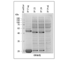
- Example 6 Verification of availability in E. coli expression system>
- the E. coli expression system is often used as an expression system on an industrial scale. Therefore, the possibility of using a stabilon-modified tag and an anti-stabilon-modified antibody in an E. coli expression system was investigated.
- the above-mentioned 6xHis-Flag-No. In Escherichia coli strain BL21 (DE3) expressing 9-Stabilon-hOct4-S19-TAT-NLS under the control of the lac promoter, cells that were induced with IPTG (IPTG (+)) and cells that were not induced with IPTG. A cell extract of (IPTG ( ⁇ )) was prepared.
- FIG. 6 The results of Western blot analysis are shown in FIG. As shown in FIG. 6, the antibody “24-8G-4G” is described in 6xHis-Flag-No. Only 9-Stabilon-hOct4-S19-TAT-NLS was detected and did not bind to other non-specific proteins of E. coli (rightmost panel in FIG. 6; Stabilon: 24). This high specificity was similar to the commercially available anti-Flag monoclonal antibody (leftmost panel in FIG. 6; M2) and anti-His monoclonal antibody (second panel from left in FIG. 6; His). The antibody “23-2E” also has 6xHis-Flag-No. Only 9-Stabilon-hOct4-S19-TAT-NLS was detected, but the band was thinner compared to the antibody "24-8G-4G” (second panel from right in FIG. 6; Stabilon: 23).
- FIG. 7 The results of Western blot analysis are shown in FIG. In FIG. 7, Flag (the leftmost panel in each reaction system) is an anti-Flag antibody, His (the second panel from the left in each reaction system) is an anti-His antibody, and 23 (the second panel from the right in each reaction system). The panel) shows the results using the antibody “23-2E”, and 24 (the rightmost panel in each reaction system) shows the results using the antibody “24-8G-4G”.
- (-) Is a lane using the extract as it is, and (+) indicates 6xHis-Flag-No. It is a lane using 9-Stabilon-hOct4-S19-TAT-NLS added. As shown in FIG.
- an antibody or an antigen-binding fragment thereof specifically recognized for a stabilon sequence a nucleic acid encoding an antibody or an antigen-binding fragment thereof, a vector, a transformant, and a stabilon variant thereof.
- Detection kits can be provided.
Landscapes
- Health & Medical Sciences (AREA)
- Life Sciences & Earth Sciences (AREA)
- Genetics & Genomics (AREA)
- Chemical & Material Sciences (AREA)
- Engineering & Computer Science (AREA)
- Organic Chemistry (AREA)
- Biomedical Technology (AREA)
- Biotechnology (AREA)
- Bioinformatics & Cheminformatics (AREA)
- Wood Science & Technology (AREA)
- Zoology (AREA)
- General Engineering & Computer Science (AREA)
- Molecular Biology (AREA)
- General Health & Medical Sciences (AREA)
- Biochemistry (AREA)
- Biophysics (AREA)
- Microbiology (AREA)
- Physics & Mathematics (AREA)
- Immunology (AREA)
- Plant Pathology (AREA)
- Mycology (AREA)
- Medicinal Chemistry (AREA)
- Urology & Nephrology (AREA)
- Hematology (AREA)
- Cell Biology (AREA)
- Proteomics, Peptides & Aminoacids (AREA)
- Analytical Chemistry (AREA)
- General Physics & Mathematics (AREA)
- Pathology (AREA)
- Food Science & Technology (AREA)
- Micro-Organisms Or Cultivation Processes Thereof (AREA)
- Peptides Or Proteins (AREA)
Abstract
スタビロン改変体に特異的に結合する抗体又はその抗原結合フラグメント。前記抗体又はその抗原結合フラグメントをコードする核酸。前記核酸を含むベクター。前記抗体又は抗原結合フラグメントからスタビロン改変体タグ融合タンパク質を解離可能なペプチド。前記核酸を含む形質転換体。前記抗体又は抗原結合フラグメントを含む、キット。
Description
本発明は、スタビロン改変体に結合する抗体又はその抗原結合フラグメントに関する。また、前記抗体又はその抗原結合フラグメントをコードする核酸又はベクター、前記核酸又はベクターを導入した形質転換体、前記抗体又は抗原結合フラグメントからスタビロン改変体タグ融合タンパク質を解離可能なペプチド、及び、前記スタビロン改変体に結合する抗体又はその抗原結合フラグメントを含むキットに関する。
本願は、2020年9月7日に、日本に出願された特願2020-150003号に基づき優先権を主張し、その内容をここに援用する。
本願は、2020年9月7日に、日本に出願された特願2020-150003号に基づき優先権を主張し、その内容をここに援用する。
一般的に、エピトープタグとして、FLAG(登録商標)タグが普及している(例えば、特許文献1)。エピトープタグは、タグを認識する抗体(抗タグ抗体)のエピトープとなるタグ配列のことをいう。FLAGタグを始めとするエピトープタグは、抗タグ抗体とともに用いることにより、目的のタンパク質の局在や発現量を調査したり、精製したりすることができる。
これまでに、他分子のタンパク質分解を強力に防ぐモチーフであるスタビロン(Stabilon)配列が知られている(例えば、特許文献2)。スタビロン配列は、天然に存在するアミノ酸配列である。
これまでに、他分子のタンパク質分解を強力に防ぐモチーフであるスタビロン(Stabilon)配列が知られている(例えば、特許文献2)。スタビロン配列は、天然に存在するアミノ酸配列である。
タンパク質の検出又は精製用のタグとしては、天然に存在しないアミノ酸配列を有することが好ましい。そのため、スタビロン改変体が提案されている(例えば、特許文献3)。特許文献3に記載のスタビロン改変体は、天然には存在しないアミノ酸配列を有する。
スタビロン改変体をエピトープタグとして実用化するためには、スタビロン改変体に特異的に結合する抗体が必要である。
スタビロン改変体に特異的に結合する抗体を実用化するためには、工業的スケールでの産生が求められるため、モノクローナル抗体であることが望ましい。また、近年では動物愛護の観点からも、モノクローナル抗体が望ましい。
スタビロン改変体に特異的に結合する抗体を実用化するためには、工業的スケールでの産生が求められるため、モノクローナル抗体であることが望ましい。また、近年では動物愛護の観点からも、モノクローナル抗体が望ましい。
そこで、本発明は、スタビロン改変体に特異的に結合する抗体又はその抗原結合フラグメントを提供することを課題とする。また、前記抗体又はその抗原結合フラグメントをコードする核酸又はベクター、前記核酸又はベクターを導入した形質転換体、前記抗体又は抗原結合フラグメントからスタビロン改変体タグ融合タンパク質を解離可能なペプチド、及び、前記抗体又は抗原結合フラグメントを含むキットを提供することを課題とする。
本発明は、以下の態様を包含する。
[1](a)配列番号1に記載のアミノ酸配列、又は、配列番号1に記載のアミノ酸配列において、1~数個のアミノ酸が欠失、置換若しくは付加されているアミノ酸配列を含むCDR-H1と、(b)配列番号2に記載のアミノ酸配列、又は、配列番号2に記載のアミノ酸配列において、1~数個のアミノ酸が欠失、置換若しくは付加されているアミノ酸配列を含むCDR-H2と、(c)配列番号3に記載のアミノ酸配列、又は、配列番号3に記載のアミノ酸配列において、1~数個のアミノ酸が欠失、置換若しくは付加されているアミノ酸配列を含むCDR-H3と、を含む重鎖可変領域と、(d)配列番号5に記載のアミノ酸配列、又は、配列番号5に記載のアミノ酸配列において、1~数個のアミノ酸が欠失、置換若しくは付加されているアミノ酸配列を含むCDR-L1と、(e)配列番号6に記載のアミノ酸配列、又は、配列番号6に記載のアミノ酸配列において、1~数個のアミノ酸が欠失、置換若しくは付加されているアミノ酸配列を含むCDR-L2と、(f)配列番号7に記載のアミノ酸配列、又は、配列番号7に記載のアミノ酸配列において、1~数個のアミノ酸が欠失、置換若しくは付加されているアミノ酸配列を含むCDR-L3と、を含む軽鎖可変領域と、を含み、スタビロン改変体に特異的に結合する、抗体、又は、その抗原結合フラグメント。
[2]配列番号1に記載のアミノ酸配列を含むCDR-H1と、配列番号2に記載のアミノ酸配列を含むCDR-H2と、配列番号3に記載のアミノ酸配列を含むCDR-H3と、を含む重鎖可変領域、及び、配列番号5に記載のアミノ酸配列を含むCDR-L1と、配列番号6に記載のアミノ酸配列を含むCDR-L2と、配列番号7に記載のアミノ酸配列を含むCDR-L3と、を含む軽鎖可変領域を含む、[1]に記載の抗体、又は、その抗原結合フラグメント。
[3]配列番号9又は10に記載のアミノ酸配列からなるペプチドに特異的に結合し、且つ配列番号11に記載のアミノ酸配列からなるペプチドに結合しない、[1]又は[2]に記載の抗体、又は、その抗原結合フラグメント。
[4][1]~[3]のいずれか1つに記載の抗体、又は、その抗原結合フラグメントをコードする核酸。
[5][4]に記載の核酸を含むベクター。
[6][4]に記載の核酸を含む形質転換体。
[7]配列番号20に記載のアミノ酸配列を含み、アミノ酸数が15以下である、ペプチド。
[8][1]~[3]のいずれか1つに記載の抗体、又は、その抗原結合フラグメントを含む、キット。
[9][7]に記載のペプチドをさらに含む、[8]に記載のキット。
[10]配列番号9又は10に記載のアミノ酸配列をコードする塩基配列を含むベクターをさらに含む、[8]又は[9]に記載のキット。
本発明によれば、スタビロン改変体に特異的に結合する抗体又はその抗原結合フラグメントを提供することができる。また、前記抗体又はその抗原結合フラグメントをコードする核酸又はベクター、前記核酸又はベクターを導入した形質転換体、前記抗体又は抗原結合フラグメントからスタビロン改変体タグ融合タンパク質を解離可能なペプチド、及び、前記抗体又は抗原結合フラグメントを含むキットを提供することができる。
「を含む」(comprise)という用語は、対象となる構成要素以外の構成要素を含んでいてもよいことを意味する。「からなる」(consist of)という用語は、対象となる構成要素以外の構成要素を含まないことを意味する。「から本質的になる」(consist essentially of)という用語は、対象となる構成要素以外の構成要素を特別な機能を発揮する態様(発明の効果を完全に喪失させる態様など)では含まないことを意味する。本明細書において、「を含む」(comprise)と記載する場合、「からなる」(consist of)態様、及び「から本質的になる」(consist essentially of)態様を包含する。
タンパク質、ペプチド、核酸(DNA、RNA)、ベクター、及び細胞は、単離されたものであり得る。「単離された」とは、天然状態から分離された状態を意味する。本明細書に記載されるタンパク質、ペプチド、ポリヌクレオチド(DNA、RNA)、ベクター、及び細胞は、単離されたタンパク質、単離されたペプチド、単離されたポリヌクレオチド(単離されたDNA、単離されたRNA)、単離されたベクター、及び単離された細胞であり得る。
本明細書において、塩基配列どうし又はアミノ酸配列どうしの配列同一性(又は相同性)は、2つの塩基配列又はアミノ酸配列を、対応する塩基又はアミノ酸が最も多く一致するように、挿入及び欠失に当たる部分にギャップを入れながら並置し、得られたアラインメント中のギャップを除く、塩基配列全体又はアミノ酸配列全体に対する一致した塩基又はアミノ酸の割合として求められる。塩基配列又はアミノ酸配列どうしの配列同一性は、当該技術分野で公知の各種相同性検索ソフトウェアを用いて求めることができる。例えば、塩基配列の配列同一性の値は、公知の相同性検索ソフトウェアBLASTNにより得られたアライメントを元にした計算によって得ることができ、アミノ酸配列の配列同一性の値は、公知の相同性検索ソフトウェアBLASTPにより得られたアライメントを元にした計算によって得ることができる。
<抗体、又は、その抗原結合フラグメント>
一態様において、本発明は、スタビロン改変体に特異的に結合する抗体、又は、その抗原結合フラグメントを提供する。
前記抗体は、
(a)配列番号1に記載のアミノ酸配列、又は、配列番号1に記載のアミノ酸配列において、1~数個のアミノ酸が欠失、置換若しくは付加されているアミノ酸配列を含むCDR-H1と、
(b)配列番号2に記載のアミノ酸配列、又は、配列番号2に記載のアミノ酸配列において、1~数個のアミノ酸が欠失、置換若しくは付加されているアミノ酸配列を含むCDR-H2と、
(c)配列番号3に記載のアミノ酸配列、又は、配列番号3に記載のアミノ酸配列において、1~数個のアミノ酸が欠失、置換若しくは付加されているアミノ酸配列を含むCDR-H3と、
を含む重鎖可変領域と、
(d)配列番号5に記載のアミノ酸配列、又は、配列番号5に記載のアミノ酸配列において、1~数個のアミノ酸が欠失、置換若しくは付加されているアミノ酸配列を含むCDR-L1と、
(e)配列番号6に記載のアミノ酸配列、又は、配列番号6に記載のアミノ酸配列において、1~数個のアミノ酸が欠失、置換若しくは付加されているアミノ酸配列を含むCDR-L2と、
(f)配列番号7に記載のアミノ酸配列、又は、配列番号7に記載のアミノ酸配列において、1~数個のアミノ酸が欠失、置換若しくは付加されているアミノ酸配列を含むCDR-L3と、
を含む軽鎖可変領域と、
を含み、スタビロン改変体に特異的に結合する。
スタビロンは、主として酸性アミノ酸からなるアミノ酸配列を有するタンパク質分解阻害モチーフである。スタビロンは、好ましくは、DP-1タンパク質(ヒトDP-1の例:GenBank No.NM_007111.5)のC末端側の酸性アミノ酸領域の全部又は一部のアミノ酸配列からなるか、又はこれを含む、タンパク質分解阻害モチーフである。スタビロン配列は、スタビロンが有するアミノ酸配列である。
具体的には、スタビロンは、DP-1タンパク質のC末端側の領域である395~410位のアミノ酸配列領域から成る分解阻害モチーフである。典型的には、スタビロンは、下記アミノ酸配列を含む。
EDDEEDDDFNENDEDD(配列番号11)
E:グルタミン酸
D:アスパラギン酸
N:アスパラギン
F:フェニルアラニン
具体的には、スタビロンは、DP-1タンパク質のC末端側の領域である395~410位のアミノ酸配列領域から成る分解阻害モチーフである。典型的には、スタビロンは、下記アミノ酸配列を含む。
EDDEEDDDFNENDEDD(配列番号11)
E:グルタミン酸
D:アスパラギン酸
N:アスパラギン
F:フェニルアラニン
本明細書において、「スタビロン改変体」とは、上記スタビロン配列の改変体を意味する。具体的には、スタビロン改変体は、配列番号11で表されるアミノ酸配列において、1~数個のアミノ酸が欠失、置換若しくは付加されているアミノ酸配列を有する。スタビロン改変体は、好ましくは下記式(I)で表されるアミノ酸配列を含む。
Xp(XFDXN)mXq(XFDXN)nXr (I)
(XはE又はDを表し;pは0~10の整数を表し;qは0~20の整数を表し;rは0~10の整数を表し;mは0~3の整数を表し;nは0~3の整数を表す。但し、p+q+r≧4である。)
Xp(XFDXN)mXq(XFDXN)nXr (I)
(XはE又はDを表し;pは0~10の整数を表し;qは0~20の整数を表し;rは0~10の整数を表し;mは0~3の整数を表し;nは0~3の整数を表す。但し、p+q+r≧4である。)
スタビロン改変体は、天然には存在しないアミノ酸配列からなることが好ましい。スタビロン改変体としては、配列番号9又は10に記載のアミノ酸配列からなるものが好ましい。
本明細書において、「天然に存在しないアミノ酸配列」とは、天然に存在するタンパク質内に存在しないアミノ酸配列を意味する。天然に存在しないアミノ酸配列は、例えば、対象のアミノ酸配列をクエリーとして、タンパク質データベースを検索し、相同性の高い(例えば、配列同一性が60%以上、70%以上、又は80%以上)天然タンパク質がヒットしないアミノ酸配列である。タンパク質データベースとしては、例えば、NCBIが提供するNon-redundant protein sequences(nr)データベースが挙げられる。天然に存在しないアミノ酸配列は、例えば、nrデータベース(ftp://ftp.ncbi.nlm.nih.gov/blast/db/)を、blastp(http://blast.ncbi.nlm.nih.gov/Blast.cgi?PAGE=Proteins)プログラムを用いて検索した結果、アライメントスコアが80以下、より好ましくは60以下、さらに好ましくは50以下のタンパク質しかヒットしないアミノ酸配列であってもよい。
本明細書において、抗体には、免疫グロブリンのすべてのクラス及びサブクラスが含まれる。
本明細書において、「モノクローナル抗体」とは、実質的に均一な抗体の集団から得た抗体を意味する。本実施形態の抗体は、モノクローナル抗体であることが好ましい。
本明細書において、「モノクローナル抗体」とは、実質的に均一な抗体の集団から得た抗体を意味する。本実施形態の抗体は、モノクローナル抗体であることが好ましい。
本明細書において、抗体の「抗原結合フラグメント」とは、抗体の一部分(部分断片)であって、当該抗体が特異的に結合する標的タンパク質(抗原)に特異的に結合するものを意味する。抗原結合フラグメントは、通常、抗体の6つのCDR(CDR-H1~3、CDR-L1~3)のいずれかを含む。抗原結合フラグメントは、6つのCDRの全てを含むことが好ましい。抗原結合フラグメントとしては、例えば、Fab、Fab’、F(ab’)2、可変領域断片(Fv)、ジスルフィド結合Fv、一本鎖Fv(scFv)、sc(Fv)2、ダイアボディー、多特異性抗体、及びこれらの重合体等が挙げられる。
本明細書において、「特異的に結合する」とは、抗体が標的タンパク質(抗原)に高い結合性を示し、他の抗原に対する結合性が認められないか、極めて低いことを意味する。抗体の抗原に対する結合性は、例えばin vitroアッセイにおける抗体の抗原への結合を定量することにより評価することができる。前記in vitroアッセイとしては、例えば、精製抗原を用いたプラズモン共鳴アッセイ(例えば、BIAcore、GE-Healthcare Uppsala、Sweden等)等が挙げられる。抗原に対する抗体の結合親和性は、ka(結合速度定数:抗体-抗原複合体からの抗体結合に関する速度定数)、kd(解離速度定数)、及び解離定数KD(kd/ka)によって規定することができる。抗体が抗原に特異的に結合している場合の解離定数(KD)は、10-8mol/L以下であることが好ましく、10-13mol/L以上10-9mol/L以下であることがより好ましい。
本明細書中において「アミノ酸が欠失されている」とは、アミノ酸配列の任意の位置のアミノ酸が欠損していることを意味する。
本明細書中において「アミノ酸が置換されている」とは、アミノ酸配列の任意の位置のアミノ酸が他のアミノ酸に置換していることを意味する。アミノ酸が置換される場合、元のアミノ酸の側鎖と置換されたアミノ酸の側鎖は、化学的性質が類似することが好ましい。
化学的性質が類似するアミノ酸側鎖を有するアミノ酸残基のグループは、本発明の属する技術分野でよく知られている。例えば、アミノ酸は、側鎖の種類により、酸性アミノ酸(アスパラギン酸及びグルタミン酸)、塩基性アミノ酸(リシン・アルギニン・ヒスチジン)、中性アミノ酸(炭化水素鎖を持つアミノ酸(グリシン・アラニン・バリン・ロイシン・イソロイシン・プロリン)、ヒドロキシ基を持つアミノ酸(セリン・スレオニン)、硫黄を含むアミノ酸(システイン・メチオニン)、アミド基を持つアミノ酸(アスパラギン・グルタミン)、イミノ基を持つアミノ酸(プロリン)、芳香族基を持つアミノ酸(フェニルアラニン・チロシン・トリプトファン))等に分類することができる。
化学的性質が類似するアミノ酸側鎖を有するアミノ酸残基のグループは、本発明の属する技術分野でよく知られている。例えば、アミノ酸は、側鎖の種類により、酸性アミノ酸(アスパラギン酸及びグルタミン酸)、塩基性アミノ酸(リシン・アルギニン・ヒスチジン)、中性アミノ酸(炭化水素鎖を持つアミノ酸(グリシン・アラニン・バリン・ロイシン・イソロイシン・プロリン)、ヒドロキシ基を持つアミノ酸(セリン・スレオニン)、硫黄を含むアミノ酸(システイン・メチオニン)、アミド基を持つアミノ酸(アスパラギン・グルタミン)、イミノ基を持つアミノ酸(プロリン)、芳香族基を持つアミノ酸(フェニルアラニン・チロシン・トリプトファン))等に分類することができる。
本明細書中において、「アミノ酸が付加されている」とは、アミノ酸配列のN末端及びC末端のいずれか又は両方にアミノ酸が付加されること、又はペプチドの任意の位置にアミノ酸が挿入されることを意味する。
前記(a)~(f)において、欠失、置換若しくは付加されているアミノ酸の数は、特に限定されないが、例えば、1~4個、1~3個、1個~2個、又は1個が好ましい。
本実施形態の抗体又はその抗原結合フラグメントは、中でも、配列番号1に記載のアミノ酸配列を含むCDR-H1と、配列番号2に記載のアミノ酸配列を含むCDR-H2と、配列番号3に記載のアミノ酸配列を含むCDR-H3と、を含む重鎖可変領域、及び、配列番号5に記載のアミノ酸配列を含むCDR-L1と、配列番号6に記載のアミノ酸配列を含むCDR-L2と、配列番号7に記載のアミノ酸配列を含むCDR-L3と、を含む軽鎖可変領域を有することが好ましい。本実施形態の抗体又はその抗原結合フラグメントは、配列番号1に記載のアミノ酸配列からなるCDR-H1と、配列番号2に記載のアミノ酸配列からなるCDR-H2と、配列番号3に記載のアミノ酸配列からなるCDR-H3と、を含む重鎖可変領域、及び、配列番号5に記載のアミノ酸配列からなるCDR-L1と、配列番号6に記載のアミノ酸配列からなるCDR-L2と、配列番号7に記載のアミノ酸配列からなるCDR-L3と、を含む軽鎖可変領域を有することが好ましい。
本明細書において、「CDR」は相補性決定領域(complementarity-determining region)を意味する。本明細書において、抗体の重鎖可変領域が有する3つのCDRをN末端側から順に、CDR-H1、CDR-H2、及びCDR-H3と記載する。本明細書において、抗体の軽鎖可変領域が有する3つのCDRをN末端側から順に、CDR-L1、CDR-L2、及びCDR-L3と記載する。
本明細書において、「CDR」は相補性決定領域(complementarity-determining region)を意味する。本明細書において、抗体の重鎖可変領域が有する3つのCDRをN末端側から順に、CDR-H1、CDR-H2、及びCDR-H3と記載する。本明細書において、抗体の軽鎖可変領域が有する3つのCDRをN末端側から順に、CDR-L1、CDR-L2、及びCDR-L3と記載する。
本実施形態の抗体又はその抗原結合フラグメントは、スタビロン改変体に特異的に結合し、且つスタビロンに結合しないことが好ましい。
すなわち、本実施形態の抗体又はその抗原結合フラグメントは、配列番号9又は10に記載のアミノ酸配列からなるペプチドに特異的に結合し、且つ配列番号11に記載のアミノ酸配列からなるペプチドに結合しないことがより好ましい。
すなわち、本実施形態の抗体又はその抗原結合フラグメントは、配列番号9又は10に記載のアミノ酸配列からなるペプチドに特異的に結合し、且つ配列番号11に記載のアミノ酸配列からなるペプチドに結合しないことがより好ましい。
本実施形態の抗体又はその抗原結合フラグメントの製造方法としては、特に限定されず、公知の抗体製造方法を用いることができる。公知の抗体製造方法としては、例えば、ハイブリドーマ法、組換えDNA法等が挙げられる。
ハイブリドーマ法としては、例えば、ケーラー及びミルスタインにより開発された方法(例えば、Kohler & Milstein, Nature, 256:495,1975. 参照)等が挙げられる。この方法における細胞融合工程に使用される抗体産生細胞としては、例えば抗原で免疫された動物(例えば、マウス、ラット、ハムスター、ウサギ、サル、ヤギ等)の脾臓細胞、リンパ節細胞、末梢血白血球等が挙げられる。また、免疫されていない動物から予め単離された上記の細胞又はリンパ球等に対して、抗原を培地中で作用させることによって得られた抗体産生細胞も使用することができる。ミエローマ細胞としては、公知の種々の細胞株を使用することができる。抗体産生細胞及びミエローマ細胞は、それらが融合可能であれば、異なる動物種起源のものでもよいが、同一の動物種起源のものであることが好ましい。ハイブリドーマを得る方法としては、例えば、抗原で免疫されたマウスから得られた脾臓細胞と、マウスミエローマ細胞との間の細胞融合により産生され、その後のスクリーニングにより、標的タンパク質に特異的なモノクローナル抗体を産生するハイブリドーマを得る方法等が挙げられる。ハイブリドーマにより産生されたモノクローナル抗体を得る方法としては、ハイブリドーマの培養液から取得する方法、または、ハイブリドーマを移植した哺乳動物の腹水から取得する方法等が挙げられる。
組換えDNA法としては、例えば本実施形態の抗体又は抗原結合フラグメントをコードする核酸(DNA)を適当なベクターに組み込んで、これを宿主細胞(例えば哺乳類細胞株、大腸菌、酵母細胞、昆虫細胞、植物細胞等)に導入し、本実施形態の抗体又は抗原結合フラグメントを組換え抗体として産生させる手法等が挙げられる(例えば、「P. J. Delves, Antibody Production : Essential Techniques, 1997 WILEY」、「P. Shepherd and C. Dean Monoclonal Antibodies, 2000 OXFORD UNIVERSITY PRESS」、「Vandamme A. M. et al., Eur. J. Biochem. 192 : 767-775 (1990)」参照)。本実施形態の抗体又は抗原結合フラグメントをコードするDNAは、これらを産生するハイブリドーマ又はB細胞等からクローニングしてもよく、ホスホロアミダイト法等により化学合成してもよい。
本実施形態の抗体をコードするDNAは、抗体の重鎖及び軽鎖をそれぞれコードするDNAをそれぞれ別の発現ベクターに組み込んで宿主細胞を形質転換してもよい。あるいは、抗体の重鎖及び軽鎖をコードするDNAを単一の発現ベクターに組み込んで宿主細胞を形質転換してもよい(例えば、国際特許出願第94/11523号参照)。本実施形態の抗体又は抗原結合フラグメントは、上記のように形質転換された宿主細胞を培養することにより、宿主細胞に産生させることができる。産生された抗体又は抗原結合フラグメントは、宿主細胞内又は培養液から分離及び精製し、実質的に純粋で均一な形態で取得することができる。抗体又は抗原結合フラグメントの分離及び精製は、通常のポリペプチドの精製で使用されている方法を使用することができる。
本実施形態の抗体をコードするDNAは、抗体の重鎖及び軽鎖をそれぞれコードするDNAをそれぞれ別の発現ベクターに組み込んで宿主細胞を形質転換してもよい。あるいは、抗体の重鎖及び軽鎖をコードするDNAを単一の発現ベクターに組み込んで宿主細胞を形質転換してもよい(例えば、国際特許出願第94/11523号参照)。本実施形態の抗体又は抗原結合フラグメントは、上記のように形質転換された宿主細胞を培養することにより、宿主細胞に産生させることができる。産生された抗体又は抗原結合フラグメントは、宿主細胞内又は培養液から分離及び精製し、実質的に純粋で均一な形態で取得することができる。抗体又は抗原結合フラグメントの分離及び精製は、通常のポリペプチドの精製で使用されている方法を使用することができる。
トランスジェニック動物を用いた方法では、例えば、抗体遺伝子が組み込まれたトランスジェニック動物(例えば、ウシ、ヤギ、ヒツジ、ブタ等)を作製し、そのトランスジェニック動物のミルクから、抗体遺伝子に由来するモノクローナル抗体を大量に取得する方法等が挙げられる。
本実施形態の抗体は、例えば、腸骨リンパ法によって作製することができる。
マウス腸骨リンパ節法としては、日本国特許第4098796号公報に記載の方法を用いることができる。マウス腸骨リンパ節法は、免疫をしたマウスの腸骨リンパ節を使用する。抗原免疫注射は、マウスの尾根部への1回のみとし、抗体免疫注射後2~3週間後に細胞融合を行う。その後、ELISAスクリーニングを行い、ハイブリドーマのクローニングを行う。
マウス腸骨リンパ節法としては、日本国特許第4098796号公報に記載の方法を用いることができる。マウス腸骨リンパ節法は、免疫をしたマウスの腸骨リンパ節を使用する。抗原免疫注射は、マウスの尾根部への1回のみとし、抗体免疫注射後2~3週間後に細胞融合を行う。その後、ELISAスクリーニングを行い、ハイブリドーマのクローニングを行う。
本実施形態の抗体又は抗原結合フラグメントは、配列番号4に記載のアミノ酸配列を含む重鎖可変領域と、配列番号8に記載のアミノ酸配列を含む軽鎖可変領域と、を含むことが好ましい。
本実施形態の抗体又はその抗原結合フラグメントは、スタビロン改変体に特異的に結合できるものであれば、アミノ酸配列変異体であってもよい。具体的には、本実施形態の抗体又はその抗原結合フラグメントは、配列番号4に記載のアミノ酸配列において、1~数個のアミノ酸が欠失、置換若しくは付加されているアミノ酸配列を含む重鎖可変領域と、配列番号8で表されるアミノ酸配列において、1~数個のアミノ酸が欠失、置換若しくは付加されているアミノ酸配列を含む軽鎖可変領域と、を含むものであってもよい。重鎖可変領域又は軽鎖可変領域において欠失、置換若しくは付加されるアミノ酸の数は、例えば、1~20個が挙げられ、1~10個が好ましく、1~5個がより好ましく、1~3個がさらに好ましく、1又は2個が特に好ましい。あるいは、本実施形態の抗体又はその抗原結合フラグメントは、配列番号4に記載のアミノ酸配列と90%以上の配列同一性を有する重鎖可変領域と、配列番号8に記載のアミノ酸配列と90%以上の配列同一性を有する軽鎖可変領域と、を含むものであってもよい。前記配列同一性は、95%以上が好ましく、98%以上がより好ましく、99%以上がさらに好ましい。
アミノ酸配列変異体は、抗体鎖をコードするDNAへの変異導入によって、又はペプチド合成によって作製することができる。抗体のアミノ酸配列において改変される部位は、改変される前の抗体と同等の抗原結合活性を有する限り、特に限定されない。改変部位は、抗体の重鎖又は軽鎖の定常領域であってもよく、可変領域(フレームワーク領域及びCDR)であってもよい。CDRのアミノ酸を改変して、抗原へのアフィニティーが高められた抗体をスクリーニングする手法等を用いてもよい(例えば、「PNAS, 102 : 8466-8471(2005)」、「Protein Engineering, Design&Selection,21 : 485-493 (2008)」、国際公開第2002/051870号、「J. Biol. Chem., 280 : 24880-24887 (2005)」、「Protein Engineering, Design&Selection, 21 : 345-351 (2008)」参照)。
アミノ酸配列変異体は、配列番号4に記載のアミノ酸配列を有する重鎖可変領域、または配列番号8に記載のアミノ酸配列を有する軽鎖可変領域において、CDRのアミノ酸配列が改変されてもよく、フレームワーク領域のアミノ酸配列が改変されてもよい。
重鎖可変領域における改変の割合は、CDR以外の重鎖可変領域(すなわち4つのフレームワーク領域)のアミノ酸配列全体を100%として、15%以下が好ましく、12%以下がより好ましく、10%以下がさらに好ましく、5%以下、3%以下、2%以下又は1%以下が特に好ましい。軽鎖可変領域における改変の割合は、CDR以外の軽鎖可変領域(すなわち4つのフレームワーク領域)のアミノ酸配列全体を100%として、15%以下が好ましく、12%以下がより好ましく、10%以下がさらに好ましく、5%以下、3%以下、2%以下、又は1%以下が特に好ましい。改変は、CDR以外の領域(フレームワーク領域)で行われることが好ましい。
重鎖可変領域における改変の割合は、CDR以外の重鎖可変領域(すなわち4つのフレームワーク領域)のアミノ酸配列全体を100%として、15%以下が好ましく、12%以下がより好ましく、10%以下がさらに好ましく、5%以下、3%以下、2%以下又は1%以下が特に好ましい。軽鎖可変領域における改変の割合は、CDR以外の軽鎖可変領域(すなわち4つのフレームワーク領域)のアミノ酸配列全体を100%として、15%以下が好ましく、12%以下がより好ましく、10%以下がさらに好ましく、5%以下、3%以下、2%以下、又は1%以下が特に好ましい。改変は、CDR以外の領域(フレームワーク領域)で行われることが好ましい。
本実施形態の抗体又はその抗原結合フラグメント(上述のアミノ酸配列変異体等も含む)のスタビロン改変体への結合活性は、例えば、ELISA法、ウエスタンブロッティング法、フローサイトメトリー法、ウエスタンブロット法、ドットブロット法、ラジオイムノアッセイ法、免疫沈降法、免疫染色法、プラズモン共鳴アッセイ等により評価することができる。
本実施形態の抗体又はその抗原結合フラグメントは、スタビロン改変体を含むペプチド又はタンパク質を検出するために用いることができる。また、本実施形態の抗体又はその抗原結合フラグメントは、スタビロン改変体を含むペプチド又はタンパク質を単離及び/又は精製するために用いることができる。
本明細書において、スタビロン改変体を含むペプチド又はタンパク質を「スタビロン改変体タグ融合タンパク質」又は「スタビロン改変体タグ融合ペプチド」という場合がある。
本明細書において、スタビロン改変体を含むペプチド又はタンパク質を「スタビロン改変体タグ融合タンパク質」又は「スタビロン改変体タグ融合ペプチド」という場合がある。
<抗体又はその抗原結合フラグメントをコードする核酸>
本発明の一実施形態に係る核酸は、上述した抗体又はその抗原結合フラグメント(以下、まとめて「抗スタビロン改変体抗体」ともいう)をコードする。本実施形態の核酸を、適切な宿主細胞に導入して発現させることにより、抗スタビロン改変体抗体を製造することができる。
本発明の一実施形態に係る核酸は、上述した抗体又はその抗原結合フラグメント(以下、まとめて「抗スタビロン改変体抗体」ともいう)をコードする。本実施形態の核酸を、適切な宿主細胞に導入して発現させることにより、抗スタビロン改変体抗体を製造することができる。
本実施形態の核酸としては、例えば、上述した抗体の軽鎖可変領域を含む軽鎖をコードする遺伝子、及び上述した抗体の重鎖可変領域を含む重鎖をコードする遺伝子等が挙げられる。
本実施形態の核酸としては、例えば、前記抗体と同じ抗原結合性を有する抗原結合フラグメントをコードする遺伝子が挙げられる。
本実施形態の核酸の好適な例としては、配列番号4に記載のアミノ酸配列を含む重鎖可変領域、及び配列番号8に記載のアミノ酸配列を含む軽鎖可変領域、を含む抗体又はその抗体フラグメントをコードする核酸が挙げられる。配列番号4に記載のアミノ酸配列をコードする塩基配列の具体例としては、配列番号12に記載の塩基配列が挙げられる。前記核酸の具体例としては、例えば、重鎖可変領域をコードする塩基配列として配列番号12に記載の塩基配列を含み、及び軽鎖可変領域をコードする塩基配列として配列番号13に記載の塩基配列を含む核酸が挙げられる。
<ベクター>
本発明の一実施形態に係るベクターは、上述した核酸を含む。本実施形態のベクターは、発現ベクターであってもよい。本実施形態のベクターを適切な宿主に導入することにより、抗スタビロン改変体抗体を製造することができる。
本発明の一実施形態に係るベクターは、上述した核酸を含む。本実施形態のベクターは、発現ベクターであってもよい。本実施形態のベクターを適切な宿主に導入することにより、抗スタビロン改変体抗体を製造することができる。
発現ベクターとしては、導入対象の細胞中でベクターが含む上述の核酸がコードする抗体又は抗原結合フラグメントを発現可能なものであれば特に限定されない。発現ベクターは、プラスミドベクターであってもよく、ウイルスベクターであってもよい。発現ベクターとしては、例えば、pBR322、pBR325、pUC12、pUC13等の大腸菌由来のベクター;pUB110、pTP5、pC194等の枯草菌由来のベクター;pSH19、pSH15等の酵母由来ベクター;λファージ等のバクテリオファージベクター;アデノウイルス、アデノ随伴ウイルス、レンチウイルス、ワクシニアウイルス、バキュロウイルス等のウイルスベクター;及びこれらを改変したベクター等が挙げられる。
本明細書において、「発現ベクター」とは、対象核酸を含むベクターであって、該ベクターを導入した細胞内で、対象核酸を発現可能な状態にするシステムを備えたベクターを意味する。「発現可能な状態」とは、対象核酸が導入された細胞内で、対象核酸が転写され得る状態にあることを意味する。
発現ベクターは、上述の核酸に加えて、他の塩基配列を含んでもよい。他の塩基配列としては、例えば、プロモーター、エンハンサー、マーカー遺伝子、複製開始点等が挙げられる。発現ベクターは、プロモーターを含むことが好ましく、プロモーターに上述の核酸(遺伝子)が機能的に連結されていることが好ましい。核酸(遺伝子)がプロモーターに機能的に連結されているとは、当該核酸が、当該プロモーターの制御下で発現するように連結されていることを意味する。
発現ベクターは、上述の核酸に加えて、他の塩基配列を含んでもよい。他の塩基配列としては、例えば、プロモーター、エンハンサー、マーカー遺伝子、複製開始点等が挙げられる。発現ベクターは、プロモーターを含むことが好ましく、プロモーターに上述の核酸(遺伝子)が機能的に連結されていることが好ましい。核酸(遺伝子)がプロモーターに機能的に連結されているとは、当該核酸が、当該プロモーターの制御下で発現するように連結されていることを意味する。
<形質転換体>
本発明の一実施形態に係る形質転換体は、上述した核酸又はベクターを含む。本実施形態の形質転換体が微生物又は培養細胞である場合、適切な培地で培養することにより、スタビロン改変体に特異的に結合する抗スタビロン改変体抗体を製造することができる。形質転換体が植物体又は動物体である場合、当該植物体又は動物体を生育させ、当該植物体又は動物体から抗スタビロン改変体抗体を抽出、精製することにより、抗スタビロン改変体抗体を製造することができる。
本発明の一実施形態に係る形質転換体は、上述した核酸又はベクターを含む。本実施形態の形質転換体が微生物又は培養細胞である場合、適切な培地で培養することにより、スタビロン改変体に特異的に結合する抗スタビロン改変体抗体を製造することができる。形質転換体が植物体又は動物体である場合、当該植物体又は動物体を生育させ、当該植物体又は動物体から抗スタビロン改変体抗体を抽出、精製することにより、抗スタビロン改変体抗体を製造することができる。
形質転換体としては、例えば、上述した核酸又はベクターが導入された、大腸菌、酵母、植物細胞、昆虫細胞、動物細胞等の培養細胞;上述した核酸又はベクターが導入された、カイコ等の昆虫生体;上述した核酸又はベクターが導入された、タバコ等の植物体;乳中、卵中に上述した抗スタビロン改変体抗体を発現するように遺伝子改変された、ヤギ、ヒツジ、ウシ、ニワトリ等の動物等が挙げられる。
<溶出ペプチド>
本発明の一実施形態に係るペプチドは、配列番号20に記載のアミノ酸配列(EFNDNDE)を含み、アミノ酸数が15以下である。
本発明の一実施形態に係るペプチドは、配列番号20に記載のアミノ酸配列(EFNDNDE)を含み、アミノ酸数が15以下である。
本実施形態のペプチドは、スタビロン改変体のアミノ酸配列(例えば、配列番号9又は10)の一部(EFNDNDE:配列番号20)を含む。本実施形態のペプチドは、上述の抗スタビロン改変体抗体のエピトープを含む。本実施形態のペプチドは、スタビロン改変体タグ融合タンパク質と抗スタビロン改変体抗体との抗原抗体複合体と接触させることにより、前記抗原抗体複合体から、スタビロン改変体タグ融合タンパク質を解離させることができる。
本実施形態のペプチドに含まれるアミノ酸数は、7~15個である。本実施形態のペプチドのアミノ酸数は、14個以下、13個以下、12個以下、11個以下、10個以下、9個以下、8個以下、又は7個が好ましい。アミノ酸数が7個であるペプチドは、配列番号20に記載のアミノ酸配列からなるペプチドである。
配列番号20に記載のアミノ酸配列に付加されるアミノ酸の種類は、特に限定されない。アミノ酸の付加は、配列番号20に記載のアミノ酸配列のN末端側であってもよく、C末端側であってもよく、N末端側及びC末端側の両方であってもよい。
本実施形態のペプチドは、例えば、抗スタビロン改変体抗体結合レジンに、抗スタビロン改変体抗体を介して捕捉されたスタビロン改変体タグ融合タンパク質を溶出するために用いることができる。例えば、スタビロン改変体タグ融合タンパク質を含む細胞抽出液等を抗スタビロン改変体抗体結合担体に接触させて、抗スタビロン改変体抗体によりスタビロン改変体タグ融合タンパク質を捕捉させる。次いで、適宜、抗スタビロン改変体抗体結合担体を洗浄した後、本実施形態のペプチドを抗スタビロン改変体抗体結合担体に接触させる。これにより、スタビロン改変体タグ融合タンパク質を抗スタビロン改変体抗体結合担体から溶出することができる。したがって、本実施形態のペプチドを、抗スタビロン改変体抗体と共に用いることにより、細胞抽出液等のタンパク質混合物から、スタビロン改変体タグ融合タンパク質を単離及び精製することができる。
抗スタビロン改変体抗体を結合させる担体としては、Protein G等の免疫グロブリン結合タンパク質を結合させた担体が挙げられる。免疫グロブリン結合タンパク質を結合させた担体に、抗スタビロン改変体抗体を接触させることにより、抗スタビロン改変体抗体結合担体を作製することができる。担体としては、特に限定されないが、例えば、樹脂ビーズ(Sepharoseビーズ、アガロースビーズ等)、磁気ビーズ等が挙げられるが、これらに限定されない。
<スタビロン改変体検出キット>
本発明の一実施形態に係るスタビロン改変体検出キットは、上述の抗体又はその抗原結合フラグメント(抗スタビロン改変体抗体)を含む。本実施形態に係るスタビロン改変体検出キットは、抗スタビロン改変体抗体に加えて、他の構成を含んでいてもよい。他の構成としては、例えば、上述の溶出ペプチド、スタビロン改変体をコードする塩基配列を含むベクター等が挙げられる。また、本実施形態のキットは、さらに、抗スタビロン改変抗体結合用担体、各種バッファー類、細胞溶解液、及び使用説明書等を含んでいてもよい。本実施形態のキットにおいて、抗スタビロン改変体抗体は、担体に結合されていてもよい。担体としては、前記<溶出ペプチド>で挙げたものと同様のものが挙げられる。
本発明の一実施形態に係るスタビロン改変体検出キットは、上述の抗体又はその抗原結合フラグメント(抗スタビロン改変体抗体)を含む。本実施形態に係るスタビロン改変体検出キットは、抗スタビロン改変体抗体に加えて、他の構成を含んでいてもよい。他の構成としては、例えば、上述の溶出ペプチド、スタビロン改変体をコードする塩基配列を含むベクター等が挙げられる。また、本実施形態のキットは、さらに、抗スタビロン改変抗体結合用担体、各種バッファー類、細胞溶解液、及び使用説明書等を含んでいてもよい。本実施形態のキットにおいて、抗スタビロン改変体抗体は、担体に結合されていてもよい。担体としては、前記<溶出ペプチド>で挙げたものと同様のものが挙げられる。
≪スタビロン改変体をコードする塩基配列を含むベクター≫
本実施形態のキットは、抗スタビロン改変体抗体に加えて、スタビロン改変体をコードする塩基配列(以下、「スタビロン改変体遺伝子」ともいう)を含むベクターを含んでもよい。スタビロン改変体をコードする塩基配列は、スタビロン改変体をコードする限り、特に限定されない。スタビロン改変体は、配列番号9又は10に記載のアミノ酸配列を含むことが好ましい。配列番号9に記載のアミノ酸配列をコードする塩基配列としては、例えば、配列番号22に記載の塩基配列が挙げられる。配列番号10に記載のアミノ酸配列をコードする塩基配列としては、例えば、配列番号23に記載の塩基配列が挙げられる。
本実施形態のキットは、抗スタビロン改変体抗体に加えて、スタビロン改変体をコードする塩基配列(以下、「スタビロン改変体遺伝子」ともいう)を含むベクターを含んでもよい。スタビロン改変体をコードする塩基配列は、スタビロン改変体をコードする限り、特に限定されない。スタビロン改変体は、配列番号9又は10に記載のアミノ酸配列を含むことが好ましい。配列番号9に記載のアミノ酸配列をコードする塩基配列としては、例えば、配列番号22に記載の塩基配列が挙げられる。配列番号10に記載のアミノ酸配列をコードする塩基配列としては、例えば、配列番号23に記載の塩基配列が挙げられる。
ベクターは、任意のペプチド又はタンパク質(以下、「被験ペプチド」ともいう)をコードする塩基配列を含む核酸(以下、「被験ペプチド遺伝子」ともいう)の挿入サイトを含むことが好ましい。ベクターは、前記挿入サイトに被験ペプチド遺伝子を挿入することで、スタビロン改変体と被験ペプチドとの融合タンパク質を発現し得る構造を有することが好ましい。
ベクターは、例えば、スタビロン改変体遺伝子の上流に前記挿入サイトを有してもよい。前記挿入サイトに被験ペプチド遺伝子を挿入することで、スタビロン改変体遺伝子と被験ペプチド遺伝子とがインフレームで連結されることが好ましい。この場合、N末端側にスタビロン改変体タグが融合された被験ペプチドを発現させることができる。
あるいは、ベクターは、例えば、スタビロン改変体遺伝子の下流に前記挿入サイトを有してもよい。前記挿入サイトに被験ペプチド遺伝子を挿入することで、被験ペプチド遺伝子とスタビロン改変体遺伝子とがインフレームで連結されることが好ましい。この場合、前記挿入サイトに被験ペプチド遺伝子を挿入することで、C末端側にスタビロン改変体タグが融合された被験ペプチドを発現させることができる。
ベクターは、スタビロン改変体遺伝子と前記挿入サイトとの間に、任意のプロテアーゼ認識配列をコードする塩基配列を含んでもよい。プロテアーゼ認識配列は、プロテアーゼに認識されて、切断されるアミノ酸配列である。プロテアーゼとしては、例えば、トロンビンや、TEVプロテアーゼ等が挙げられるが、これらに限定されない。ベクターが、プロテアーゼ認識配列をコードする塩基配列を含むことで、前記挿入サイトに被験ペプチド遺伝子が挿入された場合に、スタビロン改変体タグと被験ペプチドとの間にプロテアーゼ認識配列を有する融合タンパク質を発現させることができる。これにより、必要に応じて、プロテアーゼにより前記プロテアーゼ認識配列を切断し、スタビロン改変体タグと被験ペプチドとを分離することができる。
ベクターの種類は、特に限定されず、<ベクター>の項で挙げたものと同様のものを用いることができる。ベクターは、さらに、プロモーター、エンハンサー、マーカー遺伝子、複製開始点等を含んでもよい。ベクターは、例えば、前記挿入サイトに被験ペプチド遺伝子が挿入されることにより形成されるスタビロン改変体遺伝子/被験ペプチド遺伝子の融合遺伝子に対して、その発現を制御するプロモーターを含むことが好ましい。
≪スタビロン改変体の検出方法≫
本実施形態に係るスタビロン改変体検出キットは、スタビロン改変体の検出に有効に用いることができる。
スタビロン改変体の検出方法は、抗スタビロン改変体抗体をタグ抗体として用いることを特徴としている。具体的には、スタビロン改変体を結合又は融合させた被験物質と、抗スタビロン改変体抗体との抗原抗体反応を利用して、被験物質を検出することができる。スタビロン改変体の検出方法は、以下の工程を含むことができる。
本実施形態に係るスタビロン改変体検出キットは、スタビロン改変体の検出に有効に用いることができる。
スタビロン改変体の検出方法は、抗スタビロン改変体抗体をタグ抗体として用いることを特徴としている。具体的には、スタビロン改変体を結合又は融合させた被験物質と、抗スタビロン改変体抗体との抗原抗体反応を利用して、被験物質を検出することができる。スタビロン改変体の検出方法は、以下の工程を含むことができる。
(1)抗スタビロン改変体抗体をタグ抗体として用いて、スタビロン改変体で標識された被験物質と抗原抗体反応を行う工程。
(2)タグ抗体が結合した抗原抗体複合体を指標として、被験物質を検出する工程。
(2)タグ抗体が結合した抗原抗体複合体を指標として、被験物質を検出する工程。
「スタビロン改変体で標識された被験物質」としては、スタビロン改変体をエピトープタグとして融合した被験ペプチド(以下、「スタビロン改変体タグ融合ペプチド」ともいう)が挙げられる。スタビロン改変体タグ融合ペプチドは、公知の遺伝子工学的手法により作製することができる。例えば、スタビロン改変体遺伝子と被験ペプチド遺伝子とを含む融合遺伝子を作製し、前記融合遺伝子を適切な発現系(細胞発現系、又は無細胞発現系)で発現させることにより、スタビロン改変体タグ融合ペプチドを調製することができる。具体的には、例えば、スタビロン改変体をコードする塩基配列を有し、スタビロン改変体タグを被験ペプチドの末端等に融合した状態で発現することが可能な「融合ペプチド発現用ベクター」を用いてもよい。この場合、まず被験ペプチドをコードする塩基配列を有する核酸を融合ペプチド発現用ベクターに組み込み、前記ベクターをそれに適した発現系に供してスタビロン改変体タグ融合ペプチドを発現させる。このように発現産生されたスタビロン改変体タグ融合ペプチドは、抗スタビロン改変体抗体とスタビロン改変体タグとの親和性を利用したアフィニティー精製法により単離回収することができる。この際に、上述の溶出ペプチドを用いて、スタビロン改変体タグ融合ペプチドを溶出してもよい。
タグ融合ペプチドは、スタビロン改変体タグと被験ペプチドとの間に任意のプロテアーゼ認識配列を含んでもよい。プロテアーゼ認識配列を有するスタビロン改変体タグ融合ペプチドは、必要に応じて、精製した後に、プロテアーゼを用いてプロテアーゼ認識配列を切断することにより、スタビロン改変体タグと被験ペプチドとを分離することができる。スタビロン改変体タグ及び消化されなかったスタビロン改変体タグ融合ペプチドは、抗スタビロン改変体抗体を用いたアフィニティー精製法により回収又は除去することができる。これにより、被験ペプチドを単離回収することができる。
抗原抗体反応に採用される条件は、抗スタビロン改変体抗体がスタビロン改変体タグ融合タンパク質のスタビロン改変体タグを認識し結合し得る条件であればよく、特に制限されない。抗原抗体反応は、タグ融合タンパク質を発現させた細胞内の生体環境条件下で実施してもよい。この場合、所望のタンパク質の発現プロファイリング等に応用することもできる。
被験物質(スタビロン改変体タグ融合ペプチド)の検出は、抗原抗体反応により生成した抗原抗体複合体を指標として行うことができる(免疫染色)。この場合、抗スタビロン改変体抗体を標識物質で標識して用いてもよい。標識物質としては、例えば、放射性同位体、非放射性同位体、蛍光色素、酵素等が挙げられるが、これらに限定されない。抗原抗体複合体は、抗スタビロン改変体抗体の標識に用いられた標識物質を指標として検出することができる。
以上の通り、本実施形態のスタビロン改変体検出キットによれば、スタビロン改変体、および抗スタビロン改変体抗体を用いることにより、スタビロン改変体で標識された被験物質を特異的に検出することができる。そのため、所望のペプチド又はタンパク質の精製、及び細胞内での所望のペプチド又はタンパク質の発現解析又は動態解析等に使用することができる。
以下、実施例により本発明を説明するが、本発明は以下の実施例に限定されるものではない。
[1]ハイブリドーマの樹立
ハイブリドーマの樹立及びモノクローナル抗体の作製は、株式会社アイティーエム(ITM社)に委託して行った。ITM社では、マウス腸骨リンパ節法(特許第4098796号公報、参照。重井医学研究所からのライセンス許諾により実施。)を用いて、ハイブリドーマの樹立及びモノクローナル抗体の作製を行った。抗原としては、配列番号9に記載のアミノ酸配列からなるスタビロン改変体及び配列番号10に記載のアミノ酸配列からなるスタビロン改変体の合成ペプチド混合液を用いた。抗原性向上の観点から、これらのペプチドのN末側にはシステイン(Cys)を配置し、KLHコンジュゲートとした。
ハイブリドーマの樹立及びモノクローナル抗体の作製は、株式会社アイティーエム(ITM社)に委託して行った。ITM社では、マウス腸骨リンパ節法(特許第4098796号公報、参照。重井医学研究所からのライセンス許諾により実施。)を用いて、ハイブリドーマの樹立及びモノクローナル抗体の作製を行った。抗原としては、配列番号9に記載のアミノ酸配列からなるスタビロン改変体及び配列番号10に記載のアミノ酸配列からなるスタビロン改変体の合成ペプチド混合液を用いた。抗原性向上の観点から、これらのペプチドのN末側にはシステイン(Cys)を配置し、KLHコンジュゲートとした。
ELISA法によるスクリーニングで、ハイブリドーマ2株を選択した。これらのハイブリドーマは、抗原ペプチドに対する力価が高い抗体を産生すると予想された。得られた2株のハイブリドーマが産生するモノクローナル抗体を、抗体「23-2E」、及び抗体「24-8G-4G」と命名した。
[2]抗体精製
抗体「23-2E」及び「24-8G-4G」を産生する各ハイブリドーマ株をギット培地(和光純薬工業;637-25715)で培養した。75cm2フラスコ(ThermoFisherSCIENTIFIC;156800-Nunc Non-treated T75 EasyFlask, Filter Cap)を用いて、ハイブリドーマを4日間培養した。その後、3000rpm,5分間の遠心分離を行い、細胞と上清とを分離した。上清を新しいチューブに移し、プロテインGセファロース(GE ヘルスケア)を加えた。1時間4℃で反応後、プロテインGセファロースをTNE-Tで洗浄し、グリシンバッファー(pH3.0)で抗体を溶出した。溶出後は直ちに1M Tris-HCl(pH9.5)を加えて中和し、セントリコン(アミコンウルトラ-15;メルクミリポア)で抗体を濃縮した。
抗体「23-2E」及び「24-8G-4G」を産生する各ハイブリドーマ株をギット培地(和光純薬工業;637-25715)で培養した。75cm2フラスコ(ThermoFisherSCIENTIFIC;156800-Nunc Non-treated T75 EasyFlask, Filter Cap)を用いて、ハイブリドーマを4日間培養した。その後、3000rpm,5分間の遠心分離を行い、細胞と上清とを分離した。上清を新しいチューブに移し、プロテインGセファロース(GE ヘルスケア)を加えた。1時間4℃で反応後、プロテインGセファロースをTNE-Tで洗浄し、グリシンバッファー(pH3.0)で抗体を溶出した。溶出後は直ちに1M Tris-HCl(pH9.5)を加えて中和し、セントリコン(アミコンウルトラ-15;メルクミリポア)で抗体を濃縮した。
<試験例1:ウエスタンブロット解析>
抗原タンパク質の発現プラスミドとして、表1に示す6種の発現プラスミドを用いたウエスタンブロット解析を行った。表1において、「/」の左側は抗原タンパク質を示す。「/」の右側はプラスミドの種類を示す。No.9-Stabilonは、配列番号9に記載のアミノ酸配列からなるスタビロン改変体を示す。No.10-Stabilonは、配列番号10に記載のアミノ酸配列からなるスタビロン改変体を示す。No.0-Stabilon(DP-1 Stabilon)は、配列番号11に記載のアミノ酸配列からなるスタビロンを示す。
抗原タンパク質の発現プラスミドとして、表1に示す6種の発現プラスミドを用いたウエスタンブロット解析を行った。表1において、「/」の左側は抗原タンパク質を示す。「/」の右側はプラスミドの種類を示す。No.9-Stabilonは、配列番号9に記載のアミノ酸配列からなるスタビロン改変体を示す。No.10-Stabilonは、配列番号10に記載のアミノ酸配列からなるスタビロン改変体を示す。No.0-Stabilon(DP-1 Stabilon)は、配列番号11に記載のアミノ酸配列からなるスタビロンを示す。

上記A~Fの6種の発現プラスミドは、大腸菌DH5αに形質転換後、アンピシリン含有LB培地で培養し、MAXI prep kit(QIAGEN)で精製した。
次に、ヒト胎児腎臓由来HEK293T細胞(以下、「HEK293T細胞」ともいう。)を培養した。培地は、D-MEM液体培地(和光純薬)に、10%(V/V)牛胎児血清、及び、1/100希釈ストレプトマイシンペニシリン(和光純薬)を混合した培地を使用した。
HEK293T細胞を12wellプレートに播種し、60%コンフルエントになったところで、リン酸カルシウム法でA~Fのいずれかの発現プラスミドをトランスフェクションした。発現プラスミドを培地に添加してから、8時間後に、1mlの培地を追加し、更に24時間後に培地を除去し、細胞抽出を行った。
HEK293T細胞を12wellプレートに播種し、60%コンフルエントになったところで、リン酸カルシウム法でA~Fのいずれかの発現プラスミドをトランスフェクションした。発現プラスミドを培地に添加してから、8時間後に、1mlの培地を追加し、更に24時間後に培地を除去し、細胞抽出を行った。
細胞抽出は、1well当たり80μlのTNE-N buffer(20mM Tris-HCl[pH7.8],150mM NaCl,1mM EDTA[pH7.9],1/100 Protease inhibitor cocktail)を用いて、ピペッティングで行った。破砕した細胞残渣は、高速遠心(14000rpm、5分間、4℃)で分離した。高速遠心後、上清60μlを別チューブに移し、更に60μlの2xSDS-PAGE sample buffer(125mM Tris-HCl[pH6.8],4% SDS,20% Glycerol,0.01% BPB,10% 2-Mercaptoethanol)を加え、98℃で2分間煮沸した。ウエスタンブロット解析には、前記のように調製したサンプルを各々4μl用いた。
電気泳動は通常のSDS-PAGEで行い、10%アクリルアミド濃度の分離ゲルを使用した。泳動後、ゲル板から分離ゲルを外し、トランファーバッファーに浸し、セミドライトランスファー装置(BioRAD;Trans Blot Turbo)でイモビロン;PVDFメンブレン(メルクミリポア)に移した。5%スキムミルク/TBSTでブロッキングした後、1xTBSTで4回洗浄した。
1枚目のメンブレンには、抗Flag抗体であるM2抗体-HRP標識マウス宿主抗体(Sigma;M2-HRP;A8592)を1μl添加し、1時間室温で震盪しながら反応させた。その後、前記メンブレンを1xTBSTで4回洗浄した。
2枚目のメンブレンには、抗体「24-8G-4G」を1μl添加し、1時間室温で震盪しながら反応させた。このメンブレンを1xTBSTで4回洗浄した後、2次抗体としてAnti-mouse IgG-HRP融合(ジャクソンリサーチ社;115-035-003)を1μl添加し、1時間室温で震盪しながら反応させた。その後、当該メンブレンを1xTBSTで4回洗浄した。
洗浄が終わった1枚目及び2枚目のメンブレンは、ハイブリバックに移し、ケミルミワンL(ナカライテスク)を加え、暗室でHyperFilm ECL(GEヘルスケア)に感光した。ウエスタンブロット解析の結果を図1に示す。
2枚目のメンブレン(抗体「24-8G-4G」を使用)では、No.9-Stabilonタグ融合のhRARα及びNo.10-Stabilonタグ融合のhRARαに特異的な結合が見られた(図1のレーンD,E参照)。
この結果より、抗体「24-8G-4G」は、抗Flag抗体であるM2抗体と同様に、HEK293T細胞抽出液中の夾雑タンパク質には結合せず、目的のペプチドモチーフ(No9-Stabilon,No10-Stabilon)のみに極めて特異的に結合することが判明した。
この結果より、抗体「24-8G-4G」は、抗Flag抗体であるM2抗体と同様に、HEK293T細胞抽出液中の夾雑タンパク質には結合せず、目的のペプチドモチーフ(No9-Stabilon,No10-Stabilon)のみに極めて特異的に結合することが判明した。
<試験例2:抗体「24-8G-4G」の軽鎖可変領域及び重鎖可変領域の配列解析>
Iosgen(ニッポンジーン)を用い、ギット培地で培養した抗体「24-8G-4G」のハイブリドーマからTotal RNAを抽出した。このTotal RNAから、IgG1抗体の可変領域のcDNAを合成し、クローニングした。cDNA合成には、SMARTer PCR cDNA合成キット(タカラバイオ)を用いた。
Iosgen(ニッポンジーン)を用い、ギット培地で培養した抗体「24-8G-4G」のハイブリドーマからTotal RNAを抽出した。このTotal RNAから、IgG1抗体の可変領域のcDNAを合成し、クローニングした。cDNA合成には、SMARTer PCR cDNA合成キット(タカラバイオ)を用いた。
反応1:逆転写用プライマーのアニーリング
軽鎖用と重鎖用とに分けて、逆転写用プライマーのアニーリングを行った。軽鎖用の反応液の組成を表2に示す。重鎖用の反応液の組成を表3に示す。表2及び表3に示す試薬をそれぞれ混合した後、72℃で2分間、42℃で2分間インキュベートし、反応液を得た。
軽鎖用と重鎖用とに分けて、逆転写用プライマーのアニーリングを行った。軽鎖用の反応液の組成を表2に示す。重鎖用の反応液の組成を表3に示す。表2及び表3に示す試薬をそれぞれ混合した後、72℃で2分間、42℃で2分間インキュベートし、反応液を得た。


反応2:逆転写反応とSMARTer oligoのアニーリング
上記反応1で得られた軽鎖用及び重鎖用の反応液に、表4に示す混合液を分注して加え、42℃で1時間、逆転写反応を行った。さらに70℃で10分間反応させた後、40μlのTEを加えた。
上記反応1で得られた軽鎖用及び重鎖用の反応液に、表4に示す混合液を分注して加え、42℃で1時間、逆転写反応を行った。さらに70℃で10分間反応させた後、40μlのTEを加えた。

反応3:PCR増幅
表5に示す反応液を作製し、PCR反応を行った。表5中、「RT産物」は反応2で得られた逆転写反応産物を示す。PCR反応は、(1)98℃、8秒、及び(2)72℃、1分を1サイクルとし、35サイクル繰り返した。
表5に示す反応液を作製し、PCR反応を行った。表5中、「RT産物」は反応2で得られた逆転写反応産物を示す。PCR反応は、(1)98℃、8秒、及び(2)72℃、1分を1サイクルとし、35サイクル繰り返した。

PCR反応後、2%アガロースゲルで400-500bp付近のサイズのDNA断片の増幅を確認した。
反応4:A付加反応
PrimeStar DNA polymeraseでは、PCR産物の3’末端へA(アデニン)が付加されない。そのため、上記反応3で増幅がみられた産物をフェノールクロロホルム処理後、エタノール沈殿し、更にEx-Taq DNA polymeraseでA付加反応を行った。反応液を表6に示す。反応は、72℃で1分行った。
PrimeStar DNA polymeraseでは、PCR産物の3’末端へA(アデニン)が付加されない。そのため、上記反応3で増幅がみられた産物をフェノールクロロホルム処理後、エタノール沈殿し、更にEx-Taq DNA polymeraseでA付加反応を行った。反応液を表6に示す。反応は、72℃で1分行った。

A付加反応後、フェノールクロロホルム処理し、エタノール沈殿した。これにより、A付加反応産物を得た。
A付加反応産物は、T vector(pMD20)にLigationした。その後、大腸菌に形質転換し、青白選択(ポンドプレートにX-galとIPTGを添加)を行った。白いコロニーのみを選択し、3mlのアンピシリン含有LB液体培地で終夜震盪培養後、集菌を行い、アルカリSDS法でプラスミドのミニプレップを行った。
精製したプラスミドのHindIII-EcoRI消化を行い、500bp付近にバンドが出るクローンを確認した。
これらのクローンに関して、BigDyeTerminaterを使用した配列解析を行い、塩基配列を解読した。解読した塩基配列のうち、Reverse primerの直後が定常領域であるものをクローン化できたと判断し、各々フレームを合わせアミノ酸配列に翻訳した。これらを軽鎖可変領域、及び重鎖可変領域のアミノ酸配列とした。得られた重鎖可変領域のアミノ酸配列及び塩基配列、並びにKabatの定義により特定したCDRを表7に示す。得られた軽鎖可変領域のアミノ酸配列、塩基配列、及びKabatの定義により特定したCDRを表8に示す。
これらのクローンに関して、BigDyeTerminaterを使用した配列解析を行い、塩基配列を解読した。解読した塩基配列のうち、Reverse primerの直後が定常領域であるものをクローン化できたと判断し、各々フレームを合わせアミノ酸配列に翻訳した。これらを軽鎖可変領域、及び重鎖可変領域のアミノ酸配列とした。得られた重鎖可変領域のアミノ酸配列及び塩基配列、並びにKabatの定義により特定したCDRを表7に示す。得られた軽鎖可変領域のアミノ酸配列、塩基配列、及びKabatの定義により特定したCDRを表8に示す。


尚、抗体「23-2E」のハイブリドーマ株及び抗体「24-8G-4G」のハイブリドーマ株が産生したIgG1抗体の重鎖可変領域及び軽鎖可変領域は、共に同じアミノ酸配列であったが、抗体「24-8G-4G」の方がウエスタンブロット解析の結果が明瞭であった。
<試験例3(1):抗原タンパク質の調製>
HEK293T細胞を10cmデュッシュに40%コンフレントになるように播種した。リン酸カルシウム法により、Flag-hRARα-StabilonNo9/pcDNA3(表1のDのプラスミド)をHEK293T細胞にトランスフェクトした。8時間後に培地(D-MEM低グルコース、10% FBS、1/100希釈ペニシリンストレプトマイシン含有)交換し、さらに12時間後に細胞をピペッティングにより剥離した。別の新しい1.5mlエッペンチューブに細胞を移し、1xPBSで2回洗浄した。回収した細胞に1mlのTNE-N buffer(20mM Tris-HCl[pH7.8],150mM NaCl,1mM EDTA[pH7.9],1/100 Protease inhibitor cocktail)を加え、ピペッティングで細胞を破砕した。細胞破砕液の高速遠心分離(14000rpm、5min、4℃)後、上清を得た。
HEK293T細胞を10cmデュッシュに40%コンフレントになるように播種した。リン酸カルシウム法により、Flag-hRARα-StabilonNo9/pcDNA3(表1のDのプラスミド)をHEK293T細胞にトランスフェクトした。8時間後に培地(D-MEM低グルコース、10% FBS、1/100希釈ペニシリンストレプトマイシン含有)交換し、さらに12時間後に細胞をピペッティングにより剥離した。別の新しい1.5mlエッペンチューブに細胞を移し、1xPBSで2回洗浄した。回収した細胞に1mlのTNE-N buffer(20mM Tris-HCl[pH7.8],150mM NaCl,1mM EDTA[pH7.9],1/100 Protease inhibitor cocktail)を加え、ピペッティングで細胞を破砕した。細胞破砕液の高速遠心分離(14000rpm、5min、4℃)後、上清を得た。
<試験例3(2):免疫沈降サンプルの調製>≪抗体「23-2E」免疫沈降サンプル≫
試験例3(1)で得られた上清230μlをチューブに分注し、抗体「23-2E」を10μl加え、1時間、4℃で反応させた。更に、平衡化済みProteinG sepharose(GEヘルスケア)を加え、1時間、4℃で反応させた。その後、TNE-N bufferで4回洗浄し、沈殿したProteinG sepharoseに、2xSDS-PAGE sample bufferを加え、98℃で、2分間煮沸した。
試験例3(1)で得られた上清230μlをチューブに分注し、抗体「23-2E」を10μl加え、1時間、4℃で反応させた。更に、平衡化済みProteinG sepharose(GEヘルスケア)を加え、1時間、4℃で反応させた。その後、TNE-N bufferで4回洗浄し、沈殿したProteinG sepharoseに、2xSDS-PAGE sample bufferを加え、98℃で、2分間煮沸した。
≪抗体「24-8G-4G」免疫沈降サンプル≫
試験例3(1)で得られた上清230μlをチューブに分注し、抗体「24-8G-4G」を10μl加え、1時間、4℃で反応させた。更に、平衡化済みProteinG sepharose(GEヘルスケア)を加え、1時間、4℃で反応させた。その後、TNE-N bufferで4回洗浄し、沈殿したProteinG sepharoseに、2xSDS-PAGE sample bufferを加え、98℃で、2分間煮沸した。
試験例3(1)で得られた上清230μlをチューブに分注し、抗体「24-8G-4G」を10μl加え、1時間、4℃で反応させた。更に、平衡化済みProteinG sepharose(GEヘルスケア)を加え、1時間、4℃で反応させた。その後、TNE-N bufferで4回洗浄し、沈殿したProteinG sepharoseに、2xSDS-PAGE sample bufferを加え、98℃で、2分間煮沸した。
≪抗Flag抗体免疫沈降サンプル≫
試験例3(1)で得られた上清230μlをチューブに分注し、抗Flag抗体を10μl加え、1時間、4℃で反応させた。更に、平衡化済みProteinG sepharose(GEヘルスケア)を加え、1時間、4℃で反応させた。その後、TNE-N bufferで4回洗浄し、沈殿したProteinG sepharoseに、2xSDS-PAGE sample bufferを加え、98℃で、2分間煮沸した。
試験例3(1)で得られた上清230μlをチューブに分注し、抗Flag抗体を10μl加え、1時間、4℃で反応させた。更に、平衡化済みProteinG sepharose(GEヘルスケア)を加え、1時間、4℃で反応させた。その後、TNE-N bufferで4回洗浄し、沈殿したProteinG sepharoseに、2xSDS-PAGE sample bufferを加え、98℃で、2分間煮沸した。
≪抗体非添加サンプル≫
試験例3(1)で得られた上清230μlをチューブに分注し、平衡化済みProteinG sepharose(GEヘルスケア)を加え、1時間、4℃で反応させた。その後、TNE-N bufferで4回洗浄し、沈殿したProteinG sepharoseに、2xSDS-PAGE sample bufferを加え、98℃で、2分間煮沸した。
試験例3(1)で得られた上清230μlをチューブに分注し、平衡化済みProteinG sepharose(GEヘルスケア)を加え、1時間、4℃で反応させた。その後、TNE-N bufferで4回洗浄し、沈殿したProteinG sepharoseに、2xSDS-PAGE sample bufferを加え、98℃で、2分間煮沸した。
<試験例3(3):ウエスタンブロット解析>
試験例3(1)で得た上清50μlに、50μlの2xSDS-PAGE sample bufferを加え、90℃で、2分間煮沸し、Lysateサンプルを調製した。ウエスタンブロット解析には、前記Lysateサンプル、及び試験例3(2)で調製した免疫沈降サンプル(抗Flag抗体免疫沈降サンプル、抗体「23-2E」免疫沈降サンプル、抗体「24-8G-4G」免疫沈降サンプル)を用いた。検出用の抗体には、抗Flag抗体(Sigma;M2-HRP;A8592)を用い、試験例1と同様の条件でウエスタンブロット解析を行った。解析結果を、図2に示す。
試験例3(1)で得た上清50μlに、50μlの2xSDS-PAGE sample bufferを加え、90℃で、2分間煮沸し、Lysateサンプルを調製した。ウエスタンブロット解析には、前記Lysateサンプル、及び試験例3(2)で調製した免疫沈降サンプル(抗Flag抗体免疫沈降サンプル、抗体「23-2E」免疫沈降サンプル、抗体「24-8G-4G」免疫沈降サンプル)を用いた。検出用の抗体には、抗Flag抗体(Sigma;M2-HRP;A8592)を用い、試験例1と同様の条件でウエスタンブロット解析を行った。解析結果を、図2に示す。
図2に示す通り、Lysateサンプルには、Flag-RARα-No.9-Stabilonの薄いバンドが観察された。抗Flag抗体免疫沈降サンプル(IP;Flag)には、Flag-RARα-No.9-Stabilonの濃いバンドが観察された。抗体「23-2E」免疫沈降サンプル(IP;23)、及び抗体「24-8G-4G」免疫沈降サンプル(IP;24)にも、Flag-RARα-No.9-Stabilonの濃いバンドが観察された。これらの結果から、抗体「23-2E」、及び抗体「24-8G-4G」は、ProteinG sepharoseを利用した免疫沈降に使用可能であることが確認された。
<試験例3(4):CBB染色>
BlueStarマーカー、抗Flag抗体免疫沈降サンプル(IP;Flag)、抗体「23-2E」免疫沈降サンプル(IP;23)、抗体「24-8G-4G」免疫沈降サンプル(IP;24)、および抗体非添加サンプル(IP;ProG)のアクリルアミドゲル電気泳動を行い、電気泳動後のアクリルアミドゲルをCBB(Coomassie Brilliant Blue)染色した。
CBB染色は、泳動後のアクリルアミドゲルを、0.25% CBB溶液(0.25% CBB-R250、50% メタノール、10%酢酸)に1時間程度浸漬することにより行った。アクリルアミドゲルに付着した染色液を除去した後、アクリルアミドゲルを脱色液(10%酢酸)に浸し、キムワイプを入れて、バンド像がはっきりするまで脱色した。アクリルアミドゲルの脱色後、脱色液をH20に置き換えた。
CBB染色の結果を図3に示す。図3中、アスタリスク2つ(**)は抗体の重鎖、アスタリスク1つ(*)は抗体の軽鎖を示す。
<試験例4:免疫染色>
HEK293T細胞をボトムグラスデュッシュに40%コンフレントになるように播種した。リン酸カルシウム法により、Flag-hRARα-No.9-Stabilon/pcDNA3(表1のDのプラスミド)をHEK293T細胞にトランスフェクトした。8時間後に培地(D-MEM低グルコース、10% FBS、1/100希釈ペニシリンストレプトマイシン含有)交換し、さらに12時間後に細胞を4%パラホルムアルデヒド/PBSで固定した。
1%BSA/1xPBST(1xPBS + 0.1%Tween20)でブロッキング後、1xPBSTで細胞を4回洗浄した。次いで、抗体「24-8G-4G」を添加して細胞と反応させた。1xPBSTで細胞を4回洗浄し、Alexa594標識抗マウスIgG抗体(Thermo Fisher Scientific)を添加して細胞と反応させた。1xPBSTで細胞を4回洗浄し、VECTASHIELD Mounting Medium without DAPI(Funakoshi)で退色防止を行った。前記のように調製したサンプルを、共焦点レーザー走査型顕微鏡FV1000-D(OLYNPUS)で観察した。
免疫染色の結果を図4に示す。図4に示す通り、抗体「24-8G-4G」により、核に移行したFlag-hRARα-No.9-Stabilonを検出することができた。この結果から、抗体「24-8G-4G」は、免疫染色法においても高い特異性を発揮することが確認された。
<試験例5:溶出ペプチドとエピトープ部位の検証>
上記試験例から、抗体「24-8G-4G」が、極めて特異的にスタビロン改変体(No.9-Stabilon、No.10-Stabilon)を認識することが確認された。
上記試験例から、抗体「24-8G-4G」が、極めて特異的にスタビロン改変体(No.9-Stabilon、No.10-Stabilon)を認識することが確認された。
この様な抗原モチーフをタグのシステムとして実用化する場合、生理的にマイルドな条件(生理活性を阻害しない条件)で抗体レジンから抗原モチーフ融合タンパク質を剥離させることが可能な溶出用ペプチドが有用である。
そこで、No.9-Stabilon(配列番号9)の前半部(ペプチドA:EFNDNDE(配列番号20))と後半部(ペプチドB:EFNDNED(配列番号21))の合成ペプチドを作製し(ユーロフィンジェノミクス)、溶出効率の検証を行った。
ペプチドA及びペプチドBの合成は、90%以上の純度(HPLC)保証で、1mg合成系にて実施した。
大腸菌発現系により、下記(1)及び(2)のタンパク質を得た。
(1)6xHis-Flag-No.9-Stabilon-hSox2-S19-TAT-NL
(2)6xHis-Flag-No.9-Stabilon-hOct4-S19-TAT-NLS
大腸菌発現系により、下記(1)及び(2)のタンパク質を得た。
(1)6xHis-Flag-No.9-Stabilon-hSox2-S19-TAT-NL
(2)6xHis-Flag-No.9-Stabilon-hOct4-S19-TAT-NLS
前記(1)及び(2)のタンパク質を発現する大腸菌株BL21(DE3)を、1LのLBカナマイシン培地を用いて、37℃で一晩培養した。
大腸菌のO.D.600値が0.5に達したところで、培養液にIPTGを添加(終濃度0.1mM)した。大腸菌は、更に25℃で3時間発現誘導後、遠心分離で集菌した。集菌した大腸菌は、1xTBSTに懸濁後、超音波破砕し、高速遠心分離(14,000rpm,5min,4℃)を行った。上清を別チューブに移した後にNiレジンを加えた。Niレジンを加えた上清は、1xPBSTで5回洗浄後、5mMイミダゾール含有1xPBSTで1回洗浄した。
Niレジンを加えた上清は、更に500mMイミダゾール含有1xPBSTで溶出し、溶出タンパク質を得た。得られた2種の溶出タンパク質に抗体「24-8G-4G」を加え、4℃で1時間震盪しながら反応させて、反応液を得た。反応液に、更に1xPBSTで平衡化したProteinG sepharoseを加え、4℃で1時間震盪しながら反応させた。1xPBSTで4回洗浄後、ProteinG sepharoseレジンを各々3本の1.5mlマイクロチューブに分けた。
3本のチューブに、ペプチドA、ペプチドB、又は0.1M Glycine Buffer(pH3.0)を20μl加え、ペプチド溶出を行った。ペプチド溶出は4℃で1時間、Glycine Bufferは4℃で5分間行った。ペプチド溶出後は、低速遠心分離(2,000rpm,1分間,4℃)し、上清を別チューブに分けた。Glycine Buffer溶出上清には1M Tris-HCl(pH9.5)を加え中和し、溶出画分を得た。
得られた溶出画分に、2xSDS-PAGE sample bufferを等量加え、98℃で2分間煮沸した。これらを10%アクリルアミドゲルでSDS-PAGEし、抗Flag抗体を用いてウエスタンブロット解析を行った。
結果を図5に示す。図5に示す通り、(1)のタンパク質(図5中の「Sox2」及び(2)のタンパク質(図5中の「Oct4」)のいずれにおいても、ペプチドAによる溶出効率は良好であった。この結果から、抗体「24-8G-4G」のエピトープはNo.9 StabilonのN末端寄りの「EFNDNDE」であると推測された。また、ペプチドAにより、抗体[24-8G-4G]からのスタビロン改変体タグ融合タンパク質の解離が可能であることが確認された。
<試験例6:大腸菌発現系での利用可能性の検証>
工業的スケールでの発現系として、大腸菌発現系がよく利用される。そこで、大腸菌発現系における、スタビロン改変体タグ及び抗スタビロン改変体抗体の利用可能性の検討を行った。
上述の6xHis-Flag-No.9-Stabilon-hOct4-S19-TAT-NLSをlacプロモーター制御下で発現する大腸菌株BL21(DE3)において、IPTG誘導を行った菌体(IPTG(+))、及びIPTG誘導を行わなかった菌体(IPTG(-))の菌体抽出液を調製した。これらの菌体抽出液に対する抗体「24-8G-4G」の反応性をウエスタンブロット解析にて確認した。比較として、抗FLAG抗体(M2;Flag-HRP;Sigma)、抗His抗体(クローン2D8;MBL)、及び抗体「23-2E」を用いたウエスタンブロット解析も行った。抗体「24-8G-4G」、抗His抗体、及び抗体「23-2E」を用いたウエスタンブロット解析では、2次抗体として抗マウスIgG抗体(Anti-mouse IgG-HRP融合;ジャクソンリサーチ社;115-035-003)を用いた。
ウエスタンブロット解析結果を図6に示す。図6に示す通り、抗体「24-8G-4G」は、6xHis-Flag-No.9-Stabilon-hOct4-S19-TAT-NLSのみを検出し、その他の大腸菌の非特異的タンパク質には結合しなかった(図6の1番右のパネル;Stabilon:24)。この高い特異性は、市販の抗Flagモノクローナル抗体(図6の1番左のパネル;M2)、および抗Hisモノクローナル抗体(図6の左から2番目のパネル;His)と同様であった。抗体「23-2E」も、6xHis-Flag-No.9-Stabilon-hOct4-S19-TAT-NLSのみを検出したが、抗体「24-8G-4G」と比較して、バンドは薄かった(図6の右から2番目のパネル;Stabilon:23)。
<試験例7:各種発現系における利用可能性の検証>
植物及び昆虫の発現系における、スタビロン改変体タグ及び抗スタビロン改変体抗体の利用可能性の検討を行った。発現系として、イチゴ、コセンダングサの葉(葉の発現系の代表として用いた)、トマト、シャインマスカット、及びカイコを検討した。これらは、発現系として利用価値があると考えられる。これらの抽出液に対する抗体「24-8G-4G」の反応性を、ウエスタンブロット解析により確認した。
植物及び昆虫の発現系における、スタビロン改変体タグ及び抗スタビロン改変体抗体の利用可能性の検討を行った。発現系として、イチゴ、コセンダングサの葉(葉の発現系の代表として用いた)、トマト、シャインマスカット、及びカイコを検討した。これらは、発現系として利用価値があると考えられる。これらの抽出液に対する抗体「24-8G-4G」の反応性を、ウエスタンブロット解析により確認した。
上記生物種の抽出液5mgをそのまま(-)、又は前記抽出液5mgに上述の6xHis-Flag-No.9-Stabilon-hOct4-S19-TAT-NLSのニッケルレジン精製タンパク質20ngを加えたもの(+)を、SDS-PAGE電気泳動した。
抗Flag抗体、抗His抗体、抗体「23-2E」、及び抗体「24-8G-4G」を用いて、ウエスタンブロット解析を行った。抗His抗体、抗体「23-2E」、及び抗体「24-8G-4G」を用いた場合、2次抗体として抗マウスIgG抗体(Anti-mouse IgG-HRP融合)を用いた。
抗Flag抗体、抗His抗体、抗体「23-2E」、及び抗体「24-8G-4G」を用いて、ウエスタンブロット解析を行った。抗His抗体、抗体「23-2E」、及び抗体「24-8G-4G」を用いた場合、2次抗体として抗マウスIgG抗体(Anti-mouse IgG-HRP融合)を用いた。
ウエスタンブロット解析の結果を図7に示す。図7中、Flag(各反応系における1番左のパネル)は抗Flag抗体、His(各反応系における左から2番のパネル)は抗His抗体、23(各反応系における右から2番目のパネル)は抗体「23-2E」、及び24(各反応系における1番右のパネル)は抗体「24-8G-4G」を用いた結果を示す。(-)は抽出液そのままを用いたレーンであり、(+)は抽出液に6xHis-Flag-No.9-Stabilon-hOct4-S19-TAT-NLSを添加したものを用いたレーンである。図7に示す通り、抗体「24-8G-4G」を使用したウエスタンブロット解析では、コセンダングサの葉、キウイ、トマト、シャインマスカット、及びカイコ抽出液では、(-)でバンドは観察されなかった。この結果から、抗体「24-8G-4G」は、これらの抽出物中のタンパク質には結合しないことが確認された。一方、(+)では、いずれの発現系でも6xHis-Flag-No.9-Stabilon-hOct4-S19-TAT-NLSのバンドが確認された。
以上の結果より、抗体「24-8G-4G」は、植物及び昆虫の発現系においても、スタビロン改変体タグ融合タンパク質のみを特異的に検出可能であることが判明した。
以上、本発明を適用した実施例によれば、スタビロン配列に特異的に認識する抗体又はその抗原結合フラグメント、抗体又はその抗原結合フラグメントをコードする核酸、ベクター、形質転換体、及び、スタビロン改変体検出キットを提供することができる。
以上、本発明の好ましい実施形態を説明および図示してきたが、これらは本発明を例示するものであり、限定的なものとみなされるべきではないことを理解すべきである。本発明の精神または範囲から逸脱することなく、追加、省略、置換、およびその他の変更を行うことができる。したがって、本発明は、前述の説明によって限定されるものとはみなされず、添付の請求項の範囲によってのみ限定される。
Claims (10)
- (a)配列番号1に記載のアミノ酸配列、又は、配列番号1に記載のアミノ酸配列において、1~数個のアミノ酸が欠失、置換若しくは付加されているアミノ酸配列を含むCDR-H1と、
(b)配列番号2に記載のアミノ酸配列、又は、配列番号2に記載のアミノ酸配列において、1~数個のアミノ酸が欠失、置換若しくは付加されているアミノ酸配列を含むCDR-H2と、
(c)配列番号3に記載のアミノ酸配列、又は、配列番号3に記載のアミノ酸配列において、1~数個のアミノ酸が欠失、置換若しくは付加されているアミノ酸配列を含むCDR-H3と、
を含む重鎖可変領域と、
(d)配列番号5に記載のアミノ酸配列、又は、配列番号5に記載のアミノ酸配列において、1~数個のアミノ酸が欠失、置換若しくは付加されているアミノ酸配列を含むCDR-L1と、
(e)配列番号6に記載のアミノ酸配列、又は、配列番号6に記載のアミノ酸配列において、1~数個のアミノ酸が欠失、置換若しくは付加されているアミノ酸配列を含むCDR-L2と、
(f)配列番号7に記載のアミノ酸配列、又は、配列番号7に記載のアミノ酸配列において、1~数個のアミノ酸が欠失、置換若しくは付加されているアミノ酸配列を含むCDR-L3と、
を含む軽鎖可変領域と、を含み、
スタビロン改変体に特異的に結合する、抗体、又は、その抗原結合フラグメント。 - 配列番号1に記載のアミノ酸配列を含むCDR-H1と、配列番号2に記載のアミノ酸配列を含むCDR-H2と、配列番号3に記載のアミノ酸配列を含むCDR-H3と、を含む重鎖可変領域、及び、
配列番号5に記載のアミノ酸配列を含むCDR-L1と、配列番号6に記載のアミノ酸配列を含むCDR-L2と、配列番号7に記載のアミノ酸配列を含むCDR-L3と、を含む軽鎖可変領域を含む、
請求項1に記載の抗体、又は、その抗原結合フラグメント。 - 配列番号9又は10に記載のアミノ酸配列からなるペプチドに特異的に結合し、且つ配列番号11に記載のアミノ酸配列からなるペプチドに結合しない、
請求項1又は2に記載の抗体、又は、その抗原結合フラグメント。 - 請求項1~3のいずれか一項に記載の抗体、又は、その抗原結合フラグメントをコードする核酸。
- 請求項4に記載の核酸を含むベクター。
- 請求項4に記載の核酸を含む形質転換体。
- 配列番号20に記載のアミノ酸配列を含み、アミノ酸数が15以下である、ペプチド。
- 請求項1~3のいずれか一項に記載の抗体、又は、その抗原結合フラグメントを含む、キット。
- 請求項7に記載のペプチドをさらに含む、請求項8に記載のキット。
- 配列番号9又は10に記載のアミノ酸配列をコードする塩基配列を含むベクターをさらに含む、請求項8又は9に記載のキット。
Priority Applications (1)
| Application Number | Priority Date | Filing Date | Title |
|---|---|---|---|
| JP2022547007A JPWO2022050423A1 (ja) | 2020-09-07 | 2021-09-07 |
Applications Claiming Priority (2)
| Application Number | Priority Date | Filing Date | Title |
|---|---|---|---|
| JP2020150003 | 2020-09-07 | ||
| JP2020-150003 | 2020-09-07 |
Publications (1)
| Publication Number | Publication Date |
|---|---|
| WO2022050423A1 true WO2022050423A1 (ja) | 2022-03-10 |
Family
ID=80491150
Family Applications (1)
| Application Number | Title | Priority Date | Filing Date |
|---|---|---|---|
| PCT/JP2021/032827 WO2022050423A1 (ja) | 2020-09-07 | 2021-09-07 | スタビロン改変体に結合する抗体又はその抗原結合フラグメント |
Country Status (2)
| Country | Link |
|---|---|
| JP (1) | JPWO2022050423A1 (ja) |
| WO (1) | WO2022050423A1 (ja) |
Citations (2)
| Publication number | Priority date | Publication date | Assignee | Title |
|---|---|---|---|---|
| WO2010134226A1 (ja) * | 2009-05-20 | 2010-11-25 | 学校法人日本大学 | タンパク質療法や細胞の分化/未分化制御、抗体療法に応用可能な細胞内でのタンパク質の安定化を可能にする酸性アミノ酸からなるモチーフの確立 |
| WO2016181836A1 (ja) * | 2015-05-08 | 2016-11-17 | 学校法人日本大学 | 標識ペプチドおよびその使用 |
-
2021
- 2021-09-07 WO PCT/JP2021/032827 patent/WO2022050423A1/ja active Application Filing
- 2021-09-07 JP JP2022547007A patent/JPWO2022050423A1/ja active Pending
Patent Citations (2)
| Publication number | Priority date | Publication date | Assignee | Title |
|---|---|---|---|---|
| WO2010134226A1 (ja) * | 2009-05-20 | 2010-11-25 | 学校法人日本大学 | タンパク質療法や細胞の分化/未分化制御、抗体療法に応用可能な細胞内でのタンパク質の安定化を可能にする酸性アミノ酸からなるモチーフの確立 |
| WO2016181836A1 (ja) * | 2015-05-08 | 2016-11-17 | 学校法人日本大学 | 標識ペプチドおよびその使用 |
Non-Patent Citations (2)
| Title |
|---|
| ARAKAWA TAKASHI; MASUHIRO YOSHIKAZU; KAMIYA YOSHIAKI; KOJIMA HIROHISA; HANAZAWA SHIGEMASA: "Identification of significant regions of transcription factor DP-1 (TFDP-1) involved in stability/instability of the protein", BIOCHEMICAL AND BIOPHYSICAL RESEARCH COMMUNICATIONS, ELSEVIER, AMSTERDAM NL, vol. 397, no. 2, 1 January 1900 (1900-01-01), Amsterdam NL , pages 345 - 349, XP029620699, ISSN: 0006-291X, DOI: 10.1016/j.bbrc.2010.05.127 * |
| MASUHIRO YOSHIKAZU: "Development of Stabilon tag, which enables high expression and stabilization of proteins and is promising for advanced medical treatment", NUBIC NEWS, NIHON UNIVERSITY BUSSINESS, RESEARCH AND INTELLECTUAL PROPERTY CENTER, no. 7, 1 July 2017 (2017-07-01), pages 11 - 12, XP055907441 * |
Also Published As
| Publication number | Publication date |
|---|---|
| JPWO2022050423A1 (ja) | 2022-03-10 |
Similar Documents
| Publication | Publication Date | Title |
|---|---|---|
| WO2015067768A1 (en) | High-affinity monoclonal anti-strep-tag antibody | |
| US20170107299A1 (en) | Anti-vasa antibodies, and methods of production and use thereof | |
| CN113195516A (zh) | 由特异性结合物识别的表位标签 | |
| KR101241667B1 (ko) | 신규한 펩타이드 태그 및 그의 용도 | |
| EP3166660B1 (en) | Substances and methods for the use in prevention and/or treatment in huntington's disease | |
| WO2022050423A1 (ja) | スタビロン改変体に結合する抗体又はその抗原結合フラグメント | |
| JP2022527812A (ja) | リラグルチドに対する抗体およびその使用 | |
| JP2022512388A (ja) | ミスフォールドtdp-43に対する抗体およびその使用方法 | |
| CN111892657B (zh) | 用于检测米田堡血型抗原的抗体及其片段、试剂盒及方法 | |
| CN111763255B (zh) | 一种经基因修饰的vegfa蛋白及其单克隆抗体与应用 | |
| JP6574033B2 (ja) | Wnt蛋白質の製造方法および保存方法 | |
| WO2021058718A1 (en) | Anti-csf-1r antibody | |
| CN112250767B (zh) | 一种结合Strep-Tag II标签的抗体及其应用 | |
| WO2019077951A1 (ja) | Muc1に対する抗体またはその抗原結合断片、それらのコード遺伝子、およびその用途 | |
| WO2012124683A1 (ja) | 新規モノクローナル抗体、及びそのタグ抗体としての利用 | |
| US20230296605A1 (en) | Antibody for porcine reproductive and respiratory syndrome virus and uses thereof | |
| JP2023178884A (ja) | 膜タンパク質のC末端に付加したHis-tagに対する高親和性モノクローナル抗体 | |
| JPWO2019152810A5 (ja) | ||
| KR20210116113A (ko) | c-Myc 펩타이드 특이적인 항체 및 이의 용도 | |
| WO2022241212A2 (en) | Mertk peptides and uses thereof | |
| JP2022060048A (ja) | 抗体及びその使用 | |
| JP2023073687A (ja) | 改変型抗体 | |
| KR100215391B1 (ko) | 항-맥아당 결합 단백질 단일클론항체, 이를 분비하는 하이브리도마 세포주 및 그의 제조방법 | |
| JP2018080131A (ja) | Drd1由来のタグペプチド及びdrd1を認識する抗体 | |
| CN111733162A (zh) | 一种经基因修饰的cd47蛋白及其单克隆抗体和应用 |
Legal Events
| Date | Code | Title | Description |
|---|---|---|---|
| 121 | Ep: the epo has been informed by wipo that ep was designated in this application |
Ref document number: 21864474 Country of ref document: EP Kind code of ref document: A1 |
|
| DPE1 | Request for preliminary examination filed after expiration of 19th month from priority date (pct application filed from 20040101) | ||
| ENP | Entry into the national phase |
Ref document number: 2022547007 Country of ref document: JP Kind code of ref document: A |
|
| NENP | Non-entry into the national phase |
Ref country code: DE |
|
| 122 | Ep: pct application non-entry in european phase |
Ref document number: 21864474 Country of ref document: EP Kind code of ref document: A1 |