WO2022039165A1 - 多能性幹細胞の外胚葉、中胚葉及び内胚葉系細胞への分化誘導方法 - Google Patents
多能性幹細胞の外胚葉、中胚葉及び内胚葉系細胞への分化誘導方法 Download PDFInfo
- Publication number
- WO2022039165A1 WO2022039165A1 PCT/JP2021/030058 JP2021030058W WO2022039165A1 WO 2022039165 A1 WO2022039165 A1 WO 2022039165A1 JP 2021030058 W JP2021030058 W JP 2021030058W WO 2022039165 A1 WO2022039165 A1 WO 2022039165A1
- Authority
- WO
- WIPO (PCT)
- Prior art keywords
- cells
- cell
- differentiation
- region
- inducing
- Prior art date
Links
Images


Classifications
-
- C—CHEMISTRY; METALLURGY
- C12—BIOCHEMISTRY; BEER; SPIRITS; WINE; VINEGAR; MICROBIOLOGY; ENZYMOLOGY; MUTATION OR GENETIC ENGINEERING
- C12N—MICROORGANISMS OR ENZYMES; COMPOSITIONS THEREOF; PROPAGATING, PRESERVING, OR MAINTAINING MICROORGANISMS; MUTATION OR GENETIC ENGINEERING; CULTURE MEDIA
- C12N5/00—Undifferentiated human, animal or plant cells, e.g. cell lines; Tissues; Cultivation or maintenance thereof; Culture media therefor
- C12N5/06—Animal cells or tissues; Human cells or tissues
- C12N5/0602—Vertebrate cells
- C12N5/0618—Cells of the nervous system
-
- C—CHEMISTRY; METALLURGY
- C12—BIOCHEMISTRY; BEER; SPIRITS; WINE; VINEGAR; MICROBIOLOGY; ENZYMOLOGY; MUTATION OR GENETIC ENGINEERING
- C12N—MICROORGANISMS OR ENZYMES; COMPOSITIONS THEREOF; PROPAGATING, PRESERVING, OR MAINTAINING MICROORGANISMS; MUTATION OR GENETIC ENGINEERING; CULTURE MEDIA
- C12N5/00—Undifferentiated human, animal or plant cells, e.g. cell lines; Tissues; Cultivation or maintenance thereof; Culture media therefor
- C12N5/06—Animal cells or tissues; Human cells or tissues
- C12N5/0602—Vertebrate cells
- C12N5/0696—Artificially induced pluripotent stem cells, e.g. iPS
-
- C—CHEMISTRY; METALLURGY
- C12—BIOCHEMISTRY; BEER; SPIRITS; WINE; VINEGAR; MICROBIOLOGY; ENZYMOLOGY; MUTATION OR GENETIC ENGINEERING
- C12N—MICROORGANISMS OR ENZYMES; COMPOSITIONS THEREOF; PROPAGATING, PRESERVING, OR MAINTAINING MICROORGANISMS; MUTATION OR GENETIC ENGINEERING; CULTURE MEDIA
- C12N13/00—Treatment of microorganisms or enzymes with electrical or wave energy, e.g. magnetism, sonic waves
-
- C—CHEMISTRY; METALLURGY
- C12—BIOCHEMISTRY; BEER; SPIRITS; WINE; VINEGAR; MICROBIOLOGY; ENZYMOLOGY; MUTATION OR GENETIC ENGINEERING
- C12N—MICROORGANISMS OR ENZYMES; COMPOSITIONS THEREOF; PROPAGATING, PRESERVING, OR MAINTAINING MICROORGANISMS; MUTATION OR GENETIC ENGINEERING; CULTURE MEDIA
- C12N2506/00—Differentiation of animal cells from one lineage to another; Differentiation of pluripotent cells
- C12N2506/45—Differentiation of animal cells from one lineage to another; Differentiation of pluripotent cells from artificially induced pluripotent stem cells
-
- C—CHEMISTRY; METALLURGY
- C12—BIOCHEMISTRY; BEER; SPIRITS; WINE; VINEGAR; MICROBIOLOGY; ENZYMOLOGY; MUTATION OR GENETIC ENGINEERING
- C12N—MICROORGANISMS OR ENZYMES; COMPOSITIONS THEREOF; PROPAGATING, PRESERVING, OR MAINTAINING MICROORGANISMS; MUTATION OR GENETIC ENGINEERING; CULTURE MEDIA
- C12N2513/00—3D culture
-
- C—CHEMISTRY; METALLURGY
- C12—BIOCHEMISTRY; BEER; SPIRITS; WINE; VINEGAR; MICROBIOLOGY; ENZYMOLOGY; MUTATION OR GENETIC ENGINEERING
- C12N—MICROORGANISMS OR ENZYMES; COMPOSITIONS THEREOF; PROPAGATING, PRESERVING, OR MAINTAINING MICROORGANISMS; MUTATION OR GENETIC ENGINEERING; CULTURE MEDIA
- C12N2533/00—Supports or coatings for cell culture, characterised by material
- C12N2533/50—Proteins
- C12N2533/52—Fibronectin; Laminin
-
- C—CHEMISTRY; METALLURGY
- C12—BIOCHEMISTRY; BEER; SPIRITS; WINE; VINEGAR; MICROBIOLOGY; ENZYMOLOGY; MUTATION OR GENETIC ENGINEERING
- C12N—MICROORGANISMS OR ENZYMES; COMPOSITIONS THEREOF; PROPAGATING, PRESERVING, OR MAINTAINING MICROORGANISMS; MUTATION OR GENETIC ENGINEERING; CULTURE MEDIA
- C12N2533/00—Supports or coatings for cell culture, characterised by material
- C12N2533/50—Proteins
- C12N2533/54—Collagen; Gelatin
-
- C—CHEMISTRY; METALLURGY
- C12—BIOCHEMISTRY; BEER; SPIRITS; WINE; VINEGAR; MICROBIOLOGY; ENZYMOLOGY; MUTATION OR GENETIC ENGINEERING
- C12N—MICROORGANISMS OR ENZYMES; COMPOSITIONS THEREOF; PROPAGATING, PRESERVING, OR MAINTAINING MICROORGANISMS; MUTATION OR GENETIC ENGINEERING; CULTURE MEDIA
- C12N2533/00—Supports or coatings for cell culture, characterised by material
- C12N2533/90—Substrates of biological origin, e.g. extracellular matrix, decellularised tissue
Definitions
- the present invention relates to a method for inducing differentiation from pluripotent stem cells to ectoderm cells and a method for producing them, a method for inducing differentiation from pluripotent stem cells to mesodermal cells, and a method for inducing differentiation from pluripotent stem cells to ectoderm cells, which are excellent in differentiation induction efficiency and culture operability.
- the present invention relates to a method for inducing differentiation of a capable stem cell into an endoderm cell.
- ALS amyotrophic lateral sclerosis
- iPS cells nerve cells induced to differentiate from induced pluripotent stem cells
- endometrial cells such as liver cells, small intestinal cells, and pancreatic cells, myocardial cells, skeletal muscle cells, or vascular endothelial cells, which have been induced to differentiate from induced pluripotent stem cells (iPS cells). It is increasing. By utilizing iPS cells derived from diseased patients, it is expected to elucidate the disease mechanism, drug discovery research, and regenerative medicine, which have been difficult in the past.
- embryoid bodies embryoid bodies
- neural stem cell aggregates called neurospheres
- Differentiation is induced.
- mesoderm bodies a process of forming pluripotent stem cell aggregates called embryoid bodies (embryoid bodies).
- embryoid bodies a process of forming pluripotent stem cell aggregates called embryoid bodies (embryoid bodies).
- embryoid bodies a process of forming pluripotent stem cell aggregates called embryoid bodies (embryoid bodies).
- embryoid bodies embryoid bodies
- the state of the embryoid body (embryoid body) in the process of inducing differentiation into the target cell greatly contributes to the cell fate such as the differentiation pathway, and how efficient the homogeneous embryoid body (embryoid body) is. The issue is whether it can be manufactured well.
- germ layers embryoid bodies
- neurospheres have been prepared by suspension culture methods such as Hanging drop culture (for example, Patent Document 1) and Non-adhesive surface culture (for example, Patent Document 2) (for example, Patent Document 2).
- suspension culture methods such as Hanging drop culture (for example, Patent Document 1) and Non-adhesive surface culture (for example, Patent Document 2) (for example, Patent Document 2).
- Patent Document 3 For example, Patent Document 3).
- cells are seeded on a cell non-adhesive substrate having a fine concavo-convex structure on the surface, and cells are settled to form cell aggregates according to the concavo-convex size.
- a cell non-adhesive substrate having a fine concavo-convex structure on the surface
- cells are settled to form cell aggregates according to the concavo-convex size.
- Patent Document 5 describes a cell culture substrate capable of efficiently forming spheroids in cells, a cell culture substrate having a high cell viability, a uniform size, and an arbitrary shape of spheroids, and the cell culture substrate. A method for producing a spheroid having an excellent cell viability inside the spheroid using the cell culture substrate is described.
- Patent Document 6 describes that ES cells were seeded on a substrate coated with 0.1% human recombinant type I collagen peptide or vitronectin to induce differentiation and produce an organoid having peristaltic movement similar to that of the intestine. Has been done.
- Patent Documents 1 to 3 have a problem that the efficiency of differentiation induction tends to differ among individuals because the size of the aggregates formed is non-uniform.
- aggregates of different sizes are arranged three-dimensionally, there is a risk of local malnutrition.
- the method described in Patent Document 4 since the aggregates are not immobilized on the surface of the substrate, a part of the aggregates is removed together with the medium when the medium is exchanged, and the culture operation requires proficiency. There was a problem that it was.
- due to the nature of the base material it is difficult to replace the entire amount of the medium, so that nutritional deficiencies such as differentiation-inducing factors are likely to occur, and there is a concern that the differentiation-inducing efficiency will be inferior.
- the formed organoids spontaneously exfoliate from the substrate about 30 days after the start of differentiation induction. If the organoid is exfoliated, there is a problem that it is difficult to handle it at the time of immunostaining for culture operation and cell function evaluation.
- An object of the present invention is to provide a method for inducing differentiation from pluripotent stem cells to ectoderm cells and a method for producing them, which are excellent in differentiation induction efficiency and culture operability.
- An object of the present invention is also to provide a method for inducing differentiation from pluripotent stem cells to mesoderm cells, which is excellent in differentiation induction efficiency and culture operability.
- An object of the present invention is also from pluripotent stem cells, which are excellent in the convenience of culture operations such as medium exchange and cell observation, and also in the operability of cell function evaluation such as immunostaining and secretion measurement test.
- the present invention is to provide a method for inducing differentiation into germ layer cells.
- the present inventors form germ layer (embryoid body) adhered on the cell culture substrate by inoculating pluripotent stem cells on the cell culture substrate having the cell adhesion region formed in an island shape. We have found that the above-mentioned problems can be solved by inducing differentiation of the adherent germ layer (embryoid body), and have completed the present invention.
- one aspect of the present invention is a method for inducing differentiation of ectoderm cells from pluripotent stem cells and a method for producing ectoderm cells, which comprises the following steps (1-1) to (1-4).
- a method for inducing differentiation of ectoderm cells from pluripotent stem cells which comprises the following steps (1-1) to (1-4).
- (1-1) Coating of a single substance or a plurality of substances selected from the group consisting of matrigel, laminin, fibronectin, vitronectin and collagen on a cell culture substrate having the following two regions (A) and (B). , And the step of seeding pluripotent stem cells.
- A An island-like region having an area of 0.001 to 5 mm 2 having cell adhesion and cell proliferation.
- B A region adjacent to the region (A) and having no cell adhesion or cell proliferation.
- 1-2 Pluripotent stem cell coagulation in which pluripotent stem cells seeded in the above step (1-1) were adherently cultured in the presence of an undifferentiated maintenance medium in a feeder-free manner and adhered to a cell culture substrate. The process of forming an aggregate.
- Pluripotent stem cell aggregates formed in the above step (1-2) are cultured in the presence of a medium containing a differentiation-inducing factor in a state of being adhered to a cell culture substrate, and embryoid bodies.
- the process of forming (embryoid body).
- the germ layer (embryoid body) formed in the above step (1-3) is cultivated in the presence of a medium containing a differentiation-inducing factor in a state of being adhered to a cell culture substrate, and then outside.
- the cell culture substrate contains a layer made of a hydrophilic polymer on the surface, and the region (A) is made of the hydrophilic polymer by any one of plasma treatment, ultraviolet treatment, corona discharge treatment, or a combination thereof.
- the method for inducing differentiation according to ⁇ 1> which is a region obtained by decomposing or modifying a part of the layer.
- the differentiation according to ⁇ 1> or ⁇ 2>, wherein the differentiation-inducing factor in the step (1-3) contains an ectoderm-inducing factor, a mesoderm-inducing factor, and an endoderm-inducing factor. Guidance method.
- ⁇ 4> The item according to any one of ⁇ 1> to ⁇ 3>, wherein the ectoderm cell aggregate formed in the step (1-4) has PAX6 and SOX1. Differentiation induction method.
- ⁇ 5> The method for inducing differentiation according to any one of ⁇ 1> to ⁇ 4>, wherein the ectoderm cells are neural stem cells or neural cells.
- ⁇ 6> The method for inducing differentiation according to any one of ⁇ 1> to ⁇ 5>, wherein the ectoderm cells are motor neurons.
- a method for producing cells in which pluripotent stem cells are induced to differentiate into ectoderm cells which comprises the following steps (1-1) to (1-4). .. (1-1) Coating of a single substance or a plurality of substances selected from the group consisting of matrigel, laminin, fibronectin, vitronectin and collagen on a cell culture substrate having the following two regions (A) and (B). , And the step of seeding pluripotent stem cells.
- A An island-like region having an area of 0.001 to 5 mm 2 having cell adhesion and cell proliferation.
- B A region adjacent to the region (A) and having no cell adhesion or cell proliferation.
- Pluripotent stem cell coagulation in which pluripotent stem cells seeded in the above step (1-1) were adherently cultured in the presence of an undifferentiated maintenance medium in a feeder-free manner and adhered to a cell culture substrate.
- the process of forming an aggregate (1-3) Pluripotent stem cell aggregates formed in the above step (1-2) are cultured in the presence of a medium containing a differentiation-inducing factor in a state of being adhered to a cell culture substrate, and embryoid bodies. The process of forming (embryoid body).
- the germ layer (embryoid body) formed in the above step (1-3) is cultivated in the presence of a medium containing a differentiation-inducing factor in a state of being adhered to a cell culture substrate, and then outside. The process of forming germ layer cell aggregates.
- the present invention includes each of the following aspects [1] to [8].
- a method for inducing differentiation of mesoderm cells from pluripotent stem cells which comprises the following steps (2-1) to (2-3).
- (2-1) Coating of a single substance or a plurality of substances selected from the group consisting of matrigel, laminin, fibronectin, vitronectin and collagen on a cell culture substrate having the following two regions (A) and (B). , And the step of seeding pluripotent stem cells.
- (B) A region adjacent to the region (A) and having no cell adhesion or cell proliferation.
- (2-2) The pluripotent stem cells seeded in the above step (2-1) were adherently cultured in the presence of an undifferentiated maintenance medium in a feeder-free manner, and the embryoid body (embryoid body) adhered onto the cell culture substrate. The process of forming the body).
- (2-3) The germ layer (embryoid body) formed in the above step (2-2) is cultured in the presence of a medium containing a differentiation-inducing factor in a state of being adhered to a cell culture substrate. The process of forming germ layer cell aggregates.
- the cell culture substrate has a layer containing a hydrophilic polymer on the surface, and the region (A) decomposes or decomposes the layer containing the hydrophilic polymer by plasma treatment, ultraviolet treatment and / or corona discharge treatment.
- the germ layer (embryoid body) is cultured until the number of cells per unit area of the region (A) is 1.0 ⁇ 10 4 cells / cm 2 or more.
- Method [6] The method for inducing differentiation according to any one of [1] to [5], wherein the differentiation-inducing factor in the step (2-3) contains a mesoderm-inducing factor.
- the mesoderm inducing factor is a single or multiple differentiation inducing factor selected from the group consisting of a GSK3 ⁇ inhibitor, a bone morphogenetic protein (BMP) and activin. Differentiation induction method.
- BMP bone morphogenetic protein
- One aspect of the present invention is a method for inducing differentiation of pluripotent stem cells into endoderm cells.
- (3-1) A composition containing at least one selected from laminin and a fragment thereof in a cell culture substrate having the following region (A) and the following region (B) is applied to the culture surface of the cell culture substrate.
- A An island-like region having an area of 0.001 to 5 mm 2 having cell adhesion and cell proliferation.
- step (3-1) A region adjacent to the region (A) and not having cell adhesion or cell proliferation
- step (3-3) The pluripotent stem cells seeded in step (3-1) are adherently cultured in the presence of an undifferentiated maintenance medium in a feeder-free manner to form adherent embryo-like bodies on the culture surface of the cell culture substrate.
- step (3-3) step (3-2) are cultured in the presence of a medium containing a differentiation-inducing factor in a state of being adhered to the culture surface of the cell culture substrate.
- the present invention relates to a method comprising the steps of inducing differentiation and forming aggregates of endometrial cells.
- One aspect of the present invention is a kit for inducing differentiation into trigermoid cells, which comprises a cell culture substrate having the following region (A) and the following region (B).
- A Island-like region having an area of 0.001 to 5 mm 2 having cell adhesion and cell proliferation
- B A region adjacent to the region (A) and not having cell adhesion or proliferation.
- the embryoid body whose differentiation is induced by the culture system fixed on the cell culture substrate having the above-mentioned regions (A) and (B) has an arbitrary shape (the diameter of the embryoid body (embryoid body)).
- the aspect ratio of the embryoid body can be maintained, and the influence of the concentration gradient of the differentiation-inducing factor is low, so that the differentiation progress of each cell can be synchronized.
- contamination with contaminants can be reduced.
- pluripotent stem cells to endoderm cells are excellent in the convenience of culture operations such as medium exchange and cell observation, and also in the operability of cell function evaluation such as immunostaining and secretion measurement test.
- a method for inducing differentiation into a cell and a kit for inducing differentiation can be provided.
- the differentiation induction method and the differentiation induction kit according to the present invention agglomerates of pluripotent stem cells can be efficiently formed, and the cells are consistently adhered from the induction of differentiation to endoderm cells to the evaluation of cell function. Since the cells can be cultured in the state of being allowed to grow, the convenience of the culture operation and the operability at the time of evaluating the cell function are excellent.
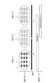
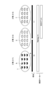
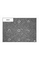
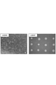
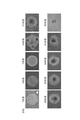
- FIG. 1 Phase-contrast micrographs showing the state of pseudopodia extension of the outer edge of the embryoid body (embryoid body) in Example 1 and Comparative Example 1. Phase-contrast micrographs of embryoid bodies (embryoid bodies) in Comparative Examples 2 and 3.
- FIG. 3 is a phase-contrast micrograph of the embryoid body in Example 3.
- the scale bar indicates 100 ⁇ m.
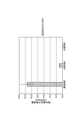
- the scale bar indicates 100 ⁇ m. It is a graph which shows the evaluation result of the gene expression level of the undifferentiated marker (NANOG) and the endoderm marker (SOX17) in Example 3 and Comparative Examples 6 and 7.
- 6 is a phase-contrast micrograph of the cells induced to differentiate in Example 4 taken over time.
- the scale bar indicates 200 ⁇ m. It is a phase-contrast micrograph and a fluorescence micrograph after immunostaining of the cells induced to differentiate in Examples 4 and 5.
- the scale bar indicates 200 ⁇ m. 3 is a phase-contrast micrograph of the cells induced to differentiate in Comparative Example 8 and Example 4.
- the scale bar indicates 200 ⁇ m. Arrows indicate cell clumps detached from the substrate.
- the present embodiment a mode for carrying out the present invention (hereinafter, simply referred to as “the present embodiment”) will be described in detail.
- the following embodiments of the present invention are examples for explaining the present invention, and are not intended to limit the present invention to the following contents.
- the present invention can be appropriately modified and carried out within the scope of the purpose.
- pluripotent stem cell refers to a cell having a characteristic (undifferentiated or pluripotent) capable of differentiating into various cells.
- ectoderm cell refers to a cell contained in the ectoderm formed by an early embryo (ectoderm cell) or a cell contained in a tissue derived from the ectoderm (nerve stem cell or nerve cell). Including).
- neural stem cells are glial cells (astrocytes, oligodendrocytes), central nerves (dopaminergic nerves, GABA nerves), and peripheral nerves (motor nerves, sensory nerves), as the induction of differentiation into nerve ectoblasts progresses. Shows cells in a state capable of inducing differentiation into.
- the “mesoderm cell” is a general term for cells contained in the mesoderm formed by the early embryo (mesoderm cells) or cells contained in the tissue derived from the mesoderm.
- endoderm lineage cell is a general term for cells contained in endoderm formed by early embryos (endoderm cells) and cells contained in tissues derived from endoderm.
- "maintenance of undifferentiated state” means a state in which cultured cells have pluripotency.
- the evaluation method for maintaining undifferentiated state is not particularly limited, but for example, analysis of cell surface markers by alkaline phosphatase staining, analysis of cell surface / intranuclear markers by immunostaining and flow cytometry, and gene expression level by real-time RT-PCR. , Confirmation of embryoid body (embryoid body) formation, which is a unique structure formed by pluripotent stem cells, and tera-toma assay for determining trigerm differentiation in In vivo from pathological sections.
- differentiation is a state in which expression of a cell type-specific membrane protein, transcription factor, etc. located downstream of the pluripotent stem cell is confirmed in the differentiation induction pathway of the pluripotent stem cell. Is shown. Furthermore, “induction of differentiation” indicates that the differentiation of cells is promoted by culturing the cells in the presence of a specific protein, gene, natural product, synthetic chemical substance, or the like. Further, in the present specification, the “cell aggregate (pluripotent stem cell aggregate)” refers to a three-dimensional structure formed by aggregating a plurality of cells (pluripotent stem cells).
- embryoid body is a spherical cell aggregate (aggregate of pluripotent stem cells) found in the early stage of embryogenesis, and is the ectoderm, mesoderm and endoderm as in the early embryo. Shows the properties of the germ layer.
- the "marker” indicates a protein or gene peculiar to a specific cell.
- the marker possessed by pluripotent stem cells is “undifferentiated marker”
- the marker possessed by ectoderm cells is “ectoderm marker”
- the marker possessed by mesoderm cells is “mesoderm marker”
- the marker possessed by endoderm cells is “endoderm”.
- the marker possessed by the nerve stem cell is referred to as “marker”
- the marker possessed by the nerve stem cell is referred to as “nerve stem cell marker”
- the marker possessed by the nerve cell is referred to as “nerve cell marker”
- the marker possessed by the small intestinal tissue cell is referred to as "intestinal epithelial cell marker”.
- Pluripotent stem cells do not have ectoderm markers, mesoderm markers and endoderm markers.
- the embryoid body (embryoid body) has an ectoderm marker, a mesoderm marker, and an endoderm marker.
- cell adhesion indicates the ease of adhesion to a cell culture substrate at a culture temperature
- having cell adhesion means that a cell has a cell culture substrate at a culture temperature. Indicates that it can be adhered to.
- not having cell adhesion means that cells cannot adhere to the cell culture substrate at the culture temperature.
- cell proliferation means the ease of cell proliferation at the culture temperature
- having cell proliferation means that the cells can grow at the culture temperature
- not having cell proliferation means that cells cannot grow at the culture temperature
- the method for inducing differentiation of ectoderm cells uses a cell culture substrate having the following two regions.
- A An island-like region having an area of 0.001 to 5 mm 2 having cell adhesion and cell proliferation.
- B A region adjacent to the region (A) and having no cell adhesion.
- pluripotent stem cells cannot be adherently cultured on the cell culture substrate, and germ layers (embryoid bodies) adhered to the cell culture substrate cannot be formed. Further, by using the cell culture substrate having the region (B) as the cell culture substrate, pluripotent stem cells can be proliferated only in the early (A) region, and the germ layer (embryoid body) of uniform size can be grown. ) Can be formed. By forming a germ layer (embryoid body) of uniform size, the efficiency of differentiation induction can be enhanced. If it does not have the region (B), it is not possible to form a germ layer (embryoid body) of uniform size.
- the area of the region (A) is more preferably 0.005 to 3 mm 2 because it is suitable for forming an embryoid body (embryoid body) having a size suitable for inducing differentiation into ectoderm cells.
- An area of 0.01 to 1.0 mm 2 is particularly preferable, and an area of 0.03 to 0.8 mm 2 is most preferable.
- the shape of the region (A) is not particularly limited, but a circle, an ellipse, or a regular polygon is preferable, and a circle is more preferable, because it is suitable for enhancing the differentiation induction efficiency.
- the shape of the region (B) is not limited except that it is adjacent to the region (A), but since it is suitable for producing an aggregate having a uniform size and shape, it is a boundary with the region (A). It is preferable that the region (B) is adjacent to the length of 20% or more of the line, 50% or more is more preferable, 80% or more is further preferable, and the periphery of the region (A) is all the region (B). Is most preferable. Further, since it is suitable for increasing the mass productivity of the cell culture substrate, it is preferable that the region (A) has an island-like structure and the region (B) has a sea-like sea-island structure.
- the area ratio of the region (A) and the region (B) is not particularly limited, but is suitable for increasing the number of cell aggregates that can be produced per unit area of the cell culture substrate (A).
- the area of the region is preferably 10% or more, more preferably 30% or more, further preferably 50% or more, and 70% or more with respect to the total area of the (A) region and the (B) region. Most preferred. Further, since it is suitable to provide a sufficient distance between the plurality of (A) regions and prevent the aggregates of the plurality of (A) regions from fusing to form a non-uniform shape, (B). )
- the area of the region is preferably 20% or more, more preferably 40% or more, further preferably 60% or more, and 80% or more with respect to the total area of the (A) region and the (B) region. Most preferred.
- the cell culture substrate used in the differentiation induction method of the present invention contains a layer made of a hydrophilic polymer on the surface in order to form a germ layer (embryoid body) of uniform size and to increase the efficiency of differentiation induction.
- (A) is preferably a region obtained by decomposing or modifying a part of the layer made of the hydrophilic polymer by any one of plasma treatment, ultraviolet treatment, corona discharge treatment, or a combination thereof.
- the region (A) is suitable for enhancing cell adhesion and cell proliferation and forming a germ layer (embryoid body) in a short time, the region (A) is further a plasma-treated region. preferable.
- the layer thickness of the layer made of the hydrophilic polymer is preferably 10 nm or more, more preferably 50 nm or more, and further preferably 100 nm or more because the region (B) is suitable for forming a region having no cell adhesion or cell proliferation. It is particularly preferable, and 500 nm or more is most preferable. Further, since the region (A) is suitable for a region having cell adhesion and cell proliferation, the layer thickness is preferably 1000 nm or less, more preferably 500 nm or less, particularly preferably 100 nm or less, and most preferably 50 nm or less.
- the method for forming the layer with the hydrophilic polymer at least one of a method of forming a chemical bond and a method of physical interaction can be used.
- the method for forming a chemical bond include a method for forming a reactive functional group such as ultraviolet irradiation, electron beam irradiation, gamma ray irradiation, plasma treatment, and corona treatment. It is also possible to carry out a cross-linking reaction to the surface of the substrate by an organic reaction using ions or radicals as a reaction source.
- coating, brush coating, dip coating, spin coating, bar coating, sink coating, and spraying using a matrix with excellent compatibility with the target hydrophilic polymer as a coating material It is possible to use techniques such as painting, roll coating, air knife coating, blade coating, gravure coating, microgravia coating, slot die coating and the like.
- the type of the hydrophilic polymer is not particularly limited, and examples thereof include those having a polar group such as a hydroxy group, an amino group and a polyethylene glycol group, and those having an amphoteric ionic structure such as a betaine structure and a phosphorylcholine group.
- a hydroxy group, a phosphorylcholine group, or a polyethylene glycol group is preferable, a hydroxy group or a phosphorylcholine group is more preferable, and a phosphorylcholine group is particularly preferable, because the region (B) is suitable for a region having no cell adhesion and cell proliferation. preferable.
- the hydrophilic polymer suppresses the elution of the hydrophilic polymer from the cell culture substrate, and suppresses the influence on the quality due to the mixing of the polymer in cell aggregates and embryonic bodies (embryonic bodies).
- a random copolymer or a block copolymer having both a hydrophilic monomer unit and a hydrophobic monomer unit is preferable, and a hydrophilic monomer unit and a hydrophilic monomer unit are preferable. More preferably, it is a random copolymer having both hydrophobic monomeric units.
- the hydrophilic monomer unit is 30 wt% or more because it is suitable to make the region (B) a region having no cell adhesion and cell proliferation. Is preferable, 40 wt% or more is more preferable, 50 wt% or more is particularly preferable, and 60 wt% or more is most preferable. Further, since it is suitable for suppressing the elution of the hydrophilic polymer, the hydrophobic monomer unit is preferably 20 wt% or more, more preferably 30 wt% or more, and particularly preferably 40 wt% or more. Most preferably 50 wt% or more.
- the hydrophilic monomer unit is not particularly limited except that it is hydrophilic, and for example, 2-dimethylaminoethyl acrylate, 2-dimethylaminoethyl methacrylate, 2-diethylaminoethyl acrylate, and 2-diethylaminoethyl.
- Those having an amino group such as methacrylate, N- [3- (dimethylamino) propyl] acrylamide; N- (3-sulfopropyl) -N-methacloyloxyethyl-N, N-dimethylammonium betaine, N-methacryloyloxy Those having betaine such as ethyl-N, N-dimethylammonium- ⁇ -N-methylcarboxybetaine; hydroxyethyl acrylate, hydroxyethyl methacrylate, N- (2-hydroxyethyl) acrylamide, polyethylene glycol monoacrylate, polyethylene glycol monomethacrylate.
- the hydrophobic monomer unit is not particularly limited except that it is hydrophobic, but for example, n-butyl acrylate, n-butyl methacrylate, isobutyl acrylate, isobutyl methacrylate, t-butyl acrylate, and t-butyl.
- Methacrylate, n-hexyl acrylate, n-hexyl methacrylate, n-octyl acrylate, n-octyl methacrylate, n-decyl acrylate, n-decyl methacrylate, n-dodecyl acrylate, n-dodecyl methacrylate, n-tetradecyl acrylate, n- Tetradecyl methacrylate and the like can be mentioned.
- the hydrophilic polymer is also suitable for suppressing the elution of the hydrophilic polymer, it is preferable that the hydrophilic polymer contains a monomer unit having reactivity.
- a UV-reactive monomer unit is preferable because a hydrophilic polymer can be immobilized on the substrate by a short treatment time, for example, 4-. Examples thereof include azidophenyl acrylate, 4-azidophenyl methacrylate, 2-((4-azidobenzoyl) oxy) ethyl acrylate, 2-((4-azidobenzoyl) oxy) ethyl methacrylate and the like.
- the hydrophilic polymer may also contain temperature-responsive monomeric units, eg, (meth) acrylamide compounds such as acrylamide, methacrylamide; N, N-diethylacrylamide, N-ethylacrylamide, N. -N-propylacrylamide, N-n-propyl Acrylamide, N-Isopropylacrylamide, N-Isopropyl Acrylamide, N-Cyclopropylacrylamide, N-Cyclopropyl Acrylamide, Nt-Butyl Acrylamide, N-ethoxyethylacrylamide , N-alkyl substituted (meth) acrylamide derivatives such as N-ethoxyethylmethacrylamide, N-tetrahydrofuruffle acrylamide, N-tetrahydrofuruffle methacrylamide; N, N-dimethyl (meth) acrylamide, N, N-ethylmethyl N, N-dialkyl-substituted (meth)
- a substance that promotes or inhibits cell adhesion to a part of the region on the cell culture substrate by a photolithography method or an inkjet method.
- a method of coating the substrate, a method of coating the surface of the cell culture substrate with any of plasma treatment, ultraviolet treatment, corona discharge treatment, or a combination thereof, and then coating the temperature-responsive polymer, etc. Can be mentioned.
- the cell culture substrate used in the differentiation induction method of the present invention may be sterilized.
- the sterilization method is not particularly limited, but high-pressure steam sterilization, UV sterility, ⁇ -ray sterility, ethylene oxide gas sterilization, and the like can be used.
- High-pressure steam sterilization, UV sterilization, and ethylene oxide gas sterilization are preferable because they are suitable for suppressing denaturation of block copolymers, and UV sterilization or ethylene oxide gas sterilization is preferable because they are suitable for suppressing deformation of the substrate.
- ethylene oxide gas sterilization is particularly preferable because the cell culture base material is excellent in mass productivity.
- the substrate used for producing the cell culture substrate is not particularly limited, but commonly used polymer compounds such as glass, polystyrene, polycarbonate, polyethylene terephthalate, polyvinylidene fluoride, polyethylene, polypropylene, polyethylene methacrylate, and ceramics. , And metals can be used. Polystyrene is most preferable because it has excellent transparency and is easy to mold and surface modify.
- the method for inducing differentiation and the method for producing the present invention are characterized in that differentiation is induced through the following steps (1-1) to (1-4).
- (1-1) Coating of a single substance or a plurality of substances selected from the group consisting of matrigel, laminin, fibronectin, vitronectin, collagen and fragments thereof, and seeding of pluripotent stem cells on a cell culture substrate. The process to be performed.
- (1-2) Pluripotent stem cell coagulation in which pluripotent stem cells seeded in the above step (1-1) were adherently cultured in the presence of an undifferentiated maintenance medium in a feeder-free manner and adhered to a cell culture substrate. The process of forming an aggregate.
- Pluripotent stem cell aggregates formed in the above step (1-2) are cultured in the presence of a medium containing a differentiation-inducing factor in a state of being adhered to a cell culture substrate, and embryoid bodies. The process of forming (embryoid body).
- the germ layer (embryoid body) formed in the above step (1-3) is cultivated in the presence of a medium containing a differentiation-inducing factor in a state of being adhered to a cell culture substrate, and then outside. The process of forming germ layer cell aggregates.
- step (1-1) in the method for inducing differentiation of the present invention a single or a plurality of substances selected from the group consisting of matrigel, laminin, fibronectin, vitronectin, collagen and fragments thereof are coated on the cell culture substrate.
- This is the process of seeding pluripotent stem cells.
- the region (A) can be imparted with cell adhesion and cell proliferation. can.
- the matrigel, laminin, vitronectin, fibronectin, collagen and fragments thereof may be natural products, artificially synthesized by genetic recombination technology or the like, fragments cleaved with restriction enzymes or the like, or fragments thereof. It may be a synthetic protein or a synthetic peptide based on these biological substances.
- Matrigel for example, Matrigel (manufactured by Corning Inc.) or Geltrex (manufactured by Gibco) can be preferably used as a commercially available product because of its availability.
- laminin is not particularly limited, but for example, laminin 511, laminin 521 or laminin 511- are reported to show high activity against ⁇ 6 ⁇ 1 integrin expressed on the surface of human iPS cells. E8 fragments can be used.
- the laminin may be a natural product, may be artificially synthesized by a genetic recombination technique or the like, or may be a synthetic protein or a synthetic peptide based on the laminin. From the viewpoint of availability, for example, iMatrix-511 (manufactured by Nippi Co., Ltd.) can be preferably used as a commercially available product.
- the vitronectin may be a natural product, may be artificially synthesized by a gene recombination technique or the like, or may be a synthetic protein or a synthetic peptide based on the vitronectin.
- vitronectin human plasma-derived (manufactured by Wako Pure Chemical Industries, Ltd.), synthemax (manufactured by Corning Inc.), and Vitronectin (manufactured by Gibco) can be preferably used as commercially available products. can.
- the fibronectin may be a natural product, may be artificially synthesized by a genetic recombination technique or the like, or may be a synthetic protein or a synthetic peptide based on the fibronectin.
- fibronectin solution human plasma-derived (manufactured by Wako Pure Chemical Industries, Ltd.) and Retronectin (manufactured by Takara Bio Inc.) can be preferably used as commercially available products.
- the type of collagen is not particularly limited, but for example, typeI collagen and typeIV collagen can be used.
- the collagen may be a natural product, may be artificially synthesized by a genetic recombination technique or the like, or may be a synthetic peptide based on the collagen. From the viewpoint of availability, for example, collagen I, human (manufactured by Corning Incorporated) or collagen IV, human (manufactured by Corning Incorporated) can be preferably used as a commercially available product.
- the cell type in the step (1-1) can be appropriately selected from stem cells having pluripotency such as ES cells and iPS cells, and the method for inducing differentiation of the present invention can be applied to regenerative medicine and drug discovery.
- IPS cells are preferred because they are suitable for application.
- the cell seeding method in the step (1-1) is not particularly limited, but a method of adding a cell suspension in which cells are monodispersed to a cell culture substrate or a method of adding cell aggregates to a cell culture substrate is available. In addition, since cell aggregates are uniformly formed in the region (A), it is preferable to disperse and disperse the cells uniformly.
- ROCK Rho-binding kinase
- Examples of the ROCK inhibitor include (R)-(+)-trans-N- (4-pyridyl) -4- (1-aminoethyl) -cyclohexanecarboxamide, 2HCl, H2O (manufactured by Fuji Film Wako Pure Chemical Industries, Ltd.). , Product name: Y-27632) and the like can be used.
- the concentration of the ROCK inhibitor added to the medium is a range effective for maintaining the survival of human cells and does not affect the undifferentiated state of human cells, preferably 1 ⁇ M to 50 ⁇ M. It is more preferably 3 ⁇ M to 20 ⁇ M, further preferably 5 ⁇ M to 15 ⁇ M, and most preferably 8 ⁇ M to 12 ⁇ M. Further, it is preferable to remove the ROCK inhibitor by a method such as replacement with a medium containing no ROCK inhibitor 24 hours after seeding the cells.
- the cell recovery method is not particularly limited, but for example, enzymatic treatment such as trypsin or collagenase, chelation treatment with ethylenediamineacetic acid (EDTA), and physical treatment such as scraper can be used alone or in combination.
- enzyme treatment it is preferable that the animal-derived component is free.
- TrypLE Select manufactured by Gibco
- TrypLE Express Gibco
- Accutase manufactured by Nacalai Tesque
- Accumax manufactured by Nacalai Tesque
- the cell seeding density in the step (1-1) is preferably 1.0 ⁇ 10 2 cells / cm 2 or more, preferably 1.2 ⁇ 10 because it is suitable for forming uniform cell aggregates in a short time.
- 3 cells / cm 2 or more is more preferable, 2.0 ⁇ 10 3 cells / cm 2 or more is particularly preferable, and 3.0 ⁇ 10 3 cells / cm 2 or more is most preferable.
- 1.0 ⁇ 10 5 cells / cm 2 or less is preferable, 5.0 ⁇ 10 4 cells / cm 2 or less is more preferable, and 2.5.
- X10 4 cells / cm 2 or less is particularly preferable, and 5.0 ⁇ 10 3 cells / cm 2 or less is most preferable.
- pluripotent stem cells seeded in step (1-1) are adherently cultured in the presence of an undifferentiated maintenance medium in a feeder-free manner, and a cell culture group is used. It forms pluripotent stem cell aggregates that adhere to the material.
- feeder-free refers to a method of directly seeding and culturing pluripotent stem cells on a cell culture substrate without using cells inactivated by gamma ray irradiation or the like (feeder cells).
- the pluripotent stem cells proliferate in the region (A) and become a substrate. It is possible to form cell aggregates layered in the out-of-plane direction. When an undifferentiated maintenance medium is not used, cell aggregates lose their pluripotency and the efficiency of inducing differentiation into ectoderm cells decreases. In addition, by culturing in a feeder-free manner, it is easy to maintain the nutrition of the medium during culturing, and it is possible to enhance the cell viability and the maintenance of undifferentiated cells.
- the adhesive culture facilitates the medium exchange work as compared with the suspension culture, the culture workability is excellent, and the total amount of the medium can be exchanged, so that the concentration of waste products discharged from the cells is low. Can be kept in a state.
- the influence of the concentration gradient in the culture system is small, the differentiation induction efficiency is excellent.
- the undifferentiated maintenance medium includes, for example, basic fibroblast growth factor (bFGF), transforming growth factor ⁇ (TGF- ⁇ ), and insulin-like growth factor as factors that act to maintain the undifferentiated state of pluripotent stem cells.
- bFGF basic fibroblast growth factor
- TGF- ⁇ transforming growth factor ⁇
- IGF insulin-like growth factor
- activin A activin A
- Wnt insulin
- transferase transferase
- ethanolamine 2-mercaptoethanol
- selenic acid oleic acid
- sodium hydrogen carbonate sodium hydrogen carbonate
- the type of medium is not particularly limited, but for example, culture in which a culture supplement such as a factor acting to maintain undifferentiated state and a non-essential amino acid is added to a basal medium such as DMEM, Ham's F12, D-MEM / Ham's F12.
- a culture supplement such as a factor acting to maintain undifferentiated state and a non-essential amino acid is added to a basal medium such as DMEM, Ham's F12, D-MEM / Ham's F12.
- Examples thereof include commercially available undifferentiated maintenance media such as Bio (manufactured by Bio Co., Ltd.), GS2-M (manufactured by Takara Bio Co., Ltd.), hPSC Growth Medium DXF (manufactured by PromoCell Co., Ltd.). Since it is suitable for stably maintaining the undifferentiated state of cells, Primate ES Cell Medium, StemFit AK02N, and StemFit AK03 are preferable, and StemFit AK02N and StemFit AK03 are more preferable, and StemFit AK02N (Ajinomoto Co., Inc.) Is the most preferable. In addition, since serum generally used as a cell culture medium also contains a differentiation-inducing factor, it is preferable that the undifferentiated maintenance medium for pluripotent stem cells is a serum-free medium.
- the undifferentiated maintenance medium for pluripotent stem cells is a serum-free medium.
- the same ROCK inhibitor as in the step (1-1) may be added in order to increase the survival rate of pluripotent stem cells.
- the types and concentrations of suitable ROCK inhibitors are the same as described above.
- the number of cells per unit area in the region (A) is increased. It is preferable to incubate until 1.0 ⁇ 10 4 cells / cm 2 or more, more preferably 2.0 ⁇ 10 4 cells / cm 2 or more, and particularly preferably 5.0 ⁇ 10 4 cells / cm 2 or more. 7.5 ⁇ 10 4 cells / cm 2 or more is most preferable.
- the number of cells per unit area of the region (A) is 5.0 ⁇ 106 .
- step (1-3) It is preferable to proceed to the step (1-3) at cells / cm 2 or less, more preferably 1.0 ⁇ 10 6 cells / cm 2 or less, and particularly preferably 5.0 ⁇ 10 5 cells / cm 2 or less. Most preferably 0 ⁇ 10 5 cells / cm 2 or less.
- step (1-3) in the method for inducing differentiation of the present invention pluripotent stem cell aggregates formed in step (1-2) above are placed on a cell culture substrate in the presence of a medium containing a differentiation-inducing factor. It is cultured in a state of being adhered to the embryo to form a germ layer (embryoid body).
- the differentiation-inducing factor in the above step (1-3) contains an ectoderm-inducing factor, a mesoderm-inducing factor, and an endoderm-inducing factor because it is suitable for increasing the efficiency of inducing differentiation into the embryoid body (embryoid body). Is preferable.
- the ectoderm-inducing factor is Noggin (BMP inhibitor), dorsomorpin (BMP inhibitor), SB431542 (TGF- ⁇ inhibitor) because it is suitable for enhancing the efficiency of inducing differentiation into the embryoid body (embryoid body).
- Activin inhibitor and more preferably BMP inhibitor and TGF- ⁇ inhibitor.
- any one of activin A, CHIR99021 (GSK3 ⁇ inhibitor), and Bone morphogenetic protein (BMP) 4 is suitable for enhancing the efficiency of inducing differentiation into the embryoid body (embryoid body). Is preferable, and it is more preferable to contain a GSK3 ⁇ inhibitor.
- examples of the endoderm-inducing factor include small molecule compounds such as activin A and CHIR99021.
- the concentration of the differentiation-inducing factor added to the medium is not particularly limited, but is preferably 0.1 to 10 ⁇ M because it is suitable for increasing the efficiency of inducing differentiation into the germ layer (embryoid body). It is preferably 1.0 to 7.5 ⁇ M, and most preferably 2.0 to 5.0 ⁇ M.
- the medium containing the differentiation-inducing factor is preferably replaced every 24:00 during the culture in order to sufficiently promote the differentiation induction. By exchanging the medium within 24 hours, it is possible to prevent a shortage of differentiation-inducing factors in the medium and uniformly differentiate all cells.
- PAX6, Nestin, SOX1, SOX2, SOX10, Notch1, E Cadherin, MAP2 are preferable, and PAX6, Nestin, SOX1 , SOX2 is more preferred.
- PAX6, Nestin, SOX1 , SOX2 is more preferred.
- the mesoderm marker Tbx1, Brachyury, MSX1 and Flk-1 are preferable, and Tbx1 and Brachyury are even more preferable.
- FOXA2, SOX17, GATA4, GATA6, CXCR4, HNF3 ⁇ , HNF4 ⁇ , ⁇ FP are preferable, and FOXA2 and SOX17 are more preferable.
- PAX6, Nestin, Olig2, SOX1, SOX2 and DCX are preferable, and PAX6, Nestin, SOX1 and Olig2 are more preferable.
- step (1-4) in the method for inducing differentiation of the present invention the germ layer (embryoid body) formed in step (1-3) is used as a cell culture substrate in the presence of a medium containing a differentiation inducing factor. It is cultured in a state of being adhered to the top to form ectoderm cell aggregates.
- a differentiation inducing factor By inducing differentiation of the embryoid body (embryoid body) formed in the above step (1-3) in a state of being adhered to the cell culture substrate, the influence of the concentration gradient of the above-mentioned inducing factor is small, so that it is uniform. It is expected that differentiation will progress.
- the medium is not particularly limited, and examples thereof include a culture medium in which a factor acting for ectoderm induction and a culture supplement are added to a basal medium such as DMEM, Ham's F12, D-MEM / Ham's F12.
- a basal medium such as DMEM, Ham's F12, D-MEM / Ham's F12.
- the type of cell that induces differentiation from the germ layer (germ-like body) in the step (1-4) is not particularly limited except that it is an ectoderm lineage cell, and for example, epidermal ectoderm-derived cells and neuroectodermal leaves. Derived cells are preferable, and since they are suitable for use in regenerative medicine and drug discovery, neuroectodermal-derived cells are even more preferable.
- the neurons derived from neuroendoblasts are not particularly limited, and for example, schwan precursor cells, myelin schwan cells, non-myelin schwan cells, radial glial cells, oligodendrocyte precursor cells, and glial cells such as oligodendrocytes and astrosites.
- Nerve cells such as glutamate-operated neurons, GABA-operated neurons, dopaminergic neurons, serotoninergic neurons, cholinergic neurons, motor neurons and sensory neurons are preferred.
- the ectoderm-derived cells are nerve cells, they are cultured for 5 to 14 days in the presence of a medium containing B27 supplements, TGF- ⁇ inhibitor, GSK3 ⁇ inhibitor, LIF, bFGF, retinoic acid, and Purmorphamine, and then B27 supplements and TGF. -Preferably, the cells are cultured for 3 to 5 days in the presence of a medium containing a ⁇ inhibitor, a GSK3 ⁇ inhibitor, LIF, bFGF, retinoic acid, Purmorphamine, and DAPT.
- a medium containing B27 supplements rhBDNF, rhGDNF, ascorbic acid, retinoic acid, and DAPT after culturing the neurons as described above.
- the method for inducing differentiation of the present invention uses a cell culture substrate having the following two regions (A) and (B).
- A An island-like region having an area of 0.001 to 5 mm 2 having cell adhesion and cell proliferation.
- B A region adjacent to the region (A) and having no cell adhesion or cell proliferation.
- pluripotent stem cells can be adherently cultured on the cell culture substrate in the step (2-2) described later, and also. It is possible to form an embryoid body (embryoid body) adhered to a cell culture substrate.
- the region (A) is not provided, pluripotent stem cells cannot be adherently cultured on the cell culture substrate, and germ layers (embryoid bodies) adhered to the cell culture substrate cannot be formed.
- pluripotent stem cells can be proliferated only in the early (A) region, and the germ layer (embryoid body) of uniform size can be grown.
- the area of the region (A) is more preferably 0.005 to 3 mm 2 because it is suitable for forming an embryoid body (embryoid body) having a size suitable for inducing differentiation into mesoderm cells.
- An area of 0.01 to 1.0 mm 2 is particularly preferable, and an area of 0.03 to 0.8 mm 2 is most preferable.
- the shape of the region (A) is the same as ⁇ Method for inducing differentiation of ectoderm cells>.
- the shape of the region (B) is the same as that of ⁇ Method for inducing differentiation of ectoderm cells>.
- the area ratios of the (A) region and the (B) region are the same as in ⁇ Method for inducing differentiation of ectoderm cells>.
- the cell culture substrate used in the differentiation induction method of the present invention has a layer containing a hydrophilic polymer on the surface because it is suitable for forming a germ layer (embryoid body) of uniform size and increasing the efficiency of differentiation induction.
- the region (A) is a region obtained by decomposing or modifying the layer containing the hydrophilic polymer by plasma treatment, ultraviolet treatment and / or corona discharge treatment. Since the cell culture substrate has a layer containing a hydrophilic polymer on the surface, it suppresses the adsorption of proteins that contribute to the substrate-cell adhesion in the region (B), and does not have cell adhesion or cell proliferation. It can be an area.
- the region (A) is suitable for enhancing cell adhesion and cell proliferation and forming a germ layer (embryoid body) in a short time, the region (A) is further a plasma-treated region. preferable.
- the region (A) is a region having no hydrophilic polymer and the region (B) is a hydrophilic polymer. It is preferable that the region has a layer containing the above, the region (A) is a region where the surface of the substrate modified by plasma treatment, ultraviolet treatment and / or corona discharge treatment is exposed, and the region (A) is hydrophilic. It is more preferable that the region having no polymer and the region (B) have a layer containing a hydrophilic polymer.
- the layer thickness of the layer containing the hydrophilic polymer is the same as ⁇ Method for inducing differentiation of ectoderm cells>.
- the method for forming the layer containing the hydrophilic polymer is the same as the ⁇ method for inducing differentiation of ectoderm cells>.
- the type of the hydrophilic polymer is the same as ⁇ Method for inducing differentiation of ectoderm cells>.
- the hydrophilic polymer suppresses the elution of the hydrophilic polymer from the cell culture substrate, and suppresses the influence on the quality due to the mixing of the polymer in cell aggregates and embryonic bodies (embryonic bodies).
- a random copolymer or a block copolymer having both a hydrophilic monomer unit and a hydrophobic monomer unit is preferable, and a hydrophilic monomer unit and a hydrophilic monomer unit are preferable. More preferably, it is a random copolymer having both hydrophobic monomeric units.
- the composition ratio of the copolymer is the same as that in ⁇ Method for inducing differentiation of ectoderm cells>.
- the hydrophilic monomer unit is the same as ⁇ Method for inducing differentiation of ectoderm cells>.
- the hydrophobic monomer unit is the same as ⁇ Method for inducing differentiation of ectoderm cells>.
- the hydrophilic polymer is also suitable for suppressing the elution of the hydrophilic polymer, it is preferable that the hydrophilic polymer contains a monomer unit having reactivity.
- the reactive monomer unit is the same as ⁇ Method for inducing differentiation of ectoderm cells>.
- the hydrophilic polymer may also contain a temperature-responsive monomer unit, and the temperature-responsive monomer unit is the same as in ⁇ Method for inducing differentiation of ectoderm cells>.
- Another method for producing a cell culture substrate used in the method for inducing differentiation of the present invention is the same as ⁇ Method for inducing differentiation of ectoderm cells>.
- the cell culture substrate used in the differentiation induction method of the present invention may be sterilized.
- the method of sterilization is the same as ⁇ Method of inducing differentiation of ectoderm cells>.
- the substrate used for producing the cell culture substrate is the same as ⁇ method for inducing differentiation of ectoderm cells>.
- the method for inducing differentiation of the present invention is characterized in that differentiation is induced through the following steps (2-1) to (2-3).
- (2-1) Coating of a single substance or a plurality of substances selected from the group consisting of matrigel, laminin, fibronectin, vitronectin, collagen and fragments thereof, and seeding of pluripotent stem cells on a cell culture substrate. The process to be performed.
- (2-2) The pluripotent stem cells seeded in the above step (2-1) were adherently cultured in the presence of an undifferentiated maintenance medium in a feeder-free manner, and the embryoid body (embryoid body) adhered onto the cell culture substrate. The process of forming the body).
- the germ layer (embryoid body) formed in the above step (2-2) is cultured in the presence of a medium containing a differentiation-inducing factor in a state of being adhered to a cell culture substrate.
- step (2-1) in the method for inducing differentiation of the present invention a single or a plurality of substances selected from the group consisting of matrigel, laminin, fibronectin, vitronectin, collagen and fragments thereof are coated on the cell culture substrate. This is the process of seeding pluripotent stem cells.
- the region (A) can be imparted with cell adhesion and cell proliferation.
- the region (A) can be imparted with cell adhesion and cell proliferation.
- a combination of at least four types containing laminin is more preferable, and any combination of laminin and matrigel, laminin and fibronectin, or laminin and collagen is preferable. Is particularly preferable, and laminin alone is most preferable.
- matrigel The above-mentioned matrigel, laminin, fibronectin, vitronectin, collagen and fragments thereof are the same as in ⁇ Method for inducing differentiation of ectoderm cells>.
- the cell type in the step (2-1) can be appropriately selected from stem cells having pluripotency such as ES cells and iPS cells, and the method for inducing differentiation of the present invention can be applied to regenerative medicine and drug discovery.
- IPS cells are preferred because they are suitable for application.
- the cell seeding method in the step (2-1) is not particularly limited, but a method of adding a cell suspension in which cells are monodispersed to a cell culture substrate or a method of adding cell aggregates to a cell culture substrate is available. In addition, since cell aggregates are uniformly formed in the region (A), it is preferable to disperse and disperse the cells uniformly.
- ROCK Rho-binding kinase
- the cell seeding density in the step (2-1) is preferably 1.0 ⁇ 10 2 cells / cm 2 or more, preferably 1.2 ⁇ 10 because it is suitable for forming uniform cell aggregates in a short time.
- 3 cells / cm 2 or more is more preferable, 2.0 ⁇ 10 3 cells / cm 2 or more is particularly preferable, and 3.0 ⁇ 10 3 cells / cm 2 or more is most preferable.
- 1.0 ⁇ 10 6 cells / cm 2 or less is preferable, 5.0 ⁇ 10 5 cells / cm 2 or less is more preferable, and 2.5.
- X10 4 cells / cm 2 or less is particularly preferable, and 5.0 ⁇ 10 3 cells / cm 2 or less is most preferable.
- pluripotent stem cells seeded in step (2-1) are adherently cultured in the presence of an undifferentiated maintenance medium in a feeder-free manner, and a cell culture group is used. It forms a germ layer (embryoid body) adhered to the wood.
- feeder-free refers to a method of directly seeding and culturing pluripotent stem cells on a cell culture substrate without using cells inactivated by gamma ray irradiation or the like (feeder cells).
- the pluripotent stem cells proliferate in the region (A) and become a substrate.
- Embryoid bodies layered in the out-of-plane direction can be formed.
- the embryoid body loses pluripotency and the efficiency of inducing differentiation into mesoderm lineage cells decreases.
- the adhesive culture facilitates the medium exchange work as compared with the suspension culture, the culture workability is excellent, and the total amount of the medium can be exchanged, so that the concentration of waste products discharged from the cells is low. Can be kept in a state.
- the influence of the concentration gradient in the culture system is small, the differentiation induction efficiency is excellent.
- the undifferentiated maintenance medium is the same as ⁇ Method for inducing differentiation of ectoderm cells>.
- the same ROCK inhibitor as in the step (2-1) may be added in order to increase the survival rate of pluripotent stem cells.
- the types and concentrations of suitable ROCK inhibitors are the same as described above.
- the number of cells per unit area of the region (A) is 1.0 ⁇ 10 4 because it is suitable for increasing the differentiation induction efficiency in the step (2-3) described later. It is preferable to culture until cells / cm 2 or more, 2.0 ⁇ 10 4 cells / cm 2 or more is more preferable, 5.0 ⁇ 10 4 cells / cm 2 or more is particularly preferable, and 7.5 ⁇ 10 4 is particularly preferable. Most preferably cells / cm 2 or more. In addition, since it is suitable for maintaining the nutrition of the medium and increasing the viability of the cells in the step (2-3), the number of cells per unit area of the region (A) is 5.0 ⁇ 106 cells / cm 2 or less.
- 1.0 ⁇ 10 6 cells / cm 2 or less is more preferable, 5.0 ⁇ 10 5 cells / cm 2 or less is particularly preferable, and 1.0 ⁇ 10 5 cells / cm /. Most preferably cm 2 or less.
- the number of cells per unit area of the region (A) it is preferable to culture for 1 to 48 hours in the step (2-2), more preferably 6 to 36 hours, and 12 to 36 hours. It is particularly preferable, and 18 to 30 hours is most preferable.
- the shape of the germ layer (embryoid body) formed in the step (2-2) can be appropriately set.
- a closed shape consisting of a circle, an ellipse, a polygon, an irregular straight line or a curved line, and the like can be mentioned.
- a circle, an ellipse, or a polygon is preferable, a circle, an ellipse, or a rectangle is more preferable, and a circle, an ellipse, or a square is particularly preferable. Often, circles or ellipses are most preferred.
- the aspect ratio of the island-shaped shape is preferably 5 or less, more preferably 2 or less, and particularly preferably 1.5 or less. , 1.1 or less is most preferable.
- the "aspect ratio" indicates a major axis / minor axis which is a ratio of a maximum diameter (major diameter) to a minimum diameter (minor diameter) of a shape.
- step (2-3) in the method for inducing differentiation of the present invention the germ layer (embryoid body) formed in step (2-2) is used as a cell culture substrate in the presence of a medium containing a differentiation inducing factor. It is cultured in a state of being adhered to the top to form mesoderm lineage cells.
- the differentiation-inducing factor in the step (2-3) above contains the mesoderm-inducing factor.
- the mesoderm-inducing factor a single or single selected from the group consisting of GSK3 ⁇ inhibitor, Bone morphogenetic protein (BMP) and activin because it is suitable for enhancing the efficiency of inducing differentiation into the embryoid body (embryoid body). It is preferably a plurality of differentiation-inducing factors, and particularly preferably contains any one of activin A, CHIR99021 (GSK3 ⁇ inhibitor), and BMP4.
- the concentration of the differentiation-inducing factor added to the medium is not particularly limited, but is preferably 1000 to 500 ng / mL because it is suitable for increasing the efficiency of inducing differentiation into the germ layer (embryoid body). It is preferably 500 to 100 ng / mL, and most preferably 100 ng / mL or less.
- the medium containing the differentiation-inducing factor every 24:00 during the culture in order to sufficiently promote the differentiation induction.
- By exchanging the medium within 24 hours it is possible to prevent a shortage of differentiation-inducing factors in the medium and uniformly differentiate all cells.
- the mesoderm markers contained in the germ layer (embryoid body) formed in the step (2-2) above include FLK-1, MESP1, MESS2, FOXF1, HAND1, EVX1, IRX3, CDX2, TBX6, MIXL1, and SNAI1.
- FOXC1 and PDGFR ⁇ are preferable, and BRACHYURY is most preferable.
- step (2-3) in the method for inducing differentiation of the present invention the germ layer (embryoid body) formed in step (2-2) is used as a cell culture substrate in the presence of a medium containing a differentiation inducing factor. It is cultured in a state of being adhered to the top to form mesoderm cell aggregates.
- a differentiation inducing factor By inducing differentiation of the germ layer (embryoid body) formed in the above step (2-2) in a state of being adhered to the cell culture substrate, the influence of the concentration gradient of the above-mentioned inducing factor is small, so that it is uniform. It is expected that differentiation will progress.
- the medium used in the step (2-3) is not particularly limited, but is a culture medium obtained by adding a mesoderm differentiation inducing factor and a culture supplement to a basal medium such as DMEM, Ham's F12, D-MEM / Ham's F12, for example. Can be mentioned.
- the type of cell that induces differentiation from the embryonic body (embryonic body) in the step (2-3) is not particularly limited except that it is a mesenchymal cell, but for example, blood cells, smooth muscle cells, and germ cells. Is preferable, and bone cells, myocardial cells, skeletal muscle cells, and renal cells are more preferable because they are suitable for use in regenerative medicine and drug discovery.
- a cell culture substrate having the following regions (A) and (B) is used.
- B A region adjacent to the region (A) and having no cell adhesion or cell proliferation.
- pluripotent stem cells can be adherently cultured on the cell culture substrate in the step (3-2) described later, and the cells can also be adherently cultured. It is possible to form an embryoid body adhered to a culture substrate.
- the cell culture substrate does not have the region (A)
- pluripotent stem cells cannot be adherently cultured on the cell culture substrate, and embryoid bodies adhered to the cell culture substrate cannot be formed.
- a cell culture substrate having a region (B) as a cell culture substrate pluripotent stem cells can be proliferated only in the region (A), and an embryoid body of uniform size can be formed. can. By forming an embryoid body of uniform size, the efficiency of differentiation induction can be enhanced. If the cell culture substrate does not have the region (B), it is not possible to form embryoid bodies of uniform size.
- 0.005 to 3 mm 2 is more preferable, and 0.02 to 2. 5 mm 2 is more preferable, and 0.03 to 2.0 mm 2 is most preferable.
- the shape of the region (A) is the same as ⁇ Method for inducing differentiation of ectoderm cells>.
- the cell culture substrate used in the method for inducing differentiation of the present invention has a layer containing a hydrophilic polymer on the surface because it is suitable for forming embryoid bodies of uniform size and increasing the efficiency of inducing differentiation (A).
- Region is preferably a region obtained by decomposing or modifying a layer containing a hydrophilic polymer by at least one treatment selected from the group consisting of plasma treatment, ultraviolet treatment and corona discharge treatment.
- the region (A) can be a region that does not have proliferation. Further, since a part of the layer containing the hydrophilic polymer is decomposed or modified, cell adhesion and cell proliferation can be imparted to the region (A). Further, since the region (A) is suitable for enhancing cell adhesion and cell proliferation and forming an embryoid body in a short time, the region (A) may be a region decomposed or modified by plasma treatment. More preferred.
- the region (A) is a region having no hydrophilic polymer, and the region (B) is a hydrophilic polymer. It is preferable that the region has a layer containing the above, the region (A) is a region where the surface of the substrate modified by plasma treatment, ultraviolet treatment and / or corona discharge treatment is exposed, and the region (B) is hydrophilic. It is more preferable that the region has a layer containing a polymer.
- the layer thickness of the layer containing the hydrophilic polymer is the same as ⁇ Method for inducing differentiation of ectoderm cells>.
- the same method as that exemplified for the method for inducing differentiation of ectoderm cells can be mentioned.
- hydrophilic polymer examples include the same hydrophilic polymers as those exemplified for the method for inducing the differentiation of ectoderm cells.
- the above-mentioned hydrophilic polymer is suitable for suppressing the elution of the hydrophilic polymer from the cell culture substrate and suppressing the influence on the quality due to the contamination of the cell aggregate or embryo-like body with the polymer. Therefore, it is preferable that it is a random copolymer or a block copolymer having both a hydrophilic monomer unit and a hydrophobic monomer unit, and a hydrophilic monomer unit and a hydrophobic simple polymer are preferable. More preferably, it is a random copolymer having both weight units.
- the composition ratio of the above-mentioned copolymer is the same as that in ⁇ Method for inducing differentiation of ectoderm cells>.
- the hydrophilic monomer unit is the same as ⁇ Method for inducing differentiation of ectoderm cells>.
- the hydrophobic monomer unit is the same as ⁇ Method for inducing differentiation of ectoderm cells>.
- the hydrophilic polymer is also suitable for suppressing the elution of the hydrophilic polymer, it is preferable that the hydrophilic polymer contains a monomer unit having reactivity.
- the reactive monomer unit is the same as ⁇ Method for inducing differentiation of ectoderm cells>.
- the hydrophilic polymer may also contain a temperature-responsive monomeric unit.
- the temperature-responsive monomer unit is the same as ⁇ Method for inducing differentiation of ectoderm cells>.
- Another method for producing a cell culture substrate used in the method for inducing differentiation of the present invention is the same as ⁇ Method for inducing differentiation of ectoderm cells>.
- the region (B) is adjacent to the region (A) and does not have cell adhesion or cell proliferation. Since the region (B) is adjacent to the region (A) and does not have cell proliferation, when cells are cultured, cell aggregates are formed only in the region (A), and (A). It is possible to form a cell-free state around the region. Further, since it is suitable for uniformizing the size and shape of the aggregate to be produced, it is preferable that the region (B) has not only cell proliferation but also cell adhesion.
- the shape of the region (B) is the same as that of ⁇ Method for inducing differentiation of ectoderm cells>.
- the area ratios of the (A) region and the (B) region are the same as in ⁇ Method for inducing differentiation of ectoderm cells>.
- the cell culture substrate used in the differentiation induction method of the present invention may be sterilized.
- the method of sterilization is the same as ⁇ Method of inducing differentiation of ectoderm cells>.
- the substrate used for producing the cell culture substrate is the same as ⁇ method for inducing differentiation of ectoderm cells>.
- the method for inducing differentiation of the present invention includes the following steps (3-1), step (3-2) and step (3-3).
- Step (3-1) A composition containing at least one selected from laminin and fragments thereof in the cell culture substrate having the above-mentioned regions (A) and (B) is added to the culture surface of the cell culture substrate.
- Step (3-3) The embryoid body formed in step (3-2) is cultured in the presence of a medium containing a differentiation-inducing factor in a state of being adhered to the culture surface of the cell culture substrate. A step of inducing differentiation to form aggregates of endometrial cells.
- a composition containing at least one selected from laminin and a fragment thereof in the above-mentioned cell culture substrate is mixed with laminin and its fragments based on the area of the culture surface of the cell culture substrate. It is a step of adding so that the total amount of the fragments is 1 to 100 ⁇ g / cm 2 , and seeding the pluripotent stem cells.
- the culture surface of the cell culture substrate is a surface that is in contact with the medium during culture and to which cells can adhere (usually a surface perpendicular to the vertical direction because the cells settle due to gravity).
- a composition containing at least one selected from laminin and its fragments is at least one selected from laminin and its fragments, as it is suitable for maintaining cell adhesion during culture during differentiation induction. It is more preferable that the composition contains only.
- the amount of the composition containing at least one selected from laminin and its fragments is such that the total amount of laminin and its fragments is 1.2 to 50 ⁇ g / cm 2 based on the area of the culture surface of the cell culture substrate.
- the amount of the culture medium is more preferable, and the amount of the culture medium is most preferably 1.5 to 10 ⁇ g / cm 2 .
- the addition amount is less than 1 ⁇ g / cm 2 , cell adhesion cannot be maintained during the culture during the induction of differentiation.
- a solution (composition) obtained by diluting at least one selected from laminin and its fragments with PBS or the like is used as a cell culture group.
- a precoat method may be used in which the material is added to the material and allowed to stand for several hours, and a composition containing at least one selected from laminin and fragments thereof is used as a cell suspension for seeding pluripotent stem cells.
- You may use the addition method of adding to the cell culture substrate in the state of being mixed with. That is, in step (3-1), the addition of the composition containing at least one selected from laminin and fragments thereof may be carried out prior to the seeding of pluripotent stem cells, or may be carried out at the same time. good.
- Laminin and its fragments are the same as ⁇ Method for inducing differentiation of ectoderm cells>.
- the type of pluripotent stem cell seeded in step (3-1) can be appropriately selected from stem cells having pluripotent differentiation such as ES cells and iPS cells, but it is applied to regenerative medicine and drug discovery. Therefore, iPS cells are preferable.
- the method for seeding pluripotent stem cells in step (3-1) is not particularly limited, but a cell suspension in which pluripotent stem cells are monodispersed is added to a cell culture substrate, or cell aggregates are used as a cell culture group. Examples include a method of adding to the material. Of these methods, since cell aggregates are uniformly formed in the region (A), it is preferable to disperse and seed the cells uniformly.
- ROCK Rho-binding kinase
- the cell seeding density in the step (3-1) is preferably 1.0 ⁇ 10 2 cells / cm 2 or more, preferably 1.2 ⁇ 10 3 because it is suitable for forming uniform cell aggregates in a short time. More preferably, cells / cm 2 or more, more preferably 2.0 ⁇ 10 3 cells / cm 2 or more, and most preferably 3.0 ⁇ 10 3 cells / cm 2 or more. Further, since it is suitable for suppressing cell death due to insufficient nutrition of the medium, 1.0 ⁇ 10 6 cells / cm 2 or less is preferable, 5.0 ⁇ 10 5 cells / cm 2 or less is more preferable, and 1.0. ⁇ 10 5 cells / cm 2 or less is more preferable, and 2.5 ⁇ 10 4 cells / cm 2 or less is most preferable.
- the pluripotent stem cells seeded in the step (3-1) are adherently cultured in the presence of an undifferentiated maintenance medium in a feeder-free manner and placed on the culture surface of the cell culture substrate. This is the process of forming an adhered embryoid body.
- the term "feeder-free” refers to a method of directly seeding and culturing pluripotent stem cells on a cell culture substrate without using cells inactivated by gamma-ray irradiation or the like (feeder cells).
- the pluripotent stem cells proliferate in the region (A) and the substrate surface. It is possible to form an outwardly layered embryonic body.
- the undifferentiated maintenance medium is not used, the embryoid body loses pluripotency and the efficiency of inducing differentiation into endoderm cells is reduced.
- the cell viability can be increased, and the maintenance of undifferentiated state becomes easy.
- the adhesive culture facilitates the medium exchange work as compared with the suspension culture, the culture workability is excellent, and the total amount of the medium can be exchanged, so that the concentration of waste products discharged from the cells is low. Can be kept in a state.
- the influence of the concentration gradient in the culture system is small, the differentiation induction efficiency is excellent.
- the above-mentioned undifferentiated maintenance medium is the same as ⁇ Method for inducing differentiation of ectoderm cells>.
- step (3-2) the same ROCK inhibitor as in step (3-1) may be added in order to increase the survival rate of pluripotent stem cells.
- the types and concentrations of suitable ROCK inhibitors are the same as described above.
- the number of cells per unit area of the region (A) is 1.0 ⁇ 10 4 cells / cm because it is suitable for increasing the differentiation induction efficiency in the step (3-3) described later. It is preferably cultured until it becomes 2 or more, 2.0 ⁇ 10 4 cells / cm 2 or more is more preferable, 5.0 ⁇ 10 4 cells / cm 2 or more is more preferable, and 7.5 ⁇ 10 4 cells / cm. 2 or more is most preferable. In addition, since it is suitable for maintaining the nutrition of the medium and increasing the viability of the cells in the step (3-3), the number of cells per unit area of the region (A) is 5.0 ⁇ 106 cells / cm 2 or less.
- 1.0 ⁇ 10 6 cells / cm 2 or less is more preferable, 5.0 ⁇ 10 5 cells / cm 2 or less is further preferable, and 1.0 ⁇ 10 5 cells / cm /. Most preferably cm 2 or less.
- the culture for 1 to 48 hours in the step (3-2), more preferably 6 to 36 hours, and 12 to 12 to. 36 hours is more preferred, and 18-30 hours is most preferred.
- the shape of the embryoid body formed in step (3-2) can be appropriately set, and examples thereof include a closed shape consisting of a sphere, an ellipsoid, a polyhedron, and an amorphous plane and / or curved surface. Can be done.
- the shape of the embryoid body formed in the step (3-2) is preferably spherical or hemispherical because it is suitable for increasing the differentiation induction efficiency in the step (3-3) described later.
- the shape of the embryoid body can be controlled by the shape of the region (A) (island-shaped shape) of the cell culture substrate.
- a circle, an ellipse, or a polygon is preferable, and a circle, an ellipse, or a rectangle is more preferable, because it is suitable for producing an embryoid body having a shape close to a sphere (for example, a sphere or a hemisphere).
- Circles, ellipses or squares are particularly preferred, with circles or ellipses being most preferred.
- the aspect ratio of the island-shaped shape is preferably 5 or less, more preferably 2 or less, and 1 5.5 or less is particularly preferable, and 1.1 or less is most preferable.
- the "aspect ratio" indicates a major axis / minor axis which is a ratio of a maximum diameter (major diameter) and a minimum diameter (minor diameter) of a shape.
- step (3-3) the embryoid body formed in step (3-2) is cultured in the presence of a medium containing a differentiation-inducing factor in a state of being adhered to the culture surface of the cell culture substrate. This is a step of inducing differentiation to form aggregates of endometrial cells.
- differentiation-inducing factor examples include a TGF- ⁇ inhibitor, an ATP competitive inhibitor and a GSK3 inhibitor.
- the differentiation-inducing factor may be used alone or in combination of two or more.
- the differentiation-inducing factor in step (3-3) contains an endoderm differentiation-inducing factor.
- the endometrial differentiation-inducing factor at least one differentiation-inducing factor selected from the group consisting of Wnt protein, Bone morphogenetic protein (BMP), insulin-like growth factor and activin because it is suitable for enhancing the differentiation-inducing efficiency. It is more preferable to contain any one of Wnt3a, BMP4, IGFI and activin A.
- the concentration of the above-mentioned differentiation-inducing factor added to the medium is not particularly limited, but is preferably 1000 to 500 ng / mL, more preferably 500 to 100 ng / mL because it is suitable for increasing the differentiation-inducing efficiency. Most preferably, it is 100 ng / mL or less.
- FOXA2, CXCR4, NKX2.1, AFP, SERPINA1, SST, ISL1, IPF1, IAPP, PAX4 and TAT are preferable, and SOX17 is preferable. Most preferred.
- step (3-3) the embryoid body is cultured in a state of being adhered to the culture surface of the cell culture substrate.
- the medium used in step (3-3) is not particularly limited, but for example, culture in which an endoderm differentiation-inducing factor and a culture supplement are added to a basal medium such as DMEM, Ham's F12, D-MEM / Ham's F12. Medium is mentioned.
- the type of cell that induces differentiation from the embryo-like body in step (3-3) is not particularly limited except that it is an endoderm cell, but for example, thyroid cells and urinary tract cells are preferable, and regenerative medicine or Gastric epithelial cells, liver cells, pancreatic cells and intestinal epithelial cells are more preferable because they are suitable for use in drug discovery.
- the intestinal epithelial cells preferably have at least one cell selected from the group consisting of enterocytes, cup cells, intestinal endocrine cells, Paneth cells and intestinal epithelial stem cells, and enterocytes, cup cells, intestinal endocrine cells, and the like. It is more preferable to include all Paneth cells and intestinal epithelial stem cells.
- Enterocytes have unique markers. For example, CDX2 as an enterocyte marker, VIL1 as an absorptive epithelial cell marker, MUC2 as an embryonic cell marker, CGA as an intestinal endocrine cell marker, DEFA6 as a Paneth cell marker, and LGR5 as an intestinal epithelial stem cell marker.
- the shape of the aggregate of endoderm cells obtained in step (3-3) is usually the same as the shape of the embryoid body formed in step (3-2).
- Specific examples of the shape of the agglomerate include a shape close to a sphere (for example, a sphere or a hemisphere).
- the differentiation induction kit into triembryonic cells which comprises a cell culture substrate having the following region (A) and the following region (B), is selected from the group consisting of Matrigel, laminin, fibronectin, vitronectin, collagen and fragments thereof.
- a single or a plurality of substances (substrates) may be further provided, a medium containing a differentiation-inducing factor (differentiation-inducing medium) may be further provided, and a substrate and a differentiation-inducing medium may be further provided.
- A Island-like region having an area of 0.001 to 5 mm 2 having cell adhesion and cell proliferation
- B A region adjacent to the region (A) and not having cell adhesion or proliferation.
- a composition containing the above-mentioned substrate was added to a cell culture substrate having the following regions (A) and (B), based on the area of the culture surface of the cell culture substrate.
- the total amount of the substrate is 1 to 100 ⁇ g / cm 2 and the amount of the substrate is 1 to 100 ⁇ g / cm 2.
- Matrigel, laminin, fibronectin, vitronectin, collagen and their fragments are the same as in ⁇ Method for inducing differentiation of ectoderm cells>.
- Three germ layer cells is a general term for ectoderm cells, mesoderm cells, and endoderm cells.
- the concentration of the ectoderm-inducing factor exemplified in ⁇ method of inducing differentiation of ectoderm cells> to the medium exemplified in ⁇ method of inducing differentiation of ectoderm cells> can be used in.
- the concentration of the mesoderm-inducing factor exemplified in ⁇ method for inducing mesoderm cell differentiation> to the medium exemplified in ⁇ method for inducing mesoderm cell differentiation> is added.
- the concentration of the mesoderm-inducing factor exemplified in ⁇ method for inducing mesoderm cell differentiation> to the medium exemplified in ⁇ method for inducing mesoderm cell differentiation> is added.
- the endoderm differentiation-inducing factor exemplified in ⁇ method for inducing differentiation of endoderm cells> is added to the medium exemplified in ⁇ method for inducing differentiation of endoderm cells>.
- the aspect ratio (major axis / minor axis) was calculated from the obtained major axis and minor axis lengths and used for evaluation of the germ layer (embryoid body) morphology.
- a circular pattern is formed, and the aspect ratio is theoretically 1.0.
- Differentiation-inducing medium 1 80% DMEM / F-12 (Fuji Film Wako Pure Chemical Industries, Ltd.), 20% KnockOut Serum Solution XenoFree (manufactured by Thermo Fisher), 0.1 mM non-essential amino acid (manufactured by Sigma Aldrich), 0.
- StemFitAK02N manufactured by Ajinomoto Co., Inc.
- Nippi Co., Ltd. was added at a concentration of 2.5 ⁇ L / mL.
- the cells were cultured in an environment of 37 ° C. and a CO 2 concentration of 5%.
- Y-27632 manufactured by Fuji Film Wako Pure Chemical Industries, Ltd.
- concentration 10 ⁇ M was added to the medium until 24 hours after cell seeding.
- FIG. 3 is a phase-contrast micrograph of the embryoid body (embryoid body) on the cell culture substrate in Example 1. The time point at which the differentiation-inducing medium 1 was added was defined as "day 0 of differentiation”. Then, the differentiation-inducing medium 1 was replaced every 24 hours of culture until "5th day of differentiation”.
- FIG. 4 is a graph showing changes over time in the diameter and aspect ratio of the germ layer (embryoid body) in Example 1.
- the vertical axis (left) in FIG. 4 shows the diameter ( ⁇ m) of the germ layer, and the vertical axis (right) is the aspect ratio (the ratio of the maximum diameter (major diameter) to the minimum diameter (minor diameter) of the shape).
- Diameter is shown, and the horizontal axis shows the number of days from the 0th day of differentiation to the 5th day of differentiation.
- the central portion means a portion including the center of the cell adhesion surface in the dish, and the outer edge portion means a portion in the dish including the boundary between the cell adhesion surface and the side surface of the dish.
- the aspect ratio is maintained at 1.0 until the 5th day of differentiation, and the shape of the embryoid body (embryoid body) is uniform, which constitutes the germ layer (embryoid body). It was suggested that the progress of cell differentiation was synchronized.
- the cell exfoliating solution is removed, 1.0 mL / dish of differentiation-inducing medium 1 containing Y-27632 (concentration 10 ⁇ M) is added, and germ layers are added from the substrate using a cell scraper (manufactured by IWAKI).
- the body was exfoliated and dispersed.
- the obtained cell suspension was collected and the number of cells was counted using an automatic Luna cell counting device (manufactured by Logos Biosystems).
- 0.5 mL / tube of 4% paraformaldehyde (manufactured by Fuji Film Wako Pure Chemical Industries, Ltd.) was added, dispersed, and then allowed to stand at room temperature for 20 minutes for immobilization treatment. After the immobilization treatment, centrifugation (room temperature, 800 ⁇ g, 5 minutes) was performed, the supernatant was removed, and 0.5 mL / tube of PBS ( ⁇ ) was added for washing. The cleaning operation was repeated 3 times. After washing, 0.5 mL / tube of the membrane permeation treatment solution was added, dispersed, and then allowed to stand at room temperature for 15 minutes for membrane permeation treatment. After the membrane permeation treatment, centrifugation was performed, the supernatant was removed, and 0.5 mL / tube of PBS (-) was added for washing.
- paraformaldehyde manufactured by Fuji Film Wako Pure Chemical Industries, Ltd.
- Table 1 shows the results of cell flow cytometry analysis on "day 5 of differentiation induction”
- Table 2 shows the results of flow cytometry analysis of cells on "day 19 of differentiation induction”.
- 20.0% of the cells had the ectoderm marker
- PAX6, 26.3% of the cells had the mesodermal marker TBX1, and 28.3% of the cells had the inside.
- FOXA2, which is a germ layer marker, and 20.8% of cells have SOX1, which is an endoderm marker, confirming the preparation of a germ layer (embryonic body) -like structure.
- Example 1 Undifferentiated maintenance culture on cell culture substrate
- the undifferentiated maintenance medium StemFitAK02N manufactured by Ajinomoto Co., Inc.
- the undifferentiated maintenance medium StemFitAK02N manufactured by Ajinomoto Co., Inc.
- Culture was performed (until "5th day of differentiation induction” in Example 1).
- Day 1 of differentiation induction (“Day 1 of differentiation induction” in Example)
- pseudopodia extension of the outer edge of the germ layer (embryoid body) confirmed in Example 1 was not confirmed, and it worked to maintain undifferentiation. It was confirmed that there was.
- ⁇ Preparation of ectoderm cell aggregates The germ layer (embryoid body) was induced to differentiate into an ectodermal cell aggregate according to the method described in Example 1. It was confirmed that 100% confluence was reached on the "7th to 10th day of differentiation", and on the "16th day of differentiation", the phenomenon that the germ layer (embryoid body) peeled into a sheet during the medium exchange was confirmed ( FIG. 6).
- ⁇ Flow cytometry analysis> Flow cytometry analysis was performed according to the method described in Example 1. Table 1 shows the results of flow cytometry analysis of cells on "day 5 of differentiation induction", and Table 2 shows the results of flow cytometry analysis of cells on "day 19 of differentiation induction”. On the "19th day of differentiation induction", 10.6% of the cells had NCAM1 which is a nerve cell marker, and 26.1% of the cells had TUBB3. It was confirmed to be inferior.
- Example 3 Undifferentiated maintenance culture on polystyrene substrate for cell culture
- the human iPS cells used in the seeding of Example 1 were directly analyzed by flow cytometry by the same method as in Example 1 without performing all the differentiation induction operations in Example 1.
- Differentiation induction medium 1 STEMdiff Trilineage Mesoderm Medium (manufactured by Stemcell Technologies)
- Cell stripping solution A 1: 1 mixture of TrypLE select (manufactured by Thermo Fisher) and 0.5 mM EDTA solution (manufactured by Invitrogen) [Example]
- a dish manufactured by Sumitomo Bakelite Co., Ltd., trade name: Prime Surface (registered trademark) coated with a hydrophilic polymer having a diameter of 35 mm has a diameter of 0.
- a metal mask manufactured by Mitani Micronix Co., Ltd.
- a plasma irradiation device manufactured by Vacuum Device Co., Ltd., trade name Plasma Ion Bomberda PIB-20
- a cell culture substrate having a region (A) having cell adhesion and cell proliferation was prepared.
- StemFitAK02N manufactured by Ajinomoto Co., Inc.
- Nippi was added at a concentration of 2.5 ⁇ L / mL.
- the cells were cultured in an environment of 37 ° C. and a CO 2 concentration of 5%.
- Y-27632 manufactured by Fuji Film Wako Pure Chemical Industries, Ltd.
- concentration 10 ⁇ M was added to the medium until 24 hours after cell seeding.
- the cell exfoliating solution is removed, 1.0 mL / dish of the differentiation-inducing medium 1 is added, and the germ layer (embryoid body) is exfoliated from the substrate using a cell scraper (manufactured by IWAKI). Dispersed. The obtained cell suspension was collected and the number of cells was counted using an automatic Luna cell counting device (manufactured by Logos Biosystems).
- mesoderm markers BRACHYURY
- NANOG undifferentiated markers
- RNase-free Water manufactured by Takara Bio Inc.
- FIGS. 10 and 11 The results of gene expression analysis of cells on "6th day of differentiation induction" are shown in FIGS. 10 and 11.
- the vertical axis of FIG. 10 shows the relative gene expression level of BRACHYURY with respect to GAPDH
- the vertical axis of FIG. 11 shows the relative gene expression level of NANOG with respect to GAPDH.
- the horizontal axis is Example 2, Comparative Example 4 or Comparative Example 5.
- the production of the germ layer (embryoid body) -like structure was confirmed on the “6th day of differentiation induction”, the cells lost the undifferentiated marker NANOG and had the mesoderm marker BRACHYURY. It was confirmed.
- the expression level of BRACHYURY was increased by patterning the cells, it was confirmed that the adhesive culture by patterning significantly promoted the differentiation.
- Differentiation induction medium 1 STEMdiff Trilineage Mesoderm Medium (manufactured by Stemcell Technologies)
- Differentiation induction medium 2 85% KnockOut DMEM (manufactured by Thermo Fisher), 15% KnockOut Serum Replacement XenoFree (manufactured by Thermo Fisher), 0.1 mM non-essential amino acid (manufactured by Sigma Aldrich), 2 mM Glut Machine (manufactured by Sigma Aldrich), 2 mM GlutaMax / ML Basic fibroblast growth factor (bFGF, manufactured by PEPRO TECH), 50 ⁇ g / mL L (+)-ascorbic acid (Fuji Film Wako Pure Chemical Industries, Ltd.), 10 ng / mL Helegulin- ⁇ -1 (Fuji Film Wako) Kojunyaku Co., Ltd.), 200 ng / mL Long (R) R3I
- FIG. 12 is a diagram showing an outline of the culture method.
- FIG. 1 is a schematic view (cross-sectional view) of a cell culture substrate.
- FIGS. 1 and 12 description will be made with reference to FIGS. 1 and 12 as necessary.
- a dish with a diameter of 35 mm (manufactured by Sumitomo Bakelite Co., Ltd., trade name: Plasma Surface (registered trademark)) whose surface is coated with a hydrophilic polymer is a metal mask having a plurality of circular holes with a diameter of 0.2 mm (Mitani Micronics Co., Ltd.). ), And plasma treatment from the top of the metal mask using a plasma irradiation device (manufactured by Vacuum Device Co., Ltd., trade name Plasma Ion Bomberda PIB-20) (under 20 Pa gas pressure, conductive current 20 mA, irradiation time 30 seconds).
- a plasma irradiation device manufactured by Vacuum Device Co., Ltd., trade name Plasma Ion Bomberda PIB-20
- a region ((A) region) having cell adhesion and cell proliferation was formed in the plasma-treated portion. Further, the region (B) was formed in the portion masked with the metal mask (the portion not subjected to plasma treatment).
- the outline of the obtained cell culture substrate is as shown in the schematic diagram of FIG. That is, in the cell culture substrate 30 shown in FIG. 1, the surface of the substrate 21 is covered with the hydrophilic polymer 20, and in the portion further plasma-treated, the hydrophilic polymer 20 is decomposed and shown by A (A). ) Region is formed. Further, in the portion not treated with plasma, the hydrophilic polymer 20 remains as it is, and the region (B) indicated by B is formed. This was used as a cell culture substrate.
- Step (3-1) As shown in step (3-1) of FIG. 12, the seeded human iPS cell 201B7 strain (indicated by reference numeral 1 in FIG.
- GPDH Housekeeping gene markers
- NANOG undifferentiated markers
- SOX17 endoderm markers
- the expression level was quantified.
- the cells of "6th day of differentiation" collected by cell counting were taken into 1.5 mL sample tubes at 1.0 ⁇ 10 6 cells / tube. Centrifugation (room temperature, 800 ⁇ g, 5 minutes) was performed to remove the supernatant.
- RNA was extracted from cells using RNeasy Plus Mini Kit (manufactured by QIAGEN).
- the concentration of the extracted RNA was measured with a Qubit4 Fluorometer, and the concentration was adjusted with RNase-free Water (manufactured by Takara Bio Inc.) so as to be 1 ⁇ g / 6 ⁇ L.
- RNase-free Water manufactured by Takara Bio Inc.
- RiverTra Ace qRT Master Mix with gDNA Remover manufactured by Toyobo Co., Ltd.
- FIG. 15 is a graph showing the evaluation results of the gene expression levels of the undifferentiated marker (NANOG) and the endoderm marker (SOX17).
- the vertical axis of FIG. 15 shows the relative gene expression level of NANOG or SOX17 with respect to GAPDH, and the horizontal axis is Example 3, Comparative Example 6 or Comparative Example 7.
- Example 3 the formation of embryoid bodies was confirmed, and from FIG. 15, the expression of SOX17, which is an endoderm marker, was confirmed as a result of the evaluation of the gene expression level in the cells on the “6th day of differentiation induction”.
- iMatrix-511 solution (Laminine fragment solution, manufactured by Nippi Co., Ltd.) was added at a concentration of 1.5 ⁇ g / cm 2 based on the area of the culture surface of the cell culture substrate.
- the cells were cultured in an environment of 37 ° C. and a CO 2 concentration of 5%.
- Y-27632 manufactured by Fuji Film Wako Pure Chemical Industries, Ltd.
- concentration 10 ⁇ M was added to the medium 24 hours after seeding of the cells.
- FIG. 14 shows the state of the 6th day of differentiation induction. As can be seen from FIG. 14 (left figure), the cells aggregated.
- FIG. 14 shows the state of the 6th day of differentiation induction.
- Example 4 ⁇ Preparation of pluripotent stem cell aggregates> A 35 mm diameter dish (manufactured by Sumitomo Bakelite Co., Ltd., trade name: Prime Surface (registered trademark)) whose surface is coated with a hydrophilic polymer is a metal mask (Mitani Micronics Co., Ltd.) having a plurality of circular holes with a diameter of 0.8 mm. ), And plasma treatment was performed under the same conditions as in Example 3 to form regions ((A) region) and (B) regions having cell adhesion and cell proliferation. This was used as a cell culture substrate.
- Prime Surface registered trademark
- FIG. 16 is a phase-contrast micrograph of cells induced to differentiate over time. The scale bar indicates 200 ⁇ m. As shown in FIG. 16, it was confirmed that cell culture was possible in a state of being adhered to the cell culture substrate from the 0th day to the 64th day of differentiation.
- VILLIN absorptive epithelial cell marker
- Villin Polyclonal Antibody Alexa Fluor 488 Conjugated (1 ⁇ g / ⁇ L, manufactured by Bioss) was used as a fluorescently labeled antibody.
- 1.0 mL / dish of 4% paraformaldehyde manufactured by Fuji Film Wako Pure Chemical Industries, Ltd. was added, and the cells were allowed to stand at room temperature for 20 minutes for immobilization treatment.
- FIG. 17 is a phase-contrast micrograph of cells induced to differentiate (64th day of differentiation) and a fluorescence micrograph after immunostaining.
- Example 17 also shows a phase-contrast micrograph of the cells induced to differentiate in Example 5 described later and a fluorescence micrograph after immunostaining.
- the scale bar indicates 200 ⁇ m.
- the cells that had been induced to differentiate for 64 days showed positive VILLIN, a marker for absorptive epithelial cells. That is, it was confirmed that pluripotent stem cells can be induced to differentiate into intestinal epithelial cells with the cells adhered to the cell culture substrate.
- Example 5 A cell culture substrate was prepared in the same manner as in Example 4 except that a metal mask having a plurality of circular holes having a diameter of 1.5 mm was used instead of the metal mask having a plurality of circular holes having a diameter of 0.8 mm. .. Further, using the obtained cell culture substrate, agglomerates of pluripotent stem cells were prepared and differentiation of pluripotent stem cells into intestinal epithelial cells was induced in the same manner as in Example 4.
- Example 4 Immunostaining analysis was performed on the cells on the 64th day of differentiation induction in the same manner as in Example 4.
- the cells on the 64th day of differentiation induction showed positive VILLIN of the absorptive epithelial cell marker while maintaining the adhesive state with the substrate (FIG. 17). From this result, it was found that even if the diameter of the region (A) is expanded to 1.5 mm, it is possible to induce the differentiation of pluripotent stem cells into intestinal epithelial cells with the cells adhered to the cell culture substrate. confirmed.
- Y-27632 manufactured by Fuji Film Wako Pure Chemical Industries, Ltd.
- concentration 10 ⁇ M concentration 10 ⁇ M
- FIG. 18 is a phase-contrast micrograph of the cells induced to differentiate in Comparative Examples 8 and 4.
- the scale bar indicates 200 ⁇ m.
- the cell culture substrate of Comparative Example 8 in which the laminin fragment (laminin 511-E8) was coated under the condition of 0.9 ⁇ g / cm 2 based on the area of the culture surface of the cell culture substrate was pluripotent immediately after seeding. Adhesion of sexual stem cells was observed, but after 24 hours, cell detachment from the cell adhesion region was confirmed. It was confirmed that under the covering conditions of Comparative Example 8, it was difficult to culture the cells in a state where they were adhered and maintained on the cell culture substrate.
- FIG. 19 shows the state of cells (phase-contrast micrograph) on the 55th day of differentiation induction. On the 55th day of differentiation induction, it was confirmed that the cells were detached from the cell culture substrate. It was confirmed that under the culture conditions of Comparative Example 9, the cells under induction of differentiation could not be adhered and maintained on the cell culture substrate.
Landscapes
- Health & Medical Sciences (AREA)
- Engineering & Computer Science (AREA)
- Life Sciences & Earth Sciences (AREA)
- Biomedical Technology (AREA)
- Wood Science & Technology (AREA)
- Organic Chemistry (AREA)
- Chemical & Material Sciences (AREA)
- Zoology (AREA)
- Bioinformatics & Cheminformatics (AREA)
- Genetics & Genomics (AREA)
- Biotechnology (AREA)
- Microbiology (AREA)
- Biochemistry (AREA)
- General Engineering & Computer Science (AREA)
- General Health & Medical Sciences (AREA)
- Cell Biology (AREA)
- Neurology (AREA)
- Neurosurgery (AREA)
- Transplantation (AREA)
- Developmental Biology & Embryology (AREA)
- Micro-Organisms Or Cultivation Processes Thereof (AREA)
- Apparatus Associated With Microorganisms And Enzymes (AREA)
Abstract
本発明は、多能性幹細胞から外胚葉系細胞を分化誘導する方法であり、以下の(1-1)~(1-4)工程を含むことを特徴とする、分化誘導方法等に関する。(1-1)下記(A)及び(B)の2つの領域を有する細胞培養基材上に、マトリゲル等の被覆、及び多能性幹細胞の播種を行う工程。(A)細胞接着性等を有する面積0.001~5mm2の島状の領域。(B)前記(A)領域に隣接し、細胞接着性等を有しない領域。(1-2)多能性幹細胞を未分化性維持培地の存在下、フィーダーフリーで接着培養し、細胞培養基材上に接着した多能性幹細胞凝集体を形成する工程。(1-3)多能性幹細胞凝集体を、分化誘導因子を含有する培地の存在下、細胞培養基材上に接着した状態で培養し、胚様体を形成する工程。(1-4)胚様体を、分化誘導因子を含有する培地の存在下、細胞培養基材上に接着した状態で培養し、外胚葉系細胞凝集体を形成する工程。
Description
本発明は、分化誘導効率及び培養操作性に優れる、多能性幹細胞から外胚葉系細胞への分化誘導方法及び製造方法、多能性幹細胞から中胚葉系細胞への分化誘導方法、並びに、多能性幹細胞の内胚葉系細胞への分化誘導方法に関する。
筋委縮性側索硬化症(ALS)等の神経変性疾患に対して、人工多能性幹細胞(iPS細胞)から分化誘導した神経細胞を用いた創薬への関心が高まっている。また、人工多能性幹細胞(iPS細胞)から分化誘導した、肝臓細胞、小腸細胞、膵臓細胞等の内胚葉系細胞、心筋細胞、骨格筋細胞又は血管内皮細胞を用いた創薬への関心も高まっている。疾患患者由来iPS細胞を活用することによって、従来困難であった疾患メカニズムの解明、創薬研究及び再生医療の発展が期待される。一般的に神経細胞への分化誘導においては、胚葉体(胚様体)と呼ばれる多能性幹細胞凝集体の形成工程、ニューロスフェアと呼ばれる神経幹細胞凝集体の形成工程をたどって各末梢神経細胞へ分化誘導される。一般的に中胚葉系への分化誘導においては、胚葉体(胚様体)と呼ばれる多能性幹細胞凝集体の形成工程をたどって各中胚葉細胞由来の組織へ分化誘導される。一般的に内胚葉系細胞への分化誘導においては、胚様体と呼ばれる多能性幹細胞の凝集体の形成を経て、各内胚葉系細胞由来の組織へ分化誘導される。また、目的細胞への分化誘導過程における胚葉体(胚様体)の状態は、分化経路等の細胞運命に大きく寄与することが知られており、均質な胚葉体(胚様体)をいかに効率良く作製できるかが課題となっている。
従来の方法では、Hanging drop培養(例えば、特許文献1)やNon-adhesive surface培養(例えば、特許文献2)等の浮遊培養法によって胚葉体(胚様体)及びニューロスフェアを作製していた(例えば、特許文献3)。
浮遊培養法を利用した他の方法としては、表面に微細凹凸構造を有する細胞非接着性の基材上に細胞播種し、細胞を沈降させることで凹凸サイズに応じた細胞凝集体の形成を利用するものがある(例えば、特許文献4)。
特許文献5には、細胞の効率的なスフェロイド形成を可能とすると共に、細胞の生存率が高く、サイズが均質かつ任意の形状のスフェロイドを形成可能な細胞培養基材、及び該細胞培養基材の製造方法、該細胞培養基材を用いたスフェロイド内部の細胞生存率に優れるスフェロイド製造方法が記載されている。
特許文献6には、ES細胞を0.1%ヒトリコンビナントI型コラーゲンペプチド又はビトロネクチンを被覆した基材に播種して、分化誘導し、腸に類似した蠕動運動をするオルガノイドを作製したことが記載されている。
特許文献1~3に記載される培養方法では、形成される凝集体のサイズが不均一であるため、個体間で分化誘導の効率に差異を生じやすいという問題があった。また、サイズの異なる凝集体が三次元的に配置されるため、局所的に栄養不足になる恐れもある。特許文献4に記載される方法では、凝集体が基材表面に固定化されていないことから、培地交換の際に凝集体の一部が培地とともに除去されてしまう等、培養操作に習熟が必要であるという課題があった。また基材の性質上、培地の全量交換が困難であるため分化誘導因子等の栄養不足が起こりやすく、分化誘導効率で劣ることが懸念される。特許文献6に記載される培養方法では、形成されるオルガノイドは分化誘導の開始から約30日後に基材から自然に剥離をしてしまう。オルガノイドが剥離してしまうと、培養操作や細胞機能評価のための免疫染色時の取り扱いが難しいという課題があった。
本発明の目的は、分化誘導効率及び培養操作性に優れる、多能性幹細胞から外胚葉系細胞への分化誘導方法及び製造方法を提供することにある。
本発明の目的はまた、分化誘導効率及び培養操作性に優れる、多能性幹細胞から中胚葉系細胞への分化誘導方法を提供することにある。
本発明の目的はまた、培地交換及び細胞観察等の培養操作の簡便性に優れ、かつ免疫染色及び分泌物測定試験等の細胞機能評価の際の操作性にも優れる、多能性幹細胞から内胚葉系細胞への分化誘導方法を提供することにある。
本発明者らは、島状に形成された細胞接着領域を有する細胞培養基材に多能性幹細胞を播種することで細胞培養基材上に接着した胚葉体(胚様体)を形成し、接着状態の胚葉体(胚様体)を分化誘導することで上記課題を解決できることを見出し、本発明を完成するに至った。
すなわち本発明の一態様は、多能性幹細胞から外胚葉系細胞を分化誘導する方法及び外胚葉系細胞の製造方法であり、以下の(1-1)~(1-4)工程を含む。
<1>多能性幹細胞から外胚葉系細胞を分化誘導する方法であり、以下の(1-1)~(1-4)工程を含むことを特徴とする、分化誘導方法。(1-1)下記(A)及び(B)の2つの領域を有する細胞培養基材上に、マトリゲル、ラミニン、フィブロネクチン、ビトロネクチン及びコラーゲンからなる群より選択される単一又は複数の物質の被覆、及び多能性幹細胞の播種を行う工程。(A)細胞接着性及び細胞増殖性を有する面積0.001~5mm2の島状の領域。(B)前記(A)領域に隣接し、細胞接着性又は細胞増殖性を有しない領域。(1-2)前記(1-1)工程で播種された多能性幹細胞を未分化性維持培地の存在下、フィーダーフリーで接着培養し、細胞培養基材上に接着した多能性幹細胞凝集体を形成する工程。(1-3)前記(1-2)工程で形成された多能性幹細胞凝集体を、分化誘導因子を含有する培地の存在下、細胞培養基材上に接着した状態で培養し、胚葉体(胚様体)を形成する工程。(1-4)前記(1-3)工程で形成された胚葉体(胚様体)を、分化誘導因子を含有する培地の存在下、細胞培養基材上に接着した状態で培養し、外胚葉系細胞凝集体を形成する工程。
<1>多能性幹細胞から外胚葉系細胞を分化誘導する方法であり、以下の(1-1)~(1-4)工程を含むことを特徴とする、分化誘導方法。(1-1)下記(A)及び(B)の2つの領域を有する細胞培養基材上に、マトリゲル、ラミニン、フィブロネクチン、ビトロネクチン及びコラーゲンからなる群より選択される単一又は複数の物質の被覆、及び多能性幹細胞の播種を行う工程。(A)細胞接着性及び細胞増殖性を有する面積0.001~5mm2の島状の領域。(B)前記(A)領域に隣接し、細胞接着性又は細胞増殖性を有しない領域。(1-2)前記(1-1)工程で播種された多能性幹細胞を未分化性維持培地の存在下、フィーダーフリーで接着培養し、細胞培養基材上に接着した多能性幹細胞凝集体を形成する工程。(1-3)前記(1-2)工程で形成された多能性幹細胞凝集体を、分化誘導因子を含有する培地の存在下、細胞培養基材上に接着した状態で培養し、胚葉体(胚様体)を形成する工程。(1-4)前記(1-3)工程で形成された胚葉体(胚様体)を、分化誘導因子を含有する培地の存在下、細胞培養基材上に接着した状態で培養し、外胚葉系細胞凝集体を形成する工程。
<2> 前記細胞培養基材が親水性高分子による層を表面に含有し、(A)領域がプラズマ処理、紫外線処理、コロナ放電処理のいずれか、またはこれらの組み合わせによって前記親水性高分子による層の一部を分解又は改質した領域であることを特徴とする、<1>に記載の分化誘導方法。
<3>前記(1-3)工程における分化誘導因子が、外胚葉誘導因子、中胚葉誘導因子及び内胚葉誘導因子を含有することを特徴とする、<1>または<2>に記載の分化誘導方法。
<4>前記(1-4)工程で形成された外胚葉系細胞凝集体が、PAX6及びSOX1を有していることを特徴とする、<1>~<3>のいずれか一項に記載の分化誘導方法。
<5>外胚葉系細胞が、神経幹細胞又は神経細胞であることを特徴とする、<1>~<4>のいずれか一項に記載の分化誘導方法。
<6>外胚葉系細胞が、運動神経細胞であることを特徴とする、<1>~<5>のいずれか一項に記載の分化誘導方法。
<3>前記(1-3)工程における分化誘導因子が、外胚葉誘導因子、中胚葉誘導因子及び内胚葉誘導因子を含有することを特徴とする、<1>または<2>に記載の分化誘導方法。
<4>前記(1-4)工程で形成された外胚葉系細胞凝集体が、PAX6及びSOX1を有していることを特徴とする、<1>~<3>のいずれか一項に記載の分化誘導方法。
<5>外胚葉系細胞が、神経幹細胞又は神経細胞であることを特徴とする、<1>~<4>のいずれか一項に記載の分化誘導方法。
<6>外胚葉系細胞が、運動神経細胞であることを特徴とする、<1>~<5>のいずれか一項に記載の分化誘導方法。
<7>多能性幹細胞から外胚葉系細胞に分化誘導された細胞の製造方法であり、以下の(1-1)~(1-4)工程を含むことを特徴とする、細胞の製造方法。(1-1)下記(A)及び(B)の2つの領域を有する細胞培養基材上に、マトリゲル、ラミニン、フィブロネクチン、ビトロネクチン及びコラーゲンからなる群より選択される単一又は複数の物質の被覆、及び多能性幹細胞の播種を行う工程。(A)細胞接着性及び細胞増殖性を有する面積0.001~5mm2の島状の領域。(B)前記(A)領域に隣接し、細胞接着性又は細胞増殖性を有しない領域。(1-2)前記(1-1)工程で播種された多能性幹細胞を未分化性維持培地の存在下、フィーダーフリーで接着培養し、細胞培養基材上に接着した多能性幹細胞凝集体を形成する工程。(1-3)前記(1-2)工程で形成された多能性幹細胞凝集体を、分化誘導因子を含有する培地の存在下、細胞培養基材上に接着した状態で培養し、胚葉体(胚様体)を形成する工程。(1-4)前記(1-3)工程で形成された胚葉体(胚様体)を、分化誘導因子を含有する培地の存在下、細胞培養基材上に接着した状態で培養し、外胚葉系細胞凝集体を形成する工程。
本発明は以下に示す[1]~[8]の各態様を含む。
[1] 多能性幹細胞から中胚葉系細胞を分化誘導する方法であり、以下の(2-1)~(2-3)工程を含むことを特徴とする、分化誘導方法。(2-1)下記(A)及び(B)の2つの領域を有する細胞培養基材上に、マトリゲル、ラミニン、フィブロネクチン、ビトロネクチン及びコラーゲンからなる群より選択される単一又は複数の物質の被覆、及び、多能性幹細胞の播種を行う工程。
(A)細胞接着性及び細胞増殖性を有する面積0.001~5mm2の島状の領域。
(B)前記(A)領域に隣接し、細胞接着性又は細胞増殖性を有しない領域。(2-2)前記(2-1)工程で播種された多能性幹細胞を未分化性維持培地の存在下、フィーダーフリーで接着培養し、細胞培養基材上に接着した胚葉体(胚様体)を形成する工程。(2-3)前記(2-2)工程で形成された胚葉体(胚様体)を、分化誘導因子を含有する培地の存在下、細胞培養基材上に接着した状態で培養し、中胚葉系細胞凝集体を形成する工程。
[2] 前記細胞培養基材が親水性高分子を含む層を表面に有し、(A)領域がプラズマ処理、紫外線処理及び/又はコロナ放電処理によって前記親水性高分子を含む層を分解又は改質した領域であることを特徴とする[1]に記載の分化誘導方法。
[3] 前記(2-1)工程の細胞播種密度が、1.0×10²~1.0×10⁶cells/cm2であることを特徴とする、[1]又は[2]に記載の分化誘導方法。
[4] 前記(2-2)工程において、(A)領域の単位面積当たりの細胞数が1.0×10⁴cells/cm2以上となるまで胚葉体(胚様体)を培養することを特徴とする、[1]~[3]のいずれかに記載の分化誘導方法。
[5] 前記(2-2)工程で形成された胚葉体(胚様体)の形状が、島状であることを特徴とする、[1]~[4]のいずれかに記載の分化誘導方法。
[6] 前記(2-3)工程における分化誘導因子が、中胚葉誘導因子を含有することを特徴とする、[1]~[5]のいずれかに記載の分化誘導方法。
[7] 中胚葉誘導因子が、GSK3β阻害剤、Bone morphogenetic protein(BMP)およびアクチビンからなる群より選択される単一又は複数の分化誘導因子であることを特徴とする、[6]に記載の分化誘導方法。
[8] 前記(2-2)工程で形成された胚葉体(胚様体)が、中胚葉マーカーを有していることを特徴とする、[1]~[7]のいずれかに記載の分化誘導方法。
本発明の一態様は、多能性幹細胞の内胚葉系細胞への分化誘導方法であって、
(3-1)下記(A)領域及び下記(B)領域を有する細胞培養基材に、ラミニン及びその断片から選択される少なくとも1種を含む組成物を、前記細胞培養基材の培養面の面積を基準として、ラミニン及びその断片の総量が1~100μg/cm2となるように添加すること、及び多能性幹細胞を播種することを実施する工程、
(A)細胞接着性及び細胞増殖性を有する面積0.001~5mm2の島状の領域
(B)(A)領域に隣接し、細胞接着性又は細胞増殖性を有しない領域
(3-2)工程(3-1)で播種された多能性幹細胞を、未分化性維持培地の存在下で、フィーダーフリーで接着培養し、細胞培養基材の培養面上に接着した胚様体を形成する工程、及び
(3-3)工程(3-2)で形成された胚様体を、分化誘導因子を含有する培地の存在下で、細胞培養基材の培養面上に接着した状態で培養し、分化誘導して内胚葉系細胞の凝集体を形成する工程
を含む、方法に関する。
本発明の一態様は、下記(A)領域及び下記(B)領域を有する細胞培養基材を備える、三胚葉系細胞への分化誘導キットである。
(A)細胞接着性及び細胞増殖性を有する面積0.001~5mm2の島状の領域
(B)前記(A)領域に隣接し、細胞接着性又は細胞増殖性を有しない領域
(A)細胞接着性及び細胞増殖性を有する面積0.001~5mm2の島状の領域
(B)前記(A)領域に隣接し、細胞接着性又は細胞増殖性を有しない領域
本発明によって、分化誘導効率及び培養操作性に優れる、多能性幹細胞から外胚葉系細胞への分化誘導方法及び分化誘導キットを提供することができる。前述の(A)、及び(B)領域を有する細胞培養基材上に固定した培養系で分化誘導される胚葉体(胚様体)は、任意の形状(胚葉体(胚様体)の直径、胚葉体(胚様体)のアスペクト比)を維持でき、また分化誘導因子の濃度勾配の影響も低いため、各細胞の分化進行を同期させることができる。また、細胞凝集体がフィーダーフリー、及び細胞培養基材に固定した培養系で分化誘導することで、夾雑物の混入を低減することができる。
本発明によって、分化誘導効率及び培養操作性に優れる、多能性幹細胞から中胚葉系細胞への分化誘導方法及び分化誘導キットを提供することができる。
本発明によって、培地交換及び細胞観察等の培養操作の簡便性に優れ、かつ免疫染色及び分泌物測定試験等の細胞機能評価の際の操作性にも優れる、多能性幹細胞から内胚葉系細胞への分化誘導方法及び分化誘導キットを提供することができる。本発明に係る分化誘導方法及び分化誘導キットによれば、多能性幹細胞の凝集体を効率的に形成することができると共に、内胚葉系細胞への分化誘導から細胞機能評価まで一貫して接着させた状態で培養することができるため、培養操作の簡便性、及び細胞機能評価の際の操作性が優れたものとなる。
以下、本発明を実施するための形態(以下、単に「本実施の形態」という。)について詳細に説明する。以下の本実施の形態は、本発明を説明するための例示であり、本発明を以下の内容に限定する趣旨ではない。本発明は、その趣旨の範囲内で適宜に変形して実施できる。
本明細書において、「多能性幹細胞」とは、様々な細胞へと分化することが可能な特性(未分化性又は分化多能性)を有する細胞を示す。また、本明細書において「外胚葉系細胞」とは、初期胚が形成する外胚葉に含まれる細胞(外胚葉細胞)又は、外胚葉に由来する組織に含まれる細胞(神経幹細胞や神経細胞を含む)の総称を示す。
さらに、「神経幹細胞」とは、神経外胚葉への分化誘導が進行し、グリア細胞(アストロサイト、オリゴデンドロサイト)、中枢神経(ドーパミン神経、GABA神経)、末梢神経(運動神経、感覚神経)への分化誘導が可能な状態の細胞を示す。また、本明細書において「中胚葉系細胞」とは、初期胚が形成する中胚葉に含まれる細胞(中胚葉細胞)又は、中胚葉に由来する組織に含まれる細胞の総称を示す。また、本明細書において「内胚葉系細胞」とは、初期胚が形成する内胚葉に含まれる細胞(内胚葉細胞)、及び内胚葉に由来する組織に含まれる細胞の総称を示す。また、本明細書において、「未分化性維持」とは、培養した細胞が分化多能性を有する状態を示す。未分化性維持の評価方法として特に限定はされないが、例えば、アルカリホスファターゼ染色による細胞表面マーカーの解析、免疫染色及びフローサイトメトリーによる細胞表面/細胞核内マーカーの解析、リアルタイムRT-PCRによる遺伝子発現量の解析、多能性幹細胞が形成する特有の構造体である胚葉体(胚様体)形成の確認、In vivoにおける三胚葉分化を病理切片より判断するテラトーマアッセイ等が挙げられる。
また、本明細書において、「分化」とは、多能性幹細胞の分化誘導経路において、多能性幹細胞よりも下流に位置する細胞種特有の膜タンパク質、転写因子等の発現が確認される状態を示す。さらに、「分化誘導」とは、特定のタンパク質、遺伝子、天然物又は合成化学物質等の存在下で細胞を培養することにより、細胞の分化を進行させることを示す。また、本明細書において、「細胞凝集体(多能性幹細胞の凝集体)」とは、複数の細胞(多能性幹細胞)が凝集して形成される三次元構造体を示す。さらに、「胚葉体(胚様体)」とは、胚発生の初期に見られる球状の細胞凝集塊(多能性幹細胞の凝集体)であり、初期胚と同様に外胚葉、中胚葉及び内胚葉の性質を示す。
また、本明細書において、「マーカー」とは、特定の細胞に特有のタンパク質又は遺伝子を示す。多能性幹細胞が有するマーカーを「未分化マーカー」、外胚葉細胞が有するマーカーを「外胚葉マーカー」、中胚葉細胞が有するマーカーを「中胚葉マーカー」、内胚葉細胞が有するマーカーを「内胚葉マーカー」、神経幹細胞が有するマーカーを「神経幹細胞マーカー」、神経細胞が有するマーカーを「神経細胞マーカー」、小腸組織細胞が有するマーカーを「腸上皮細胞マーカー」とそれぞれ表記する。多能性幹細胞は外胚葉マーカー、中胚葉マーカー及び内胚葉マーカーを有しない。また、胚葉体(胚様体)は外胚葉マーカー、中胚葉マーカー及び内胚葉マーカーを有する。
また、本明細書において、「細胞接着性」とは、培養温度における細胞培養基材への接着しやすさを示し、「細胞接着性を有する」とは、細胞が培養温度において細胞培養基材に接着可能であることを示す。また、「細胞接着性を有しない」とは、培養温度において細胞が細胞培養基材に接着できないことを示す。
また、本明細書において、「細胞増殖性」とは、培養温度における細胞の増殖しやすさを示し、「細胞増殖性を有する」とは、細胞が培養温度において増殖可能であることを示す。また、「細胞増殖性を有しない」とは、培養温度において細胞が増殖できないことを示す。
<外胚葉系細胞の分化誘導方法>
本発明に係る外胚葉系細胞の分化誘導方法は、以下2つの領域を有する細胞培養基材を用いる。(A)細胞接着性及び細胞増殖性を有する面積0.001~5mm2の島状の領域。(B)前記(A)領域に隣接し、細胞接着性を有しない領域。細胞培養基材として前記(A)領域を有する細胞培養基材を用いることで、後述する(1-1)工程において多能性幹細胞を細胞培養基材上で接着培養することができ、また、後述する(1-2)工程において細胞培養基材に接着した胚葉体(胚様体)を形成することができる。前記(A)領域を有しない場合、多能性幹細胞を細胞培養基材上で接着培養することができず、細胞培養基材に接着した胚葉体(胚様体)を形成することができない。また、細胞培養基材として前記(B)領域を有する細胞培養基材を用いることで、前期(A)領域のみで多能性幹細胞を増殖させることができ、均一サイズの胚葉体(胚様体)を形成することができる。均一サイズの胚葉体(胚様体)を形成することで、分化誘導効率を高めることができる。前記(B)領域を有しない場合、均一サイズの胚葉体(胚様体)を形成することができない。
本発明に係る外胚葉系細胞の分化誘導方法は、以下2つの領域を有する細胞培養基材を用いる。(A)細胞接着性及び細胞増殖性を有する面積0.001~5mm2の島状の領域。(B)前記(A)領域に隣接し、細胞接着性を有しない領域。細胞培養基材として前記(A)領域を有する細胞培養基材を用いることで、後述する(1-1)工程において多能性幹細胞を細胞培養基材上で接着培養することができ、また、後述する(1-2)工程において細胞培養基材に接着した胚葉体(胚様体)を形成することができる。前記(A)領域を有しない場合、多能性幹細胞を細胞培養基材上で接着培養することができず、細胞培養基材に接着した胚葉体(胚様体)を形成することができない。また、細胞培養基材として前記(B)領域を有する細胞培養基材を用いることで、前期(A)領域のみで多能性幹細胞を増殖させることができ、均一サイズの胚葉体(胚様体)を形成することができる。均一サイズの胚葉体(胚様体)を形成することで、分化誘導効率を高めることができる。前記(B)領域を有しない場合、均一サイズの胚葉体(胚様体)を形成することができない。
前記(A)領域の面積としては、外胚葉系細胞への分化誘導に適したサイズの胚葉体(胚様体)を形成するのに好適のため、面積0.005~3mm2がさらに好ましく、面積0.01~1.0mm2が特に好ましく、面積0.03~0.8mm2が最も好ましい。
前記(A)領域の形状としては特に限定はないが、分化誘導効率を高めるのに好適のため、円又は楕円、正多角形が好ましく、円がさらに好ましい。
(B)領域の形状としては、(A)領域に隣接すること以外に限定はないが、均一なサイズ及び形状の凝集体を製造するのに好適であることから、(A)領域との境界線の20%以上の長さに(B)領域が隣接していることが好ましく、50%以上がより好ましく、80%以上が更に好ましく、(A)領域の周囲が全て(B)領域であることが最も好ましい。また、細胞培養基材の量産性を高めるのに好適であることから、(A)領域が島状で(B)領域が海状の海島構造であることが好ましい。
(A)領域及び(B)領域の面積比としては、特に限定はないが、細胞培養基材の単位面積当たりに製造可能な細胞凝集体の数量を高めるのに好適であることから、(A)領域の面積が、(A)領域及び(B)領域の面積の合計に対して、10%以上であることが好ましく、30%以上がより好ましく、50%以上が更に好ましく、70%以上が最も好ましい。また、複数の(A)領域の間に充分な距離を設け、複数の(A)領域の凝集体が融合して不均一な形状となることを抑制するのに好適であることから、(B)領域の面積が、(A)領域及び(B)領域の面積の合計に対して、20%以上であることが好ましく、40%以上がより好ましく、60%以上が更に好ましく、80%以上が最も好ましい。
本発明の分化誘導方法で用いる細胞培養基材は、均一サイズの胚葉体(胚様体)を形成し、分化誘導効率を高めるのに好適のため、親水性高分子による層を表面に含有し、(A)領域がプラズマ処理、紫外線処理、コロナ放電処理のいずれか、またはこれらの組み合わせによって前記親水性高分子による層の一部を分解又は改質した領域であることが好ましい。細胞培養基材が親水性高分子による層を表面に有することにより、(B)領域における基材-細胞間接着に寄与するタンパク質の吸着を抑制し、細胞接着性又は細胞増殖性を有しない領域とすることができる。
また、親水性高分子による層の一部が分解又は改質されていることで、(A)領域に細胞接着性又は細胞増殖性を付与することができる。また、前記(A)領域の細胞接着性及び細胞増殖性を高め、短時間で胚葉体(胚様体)を形成するのに好適のため、(A)領域がプラズマ処理領域であることがさらに好ましい。
前記親水性高分子による層の層厚は、(B)領域を細胞接着性又は細胞増殖性を有しない領域とするのに好適のため、10nm以上が好ましく、50nm以上がさらに好ましく、100nm以上が特に好ましく、500nm以上が最も好ましい。また、(A)領域を細胞接着性及び細胞増殖性を有する領域とするのに好適のため、層厚1000nm以下が好ましく、500nm以下がさらに好ましく、100nm以下が特に好ましく、50nm以下が最も好ましい。
前記親水性高分子による層の形成方法としては、化学的な結合を形成させる方法、及び物理的な相互作用による方法の内、少なくとも一つを用いて行うことができる。化学的な結合を形成させる方法としては、紫外線照射、電子線照射、ガンマ線照射、プラズマ処理、コロナ処理等の反応性官能基を形成させる手法が挙げられる。また、イオンやラジカルを反応源とした有機反応による基材表面への架橋反応も可能である。物理的な相互作用による方法としては、対象とする親水性高分子との相溶性に優れたマトリクスを塗材とした、塗布、はけ塗り、ディップコーティング、スピンコーティング、バーコーティング、流し塗り、スプレー塗装、ロール塗装、エアーナイフコーティング、ブレードコーティング、グラビアコーティング、マイクログラビアコーティング、スロットダイコーティング等の手法を用いることが可能である。
前記親水性高分子の種類としては特に限定はないが、ヒドロキシ基、アミノ基、ポリエチレングリコール基等の極性基を有するもの、ベタイン構造、ホスホリルコリン基等の両性イオン構造を有するものが挙げられる。(B)領域を細胞接着性及び細胞増殖性を有しない領域とするのに好適のため、ヒドロキシ基、ホスホリルコリン基、又はポリエチレングリコール基が好ましく、ヒドロキシ基又はホスホリルコリン基がさらに好ましく、ホスホリルコリン基が特に好ましい。
前記親水性高分子は、細胞培養基材から親水性高分子が溶出するのを抑制し、細胞凝集体や胚葉体(胚様体)に高分子が混入することによる品質への影響を抑制するのに好適であることから、親水性の単量体単位と疎水性の単量体単位の両方を有するランダム共重合体又はブロック共重合体であることが好ましく、親水性の単量体単位と疎水性の単量体単位の両方を有するランダム共重合体であることがさらに好ましい。また、前記共重合体の組成比としては、(B)領域を細胞接着性及び細胞増殖性を有しない領域とするのに好適のため、親水性の単量体単位が30wt%以上であることが好ましく、40wt%以上がさらに好ましく、50wt%以上が特に好ましく、60wt%以上が最も好ましい。さらに、親水性高分子が溶出するのを抑制するのに好適のため、疎水性の単量体単位が20wt%以上であることが好ましく、30wt%以上がさらに好ましく、40wt%以上が特に好ましく、50wt%以上が最も好ましい。
前記親水性の単量体単位としては、親水性であること以外に特に限定はないが、例えば、2-ジメチルアミノエチルアクリレート、2-ジメチルアミノエチルメタクリレート、2-ジエチルアミノエチルアクリレート、2-ジエチルアミノエチルメタクリレート、N-[3-(ジメチルアミノ)プロピル]アクリルアミド等のアミノ基を有するもの;N-(3-スルホプロピル)-N-メタクロイルオキシエチル-N,N-ジメチルアンモニウムベタイン、N-メタクリロイルオキシエチル-N、N-ジメチルアンモニウム-α-N-メチルカルボキシベタイン等のベタインを有するもの;ヒドロキシエチルアクリレート、ヒドロキシエチルメタクリレート、N-(2-ヒドロキシエチル)アクリルアミド、ポリエチレングリコールモノアクリレート、ポリエチレングリコールモノメタクリレート、ポリプロピレングリコールモノアクリレート、ポリプロピレングリコールモノメタクリレート、メトキシポリエチレングリコールモノアクリレート、メトキシポリエチレングリコールモノメタクリレート、ジエチレングリコールモノメチルエーテルアクリレート、ジエチレングリコールモノメチルエーテルメタクリレート、ジエチレングリコールモノエチルエーテルアクリレート、ジエチレングリコールモノエチルエーテルメタクリレート、2-メトキシエチルアクリレート、2-メトキシエチルメタクリレート、2-エトキシエチルアクリレート、2-エトキシエチルメタクリレート、3-ブトキシエチルアクリレート、3-ブトキシエチルメタクリレート、3-ブトキシエチルアクリルアミド、フルフリルアクリレート、フルフリルメタクリレート、テトラヒドロフルフリルアクリレート、テトラヒドロフルフリルメタクリレート等のポリエチレングリコール基、メトキシエチル基を有するもの;メトキシメチルアクリレート、メトキシメチルメタクリレート、2-エトキシメチルアクリレート、2-エトキシメチルメタクリレート、3-ブトキシメチルアクリレート、3-ブトキシメチルメタクリレート、3-ブトキシメチルアクリルアミド等のアクリレート基を有するもの;2-メタクリロイルオキシエチルホスホリルコリン、2-アクリロイルオキシエチルホスホリルコリン、3-(メタ)アクリロイルオキシプロピルホスホリルコリン、4-(メタ)アクリロイルオキシブチルホスホリルコリン、6-(メタ)アクリロイルオキシヘキシルホスホリルコリン、10-(メタ)アクリロイルオキシデシルホスホリルコリン、ω-(メタ)アクリロイル(ポリ)オキシエチレンホスホリルコリン、2-アクリルアミドエチルホスホリルコリン、3-アクリルアミドプロピルホスホリルコリン、4-アクリルアミドブチルホスホリルコリン、6-アクリルアミドヘキシルホスホリルコリン、10-アクリルアミドデシルホスホリルコリン、ω-(メタ)アクリルアミド(ポリ)オキシエチレンホスホリルコリン等のホスホリルコリン基を有するものを挙げることができる。
前記疎水性の単量体単位としては、疎水性であること以外に特に限定はないが、例えば、n-ブチルアクリレート、n-ブチルメタクリレート、イソブチルアクリレート、イソブチルメタクリレート、t-ブチルアクリレート、t-ブチルメタクリレート、n-ヘキシルアクリレート、n-ヘキシルメタクリレート、n-オクチルアクリレート、n-オクチルメタクリレート、n-デシルアクリレート、n-デシルメタクリレート、n-ドデシルアクリレート、n-ドデシルメタクリレート、n-テトラデシルアクリレート、n-テトラデシルメタクリレート等を挙げることができる。
前記親水性高分子はまた、親水性高分子の溶出を抑制するのに好適であることから、反応性を有する単量体単位を含んでいることが好ましい。反応性を有する単量体単位としては、短時間の処理で親水性高分子を基材に固定化することが可能であることからUV反応性を有する単量体単位が好ましく、例えば、4-アジドフェニルアクリレート、4-アジドフェニルメタクリレート、2-((4-アジドベンゾイル)オキシ)エチルアクリレート、2-((4-アジドベンゾイル)オキシ)エチルメタクリレート等を挙げることができる。
前記親水性高分子はまた、温度応答性の単量体単位を含んでいてもよく、例えば、アクリルアミド、メタクリルアミド等の(メタ)アクリルアミド化合物;N,N-ジエチルアクリルアミド、N-エチルアクリルアミド、N-n-プロピルアクリルアミド、N-n-プロピルメタクリルアミド、N-イソプロピルアクリルアミド、N-イソプロピルメタクリルアミド、N-シクロプロピルアクリルアミド、N-シクロプロピルメタクリルアミド、N-t-ブチルアクリルアミド、N-エトキシエチルアクリルアミド、N-エトキシエチルメタクリルアミド、N-テトラヒドロフルフリルアクリルアミド、N-テトラヒドロフルフリルメタクリルアミド等のN-アルキル置換(メタ)アクリルアミド誘導体;N,N-ジメチル(メタ)アクリルアミド、N,N-エチルメチルアクリルアミド、N,N-ジエチルアクリルアミド等のN,N-ジアルキル置換(メタ)アクリルアミド誘導体;1-(1-オキソ-2-プロペニル)-ピロリジン、1-(1-オキソ-2-プロペニル)-ピペリジン、4-(1-オキソ-2-プロペニル)-モルホリン、1-(1-オキソ-2-メチル-2-プロペニル)-ピロリジン、1-(1-オキソ-2-メチル-2-プロペニル)- ピペリジン、4-(1-オキソ-2-メチル-2-プロペニル)-モルホリン等の環状基を有する(メタ)アクリルアミド誘導体;メチルビニルエーテル等のビニルエーテル;N-プロリンメチルエステルアクリルアミド等のプロリン誘導体を挙げることができる。
また、本発明の分化誘導方法で用いる細胞培養基材の別の作製方法としては、フォトリソグラフィー法やインクジェット法により、細胞培養基材上の一部の領域に細胞接着性を促進又は阻害する物質を基材に被覆する方法、細胞培養基材の表面にプラズマ処理、紫外線処理、コロナ放電処理のいずれか、またはこれらの組み合わせによる処理を施した後に、温度応答性高分子を被覆する方法等を挙げることができる。
また、本発明の分化誘導方法で用いる細胞培養基材は、滅菌を施してあってもよい。滅菌の方法に特に限定はないが、高圧蒸気滅菌、UV滅菌、γ線滅菌、エチレンオキシドガス滅菌等を用いることができる。ブロック共重合体の変性を抑制するのに好適であることから、高圧蒸気滅菌、UV滅菌、エチレンオキシドガス滅菌が好ましく、基材の変形を抑制するために好適であることからUV滅菌又はエチレンオキシドガス滅菌がさらに好ましく、細胞培養基材の量産性に優れることからエチレンオキシドガス滅菌が特に好ましい。
本発明において、細胞培養基材の作製に用いられる基材は特に限定されないが、通常用いられるガラス、ポリスチレン、ポリカーボネート、ポリエチレンテレフタレート、ポリフッ化ビニリデン、ポリエチレン、ポリプロピレン、ポリエチレンメタクリレート等の高分子化合物、セラミックス、及び金属類を用いることができる。透明性に優れ、また成型加工及び表面改質が容易であることからポリスチレンが最も好ましい。
本発明の分化誘導方法及び製造方法は、下記(1-1)~(1-4)工程を経て分化誘導を行うことを特徴とする。(1-1)細胞培養基材上に、マトリゲル、ラミニン、フィブロネクチン、ビトロネクチン、コラーゲン及びそれらの断片からなる群より選択される単一又は複数の物質の被覆、及び、多能性幹細胞の播種を行う工程。(1-2)前記(1-1)工程で播種された多能性幹細胞を未分化性維持培地の存在下、フィーダーフリーで接着培養し、細胞培養基材上に接着した多能性幹細胞凝集体を形成する工程。(1-3)前記(1-2)工程で形成された多能性幹細胞凝集体を、分化誘導因子を含有する培地の存在下、細胞培養基材上に接着した状態で培養し、胚葉体(胚様体)を形成する工程。(1-4)前記(1-3)工程で形成された胚葉体(胚様体)を、分化誘導因子を含有する培地の存在下、細胞培養基材上に接着した状態で培養し、外胚葉系細胞凝集体を形成する工程。本発明の分化誘導方法における(1-1)工程は、前記細胞培養基材上にマトリゲル、ラミニン、フィブロネクチン、ビトロネクチン、コラーゲン及びそれらの断片からなる群より選択される単一又は複数の物質を被覆し、多能性幹細胞の播種を行う工程である。前記マトリゲル、ラミニン、ビトロネクチン、フィブロネクチン、コラーゲン及びそれらの断片からなる群より選択される単一又は複数の物質を被覆することにより、(A)領域に細胞接着性及び細胞増殖性を付与することができる。マトリゲル、ラミニン、ビトロネクチン、フィブロネクチン、コラーゲン及びそれらの断片のいずれも被覆しない場合、細胞接着性及び細胞増殖性を付与することができない。細胞接着性及び細胞増殖性を付与するのに好適であることから、少なくともラミニンを含む4種類の組み合わせであることがさらに好ましく、ラミニンとマトリゲル、ラミニンとフィブロネクチン、又はラミニンとコラーゲンのいずれかの組み合わせであることが特に好ましく、ラミニン単独であることが最も好ましい。
前記マトリゲル、ラミニン、ビトロネクチン、フィブロネクチン、コラーゲン及びそれらの断片は、天然物であってもよく、遺伝子組み換え技術等で人工的に合成したものであってもよく、制限酵素等で切断した断片や、これら生体由来物質をベースとした合成タンパク質あるいは合成ペプチドであっても良い。
本発明において、前記マトリゲルとしては、入手容易性から、市販品としては例えば、Matrigel(Corning Incorporated製)やGeltrex(Gibco製)を好適に用いることができる。
前記ラミニンの種類は特に限定されるものではないが、例えば、ヒトiPS細胞の表面に発現しているα6β1インテグリンに対して高活性を示すことが報告されているラミニン511、ラミニン521又はラミニン511-E8フラグメントを用いることができる。前記ラミニンは、天然物であってもよく、遺伝子組み換え技術等で人工的に合成したものであってもよく、また、前記ラミニンをベースとした合成タンパク質あるいは合成ペプチドであっても良い。入手容易性から、市販品としては例えば、iMatrix-511((株)ニッピ製)を好適に用いることができる。
前記ビトロネクチンは、天然物であってもよく、遺伝子組み換え技術等で人工的に合成したものであってもよく、また、前記ビトロネクチンをベースとした合成タンパク質あるいは合成ペプチドであっても良い。入手容易性から、市販品としては例えば、ビトロネクチン,ヒト血漿由来(和光純薬工業(株)製)やsynthemax(Corning Incorporated製)、Vitronectin(VTN-N)(Gibco製)を好適に用いることができる。
前記フィブロネクチンは、天然物であってもよく、遺伝子組み換え技術等で人工的に合成したものであってもよく、また、前記フィブロネクチンをベースとした合成タンパク質あるいは合成ペプチドであっても良い。入手容易性から、市販品としては例えば、フィブロネクチン溶液、ヒト血漿由来(和光純薬工業(株)製)やRetronectin(タカラバイオ(株)製)を好適に用いることができる。
前記コラーゲンの種類は特に限定されるものではないが、例えば、typeIコラーゲンやtypeIVコラーゲンを用いることができる。前記コラーゲンは、天然物であってもよく、遺伝子組み換え技術等で人工的に合成したものであってもよく、また、前記コラーゲンをベースとした合成ペプチドであっても良い。入手容易性から、市販品としては例えば、コラーゲンI,ヒト(Corning Incorporated製)やコラーゲンIV,ヒト(Corning Incorporated製)を好適に用いることができる。
前記(1-1)工程における細胞の種類としては、ES細胞、iPS細胞等の分化多能性を有する幹細胞から適宜選択することができるが、再生医療や創薬へ本発明の分化誘導方法を適用するのに好適のため、iPS細胞が好ましい。前記(1-1)工程における細胞播種方法は特に限定されないが、細胞を単分散させた細胞懸濁液を細胞培養基材に添加する、または細胞凝集体を細胞培養基材に添加する方法が挙げられ、前記(A)領域に均一に細胞凝集体が形成するため、単分散させて播種することが好ましい。ヒト由来iPS細胞を単分散させて培養する場合、低細胞密度によるアポトーシスを抑制するために、Rho結合キナーゼ(ROCK)阻害剤を播種時に添加することが好ましい。ROCK阻害剤としては、例えば(R)-(+)-trans-N-(4-pyridyl)-4-(1-aminoethyl)-cyclohexanecarboxamide・2HCl・H2O(富士フィルム和光純薬(株)製、商品名:Y-27632)等を用いることができる。培地に添加されるROCK阻害剤の濃度としては、ヒトの細胞の生存維持に有効な範囲であってヒトの細胞の未分化状態に影響を与えない範囲であり、好ましくは1μM~50μMであり、より好ましくは3μM~20μMであり、さらに好ましくは5μM~15μMであり、最も好ましくは8μM~12μMである。また、ROCK阻害剤は細胞播種して24時間後に、ROCK阻害剤を含有しない培地に交換する等の方法により、取り除くことが好ましい。
細胞の回収方法は特に限定されないが、例えば、トリプシンやコラゲナーゼ等の酵素処理やエチレンジアミン酢酸(EDTA)によるキレート処理、スクレーパ等の物理処理を単体、または組み合わせて用いることができる。酵素処理を用いる場合、動物由来成分フリーであることが好ましい。入手容易性の観点から、市販品としてTrypLE Select(Gibco製)やTrypLE Express(Gibco)、Accutase(ナカライテスク製)、Accumax(ナカライテスク製)を好適に用いることができる。
前記(1-1)工程における細胞播種密度としては、短時間で均一な細胞凝集体を形成するのに好適のため、1.0×10²cells/cm2以上が好ましく、1.2×103cells/cm2以上がさらに好ましく、2.0×103cells/cm2以上が特に好ましく、3.0×103cells/cm2以上が最も好ましい。また、培地の栄養不足による細胞死を抑制するのに好適のため、1.0×105cells/cm2以下が好ましく、5.0×104cells/cm2以下がさらに好ましく、2.5×104cells/cm2以下が特に好ましく、5.0×103cells/cm2以下が最も好ましい。
本発明の分化誘導方法における(1-2)工程は、前記(1-1)工程で播種された多能性幹細胞を未分化性維持培地の存在下、フィーダーフリーで接着培養し、細胞培養基材上に接着した多能性幹細胞凝集体を形成する。本発明において「フィーダーフリー」とは、ガンマ線照射等で不活化した細胞(フィーダー細胞)を用いることなく、多能性幹細胞を細胞培養基材上に直接播種して培養する手法を示す。前記(1-1)工程で播種された多能性幹細胞を未分化性維持培地の存在下、フィーダーフリーで接着培養することで、多能性幹細胞が(A)領域内で増殖し、基材面外方向に重層化した細胞凝集体を形成することができる。 未分化性維持培地を用いない場合、細胞凝集体が分化多能性を失い、外胚葉系細胞への分化誘導効率が低下する。また、フィーダーフリーで培養することにより、培養中の培地の栄養を保ちやすく、細胞生存率や未分化維持を高めることができる。また、接着培養であることにより、浮遊培養と比較して培地交換作業が容易であるため培養作業性に優れ、全量培地交換が可能であることから、細胞から排出される老廃物の濃度を低い状態に保つことができる。また、培養系中での濃度勾配の影響も低いため分化誘導効率に優れる。
前記未分化性維持培地としては、多能性幹細胞の未分化性維持に働く因子として、例えば塩基性繊維芽細胞増殖因子(bFGF)、トランスフォーミング増殖因子β(TGF-β)、インスリン様増殖因子(IGF)、アクチビンA、Wnt、インスリン、トランスフェリン、エタノールアミン、2-メルカプトエタノール、セレン酸、オレイン酸、炭酸水素ナトリウムが挙げられ、これらの内少なくとも一つを含有していることが好ましく、高い未分化性維持に働くbFGFを含むことが最も好ましい。培地の種類は特に限定されないが、例えばDMEM、Ham’s F12、D-MEM/Ham’s F12等の基礎培地に前記未分化性維持に働く因子及び非必須アミノ酸等の培養サプリメントを添加した培養培地、Primate ES Cell Medium((株)REPROCELL製)、StemFit AK02N(味の素(株)製)、StemFit AK03(味の素(株)製)、mTeSR1(STEMCELL TECHNOLOGIES製)、TeSR-E8(STEMCELL TECHNOLOGIES製)、ReproNaive((株)REPROCELL製)、ReproXF((株)REPROCELL製)、ReproFF((株)REPROCELL製)、ReproFF2((株)REPROCELL製)、NutriStem(バイオロジカルインタストリーズ社製)、iSTEM(タカラバイオ(株)製)、GS2-M(タカラバイオ(株)製)、hPSC Growth Medium DXF(PromoCell(株)製)等の市販の未分化維持性培地が挙げられる。細胞の未分化状態を安定的に維持するのに好適であることから、Primate ES Cell Medium、StemFit AK02N、StemFit AK03が好ましく、StemFit AK02N、StemFit AK03がさらに好ましく、StemFit AK02N(味の素(株)製)が最も好ましい。また、一般的に細胞培養培地に用いられる血清には分化誘導因子も含有されていることから、多能性幹細胞の未分化維持性培地は無血清培地であることが好ましい。
また、前記(1-2)工程において、多能性幹細胞の生存率を高めるために前記(1-1)工程と同様のROCK阻害剤を添加してもよい。好適なROCK阻害剤の種類や濃度は前述と同様である。
また、前記(1-2)工程においては、後述する(1-3)~(1-4)工程における分化誘導効率を高めるのに好適のため、(A)領域の単位面積当たりの細胞数が1.0×10⁴cells/cm2以上となるまで培養することが好ましく、2.0×104cells/cm2以上がさらに好ましく、5.0×104cells/cm2以上が特に好ましく、7.5×104cells/cm2以上が最も好ましい。また、培地の栄養を保ち(1-3)~(1-4)工程における細胞の生存率を高めるのに好適のため、(A)領域の単位面積当たりの細胞数が5.0×106cells/cm2以下で(1-3)工程に進むことが好ましく、1.0×106cells/cm2以下がさらに好ましく、5.0×105cells/cm2以下が特に好ましく、1.0×105cells/cm2以下が最も好ましい。前記(A)領域の単位面積当たりの細胞数とするためには、(1-2)工程において1~48時間の培養を行うことが好ましく、6~36時間がさらに好ましく、12~36時間が特に好ましく、18~30時間が最も好ましい。
また、後述する(1-3)~(1-4)工程における分化誘導効率を高めるのに好適のため、前記(1-2)工程で形成された胚葉体(胚様体)の形状が、半球状であることが好ましい。本発明の分化誘導方法における(1-3)工程は、前記(1-2)工程で形成された多能性幹細胞凝集体を、分化誘導因子を含有する培地の存在下、細胞培養基材上に接着した状態で培養し、胚葉体(胚様体)を形成する。
胚葉体(胚様体)への分化誘導効率を高めるのに好適のため、前記(1-3)工程における分化誘導因子が、外胚葉誘導因子、中胚葉誘導因子及び内胚葉誘導因子を含有することが好ましい。前記外胚葉誘導因子としては、胚葉体(胚様体)への分化誘導効率を高めるのに好適のため、Noggin(BMP阻害剤)、dorsomorphin(BMP阻害剤)、SB431542(TGF-β阻害剤)、アクチビン阻害剤、のいずれかを含有することが好ましく、BMP阻害剤及びTGF-β阻害剤を含有することがさらに好ましい。また、前記中胚葉誘導因子としては、胚葉体(胚様体)への分化誘導効率を高めるのに好適のため、アクチビンA、CHIR99021(GSK3β阻害剤)、Bone morphogenetic protein(BMP)4のいずれかを含有することが好ましく、GSK3β阻害剤を含有することがさらに好ましい。さらに、前記内胚葉誘導因子としては、はアクチビンA、CHIR99021等の低分子化合物が挙げられる。
また、前記分化誘導因子の培地への添加濃度については特に限定されないが、胚葉体(胚様体)への分化誘導効率を高めるのに好適のため、好ましくは0.1~10μMであり、より好ましくは1.0~7.5μMであり、最も好ましくは2.0~5.0μMである。前記分化誘導因子を含む培地は、十分に分化誘導が進行するために、培養中は24時ごとに交換することが好ましい。24時間以内に培地を交換することで、培地中の分化誘導因子が不足することを防ぎ、均一に全ての細胞を分化させることができる。
前記(1-3)工程で形成された胚葉体(胚様体)が有する外胚葉マーカーとしては、PAX6、Nestin、SOX1、SOX2、SOX10、Notch1、E Cadherin、MAP2が好ましく、PAX6、Nestin、SOX1、SOX2がさらに好ましい。また、中胚葉マーカーとしては、Tbx1、Brachyury、MSX1、Flk-1が好ましく、Tbx1、Brachyuryがさらに好ましい。また、内胚葉マーカーとしては、FOXA2、SOX17、GATA4、GATA6、CXCR4、HNF3β、HNF4α、αFPが好ましく、FOXA2、SOX17がさらに好ましい。また、神経幹細胞マーカーとしては、PAX6、Nestin、Olig2、SOX1、SOX2、DCXが好ましく、PAX6、Nestin、SOX1、Olig2がさらに好ましい。
本発明の分化誘導方法における(1-4)工程は、前記(1-3)工程で形成された胚葉体(胚様体)を、分化誘導因子を含有する培地の存在下、細胞培養基材上に接着した状態で培養し、外胚葉系細胞凝集体を形成する。前記(1-3)工程で形成された胚葉体(胚様体)を細胞培養基材上に接着した状態で分化誘導することにより、前述した誘導因子の濃度勾配による影響が少ないため、均一に分化進行することが期待される。培地は特に限定はされないが、例えばDMEM、Ham’s F12、D-MEM/Ham’s F12等の基礎培地に外胚葉誘導に働く因子及び培養サプリメントを添加した培養培地が挙げられる。
前記(1-4)工程で胚葉体(胚様体)から分化誘導する細胞の種類としては、外胚葉系細胞であること以外に特に限定はないが、例えば表皮外胚葉由来細胞及び神経外胚葉由来細胞が好ましく、再生医療や創薬への利用に適していることから、神経外胚葉由来細胞がさらに好ましい。神経外胚葉由来細胞としては特に限定はないが、例えば、シュワン前駆細胞、ミエリンシュワン細胞、非ミエリンシュワン細胞、放射状グリア細胞、オリゴデンドロサイト前駆細胞、オリゴデンドロサイト及びアストロサイト等の神経膠細胞、グルタミン酸作動性ニューロン、GABA作動性ニューロン、ドーパミン作動性神経細胞、セロトニン作動性神経細胞、コリン作動性神経細胞、運動神経細胞及び感覚神経細胞等の神経細胞が好ましい。外胚葉由来細胞が神経細胞の場合、B27 supplements、TGF-β阻害剤、GSK3β阻害剤、LIF、bFGF、レチノイン酸、Purmorphamineを含有する培地の存在下で5~14日間培養後、B27 supplements、TGF-β阻害剤、GSK3β阻害剤、LIF、bFGF、レチノイン酸、Purmorphamine、DAPTを含有する培地の存在下で3~5日間培養することが好ましい。さらに運動神経細胞に分化させる場合、前述した神経細胞の培養後、B27 supplements、rhBDNF、rhGDNF、アスコルビン酸、レチノイン酸、DAPTを含有する培地の存在下で5~40日間培養することが好ましい。
<中胚葉系細胞の分化誘導方法>
以下、本発明に係る中胚葉系細胞の分化誘導方法を実施するための形態について詳細に説明する。
以下、本発明に係る中胚葉系細胞の分化誘導方法を実施するための形態について詳細に説明する。
本発明の分化誘導方法は、下記(A),(B)の2つの領域を有する細胞培養基材を用いる。(A)細胞接着性及び細胞増殖性を有する面積0.001~5mm2の島状の領域。(B)前記(A)領域に隣接し、細胞接着性又は細胞増殖性を有しない領域。
細胞培養基材として前記(A)領域を有する細胞培養基材を用いることで、後述する(2-2)工程において多能性幹細胞を細胞培養基材上で接着培養することができ、また、細胞培養基材に接着した胚葉体(胚様体)を形成することができる。前記(A)領域を有しない場合、多能性幹細胞を細胞培養基材上で接着培養することができず、細胞培養基材に接着した胚葉体(胚様体)を形成することができない。また、細胞培養基材として前記(B)領域を有する細胞培養基材を用いることで、前期(A)領域のみで多能性幹細胞を増殖させることができ、均一サイズの胚葉体(胚様体)を形成することができる。均一サイズの胚葉体(胚様体)を形成することで、分化誘導効率を高めることができる。前記(B)領域を有しない場合、均一サイズの胚葉体(胚様体)を形成することができない。
前記(A)領域の面積としては、中胚葉系細胞への分化誘導に適したサイズの胚葉体(胚様体)を形成するのに好適のため、面積0.005~3mm2がさらに好ましく、面積0.01~1.0mm2が特に好ましく、面積0.03~0.8mm2が最も好ましい。
前記(A)領域の形状については、<外胚葉系細胞の分化誘導方法>と同様である。
(B)領域の形状については、<外胚葉系細胞の分化誘導方法>と同様である。
(A)領域及び(B)領域の面積比については、<外胚葉系細胞の分化誘導方法>と同様である。
本発明の分化誘導方法で用いる細胞培養基材は、均一サイズの胚葉体(胚様体)を形成し、分化誘導効率を高めるのに好適のため、親水性高分子を含む層を表面に有し、(A)領域がプラズマ処理、紫外線処理及び/又はコロナ放電処理によって前記親水性高分子を含む層を分解又は改質した領域であることが好ましい。細胞培養基材が親水性高分子を含む層を表面に有することにより、(B)領域における基材-細胞間接着に寄与するタンパク質の吸着を抑制し、細胞接着性又は細胞増殖性を有しない領域とすることができる。また、親水性高分子を含む層の一部が分解又は改質されていることで、(A)領域に細胞接着性又は細胞増殖性を付与することができる。また、前記(A)領域の細胞接着性及び細胞増殖性を高め、短時間で胚葉体(胚様体)を形成するのに好適のため、(A)領域がプラズマ処理領域であることがさらに好ましい。
また、本発明の分化誘導方法で用いる細胞培養基材は、細胞増殖性を高めるのに好適のため、(A)領域は親水性高分子を有しない領域、(B)領域は親水性高分子を含む層を有する領域であることが好ましく、(A)領域がプラズマ処理、紫外線処理及び/又はコロナ放電処理によって改質された基材表面が露出した領域であり、(A)領域は親水性高分子を有しない領域、(B)領域は親水性高分子を含む層を有する領域であることがさらに好ましい。
前記親水性高分子を含む層の層厚については、<外胚葉系細胞の分化誘導方法>と同様である。
前記親水性高分子を含む層の形成方法については、<外胚葉系細胞の分化誘導方法>と同様である。
前記親水性高分子の種類については、<外胚葉系細胞の分化誘導方法>と同様である。
前記親水性高分子は、細胞培養基材から親水性高分子が溶出するのを抑制し、細胞凝集体や胚葉体(胚様体)に高分子が混入することによる品質への影響を抑制するのに好適であることから、親水性の単量体単位と疎水性の単量体単位の両方を有するランダム共重合体又はブロック共重合体であることが好ましく、親水性の単量体単位と疎水性の単量体単位の両方を有するランダム共重合体であることがさらに好ましい。また、前記共重合体の組成比については、<外胚葉系細胞の分化誘導方法>と同様である。
前記親水性の単量体単位については、<外胚葉系細胞の分化誘導方法>と同様である。
前記疎水性の単量体単位については、<外胚葉系細胞の分化誘導方法>と同様である。
前記親水性高分子はまた、親水性高分子の溶出を抑制するのに好適であることから、反応性を有する単量体単位を含んでいることが好ましい。反応性を有する単量体単位については、<外胚葉系細胞の分化誘導方法>と同様である。
前記親水性高分子はまた、温度応答性の単量体単位を含んでいてもよく、温度応答性の単量体単位については、<外胚葉系細胞の分化誘導方法>と同様である。
また、本発明の分化誘導方法で用いる細胞培養基材の別の作製方法については、<外胚葉系細胞の分化誘導方法>と同様である。
また、本発明の分化誘導方法で用いる細胞培養基材は、滅菌を施してあってもよい。滅菌の方法については、<外胚葉系細胞の分化誘導方法>と同様である。
本発明において、細胞培養基材の作製に用いられる基材については、<外胚葉系細胞の分化誘導方法>と同様である。
本発明の分化誘導方法は、下記(2-1)~(2-3)工程を経て分化誘導を行うことを特徴とする。(2-1)細胞培養基材上に、マトリゲル、ラミニン、フィブロネクチン、ビトロネクチン、コラーゲン及びそれらの断片からなる群より選択される単一又は複数の物質の被覆、及び、多能性幹細胞の播種を行う工程。(2-2)前記(2-1)工程で播種された多能性幹細胞を未分化性維持培地の存在下、フィーダーフリーで接着培養し、細胞培養基材上に接着した胚葉体(胚様体)を形成する工程。(2-3)前記(2-2)工程で形成された胚葉体(胚様体)を、分化誘導因子を含有する培地の存在下、細胞培養基材上に接着した状態で培養し、中胚葉系細胞凝集体を形成する工程。
本発明の分化誘導方法における(2-1)工程は、前記細胞培養基材上にマトリゲル、ラミニン、フィブロネクチン、ビトロネクチン、コラーゲン及びそれらの断片からなる群より選択される単一又は複数の物質を被覆し、多能性幹細胞の播種を行う工程である。
前記マトリゲル、ラミニン、フィブロネクチン、ビトロネクチン、コラーゲン及びそれらの断片からなる群より選択される単一又は複数の物質を被覆することにより、(A)領域に細胞接着性及び細胞増殖性を付与することができる。マトリゲル、ラミニン、フィブロネクチン、ビトロネクチン、コラーゲン及びそれらの断片のいずれも被覆しない場合、細胞接着性及び細胞増殖性を付与することができない。細胞接着性及び細胞増殖性を付与するのに好適であることから、少なくともラミニンを含む4種類の組み合わせであることがさらに好ましく、ラミニンとマトリゲル、ラミニンとフィブロネクチン、又はラミニンとコラーゲンのいずれかの組み合わせであることが特に好ましく、ラミニン単独であることが最も好ましい。
前記マトリゲル、ラミニン、フィブロネクチン、ビトロネクチン、コラーゲン及びそれらの断片については、<外胚葉系細胞の分化誘導方法>と同様である。
前記(2-1)工程における細胞の種類としては、ES細胞、iPS細胞等の分化多能性を有する幹細胞から適宜選択することができるが、再生医療や創薬へ本発明の分化誘導方法を適用するのに好適のため、iPS細胞が好ましい。前記(2-1)工程における細胞播種方法は特に限定されないが、細胞を単分散させた細胞懸濁液を細胞培養基材に添加する、または細胞凝集体を細胞培養基材に添加する方法が挙げられ、前記(A)領域に均一に細胞凝集体が形成するため、単分散させて播種することが好ましい。ヒト由来iPS細胞を単分散させて培養する場合、低細胞密度によるアポトーシスを抑制するために、Rho結合キナーゼ(ROCK)阻害剤を播種時に添加することが好ましい。ROCK阻害剤については、<外胚葉系細胞の分化誘導方法>と同様である。
前記(2-1)工程における細胞播種密度としては、短時間で均一な細胞凝集体を形成するのに好適のため、1.0×10²cells/cm2以上が好ましく、1.2×103cells/cm2以上がさらに好ましく、2.0×103cells/cm2以上が特に好ましく、3.0×103cells/cm2以上が最も好ましい。また、培地の栄養不足による細胞死を抑制するのに好適のため、1.0×106cells/cm2以下が好ましく、5.0×105cells/cm2以下がさらに好ましく、2.5×104cells/cm2以下が特に好ましく、5.0×103cells/cm2以下が最も好ましい。
本発明の分化誘導方法における(2-2)工程は、前記(2-1)工程で播種された多能性幹細胞を未分化性維持培地の存在下、フィーダーフリーで接着培養し、細胞培養基材上に接着した胚葉体(胚様体)を形成する。本発明において「フィーダーフリー」とは、ガンマ線照射等で不活化した細胞(フィーダー細胞)を用いることなく、多能性幹細胞を細胞培養基材上に直接播種して培養する手法を示す。前記(2-1)工程で播種された多能性幹細胞を未分化性維持培地の存在下、フィーダーフリーで接着培養することで、多能性幹細胞が(A)領域内で増殖し、基材面外方向に重層化した胚葉体(胚様体)を形成することができる。未分化性維持培地を用いない場合、胚葉体(胚様体)が分化多能性を失い、中胚葉系細胞への分化誘導効率が低下する。また、フィーダーフリーで培養することにより、培養中の培地の栄養を保ちやすく、細胞生存率や未分化維持を高めることができる。また、接着培養であることにより、浮遊培養と比較して培地交換作業が容易であるため培養作業性に優れ、全量培地交換が可能であることから、細胞から排出される老廃物の濃度を低い状態に保つことができる。また、培養系中での濃度勾配の影響も低いため分化誘導効率に優れる。
前記未分化性維持培地については、<外胚葉系細胞の分化誘導方法>と同様である。
また、前記(2-2)工程において、多能性幹細胞の生存率を高めるために前記(2-1)工程と同様のROCK阻害剤を添加してもよい。好適なROCK阻害剤の種類や濃度は前述と同様である。
また、前記(2-2)工程においては、後述する(2-3)工程における分化誘導効率を高めるのに好適のため、(A)領域の単位面積当たりの細胞数が1.0×10⁴cells/cm2以上となるまで培養することが好ましく、2.0×104cells/cm2以上がさらに好ましく、5.0×104cells/cm2以上が特に好ましく、7.5×104cells/cm2以上が最も好ましい。また、培地の栄養を保ち(2-3)工程における細胞の生存率を高めるのに好適のため、(A)領域の単位面積当たりの細胞数が5.0×106cells/cm2以下で(2-3)工程に進むことが好ましく、1.0×106cells/cm2以下がさらに好ましく、5.0×105cells/cm2以下が特に好ましく、1.0×105cells/cm2以下が最も好ましい。
前記(A)領域の単位面積当たりの細胞数とするためには、(2-2)工程において1~48時間の培養を行うことが好ましく、6~36時間がさらに好ましく、12~36時間が特に好ましく、18~30時間が最も好ましい。
また、後述する(2-3)工程における分化誘導効率を高めるのに好適のため、前記(2-2)工程で形成された胚葉体(胚様体)の形状は適宜設定可能であるが、例えば、円や楕円、多角形、不定形の直線又は曲線からなる閉じた形状等を挙げることができる。また、球に近い形状のスフェロイドを製造するのに好適であることから、島状の形状として、円又は楕円、多角形が好ましく、円又は楕円、長方形がさらに好ましく、円又は楕円、正方形が特に好ましく、円又は楕円が最も好ましい。
また、本発明において、球に近い形状のスフェロイドを製造するのに好適であることから、島状の形状のアスペクト比としては5以下が好ましく、2以下がさらに好ましく、1.5以下が特に好ましく、1.1以下が最も好ましい。ここで、本発明において「アスペクト比」とは、形状の最大径(長径)と最小径(短径)の比である長径/短径を示す。
本発明の分化誘導方法における(2-3)工程は、前記(2-2)工程で形成された胚葉体(胚様体)を、分化誘導因子を含有する培地の存在下、細胞培養基材上に接着した状態で培養し、中胚葉系細胞を形成する。
中胚葉系細胞への分化誘導効率を高めるのに好適のため、前記(2-3)工程における分化誘導因子が、中胚葉誘導因子を含有することが好ましい。前記中胚葉誘導因子としては、胚葉体(胚様体)への分化誘導効率を高めるのに好適のため、GSK3β阻害剤、Bone morphogenetic protein(BMP)およびアクチビンからなる群より選択される単一又は複数の分化誘導因子であることが好ましく、特にアクチビンA、CHIR99021(GSK3β阻害剤)、BMP4のいずれかを含有することが好ましい。
また、前記分化誘導因子の培地への添加濃度については特に限定されないが、胚葉体(胚様体)への分化誘導効率を高めるのに好適のため、好ましくは1000~500ng/mLであり、より好ましくは500~100ng/mLであり、最も好ましくは100ng/mL以下である。
前記分化誘導因子を含む培地は、十分に分化誘導が進行するために、培養中は24時ごとに交換することが好ましい。24時間以内に培地を交換することで、培地中の分化誘導因子が不足することを防ぎ、均一に全ての細胞を分化させることができる。
前記(2-2)工程で形成された胚葉体(胚様体)が有する中胚葉マーカーとしては、FLK-1、MESP1、MESP2、FOXF1、HAND1、EVX1、IRX3、CDX2、TBX6、MIXL1、SNAI1、FOXC1、PDGFRαが好ましく、BRACHYURYが最も好ましい。
本発明の分化誘導方法における(2-3)工程は、前記(2-2)工程で形成された胚葉体(胚様体)を、分化誘導因子を含有する培地の存在下、細胞培養基材上に接着した状態で培養し、中胚葉系細胞凝集体を形成する。前記(2-2)工程で形成された胚葉体(胚様体)を細胞培養基材上に接着した状態で分化誘導することにより、前述した誘導因子の濃度勾配による影響が少ないため、均一に分化進行することが期待される。(2-3)工程で用いる培地は特に限定はされないが、例えばDMEM、Ham’s F12、D-MEM/Ham’s F12等の基礎培地に中胚葉分化誘導因子及び培養サプリメントを添加した培養培地が挙げられる。
前記(2-3)工程で胚葉体(胚様体)から分化誘導する細胞の種類としては、中胚葉系細胞であること以外に特に限定はないが、例えば血液細胞、平滑筋細胞、生殖細胞が好ましく、再生医療や創薬への利用に適していることから、骨細胞、心筋細胞、骨格筋細胞及び腎細胞がさらに好ましい。
<内胚葉系細胞の分化誘導方法>
以下、本発明に係る内胚葉系細胞の分化誘導方法を実施するための形態について詳細に説明する。
以下、本発明に係る内胚葉系細胞の分化誘導方法を実施するための形態について詳細に説明する。
本発明の分化誘導方法では、下記(A)領域及び(B)領域を有する細胞培養基材を用いる。
(A)細胞接着性及び細胞増殖性を有する面積0.001~5mm2の島状の領域。
(B)(A)領域に隣接し、細胞接着性又は細胞増殖性を有しない領域。
(A)細胞接着性及び細胞増殖性を有する面積0.001~5mm2の島状の領域。
(B)(A)領域に隣接し、細胞接着性又は細胞増殖性を有しない領域。
細胞培養基材として(A)領域を有する細胞培養基材を用いることで、後述する工程(3-2)において多能性幹細胞を細胞培養基材上で接着培養することができ、また、細胞培養基材に接着した胚様体を形成することができる。細胞培養基材が(A)領域を有しない場合、多能性幹細胞を細胞培養基材上で接着培養することができず、細胞培養基材に接着した胚様体を形成することができない。また、細胞培養基材として(B)領域を有する細胞培養基材を用いることで、(A)領域のみで多能性幹細胞を増殖させることができ、均一サイズの胚様体を形成することができる。均一サイズの胚様体を形成することで、分化誘導効率を高めることができる。細胞培養基材が(B)領域を有しない場合、均一サイズの胚様体を形成することができない。
(A)領域の面積としては、内胚葉系細胞への分化誘導に適したサイズの胚様体を形成するのに好適のため、0.005~3mm2がより好ましく、0.02~2.5mm2が更に好ましく、0.03~2.0mm2が最も好ましい。
(A)領域の形状については<外胚葉系細胞の分化誘導方法>と同様である。
本発明の分化誘導方法で用いる細胞培養基材は、均一サイズの胚様体を形成し、分化誘導効率を高めるのに好適のため、親水性高分子を含む層を表面に有し、(A)領域が、プラズマ処理、紫外線処理及びコロナ放電処理からなる群より選択される少なくとも1種の処理によって親水性高分子を含む層を分解又は改質した領域であることが好ましい。細胞培養基材が親水性高分子を含む層を表面に有することにより、(B)領域における基材-細胞間接着に寄与するタンパク質の吸着を抑制し、(B)領域を細胞接着性又は細胞増殖性を有しない領域とすることができる。また、親水性高分子を含む層の一部が分解又は改質されていることで、(A)領域に細胞接着性及び細胞増殖性を付与することができる。また、(A)領域の細胞接着性及び細胞増殖性を高め、短時間で胚様体を形成するのに好適のため、(A)領域がプラズマ処理により分解又は改質した領域であることがさらに好ましい。
また、本発明の分化誘導方法で用いる細胞培養基材は、細胞増殖性を高めるのに好適のため、(A)領域は親水性高分子を有しない領域、(B)領域は親水性高分子を含む層を有する領域であることが好ましく、(A)領域がプラズマ処理、紫外線処理及び/又はコロナ放電処理によって改質された基材表面が露出した領域であり、(B)領域は親水性高分子を含む層を有する領域であることがより好ましい。
上記親水性高分子を含む層の層厚については、<外胚葉系細胞の分化誘導方法>と同様である。
上記親水性高分子を含む層の形成方法としては、外胚葉系細胞の分化誘導方法に関して例示した形成方法と同じ方法を挙げることができる。
上記親水性高分子の種類としては、外胚葉系細胞の分化誘導方法に関して例示したものと同じ親水性高分子を挙げることができる。
上記親水性高分子は、細胞培養基材から親水性高分子が溶出するのを抑制し、細胞凝集体や胚様体に高分子が混入することによる品質への影響を抑制するのに好適であることから、親水性の単量体単位と疎水性の単量体単位の両方を有するランダム共重合体又はブロック共重合体であることが好ましく、親水性の単量体単位と疎水性の単量体単位の両方を有するランダム共重合体であることがより好ましい。また、上記共重合体の組成比については、<外胚葉系細胞の分化誘導方法>と同様である。
上記親水性の単量体単位については、<外胚葉系細胞の分化誘導方法>と同様である。
上記疎水性の単量体単位については、<外胚葉系細胞の分化誘導方法>と同様である。
上記親水性高分子はまた、親水性高分子の溶出を抑制するのに好適であることから、反応性を有する単量体単位を含んでいることが好ましい。反応性を有する単量体単位については、<外胚葉系細胞の分化誘導方法>と同様である。
上記親水性高分子はまた、温度応答性の単量体単位を含んでいてもよい。温度応答性の単量体単位については、<外胚葉系細胞の分化誘導方法>と同様である。
また、本発明の分化誘導方法で用いる細胞培養基材の別の作製方法については、<外胚葉系細胞の分化誘導方法>と同様である。
(B)領域は、(A)領域に隣接し、細胞接着性又は細胞増殖性を有しない。(B)領域が、(A)領域に隣接し、細胞増殖性を有しない領域であることにより、細胞を培養した際に、(A)領域のみに細胞の凝集体を形成し、(A)領域の周囲には細胞が存在しない状態を形成することが可能である。また、製造される凝集体のサイズ及び形状を均一化するのに好適であることから、(B)領域が細胞増殖性だけでなく細胞接着性も有しないものであることが好ましい。
(B)領域の形状については、<外胚葉系細胞の分化誘導方法>と同様である。
(A)領域及び(B)領域の面積比については、<外胚葉系細胞の分化誘導方法>と同様である。
また、本発明の分化誘導方法で用いる細胞培養基材は、滅菌を施してあってもよい。滅菌の方法については、<外胚葉系細胞の分化誘導方法>と同様である。
本発明において、細胞培養基材の作製に用いられる基材については、<外胚葉系細胞の分化誘導方法>と同様である。
本発明の分化誘導方法は、下記工程(3-1)、工程(3-2)及び工程(3-3)を含む。
工程(3-1):上述した(A)領域及び(B)領域を有する細胞培養基材に、ラミニン及びその断片から選択される少なくとも1種を含む組成物を、細胞培養基材の培養面の面積を基準として、ラミニン及びその断片の総量が1~100μg/cm2となるように添加すること、及び多能性幹細胞の播種することを実施する工程。
工程(3-2):工程(3-1)で播種された多能性幹細胞を、未分化性維持培地の存在下で、フィーダーフリーで接着培養し、細胞培養基材の培養面上に接着した胚様体を形成する工程。
工程(3-3):工程(3-2)で形成された胚様体を、分化誘導因子を含有する培地の存在下で、細胞培養基材の培養面上に接着した状態で培養し、分化誘導して内胚葉系細胞の凝集体を形成する工程。
工程(3-1)は、上述した細胞培養基材に、ラミニン及びその断片から選択される少なくとも1種を含む組成物を、前記細胞培養基材の培養面の面積を基準として、ラミニン及びその断片の総量が1~100μg/cm2となるように添加すること、及び多能性幹細胞を播種することを実施する工程である。なお、細胞培養基材の培養面とは、培養時に培地と接する面のうち細胞が付着しうる面(細胞は重力で沈降するので、通常は鉛直方向と直角の面)である。ラミニン及びその断片から選択される少なくとも1種を含む組成物を添加することにより、ラミニン及びその断片から選択される少なくとも1種で細胞培養基材の培養面が被覆される。これにより、(A)領域が有する細胞接着性及び細胞増殖性が、分化誘導の際の培養中も維持されるようになる。ラミニン及びその断片から選択される少なくとも1種で被覆しない場合、細胞接着性及び細胞増殖性を維持することができない。分化誘導の際の培養中の細胞接着性を維持するのに好適であることから、ラミニン及びその断片から選択される少なくとも1種を含む組成物は、ラミニン及びその断片から選択される少なくとも1種のみを含む組成物であることがより好ましい。ラミニン及びその断片から選択される少なくとも1種を含む組成物の添加量は、細胞培養基材の培養面の面積を基準として、ラミニン及びその断片の総量が、1.2~50μg/cm2となる量がより好ましく、1.5~10μg/cm2となる量が最も好ましい。上記添加量が1μg/cm2未満である場合、分化誘導の際の培養中、細胞接着性を維持ができない。
ラミニン及びその断片から選択される少なくとも1種で細胞培養基材の培養面を被覆する際、ラミニン及びその断片から選択される少なくとも1種をPBS等で希釈した溶液(組成物)を細胞培養基材に添加して、数時間静置させるプレコート法を使用してもよく、ラミニン及びその断片から選択される少なくとも1種を含む組成物を、多能性幹細胞を播種する際の細胞懸濁液と混合した状態で細胞培養基材に添加する添加法を使用してもよい。すなわち、工程(3-1)では、ラミニン及びその断片から選択される少なくとも1種を含む組成物の添加は、多能性幹細胞の播種よりも前に実施してもよく、同時に実施してもよい。
ラミニン及びその断片については、<外胚葉系細胞の分化誘導方法>と同様である。
工程(3-1)で播種する多能性幹細胞の種類としては、ES細胞、iPS細胞等の分化多能性を有する幹細胞から適宜選択することができるが、再生医療や創薬へ適用するのに好適であるため、iPS細胞が好ましい。工程(3-1)における多能性幹細胞の播種方法は特に限定されないが、多能性幹細胞を単分散させた細胞懸濁液を細胞培養基材に添加する、又は細胞凝集体を細胞培養基材に添加する方法が挙げられる。これらの方法のうち、(A)領域に均一に細胞凝集体が形成するため、単分散させて播種することが好ましい。ヒト由来iPS細胞を単分散させて培養する場合、低細胞密度によるアポトーシスを抑制するために、Rho結合キナーゼ(ROCK)阻害剤を播種時に添加することが好ましい。ROCK阻害剤については、<外胚葉系細胞の分化誘導方法>と同様である。
工程(3-1)における細胞播種密度としては、短時間で均一な細胞凝集体を形成するのに好適のため、1.0×10²cells/cm2以上が好ましく、1.2×103cells/cm2以上がより好ましく、2.0×103cells/cm2以上が更に好ましく、3.0×103cells/cm2以上が最も好ましい。また、培地の栄養不足による細胞死を抑制するのに好適のため、1.0×106cells/cm2以下が好ましく、5.0×105cells/cm2以下がより好ましく、1.0×105cells/cm2以下が更に好ましく、2.5×104cells/cm2以下が最も好ましい。
工程(3-2)は、工程(3-1)で播種された多能性幹細胞を、未分化性維持培地の存在下で、フィーダーフリーで接着培養し、細胞培養基材の培養面上に接着した胚様体を形成する工程である。本明細書において「フィーダーフリー」とは、ガンマ線照射等で不活化した細胞(フィーダー細胞)を用いることなく、多能性幹細胞を細胞培養基材に直接播種して培養する手法を示す。工程(3-1)で播種された多能性幹細胞を未分化性維持培地の存在下、フィーダーフリーで接着培養することで、多能性幹細胞が(A)領域内で増殖し、基材面外方向に重層化した胚様体を形成することができる。未分化性維持培地を用いない場合、胚様体が分化多能性を失い、内胚葉系細胞への分化誘導効率が低下する。また、フィーダーフリーで培養することにより、培養中の培地の栄養を保ちやすく、細胞生存率を高めることができ、未分化性維持が容易になる。また、接着培養であることにより、浮遊培養と比較して培地交換作業が容易であるため培養作業性に優れ、全量培地交換が可能であることから、細胞から排出される老廃物の濃度を低い状態に保つことができる。また、培養系中での濃度勾配の影響も低いため分化誘導効率に優れる。
上記未分化性維持培地については、<外胚葉系細胞の分化誘導方法>と同様である。
工程(3-2)において、多能性幹細胞の生存率を高めるために工程(3-1)と同様のROCK阻害剤を添加してもよい。好適なROCK阻害剤の種類や濃度は前述と同様である。
工程(3-2)においては、後述する工程(3-3)における分化誘導効率を高めるのに好適のため、(A)領域の単位面積当たりの細胞数が1.0×10⁴cells/cm2以上となるまで培養することが好ましく、2.0×104cells/cm2以上がより好ましく、5.0×104cells/cm2以上が更に好ましく、7.5×104cells/cm2以上が最も好ましい。また、培地の栄養を保ち工程(3-3)における細胞の生存率を高めるのに好適のため、(A)領域の単位面積当たりの細胞数が5.0×106cells/cm2以下で工程(3-3)に進むことが好ましく、1.0×106cells/cm2以下がより好ましく、5.0×105cells/cm2以下が更に好ましく、1.0×105cells/cm2以下が最も好ましい。
(A)領域の単位面積当たりの細胞数を上記範囲内とするためには、工程(3-2)において1~48時間の培養を行うことが好ましく、6~36時間がより好ましく、12~36時間が更に好ましく、18~30時間が最も好ましい。
工程(3-2)で形成された胚様体の形状は適宜設定可能であるが、例えば、球、楕円体、多面体、並びに不定形の平面及び/又は曲面からなる閉じた形状等を挙げることができる。後述する工程(3-3)における分化誘導効率を高めるのに好適のため、工程(3-2)で形成された胚様体の形状は、球状又は半球状であることが好ましい。胚様体の形状は、細胞培養基材の(A)領域の形状(島状の形状)により制御することができる。
球に近い形状(例えば、球状、半球状)の胚様体を製造するのに好適であることから、島状の形状として、円又は楕円、多角形が好ましく、円又は楕円、長方形がさらに好ましく、円、楕円又は正方形が特に好ましく、円又は楕円が最も好ましい。
また、球に近い形状(例えば、球状、半球状)の胚様体を製造するのに好適であることから、島状の形状のアスペクト比としては5以下が好ましく、2以下がさらに好ましく、1.5以下が特に好ましく、1.1以下が最も好ましい。ここで、本明細書において「アスペクト比」とは、形状の最大径(長径)と最小径(短径)の比である長径/短径を示す。
工程(3-3)は、工程(3-2)で形成された胚様体を、分化誘導因子を含有する培地の存在下で、細胞培養基材の培養面上に接着した状態で培養し、分化誘導して内胚葉系細胞の凝集体を形成する工程である。
分化誘導因子としては、例えば、TGF-β阻害剤、ATP競合阻害剤及びGSK3阻害剤が挙げられる。分化誘導因子は、1種単独で使用してもよく、2種以上を組み合わせて使用してもよい。
内胚葉系細胞への分化誘導効率を高めるのに好適のため、工程(3-3)における分化誘導因子が、内胚葉分化誘導因子を含有することが好ましい。上記内胚葉分化誘導因子としては、分化誘導効率を高めるのに好適のため、Wntタンパク質、Bone morphogenetic protein(BMP)、インスリン様成長因子及びアクチビンからなる群より選択される少なくとも1種の分化誘導因子であることが好ましく、特にWnt3a、BMP4、IGFI及びアクチビンAのいずれかを含有することがより好ましい。
また、上記分化誘導因子の培地への添加濃度については特に限定されないが、分化誘導効率を高めるのに好適のため、好ましくは1000~500ng/mLであり、より好ましくは500~100ng/mLであり、最も好ましくは100ng/mL以下である。
上記分化誘導因子を含む培地は、充分に分化誘導が進行するために、培養中は24時ごとに交換することが好ましい。24時間以内に培地を交換することで、培地中の分化誘導因子が不足することを防ぎ、均一に全ての細胞を分化させることができる。
工程(3-3)で形成された内胚葉系細胞が有する内胚葉マーカーとしては、FOXA2、CXCR4、NKX2.1、AFP、SERPINA1、SST、ISL1、IPF1、IAPP、PAX4、TATが好ましく、SOX17が最も好ましい。
工程(3-3)では、胚様体を、細胞培養基材の培養面上に接着した状態で培養する。工程(3-2)で形成された胚様体を細胞培養基材の培養面上に接着した状態で分化誘導することにより、前述した分化誘導因子の濃度勾配による影響が少ないため、均一に分化進行することが期待される。工程(3-3)で用いる培地は特に限定はされないが、例えば、DMEM、Ham’s F12、D-MEM/Ham’s F12等の基礎培地に内胚葉分化誘導因子及び培養サプリメントを添加した培養培地が挙げられる。
工程(3-3)で胚様体から分化誘導する細胞の種類としては、内胚葉系細胞であること以外に特に限定はないが、例えば、甲状腺細胞、尿路系細胞が好ましく、再生医療や創薬への利用に適していることから、胃上皮細胞、肝臓細胞、膵臓細胞及び腸上皮細胞がより好ましい。
上記腸上皮細胞は、腸細胞、杯細胞、腸管内分泌細胞、パネート細胞及び腸上皮幹細胞からなる群より選択される少なくとも1種の細胞を有するのが好ましく、腸細胞、杯細胞、腸管内分泌細胞、パネート細胞及び腸上皮幹細胞を全て含むことがより好ましい。腸上皮細胞は特有のマーカーを有する。例えば、腸細胞マーカーとしてCDX2、吸収上皮細胞マーカーとしてVIL1、胚細胞マーカーとしてMUC2、腸管内分泌細胞マーカーとしてCGA、パネート細胞マーカーとしてDEFA6、腸上皮幹細胞マーカーとしてLGR5が挙げられる。
工程(3-3)で得られる内胚葉系細胞の凝集体の形状は、通常、工程(3-2)で形成される胚様体の形状と同じである。凝集体の形状として具体的には、例えば、球に近い形状(例えば、球状、半球状)が挙げられる。
<三胚葉系細胞の分化誘導キット>
下記(A)領域及び下記(B)領域を有する細胞培養基材を備える、三胚葉系細胞への分化誘導キットは、マトリゲル、ラミニン、フィブロネクチン、ビトロネクチン、コラーゲン及びそれらの断片からなる群より選択される単一又は複数の物質(基質)を更に備えていてよく、分化誘導因子を含有する培地(分化誘導培地)を更に備えていてよく、基質及び分化誘導培地を更に備えていてもよい。
(A)細胞接着性及び細胞増殖性を有する面積0.001~5mm2の島状の領域
(B)前記(A)領域に隣接し、細胞接着性又は細胞増殖性を有しない領域
下記(A)領域及び下記(B)領域を有する細胞培養基材を備える、三胚葉系細胞への分化誘導キットは、マトリゲル、ラミニン、フィブロネクチン、ビトロネクチン、コラーゲン及びそれらの断片からなる群より選択される単一又は複数の物質(基質)を更に備えていてよく、分化誘導因子を含有する培地(分化誘導培地)を更に備えていてよく、基質及び分化誘導培地を更に備えていてもよい。
(A)細胞接着性及び細胞増殖性を有する面積0.001~5mm2の島状の領域
(B)前記(A)領域に隣接し、細胞接着性又は細胞増殖性を有しない領域
上記キットを分化誘導に使用する前に、下記(A)領域及び下記(B)領域を有する細胞培養基材に、上述の基質を含む組成物を、細胞培養基材の培養面の面積を基準として、基質の総量が1~100μg/cm2となるように添加することが想定される。
マトリゲル、ラミニン、フィブロネクチン、ビトロネクチン、コラーゲン及びそれらの断片については、<外胚葉系細胞の分化誘導方法>と同様である。
「三胚葉系細胞」とは、外胚葉系細胞、中胚葉系細胞及び内胚葉系細胞の総称である。分化誘導キットとともに用いられる分化誘導因子又は分化誘導キットが備える分化誘導因子を使い分けることによって、外胚葉系細胞、中胚葉系細胞及び内胚葉系細胞のいずれかを分化誘導することができる。
外胚葉系細胞の分化誘導を目的とする場合、<外胚葉系細胞の分化誘導方法>で例示した外胚葉誘導因子を、<外胚葉系細胞の分化誘導方法>で例示した培地への添加濃度で用いることができる。中胚葉系細胞の分化誘導を目的とする場合、<中胚葉系細胞の分化誘導方法>で例示した中胚葉誘導因子を、<中胚葉系細胞の分化誘導方法>で例示した培地への添加濃度で用いることができる。内胚葉系細胞の分化誘導を目的とする場合、<内胚葉系細胞の分化誘導方法>で例示した内胚葉分化誘導因子を、<内胚葉系細胞の分化誘導方法>で例示した培地への添加濃度で用いることができる。
以下、本発明を実施するための形態を挙げて本発明に係る外胚葉系細胞の分化誘導方法について詳細に説明するが、本発明を説明するための例示であり、本発明を以下の内容に限定する趣旨ではない。また本発明の要旨の範囲内で適宜に変更して実施することができる。なお、断りのない限り、試薬は市販品を用いた。
<胚葉体(胚様体)の径及びアスペクト比>
倒立型位相差顕微鏡(オリンパス(株)製、型番:IX73)及びデジタルカメラ(オリンパス(株)製、型番:DP73)を用いて胚葉体(胚様体)を経時的に観察し、長軸・短軸長さを測定した。求めた長軸及び短軸長さより、アスペクト比(長軸/短軸)を算出して胚葉体(胚様体)形態の評価に用いた。なお、本発明における実施例では円状のパターンを形成しており、アスペクト比は理論上1.0となる。
<胚葉体(胚様体)の径及びアスペクト比>
倒立型位相差顕微鏡(オリンパス(株)製、型番:IX73)及びデジタルカメラ(オリンパス(株)製、型番:DP73)を用いて胚葉体(胚様体)を経時的に観察し、長軸・短軸長さを測定した。求めた長軸及び短軸長さより、アスペクト比(長軸/短軸)を算出して胚葉体(胚様体)形態の評価に用いた。なお、本発明における実施例では円状のパターンを形成しており、アスペクト比は理論上1.0となる。
[実施例1]
<分化誘導培地等の組成>
分化誘導培地1:80%DMEM/F-12(富士フィルム和光純薬(株))、20%KnockOut Serum Replacement XenoFree(サーモフィッシャー製)、0.1mM非必須アミノ酸(シグマ・アルドリッチ製)、0.1mM 2-メルカプトエタノール(シグマ・アルドリッチ製)、4ng/mL 塩基性繊維芽細胞増殖因子(bFGF)、3μM SB431542(富士フィルム和光純薬(株)製)、3μM dorsomorphin(シグマ・アルドリッチ製)、3μM CHIR99021(REPROCELL社製)分化誘導培地2:98% KBM神経幹細胞培養用無血清培地(富士フィルム和光純薬(株))、2% B27 supplement(Invitrogen社製)、20ng/mL bFGF、10ng/mL リコンビナントヒト由来LIF(hLIF、メルク(株)製)、2μM SB431542、3μM CHIR99021、2μM レチノイン酸(シグマ・アルドリッチ製)、1μM Purmorphamine(メルク(株)製) 分化誘導培地3:98% KBM神経幹細胞培養用無血清培地、2% B27 supplement、2ng/mL bFGF、10ng/mL hLIF、2μM SB431542、3μM CHIR99021、2μM レチノイン酸、1μM Purmorphamine、5μM DAPT(“分化16日目”より添加、シグマ・アルドリッチ製) 細胞剥離液:TrypLE select(Gibco製)と0.5mM EDTA溶液(Invitrogen社製)の1:1混合物膜透過処理液:0.3%TritonX-100(富士フィルム和光純薬(株))、1%ウシ血清アルブミン含有PBS(-)(富士フィルム和光純薬(株))溶液ブロッキング液、抗体希釈液、及びフローサイトメトリー解析用バッファー:1%BSA含有PBS(-)溶液
[実施例1]
<多能性幹細胞凝集体の作製>
直径35mmの親水性高分子を表面に被覆したディッシュ(住友ベークライト(株)製、商品名:PrimeSurface(登録商標))を直径0.2mmの円形の孔を複数有するメタルマスク(三谷マイクロニクス(株)製)で覆い、プラズマ照射装置((株)真空デバイス製、商品名:プラズマイオンボンバーダPIB-20)を用いてメタルマスク上部からプラズマ処理(20Paガス圧下、導電電流20mA、照射時間30秒間)を行うことで、細胞接着性及び細胞増殖性の領域を有する細胞培養基材を作製した。
前記パターニングした細胞培養基材に未分化性維持培地であるStemFitAK02N(味の素(株)製)を2.0mL/ディッシュ加え、さらにヒトiPS細胞201B7株を3900個/cm2、iMatrix-511溶液((株)ニッピ製)を2.5μL/mLの濃度で加えた。37℃、CO2濃度5%の環境下で培養した。また、細胞播種から24時間後までは、培地にY-27632(富士フィルム和光純薬(株)製)(濃度10μM)を添加した。位相差顕微鏡観察により、細胞培養基材上に接着した複数の半球状の細胞凝集体を確認した。
<胚葉体(胚様体)の作製>
細胞播種から24時間後、分化誘導培地1を2.0mL/ディッシュ加え、分化誘導を開始した。図3は、実施例1における細胞培養基材上の胚葉体(胚様体)の位相差顕微鏡写真である。なお、分化誘導培地1を添加した時点を“分化0日目”とした。その後、“分化5日目”まで培養24時間おきに分化誘導培地1を交換した。また、培地交換時に位相差顕微鏡を用いて胚葉体(胚様体)の長軸・短軸を前記手順にしたがって計測し、メタルマスクの孔サイズである直径0.2mmの胚葉体(胚様体)を均一に形成していることを確認した。図4は、実施例1における胚葉体(胚様体)の直径とアスペクト比の経時的変化を示すグラフである。図4の縦軸(左)は、胚葉体の直径(μm)を示し、縦軸(右)はアスペクト比(形状の最大径(長径)と最小径(短径)の比である長径/短径)を示し、横軸は分化0日目から分化5日目までの日数を示す。なお、図4において中央部とは、ディッシュ内の細胞接着面の中心を含む部分を意味し、外縁部とは、ディッシュ内の、細胞接着面とディッシュ側面との境界を含む部分を意味する。図4からわかるように、分化5日目までアスペクト比は1.0を維持しており、胚葉体(胚様体)の形状も均一であることから、胚葉体(胚様体)を構成する細胞の分化進行が同期していることが示唆された。
細胞播種から24時間後、分化誘導培地1を2.0mL/ディッシュ加え、分化誘導を開始した。図3は、実施例1における細胞培養基材上の胚葉体(胚様体)の位相差顕微鏡写真である。なお、分化誘導培地1を添加した時点を“分化0日目”とした。その後、“分化5日目”まで培養24時間おきに分化誘導培地1を交換した。また、培地交換時に位相差顕微鏡を用いて胚葉体(胚様体)の長軸・短軸を前記手順にしたがって計測し、メタルマスクの孔サイズである直径0.2mmの胚葉体(胚様体)を均一に形成していることを確認した。図4は、実施例1における胚葉体(胚様体)の直径とアスペクト比の経時的変化を示すグラフである。図4の縦軸(左)は、胚葉体の直径(μm)を示し、縦軸(右)はアスペクト比(形状の最大径(長径)と最小径(短径)の比である長径/短径)を示し、横軸は分化0日目から分化5日目までの日数を示す。なお、図4において中央部とは、ディッシュ内の細胞接着面の中心を含む部分を意味し、外縁部とは、ディッシュ内の、細胞接着面とディッシュ側面との境界を含む部分を意味する。図4からわかるように、分化5日目までアスペクト比は1.0を維持しており、胚葉体(胚様体)の形状も均一であることから、胚葉体(胚様体)を構成する細胞の分化進行が同期していることが示唆された。
<外胚葉系細胞凝集体の作製>
“分化5日目”まで分化誘導させた後、ディッシュから分化誘導培地1を除去し、分化誘導培地2を2.0mL/ディッシュ加えた。その後、“分化12日目”まで培養48時間おきに分化誘導培地2を交換した。“分化10日目”において、基材表面から胚葉体(胚様体)が遊離する様子が観察された。“分化12日目”において、ディッシュ内の分化誘導培地2を除去し、分化誘導培地3を2.0mL/ディッシュ加えた。その後、“分化19日目”まで培養48時間おきに分化誘導培地3を交換した。
“分化5日目”まで分化誘導させた後、ディッシュから分化誘導培地1を除去し、分化誘導培地2を2.0mL/ディッシュ加えた。その後、“分化12日目”まで培養48時間おきに分化誘導培地2を交換した。“分化10日目”において、基材表面から胚葉体(胚様体)が遊離する様子が観察された。“分化12日目”において、ディッシュ内の分化誘導培地2を除去し、分化誘導培地3を2.0mL/ディッシュ加えた。その後、“分化19日目”まで培養48時間おきに分化誘導培地3を交換した。
<胚葉体(胚様体)の細胞数計測>
“分化5日目”において、ディッシュ内の分化誘導培地1を除去し、PBS(-)を2.0mL/ディッシュ加え、洗浄した。洗浄後、ディッシュに細胞剥離液を1.0mL/ディッシュ加え、37℃、CO2濃度5%の環境下で3分間静置させた。所定時間静置させた後に、細胞剥離液を除去し、Y-27632(濃度10μM)を含む分化誘導培地1を1.0mL/ディッシュ加え、セルスクレーパー(IWAKI製)を用いて基材上から胚葉体(胚様体)を剥離及び分散させた。得られた細胞懸濁液を回収し、Luna自動細胞計数装置(ロゴスバイオシステムズ製)を用いて細胞数を計測した。
“分化5日目”において、ディッシュ内の分化誘導培地1を除去し、PBS(-)を2.0mL/ディッシュ加え、洗浄した。洗浄後、ディッシュに細胞剥離液を1.0mL/ディッシュ加え、37℃、CO2濃度5%の環境下で3分間静置させた。所定時間静置させた後に、細胞剥離液を除去し、Y-27632(濃度10μM)を含む分化誘導培地1を1.0mL/ディッシュ加え、セルスクレーパー(IWAKI製)を用いて基材上から胚葉体(胚様体)を剥離及び分散させた。得られた細胞懸濁液を回収し、Luna自動細胞計数装置(ロゴスバイオシステムズ製)を用いて細胞数を計測した。
<外胚葉系細胞凝集体の細胞数計測>
“分化19日目”において、ディッシュ内の分化誘導培地3を除去し、PBS(-)を2.0mL/ディッシュ加え、洗浄した。洗浄後、ディッシュに細胞剥離液を1.0mL/ディッシュ加え、37℃、CO2濃度5%の環境下で3分間静置させた。所定時間静置させた後に、細胞剥離液を除去し、Y-27632(濃度10μM)を含む分化誘導培地1を1.0mL/ディッシュ加え、セルスクレーパー(IWAKI製)を用いて基材上から胚葉体(胚様体)を剥離及び分散させた。得られた細胞懸濁液を回収し、Luna自動細胞計数装置(ロゴスバイオシステムズ製)を用いて細胞数を計測した。
“分化19日目”において、ディッシュ内の分化誘導培地3を除去し、PBS(-)を2.0mL/ディッシュ加え、洗浄した。洗浄後、ディッシュに細胞剥離液を1.0mL/ディッシュ加え、37℃、CO2濃度5%の環境下で3分間静置させた。所定時間静置させた後に、細胞剥離液を除去し、Y-27632(濃度10μM)を含む分化誘導培地1を1.0mL/ディッシュ加え、セルスクレーパー(IWAKI製)を用いて基材上から胚葉体(胚様体)を剥離及び分散させた。得られた細胞懸濁液を回収し、Luna自動細胞計数装置(ロゴスバイオシステムズ製)を用いて細胞数を計測した。
<フローサイトメトリー解析>
三胚葉体(胚様体)、神経幹細胞及び神経細胞への分化誘導を評価するために、外胚葉マーカー(PAX6)、内胚葉マーカー(FOXA2)、中胚葉マーカー(TBX1)、神経幹細胞マーカー(SOX1)、神経細胞マーカー(NCAM1及びTUBB3)について評価した。細胞数計測で回収した“分化5日目”及び“分化19日目”の細胞を、1.5mLサンプルチューブにそれぞれ1.0×10⁵個/チューブ分取した。4%パラホルムアルデヒド(富士フィルム和光純薬(株)製)を0.5mL/チューブ加え、分散せた後に室温で20分間静置して固定化処理を行った。固定化処理後、遠心分離(室温、800×g、5分)を行い、上澄み液を除去しPBS(-)を0.5mL/チューブ加え洗浄した。洗浄操作は3回繰り返した。洗浄後、膜透過処理液を0.5mL/チューブ加え、分散させた後に室温で15分間静置して膜透過処理を行った。膜透過処理後、遠心分離を行い、上澄み液を除去しPBS(-)を0.5mL/チューブ加え洗浄した。
三胚葉体(胚様体)、神経幹細胞及び神経細胞への分化誘導を評価するために、外胚葉マーカー(PAX6)、内胚葉マーカー(FOXA2)、中胚葉マーカー(TBX1)、神経幹細胞マーカー(SOX1)、神経細胞マーカー(NCAM1及びTUBB3)について評価した。細胞数計測で回収した“分化5日目”及び“分化19日目”の細胞を、1.5mLサンプルチューブにそれぞれ1.0×10⁵個/チューブ分取した。4%パラホルムアルデヒド(富士フィルム和光純薬(株)製)を0.5mL/チューブ加え、分散せた後に室温で20分間静置して固定化処理を行った。固定化処理後、遠心分離(室温、800×g、5分)を行い、上澄み液を除去しPBS(-)を0.5mL/チューブ加え洗浄した。洗浄操作は3回繰り返した。洗浄後、膜透過処理液を0.5mL/チューブ加え、分散させた後に室温で15分間静置して膜透過処理を行った。膜透過処理後、遠心分離を行い、上澄み液を除去しPBS(-)を0.5mL/チューブ加え洗浄した。
洗浄後、1%BSA含有PBS(-)を0.5mL/チューブ加え、分散させた後に室温で1時間静置しブロッキング処理を行った。ブロッキング処理後、遠心分離を行い、上澄み液を除去しPBS(-)を0.5mL/チューブ加え洗浄した。洗浄後、希釈した抗体溶液を0.1mL/チューブ加え、分散した後に室温及び遮光下で1時間反応させた。反応後、遠心分離を行い、染色液を除去しPBS(-)を0.5mL/チューブ加え洗浄した。前記抗体反応で蛍光標識されていない抗体を使用した場合は、続けて希釈した蛍光標識2次抗体を0.1mL/チューブ加え、分散させた後に室温及び遮光下で1時間反応させた。反応後、遠心分離を行い、染色液を除去しPBS(-)を0.5mL/チューブ加え洗浄した。洗浄後、抗体希釈液を0.5mL/チューブ加え、分散させたサンプルをフローサイトメーター(日本ベクトンデッキンソン(株)製、商品名:BD Accuri C6 Plus)によって解析した。
“分化誘導5日目”の細胞のフローサイトメトリー解析結果を表1に、“分化誘導19日目”における細胞のフローサイトメトリー解析結果を表2にそれぞれ示す。“分化誘導5日目”において、20.0%の細胞が外胚葉マーカーであるPAX6を有し、また、26.3%の細胞が中胚葉マーカーであるTBX1、28.3%の細胞が内胚葉マーカーであるFOXA2、20.8%の細胞が内胚葉マーカーであるSOX1を有しており、胚葉体(胚様体)様構造体の作製を確認した。また、“分化誘導19日目”において、11.4%の細胞が神経細胞マーカーであるNCAM1及び46.9%の細胞が神経細胞マーカーであるTUBB3を有していることを確認し、神経細胞への分化誘導効率に優れることを確認した。


[比較例1:細胞培養基材上における未分化性維持培養]
実施例1における培地として、分化誘導培地1~3の代わりに、未分化性維持培地StemFitAK02N(味の素(株)製)を使用し、その他はすべて実施例1と同条件で培養を行い、6日間(実施例1における“分化誘導5日目”まで)培養を行った。培養2日目(実施例における“分化誘導1日目”)では、実施例1で確認された胚葉体(胚様体)外縁部の仮足伸展が確認されず、未分化性維持に働いていることが確認された。
実施例1における培地として、分化誘導培地1~3の代わりに、未分化性維持培地StemFitAK02N(味の素(株)製)を使用し、その他はすべて実施例1と同条件で培養を行い、6日間(実施例1における“分化誘導5日目”まで)培養を行った。培養2日目(実施例における“分化誘導1日目”)では、実施例1で確認された胚葉体(胚様体)外縁部の仮足伸展が確認されず、未分化性維持に働いていることが確認された。
[比較例2:細胞培養用ポリスチレン基材上における分化誘導]
<多能性幹細胞凝集体の作製、及び胚葉体(胚様体)の作製>
市販の培養基材(Tissue culture処理ポリスチレン、eppemdorf社製)、を用いて、実施例1に記載の方法にしたがって胚葉体(胚様体)を作製した。形成された構造体の形態は、全体的に鋭角な形状を示し、大きさも不均一であった(図6)。
<多能性幹細胞凝集体の作製、及び胚葉体(胚様体)の作製>
市販の培養基材(Tissue culture処理ポリスチレン、eppemdorf社製)、を用いて、実施例1に記載の方法にしたがって胚葉体(胚様体)を作製した。形成された構造体の形態は、全体的に鋭角な形状を示し、大きさも不均一であった(図6)。
<外胚葉系細胞凝集体の作製>
前記胚葉体(胚様体)を、実施例1に記載の方法にしたがって外胚葉系細胞凝集体へ分化誘導させた。“分化7~10日目”で、100%コンフルエントに達したことを確認し、“分化16日目”では培地交換時に胚葉体(胚様体)がシート状に剥離する現象が確認された(図6)。
<フローサイトメトリー解析>
実施例1に記載の方法にしたがってフローサイトメトリー解析を実施した。“分化誘導5日目”の細胞のフローサイトメトリー解析結果を表1に、“分化誘導19日目”における細胞のフローサイトメトリー解析結果を表2にそれぞれ示す。“分化誘導19日目”において、神経細胞マーカーであるNCAM1を有している細胞は10.6%、TUBB3を有している細胞は26.1%であり、神経細胞への分化誘導効率に劣ることが確認された。
[比較例3:細胞培養用ポリスチレン基材上における未分化性維持培養]
実施例1における分化誘導操作を全て行わず、実施例1の播種で使用したヒトiPS細胞をそのまま、実施例1と同様の方法にてフローサイトメトリー解析した。
実施例1における分化誘導操作を全て行わず、実施例1の播種で使用したヒトiPS細胞をそのまま、実施例1と同様の方法にてフローサイトメトリー解析した。
以下、本発明を実施するための形態を挙げて本発明に係る中胚葉系細胞の分化誘導方法について詳細に説明するが、本発明を説明するための例示であり、本発明を以下の内容に限定する趣旨ではない。また本発明の要旨の範囲内で適宜に変更して実施することができる。なお、断りのない限り、試薬は市販品を用いた。
<胚葉体(胚様体)の撮像>
倒立型位相差顕微鏡(オリンパス(株)製、型番:IX73)及びデジタルカメラ(オリンパス(株)製、型番:DP73)を用いて胚葉体(胚様体)を観察した。
<分化誘導培地等の組成>
分化誘導培地1:STEMdiff Trilineage Mesoderm Medium(Stemcell Technologies製)細胞剥離液:TrypLE select(ThermoFisher製)と0.5mM EDTA溶液(Invitrogen社製)の1:1混合物
[実施例2]
<多能性幹細胞凝集体の作製>
直径35mmの親水性高分子を表面に被覆したディッシュ(住友ベークライト(株)製、商品名:PrimeSurface(登録商標))を直径0.
2mmの円形の孔を複数有するメタルマスク(ミタニマイクロニクス(株)製)で覆い、プラズマ照射装置((株)真空デバイス製、商品名プラズマイオンボンバーダPIB-20)を用いてメタルマスク上部からプラズマ処理(20Paガス圧下、導電電流20mA、照射時間30秒間)を行うことで、細胞接着性及び細胞増殖性を有する領域(A)を有する細胞培養基材を作製した。
前記細胞培養基材に未分化性維持培地であるStemFitAK02N(味の素(株)製)を2.0mL/ディッシュ加え、さらにヒトiPS細胞201B7株を3900個/cm2、iMatrix-511溶液((株)ニッピ製)を2.5μL/mLの濃度で加えた。37℃、CO2濃度5%の環境下で培養した。また、細胞播種から24時間後までは、培地にY-27632(富士フィルム和光純薬(株)製)(濃度10μM)を添加した。位相差顕微鏡観察により、細胞培養基材上に接着した複数の円形の胚葉体(胚様体)を確認した(図8)。
<中胚葉系細胞凝集体の作製>
細胞播種から24時間後、分化誘導培地1を2.0mL/ディッシュ加え、分化誘導を開始した。なお、分化誘導培地1を添加した時点を“分化0日目”とした。その後、“分化6日目”まで培養24時間おきに分化誘導培地1を交換した。
<中胚葉系細胞凝集体の細胞数計測>
“分化6日目”において、ディッシュ内の分化誘導培地1を除去し、PBS(-)を2.0mL/ディッシュ加え、洗浄した。洗浄後、ディッシュに細胞剥離液を1.0mL/ディッシュ加え、37℃、CO2濃度5%の環境下で5分間静置させた。所定時間静置させた後に、細胞剥離液を除去し、分化誘導培地1を1.0mL/ディッシュ加え、セルスクレーパー(IWAKI製)を用いて基材上から胚葉体(胚様体)を剥離及び分散させた。得られた細胞懸濁液を回収し、Luna自動細胞計数装置(ロゴスバイオシステムズ製)を用いて細胞数を計測した。
<遺伝子発現解析>
中胚葉系細胞への分化誘導を評価するために、中胚葉マーカー(BRACHYURY)、未分化マーカー(NANOG)について評価した。細胞数計測で回収した“分化6日目”の細胞を、1.5mLサンプルチューブにそれぞれ1.0×10⁶個/チューブ分取した。遠心分離(室温、800×g、5分)を行い、上澄み液を除去した。RNeasy Plus Mini Kit(QIAGEN社製)を使用して、細胞からRNAを抽出した。抽出したRNAの濃度をQubit4 Fluorometerで測定し、1μg/6μLになるようにRNase―free Water(タカラバイオ(株)製)で濃度調製した。逆転写反応には、ReverTra Ace qRT Master Mix with gDNA Remover(東洋紡(株)製)を使用した。1μLの5倍希釈cDNA(逆転写産物の1/100に相当)について下記表3の遺伝子配列(BRACHYURY:配列番号4、NANOG:配列番号2、GAPDH:配列番号3)を有するオリゴdTプライマー(INTEGRATED DNA TECHNOLOGIES社製)およびTHUNDERBIRD Probe qPCR Mix(東洋紡(株)製)を用いてリアルタイムPCR解析をQuantStudio3リアルタイムPCRシステム(Thermo Fisher Scientific(株)製)で解析した。
“分化誘導6日目”の細胞の遺伝子発現解析結果を図10、11に示す。図10の縦軸はGAPDHに対するBRACHYURYの相対遺伝子発現量を示し、図11の縦軸はGAPDHに対するNANOGの相対遺伝子発現量を示す。いずれのグラフも、横軸は実施例2、比較例4又は比較例5である。図10,11から、“分化誘導6日目”において、胚葉体(胚様体)様構造体の作製を確認し、細胞が未分化マーカーであるNANOGを失い、中胚葉マーカーであるBRACHYURYを有することを確認した。また、細胞をパターニングすることで、BRACHYURYの発現量が増大したことから、パターニングでの接着培養が有意に分化を進行させることを確認した。

[比較例4:細胞培養用ポリスチレン基材上における分化誘導]
<多能性幹細胞凝集体の単層培養>
市販の培養基材(Tissue culture処理ポリスチレン、eppendorf社製)に未分化性維持培地であるStemFitAK02N(味の素(株)製)を2.0mL/ディッシュ加え、ヒトiPS細胞201B7株を5000個/cm2、iMatrix-511溶液((株)ニッピ製)を2.5μL/mLの濃度で加えた。37℃、CO2濃度5%の環境下で培養した。また、細胞播種から24時間後までは、培地にY-27632(富士フィルム和光純薬(株)製)(濃度10μM)を添加した。
<中胚葉系分化誘導>
前記単層培養した細胞に対して分化培地1を用いて、6日間中胚葉系細胞へ分化誘導させた。図9(左図)は、分化誘導6日目の様子である。図9からわかるように、細胞は基材全体に接着し、球形状の胚様体を形成しなかった。
<遺伝子発現解析>
実施例2に記載の方法にしたがって遺伝子発現解析を実施した。
[比較例5:細胞培養用ポリスチレン基材上における未分化維持培養]
<多能性幹細胞凝集体の作製、及び胚葉体(胚様体)の作製>
実施例2と同等の基材を用いて、実施例2に記載の方法にしたがって胚葉体(胚様体)を作製した。
<未分化維持培養>
前記胚葉体(胚様体)に対して、StemFitAK02Nを用いて6日間未分化維持培養させた。図9(右図)は、未分化維持培養6日目の様子である。
<遺伝子発現解析>
実施例2に記載の方法にしたがって遺伝子発現解析を実施した。
以下、実施例に基づいて本発明に係る内胚葉系細胞の分化誘導方法をより具体的に説明する。ただし、本発明は、以下の実施例により限定されるものではない。なお、断りのない限り、試薬は市販品を用いた。
<(A)領域の面積の測定>
レーザー顕微鏡(キーエンス(株)製、製品名VK-X200)を用いて、細胞培養基材の表面の画像を取得した。得られた画像を使用して、解析ソフト(VK-X Viewer)上で20点の(A)領域の面積を求め、それらの面積を平均して(A)領域の面積とした。
レーザー顕微鏡(キーエンス(株)製、製品名VK-X200)を用いて、細胞培養基材の表面の画像を取得した。得られた画像を使用して、解析ソフト(VK-X Viewer)上で20点の(A)領域の面積を求め、それらの面積を平均して(A)領域の面積とした。
<細胞、及び細胞の凝集体の撮像>
倒立型位相差顕微鏡(オリンパス(株)製、型番:IX73)、デジタルカメラ(オリンパス(株)製、型番:DP73)を用いて、細胞、及び細胞の凝集体の位相差顕微鏡画像及び傾向顕微鏡画像を撮像した。
倒立型位相差顕微鏡(オリンパス(株)製、型番:IX73)、デジタルカメラ(オリンパス(株)製、型番:DP73)を用いて、細胞、及び細胞の凝集体の位相差顕微鏡画像及び傾向顕微鏡画像を撮像した。
<分化誘導培地等の組成>
分化誘導培地1:STEMdiff Trilineage Mesoderm Medium(Stemcell Technologies製)
分化誘導培地2:85% KnockOut DMEM(ThermoFisher製)、15%KnockOut Serum Replacement XenoFree(ThermoFisher製)、0.1mM 非必須アミノ酸(シグマ・アルドリッチ製)、2mM Gluta Max-I Supplement(ThermoFisher製)、20ng/mL 塩基性繊維芽細胞増殖因子(bFGF、PEPRO TECH製)、50μg/mL L(+)-アスコルビン酸(富士フィルム和光純薬(株))、10ng/mL ヘレグリン-β-1(富士フィルム和光純薬(株))、200ng/mL Long(R)R3IGF-I(シグマ・アルドリッチ製)、1% ペニシリン-ストレプトマイシン溶液(×100)(富士フィルム和光純薬(株))
細胞剥離液:TrypLE select(ThermoFisher製)と0.5mM EDTA溶液(Invitrogen社製)の1:1混合物
[実施例3]
図12は、培養方法の概要を示す図である。図1は、細胞培養基材の模式図(断面図)である。以下、必要に応じて図1及び図12を参照しながら説明する。
図12は、培養方法の概要を示す図である。図1は、細胞培養基材の模式図(断面図)である。以下、必要に応じて図1及び図12を参照しながら説明する。
<多能性幹細胞の凝集体の作製>
親水性高分子で表面を被覆した直径35mmのディッシュ(住友ベークライト(株)製、商品名:PrimeSurface(登録商標))を直径0.2mmの円形の孔を複数有するメタルマスク(ミタニマイクロニクス(株)製)で覆い、プラズマ照射装置((株)真空デバイス製、商品名プラズマイオンボンバーダPIB-20)を用いてメタルマスク上部からプラズマ処理(20Paガス圧下、導電電流20mA、照射時間30秒間)を行うことで、プラズマ処理した部分に細胞接着性及び細胞増殖性を有する領域((A)領域)を形成した。また、メタルマスクでマスクした部分(プラズマ処理していない部分)に(B)領域を形成した。得られた細胞培養基材の概略は、図1の模式図に示すとおりである。すなわち、図1に示す細胞培養基材30は、親水性高分子20で基材21の表面が被覆され、更にプラズマ処理した部分では、親水性高分子20が分解されて、Aで示す(A)領域が形成されている。またプラズマ処理していない部分では、親水性高分子20がそのまま残っており、Bで示す(B)領域が形成されている。これを細胞培養基材として用いた。
親水性高分子で表面を被覆した直径35mmのディッシュ(住友ベークライト(株)製、商品名:PrimeSurface(登録商標))を直径0.2mmの円形の孔を複数有するメタルマスク(ミタニマイクロニクス(株)製)で覆い、プラズマ照射装置((株)真空デバイス製、商品名プラズマイオンボンバーダPIB-20)を用いてメタルマスク上部からプラズマ処理(20Paガス圧下、導電電流20mA、照射時間30秒間)を行うことで、プラズマ処理した部分に細胞接着性及び細胞増殖性を有する領域((A)領域)を形成した。また、メタルマスクでマスクした部分(プラズマ処理していない部分)に(B)領域を形成した。得られた細胞培養基材の概略は、図1の模式図に示すとおりである。すなわち、図1に示す細胞培養基材30は、親水性高分子20で基材21の表面が被覆され、更にプラズマ処理した部分では、親水性高分子20が分解されて、Aで示す(A)領域が形成されている。またプラズマ処理していない部分では、親水性高分子20がそのまま残っており、Bで示す(B)領域が形成されている。これを細胞培養基材として用いた。
上記細胞培養基材に未分化性維持培地(図12中、符号10で示す。)であるStemFitAK02N(味の素(株)製)を2.0mL/ディッシュ加え、さらにヒトiPS細胞201B7株を3900個/cm2、iMatrix-511溶液(ラミニンフラグメント溶液、(株)ニッピ製)を、細胞培養基材の培養面の面積を基準として、1.5μg/cm2となるように加えた(図12に示す工程(3-1))。図12の工程(3-1)に示すとおり、播種したヒトiPS細胞201B7株(図12中、符号1で示す。)は、Aで示す(A)領域に接着し、Bで示す(B)領域には接着しない。次いで、37℃、CO2濃度5%の環境下で培養した。また、細胞の播種から24時間後までは、培地にY-27632(富士フィルム和光純薬(株)製)(濃度10μM)を添加した。位相差顕微鏡観察により、細胞培養基材上に接着した複数の円形の胚様体(図12中、符号3で示す。)を確認した(図12に示す工程(3-2))。
<内胚葉系細胞の凝集体の作製>
細胞の播種から24時間後、分化誘導培地1(図12中、符号11で示す。)を2.0mL/ディッシュ加え、分化誘導を開始した。なお、分化誘導培地1を添加した時点を“分化0日目”とした。その後、“分化6日目”まで培養24時間おきに分化誘導培地1を交換した(図12に示す工程(3-3))。“分化6日目”に内胚葉系細胞の凝集体(図12中、符号7で示す。)が得られた(図13)。
細胞の播種から24時間後、分化誘導培地1(図12中、符号11で示す。)を2.0mL/ディッシュ加え、分化誘導を開始した。なお、分化誘導培地1を添加した時点を“分化0日目”とした。その後、“分化6日目”まで培養24時間おきに分化誘導培地1を交換した(図12に示す工程(3-3))。“分化6日目”に内胚葉系細胞の凝集体(図12中、符号7で示す。)が得られた(図13)。
<内胚葉系細胞の凝集体の細胞数計測>
“分化6日目”において、ディッシュ内の分化誘導培地1を除去し、PBS(-)を2.0mL/ディッシュ加え、洗浄した。洗浄後、ディッシュに細胞剥離液を1.0mL/ディッシュ加え、37℃、CO2濃度5%の環境下で5分間静置させた。静置後、細胞剥離液を除去し、分化誘導培地1を1.0mL/ディッシュ加え、セルスクレーパー(IWAKI製)を用いて細胞培養基材から内胚葉系細胞の凝集体を剥離し、細胞を分散させた。得られた細胞懸濁液を回収し、Luna自動細胞計数装置(ロゴスバイオシステムズ製)を用いて細胞数を計測した。
“分化6日目”において、ディッシュ内の分化誘導培地1を除去し、PBS(-)を2.0mL/ディッシュ加え、洗浄した。洗浄後、ディッシュに細胞剥離液を1.0mL/ディッシュ加え、37℃、CO2濃度5%の環境下で5分間静置させた。静置後、細胞剥離液を除去し、分化誘導培地1を1.0mL/ディッシュ加え、セルスクレーパー(IWAKI製)を用いて細胞培養基材から内胚葉系細胞の凝集体を剥離し、細胞を分散させた。得られた細胞懸濁液を回収し、Luna自動細胞計数装置(ロゴスバイオシステムズ製)を用いて細胞数を計測した。
<遺伝子発現解析>
内胚葉系細胞への分化誘導を評価するために、ハウスキーピング遺伝子マーカー(GAPDH)、未分化マーカー(NANOG)、内胚葉マーカー(SOX17)を採用し、未分化マーカー及び内胚葉マーカーのmRNAの相対発現レベルを定量した。細胞数計測で回収した“分化6日目”の細胞を1.5mLサンプルチューブに1.0×10⁶個/チューブ分取した。遠心分離(室温、800×g、5分)を行い、上澄み液を除去した。RNeasy Plus Mini Kit(QIAGEN社製)を使用して、細胞からRNAを抽出した。抽出したRNAの濃度をQubit4 Fluorometerで測定し、1μg/6μLになるようにRNase-free Water(タカラバイオ(株)製)で濃度調製した。逆転写反応には、ReverTra Ace qRT Master Mix with gDNA Remover(東洋紡(株)製)を使用した。1μLの5倍希釈cDNA(逆転写産物の1/100に相当)に対して、オリゴdTプライマー、下記表4に示す塩基配列を有するプライマー(INTEGRATED DNA TECHNOLOGIES社製)及びTHUNDERBIRD Probe qPCR Mix(東洋紡(株)製)を用いてリアルタイムPCR解析をQuantStudio3リアルタイムPCRシステム(Thermo Fisher Scientific(株)製)で解析した。
内胚葉系細胞への分化誘導を評価するために、ハウスキーピング遺伝子マーカー(GAPDH)、未分化マーカー(NANOG)、内胚葉マーカー(SOX17)を採用し、未分化マーカー及び内胚葉マーカーのmRNAの相対発現レベルを定量した。細胞数計測で回収した“分化6日目”の細胞を1.5mLサンプルチューブに1.0×10⁶個/チューブ分取した。遠心分離(室温、800×g、5分)を行い、上澄み液を除去した。RNeasy Plus Mini Kit(QIAGEN社製)を使用して、細胞からRNAを抽出した。抽出したRNAの濃度をQubit4 Fluorometerで測定し、1μg/6μLになるようにRNase-free Water(タカラバイオ(株)製)で濃度調製した。逆転写反応には、ReverTra Ace qRT Master Mix with gDNA Remover(東洋紡(株)製)を使用した。1μLの5倍希釈cDNA(逆転写産物の1/100に相当)に対して、オリゴdTプライマー、下記表4に示す塩基配列を有するプライマー(INTEGRATED DNA TECHNOLOGIES社製)及びTHUNDERBIRD Probe qPCR Mix(東洋紡(株)製)を用いてリアルタイムPCR解析をQuantStudio3リアルタイムPCRシステム(Thermo Fisher Scientific(株)製)で解析した。
図15は、未分化マーカー(NANOG)及び内胚葉マーカー(SOX17)の遺伝子発現量の評価結果を示すグラフである。図15の縦軸は、GAPDHに対するNANOG又はSOX17の相対遺伝子発現量を示し、横軸は、実施例3、比較例6又は比較例7である。実施例3では、胚様体の形成が確認され、また、図15から、“分化誘導6日目”の細胞における遺伝子発現量の評価の結果、内胚葉マーカーであるSOX17の発現を確認した。

[比較例6:細胞培養用ポリスチレン基材上における分化誘導]
<多能性幹細胞の凝集体の単層培養>
市販の細胞培養基材(Tissue culture処理ポリスチレン、eppendorf社製)に未分化性維持培地であるStemFitAK02N(味の素(株)製)を2.0mL/ディッシュ加え、ヒトiPS細胞201B7株を3900個/cm2、iMatrix-511溶液(ラミニンフラグメント溶液、(株)ニッピ製)を、細胞培養基材の培養面の面積を基準として、1.5μg/cm2の濃度で加えた。37℃、CO2濃度5%の環境下で培養した。また、細胞の播種から24時間後までは、培地にY-27632(富士フィルム和光純薬(株)製)(濃度10μM)を添加した。
<多能性幹細胞の凝集体の単層培養>
市販の細胞培養基材(Tissue culture処理ポリスチレン、eppendorf社製)に未分化性維持培地であるStemFitAK02N(味の素(株)製)を2.0mL/ディッシュ加え、ヒトiPS細胞201B7株を3900個/cm2、iMatrix-511溶液(ラミニンフラグメント溶液、(株)ニッピ製)を、細胞培養基材の培養面の面積を基準として、1.5μg/cm2の濃度で加えた。37℃、CO2濃度5%の環境下で培養した。また、細胞の播種から24時間後までは、培地にY-27632(富士フィルム和光純薬(株)製)(濃度10μM)を添加した。
<内胚葉系細胞への分化誘導>
このように単層培養した細胞に対して、分化誘導培地1を添加し、6日間内胚葉系細胞への分化誘導を行った。図14(左図)に、分化誘導6日目の様子を示す。図14(左図)からわかるように、細胞はアグリゲーションを起した。
このように単層培養した細胞に対して、分化誘導培地1を添加し、6日間内胚葉系細胞への分化誘導を行った。図14(左図)に、分化誘導6日目の様子を示す。図14(左図)からわかるように、細胞はアグリゲーションを起した。
<遺伝子発現解析>
実施例3に記載の方法にしたがって遺伝子発現解析を実施した。結果は図15に示す。比較例6では、遺伝子発現量の評価の結果、内胚葉マーカーの発現は確認されず、未分化マーカーのみの発現を確認した。
実施例3に記載の方法にしたがって遺伝子発現解析を実施した。結果は図15に示す。比較例6では、遺伝子発現量の評価の結果、内胚葉マーカーの発現は確認されず、未分化マーカーのみの発現を確認した。
[比較例7:細胞培養用ポリスチレン基材上における未分化維持培養]
<多能性幹細胞の凝集体の作製>
実施例3と同等の基材を用いて、実施例3に記載の方法にしたがって胚様体を作製した。
<多能性幹細胞の凝集体の作製>
実施例3と同等の基材を用いて、実施例3に記載の方法にしたがって胚様体を作製した。
<未分化維持培養>
得られた胚様体に対して、StemFitAK02Nを添加して6日間未分化維持培養させた。図14(右図)に、分化誘導6日目の様子を示す。
得られた胚様体に対して、StemFitAK02Nを添加して6日間未分化維持培養させた。図14(右図)に、分化誘導6日目の様子を示す。
<遺伝子発現解析>
実施例3に記載の方法にしたがって遺伝子発現解析を実施した。結果は図15に示す。比較例7では、遺伝子発現量の評価の結果、内胚葉マーカーの発現は確認されず、未分化マーカーのみの発現を確認した。
実施例3に記載の方法にしたがって遺伝子発現解析を実施した。結果は図15に示す。比較例7では、遺伝子発現量の評価の結果、内胚葉マーカーの発現は確認されず、未分化マーカーのみの発現を確認した。
[実施例4]
<多能性幹細胞の凝集体の作製>
親水性高分子で表面を被覆した直径35mmのディッシュ(住友ベークライト(株)製、商品名:PrimeSurface(登録商標))を直径0.8mmの円形の孔を複数有するメタルマスク(ミタニマイクロニクス(株)製)で覆い、実施例3と同様の条件でプラズマ処理を行い、細胞接着性及び細胞増殖性を有する領域((A)領域)及び(B)領域を形成した。これを細胞培養基材として用いた。
<多能性幹細胞の凝集体の作製>
親水性高分子で表面を被覆した直径35mmのディッシュ(住友ベークライト(株)製、商品名:PrimeSurface(登録商標))を直径0.8mmの円形の孔を複数有するメタルマスク(ミタニマイクロニクス(株)製)で覆い、実施例3と同様の条件でプラズマ処理を行い、細胞接着性及び細胞増殖性を有する領域((A)領域)及び(B)領域を形成した。これを細胞培養基材として用いた。
得られた細胞培養基材にStemFitAK02N(味の素(株)製)を2.0mL/ディッシュ加え、さらにヒトiPS細胞201B7株を24000個/cm2、iMatrix-511溶液(ラミニンフラグメント溶液、(株)ニッピ製)を、細胞培養基材の培養面の面積を基準として、1.5μg/cm2となるように加え、37℃、CO2濃度5%の環境下で培養した。また、細胞の播種から24時間後までは、培地にY-27632(富士フィルム和光純薬(株)製)(濃度10μM)を添加した。位相差顕微鏡観察により、細胞培養基材上に接着した複数の円形の胚様体を確認した。
<多能性幹細胞の腸上皮細胞への分化誘導>
細胞播種から24時間後、分化誘導培地2を2.0mL/ディッシュ加え、分化誘導を開始した。なお、分化誘導培地2を添加した時点を“分化0日目”とした。その後、“分化64日目”まで培養72時間おきに分化誘導培地2を交換した。図16は、分化誘導した細胞を経時的に撮影した位相差顕微鏡写真である。なお、スケールバーは200μmを示す。図16に示すとおり、分化0日目から64日目まで細胞培養基材に接着させた状態で細胞培養が可能であることを確認した。
細胞播種から24時間後、分化誘導培地2を2.0mL/ディッシュ加え、分化誘導を開始した。なお、分化誘導培地2を添加した時点を“分化0日目”とした。その後、“分化64日目”まで培養72時間おきに分化誘導培地2を交換した。図16は、分化誘導した細胞を経時的に撮影した位相差顕微鏡写真である。なお、スケールバーは200μmを示す。図16に示すとおり、分化0日目から64日目まで細胞培養基材に接着させた状態で細胞培養が可能であることを確認した。
<免疫染色解析>
腸上皮細胞への分化を評価するために、吸収上皮細胞マーカー(VILLIN)を採用し、蛍光標識抗体としてVillin Polyclonal Antibody,Alexa Fluor 488 Conjugated(1μg/μL,Bioss製)を使用した。分化64日目の培養細胞に対して4%パラホルムアルデヒド(富士フィルム和光純薬(株)製)を1.0mL/ディッシュ加え、室温で20分間静置して固定化処理を行った。固定化処理後、溶液をすべて除去し、PBS(-)を1.0mL/ディッシュ加え洗浄した。洗浄操作は3回繰り返した。洗浄後、膜透過処理液を0.5mL/ディッシュ加え、分散させた後に室温で15分間静置して膜透過処理を行った。膜透過処理後、溶液を除去しPBS(-)を1.0mL/ディッシュ加え洗浄した。洗浄後、1%BSA含有PBS(-)を1.0mL/ディッシュ加え、室温で1時間静置しブロッキング処理を行った。ブロッキング処理後、溶液を除去しPBS(-)を1.0mL/ディッシュ加え洗浄した。洗浄後、1%BSA含有PBS(-)にて200倍希釈した抗体溶液を1.0mL/ディッシュ加え、分散した後に室温及び遮光下で2時間反応させた(抗体量:5.0μg)。反応後、染色液を除去しPBS(-)を1.0mL/ディッシュ加え洗浄した。洗浄後、抗体希釈液を1.0mL/ディッシュ加えたサンプルを蛍光顕微鏡で撮像した。取得した蛍光画像を図17に示す。図17は、分化誘導した細胞(分化64日目)の位相差顕微鏡写真及び免疫染色後の蛍光顕微鏡写真である。図17には、後述する実施例5で分化誘導した細胞の位相差顕微鏡写真及び免疫染色後の蛍光顕微鏡写真も示す。なお、スケールバーは200μmを示す。分化誘導を64日間実施した細胞からは吸収上皮細胞マーカーのVILLINの陽性を示した。すなわち、細胞を細胞培養基材に接着させた状態で、多能性幹細胞の腸上皮細胞への分化誘導が可能であることを確認した。
腸上皮細胞への分化を評価するために、吸収上皮細胞マーカー(VILLIN)を採用し、蛍光標識抗体としてVillin Polyclonal Antibody,Alexa Fluor 488 Conjugated(1μg/μL,Bioss製)を使用した。分化64日目の培養細胞に対して4%パラホルムアルデヒド(富士フィルム和光純薬(株)製)を1.0mL/ディッシュ加え、室温で20分間静置して固定化処理を行った。固定化処理後、溶液をすべて除去し、PBS(-)を1.0mL/ディッシュ加え洗浄した。洗浄操作は3回繰り返した。洗浄後、膜透過処理液を0.5mL/ディッシュ加え、分散させた後に室温で15分間静置して膜透過処理を行った。膜透過処理後、溶液を除去しPBS(-)を1.0mL/ディッシュ加え洗浄した。洗浄後、1%BSA含有PBS(-)を1.0mL/ディッシュ加え、室温で1時間静置しブロッキング処理を行った。ブロッキング処理後、溶液を除去しPBS(-)を1.0mL/ディッシュ加え洗浄した。洗浄後、1%BSA含有PBS(-)にて200倍希釈した抗体溶液を1.0mL/ディッシュ加え、分散した後に室温及び遮光下で2時間反応させた(抗体量:5.0μg)。反応後、染色液を除去しPBS(-)を1.0mL/ディッシュ加え洗浄した。洗浄後、抗体希釈液を1.0mL/ディッシュ加えたサンプルを蛍光顕微鏡で撮像した。取得した蛍光画像を図17に示す。図17は、分化誘導した細胞(分化64日目)の位相差顕微鏡写真及び免疫染色後の蛍光顕微鏡写真である。図17には、後述する実施例5で分化誘導した細胞の位相差顕微鏡写真及び免疫染色後の蛍光顕微鏡写真も示す。なお、スケールバーは200μmを示す。分化誘導を64日間実施した細胞からは吸収上皮細胞マーカーのVILLINの陽性を示した。すなわち、細胞を細胞培養基材に接着させた状態で、多能性幹細胞の腸上皮細胞への分化誘導が可能であることを確認した。
[実施例5]
直径0.8mmの円形の孔を複数有するメタルマスクに代えて、直径1.5mmの円形の穴を複数有するメタルマスクを使用したこと以外は実施例4と同様にして細胞培養基材を作製した。また得られた細胞培養基材を使用して、実施例4と同様にして多能性幹細胞の凝集体の作製、及び多能性幹細胞の腸上皮細胞への分化誘導を行った。
直径0.8mmの円形の孔を複数有するメタルマスクに代えて、直径1.5mmの円形の穴を複数有するメタルマスクを使用したこと以外は実施例4と同様にして細胞培養基材を作製した。また得られた細胞培養基材を使用して、実施例4と同様にして多能性幹細胞の凝集体の作製、及び多能性幹細胞の腸上皮細胞への分化誘導を行った。
分化誘導64日目の細胞に対して、実施例4と同様にして免疫染色解析を実施した。その結果、実施例4と同様、分化誘導64日目の細胞は基材との接着状態を保ったまま、吸収上皮細胞マーカーのVILLINの陽性を示した(図17)。この結果から、(A)領域の直径を1.5mmに拡大しても細胞を細胞培養基材に接着させた状態で、多能性幹細胞の腸上皮細胞への分化誘導が可能であることを確認した。
[比較例8:ラミニン511-E8フラグメント被覆条件検討]
実施例5と同じ細胞培養基材に未分化性維持培地であるStemFitAK02N(味の素(株)製)を2.0mL/ディッシュ加え、さらにヒトiPS細胞201B7株を24000個/cm2、iMatrix-511溶液(ラミニンフラグメント溶液、(株)ニッピ製)を、細胞培養基材の培養面の面積を基準として、0.9μg/cm2となるように加えた。37℃、CO2濃度5%の環境下で培養した。また、細胞の播種から24時間後までは、培地にY-27632(富士フィルム和光純薬(株)製)(濃度10μM)を添加した。細胞の播種をした日を“培養0日目”とし、24時間後を“培養1日目”として、細胞の様子を位相差顕微鏡で撮像した。
実施例5と同じ細胞培養基材に未分化性維持培地であるStemFitAK02N(味の素(株)製)を2.0mL/ディッシュ加え、さらにヒトiPS細胞201B7株を24000個/cm2、iMatrix-511溶液(ラミニンフラグメント溶液、(株)ニッピ製)を、細胞培養基材の培養面の面積を基準として、0.9μg/cm2となるように加えた。37℃、CO2濃度5%の環境下で培養した。また、細胞の播種から24時間後までは、培地にY-27632(富士フィルム和光純薬(株)製)(濃度10μM)を添加した。細胞の播種をした日を“培養0日目”とし、24時間後を“培養1日目”として、細胞の様子を位相差顕微鏡で撮像した。
図18は、比較例8及び実施例4において分化誘導した細胞の位相差顕微鏡写真である。なお、スケールバーは200μmを示す。ラミニンフラグメント(ラミニン511-E8)を、細胞培養基材の培養面の面積を基準として、0.9μg/cm2の条件で被覆した比較例8の細胞培養基材では、播種直後には多能性幹細胞の接着が見られたが、24時間後には細胞の接着領域からの細胞剥離を確認した。比較例8の被覆条件では、細胞を細胞培養基材に接着維持させた状態での培養が困難であることを確認した。
[比較例9:ビトロネクチンを用いた被覆条件検討]
実施例5と同じ細胞培養基材に1% Vitronectin(VTN-N)Recombinant Human Protein溶液(Gibco製)を1.0mL/ディッシュ加え、1時間25℃下で静置した。1時間後1%VTN-N溶液を除去し、未分化性維持培地であるStemFitAK02N(味の素(株)製)を2.0mL/ディッシュ加え、さらにヒトiPS細胞201B7株を24000個/cm2となるように加え、実施例4と同様にして分化誘導培養を実施した。分化誘導55日目の細胞の様子(位相差顕微鏡写真)を図19に示す。分化誘導55日目において、細胞培養基材から細胞が剥離していることを確認した。比較例9の培養条件では、分化誘導中の細胞を細胞培養基材に接着維持させることができないことを確認した。
実施例5と同じ細胞培養基材に1% Vitronectin(VTN-N)Recombinant Human Protein溶液(Gibco製)を1.0mL/ディッシュ加え、1時間25℃下で静置した。1時間後1%VTN-N溶液を除去し、未分化性維持培地であるStemFitAK02N(味の素(株)製)を2.0mL/ディッシュ加え、さらにヒトiPS細胞201B7株を24000個/cm2となるように加え、実施例4と同様にして分化誘導培養を実施した。分化誘導55日目の細胞の様子(位相差顕微鏡写真)を図19に示す。分化誘導55日目において、細胞培養基材から細胞が剥離していることを確認した。比較例9の培養条件では、分化誘導中の細胞を細胞培養基材に接着維持させることができないことを確認した。
A…(A)領域(細胞接着性及び細胞増殖性を有する領域)、B…(B)領域(細胞接着性又は細胞増殖性を有しない領域)、1…多能性幹細胞、2…多能性幹細胞凝集体、3…胚様体、4…外胚葉系細胞凝集体、5…神経細胞凝集体、6:中胚葉系細胞凝集体、7…内胚葉系細胞の凝集体、10…未分化性維持培地、11…分化誘導培地1、12…分化誘導培地2、13…分化誘導培地3、20…親水性高分子、21…基材、30…細胞培養基材。
Claims (24)
- 多能性幹細胞から外胚葉系細胞を分化誘導する方法であり、以下の(1-1)~(1-4)工程を含むことを特徴とする、分化誘導方法。(1-1)下記(A)及び(B)の2つの領域を有する細胞培養基材上に、マトリゲル、ラミニン、フィブロネクチン、ビトロネクチン及びコラーゲンからなる群より選択される単一又は複数の物質の被覆、及び多能性幹細胞の播種を行う工程。(A)細胞接着性及び細胞増殖性を有する面積0.001~5mm2の島状の領域。(B)前記(A)領域に隣接し、細胞接着性又は細胞増殖性を有しない領域。(1-2)前記(1-1)工程で播種された多能性幹細胞を未分化性維持培地の存在下、フィーダーフリーで接着培養し、細胞培養基材上に接着した多能性幹細胞凝集体を形成する工程。(1-3)前記(1-2)工程で形成された多能性幹細胞凝集体を、分化誘導因子を含有する培地の存在下、細胞培養基材上に接着した状態で培養し、胚葉体(胚様体)を形成する工程。(1-4)前記(1-3)工程で形成された胚葉体(胚様体)を、分化誘導因子を含有する培地の存在下、細胞培養基材上に接着した状態で培養し、外胚葉系細胞凝集体を形成する工程。
- 前記細胞培養基材が親水性高分子による層を表面に含有し、(A)領域がプラズマ処理、紫外線処理、コロナ放電処理のいずれか、またはこれらの組み合わせによって前記親水性高分子による層の一部を分解又は改質した領域であることを特徴とする、請求項1に記載の分化誘導方法。
- 前記(1-3)工程における分化誘導因子が、外胚葉誘導因子、中胚葉誘導因子及び内胚葉誘導因子を含有することを特徴とする、請求項1または2に記載の分化誘導方法。
- 前記(1-4)工程で形成された外胚葉系細胞凝集体が、PAX6及びSOX1を有していることを特徴とする、請求項1~3のいずれか一項に記載の分化誘導方法。
- 外胚葉系細胞が、神経幹細胞又は神経細胞であることを特徴とする、請求項1~4のいずれか一項に記載の分化誘導方法。
- 外胚葉系細胞が、運動神経細胞であることを特徴とする、請求項1~5のいずれか一項に記載の分化誘導方法。
- 多能性幹細胞から外胚葉系細胞に分化誘導された細胞の製造方法であり、以下の(1-1)~(1-4)工程を含むことを特徴とする、細胞の製造方法。(1-1)下記(A)及び(B)の2つの領域を有する細胞培養基材上に、マトリゲル、ラミニン、フィブロネクチン、ビトロネクチン及びコラーゲンからなる群より選択される単一又は複数の物質の被覆、及び多能性幹細胞の播種を行う工程。(A)細胞接着性及び細胞増殖性を有する面積0.001~5mm2の島状の領域。(B)前記(A)領域に隣接し、細胞接着性又は細胞増殖性を有しない領域。(1-2)前記(1-1)工程で播種された多能性幹細胞を未分化性維持培地の存在下、フィーダーフリーで接着培養し、細胞培養基材上に接着した多能性幹細胞凝集体を形成する工程。(1-3)前記(1-2)工程で形成された多能性幹細胞凝集体を、分化誘導因子を含有する培地の存在下、細胞培養基材上に接着した状態で培養し、胚葉体(胚様体)を形成する工程。(1-4)前記(1-3)工程で形成された胚葉体(胚様体)を、分化誘導因子を含有する培地の存在下、細胞培養基材上に接着した状態で培養し、外胚葉系細胞凝集体を形成する工程。
- 多能性幹細胞から中胚葉系細胞を分化誘導する方法であり、以下の(2-1)~(2-3)工程を含むことを特徴とする、分化誘導方法。(2-1)下記(A)及び(B)の2つの領域を有する細胞培養基材上に、マトリゲル、ラミニン、フィブロネクチン、ビトロネクチン及びコラーゲンからなる群より選択される単一又は複数の物質の被覆、及び、多能性幹細胞の播種を行う工程。
(A)細胞接着性及び細胞増殖性を有する面積0.001~5mm2の島状の領域。
(B)前記(A)領域に隣接し、細胞接着性又は細胞増殖性を有しない領域。(2-2)前記(2-1)工程で播種された多能性幹細胞を未分化性維持培地の存在下、フィーダーフリーで接着培養し、細胞培養基材上に接着した胚葉体(胚様体)を形成する工程。(2-3)前記(2-2)工程で形成された胚葉体(胚様体)を、分化誘導因子を含有する培地の存在下、細胞培養基材上に接着した状態で培養し、中胚葉系細胞凝集体を形成する工程。 - 前記細胞培養基材が親水性高分子を含む層を表面に有し、(A)領域がプラズマ処理、紫外線処理及び/又はコロナ放電処理によって前記親水性高分子を含む層を分解又は改質した領域であることを特徴とする請求項8に記載の分化誘導方法。
- 前記(2-1)工程の細胞播種密度が、1.0×10²~1.0×10⁶cells/cm2であることを特徴とする、請求項8又は請求項9に記載の分化誘導方法。
- 前記(2-2)工程において、(A)領域の単位面積当たりの細胞数が1.0×10⁴cells/cm2以上となるまで胚葉体(胚様体)を培養することを特徴とする、請求項8~請求項10のいずれか一項に記載の分化誘導方法。
- 前記(2-2)工程で形成された胚葉体(胚様体)の形状が、島状であることを特徴とする、請求項8~請求項11のいずれか一項に記載の分化誘導方法。
- 前記(2-3)工程における分化誘導因子が、中胚葉誘導因子を含有することを特徴とする、請求項8~請求項12のいずれか一項に記載の分化誘導方法。
- 中胚葉誘導因子が、GSK3β阻害剤、Bone morphogenetic proteinおよびアクチビンからなる群より選択される単一又は複数の分化誘導因子であることを特徴とする、請求項13に記載の分化誘導方法。
- 前記(2-2)工程で形成された胚葉体(胚様体)が、中胚葉マーカーを有していることを特徴とする、請求項8~請求項14のいずれか一項に記載の分化誘導方法。
- 多能性幹細胞の内胚葉系細胞への分化誘導方法であって、
(3-1)下記(A)領域及び下記(B)領域を有する細胞培養基材に、ラミニン及びその断片から選択される少なくとも1種を含む組成物を、前記細胞培養基材の培養面の面積を基準として、ラミニン及びその断片の総量が1~100μg/cm2となるように添加すること、及び多能性幹細胞を播種することを実施する工程、
(A)細胞接着性及び細胞増殖性を有する面積0.001~5mm2の島状の領域
(B)前記(A)領域に隣接し、細胞接着性又は細胞増殖性を有しない領域
(3-2)工程(3-1)で播種された多能性幹細胞を、未分化性維持培地の存在下で、フィーダーフリーで接着培養し、前記細胞培養基材の培養面上に接着した胚様体を形成する工程、及び
(3-3)工程(3-2)で形成された胚様体を、分化誘導因子を含有する培地の存在下で、細胞培養基材の培養面上に接着した状態で培養し、分化誘導して内胚葉系細胞の凝集体を形成する工程
を含む、方法。 - 工程(3-3)で分化誘導した内胚葉系細胞が、内胚葉マーカーであるSOX17を発現している、請求項16に記載の分化誘導方法。
- 工程(3-3)で分化誘導した内胚葉系細胞が、腸上皮細胞である、請求項16又は17に記載の分化誘導方法。
- 工程(3-3)で分化誘導した腸上皮細胞が、吸収上皮細胞マーカーであるVIL1を有する、請求項18に記載の分化誘導方法。
- 前記細胞培養基材が、親水性高分子を含有する層を表面に有し、
前記(A)領域が、プラズマ処理、紫外線処理及びコロナ放電処理からなる群より選択される少なくとも1種の処理により、前記親水性高分子を含有する層の一部を分解又は改質した領域である、請求項16~19のいずれか一項に記載の分化誘導方法。 - 工程(3-1)において、多能性幹細胞を播種する際の細胞密度が、前記細胞培養基材の培養面の面積を基準として、1.0×10²~5.0×10⁵cells/cm2である、請求項16~20のいずれか一項に記載の分化誘導方法。
- 前記凝集体の形状が、半球状である、請求項16~21のいずれか一項に記載の分化誘導方法。
- 前記分化誘導因子が、TGF-β阻害剤、ATP競合阻害剤及びGSK3阻害剤からなる群より選択される少なくとも1種である、請求項16~22のいずれか一項に記載の分化誘導方法。
- 下記(A)領域及び下記(B)領域を有する細胞培養基材を備える、三胚葉系細胞への分化誘導キット。
(A)細胞接着性及び細胞増殖性を有する面積0.001~5mm2の島状の領域
(B)前記(A)領域に隣接し、細胞接着性又は細胞増殖性を有しない領域
Priority Applications (4)
| Application Number | Priority Date | Filing Date | Title |
|---|---|---|---|
| US18/021,475 US20230332102A1 (en) | 2020-08-18 | 2021-08-17 | Method for inducing differentiation of pluripotent stem cells into ectodermal, mesodermal, and endodermal cells |
| EP21858309.4A EP4180515A1 (en) | 2020-08-18 | 2021-08-17 | Method for inducing differentiation of pluripotent stem cells into ectodermal, mesodermal, and endodermal cells |
| CN202180070010.0A CN116368218A (zh) | 2020-08-18 | 2021-08-17 | 分化诱导为多能干细胞的外胚层、中胚层和内胚层系细胞的方法 |
| JP2022543959A JPWO2022039165A1 (ja) | 2020-08-18 | 2021-08-17 |
Applications Claiming Priority (4)
| Application Number | Priority Date | Filing Date | Title |
|---|---|---|---|
| JP2020-138106 | 2020-08-18 | ||
| JP2020138106 | 2020-08-18 | ||
| JP2021-075272 | 2021-04-27 | ||
| JP2021075272 | 2021-04-27 |
Publications (1)
| Publication Number | Publication Date |
|---|---|
| WO2022039165A1 true WO2022039165A1 (ja) | 2022-02-24 |
Family
ID=80350402
Family Applications (1)
| Application Number | Title | Priority Date | Filing Date |
|---|---|---|---|
| PCT/JP2021/030058 WO2022039165A1 (ja) | 2020-08-18 | 2021-08-17 | 多能性幹細胞の外胚葉、中胚葉及び内胚葉系細胞への分化誘導方法 |
Country Status (5)
| Country | Link |
|---|---|
| US (1) | US20230332102A1 (ja) |
| EP (1) | EP4180515A1 (ja) |
| JP (1) | JPWO2022039165A1 (ja) |
| CN (1) | CN116368218A (ja) |
| WO (1) | WO2022039165A1 (ja) |
Cited By (1)
| Publication number | Priority date | Publication date | Assignee | Title |
|---|---|---|---|---|
| WO2022230734A1 (ja) * | 2021-04-27 | 2022-11-03 | 東ソー株式会社 | 細胞培養基材及びその製造方法、多能性幹細胞の分化誘導方法、並びに細胞培養キット |
Families Citing this family (1)
| Publication number | Priority date | Publication date | Assignee | Title |
|---|---|---|---|---|
| CN116731967B (zh) * | 2023-08-16 | 2023-11-17 | 南京大学 | 从多能干细胞通过诱导分化制备巨噬细胞的方法 |
Citations (15)
| Publication number | Priority date | Publication date | Assignee | Title |
|---|---|---|---|---|
| JP2002291469A (ja) | 2001-03-30 | 2002-10-08 | Japan Science & Technology Corp | 胚性幹細胞からの神経幹細胞、運動ニューロン及びgaba作動性ニューロンの製造法 |
| WO2008001938A1 (fr) | 2006-06-27 | 2008-01-03 | Shiseido Company, Ltd. | Groupe de cellules renfermant des types diversifiés de cellules dérivées du soma, capables de former une structure primitive de type organique |
| WO2011118211A1 (ja) * | 2010-03-23 | 2011-09-29 | 株式会社クラレ | 多能性哺乳細胞を分化させる培養方法 |
| JP2014236716A (ja) * | 2013-06-10 | 2014-12-18 | 大日本印刷株式会社 | 腸構造体を分化誘導する方法 |
| JP2015015943A (ja) * | 2013-06-10 | 2015-01-29 | 大日本印刷株式会社 | 人工多能性幹細胞の分化誘導方法 |
| WO2017115865A1 (ja) * | 2015-12-29 | 2017-07-06 | 株式会社クラレ | 幹細胞の凝集塊の集団の調製方法 |
| WO2017123791A1 (en) | 2016-01-14 | 2017-07-20 | Ohio State Innovation Foundation | A neural organoid composition and methods of use |
| JP2017522889A (ja) * | 2014-07-29 | 2017-08-17 | ユニヴェルシテ・ピエール・エ・マリ・キュリ・(パリ・6) | 脂肪前駆細胞及び脂肪細胞のインビトロにおける作製のための方法 |
| WO2018123663A1 (ja) | 2016-12-28 | 2018-07-05 | Agcテクノグラス株式会社 | 細胞培養基材及びその製造方法 |
| JP2019000014A (ja) | 2017-06-13 | 2019-01-10 | 大日本印刷株式会社 | 腸オルガノイド及びその作製方法 |
| JP2019146492A (ja) * | 2018-02-26 | 2019-09-05 | 日本ゼオン株式会社 | 人工多能性幹細胞の分化誘導方法 |
| WO2019204817A1 (en) * | 2018-04-20 | 2019-10-24 | FUJIFILM Cellular Dynamics, Inc. | Method for differentiation of ocular cells and use thereof |
| JP2020062009A (ja) | 2018-10-16 | 2020-04-23 | 東ソー株式会社 | 細胞培養基材、細胞培養基材の製造方法、及びスフェロイドの製造方法 |
| WO2020218579A1 (ja) * | 2019-04-26 | 2020-10-29 | 国立大学法人京都大学 | 分化誘導のために馴化された多能性幹細胞の作製方法 |
| JP2021158960A (ja) * | 2020-03-31 | 2021-10-11 | 東ソー株式会社 | 多能性幹細胞から外胚葉系細胞を分化誘導する方法及び製造方法 |
-
2021
- 2021-08-17 EP EP21858309.4A patent/EP4180515A1/en active Pending
- 2021-08-17 WO PCT/JP2021/030058 patent/WO2022039165A1/ja unknown
- 2021-08-17 CN CN202180070010.0A patent/CN116368218A/zh active Pending
- 2021-08-17 US US18/021,475 patent/US20230332102A1/en active Pending
- 2021-08-17 JP JP2022543959A patent/JPWO2022039165A1/ja active Pending
Patent Citations (15)
| Publication number | Priority date | Publication date | Assignee | Title |
|---|---|---|---|---|
| JP2002291469A (ja) | 2001-03-30 | 2002-10-08 | Japan Science & Technology Corp | 胚性幹細胞からの神経幹細胞、運動ニューロン及びgaba作動性ニューロンの製造法 |
| WO2008001938A1 (fr) | 2006-06-27 | 2008-01-03 | Shiseido Company, Ltd. | Groupe de cellules renfermant des types diversifiés de cellules dérivées du soma, capables de former une structure primitive de type organique |
| WO2011118211A1 (ja) * | 2010-03-23 | 2011-09-29 | 株式会社クラレ | 多能性哺乳細胞を分化させる培養方法 |
| JP2014236716A (ja) * | 2013-06-10 | 2014-12-18 | 大日本印刷株式会社 | 腸構造体を分化誘導する方法 |
| JP2015015943A (ja) * | 2013-06-10 | 2015-01-29 | 大日本印刷株式会社 | 人工多能性幹細胞の分化誘導方法 |
| JP2017522889A (ja) * | 2014-07-29 | 2017-08-17 | ユニヴェルシテ・ピエール・エ・マリ・キュリ・(パリ・6) | 脂肪前駆細胞及び脂肪細胞のインビトロにおける作製のための方法 |
| WO2017115865A1 (ja) * | 2015-12-29 | 2017-07-06 | 株式会社クラレ | 幹細胞の凝集塊の集団の調製方法 |
| WO2017123791A1 (en) | 2016-01-14 | 2017-07-20 | Ohio State Innovation Foundation | A neural organoid composition and methods of use |
| WO2018123663A1 (ja) | 2016-12-28 | 2018-07-05 | Agcテクノグラス株式会社 | 細胞培養基材及びその製造方法 |
| JP2019000014A (ja) | 2017-06-13 | 2019-01-10 | 大日本印刷株式会社 | 腸オルガノイド及びその作製方法 |
| JP2019146492A (ja) * | 2018-02-26 | 2019-09-05 | 日本ゼオン株式会社 | 人工多能性幹細胞の分化誘導方法 |
| WO2019204817A1 (en) * | 2018-04-20 | 2019-10-24 | FUJIFILM Cellular Dynamics, Inc. | Method for differentiation of ocular cells and use thereof |
| JP2020062009A (ja) | 2018-10-16 | 2020-04-23 | 東ソー株式会社 | 細胞培養基材、細胞培養基材の製造方法、及びスフェロイドの製造方法 |
| WO2020218579A1 (ja) * | 2019-04-26 | 2020-10-29 | 国立大学法人京都大学 | 分化誘導のために馴化された多能性幹細胞の作製方法 |
| JP2021158960A (ja) * | 2020-03-31 | 2021-10-11 | 東ソー株式会社 | 多能性幹細胞から外胚葉系細胞を分化誘導する方法及び製造方法 |
Non-Patent Citations (1)
| Title |
|---|
| CÉLINE LIU BAUWENS, RAHEEM PEERANI, SYLVIA NIEBRUEGGE, KIMBERLY A. WOODHOUSE, EUGENIA KUMACHEVA, MANSOOR HUSAIN, PETER W. ZANDSTRA: "Control of Human Embryonic Stem Cell Colony and Aggregate Size Heterogeneity Influences Differentiation Trajectories", STEM CELLS, vol. 26, no. 9, 1 September 2008 (2008-09-01), pages 2300 - 2310, XP055067257, ISSN: 10665099, DOI: 10.1634/stemcells.2008-0183 * |
Cited By (1)
| Publication number | Priority date | Publication date | Assignee | Title |
|---|---|---|---|---|
| WO2022230734A1 (ja) * | 2021-04-27 | 2022-11-03 | 東ソー株式会社 | 細胞培養基材及びその製造方法、多能性幹細胞の分化誘導方法、並びに細胞培養キット |
Also Published As
| Publication number | Publication date |
|---|---|
| EP4180515A1 (en) | 2023-05-17 |
| CN116368218A (zh) | 2023-06-30 |
| JPWO2022039165A1 (ja) | 2022-02-24 |
| US20230332102A1 (en) | 2023-10-19 |
Similar Documents
| Publication | Publication Date | Title |
|---|---|---|
| JP7088496B2 (ja) | 網膜組織の製造方法 | |
| JP5784266B2 (ja) | 規定細胞培養表面及び使用方法 | |
| JP5761816B2 (ja) | 多能性幹細胞から神経前駆細胞への分化誘導法 | |
| Bae et al. | Nanotopographical control for maintaining undifferentiated human embryonic stem cell colonies in feeder free conditions | |
| AU2010306377B2 (en) | Manipulation of osmolality for differentiating stem cells | |
| JP2021158960A (ja) | 多能性幹細胞から外胚葉系細胞を分化誘導する方法及び製造方法 | |
| WO2022039165A1 (ja) | 多能性幹細胞の外胚葉、中胚葉及び内胚葉系細胞への分化誘導方法 | |
| KR20110091768A (ko) | 마이크로-캐리어 상의 만능 줄기 세포 배양 | |
| US10570374B2 (en) | Adhesive signature-based methods for the isolation of stem cells and cells derived therefrom | |
| US20130224857A1 (en) | Method of differentiating stem cells | |
| WO2012073238A1 (en) | Methods of generating corneal cells and cell populations comprising same | |
| CA3007107A1 (en) | Methods of differentiating retinal cells | |
| JP2007228815A (ja) | 胚性幹細胞の維持方法 | |
| Wen et al. | Production of neural stem cells from human pluripotent stem cells | |
| WO2021079992A1 (ja) | 多能性幹細胞からの胚盤胞様構造体の作製法 | |
| JPWO2016133208A1 (ja) | 新規軟骨細胞誘導方法 | |
| Kim et al. | Nanotopographical regulation of pancreatic islet-like cluster formation from human pluripotent stem cells using a gradient-pattern chip | |
| JP2016202172A (ja) | 疑似膵島の製造方法 | |
| Liu et al. | Dynamic behavior and spontaneous differentiation of mouse embryoid bodies on hydrogel substrates of different surface charge and chemical structures | |
| US20240218322A1 (en) | Cell culture substrate and method for producing same, method for inducing differentiation of pluripotent stem cell, and cell culture kit | |
| Bosch et al. | A new alternative for corneal endothelial regeneration using autologous dental pulp stem cells | |
| Saykali et al. | Lineage-specific CDK activity dynamics characterize early mammalian development | |
| WO2020130147A1 (ja) | ルブリシン局在軟骨様組織、その製造方法及びそれを含む関節軟骨損傷治療用組成物 | |
| CA3199729A1 (en) | Induced stem cells | |
| Karamil | Soft tissue stiffness influences early commitment of mouse embryonic stem cells towards endodermal lineage |
Legal Events
| Date | Code | Title | Description |
|---|---|---|---|
| 121 | Ep: the epo has been informed by wipo that ep was designated in this application |
Ref document number: 21858309 Country of ref document: EP Kind code of ref document: A1 |
|
| ENP | Entry into the national phase |
Ref document number: 2022543959 Country of ref document: JP Kind code of ref document: A |
|
| ENP | Entry into the national phase |
Ref document number: 2021858309 Country of ref document: EP Effective date: 20230210 |
|
| NENP | Non-entry into the national phase |
Ref country code: DE |