WO2020209316A1 - 組織再生能が高い軟骨細胞培養物 - Google Patents
組織再生能が高い軟骨細胞培養物 Download PDFInfo
- Publication number
- WO2020209316A1 WO2020209316A1 PCT/JP2020/015896 JP2020015896W WO2020209316A1 WO 2020209316 A1 WO2020209316 A1 WO 2020209316A1 JP 2020015896 W JP2020015896 W JP 2020015896W WO 2020209316 A1 WO2020209316 A1 WO 2020209316A1
- Authority
- WO
- WIPO (PCT)
- Prior art keywords
- culture
- tissue
- chondrocyte
- cartilage
- present
- Prior art date
- Legal status (The legal status is an assumption and is not a legal conclusion. Google has not performed a legal analysis and makes no representation as to the accuracy of the status listed.)
- Ceased
Links
Images




Classifications
-
- C—CHEMISTRY; METALLURGY
- C12—BIOCHEMISTRY; BEER; SPIRITS; WINE; VINEGAR; MICROBIOLOGY; ENZYMOLOGY; MUTATION OR GENETIC ENGINEERING
- C12N—MICROORGANISMS OR ENZYMES; COMPOSITIONS THEREOF; PROPAGATING, PRESERVING, OR MAINTAINING MICROORGANISMS; MUTATION OR GENETIC ENGINEERING; CULTURE MEDIA
- C12N5/00—Undifferentiated human, animal or plant cells, e.g. cell lines; Tissues; Cultivation or maintenance thereof; Culture media therefor
- C12N5/06—Animal cells or tissues; Human cells or tissues
- C12N5/0602—Vertebrate cells
- C12N5/0652—Cells of skeletal and connective tissues; Mesenchyme
- C12N5/0655—Chondrocytes; Cartilage
-
- A—HUMAN NECESSITIES
- A61—MEDICAL OR VETERINARY SCIENCE; HYGIENE
- A61K—PREPARATIONS FOR MEDICAL, DENTAL OR TOILETRY PURPOSES
- A61K35/00—Medicinal preparations containing materials or reaction products thereof with undetermined constitution
- A61K35/12—Materials from mammals; Compositions comprising non-specified tissues or cells; Compositions comprising non-embryonic stem cells; Genetically modified cells
-
- A—HUMAN NECESSITIES
- A61—MEDICAL OR VETERINARY SCIENCE; HYGIENE
- A61K—PREPARATIONS FOR MEDICAL, DENTAL OR TOILETRY PURPOSES
- A61K35/00—Medicinal preparations containing materials or reaction products thereof with undetermined constitution
- A61K35/12—Materials from mammals; Compositions comprising non-specified tissues or cells; Compositions comprising non-embryonic stem cells; Genetically modified cells
- A61K35/32—Bones; Osteocytes; Osteoblasts; Tendons; Tenocytes; Teeth; Odontoblasts; Cartilage; Chondrocytes; Synovial membrane
-
- A—HUMAN NECESSITIES
- A61—MEDICAL OR VETERINARY SCIENCE; HYGIENE
- A61K—PREPARATIONS FOR MEDICAL, DENTAL OR TOILETRY PURPOSES
- A61K35/00—Medicinal preparations containing materials or reaction products thereof with undetermined constitution
- A61K35/12—Materials from mammals; Compositions comprising non-specified tissues or cells; Compositions comprising non-embryonic stem cells; Genetically modified cells
- A61K35/48—Reproductive organs
- A61K35/54—Ovaries; Ova; Ovules; Embryos; Foetal cells; Germ cells
- A61K35/545—Embryonic stem cells; Pluripotent stem cells; Induced pluripotent stem cells; Uncharacterised stem cells
-
- A—HUMAN NECESSITIES
- A61—MEDICAL OR VETERINARY SCIENCE; HYGIENE
- A61P—SPECIFIC THERAPEUTIC ACTIVITY OF CHEMICAL COMPOUNDS OR MEDICINAL PREPARATIONS
- A61P19/00—Drugs for skeletal disorders
- A61P19/02—Drugs for skeletal disorders for joint disorders, e.g. arthritis, arthrosis
-
- C—CHEMISTRY; METALLURGY
- C12—BIOCHEMISTRY; BEER; SPIRITS; WINE; VINEGAR; MICROBIOLOGY; ENZYMOLOGY; MUTATION OR GENETIC ENGINEERING
- C12N—MICROORGANISMS OR ENZYMES; COMPOSITIONS THEREOF; PROPAGATING, PRESERVING, OR MAINTAINING MICROORGANISMS; MUTATION OR GENETIC ENGINEERING; CULTURE MEDIA
- C12N2500/00—Specific components of cell culture medium
- C12N2500/50—Soluble polymers, e.g. polyethyleneglycol [PEG]
-
- C—CHEMISTRY; METALLURGY
- C12—BIOCHEMISTRY; BEER; SPIRITS; WINE; VINEGAR; MICROBIOLOGY; ENZYMOLOGY; MUTATION OR GENETIC ENGINEERING
- C12N—MICROORGANISMS OR ENZYMES; COMPOSITIONS THEREOF; PROPAGATING, PRESERVING, OR MAINTAINING MICROORGANISMS; MUTATION OR GENETIC ENGINEERING; CULTURE MEDIA
- C12N2501/00—Active agents used in cell culture processes, e.g. differentation
- C12N2501/65—MicroRNA
-
- C—CHEMISTRY; METALLURGY
- C12—BIOCHEMISTRY; BEER; SPIRITS; WINE; VINEGAR; MICROBIOLOGY; ENZYMOLOGY; MUTATION OR GENETIC ENGINEERING
- C12N—MICROORGANISMS OR ENZYMES; COMPOSITIONS THEREOF; PROPAGATING, PRESERVING, OR MAINTAINING MICROORGANISMS; MUTATION OR GENETIC ENGINEERING; CULTURE MEDIA
- C12N2501/00—Active agents used in cell culture processes, e.g. differentation
- C12N2501/90—Polysaccharides
- C12N2501/905—Hyaluronic acid
-
- C—CHEMISTRY; METALLURGY
- C12—BIOCHEMISTRY; BEER; SPIRITS; WINE; VINEGAR; MICROBIOLOGY; ENZYMOLOGY; MUTATION OR GENETIC ENGINEERING
- C12N—MICROORGANISMS OR ENZYMES; COMPOSITIONS THEREOF; PROPAGATING, PRESERVING, OR MAINTAINING MICROORGANISMS; MUTATION OR GENETIC ENGINEERING; CULTURE MEDIA
- C12N2533/00—Supports or coatings for cell culture, characterised by material
- C12N2533/30—Synthetic polymers
-
- C—CHEMISTRY; METALLURGY
- C12—BIOCHEMISTRY; BEER; SPIRITS; WINE; VINEGAR; MICROBIOLOGY; ENZYMOLOGY; MUTATION OR GENETIC ENGINEERING
- C12N—MICROORGANISMS OR ENZYMES; COMPOSITIONS THEREOF; PROPAGATING, PRESERVING, OR MAINTAINING MICROORGANISMS; MUTATION OR GENETIC ENGINEERING; CULTURE MEDIA
- C12N2537/00—Supports and/or coatings for cell culture characterised by physical or chemical treatment
- C12N2537/10—Cross-linking
-
- C—CHEMISTRY; METALLURGY
- C12—BIOCHEMISTRY; BEER; SPIRITS; WINE; VINEGAR; MICROBIOLOGY; ENZYMOLOGY; MUTATION OR GENETIC ENGINEERING
- C12N—MICROORGANISMS OR ENZYMES; COMPOSITIONS THEREOF; PROPAGATING, PRESERVING, OR MAINTAINING MICROORGANISMS; MUTATION OR GENETIC ENGINEERING; CULTURE MEDIA
- C12N2539/00—Supports and/or coatings for cell culture characterised by properties
- C12N2539/10—Coating allowing for selective detachment of cells, e.g. thermoreactive coating
Definitions
- the present invention relates to a method for producing a chondrocyte culture, a chondrocyte culture produced by the method, and a treatment method using the chondrocyte culture.
- Osteoarthritis is characterized by progressive cartilage damage of joint tissue, caused by various factors including aging, joint damage and obesity, and causes loss of joint ability and hardening, especially in the elderly.
- the articular cartilage tissue consists of thin hyaline cartilage existing on the articular surface of the epiphysis.
- Hyaline cartilage has a tissue structure in which the intercellular spaces of chondrocytes are filled with a chondrocyte extracellular matrix (ECM) that forms a higher-order structure by interaction with a hyaluronic acid-aglycan network and type II collagen fibers. Degradation of such ECM is greatly involved in the cartilage tissue damage process in OA.
- ECM extracellular matrix
- ECM e.g., type II collagen fibers in ECM are destroyed, which causes the excretion of hyaluronic acid and aggrecan.
- the released hyaluronic acid and aggrecan cause chondrocyte apoptosis and the production of the inflammatory cytokine interleukin-1, which promotes the production and secretion of ECM-degrading enzymes such as MMP, ADAMTS and HYBID, and further cartilage.
- ECM-degrading enzymes such as MMP, ADAMTS and HYBID
- Treatments for such osteoarthritis include autologous chondrocyte implantation (ACI) using chondrocytes derived from healthy cartilage tissue collected from unloaded parts of articular cartilage, and matrix-inducible. Matrix-Induced Autologous Chondrocyte Implantation (MACI) has been performed. Such transplantation is effective if there is healthy cartilage tissue around the transplant site, such as traumatic knee cartilage injury, but degenerative sites, such as osteoarthritis, in which the homeostasis of cartilage tissue is disrupted.
- ACI autologous chondrocyte implantation
- MMI Matrix-Induced Autologous Chondrocyte Implantation
- Non-Patent Document 3 As described above, in the treatment of osteoarthritis, a treatment that normalizes the degenerated state of cartilage tissue and can be repaired and maintained for a long period of time is required.
- the present invention relates to the following: [1] A method for producing a chondrocyte culture having high tissue regeneration ability, which comprises a step of culturing a cell population separated from cartilage tissue with a thermoreversible polymer. [2]
- the tissue regeneration ability is the ability to express one or more genes selected from SOX9, COL2A1, miR140 and miR21 of chondrocyte culture, the ability to retain miRNA, the content of stem cells, or the ability to secrete hyaluronic acid. , [1].
- the stem cell is a pluripotent stem cell or a somatic stem cell having a high differentiation potential.
- the content of stem cells is the content of 1-2 fucose or ⁇ 2-6 sialic acid in the chondrocyte culture.
- the cartilage tissue is derived from a subject with osteoarthritis over 50 years old.
- the thermoreversible polymer is a thermoreversible polymer to which a growth factor other than serum is not added.
- [8] A chondrocyte culture produced by the method according to [1] to [7].
- [9] A method for treating a disease in a subject, comprising applying an effective amount of the chondrocyte culture according to [8] to the subject in need thereof.
- the treatment method according to [9], wherein the disease is osteoarthritis.
- a TGP-containing composition for repairing damaged cartilage tissue [2] The composition according to [1], wherein the damaged cartilage tissue is a cartilage tissue damaged by osteoarthritis. [3] The composition according to [1] or [2], wherein the repair is the normalization of the degenerated state of the damaged cartilage tissue. [4] One or more repairs selected from the group consisting of chondrocyte proliferation, hyaluronic secretion promotion or retention, miR140 secretion promotion, CD44-positive cell proliferation, and mesenchymal stem cell (MSCs) proliferation. The composition according to any one of [1] to [3], which is represented by the index of.
- composition according to any one of [1] to [4], wherein the repair is performed in vitro or in vivo.
- Composition [7] The composition according to any one of [1] to [6], wherein the chondrocyte or cartilage tissue is an autologous cell or autologous tissue.
- a method for producing a chondrocyte culture and a cultured secretion which comprises a step of culturing cells derived from cartilage tissue of an elderly person in a TGP-containing composition.
- [11] A method for proliferating or maintaining undifferentiated cells.
- the method comprising culturing cells, including cells obtained from cartilage tissue, in a TGP-containing composition.
- the method of [11], wherein the undifferentiated cells are proliferated or maintained in a chondrocyte culture.
- the method according to any one of [11] to [13], wherein the undifferentiated cells are MSCs.
- the method according to any one of [11] to [14], wherein the cartilage tissue is a cartilage tissue damaged by osteoarthritis.
- [18] A method for producing a polymer component secreted from a cell, which comprises a step of culturing the cell in a TGP-containing composition.
- the cell is a human cell, and the human cell is one or more cells selected from the group consisting of human smooth muscle cells, human chondrocytes, human fibroblasts, human adipocytes, and human synovial cells. , The composition or method according to any one of [17] to [19].
- the chondrocyte culture produced by the method of the present invention is rich in ECM and therefore highly engraftable, and not only has a healthy cartilage function and tissue structure, but also is characterized by inflammation, decomposition of ECM, and the like.
- the degenerative state of can be normalized.
- chondrocyte cultures are rich in stem cells that contribute to cartilage regeneration. Therefore, it has the effect of repairing and maintaining degenerated cartilage tissue such as osteoarthritis over a long period of time.
- FIG. 1 shows histological images of samples obtained by planar culture (Comparative Example 1) and TGP culture (Example 1).
- FIG. 2 shows an immunostained image of CD44 of a sample obtained by planar culture (Comparative Example 1) and TGP culture (Example 1).
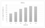
- FIG. 3 shows the change in the amount of ⁇ 2-6 sialic acid in response to SNA present on the cell membrane of the TGP culture sample.
- FIG. 4 shows the change in the amount of ⁇ 2-6 sialic acid in response to SSA present on the cell membrane of the TGP culture sample.
- FIG. 5 shows the change in the amount of ⁇ 2-6 sialic acid in response to TJA-1 present on the cell membrane of the TGP culture sample.
- FIG. 6 shows the change in the amount of ⁇ 1-3 fucose in response to UEA-1 present on the cell membrane of the TGP culture sample.
- the present invention is a method for producing a chondrocyte culture having high tissue regeneration ability, and includes a step of culturing a cell population separated from cartilage tissue with a thermoreversible polymer.
- Cartilage tissue in the present disclosure refers to cartilage-derived tissue. Cartilage is not limited to articular cartilage, osteochondral plate, costal cartilage, tracheal cartilage, laryngeal cartilage, sacroiliac joint, temporomandibular joint, sternoclavicular joint, intervertebral disc, shamebone connection, articular meniscus, articular disc, external auditory canal , Includes temporomandibular canal, epicartilage, laryngeal cartilage, etc.
- Cartilage tissue can be derived from any organism.
- Human cartilage is preferred because there is less rejection when the bone tissue culture is transplanted into human damaged cartilage. It may be an autologous organization or an allogeneic organization, but an autologous organization is preferable because there is little rejection when applied to the subject.
- the cartilage tissue used in the present invention can form a chondrocyte culture having a tissue structure similar to that of a healthy cartilage tissue regardless of the state of the object from which the cartilage tissue is derived. Therefore, it may be cartilage tissue derived from a subject in any state, for example, articular cartilage collected from a subject having osteoarthritis.
- the collection site of such articular cartilage may be a loaded portion or a non-loaded portion of the body weight, or may be a cartilage tissue collected from a loaded portion damaged by osteoarthritis.
- the chondrocyte culture produced by the present invention does not cause abnormalities such as calcification even after long-term culture, it is produced by collecting it from a grade 1 subject and culturing it until it progresses to grade 3 or later. It is also possible to transplant a chondrocyte culture into a subject. Therefore, the OA grade to be collected may be any of 1 to 4.
- the age at which the cartilage tissue is collected is not particularly limited. Since the chondrocyte culture produced by the present invention has high regenerative ability, for example, even if it is 0 to 39 years old or 40 years old or older, it is 50 years old or older, 60 years old or older, 70 years old or older, 80 years old or older, 90 years old or older. It may be cartilage tissue derived from the subject of.
- the method for collecting cartilage tissue may be a usual method used when collecting living tissue in the art, and can be collected from cartilage using a scalpel, tweezers, a biopsy punch, or the like without limitation. ..
- the "cell population separated from cartilage tissue” in the method of the present invention means a cell population composed of the same or different types of cells obtained by cartilage tissue separation treatment (for example, enzyme treatment, shredding treatment). ..
- the cell population is not limited as long as it is a cell population contained in cartilage tissue, and includes chondrocytes, cartilage progenitor cells, mesenchymal stem cells (MSCs), pluripotent stem cells, and the like.
- cartilage tissue separation treatment a method usually used as a biological tissue separation treatment in the technical field, for example, an enzymatic method or a physical method can be used.
- the enzymatic method is, but is not limited to, incubating the cells constituting the cartilage tissue at a viable temperature (for example, 37 ° C.) using one or more enzymes.
- a viable temperature for example, 37 ° C.
- an enzyme commonly used in the art in separating cells from cartilage tissue can be used under treatment conditions of corresponding temperature, concentration and time.
- dispase I or II collagenase I or II, metalloprotease, trypsin, hyaluronidase, pepsin, aminopeptidase, lipase, amylase and the like can be used without limitation as long as the cartilage tissue can be separated.
- cartilage tissue can be decomposed in a short time and with minimal invasiveness, preferably, even if any one of dispase I or II, collagenase, metalloprotease, actase or trypsin is used alone as an enzyme, two or more kinds are used. It may be used in combination.
- trypsin When trypsin is used, it may be, but is not limited to, 0.05% to 2.5%, 0.1% to 1%, 0.15% to 0.3% in the form of trypsin-EDTA, which is low. From the viewpoint of separation by invasiveness, 0.2 to 0.25% is preferable.
- the separation treatment time with trypsin-EDTA is not limited, and may be 1 minute to 2 hours, 3 minutes to 1 hour, 10 minutes to 40 minutes, and is preferably 30 minutes from the viewpoint of not damaging the stem cells.
- collagenase II When collagenase II is used, it is not limited to 0.1 to 30 mg / ml, 0.5 to 10 mg / ml, 0.75 to 5 mg / ml, and 0.8 to 2 mg from the viewpoint of minimally invasive separation.
- the separation treatment time is not limited as long as the living cells are not killed, and may be, for example, 1 to 24 hours, 3 to 20 hours, 7 to 18 hours, 10 to 16 hours, from the viewpoint of minimally invasive separation of stem cells. 12 to 16 hours is preferred.
- each concentration and treatment time can be arbitrarily combined without limitation.
- a combination of 12 to 16 hours is preferred.
- the physical method is not limited to this, and examples thereof include a method of chopping or crushing cartilage tissue using a scalpel, ultrasonic waves, a homogenizer, a strainer, or the like.
- the physical method can be optionally combined with the enzymatic method detailed above.
- any known method such as a filter or a cell strainer may be used to separate the cells separated by the separation treatment from the tissue pieces or the matrix that cannot be completely separated. Separation results in a cell population with a uniform size or function.
- thermoreversible polymer in the method of the present invention (also referred to as "TGP" in the present specification) is thermally reversibly formed a crosslinked structure or a network structure, and based on the structure, the inside thereof.
- the hydrogel refers to a gel containing a crosslinked or network structure made of a polymer and water supported or retained in the structure.
- the thermoreversible polymer can be any polymer as long as it is a thermoreversible polymer because an environment similar to cartilage tissue in a living body is formed by a crosslinked structure or a network structure peculiar to the thermoreversible polymer. Can also be used in the method of the present invention.
- Measuring equipment (trade name): Stress-controlled rheometer AR500, TA Instrument's sample solution (or separation solution) concentration (however, as the concentration of "hydrogel-forming polymer with sol-gel transition temperature") : 10 (weight)%
- Amount of sample solution Approximately 0.8 g
- Shape and dimensions of measuring cell Acrylic parallel disk (diameter 4.0 cm), gap 600 ⁇ m
- Measurement frequency 1Hz Applied stress: within the linear region
- the sol-gel transition temperature is preferably higher than 0 ° C. and preferably 37 ° C. or lower, and further preferably higher than 5 ° C. and 35 ° C. or lower (particularly 10 ° C. or higher and 33 ° C. or lower). ..
- a TGP having such a suitable sol-gel transition temperature can be easily selected from specific compounds as described later according to the above-mentioned screening method (sol-gel transition temperature measurement method).
- the TGP of the present invention is not particularly limited as long as it exhibits the thermoreversible sol-gel transition as described above (that is, has a sol-gel transition temperature).
- a specific example of a polymer in which the aqueous solution has a sol-gel transition temperature and reversibly exhibits a sol state at a temperature lower than the transition temperature is represented by, for example, a block copolymer of polypropylene oxide and polyethylene oxide.
- Polyalkylene oxide block copolymers; etherified cellulose such as methyl cellulose and hydroxypropyl cellulose; chitosan derivatives (KRHolme.et al. Macromolecules, 24,3828 (1991)) and the like are known.
- the hydrogel-forming polymer using a hydrophobic bond for cross-linking which can be suitably used as the TGP of the present invention, is a cloud point from the viewpoint that the TGP and the medium do not separate and the surroundings of the cell population can be stably maintained. It is preferable that a plurality of blocks having the above and a hydrophilic block are combined.
- the hydrophilic block is preferably present because the hydrogel becomes water soluble at a temperature lower than the sol-gel transition temperature, and the plurality of blocks having cloud points are the sol-gel transition temperature of the hydrogel. It is preferably present because it changes to a gel state at higher temperatures.
- a block with a cloud point dissolves in water at a temperature lower than the cloud point and becomes insoluble in water at a temperature higher than the cloud point, so that the block is gelled at a temperature higher than the cloud point.
- It serves as a cross-linking point consisting of hydrophobic bonds to form. That is, the cloud point derived from the hydrophobic bond corresponds to the sol-gel transition temperature of the hydrogel.
- the cloud point and the sol-gel transition temperature do not necessarily have to match. This is because the cloud point of the above-mentioned "block having a cloud point" is generally affected by the binding between the block and the hydrophilic block.
- the hydrogel used in the present invention utilizes the property that the hydrophobic bond not only becomes stronger as the temperature rises, but the change is reversible with respect to temperature.
- TGP is a "cloud point". It is preferable to have a plurality of "blocks having".
- the hydrophilic block in the TGP has a function of changing the TGP to water-soluble at a temperature lower than the sol-gel transition temperature, and has a hydrophobic binding force at a temperature higher than the transition temperature. It has a function of forming a state of a hydrogel while preventing the hydrogel from coagulating and precipitating due to excessive increase.
- the TGP used in the present invention is preferably one that is decomposed and absorbed in vivo from the viewpoint of using the chondrocyte culture of the present invention in cell therapy. That is, it is preferable that the TGP of the present invention is decomposed in a living body by a hydrolysis reaction or an enzymatic reaction to become a low molecular weight substance harmless to the living body, and is absorbed and excreted.
- the TGP of the present invention is formed by combining a plurality of blocks having cloud points and hydrophilic blocks, at least one of the blocks having cloud points and the hydrophilic blocks, preferably both of them, is in vivo. It is preferable that it is decomposed and absorbed by.
- the block having a cloud point is preferably a polymer block having a negative solubility in water-temperature coefficient, and more specifically, polypropylene oxide, a copolymer of propylene oxide and another alkylene oxide, Polymer selected from the group consisting of poly N-substituted acrylamide derivatives, poly N-substituted metaacrylamide derivatives, copolymers of N-substituted acrylamide derivatives and N-substituted metaacrylamide derivatives, polyvinyl methyl ether, and polyvinyl alcohol partially vinegared products. Can be preferably used.
- the poly N-substituted acrylamide derivative is preferable from the viewpoint of stably culturing the cell population or chondrocyte culture of the present invention.
- a polypeptide composed of a hydrophobic amino acid and a hydrophilic amino acid In order to decompose and absorb a block having a cloud point in a living body, it is effective to make the block having a cloud point a polypeptide composed of a hydrophobic amino acid and a hydrophilic amino acid.
- a polyester-type biodegradable polymer such as polylactic acid or polyglycolic acid can be used as a block having a cloud point that is decomposed and absorbed in the living body.
- the cloud point of the above polymer is higher than 4 ° C and 40 ° C or less, which is a compound in which a plurality of blocks having a cloud point and a hydrophilic block are bonded.
- the sol-gel transition temperature is higher than 0 ° C. and 37 ° C. or lower.
- the cloud point is measured, for example, by cooling an aqueous solution of about 1% by mass of the above polymer (block having a cloud point) to obtain a transparent uniform solution, and then gradually raising the temperature (heating rate of about 1). This can be done by setting the point at which the solution becomes cloudy for the first time as a cloud point at ° C./min).
- poly-substituted acrylamide derivative and the poly N-substituted meta acrylamide derivative that can be used in the present invention are listed below.
- Poly-N-isopropylacrylamide is preferable from the viewpoint of stably culturing the cell population or chondrocyte culture of the present invention.
- the above polymer may be a homopolymer or a copolymer of a monomer constituting the polymer and another monomer.
- a hydrophilic monomer or a hydrophobic monomer can be used as the other monomer constituting such a copolymer.
- copolymerization with a hydrophilic monomer raises the cloud point of the product, and copolymerization with a hydrophobic monomer lowers the cloud point of the product. Therefore, by selecting these monomers to be copolymerized, a polymer having a desired cloud point (for example, a cloud point higher than 4 ° C and 40 ° C or lower) can be obtained.
- hydrophilic monomer examples include N-vinylpyrrolidone, vinylpyridine, acrylamide, metaacrylamide, N-methylacrylamide, hydroxyethyl methacrylate, hydroxyethyl acrylate, hydroxymethyl methacrylate, hydroxymethyl acrylate, and acrylic having an acidic group. Acids, methacrylic acid and salts thereof, vinyl sulfonic acid, styrene sulfonic acid, etc., and N, N-dimethylaminoethyl methacrylate, N, N-diethylaminoethyl methacrylate, N, N-dimethylaminopropyl having a basic group. Examples thereof include, but are not limited to, acrylamide and salts thereof.
- hydrophobic monomer acrylate derivatives and methacrylate derivatives such as ethyl acrylate, methyl methacrylate and glycidyl methacrylate, N-substituted alkyl metaacrylamide derivatives such as Nn-butyl metaacrylamide, vinyl chloride, acrylonitrile and styrene , Vinyl acetate and the like, but are not limited thereto.
- hydrophilic block On the other hand, specific examples of the hydrophilic block to be bound to the above-mentioned block having a cloud point include methyl cellulose, dextran, polyethylene oxide, polyvinyl alcohol, poly N-vinylpyrrolidone, polyvinyl pyridine, polyacrylamide, and polymethacrylamide.
- poly-N-isopropylacrylamide when poly-N-isopropylacrylamide is used as a plurality of blocks having a cloud point of a thermoreversible gel, an environment similar to the in vivo environment of cartilage tissue is formed to proliferate the cell population.
- Polyethylene oxide is preferable as the hydrophilic block that binds to the acrylamide because of its high ability.
- hydrophilic block is decomposed, metabolized and excreted in the living body, and a hydrophilic biopolymer such as a protein such as albumin and gelatin and a polysaccharide such as hyaluronic acid, heparin, chitin and chitosan is preferably used. ..
- the method of binding the block having a cloud point and the above hydrophilic block is not particularly limited, but for example, a polymerizable functional group (for example, an acryloyl group) is introduced into any of the above blocks to give the other block. This can be done by copolymerizing the monomers. Further, the bond between the block having a cloud point and the above-mentioned hydrophilic block can also be obtained by block copolymerization of a monomer giving a block having a cloud point and a monomer giving a hydrophilic block. It is possible.
- a polymerizable functional group for example, an acryloyl group
- a functional group for example, a hydroxyl group, an amino group, a carboxyl group, an isocyanate group, etc.
- a chemical reaction it can also be done by.
- the bond between the polypropylene oxide having a cloud point and the hydrophilic block is, for example, anionic polymerization or cationic polymerization, in which the propylene oxide and the monomer constituting the "other hydrophilic block" (for example, ethylene oxide) are repeatedly and sequentially obtained.
- anionic polymerization or cationic polymerization in which the propylene oxide and the monomer constituting the "other hydrophilic block" (for example, ethylene oxide) are repeatedly and sequentially obtained.
- Such a block copolymer can also be obtained by introducing a polymerizable group (for example, an acryloyl group) at the end of polypropylene oxide and then copolymerizing the monomers constituting the hydrophilic block.
- the polymer used in the present invention can also be obtained by introducing a functional group capable of binding reaction with a functional group (for example, a hydroxyl group) at the terminal of polypropylene oxide into a hydrophilic block and reacting both of them. ..
- the TGP used in the present invention can also be obtained by connecting a material such as Pluronic (registered trademark) F-127 (trade name, manufactured by Asahi Denka Kogyo Co., Ltd.) in which polyethylene glycol is bonded to both ends of polypropylene glycol. be able to.
- Pluronic registered trademark
- F-127 trade name, manufactured by Asahi Denka Kogyo Co., Ltd.
- the above-mentioned "block having a cloud point” existing in the molecule is water-soluble together with the hydrophilic block at a temperature lower than the cloud point. It is completely dissolved in water and shows a sol state.
- the "block having a cloud point” existing in the molecule becomes hydrophobic and associates between separate molecules by hydrophobic interaction. To do.
- the polymer of the present invention since the hydrophilic block is water-soluble even at this time (when heated to a temperature higher than the cloud point), the polymer of the present invention has a hydrophobic association between blocks having a cloud point in water.
- a hydrogel having a three-dimensional network structure with the above as a cross-linking point is produced.
- the temperature of this hydrogel is cooled again to a temperature lower than the cloud point of the "block having a cloud point" existing in the molecule, the block having the cloud point becomes water-soluble and the cross-linking point due to the hydrophobic association is released. , The hydrogel structure disappears and the TGP of the present invention becomes a complete aqueous solution again.
- the sol-gel transition of the polymer of the present invention in a preferred embodiment is based on a reversible change in hydrophilicity and hydrophobicity at the cloud point of the block having a cloud point existing in the molecule. Therefore, it has complete reversibility in response to temperature changes. According to the studies by the present inventors, it is considered that the delicate hydrophilic-hydrophobic balance of TGP in water contributes to the stability of cells when they are stored at low temperature in water.
- the hydrogel-forming polymer of the present invention containing at least a polymer having a sol-gel transition temperature in an aqueous solution is substantially water-insoluble at a temperature (d ° C.) higher than the sol-gel transition temperature. Is reversibly water-soluble at a temperature (e ° C.) lower than the sol-gel transition temperature.
- the high temperature (d ° C.) described above is preferably a temperature 1 ° C. or higher higher than the sol-gel transition temperature, and more preferably 2 ° C. or higher (particularly 5 ° C. or higher).
- the above-mentioned "substantially water-insoluble” means that the amount of the above-mentioned polymer dissolved in 100 ml (liter) of water at the above-mentioned temperature (d ° C.) is 5.0 g or less (further, 0.5 g or less, particularly 0). .1 g or less) is preferable.
- the above-mentioned low temperature (e ° C.) is preferably 1 ° C. or higher (absolute value) lower than the sol-gel transition temperature, and further preferably 2 ° C. or higher (particularly 5 ° C. or higher) lower. preferable.
- water-soluble means that the amount of the polymer dissolved in 100 ml (liter) of water at the temperature (e ° C.) is preferably 0.5 g or more (further 1.0 g or more). ..
- reversibly water-soluble means that even after the aqueous solution of TGP is once gelled (at a temperature higher than the sol-gel transition temperature), at a temperature lower than the sol-gel transition temperature, It means to show the above-mentioned water solubility.
- the 10% aqueous solution of the polymer exhibits a viscosity of 10 to 3,000 cmpoise (further, 50 to 1,000 cmpoise) at 5 ° C.
- Viscosity is preferably measured under the following measurement conditions, for example.
- Viscometer Stress-controlled rheometer (model name: AR500, manufactured by TA Instruments) Rotor diameter: 60 mm Rotor shape: Parallel flat plate
- the aqueous solution of TGP of the present invention is gelled at a temperature higher than the sol-gel transition temperature and then immersed in a large amount of water, the gel does not substantially dissolve.
- the above-mentioned characteristics of the hydrogel formed by the above-mentioned TGP can be confirmed, for example, as follows. That is, 0.15 g of TGP is dissolved in 1.35 g of distilled water at a temperature lower than the sol-gel transition temperature (for example, under ice cooling) to prepare a 10 wt% aqueous solution, and the aqueous solution is used as a plastic petri dish having a diameter of 35 mm.
- a gel having a thickness of about 1.5 mm is formed in the petri dish by injecting into the petri dish and heating to 37 ° C., and then the weight (fgram) of the entire petri dish containing the gel is measured.
- the entire petri dish containing the gel was allowed to stand in water in 250 ml (liter) at 37 ° C. for 10 hours, and then the weight (g grams) of the entire petri dish containing the gel was measured to measure the gel from the gel surface. Evaluate the presence or absence of dissolution.
- the weight loss rate of the gel that is, (fg) / f is preferably 5.0% or less, and further 1.0%. It is preferably less than or equal to (particularly 0.1% or less).
- the aqueous solution of TGP of the present invention is gelled at a temperature higher than the sol-gel transition temperature, and then immersed in a large amount of water (about 0.1 to 100 times the gel in terms of volume) for a long period of time.
- the gel does not dissolve over time.
- Such properties of the polymer used in the present invention are achieved, for example, by the presence of two or more (plurality) blocks having cloud points in the polymer.
- Pluronic registered trademark
- F-127 registered trademark
- the concentration with respect to water that is, ⁇ (polymer) / (polymer + water) x 100 (%), is 20% or less (further, 15% or less).
- TGP capable of gelation at a concentration of 10% or less).
- the molecular weight of the TGP used in the present invention is preferably 30,000 or more and 30 million or less, more preferably 100,000 or more and 10 million or less, and further preferably 500,000 or more and 5 million or less.
- the step of culturing the cell population with the thermoreversible polymer may be performed by mixing the cell population separated from the cartilage tissue with the thermoreversible polymer by a known method and culturing.
- a cell population separated from cartilage tissue is dispersed in a TGP solution at a temperature lower than the sol-gel transition temperature, the TGP is gelled at a temperature higher than the sol-gel transition temperature, and then a culture solution or the like is used.
- a method of adding a medium and culturing can be used. By adding a medium to the gelled TGP, it is possible to provide a stable nutrient supply to the cell population in the gel by periodically exchanging only such a medium.
- the culture in the present invention can be carried out under the conditions usually used in the art.
- typical culture conditions include culturing at 37 ° C. and 5% CO2.
- the culture can be carried out at normal atmospheric pressure (atmospheric pressure).
- the culture period is not particularly limited because abnormal chondrocyte cultures such as calcification do not occur even if the cell population is cultured for a long period of time.
- Culturing can be performed in a container of any size and shape.
- the medium for dissolving TGP and the medium for adding to gelled TGP (sometimes referred to as "liquid medium” in the present specification) are not particularly limited as long as they can maintain cell survival, but are typically limited. , Amino acids, vitamins, and electrolytes are the main components.
- the medium for dissolving TGP and the medium for adding to the gelled TGP may be common or different.
- the medium for dissolving TGP and the medium for adding to gelled TGP are based on a basal medium for cell culture.
- a basal medium for cell culture is not limited to, for example, DMEM, MEM, F12, DME, RPMI1640, MCDB (MCDB102, 104, 107, 120, 131, 153, 199, etc.), L15, SkBM, RITC80-7, CnT. -PR etc. are included.
- Many of these basal media are commercially available, and their compositions are also known.
- the basal medium may be used as it has a standard composition (for example, as it is on the market), or the composition may be appropriately changed depending on the cell type and cell conditions. Therefore, the basal medium used in the present invention is not limited to those having a known composition, and includes those in which one or more components are added, removed, increased or decreased.
- the medium may contain one or more additives such as serum, growth factors (for example, FGF-2, TGF-b1, etc.), steroid component, selenium component, and the like.
- the serum may be a heterologous serum or an allogeneic serum. Allogeneic sera are preferred, and autologous sera are even more preferred among allogeneic sera.
- the medium does not contain growth factors other than the growth factors contained in the autologous serum. When culturing using TGP lysed by such a medium, the cell population will be cultivated in TGP to which growth factors other than autologous serum have not been added.
- the concentration of serum is not particularly limited, and 3%, 5%, 10%, and 20% may be contained in the medium added to TGP. It is preferably 10%.
- the medium for dissolving TGP and the medium for adding to gelled TGP may consist of a common component.
- the "chondrocyte culture” produced by the method of the present invention refers to a cell population containing chondrocytes obtained by culturing a cell population separated from cartilage tissue.
- the chondrocyte culture produced by the method of the invention may have a cartilage tissue-like tissue structure in which the cell gaps of the cell population are filled by ECM.
- the chondrocyte culture obtained by the method of the present invention is more than a chondrocyte culture (referred to herein as a "control culture”) cultured by a method that does not include the step of culturing a cell population with a thermoreversible polymer.
- High tissue regeneration ability means the tissue regeneration ability of cartilage tissue to which a cell population is applied.
- the tissue regeneration ability is applied to a cartilage cell culture of interest (transplantation), and after a lapse of a predetermined time, the state of the tissue at the application site (transplant destination), for example, the size of the tissue, the fineness of the tissue. It can be quantified by observing the structure, the ratio of damaged tissue to normal tissue, the function of tissue, etc. and scoring these.
- the tissue regeneration ability may be the expression ability of one or more genes selected from SOX9, COL2A1, miR140 and miR21.
- the chondrocyte culture produced by the method of the present invention has a higher expression of one or more genes selected from SOX9, COL2A1, miR140 and miR21 than the control culture. Therefore, the present invention may be a method for enhancing the expression ability of one or more genes selected from SOX9, COL2A1, miR140 and miR21 of a cell population isolated from cartilage tissue.
- SOX9 or COL2A1 is known as a gene expressed during normal cartilage tissue formation.
- miR140-3p or -5p or miR21-5p derived from miR140 or miR21 is a degenerated state of cartilage tissue characterized by inflammation in cartilage tissue generated in osteoarthritis, decomposition of ECM, and the like. Effective for normalization (Karlsen et al., Mol Ther Nucleic Acids. 2016 Oct 11; 5 (10), Miyaki et al., Arthritis Rheum. 2009 Sep; 60 (9): 2723-30, Si et al. , Osteoarthritis Cartilage. 2017 Oct; 25 (10): 1698-1707, Miyaki et al., Genes Dev. 2010 Jun 1; 24 (11): 1173-85, Hai et al ,. J Orthop Surg Res. 2019; 14 : See 118 etc.).
- miR140 in cartilage tissue results in increased miR140-3p or 5p in cartilage tissue, resulting in, but not limited to, expression or secretion of ECM-degrading enzymes such as MMP-13 and ADAMTS-5.
- ECM-degrading enzymes such as MMP-13 and ADAMTS-5.
- IL1B IL1B
- IL6 IL6
- IL8 proteins involved in ECM synthesis
- SOX9 proteins involved in ECM synthesis
- ACAN chondroitin sulfate N-acetylgalactosaminyl transferase 1
- the chondrocyte culture of the present invention when the chondrocyte culture of the present invention is applied to the cartilage tissue of a subject, particularly a subject having osteoarthritis, not only can the normal cartilage tissue be complemented at the application site, but also the cartilage tissue around the application site can be applied. Since the degenerative state can be normalized, the therapeutic effect can be maintained for a long period of time.
- miR21-5p is increased in cartilage tissue, and as a result, the expression or secretion of ECM-degrading enzymes such as MMP-13 and ADAMTS-5 is decreased, and ECM such as COL2A1 is not limited. It normalizes the degenerative state of cartilage tissue through increased expression of proteins involved in synthesis. Therefore, the therapeutic effect can be maintained for a long period of time like miR140.
- high gene expression is not limited to 101% or more, 105% or more, 110% or more, 130% or more, 140% or more, 150% or more, 160% or more, based on the control culture. It means that the expression level of a predetermined gene is high at 170% or more, 180% or more, 190% or more or 200% or more.
- Techniques for measuring gene expression levels are well known in the art and are not limited as techniques for measuring protein expression levels, for example, Western using the monoclonal or polyclonal antibodies detailed above. Blotting, EIA, ELISA, RIA, immunoorganization, immunocytochemistry, flow cytometer, mass assay, etc. are not limited as methods for measuring gene expression levels, for example, Northern blotting. Examples thereof include a method, a Southern blotting method, a DNA microarray analysis, an RNase protection assay, an RTPCR, a PCR method such as real-time PCR (qPCR), and an in situ hybridization method.
- qPCR real-time PCR
- SOX9 (also referred to as CMD1, CMPD1, SRA1, SRXX2, SRXY10) is a gene that coordinates SRY-box transcription factor 9, and the gene sequence of human SOX9 is registered as accession number NM_000346 or the like. The sequence is as shown in SEQ ID NO: 1.
- COL2A1 (also referred to as ANFH, AOM, COL11A3, SEDC, STL1) is a gene that links collagen type II alpha 1 chain, and the gene sequence of human COL2A1 is registered as accession number NM_001844.5, etc. The sequence is as shown in SEQ ID NO: 2.
- microRNA is transcribed as an RNA precursor having a hairpin-like structure, cleaved by a dsRNA cleaving enzyme having RNase III cleaving activity, incorporated into a protein complex called RISC, and suppresses translation of mRNA. 10-25 bases of RNA involved in are intended to be used.
- the “miRNA” also includes “miRNA” and precursors of the “miRNA” (pre-miRNA, pri-miRNA), and miRNAs having biological functions equivalent to those of the miRNA encoded by them, such as homologues (ie,). , Homolog), variants such as gene polymorphisms, and "miRNAs" encoding derivatives.
- RNA encoding such a precursor, homologue, mutant or derivative
- miRBase http://www.mirbase.org/
- miRBase http://www.mirbase.org/
- Examples thereof include “miRNA” having a base sequence that hybridizes with the complementary sequence of.
- human mir-140 is miRbase ID: Stem-loop sequence hsa-mir-140, miRbase accession number. It is registered as MI0000456 and is as shown in SEQ ID NO: 3.
- the human miR140-3p is miRbase ID: Mature sequence hsa-miR-140-3p, miRbase accession number. It is registered as MIMAT0004597 and is as shown in the RNA sequence "uaccacaggguagaaccacgg" shown in SEQ ID NO: 4.
- human miR140-5p is miRbase ID: Mature sequence hsa-miR-140-5p, miRbase accession number. It is registered as MIMAT0000431 and is as shown in the RNA distribution "cagugguuuuacccuaugguag" shown in SEQ ID NO: 5.
- human mir-21 is referred to as miRbase ID: Stem-loop sequence hsa-mir-21, miRbase accession number. It is registered as MI0000077 and is as shown in SEQ ID NO: 6.
- the human miR21-3p is miRbase ID: Mature sequence hsa-miR-21-3p, miRbase accession number. It is registered as MIMAT0004494 and is as shown in the RNA sequence "caacaccagucgaugggcugu" shown in SEQ ID NO: 7.
- human miR21-5p is miRbase ID: Mature sequence hsa-miR-21-5p, miRbase accession number. It is registered as MIMAT0000076 and is as shown in the RNA sequence "uagcuuaucagacugauguuga" shown in SEQ ID NO: 8.
- the tissue regeneration ability is the ability of the chondrocyte culture to retain miRNA.
- Retention capacity is the ability of a chondrocyte culture to retain miRNA in a chondrocyte culture.
- the ability to retain miRNAs in chondrocyte cultures may be the ability to retain miRNAs in chondrocyte cultures without secreting them during culture.
- the miRNA is not limited to miR140, miR21, miR-125b, Has-miR-15a, miR-30a, miR-199a, miR-210, miR-221-3p, miR-92a-3p, miR-142. -3p, miR-27a, miR-27b, miR26a-5p, miR-26a, miR-26b, miR-373, miR-127-5p, miR-320, miR-9, miR-634, miR-221-3p , MiR-370miR-145, miR-130A, miR-145, miR-562-5p and other miRNAs capable of normalizing the degenerative state of cartilage tissue (Zhang.et al. J Arthritis.
- High miRNA retention capacity is not limited to 101% or more, 105% or more, 110% or more, 130% or more, 140% or more, 150% or more, 160% based on the control culture at the time of culturing. As mentioned above, it means that miRNA is present in the chondrocyte culture at the time of culturing at a high concentration of 170% or more, 180% or more, 190% or more, or 200% or more. In one aspect, high miRNA retention capacity is defined as 99% or less, 95% or less, 97%% or less, 95% or less, without limitation, based on the medium (liquid medium) of the control culture at the time of culturing.
- the high retention capacity of miRNA means that miRNA is present in a high concentration in the cartilage cell culture at the time of culturing based on the control culture at the time of culturing, and the liquid medium of the control culture is used as a reference. It means that miRNA is present at a low concentration in the medium of the cartilage cell culture at the time of culturing.
- the tissue regeneration ability may be the content of stem cells in the chondrocyte culture.
- the chondrocyte culture produced by the method of the present invention has a higher stem cell content than the control culture. Therefore, the present invention may be a method for maintaining and proliferating stem cells in a cell population isolated from cartilage tissue.
- the content of stem cells is not limited to, but may be the amount or proportion of stem cells in the cell culture.
- Stem cells are not limited as long as they have the ability to differentiate into chondrocytes, such as somatic stem cells such as chondrocyte progenitor cells and mesenchymal stem cells, or pluripotent stem cells such as ES cells, iPS cells, and ntES cells. Is included.
- the content of stem cells can be measured by a method known in the art for measuring stem cells, and is not limited to reacting with lectins such as UEA-1 present on the cell membrane of cells contained in cartilage cell cultures. Measuring the amount of ⁇ 1-2 sialic acid that reacts with ⁇ 1-2 fucose or lectins such as SNA, SSA, TJA-I, or changes in the amount of ⁇ 1-2 fucolic acid or ⁇ 2-6 sialic acid. It can be quantified by measuring other cell surface markers or gene expression that correlate with.
- the level of ⁇ 1-2 fucose present in the cell membrane of the cells contained in the cartilage cell culture is high, many pluripotent stem cells are present, and if the level of ⁇ 2-6 sialic acid is high, the differentiation potential is high. It means that there are many stem cells. (See Wang et al, Cell Res. 2011 Nov; 21 (11): 1551-63, Tateno et al, Glycobiology. 2016 Dec; 26 (12): 1328-1337, WO2016006712A1).
- “Differentiation potential” means that when a certain cell is placed in an appropriate differentiation-inducing state, it has the ability to change into another type of cell such as a progenitor cell or a somatic cell.
- somatic stem cells In general, long-term culture of somatic stem cells tends to reduce their differentiation potential as well as their proliferative capacity. Since somatic stem cells with reduced differentiation potential have somatic stem cell markers on the cell surface but cannot be transformed into other types of cells such as progenitor cells and somatic cells, such somatic stem cells have the ability to regenerate tissues. do not have.
- the chondrocyte culture produced by the method of the present invention has a high content of somatic stem cells having a high differentiation potential.
- the content of somatic stem cells having a high differentiation potential may be, but is not limited to, the amount or proportion of somatic stem cells having a high differentiation potential in the cell culture per unit amount.
- the present invention may be a method of maintaining or increasing the differentiation potential of somatic stem cells in a cell population isolated from cartilage tissue.
- the present invention may be a method of maintaining or proliferating somatic stem cells having a high differentiation potential in a cell population separated from cartilage tissue.
- the somatic stem cell is not limited as long as it is a somatic stem cell capable of differentiating into a chondrocyte, and may be a cartilage progenitor cell, a mesenchymal stem cell, or the like.
- chondrocytes differentiated from cells having a high differentiation potential can be constantly supplied to the application site. , Can repair and maintain normal chondrocyte tissue for a long period of time.
- the stem cell or somatic stem cell with high differentiation potential is a mesenchymal stem cell, it has the ability to normalize damaged cartilage tissue, such as for protecting chondrocyte apoptosis, for anti-inflammatory of cartilage tissue, and for anti-fibrosis. Since the ability to recover from damage is high, the therapeutic effect can be maintained for a long period of time.
- the method for measuring other cell surface markers or gene expression that correlates with the change in the amount of ⁇ 1-2 fucose acid or ⁇ 2-6 sialic acid is the conventional method for measuring cell surface markers or gene expression in the art. Can be used.
- the chondrocyte culture produced by the method of the present invention contains more stem cells or somatic stem cells having a higher differentiation potential than the control culture.
- the term "rich in stem cells or somatic stem cells with high differentiation potential” is not limited to 101% or more, 105% or more, 110% or more, 130% or more, 140% or more, 150% or more based on the control culture. , 160% or more, 170% or more, 180% or more, 190% or more or 200% or more, the content of cells having ⁇ 1-2 fucolic acid or ⁇ 2-6 sialic acid, or cell surface marker or gene expression that correlates with these. Means that is high.
- the tissue regeneration ability may be the ability to secrete hyaluronic acid from chondrocyte cultures.
- the chondrocyte culture produced by the method of the present invention has a higher ability to secrete hyaluronic acid than the control culture. Therefore, the present invention may be a method for enhancing the secretory capacity in a cell population separated from cartilage tissue.
- Hyaluronic acid is known to act suppressively on the expression of ECM-degrading enzymes, alleviate the matrix and anti-inflammatory factors released from cartilage, and regulate the expression of inflammation-related factors (Ohtsuki.et). al.J Orthoped Res. 2018 Aug 17. doi: 10.1002 / jor.24126).
- the chondrocyte culture produced by the method of the present invention when applied to the target cartilage tissue, not only can the normal cartilage tissue be complemented at the application site, but also ECM decomposition or inflammation around the application site is suppressed. Therefore, the degenerated state of the cartilage tissue around the application site can be normalized, and the normal cartilage tissue can be repaired and maintained for a long period of time.
- hyaluronic acid in a medium (liquid medium) or TGP may be quantified over time.
- Hyaluronic acid can be quantified by mass spectrometry, liquid chromatography, MALDI-TOF, etc., as well as commercially available kits, without limitation.
- High hyaluronic acid secretory capacity is not limited to 101% or more, 105% or more, 110% or more, 130% or more, 140% or more, 150% or more, 160% or more, based on the control culture. It means that the secretory capacity of hyaluronic acid is high at 170% or more, 180% or more, 190% or more or 200% or more.
- the chondrocyte culture produced by the method of the present invention is rich in ECM and therefore easily engrafts at the injured site. Therefore, the chondrocyte culture produced by the method of the present invention can be used for transplantation.
- the expression levels of miR140 and miR21 are high, the amount of stem cells is high, and the amount of hyaluronic acid secreted is high, it is preferably used for the treatment of grade 2 or higher or elderly osteoarthritis.
- the present invention further comprises chondrocyte cultures produced by the methods of the present invention.
- the chondrocyte culture of the present invention is as described above.
- the present invention includes methods of treating a disease in a subject, comprising applying an effective amount of the chondrocyte culture of the present invention to a subject in need thereof.
- the disease is not limited to cartilage-related diseases, but is due to osteoarthritis, rheumatoid arthritis, osteosarcoma, femoral head necrosis, acetabular dysplasia, meniscus injury, traumatic arthritis, sports, accidents, etc. Includes physical cartilage loss or damage.
- the method of application (administration) to the subject may be transplantation of a chondrocyte culture.
- Transplantation of chondrocyte cultures is not limited to delivering the chondrocyte cultures to the affected area by injection into the cartilage tissue, or arthroscopic surgery and incision, and any tissue adhesive such as fibring gel. It can be fixed to the affected area by use or by arthroscopy.
- various additional ingredients such as a pharmaceutically acceptable carrier, an ingredient that enhances engraftment, and a further active ingredient can be contained and applied. Any known additional ingredient can be used as such additional ingredient, and those skilled in the art are familiar with these additional ingredients.
- the present invention will be described in more detail below with reference to specific aspects of the present invention, but the present invention is not limited to these specific examples.
- the storage elastic modulus of this aqueous solution was measured at an application frequency of 1 Hz using a stress-controlled rheometer (AR500, manufactured by TA Instruments), and found to be 43 Pa at 10 ° C, 680 Pa at 25 ° C, and 1310 Pa at 37 ° C. This temperature-dependent storage modulus change was reversibly observed repeatedly.
- AR500 stress-controlled rheometer
- ⁇ Separation of cartilage tissue Each piece of tissue was cut into 1 mm 2 or less with a scalpel. The slices were treated with Trypsin-EDTA solution (0.25%) at 37 C ° for 30 minutes, then enzymatically treated with collagenase II solution (1 mg / ml) at 37 C ° for 12-16 hours. After washing with DMEM solution, the mixture was filtered through a filter (100 ⁇ m) and centrifuged (1800 rpm, 10 minutes). The cell population thus obtained was counted on the cell calculation board. The number of cells was 1 ⁇ 10 4 to 4 ⁇ 10 4 cells / ml.
- Example 1 ⁇ Culture with TGP gel> 1 g of TGP prepared in Production Example 1 was dissolved in 9 mL of DMEM at 4 ° C. to prepare a 10% TGP solution, and then cells derived from cartilage tissue were dispersed and dispensed into a 6-well plate. After leaving at room temperature to gel, add antibiotics (gentamicin (50 ⁇ g / ml), amphotericin (0.25 ⁇ g / ml), penicillin (100 Units / ml) / streptomycin (100 ⁇ g / ml) to DMEM solution containing 10% autologous serum.
- antibiotics gentamicin (50 ⁇ g / ml)
- amphotericin (0.25 ⁇ g / ml
- penicillin 100 Units / ml
- streptomycin 100 ⁇ g / ml
- Example 1 chondrocyte cultures having a size of 200 to 300 ⁇ m were confirmed on the 3rd to 5th days of culture, and after 21 days, they grew to a size of 0.5 to 1.0 mm in diameter, and all the samples. Growth was maintained for a 20-week culture period. In Example 1, no proliferation of fibroblast-like cells was observed.
- RNAlater registered trademark
- Stabilization Solution Invitrogen, CatNo.AM7020
- Small RNA RNA of 200 bases or less
- NucleoSpin® miRNA kit TeKaRa, U0971
- primers of SEQ ID NOs: 9 to 12 and mRQ3'Primer U6Forward U6, miR-21 by Thermal Cycler Dice (registered trademark) Real Time System Lite (TP700, TaKaRa) using Primer and U6 Reverse Primer (Mir-X TM miRNA qRT-PCR TB Green Kit (TaKaRa, Z8314N))
- QPCR of 3p, miR-21-5p, miR-140-3p and miR-140-5p was performed. Quantitative values of each miRNA were normalized with the expression level of U6 as 1.
- QPCR of GAPDH, SOX9 and COL2A1 was performed by Thermal Cycler Dice (registered trademark) Real Time System Lite. The quantitative value of each gene was normalized with the expression level of GAPDH as 1.
- the primers used are shown in Table 2, and the results are shown in Tables 3-5.
- the expression level of miR-21 and miR-140 was increased in Example 1 cultured in TGP gel as compared with the sample of Comparative Example 1, and miR21-5p, It was found that miR-140-3p and miR-140-5p were significantly increased.
- the expression level of SOX9 was significantly increased in the sample of Example 1 as compared with the sample of Comparative Example 1. It was also found that the expression level of 1068, in which COL2A1 was measured, was increased in Example 1 as compared with the sample of Comparative Example 1.
- the chondrocyte culture produced in Example 1 has a healthy function. It turned out to be cartilage tissue.
- miR-140 and miR21 are known to improve the microenvironment of osteoarthritis, so the chondrocyte culture produced in Example 1 only promotes the regeneration of the cartilage tissue in the transplanted portion. Instead, it can improve the degenerative conditions around the transplant.
- RNAs of 200 bases or less were purified from some of these samples using the NucleoSpin® miRNA kit (TaKaRa, U0971), and the primers of SEQ ID NO: 3 and mRQ 3'Primer (Mir-X (Mir-X) Using miRNA qRT-PCR TB Green (registered trademark) Kit (TaKaRa, Z8314N)), qPCR of miR-140-3p was performed by Thermal Cycler Dice (registered trademark) Real Time System Lite (TP700, TaKaRa). The results are shown in Table 6.
- the average Ct value is the number of cycles until a predetermined Threshold is reached, and the lower the average Ct value, the more miR-140-3p is present in the liquid medium.
- Threshold the threshold for the average Ct value.
- Example 1 Analysis of glycans on the cell surface of cells constituting the cultured chondrocyte culture
- the chondrocyte culture cultured in Example 1 was collected between 14 and 126 days after the culture, and each TGP gel was collected in 4 It was cooled to ° C to form an aqueous solution and recovered.
- the sample was dispersed in cells with Trypsin-EDTA (0.25%) solution, and the cells were collected. Then, it was centrifuged in DMEM three times at 4 ° C. (1800 rpm, 15 minutes), and each sample was cryopreserved.
- Glyco Technica Ltd Glycan Profiling Analysis Service by Glycan Profiling Analysis Service.
- the cell membrane protein was extracted from the thawed sample, and the protein concentration was measured by the BCA method (TaKaRa BCA Protein Assay Kit). Based on the measured protein concentration, PBSTx was added to dilute the protein concentration to 10 ⁇ g / mL. A 100 ⁇ L sample (concentration of 10 ⁇ g / mL) was added to a tube containing 100 ⁇ g of Cy3 Mono-reactive Dye Pack (GE healthcare, catalog number: PA23011), mixed with a pipette, and spun down. The tube was placed in a light-shielding bag and incubated at room temperature (25 ° C.) for 1 hour.
- BCA method TaKaRa BCA Protein Assay Kit
- the fluorescence pattern of LecChip TM was measured four times cumulatively by GlycoLite2200 ( TM , Glyco technica), the exposure time, 1996, 2995, 3991, 4992, 6988, 9998 (milliseconds), and the camera gain was measured at fixed values.
- the obtained 45-lectin signal was measured by GlycoStaion® ToolsPro Suite 1.5 (GlycoStaion® ToolsPro Suite 1.5.), And A. Kuno et al., J. of Proteomics & Bioinformatics, Vol.1, May 2008. , P.68., Divided by the average intensity of 45 lectins, multiplied by 100 and average normalized.
- ⁇ 2-6 sialic acid-binding lectins such as SNA, SSA and TJA-I and ⁇ 1-2 fucose-binding lectins such as UEA-1 were cultured for days. It can be seen that it increases according to.
- ⁇ 2-6 sialic acid which reacts with SNA, SSA and TJA-I, is a marker for somatic stem cells such as mesenchymal stem cells (MSCs) or cartilage progenitor cells with high differentiation potential (WO2016 / 006712A1) and UEA-1.
- ⁇ 1-2 fucose-binding lectins such as ⁇ 1-2 fucose-binding lectin are known to be markers of pluripotent stem cells (Wang et al., Cell Res. 2011 Nov; 21 (11): 1551-63. Doi: 10.1038 / cr. .2011.148. Epub2011 Sep 6.).
- ⁇ 2-6 sialic acid and ⁇ 1-2 fucose in response to SNA, SSA, TJA-I and UEA-1 was confirmed with the number of culture days. It was found that the tissue culture contained a large amount of somatic stem cells and pluripotent stem cells having high differentiation potential. Therefore, it was clarified that the chondrocyte culture of Example 1 is a tissue culture having high tissue regeneration ability.
- Hyaluronic acid retention test of TGP gel [Example 2] Hyaluronic acid (Teijin Pharma Co., Ltd. 5 mg to DMEM with antibiotics (gentamicin (50 ⁇ g / ml), amphotericin (0.25 ⁇ g / ml), penicillin (100 Units / ml) / streptomycin (100 ⁇ g / ml) and L-Ascorbic acid It was dissolved in 2.5 ml of a solution containing (5 mg / ml) to give liquid compositions 1 and 2. 1 g of TGP prepared in Production Example 1 was dissolved in 9 ml of DMEM at ° C.
- TGP solution hyaluronic acid was added thereto so as to have the same hyaluronic acid concentration as liquid compositions 1 and 2, and hyaluronic acid in the TGP solution was added. Shake to homogenize the acid.
- the TGP solution was gelled at 37 ° C., and then 1.0 ml of liquid medium (DMEM) was added to make TGP composition 1 and TGP composition 2.
- DMEM liquid medium
- the prepared hyaluronic acid-containing liquid composition and TGP composition were placed in a 5% carbon dioxide incubator at 37 ° C., and the hyaluronic acid concentration contained in each liquid medium portion was measured by the same method as ⁇ Measurement of hyaluronic acid> described above. .. The results are shown in Table 9.
- hyaluronic acid was present at about 13000 ng / ml on the 7th day, but decreased to 5.6 ng / ml on the 21st day.
- TGP compositions 1 and 2 it was 6.1 and 11.4 ng / ml at the 7th day, which corresponds to a low concentration of about 1/1000 with respect to liquid compositions 1 and 2.
- concentrations were 10.3 and 11.5 ng / ml in the 21st order, and their concentrations hardly changed. Therefore, it is clear that the TGP gel has the ability to stably retain hyaluronic acid for a long period of time without being decomposed. It became.
Landscapes
- Health & Medical Sciences (AREA)
- Life Sciences & Earth Sciences (AREA)
- Engineering & Computer Science (AREA)
- Biomedical Technology (AREA)
- Chemical & Material Sciences (AREA)
- Biotechnology (AREA)
- Zoology (AREA)
- Cell Biology (AREA)
- Rheumatology (AREA)
- General Health & Medical Sciences (AREA)
- Developmental Biology & Embryology (AREA)
- Organic Chemistry (AREA)
- Bioinformatics & Cheminformatics (AREA)
- Animal Behavior & Ethology (AREA)
- Veterinary Medicine (AREA)
- Public Health (AREA)
- Immunology (AREA)
- Medicinal Chemistry (AREA)
- Pharmacology & Pharmacy (AREA)
- Wood Science & Technology (AREA)
- Genetics & Genomics (AREA)
- Virology (AREA)
- Epidemiology (AREA)
- Orthopedic Medicine & Surgery (AREA)
- Microbiology (AREA)
- General Engineering & Computer Science (AREA)
- Biochemistry (AREA)
- Reproductive Health (AREA)
- Gynecology & Obstetrics (AREA)
- Physical Education & Sports Medicine (AREA)
- Chemical Kinetics & Catalysis (AREA)
- General Chemical & Material Sciences (AREA)
- Nuclear Medicine, Radiotherapy & Molecular Imaging (AREA)
- Micro-Organisms Or Cultivation Processes Thereof (AREA)
- Medicines Containing Material From Animals Or Micro-Organisms (AREA)
- Materials For Medical Uses (AREA)
Priority Applications (4)
| Application Number | Priority Date | Filing Date | Title |
|---|---|---|---|
| CN202080034375.3A CN114096660A (zh) | 2019-04-08 | 2020-04-08 | 高组织再生能力的软骨细胞培养物 |
| EP20787633.5A EP3954761A4 (en) | 2019-04-08 | 2020-04-08 | Chondrocyte culture with high tissue regeneration ability |
| JP2021513690A JP7578958B2 (ja) | 2019-04-08 | 2020-04-08 | 組織再生能が高い軟骨細胞培養物 |
| US17/601,861 US20230092155A1 (en) | 2019-04-08 | 2020-04-08 | Chondrocyte culture with high tissue regeneration ability |
Applications Claiming Priority (2)
| Application Number | Priority Date | Filing Date | Title |
|---|---|---|---|
| JP2019073783 | 2019-04-08 | ||
| JP2019-073783 | 2019-04-08 |
Publications (2)
| Publication Number | Publication Date |
|---|---|
| WO2020209316A1 true WO2020209316A1 (ja) | 2020-10-15 |
| WO2020209316A9 WO2020209316A9 (ja) | 2021-09-16 |
Family
ID=72752054
Family Applications (1)
| Application Number | Title | Priority Date | Filing Date |
|---|---|---|---|
| PCT/JP2020/015896 Ceased WO2020209316A1 (ja) | 2019-04-08 | 2020-04-08 | 組織再生能が高い軟骨細胞培養物 |
Country Status (5)
| Country | Link |
|---|---|
| US (1) | US20230092155A1 (https=) |
| EP (1) | EP3954761A4 (https=) |
| JP (1) | JP7578958B2 (https=) |
| CN (1) | CN114096660A (https=) |
| WO (1) | WO2020209316A1 (https=) |
Cited By (1)
| Publication number | Priority date | Publication date | Assignee | Title |
|---|---|---|---|---|
| WO2022065328A1 (ja) * | 2020-09-23 | 2022-03-31 | 株式会社Jbm | 細胞集団の製造方法 |
Citations (2)
| Publication number | Priority date | Publication date | Assignee | Title |
|---|---|---|---|---|
| JP2009508596A (ja) * | 2005-09-19 | 2009-03-05 | ヒストジェニックス コーポレイション | 細胞支持基材及びその調製方法 |
| WO2016006712A1 (ja) | 2014-07-11 | 2016-01-14 | 国立研究開発法人産業技術総合研究所 | 細胞分化ポテンシャル判別法 |
Family Cites Families (8)
| Publication number | Priority date | Publication date | Assignee | Title |
|---|---|---|---|---|
| WO1995024430A2 (en) * | 1994-03-04 | 1995-09-14 | University Of Washington | Block and graft copolymers and methods relating thereto |
| US6103528A (en) * | 1998-04-17 | 2000-08-15 | Battelle Memorial Institute | Reversible gelling culture media for in-vitro cell culture in three-dimensional matrices |
| CA2453539A1 (en) * | 2001-07-13 | 2002-07-12 | Mebiol Inc. | Cell or tissue-culturing carrier, and culturing method |
| US7537780B2 (en) * | 2002-03-22 | 2009-05-26 | Histogenics Corporation | Method for preparing and implanting a cartilage construct to treat cartilage lesions |
| JPWO2004074463A1 (ja) * | 2003-02-19 | 2006-06-01 | 学校法人早稲田大学 | 細胞分離用ハイドロゲルおよび細胞の分離方法 |
| EP1529543A1 (fr) * | 2003-11-04 | 2005-05-11 | Institut National De La Sante Et De La Recherche Medicale (Inserm) | Utilisation d'un hydrogel pour la culture de chondrocytes |
| CN106350482A (zh) * | 2016-09-30 | 2017-01-25 | 广州赛莱拉干细胞科技股份有限公司 | 一种培养体系及其应用与培养软骨细胞的方法 |
| JPWO2018154813A1 (ja) * | 2017-02-24 | 2019-02-28 | 株式会社セルシード | 組織再生培養細胞シート、製造方法及びその利用方法 |
-
2020
- 2020-04-08 WO PCT/JP2020/015896 patent/WO2020209316A1/ja not_active Ceased
- 2020-04-08 US US17/601,861 patent/US20230092155A1/en active Pending
- 2020-04-08 EP EP20787633.5A patent/EP3954761A4/en active Pending
- 2020-04-08 CN CN202080034375.3A patent/CN114096660A/zh active Pending
- 2020-04-08 JP JP2021513690A patent/JP7578958B2/ja active Active
Patent Citations (2)
| Publication number | Priority date | Publication date | Assignee | Title |
|---|---|---|---|---|
| JP2009508596A (ja) * | 2005-09-19 | 2009-03-05 | ヒストジェニックス コーポレイション | 細胞支持基材及びその調製方法 |
| WO2016006712A1 (ja) | 2014-07-11 | 2016-01-14 | 国立研究開発法人産業技術総合研究所 | 細胞分化ポテンシャル判別法 |
Non-Patent Citations (15)
| Title |
|---|
| A. KUNO ET AL., J. OF PROTEOMICS BIOINFORMATICS, vol. 1, May 2008 (2008-05-01), pages 68 |
| CHEN ET AL., BONE RES, vol. 5, 17 January 2017 (2017-01-17), pages 16044 |
| H. YOSHIOKA ET AL., JOURNAL OF MACROMOLECULAR SCIENCE, vol. A31, no. 1, 1994, pages 113 |
| HAI ET AL., J ORTHOP SURG RES, vol. 14, 2019, pages 118 |
| KARLSEN ET AL., MOL THER NUCLEIC ACIDS, vol. 5, no. 10, 11 October 2016 (2016-10-11) |
| MIYAKI ET AL., ARTHRITIS RHEUM, vol. 60, no. 9, September 2009 (2009-09-01), pages 2723 - 30 |
| MIYAKI ET AL., GENES DEV, vol. 24, no. 11, 1 June 2010 (2010-06-01), pages 1173 - 85 |
| NIEMEYERA, THE KNEE, vol. 23, June 2016 (2016-06-01), pages 426 - 435 |
| OHTSUKI, J ORTHOP RES, 17 August 2018 (2018-08-17) |
| See also references of EP3954761A4 |
| SI ET AL., OSTEOARTHRITIS CARTILAGE, vol. 25, no. 10, October 2017 (2017-10-01), pages 1698 - 1707 |
| TATENO ET AL., GLYCOBIOLOGY, vol. 26, no. 12, December 2016 (2016-12-01), pages 1328 - 1337 |
| WANG ET AL., CELL RES, vol. 21, no. 11, November 2011 (2011-11-01), pages 1551 - 63 |
| ZHANG ET AL., BONE RESEARCH, vol. 4, 2016 |
| ZHANG, J ARTHRITIS, vol. 6, no. 2, April 2017 (2017-04-01) |
Cited By (1)
| Publication number | Priority date | Publication date | Assignee | Title |
|---|---|---|---|---|
| WO2022065328A1 (ja) * | 2020-09-23 | 2022-03-31 | 株式会社Jbm | 細胞集団の製造方法 |
Also Published As
| Publication number | Publication date |
|---|---|
| WO2020209316A9 (ja) | 2021-09-16 |
| JP7578958B2 (ja) | 2024-11-07 |
| EP3954761A1 (en) | 2022-02-16 |
| US20230092155A1 (en) | 2023-03-23 |
| JPWO2020209316A1 (https=) | 2020-10-15 |
| EP3954761A4 (en) | 2023-05-17 |
| CN114096660A (zh) | 2022-02-25 |
Similar Documents
| Publication | Publication Date | Title |
|---|---|---|
| JP7027484B2 (ja) | 瘻の治療における脂肪組織由来間質幹細胞の使用 | |
| Li et al. | Cartilage tissue formation through assembly of microgels containing mesenchymal stem cells | |
| Han et al. | Extracellular matrix-based cryogels for cartilage tissue engineering | |
| Goodstone et al. | Effects of high molecular weight hyaluronan on chondrocytes cultured within a resorbable gelatin sponge | |
| Agrawal et al. | Enhanced chondrogenic differentiation of human mesenchymal stem cells in silk fibroin/chitosan/glycosaminoglycan scaffolds under dynamic culture condition | |
| Agrawal et al. | Enhanced chondrogenesis of mesenchymal stem cells over silk fibroin/chitosan‐chondroitin sulfate three dimensional scaffold in dynamic culture condition | |
| CN107405388A (zh) | 用于体内关节软骨再生的方法 | |
| JP2020525003A (ja) | 腎臓病治療のための免疫特権を有する生物活性腎細胞 | |
| Tee et al. | Improved zonal chondrocyte production protocol integrating size-based inertial spiral microchannel separation and dynamic microcarrier culture for clinical application | |
| Yang et al. | The differential in vitro and in vivo responses of bone marrow stromal cells on novel porous gelatin–alginate scaffolds | |
| CN100509064C (zh) | 水凝胶用于培养软骨细胞的用途和用于制备在水凝胶中整合的细胞的复合物的体外方法 | |
| JP2021535135A (ja) | 細胞由来の小胞を含む組成物及びその使用 | |
| KR20200043983A (ko) | 인공 연골 및 그의 제조 방법 | |
| CN110662563A (zh) | 可注射细胞和支架组合物 | |
| Jiang et al. | Biomimetic poly (poly (ε-caprolactone)-polytetrahydrofuran urethane) based nanofibers enhanced chondrogenic differentiation and cartilage regeneration | |
| Liu et al. | Bioprinted biomimetic hydrogel matrices guiding stem cell aggregates for enhanced chondrogenesis and cartilage regeneration | |
| US10221389B2 (en) | Method of producing cell population with high target cell purity | |
| JP7578958B2 (ja) | 組織再生能が高い軟骨細胞培養物 | |
| Chen et al. | Transplantation of Gelatin Microspheres Loaded with Wharton's Jelly Derived Mesenchymal Stem Cells Facilitates Cartilage Repair in Mice | |
| WO2022065328A1 (ja) | 細胞集団の製造方法 | |
| JP7724548B2 (ja) | 上皮組織由来の細胞の培養方法および該培養方法で培養された細胞を含む組成物 | |
| Jakob | Characterization of cells from microtia cartilage and its perichondrium for autologous reconstruction of the human auricle in tissue engineering approaches | |
| WO2025206252A1 (ja) | 滑膜由来間葉系幹細胞、その製造方法、およびその利用 | |
| Yu et al. | Wu J, Wang S, Yu J, Wang W and Ye X (2021) Genipin Cross-Linked Decellularized Nucleus Pulposus Hydrogel-Like Cell Delivery System Induces Differentiation of ADSCs and Retards Intervertebral Disc Degeneration | |
| WO2021167049A1 (ja) | 生体材料の保存用組成物 |
Legal Events
| Date | Code | Title | Description |
|---|---|---|---|
| 121 | Ep: the epo has been informed by wipo that ep was designated in this application |
Ref document number: 20787633 Country of ref document: EP Kind code of ref document: A1 |
|
| ENP | Entry into the national phase |
Ref document number: 2021513690 Country of ref document: JP Kind code of ref document: A |
|
| NENP | Non-entry into the national phase |
Ref country code: DE |
|
| ENP | Entry into the national phase |
Ref document number: 2020787633 Country of ref document: EP Effective date: 20211108 |