WO2020209316A1 - Chondrocyte culture with high tissue regeneration ability - Google Patents
Chondrocyte culture with high tissue regeneration ability Download PDFInfo
- Publication number
- WO2020209316A1 WO2020209316A1 PCT/JP2020/015896 JP2020015896W WO2020209316A1 WO 2020209316 A1 WO2020209316 A1 WO 2020209316A1 JP 2020015896 W JP2020015896 W JP 2020015896W WO 2020209316 A1 WO2020209316 A1 WO 2020209316A1
- Authority
- WO
- WIPO (PCT)
- Prior art keywords
- culture
- tissue
- chondrocyte
- cartilage
- present
- Prior art date
Links
Images


Classifications
-
- C—CHEMISTRY; METALLURGY
- C12—BIOCHEMISTRY; BEER; SPIRITS; WINE; VINEGAR; MICROBIOLOGY; ENZYMOLOGY; MUTATION OR GENETIC ENGINEERING
- C12N—MICROORGANISMS OR ENZYMES; COMPOSITIONS THEREOF; PROPAGATING, PRESERVING, OR MAINTAINING MICROORGANISMS; MUTATION OR GENETIC ENGINEERING; CULTURE MEDIA
- C12N5/00—Undifferentiated human, animal or plant cells, e.g. cell lines; Tissues; Cultivation or maintenance thereof; Culture media therefor
- C12N5/06—Animal cells or tissues; Human cells or tissues
- C12N5/0602—Vertebrate cells
- C12N5/0652—Cells of skeletal and connective tissues; Mesenchyme
- C12N5/0655—Chondrocytes; Cartilage
-
- A—HUMAN NECESSITIES
- A61—MEDICAL OR VETERINARY SCIENCE; HYGIENE
- A61K—PREPARATIONS FOR MEDICAL, DENTAL OR TOILETRY PURPOSES
- A61K35/00—Medicinal preparations containing materials or reaction products thereof with undetermined constitution
- A61K35/12—Materials from mammals; Compositions comprising non-specified tissues or cells; Compositions comprising non-embryonic stem cells; Genetically modified cells
- A61K35/32—Bones; Osteocytes; Osteoblasts; Tendons; Tenocytes; Teeth; Odontoblasts; Cartilage; Chondrocytes; Synovial membrane
-
- A—HUMAN NECESSITIES
- A61—MEDICAL OR VETERINARY SCIENCE; HYGIENE
- A61K—PREPARATIONS FOR MEDICAL, DENTAL OR TOILETRY PURPOSES
- A61K35/00—Medicinal preparations containing materials or reaction products thereof with undetermined constitution
- A61K35/12—Materials from mammals; Compositions comprising non-specified tissues or cells; Compositions comprising non-embryonic stem cells; Genetically modified cells
- A61K35/48—Reproductive organs
- A61K35/54—Ovaries; Ova; Ovules; Embryos; Foetal cells; Germ cells
- A61K35/545—Embryonic stem cells; Pluripotent stem cells; Induced pluripotent stem cells; Uncharacterised stem cells
-
- A—HUMAN NECESSITIES
- A61—MEDICAL OR VETERINARY SCIENCE; HYGIENE
- A61P—SPECIFIC THERAPEUTIC ACTIVITY OF CHEMICAL COMPOUNDS OR MEDICINAL PREPARATIONS
- A61P19/00—Drugs for skeletal disorders
- A61P19/02—Drugs for skeletal disorders for joint disorders, e.g. arthritis, arthrosis
-
- C—CHEMISTRY; METALLURGY
- C12—BIOCHEMISTRY; BEER; SPIRITS; WINE; VINEGAR; MICROBIOLOGY; ENZYMOLOGY; MUTATION OR GENETIC ENGINEERING
- C12N—MICROORGANISMS OR ENZYMES; COMPOSITIONS THEREOF; PROPAGATING, PRESERVING, OR MAINTAINING MICROORGANISMS; MUTATION OR GENETIC ENGINEERING; CULTURE MEDIA
- C12N2500/00—Specific components of cell culture medium
- C12N2500/50—Soluble polymers, e.g. polyethyleneglycol [PEG]
-
- C—CHEMISTRY; METALLURGY
- C12—BIOCHEMISTRY; BEER; SPIRITS; WINE; VINEGAR; MICROBIOLOGY; ENZYMOLOGY; MUTATION OR GENETIC ENGINEERING
- C12N—MICROORGANISMS OR ENZYMES; COMPOSITIONS THEREOF; PROPAGATING, PRESERVING, OR MAINTAINING MICROORGANISMS; MUTATION OR GENETIC ENGINEERING; CULTURE MEDIA
- C12N2501/00—Active agents used in cell culture processes, e.g. differentation
- C12N2501/65—MicroRNA
-
- C—CHEMISTRY; METALLURGY
- C12—BIOCHEMISTRY; BEER; SPIRITS; WINE; VINEGAR; MICROBIOLOGY; ENZYMOLOGY; MUTATION OR GENETIC ENGINEERING
- C12N—MICROORGANISMS OR ENZYMES; COMPOSITIONS THEREOF; PROPAGATING, PRESERVING, OR MAINTAINING MICROORGANISMS; MUTATION OR GENETIC ENGINEERING; CULTURE MEDIA
- C12N2501/00—Active agents used in cell culture processes, e.g. differentation
- C12N2501/90—Polysaccharides
- C12N2501/905—Hyaluronic acid
-
- C—CHEMISTRY; METALLURGY
- C12—BIOCHEMISTRY; BEER; SPIRITS; WINE; VINEGAR; MICROBIOLOGY; ENZYMOLOGY; MUTATION OR GENETIC ENGINEERING
- C12N—MICROORGANISMS OR ENZYMES; COMPOSITIONS THEREOF; PROPAGATING, PRESERVING, OR MAINTAINING MICROORGANISMS; MUTATION OR GENETIC ENGINEERING; CULTURE MEDIA
- C12N2533/00—Supports or coatings for cell culture, characterised by material
- C12N2533/30—Synthetic polymers
-
- C—CHEMISTRY; METALLURGY
- C12—BIOCHEMISTRY; BEER; SPIRITS; WINE; VINEGAR; MICROBIOLOGY; ENZYMOLOGY; MUTATION OR GENETIC ENGINEERING
- C12N—MICROORGANISMS OR ENZYMES; COMPOSITIONS THEREOF; PROPAGATING, PRESERVING, OR MAINTAINING MICROORGANISMS; MUTATION OR GENETIC ENGINEERING; CULTURE MEDIA
- C12N2537/00—Supports and/or coatings for cell culture characterised by physical or chemical treatment
- C12N2537/10—Cross-linking
-
- C—CHEMISTRY; METALLURGY
- C12—BIOCHEMISTRY; BEER; SPIRITS; WINE; VINEGAR; MICROBIOLOGY; ENZYMOLOGY; MUTATION OR GENETIC ENGINEERING
- C12N—MICROORGANISMS OR ENZYMES; COMPOSITIONS THEREOF; PROPAGATING, PRESERVING, OR MAINTAINING MICROORGANISMS; MUTATION OR GENETIC ENGINEERING; CULTURE MEDIA
- C12N2539/00—Supports and/or coatings for cell culture characterised by properties
- C12N2539/10—Coating allowing for selective detachment of cells, e.g. thermoreactive coating
Definitions
- the present invention relates to a method for producing a chondrocyte culture, a chondrocyte culture produced by the method, and a treatment method using the chondrocyte culture.
- Osteoarthritis is characterized by progressive cartilage damage of joint tissue, caused by various factors including aging, joint damage and obesity, and causes loss of joint ability and hardening, especially in the elderly.
- the articular cartilage tissue consists of thin hyaline cartilage existing on the articular surface of the epiphysis.
- Hyaline cartilage has a tissue structure in which the intercellular spaces of chondrocytes are filled with a chondrocyte extracellular matrix (ECM) that forms a higher-order structure by interaction with a hyaluronic acid-aglycan network and type II collagen fibers. Degradation of such ECM is greatly involved in the cartilage tissue damage process in OA.
- ECM extracellular matrix
- ECM e.g., type II collagen fibers in ECM are destroyed, which causes the excretion of hyaluronic acid and aggrecan.
- the released hyaluronic acid and aggrecan cause chondrocyte apoptosis and the production of the inflammatory cytokine interleukin-1, which promotes the production and secretion of ECM-degrading enzymes such as MMP, ADAMTS and HYBID, and further cartilage.
- ECM-degrading enzymes such as MMP, ADAMTS and HYBID
- Treatments for such osteoarthritis include autologous chondrocyte implantation (ACI) using chondrocytes derived from healthy cartilage tissue collected from unloaded parts of articular cartilage, and matrix-inducible. Matrix-Induced Autologous Chondrocyte Implantation (MACI) has been performed. Such transplantation is effective if there is healthy cartilage tissue around the transplant site, such as traumatic knee cartilage injury, but degenerative sites, such as osteoarthritis, in which the homeostasis of cartilage tissue is disrupted.
- ACI autologous chondrocyte implantation
- MMI Matrix-Induced Autologous Chondrocyte Implantation
- Non-Patent Document 3 As described above, in the treatment of osteoarthritis, a treatment that normalizes the degenerated state of cartilage tissue and can be repaired and maintained for a long period of time is required.
- the present invention relates to the following: [1] A method for producing a chondrocyte culture having high tissue regeneration ability, which comprises a step of culturing a cell population separated from cartilage tissue with a thermoreversible polymer. [2]
- the tissue regeneration ability is the ability to express one or more genes selected from SOX9, COL2A1, miR140 and miR21 of chondrocyte culture, the ability to retain miRNA, the content of stem cells, or the ability to secrete hyaluronic acid. , [1].
- the stem cell is a pluripotent stem cell or a somatic stem cell having a high differentiation potential.
- the content of stem cells is the content of 1-2 fucose or ⁇ 2-6 sialic acid in the chondrocyte culture.
- the cartilage tissue is derived from a subject with osteoarthritis over 50 years old.
- the thermoreversible polymer is a thermoreversible polymer to which a growth factor other than serum is not added.
- [8] A chondrocyte culture produced by the method according to [1] to [7].
- [9] A method for treating a disease in a subject, comprising applying an effective amount of the chondrocyte culture according to [8] to the subject in need thereof.
- the treatment method according to [9], wherein the disease is osteoarthritis.
- a TGP-containing composition for repairing damaged cartilage tissue [2] The composition according to [1], wherein the damaged cartilage tissue is a cartilage tissue damaged by osteoarthritis. [3] The composition according to [1] or [2], wherein the repair is the normalization of the degenerated state of the damaged cartilage tissue. [4] One or more repairs selected from the group consisting of chondrocyte proliferation, hyaluronic secretion promotion or retention, miR140 secretion promotion, CD44-positive cell proliferation, and mesenchymal stem cell (MSCs) proliferation. The composition according to any one of [1] to [3], which is represented by the index of.
- composition according to any one of [1] to [4], wherein the repair is performed in vitro or in vivo.
- Composition [7] The composition according to any one of [1] to [6], wherein the chondrocyte or cartilage tissue is an autologous cell or autologous tissue.
- a method for producing a chondrocyte culture and a cultured secretion which comprises a step of culturing cells derived from cartilage tissue of an elderly person in a TGP-containing composition.
- [11] A method for proliferating or maintaining undifferentiated cells.
- the method comprising culturing cells, including cells obtained from cartilage tissue, in a TGP-containing composition.
- the method of [11], wherein the undifferentiated cells are proliferated or maintained in a chondrocyte culture.
- the method according to any one of [11] to [13], wherein the undifferentiated cells are MSCs.
- the method according to any one of [11] to [14], wherein the cartilage tissue is a cartilage tissue damaged by osteoarthritis.
- [18] A method for producing a polymer component secreted from a cell, which comprises a step of culturing the cell in a TGP-containing composition.
- the cell is a human cell, and the human cell is one or more cells selected from the group consisting of human smooth muscle cells, human chondrocytes, human fibroblasts, human adipocytes, and human synovial cells. , The composition or method according to any one of [17] to [19].
- the chondrocyte culture produced by the method of the present invention is rich in ECM and therefore highly engraftable, and not only has a healthy cartilage function and tissue structure, but also is characterized by inflammation, decomposition of ECM, and the like.
- the degenerative state of can be normalized.
- chondrocyte cultures are rich in stem cells that contribute to cartilage regeneration. Therefore, it has the effect of repairing and maintaining degenerated cartilage tissue such as osteoarthritis over a long period of time.
- FIG. 1 shows histological images of samples obtained by planar culture (Comparative Example 1) and TGP culture (Example 1).
- FIG. 2 shows an immunostained image of CD44 of a sample obtained by planar culture (Comparative Example 1) and TGP culture (Example 1).
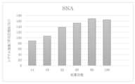
- FIG. 3 shows the change in the amount of ⁇ 2-6 sialic acid in response to SNA present on the cell membrane of the TGP culture sample.
- FIG. 4 shows the change in the amount of ⁇ 2-6 sialic acid in response to SSA present on the cell membrane of the TGP culture sample.
- FIG. 5 shows the change in the amount of ⁇ 2-6 sialic acid in response to TJA-1 present on the cell membrane of the TGP culture sample.
- FIG. 6 shows the change in the amount of ⁇ 1-3 fucose in response to UEA-1 present on the cell membrane of the TGP culture sample.
- the present invention is a method for producing a chondrocyte culture having high tissue regeneration ability, and includes a step of culturing a cell population separated from cartilage tissue with a thermoreversible polymer.
- Cartilage tissue in the present disclosure refers to cartilage-derived tissue. Cartilage is not limited to articular cartilage, osteochondral plate, costal cartilage, tracheal cartilage, laryngeal cartilage, sacroiliac joint, temporomandibular joint, sternoclavicular joint, intervertebral disc, shamebone connection, articular meniscus, articular disc, external auditory canal , Includes temporomandibular canal, epicartilage, laryngeal cartilage, etc.
- Cartilage tissue can be derived from any organism.
- Human cartilage is preferred because there is less rejection when the bone tissue culture is transplanted into human damaged cartilage. It may be an autologous organization or an allogeneic organization, but an autologous organization is preferable because there is little rejection when applied to the subject.
- the cartilage tissue used in the present invention can form a chondrocyte culture having a tissue structure similar to that of a healthy cartilage tissue regardless of the state of the object from which the cartilage tissue is derived. Therefore, it may be cartilage tissue derived from a subject in any state, for example, articular cartilage collected from a subject having osteoarthritis.
- the collection site of such articular cartilage may be a loaded portion or a non-loaded portion of the body weight, or may be a cartilage tissue collected from a loaded portion damaged by osteoarthritis.
- the chondrocyte culture produced by the present invention does not cause abnormalities such as calcification even after long-term culture, it is produced by collecting it from a grade 1 subject and culturing it until it progresses to grade 3 or later. It is also possible to transplant a chondrocyte culture into a subject. Therefore, the OA grade to be collected may be any of 1 to 4.
- the age at which the cartilage tissue is collected is not particularly limited. Since the chondrocyte culture produced by the present invention has high regenerative ability, for example, even if it is 0 to 39 years old or 40 years old or older, it is 50 years old or older, 60 years old or older, 70 years old or older, 80 years old or older, 90 years old or older. It may be cartilage tissue derived from the subject of.
- the method for collecting cartilage tissue may be a usual method used when collecting living tissue in the art, and can be collected from cartilage using a scalpel, tweezers, a biopsy punch, or the like without limitation. ..
- the "cell population separated from cartilage tissue” in the method of the present invention means a cell population composed of the same or different types of cells obtained by cartilage tissue separation treatment (for example, enzyme treatment, shredding treatment). ..
- the cell population is not limited as long as it is a cell population contained in cartilage tissue, and includes chondrocytes, cartilage progenitor cells, mesenchymal stem cells (MSCs), pluripotent stem cells, and the like.
- cartilage tissue separation treatment a method usually used as a biological tissue separation treatment in the technical field, for example, an enzymatic method or a physical method can be used.
- the enzymatic method is, but is not limited to, incubating the cells constituting the cartilage tissue at a viable temperature (for example, 37 ° C.) using one or more enzymes.
- a viable temperature for example, 37 ° C.
- an enzyme commonly used in the art in separating cells from cartilage tissue can be used under treatment conditions of corresponding temperature, concentration and time.
- dispase I or II collagenase I or II, metalloprotease, trypsin, hyaluronidase, pepsin, aminopeptidase, lipase, amylase and the like can be used without limitation as long as the cartilage tissue can be separated.
- cartilage tissue can be decomposed in a short time and with minimal invasiveness, preferably, even if any one of dispase I or II, collagenase, metalloprotease, actase or trypsin is used alone as an enzyme, two or more kinds are used. It may be used in combination.
- trypsin When trypsin is used, it may be, but is not limited to, 0.05% to 2.5%, 0.1% to 1%, 0.15% to 0.3% in the form of trypsin-EDTA, which is low. From the viewpoint of separation by invasiveness, 0.2 to 0.25% is preferable.
- the separation treatment time with trypsin-EDTA is not limited, and may be 1 minute to 2 hours, 3 minutes to 1 hour, 10 minutes to 40 minutes, and is preferably 30 minutes from the viewpoint of not damaging the stem cells.
- collagenase II When collagenase II is used, it is not limited to 0.1 to 30 mg / ml, 0.5 to 10 mg / ml, 0.75 to 5 mg / ml, and 0.8 to 2 mg from the viewpoint of minimally invasive separation.
- the separation treatment time is not limited as long as the living cells are not killed, and may be, for example, 1 to 24 hours, 3 to 20 hours, 7 to 18 hours, 10 to 16 hours, from the viewpoint of minimally invasive separation of stem cells. 12 to 16 hours is preferred.
- each concentration and treatment time can be arbitrarily combined without limitation.
- a combination of 12 to 16 hours is preferred.
- the physical method is not limited to this, and examples thereof include a method of chopping or crushing cartilage tissue using a scalpel, ultrasonic waves, a homogenizer, a strainer, or the like.
- the physical method can be optionally combined with the enzymatic method detailed above.
- any known method such as a filter or a cell strainer may be used to separate the cells separated by the separation treatment from the tissue pieces or the matrix that cannot be completely separated. Separation results in a cell population with a uniform size or function.
- thermoreversible polymer in the method of the present invention (also referred to as "TGP" in the present specification) is thermally reversibly formed a crosslinked structure or a network structure, and based on the structure, the inside thereof.
- the hydrogel refers to a gel containing a crosslinked or network structure made of a polymer and water supported or retained in the structure.
- the thermoreversible polymer can be any polymer as long as it is a thermoreversible polymer because an environment similar to cartilage tissue in a living body is formed by a crosslinked structure or a network structure peculiar to the thermoreversible polymer. Can also be used in the method of the present invention.
- Measuring equipment (trade name): Stress-controlled rheometer AR500, TA Instrument's sample solution (or separation solution) concentration (however, as the concentration of "hydrogel-forming polymer with sol-gel transition temperature") : 10 (weight)%
- Amount of sample solution Approximately 0.8 g
- Shape and dimensions of measuring cell Acrylic parallel disk (diameter 4.0 cm), gap 600 ⁇ m
- Measurement frequency 1Hz Applied stress: within the linear region
- the sol-gel transition temperature is preferably higher than 0 ° C. and preferably 37 ° C. or lower, and further preferably higher than 5 ° C. and 35 ° C. or lower (particularly 10 ° C. or higher and 33 ° C. or lower). ..
- a TGP having such a suitable sol-gel transition temperature can be easily selected from specific compounds as described later according to the above-mentioned screening method (sol-gel transition temperature measurement method).
- the TGP of the present invention is not particularly limited as long as it exhibits the thermoreversible sol-gel transition as described above (that is, has a sol-gel transition temperature).
- a specific example of a polymer in which the aqueous solution has a sol-gel transition temperature and reversibly exhibits a sol state at a temperature lower than the transition temperature is represented by, for example, a block copolymer of polypropylene oxide and polyethylene oxide.
- Polyalkylene oxide block copolymers; etherified cellulose such as methyl cellulose and hydroxypropyl cellulose; chitosan derivatives (KRHolme.et al. Macromolecules, 24,3828 (1991)) and the like are known.
- the hydrogel-forming polymer using a hydrophobic bond for cross-linking which can be suitably used as the TGP of the present invention, is a cloud point from the viewpoint that the TGP and the medium do not separate and the surroundings of the cell population can be stably maintained. It is preferable that a plurality of blocks having the above and a hydrophilic block are combined.
- the hydrophilic block is preferably present because the hydrogel becomes water soluble at a temperature lower than the sol-gel transition temperature, and the plurality of blocks having cloud points are the sol-gel transition temperature of the hydrogel. It is preferably present because it changes to a gel state at higher temperatures.
- a block with a cloud point dissolves in water at a temperature lower than the cloud point and becomes insoluble in water at a temperature higher than the cloud point, so that the block is gelled at a temperature higher than the cloud point.
- It serves as a cross-linking point consisting of hydrophobic bonds to form. That is, the cloud point derived from the hydrophobic bond corresponds to the sol-gel transition temperature of the hydrogel.
- the cloud point and the sol-gel transition temperature do not necessarily have to match. This is because the cloud point of the above-mentioned "block having a cloud point" is generally affected by the binding between the block and the hydrophilic block.
- the hydrogel used in the present invention utilizes the property that the hydrophobic bond not only becomes stronger as the temperature rises, but the change is reversible with respect to temperature.
- TGP is a "cloud point". It is preferable to have a plurality of "blocks having".
- the hydrophilic block in the TGP has a function of changing the TGP to water-soluble at a temperature lower than the sol-gel transition temperature, and has a hydrophobic binding force at a temperature higher than the transition temperature. It has a function of forming a state of a hydrogel while preventing the hydrogel from coagulating and precipitating due to excessive increase.
- the TGP used in the present invention is preferably one that is decomposed and absorbed in vivo from the viewpoint of using the chondrocyte culture of the present invention in cell therapy. That is, it is preferable that the TGP of the present invention is decomposed in a living body by a hydrolysis reaction or an enzymatic reaction to become a low molecular weight substance harmless to the living body, and is absorbed and excreted.
- the TGP of the present invention is formed by combining a plurality of blocks having cloud points and hydrophilic blocks, at least one of the blocks having cloud points and the hydrophilic blocks, preferably both of them, is in vivo. It is preferable that it is decomposed and absorbed by.
- the block having a cloud point is preferably a polymer block having a negative solubility in water-temperature coefficient, and more specifically, polypropylene oxide, a copolymer of propylene oxide and another alkylene oxide, Polymer selected from the group consisting of poly N-substituted acrylamide derivatives, poly N-substituted metaacrylamide derivatives, copolymers of N-substituted acrylamide derivatives and N-substituted metaacrylamide derivatives, polyvinyl methyl ether, and polyvinyl alcohol partially vinegared products. Can be preferably used.
- the poly N-substituted acrylamide derivative is preferable from the viewpoint of stably culturing the cell population or chondrocyte culture of the present invention.
- a polypeptide composed of a hydrophobic amino acid and a hydrophilic amino acid In order to decompose and absorb a block having a cloud point in a living body, it is effective to make the block having a cloud point a polypeptide composed of a hydrophobic amino acid and a hydrophilic amino acid.
- a polyester-type biodegradable polymer such as polylactic acid or polyglycolic acid can be used as a block having a cloud point that is decomposed and absorbed in the living body.
- the cloud point of the above polymer is higher than 4 ° C and 40 ° C or less, which is a compound in which a plurality of blocks having a cloud point and a hydrophilic block are bonded.
- the sol-gel transition temperature is higher than 0 ° C. and 37 ° C. or lower.
- the cloud point is measured, for example, by cooling an aqueous solution of about 1% by mass of the above polymer (block having a cloud point) to obtain a transparent uniform solution, and then gradually raising the temperature (heating rate of about 1). This can be done by setting the point at which the solution becomes cloudy for the first time as a cloud point at ° C./min).
- poly-substituted acrylamide derivative and the poly N-substituted meta acrylamide derivative that can be used in the present invention are listed below.
- Poly-N-isopropylacrylamide is preferable from the viewpoint of stably culturing the cell population or chondrocyte culture of the present invention.
- the above polymer may be a homopolymer or a copolymer of a monomer constituting the polymer and another monomer.
- a hydrophilic monomer or a hydrophobic monomer can be used as the other monomer constituting such a copolymer.
- copolymerization with a hydrophilic monomer raises the cloud point of the product, and copolymerization with a hydrophobic monomer lowers the cloud point of the product. Therefore, by selecting these monomers to be copolymerized, a polymer having a desired cloud point (for example, a cloud point higher than 4 ° C and 40 ° C or lower) can be obtained.
- hydrophilic monomer examples include N-vinylpyrrolidone, vinylpyridine, acrylamide, metaacrylamide, N-methylacrylamide, hydroxyethyl methacrylate, hydroxyethyl acrylate, hydroxymethyl methacrylate, hydroxymethyl acrylate, and acrylic having an acidic group. Acids, methacrylic acid and salts thereof, vinyl sulfonic acid, styrene sulfonic acid, etc., and N, N-dimethylaminoethyl methacrylate, N, N-diethylaminoethyl methacrylate, N, N-dimethylaminopropyl having a basic group. Examples thereof include, but are not limited to, acrylamide and salts thereof.
- hydrophobic monomer acrylate derivatives and methacrylate derivatives such as ethyl acrylate, methyl methacrylate and glycidyl methacrylate, N-substituted alkyl metaacrylamide derivatives such as Nn-butyl metaacrylamide, vinyl chloride, acrylonitrile and styrene , Vinyl acetate and the like, but are not limited thereto.
- hydrophilic block On the other hand, specific examples of the hydrophilic block to be bound to the above-mentioned block having a cloud point include methyl cellulose, dextran, polyethylene oxide, polyvinyl alcohol, poly N-vinylpyrrolidone, polyvinyl pyridine, polyacrylamide, and polymethacrylamide.
- poly-N-isopropylacrylamide when poly-N-isopropylacrylamide is used as a plurality of blocks having a cloud point of a thermoreversible gel, an environment similar to the in vivo environment of cartilage tissue is formed to proliferate the cell population.
- Polyethylene oxide is preferable as the hydrophilic block that binds to the acrylamide because of its high ability.
- hydrophilic block is decomposed, metabolized and excreted in the living body, and a hydrophilic biopolymer such as a protein such as albumin and gelatin and a polysaccharide such as hyaluronic acid, heparin, chitin and chitosan is preferably used. ..
- the method of binding the block having a cloud point and the above hydrophilic block is not particularly limited, but for example, a polymerizable functional group (for example, an acryloyl group) is introduced into any of the above blocks to give the other block. This can be done by copolymerizing the monomers. Further, the bond between the block having a cloud point and the above-mentioned hydrophilic block can also be obtained by block copolymerization of a monomer giving a block having a cloud point and a monomer giving a hydrophilic block. It is possible.
- a polymerizable functional group for example, an acryloyl group
- a functional group for example, a hydroxyl group, an amino group, a carboxyl group, an isocyanate group, etc.
- a chemical reaction it can also be done by.
- the bond between the polypropylene oxide having a cloud point and the hydrophilic block is, for example, anionic polymerization or cationic polymerization, in which the propylene oxide and the monomer constituting the "other hydrophilic block" (for example, ethylene oxide) are repeatedly and sequentially obtained.
- anionic polymerization or cationic polymerization in which the propylene oxide and the monomer constituting the "other hydrophilic block" (for example, ethylene oxide) are repeatedly and sequentially obtained.
- Such a block copolymer can also be obtained by introducing a polymerizable group (for example, an acryloyl group) at the end of polypropylene oxide and then copolymerizing the monomers constituting the hydrophilic block.
- the polymer used in the present invention can also be obtained by introducing a functional group capable of binding reaction with a functional group (for example, a hydroxyl group) at the terminal of polypropylene oxide into a hydrophilic block and reacting both of them. ..
- the TGP used in the present invention can also be obtained by connecting a material such as Pluronic (registered trademark) F-127 (trade name, manufactured by Asahi Denka Kogyo Co., Ltd.) in which polyethylene glycol is bonded to both ends of polypropylene glycol. be able to.
- Pluronic registered trademark
- F-127 trade name, manufactured by Asahi Denka Kogyo Co., Ltd.
- the above-mentioned "block having a cloud point” existing in the molecule is water-soluble together with the hydrophilic block at a temperature lower than the cloud point. It is completely dissolved in water and shows a sol state.
- the "block having a cloud point” existing in the molecule becomes hydrophobic and associates between separate molecules by hydrophobic interaction. To do.
- the polymer of the present invention since the hydrophilic block is water-soluble even at this time (when heated to a temperature higher than the cloud point), the polymer of the present invention has a hydrophobic association between blocks having a cloud point in water.
- a hydrogel having a three-dimensional network structure with the above as a cross-linking point is produced.
- the temperature of this hydrogel is cooled again to a temperature lower than the cloud point of the "block having a cloud point" existing in the molecule, the block having the cloud point becomes water-soluble and the cross-linking point due to the hydrophobic association is released. , The hydrogel structure disappears and the TGP of the present invention becomes a complete aqueous solution again.
- the sol-gel transition of the polymer of the present invention in a preferred embodiment is based on a reversible change in hydrophilicity and hydrophobicity at the cloud point of the block having a cloud point existing in the molecule. Therefore, it has complete reversibility in response to temperature changes. According to the studies by the present inventors, it is considered that the delicate hydrophilic-hydrophobic balance of TGP in water contributes to the stability of cells when they are stored at low temperature in water.
- the hydrogel-forming polymer of the present invention containing at least a polymer having a sol-gel transition temperature in an aqueous solution is substantially water-insoluble at a temperature (d ° C.) higher than the sol-gel transition temperature. Is reversibly water-soluble at a temperature (e ° C.) lower than the sol-gel transition temperature.
- the high temperature (d ° C.) described above is preferably a temperature 1 ° C. or higher higher than the sol-gel transition temperature, and more preferably 2 ° C. or higher (particularly 5 ° C. or higher).
- the above-mentioned "substantially water-insoluble” means that the amount of the above-mentioned polymer dissolved in 100 ml (liter) of water at the above-mentioned temperature (d ° C.) is 5.0 g or less (further, 0.5 g or less, particularly 0). .1 g or less) is preferable.
- the above-mentioned low temperature (e ° C.) is preferably 1 ° C. or higher (absolute value) lower than the sol-gel transition temperature, and further preferably 2 ° C. or higher (particularly 5 ° C. or higher) lower. preferable.
- water-soluble means that the amount of the polymer dissolved in 100 ml (liter) of water at the temperature (e ° C.) is preferably 0.5 g or more (further 1.0 g or more). ..
- reversibly water-soluble means that even after the aqueous solution of TGP is once gelled (at a temperature higher than the sol-gel transition temperature), at a temperature lower than the sol-gel transition temperature, It means to show the above-mentioned water solubility.
- the 10% aqueous solution of the polymer exhibits a viscosity of 10 to 3,000 cmpoise (further, 50 to 1,000 cmpoise) at 5 ° C.
- Viscosity is preferably measured under the following measurement conditions, for example.
- Viscometer Stress-controlled rheometer (model name: AR500, manufactured by TA Instruments) Rotor diameter: 60 mm Rotor shape: Parallel flat plate
- the aqueous solution of TGP of the present invention is gelled at a temperature higher than the sol-gel transition temperature and then immersed in a large amount of water, the gel does not substantially dissolve.
- the above-mentioned characteristics of the hydrogel formed by the above-mentioned TGP can be confirmed, for example, as follows. That is, 0.15 g of TGP is dissolved in 1.35 g of distilled water at a temperature lower than the sol-gel transition temperature (for example, under ice cooling) to prepare a 10 wt% aqueous solution, and the aqueous solution is used as a plastic petri dish having a diameter of 35 mm.
- a gel having a thickness of about 1.5 mm is formed in the petri dish by injecting into the petri dish and heating to 37 ° C., and then the weight (fgram) of the entire petri dish containing the gel is measured.
- the entire petri dish containing the gel was allowed to stand in water in 250 ml (liter) at 37 ° C. for 10 hours, and then the weight (g grams) of the entire petri dish containing the gel was measured to measure the gel from the gel surface. Evaluate the presence or absence of dissolution.
- the weight loss rate of the gel that is, (fg) / f is preferably 5.0% or less, and further 1.0%. It is preferably less than or equal to (particularly 0.1% or less).
- the aqueous solution of TGP of the present invention is gelled at a temperature higher than the sol-gel transition temperature, and then immersed in a large amount of water (about 0.1 to 100 times the gel in terms of volume) for a long period of time.
- the gel does not dissolve over time.
- Such properties of the polymer used in the present invention are achieved, for example, by the presence of two or more (plurality) blocks having cloud points in the polymer.
- Pluronic registered trademark
- F-127 registered trademark
- the concentration with respect to water that is, ⁇ (polymer) / (polymer + water) x 100 (%), is 20% or less (further, 15% or less).
- TGP capable of gelation at a concentration of 10% or less).
- the molecular weight of the TGP used in the present invention is preferably 30,000 or more and 30 million or less, more preferably 100,000 or more and 10 million or less, and further preferably 500,000 or more and 5 million or less.
- the step of culturing the cell population with the thermoreversible polymer may be performed by mixing the cell population separated from the cartilage tissue with the thermoreversible polymer by a known method and culturing.
- a cell population separated from cartilage tissue is dispersed in a TGP solution at a temperature lower than the sol-gel transition temperature, the TGP is gelled at a temperature higher than the sol-gel transition temperature, and then a culture solution or the like is used.
- a method of adding a medium and culturing can be used. By adding a medium to the gelled TGP, it is possible to provide a stable nutrient supply to the cell population in the gel by periodically exchanging only such a medium.
- the culture in the present invention can be carried out under the conditions usually used in the art.
- typical culture conditions include culturing at 37 ° C. and 5% CO2.
- the culture can be carried out at normal atmospheric pressure (atmospheric pressure).
- the culture period is not particularly limited because abnormal chondrocyte cultures such as calcification do not occur even if the cell population is cultured for a long period of time.
- Culturing can be performed in a container of any size and shape.
- the medium for dissolving TGP and the medium for adding to gelled TGP (sometimes referred to as "liquid medium” in the present specification) are not particularly limited as long as they can maintain cell survival, but are typically limited. , Amino acids, vitamins, and electrolytes are the main components.
- the medium for dissolving TGP and the medium for adding to the gelled TGP may be common or different.
- the medium for dissolving TGP and the medium for adding to gelled TGP are based on a basal medium for cell culture.
- a basal medium for cell culture is not limited to, for example, DMEM, MEM, F12, DME, RPMI1640, MCDB (MCDB102, 104, 107, 120, 131, 153, 199, etc.), L15, SkBM, RITC80-7, CnT. -PR etc. are included.
- Many of these basal media are commercially available, and their compositions are also known.
- the basal medium may be used as it has a standard composition (for example, as it is on the market), or the composition may be appropriately changed depending on the cell type and cell conditions. Therefore, the basal medium used in the present invention is not limited to those having a known composition, and includes those in which one or more components are added, removed, increased or decreased.
- the medium may contain one or more additives such as serum, growth factors (for example, FGF-2, TGF-b1, etc.), steroid component, selenium component, and the like.
- the serum may be a heterologous serum or an allogeneic serum. Allogeneic sera are preferred, and autologous sera are even more preferred among allogeneic sera.
- the medium does not contain growth factors other than the growth factors contained in the autologous serum. When culturing using TGP lysed by such a medium, the cell population will be cultivated in TGP to which growth factors other than autologous serum have not been added.
- the concentration of serum is not particularly limited, and 3%, 5%, 10%, and 20% may be contained in the medium added to TGP. It is preferably 10%.
- the medium for dissolving TGP and the medium for adding to gelled TGP may consist of a common component.
- the "chondrocyte culture” produced by the method of the present invention refers to a cell population containing chondrocytes obtained by culturing a cell population separated from cartilage tissue.
- the chondrocyte culture produced by the method of the invention may have a cartilage tissue-like tissue structure in which the cell gaps of the cell population are filled by ECM.
- the chondrocyte culture obtained by the method of the present invention is more than a chondrocyte culture (referred to herein as a "control culture”) cultured by a method that does not include the step of culturing a cell population with a thermoreversible polymer.
- High tissue regeneration ability means the tissue regeneration ability of cartilage tissue to which a cell population is applied.
- the tissue regeneration ability is applied to a cartilage cell culture of interest (transplantation), and after a lapse of a predetermined time, the state of the tissue at the application site (transplant destination), for example, the size of the tissue, the fineness of the tissue. It can be quantified by observing the structure, the ratio of damaged tissue to normal tissue, the function of tissue, etc. and scoring these.
- the tissue regeneration ability may be the expression ability of one or more genes selected from SOX9, COL2A1, miR140 and miR21.
- the chondrocyte culture produced by the method of the present invention has a higher expression of one or more genes selected from SOX9, COL2A1, miR140 and miR21 than the control culture. Therefore, the present invention may be a method for enhancing the expression ability of one or more genes selected from SOX9, COL2A1, miR140 and miR21 of a cell population isolated from cartilage tissue.
- SOX9 or COL2A1 is known as a gene expressed during normal cartilage tissue formation.
- miR140-3p or -5p or miR21-5p derived from miR140 or miR21 is a degenerated state of cartilage tissue characterized by inflammation in cartilage tissue generated in osteoarthritis, decomposition of ECM, and the like. Effective for normalization (Karlsen et al., Mol Ther Nucleic Acids. 2016 Oct 11; 5 (10), Miyaki et al., Arthritis Rheum. 2009 Sep; 60 (9): 2723-30, Si et al. , Osteoarthritis Cartilage. 2017 Oct; 25 (10): 1698-1707, Miyaki et al., Genes Dev. 2010 Jun 1; 24 (11): 1173-85, Hai et al ,. J Orthop Surg Res. 2019; 14 : See 118 etc.).
- miR140 in cartilage tissue results in increased miR140-3p or 5p in cartilage tissue, resulting in, but not limited to, expression or secretion of ECM-degrading enzymes such as MMP-13 and ADAMTS-5.
- ECM-degrading enzymes such as MMP-13 and ADAMTS-5.
- IL1B IL1B
- IL6 IL6
- IL8 proteins involved in ECM synthesis
- SOX9 proteins involved in ECM synthesis
- ACAN chondroitin sulfate N-acetylgalactosaminyl transferase 1
- the chondrocyte culture of the present invention when the chondrocyte culture of the present invention is applied to the cartilage tissue of a subject, particularly a subject having osteoarthritis, not only can the normal cartilage tissue be complemented at the application site, but also the cartilage tissue around the application site can be applied. Since the degenerative state can be normalized, the therapeutic effect can be maintained for a long period of time.
- miR21-5p is increased in cartilage tissue, and as a result, the expression or secretion of ECM-degrading enzymes such as MMP-13 and ADAMTS-5 is decreased, and ECM such as COL2A1 is not limited. It normalizes the degenerative state of cartilage tissue through increased expression of proteins involved in synthesis. Therefore, the therapeutic effect can be maintained for a long period of time like miR140.
- high gene expression is not limited to 101% or more, 105% or more, 110% or more, 130% or more, 140% or more, 150% or more, 160% or more, based on the control culture. It means that the expression level of a predetermined gene is high at 170% or more, 180% or more, 190% or more or 200% or more.
- Techniques for measuring gene expression levels are well known in the art and are not limited as techniques for measuring protein expression levels, for example, Western using the monoclonal or polyclonal antibodies detailed above. Blotting, EIA, ELISA, RIA, immunoorganization, immunocytochemistry, flow cytometer, mass assay, etc. are not limited as methods for measuring gene expression levels, for example, Northern blotting. Examples thereof include a method, a Southern blotting method, a DNA microarray analysis, an RNase protection assay, an RTPCR, a PCR method such as real-time PCR (qPCR), and an in situ hybridization method.
- qPCR real-time PCR
- SOX9 (also referred to as CMD1, CMPD1, SRA1, SRXX2, SRXY10) is a gene that coordinates SRY-box transcription factor 9, and the gene sequence of human SOX9 is registered as accession number NM_000346 or the like. The sequence is as shown in SEQ ID NO: 1.
- COL2A1 (also referred to as ANFH, AOM, COL11A3, SEDC, STL1) is a gene that links collagen type II alpha 1 chain, and the gene sequence of human COL2A1 is registered as accession number NM_001844.5, etc. The sequence is as shown in SEQ ID NO: 2.
- microRNA is transcribed as an RNA precursor having a hairpin-like structure, cleaved by a dsRNA cleaving enzyme having RNase III cleaving activity, incorporated into a protein complex called RISC, and suppresses translation of mRNA. 10-25 bases of RNA involved in are intended to be used.
- the “miRNA” also includes “miRNA” and precursors of the “miRNA” (pre-miRNA, pri-miRNA), and miRNAs having biological functions equivalent to those of the miRNA encoded by them, such as homologues (ie,). , Homolog), variants such as gene polymorphisms, and "miRNAs" encoding derivatives.
- RNA encoding such a precursor, homologue, mutant or derivative
- miRBase http://www.mirbase.org/
- miRBase http://www.mirbase.org/
- Examples thereof include “miRNA” having a base sequence that hybridizes with the complementary sequence of.
- human mir-140 is miRbase ID: Stem-loop sequence hsa-mir-140, miRbase accession number. It is registered as MI0000456 and is as shown in SEQ ID NO: 3.
- the human miR140-3p is miRbase ID: Mature sequence hsa-miR-140-3p, miRbase accession number. It is registered as MIMAT0004597 and is as shown in the RNA sequence "uaccacaggguagaaccacgg" shown in SEQ ID NO: 4.
- human miR140-5p is miRbase ID: Mature sequence hsa-miR-140-5p, miRbase accession number. It is registered as MIMAT0000431 and is as shown in the RNA distribution "cagugguuuuacccuaugguag" shown in SEQ ID NO: 5.
- human mir-21 is referred to as miRbase ID: Stem-loop sequence hsa-mir-21, miRbase accession number. It is registered as MI0000077 and is as shown in SEQ ID NO: 6.
- the human miR21-3p is miRbase ID: Mature sequence hsa-miR-21-3p, miRbase accession number. It is registered as MIMAT0004494 and is as shown in the RNA sequence "caacaccagucgaugggcugu" shown in SEQ ID NO: 7.
- human miR21-5p is miRbase ID: Mature sequence hsa-miR-21-5p, miRbase accession number. It is registered as MIMAT0000076 and is as shown in the RNA sequence "uagcuuaucagacugauguuga" shown in SEQ ID NO: 8.
- the tissue regeneration ability is the ability of the chondrocyte culture to retain miRNA.
- Retention capacity is the ability of a chondrocyte culture to retain miRNA in a chondrocyte culture.
- the ability to retain miRNAs in chondrocyte cultures may be the ability to retain miRNAs in chondrocyte cultures without secreting them during culture.
- the miRNA is not limited to miR140, miR21, miR-125b, Has-miR-15a, miR-30a, miR-199a, miR-210, miR-221-3p, miR-92a-3p, miR-142. -3p, miR-27a, miR-27b, miR26a-5p, miR-26a, miR-26b, miR-373, miR-127-5p, miR-320, miR-9, miR-634, miR-221-3p , MiR-370miR-145, miR-130A, miR-145, miR-562-5p and other miRNAs capable of normalizing the degenerative state of cartilage tissue (Zhang.et al. J Arthritis.
- High miRNA retention capacity is not limited to 101% or more, 105% or more, 110% or more, 130% or more, 140% or more, 150% or more, 160% based on the control culture at the time of culturing. As mentioned above, it means that miRNA is present in the chondrocyte culture at the time of culturing at a high concentration of 170% or more, 180% or more, 190% or more, or 200% or more. In one aspect, high miRNA retention capacity is defined as 99% or less, 95% or less, 97%% or less, 95% or less, without limitation, based on the medium (liquid medium) of the control culture at the time of culturing.
- the high retention capacity of miRNA means that miRNA is present in a high concentration in the cartilage cell culture at the time of culturing based on the control culture at the time of culturing, and the liquid medium of the control culture is used as a reference. It means that miRNA is present at a low concentration in the medium of the cartilage cell culture at the time of culturing.
- the tissue regeneration ability may be the content of stem cells in the chondrocyte culture.
- the chondrocyte culture produced by the method of the present invention has a higher stem cell content than the control culture. Therefore, the present invention may be a method for maintaining and proliferating stem cells in a cell population isolated from cartilage tissue.
- the content of stem cells is not limited to, but may be the amount or proportion of stem cells in the cell culture.
- Stem cells are not limited as long as they have the ability to differentiate into chondrocytes, such as somatic stem cells such as chondrocyte progenitor cells and mesenchymal stem cells, or pluripotent stem cells such as ES cells, iPS cells, and ntES cells. Is included.
- the content of stem cells can be measured by a method known in the art for measuring stem cells, and is not limited to reacting with lectins such as UEA-1 present on the cell membrane of cells contained in cartilage cell cultures. Measuring the amount of ⁇ 1-2 sialic acid that reacts with ⁇ 1-2 fucose or lectins such as SNA, SSA, TJA-I, or changes in the amount of ⁇ 1-2 fucolic acid or ⁇ 2-6 sialic acid. It can be quantified by measuring other cell surface markers or gene expression that correlate with.
- the level of ⁇ 1-2 fucose present in the cell membrane of the cells contained in the cartilage cell culture is high, many pluripotent stem cells are present, and if the level of ⁇ 2-6 sialic acid is high, the differentiation potential is high. It means that there are many stem cells. (See Wang et al, Cell Res. 2011 Nov; 21 (11): 1551-63, Tateno et al, Glycobiology. 2016 Dec; 26 (12): 1328-1337, WO2016006712A1).
- “Differentiation potential” means that when a certain cell is placed in an appropriate differentiation-inducing state, it has the ability to change into another type of cell such as a progenitor cell or a somatic cell.
- somatic stem cells In general, long-term culture of somatic stem cells tends to reduce their differentiation potential as well as their proliferative capacity. Since somatic stem cells with reduced differentiation potential have somatic stem cell markers on the cell surface but cannot be transformed into other types of cells such as progenitor cells and somatic cells, such somatic stem cells have the ability to regenerate tissues. do not have.
- the chondrocyte culture produced by the method of the present invention has a high content of somatic stem cells having a high differentiation potential.
- the content of somatic stem cells having a high differentiation potential may be, but is not limited to, the amount or proportion of somatic stem cells having a high differentiation potential in the cell culture per unit amount.
- the present invention may be a method of maintaining or increasing the differentiation potential of somatic stem cells in a cell population isolated from cartilage tissue.
- the present invention may be a method of maintaining or proliferating somatic stem cells having a high differentiation potential in a cell population separated from cartilage tissue.
- the somatic stem cell is not limited as long as it is a somatic stem cell capable of differentiating into a chondrocyte, and may be a cartilage progenitor cell, a mesenchymal stem cell, or the like.
- chondrocytes differentiated from cells having a high differentiation potential can be constantly supplied to the application site. , Can repair and maintain normal chondrocyte tissue for a long period of time.
- the stem cell or somatic stem cell with high differentiation potential is a mesenchymal stem cell, it has the ability to normalize damaged cartilage tissue, such as for protecting chondrocyte apoptosis, for anti-inflammatory of cartilage tissue, and for anti-fibrosis. Since the ability to recover from damage is high, the therapeutic effect can be maintained for a long period of time.
- the method for measuring other cell surface markers or gene expression that correlates with the change in the amount of ⁇ 1-2 fucose acid or ⁇ 2-6 sialic acid is the conventional method for measuring cell surface markers or gene expression in the art. Can be used.
- the chondrocyte culture produced by the method of the present invention contains more stem cells or somatic stem cells having a higher differentiation potential than the control culture.
- the term "rich in stem cells or somatic stem cells with high differentiation potential” is not limited to 101% or more, 105% or more, 110% or more, 130% or more, 140% or more, 150% or more based on the control culture. , 160% or more, 170% or more, 180% or more, 190% or more or 200% or more, the content of cells having ⁇ 1-2 fucolic acid or ⁇ 2-6 sialic acid, or cell surface marker or gene expression that correlates with these. Means that is high.
- the tissue regeneration ability may be the ability to secrete hyaluronic acid from chondrocyte cultures.
- the chondrocyte culture produced by the method of the present invention has a higher ability to secrete hyaluronic acid than the control culture. Therefore, the present invention may be a method for enhancing the secretory capacity in a cell population separated from cartilage tissue.
- Hyaluronic acid is known to act suppressively on the expression of ECM-degrading enzymes, alleviate the matrix and anti-inflammatory factors released from cartilage, and regulate the expression of inflammation-related factors (Ohtsuki.et). al.J Orthoped Res. 2018 Aug 17. doi: 10.1002 / jor.24126).
- the chondrocyte culture produced by the method of the present invention when applied to the target cartilage tissue, not only can the normal cartilage tissue be complemented at the application site, but also ECM decomposition or inflammation around the application site is suppressed. Therefore, the degenerated state of the cartilage tissue around the application site can be normalized, and the normal cartilage tissue can be repaired and maintained for a long period of time.
- hyaluronic acid in a medium (liquid medium) or TGP may be quantified over time.
- Hyaluronic acid can be quantified by mass spectrometry, liquid chromatography, MALDI-TOF, etc., as well as commercially available kits, without limitation.
- High hyaluronic acid secretory capacity is not limited to 101% or more, 105% or more, 110% or more, 130% or more, 140% or more, 150% or more, 160% or more, based on the control culture. It means that the secretory capacity of hyaluronic acid is high at 170% or more, 180% or more, 190% or more or 200% or more.
- the chondrocyte culture produced by the method of the present invention is rich in ECM and therefore easily engrafts at the injured site. Therefore, the chondrocyte culture produced by the method of the present invention can be used for transplantation.
- the expression levels of miR140 and miR21 are high, the amount of stem cells is high, and the amount of hyaluronic acid secreted is high, it is preferably used for the treatment of grade 2 or higher or elderly osteoarthritis.
- the present invention further comprises chondrocyte cultures produced by the methods of the present invention.
- the chondrocyte culture of the present invention is as described above.
- the present invention includes methods of treating a disease in a subject, comprising applying an effective amount of the chondrocyte culture of the present invention to a subject in need thereof.
- the disease is not limited to cartilage-related diseases, but is due to osteoarthritis, rheumatoid arthritis, osteosarcoma, femoral head necrosis, acetabular dysplasia, meniscus injury, traumatic arthritis, sports, accidents, etc. Includes physical cartilage loss or damage.
- the method of application (administration) to the subject may be transplantation of a chondrocyte culture.
- Transplantation of chondrocyte cultures is not limited to delivering the chondrocyte cultures to the affected area by injection into the cartilage tissue, or arthroscopic surgery and incision, and any tissue adhesive such as fibring gel. It can be fixed to the affected area by use or by arthroscopy.
- various additional ingredients such as a pharmaceutically acceptable carrier, an ingredient that enhances engraftment, and a further active ingredient can be contained and applied. Any known additional ingredient can be used as such additional ingredient, and those skilled in the art are familiar with these additional ingredients.
- the present invention will be described in more detail below with reference to specific aspects of the present invention, but the present invention is not limited to these specific examples.
- the storage elastic modulus of this aqueous solution was measured at an application frequency of 1 Hz using a stress-controlled rheometer (AR500, manufactured by TA Instruments), and found to be 43 Pa at 10 ° C, 680 Pa at 25 ° C, and 1310 Pa at 37 ° C. This temperature-dependent storage modulus change was reversibly observed repeatedly.
- AR500 stress-controlled rheometer
- ⁇ Separation of cartilage tissue Each piece of tissue was cut into 1 mm 2 or less with a scalpel. The slices were treated with Trypsin-EDTA solution (0.25%) at 37 C ° for 30 minutes, then enzymatically treated with collagenase II solution (1 mg / ml) at 37 C ° for 12-16 hours. After washing with DMEM solution, the mixture was filtered through a filter (100 ⁇ m) and centrifuged (1800 rpm, 10 minutes). The cell population thus obtained was counted on the cell calculation board. The number of cells was 1 ⁇ 10 4 to 4 ⁇ 10 4 cells / ml.
- Example 1 ⁇ Culture with TGP gel> 1 g of TGP prepared in Production Example 1 was dissolved in 9 mL of DMEM at 4 ° C. to prepare a 10% TGP solution, and then cells derived from cartilage tissue were dispersed and dispensed into a 6-well plate. After leaving at room temperature to gel, add antibiotics (gentamicin (50 ⁇ g / ml), amphotericin (0.25 ⁇ g / ml), penicillin (100 Units / ml) / streptomycin (100 ⁇ g / ml) to DMEM solution containing 10% autologous serum.
- antibiotics gentamicin (50 ⁇ g / ml)
- amphotericin (0.25 ⁇ g / ml
- penicillin 100 Units / ml
- streptomycin 100 ⁇ g / ml
- Example 1 chondrocyte cultures having a size of 200 to 300 ⁇ m were confirmed on the 3rd to 5th days of culture, and after 21 days, they grew to a size of 0.5 to 1.0 mm in diameter, and all the samples. Growth was maintained for a 20-week culture period. In Example 1, no proliferation of fibroblast-like cells was observed.
- RNAlater registered trademark
- Stabilization Solution Invitrogen, CatNo.AM7020
- Small RNA RNA of 200 bases or less
- NucleoSpin® miRNA kit TeKaRa, U0971
- primers of SEQ ID NOs: 9 to 12 and mRQ3'Primer U6Forward U6, miR-21 by Thermal Cycler Dice (registered trademark) Real Time System Lite (TP700, TaKaRa) using Primer and U6 Reverse Primer (Mir-X TM miRNA qRT-PCR TB Green Kit (TaKaRa, Z8314N))
- QPCR of 3p, miR-21-5p, miR-140-3p and miR-140-5p was performed. Quantitative values of each miRNA were normalized with the expression level of U6 as 1.
- QPCR of GAPDH, SOX9 and COL2A1 was performed by Thermal Cycler Dice (registered trademark) Real Time System Lite. The quantitative value of each gene was normalized with the expression level of GAPDH as 1.
- the primers used are shown in Table 2, and the results are shown in Tables 3-5.
- the expression level of miR-21 and miR-140 was increased in Example 1 cultured in TGP gel as compared with the sample of Comparative Example 1, and miR21-5p, It was found that miR-140-3p and miR-140-5p were significantly increased.
- the expression level of SOX9 was significantly increased in the sample of Example 1 as compared with the sample of Comparative Example 1. It was also found that the expression level of 1068, in which COL2A1 was measured, was increased in Example 1 as compared with the sample of Comparative Example 1.
- the chondrocyte culture produced in Example 1 has a healthy function. It turned out to be cartilage tissue.
- miR-140 and miR21 are known to improve the microenvironment of osteoarthritis, so the chondrocyte culture produced in Example 1 only promotes the regeneration of the cartilage tissue in the transplanted portion. Instead, it can improve the degenerative conditions around the transplant.
- RNAs of 200 bases or less were purified from some of these samples using the NucleoSpin® miRNA kit (TaKaRa, U0971), and the primers of SEQ ID NO: 3 and mRQ 3'Primer (Mir-X (Mir-X) Using miRNA qRT-PCR TB Green (registered trademark) Kit (TaKaRa, Z8314N)), qPCR of miR-140-3p was performed by Thermal Cycler Dice (registered trademark) Real Time System Lite (TP700, TaKaRa). The results are shown in Table 6.
- the average Ct value is the number of cycles until a predetermined Threshold is reached, and the lower the average Ct value, the more miR-140-3p is present in the liquid medium.
- Threshold the threshold for the average Ct value.
- Example 1 Analysis of glycans on the cell surface of cells constituting the cultured chondrocyte culture
- the chondrocyte culture cultured in Example 1 was collected between 14 and 126 days after the culture, and each TGP gel was collected in 4 It was cooled to ° C to form an aqueous solution and recovered.
- the sample was dispersed in cells with Trypsin-EDTA (0.25%) solution, and the cells were collected. Then, it was centrifuged in DMEM three times at 4 ° C. (1800 rpm, 15 minutes), and each sample was cryopreserved.
- Glyco Technica Ltd Glycan Profiling Analysis Service by Glycan Profiling Analysis Service.
- the cell membrane protein was extracted from the thawed sample, and the protein concentration was measured by the BCA method (TaKaRa BCA Protein Assay Kit). Based on the measured protein concentration, PBSTx was added to dilute the protein concentration to 10 ⁇ g / mL. A 100 ⁇ L sample (concentration of 10 ⁇ g / mL) was added to a tube containing 100 ⁇ g of Cy3 Mono-reactive Dye Pack (GE healthcare, catalog number: PA23011), mixed with a pipette, and spun down. The tube was placed in a light-shielding bag and incubated at room temperature (25 ° C.) for 1 hour.
- BCA method TaKaRa BCA Protein Assay Kit
- the fluorescence pattern of LecChip TM was measured four times cumulatively by GlycoLite2200 ( TM , Glyco technica), the exposure time, 1996, 2995, 3991, 4992, 6988, 9998 (milliseconds), and the camera gain was measured at fixed values.
- the obtained 45-lectin signal was measured by GlycoStaion® ToolsPro Suite 1.5 (GlycoStaion® ToolsPro Suite 1.5.), And A. Kuno et al., J. of Proteomics & Bioinformatics, Vol.1, May 2008. , P.68., Divided by the average intensity of 45 lectins, multiplied by 100 and average normalized.
- ⁇ 2-6 sialic acid-binding lectins such as SNA, SSA and TJA-I and ⁇ 1-2 fucose-binding lectins such as UEA-1 were cultured for days. It can be seen that it increases according to.
- ⁇ 2-6 sialic acid which reacts with SNA, SSA and TJA-I, is a marker for somatic stem cells such as mesenchymal stem cells (MSCs) or cartilage progenitor cells with high differentiation potential (WO2016 / 006712A1) and UEA-1.
- ⁇ 1-2 fucose-binding lectins such as ⁇ 1-2 fucose-binding lectin are known to be markers of pluripotent stem cells (Wang et al., Cell Res. 2011 Nov; 21 (11): 1551-63. Doi: 10.1038 / cr. .2011.148. Epub2011 Sep 6.).
- ⁇ 2-6 sialic acid and ⁇ 1-2 fucose in response to SNA, SSA, TJA-I and UEA-1 was confirmed with the number of culture days. It was found that the tissue culture contained a large amount of somatic stem cells and pluripotent stem cells having high differentiation potential. Therefore, it was clarified that the chondrocyte culture of Example 1 is a tissue culture having high tissue regeneration ability.
- Hyaluronic acid retention test of TGP gel [Example 2] Hyaluronic acid (Teijin Pharma Co., Ltd. 5 mg to DMEM with antibiotics (gentamicin (50 ⁇ g / ml), amphotericin (0.25 ⁇ g / ml), penicillin (100 Units / ml) / streptomycin (100 ⁇ g / ml) and L-Ascorbic acid It was dissolved in 2.5 ml of a solution containing (5 mg / ml) to give liquid compositions 1 and 2. 1 g of TGP prepared in Production Example 1 was dissolved in 9 ml of DMEM at ° C.
- TGP solution hyaluronic acid was added thereto so as to have the same hyaluronic acid concentration as liquid compositions 1 and 2, and hyaluronic acid in the TGP solution was added. Shake to homogenize the acid.
- the TGP solution was gelled at 37 ° C., and then 1.0 ml of liquid medium (DMEM) was added to make TGP composition 1 and TGP composition 2.
- DMEM liquid medium
- the prepared hyaluronic acid-containing liquid composition and TGP composition were placed in a 5% carbon dioxide incubator at 37 ° C., and the hyaluronic acid concentration contained in each liquid medium portion was measured by the same method as ⁇ Measurement of hyaluronic acid> described above. .. The results are shown in Table 9.
- hyaluronic acid was present at about 13000 ng / ml on the 7th day, but decreased to 5.6 ng / ml on the 21st day.
- TGP compositions 1 and 2 it was 6.1 and 11.4 ng / ml at the 7th day, which corresponds to a low concentration of about 1/1000 with respect to liquid compositions 1 and 2.
- concentrations were 10.3 and 11.5 ng / ml in the 21st order, and their concentrations hardly changed. Therefore, it is clear that the TGP gel has the ability to stably retain hyaluronic acid for a long period of time without being decomposed. It became.
Abstract
The purpose of the present invention is to provide a chondrocyte culture with high tissue regeneration ability. This purpose is met by a method involving a step in which a cell population separated from cartilage tissue is cultured on a thermoreversible polymer.
Description
本発明は、軟骨細胞培養物を製造する方法、当該方法によって製造された軟骨細胞培養物、軟骨細胞培養物を使用した治療方法に関する。
The present invention relates to a method for producing a chondrocyte culture, a chondrocyte culture produced by the method, and a treatment method using the chondrocyte culture.
変形性関節症は、関節組織の進行性の軟骨損傷を特徴とし、加齢、関節損傷、肥満を含む様々な因子により引き起こされ、特に高齢者における関節能力の喪失や硬化の原因となる。
Osteoarthritis is characterized by progressive cartilage damage of joint tissue, caused by various factors including aging, joint damage and obesity, and causes loss of joint ability and hardening, especially in the elderly.
関節軟骨組織は、骨端の関節面に存在する薄い硝子軟骨からなる。硝子軟骨は、ヒアルロン酸-アグリカンネットワークおよびII型コラーゲン線維等との相互作用により高次構造を形成した軟骨細胞外マトリックス(ECM)により軟骨細胞の細胞間隙が充填された組織構造を持つ。OAにおける軟骨組織の損傷過程には、かかるECMの分解が大きく関与する。
The articular cartilage tissue consists of thin hyaline cartilage existing on the articular surface of the epiphysis. Hyaline cartilage has a tissue structure in which the intercellular spaces of chondrocytes are filled with a chondrocyte extracellular matrix (ECM) that forms a higher-order structure by interaction with a hyaluronic acid-aglycan network and type II collagen fibers. Degradation of such ECM is greatly involved in the cartilage tissue damage process in OA.
軟骨組織が損傷するとECM中のII型コラーゲン線維が破壊され、これによりヒアルロン酸やアグリカンの遊出を引き起こす。遊出したヒアルロン酸やアグリカンは、軟骨細胞のアポトーシスや、炎症性サイトカインであるインターロイキン-1の産生を引き起こし、これによりMMP、ADAMTSおよびHYBIDといったECM分解酵素の産生および分泌が促され、さらに軟骨組織中のECMの分解が加速する。
When cartilage tissue is damaged, type II collagen fibers in ECM are destroyed, which causes the excretion of hyaluronic acid and aggrecan. The released hyaluronic acid and aggrecan cause chondrocyte apoptosis and the production of the inflammatory cytokine interleukin-1, which promotes the production and secretion of ECM-degrading enzymes such as MMP, ADAMTS and HYBID, and further cartilage. Decomposition of ECM in tissue is accelerated.
このようにECMが分解された軟骨組織の弾性は、著しく低下し脆くなるため軽い負荷でも容易に損傷するという悪循環をきたす。また関節組織は、通常の組織と異なり、血液供給がなく拡散によって軟骨組織内外と物質交換をしており、細胞の流入もないことから、変形性関節症のように組織の恒常性が一旦破壊されると、その変性状態が維持されるため修復困難となる(非特許文献1、2)。
In this way, the elasticity of the cartilage tissue in which ECM is decomposed is significantly reduced and becomes brittle, which causes a vicious cycle in which it is easily damaged even with a light load. In addition, unlike normal tissue, joint tissue exchanges substances with and outside cartilage tissue by diffusion without blood supply, and there is no inflow of cells, so tissue homeostasis is temporarily destroyed like osteoarthritis. If this is done, the denatured state is maintained and repair becomes difficult (Non-Patent Documents 1 and 2).
このような変形性関節症の治療として、関節軟骨の非荷重部から採取された健全な軟骨組織に由来する軟骨細胞を利用した自家培養軟骨細胞移植(Autologous Chondrocyte Implantation;ACI)や、マトリックス誘導性自家培養軟骨細胞移植(Matrix-Induced Autologous Chondrocyte Implantation;MACI)が行なわれてきた。かかる移植は、外傷性膝軟骨損傷のように移植部位の周囲に健全な軟骨組織が存在していれば有効であるが、変形性関節症のように軟骨組織の恒常性が破壊された変性部位に施しても、一時的な改善は見られるものの、すぐに移植片中の軟骨細胞がアポトーシスによって死滅したり、繊維軟骨が形成され、十分な治療効果は得られていない(非特許文献3)。
このように変形性関節症の治療において、軟骨組織の変性状態を正常化し、長期間修復、維持可能な治療が求められている。 Treatments for such osteoarthritis include autologous chondrocyte implantation (ACI) using chondrocytes derived from healthy cartilage tissue collected from unloaded parts of articular cartilage, and matrix-inducible. Matrix-Induced Autologous Chondrocyte Implantation (MACI) has been performed. Such transplantation is effective if there is healthy cartilage tissue around the transplant site, such as traumatic knee cartilage injury, but degenerative sites, such as osteoarthritis, in which the homeostasis of cartilage tissue is disrupted. Although temporary improvement is seen, the chondrocytes in the transplant piece are immediately killed by apoptosis or fibrocartilage is formed, and a sufficient therapeutic effect has not been obtained (Non-Patent Document 3). ..
As described above, in the treatment of osteoarthritis, a treatment that normalizes the degenerated state of cartilage tissue and can be repaired and maintained for a long period of time is required.
組織再生能が高い軟骨細胞培養物を製造する方法、当該方法によって製造された軟骨細胞培養物、軟骨細胞培養物を使用した治療方法を提供することにある。
It is an object of the present invention to provide a method for producing a chondrocyte culture having high tissue regeneration ability, a chondrocyte culture produced by the method, and a therapeutic method using the chondrocyte culture.
すなわち、本発明に下記に掲げるものに関する:
[1] 組織再生能が高い軟骨細胞培養物を製造する方法であって、軟骨組織から分離された細胞集団を熱可逆性ポリマーで培養するステップを含む、前記方法。
[2] 組織再生能が、軟骨細胞培養物のSOX9、COL2A1、miR140およびmiR21から選択される1以上の遺伝子の発現能、miRNAの保持能力、幹細胞の含有量、またはヒアルロン酸の分泌能である、[1]に記載の方法。
[3] miRNAが、miR140である、[2]に記載の方法。
[4] 幹細胞が、多能性幹細胞または分化ポテンシャルが高い体性幹細胞である、[2]または[3]に記載の方法。
[5] 幹細胞の含有量が、軟骨細胞培養物中の1-2フコースまたはα2-6シアル酸の含有量である、[2]~[4]のいずれか一項に記載の方法。
[6] 軟骨組織が、50歳以上の変形性関節症の対象に由来する、[1]~[5]に記載の方法。
[7] 熱可逆性ポリマーが、血清以外の成長因子が添加されていない熱可逆性ポリマーである、[1]~[6]に記載の方法。
[8] [1]~[7]に記載の方法によって製造された軟骨細胞培養物。
[9] [8]に記載の軟骨細胞培養物の有効量を、それを必要とする対象に適用することを含む、対象における疾患を治療する方法。
[10] 疾患が、変形性関節症である[9]に記載の治療方法。 That is, the present invention relates to the following:
[1] A method for producing a chondrocyte culture having high tissue regeneration ability, which comprises a step of culturing a cell population separated from cartilage tissue with a thermoreversible polymer.
[2] The tissue regeneration ability is the ability to express one or more genes selected from SOX9, COL2A1, miR140 and miR21 of chondrocyte culture, the ability to retain miRNA, the content of stem cells, or the ability to secrete hyaluronic acid. , [1].
[3] The method according to [2], wherein the miRNA is miR140.
[4] The method according to [2] or [3], wherein the stem cell is a pluripotent stem cell or a somatic stem cell having a high differentiation potential.
[5] The method according to any one of [2] to [4], wherein the content of stem cells is the content of 1-2 fucose or α2-6 sialic acid in the chondrocyte culture.
[6] The method according to [1] to [5], wherein the cartilage tissue is derived from a subject with osteoarthritis over 50 years old.
[7] The method according to [1] to [6], wherein the thermoreversible polymer is a thermoreversible polymer to which a growth factor other than serum is not added.
[8] A chondrocyte culture produced by the method according to [1] to [7].
[9] A method for treating a disease in a subject, comprising applying an effective amount of the chondrocyte culture according to [8] to the subject in need thereof.
[10] The treatment method according to [9], wherein the disease is osteoarthritis.
[1] 損傷した軟骨組織を修復するためのTGP含有組成物。
[2] 損傷した軟骨組織が、変形性関節症により損傷した軟骨組織である、[1]に記載の組成物。
[3] 修復が、損傷した軟骨組織の変性状態の正常化である、[1]または[2]に記載の組成物。
[4] 修復が、軟骨細胞の増殖、ヒアルロン産の分泌促進または保持、miR140の分泌促進、CD44陽性細胞の増加、および、間葉系幹細胞(MSCs)の増殖からなる群から選択される1以上の指標により表わされる、[1]~[3]のいずれか一項に記載の組成物。
[5] 修復が、in vitroまたはin vivoで行なわれる、[1]~[4]のいずれか一項に記載の組成物。
[6] 軟骨細胞、軟骨前駆細胞またはMSCs、軟骨細胞培養物および軟骨組織からなる群から選択される1以上の組織または細胞を含む、[1]~[5]のいずれか一項に記載の組成物。
[7] 軟骨細胞または軟骨組織が、自家細胞または自家組織である、[1]~[6]のいずれか一項に記載の組成物。
[8] 高齢者の軟骨組織に由来する細胞をTGP含有組成物中で培養するステップを含む、軟骨細胞培養物および培養分泌物の製造方法。
[9]
軟骨組織が、変形性膝軟骨関節症により損傷した軟骨組織である、[8]に記載の方法。
[10] さらに軟骨細胞培養物と培養分泌物とを分離するステップを含む、[8]または[9]に記載の方法。 [1] A TGP-containing composition for repairing damaged cartilage tissue.
[2] The composition according to [1], wherein the damaged cartilage tissue is a cartilage tissue damaged by osteoarthritis.
[3] The composition according to [1] or [2], wherein the repair is the normalization of the degenerated state of the damaged cartilage tissue.
[4] One or more repairs selected from the group consisting of chondrocyte proliferation, hyaluronic secretion promotion or retention, miR140 secretion promotion, CD44-positive cell proliferation, and mesenchymal stem cell (MSCs) proliferation. The composition according to any one of [1] to [3], which is represented by the index of.
[5] The composition according to any one of [1] to [4], wherein the repair is performed in vitro or in vivo.
[6] The item according to any one of [1] to [5], which comprises one or more tissues or cells selected from the group consisting of chondrocytes, cartilage progenitor cells or MSCs, chondrocyte cultures and cartilage tissues. Composition.
[7] The composition according to any one of [1] to [6], wherein the chondrocyte or cartilage tissue is an autologous cell or autologous tissue.
[8] A method for producing a chondrocyte culture and a cultured secretion, which comprises a step of culturing cells derived from cartilage tissue of an elderly person in a TGP-containing composition.
[9]
The method according to [8], wherein the cartilage tissue is a cartilage tissue damaged by osteoarthritis of the knee.
[10] The method according to [8] or [9], further comprising a step of separating the chondrocyte culture and the culture secretion.
[11] 未分化細胞を増殖または維持させる方法であって、
軟骨組織から得られた細胞を含む細胞をTGP含有組成物で培養するステップを含む、前記方法。
[12] 未分化細胞が、軟骨細胞培養物中で増殖または維持される、[11]に記載の方法。
[13] さらに未分化細胞を添加するステップを含む、[11]または[12]に記載の方法。
[14] 未分化細胞がMSCである、[11]~[13]のいずれか一項に記載の方法。
[15] 軟骨組織が、変形性関節症により損傷した軟骨組織である、[11]~[14]のいずれか一項に記載の方法。 [11] A method for proliferating or maintaining undifferentiated cells.
The method comprising culturing cells, including cells obtained from cartilage tissue, in a TGP-containing composition.
[12] The method of [11], wherein the undifferentiated cells are proliferated or maintained in a chondrocyte culture.
[13] The method according to [11] or [12], which further comprises the step of adding undifferentiated cells.
[14] The method according to any one of [11] to [13], wherein the undifferentiated cells are MSCs.
[15] The method according to any one of [11] to [14], wherein the cartilage tissue is a cartilage tissue damaged by osteoarthritis.
[16] [11]~[15]のいずれか一項に記載の方法により製造された未分化細胞。
[17] 細胞から分泌された高分子成分の分解を防ぐためのTGP含有組成物。
[18] 細胞から分泌された高分子成分の製造方法であって、細胞をTGP含有組成物中で培養するステップを含む、前記方法。
[19] 細胞から分泌された高分子成分がヒアルロン酸である、[17]または[18]に記載の組成物または方法。
[20] 細胞が、ヒト細胞であり、ヒト細胞が、ヒト平滑筋細胞、ヒト軟骨細胞、ヒト繊維芽細胞、ヒト脂肪細胞、ヒト滑膜細胞からなる群から選択される1以上の細胞である、[17]~[19]のいずれか一項に記載の組成物または方法。 [16] Undifferentiated cells produced by the method according to any one of [11] to [15].
[17] A TGP-containing composition for preventing decomposition of a polymer component secreted from cells.
[18] A method for producing a polymer component secreted from a cell, which comprises a step of culturing the cell in a TGP-containing composition.
[19] The composition or method according to [17] or [18], wherein the polymeric component secreted from the cell is hyaluronic acid.
[20] The cell is a human cell, and the human cell is one or more cells selected from the group consisting of human smooth muscle cells, human chondrocytes, human fibroblasts, human adipocytes, and human synovial cells. , The composition or method according to any one of [17] to [19].
本発明の方法によって製造された軟骨細胞培養物は、ECMに富むため生着性が高く、健全な軟骨の機能および組織構造を有するだけでなく、炎症、ECMの分解等を特徴とする軟骨組織の変性状態を正常化することが出来る。さらに、軟骨細胞培養物には軟骨再生に寄与する幹細胞が多く含まれている。したがって、変形性関節症のような変性状態の軟骨組織を長期わたって修復、維持する効果を奏する。
The chondrocyte culture produced by the method of the present invention is rich in ECM and therefore highly engraftable, and not only has a healthy cartilage function and tissue structure, but also is characterized by inflammation, decomposition of ECM, and the like. The degenerative state of can be normalized. In addition, chondrocyte cultures are rich in stem cells that contribute to cartilage regeneration. Therefore, it has the effect of repairing and maintaining degenerated cartilage tissue such as osteoarthritis over a long period of time.
以下、本発明を詳細に説明する。
本明細書において別様に定義されない限り、本明細書で用いる全ての技術用語および科学用語は、当業者が通常理解しているものと同じ意味を有する。本明細書中で参照する全ての特許、出願および他の出版物や情報は、その全体を参照により本明細書に援用する。また本明細書において参照された出版物と本明細書の記載に矛盾が生じた場合は、本明細書の記載が優先されるものとする Hereinafter, the present invention will be described in detail.
Unless defined otherwise herein, all technical and scientific terms used herein have the same meaning as those commonly understood by one of ordinary skill in the art. All patents, applications and other publications and information referenced herein are hereby incorporated by reference in their entirety. In the event of a conflict between the publications referred to herein and the description herein, the description herein shall prevail.
本発明は、組織再生能が高い軟骨細胞培養物を製造する方法であって、軟骨組織から分離された細胞集団を熱可逆性ポリマーで培養するステップを含む。本開示における「軟骨組織」は、軟骨由来の組織を指す。軟骨は、限定されずに関節軟骨、骨端板、肋軟骨、気管軟骨、喉頭軟骨、仙腸関節、顎関節、胸鎖関節、椎間円板、恥骨結合、関節半月、関節円板、外耳道、耳管、耳介軟骨及び喉頭蓋軟骨等を含む。軟骨組織は、任意の生物に由来し得る。ヒトの損傷した軟骨へ骨組織培養物を移植した際に拒絶反応が少ないことからヒトの軟骨組織が好ましい。自家組織であっても他家組織であってもよいが、対象に適用した際に拒絶反応が少ないことから自家組織が好ましい。
The present invention is a method for producing a chondrocyte culture having high tissue regeneration ability, and includes a step of culturing a cell population separated from cartilage tissue with a thermoreversible polymer. "Cartilage tissue" in the present disclosure refers to cartilage-derived tissue. Cartilage is not limited to articular cartilage, osteochondral plate, costal cartilage, tracheal cartilage, laryngeal cartilage, sacroiliac joint, temporomandibular joint, sternoclavicular joint, intervertebral disc, shamebone connection, articular meniscus, articular disc, external auditory canal , Includes temporomandibular canal, epicartilage, laryngeal cartilage, etc. Cartilage tissue can be derived from any organism. Human cartilage is preferred because there is less rejection when the bone tissue culture is transplanted into human damaged cartilage. It may be an autologous organization or an allogeneic organization, but an autologous organization is preferable because there is little rejection when applied to the subject.
本発明に使用する軟骨組織は、軟骨組織が由来する対象の状態に関わらず、健全な軟骨組織と同様の組織構造を持つ軟骨細胞培養物を形成できる。したがって、いかなる状態の対象に由来する軟骨組織であってもよく、例えば、変形性関節症を有する対象から採取した関節軟骨であってもよい。かかる関節軟骨の採取部位は、体重の荷重部または非荷重部であってもよく、変形性関節症によって損傷した荷重部から採取した軟骨組織であってもよい。
The cartilage tissue used in the present invention can form a chondrocyte culture having a tissue structure similar to that of a healthy cartilage tissue regardless of the state of the object from which the cartilage tissue is derived. Therefore, it may be cartilage tissue derived from a subject in any state, for example, articular cartilage collected from a subject having osteoarthritis. The collection site of such articular cartilage may be a loaded portion or a non-loaded portion of the body weight, or may be a cartilage tissue collected from a loaded portion damaged by osteoarthritis.
本発明によって製造される軟骨細胞培養物は、長期培養しても石灰化などの異常が生じないため、例えばグレード1の対象から採取し、グレード3以降に進行するまでの間培養し、製造された軟骨細胞培養物を対象へ移植することも可能である。したがって、採取する対象のOAグレードは、1~4のいずれでもよい。
Since the chondrocyte culture produced by the present invention does not cause abnormalities such as calcification even after long-term culture, it is produced by collecting it from a grade 1 subject and culturing it until it progresses to grade 3 or later. It is also possible to transplant a chondrocyte culture into a subject. Therefore, the OA grade to be collected may be any of 1 to 4.
軟骨組織を採取する対象の年齢は、特に限定されない。本発明によって製造される軟骨細胞培養物は再生能が高いため、例えば0~39歳、40歳以上であっても、50歳以上、60歳以上、70歳以上、80歳以上、90歳以上の対象に由来する軟骨組織であってもよい。
軟骨組織の採取方法は、当該技術分野において生体組織を採取する際に用いられる通常の方法であってよく、限定されずに、メス、ピンセット、バイオプシーパンチなどを用いて軟骨から採取することができる。 The age at which the cartilage tissue is collected is not particularly limited. Since the chondrocyte culture produced by the present invention has high regenerative ability, for example, even if it is 0 to 39 years old or 40 years old or older, it is 50 years old or older, 60 years old or older, 70 years old or older, 80 years old or older, 90 years old or older. It may be cartilage tissue derived from the subject of.
The method for collecting cartilage tissue may be a usual method used when collecting living tissue in the art, and can be collected from cartilage using a scalpel, tweezers, a biopsy punch, or the like without limitation. ..
本発明の方法における「軟骨組織から分離された細胞集団」は、軟骨組織の分離処理(例えば酵素処理、細切処理)によって得られた同一または異なる種類の細胞から構成される細胞集団を意味する。当該細胞集団は、軟骨組織に含まれている細胞集団であれば限定されずに、軟骨細胞、軟骨前駆細胞、間葉系幹細胞(MSCs)および多能性幹細胞などを含む。
The "cell population separated from cartilage tissue" in the method of the present invention means a cell population composed of the same or different types of cells obtained by cartilage tissue separation treatment (for example, enzyme treatment, shredding treatment). .. The cell population is not limited as long as it is a cell population contained in cartilage tissue, and includes chondrocytes, cartilage progenitor cells, mesenchymal stem cells (MSCs), pluripotent stem cells, and the like.
軟骨組織の分離処理としては、当該技術分野で生体組織の分離処理として通常に用いられている方法、例えば酵素的方法または物理的方法を用いることができる。酵素的方法としては、これに限定されないが、1種または2種以上の酵素を使用して軟骨組織を構成する細胞が生存可能な温度(例えば37℃)でインキュベートすることである。酵素的な方法による分離処理は、軟骨組織から細胞を分離する際に当該技術分野において通常に用いられる酵素をそれに応じた温度、濃度および時間の処理条件で使用できる。
As the cartilage tissue separation treatment, a method usually used as a biological tissue separation treatment in the technical field, for example, an enzymatic method or a physical method can be used. The enzymatic method is, but is not limited to, incubating the cells constituting the cartilage tissue at a viable temperature (for example, 37 ° C.) using one or more enzymes. In the separation treatment by an enzymatic method, an enzyme commonly used in the art in separating cells from cartilage tissue can be used under treatment conditions of corresponding temperature, concentration and time.
酵素としては、軟骨組織が分離できれば限定されずに、ディスパーゼIまたはII、コラゲナーゼIまたはII、メタロプロテアーゼ、トリプシン、ヒアルロニダーゼ、ペプシン、アミノペプチダーゼ、リパーゼ、アミラーゼなどを用いることができる。軟骨組織を短時間に、かつ低侵襲で分解し得る観点から、好ましくは、酵素として、ディスパーゼIまたはII、コラゲナーゼ、メタロプロテアーゼ、アクターゼまたはトリプシンのいずれかを単独で使用しても、2種以上組み合わせて使用してもよい。
As the enzyme, dispase I or II, collagenase I or II, metalloprotease, trypsin, hyaluronidase, pepsin, aminopeptidase, lipase, amylase and the like can be used without limitation as long as the cartilage tissue can be separated. From the viewpoint that cartilage tissue can be decomposed in a short time and with minimal invasiveness, preferably, even if any one of dispase I or II, collagenase, metalloprotease, actase or trypsin is used alone as an enzyme, two or more kinds are used. It may be used in combination.
トリプシンを使用する場合、限定されずに、トリプシン-EDTAの形態で0.05%~2.5%、0.1%~1%、0.15%~0.3%であってよく、低侵襲で分離できる観点から0.2~0.25%が好ましい。トリプシン-EDTAによる分離処理時間は、限定されず、1分~2時間、3分~1時間、10分~40分であってよく、幹細胞を損傷しない観点から、好ましくは30分である。
コラゲナーゼIIを使用する場合、限定されずに、0.1~30mg/ml、0.5~10mg/ml、0.75~5mg/mlが好ましく、低侵襲で分離できる観点から0.8~2mg/mlが特に好ましい。分離処理時間は、生細胞が死滅しない限りは限定されず、例えば1~24時間、3~20時間、7~18時間、10~16時間であってよく、幹細胞を低侵襲で分離する観点から12~16時間が好ましい。 When trypsin is used, it may be, but is not limited to, 0.05% to 2.5%, 0.1% to 1%, 0.15% to 0.3% in the form of trypsin-EDTA, which is low. From the viewpoint of separation by invasiveness, 0.2 to 0.25% is preferable. The separation treatment time with trypsin-EDTA is not limited, and may be 1 minute to 2 hours, 3 minutes to 1 hour, 10 minutes to 40 minutes, and is preferably 30 minutes from the viewpoint of not damaging the stem cells.
When collagenase II is used, it is not limited to 0.1 to 30 mg / ml, 0.5 to 10 mg / ml, 0.75 to 5 mg / ml, and 0.8 to 2 mg from the viewpoint of minimally invasive separation. / Ml is particularly preferred. The separation treatment time is not limited as long as the living cells are not killed, and may be, for example, 1 to 24 hours, 3 to 20 hours, 7 to 18 hours, 10 to 16 hours, from the viewpoint of minimally invasive separation of stem cells. 12 to 16 hours is preferred.
トリプシンとコラゲナーゼIIを組み合わせて使用する場合、限定されず、各濃度および処理時間を任意に組み合わせることができる。軟骨細胞の形質を維持しながら、幹細胞を低侵襲で分離する観点からトリプシン-EDTA(0.25%溶液)で37℃、30分間の処理およびコラゲナーゼII液(1mg/ml)にて37C°で12~16時間の組み合わせが好ましい。
When using trypsin and collagenase II in combination, each concentration and treatment time can be arbitrarily combined without limitation. Treatment with trypsin-EDTA (0.25% solution) for 30 minutes and collagenase II solution (1 mg / ml) at 37 C ° from the viewpoint of minimally invasive separation of stem cells while maintaining chondrocyte traits. A combination of 12 to 16 hours is preferred.
物理的方法としては、これに限定されないが、メス、超音波、ホモジナイザーあるいは濾し器等を使用して軟骨組織を細切または破砕する方法が挙げられる。物理的方法は上で詳述した酵素的方法と任意に組み合わせることができる。
分離された細胞は、フィルター、セルストレーナなど公知の任意の方法を使って、分離処理によって分離された細胞と、分離しきらない組織片またはマトリクス等とを分別してもよい。分別することにより、サイズまたは機能が統一された細胞集団が得られる。 The physical method is not limited to this, and examples thereof include a method of chopping or crushing cartilage tissue using a scalpel, ultrasonic waves, a homogenizer, a strainer, or the like. The physical method can be optionally combined with the enzymatic method detailed above.
For the separated cells, any known method such as a filter or a cell strainer may be used to separate the cells separated by the separation treatment from the tissue pieces or the matrix that cannot be completely separated. Separation results in a cell population with a uniform size or function.
本発明の方法における、熱可逆性ポリマーは、(Thermoreversible Gelation Polymer:本明細書において「TGP」ともいう)とは、架橋構造ないし網目構造を熱可逆的に生成し、該構造に基づき、その内部に水等の分離液体を保持するハイドロゲルを熱可逆的に形成可能な性質を有する高分子をいう。またハイドロゲルとは高分子からなる架橋ないし網目構造と該構造中に支持ないし保持された水を含むゲルをいう。本発明において、熱可逆性ポリマーは、熱可逆性ポリマー特有の架橋構造ないし網目構造によって、生体中の軟骨組織と類似した環境が形成されるため、熱可逆性ポリマーであればいかなるポリマーであっても、本発明の方法に用いることができる。
The thermoreversible polymer in the method of the present invention (also referred to as "TGP" in the present specification) is thermally reversibly formed a crosslinked structure or a network structure, and based on the structure, the inside thereof. A polymer that has the property of being able to thermally reversibly form a hydrogel that holds a separation liquid such as water. The hydrogel refers to a gel containing a crosslinked or network structure made of a polymer and water supported or retained in the structure. In the present invention, the thermoreversible polymer can be any polymer as long as it is a thermoreversible polymer because an environment similar to cartilage tissue in a living body is formed by a crosslinked structure or a network structure peculiar to the thermoreversible polymer. Can also be used in the method of the present invention.
(ゾル-ゲル転移温度)
本発明において「ゾル状態」、「ゲル状態」および「ゾル-ゲル転移温度の定義および測定は、文献(H. Yoshioka ら、Journal of Macromolecular Science, A31(1), 113 (19 94))に記載された定義および方法に基づく。即ち、観測周波数1Hzにおける試料の動的弾性率を低温側から高温側へ徐々に温度を変化(1℃/1分)させて測定し、該試料の貯蔵弾性率(G’、弾性項)が損失弾性率(G”、粘性項)を上回る点の温度をゾル-ゲル転移温度とする。一般に、G”>G’の状態がゾルであり、G”<G’の状態がゲルであると定義される。このゾル-ゲル転移温度の測定に際しては、下記の測定条件が好適に使用可能である。 (Sol-gel transition temperature)
Definitions and measurements of "sol state", "gel state" and "sol-gel transition temperature" in the present invention are described in the literature (H. Yoshioka et al., Journal of Macromolecular Science, A31 (1), 113 (19 94)). Based on the defined definitions and methods, that is, the dynamic elastic coefficient of a sample at an observation frequency of 1 Hz is measured by gradually changing the temperature (1 ° C./1 minute) from the low temperature side to the high temperature side, and the storage elasticity of the sample is measured. The temperature at which (G', elastic term) exceeds the loss elastic coefficient (G', viscous term) is defined as the sol-gel transition temperature. Generally, the G ">G'state is defined as a sol and the G"<G'state is defined as a gel. In measuring the sol-gel transition temperature, the following measurement conditions can be preferably used.
<動的・損失弾性率の測定条件>
測定機器(商品名):ストレス制御式レオメーター AR500、TAインスツルメント社製
試料溶液(ないし分離液)の濃度(ただし「ゾル-ゲル転移温度を有するハイドロゲル形成性高分子」の濃度として):10(重量)%
試料溶液の量:約0.8g
測定用セルの形状・寸法:アクリル製平行円盤(直径4.0cm)、ギャップ600μm
測定周波数:1Hz
適用ストレス:線形領域内 <Measurement conditions for dynamic / loss elastic modulus>
Measuring equipment (trade name): Stress-controlled rheometer AR500, TA Instrument's sample solution (or separation solution) concentration (however, as the concentration of "hydrogel-forming polymer with sol-gel transition temperature") : 10 (weight)%
Amount of sample solution: Approximately 0.8 g
Shape and dimensions of measuring cell: Acrylic parallel disk (diameter 4.0 cm), gap 600 μm
Measurement frequency: 1Hz
Applied stress: within the linear region
本発明においては、上記ゾル-ゲル転移温度は0℃より高く、37℃以下であることが好ましく、更には、5℃より高く35℃以下(特に10℃以上33℃以下である)ことが好ましい。このような好適なゾル-ゲル転移温度を有するTGPは、後述するような具体的な化合物の中から、上記したスクリーニング方法(ゾル-ゲル転移温度測定法)に従って容易に選択することができる。
In the present invention, the sol-gel transition temperature is preferably higher than 0 ° C. and preferably 37 ° C. or lower, and further preferably higher than 5 ° C. and 35 ° C. or lower (particularly 10 ° C. or higher and 33 ° C. or lower). .. A TGP having such a suitable sol-gel transition temperature can be easily selected from specific compounds as described later according to the above-mentioned screening method (sol-gel transition temperature measurement method).
上述したような熱可逆的なゾル-ゲル転移を示す(すなわち、ゾル-ゲル転移温度を有する)限り、本発明のTGPは特に制限されない。その水溶液がゾル-ゲル転移温度を有し、該転移温度より低い温度で可逆的にゾル状態を示す高分子の具体例としては、例えば、ポリプロピレンオキサイドとポリエチレンオキサイドとのブロック共重合体等に代表されるポリアルキレンオキサイドブロック共重合体;メチルセルロース、ヒドロキシプロピルセルロース等のエーテル化セルロース;キトサン誘導体(K.R.Holme.et al. Macromolecules,24,3828(1991))等が知られている。
The TGP of the present invention is not particularly limited as long as it exhibits the thermoreversible sol-gel transition as described above (that is, has a sol-gel transition temperature). A specific example of a polymer in which the aqueous solution has a sol-gel transition temperature and reversibly exhibits a sol state at a temperature lower than the transition temperature is represented by, for example, a block copolymer of polypropylene oxide and polyethylene oxide. Polyalkylene oxide block copolymers; etherified cellulose such as methyl cellulose and hydroxypropyl cellulose; chitosan derivatives (KRHolme.et al. Macromolecules, 24,3828 (1991)) and the like are known.
(好適なハイドロゲル形成性高分子)
本発明のTGPとして好適に使用可能な、架橋形成に疎水結合を利用したハイドロゲル形成性高分子は、TGPと媒体が分離せずに細胞集団の周囲を安定的に維持できる観点から、曇点を有する複数のブロックと親水性のブロックが結合してなることが好ましい。
該親水性のブロックは、ゾル-ゲル転移温度より低い温度で該ハイドロゲルが水溶性になるために存在することが好ましく、また曇点を有する複数のブロックは、ハイドロゲルがゾル-ゲル転移温度より高い温度でゲル状態に変化するために存在することが好ましい。 (Suitable hydrogel-forming polymer)
The hydrogel-forming polymer using a hydrophobic bond for cross-linking, which can be suitably used as the TGP of the present invention, is a cloud point from the viewpoint that the TGP and the medium do not separate and the surroundings of the cell population can be stably maintained. It is preferable that a plurality of blocks having the above and a hydrophilic block are combined.
The hydrophilic block is preferably present because the hydrogel becomes water soluble at a temperature lower than the sol-gel transition temperature, and the plurality of blocks having cloud points are the sol-gel transition temperature of the hydrogel. It is preferably present because it changes to a gel state at higher temperatures.
換言すれば、曇点を有するブロックは該曇点より低い温度では水に溶解し、該曇点より高い温度では水に不溶性に変化するために、曇点より高い温度で、該ブロックはゲルを形成するための疎水結合からなる架橋点としての役割を果たす。すなわち、疎水性結合に由来する曇点が、上記ハイドロゲルのゾル-ゲル転移温度に対応する。
ただし、該曇点とゾル-ゲル転移温度とは必ずしも一致しなくてもよい。これは、上記した「曇点を有するブロック」の曇点は、一般に、該ブロックと親水性ブロックとの結合によって影響を受けるためである。 In other words, a block with a cloud point dissolves in water at a temperature lower than the cloud point and becomes insoluble in water at a temperature higher than the cloud point, so that the block is gelled at a temperature higher than the cloud point. It serves as a cross-linking point consisting of hydrophobic bonds to form. That is, the cloud point derived from the hydrophobic bond corresponds to the sol-gel transition temperature of the hydrogel.
However, the cloud point and the sol-gel transition temperature do not necessarily have to match. This is because the cloud point of the above-mentioned "block having a cloud point" is generally affected by the binding between the block and the hydrophilic block.
本発明に用いるハイドロゲルは、疎水性結合が温度の上昇と共に強くなるのみならず、その変化が温度に対して可逆的であるという性質を利用したものである。1分子内に複数個の架橋点が形成され、安定性に優れたゲルが形成され、可逆的な温度変化に対して軟骨細胞培養物が安定に増殖し得る点からは、TGPが「曇点を有するブロック」を複数個有することが好ましい。
一方、上記TGP中の親水性ブロックは、前述したように、該TGPがゾル-ゲル転移温度よりも低い温度で水溶性に変化させる機能を有し、上記転移温度より高い温度で疎水性結合力が増大しすぎて上記ハイドロゲルが凝集沈澱してしまうことを防止しつつ、含水ゲルの状態を形成させる機能を有する。 The hydrogel used in the present invention utilizes the property that the hydrophobic bond not only becomes stronger as the temperature rises, but the change is reversible with respect to temperature. From the viewpoint that multiple cross-linking points are formed in one molecule, a gel having excellent stability is formed, and the chondrocyte culture can proliferate stably against a reversible temperature change, TGP is a "cloud point". It is preferable to have a plurality of "blocks having".
On the other hand, as described above, the hydrophilic block in the TGP has a function of changing the TGP to water-soluble at a temperature lower than the sol-gel transition temperature, and has a hydrophobic binding force at a temperature higher than the transition temperature. It has a function of forming a state of a hydrogel while preventing the hydrogel from coagulating and precipitating due to excessive increase.
さらに本発明に用いるTGPは、本発明の軟骨細胞培養物を細胞治療で使用する観点から生体内で分解、吸収されるものであることが望ましい。すなわち、本発明のTGPが生体内で加水分解反応や酵素反応により分解されて、生体に無害な低分子量体となって吸収、排泄されることが好ましい。
本発明のTGPが曇点を有する複数のブロックと親水性のブロックが結合してなるものである場合には、曇点を有するブロックと親水性のブロックの少なくともいずれか、好ましくは両方が生体内で分解、吸収されるものであることが好ましい。 Further, the TGP used in the present invention is preferably one that is decomposed and absorbed in vivo from the viewpoint of using the chondrocyte culture of the present invention in cell therapy. That is, it is preferable that the TGP of the present invention is decomposed in a living body by a hydrolysis reaction or an enzymatic reaction to become a low molecular weight substance harmless to the living body, and is absorbed and excreted.
When the TGP of the present invention is formed by combining a plurality of blocks having cloud points and hydrophilic blocks, at least one of the blocks having cloud points and the hydrophilic blocks, preferably both of them, is in vivo. It is preferable that it is decomposed and absorbed by.
(曇点を有する複数のブロック)
曇点を有するブロックとしては、水に対する溶解度-温度係数が負を示す高分子のブロックであることが好ましく、より具体的には、ポリプロピレンオキサイド、プロピレンオキサイドと他のアルキレンオキサイドとの共重合体、ポリN-置換アクリルアミド誘導体、ポリN-置換メタアクリルアミド誘導体、N-置換アクリルアミド誘導体とN-置換メタアクリルアミド誘導体との共重合体、ポリビニルメチルエーテル、ポリビニルアルコール部分酢化物からなる群より選ばれる高分子が好ましく使用可能である。本発明の細胞集団または軟骨細胞培養物を安定して培養できる観点から、ポリN-置換アクリルアミド誘導体が好ましい。 (Multiple blocks with cloud points)
The block having a cloud point is preferably a polymer block having a negative solubility in water-temperature coefficient, and more specifically, polypropylene oxide, a copolymer of propylene oxide and another alkylene oxide, Polymer selected from the group consisting of poly N-substituted acrylamide derivatives, poly N-substituted metaacrylamide derivatives, copolymers of N-substituted acrylamide derivatives and N-substituted metaacrylamide derivatives, polyvinyl methyl ether, and polyvinyl alcohol partially vinegared products. Can be preferably used. The poly N-substituted acrylamide derivative is preferable from the viewpoint of stably culturing the cell population or chondrocyte culture of the present invention.
曇点を有するブロックを生体内で分解、吸収されるものとするには、曇点を有するブロックを疎水性アミノ酸と親水性アミノ酸から成るポリペプチドとすることが有効である。あるいはポリ乳酸やポリグリコール酸などのポリエステル型生分解性ポリマーを生体内で分解、吸収される曇点を有するブロックとして利用することもできる。
In order to decompose and absorb a block having a cloud point in a living body, it is effective to make the block having a cloud point a polypeptide composed of a hydrophobic amino acid and a hydrophilic amino acid. Alternatively, a polyester-type biodegradable polymer such as polylactic acid or polyglycolic acid can be used as a block having a cloud point that is decomposed and absorbed in the living body.
上記の高分子(曇点を有するブロック)の曇点が4℃より高く40℃以下であることが、本発明に用いる高分子(曇点を有する複数のブロックと親水性のブロックが結合した化合物)のゾル-ゲル転移温度を0℃より高く37℃以下とする点から好ましい。
ここで曇点の測定は、例えば、上記の高分子(曇点を有するブロック)の約1質量%の水溶液を冷却して透明な均一溶液とした後、除々に昇温(昇温速度約1℃/min)して、該溶液がはじめて白濁する点を曇点とすることによって行うことが可能である。 The cloud point of the above polymer (block having a cloud point) is higher than 4 ° C and 40 ° C or less, which is a compound in which a plurality of blocks having a cloud point and a hydrophilic block are bonded. ) Is preferable because the sol-gel transition temperature is higher than 0 ° C. and 37 ° C. or lower.
Here, the cloud point is measured, for example, by cooling an aqueous solution of about 1% by mass of the above polymer (block having a cloud point) to obtain a transparent uniform solution, and then gradually raising the temperature (heating rate of about 1). This can be done by setting the point at which the solution becomes cloudy for the first time as a cloud point at ° C./min).
本発明に使用可能なポリN-置換アクリルアミド誘導体、ポリN-置換メタアクリルアミド誘導体の具体的な例を以下に列挙する。ポリ-N-アクロイルピペリジン;ポリ-N-n-プロピルメタアクリルアミド;ポリ-N-イソプロピルアクリルアミド;ポリ-N,N-ジエチルアクリルアミド;ポリ-N-イソプロピルメタアクリルアミド;ポリ-N-シクロプロピルアクリルアミド;ポリ-N-アクリロイルピロリジン;ポリ-N,N-エチルメチルアクリルアミド;ポリ-N-シクロプロピルメタアクリルアミド;ポリ-N-エチルアクリルアミド。本発明の細胞集団または軟骨細胞培養物を安定的に培養できる観点からポリ-N-イソプロピルアクリルアミドが好ましい。
Specific examples of the poly N-substituted acrylamide derivative and the poly N-substituted meta acrylamide derivative that can be used in the present invention are listed below. Poly-N-acroylpiperidin; poly-Nn-propylmethacrylamide; poly-N-isopropylacrylamide; poly-N, N-diethylacrylamide; poly-N-isopropylmethacrylamide; poly-N-cyclopropylacrylamide; Poly-N-acryloylpyrrolidin; poly-N, N-ethylmethylacrylamide; poly-N-cyclopropylmethacrylamide; poly-N-ethylacrylamide. Poly-N-isopropylacrylamide is preferable from the viewpoint of stably culturing the cell population or chondrocyte culture of the present invention.
上記の高分子は単独重合体(ホモポリマー)であっても、上記重合体を構成する単量体と他の単量体との共重合体であってもよい。このような共重合体を構成する他の単量体としては、親水性単量体、疎水性単量体のいずれも用いることができる。一般的には、親水性単量体と共重合すると生成物の曇点は上昇し、疎水性単量体と共重合すると生成物の曇点は下降する。したがって、これらの共重合すべき単量体を選択することによっても、所望の曇点(例えば4℃より高く40℃以下の曇点)を有する高分子を得ることができる。
The above polymer may be a homopolymer or a copolymer of a monomer constituting the polymer and another monomer. As the other monomer constituting such a copolymer, either a hydrophilic monomer or a hydrophobic monomer can be used. In general, copolymerization with a hydrophilic monomer raises the cloud point of the product, and copolymerization with a hydrophobic monomer lowers the cloud point of the product. Therefore, by selecting these monomers to be copolymerized, a polymer having a desired cloud point (for example, a cloud point higher than 4 ° C and 40 ° C or lower) can be obtained.
(親水性単量体)
上記親水性単量体としては、N-ビニルピロリドン、ビニルピリジン、アクリルアミド、メタアクリルアミド、N-メチルアクリルアミド、ヒドロキシエチルメタアクリレート、ヒドロキシエチルアクリレート、ヒドロキシメチルメタアクリレート、ヒドロキシメチルアクリレート、酸性基を有するアクリル酸、メタアクリル酸およびそれらの塩、ビニルスルホン酸、スチレンスルホン酸等、並びに塩基性基を有するN,N-ジメチルアミノエチルメタクリレート、N,N-ジエチルアミノエチルメタクリート、N,N-ジメチルアミノプロピルアクリルアミドおよびそれらの塩等が挙げられるが、これらに限定されるものではない。 (Hydrophilic monomer)
Examples of the hydrophilic monomer include N-vinylpyrrolidone, vinylpyridine, acrylamide, metaacrylamide, N-methylacrylamide, hydroxyethyl methacrylate, hydroxyethyl acrylate, hydroxymethyl methacrylate, hydroxymethyl acrylate, and acrylic having an acidic group. Acids, methacrylic acid and salts thereof, vinyl sulfonic acid, styrene sulfonic acid, etc., and N, N-dimethylaminoethyl methacrylate, N, N-diethylaminoethyl methacrylate, N, N-dimethylaminopropyl having a basic group. Examples thereof include, but are not limited to, acrylamide and salts thereof.
(疎水性単量体)
一方、上記疎水性単量体としては、エチルアクリレート、メチルメタクリレート、グリシジルメタクリレート等のアクリレート誘導体およびメタクリレート誘導体、N-n-ブチルメタアクリルアミド等のN-置換アルキルメタアクリルアミド誘導体、塩化ビニル、アクリロニトリル、スチレン、酢酸ビニル等が挙げられるが、これらに限定されるものではない。 (Hydrophobic monomer)
On the other hand, as the hydrophobic monomer, acrylate derivatives and methacrylate derivatives such as ethyl acrylate, methyl methacrylate and glycidyl methacrylate, N-substituted alkyl metaacrylamide derivatives such as Nn-butyl metaacrylamide, vinyl chloride, acrylonitrile and styrene , Vinyl acetate and the like, but are not limited thereto.
(親水性のブロック)
一方、上記した曇点を有するブロックと結合すべき親水性のブロックとしては、具体的には、メチルセルロース、デキストラン、ポリエチレンオキサイド、ポリビニルアルコール、ポリN-ビニルピロリドン、ポリビニルピリジン、ポリアクリルアミド、ポリメタアクリルアミド、ポリN-メチルアクリルアミド、ポリヒドロキシメチルアクリレート、ポリアクリル酸、ポリメタクリル酸、ポリビニルスルホン酸、ポリスチレンスルホン酸およびそれらの塩;ポリN,N-ジメチルアミノエチルメタクリレート、ポリN,N-ジエチルアミノエチルメタクリレート、ポリN,N-ジメチルアミノプロピルアクリルアミドおよびそれらの塩等が挙げられる。本発明の一態様において、熱可逆性ゲルの曇点を有する複数のブロックとしてポリ-N-イソプロピルアクリルアミドを用いる場合、軟骨組織の生体内環境と類似の環境が形成されることで細胞集団の増殖能が高くなることから、該アクリルアミドに結合する親水性のブロックとしてポリエチレンオキサイドが好ましい。 (Hydrophilic block)
On the other hand, specific examples of the hydrophilic block to be bound to the above-mentioned block having a cloud point include methyl cellulose, dextran, polyethylene oxide, polyvinyl alcohol, poly N-vinylpyrrolidone, polyvinyl pyridine, polyacrylamide, and polymethacrylamide. , Poly N-methylacrylamide, polyhydroxymethyl acrylate, polyacrylic acid, polymethacrylic acid, polyvinylsulfonic acid, polystyrene sulfonic acid and salts thereof; polyN, N-dimethylaminoethyl methacrylate, poly N, N-diethylaminoethyl methacrylate , Poly N, N-dimethylaminopropyl acrylamide and salts thereof. In one embodiment of the present invention, when poly-N-isopropylacrylamide is used as a plurality of blocks having a cloud point of a thermoreversible gel, an environment similar to the in vivo environment of cartilage tissue is formed to proliferate the cell population. Polyethylene oxide is preferable as the hydrophilic block that binds to the acrylamide because of its high ability.
また親水性のブロックは生体内で分解、代謝、排泄されることが望ましく、アルブミン、ゼラチンなどのたんぱく質、ヒアルロン酸、ヘパリン、キチン、キトサンなどの多糖類などの親水性生体高分子が好ましく用いられる。
Further, it is desirable that the hydrophilic block is decomposed, metabolized and excreted in the living body, and a hydrophilic biopolymer such as a protein such as albumin and gelatin and a polysaccharide such as hyaluronic acid, heparin, chitin and chitosan is preferably used. ..
曇点を有するブロックと上記の親水性のブロックとを結合する方法は特に制限されないが、例えば、上記いずれかのブロック中に重合性官能基(例えばアクリロイル基)を導入し、他方のブロックを与える単量体を共重合させることによって行うことができる。また、曇点を有するブロックと上記の親水性のブロックとの結合物は、曇点を有するブロックを与える単量体と、親水性のブロックを与える単量体とのブロック共重合によって得ることも可能である。また、曇点を有するブロックと親水性のブロックとの結合は、予め両者に反応活性な官能基(例えば水酸基、アミノ基、カルボキシル基、イソシアネート基等)を導入し、両者を化学反応により結合させることによって行うこともできる。
The method of binding the block having a cloud point and the above hydrophilic block is not particularly limited, but for example, a polymerizable functional group (for example, an acryloyl group) is introduced into any of the above blocks to give the other block. This can be done by copolymerizing the monomers. Further, the bond between the block having a cloud point and the above-mentioned hydrophilic block can also be obtained by block copolymerization of a monomer giving a block having a cloud point and a monomer giving a hydrophilic block. It is possible. Further, for the bond between the block having a cloud point and the hydrophilic block, a functional group (for example, a hydroxyl group, an amino group, a carboxyl group, an isocyanate group, etc.) that is reactive with the reaction is introduced into both in advance, and the two are bonded by a chemical reaction. It can also be done by.
この際、親水性のブロック中には通常、反応活性な官能基を複数導入する。また、曇点を有するポリプロピレンオキサイドと親水性のブロックとの結合は、例えば、アニオン重合またはカチオン重合で、プロピレンオキサイドと「他の親水性ブロック」を構成するモノマー(例えばエチレンオキサイド)とを繰り返し逐次重合させることで、ポリプロピレンオキサイドと「親水性ブロック」(例えばポリエチレンオキサイド)が結合したブロック共重合体を得ることができる。
At this time, usually, a plurality of reactive functional groups are introduced into the hydrophilic block. Further, the bond between the polypropylene oxide having a cloud point and the hydrophilic block is, for example, anionic polymerization or cationic polymerization, in which the propylene oxide and the monomer constituting the "other hydrophilic block" (for example, ethylene oxide) are repeatedly and sequentially obtained. By polymerizing, a block copolymer in which polypropylene oxide and a "hydrophilic block" (for example, polyethylene oxide) are bonded can be obtained.
このようなブロック共重合体は、ポリプロピレンオキサイドの末端に重合性基(例えばアクリロイル基)を導入後、親水性のブロックを構成するモノマーを共重合させることによっても得ることができる。更には、親水性のブロック中に、ポリプロピレンオキサイド末端の官能基(例えば水酸基)と結合反応し得る官能基を導入し、両者を反応させることによっても、本発明に用いる高分子を得ることができる。また、ポリプロピレングリコールの両端にポリエチレングリコールが結合した、プルロニック(登録商標)F-127(商品名、旭電化工業(株)製)等の材料を連結させることによっても、本発明に用いるTGPを得ることができる。
Such a block copolymer can also be obtained by introducing a polymerizable group (for example, an acryloyl group) at the end of polypropylene oxide and then copolymerizing the monomers constituting the hydrophilic block. Furthermore, the polymer used in the present invention can also be obtained by introducing a functional group capable of binding reaction with a functional group (for example, a hydroxyl group) at the terminal of polypropylene oxide into a hydrophilic block and reacting both of them. .. The TGP used in the present invention can also be obtained by connecting a material such as Pluronic (registered trademark) F-127 (trade name, manufactured by Asahi Denka Kogyo Co., Ltd.) in which polyethylene glycol is bonded to both ends of polypropylene glycol. be able to.
この曇点を有するブロックを含む態様における本発明の高分子は、曇点より低い温度においては、分子内に存在する上記「曇点を有するブロック」が親水性のブロックとともに水溶性であるので、完全に水に溶解し、ゾル状態を示す。しかし、この高分子の水溶液の温度を上記曇点より高い温度に加温すると、分子内に存在する「曇点を有するブロック」が疎水性となり、疎水的相互作用によって、別個の分子間で会合する。
In the polymer of the present invention in the embodiment including the block having a cloud point, the above-mentioned "block having a cloud point" existing in the molecule is water-soluble together with the hydrophilic block at a temperature lower than the cloud point. It is completely dissolved in water and shows a sol state. However, when the temperature of the aqueous solution of this polymer is heated to a temperature higher than the cloud point, the "block having a cloud point" existing in the molecule becomes hydrophobic and associates between separate molecules by hydrophobic interaction. To do.
一方、親水性のブロックは、この時(曇点より高い温度に加温された際)でも水溶性であるので、本発明の高分子は水中において、曇点を有するブロック間の疎水性会合部を架橋点とした三次元網目構造を持つハイドロゲルを生成する。このハイドロゲルの温度を再び、分子内に存在する「曇点を有するブロック」の曇点より低い温度に冷却すると、該曇点を有するブロックが水溶性となり、疎水性会合による架橋点が解放され、ハイドロゲル構造が消失して、本発明のTGPは、再び完全な水溶液となる。
On the other hand, since the hydrophilic block is water-soluble even at this time (when heated to a temperature higher than the cloud point), the polymer of the present invention has a hydrophobic association between blocks having a cloud point in water. A hydrogel having a three-dimensional network structure with the above as a cross-linking point is produced. When the temperature of this hydrogel is cooled again to a temperature lower than the cloud point of the "block having a cloud point" existing in the molecule, the block having the cloud point becomes water-soluble and the cross-linking point due to the hydrophobic association is released. , The hydrogel structure disappears and the TGP of the present invention becomes a complete aqueous solution again.
このように、好適な態様における本発明の高分子のゾル-ゲル転移は、分子内に存在する曇点を有するブロックの該曇点における可逆的な親水性、疎水性の変化に基づくものであるので、温度変化に対応して、完全な可逆性を有する。本発明者らの検討によれば、上記したTGPの水中における微妙な親水性-疎水性のバランスが、細胞を水中で低温保存する際の細胞の安定性に寄与しているものと考えられる。
Thus, the sol-gel transition of the polymer of the present invention in a preferred embodiment is based on a reversible change in hydrophilicity and hydrophobicity at the cloud point of the block having a cloud point existing in the molecule. Therefore, it has complete reversibility in response to temperature changes. According to the studies by the present inventors, it is considered that the delicate hydrophilic-hydrophobic balance of TGP in water contributes to the stability of cells when they are stored at low temperature in water.
(ゲルの溶解性)
上述したように水溶液中でゾル-ゲル転移温度を有する高分子を少なくとも含む本発明のハイドロゲル形成性の高分子は、該ゾル-ゲル転移温度より高い温度(d℃)で実質的に水不溶性を示し、ゾル-ゲル転移温度より低い温度(e℃)で可逆的に水可溶性を示す。 (Gel solubility)
As described above, the hydrogel-forming polymer of the present invention containing at least a polymer having a sol-gel transition temperature in an aqueous solution is substantially water-insoluble at a temperature (d ° C.) higher than the sol-gel transition temperature. Is reversibly water-soluble at a temperature (e ° C.) lower than the sol-gel transition temperature.
上記した高い温度(d℃)は、ゾル-ゲル転移温度より1℃以上高い温度であることが好ましく、2℃以上(特に5℃以上)高い温度であることが更に好ましい。また、上記「実質的に水不溶性」とは、上記温度(d℃)において、水100ml(リットル)に溶解する上記高分子の量が、5.0g以下(更には0.5g以下、特に0.1g以下)であることが好ましい。
The high temperature (d ° C.) described above is preferably a temperature 1 ° C. or higher higher than the sol-gel transition temperature, and more preferably 2 ° C. or higher (particularly 5 ° C. or higher). Further, the above-mentioned "substantially water-insoluble" means that the amount of the above-mentioned polymer dissolved in 100 ml (liter) of water at the above-mentioned temperature (d ° C.) is 5.0 g or less (further, 0.5 g or less, particularly 0). .1 g or less) is preferable.
一方、上記した低い温度(e℃)は、ゾル-ゲル転移温度より(絶対値で)1℃以上低い温度であることが好ましく、2℃以上(特に5℃以上)低い温度であることが更に好ましい。また、上記「水可溶性」とは、上記温度(e℃)において、水100ml(リットル)に溶解する上記高分子の量が、0.5g以上(更には1.0g以上)であることが好ましい。更に「可逆的に水可溶性を示す」とは、上記TGPの水溶液が、一旦(ゾル-ゲル転移温度より高い温度において)ゲル化された後においても、ゾル-ゲル転移温度より低い温度においては、上記した水可溶性を示すことをいう。
On the other hand, the above-mentioned low temperature (e ° C.) is preferably 1 ° C. or higher (absolute value) lower than the sol-gel transition temperature, and further preferably 2 ° C. or higher (particularly 5 ° C. or higher) lower. preferable. The term "water-soluble" means that the amount of the polymer dissolved in 100 ml (liter) of water at the temperature (e ° C.) is preferably 0.5 g or more (further 1.0 g or more). .. Further, "reversibly water-soluble" means that even after the aqueous solution of TGP is once gelled (at a temperature higher than the sol-gel transition temperature), at a temperature lower than the sol-gel transition temperature, It means to show the above-mentioned water solubility.
上記高分子は、その10%水溶液が5℃で、10~3,000センチポイズ(更には50~1,000センチポイズ)の粘度を示すことが好ましい。このような粘度は、例えば以下のような測定条件下で測定することが好ましい。
粘度計:ストレス制御式レオメーター(機種名:AR500、TAインスツルメンツ社製)
ローター直径:60mm
ローター形状:平行平板 It is preferable that the 10% aqueous solution of the polymer exhibits a viscosity of 10 to 3,000 cmpoise (further, 50 to 1,000 cmpoise) at 5 ° C. Such viscosity is preferably measured under the following measurement conditions, for example.
Viscometer: Stress-controlled rheometer (model name: AR500, manufactured by TA Instruments)
Rotor diameter: 60 mm
Rotor shape: Parallel flat plate
本発明のTGPの水溶液は、上記ゾル-ゲル転移温度より高い温度でゲル化させた後、多量の水中に浸漬しても、該ゲルは実質的に溶解しない。上記TGPが形成するハイドロゲルの上記特性は、例えば、以下のようにして確認することが可能である。
すなわち、TGP0.15gを、上記ゾル-ゲル転移温度より低い温度(例えば氷冷下)で、蒸留水1.35gに溶解して10wt%の水溶液を作製し、該水溶液を径が35mmのプラスチックシャーレ中に注入し、37℃に加温することによって、厚さ約1.5mmのゲルを該シャーレ中に形成させた後、該ゲルを含むシャーレ全体の重量(fグラム)を測定する。次いで、該ゲルを含むシャーレ全体を250ml(リットル)中の水中に37℃で10時間静置した後、該ゲルを含むシャーレ全体の重量(gグラム)を測定して、ゲル表面からの該ゲルの溶解の有無を評価する。この際、本発明のハイドロゲル形成性の高分子においては、上記ゲルの重量減少率、すなわち(f-g)/fが、5.0%以下であることが好ましく、更には1.0%以下(特に0.1%以下)であることが好ましい。 Even if the aqueous solution of TGP of the present invention is gelled at a temperature higher than the sol-gel transition temperature and then immersed in a large amount of water, the gel does not substantially dissolve. The above-mentioned characteristics of the hydrogel formed by the above-mentioned TGP can be confirmed, for example, as follows.
That is, 0.15 g of TGP is dissolved in 1.35 g of distilled water at a temperature lower than the sol-gel transition temperature (for example, under ice cooling) to prepare a 10 wt% aqueous solution, and the aqueous solution is used as a plastic petri dish having a diameter of 35 mm. A gel having a thickness of about 1.5 mm is formed in the petri dish by injecting into the petri dish and heating to 37 ° C., and then the weight (fgram) of the entire petri dish containing the gel is measured. Next, the entire petri dish containing the gel was allowed to stand in water in 250 ml (liter) at 37 ° C. for 10 hours, and then the weight (g grams) of the entire petri dish containing the gel was measured to measure the gel from the gel surface. Evaluate the presence or absence of dissolution. At this time, in the hydrogel-forming polymer of the present invention, the weight loss rate of the gel, that is, (fg) / f is preferably 5.0% or less, and further 1.0%. It is preferably less than or equal to (particularly 0.1% or less).
本発明のTGPの水溶液は、上記ゾル-ゲル転移温度より高い温度でゲル化させた後、多量(体積比で、ゲルの0.1~100倍程度)の水中に浸漬しても、長期間に亘って該ゲルは溶解することがない。このような本発明に用いる高分子の性質は、例えば、該高分子内に曇点を有するブロックが2個以上(複数個)存在することによって達成される。
これに対して、ポリプロピレンオキサイドの両端にポリエチレンオキサイドが結合してなる前述のプルロニック(登録商標)F-127を用いて同様のゲルを作成した場合には、数時間の静置で該ゲルは完全に水に溶解することを、本発明者らは見出している。 The aqueous solution of TGP of the present invention is gelled at a temperature higher than the sol-gel transition temperature, and then immersed in a large amount of water (about 0.1 to 100 times the gel in terms of volume) for a long period of time. The gel does not dissolve over time. Such properties of the polymer used in the present invention are achieved, for example, by the presence of two or more (plurality) blocks having cloud points in the polymer.
On the other hand, when a similar gel was prepared using the above-mentioned Pluronic (registered trademark) F-127 in which polyethylene oxide was bonded to both ends of polypropylene oxide, the gel was completely left to stand for several hours. The present inventors have found that it is soluble in water.
非ゲル化時の細胞毒性をできる限り低いレベルに抑える点からは、水に対する濃度、すなわち{(高分子)/(高分子+水)x100(%)で、20%以下(更には15%以下、特に10%以下)の濃度でゲル化が可能なTGPを用いることが好ましい。
本発明に用いられるTGPの分子量は3万以上3,000万以下が好ましく、より好ましくは10万以上1,000万以下、さらに好ましくは50万以上500万以下である。 From the viewpoint of suppressing the cytotoxicity during non-gelation to the lowest possible level, the concentration with respect to water, that is, {(polymer) / (polymer + water) x 100 (%), is 20% or less (further, 15% or less). , Especially, it is preferable to use TGP capable of gelation at a concentration of 10% or less).
The molecular weight of the TGP used in the present invention is preferably 30,000 or more and 30 million or less, more preferably 100,000 or more and 10 million or less, and further preferably 500,000 or more and 5 million or less.
本発明の方法における、熱可逆性ポリマー(TGP)で細胞集団を培養するステップは、軟骨組織から分離された細胞集団を既知の方法で、熱可逆性ポリマーに混合し培養すればよい。典型的には、軟骨組織から分離された細胞集団をゾル-ゲル転移温度より低い低温でTGP溶液に分散し、ゾル-ゲル転移温度より高い温度でTGPをゲル化させた後、培養液などの媒体を添加して培養する方法を用いることができる。ゲル化したTGPに媒体を添加することで、かかる媒体だけを定期的に交換することで、ゲル中の細胞集団に安定した栄養供給をすることができる。
In the method of the present invention, the step of culturing the cell population with the thermoreversible polymer (TGP) may be performed by mixing the cell population separated from the cartilage tissue with the thermoreversible polymer by a known method and culturing. Typically, a cell population separated from cartilage tissue is dispersed in a TGP solution at a temperature lower than the sol-gel transition temperature, the TGP is gelled at a temperature higher than the sol-gel transition temperature, and then a culture solution or the like is used. A method of adding a medium and culturing can be used. By adding a medium to the gelled TGP, it is possible to provide a stable nutrient supply to the cell population in the gel by periodically exchanging only such a medium.
本発明における培養は、当該技術分野で通常なされている条件で行うことができる。例えば、典型的な培養条件としては、37℃、5%CO2での培養が挙げられる。また、培養は通常の気圧(大気圧)で行うことができる。本発明の方法は、細胞集団を長期培養しても石灰化等の異常な軟骨細胞培養物が生じることがないため、培養期間は、特に限定されない。例えば、4日以上、8日以上、16日以上、21日以上、32日以上、48日以上、64日以上、72日以上、90日以上、102日以上、114日以上、126日以上、140日以上、160日以上、180日以上、250日以上であってよく、十分なECMが形成される観点から21日以上が好ましく、より好ましくは32日以上、さらに好ましくは72日以上である。
The culture in the present invention can be carried out under the conditions usually used in the art. For example, typical culture conditions include culturing at 37 ° C. and 5% CO2. In addition, the culture can be carried out at normal atmospheric pressure (atmospheric pressure). In the method of the present invention, the culture period is not particularly limited because abnormal chondrocyte cultures such as calcification do not occur even if the cell population is cultured for a long period of time. For example, 4 days or more, 8 days or more, 16 days or more, 21 days or more, 32 days or more, 48 days or more, 64 days or more, 72 days or more, 90 days or more, 102 days or more, 114 days or more, 126 days or more, It may be 140 days or more, 160 days or more, 180 days or more, 250 days or more, preferably 21 days or more, more preferably 32 days or more, still more preferably 72 days or more from the viewpoint of forming sufficient ECM. ..
培養は、任意の大きさおよび形状の容器で行うことができる。TGPを溶解する媒体およびゲル化したTGPに添加する媒体(本明細書で「液体培地」と言うこともある)は、細胞の生存を維持できるものであれば特に限定されないが、典型的には、アミノ酸、ビタミン類、電解質を主成分としたものが利用できる。本発明において、TGPを溶解する媒体およびゲル化したTGPに添加する媒体は、共通のものであっても異なるものであってもよい。
Culturing can be performed in a container of any size and shape. The medium for dissolving TGP and the medium for adding to gelled TGP (sometimes referred to as "liquid medium" in the present specification) are not particularly limited as long as they can maintain cell survival, but are typically limited. , Amino acids, vitamins, and electrolytes are the main components. In the present invention, the medium for dissolving TGP and the medium for adding to the gelled TGP may be common or different.
本発明の一態様において、TGPを溶解する媒体およびゲル化したTGPに添加する媒体は、細胞培養用の基礎培地をベースにしたものである。かかる基礎培地には、限定されずに、例えば、DMEM、MEM、F12、DME、RPMI1640、MCDB(MCDB102、104、107、120、131、153、199など)、L15、SkBM、RITC80-7、CnT-PRなどが含まれる。これらの基礎培地の多くは市販されており、その組成も公知となっている。基礎培地は、標準的な組成のまま(例えば、市販されたままの状態で)用いてもよいし、細胞種や細胞条件に応じてその組成を適宜変更してもよい。したがって、本発明に用いる基礎培地は、公知の組成のものに限定されず、1または2以上の成分が追加、除去、増量もしくは減量されたものを含む。
In one aspect of the present invention, the medium for dissolving TGP and the medium for adding to gelled TGP are based on a basal medium for cell culture. Such basal medium is not limited to, for example, DMEM, MEM, F12, DME, RPMI1640, MCDB (MCDB102, 104, 107, 120, 131, 153, 199, etc.), L15, SkBM, RITC80-7, CnT. -PR etc. are included. Many of these basal media are commercially available, and their compositions are also known. The basal medium may be used as it has a standard composition (for example, as it is on the market), or the composition may be appropriately changed depending on the cell type and cell conditions. Therefore, the basal medium used in the present invention is not limited to those having a known composition, and includes those in which one or more components are added, removed, increased or decreased.
媒体は、上記のほか、血清、成長因子(例えばFGF-2、TGF-b1など)、ステロイド剤成分、セレン成分などの1種または2種以上の添加物を含んでもよい。血清としては、異種血清でも同種血清でもよい。同種血清が好ましく、同種血清のなかでも自己血清がさらに好ましい。また、媒体は、自己血清に含まれる成長因子以外の成長因子を媒体中に含まない。このような媒体によって溶解されたTGPを使用して培養する場合、自己血清以外の成長因子が添加されていないTGPで細胞集団を培養することになる。血清の濃度は特に限定されず、TGPに添加する媒体中に3%、5%、10%、20%含んでよい。好ましくは10%である。本発明の一態様において、TGPを溶解する媒体とゲル化したTGPに添加する媒体は共通の成分からなってもよい。
In addition to the above, the medium may contain one or more additives such as serum, growth factors (for example, FGF-2, TGF-b1, etc.), steroid component, selenium component, and the like. The serum may be a heterologous serum or an allogeneic serum. Allogeneic sera are preferred, and autologous sera are even more preferred among allogeneic sera. In addition, the medium does not contain growth factors other than the growth factors contained in the autologous serum. When culturing using TGP lysed by such a medium, the cell population will be cultivated in TGP to which growth factors other than autologous serum have not been added. The concentration of serum is not particularly limited, and 3%, 5%, 10%, and 20% may be contained in the medium added to TGP. It is preferably 10%. In one aspect of the present invention, the medium for dissolving TGP and the medium for adding to gelled TGP may consist of a common component.
本発明の方法により製造される「軟骨細胞培養物」とは、軟骨組織から分離された細胞集団を培養することにより得られた軟骨細胞を含む細胞集団を指す。
一態様において、本発明の方法により製造される軟骨細胞培養物は、ECMにより細胞集団の細胞間隙が充填された軟骨組織様の組織構造を持っていてよい。 The "chondrocyte culture" produced by the method of the present invention refers to a cell population containing chondrocytes obtained by culturing a cell population separated from cartilage tissue.
In one aspect, the chondrocyte culture produced by the method of the invention may have a cartilage tissue-like tissue structure in which the cell gaps of the cell population are filled by ECM.
本発明の方法により得られる軟骨細胞培養物は、熱可逆性ポリマーで細胞集団を培養するステップを含まない方法により培養された軟骨細胞培養物(本明細書において「対照培養物」という)よりも組織再生能が高い。
組織再生能は、細胞集団が適用された軟骨組織の組織再生能を意味する。
一態様において、組織再生能は、軟骨細胞培養物を対象の軟骨組織に適用(移植)し、所定時間経過後に、適用部位(移植先)の組織の状態、例えば、組織のサイズ、組織の微細構造、損傷組織と正常組織との割合、組織の機能などを観察し、これらをスコア化することなどにより定量化できる。 The chondrocyte culture obtained by the method of the present invention is more than a chondrocyte culture (referred to herein as a "control culture") cultured by a method that does not include the step of culturing a cell population with a thermoreversible polymer. High tissue regeneration ability.
Tissue regeneration ability means the tissue regeneration ability of cartilage tissue to which a cell population is applied.
In one aspect, the tissue regeneration ability is applied to a cartilage cell culture of interest (transplantation), and after a lapse of a predetermined time, the state of the tissue at the application site (transplant destination), for example, the size of the tissue, the fineness of the tissue. It can be quantified by observing the structure, the ratio of damaged tissue to normal tissue, the function of tissue, etc. and scoring these.
一態様において組織再生能は、SOX9、COL2A1、miR140およびmiR21から選択される1以上の遺伝子の発現能であってよい。本発明の方法によって製造された軟骨細胞培養物は、対照培養物よりもSOX9、COL2A1、miR140およびmiR21から選択される1以上の遺伝子の発現が高い。したがって、本発明は、軟骨組織から分離された細胞集団のSOX9、COL2A1、miR140およびmiR21から選択される1以上の遺伝子の発現能を高めるための方法であってもよい。
In one embodiment, the tissue regeneration ability may be the expression ability of one or more genes selected from SOX9, COL2A1, miR140 and miR21. The chondrocyte culture produced by the method of the present invention has a higher expression of one or more genes selected from SOX9, COL2A1, miR140 and miR21 than the control culture. Therefore, the present invention may be a method for enhancing the expression ability of one or more genes selected from SOX9, COL2A1, miR140 and miR21 of a cell population isolated from cartilage tissue.
SOX9またはCOL2A1は、正常な軟骨の組織形成時に発現する遺伝子として知られている。また、miR140またはmiR21に由来する、miR140-3pもしくは-5p、または、miR21―5pは、変形性関節症で発生する軟骨組織中の炎症、ECMの分解等を特徴とする軟骨組織の変性状態の正常化に有効である(Karlsen et al., Mol Ther Nucleic Acids. 2016 Oct 11;5(10)、Miyaki et al., Arthritis Rheum. 2009 Sep;60(9):2723-30、Si et al., Osteoarthritis Cartilage. 2017 Oct;25(10):1698-1707、Miyaki et al., Genes Dev. 2010 Jun 1;24(11):1173-85、Hai et al,. J Orthop Surg Res. 2019; 14: 118など参照)。
SOX9 or COL2A1 is known as a gene expressed during normal cartilage tissue formation. In addition, miR140-3p or -5p or miR21-5p derived from miR140 or miR21 is a degenerated state of cartilage tissue characterized by inflammation in cartilage tissue generated in osteoarthritis, decomposition of ECM, and the like. Effective for normalization (Karlsen et al., Mol Ther Nucleic Acids. 2016 Oct 11; 5 (10), Miyaki et al., Arthritis Rheum. 2009 Sep; 60 (9): 2723-30, Si et al. , Osteoarthritis Cartilage. 2017 Oct; 25 (10): 1698-1707, Miyaki et al., Genes Dev. 2010 Jun 1; 24 (11): 1173-85, Hai et al ,. J Orthop Surg Res. 2019; 14 : See 118 etc.).
具体的には、軟骨組織においてmiR140が高発現すると、miR140-3pまたは5pが軟骨組織中で増加する結果、限定されずに、MMP-13およびADAMTS-5などのECM分解酵素の発現または分泌の低下、IL1B、IL6およびIL8などの炎症メディエーターの発現または分泌の低下、SOX9、ACANおよびコンドロイチン硫酸N-アセチルガラクトサミニルトランスフェラーゼ1(CSGALNACT1)などECM合成に関与するタンパク等の発現の増加を介して軟骨組織の変性状態を正常化する。したがって、本発明の軟骨細胞培養物を対象、特に変形性関節症を有する対象の軟骨組織に適用すると、適用箇所に正常な軟骨組織を補完し得るだけでなく、適用箇所の周囲の軟骨組織の変性状態を正常化することができるため、長期にわたって、治療効果を維持することが出来る。
Specifically, high expression of miR140 in cartilage tissue results in increased miR140-3p or 5p in cartilage tissue, resulting in, but not limited to, expression or secretion of ECM-degrading enzymes such as MMP-13 and ADAMTS-5. Through decreased expression, decreased expression or secretion of inflammatory mediators such as IL1B, IL6 and IL8, and increased expression of proteins involved in ECM synthesis such as SOX9, ACAN and chondroitin sulfate N-acetylgalactosaminyl transferase 1 (CSGALNACT1). Normalizes the degenerative state of cartilage tissue. Therefore, when the chondrocyte culture of the present invention is applied to the cartilage tissue of a subject, particularly a subject having osteoarthritis, not only can the normal cartilage tissue be complemented at the application site, but also the cartilage tissue around the application site can be applied. Since the degenerative state can be normalized, the therapeutic effect can be maintained for a long period of time.
また、軟骨組織においてmiR21が高発現すると、mi21-5pが軟骨組織中で増加する結果、限定されずに、MMP-13およびADAMTS-5などのECM分解酵素の発現または分泌の低下、COL2A1などECM合成に関与するタンパク等の発現の増加を介して軟骨組織の変性状態を正常化する。したがって、miR140と同じく長期にわたって、治療効果を維持することが出来る。
In addition, when miR21 is highly expressed in cartilage tissue, mi21-5p is increased in cartilage tissue, and as a result, the expression or secretion of ECM-degrading enzymes such as MMP-13 and ADAMTS-5 is decreased, and ECM such as COL2A1 is not limited. It normalizes the degenerative state of cartilage tissue through increased expression of proteins involved in synthesis. Therefore, the therapeutic effect can be maintained for a long period of time like miR140.
ここで遺伝子の発現が高いとは、対照培養物を基準として、限定されずに、101%以上、105%以上、110%以上、130%以上、140%以上、150%以上、160%以上、170%以上、180%以上、190%以上または200%以上、所定の遺伝子の発現量が高いことを意味する。
Here, high gene expression is not limited to 101% or more, 105% or more, 110% or more, 130% or more, 140% or more, 150% or more, 160% or more, based on the control culture. It means that the expression level of a predetermined gene is high at 170% or more, 180% or more, 190% or more or 200% or more.
遺伝子の発現量を測定する手法は、当該技術分野において周知であり、タンパク質の発現レベルを測定する手法としては、限定されずに、例えば、上で詳述したモノクローナル抗体またはポリクローナル抗体を用いたウェスタンブロッティング法、EIA、ELISA、RIA、免疫組織化 学法、免疫細胞化学法、フローサイトメータ、質量分析法などが、遺伝子の発現レベルを 測定する手法としては、限定されずに、例えば、ノーザンブロッティング法、サザンブロ ッティング法、DNAマイクロアレイ解析、RNaseプロテクションアッセイ、RTPCR、リアルタイムPCR(qPCR)等のPCR法、in situハイブリダイゼーション法などが、それぞれ挙げられる。
Techniques for measuring gene expression levels are well known in the art and are not limited as techniques for measuring protein expression levels, for example, Western using the monoclonal or polyclonal antibodies detailed above. Blotting, EIA, ELISA, RIA, immunoorganization, immunocytochemistry, flow cytometer, mass assay, etc. are not limited as methods for measuring gene expression levels, for example, Northern blotting. Examples thereof include a method, a Southern blotting method, a DNA microarray analysis, an RNase protection assay, an RTPCR, a PCR method such as real-time PCR (qPCR), and an in situ hybridization method.
本開示においてSOX9(CMD1、CMPD1、SRA1、SRXX2、SRXY10とも称する)は、SRY-box transcription factor 9をコーする遺伝子であり、ヒトSOX9の遺伝子配列は、accession番号NM_000346等として登録されており、その配列は配列番号1に示すとおりである。
本開示においてCOL2A1(ANFH、AOM、COL11A3、SEDC、STL1とも称する)は、collagen type II alpha 1 chainをコーする遺伝子であり、ヒトCOL2A1の遺伝子配列は、accession番号NM_001844.5、等として登録されており、その配列は配列番号2に示すとおりである。 In the present disclosure, SOX9 (also referred to as CMD1, CMPD1, SRA1, SRXX2, SRXY10) is a gene that coordinates SRY-box transcription factor 9, and the gene sequence of human SOX9 is registered as accession number NM_000346 or the like. The sequence is as shown in SEQ ID NO: 1.
In the present disclosure, COL2A1 (also referred to as ANFH, AOM, COL11A3, SEDC, STL1) is a gene that links collagen type II alpha 1 chain, and the gene sequence of human COL2A1 is registered as accession number NM_001844.5, etc. The sequence is as shown in SEQ ID NO: 2.
本明細書において「マイクロRNA(miRNA)」は、ヘアピン様構造のRNA前駆体として転写され、RNaseIII切断活性を有するdsRNA切断酵素により切断され、RISCと称するタンパク質複合体に取り込まれ、mRNAの翻訳抑制に関与する10~25塩基のRNAを意図して用いられる。また「miRNA」は、「miRNA」および「miRNA」の前駆体(pre-miRNA、pri-miRNA)を含み、これらによってコードされるmiRNAと生物学的機能が同等であるmiRNA、例えば同族体(すなわち、ホモログ)、遺伝子多型などの変異体、及び誘導体をコードする「miRNA」も含む。かかる前駆体、同族体、変異体又は誘導体をコードする「miRNA」としては、miRBase(http://www.mirbase.org/)により同定することができ、ストリンジェントな条件下で、特定塩基配列の相補配列とハイブリダイズする塩基配列を有する「miRNA」が挙げられる。
In the present specification, "microRNA (miRNA)" is transcribed as an RNA precursor having a hairpin-like structure, cleaved by a dsRNA cleaving enzyme having RNase III cleaving activity, incorporated into a protein complex called RISC, and suppresses translation of mRNA. 10-25 bases of RNA involved in are intended to be used. The "miRNA" also includes "miRNA" and precursors of the "miRNA" (pre-miRNA, pri-miRNA), and miRNAs having biological functions equivalent to those of the miRNA encoded by them, such as homologues (ie,). , Homolog), variants such as gene polymorphisms, and "miRNAs" encoding derivatives. The "miRNA" encoding such a precursor, homologue, mutant or derivative can be identified by miRBase (http://www.mirbase.org/) and has a specific base sequence under stringent conditions. Examples thereof include "miRNA" having a base sequence that hybridizes with the complementary sequence of.
本開示において、ヒトmir-140は、miRbase ID: Stem-loop sequence hsa-mir-140、miRbase accession番号.MI0000456として登録されており、配列番号3に示すとおりである。
本開示において、ヒトmiR140-3pは、miRbase ID: Mature sequence hsa-miR-140-3p、miRbase accession番号.MIMAT0004597として登録されており、配列番号4に示されるRNA配列「uaccacaggguagaaccacgg」に示すとおりである。
本開示においてヒトmiR140-5pは、miRbase ID: Mature sequence hsa-miR-140-5p、miRbase accession番号.MIMAT0000431として登録されており、配列番号5に示されるRNA配「cagugguuuuacccuaugguag」に示すとおりである。 In the present disclosure, human mir-140 is miRbase ID: Stem-loop sequence hsa-mir-140, miRbase accession number. It is registered as MI0000456 and is as shown in SEQ ID NO: 3.
In the present disclosure, the human miR140-3p is miRbase ID: Mature sequence hsa-miR-140-3p, miRbase accession number. It is registered as MIMAT0004597 and is as shown in the RNA sequence "uaccacaggguagaaccacgg" shown in SEQ ID NO: 4.
In the present disclosure, human miR140-5p is miRbase ID: Mature sequence hsa-miR-140-5p, miRbase accession number. It is registered as MIMAT0000431 and is as shown in the RNA distribution "cagugguuuuacccuaugguag" shown in SEQ ID NO: 5.
本開示において、ヒトmir-21は、miRbase ID:Stem-loop sequence hsa-mir-21、miRbase accession番号.MI0000077として登録されており、配列番号6に示すとおりである。
本開示において、ヒトmiR21-3pは、miRbase ID: Mature sequence hsa-miR-21-3p、miRbase accession番号.MIMAT0004494として登録されており、配列番号7に示されるRNA配列「caacaccagucgaugggcugu」に示すとおりである。
本開示においてヒトmiR21-5pは、miRbase ID: Mature sequence hsa-miR-21-5p、miRbase accession番号.MIMAT0000076として登録されており、配列番号8に示されるRNA配列「uagcuuaucagacugauguuga」に示すとおりである。 In the present disclosure, human mir-21 is referred to as miRbase ID: Stem-loop sequence hsa-mir-21, miRbase accession number. It is registered as MI0000077 and is as shown in SEQ ID NO: 6.
In the present disclosure, the human miR21-3p is miRbase ID: Mature sequence hsa-miR-21-3p, miRbase accession number. It is registered as MIMAT0004494 and is as shown in the RNA sequence "caacaccagucgaugggcugu" shown in SEQ ID NO: 7.
In the present disclosure, human miR21-5p is miRbase ID: Mature sequence hsa-miR-21-5p, miRbase accession number. It is registered as MIMAT0000076 and is as shown in the RNA sequence "uagcuuaucagacugauguuga" shown in SEQ ID NO: 8.
一態様において組織再生能は、軟骨細胞培養物のmiRNAの保持能力である。保持能力とは、軟骨細胞培養物がmiRNAを軟骨細胞培養物中に保持する能力である。miRNAを軟骨細胞培養物中に保持する能力とは、miRNAを培養時に分泌せずに、軟骨細胞培養物中に保持する能力であってよい。miRNAが培養時に軟骨細胞培養物中に保持されることで、当該軟骨細胞培養物を対象に適用した場合、適用箇所および適用箇所周囲にmiRNAを高濃度で分泌し得る軟骨細胞培養物を得ることができる。
In one aspect, the tissue regeneration ability is the ability of the chondrocyte culture to retain miRNA. Retention capacity is the ability of a chondrocyte culture to retain miRNA in a chondrocyte culture. The ability to retain miRNAs in chondrocyte cultures may be the ability to retain miRNAs in chondrocyte cultures without secreting them during culture. By retaining miRNA in the chondrocyte culture during culturing, when the chondrocyte culture is applied to a subject, a chondrocyte culture capable of secreting a high concentration of miRNA at the application site and around the application site can be obtained. Can be done.
miRNAとしては、限定されずに、miR140、miR21、miR-125b、Has-miR-15a、miR-30a、miR-199a、miR-210、miR-221-3p、miR-92a-3p、miR-142-3p、miR-27a、miR-27b、miR26a-5p、miR-26a、miR-26b、miR-373、miR-127-5p、miR-320、miR-9、miR-634、miR-221-3p、miR-370miR-145、miR-130A、miR-145、miR-562-5pなど軟骨組織の変性状態を正常化し得るmiRNAを含み(Zhang.et al.J Arthritis. 2017 Apr;6(2). pii: 239. doi: 10.4172/2167-7921.1000239参照)、好ましくはmiR140(miR140-3pおよび-5pを含む)、miR21(miR21-3pおよび-5pを含む)、特に好ましくはmiR140(miR140-3pおよび-5pを含む)である。
The miRNA is not limited to miR140, miR21, miR-125b, Has-miR-15a, miR-30a, miR-199a, miR-210, miR-221-3p, miR-92a-3p, miR-142. -3p, miR-27a, miR-27b, miR26a-5p, miR-26a, miR-26b, miR-373, miR-127-5p, miR-320, miR-9, miR-634, miR-221-3p , MiR-370miR-145, miR-130A, miR-145, miR-562-5p and other miRNAs capable of normalizing the degenerative state of cartilage tissue (Zhang.et al. J Arthritis. 2017 Apr; 6 (2). pii: 239. Doi: 10.4172 / 2167-7921.1000239), preferably miR140 (including miR140-3p and -5p), miR21 (including miR21-3p and -5p), especially preferably miR140 (miR140-3p and-). (Including 5p).
miRNAの保持能力が高いとは、培養時の対照培養物を基準として、限定されずに、101%以上、105%以上、110%以上、130%以上、140%以上、150%以上、160%以上、170%以上、180%以上、190%以上または200%以上高い濃度で培養時の軟骨細胞培養物中にmiRNAが存在することを言う。一態様において、miRNAの保持能力が高いとは、培養時の対照培養物の媒体(液体培地)を基準として、限定されずに、99%以下、95%以下、97%%以下、95%以下、90%以下、80%以下、70%以下、60%以下、50%以下、40%以下、30%以下、20%以下、10%以下低い濃度で培養時の軟骨細胞培養物の液体培地中にmiRNAが存在することを言う。一態様において、miRNAの保持能力が高いとは、培養時の対照培養物を基準として、培養時の軟骨細胞培養物中に高い濃度でmiRNAが存在し、および、対照培養物の液体培地を基準として、培養時の軟骨細胞培養物の媒体中に低い濃度でmiRNAが存在することを言う。
High miRNA retention capacity is not limited to 101% or more, 105% or more, 110% or more, 130% or more, 140% or more, 150% or more, 160% based on the control culture at the time of culturing. As mentioned above, it means that miRNA is present in the chondrocyte culture at the time of culturing at a high concentration of 170% or more, 180% or more, 190% or more, or 200% or more. In one aspect, high miRNA retention capacity is defined as 99% or less, 95% or less, 97%% or less, 95% or less, without limitation, based on the medium (liquid medium) of the control culture at the time of culturing. 90% or less, 80% or less, 70% or less, 60% or less, 50% or less, 40% or less, 30% or less, 20% or less, 10% or less in the liquid medium of the chondrocyte culture at the time of culturing. Says that miRNA is present in. In one embodiment, the high retention capacity of miRNA means that miRNA is present in a high concentration in the cartilage cell culture at the time of culturing based on the control culture at the time of culturing, and the liquid medium of the control culture is used as a reference. It means that miRNA is present at a low concentration in the medium of the cartilage cell culture at the time of culturing.
一態様において組織再生能は、軟骨細胞培養物中の幹細胞の含有量であってよい。本発明の方法によって製造された軟骨細胞培養物は、対照培養物よりも幹細胞の含有量が高い。したがって、本発明は、軟骨組織から分離された細胞集団中の幹細胞を維持また増殖するための方法であってもよい。一態様において、幹細胞の含有量とは限定されずに、細胞培養物に占める幹細胞の量または割合であってよい。幹細胞は、軟骨細胞に分化し得る能力を有する細胞あれば限定されずに、軟骨前駆細胞、間葉系幹細胞などの体性幹細胞、またはES細胞、iPS細胞、ntES細胞などの多能性幹細胞などが含まれる。
In one embodiment, the tissue regeneration ability may be the content of stem cells in the chondrocyte culture. The chondrocyte culture produced by the method of the present invention has a higher stem cell content than the control culture. Therefore, the present invention may be a method for maintaining and proliferating stem cells in a cell population isolated from cartilage tissue. In one aspect, the content of stem cells is not limited to, but may be the amount or proportion of stem cells in the cell culture. Stem cells are not limited as long as they have the ability to differentiate into chondrocytes, such as somatic stem cells such as chondrocyte progenitor cells and mesenchymal stem cells, or pluripotent stem cells such as ES cells, iPS cells, and ntES cells. Is included.
幹細胞の含有量は、当該技術分野で既知の幹細胞の測定方法により測定することができ、限定されずに、軟骨細胞培養物に含まれる細胞の細胞膜に存在するUEA-1などのレクチンに反応するα1-2フコース、もしくはSNA、SSA、TJA-Iなどのレクチンに反応するα2-6シアル酸の量を測定すること、または、前記α1-2フコース酸もしくは前記α2-6シアル酸の量の変化と相関する他の細胞表面マーカーまたは遺伝子発現などを測定することで定量できる。軟骨細胞培養物に含まれる細胞の細胞膜に存在する前記α1-2フコースのレベルが高ければ、多能性幹細胞が多く存在し、前記α2-6シアル酸レベルが高ければ、分化ポテンシャルが高い体性幹細胞が多く存在することを意味する。(Wang et al, Cell Res. 2011 Nov;21(11):1551-63、Tateno et al, Glycobiology. 2016 Dec;26(12):1328-1337,WO2016006712A1を参照)。
The content of stem cells can be measured by a method known in the art for measuring stem cells, and is not limited to reacting with lectins such as UEA-1 present on the cell membrane of cells contained in cartilage cell cultures. Measuring the amount of α1-2 sialic acid that reacts with α1-2 fucose or lectins such as SNA, SSA, TJA-I, or changes in the amount of α1-2 fucolic acid or α2-6 sialic acid. It can be quantified by measuring other cell surface markers or gene expression that correlate with. If the level of α1-2 fucose present in the cell membrane of the cells contained in the cartilage cell culture is high, many pluripotent stem cells are present, and if the level of α2-6 sialic acid is high, the differentiation potential is high. It means that there are many stem cells. (See Wang et al, Cell Res. 2011 Nov; 21 (11): 1551-63, Tateno et al, Glycobiology. 2016 Dec; 26 (12): 1328-1337, WO2016006712A1).
「分化ポテンシャル」とは、ある細胞が適切な分化誘導状態に置かれた場合に、前駆細胞、体細胞などの別種の細胞に変化することができる能力を有していることをいう。一般的に、体性幹細胞を長期培養すると、増殖能と共に分化ポテンシャルが低下する傾向がある。分化ポテンシャルが低下した体性幹細胞は体性幹細胞マーカーを細胞表面に持ちながらも前駆細胞、体細胞などの別種の細胞に変化することができないため、このような体性幹細胞は、組織再生能を持たない。
"Differentiation potential" means that when a certain cell is placed in an appropriate differentiation-inducing state, it has the ability to change into another type of cell such as a progenitor cell or a somatic cell. In general, long-term culture of somatic stem cells tends to reduce their differentiation potential as well as their proliferative capacity. Since somatic stem cells with reduced differentiation potential have somatic stem cell markers on the cell surface but cannot be transformed into other types of cells such as progenitor cells and somatic cells, such somatic stem cells have the ability to regenerate tissues. do not have.
本発明の方法によって製造された軟骨細胞培養物は、分化ポテンシャルが高い体性幹細胞の含有量が高い。分化ポテンシャルが高い体性幹細胞の含有量とは、限定されずに、単位量当たり細胞培養物に占める分化ポテンシャルが高い体性幹細胞の量または割合であってよい。ある態様において、本発明は、軟骨組織から分離された細胞集団中の体性幹細胞の分化ポテンシャルを維持または増加させる方法であってもよい。別の態様において、本発明は、軟骨組織から分離された細胞集団中の分化ポテンシャルが高い体性幹細胞を維持または増殖する方法であってもよい。体性幹細胞は、軟骨細胞に分化し得る体性幹細胞であれば限定されずに、軟骨前駆細胞、間葉系幹細胞などであってよい。
The chondrocyte culture produced by the method of the present invention has a high content of somatic stem cells having a high differentiation potential. The content of somatic stem cells having a high differentiation potential may be, but is not limited to, the amount or proportion of somatic stem cells having a high differentiation potential in the cell culture per unit amount. In some embodiments, the present invention may be a method of maintaining or increasing the differentiation potential of somatic stem cells in a cell population isolated from cartilage tissue. In another aspect, the present invention may be a method of maintaining or proliferating somatic stem cells having a high differentiation potential in a cell population separated from cartilage tissue. The somatic stem cell is not limited as long as it is a somatic stem cell capable of differentiating into a chondrocyte, and may be a cartilage progenitor cell, a mesenchymal stem cell, or the like.
このような幹細胞または分化ポテンシャルが高い体性幹細胞が多く含まれる軟骨細胞培養物を対象の軟骨組織に適用すると、適用箇所に分化ポテンシャルが高い細胞から分化した軟骨細胞が恒常的に供給し得るため、長期にわたり正常な軟骨組織を修復し、維持することができる。また、幹細胞または分化ポテンシャルが高い体性幹細胞が、間葉系幹細胞であれば、軟骨細胞のアポトーシス保護用、軟骨組織の抗炎症用、抗繊維化用など、損傷した軟骨組織を正常化する能力、損傷に対する回復能力などが高いため、長期にわたって、治療効果を維持することができる。
When a chondrocyte culture containing a large amount of such stem cells or somatic stem cells having a high differentiation potential is applied to a target chondrocyte tissue, chondrocytes differentiated from cells having a high differentiation potential can be constantly supplied to the application site. , Can repair and maintain normal chondrocyte tissue for a long period of time. If the stem cell or somatic stem cell with high differentiation potential is a mesenchymal stem cell, it has the ability to normalize damaged cartilage tissue, such as for protecting chondrocyte apoptosis, for anti-inflammatory of cartilage tissue, and for anti-fibrosis. Since the ability to recover from damage is high, the therapeutic effect can be maintained for a long period of time.
軟骨細胞培養物に含まれる細胞の細胞膜に存在するのUEA-1などのレクチンに反応するα1-2フコース、もしくはSNA、SSA、TJA-Iなどのレクチンに反応するα2-6シアル酸の量は、限定されずに、前記レクチンまたは前記レクチンが反応するα1-2フコースもしくはα2-6シアル酸に特異的に反応する抗体を使用したフローサイトメトリー、ウエスタンブロット、免疫染色、または抗体オーバーレイ・レクチンマイクロアレイ等を用いて測定することができる。前記α1-2フコース酸またはα2-6シアル酸の量の変化と相関する他の細胞表面マーカーまたは遺伝子発現などを測定する方法は、当該技術分野において細胞表面マーカーまたは遺伝子発現を測定する常法を用いることができる。
The amount of α1-2 fucose that reacts with lectins such as UEA-1 or α2-6 sialic acid that reacts with lectins such as SNA, SSA, and TJA-I present in the cell membrane of cells contained in cartilage cell culture Flow cytometry, western blot, immunostaining, or antibody overlay lectin microarray using the lectin or an antibody that specifically reacts with the α1-2 fucose or α2-6 sialic acid with which the lectin reacts. Etc. can be used for measurement. The method for measuring other cell surface markers or gene expression that correlates with the change in the amount of α1-2 fucose acid or α2-6 sialic acid is the conventional method for measuring cell surface markers or gene expression in the art. Can be used.
本発明の方法によって製造された軟骨細胞培養物は、対照培養物よりも幹細胞、または分化ポテンシャルが高い体性幹細胞を多く含む。幹細胞または分化ポテンシャルが高い体性幹細胞を多く含むとは、対照培養物を基準として、限定されずに、101%以上、105%以上、110%以上、130%以上、140%以上、150%以上、160%以上、170%以上、180%以上、190%以上または200%以上、α1-2フコース酸もしくはα2-6シアル酸を有する細胞の含有量、またはこれらと相関する細胞表面マーカーまたは遺伝子発現が高いことを意味する。
The chondrocyte culture produced by the method of the present invention contains more stem cells or somatic stem cells having a higher differentiation potential than the control culture. The term "rich in stem cells or somatic stem cells with high differentiation potential" is not limited to 101% or more, 105% or more, 110% or more, 130% or more, 140% or more, 150% or more based on the control culture. , 160% or more, 170% or more, 180% or more, 190% or more or 200% or more, the content of cells having α1-2 fucolic acid or α2-6 sialic acid, or cell surface marker or gene expression that correlates with these. Means that is high.
一態様において組織再生能は、軟骨細胞培養物のヒアルロン酸の分泌能であってよい。本発明の方法によって製造された軟骨細胞培養物は、対照培養物よりもヒアルロン酸の分泌能が高い。したがって、本発明は、軟骨組織から分離された細胞集団中の分泌能を高める方法であってよい。ヒアルロン酸は、ECM分解系酵素の発現に対して抑制的に作用し、軟骨から遊出したマトリクスや抗炎症因子の緩和、炎症関連因子の発現を制御することが知られている(Ohtsuki.et al.J Orthop Res. 2018 Aug 17. doi: 10.1002/jor.24126)。
In one embodiment, the tissue regeneration ability may be the ability to secrete hyaluronic acid from chondrocyte cultures. The chondrocyte culture produced by the method of the present invention has a higher ability to secrete hyaluronic acid than the control culture. Therefore, the present invention may be a method for enhancing the secretory capacity in a cell population separated from cartilage tissue. Hyaluronic acid is known to act suppressively on the expression of ECM-degrading enzymes, alleviate the matrix and anti-inflammatory factors released from cartilage, and regulate the expression of inflammation-related factors (Ohtsuki.et). al.J Orthoped Res. 2018 Aug 17. doi: 10.1002 / jor.24126).
したがって、本発明の方法によって製造された軟骨細胞培養物を対象の軟骨組織に適用すると、適用箇所に正常な軟骨組織を補完し得るだけでなく、適用箇所周囲のECM分解または炎症が抑制されるため、適用箇所の周囲の軟骨組織の変性状態を正常化することができ、長期にわたって、正常な軟骨組織を修復し維持することができる。
Therefore, when the chondrocyte culture produced by the method of the present invention is applied to the target cartilage tissue, not only can the normal cartilage tissue be complemented at the application site, but also ECM decomposition or inflammation around the application site is suppressed. Therefore, the degenerated state of the cartilage tissue around the application site can be normalized, and the normal cartilage tissue can be repaired and maintained for a long period of time.
ヒアルロン酸の分泌能の測定方法は、媒体(液体培地)またはTGP中のヒアルロン酸を経時的に定量すればよい。ヒアルロン酸は、限定されずに、質量分析、液体クロマトグラフィー、MALDI-TOFなどの他、市販のキットによって定量し得る。
ヒアルロン酸の分泌能が高いとは、対照培養物を基準として、限定されずに、101%以上、105%以上、110%以上、130%以上、140%以上、150%以上、160%以上、170%以上、180%以上、190%以上または200%以上ヒアルロン酸の分泌能が高いことを意味する。 As a method for measuring the secretory capacity of hyaluronic acid, hyaluronic acid in a medium (liquid medium) or TGP may be quantified over time. Hyaluronic acid can be quantified by mass spectrometry, liquid chromatography, MALDI-TOF, etc., as well as commercially available kits, without limitation.
High hyaluronic acid secretory capacity is not limited to 101% or more, 105% or more, 110% or more, 130% or more, 140% or more, 150% or more, 160% or more, based on the control culture. It means that the secretory capacity of hyaluronic acid is high at 170% or more, 180% or more, 190% or more or 200% or more.
さらに本発明の方法により製造された軟骨細胞培養物は、ECMに富むため損傷部位に生着し易い。したがって、本発明の方法により製造された軟骨細胞培養物は移植用に用いることができる。また、miR140およびmiR21の発現量が高く、幹細胞を多く含み、ヒアルロン酸の分泌量が高いためグレード2以上または高齢者の変形性関節症の治療用に好ましく用いられる。
Furthermore, the chondrocyte culture produced by the method of the present invention is rich in ECM and therefore easily engrafts at the injured site. Therefore, the chondrocyte culture produced by the method of the present invention can be used for transplantation. In addition, since the expression levels of miR140 and miR21 are high, the amount of stem cells is high, and the amount of hyaluronic acid secreted is high, it is preferably used for the treatment of grade 2 or higher or elderly osteoarthritis.
本発明は、さらに本発明の方法により製造された軟骨細胞培養物を含む。本発明の軟骨細胞培養物は上述した通りである。
本発明は、本発明の軟骨細胞培養物の有効量を、それを必要とする対象に適用することを含む、対象における疾患を治療する方法を含む。疾患は、軟骨に関連する疾患であれば限定されずに、変形性関節症、関節リウマチ、骨肉腫、大腿骨頭壊死症、臼蓋形成不全、半月板損傷、外傷性関節炎、スポーツや事故等による物理的な軟骨の欠損または損傷を含む。
対象への適用(投与)方法は、軟骨細胞培養物の移植であってよい。軟骨細胞培養物の移植は、限定されずに、軟骨組織への注射、または関節鏡視下手術および切開等により、患部に軟骨細胞培養物を送達し、フィブリンゲル等の任意の組織接着剤を使用するか、または吻合することにより患部に固定し得る。
本発明の治療方法において、種々の追加成分、例えば、薬学的に許容し得る担体、生着性などを高める成分、さらなる有効成分などを含有させて適用することができる。かかる追加成分としては、既知の任意のものを使用することができ、当業者はこれらの追加成分について精通している。
以下に本発明の具体的な態様を挙げて本発明をさらに詳細に説明するが、本発明はこれらの具体例に限定されるものではない。 The present invention further comprises chondrocyte cultures produced by the methods of the present invention. The chondrocyte culture of the present invention is as described above.
The present invention includes methods of treating a disease in a subject, comprising applying an effective amount of the chondrocyte culture of the present invention to a subject in need thereof. The disease is not limited to cartilage-related diseases, but is due to osteoarthritis, rheumatoid arthritis, osteosarcoma, femoral head necrosis, acetabular dysplasia, meniscus injury, traumatic arthritis, sports, accidents, etc. Includes physical cartilage loss or damage.
The method of application (administration) to the subject may be transplantation of a chondrocyte culture. Transplantation of chondrocyte cultures is not limited to delivering the chondrocyte cultures to the affected area by injection into the cartilage tissue, or arthroscopic surgery and incision, and any tissue adhesive such as fibring gel. It can be fixed to the affected area by use or by arthroscopy.
In the therapeutic method of the present invention, various additional ingredients such as a pharmaceutically acceptable carrier, an ingredient that enhances engraftment, and a further active ingredient can be contained and applied. Any known additional ingredient can be used as such additional ingredient, and those skilled in the art are familiar with these additional ingredients.
The present invention will be described in more detail below with reference to specific aspects of the present invention, but the present invention is not limited to these specific examples.
[製造例1]
N-イソプロピルアクリルアミド42.0gおよびn-ブチルメタクリレート4.0g をエタノール592gに溶解した。これにポリエチレングリコールジメタクリレート(PD E6000、日本油脂(株)製)11.5gを水65.1gに溶解した水溶液を加え、窒素気 流下70℃に加温した。窒素気流下70℃を保ちながら、N,N,N’,N’-テトラメ チルエチレンジアミン(TEMED)0.4mLと10%過硫酸アンモニウム(APS) 水溶液4mLを加え30分間攪拌反応させた。さらにTEMED0.4mLと10%AP S水溶液4mLを30分間隔で4回加えて重合反応を完結させた。反応液を5℃以下に冷 却後、5℃の冷却蒸留水5Lを加えて希釈し、分画分子量10万の限外ろ過膜を用いて5℃で2Lまで濃縮した。 [Manufacturing Example 1]
42.0 g of N-isopropylacrylamide and 4.0 g of n-butyl methacrylate were dissolved in 592 g of ethanol. An aqueous solution prepared by dissolving 11.5 g of polyethylene glycol dimethacrylate (PD E6000, manufactured by NOF CORPORATION) in 65.1 g of water was added thereto, and the mixture was heated to 70 ° C. under a nitrogen stream. While maintaining 70 ° C. under a nitrogen stream, 0.4 mL of N, N, N', N'-tetramethylethylenediamine (TEMED) and 4 mL of a 10% ammonium persulfate (APS) aqueous solution were added and stirred for 30 minutes. Further, 0.4 mL of TEMED and 4 mL of a 10% APS aqueous solution were added 4 times at intervals of 30 minutes to complete the polymerization reaction. The reaction mixture was cooled to 5 ° C. or lower, diluted with 5 L of cold distilled water at 5 ° C., and concentrated to 2 L at 5 ° C. using an ultrafiltration membrane having a molecular weight cut off of 100,000.
該濃縮液に冷却蒸留水4Lを加えて希釈し、上記限外ろ過濃縮操作を再度行った。上記 の希釈、限外ろ過濃縮操作を更に5回繰り返し、分子量10万以下のものを除去した。こ の限外ろ過によりろ過されなかったもの(限外ろ過膜内に残留したもの)を回収して凍結 乾燥し、分子量10万以上の本発明のTGP40gを得た。上記により得た本発明のTGP1gを、9gの蒸留水に氷冷下で溶解し10wt% の水溶液を得た。この水溶液の貯蔵弾性率をストレス制御式レオメーター(AR500、TAイ ンスツルメント社製)を用い、適用周波数1Hzで測定したところ、10℃で43Pa、 25℃で680Pa、37℃で1310Paであった。この温度依存性貯蔵弾性率変化は 、可逆的に繰り返し観測された。
4 L of cooled distilled water was added to the concentrated solution to dilute it, and the above ultrafiltration concentration operation was performed again. The above dilution and ultrafiltration concentration operations were repeated 5 times to remove those having a molecular weight of 100,000 or less. Those that were not filtered by this ultrafiltration (the ones that remained in the ultrafiltration membrane) were collected, freeze-dried, and 40 g of TGP of the present invention having a molecular weight of 100,000 or more was obtained. 1 g of the TGP of the present invention obtained as described above was dissolved in 9 g of distilled water under ice cooling to obtain a 10 wt% aqueous solution. The storage elastic modulus of this aqueous solution was measured at an application frequency of 1 Hz using a stress-controlled rheometer (AR500, manufactured by TA Instruments), and found to be 43 Pa at 10 ° C, 680 Pa at 25 ° C, and 1310 Pa at 37 ° C. This temperature-dependent storage modulus change was reversibly observed repeatedly.
1.軟骨組織から分離された細胞集団の培養
<サンプルの回収>
変形性関節症グレードIIIまたはIVの患者6名から人工関節置換術により切除したOA患者6名の膝関節の損傷部における軟骨組織(約10×5mm、厚さ3mm)を一部採取し、抗生物質(gentamicin(50μg/ml), amphotericin(0.25μg/ml), penicillin(100 Units/ml)/streptomycin(100 μg/ml)含有PBSに30分間浸漬した。各患者の年齢および性別を以下に記す。

1. 1. Culture of cell population isolated from cartilage tissue < Collection of samples>
Part of the cartilage tissue (about 10 x 5 mm, thickness 3 mm) at the injured part of the knee joint of 6 OA patients resected by artificial joint replacement from 6 patients with osteoarthritis grade III or IV was collected and antibiotics were taken. Soaked in PBS containing substances (gentamicin (50 μg / ml), amphotericin (0.25 μg / ml), penicillin (100 Units / ml) / streptomycin (100 μg / ml) for 30 minutes. The age and sex of each patient are described below. ..
<サンプルの回収>
変形性関節症グレードIIIまたはIVの患者6名から人工関節置換術により切除したOA患者6名の膝関節の損傷部における軟骨組織(約10×5mm、厚さ3mm)を一部採取し、抗生物質(gentamicin(50μg/ml), amphotericin(0.25μg/ml), penicillin(100 Units/ml)/streptomycin(100 μg/ml)含有PBSに30分間浸漬した。各患者の年齢および性別を以下に記す。
Part of the cartilage tissue (about 10 x 5 mm, thickness 3 mm) at the injured part of the knee joint of 6 OA patients resected by artificial joint replacement from 6 patients with osteoarthritis grade III or IV was collected and antibiotics were taken. Soaked in PBS containing substances (gentamicin (50 μg / ml), amphotericin (0.25 μg / ml), penicillin (100 Units / ml) / streptomycin (100 μg / ml) for 30 minutes. The age and sex of each patient are described below. ..
<軟骨組織の分離>
各組織片をメスにて1mm2以下に細切した。この細切片をTripsin-EDTA液(0.25%)にて37C°、30分間処理し、次いでコラゲナーゼII液(1mg/ml)にて37C°で12~16時間酵素処理した。DMEM液にて洗浄した後フィルター(100μm)にてろ過し、遠心分離(1800rpm、10分間)した。このようにして得られた細胞集団を細胞算定盤で計数した。細胞数は、1×104~4×104cells/mlであった。 <Separation of cartilage tissue>
Each piece of tissue was cut into 1 mm 2 or less with a scalpel. The slices were treated with Trypsin-EDTA solution (0.25%) at 37 C ° for 30 minutes, then enzymatically treated with collagenase II solution (1 mg / ml) at 37 C ° for 12-16 hours. After washing with DMEM solution, the mixture was filtered through a filter (100 μm) and centrifuged (1800 rpm, 10 minutes). The cell population thus obtained was counted on the cell calculation board. The number of cells was 1 × 10 4 to 4 × 10 4 cells / ml.
[比較例1]
10%自己血清含有DMEM液に抗生物質(gentamicin(50μg/ml), amphotericin(0.25μg/ml), penicillin(100 Units/ml)/streptomycin(100 μg/ml)を含有した培地に各サンプルの軟骨組織由来の細胞を分注し、37℃、5%炭酸ガスインキュベータにて培養した。培養液は1週間ごとに交換して4~7週間培養した。 [Comparative Example 1]
Cartilage of each sample in a medium containing antibiotics (gentamicin (50 μg / ml), amphotericin (0.25 μg / ml), penicillin (100 Units / ml) / streptomycin (100 μg / ml) in a DMEM solution containing 10% autologous serum. Tissue-derived cells were dispensed and cultured in a 5% carbon dioxide incubator at 37 ° C. The culture medium was changed every week and cultured for 4 to 7 weeks.
[実施例1]
<TGPゲルでの培養>
製造例1で作製したTGP1gを4℃でDMEM9mLに溶解して10%TGP溶液を作成し、次、次いで軟骨組織由来の細胞を分散し、6wellプレートに分注した。室温に放置してゲル状にした後、10%自己血清含有DMEM液に抗生物質(gentamicin(50μg/ml)、amphotericin(0.25μg/ml), penicillin(100 Units/ml)/streptomycin(100μg/ml)及びL-Ascorbic acid(5mg/ml)含有した培地7~8mlを加えて5%炭酸ガス培養器(ESPEC BNA-111)にて培養した。培養液は1週間ごとに交換して20週間培養した。 [Example 1]
<Culture with TGP gel>
1 g of TGP prepared in Production Example 1 was dissolved in 9 mL of DMEM at 4 ° C. to prepare a 10% TGP solution, and then cells derived from cartilage tissue were dispersed and dispensed into a 6-well plate. After leaving at room temperature to gel, add antibiotics (gentamicin (50 μg / ml), amphotericin (0.25 μg / ml), penicillin (100 Units / ml) / streptomycin (100 μg / ml) to DMEM solution containing 10% autologous serum. ) And 7-8 ml of medium containing L-Ascorbic acid (5 mg / ml) were added and cultured in a 5% carbon dioxide gas incubator (ESPEC BNA-111). The culture medium was changed every week and cultured for 20 weeks. did.
<培養された軟骨組織の観察>
(1)位相差顕微鏡観察
比較例1は、培養を開始した当初は球形の正常な軟骨細胞が確認されたが、培養経過に伴って徐々に細長い線維芽細胞様の細胞が出現し始め、12日目頃から20日目頃までに徐々に変性または死滅を開始し、培養開始から約4週間で完全に変性または死滅した。例えば、サンプル1059は28日目に、1066は23日目、1068は18日目に死滅した。 <Observation of cultured cartilage tissue>
(1) Phase-contrast microscopic observation In Comparative Example 1, spherical normal chondrocytes were confirmed at the beginning of the culture, but elongated fibroblast-like cells gradually began to appear as the culture progressed, 12 Degeneration or death gradually started from about the day to about the 20th day, and completely degenerated or died about 4 weeks after the start of the culture. For example, sample 1059 died on day 28, 1066 on day 23, and 1068 onday 18.
(1)位相差顕微鏡観察
比較例1は、培養を開始した当初は球形の正常な軟骨細胞が確認されたが、培養経過に伴って徐々に細長い線維芽細胞様の細胞が出現し始め、12日目頃から20日目頃までに徐々に変性または死滅を開始し、培養開始から約4週間で完全に変性または死滅した。例えば、サンプル1059は28日目に、1066は23日目、1068は18日目に死滅した。 <Observation of cultured cartilage tissue>
(1) Phase-contrast microscopic observation In Comparative Example 1, spherical normal chondrocytes were confirmed at the beginning of the culture, but elongated fibroblast-like cells gradually began to appear as the culture progressed, 12 Degeneration or death gradually started from about the day to about the 20th day, and completely degenerated or died about 4 weeks after the start of the culture. For example, sample 1059 died on day 28, 1066 on day 23, and 1068 on
実施例1では、培養3~5日目には大きさ200~300μmの軟骨細胞培養物が確認され、21日後には直径0.5~1.0mmの大きさにまで成長し、全てのサンプルで20週間の培養期間、成長が維持された。実施例1では、線維芽細胞様の細胞の増殖は認められなかった。
In Example 1, chondrocyte cultures having a size of 200 to 300 μm were confirmed on the 3rd to 5th days of culture, and after 21 days, they grew to a size of 0.5 to 1.0 mm in diameter, and all the samples. Growth was maintained for a 20-week culture period. In Example 1, no proliferation of fibroblast-like cells was observed.
(2)HE染色
比較例1のサンプル(1059)の一部を25日目に、および、実施例1のTGPゲル培養のサンプルの一部(1059)を培養から49日目に採取し、ホルマリンで固定し、パラフィンで固めて組織のブロックを作製した後10μmに切断して、組織切片を作成した。光学顕微鏡(×200)で観察したこれらの像を図1に示す。
比較例1のサンプルにおいてECMは確認されなかった。一方、実施例1のサンプルでは一般的な健常な軟骨組織と同様、軟骨細胞がECMに取り囲まれてお健常な硝子軟骨の組織と同様の組織構造を持つことが分かった。 (2) HE staining A part of the sample (1059) of Comparative Example 1 was collected on the 25th day, and a part of the sample (1059) of the TGP gel culture of Example 1 was collected on the 49th day from the culture, and formalin was collected. After fixing with paraffin and hardening with paraffin to prepare a tissue block, the tissue was cut into 10 μm to prepare a tissue section. These images observed with an optical microscope (× 200) are shown in FIG.
No ECM was confirmed in the sample of Comparative Example 1. On the other hand, in the sample of Example 1, it was found that chondrocytes are surrounded by ECM and have a tissue structure similar to that of healthy hyaline cartilage tissue, similar to general healthy cartilage tissue.
(3)CD44免疫染色
健常な軟骨組織、比較例1のサンプル、および、実施例1のサンプルから得られた組織切片を、ホルマリンで固定し、パラフィンで固めて組織のブロックを作製した後5μmで切断して、組織切片を作成した。脱パラフィンン後、一次抗体ウサギモノクローナル抗CD44抗体SP37(Ventana Medical Systems Inc. USA)を室温で32分間反応させた。次いでiView DAB detection kit(Ventana Medical Systems Inc. USA)で発色させた。結果を図2に示す。
軟骨細胞培養物では、CD44陽性である軟骨細胞とECMが散在していた。比較例1の平面培養サンプルでは、ECMが存在せず、CD44陽性細胞のみが存在していた。 (3) CD44 immunostaining Tissue sections obtained from healthy cartilage tissue, a sample of Comparative Example 1, and a sample of Example 1 were fixed with formalin and solidified with paraffin to prepare a tissue block, and then at 5 μm. It was cut to prepare a tissue section. After deparaffinization, the primary antibody rabbit monoclonal anti-CD44 antibody SP37 (Ventana Medical Systems Inc. USA) was reacted at room temperature for 32 minutes. Then, the color was developed with the iView DAB detection kit (Ventana Medical Systems Inc. USA). The results are shown in FIG.
In the chondrocyte culture, CD44-positive chondrocytes and ECM were scattered. In the planar culture sample of Comparative Example 1, ECM was absent and only CD44-positive cells were present.
(4)各サンプルにおける遺伝子発現の測定
比較例1のサンプル1059、1066および1068の一部を培養開始から14、17および26日目に回収した。また、実施例1のサンプル1059、1066および1068の一部を培養開始から42日目に回収した。なお、比較例1と実施例1のサンプルでは回収日に違いがあるが、これは比較例1のいずれのサンプルも42日目には軟骨細胞が完全に死滅しており、これらのサンプル中で軟骨細胞が生存していた期間に回収できた最後のタイミングのものを使用した。 (4) Measurement of gene expression in each sample A part of Samples 1059, 1066 and 1068 of Comparative Example 1 was collected on the 14, 17 and 26 days from the start of culture. In addition, a part of Samples 1059, 1066 and 1068 of Example 1 was collected on the 42nd day from the start of culture. There is a difference in the collection date between the samples of Comparative Example 1 and Example 1, but this is because the chondrocytes were completely killed on the 42nd day in all the samples of Comparative Example 1, and among these samples The one with the last timing that could be recovered during the period when the chondrocytes were alive was used.
比較例1のサンプル1059、1066および1068の一部を培養開始から14、17および26日目に回収した。また、実施例1のサンプル1059、1066および1068の一部を培養開始から42日目に回収した。なお、比較例1と実施例1のサンプルでは回収日に違いがあるが、これは比較例1のいずれのサンプルも42日目には軟骨細胞が完全に死滅しており、これらのサンプル中で軟骨細胞が生存していた期間に回収できた最後のタイミングのものを使用した。 (4) Measurement of gene expression in each sample A part of Samples 1059, 1066 and 1068 of Comparative Example 1 was collected on the 14, 17 and 26 days from the start of culture. In addition, a part of Samples 1059, 1066 and 1068 of Example 1 was collected on the 42nd day from the start of culture. There is a difference in the collection date between the samples of Comparative Example 1 and Example 1, but this is because the chondrocytes were completely killed on the 42nd day in all the samples of Comparative Example 1, and among these samples The one with the last timing that could be recovered during the period when the chondrocytes were alive was used.
回収したサンプルをRNAlater(登録商標) Stabilization Solution(Invitrogen、CatNo.AM7020)-20℃で保存した。これらのサンプルの一部をNucleoSpin(登録商標)miRNAキット(TaKaRa、U0971)を用いてスモールRNA(200塩基以下のRNA)を精製し、配列番号9~12のプライマーとmRQ 3'Primer、U6 Forward PrimerとU6 Reverse Primer(Mir-X(商標)miRNA qRT-PCR TB Green Kit (TaKaRa、Z8314N)を使用しThermal Cycler Dice(登録商標) Real Time System Lite (TP700、TaKaRa)によりU6、miR-21-3p、miR-21-5p、miR-140-3pおよびmiR-140-5pのqPCRを行った。U6の発現量を1として、各miRNAの定量値をノーマライズした。
The collected sample was stored at RNAlater (registered trademark) Stabilization Solution (Invitrogen, CatNo.AM7020) -20 ° C. Small RNA (RNA of 200 bases or less) was purified from some of these samples using the NucleoSpin® miRNA kit (TaKaRa, U0971), and the primers of SEQ ID NOs: 9 to 12 and mRQ3'Primer, U6Forward U6, miR-21 by Thermal Cycler Dice (registered trademark) Real Time System Lite (TP700, TaKaRa) using Primer and U6 Reverse Primer (Mir-X ™ miRNA qRT-PCR TB Green Kit (TaKaRa, Z8314N)) QPCR of 3p, miR-21-5p, miR-140-3p and miR-140-5p was performed. Quantitative values of each miRNA were normalized with the expression level of U6 as 1.
同じようにmiRNAeasy Mini Kit ( QIAGEN、217004)を使用してtotal RNAを精製した。Superscript III reverse transcriptase (Invitrogen、18080044)を用いて逆転写によりcDNAを合成し、配列番号13~16のプライマーとTB Green Premix Ex Taq II(Takara,Cat No. RR820S/A/B)を用いて、Thermal Cycler Dice(登録商標) Real Time System LiteによりGAPDH、SOX9およびCOL2A1のqPCRを行った。GAPDHの発現量を1として、各遺伝子の定量値をノーマライズした。使用したプライマーを表2に、結果を表3~5に示す。
In the same way, total RNA was purified using miRNA easy Mini Kit (QIAGEN, 217004). CDNA was synthesized by reverse transcription using Superscript III reverse transcriptase (Invitrogen, 18080044), and using primers of SEQ ID NOs: 13 to 16 and TB Green Premix Ex Taq II (Takara, Cat No. RR820S / A / B). QPCR of GAPDH, SOX9 and COL2A1 was performed by Thermal Cycler Dice (registered trademark) Real Time System Lite. The quantitative value of each gene was normalized with the expression level of GAPDH as 1. The primers used are shown in Table 2, and the results are shown in Tables 3-5.


1059、1066および1068のいずれのサンプルでも、TGPゲル中で培養した実施例1が、比較例1のサンプルと比較してmiR―21およびmiR-140の発現量が増加して、miR21-5p、miR-140-3pおよびmiR-140-5pが大幅に増加することが分かった。
また、SOX9の発現量を測定した1059および1068のいずれのサンプルでも、実施例1のサンプルで、比較例1のサンプルと比較して、SOX9の発現量が大幅に増加することが分かった。またCOL2A1を測定した1068でも実施例1で、比較例1のサンプルと比較して発現量が増加することが分かった。miR21、miR-140、SOX9およびCOL2A1は、いずれも健全な軟骨組織の形成時に発現が上昇することが知られていることから、実施例1で製造された軟骨細胞培養物は健全な機能を有する軟骨組織であることが分かった。なかでもmiR-140およびmiR21は、変形性関節症の微小環境を改善することが知られているため、実施例1で製造された軟骨細胞培養物は移植した部分の軟骨組織の再生を促すだけでなく、移植された周囲の変性状態を改善し得る。 In any of the samples of 1059, 1066 and 1068, the expression level of miR-21 and miR-140 was increased in Example 1 cultured in TGP gel as compared with the sample of Comparative Example 1, and miR21-5p, It was found that miR-140-3p and miR-140-5p were significantly increased.
In addition, it was found that in both the 1059 and 1068 samples in which the expression level of SOX9 was measured, the expression level of SOX9 was significantly increased in the sample of Example 1 as compared with the sample of Comparative Example 1. It was also found that the expression level of 1068, in which COL2A1 was measured, was increased in Example 1 as compared with the sample of Comparative Example 1. Since the expression of miR21, miR-140, SOX9 and COL2A1 is known to increase during the formation of healthy cartilage tissue, the chondrocyte culture produced in Example 1 has a healthy function. It turned out to be cartilage tissue. Among them, miR-140 and miR21 are known to improve the microenvironment of osteoarthritis, so the chondrocyte culture produced in Example 1 only promotes the regeneration of the cartilage tissue in the transplanted portion. Instead, it can improve the degenerative conditions around the transplant.
(5)液体培地中のmiR140の定量
比較例1および実施例1で培養したサンプル1067および1068の液体培地を培養開始から7または8日目に2.0mずつ採取し、RNAlater(商標) Stabilization Solution(Invitrogen、CatNo.AM7020)-20℃で保存した。これらのサンプルの一部をNucleoSpin(登録商標) miRNAキット(TaKaRa、U0971)を用いてスモールRNA(200塩基以下のRNA)を精製し、配列番号3のプライマーとmRQ 3'Primer(Mir-X(商標) miRNA qRT-PCR TB Green(登録商標)Kit (TaKaRa、Z8314N))を使用しThermal Cycler Dice(登録商標)Real Time System Lite (TP700、TaKaRa)によりmiR-140-3pのqPCRを行った。結果を表6に示す。平均Ct値は、予め定めたThresholdに達するまでのサイクル数であり、平均Ct値が低いほどmiR-140-3pが液体培地中に多く存在することを示すものである。表中の「SNP」は、採取した後、遠心分離およびフィルタレーションした後に保存したものであり、「SNF」は、かかる処理せずに保存したものである。 (5) Quantification of miR140 in liquid medium The liquid media of samples 1067 and 1068 cultured in Comparative Example 1 and Example 1 were collected at 2.0 m each on the 7th or 8th day from the start of the culture, and RNAlater ™ Stabilization Solution. (Invitrogen, CatNo.AM7020) Stored at -20 ° C. Small RNAs (RNAs of 200 bases or less) were purified from some of these samples using the NucleoSpin® miRNA kit (TaKaRa, U0971), and the primers of SEQ ID NO: 3 and mRQ 3'Primer (Mir-X (Mir-X) Using miRNA qRT-PCR TB Green (registered trademark) Kit (TaKaRa, Z8314N)), qPCR of miR-140-3p was performed by Thermal Cycler Dice (registered trademark) Real Time System Lite (TP700, TaKaRa). The results are shown in Table 6. The average Ct value is the number of cycles until a predetermined Threshold is reached, and the lower the average Ct value, the more miR-140-3p is present in the liquid medium. In the table, "SNP" is collected, centrifuged and filtered, and then stored, and "SNF" is stored without such treatment.
比較例1および実施例1で培養したサンプル1067および1068の液体培地を培養開始から7または8日目に2.0mずつ採取し、RNAlater(商標) Stabilization Solution(Invitrogen、CatNo.AM7020)-20℃で保存した。これらのサンプルの一部をNucleoSpin(登録商標) miRNAキット(TaKaRa、U0971)を用いてスモールRNA(200塩基以下のRNA)を精製し、配列番号3のプライマーとmRQ 3'Primer(Mir-X(商標) miRNA qRT-PCR TB Green(登録商標)Kit (TaKaRa、Z8314N))を使用しThermal Cycler Dice(登録商標)Real Time System Lite (TP700、TaKaRa)によりmiR-140-3pのqPCRを行った。結果を表6に示す。平均Ct値は、予め定めたThresholdに達するまでのサイクル数であり、平均Ct値が低いほどmiR-140-3pが液体培地中に多く存在することを示すものである。表中の「SNP」は、採取した後、遠心分離およびフィルタレーションした後に保存したものであり、「SNF」は、かかる処理せずに保存したものである。 (5) Quantification of miR140 in liquid medium The liquid media of samples 1067 and 1068 cultured in Comparative Example 1 and Example 1 were collected at 2.0 m each on the 7th or 8th day from the start of the culture, and RNAlater ™ Stabilization Solution. (Invitrogen, CatNo.AM7020) Stored at -20 ° C. Small RNAs (RNAs of 200 bases or less) were purified from some of these samples using the NucleoSpin® miRNA kit (TaKaRa, U0971), and the primers of SEQ ID NO: 3 and mRQ 3'Primer (Mir-X (Mir-X) Using miRNA qRT-PCR TB Green (registered trademark) Kit (TaKaRa, Z8314N)), qPCR of miR-140-3p was performed by Thermal Cycler Dice (registered trademark) Real Time System Lite (TP700, TaKaRa). The results are shown in Table 6. The average Ct value is the number of cycles until a predetermined Threshold is reached, and the lower the average Ct value, the more miR-140-3p is present in the liquid medium. In the table, "SNP" is collected, centrifuged and filtered, and then stored, and "SNF" is stored without such treatment.

表6より、いずれのサンプルでも実施例1と比較して、比較例1の液体培地中にmiR-140-3pが多く存在していることが分かった。(4)の遺伝子発現において、比較例1と比較して、実施例1の軟骨細胞培養物でmiR-140-3pを高く発現していたことに照らすと、実施例1の軟骨細胞培養物で発現したmiR-140-3pの殆どが液体培地中に分泌されず、軟骨細胞培養物中に保持されることが分かった。したがって、TGPゲル中で組織培養するとmiRNAの含有量が高い組織培養物が得られることが明らかとなった。
From Table 6, it was found that more miR-140-3p was present in the liquid medium of Comparative Example 1 than in Example 1 in any of the samples. In the gene expression of (4), in light of the fact that miR-140-3p was expressed higher in the chondrocyte culture of Example 1 as compared with Comparative Example 1, the chondrocyte culture of Example 1 was used. It was found that most of the expressed miR-140-3p was not secreted into the liquid medium and was retained in the chondrocyte culture. Therefore, it was clarified that tissue culture with a high miRNA content can be obtained by tissue culture in a TGP gel.
(6)ヒアルロン酸の測定
実施例1のTGPゲル培養において培養開始から0日、32日および113日に液体培地を2.0mlずつ採取し、これらをそれぞれ-20℃で凍結した。各サンプルに存在するヒアルロン酸をヒアルロン酸測定キット(PGリサーチHA-Kit. Lot. 18H484)により測定した。結果を表7に示す。
培養当初は存在しなかったヒアルロン酸が、32日目には822.8ng/ml、113日後には670.1ng/ml存在した。このことから軟骨細胞培養物からヒアルロン酸が安定的に分泌されていることが分かった。したがって、TGPゲルで培養された軟骨細胞培養物は、健全な状態を示す軟骨組織であることが確認された。

(6) Measurement of hyaluronic acid In the TGP gel culture of Example 1, 2.0 ml of liquid medium was collected at 0 days, 32 days, and 113 days from the start of the culture, and these were frozen at −20 ° C., respectively. The hyaluronic acid present in each sample was measured with a hyaluronic acid measurement kit (PG Research HA-Kit. Lot. 18H484). The results are shown in Table 7.
Hyaluronic acid, which was absent at the beginning of the culture, was present at 822.8 ng / ml on the 32nd day and 670.1 ng / ml after 113 days. From this, it was found that hyaluronic acid was stably secreted from the chondrocyte culture. Therefore, it was confirmed that the chondrocyte culture cultured in the TGP gel is a cartilage tissue showing a healthy state.
実施例1のTGPゲル培養において培養開始から0日、32日および113日に液体培地を2.0mlずつ採取し、これらをそれぞれ-20℃で凍結した。各サンプルに存在するヒアルロン酸をヒアルロン酸測定キット(PGリサーチHA-Kit. Lot. 18H484)により測定した。結果を表7に示す。
培養当初は存在しなかったヒアルロン酸が、32日目には822.8ng/ml、113日後には670.1ng/ml存在した。このことから軟骨細胞培養物からヒアルロン酸が安定的に分泌されていることが分かった。したがって、TGPゲルで培養された軟骨細胞培養物は、健全な状態を示す軟骨組織であることが確認された。
Hyaluronic acid, which was absent at the beginning of the culture, was present at 822.8 ng / ml on the 32nd day and 670.1 ng / ml after 113 days. From this, it was found that hyaluronic acid was stably secreted from the chondrocyte culture. Therefore, it was confirmed that the chondrocyte culture cultured in the TGP gel is a cartilage tissue showing a healthy state.
(7)培養された軟骨細胞培養物を構成する細胞の細胞表面のグリカン解析
実施例1において培養した軟骨細胞培養物を、培養から14~126日の間に採取し、それぞれのTGPゲルを4°Cに冷却して水溶液状にして回収した。当該サンプルをTripsin-EDTA(0.25%)液にて細胞に分散し、細胞を回収した。次いでDMEMにて4℃で3回遠心洗浄し(1800rpm、15分)、それぞれのサンプルを凍結保存した。当該サンプルをGlycoTechnica Ltd
によるGlycan Profiling Analysis Serviceによって委託解析した。具体的には、融解したサンプルから細胞膜タンパク質を抽出し、タンパク質をBCA法(TaKaRa BCA Protein Assay Kit)により、当該膜タンパク質濃度を測定した。測定したタンパク質濃度をもとにPBSTxを加えてタンパク質濃度が10μg/mLになるように希釈した。100μgのCy3 Mono-reactive Dye Pack(GE healthcare、カタログ番号:PA23011)を含むチューブに100μLのサンプル(10μg/mLの濃度)を加え、ピペットで混和しスピンダウンした。チューブを遮光袋に入れて、室温(25℃)で1時間インキュベートした。次いで、Desalt Spin Columns(商標)0.5ml(25カラム)(Thermo、カタログ番号:89882)スピンカラムを2mLチューブに入れ、1500×g、4℃で1分間遠心し、遊離のCy3を除去した。300μLのTBSをカラムに添加し、1500×g、4℃で1分間遠心し、このプロセスを3回繰り返した。カラムを新しい1.5mLチューブに入れ、ラベル付きサンプル(100μL)をカラムに加え1500×g、4℃で2分間遠心した。 (7) Analysis of glycans on the cell surface of cells constituting the cultured chondrocyte culture The chondrocyte culture cultured in Example 1 was collected between 14 and 126 days after the culture, and each TGP gel was collected in 4 It was cooled to ° C to form an aqueous solution and recovered. The sample was dispersed in cells with Trypsin-EDTA (0.25%) solution, and the cells were collected. Then, it was centrifuged in DMEM three times at 4 ° C. (1800 rpm, 15 minutes), and each sample was cryopreserved. Glyco Technica Ltd
Glycan Profiling Analysis Service by Glycan Profiling Analysis Service. Specifically, the cell membrane protein was extracted from the thawed sample, and the protein concentration was measured by the BCA method (TaKaRa BCA Protein Assay Kit). Based on the measured protein concentration, PBSTx was added to dilute the protein concentration to 10 μg / mL. A 100 μL sample (concentration of 10 μg / mL) was added to a tube containing 100 μg of Cy3 Mono-reactive Dye Pack (GE healthcare, catalog number: PA23011), mixed with a pipette, and spun down. The tube was placed in a light-shielding bag and incubated at room temperature (25 ° C.) for 1 hour. Desalt Spin Columns ™ 0.5 ml (25 columns) (Thermo, Catalog No .: 89882) spin columns were then placed in 2 mL tubes and centrifuged at 1500 xg at 4 ° C. for 1 minute to remove free Cy3. 300 μL of TBS was added to the column and centrifuged at 1500 × g at 4 ° C. for 1 minute and this process was repeated 3 times. The column was placed in a new 1.5 mL tube, a labeled sample (100 μL) was added to the column and centrifuged at 1500 xg at 4 ° C. for 2 minutes.
サンプルを回収し、2μg/mLの濃度で500μLのサンプル量を得るために405μLのProbing Solution(Glyco Technica)を加えた。すべてのサンプルは、Probing Solutionを使用して2μg/mLから15.625ng/mLの範囲の濃度に希釈した。その後、LecChip Ver.1.0(商標、Glyco technica)をProbing Solutionで3回洗浄した後、サンプルをLecChip に60μL/wellで添加した。LecChip上のサンプルを20℃で約13時間反応させた。GlycoLite2200((商標)、Glyco technica)によりLecChipTMの蛍光パターンを累積4回、露光時間、1996、2995、3993、4992、6988、9984(ミリ秒)、カメラゲインを固定値で測定した。得られた45レクチンのシグナルをGlycoStaion(登録商標) ToolsPro Suite 1.5(GlycoStaion(商標)ToolsPro Suite 1.5.)によって計測し、A. Kuno et al., J. of Proteomics & Bioinformatics, Vol.1, May 2008, p.68.に基づいて45レクチンの平均強度で除した値に、100を乗じて平均正規化した。以上の方法で得られた軟骨細胞培養物中を構成する細胞の細胞表面のSNA、SSAおよびTJA-I、UEA-1に対するシグナル強度の結果を表8および図3~図6に示す。表中の最上段に記載の14~126の数字は、回収したサンプルの培養日数を示す。
Samples were collected and 405 μL of Probing Solution (Glyco Technica) was added to obtain a sample volume of 500 μL at a concentration of 2 μg / mL. All samples were diluted using Probing Solution to concentrations in the range of 2 μg / mL to 15.625 ng / mL. Then, LecChip Ver.1.0 (trademark, Glyco technica) was washed 3 times with Probing Solution, and then the sample was added to LecChip at 60 μL / well. The sample on the LecChip was reacted at 20 ° C. for about 13 hours. The fluorescence pattern of LecChip TM was measured four times cumulatively by GlycoLite2200 ( TM , Glyco technica), the exposure time, 1996, 2995, 3991, 4992, 6988, 9998 (milliseconds), and the camera gain was measured at fixed values. The obtained 45-lectin signal was measured by GlycoStaion® ToolsPro Suite 1.5 (GlycoStaion® ToolsPro Suite 1.5.), And A. Kuno et al., J. of Proteomics & Bioinformatics, Vol.1, May 2008. , P.68., Divided by the average intensity of 45 lectins, multiplied by 100 and average normalized. The results of signal intensities for SNA, SSA and TJA-I, UEA-1 on the cell surface of the cells constituting the chondrocyte culture obtained by the above method are shown in Table 8 and FIGS. 3 to 6. The numbers 14 to 126 at the top of the table indicate the number of days of culture of the collected sample.

表8および図3~図6から、実施例1において、SNA、SSAおよびTJA-Iなどのα2-6シアル酸結合レクチン、およびUEA-1などのα1-2フコース結合レクチンのシグナルが、培養日数に応じて増強することが分かる。SNA、SSAおよびTJA-Iに反応するα2-6シアル酸は、分化ポテンシャルが高い間葉系幹細胞(MSCs)または軟骨前駆細胞などの体性幹細胞のマーカーであり(WO2016/006712A1)、UEA-1などのα1-2フコース結合レクチンは、多能性幹細胞のマーカーであることが知られている(Wang et al., Cell Res. 2011 Nov;21(11):1551-63. doi: 10.1038/cr.2011.148. Epub 2011 Sep 6.)。実施例1で作製された軟骨細胞培養物では、培養日数に伴ってSNA、SSA、TJA-IおよびUEA-1に反応するα2-6シアル酸およびα1-2フコースの増加が確認されたことから、当該組織培養物は、分化ポテンシャルが高い体性幹細胞および多能性幹細胞を多く含むことが分かった。したがって、実施例1の軟骨細胞培養物は高い組織再生力を有する組織培養物であることが明らかとなった。
From Table 8 and FIGS. 3 to 6, in Example 1, the signals of α2-6 sialic acid-binding lectins such as SNA, SSA and TJA-I and α1-2 fucose-binding lectins such as UEA-1 were cultured for days. It can be seen that it increases according to. Α2-6 sialic acid, which reacts with SNA, SSA and TJA-I, is a marker for somatic stem cells such as mesenchymal stem cells (MSCs) or cartilage progenitor cells with high differentiation potential (WO2016 / 006712A1) and UEA-1. Α1-2 fucose-binding lectins such as α1-2 fucose-binding lectin are known to be markers of pluripotent stem cells (Wang et al., Cell Res. 2011 Nov; 21 (11): 1551-63. Doi: 10.1038 / cr. .2011.148. Epub2011 Sep 6.). In the chondrocyte culture prepared in Example 1, an increase in α2-6 sialic acid and α1-2 fucose in response to SNA, SSA, TJA-I and UEA-1 was confirmed with the number of culture days. It was found that the tissue culture contained a large amount of somatic stem cells and pluripotent stem cells having high differentiation potential. Therefore, it was clarified that the chondrocyte culture of Example 1 is a tissue culture having high tissue regeneration ability.
2.TGPゲルのヒアルロン酸の保持試験
[実施例2]
ヒアルロン酸(帝人ファーマ(株)5mgをDMEMに抗生物質(gentamicin(50μg/ml), amphotericin(0.25μg/ml), penicillin(100 Units/ml)/streptomycin(100 μg/ml)及びL-Ascorbic acid(5mg/ml)含有した溶液2.5mlに溶解し、これを液体組成1および2とした。
製造例1で作製したTGP1gを℃でDMEM9mlに溶解して10%TGP溶液を作成し、ここに液体組成1および2と同じヒアルロン酸濃度になるようにヒアルロン酸を添加し、TGP溶液中のヒアルロン酸が均一になるように振盪した。TGP溶液を37℃でゲル化し、次いで、液体培地(DMEM)1.0ml添加し、これをTGP組成1およびTGP組成2とした。 2. Hyaluronic acid retention test of TGP gel [Example 2]
Hyaluronic acid (Teijin Pharma Co., Ltd. 5 mg to DMEM with antibiotics (gentamicin (50 μg / ml), amphotericin (0.25 μg / ml), penicillin (100 Units / ml) / streptomycin (100 μg / ml) and L-Ascorbic acid It was dissolved in 2.5 ml of a solution containing (5 mg / ml) to give liquid compositions 1 and 2.
1 g of TGP prepared in Production Example 1 was dissolved in 9 ml of DMEM at ° C. to prepare a 10% TGP solution, hyaluronic acid was added thereto so as to have the same hyaluronic acid concentration as liquid compositions 1 and 2, and hyaluronic acid in the TGP solution was added. Shake to homogenize the acid. The TGP solution was gelled at 37 ° C., and then 1.0 ml of liquid medium (DMEM) was added to make TGP composition 1 and TGP composition 2.
作成したヒアルロン酸含有の液体組成およびTGP組成を、37℃、5%炭酸ガスインキュベータに入れ、各液体培地部分に含まれるヒアルロン酸濃度を上述の<ヒアルロン酸の測定>と同じ方法にて計測した。結果を表9に示す。

The prepared hyaluronic acid-containing liquid composition and TGP composition were placed in a 5% carbon dioxide incubator at 37 ° C., and the hyaluronic acid concentration contained in each liquid medium portion was measured by the same method as <Measurement of hyaluronic acid> described above. .. The results are shown in Table 9.
液体組成1および2では、7日目にヒアルロン酸が、約13000ng/ml存在していたが、21日目には5.6ng/mlにまで減少した。これに対して、TGP組成1および2では、7日目の時点で6.1および11.4ng/mlであり、これは液体組成1および2に対して約1/1000の低い濃度に当たることから、TGPゲル内部にヒアルロン酸の殆どが保持されていたことが認められる。さらに21目においても10.3および11.5ng/mlであり、その濃度が殆ど変化しなかったことから、TGPゲルはヒアルロン酸が分解されずに長期安定して保持する能力を有することが明らかとなった。
In liquid compositions 1 and 2, hyaluronic acid was present at about 13000 ng / ml on the 7th day, but decreased to 5.6 ng / ml on the 21st day. On the other hand, in TGP compositions 1 and 2, it was 6.1 and 11.4 ng / ml at the 7th day, which corresponds to a low concentration of about 1/1000 with respect to liquid compositions 1 and 2. , It is recognized that most of the hyaluronic acid was retained inside the TGP gel. Furthermore, the concentrations were 10.3 and 11.5 ng / ml in the 21st order, and their concentrations hardly changed. Therefore, it is clear that the TGP gel has the ability to stably retain hyaluronic acid for a long period of time without being decomposed. It became.
Claims (10)
- 組織再生能が高い軟骨細胞培養物を製造する方法であって、軟骨組織から分離された細胞集団を熱可逆性ポリマーで培養するステップを含む、前記方法。 The method for producing a chondrocyte culture having high tissue regeneration ability, which comprises a step of culturing a cell population separated from cartilage tissue with a thermoreversible polymer.
- 組織再生能が、軟骨細胞培養物のSOX9、COL2A1、miR140およびmiR21から選択される1以上の遺伝子の発現能、miRNAの保持能力、幹細胞の含有量、またはヒアルロン酸の分泌能である、請求項1に記載の方法。 Claim that the tissue regeneration ability is the ability to express one or more genes selected from SOX9, COL2A1, miR140 and miR21 of chondrocyte cultures, the ability to retain miRNA, the content of stem cells, or the ability to secrete hyaluronic acid. The method according to 1.
- miRNAが、miR140である、請求項2に記載の方法。 The method according to claim 2, wherein the miRNA is miR140.
- 幹細胞が、多能性幹細胞または分化ポテンシャルが高い体性幹細胞である、請求項2または3に記載の方法。 The method according to claim 2 or 3, wherein the stem cell is a pluripotent stem cell or a somatic stem cell having a high differentiation potential.
- 幹細胞の含有量が、軟骨細胞培養物中の1-2フコースまたはα2-6シアル酸の含有量である、請求項2~4のいずれか一項に記載の方法。 The method according to any one of claims 2 to 4, wherein the content of stem cells is the content of 1-2 fucose or α2-6 sialic acid in a chondrocyte culture.
- 軟骨組織が、50歳以上の変形性関節症の対象に由来する、請求項1~5に記載の方法。 The method according to claims 1 to 5, wherein the cartilage tissue is derived from a subject of osteoarthritis over 50 years old.
- 熱可逆性ポリマーが、血清以外の成長因子が添加されていない熱可逆性ポリマーである、請求項1~6に記載の方法。 The method according to claims 1 to 6, wherein the thermoreversible polymer is a thermoreversible polymer to which a growth factor other than serum is not added.
- 請求項1~7に記載の方法によって製造された軟骨細胞培養物。 A chondrocyte culture produced by the method according to claims 1 to 7.
- 請求項8に記載の軟骨細胞培養物の有効量を、それを必要とする対象に適用することを含む、対象における疾患を治療する方法。 A method of treating a disease in a subject, comprising applying an effective amount of the chondrocyte culture according to claim 8 to the subject in need thereof.
- 疾患が、変形性関節症である請求項9に記載の治療方法。 The treatment method according to claim 9, wherein the disease is osteoarthritis.
Priority Applications (4)
| Application Number | Priority Date | Filing Date | Title |
|---|---|---|---|
| EP20787633.5A EP3954761A4 (en) | 2019-04-08 | 2020-04-08 | Chondrocyte culture with high tissue regeneration ability |
| JP2021513690A JPWO2020209316A1 (en) | 2019-04-08 | 2020-04-08 | |
| US17/601,861 US20230092155A1 (en) | 2019-04-08 | 2020-04-08 | Chondrocyte culture with high tissue regeneration ability |
| CN202080034375.3A CN114096660A (en) | 2019-04-08 | 2020-04-08 | Chondrocyte culture with high tissue regeneration capacity |
Applications Claiming Priority (2)
| Application Number | Priority Date | Filing Date | Title |
|---|---|---|---|
| JP2019073783 | 2019-04-08 | ||
| JP2019-073783 | 2019-04-08 |
Publications (2)
| Publication Number | Publication Date |
|---|---|
| WO2020209316A1 true WO2020209316A1 (en) | 2020-10-15 |
| WO2020209316A9 WO2020209316A9 (en) | 2021-09-16 |
Family
ID=72752054
Family Applications (1)
| Application Number | Title | Priority Date | Filing Date |
|---|---|---|---|
| PCT/JP2020/015896 WO2020209316A1 (en) | 2019-04-08 | 2020-04-08 | Chondrocyte culture with high tissue regeneration ability |
Country Status (5)
| Country | Link |
|---|---|
| US (1) | US20230092155A1 (en) |
| EP (1) | EP3954761A4 (en) |
| JP (1) | JPWO2020209316A1 (en) |
| CN (1) | CN114096660A (en) |
| WO (1) | WO2020209316A1 (en) |
Cited By (1)
| Publication number | Priority date | Publication date | Assignee | Title |
|---|---|---|---|---|
| WO2022065328A1 (en) * | 2020-09-23 | 2022-03-31 | 株式会社Jbm | Cell population production method |
Citations (2)
| Publication number | Priority date | Publication date | Assignee | Title |
|---|---|---|---|---|
| JP2009508596A (en) * | 2005-09-19 | 2009-03-05 | ヒストジェニックス コーポレイション | Cell support substrate and preparation method thereof |
| WO2016006712A1 (en) | 2014-07-11 | 2016-01-14 | 国立研究開発法人産業技術総合研究所 | Method for determining cell differentiation potential |
Family Cites Families (5)
| Publication number | Priority date | Publication date | Assignee | Title |
|---|---|---|---|---|
| US6103528A (en) * | 1998-04-17 | 2000-08-15 | Battelle Memorial Institute | Reversible gelling culture media for in-vitro cell culture in three-dimensional matrices |
| KR20040016984A (en) * | 2001-07-13 | 2004-02-25 | 메비올 가부시키가이샤 | Support for cell/tissue culture and culture method |
| EP1529543A1 (en) * | 2003-11-04 | 2005-05-11 | Institut National De La Sante Et De La Recherche Medicale (Inserm) | Use of a hydrogel for culturing chondrocytes |
| CN106350482A (en) * | 2016-09-30 | 2017-01-25 | 广州赛莱拉干细胞科技股份有限公司 | Culture system and application thereof as well as method for culturing cartilage cells |
| WO2018154813A1 (en) * | 2017-02-24 | 2018-08-30 | 株式会社セルシード | Tissue regeneration cultured cell sheet, method for producing same, and use of same |
-
2020
- 2020-04-08 WO PCT/JP2020/015896 patent/WO2020209316A1/en unknown
- 2020-04-08 EP EP20787633.5A patent/EP3954761A4/en active Pending
- 2020-04-08 CN CN202080034375.3A patent/CN114096660A/en active Pending
- 2020-04-08 JP JP2021513690A patent/JPWO2020209316A1/ja active Pending
- 2020-04-08 US US17/601,861 patent/US20230092155A1/en active Pending
Patent Citations (2)
| Publication number | Priority date | Publication date | Assignee | Title |
|---|---|---|---|---|
| JP2009508596A (en) * | 2005-09-19 | 2009-03-05 | ヒストジェニックス コーポレイション | Cell support substrate and preparation method thereof |
| WO2016006712A1 (en) | 2014-07-11 | 2016-01-14 | 国立研究開発法人産業技術総合研究所 | Method for determining cell differentiation potential |
Non-Patent Citations (15)
| Title |
|---|
| A. KUNO ET AL., J. OF PROTEOMICS BIOINFORMATICS, vol. 1, May 2008 (2008-05-01), pages 68 |
| CHEN ET AL., BONE RES, vol. 5, 17 January 2017 (2017-01-17), pages 16044 |
| H. YOSHIOKA ET AL., JOURNAL OF MACROMOLECULAR SCIENCE, vol. A31, no. 1, 1994, pages 113 |
| HAI ET AL., J ORTHOP SURG RES, vol. 14, 2019, pages 118 |
| KARLSEN ET AL., MOL THER NUCLEIC ACIDS, vol. 5, no. 10, 11 October 2016 (2016-10-11) |
| MIYAKI ET AL., ARTHRITIS RHEUM, vol. 60, no. 9, September 2009 (2009-09-01), pages 2723 - 30 |
| MIYAKI ET AL., GENES DEV, vol. 24, no. 11, 1 June 2010 (2010-06-01), pages 1173 - 85 |
| NIEMEYERA, THE KNEE, vol. 23, June 2016 (2016-06-01), pages 426 - 435 |
| OHTSUKI, J ORTHOP RES, 17 August 2018 (2018-08-17) |
| See also references of EP3954761A4 |
| SI ET AL., OSTEOARTHRITIS CARTILAGE, vol. 25, no. 10, October 2017 (2017-10-01), pages 1698 - 1707 |
| TATENO ET AL., GLYCOBIOLOGY, vol. 26, no. 12, December 2016 (2016-12-01), pages 1328 - 1337 |
| WANG ET AL., CELL RES, vol. 21, no. 11, November 2011 (2011-11-01), pages 1551 - 63 |
| ZHANG ET AL., BONE RESEARCH, vol. 4, 2016 |
| ZHANG, J ARTHRITIS, vol. 6, no. 2, April 2017 (2017-04-01) |
Cited By (1)
| Publication number | Priority date | Publication date | Assignee | Title |
|---|---|---|---|---|
| WO2022065328A1 (en) * | 2020-09-23 | 2022-03-31 | 株式会社Jbm | Cell population production method |
Also Published As
| Publication number | Publication date |
|---|---|
| EP3954761A1 (en) | 2022-02-16 |
| JPWO2020209316A1 (en) | 2020-10-15 |
| US20230092155A1 (en) | 2023-03-23 |
| EP3954761A4 (en) | 2023-05-17 |
| WO2020209316A9 (en) | 2021-09-16 |
| CN114096660A (en) | 2022-02-25 |
Similar Documents
| Publication | Publication Date | Title |
|---|---|---|
| JP7027484B2 (en) | Use of adipose tissue-derived mesenchymal stem cells in the treatment of fistulas | |
| Han et al. | Extracellular matrix-based cryogels for cartilage tissue engineering | |
| Shoae‐Hassani et al. | Differentiation of human endometrial stem cells into urothelial cells on a three‐dimensional nanofibrous silk–collagen scaffold: an autologous cell resource for reconstruction of the urinary bladder wall | |
| Lee et al. | Solution viscosity regulates chondrocyte proliferation and phenotype during 3D culture | |
| Goodstone et al. | Effects of high molecular weight hyaluronan on chondrocytes cultured within a resorbable gelatin sponge | |
| Agrawal et al. | Enhanced chondrogenesis of mesenchymal stem cells over silk fibroin/chitosan‐chondroitin sulfate three dimensional scaffold in dynamic culture condition | |
| EP3672652B1 (en) | Artificial cartilage and method for its production | |
| Talaat et al. | Nanoscale thermosensitive hydrogel scaffolds promote the chondrogenic differentiation of dental pulp stem and progenitor cells: a minimally invasive approach for cartilage regeneration | |
| Yang et al. | The differential in vitro and in vivo responses of bone marrow stromal cells on novel porous gelatin–alginate scaffolds | |
| JP2020525003A (en) | Bioactive renal cells with immune privilege for the treatment of kidney disease | |
| JP2021535135A (en) | Compositions containing cell-derived vesicles and their use | |
| Tee et al. | Improved zonal chondrocyte production protocol integrating size-based inertial spiral microchannel separation and dynamic microcarrier culture for clinical application | |
| Facchini et al. | Human chondrocytes and mesenchymal stem cells grown onto engineered scaffold | |
| CN115645612A (en) | Injectable cell and scaffold compositions | |
| Rosenzweig et al. | Cartilaginous constructs using primary chondrocytes from continuous expansion culture seeded in dense collagen gels | |
| EP1689461A1 (en) | Use of a hydrogel for the culture of chondrocytes | |
| Chen et al. | Mechanical Signal‐Tailored Hydrogel Microspheres Recruit and Train Stem Cells for Precise Differentiation | |
| Jiang et al. | Biomimetic poly (poly (ε-caprolactone)-polytetrahydrofuran urethane) based nanofibers enhanced chondrogenic differentiation and cartilage regeneration | |
| Maličev et al. | Comparison of articular and auricular cartilage as a cell source for the autologous chondrocyte implantation | |
| WO2020209316A1 (en) | Chondrocyte culture with high tissue regeneration ability | |
| Chua et al. | In vitro evaluation of fibrin mat and Tegaderm™ wound dressing for the delivery of keratinocytes—Implications of their use to treat burns | |
| US10221389B2 (en) | Method of producing cell population with high target cell purity | |
| WO2022065328A1 (en) | Cell population production method | |
| WO2021166330A1 (en) | Method for culturing cells derived from epithelial tissue, and composition containing cells cultured by said culture method | |
| WO2021167049A1 (en) | Biomaterial preserving composition |
Legal Events
| Date | Code | Title | Description |
|---|---|---|---|
| 121 | Ep: the epo has been informed by wipo that ep was designated in this application |
Ref document number: 20787633 Country of ref document: EP Kind code of ref document: A1 |
|
| ENP | Entry into the national phase |
Ref document number: 2021513690 Country of ref document: JP Kind code of ref document: A |
|
| NENP | Non-entry into the national phase |
Ref country code: DE |
|
| ENP | Entry into the national phase |
Ref document number: 2020787633 Country of ref document: EP Effective date: 20211108 |