WO2020129834A1 - 脳疾患の診断装置 - Google Patents
脳疾患の診断装置 Download PDFInfo
- Publication number
- WO2020129834A1 WO2020129834A1 PCT/JP2019/048894 JP2019048894W WO2020129834A1 WO 2020129834 A1 WO2020129834 A1 WO 2020129834A1 JP 2019048894 W JP2019048894 W JP 2019048894W WO 2020129834 A1 WO2020129834 A1 WO 2020129834A1
- Authority
- WO
- WIPO (PCT)
- Prior art keywords
- pattern
- pupil
- disease
- user
- brain
- Prior art date
- Legal status (The legal status is an assumption and is not a legal conclusion. Google has not performed a legal analysis and makes no representation as to the accuracy of the status listed.)
- Ceased
Links
Images


Classifications
-
- A—HUMAN NECESSITIES
- A61—MEDICAL OR VETERINARY SCIENCE; HYGIENE
- A61B—DIAGNOSIS; SURGERY; IDENTIFICATION
- A61B3/00—Apparatus for testing the eyes; Instruments for examining the eyes
- A61B3/10—Objective types, i.e. instruments for examining the eyes independent of the patients' perceptions or reactions
- A61B3/11—Objective types, i.e. instruments for examining the eyes independent of the patients' perceptions or reactions for measuring interpupillary distance or diameter of pupils
- A61B3/112—Objective types, i.e. instruments for examining the eyes independent of the patients' perceptions or reactions for measuring interpupillary distance or diameter of pupils for measuring diameter of pupils
-
- G—PHYSICS
- G06—COMPUTING OR CALCULATING; COUNTING
- G06F—ELECTRIC DIGITAL DATA PROCESSING
- G06F3/00—Input arrangements for transferring data to be processed into a form capable of being handled by the computer; Output arrangements for transferring data from processing unit to output unit, e.g. interface arrangements
- G06F3/14—Digital output to display device ; Cooperation and interconnection of the display device with other functional units
-
- A—HUMAN NECESSITIES
- A61—MEDICAL OR VETERINARY SCIENCE; HYGIENE
- A61B—DIAGNOSIS; SURGERY; IDENTIFICATION
- A61B3/00—Apparatus for testing the eyes; Instruments for examining the eyes
- A61B3/0016—Operational features thereof
- A61B3/0025—Operational features thereof characterised by electronic signal processing, e.g. eye models
-
- A—HUMAN NECESSITIES
- A61—MEDICAL OR VETERINARY SCIENCE; HYGIENE
- A61B—DIAGNOSIS; SURGERY; IDENTIFICATION
- A61B3/00—Apparatus for testing the eyes; Instruments for examining the eyes
- A61B3/0016—Operational features thereof
- A61B3/0041—Operational features thereof characterised by display arrangements
-
- A—HUMAN NECESSITIES
- A61—MEDICAL OR VETERINARY SCIENCE; HYGIENE
- A61B—DIAGNOSIS; SURGERY; IDENTIFICATION
- A61B3/00—Apparatus for testing the eyes; Instruments for examining the eyes
- A61B3/10—Objective types, i.e. instruments for examining the eyes independent of the patients' perceptions or reactions
- A61B3/11—Objective types, i.e. instruments for examining the eyes independent of the patients' perceptions or reactions for measuring interpupillary distance or diameter of pupils
-
- A—HUMAN NECESSITIES
- A61—MEDICAL OR VETERINARY SCIENCE; HYGIENE
- A61B—DIAGNOSIS; SURGERY; IDENTIFICATION
- A61B5/00—Measuring for diagnostic purposes; Identification of persons
- A61B5/16—Devices for psychotechnics; Testing reaction times ; Devices for evaluating the psychological state
-
- A—HUMAN NECESSITIES
- A61—MEDICAL OR VETERINARY SCIENCE; HYGIENE
- A61B—DIAGNOSIS; SURGERY; IDENTIFICATION
- A61B5/00—Measuring for diagnostic purposes; Identification of persons
- A61B5/16—Devices for psychotechnics; Testing reaction times ; Devices for evaluating the psychological state
- A61B5/163—Devices for psychotechnics; Testing reaction times ; Devices for evaluating the psychological state by tracking eye movement, gaze, or pupil change
-
- A—HUMAN NECESSITIES
- A61—MEDICAL OR VETERINARY SCIENCE; HYGIENE
- A61B—DIAGNOSIS; SURGERY; IDENTIFICATION
- A61B5/00—Measuring for diagnostic purposes; Identification of persons
- A61B5/16—Devices for psychotechnics; Testing reaction times ; Devices for evaluating the psychological state
- A61B5/165—Evaluating the state of mind, e.g. depression, anxiety
-
- A—HUMAN NECESSITIES
- A61—MEDICAL OR VETERINARY SCIENCE; HYGIENE
- A61B—DIAGNOSIS; SURGERY; IDENTIFICATION
- A61B5/00—Measuring for diagnostic purposes; Identification of persons
- A61B5/40—Detecting, measuring or recording for evaluating the nervous system
- A61B5/4058—Detecting, measuring or recording for evaluating the nervous system for evaluating the central nervous system
- A61B5/4064—Evaluating the brain
-
- A—HUMAN NECESSITIES
- A61—MEDICAL OR VETERINARY SCIENCE; HYGIENE
- A61B—DIAGNOSIS; SURGERY; IDENTIFICATION
- A61B5/00—Measuring for diagnostic purposes; Identification of persons
- A61B5/40—Detecting, measuring or recording for evaluating the nervous system
- A61B5/4076—Diagnosing or monitoring particular conditions of the nervous system
- A61B5/4088—Diagnosing of monitoring cognitive diseases, e.g. Alzheimer, prion diseases or dementia
-
- A—HUMAN NECESSITIES
- A61—MEDICAL OR VETERINARY SCIENCE; HYGIENE
- A61B—DIAGNOSIS; SURGERY; IDENTIFICATION
- A61B5/00—Measuring for diagnostic purposes; Identification of persons
- A61B5/72—Signal processing specially adapted for physiological signals or for diagnostic purposes
- A61B5/7235—Details of waveform analysis
- A61B5/7264—Classification of physiological signals or data, e.g. using neural networks, statistical classifiers, expert systems or fuzzy systems
-
- A—HUMAN NECESSITIES
- A61—MEDICAL OR VETERINARY SCIENCE; HYGIENE
- A61B—DIAGNOSIS; SURGERY; IDENTIFICATION
- A61B5/00—Measuring for diagnostic purposes; Identification of persons
- A61B5/72—Signal processing specially adapted for physiological signals or for diagnostic purposes
- A61B5/7271—Specific aspects of physiological measurement analysis
- A61B5/7275—Determining trends in physiological measurement data; Predicting development of a medical condition based on physiological measurements, e.g. determining a risk factor
-
- G—PHYSICS
- G06—COMPUTING OR CALCULATING; COUNTING
- G06T—IMAGE DATA PROCESSING OR GENERATION, IN GENERAL
- G06T7/00—Image analysis
- G06T7/0002—Inspection of images, e.g. flaw detection
- G06T7/0012—Biomedical image inspection
- G06T7/0014—Biomedical image inspection using an image reference approach
- G06T7/0016—Biomedical image inspection using an image reference approach involving temporal comparison
-
- G—PHYSICS
- G06—COMPUTING OR CALCULATING; COUNTING
- G06T—IMAGE DATA PROCESSING OR GENERATION, IN GENERAL
- G06T7/00—Image analysis
- G06T7/60—Analysis of geometric attributes
- G06T7/62—Analysis of geometric attributes of area, perimeter, diameter or volume
-
- G—PHYSICS
- G06—COMPUTING OR CALCULATING; COUNTING
- G06V—IMAGE OR VIDEO RECOGNITION OR UNDERSTANDING
- G06V40/00—Recognition of biometric, human-related or animal-related patterns in image or video data
- G06V40/10—Human or animal bodies, e.g. vehicle occupants or pedestrians; Body parts, e.g. hands
- G06V40/18—Eye characteristics, e.g. of the iris
- G06V40/19—Sensors therefor
-
- G—PHYSICS
- G16—INFORMATION AND COMMUNICATION TECHNOLOGY [ICT] SPECIALLY ADAPTED FOR SPECIFIC APPLICATION FIELDS
- G16H—HEALTHCARE INFORMATICS, i.e. INFORMATION AND COMMUNICATION TECHNOLOGY [ICT] SPECIALLY ADAPTED FOR THE HANDLING OR PROCESSING OF MEDICAL OR HEALTHCARE DATA
- G16H50/00—ICT specially adapted for medical diagnosis, medical simulation or medical data mining; ICT specially adapted for detecting, monitoring or modelling epidemics or pandemics
- G16H50/20—ICT specially adapted for medical diagnosis, medical simulation or medical data mining; ICT specially adapted for detecting, monitoring or modelling epidemics or pandemics for computer-aided diagnosis, e.g. based on medical expert systems
-
- G—PHYSICS
- G06—COMPUTING OR CALCULATING; COUNTING
- G06T—IMAGE DATA PROCESSING OR GENERATION, IN GENERAL
- G06T2207/00—Indexing scheme for image analysis or image enhancement
- G06T2207/30—Subject of image; Context of image processing
- G06T2207/30004—Biomedical image processing
- G06T2207/30041—Eye; Retina; Ophthalmic
-
- G—PHYSICS
- G06—COMPUTING OR CALCULATING; COUNTING
- G06V—IMAGE OR VIDEO RECOGNITION OR UNDERSTANDING
- G06V2201/00—Indexing scheme relating to image or video recognition or understanding
- G06V2201/03—Recognition of patterns in medical or anatomical images
Definitions
- the present invention relates to a diagnostic device for brain disease. More specifically, the present invention relates to a brain disease diagnostic apparatus that diagnoses whether or not a user has a brain disease by using changes in the pupil of the user when an image of a certain pattern is shown to the user. ..
- a viewer emotion determination device is described in the international publication WO2017-057631 pamphlet.
- it is possible to determine the emotion of the user by eliminating the influence of the breathing of the user, the influence of the light and dark of the environment, the pulse of the user, photographing the pupil of the user.
- the purpose of this invention is to provide a new application of the device for observing the pupil. More specifically, the present invention aims to provide a device capable of diagnosing a brain disease by observing a pupil diameter.
- the present invention is basically based on the knowledge that the state of the change in the pupil of a user with a brain disorder when the displayed image is changed is different from that of a healthy person.
- the brain disease diagnosis device 1 includes a display image control unit 3, a photographing unit 5, and a determination unit 7.
- the display image control unit 3 is an element for changing the display image displayed on the display unit based on the first pattern.
- the photographing unit 5 is an element for photographing the pupil of the user who visually recognizes the display image.
- the determination unit 7 is an element for determining whether or not the user is suffering from a brain disease, using the pattern of the temporal change in the size of the user's pupil photographed by the photographing unit and the first pattern. ..
- the display image control unit 3 changes the display image displayed on the display unit based on the first pattern stored in advance.
- the photographing unit 5 photographs the pupil of the user who visually recognizes the display image.
- the determination unit 7 receives the image including the user's pupil captured by the image capturing unit 5. Then, the determination unit 7 obtains a temporal change pattern of the size of the user's pupil photographed by the photographing unit, and uses the temporal change pattern and the first pattern, the user suffers from a brain disease. Determine whether or not. For example, the user may be determined to be a healthy person if there is a correlation between the pattern of the temporal change in the size of the pupil of the user and the first pattern. It is preferable that the temporal change pattern of the size of the user's pupil includes a temporal change pattern of the difference in pupil size between the dominant eye and the non-dominant eye.
- This device includes a display image control unit that changes a display image displayed on a display unit based on a first pattern that changes based on a predetermined time pattern, and an imaging unit that captures a pupil of a user who visually recognizes the display image. And a determination of whether or not the user is suffering from a brain disease using the pattern of the temporal change in the size of the user's pupil photographed by the photographing unit and the information on the predetermined time pattern of the first pattern. And a brain disease diagnostic device having a section.
- the first pattern is, for example, one or more of brightness, color, and brightness that fluctuate based on a predetermined time pattern.
- a specific example of the first pattern is a white screen and a black screen that change periodically.
- this device further includes an age storage unit that stores age information of the user.
- the display screen control unit 3 preferably controls a predetermined time pattern based on the age information of the user stored in the age storage unit.
- brain diseases are epilepsy, neurodegenerative disease, neural stem cell disease, neural progenitor cell disease, ischemic disease, neural trauma, affective disorder, neuropsychiatric disease, retinal degenerative disease, retinal damage/trauma, cognitive/learning/memory.
- disorders Alzheimer's disease, mild cognitive impairment (MCI), Parkinson's disease, Parkinson's syndrome, Huntington's disease, amyotrophic lateral sclerosis, ischemic stroke, traumatic brain injury, depression, bipolar depression/disorder, chronic fatigue syndrome, Anxiety syndrome/disorder, autism, or Asperger syndrome.
- This device is preferably a brain disease diagnostic device that diagnoses the brain disease according to a machine-learned prediction algorithm.
- a relatively simple device for example, a device capable of photographing a pupil and a computer
- An apparatus capable of making a diagnosis can be provided.
- FIG. 1-1 is a conceptual diagram for explaining the configuration of a brain disease diagnostic device.
- FIG. 1-2 is a conceptual diagram for explaining a configuration of a brain disease diagnostic apparatus.
- FIG. 2 is a conceptual diagram showing an example of the first pattern.
- FIG. 2A shows that a white screen and a black screen are alternately displayed at regular intervals.
- FIG. 2B shows an example in which a white screen, a video screen, and a screen with a predetermined brightness are displayed in a fixed pattern.
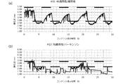
- FIG. 3 is a graph replaced with a drawing showing a pupil change and a pattern change in the example.
- FIG. 3A is a conceptual diagram showing a pupil change and a pattern display of a healthy person.
- FIG. 3A is a conceptual diagram showing a pupil change and a pattern display of a healthy person.
- FIG. 3B is a conceptual diagram showing pupil changes and pattern display of a patient suffering from Parkinson's disease.
- FIG. 4 is a conceptual diagram showing a control example of a display screen for acquiring the basic pupil value.
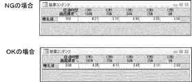
- FIG. 5 is an example showing a failure (NG) example (upper diagram) and a success (OK) example (lower diagram) when acquiring the basic pupil value.
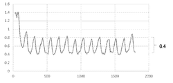
- FIG. 6 is a graph replaced with a drawing showing the change in the pupil diameter when black and white are displayed every one second and the change in the pupil diameter is measured.
- FIG. 7 is a graph replaced with a drawing showing changes in the pupil diameter when black and white are displayed every 3 seconds and changes in the pupil diameter are measured.
- FIG. 8 is a diagram showing measurement results of the difference between the maximum pupil diameter and the minimum pupil diameter.
- FIG. 9 is a graph replaced with a drawing showing how the pupils of a healthy person change.
- FIG. 10 is a graph replaced with a drawing showing a state of pupillary change in a patient suffering from a brain disease.
- FIG. 11 is a graph replaced with a drawing showing how the pupil changes.
- the brain disease diagnostic device is a device using a computer for determining one or more of whether the user is suffering from brain disease, the degree of brain disease is severe, and the risk of suffering is high.
- brain diseases are epilepsy, neurodegenerative disease, neural stem cell disease, neural progenitor cell disease, ischemic disease, neural trauma, affective disorder, neuropsychiatric disease, retinal degenerative disease, retinal damage/trauma, cognitive/learning/memory.
- disorders Alzheimer's disease, mild cognitive impairment (MCI), Parkinson's disease, Parkinson's syndrome, Huntington's disease, amyotrophic lateral sclerosis, ischemic stroke, traumatic brain injury, depression, bipolar depression/disorder, chronic fatigue syndrome, Anxiety syndrome/disorder, autism, or Asperger syndrome.
- FIG. 1-1 is a conceptual diagram for explaining the configuration of a brain disease diagnostic device. As shown in FIG. 1-1, this cerebral disease diagnostic apparatus 1 has a display image control unit 3, an imaging unit 5, and a determination unit 7.
- the display image control unit 3 is an element for changing the display image displayed on the display unit based on the first pattern. Examples of displays are monitors, screens, and projection walls.
- This device can be implemented by a computer that can exchange information with a camera or other imaging device.
- Shooting information obtained by a camera or the like may be input to the computer via a cable or the like.
- the shooting information may be wirelessly input to the computer.
- the computer includes an input/output unit, a control unit, a calculation unit, and a storage unit, and each element can exchange information by a bus or the like. Then, the computer stores the input information in the storage unit. Further, the control unit of the computer reads the control program stored in the storage unit and causes the arithmetic unit to perform various calculations using the input information or the information stored in the storage unit. Then, the calculation result is appropriately stored in the storage unit and is appropriately output via the input/output unit.
- the first pattern is, for example, one or more of brightness, color, and brightness that fluctuate based on a predetermined time pattern.
- an example of the switching frequency is 1 second or more and 10 seconds or less, and may be 2 seconds or more and 6 seconds or less, or 2 seconds or more and 5 seconds or less.
- the switching may be performed at regular intervals.
- the first screen for example, white screen
- the second screen for example, black screen
- This pattern may be such that three or more types of screens can be switched.
- FIG. 2 is a conceptual diagram showing an example of the first pattern.
- FIG. 2A shows that a white screen and a black screen are alternately displayed at regular intervals.
- FIG. 2B shows an example in which a white screen, a video screen, and a screen with a predetermined brightness are displayed in a fixed pattern.
- the photographing unit 5 is an element for photographing the pupil of the user who visually recognizes the display image.
- International Publication WO2017-057631 pamphlet Patent Document 1 describes a pupil diameter measuring instrument including a photographing unit such as a camera, so that the pupil diameter measuring instrument may be used.
- FIG. 1-2 is a conceptual diagram for explaining the configuration of a brain disease diagnostic device.
- the photographing unit 5 includes three photographing elements.
- the central camera (imaging element) 5a and the cameras (imaging elements) 5b and 5c at both ends thereof are included.
- one eyeball is tracked and captured by the camera 5b and the camera 5c by image recognition, and the distance between the camera 5b and the camera 5c and the angle to the eyeball are used to triangulate the camera 5b and the camera 5c.
- the distance from the middle camera 5a to the eyeball is calculated. Since the pupil diameter photographed by the camera of 5a varies depending on the distance, the pupil diameter photographed by 5a is corrected by the distance from 5a. As a result, an accurate pupil diameter can be obtained even if the face moves slightly.
- the pupil is the light-transmitting area of the central part of the eyeball.
- the size of the pupil changes under the influence of light.
- the pupil has a different color from the iris around the pupil.
- a plurality of eyeball images of a certain user are photographed, a region in which the size changes is analyzed, and the color (color width) of that portion is stored.
- the pupil color of the user can be stored.
- an image including the pupil of the user is photographed and the color is analyzed.
- there are continuous color areas. Of the areas, the area located at the center or matching the color of the pupil is the area of the pupil, and the pupil diameter can be measured.
- the determination unit 7 is an element for determining whether or not the user is suffering from a brain disease, using the pattern of the temporal change in the size of the user's pupil photographed by the photographing unit and the first pattern. ..
- the determination unit 7 determines the size of the user's pupil over time, for example, based on the algorithm described above. Then, the determination unit 7 analyzes the changed pupil for a change in the size of the user's pupil for a certain time. For example, it is determined whether the size of the user's pupil for a certain period of time has an increasing tendency, a decreasing tendency, or whether the amount of change has a certain width. In this way, the determination unit can analyze the change status of the size of the user's pupil.
- the determination unit 7 obtains the maximum value and the minimum value of the user's pupil for a certain time, and further obtains the difference ⁇ between them.
- This pupil may be either the dominant eye or the non-dominant eye, or both.
- a part (upper part) in a certain range for example, a range of 0.2 mm ⁇ or more and 0.5 mm ⁇
- a part (lower part) in a certain range for example, a range of 0.2 mm ⁇ or more and 0.5 mm ⁇
- the user may be determined to be a healthy person.
- the user's pupil portion is extracted from the image captured by the image capturing unit by image analysis.
- the relative sizes of the two left and right pupil portions included in the image are calculated and stored in the storage unit.
- the arithmetic unit reads out information on the size of each of the left and right pupils stored in the storage unit, and obtains the temporal changes thereof.
- the control unit controls the time for giving the maximum value and the minimum value for the pupil size, the time region for increasing the pupil size, the time region for decreasing the pupil size, and the fluctuation of the pupil size. It is possible to analyze the time domain where is a certain range. By doing so, it is possible to obtain the maximum value and the minimum value of the size of the pupil and the pattern of the temporal change of the size of the pupil.
- the correlation between the temporal change of the user's pupil and the first pattern is low, it may be determined that the degree of brain disease is high, and if the index such as the correlation coefficient is within a certain range. , It may be determined that there is a risk of contracting a brain disease.
- the temporal change pattern of the user's pupil size includes the temporal change pattern of the difference in pupil size between the dominant eye and the non-dominant eye.
- the eye with the larger average value of the size of the pupil corresponding to the situation where the bright screen is displayed after changing from the dark screen to the bright screen may be the dominant eye.
- the eyes may be closed with each eye closed and the open eye may be used to see the object, and the eyes in the ring may be the dominant eyes.
- the eyes that are not in the ring may be regarded as the non-dominant eyes.
- the pupil is smaller when viewing a bright screen. Therefore, it is possible to know whether the user is looking at a dark screen or a bright screen.
- time delay time delay: delay
- the size of the pupil corresponding to the situation where a bright screen is displayed changes to a situation where the screen itself is bright.
- the change in pupil diameter has not caught up.
- the computer may store various diseases and changes in the pupils corresponding to the diseases in the storage unit.
- the computer photographs changes in the pupils of a plurality of patients with respect to a plurality of diseases, machine-learns the changes over time of the diseases and changes of the pupils, and builds a database of various diseases and changes over time of the pupils. You can leave it.
- the determination unit 7 uses the image including the user's pupil photographed by the photographing unit 5 to determine the temporal change of the user's pupil, and performs patterning to determine whether the user has a brain disease or a brain disease. Diagnosis can be made as to whether the disease is severe or at high risk of contracting it.
- this device further includes an age storage unit 9 that stores the age information of the user.
- the display screen control unit 3 preferably controls a predetermined time pattern based on the age information of the user stored in the age storage unit. This is based on the experimental results that the older the user (subject) is, the more likely the reaction abnormality is to appear with a change in the number of screen seconds that is shorter. For example, by controlling the reciprocal of the user's age as a coefficient to multiply the display interval time, the control is performed so that the display switching time becomes shorter as the age of the user increases, or at least for users of a certain age or more. In addition to the normal display mode, it is preferable to prepare a short-time switching mode for switching the display in a short time.
- the computer reads information about the user's age from the age storage unit, reads the threshold from the storage unit, compares the age with the threshold, and if the user's age exceeds the threshold, sets the short-time switching mode or at least Control may be performed so that the short-time switching mode is also displayed.
- the device may further include an information processing unit 11 for performing various kinds of processing (for example, machine learning processing).
- This brain disease diagnostic device 1 The display image control unit 3 changes the display image displayed on the display unit based on the first pattern stored in advance. Then, the photographing unit 5 photographs the pupil of the user who visually recognizes the display image.
- the determination unit 7 receives the image including the user's pupil captured by the image capturing unit 5. Then, the determination unit 7 obtains a temporal change pattern of the size of the user's pupil photographed by the photographing unit, and uses the temporal change pattern and the first pattern, the user suffers from a brain disease. Determine whether or not. It is preferable that the determination unit 7 can also perform a diagnosis such as whether the brain disease is severe or the risk of contracting the brain disease is high.
- This device is preferably a brain disease diagnostic device that diagnoses the brain disease according to a machine-learned prediction algorithm.
- Data is entered into the system regarding pupil changes over time for individual brain disorders. Then, by performing machine learning on the input various data, it is possible to analyze the correlation between each disease and the temporal change pattern or the first pattern. In this way, a machine-learned prediction algorithm can be obtained. Then, by using the prediction algorithm that has been machine-learned, it becomes possible to diagnose each disease of the brain disease. By further repeating machine learning, it is possible to learn patterns of changes in pupil diameter of individual diseases and predict individual brain diseases.
- This specification describes a computer Means for changing the display image displayed on the display unit based on the first pattern;
- the program may further cause a computer to realize each function of the above-described brain disease diagnosis apparatus.
- This specification describes a computer Changing the display image displayed on the display unit based on the first pattern;
- the program may further cause a computer to realize each function of the above-described brain disease diagnosis apparatus.
- This specification may be a computer-readable information recording medium that stores the above program.
- Examples of the information recording medium are a CD-ROM, a DVD and a memory stick.
- FIG. 3 is a graph replaced with a drawing showing a pupil change and a pattern change in the example.
- FIG. 3A is a conceptual diagram showing a pupil change and a pattern display of a healthy person.
- FIG. 3B is a conceptual diagram showing pupil changes and pattern display of a patient suffering from Parkinson's disease. From FIG.
- the healthy person has a correlation with the change of the display pattern and the change of the pupil diameter over time (the pupil diameter changes in a cycle of about every 3 seconds).
- the vertical axis shows the pupil diameter and the horizontal axis shows the time.
- the pupil diameter decreased statistically with age (the older the normal pupil diameter, the smaller the swing range).
- the dominant eye was assumed to have a large pupil diameter.
- the non-dominant eye lags behind in order to see stereoscopically.
- the larger average pupil value when viewing a dark screen to a bright screen is the dominant eye.
- Brain disorders include stroke, cerebral hemorrhage (spontaneous bleeding or bleeding from head injuries), and, although less statistically, specific tumors or infections.
- Non-brain disorders that affect the sympathetic nervous system include tumors and injuries in the neck or upper chest. Horner's syndrome is a combination of three things: pupil constriction, eyelid sagging, and decreased sweating around the abnormal eye. Horner's syndrome, regardless of the cause, results from the disruption of the sympathetic nervous system that connects to the eye.
- FIG. 4 is a conceptual diagram showing a control example of a display screen for acquiring the basic pupil value.
- an initial display "Brightness changes, but please do not look away" is made, and screens of various colors are displayed for a predetermined period.
- the darkness ratio changes to 100%, 75%, 100%, 75%, 50%, 25%, 0%.
- the display time after the initial display is 7 seconds (initial display), 7 seconds, 19 seconds, 12 seconds, 12 seconds, 12 seconds and 12 seconds.
- FIG. 5 shows an example of failure (NG) when acquiring the basic pupil value (upper diagram) and an example of success (OK) (lower diagram).
- NG failure
- OK success
- the control unit stores the degree of darkness of each pattern and the measured value of the pupil diameter of the user. Then, the values of the pupil diameter are compared for each value of the darkness pattern, and if the value of the pupillary diameter is large when the value of the darkness is small (the brightness is high), it is considered to be abnormal. to decide. Then, the fact that there is an abnormality is output and detection is performed again.
- the values of the maximum pupil diameter and the minimum pupil diameter can be obtained using the case of 100% luminance and the case of 0% luminance.
- the obtained values of the maximum pupil diameter and the minimum pupil diameter are appropriately stored in the storage unit and can be used for the subsequent analysis.
- the basic pupil value may be obtained and analyzed, or the presence or absence of an affected brain disease may be analyzed using the pupil value obtained on the spot without obtaining the basic pupil value.
- FIG. 6 is a graph replaced with a drawing showing the change in the pupil diameter when black and white are displayed every one second and the change in the pupil diameter is measured. It can be seen from FIG. 6 that the change in the value of the pupil diameter changes stably and periodically in a healthy person.
- FIG. 7 is a graph replaced with a drawing showing changes in the pupil diameter when black and white are displayed every 3 seconds and changes in the pupil diameter are measured. From FIG. 7, it can be seen that the change in the value of the pupil diameter changes stably and periodically in a healthy person.
- FIG. 8 is a diagram showing measurement results of the difference between the maximum pupil diameter and the minimum pupil diameter. As shown in FIG. 8, it can be seen that the difference between the maximum pupil diameter and the minimum pupil diameter decreases as the age increases. From this, a plurality of differences between the maximum pupil diameter and the minimum pupil diameter of healthy persons of a certain age are obtained, stored in the storage unit, the stored differences are read, and statistical values such as the average value and the variance are obtained to obtain a certain age. The reference value of the difference in can be obtained. Further, it can be seen that the above-mentioned difference is smaller in patients suffering from brain-related diseases than in healthy people.
- the user can It may be possible to determine whether or not there is a risk of suffering from a brain-related disease. In this case, for example, when the actual measurement value is equal to or less than a predetermined ratio of the reference value, it may be determined that the user may have a brain-related disease.
- the numerical value related to the predetermined ratio is stored in the storage unit, and the measured value of the difference between the user, the reference value for the user's age, and the numerical value related to the predetermined ratio are read out, and the measured value of the difference between the user and the age
- the ratio of the reference values By determining the ratio of the reference values and performing an operation of comparing this ratio with a numerical value relating to a predetermined ratio, it is possible to judge whether the user may have a brain-related disease. Then, it may be possible to re-examine whether or not the patient is suffering from a brain-related disease by using the change in pupil diameter over time.
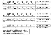
- FIG. 9 is a graph replaced with a drawing showing how the pupils of a healthy person change.
- FIG. 9 shows the pupillary change of the uppermost 46-year-old male (healthy person) in FIG.
- FIG. 10 is a graph replacing a drawing showing a change in pupil of a patient suffering from a brain disease.
- FIG. 10 shows the pupillary change of the second 76-year-old male (patient) in FIG.
- the pupil diameter changes relatively periodically from the start of measurement to the beginning (enlarged view at the bottom), but in the latter half of the measurement, the change of pupil diameter is not periodic. Therefore, it was found that the timing at which the periodicity collapses (the pattern of changes in pupil diameter over time) changes depending on the particular disease.
- pattern display is performed for 10 seconds or more (10 seconds or more and 10 minutes or less, 15 seconds or more and 5 minutes or less, or 15 seconds or more and 3 minutes or less). It was found that it is preferable to continue measuring the pupil diameter.
- FIG. 11 is a graph replaced with a drawing showing how the pupil changes.
- the upper part of FIG. 11 shows the pupillary change of the third 77-year-old male (patient) in FIG.
- the center of FIG. 11 shows the pupillary change of the fourth 82-year-old woman (healthy person) in FIG.
- the lower part of FIG. 11 shows the pupillary change of the fifth 84-year-old woman (patient) in FIG.
- the pupillary change of a healthy person is periodic and stable.
- the pupillary change in the patient reflects the pattern of the disease that is affected, and there are non-periodic parts.
- This invention can be used not only as a medical device but also as an application for mobile terminals.
Landscapes
- Health & Medical Sciences (AREA)
- Life Sciences & Earth Sciences (AREA)
- Engineering & Computer Science (AREA)
- Physics & Mathematics (AREA)
- Medical Informatics (AREA)
- General Health & Medical Sciences (AREA)
- Public Health (AREA)
- Biomedical Technology (AREA)
- Biophysics (AREA)
- Molecular Biology (AREA)
- Surgery (AREA)
- Animal Behavior & Ethology (AREA)
- Heart & Thoracic Surgery (AREA)
- Veterinary Medicine (AREA)
- Pathology (AREA)
- Neurology (AREA)
- Psychiatry (AREA)
- Physiology (AREA)
- Psychology (AREA)
- Theoretical Computer Science (AREA)
- Ophthalmology & Optometry (AREA)
- Child & Adolescent Psychology (AREA)
- Hospice & Palliative Care (AREA)
- Developmental Disabilities (AREA)
- Computer Vision & Pattern Recognition (AREA)
- General Physics & Mathematics (AREA)
- Neurosurgery (AREA)
- Educational Technology (AREA)
- Social Psychology (AREA)
- Artificial Intelligence (AREA)
- Signal Processing (AREA)
- Human Computer Interaction (AREA)
- Radiology & Medical Imaging (AREA)
- Primary Health Care (AREA)
- Epidemiology (AREA)
- Nuclear Medicine, Radiotherapy & Molecular Imaging (AREA)
- Mathematical Physics (AREA)
- Quality & Reliability (AREA)
- Data Mining & Analysis (AREA)
- Databases & Information Systems (AREA)
Priority Applications (6)
| Application Number | Priority Date | Filing Date | Title |
|---|---|---|---|
| EP19900976.2A EP3900638A4 (en) | 2018-12-17 | 2019-12-13 | BRAIN DISEASE DIAGNOSTIC DEVICE |
| CN201980083541.6A CN113194840A (zh) | 2018-12-17 | 2019-12-13 | 脑疾病的诊断装置 |
| US17/414,333 US12159076B2 (en) | 2018-12-17 | 2019-12-13 | Device for diagnosing brain disease |
| SG11202106430YA SG11202106430YA (en) | 2018-12-17 | 2019-12-13 | Device for diagnosing brain disease |
| JP2020561375A JP7396681B2 (ja) | 2018-12-17 | 2019-12-13 | 脳疾患の診断装置 |
| KR1020217019421A KR20210104738A (ko) | 2018-12-17 | 2019-12-13 | 뇌 질환 진단 장치 |
Applications Claiming Priority (2)
| Application Number | Priority Date | Filing Date | Title |
|---|---|---|---|
| JP2018-235105 | 2018-12-17 | ||
| JP2018235105 | 2018-12-17 |
Publications (1)
| Publication Number | Publication Date |
|---|---|
| WO2020129834A1 true WO2020129834A1 (ja) | 2020-06-25 |
Family
ID=71102127
Family Applications (1)
| Application Number | Title | Priority Date | Filing Date |
|---|---|---|---|
| PCT/JP2019/048894 Ceased WO2020129834A1 (ja) | 2018-12-17 | 2019-12-13 | 脳疾患の診断装置 |
Country Status (7)
| Country | Link |
|---|---|
| US (1) | US12159076B2 (https=) |
| EP (1) | EP3900638A4 (https=) |
| JP (1) | JP7396681B2 (https=) |
| KR (1) | KR20210104738A (https=) |
| CN (1) | CN113194840A (https=) |
| SG (1) | SG11202106430YA (https=) |
| WO (1) | WO2020129834A1 (https=) |
Cited By (6)
| Publication number | Priority date | Publication date | Assignee | Title |
|---|---|---|---|---|
| JP2022061146A (ja) * | 2020-10-06 | 2022-04-18 | 国立研究開発法人国立精神・神経医療研究センター | 精神神経活動推定装置 |
| WO2022186586A1 (ko) * | 2021-03-02 | 2022-09-09 | 주식회사 메디셀 | 질환 진단 장치 및 질환 진단 방법 |
| KR20220124066A (ko) | 2021-03-02 | 2022-09-13 | 주식회사 메디셀 | 질환 진단 장치 및 질환 진단 방법 |
| WO2023214535A1 (ja) * | 2022-05-02 | 2023-11-09 | Kikura株式会社 | 左右の目で独立した表示画像を目視することによる脳疾患又は自立神経失調症の診断装置及びプログラム |
| JP2025009585A (ja) * | 2023-07-03 | 2025-01-20 | Kikura株式会社 | 光刺激による瞳孔径の変化を測定し、被験者の体調を判定する装置及びプログラム |
| JP7855171B2 (ja) | 2023-07-03 | 2026-05-08 | Kikura株式会社 | 光刺激による瞳孔径の変化を測定し、被験者の体調を判定する装置及びプログラム |
Families Citing this family (1)
| Publication number | Priority date | Publication date | Assignee | Title |
|---|---|---|---|---|
| KR20210104738A (ko) * | 2018-12-17 | 2021-08-25 | 가부시키가이샤 나스메 리서치 인스티투트 | 뇌 질환 진단 장치 |
Citations (7)
| Publication number | Priority date | Publication date | Assignee | Title |
|---|---|---|---|---|
| JP2002034920A (ja) * | 2000-05-15 | 2002-02-05 | Matsushita Electric Works Ltd | 脳機能検査方法及び脳機能検査装置 |
| JP2002282208A (ja) * | 2001-03-22 | 2002-10-02 | Munetaka Haida | 痴呆症診断装置 |
| JP2002541959A (ja) * | 1999-04-23 | 2002-12-10 | ニューロプティックス・インコーポレイテッド | 瞳孔不規則性検出・瞳孔追跡・瞳孔反応検出機能、緑内障検査機能、角膜解剖学的測定機能、頭蓋内圧検出機能、および、眼異常測定機能を備えた瞳孔計 |
| JP2003070753A (ja) * | 2001-09-06 | 2003-03-11 | Scalar Corp | 診断システム、診断データ生成方法、それに用いられる情報処理装置、及び端末装置、並びに記録媒体 |
| JP2016504089A (ja) * | 2012-12-11 | 2016-02-12 | クライン、アミKLIN, Ami | エンゲージメント及び知覚刺激顕著性のマーカーとして瞬き抑制を検出するためのシステム及び方法 |
| WO2017057631A1 (ja) | 2015-10-01 | 2017-04-06 | 株式会社夏目綜合研究所 | 明暗、呼吸及び脈拍の影響を排除する視認者情感判定装置、視認者情感判定システム及びプログラム |
| JP2017184996A (ja) * | 2016-04-05 | 2017-10-12 | 渡 倉島 | 瞳孔径拡大による脳活動量判定装置およびプログラム |
Family Cites Families (25)
| Publication number | Priority date | Publication date | Assignee | Title |
|---|---|---|---|---|
| US20090312817A1 (en) * | 2003-11-26 | 2009-12-17 | Wicab, Inc. | Systems and methods for altering brain and body functions and for treating conditions and diseases of the same |
| JP4883580B2 (ja) | 2007-06-05 | 2012-02-22 | 独立行政法人産業技術総合研究所 | 精神的疲労の検出方法、装置及びプログラム |
| US10626399B2 (en) * | 2010-01-28 | 2020-04-21 | The Board Of Trustees Of The Leland Stanford Junior University | Methods of treating cognitive symptoms of an aging-associated impairment by modulating C-C chemokine receptor type 3 (CCR3) |
| US10487148B2 (en) * | 2010-01-28 | 2019-11-26 | The Board Of Trustees Of The Leland Stanford Junior University | Methods and compositions for treating aging-associated impairments |
| JP5817582B2 (ja) * | 2012-02-22 | 2015-11-18 | 株式会社Jvcケンウッド | 脳機能疾患診断支援装置および脳機能疾患診断支援方法 |
| WO2015029970A1 (ja) * | 2013-08-28 | 2015-03-05 | オリンパスメディカルシステムズ株式会社 | カプセル型内視鏡システム |
| US10905779B2 (en) * | 2013-12-09 | 2021-02-02 | The Board Of Trustees Of The Leland Stanford Junior University | Methods for screening human blood products comprising plasma using immunocompromised rodent models |
| WO2015164807A1 (en) * | 2014-04-25 | 2015-10-29 | Texas State University | Detection of brain injury and subject state with eye movement biometrics |
| WO2016064688A1 (en) * | 2014-10-25 | 2016-04-28 | Sumner Bluffs, Llc. | Pharmaceutical and biological agent delivery system having biometric data acquisition and monitoring capabilities |
| SG11201707513QA (en) * | 2015-05-29 | 2017-10-30 | Univ Leland Stanford Junior | Nucleoside agents for the reduction of the deleterious activity of extended nucleotide repeat containing genes |
| AU2016279804B2 (en) * | 2015-06-15 | 2019-03-07 | The Board Of Trustees Of The Leland Stanford Junior University | Methods and compositions for treating aging-associated conditions |
| WO2017030177A1 (ja) * | 2015-08-20 | 2017-02-23 | 日本電気株式会社 | 展示装置、表示制御装置および展示システム |
| US20180190011A1 (en) * | 2017-01-04 | 2018-07-05 | Osterhout Group, Inc. | Content rendering systems for head-worn computers |
| US20180249941A1 (en) * | 2016-05-24 | 2018-09-06 | neuroFit, Inc. | Oculometric Neurological Examination (ONE) Appliance |
| JP7017002B2 (ja) * | 2016-06-16 | 2022-02-08 | ハダシット メディカル リサーチ サービシズ アンド ディベラップメント リミテッド | 眼瞼を閉じた対象者における瞳孔の大きさを決定するための装置および方法 |
| EA039316B1 (ru) * | 2016-10-24 | 2022-01-12 | Алкахест, Инк. | Фракции плазмы крови в качестве лечения когнитивных расстройств, связанных со старением |
| JP6311002B1 (ja) * | 2016-12-28 | 2018-04-11 | 株式会社ブレインウェイ | 視覚利用脳トレーニングシステム |
| US10614623B2 (en) * | 2017-03-21 | 2020-04-07 | Canfield Scientific, Incorporated | Methods and apparatuses for age appearance simulation |
| CN110636844A (zh) * | 2017-04-05 | 2019-12-31 | 万能溶剂有限公司 | 使用ccr3-抑制剂治疗衰老相关损伤的方法及组合物 |
| MX2019012795A (es) * | 2017-04-26 | 2020-02-13 | Alkahest Inc | Régimen de dosificación para el tratamiento de deterioros cognitivos y motores con plasma de sangre y productos de plasma de sangre. |
| SG10201703570YA (en) * | 2017-05-02 | 2018-12-28 | Singapore Health Serv Pte Ltd | Hand held ophthalmic and neurological screening device |
| DE102018103334A1 (de) * | 2018-02-14 | 2019-08-14 | Thomas Recording Gmbh | Verfahren zur nicht-invasiven videookulographischen Messung von Augenbewegungen als Diagnoseunterstützung für eine (Früh-) Erkennung von neuropsychiatrischen Erkrankungen |
| KR20190108727A (ko) * | 2018-03-15 | 2019-09-25 | 민상규 | 접이식 가상현실 장비 |
| JP7247319B2 (ja) * | 2018-04-14 | 2023-03-28 | ワイ. マイケル リー | 頭部、脊椎および身体の健康をモニタリングするためのシステムおよび方法 |
| KR20210104738A (ko) * | 2018-12-17 | 2021-08-25 | 가부시키가이샤 나스메 리서치 인스티투트 | 뇌 질환 진단 장치 |
-
2019
- 2019-12-13 KR KR1020217019421A patent/KR20210104738A/ko not_active Ceased
- 2019-12-13 JP JP2020561375A patent/JP7396681B2/ja active Active
- 2019-12-13 EP EP19900976.2A patent/EP3900638A4/en not_active Withdrawn
- 2019-12-13 WO PCT/JP2019/048894 patent/WO2020129834A1/ja not_active Ceased
- 2019-12-13 US US17/414,333 patent/US12159076B2/en active Active
- 2019-12-13 CN CN201980083541.6A patent/CN113194840A/zh active Pending
- 2019-12-13 SG SG11202106430YA patent/SG11202106430YA/en unknown
Patent Citations (7)
| Publication number | Priority date | Publication date | Assignee | Title |
|---|---|---|---|---|
| JP2002541959A (ja) * | 1999-04-23 | 2002-12-10 | ニューロプティックス・インコーポレイテッド | 瞳孔不規則性検出・瞳孔追跡・瞳孔反応検出機能、緑内障検査機能、角膜解剖学的測定機能、頭蓋内圧検出機能、および、眼異常測定機能を備えた瞳孔計 |
| JP2002034920A (ja) * | 2000-05-15 | 2002-02-05 | Matsushita Electric Works Ltd | 脳機能検査方法及び脳機能検査装置 |
| JP2002282208A (ja) * | 2001-03-22 | 2002-10-02 | Munetaka Haida | 痴呆症診断装置 |
| JP2003070753A (ja) * | 2001-09-06 | 2003-03-11 | Scalar Corp | 診断システム、診断データ生成方法、それに用いられる情報処理装置、及び端末装置、並びに記録媒体 |
| JP2016504089A (ja) * | 2012-12-11 | 2016-02-12 | クライン、アミKLIN, Ami | エンゲージメント及び知覚刺激顕著性のマーカーとして瞬き抑制を検出するためのシステム及び方法 |
| WO2017057631A1 (ja) | 2015-10-01 | 2017-04-06 | 株式会社夏目綜合研究所 | 明暗、呼吸及び脈拍の影響を排除する視認者情感判定装置、視認者情感判定システム及びプログラム |
| JP2017184996A (ja) * | 2016-04-05 | 2017-10-12 | 渡 倉島 | 瞳孔径拡大による脳活動量判定装置およびプログラム |
Cited By (8)
| Publication number | Priority date | Publication date | Assignee | Title |
|---|---|---|---|---|
| JP2022061146A (ja) * | 2020-10-06 | 2022-04-18 | 国立研究開発法人国立精神・神経医療研究センター | 精神神経活動推定装置 |
| JP7545682B2 (ja) | 2020-10-06 | 2024-09-05 | 国立研究開発法人国立精神・神経医療研究センター | 精神神経活動推定装置 |
| WO2022186586A1 (ko) * | 2021-03-02 | 2022-09-09 | 주식회사 메디셀 | 질환 진단 장치 및 질환 진단 방법 |
| KR20220124066A (ko) | 2021-03-02 | 2022-09-13 | 주식회사 메디셀 | 질환 진단 장치 및 질환 진단 방법 |
| KR102547769B1 (ko) * | 2021-03-02 | 2023-06-27 | 주식회사 메디셀 | 질환 진단 장치 |
| WO2023214535A1 (ja) * | 2022-05-02 | 2023-11-09 | Kikura株式会社 | 左右の目で独立した表示画像を目視することによる脳疾患又は自立神経失調症の診断装置及びプログラム |
| JP2025009585A (ja) * | 2023-07-03 | 2025-01-20 | Kikura株式会社 | 光刺激による瞳孔径の変化を測定し、被験者の体調を判定する装置及びプログラム |
| JP7855171B2 (ja) | 2023-07-03 | 2026-05-08 | Kikura株式会社 | 光刺激による瞳孔径の変化を測定し、被験者の体調を判定する装置及びプログラム |
Also Published As
| Publication number | Publication date |
|---|---|
| JPWO2020129834A1 (https=) | 2020-06-25 |
| EP3900638A1 (en) | 2021-10-27 |
| EP3900638A4 (en) | 2022-08-17 |
| CN113194840A (zh) | 2021-07-30 |
| US12159076B2 (en) | 2024-12-03 |
| KR20210104738A (ko) | 2021-08-25 |
| SG11202106430YA (en) | 2021-07-29 |
| US20220054076A1 (en) | 2022-02-24 |
| JP7396681B2 (ja) | 2023-12-12 |
Similar Documents
| Publication | Publication Date | Title |
|---|---|---|
| WO2020129834A1 (ja) | 脳疾患の診断装置 | |
| JP7441915B2 (ja) | ライトフィールドプロセッサシステム | |
| US11612316B2 (en) | Medical system and method operable to control sensor-based wearable devices for examining eyes | |
| US20240188879A1 (en) | System and method for detecting neurological disease | |
| US11583178B2 (en) | Systems and methods for evaluating contrast sensitivity and other visual metrics | |
| Korn et al. | A solid frame for the window on cognition: Modeling event-related pupil responses | |
| US9216133B2 (en) | Using a 3D display to train a weak eye | |
| CN104219992A (zh) | 自闭症诊断辅助方法和系统以及自闭症诊断辅助装置 | |
| JPWO2020129834A5 (https=) | ||
| JP2024525812A (ja) | 被検者の視力を決定するためのコンピュータープログラム、方法、及び装置 | |
| US12383178B2 (en) | Systems and methods for using eye imaging on a wearable device to assess human health | |
| US12133567B2 (en) | Systems and methods for using eye imaging on face protection equipment to assess human health | |
| US11185224B2 (en) | Ocular monitoring headset | |
| US12213732B2 (en) | Assessing visual function | |
| KR102250999B1 (ko) | 시야장애 평가 및 안구 운동장애 평가 결과에 기반한 뇌병변 진단 장치 | |
| JP2024521567A (ja) | 脳障害を検出するための方法及び装置 | |
| CN111753628B (zh) | 训练眼睛跟踪模型 | |
| WO2024095261A1 (en) | System and method for diagnosis and treatment of various movement disorders and diseases of the eye | |
| HK40048473A (en) | Device for diagnosing brain disease | |
| JP7565566B2 (ja) | 身体状態の推定装置、身体状態の推定装置の作動方法、プログラム、および記録媒体 | |
| WO2023214535A1 (ja) | 左右の目で独立した表示画像を目視することによる脳疾患又は自立神経失調症の診断装置及びプログラム | |
| US20200390577A1 (en) | Method and system for detecting voluntary binary responses by analyzing the pupil diameter of a subject | |
| WO2025051943A1 (en) | Device and method for obtaining dynamic measurements of eye optical surfaces | |
| JP2023071507A (ja) | 判定装置、作業システムおよび判定方法 | |
| CN120501385A (zh) | 一种基于眼动特征的认知风险评估系统、方法、装置及存储介质 |
Legal Events
| Date | Code | Title | Description |
|---|---|---|---|
| 121 | Ep: the epo has been informed by wipo that ep was designated in this application |
Ref document number: 19900976 Country of ref document: EP Kind code of ref document: A1 |
|
| ENP | Entry into the national phase |
Ref document number: 2020561375 Country of ref document: JP Kind code of ref document: A |
|
| NENP | Non-entry into the national phase |
Ref country code: DE |
|
| ENP | Entry into the national phase |
Ref document number: 20217019421 Country of ref document: KR Kind code of ref document: A |
|
| ENP | Entry into the national phase |
Ref document number: 2019900976 Country of ref document: EP Effective date: 20210719 |