WO2023214535A1 - 左右の目で独立した表示画像を目視することによる脳疾患又は自立神経失調症の診断装置及びプログラム - Google Patents
左右の目で独立した表示画像を目視することによる脳疾患又は自立神経失調症の診断装置及びプログラム Download PDFInfo
- Publication number
- WO2023214535A1 WO2023214535A1 PCT/JP2023/016668 JP2023016668W WO2023214535A1 WO 2023214535 A1 WO2023214535 A1 WO 2023214535A1 JP 2023016668 W JP2023016668 W JP 2023016668W WO 2023214535 A1 WO2023214535 A1 WO 2023214535A1
- Authority
- WO
- WIPO (PCT)
- Prior art keywords
- user
- image
- autonomic nervous
- display
- brain
- Prior art date
Links
- 208000014644 Brain disease Diseases 0.000 title claims abstract description 49
- 206010003840 Autonomic nervous system imbalance Diseases 0.000 title claims abstract description 14
- 210000001508 eye Anatomy 0.000 title claims description 18
- 238000003745 diagnosis Methods 0.000 title abstract description 7
- 208000019479 dysautonomia Diseases 0.000 title abstract description 7
- 208000018290 primary dysautonomia Diseases 0.000 title abstract description 7
- 230000000007 visual effect Effects 0.000 title description 5
- 238000011156 evaluation Methods 0.000 title 1
- 210000001747 pupil Anatomy 0.000 claims abstract description 68
- 208000037265 diseases, disorders, signs and symptoms Diseases 0.000 claims abstract description 47
- 210000005252 bulbus oculi Anatomy 0.000 claims abstract description 39
- 208000035475 disorder Diseases 0.000 claims abstract description 29
- 230000036541 health Effects 0.000 claims abstract description 4
- 230000002567 autonomic effect Effects 0.000 claims description 28
- 201000010099 disease Diseases 0.000 claims description 18
- 238000003384 imaging method Methods 0.000 claims description 11
- 208000010877 cognitive disease Diseases 0.000 claims description 7
- 208000020925 Bipolar disease Diseases 0.000 claims description 6
- 208000014674 injury Diseases 0.000 claims description 6
- 208000027061 mild cognitive impairment Diseases 0.000 claims description 6
- 208000015122 neurodegenerative disease Diseases 0.000 claims description 6
- 230000008733 trauma Effects 0.000 claims description 6
- 208000018737 Parkinson disease Diseases 0.000 claims description 4
- 208000024827 Alzheimer disease Diseases 0.000 claims description 3
- 208000019901 Anxiety disease Diseases 0.000 claims description 3
- 208000036640 Asperger disease Diseases 0.000 claims description 3
- 201000006062 Asperger syndrome Diseases 0.000 claims description 3
- 206010003805 Autism Diseases 0.000 claims description 3
- 208000020706 Autistic disease Diseases 0.000 claims description 3
- 206010008874 Chronic Fatigue Syndrome Diseases 0.000 claims description 3
- 208000023105 Huntington disease Diseases 0.000 claims description 3
- 208000032382 Ischaemic stroke Diseases 0.000 claims description 3
- 208000019022 Mood disease Diseases 0.000 claims description 3
- 206010034010 Parkinsonism Diseases 0.000 claims description 3
- 206010057430 Retinal injury Diseases 0.000 claims description 3
- 208000030886 Traumatic Brain injury Diseases 0.000 claims description 3
- 206010002026 amyotrophic lateral sclerosis Diseases 0.000 claims description 3
- 230000036506 anxiety Effects 0.000 claims description 3
- 208000028683 bipolar I disease Diseases 0.000 claims description 3
- 208000025307 bipolar depression Diseases 0.000 claims description 3
- 206010015037 epilepsy Diseases 0.000 claims description 3
- 208000023589 ischemic disease Diseases 0.000 claims description 3
- 230000013016 learning Effects 0.000 claims description 3
- 208000029766 myalgic encephalomeyelitis/chronic fatigue syndrome Diseases 0.000 claims description 3
- 210000005155 neural progenitor cell Anatomy 0.000 claims description 3
- 210000001178 neural stem cell Anatomy 0.000 claims description 3
- 230000004770 neurodegeneration Effects 0.000 claims description 3
- 230000000926 neurological effect Effects 0.000 claims description 3
- 230000002207 retinal effect Effects 0.000 claims description 3
- 208000011580 syndromic disease Diseases 0.000 claims description 3
- 230000009529 traumatic brain injury Effects 0.000 claims description 3
- 206010003591 Ataxia Diseases 0.000 claims description 2
- 238000010276 construction Methods 0.000 claims description 2
- 208000020358 Learning disease Diseases 0.000 claims 1
- 208000026139 Memory disease Diseases 0.000 claims 1
- 230000001149 cognitive effect Effects 0.000 claims 1
- 238000001514 detection method Methods 0.000 claims 1
- 201000003723 learning disability Diseases 0.000 claims 1
- 238000005259 measurement Methods 0.000 abstract description 5
- 210000004556 brain Anatomy 0.000 abstract description 3
- 238000000034 method Methods 0.000 abstract description 3
- 230000002490 cerebral effect Effects 0.000 abstract 1
- 238000004364 calculation method Methods 0.000 description 7
- 238000010586 diagram Methods 0.000 description 6
- 230000006870 function Effects 0.000 description 3
- 230000015654 memory Effects 0.000 description 3
- 208000012219 Autonomic Nervous System disease Diseases 0.000 description 2
- 210000000467 autonomic pathway Anatomy 0.000 description 2
- 230000019771 cognition Effects 0.000 description 2
- 210000000695 crystalline len Anatomy 0.000 description 2
- 230000000694 effects Effects 0.000 description 2
- 230000008451 emotion Effects 0.000 description 2
- 238000010801 machine learning Methods 0.000 description 2
- 210000003205 muscle Anatomy 0.000 description 2
- 230000011514 reflex Effects 0.000 description 2
- 208000003164 Diplopia Diseases 0.000 description 1
- 206010027646 Miosis Diseases 0.000 description 1
- 230000004308 accommodation Effects 0.000 description 1
- 230000002350 accommodative effect Effects 0.000 description 1
- 238000009825 accumulation Methods 0.000 description 1
- 238000013459 approach Methods 0.000 description 1
- 210000003403 autonomic nervous system Anatomy 0.000 description 1
- 230000006399 behavior Effects 0.000 description 1
- 230000007177 brain activity Effects 0.000 description 1
- 230000001886 ciliary effect Effects 0.000 description 1
- 230000004448 convergence reflex Effects 0.000 description 1
- 210000004087 cornea Anatomy 0.000 description 1
- 230000003247 decreasing effect Effects 0.000 description 1
- 208000029444 double vision Diseases 0.000 description 1
- 238000002474 experimental method Methods 0.000 description 1
- 210000003128 head Anatomy 0.000 description 1
- 230000003547 miosis Effects 0.000 description 1
- 238000012986 modification Methods 0.000 description 1
- 230000004048 modification Effects 0.000 description 1
- 238000001028 reflection method Methods 0.000 description 1
- 230000029058 respiratory gaseous exchange Effects 0.000 description 1
- 210000001525 retina Anatomy 0.000 description 1
- 230000004256 retinal image Effects 0.000 description 1
Images


Classifications
-
- A—HUMAN NECESSITIES
- A61—MEDICAL OR VETERINARY SCIENCE; HYGIENE
- A61B—DIAGNOSIS; SURGERY; IDENTIFICATION
- A61B10/00—Other methods or instruments for diagnosis, e.g. instruments for taking a cell sample, for biopsy, for vaccination diagnosis; Sex determination; Ovulation-period determination; Throat striking implements
-
- A—HUMAN NECESSITIES
- A61—MEDICAL OR VETERINARY SCIENCE; HYGIENE
- A61B—DIAGNOSIS; SURGERY; IDENTIFICATION
- A61B3/00—Apparatus for testing the eyes; Instruments for examining the eyes
- A61B3/10—Objective types, i.e. instruments for examining the eyes independent of the patients' perceptions or reactions
- A61B3/11—Objective types, i.e. instruments for examining the eyes independent of the patients' perceptions or reactions for measuring interpupillary distance or diameter of pupils
-
- A—HUMAN NECESSITIES
- A61—MEDICAL OR VETERINARY SCIENCE; HYGIENE
- A61B—DIAGNOSIS; SURGERY; IDENTIFICATION
- A61B3/00—Apparatus for testing the eyes; Instruments for examining the eyes
- A61B3/10—Objective types, i.e. instruments for examining the eyes independent of the patients' perceptions or reactions
- A61B3/113—Objective types, i.e. instruments for examining the eyes independent of the patients' perceptions or reactions for determining or recording eye movement
Definitions
- This invention relates to a device for diagnosing a brain disease, autonomic imbalance, by analyzing eyeball movements using the principle that brain activity and autonomic nerve movements are reflected in changes in the eyeballs (pupil and viewpoint).
- this invention uses changes in the user's left and right pupils and the viewpoint within the displayed image when the same or different displayed images are shown to the user's left and right eyes, and the user is diagnosed with a brain disease.
- a diagnostic device for diagnosing whether or not someone is suffering from autonomic nervous system imbalance.
- the pamphlet of International Publication WO2017-057631 describes a user emotion determination device.
- This invention is a device that can simultaneously capture left and right eyeball images with separate cameras, and measures changes in the diameter of a user's pupils or movement of the viewpoint in the displayed images when viewing the same or different left and right displayed images at the same time. By doing so, we aim to provide a new method for diagnosing a user's brain disease, autonomic nervous disorder.
- the present invention basically focuses on changes in the pupils of a user who has a brain disease or poor physical condition due to autonomic nervous disorder and changes in the viewpoint within the displayed image when the left and right independent display images are changed. This is based on the knowledge that the appearance is different from that of healthy people.
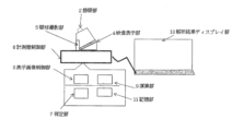
- This brain disease and autonomic nerve imbalance diagnostic device 1 includes a display image control section 3, an eyeball imaging section 5, and a determination section 7.
- the display image control section 3 is an element for changing the display image displayed on the display section.
- the eyeball photographing unit 5 is an element for photographing an eyeball image of a user viewing a displayed image.
- the determining unit 7 uses the size of the user's pupils photographed by the eyeball photographing unit 5 or the pattern of changes over time in the movement of the viewpoint in the displayed image to determine whether the user is suffering from a brain disease or not. This is an element for determining poor physical condition due to ataxia.
- the display image control section 3 changes the display image displayed on the display section based on a pre-stored pattern. Then, the eyeball photographing unit 5 simultaneously photographs left and right eyeball images of the user viewing the displayed image using separate cameras.
- the determining unit 7 receives an image including the user's eyeball image photographed by the eyeball photographing unit 5. Then, the determination unit 7 determines the pattern of changes over time in the size of the user's pupils photographed by the eyeball imaging unit and the movement of the viewpoint, and uses the patterns of changes over time to determine whether the user is suffering from brain disease or autonomic nervous disorder. Determine whether or not you are infected.
- the user is a healthy person.
- the perspective change in the 3D image or the movement of the viewpoint in the maze image is accurate and the time required to reach the destination is close to the average for a healthy person, it can be determined that the patient is not suffering from a brain disease or poor physical condition due to autonomic nervous disorder. .
- the display pattern is one in which, for example, one or more of the brightness, color, and brightness of the display for the left and right eyes, the perspective movement of the stereoscopic display image, and the maze image vary.
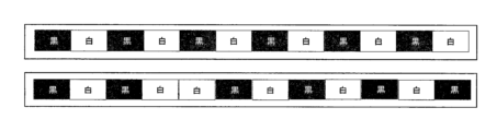
- One typical example of a display pattern is a periodically changing white screen and black screen.
- brain diseases are epilepsy, neurodegenerative diseases, neural stem cell diseases, neural progenitor cell diseases, ischemic diseases, neurological trauma, affective disorders, neuropsychiatric diseases, retinal degenerative diseases, retinal damage/trauma, and cognition, learning, and memory. disability, Alzheimer's disease, mild cognitive impairment (MCI), Parkinson's disease, Parkinson's syndrome, Huntington's disease, amyotrophic lateral sclerosis, ischemic stroke, traumatic brain injury, depression, bipolar depression/disorder, chronic fatigue syndrome, Anxiety syndrome/disorder, autism, or Asperger syndrome.
- An example of autonomic imbalance is poor physical condition.
- the user's brain can be improved by simply using a relatively simple device (for example, a device that can simultaneously capture left and right eyeball images with separate cameras, and a device that simultaneously displays the same or different left and right images). It is possible to provide a device that can diagnose whether a person is suffering from a disease, whether the brain disease is severe, whether there is a high risk of suffering from the disease, and the severity of autonomic nervous disorder.
- a relatively simple device for example, a device that can simultaneously capture left and right eyeball images with separate cameras, and a device that simultaneously displays the same or different left and right images.
- Measuring instrument image diagram Example of display screen
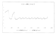
- Example of measuring left and right pupil diameters of a healthy person Representation of changes in left and right pupil diameters of healthy subjects
- Example of viewpoint trajectory in maze image (example of healthy person)
- Example of the trajectory of the viewpoint in a maze image (an example assuming a person with autonomic nervous disorder)
- Viewpoint trajectory diagram of the difference finding image spot the difference image Distribution of left and right attention (measurement example of healthy subjects)
- a brain disease diagnostic device is a device that uses a computer to determine one or more of whether a user is suffering from a brain disease, whether the degree of the brain disease is severe, or whether the user is at a high risk of developing the disease.
- brain diseases are epilepsy, neurodegenerative diseases, neural stem cell diseases, neural progenitor cell diseases, ischemic diseases, neurological trauma, affective disorders, neuropsychiatric diseases, retinal degenerative diseases, retinal damage/trauma, and cognition, learning, and memory. disability, Alzheimer's disease, mild cognitive impairment (MCI), Parkinson's disease, Parkinson's syndrome, Huntington's disease, amyotrophic lateral sclerosis, ischemic stroke, traumatic brain injury, depression, bipolar depression/disorder, chronic fatigue syndrome, Anxiety syndrome/disorder, autism, or Asperger syndrome.
- MCI mild cognitive impairment
- Parkinson's disease Parkinson's syndrome
- Huntington's disease amyotrophic lateral sclerosis
- ischemic stroke traumatic brain injury, depression, bipolar depression/disorder, chronic fatigue syndrome, Anxiety syndrome/disorder, autism, or Asperger syndrome.
- dysautonomia diagnosis examples include transportation operators such as bus drivers, taxi drivers, large and long-distance truck drivers, train drivers, and aircraft pilots, as well as construction site managers and field workers. This is a diagnosis of poor physical condition for people working in jobs such as machine workers in factories where incorrect behavior is always unacceptable due to occupational safety and health concerns.
- This brain disease/autonomic imbalance diagnosis device 1 includes a display image control section 3, an eyeball imaging section 5, and a determination section 7.
- the display image control section 3 is an element for changing the display image displayed on the display section based on the display pattern.
- An example of a display section is a monitor screen.
- This device can be implemented using a computer that is capable of exchanging information with a photographing device such as a camera.
- Information taken by a camera or the like may be input to the computer through a cable or the like. Further, the photographing information may be input to the computer wirelessly.
- a computer includes an input/output section, a control section, an arithmetic section, and a storage section, and each element is configured to be able to exchange information via a bus or the like. Then, the computer stores the input information in the storage section.
- the control section of the computer reads out the control program stored in the storage section, and causes the calculation section to perform various calculations using the input information and the information stored in the storage section. Then, the calculation results are stored in the storage section as appropriate, and are output as appropriate via the input/output section. The calculation results may be displayed on the 10 analysis result display section.
- the display pattern is one in which one or more of, for example, brightness, color, brightness, perspective change of a stereoscopic image, a maze image, and a spot-the-difference image vary based on a predetermined time pattern.
- the frequency of switching may be from 1 second to 10 seconds, or from 2 seconds to 6 seconds, or from 2 seconds to 5 seconds. Switching may be done at regular intervals. Further, the first screen (for example, a white screen) and the second screen (for example, a black screen) may be switched every different number of seconds.
- This pattern may be one in which three or more types of screens can be switched, for example.
- examples of other patterns include a perspective change image using a stereoscopic image that utilizes the feature of independent viewing by the left and right eyes, a maze image, and a difference finding image.
- FIG. 2 is a conceptual diagram showing an example of a display pattern.
- a white screen and a black screen are displayed alternately for the left and right eyes at regular intervals.
- the eyeball photographing unit 5 is an element for simultaneously photographing left and right eyeball images of the user viewing the displayed image using separate cameras.
- the pupil is the area in the center of the eye that allows light to pass through.
- the size of the pupil changes under the influence of light and other factors.
- the pupil has a different color from the surrounding area.
- multiple eyeball images of a certain user are taken, the area where the size changes is analyzed, and the color (color width) of that area is stored.
- the user's pupil color can be memorized.
- a display image including the user's pupils is photographed and the color tone is analyzed.
- the viewpoint calculation method is based on a known technique.
- the eyeball photographing unit 5 has a near-infrared LED light source, and the direction of the line of sight is calculated by a corneal reflection method, etc., which calculates the line of sight angle from the positional relationship between the reflection point of this LED on the cornea and the pupil, and the viewpoint in the displayed image is determined. There is a way to calculate it.
- the determination unit 7 determines whether or not the user is suffering from a brain disease or autonomic nervous disorder, using the pattern of changes over time in the size of the user's pupils photographed by the eyeball imaging unit and the display pattern.
- the position of the viewpoints of both eyes due to changes in perspective of stereoscopic images, changes in pupil diameter, the speed and accuracy of viewpoint movement in maze images, and the degree to which the attention distribution of spot-the-difference images differs from those of healthy people. This is an element for determining whether or not the user is suffering from a brain disease or autonomic nervous disorder.
- the determination unit 7 determines the sizes of the left and right pupils of the user and the movement of the viewpoint over time, for example, based on the above-mentioned algorithm. Then, the determination unit 7 analyzes changes in the size of the user's left and right pupils and movement of the viewpoint over a certain period of time. For example, it is determined whether the sizes of the user's left and right pupils over a certain period of time are increasing or decreasing, or whether the amount of change can be said to be within a constant range. In addition, by determining whether the speed and accuracy of viewpoint movement deviate from those of a healthy person, the determination unit can analyze changes in the user's pupil size and the speed and accuracy of viewpoint movement. .
- the determining unit 7 determines the maximum value and minimum value of the relative value of the user's pupil size for a certain period of time, and further determines the difference ⁇ . Then, the part (upper part) that is within a certain range from the maximum value (for example, the range of 0.2 ⁇ or more and 0.5 ⁇ ), and the part that is within a certain range (for example, the range of 0.2 ⁇ or more and 0.5 ⁇ ) from the minimum value (the lower part). It is also possible to determine whether there is a correlation between the appearance frequency of either or both of the upper part and the lower part and the display pattern. For example, if the lower portion appears at the same frequency as the display pattern change frequency (or within a certain time interval), the user may be determined to be healthy.
- the user can be determined to be healthy if the position of the viewpoints of both eyes due to changes in perspective of the stereoscopic image, the change in pupil diameter, and the speed and accuracy of the movement of the viewpoints in the maze image are within the average range for a healthy person.
- the degree of brain disease is high, or if the index such as the correlation coefficient is within a certain range. If so, it may be determined that there is a risk of developing a brain disease or autonomic nervous system disorder.
- changes in the viewpoint of both eyes due to changes in perspective of stereoscopic images, changes in pupil diameter, speed and accuracy of viewpoint movement in maze images, and viewpoint movement and attention distribution in spot-the-difference images were compared to the average of healthy people. If there is a deviation, it may be determined that the user is at risk of suffering from a brain disease or autonomic nervous disorder.
- Calculation of the degree of attention to a visual object uses the principle that the diameter of the pupil expands when attention is paid to it.
- the pupil diameter also changes depending on the brightness (luminance) of the object to be viewed, it is necessary to understand in advance the relationship between the user-specific reference luminance and the basic pupil diameter. Therefore, using a luminance meter 8 that measures the luminance of the display image on the video display section 4, the user can display a plain black display image with the lowest luminance, a plain white display image with the highest luminance, and an intermediate display image between them.
- the user visually observes a gray plain display image with stepwise brightness, and creates a relationship table between the standard brightness gs and the user's basic pupil diameter Ps.
- Q the total attention level of a specific part of an image is the integral value of Q when the viewpoint is on the specific part.
- the computer may store various diseases and corresponding changes in pupils and viewpoints in the storage unit.
- the computer takes pictures of the pupils and changes in viewpoint of multiple patients for multiple diseases, performs machine learning on each disease, changes in pupils, and changes in viewpoint over time, and creates a database on various diseases and changes in pupils over time. You may also build the .
- the determining unit 7 uses the user's eyeball image photographed by the eyeball photographing unit 5 to obtain and pattern changes in the user's pupils and viewpoint over time, thereby determining whether the user is suffering from a brain disease.
- the display image control section 3 changes the display image displayed on the display section based on a display pattern stored in advance. Then, the eyeball imaging unit 5 obtains an eyeball image of the user viewing the displayed image.
- the determining unit 7 receives an eyeball image including the user's pupil and line of sight, which has been photographed by the eyeball photographing unit 5. Then, the determination unit 7 determines the pattern of change over time in the size of the user's pupils photographed by the eyeball imaging unit and the change in viewpoint in the displayed image, and uses the pattern of change over time and the display pattern to It is determined whether the user is suffering from a brain disease or autonomic nervous system imbalance. Preferably, the determining unit 7 is also capable of diagnosing whether the brain disease and autonomic nervous disorder are severe and whether the risk of contracting the disease is high.
- This specification describes a computer as means for changing the display image displayed on the display unit based on the display pattern; means for receiving a photographed image of the eyeball of a user who visually views the display image photographed by the eyeball photographing section; Using the received captured images, the eyeball imaging unit determines the pattern of changes over time in the size of the user's pupils photographed and the changes in the movement of the viewpoint in the displayed image.
- the present invention also provides a program that functions as a means for determining whether a user is suffering from a brain disease or autonomic nervous disorder using a pattern of changes over time and a display pattern. This program may further enable the computer to implement each function of the above-mentioned brain disease and autonomic nervous disorder diagnostic apparatus.
- This specification describes a computer as a step of changing a display image displayed on the display unit based on the display pattern; a step of receiving a photographed image of an eyeball image of a user viewing a display image, taken by an eyeball photographing unit; Using the received photographic image, the eyeball imaging department determines the pattern of change in pupil size over time and movement of the viewpoint of the user photographed, and displays the determined pattern of change in pupil size over time and movement of the viewpoint.
- a step of determining whether the user is suffering from a brain disease using The present invention also provides a program that causes the user to perform the step of determining autonomic nervous disorder in a user. This program may further enable the computer to realize each function of the above-mentioned brain disease and autonomic nervous disorder diagnostic apparatus.
- this program by installing this program on a mobile device such as a smartphone or a game console, it can provide a simple brain diagnosis and autonomic nervous system diagnosis tool using a smartphone or other device.
- This specification may be a computer-readable information recording medium that stores the above program.
- Examples of information recording media are CD-ROM, DVD, and memory stick.
- the computer's video display section was configured to display an image for the right eye in the upper row and for the left eye in the lower row every 3 seconds, as shown in Figure 2.
- a camera that can take images of the target's eyeballs.
- FIG. 3 is a measurement diagram showing changes in pupil diameter and pattern display of a healthy person. It can be seen that for healthy subjects, there is a correlation between changes in the display pattern and changes in pupil diameter over time (pupil diameter fluctuates approximately every 3 seconds).
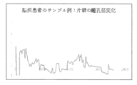
- FIG. 4 is a measurement diagram showing pupil changes and pattern display in the right eye of a patient suffering from Parkinson's disease. It can be seen that there is no correlation between changes in the display pattern and changes in pupil diameter over time, and that the pattern of change in pupil diameter is different from that of healthy individuals.
- Figure 5-1 is an example of the trajectory of the viewpoint in a maze image (an example of a healthy person) Even though his perspective is swaying considerably, he reaches the final goal by following an almost exact route.
- Figure 5-2 is an example of the trajectory of the viewpoint of a person with autonomic nervous system disorder in a maze image. haven't reached the goal.
- Figure 6 is a viewpoint trajectory diagram of a difference finding image (an example of a healthy person) The viewpoint moves to almost the same area on the left and right
- Figure 7-1 is the difference finding image.
- Figure 7-2 is the distribution of left and right attention of users who visually viewed Figure 7-1 (an example of a healthy person). In healthy subjects, the distribution of attention on the left and right sides is even.
- This invention can be used not only as a medical device but also as an application for a mobile terminal.
Landscapes
- Health & Medical Sciences (AREA)
- Life Sciences & Earth Sciences (AREA)
- Engineering & Computer Science (AREA)
- Heart & Thoracic Surgery (AREA)
- Molecular Biology (AREA)
- Veterinary Medicine (AREA)
- Public Health (AREA)
- Biomedical Technology (AREA)
- General Health & Medical Sciences (AREA)
- Medical Informatics (AREA)
- Animal Behavior & Ethology (AREA)
- Surgery (AREA)
- Physics & Mathematics (AREA)
- Ophthalmology & Optometry (AREA)
- Biophysics (AREA)
- Human Computer Interaction (AREA)
- Pathology (AREA)
- Eye Examination Apparatus (AREA)
Abstract
【課題】 脳疾患、自律神経失調症は簡単には判定できなかった。 【解決手段】脳活動が眼球の動き(目の瞳孔の変化、表示画像の中の視点の変化)に表れる原理を利用して、瞳孔の動き、視点の動きを解析して脳疾患、自律神経失調症の診断装置に関する。左右の眼球映像を別々のカメラで同時に撮影できる装置で、左右の同一または異なる表示画像を同時に見た時の眼球映像から瞳孔径の変化、表示画像の中の視点の動きを計測することにより、ユーザの脳の疾患、自律神経失調症の新たな診断手法と測定時の自律神経失調症による体調不良診断を提供することを目的とする。基本的には,脳疾患者または自律神経失調症者の瞳孔または視点の変化の様子は,健常人のものと異なるという知見に基づくものである。
Description
この発明は,脳活動と自律神経の動きが眼球(瞳孔と視点)の変化に表れる原理を利用して、眼球の動きを解析して脳疾患,自律神経失調症の診断装置に関する。より詳しく説明すると,この発明は,左右の目に同一または異なる表示画像をユーザに見せた際の,ユーザの左右の瞳孔と表示画像の中での視点の変化を用いて,そのユーザが脳疾患、自律神経失調症に罹っているか否かを診断する診断装置に関する。
国際公開WO2017-057631号パンフレットには,ユーザ感情判定装置が記載されている。上記の公報に記載された装置を用いれば,ユーザの呼吸,環境の明暗の影響,ユーザの脈拍といった影響を排除し,ユーザの瞳孔を撮影して,ユーザの感情を判定できる。
上記の装置のような,ユーザの瞳孔と視点を撮影できる装置を用いた新たな装置と診断システムの開発が望まれた。
この発明は,左右の眼球映像を別々のカメラで同時に撮影できる装置で、左右の同一または異なる表示画像を同時に見た時のユーザの瞳孔径の変化または表示画像の中の視点の動きを計測することにより、ユーザの脳の疾患、自律神経失調症の新たな診断手法を提供することを目的とする。
本発明は,基本的には,左右独立の表示画像を変化させた際のユーザの脳疾患または自律神経失調症による体調不良を有するユーザの瞳孔の変化、表示画像の中での視点の変化の様子は,健常人のものと異なるという知見に基づくものである。
この明細書に開示される複数の態様のうちのひとつは,脳疾患、自律神経失調症の診断装置に関する。
この脳疾患、自律神経失調の診断装置1は,表示画像制御部3と,眼球撮影部5と,判定部7とを有する。
表示画像制御部3は,表示部に表示される表示画像を変化させるための要素である。
眼球撮影部5は,表示画像を目視するユーザの眼球映像を撮影するための要素である。
判定部7は,眼球撮影部5が撮影したユーザの瞳孔の大きさまたは表示画像の中の視点の動きの経時変化のパターンを用いて,ユーザが脳疾患に罹患しているか否か、自律神経失調症による体調不良を判定するための要素である。
この脳疾患、自律神経失調症の診断装置1は,
表示画像制御部3が,あらかじめ記憶しておいたパターンに基づいて表示部に表示される表示画像を変化させる。
そして,眼球撮影部5は,表示画像を目視するユーザの左右の眼球映像を別々のカメラで同時に撮影する。
判定部7は,眼球撮影部5が撮影したユーザの眼球映像を含む像を受け取る。そして,判定部7は,眼球撮影部が撮影したユーザの瞳孔の大きさの経時変化のパターン、視点の動きを求め,その経時変化のパターンを用いて,ユーザが脳疾患、自律神経失調症に罹患しているか否か判定する。例えば,ユーザの両目の瞳孔の大きさの経時変化のパターンと,表示パターンとの間に相関がある場合に,ユーザは健常人であると判断するようにしてもよい。
また、立体画像の遠近変化、または迷路画像での視点の動きが正確で到達に必要な時間が健常者の平均に近ければ脳疾患でも自律神経失調症による体調不良でもないと判定することができる。
表示パターンは,例えば,左右の目に対する表示の輝度,色彩,明度、立体表示画像の遠近の動き、迷路画像のうちいずれか1つ又は2つ以上が,変動するものである。表示パターンの一つの代表的な具体例は,周期的に変化する白画面及び黒画面である。
脳疾患の例は,てんかん,神経変性疾患,神経幹細胞疾患,神経前駆細胞疾患,虚血性疾患,神経性外傷,情動障害,神経精神疾患,網膜変性疾患,網膜損傷/外傷,認知・学習・記憶障害,アルツハイマー病,軽度認知障害(MCI),パーキンソン病,パーキンソン症候群,ハンチントン病,筋萎縮性側索硬化症,虚血性脳卒中,外傷性脳損傷,鬱病,双極性鬱病/障害,慢性疲労症候群,不安症候群/障害,自閉症,又はアスペルガー症候群である。
自律神経失調症の例は体調不良である。
自律神経失調症の例は体調不良である。
この発明によれば,比較的簡便な装置(例えば、左右の眼球映像を別々のカメラで同時に撮影できる装置で、左右の同一または異なる表示画像を同時に表示する装置)を用いるだけで,ユーザが脳疾患に罹患しているか,脳疾患が重度か,罹患するリスクが高いか、自律神経失調症の重度の診断をすることができる装置を提供できる。
以下,図面を用いて本発明を実施するための形態について説明する。本発明は,以下に説明する形態に限定されるものではなく,以下の形態から当業者が自明な範囲で適宜修正したものも含む。
この明細書に開示される複数の態様のうちのひとつは,脳疾患の診断装置に関する。脳疾患の診断装置は,ユーザが脳疾患に罹患しているか,脳疾患の程度が重度か,罹患するリスクが高いかのいずれか1つ以上を判定するためのコンピュータを用いた装置である。
脳疾患の例は,てんかん,神経変性疾患,神経幹細胞疾患,神経前駆細胞疾患,虚血性疾患,神経性外傷,情動障害,神経精神疾患,網膜変性疾患,網膜損傷/外傷,認知・学習・記憶障害,アルツハイマー病,軽度認知障害(MCI),パーキンソン病,パーキンソン症候群,ハンチントン病,筋萎縮性側索硬化症,虚血性脳卒中,外傷性脳損傷,鬱病,双極性鬱病/障害,慢性疲労症候群,不安症候群/障害,自閉症,又はアスペルガー症候群である。
自律神経失調症診断の例は、バス運転者、タクシー運転者、大型及び長距離トラック運転者、列車の運転者、航空機パイロットなどの交通機関の運転者、さらに建設現場管理者および現場作業者、工場などの機械作業者など、労働安全衛生上、常に間違った行動が許されない仕事に就く時の体調不良診断である。
この脳疾患・自律神経失調症の診断装置1は,表示画像制御部3と,眼球撮影部5と,判定部7とを有する。
表示画像制御部3は,表示パターンに基づいて表示部に表示される表示画像を変化させるための要素である。表示部の例は,モニタ画面である。
表示画像制御部3は,表示パターンに基づいて表示部に表示される表示画像を変化させるための要素である。表示部の例は,モニタ画面である。
この装置は,カメラなどの撮影装置と情報のやり取りができるようにされたコンピュータにより実装できる。カメラなどによる撮影情報は,ケーブルなどを通してコンピュータに入力されてもよい。また,撮影情報は,無線により,コンピュータに入力されてもよい。コンピュータは,入出力部,制御部,演算部及び記憶部を含み,各要素はバスなどにより情報の授受を行うことができるようにされている。そして,コンピュータは,入力された情報を,記憶部に記憶する。また,コンピュータの制御部は,記憶部に記憶した制御プログラムを読み出し,入力された情報や,記憶部に記憶された情報を用いて,演算部に各種演算を行わせる。そして,演算結果は,適宜記憶部に記憶されるとともに,入出力部を介して,適宜出力される。
演算結果は10解析結果ディスプレイ部に表示できるようにしても良い。
演算結果は10解析結果ディスプレイ部に表示できるようにしても良い。
表示パターンは,例えば,輝度,色彩,明度、立体画像の遠近変化、迷路画像、間違い探し画像のうちいずれか1つ又は2つ以上が,所定の時間パターンに基づいて変動するものである。輝度パターンが2種類の画面(表示画像)を含む場合,切り替えの頻度の例は1秒以上10秒以下であり,2秒以上6秒以下でも,2秒以上5秒以下でもよい。切り替えは,一定の時間ごとでもよい。また,第1の画面(例えば白画面)と第2の画面(例えば黒画面)については,それぞれ異なる秒数ごとに切り替えを行ってもよい。このパターンは,例えば,3種類以上の画面が切り替えられるものであってもよい。
また、他のパターンの例としては、左右の目による独立目視の特徴を利用した立体画像による遠近変化の画像や迷路画像、間違い探し画像がある。
また、他のパターンの例としては、左右の目による独立目視の特徴を利用した立体画像による遠近変化の画像や迷路画像、間違い探し画像がある。
図2は,表示パターンの例を示す概念図である。この例では,一定の時間ごとに,左右の目に表示する白画面及び黒画面が交互に表示される。
眼球撮影部5は,表示画像を目視するユーザの左右の眼球映像を別々のカメラで同時に撮影するための要素である。
瞳孔は,眼球の中央部分のうち光を通す領域である。瞳孔は,光等の影響を受け大きさが変化する。一方,瞳孔は,瞳孔の周囲部分とは色が異なる。このため,あるユーザの複数の眼球像を撮影し,大きさが変化する領域を分析し,その部分の色(色の幅)を記憶する。このようにすることで,そのユーザの瞳孔色を記憶できる。そして,そのユーザの瞳孔を含む表示画像を撮影し,色味を分析する。すると,連続して色が変化する領域が存在する。その領域のうち,およそ中央に位置する領域であるか,先の瞳孔の色と一致する領域が,瞳孔の領域である。ユーザの頭部の変動が少なければ,ユーザの瞳孔の相対的な大きさを求めることができる。つまり,この装置では,ユーザの瞳孔の大きさの経時変化のパターンを求めることができればよいので瞳孔径の絶対的な大きさを求める必要がない。
視点算出方法は既知の技術による。例えば眼球撮影部5に近赤外LED光源があり、このLEDの角膜での反射点と瞳孔の位置関係から視線角度を算出する角膜反射法等により視線方向を算出し、表示画像内の視点を算出する方法がある。
視点算出方法は既知の技術による。例えば眼球撮影部5に近赤外LED光源があり、このLEDの角膜での反射点と瞳孔の位置関係から視線角度を算出する角膜反射法等により視線方向を算出し、表示画像内の視点を算出する方法がある。
判定部7は,眼球撮影部が撮影したユーザの瞳孔の大きさの経時変化のパターンと,表示パターンとを用いて,ユーザが脳疾患、自律神経失調症に罹患しているか否か判定するための要素である
また、立体画像の遠近変化による両眼の視点の位置、瞳孔径の変化、迷路画像での視点の動きの速さと正確度、間違い探し画像の注目度分布が健常者と異なる度合によりユーザが脳疾患、自律神経失調症に罹患しているか否かを判定する要素である。
また、立体画像の遠近変化による両眼の視点の位置、瞳孔径の変化、迷路画像での視点の動きの速さと正確度、間違い探し画像の注目度分布が健常者と異なる度合によりユーザが脳疾患、自律神経失調症に罹患しているか否かを判定する要素である。
判定部7は,例えば,上記したアルゴリズムに基づいて,ユーザの左右の瞳孔の大きさ、視点の動きを経時的に求める。そして,判定部7は,ある時間分のユーザの左右の瞳孔の大きさの変化、視点の動きについて,分析する。例えば,ある時間分のユーザの左右の瞳孔の大きさについて,増加傾向にあるか,減少傾向にあるか,変化量が一定幅といえるかを求める。また、視点の動きの速さと正確度が健常者と乖離しているかどうかを判定して、判定部は,ユーザの瞳孔の大きさの変化、視点の動きの速さと正確度の状況を分析できる。
また,判定部7は,ある時間分のユーザの瞳孔の大きさの相対値について,最大値と最小値とを求め,さらにその差分Δを求める。そして,最大値から一定の範囲(例えば0.2Δ以上0.5Δの範囲)の部分(上部分)と,最小値から一定の範囲(例えば0.2Δ以上0.5Δの範囲)の部分(下部分)のいずれか又は両方を求め,上部分と下部分のいずれか又は両方の出現頻度と,表示パターンとに相関があるか検討してもよい。例えば,下部分が,表示パターンの変化頻度と同じ頻度(又は一定時間以内のずれ)で出現すれば,そのユーザは健常と判断してもよい。
立体画像の遠近変化による両眼の視点の位置、瞳孔径の変化、迷路画像での視点の動きの速さと正確さが健常者の平均範囲であればユーザは健常と判断できる。
立体画像の遠近変化による両眼の視点の位置、瞳孔径の変化、迷路画像での視点の動きの速さと正確さが健常者の平均範囲であればユーザは健常と判断できる。
また,上記のユーザの左右どちらかの瞳孔の経時変化と,表示パターンとの相関が低ければ,脳疾患の程度が高くなるように判定してもよいし,相関係数といった指標が一定範囲であれば,脳疾患、自律神経失調症に罹患するリスクがあると判定してもよい。
また、立体画像の遠近変化による両眼の視点の変化、瞳孔径の変化、迷路画像での視点の動きの速さと正確度、間違い探し画像の視点の動きと注目度分布が健常者の平均と乖離していればユーザは,脳疾患、自律神経失調症に罹患するリスクがあると判定してもよい。
また、立体画像の遠近変化による両眼の視点の変化、瞳孔径の変化、迷路画像での視点の動きの速さと正確度、間違い探し画像の視点の動きと注目度分布が健常者の平均と乖離していればユーザは,脳疾患、自律神経失調症に罹患するリスクがあると判定してもよい。
目視対象物への注目度の算出は、注目すると瞳孔径が拡大するという原理を用いている。ただし、瞳孔径は目視する対象の明るさ(輝度)によっても変わるので、ユーザ特有の基準輝度と基本瞳孔径の関係をあらかじめ把握しておく必要がある。そこで、映像表示部4の表示画像の輝度を計る輝度計8を使い、ユーザに、映像表示部4に最も低い輝度の無地の黒い表示画像と最も高い輝度の無地の白い表示画像、さらにその中間の段階的に輝度の異なるグレイの無地の表示画像を目視してもらい、それらのデータを基準輝度gsとユーザの基本瞳孔径Psの関係表として作成しておく。実際に前記ユーザが映像表示部4の表示画像の対象物を目視した時の輝度gと瞳孔径Pから、対象物への注目度Qは、輝度gと同じ基準輝度gsの基本瞳孔径Psと対象物を目視した時の瞳孔径Pの比(=P/Ps)で表されるものとする。こうして、瞳孔径の明るさ(輝度)の影響を考慮した瞳孔径計測による表示画像の対象物への注目度が算出される。ここで、目視対象物を注目して見ていたら Q>1となり、目視対象物を注目しないで見ていたら Q≦1となる。
画像の特定部分の注目度合計は特定部分に視点がある時のQの積分値である。
画像の特定部分の注目度合計は特定部分に視点がある時のQの積分値である。
立体画像が近づいたとき,または近くのものを見るときに複視を自覚し(両眼視差),その補正のため,およそ160msecの潜時を経てと輻輳が生じ,250~300msecの潜時を経て像のぼやけ(網膜像のぼけ)の補正のため,水晶体が厚みを増し調節が生ずる。どんどん眼に近づく物をみつめているときに調節反射と輻輳反射が同時におこなわれる、近づいてくる物をみつめていると毛様体筋が収縮し、水晶体の厚みが増し、近くにある対象物の像が網膜上に正しく結像される。
輻輳反射は物体を近距離で注視すると、両眼の視軸が近寄る(輻湊)。すなわち、近づいてくる物をみつめると両側の内直筋が同時に収縮して寄り目になる。この時、縮瞳が起こる。
輻輳反射は物体を近距離で注視すると、両眼の視軸が近寄る(輻湊)。すなわち、近づいてくる物をみつめると両側の内直筋が同時に収縮して寄り目になる。この時、縮瞳が起こる。
コンピュータは,記憶部に,各種疾患とそれに対応した瞳孔と視点の変化を記憶していてもよい。また,コンピュータは,複数の疾患について,複数の患者の瞳孔と視点の変化を撮影し,各疾患と瞳孔の変化、視点の経時変化を機械学習させておき,各種疾患と瞳孔の経時変化に関するデータベースを構築しておいてもよい。すると,判定部7は,眼球撮影部5が撮影したユーザの眼球映像を用いて,ユーザの瞳孔と視点の経時変化を求め,パターニングすることにより,そのユーザのユーザが脳疾患に罹患しているか,脳疾患が重度か,罹患するリスクが高いか、体調不良を診断でき、機械学習を進めさせておけば、脳疾患、自律神経失調症の種類も判断にすることができる。
また、視点の動きの速さと正確度が健常者平均との乖離の度合により、脳疾患が重度か,罹患するリスクが高いかといった診断をすることもできる。またそのときの自律神経失調症による体調が不良で、その時点での労働安全衛生上の作業の危険予知度が判定できる。
また、視点の動きの速さと正確度が健常者平均との乖離の度合により、脳疾患が重度か,罹患するリスクが高いかといった診断をすることもできる。またそのときの自律神経失調症による体調が不良で、その時点での労働安全衛生上の作業の危険予知度が判定できる。
この脳疾患、自律神経失調症の診断装置1は,
表示画像制御部3が,あらかじめ記憶しておいた表示パターンに基づいて表示部に表示される表示画像を変化させる。
そして,眼球撮影部5は,表示画像を目視するユーザの眼球映像を求める。
判定部7は,眼球撮影部5が撮影した,ユーザの瞳孔と視線を含む眼球映像を受け取る。そして,判定部7は,眼球撮影部が撮影したユーザの瞳孔の大きさの経時変化のパターンと表示画像の中の視点の変化を求め,その経時変化のパターンと,表示パターンとを用いて,ユーザが脳疾患に罹患しているか、自律神経失調症か否か判定する。判定部7は,さらに,脳疾患と自律神経失調症が重度か,罹患するリスクが高いかといった診断も行えるようにすることが好ましい。
この明細書は,コンピュータを,
表示パターンに基づいて表示部に表示される表示画像を変化させる手段と,
眼球撮影部が撮影した表示画像を目視するユーザの眼球の撮影画像を受け取る手段と,
受け取った撮影画像を用いて,眼球撮影部が撮影したユーザの瞳孔の大きさの経時変化のパターンと表示画像の中の視点の動きの変化を求め,求めた瞳孔の大きさ、視点の動きの経時変化のパターンと表示パターンとを用いて,ユーザが脳疾患または自律神経失調症に罹患しているか否か判定する手段と機能させるプログラムをも提供する。このプログラムは,さらにコンピュータを上記した脳疾患や自律神経失調症の診断装置の各機能を実現するものであってもよい。
表示パターンに基づいて表示部に表示される表示画像を変化させる手段と,
眼球撮影部が撮影した表示画像を目視するユーザの眼球の撮影画像を受け取る手段と,
受け取った撮影画像を用いて,眼球撮影部が撮影したユーザの瞳孔の大きさの経時変化のパターンと表示画像の中の視点の動きの変化を求め,求めた瞳孔の大きさ、視点の動きの経時変化のパターンと表示パターンとを用いて,ユーザが脳疾患または自律神経失調症に罹患しているか否か判定する手段と機能させるプログラムをも提供する。このプログラムは,さらにコンピュータを上記した脳疾患や自律神経失調症の診断装置の各機能を実現するものであってもよい。
この明細書は,コンピュータを,
表示パターンに基づいて表示部に表示される表示画像を変化させる工程と,
眼球撮影部が撮影した,表示画像を目視するユーザの眼球映像の撮影画像を受け取る工程と,
受け取った撮影画像を用いて,眼球撮影部が撮影したユーザの瞳孔の大きさの経時変化のパターンと視点の動きを求め,求めた瞳孔の大きさの経時変化のパターンと視点の動きを表示パターンとを用いて,ユーザが脳疾患に罹患しているか否か判定する工程と,
ユーザの自立神経失調を判定する工程と
を行わせるプログラムをも提供する。このプログラムは,さらにコンピュータを上記の脳疾患、自律神経失調症の診断装置の各機能を実現するものであってもよい。
表示パターンに基づいて表示部に表示される表示画像を変化させる工程と,
眼球撮影部が撮影した,表示画像を目視するユーザの眼球映像の撮影画像を受け取る工程と,
受け取った撮影画像を用いて,眼球撮影部が撮影したユーザの瞳孔の大きさの経時変化のパターンと視点の動きを求め,求めた瞳孔の大きさの経時変化のパターンと視点の動きを表示パターンとを用いて,ユーザが脳疾患に罹患しているか否か判定する工程と,
ユーザの自立神経失調を判定する工程と
を行わせるプログラムをも提供する。このプログラムは,さらにコンピュータを上記の脳疾患、自律神経失調症の診断装置の各機能を実現するものであってもよい。
このプログラムは,例えば,スマートフォンなどの携帯端末や,ゲーム機にインストールすることで,スマート+フォンなどを用いて,簡易な,脳診断、自律神経状態診断ツールを提供できる。
この明細書は,上記のプログラムを記憶した,コンピュータが読み取ることができる情報記録媒体であってもよい。情報記録媒体の例は,CD-ROM,DVD,メモリスティックである。
以下,実施例を用いて,本発明を具体的に説明する。本発明は,以下の実施例に限定されるものではない。
コンピュータを,実験開始後,映像表示部が図2に示すように上段が右目用、下段が左目用の表示画像を3秒ごとに表示するようにした。また,対象の眼球映像を撮影できるカメラを用意した。また,眼球撮影画像から,対象の瞳孔と視点の変化を分析できるようにプログラムを組んだ。なお,この例では,対象の瞳孔径の絶対的な大きさを求めることができるようにした。
図3は,健常者の瞳孔径変化とパターン表示を示す測定図である。
健常者は,表示パターンの変化と,瞳孔径の経時変化とに相関がある(およそ3秒ごとの周期で瞳孔径が変動している)ことがわかる。
図4は,パーキンソン病に罹患している患者の右目の瞳孔変化とパターン表示を示す測定図である。表示パターンの変化と,瞳孔径の経時変化とに相関がないことと、健常者の瞳孔径の変化パターンと異なることが分かる
健常者は,表示パターンの変化と,瞳孔径の経時変化とに相関がある(およそ3秒ごとの周期で瞳孔径が変動している)ことがわかる。
図4は,パーキンソン病に罹患している患者の右目の瞳孔変化とパターン表示を示す測定図である。表示パターンの変化と,瞳孔径の経時変化とに相関がないことと、健常者の瞳孔径の変化パターンと異なることが分かる
図5-1は迷路画像での視点の軌跡の例(健常者の例)
視点がかなり揺れながらも、ほぼ正確な通路をたどって最後のゴールに到達している。
図5-2は迷路画像での自律神経失調症者の視点の軌跡の例である。ゴールに到達できていない。
視点がかなり揺れながらも、ほぼ正確な通路をたどって最後のゴールに到達している。
図5-2は迷路画像での自律神経失調症者の視点の軌跡の例である。ゴールに到達できていない。
図6は間違い探し画像の視点軌跡図(健常者の例)
ほぼ左右の同じ部分に視点が動いている
ほぼ左右の同じ部分に視点が動いている
図7-1は間違い探し画像
図7-2は図7-1を目視したユーザの左右の注目度の分布(健常者の例)
健常者は左右の注目度分布が均等である。
図7-2は図7-1を目視したユーザの左右の注目度の分布(健常者の例)
健常者は左右の注目度分布が均等である。
脳疾患、自律神経失調症の人は視点の動き、瞳孔径変化、注目度の分布が健常者とは異なった動きとなり、健常者の平均より乖離するほど、重症と言える
この発明は,医療機器として利用され得る他,携帯端末用のアプリケーションとしても利用され得る。
1 診断装置
2 接眼部
3 表示画像制御部
4 映像表示部
5 眼球撮影部
6 計測機制御部
7 判定部
8 輝度計
9 演算部
10 解析結果ディスプレイ部
11 蓄積部
2 接眼部
3 表示画像制御部
4 映像表示部
5 眼球撮影部
6 計測機制御部
7 判定部
8 輝度計
9 演算部
10 解析結果ディスプレイ部
11 蓄積部
Claims (7)
- ユーザの左右の目の表示部に表示される同一または異なる表示画像を変化させる表示画像制御部と,
前記表示画像を目視するユーザの左右の眼球映像を別々のカメラで同時に撮影する眼球撮影部と、
前記眼球撮影部が撮影した前記ユーザの左右の目の瞳孔の大きさ、表示画像の中の視点の経時変化のパターンと,表示パターンとを用いて,前記ユーザが脳疾患に罹患しているか否か、自律神経失調症を判定する判定部とを有する,脳疾患・自律神経失調症の診断装置及びプログラム。 - 請求項1に記載の脳疾患・自律神経失調症の診断装置であって,
表示パターンは,輝度,色彩,明度、遠近変化、迷路画像、間違いさがし画像のいずれか1つ又は2つ以上が,所定の時間パターンに基づいて変動するものである,装置及びプログラム。 - 請求項1に記載の脳疾患、または自律神経失調症の診断装置であって,
表示パターンの一つは,周期的に変化する独立した白画面及び黒画面である,装置及びプログラム。 - 請求項1に記載の脳疾患または自律神経失調症の診断装置であって,
表示パターンの一つは,立体画像の遠近の変化や迷路画像、間違い探し画像を目視したユーザの視点の動き、瞳孔径の記録装置及びプログラム。 - 請求項1に記載の脳疾患または自律神経失調症の診断装置であって,
前記ユーザの左右の瞳孔の大きさの経時変化のパターンと,表示パターンとの間に相関がある場合に,前記ユーザは健常人であると判断する,装置及びプログラム。
また立体画像の遠近の変化,迷路画像での視点の動きの速さと正確度、間違い探し画像の視点と注目度分布が健常者の平均の範囲であったら前記ユーザは健常人であると判断する,装置及びプログラム。 - 請求項1に記載の脳疾患・自律神経失調症の診断装置であって,
前記脳疾患は,てんかん,神経変性疾患,神経幹細胞疾患,神経前駆細胞疾患,虚血性疾患,神経性外傷,情動障害,神経精神疾患,網膜変性疾患,網膜損傷/外傷,認知・学習・記憶障害,アルツハイマー病,軽度認知障害(MCI),パーキンソン病,パーキンソン症候群,ハンチントン病,筋萎縮性側索硬化症,虚血性脳卒中,外傷性脳損傷,鬱病,双極性鬱病/障害,慢性疲労症候群,不安症候群/障害,自閉症,又はアスペルガー症候群、自律神経失調症による体調不良状態,装置及びプログラム。 - 請求項1に記載の脳疾患・自律神経失調症の診断装置であって,
バス運転者、タクシー運転者、大型及び長距離トラック運転者、列車の運転者、航空機パイロットなどの交通機関の運転者、さらに建設現場管理者および現場作業者、工場などの機械作業者など、労働安全衛生上、常に誤った行動が許されない仕事に就く時の自律神経失調症による危険予知を判定する装置及びプログラム。
Applications Claiming Priority (2)
| Application Number | Priority Date | Filing Date | Title |
|---|---|---|---|
| JP2022083400 | 2022-05-02 | ||
| JP2022-083400 | 2022-05-02 |
Publications (1)
| Publication Number | Publication Date |
|---|---|
| WO2023214535A1 true WO2023214535A1 (ja) | 2023-11-09 |
Family
ID=88646453
Family Applications (1)
| Application Number | Title | Priority Date | Filing Date |
|---|---|---|---|
| PCT/JP2023/016668 WO2023214535A1 (ja) | 2022-05-02 | 2023-04-27 | 左右の目で独立した表示画像を目視することによる脳疾患又は自立神経失調症の診断装置及びプログラム |
Country Status (1)
| Country | Link |
|---|---|
| WO (1) | WO2023214535A1 (ja) |
Citations (5)
| Publication number | Priority date | Publication date | Assignee | Title |
|---|---|---|---|---|
| JP2002253509A (ja) * | 2000-12-28 | 2002-09-10 | Matsushita Electric Works Ltd | 脳機能検査方法とその装置、脳機能検査システム、脳機能検査サービス方法及びそのプログラムと装置 |
| JP2019521379A (ja) * | 2016-06-20 | 2019-07-25 | マジック リープ, インコーポレイテッドMagic Leap,Inc. | 視覚的処理および知覚の疾患を含む神経学的疾患の評価および修正のための拡張現実ディスプレイシステム |
| WO2020129834A1 (ja) * | 2018-12-17 | 2020-06-25 | 株式会社夏目綜合研究所 | 脳疾患の診断装置 |
| JP2020524530A (ja) * | 2017-05-15 | 2020-08-20 | エムユーエスシー ファウンデーション フォー リサーチ ディベロップメントMusc Foundation For Research Development | 神経機能状態を監視するためのデバイス、システム、および方法 |
| JP2021504093A (ja) * | 2017-11-30 | 2021-02-15 | ビューマインド・エセ・ア | 神経障害の検出および一般的認知能力の測定のためのシステムおよび方法 |
-
2023
- 2023-04-27 WO PCT/JP2023/016668 patent/WO2023214535A1/ja unknown
Patent Citations (5)
| Publication number | Priority date | Publication date | Assignee | Title |
|---|---|---|---|---|
| JP2002253509A (ja) * | 2000-12-28 | 2002-09-10 | Matsushita Electric Works Ltd | 脳機能検査方法とその装置、脳機能検査システム、脳機能検査サービス方法及びそのプログラムと装置 |
| JP2019521379A (ja) * | 2016-06-20 | 2019-07-25 | マジック リープ, インコーポレイテッドMagic Leap,Inc. | 視覚的処理および知覚の疾患を含む神経学的疾患の評価および修正のための拡張現実ディスプレイシステム |
| JP2020524530A (ja) * | 2017-05-15 | 2020-08-20 | エムユーエスシー ファウンデーション フォー リサーチ ディベロップメントMusc Foundation For Research Development | 神経機能状態を監視するためのデバイス、システム、および方法 |
| JP2021504093A (ja) * | 2017-11-30 | 2021-02-15 | ビューマインド・エセ・ア | 神経障害の検出および一般的認知能力の測定のためのシステムおよび方法 |
| WO2020129834A1 (ja) * | 2018-12-17 | 2020-06-25 | 株式会社夏目綜合研究所 | 脳疾患の診断装置 |
Similar Documents
| Publication | Publication Date | Title |
|---|---|---|
| KR101966164B1 (ko) | 가상현실을 이용한 안과 검사 시스템 및 방법 | |
| EP4076139B1 (en) | System and method for determining refraction features of both first and second eyes of a subject | |
| US10231614B2 (en) | Systems and methods for using virtual reality, augmented reality, and/or a synthetic 3-dimensional information for the measurement of human ocular performance | |
| US20200085298A1 (en) | Systems, methods, and devices for measuring eye movement and pupil response | |
| US10463248B2 (en) | Systems, methods, and devices for measuring eye movement and pupil response | |
| JP2023022142A (ja) | スクリーニング装置及び方法 | |
| JP7396681B2 (ja) | 脳疾患の診断装置 | |
| JP2010511486A (ja) | 弱視及び眼球偏位の矯正のためのシステム、方法、及び装置 | |
| Hillis et al. | Are corresponding points fixed? | |
| CN113288044B (zh) | 动态视力检测系统和方法 | |
| US9572486B2 (en) | Device and method for checking human vision | |
| JPWO2020129834A5 (ja) | ||
| CN114340472B (zh) | 调节和聚散度的联合确定 | |
| WO2023214535A1 (ja) | 左右の目で独立した表示画像を目視することによる脳疾患又は自立神経失調症の診断装置及びプログラム | |
| US12064181B2 (en) | Method of analyzing a visual field of an individual and a corresponding ophthalmic lens | |
| Moon et al. | High refresh rate display for natural monocular viewing in AOSLO psychophysics experiments | |
| JP2005230459A (ja) | 三次元ディスプレイを用いた輻湊性調節対輻湊比の測定方法 | |
| US20240108213A1 (en) | Ophthalmologic apparatus | |
| Condor Montes et al. | Characterizing fixational eye motion variance over time as recorded by the tracking scanning laser ophthalmoscope. Transl Vis Sci Technol. 2022; 11 (2): 35 | |
| JPH09276226A (ja) | 眼光学装置及び視線入力装置 | |
| JP2022038942A (ja) | 検眼装置及び検眼装置の制御プログラム | |
| Horiuchi et al. | Development of a tool that increases/reverses the user's binocular parallax with optical elements to change the feeling of depth of a physical object | |
| DE102021202451A1 (de) | Verfahren, System und Computerprogrammprodukt zur Bestimmung optometrischer Parameter | |
| Frisch | Determination of stereoacuity thresholds and their inherent test retest reliabilities at various eccentricities with a monitor-based two-rod-test | |
| Broschart | Peripheral Dynamic Stereovision–A Novel Stereoscopic Test |
Legal Events
| Date | Code | Title | Description |
|---|---|---|---|
| 121 | Ep: the epo has been informed by wipo that ep was designated in this application |
Ref document number: 23799465 Country of ref document: EP Kind code of ref document: A1 |