WO2020095631A1 - 毛包原基、毛包原基の製造方法、及び毛包原基に含まれる細胞の活性化方法 - Google Patents
毛包原基、毛包原基の製造方法、及び毛包原基に含まれる細胞の活性化方法 Download PDFInfo
- Publication number
- WO2020095631A1 WO2020095631A1 PCT/JP2019/040444 JP2019040444W WO2020095631A1 WO 2020095631 A1 WO2020095631 A1 WO 2020095631A1 JP 2019040444 W JP2019040444 W JP 2019040444W WO 2020095631 A1 WO2020095631 A1 WO 2020095631A1
- Authority
- WO
- WIPO (PCT)
- Prior art keywords
- cells
- hair follicle
- mesenchymal
- hair
- stem cells
- Prior art date
Links
Images


Classifications
-
- C—CHEMISTRY; METALLURGY
- C12—BIOCHEMISTRY; BEER; SPIRITS; WINE; VINEGAR; MICROBIOLOGY; ENZYMOLOGY; MUTATION OR GENETIC ENGINEERING
- C12N—MICROORGANISMS OR ENZYMES; COMPOSITIONS THEREOF; PROPAGATING, PRESERVING, OR MAINTAINING MICROORGANISMS; MUTATION OR GENETIC ENGINEERING; CULTURE MEDIA
- C12N5/00—Undifferentiated human, animal or plant cells, e.g. cell lines; Tissues; Cultivation or maintenance thereof; Culture media therefor
- C12N5/06—Animal cells or tissues; Human cells or tissues
- C12N5/0602—Vertebrate cells
- C12N5/0625—Epidermal cells, skin cells; Cells of the oral mucosa
- C12N5/0627—Hair cells
- C12N5/0628—Hair stem cells; Hair progenitors
-
- C—CHEMISTRY; METALLURGY
- C12—BIOCHEMISTRY; BEER; SPIRITS; WINE; VINEGAR; MICROBIOLOGY; ENZYMOLOGY; MUTATION OR GENETIC ENGINEERING
- C12N—MICROORGANISMS OR ENZYMES; COMPOSITIONS THEREOF; PROPAGATING, PRESERVING, OR MAINTAINING MICROORGANISMS; MUTATION OR GENETIC ENGINEERING; CULTURE MEDIA
- C12N5/00—Undifferentiated human, animal or plant cells, e.g. cell lines; Tissues; Cultivation or maintenance thereof; Culture media therefor
- C12N5/06—Animal cells or tissues; Human cells or tissues
- C12N5/0602—Vertebrate cells
- C12N5/0652—Cells of skeletal and connective tissues; Mesenchyme
- C12N5/0662—Stem cells
- C12N5/0667—Adipose-derived stem cells [ADSC]; Adipose stromal stem cells
-
- C—CHEMISTRY; METALLURGY
- C12—BIOCHEMISTRY; BEER; SPIRITS; WINE; VINEGAR; MICROBIOLOGY; ENZYMOLOGY; MUTATION OR GENETIC ENGINEERING
- C12N—MICROORGANISMS OR ENZYMES; COMPOSITIONS THEREOF; PROPAGATING, PRESERVING, OR MAINTAINING MICROORGANISMS; MUTATION OR GENETIC ENGINEERING; CULTURE MEDIA
- C12N5/00—Undifferentiated human, animal or plant cells, e.g. cell lines; Tissues; Cultivation or maintenance thereof; Culture media therefor
- C12N5/06—Animal cells or tissues; Human cells or tissues
- C12N5/0697—Artificial constructs associating cells of different lineages, e.g. tissue equivalents
- C12N5/0698—Skin equivalents
-
- C—CHEMISTRY; METALLURGY
- C12—BIOCHEMISTRY; BEER; SPIRITS; WINE; VINEGAR; MICROBIOLOGY; ENZYMOLOGY; MUTATION OR GENETIC ENGINEERING
- C12N—MICROORGANISMS OR ENZYMES; COMPOSITIONS THEREOF; PROPAGATING, PRESERVING, OR MAINTAINING MICROORGANISMS; MUTATION OR GENETIC ENGINEERING; CULTURE MEDIA
- C12N2502/00—Coculture with; Conditioned medium produced by
- C12N2502/09—Coculture with; Conditioned medium produced by epidermal cells, skin cells, oral mucosa cells
-
- C—CHEMISTRY; METALLURGY
- C12—BIOCHEMISTRY; BEER; SPIRITS; WINE; VINEGAR; MICROBIOLOGY; ENZYMOLOGY; MUTATION OR GENETIC ENGINEERING
- C12N—MICROORGANISMS OR ENZYMES; COMPOSITIONS THEREOF; PROPAGATING, PRESERVING, OR MAINTAINING MICROORGANISMS; MUTATION OR GENETIC ENGINEERING; CULTURE MEDIA
- C12N2502/00—Coculture with; Conditioned medium produced by
- C12N2502/13—Coculture with; Conditioned medium produced by connective tissue cells; generic mesenchyme cells, e.g. so-called "embryonic fibroblasts"
- C12N2502/1352—Mesenchymal stem cells
- C12N2502/1382—Adipose-derived stem cells [ADSC], adipose stromal stem cells
Definitions
- the present invention relates to a hair follicle primordium, a method for producing the hair follicle primordia, and a method for activating cells contained in the hair follicle primordia.
- Patent Document 1 a step of seeding mesenchymal cells and epithelial cells on a micro-engraved plate composed of regularly arranged micro-recesses and performing mixed culture while supplying oxygen to form a hair follicle primordium And a method for producing an aggregate of regenerated hair follicle primordia.
- Patent Document 2 describes a method for producing a cell mass that is capable of forming a primitive organ-like structure composed of a plurality of somatic cell types derived from somatic body, and prepares a culture solution containing the plurality of somatic cell types. After mixing the plurality of somatic cell culture solutions, a Wnt signal activator is added to the mixed cell culture solution, and the culture solution containing the Wnt signal activator is subjected to non-planar contact culture for a predetermined period. And replacing the medium of the non-planar contact cultivated culture medium with a Wnt signal activator-free medium, and further culturing for a predetermined period, wherein at least one of the plurality of somatic cells is A method of maintaining an undifferentiated state. Is listed.
- the present invention has been made in view of the above problems, and has a hair follicle primordia having excellent hair growth-related properties, a method for producing the hair follicles primordia, and a method for activating cells contained in the hair follicles primordia. Is one of the purposes.
- the hair follicle primordia for solving the above-mentioned problems includes epithelial cells, mesenchymal cells, and mesenchymal stem cells. According to the present invention, a hair follicle primordia having excellent hair growth-related properties is provided.
- the mesenchymal stem cells may be adipose-derived mesenchymal stem cells.
- the mesenchymal cells may be hair papilla cells and / or hair bulb root sheath cells.
- the hair follicle primordia may be hair follicle primordia spheroids.
- the ratio of the number of mesenchymal stem cells to the total number of the epithelial cells and the mesenchymal cells may be 0.01 or more. In the hair follicle primordium, the ratio of the number of mesenchymal stem cells to the number of epithelial cells may be 0.02 or more. In the hair follicle primordium, the ratio of the number of mesenchymal stem cells to the number of mesenchymal cells may be 0.02 or more.
- a method for producing a hair follicle primordia according to an embodiment of the present invention for solving the above-mentioned problems is to provide a hair follicle primordia by co-culturing epithelial cells, mesenchymal cells, and mesenchymal stem cells. Forming a.
- a method for producing a hair follicle primordia having excellent hair growth-related properties is provided.
- the epithelial cells, the mesenchymal cells, and the mesenchymal stem cells may be co-cultured on a cell-nonadhesive surface.
- the hair follicle primordia formed by the co-culture compared to mesenchymal cells contained in the hair follicle primordia formed by the same method except not using the mesenchymal stem cells,
- the activated mesenchymal cells may be included.
- the expression level of at least one hair growth-related gene is included in the hair follicle primordium formed by the same method except that the mesenchymal stem cells are not used. The number may be higher than that of mesenchymal cells.
- the method for activating mesenchymal cells according to an embodiment of the present invention for solving the above-mentioned problems is a hair follicle primordium formed by co-culture of epithelial cells and mesenchymal cells, wherein mesenchymal cells are By further adding stem cells and performing the co-culture, the mesenchymal cells contained in the hair follicle primordia are activated.
- a method for activating mesenchymal cells which effectively improves hair growth-related properties of mesenchymal cells.
- a hair follicle primordia having excellent hair growth-related properties
- a method for producing the hair follicles primordia and a method for activating cells contained in the hair follicles primordia.
- the hair follicle primordia includes epithelial cells, mesenchymal cells, and mesenchymal stem cells.
- Hair follicle primordium is a cell aggregate formed in vitro. Therefore, the hair follicle primordia are sometimes called regenerated hair follicle primordia.
- Hair follicle primordia have properties associated with hair growth. That is, the hair follicle primordia (more specifically, cells contained in the hair follicle primordia) express a hair growth-related gene.
- the hair growth-related gene is not particularly limited as long as it is a gene related to hair growth (for example, a gene related to promotion of hair growth), and examples thereof include Versican, ALP (alkaline phosphatase), BMP4, Nexin, Notch1, Wnt10b. , LEF1 (LymphoidEnhancerFactor-1), Shh (Sonicedgehog), MSX2 (Mshhomeobox2), and ⁇ -Catenin may be one or more.
- hair follicle primordia develop hair when transplanted into a living body. Therefore, the hair follicle primordia is preferably used for transplantation into a living body. That is, the hair follicle primordia may be the hair follicle primordia for transplantation. The hair follicle primordia may be used for other purposes such as research without being used for transplantation.
- the epithelial cells included in the hair follicle primordium have hair growth-related properties (for example, expression of hair growth-related genes and / or formation of hair follicle primordia by co-culture with mesenchymal cells). If there is, it is not particularly limited.
- the epithelial cell may be a cell derived from hair follicle tissue (for example, the outermost layer of the outer root sheath of the bulge region of the hair follicle tissue, and / or the hair matrix), and is a cell derived from skin tissue.
- the epithelial cells may be primary cells collected from a living body or may be pre-cultured cells (for example, subcultured cells and / or established cells).
- Epithelial cells are identified as, for example, cells expressing cytokeratin.
- the epithelial cells are preferably epithelial stem cells.
- Epithelial stem cells are identified as cells that express, for example, cytokeratin 15 and / or CD34.
- the epithelial cells are preferably derived from humans, but may be derived from non-human animals (animals other than humans).
- the non-human animal is not particularly limited, but it is preferably a non-human vertebrate (vertebrate other than human).
- the non-human vertebrate is not particularly limited, but it is preferably a non-human mammal.
- Non-human mammals include, but are not limited to, primates (eg, monkeys), rodents (eg, mice, rats, hamsters, guinea pigs, rabbits), meats (eg, dogs, cats), or ungulates. (Eg, pig, cow, horse, goat, sheep).
- the mesenchymal cells included in the hair follicle primordium have hair growth-related properties (for example, expression of hair growth-related gene and / or formation of hair follicle primordia by co-culture with epithelial cells). If there is, it is not particularly limited.
- the mesenchymal cells may be cells derived from adult hair follicle tissue (for example, dermal papilla and / or hair bulb sheath), and skin tissue (whether fetus, juvenile, or adult skin tissue) Or a cell derived from a stem cell (for example, iPS cell, ES cell, or EG cell) in vitro.
- the mesenchymal cells may be primary cells collected from a living body, or may be pre-cultured cells (eg, subcultured cells and / or established cells).
- Mesenchymal cells are identified as cells expressing, for example, Versican and ALP.
- the mesenchymal cells are preferably hair papilla cells and / or hair bulb hair root sheath cells. Hair papilla cells and hair bulb sheath cells express Versican and ALP.
- the mesenchymal cells are preferably derived from humans, but may be derived from non-human animals (animals other than humans).
- the non-human animal is not particularly limited, but it is preferably a non-human vertebrate (vertebrate other than human).
- the non-human vertebrate is not particularly limited, but it is preferably a non-human mammal.
- Non-human mammals include, but are not limited to, primates (eg, monkeys), rodents (eg, mice, rats, hamsters, guinea pigs, rabbits), meats (eg, dogs, cats), or ungulates. (Eg, pig, cow, horse, goat, sheep).
- the mesenchymal stem cells contained in the hair follicle primordium are not particularly limited as long as they are somatic stem cells derived from mesodermal tissue (mesenchyma).
- the mesenchymal stem cells may be, for example, bone marrow-derived mesenchymal stem cells, but are preferably adipose-derived mesenchymal stem cells (hereinafter referred to as “adipose-derived stem cells”).
- the adipose-derived stem cell may be a mesenchymal stem cell derived from an adipose tissue of a living body (not limited to subcutaneous fat and may be other adipose tissue), and has an ability to differentiate into the adipose-derived stem cell. It may be a cell derived in vitro from a stem cell (eg, iPS cell, ES cell, or EG cell).
- the adipose-derived stem cell may be a primary cell collected from a living body, or may be a pre-cultured cell (eg, subcultured cell and / or established cell line).
- Adipose-derived stem cells are specified as mesenchymal stem cells that adhere to the bottom surface (surface made of resin such as polystyrene) of a commercially available culture container regarding the adhesion to the substrate in the culture system.
- the adipose-derived stem cell has a differentiating ability, for example, a mesenchymal system having an ability to differentiate into an osteocyte, an adipocyte, and a chondrocyte (that is, capable of differentiating into any of an osteocyte, an adipocyte, and a chondrocyte). Identified as a stem cell.
- Adipose-derived stem cells are identified as, for example, CD73, CD90 and CD105-positive mesenchymal stem cells with respect to their cell surface markers.
- the adipose-derived stem cells may be identified as mesenchymal stem cells that are positive for CD29, CD44, CD73, CD90, CD105 and CD166.
- the adipose-derived stem cells may be identified as mesenchymal stem cells that are positive for CD13, CD29, CD44, CD73, CD90, CD105 and CD166.
- the adipose-derived stem cell is identified as a mesenchymal stem cell that is negative for CD14, CD34, and CD45, or a mesenchymal stem cell that is negative for CD14, CD31, and CD45, in addition to being positive for the above-mentioned cell surface marker. May be.
- the adipose-derived stem cells may be identified as mesenchymal stem cells negative for CD14, CD19, CD34, CD45 and HLA-DR, or mesenchymal stem cells negative for CD14, CD31, CD34 and CD45.
- the adipose-derived stem cell may be specified as a mesenchymal stem cell that is negative for CD11b, CD14, CD19, CD34, CD45, CD79a and HLA-DR.
- the adipose-derived stem cell may be identified by any combination of the above-mentioned positive marker and the above-mentioned negative marker with respect to its cell surface marker.
- the adipose-derived stem cell is identified as a mesenchymal stem cell that is positive for CD73, CD90 and CD105 and is negative for CD11b, CD14, CD19, CD34, CD45, CD79a and HLA-DR. ..
- the adipose-derived stem cells are identified as mesenchymal stem cells that are positive for CD13, CD29, CD44, CD73, CD90, CD105, and CD166 and negative for CD14, CD31, and CD45, for example.
- the adipose-derived stem cells are identified as mesenchymal stem cells that are positive for CD73, CD90 and CD105 and negative for CD14, CD19, CD34, CD45 and HLA-DR, for example.
- the adipose-derived stem cells are identified as mesenchymal stem cells that are positive for CD29, CD44, CD73, CD90, CD105, and CD166 and negative for CD14, CD31, CD34, and CD45, for example.
- the adipose-derived stem cells are preferably derived from humans, but may be derived from non-human animals (animals other than humans).
- the non-human animal is not particularly limited, but it is preferably a non-human vertebrate (vertebrate other than human).
- the non-human vertebrate is not particularly limited, but it is preferably a non-human mammal.
- Non-human mammals include, but are not limited to, primates (eg, monkeys), rodents (eg, mice, rats, hamsters, guinea pigs, rabbits), meats (eg, dogs, cats), or ungulates. (Eg, pig, cow, horse, goat, sheep).
- the numbers of epithelial cells, mesenchymal cells, and mesenchymal stem cells contained in the hair follicle primordium are not particularly limited as long as the effects of the present invention can be obtained, and are appropriately adjusted. That is, in the hair follicle primordium, the ratio of the number of mesenchymal cells to the number of epithelial cells may be, for example, 0.01 or more, preferably 0.05 or more, and 0. It is more preferably 10 or more, still more preferably 0.15 or more, and particularly preferably 0.20 or more.
- the ratio of the number of mesenchymal cells to the number of epithelial cells may be, for example, 10.0 or less, 7.0 or less, or 5.0 or less. Good.
- the ratio of the number of mesenchymal cells to the number of epithelial cells may be specified by arbitrarily combining any of the above lower limits and any of the above upper limits.
- the ratio of the number of mesenchymal stem cells to the total number of epithelial cells and mesenchymal cells may be, for example, 0.01 or more, and 0.05 or more. Is more preferable, 0.10 or more is more preferable, 0.15 or more is still more preferable, and 0.20 or more is particularly preferable.
- the ratio of the number of mesenchymal stem cells to the total number of epithelial cells and mesenchymal cells may be, for example, 3.0 or less, or may be 1.5 or less. ..
- the ratio of the number of mesenchymal stem cells to the total number of epithelial cells and the number of mesenchymal cells may be specified by any combination of any one of the above lower limit values and any of the above upper limit values. ..
- the ratio of the number of mesenchymal stem cells to the number of epithelial cells may be, for example, 0.02 or more, preferably 0.10 or more, and 0.20 or more. Is more preferable, 0.30 or more is still more preferable, and 0.40 or more is particularly preferable.
- the ratio of the number of mesenchymal stem cells to the number of epithelial cells may be, for example, 3.0 or less, or may be 1.5 or less.
- the ratio of the number of mesenchymal stem cells to the number of epithelial cells may be specified by arbitrarily combining any of the above lower limits and any of the above upper limits.
- the ratio of the number of mesenchymal stem cells to the number of mesenchymal cells may be, for example, 0.02 or more, preferably 0.10 or more, and 0.20. It is more preferably at least 0.3, even more preferably at least 0.30, and particularly preferably at least 0.40.
- the ratio of the number of mesenchymal stem cells to the number of mesenchymal cells may be, for example, 3.0 or less, or may be 1.5 or less.
- the ratio of the number of mesenchymal stem cells to the number of mesenchymal cells may be specified by arbitrarily combining any of the above lower limit values and any of the above upper limit values.
- Hair follicle primordia are formed by co-culturing epithelial cells, mesenchymal cells, and mesenchymal stem cells. That is, the method according to the present embodiment (hereinafter referred to as “the method”) is to form a hair follicle primordium by co-culturing epithelial cells, mesenchymal cells, and mesenchymal stem cells. And a method of producing a hair follicle primordium, which comprises: Co-culture is performed by mixing epithelial cells, mesenchymal cells, and mesenchymal stem cells and culturing.
- the method of co-culture is not particularly limited as long as it is a method in which epithelial cells, mesenchymal cells, and mesenchymal stem cells aggregate to form hair follicle primordia, but, for example, the epithelial cells, It is preferable to co-culture the mesenchymal cells and the mesenchymal stem cells on a cell-nonadhesive surface.
- the cell non-adhesive surface is not particularly limited as long as it is a surface on which epithelial cells, mesenchymal cells, and mesenchymal stem cells do not substantially adhere. That is, the cell non-adhesive surface is, for example, a surface in which epithelial cells, mesenchymal cells, and mesenchymal stem cells are maintained in a floating state without adhering, or the epithelial cells, mesenchymal cells, and Although the mesenchymal stem cells adhere loosely, the epithelial cells, mesenchymal cells, and mesenchymal stem cells can be easily processed by flowing the culture solution such as pipetting without performing enzyme treatment such as trypsin treatment. It is the surface to be detached.
- the culture vessel having a cell-non-adhesive surface for example, a commercially available multi-well plate having a cell-non-adhesive coating on the bottom surface of each well can be used.
- the micro intaglio described in International Publication No. 2017/073625 can also be preferably used.
- epithelial cells, mesenchymal cells, and mesenchymal stem cells eg, epithelial cells, mesenchymal cells, and mesenchymal stem cells are dispersed in a culture solution and mixed and cultured
- epithelial cells, mesenchymal cells, and mesenchymal stem cells spontaneously aggregate to form hair follicle primordia.
- the formed hair follicle primordia are formed, for example, by an epithelial cell aggregation portion formed by spontaneous aggregation of epithelial cells and by spontaneous aggregation of mesenchymal cells, and Mesenchymal stem cells that are present in contact with the mesenchymal cells contained in the mesenchymal cell agglomerates, and including.
- the total number of mesenchymal stem cells contained in the hair follicle primordia 50% or more, preferably 70% or more, of mesenchymal stem cells are present in contact with the mesenchymal cell aggregation portion (for example, Of the total number of mesenchymal stem cells contained in the hair follicle primordium, 50% or more, preferably 70% or more, of mesenchymal stem cells are included in the mesenchymal cell aggregation part).
- 50% or more, preferably 70% or more, of the total number of mesenchymal stem cells contained in the hair follicle primordia are present without contacting the epithelial cell aggregation site.
- 50% or more, preferably 70% or more, of the total number of mesenchymal stem cells contained in the hair follicle primordium are not included in the epithelial cell aggregation portion. Good.
- the mesenchymal cell aggregates may form the inner core portion, and the epithelial cell aggregates may form the outer layer that covers the outer periphery of the mesenchymal cell aggregates ( So-called core-shell hair follicle primordia).
- mesenchymal cell aggregating portions and epithelial cell aggregating portions that are adjacent to each other may be present with their outer surfaces partially in contact with each other.
- the hair follicle primordia preferably includes a mesenchymal cell aggregation part and an epithelial cell aggregation part, but in the hair follicle primordium, for example, mesenchymal cells and epithelial cells are respectively It may be arranged in a dispersed manner (so-called random type hair follicle primordia). In these hair follicle primordia, at least some of the mesenchymal stem cells are present in contact with the mesenchymal cells as described above.
- non-adherent hair follicle primordia are produced on the non-cell-adhesive surface.
- the hair follicle primordia in a non-adhesive state is a hair follicle primordium in a floating state that does not adhere to the cell non-adhesive surface, or loosely adheres to the cell non-adhesive surface, but may be treated with trypsin. It is a hair follicle primordium that is easily detached from the cell non-adhesive surface by an operation of flowing a culture solution such as pipetting without performing enzyme treatment.
- the hair follicle primordia may be hair follicle primordia spheroids. Hair follicle primordia spheroids are roughly spherical cell aggregates. Hair follicle primordia spheroids are formed in a non-adherent state and are easily recovered. Therefore, the hair follicle primordia spheroid is preferably used for transplantation into a living body.
- Hair follicle primordia formed by co-culture containing mesenchymal stem cells for example, compared to mesenchymal cells contained in hair follicle primordia formed by the same method except that the mesenchymal stem cells are not used. And includes activated mesenchymal cells.
- activation of mesenchymal cells means improvement of hair growth-related properties of the mesenchymal cells.
- the improvement of hair growth-related properties of mesenchymal cells was achieved by, for example, increasing the expression level of hair growth-related genes of the mesenchymal cells and / or using the mesenchymal cells. It includes improving the hair-growth properties of the hair follicle primordia (for example, increasing the number and / or the length of hairs growing from the hair follicle primordia implanted in the living body).
- mesenchymal cells activated by co-culture with mesenchymal stem cells are formed by the same method except that the expression level of at least one hair growth-related gene is not used. It is increased from that of mesenchymal cells contained in the hair follicle primordia.
- the hair growth-related gene expressed in mesenchymal cells is not particularly limited as long as it is a gene related to the contribution of the mesenchymal cells to hair growth, and includes, for example, Versican, ALP, BMP4, Nexin and Notch1. It may be one or more selected from the group.
- the activated mesenchymal cells contained in the hair follicle primordia containing mesenchymal stem cells have, for example, the expression level of the Versican gene measured by RT-PCR except that the mesenchymal stem cells are not used. May be 1.2 times or more, preferably 1.5 times or more, that of mesenchymal cells contained in the hair follicle primordium formed by the same method, and 1.8 times or more. Is more preferable, and 2.0 times or more is particularly preferable.
- the activated mesenchymal cells contained in the hair follicle primordia containing mesenchymal stem cells have the same expression level of the ALP gene as measured by RT-PCR except that the mesenchymal stem cells are not used. It may be 1.2 times or more, more preferably 1.5 times or more, and preferably 1.7 times or more that of the mesenchymal cells contained in the hair follicle primordium formed by the method of Is particularly preferable.
- the activated mesenchymal cells contained in the hair follicle primordia containing mesenchymal stem cells have the same expression level of the BMP4 gene as measured by RT-PCR, except that the mesenchymal stem cells are not used. It may be 1.1 times or more, more preferably 1.2 times or more, and preferably 1.3 times or more that of the mesenchymal cells contained in the hair follicle primordium formed by the method of Is particularly preferable.
- Hair follicle primordia formed by co-culture containing mesenchymal stem cells for example, compared to epithelial cells contained in the hair follicle primordia formed by the same method except not using the mesenchymal stem cells , Including activated epithelial cells.
- activation of epithelial cells means improvement of hair growth-related properties of the epithelial cells.
- the improvement of hair growth-related properties of epithelial cells is, for example, an increase in the expression level of hair growth-related genes of the epithelial cells, and / or a hair follicle progenitor formed using the epithelial cells.
- Including improved hair growth properties of the group eg, increased number and / or length of hair that grows from the hair follicle primordium implanted in the body).
- epithelial cells activated by co-culture with mesenchymal stem cells are formed by the same method except that the expression level of at least one hair growth-related gene is not used. And that of epithelial cells contained in the hair follicle primordia.
- the hair growth-related gene expressed in epithelial cells is not particularly limited as long as it is a gene related to the contribution of the epithelial cells to hair growth, and includes, for example, Wnt10b, LEF1, Shh, MSX2 and ⁇ -Catenin. It may be one or more selected from the group.
- the activated epithelial cells contained in the hair follicle primordia including mesenchymal stem cells are, for example, hair growth-related genes (eg, Wnt10b, LEF1, Shh, MSX2 and ⁇ -) measured by RT-PCR.
- hair growth-related genes eg, Wnt10b, LEF1, Shh, MSX2 and ⁇ -
- the expression level of one or more selected from the group consisting of Catenin is larger than that of mesenchymal cells contained in the hair follicle primordium formed by the same method except that the mesenchymal stem cells are not used.
- the production of hair follicle primordia by this method can be said to be the production of hair follicle primordia containing activated mesenchymal cells, that is, the production of activated hair follicle primordia. Therefore, the present method is based on the hair follicle primordia formed by co-culture of epithelial cells and mesenchymal cells, by further adding mesenchymal stem cells and performing the co-culture, The method for activating mesenchymal cells, which comprises activating the mesenchymal cells contained in 1.
- the hair follicle primordia according to the present embodiment is preferably used for transplantation into a living body, as described above. That is, according to this method, a hair follicle primordia that produces hair by being transplanted into a living body is produced.
- the living body is not particularly limited and may be a human or a non-human animal.
- the non-human animal is not particularly limited, but it is preferably a non-human mammal.
- Non-human mammals include, but are not limited to, primates (eg, monkeys), rodents (eg, mice, rats, hamsters, guinea pigs, rabbits), meats (eg, dogs, cats), or non-human mammals. It may be a hoof (eg, pig, cow, horse, goat, sheep).
- the transplantation of the hair follicle primordia into the living body is preferably transplantation into the skin of the living body.
- the transplant to the skin may be, for example, a subcutaneous transplant or an intradermal transplant.
- hair follicle primordia and their implantation into animals may be for medical or research purposes. That is, for example, for the treatment or prevention of a disease associated with hair loss, a hair follicle progenitor is produced for the purpose of transplanting into a human patient suffering from or possibly suffering from the disease, or the hair follicle progenitor is It may be transplanted to the human patient.
- Alopecia degeneration, alopecia pityroides, traction alopecia, dysmetabolic alopecia, pressure baldness, alopecia areata, neuronal alopecia, alopecia, systemic alopecia, and symptomatic alopecia It may be one or more selected from the group.
- a hair follicle progenitor is produced, or the hair follicle precursor is produced.
- the group may be transplanted to a non-human animal.
- Hair papilla cells were used as the mesenchymal cells. That is, commercially available human dermal papilla cells (PromoCell) were subcultured, and the human dermal papilla cells of the 4th to 6th passages (P4 to P6) were used. As the culture solution, a commercially available culture medium for dermal papilla cells (PromoCell) was used.
- Adipose-derived stem cells were used as the mesenchymal stem cells. That is, commercially available human adipose-derived stem cells (Lonza) were subcultured, and the human adipose-derived stem cells of the 4th to 6th passages (P4 to P6) were used. A commercially available DMEM culture medium (Sigma) was used as the culture medium.
- epithelial cells and dermal papilla cells are 4 ⁇ 10 3 cells / well, and adipose-derived stem cells are 2 ⁇ 10 3 cells / well (total cell number is 1 ⁇ 10 4 cells / well).
- the mixture was mixed and seeded as described above, and coculture was performed for 3 days.
- a mixed medium prepared by mixing a hair papilla cell culture medium (Follicle Dermal Papilla Cell Growth Medium kit, PromoCell) and HuMedia-KG2 at a volume ratio of 1: 1 was used.
- [result] 1A, 1B, and 1C show fluorescence micrographs of hair follicle primordium formed in one well on the first day of culture, the second day of culture, and the third day of culture, respectively.
- hair follicle primordia shown in FIGS. 1A to 1C, hair papilla cells are fluorescently stained green with the fluorescent reagent VybrantDiO, and adipose-derived stem cells (“ADSC” in the figure) are fluorescently stained red with the fluorescent reagent VybrantDil.
- the epithelial cells spontaneously aggregate to form an epithelial cell aggregate
- the hair papilla cells spontaneously aggregate with each other.
- the hair follicle primordia spheroids were formed by the aggregation of hair papilla cells. In this hair follicle primordia spheroid, many adipose-derived stem cells were distributed in the hair papilla cell aggregates.
- 2A, 2B, and 2C show fluorescence micrographs of hair follicle primordia formed in one well on day 3 of culture.
- epithelial cells were fluorescently stained green through the anti-cytokeratin 15 antibody, cell nuclei of all cells were fluorescently stained blue with the fluorescent reagent DAPI, and adipose-derived stem cells were fluorescently stained with the fluorescent reagent. It was fluorescently stained red with VybrantDil.
- 2A shows a green fluorescence image corresponding to epithelial cells
- FIG. 2B shows a red fluorescence image corresponding to adipose-derived stem cells
- FIG. 2C shows epithelial cells, dermal papilla cells
- 3 shows three color fluorescence images corresponding to adipose-derived stem cells.
- the epithelial cells formed epithelial cell aggregates as part of the hair follicle primordia.
- the dermal papilla cells formed a dermal papilla cell aggregate as another part of the hair follicle primordium.
- the epithelial cell aggregation portion and the dermal papilla cell aggregation portion were formed so as to be adjacent to each other.
- most of the adipose-derived stem cells were present in the hair papilla cell aggregation portion.
- Adipose-derived stem cells were prepared as mesenchymal stem cells in the same manner as in Example 1 described above.
- a culture container As a culture container, a 96 multiwell plate was used as in Example 1 described above. Then, the epithelial cells, the dermal papilla cells, and the adipose-derived stem cells are changed in the ratio, and the epithelial cells, the dermal papilla cells, and Co-culture of adipose-derived stem cells was performed.
- the mixture was mixed and seeded as described above, and coculture was performed for 3 days.
- the mixture was mixed and seeded as described above, and coculture was performed for 3 days.
- PCR analysis of hair growth-related genes The gene expression level of the hair follicle primordium formed by the culture for 3 days was analyzed by RT-PCR.
- the nucleotide sequences of the primers used in RT-PCR were “5′-GCTGCAAAAGAGGTGTGAAAA-3 ′” for Versican, “5′-AGTGGTAACGAGATGCTTCC-3 ′” for Reverse Primer, and “Forward” for BMP4 as “Forward”.
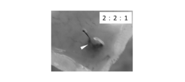
- [result] 3A, 3B, and 3C show the hair follicle primordium Versican gene, BMP4 gene, and ALP formed by co-culture in which the numbers of epithelial cells, dermal papilla cells, and adipose-derived stem cells were changed. The results of each analysis of the gene expression level are shown. Specifically, in FIGS. 3A, 3B, and 3C, the hair follicle primordia (“1: 1: 0” in the figure) and epithelium of Comparative Example 2-1 formed without using adipose-derived stem cells are shown.
- hair follicle primaries formed without using adipose-derived stem cells (“1: 1: 0 ")
- the expression levels of the Versican gene and the ALP gene were increased.
- the amount was remarkably large.
- Example 2-2 (“4: 4: 1” in the figure) and Example 2-3 (“8: 8: 1” in the figure).
- Example 2-1 (“2: 2: 1” in the figure) in which the ratio of adipose-derived stem cells was relatively large, it was formed without using adipose-derived stem cells.
- the expression level of the BMP4 gene was remarkably increased as compared with the hair follicle primordia (“1: 1: 0” in the figure).
- hair growth-related genes of the hair follicle primordia is further performed by further adding adipose-derived stem cells to the co-culture. It was confirmed that the amount (specifically, the amount of expression of Versican gene, BMP4 gene, and ALP gene by the hair papilla cells contained in the hair follicle primordia) can be effectively increased.
- a multi-well culture vessel used for co-culture to form hair follicle primordia spheroids was prepared in the same manner as in Patent Document 1 described above. That is, first, using CAD software (V Carve Pro 6.5), the pattern of the multiwell to be produced was designed by a computer. Then, the olefin resin substrate was cut in accordance with the designed pattern using a cutting machine, thereby producing a concave mold having the pattern. Epoxy resin (Crystal Lysine, manufactured by Nissin Resin Co., Ltd.) was poured into this mold, cured for 1 day, and then released to form a convex mold having the above-described designed pattern.
- the formed convex mold is fixed to the bottom surface of a 24-well plate, polydimethylsiloxane (PDMS) is poured and solidified, and then the mold is released to form a multiwell formed in a regular pattern on the PDMS substrate (each well A multi-well culture container (hereinafter referred to as "PDMS spheroid chip") having a diameter of 1 mm and a depth of 1 mm was prepared.
- PDMS spheroid chip A multi-well culture container having a diameter of 1 mm and a depth of 1 mm was prepared.
- this PDMS spheroid chip has a well formed on a substrate made of PDMS having excellent oxygen permeability, cells and cell aggregates cultured in the well have an appropriate amount of oxygen throughout the culture period. Is supplied.
- Adipose-derived stem cells were prepared as mesenchymal stem cells in the same manner as in Example 1 described above.
- the PDMS spheroid chip manufactured as described above was used as the culture container. Then, in each well of the PDMS spheroid chip, 4 ⁇ 10 3 cells / well of epithelial cells and dermal papilla cells and 2 ⁇ 10 3 cells / well of adipose-derived stem cells (total cell number of 1 ⁇ 10 4 cells / well) were obtained. Wells) were mixed and seeded, and co-cultured for 3 days. As the culture solution, a mixed medium prepared by mixing a hair papilla cell culture medium (Follicle Dermal Papilla Cell Growth Medium kit, PromoCell) and HuMedia-KG2 at a volume ratio of 1: 1 was used.
- a hair papilla cell culture medium Follicle Dermal Papilla Cell Growth Medium kit, PromoCell
- HuMedia-KG2 HuMedia-KG2
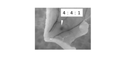
- [result] 4A and 4B show high-magnification phase-contrast micrographs of the hair follicle primordia formed in the wells on day 1 and day 3 of culture, respectively, and in FIG. 4C, day 3 of culture.
- Fig. 4 shows a phase contrast micrograph at low magnification of the hair follicle primordia formed in the well in the eye.
- hair follicle primordium was formed by co-culturing epithelial cells, dermal papilla cells, and adipose-derived stem cells in each well of the PDMS spheroid chip.
- the hair follicle primordia includes epithelial cell aggregates formed by aggregation of epithelial cells and hair papilla cell aggregates formed by aggregation of hair papilla cells.
- Most of the derived stem cells (“ADSC” in the figure) were present in the dermal papilla cell aggregation portion.
- FIG. 4C it was confirmed that a large amount of hair follicle primordia of uniform size can be simultaneously produced by using the PDMS spheroid chip.
- Adipose-derived stem cells were prepared as mesenchymal stem cells in the same manner as in Example 1 described above.
- a culture container As a culture container, a 96 multiwell plate was used as in Example 1 described above. Then, the epithelial cells, dermal papilla cells, and adipose-derived stem cells were co-cultured in each well while changing the ratio of the numbers of epithelial cells, dermal papilla cells, and adipose-derived stem cells.
- a mixed medium prepared by mixing a hair papilla cell culture medium (Follicle Dermal Papilla Cell Growth Medium kit, PromoCell) and HuMedia-KG2 at a volume ratio of 1: 1 was used.
- the mixture was mixed and seeded as described above, and coculture was performed for 3 days.
- [result] 5A, 5B, and 5C show the hair follicle primordia (“1: 1: 0” in FIG. 5A) and epithelial cells of Comparative Example 2-1 formed without using adipose-derived stem cells, respectively.
- hair growth from hair follicle primordia not containing adipose-derived stem cells was extremely small.
- the hair growth ability of the hair follicle primordia can be improved by further adding adipose-derived stem cells to epithelial cells and hair papilla cells in the co-culture for forming the hair follicle primordia. It was confirmed that the hair growth ability of the hair follicle primordia can be improved by further adding adipose-derived stem cells to epithelial cells and hair papilla cells in the co-culture for forming the hair follicle primordia. It was
Landscapes
- Health & Medical Sciences (AREA)
- Engineering & Computer Science (AREA)
- Life Sciences & Earth Sciences (AREA)
- Biomedical Technology (AREA)
- Organic Chemistry (AREA)
- Chemical & Material Sciences (AREA)
- Bioinformatics & Cheminformatics (AREA)
- Genetics & Genomics (AREA)
- Biotechnology (AREA)
- Wood Science & Technology (AREA)
- Zoology (AREA)
- General Engineering & Computer Science (AREA)
- Biochemistry (AREA)
- Cell Biology (AREA)
- General Health & Medical Sciences (AREA)
- Microbiology (AREA)
- Developmental Biology & Embryology (AREA)
- Dermatology (AREA)
- Rheumatology (AREA)
- Micro-Organisms Or Cultivation Processes Thereof (AREA)
- Materials For Medical Uses (AREA)
- Medicines Containing Material From Animals Or Micro-Organisms (AREA)
Abstract
優れた発毛関連特性を有する毛包原基、毛包原基の製造方法、及び毛包原基に含まれる細胞の活性化方法を提供する。毛包原基は、上皮系細胞と、間葉系細胞と、間葉系幹細胞とを含む。毛包原基の製造方法は、上皮系細胞と、間葉系細胞と、間葉系幹細胞とを共培養することにより毛包原基を形成することを含む。
Description
本発明は、毛包原基、毛包原基の製造方法、及び毛包原基に含まれる細胞の活性化方法に関する。
生体外で毛包原基を形成し、当該毛包原基を生体に移植して毛髪を再生させる技術の開発が進められている。
特許文献1には、規則的な配置の微小凹部からなるマイクロ凹版に、間葉系細胞及び上皮系細胞を播種し、酸素を供給しながら混合培養することにより、毛包原基を形成させる工程を備えることを特徴とする再生毛包原基の集合体の製造方法が記載されている。
特許文献2には、体性に由来する複数の体性細胞種からなる原始的な器官様をなし得る細胞塊の製造方法であって、当該複数種の体性細胞を含む培養液を用意し、当該複数種の体性細胞培養液を混合後、その混合細胞培養液にWntシグナル活性化剤を添加し、当該Wntシグナル活性化剤を含有する培養液を所定期間にわたり非平面接触性培養に委ね、当該非平面接触性培養した培養物の培地をWntシグナル活性化剤非含有培地と交換し、更に所定期間培養する、ことを含んでなり、ここで当該複数の体細胞の少なくとも1種は未分化状態を保っている、方法。が記載されている。
しかしながら、十分な発毛関連特性を有する毛包原基を製造する方法は、未だ確立されてはいない。
本発明は、上記課題に鑑みて為されたものであり、優れた発毛関連特性を有する毛包原基、毛包原基の製造方法、及び毛包原基に含まれる細胞の活性化方法を提供することをその目的の一つとする。
上記課題を解決するための本発明の一実施形態に係る毛包原基は、上皮系細胞と、間葉系細胞と、間葉系幹細胞とを含む。本発明によれば、優れた発毛関連特性を有する毛包原基が提供される。
前記毛包原基において、前記間葉系幹細胞は、脂肪由来間葉系幹細胞であることとしてもよい。前記毛包原基において、前記間葉系細胞は、毛乳頭細胞、及び/又は、毛球部毛根鞘細胞であることとしてもよい。前記毛包原基は、毛包原基スフェロイドであることとしてもよい。
前記毛包原基において、前記上皮系細胞の数と前記間葉系細胞の数との合計に対する、前記間葉系幹細胞の数の比率が0.01以上であることとしてもよい。前記毛包原基において、前記上皮系細胞の数に対する、前記間葉系幹細胞の数の比率が0.02以上であることとしてもよい。前記毛包原基において、前記間葉系細胞の数に対する、前記間葉系幹細胞の数の比率が0.02以上であることとしてもよい。
上記課題を解決するための本発明の一実施形態に係る毛包原基の製造方法は、上皮系細胞と、間葉系細胞と、間葉系幹細胞とを共培養することにより毛包原基を形成することを含む。本発明によれば、優れた発毛関連特性を有する毛包原基の製造方法が提供される。
前記方法においては、前記上皮系細胞と、前記間葉系細胞と、前記間葉系幹細胞とを細胞非接着性表面上で共培養することとしてもよい。
前記方法において、前記共培養により形成された前記毛包原基は、前記間葉系幹細胞を用いない以外は同一の方法で形成された毛包原基に含まれる間葉系細胞に比べて、活性化された前記間葉系細胞を含むこととしてもよい。この場合、前記活性化された間葉系細胞は、少なくとも一つの発毛関連遺伝子の発現量が、前記間葉系幹細胞を用いない以外は同一の方法で形成された毛包原基に含まれる間葉系細胞のそれより増加していることとしてもよい。
上記課題を解決するための本発明の一実施形態に係る間葉系細胞の活性化方法は、上皮系細胞と間葉系細胞との共培養により形成される毛包原基において、間葉系幹細胞をさらに加えて前記共培養を行うことにより、前記毛包原基に含まれる前記間葉系細胞を活性化する。本発明によれば、間葉系細胞の発毛関連特性を効果的に向上させる、間葉系細胞の活性化方法が提供される。
本発明によれば、優れた発毛関連特性を有する毛包原基、毛包原基の製造方法、及び毛包原基に含まれる細胞の活性化方法が提供される。
以下に、本発明の一実施形態について説明する。なお、本発明は本実施形態に限られるものではない。
本実施形態に係る毛包原基は、上皮系細胞と、間葉系細胞と、間葉系幹細胞とを含む。毛包原基は、生体外で形成される細胞凝集塊である。このため毛包原基は、再生毛包原基と呼ばれることもある。毛包原基は、発毛関連特性を有する。すなわち、毛包原基(より具体的には、当該毛包原基に含まれる細胞)は、発毛関連遺伝子を発現する。発毛関連遺伝子は、発毛に関連する遺伝子(例えば、発毛の促進に関連する遺伝子)であれば特に限られないが、例えば、Versican、ALP(アルカリフォスファターゼ)、BMP4、Nexin、Notch1、Wnt10b、LEF1(Lymphoid Enhancer Factor-1)、Shh(Sonic hedgehog)、MSX2(Msh homeobox2)及びβ-Cateninからなる群より選択される1以上であることとしてもよい。
また、例えば、毛包原基は、生体に移植されることで発毛する。このため、毛包原基は、生体への移植に好ましく用いられる。すなわち、毛包原基は、移植用毛包原基であることとしてもよい。なお、毛包原基は、移植に用いることなく、そのまま研究等の他の用途に供することもできる。
毛包原基に含まれる上皮系細胞は、発毛関連特性(例えば、発毛関連遺伝子の発現、及び/又は、間葉系細胞との共培養による毛包原基の形成)を有するものであれば、特に限られない。上皮系細胞は、毛包組織(例えば、毛包組織のバルジ領域の外毛根鞘最外層、及び又は、毛母基部)に由来する細胞であってもよく、皮膚組織に由来する細胞であってもよく、生体外で幹細胞(例えば、人工多能性幹(iPS)細胞、胚性幹(ES)細胞、又は胚性生殖(EG)細胞)から誘導された細胞であってもよい。上皮系細胞は、生体から採取された初代細胞であってもよく、予め培養された細胞(例えば、継代培養された細胞、及び/又は、株化された細胞)であってもよい。
上皮系細胞は、例えば、サイトケラチンを発現する細胞として特定される。上皮系細胞は、上皮幹細胞であることが好ましい。上皮幹細胞は、例えば、サイトケラチン15、及び/又は、CD34を発現する細胞として特定される。
上皮系細胞は、ヒト由来であることが好ましいが、非ヒト動物(ヒト以外の動物)由来であってもよい。非ヒト動物は、特に限られないが、非ヒト脊椎動物(ヒト以外の脊椎動物)であることが好ましい。非ヒト脊椎動物は、特に限られないが、非ヒト哺乳類であることが好ましい。非ヒト哺乳類は、特に限られないが、霊長類(例えば、サル)、げっ歯類(例えば、マウス、ラット、ハムスター、モルモット、ウサギ)、食肉類(例えば、イヌ、ネコ)、又は有蹄類(例えば、ブタ、ウシ、ウマ、ヤギ、ヒツジ)であってもよい。
毛包原基に含まれる間葉系細胞は、発毛関連特性(例えば、発毛関連遺伝子の発現、及び/又は、上皮系細胞との共培養による毛包原基の形成)を有するものであれば、特に限られない。間葉系細胞は、成体毛包組織(例えば、毛乳頭及び/又は毛球部毛根鞘)に由来する細胞であってもよく、皮膚組織(胎児、幼体、成体のいずれの皮膚組織であってもよい。)に由来する細胞であってもよく、生体外で幹細胞(例えば、iPS細胞、ES細胞、又はEG細胞)から誘導された細胞であってもよい。間葉系細胞は、生体から採取された初代細胞であってもよく、予め培養された細胞(例えば、継代培養された細胞、及び/又は、株化された細胞)であってもよい。
間葉系細胞は、例えば、Versican及びALPを発現する細胞として特定される。具体的に、間葉系細胞は、毛乳頭細胞、及び/又は、毛球部毛根鞘細胞であることが好ましい。毛乳頭細胞及び毛球部毛根鞘細胞は、Versican及びALPを発現する。
間葉系細胞は、ヒト由来であることが好ましいが、非ヒト動物(ヒト以外の動物)由来であってもよい。非ヒト動物は、特に限られないが、非ヒト脊椎動物(ヒト以外の脊椎動物)であることが好ましい。非ヒト脊椎動物は、特に限られないが、非ヒト哺乳類であることが好ましい。非ヒト哺乳類は、特に限られないが、霊長類(例えば、サル)、げっ歯類(例えば、マウス、ラット、ハムスター、モルモット、ウサギ)、食肉類(例えば、イヌ、ネコ)、又は有蹄類(例えば、ブタ、ウシ、ウマ、ヤギ、ヒツジ)であってもよい。
毛包原基に含まれる間葉系幹細胞は、中胚葉性組織(間葉)に由来する体性幹細胞であれば、特に限られない。間葉系幹細胞は、例えば、骨髄由来間葉系幹細胞であることとしてもよいが、脂肪由来間葉系幹細胞(以下、「脂肪由来幹細胞」という。)であることが好ましい。
脂肪由来幹細胞は、生体の脂肪組織(皮下脂肪に限られず、他の脂肪組織であってもよい。)に由来する間葉系幹細胞であってもよく、当該脂肪由来幹細胞への分化能を有する幹細胞(例えば、iPS細胞、ES細胞、又はEG細胞)から生体外で誘導された細胞であってもよい。脂肪由来幹細胞は、生体から採取された初代細胞であってもよく、予め培養された細胞(例えば、継代培養された細胞、及び/又は、株化された細胞)であってもよい。
脂肪由来幹細胞は、その培養系における基材への接着性に関し、商業的に入手可能な培養容器の底面(ポリスチレン等の樹脂製の表面)に接着する間葉系幹細胞として特定される。脂肪由来幹細胞は、その分化能に関し、例えば、骨細胞、脂肪細胞、及び軟骨細胞への分化能を有する(すなわち、骨細胞、脂肪細胞、及び軟骨細胞のいずれにも分化可能な)間葉系幹細胞として特定される。
脂肪由来幹細胞は、その細胞表面マーカーに関し、例えば、CD73、CD90及びCD105が陽性である間葉系幹細胞として特定される。この場合、脂肪由来幹細胞は、CD29、CD44、CD73、CD90、CD105及びCD166が陽性である間葉系幹細胞として特定されてもよい。さらにこの場合、脂肪由来幹細胞は、CD13、CD29、CD44、CD73、CD90、CD105及びCD166が陽性である間葉系幹細胞として特定されてもよい。
脂肪由来幹細胞は、上記の細胞表面マーカーが陽性であることに加え、CD14、CD34及びCD45が陰性である間葉系幹細胞、又は、CD14、CD31及びCD45が陰性である間葉系幹細胞として特定されてもよい。この場合、脂肪由来幹細胞は、CD14、CD19、CD34、CD45及びHLA-DRが陰性である間葉系幹細胞、又は、CD14、CD31、CD34及びCD45が陰性である間葉系幹細胞として特定されてもよい。また、前者の場合、脂肪由来幹細胞は、CD11b、CD14、CD19、CD34、CD45、CD79a及びHLA-DRが陰性である間葉系幹細胞として特定されてもよい。
脂肪由来幹細胞は、その細胞表面マーカーに関し、上記陽性マーカーと、上記陰性マーカーとの任意の組み合わせにより特定されてもよい。具体的に、脂肪由来幹細胞は、例えば、CD73、CD90及びCD105が陽性であり、且つ、CD11b、CD14、CD19、CD34、CD45、CD79a及びHLA-DRが陰性である間葉系幹細胞として特定される。また、脂肪由来幹細胞は、例えば、CD13、CD29、CD44、CD73、CD90、CD105及びCD166が陽性であり、CD14、CD31及びCD45が陰性である間葉系幹細胞として特定される。また、脂肪由来幹細胞は、例えば、CD73、CD90及びCD105が陽性であり、且つ、CD14、CD19、CD34、CD45及びHLA-DRが陰性である間葉系幹細胞として特定される。また、脂肪由来幹細胞は、例えば、CD29、CD44、CD73、CD90、CD105及びCD166が陽性であり、且つ、CD14、CD31、CD34及びCD45が陰性である間葉系幹細胞として特定される。
脂肪由来幹細胞は、ヒト由来であることが好ましいが、非ヒト動物(ヒト以外の動物)由来であってもよい。非ヒト動物は、特に限られないが、非ヒト脊椎動物(ヒト以外の脊椎動物)であることが好ましい。非ヒト脊椎動物は、特に限られないが、非ヒト哺乳類であることが好ましい。非ヒト哺乳類は、特に限られないが、霊長類(例えば、サル)、げっ歯類(例えば、マウス、ラット、ハムスター、モルモット、ウサギ)、食肉類(例えば、イヌ、ネコ)、又は有蹄類(例えば、ブタ、ウシ、ウマ、ヤギ、ヒツジ)であってもよい。
毛包原基に含まれる上皮系細胞、間葉系細胞、及び間葉系幹細胞の各々の数は、本発明の効果が得られる範囲であれば特に限られず、適宜調整される。すなわち、毛包原基において、上皮系細胞の数に対する、間葉系細胞の数の比率は、例えば、0.01以上であることとしてもよく、0.05以上であることが好ましく、0.10以上であることがより好ましく、0.15以上であることがより一層好ましく、0.20以上であることが特に好ましい。
上皮系細胞の数に対する、間葉系細胞の数の比率は、例えば、10.0以下であることとしてもよく、7.0以下であることとしてもよく、5.0以下であることとしてもよい。上皮系細胞の数に対する、間葉系細胞の数の比率は、上記下限値のいずれかと、上記上限値のいずれかとを任意に組み合わせて特定されてもよい。
毛包原基において、上皮系細胞の数と間葉系細胞の数との合計に対する、間葉系幹細胞の数の比率は、例えば、0.01以上であることとしてもよく、0.05以上であることが好ましく、0.10以上であることがより好ましく、0.15以上であることがより一層好ましく、0.20以上であることが特に好ましい。
上皮系細胞の数と間葉系細胞の数との合計に対する、間葉系幹細胞の数の比率は、例えば、3.0以下であることとしてもよく、1.5以下であることとしてもよい。上皮系細胞の数と間葉系細胞の数との合計に対する、間葉系幹細胞の数の比率は、上記下限値のいずれかと、上記上限値のいずれかとを任意に組み合わせて特定されてもよい。
毛包原基において、上皮系細胞の数に対する、間葉系幹細胞の数の比率は、例えば、0.02以上であることとしてもよく、0.10以上であることが好ましく、0.20以上であることがより好ましく、0.30以上であることがより一層好ましく、0.40以上であることが特に好ましい。
上皮系細胞の数に対する、間葉系幹細胞の数の比率は、例えば、3.0以下であることとしてもよく、1.5以下であることとしてもよい。上皮系細胞の数に対する、間葉系幹細胞の数の比率は、上記下限値のいずれかと、上記上限値のいずれかとを任意に組み合わせて特定されてもよい。
毛包原基において、間葉系細胞の数に対する、間葉系幹細胞の数の比率は、例えば、0.02以上であることとしてもよく、0.10以上であることが好ましく、0.20以上であることがより好ましく、0.30以上であることがより一層好ましく、0.40以上であることが特に好ましい。
間葉系細胞の数に対する、間葉系幹細胞の数の比率は、例えば、3.0以下であることとしてもよく、1.5以下であることとしてもよい。間葉系細胞の数に対する、間葉系幹細胞の数の比率は、上記下限値のいずれかと、上記上限値のいずれかとを任意に組み合わせて特定されてもよい。
毛包原基は、上皮系細胞と、間葉系細胞と、間葉系幹細胞とを共培養することにより形成される。すなわち、本実施形態に係る方法(以下、「本方法」という。)は、上皮系細胞と、間葉系細胞と、間葉系幹細胞とを共培養することにより毛包原基を形成することを含む、毛包原基の製造方法を包含する。共培養は、上皮系細胞と、間葉系細胞と、間葉系幹細胞とを混合して培養することにより行う。
共培養の方法は、上皮系細胞、間葉系細胞、及び間葉系幹細胞が凝集して毛包原基を形成する方法であれば特に限られないが、例えば、当該上皮系細胞と、当該間葉系細胞と、当該間葉系幹細胞とを細胞非接着性表面上で共培養することが好ましい。
ここで、細胞非接着性表面は、上皮系細胞、間葉系細胞、及び間葉系幹細胞が実質的に接着しない表面であれば特に限られない。すなわち、細胞非接着性表面は、例えば、上皮系細胞、間葉系細胞、及び間葉系幹細胞が接着せず浮遊状態で維持される表面、又は、当該上皮系細胞、間葉系細胞、及び間葉系幹細胞が緩く接着するものの、トリプシン処理等の酵素処理を施すことなく、ピペッティング等の培養液を流動させる操作で当該上皮系細胞、間葉系細胞、及び間葉系幹細胞が容易に脱離する表面である。細胞非接着性表面を有する培養容器としては、例えば、商業的に入手可能な、各ウェルの底面に細胞非接着コーティングが施されたマルチウェルプレートを用いることができる。また、国際公開第2017/073625号に記載されたマイクロ凹版も好ましく用いることができる。
上皮系細胞と、間葉系細胞と、間葉系幹細胞とを共培養する(例えば、上皮系細胞、間葉系細胞、及び間葉系幹細胞を培養液中に分散し混合して培養する)ことにより、当該上皮系細胞、間葉系細胞、及び間葉系幹細胞が自発的に凝集して、毛包原基が形成される。形成された毛包原基は、例えば、上皮系細胞同士が自発的に凝集して形成された上皮系細胞凝集部と、間葉系細胞同士が自発的に凝集して形成され、且つその一部が当該上皮系細胞凝集部の一部と結合した間葉系細胞凝集部と、を含み、さらに、当該間葉系細胞凝集部に含まれる間葉系細胞と接して存在する間葉系幹細胞を含む。
この場合、毛包原基に含まれる間葉系幹細胞の総数のうち、50%以上、好ましくは70%以上の数の間葉系幹細胞は、間葉系細胞凝集部に接して存在する(例えば、毛包原基に含まれる間葉系幹細胞の総数のうち、50%以上、好ましくは70%以上の数の間葉系幹細胞は、間葉系細胞凝集部に含まれる)こととしてもよい。また、これらの場合、毛包原基に含まれる間葉系幹細胞の総数のうち、50%以上、好ましくは70%以上の数の間葉系幹細胞は、上皮系細胞凝集部に接することなく存在する(例えば、毛包原基に含まれる間葉系幹細胞の総数のうち、50%以上、好ましくは70%以上の数の間葉系幹細胞は、上皮系細胞凝集部に含まれない)こととしてもよい。
毛包原基においては、例えば、間葉系細胞凝集部が内核部を構成するとともに、上皮系細胞凝集部が、当該間葉系細胞凝集塊の外周を覆う外層を構成することとしてもよい(いわゆるコアシェル型の毛包原基)。また、毛包原基においては、例えば、互いに隣接する間葉系細胞凝集部と上皮系細胞凝集部とが、その外表面の一部同士を接して存在することとしてもよい。このように、毛包原基は、間葉系細胞凝集部と上皮系細胞凝集部とを含むことが好ましいが、毛包原基においては、例えば、間葉系細胞と上皮系細胞とがそれぞれ分散して配置されることとしてもよい(いわゆるランダム型の毛包原基)。これら毛包原基において、少なくとも一部の間葉系幹細胞は、上述のとおり、間葉系細胞と接して存在する。
細胞非接着表面上では、非接着状態の毛包原基が製造される。ここで、非接着状態の毛包原基とは、細胞非接着表面に接着せず浮遊状態にある毛包原基、又は、当該細胞非接着表面に緩く接着しているが、トリプシン処理等の酵素処理を施すことなく、ピペッティング等の培養液を流動させる操作で当該細胞非接着性表面から容易に脱離する毛包原基である。
毛包原基は、毛包原基スフェロイドであることとしてもよい。毛包原基スフェロイドは、略球状の細胞凝集塊である。毛包原基スフェロイドは、非接着状態で形成され、容易に回収される。このため、毛包原基スフェロイドは、生体への移植に好ましく用いられる。
間葉系幹細胞を含む共培養により形成された毛包原基は、例えば、当該間葉系幹細胞を用いない以外は同一の方法で形成された毛包原基に含まれる間葉系細胞に比べて、活性化された間葉系細胞を含む。
ここで、間葉系細胞の活性化は、当該間葉系細胞の発毛関連特性の向上を意味する。具体的に、間葉系細胞の発毛関連特性の向上は、例えば、当該間葉系細胞の発毛関連遺伝子の発現量の増加、及び/又は、当該間葉系細胞を用いて形成された毛包原基の発毛特性の向上(例えば、生体に移植された当該毛包原基から生える毛の数及び/又は長さの増加)を含む。
すなわち、例えば、間葉系幹細胞との共培養により活性化された間葉系細胞は、少なくとも一つの発毛関連遺伝子の発現量が、当該間葉系幹細胞を用いない以外は同一の方法で形成された毛包原基に含まれる間葉系細胞のそれより増加している。
間葉系細胞において発現する発毛関連遺伝子は、当該間葉系細胞の発毛への寄与に関連する遺伝子であれば特に限られないが、例えば、Versican、ALP、BMP4、Nexin及びNotch1からなる群より選択される1以上であることとしてもよい。
具体的に、間葉系幹細胞を含む毛包原基に含まれる活性化間葉系細胞は、例えば、RT-PCRで測定されるVersican遺伝子の発現量が、当該間葉系幹細胞を用いない以外は同一の方法で形成された毛包原基に含まれる間葉系細胞のそれの1.2倍以上であることとしてもよく、1.5倍以上であることが好ましく、1.8倍以上であることがより好ましく、2.0倍以上であることが特に好ましい。
また、間葉系幹細胞を含む毛包原基に含まれる活性化間葉系細胞は、例えば、RT-PCRで測定されるALP遺伝子の発現量が、当該間葉系幹細胞を用いない以外は同一の方法で形成された毛包原基に含まれる間葉系細胞のそれの1.2倍以上であることとしてもよく、1.5倍以上であることが好ましく、1.7倍以上であることが特に好ましい。
また、間葉系幹細胞を含む毛包原基に含まれる活性化間葉系細胞は、例えば、RT-PCRで測定されるBMP4遺伝子の発現量が、当該間葉系幹細胞を用いない以外は同一の方法で形成された毛包原基に含まれる間葉系細胞のそれの1.1倍以上であることとしてもよく、1.2倍以上であることが好ましく、1.3倍以上であることが特に好ましい。
間葉系幹細胞を含む共培養により形成された毛包原基は、例えば、当該間葉系幹細胞を用いない以外は同一の方法で形成された毛包原基に含まれる上皮系細胞に比べて、活性化された上皮系細胞を含む。
ここで、上皮系細胞の活性化は、当該上皮系細胞の発毛関連特性の向上を意味する。具体的に、上皮系細胞の発毛関連特性の向上は、例えば、当該上皮系細胞の発毛関連遺伝子の発現量の増加、及び/又は、当該上皮系細胞を用いて形成された毛包原基の発毛特性の向上(例えば、生体に移植された当該毛包原基から生える毛の数及び/又は長さの増加)を含む。
すなわち、例えば、間葉系幹細胞との共培養により活性化された上皮系細胞は、少なくとも一つの発毛関連遺伝子の発現量が、当該間葉系幹細胞を用いない以外は同一の方法で形成された毛包原基に含まれる上皮系細胞のそれより増加している。
上皮系細胞において発現する発毛関連遺伝子は、当該上皮系細胞の発毛への寄与に関連する遺伝子であれば特に限られないが、例えば、Wnt10b、LEF1、Shh、MSX2及びβ-Cateninからなる群より選択される1以上であることとしてもよい。
具体的に、間葉系幹細胞を含む毛包原基に含まれる活性化上皮系細胞は、例えば、RT-PCRで測定される発毛関連遺伝子(例えば、Wnt10b、LEF1、Shh、MSX2及びβ-Cateninからなる群より選択される1以上)の発現量が、当該間葉系幹細胞を用いない以外は同一の方法で形成された毛包原基に含まれる間葉系細胞のそれより大きい。
このように、本方法による毛包原基の製造は、活性化された間葉系細胞を含む毛包原基の製造、すなわち、活性化された毛包原基の製造であるともいえる。このため、本方法は、上皮系細胞と間葉系細胞との共培養により形成される毛包原基において、間葉系幹細胞をさらに加えて当該共培養を行うことにより、当該毛包原基に含まれる当該間葉系細胞を活性化する、間葉系細胞の活性化方法を含む。
本実施形態に係る毛包原基は、上述のとおり、生体への移植に好ましく用いられる。すなわち、本方法によれば、生体に移植されることで毛が生える毛包原基が製造される。
毛包原基を生体に移植して当該毛包原基から毛を生やす場合、当該生体は特に限られず、ヒトであってもよいし、非ヒト動物であってもよい。非ヒト動物は特に限られないが、非ヒト哺乳類であることが好ましい。非ヒト哺乳類は、特に限られないが、例えば、霊長類(例えば、サル)、げっ歯類(例えば、マウス、ラット、ハムスター、モルモット、ウサギ)、食肉類(例えば、イヌ、ネコ)、又は有蹄類(例えば、ブタ、ウシ、ウマ、ヤギ、ヒツジ)であってもよい。毛包原基の生体への移植は、当該生体の皮膚への移植であることが好ましい。皮膚への移植は、例えば、皮下移植であってもよいし、皮内移植であってもよい。
毛包原基の製造及びその動物への移植は、医学的用途であってもよいし、研究用途であってもよい。すなわち、例えば、脱毛を伴う疾患の治療又は予防のために、当該疾患を患っている又は患う可能性のあるヒト患者に移植する目的で毛包原基を製造し、又は当該毛包原基を当該ヒト患者に移植することとしてもよい。
脱毛を伴う疾患は、特に限られないが、例えば、男性型脱毛症(Androgenetic Alopecia:AGA)、女子男性型脱毛症(Female Androgenetic Alopecia:FAGA)、分娩後脱毛症、びまん性脱毛症、脂漏性脱毛症、粃糠性脱毛症、牽引性脱毛症、代謝異常性脱毛症、圧迫性脱毛症、円形脱毛症、神経性脱毛症、抜毛症、全身性脱毛症、及び症候性脱毛症からなる群より選択される1以上であることとしてもよい。
また、例えば、脱毛を伴う疾患の治療又は予防に使用され得る物質の探索、及び/又は当該疾患の機構に関与する物質の探索のために、毛包原基を製造し、又は当該毛包原基を非ヒト動物に移植することとしてもよい。
次に、本実施形態に係る具体的な実施例について説明する。
[上皮系細胞の採取]
胎齢18日のC57BL/6マウス胎児より背部の皮膚組織を採取し、中尾らが報告した方法(Koh-ei Toyoshima et al. Nature Communications, 3, 784, 2012)を一部改変して、ディスパーゼ処理を4℃で1時間、30rpm震盪条件で行い、当該皮膚組織の上皮層と間葉層とを分離した。その後、上皮層に100U/mLのコラゲナーゼ処理を1時間20分施し、さらにトリプシン処理を10分施すことで、上皮系細胞を単離した。
胎齢18日のC57BL/6マウス胎児より背部の皮膚組織を採取し、中尾らが報告した方法(Koh-ei Toyoshima et al. Nature Communications, 3, 784, 2012)を一部改変して、ディスパーゼ処理を4℃で1時間、30rpm震盪条件で行い、当該皮膚組織の上皮層と間葉層とを分離した。その後、上皮層に100U/mLのコラゲナーゼ処理を1時間20分施し、さらにトリプシン処理を10分施すことで、上皮系細胞を単離した。
[間葉系細胞の調製]
間葉系細胞としては、毛乳頭細胞を用いた。すなわち、商業的に入手可能なヒト毛乳頭細胞(PromoCell社)を継代培養し、継代4代目~6代目(P4~P6)の当該ヒト毛乳頭細胞を用いた。培養液としては、商業的に入手可能な毛乳頭細胞用培養培地(PromoCell社)を用いた。
間葉系細胞としては、毛乳頭細胞を用いた。すなわち、商業的に入手可能なヒト毛乳頭細胞(PromoCell社)を継代培養し、継代4代目~6代目(P4~P6)の当該ヒト毛乳頭細胞を用いた。培養液としては、商業的に入手可能な毛乳頭細胞用培養培地(PromoCell社)を用いた。
[間葉系幹細胞の調製]
間葉系幹細胞としては、脂肪由来幹細胞を用いた。すなわち、商業的に入手可能なヒト脂肪由来幹細胞(Lonza社)を継代培養し、継代4代目~6代目(P4~P6)の当該ヒト脂肪由来幹細胞を用いた。培養液としては、商業的に入手可能なDMEM培養培地(Sigma社)を用いた。
間葉系幹細胞としては、脂肪由来幹細胞を用いた。すなわち、商業的に入手可能なヒト脂肪由来幹細胞(Lonza社)を継代培養し、継代4代目~6代目(P4~P6)の当該ヒト脂肪由来幹細胞を用いた。培養液としては、商業的に入手可能なDMEM培養培地(Sigma社)を用いた。
[毛包原基の製造]
毛包原基を形成するための培養容器として、各ウェルが、細胞非接着コーティングが施された丸底(凹状に湾曲した細胞非接着性底面)を有する、商業的に入手可能な96マルチウェルプレートを用いた。
毛包原基を形成するための培養容器として、各ウェルが、細胞非接着コーティングが施された丸底(凹状に湾曲した細胞非接着性底面)を有する、商業的に入手可能な96マルチウェルプレートを用いた。
そして、各ウェルに、上皮系細胞及び毛乳頭細胞をそれぞれ4×10³個/ウェル、及び脂肪由来幹細胞を2×103個/ウェル(全細胞数が1×104個/ウェル)となるように混合して播種し、共培養を3日間行った。培養液としては、毛乳頭細胞培養培地(Follicle Dermal Papilla Cell Growth Medium kit、PromoCell社)と、HuMedia-KG2とを体積比1:1で混合して調製された混合培地を用いた。
[結果]
図1A、図1B、及び図1Cには、それぞれ培養1日目、培養2日目、及び培養3日目に、1つのウェル内で形成された毛包原基の蛍光顕微鏡写真を示す。図1A~図1Cに示す毛包原基において、毛乳頭細胞は、蛍光試薬VybrantDiOにより緑色に蛍光染色され、脂肪由来幹細胞(図中の「ADSC」)は、蛍光試薬VybrantDilにより赤色に蛍光染色された。
図1A、図1B、及び図1Cには、それぞれ培養1日目、培養2日目、及び培養3日目に、1つのウェル内で形成された毛包原基の蛍光顕微鏡写真を示す。図1A~図1Cに示す毛包原基において、毛乳頭細胞は、蛍光試薬VybrantDiOにより緑色に蛍光染色され、脂肪由来幹細胞(図中の「ADSC」)は、蛍光試薬VybrantDilにより赤色に蛍光染色された。
図1A、図1B、及び図1Cに示すように、培養時間が経過するにつれて、上皮系細胞同士が自発的に凝集して上皮系細胞凝集部を形成するとともに、毛乳頭細胞同士も自発的に凝集して毛乳頭細胞凝集部を形成して、毛包原基スフェロイドが形成された。この毛包原基スフェロイドにおいて、多くの脂肪由来幹細胞は、毛乳頭細胞凝集部に分布していた。
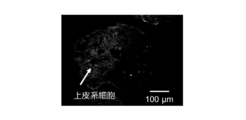
図2A、図2B、及び図2Cには、培養3日目に、1つのウェルで形成された毛包原基の蛍光顕微鏡写真を示す。この毛包原基において、上皮系細胞は、抗サイトケラチン15抗体を介して緑色に蛍光染色され、全ての細胞の細胞核は、蛍光試薬DAPIにより青色に蛍光染色され、脂肪由来幹細胞は、蛍光試薬VybrantDilにより赤色に蛍光染色された。図2Aには、上皮系細胞に対応する緑色の蛍光画像を示し、図2Bには、脂肪由来幹細胞に対応する赤色の蛍光画像を示し、図2Cには、上皮系細胞、毛乳頭細胞、及び脂肪由来幹細胞に対応する3色の蛍光画像を示している。
図2Aに示すように、上皮系細胞は、毛包原基の一部として、上皮系細胞凝集部を形成した。一方、図2Cに示すように、毛乳頭細胞は、毛包原基の他の一部として、毛乳頭細胞凝集部を形成した。これら上皮系細胞凝集部と毛乳頭細胞凝集部とは、その一部が互いに接するよう隣接して形成されていた。そして、図2B及び図2Cに示すように、脂肪由来幹細胞の多くは、毛乳頭細胞凝集部に存在していた。
[上皮系細胞の採取]
上述の実施例1と同様に、胎齢18日のC57BL/6マウス胎児の皮膚組織から、上皮系細胞を単離した。
上述の実施例1と同様に、胎齢18日のC57BL/6マウス胎児の皮膚組織から、上皮系細胞を単離した。
[間葉系細胞の調製]
上述の実施例1と同様に、間葉系細胞として、毛乳頭細胞を調製した。
上述の実施例1と同様に、間葉系細胞として、毛乳頭細胞を調製した。
[間葉系幹細胞の調製]
上述の実施例1と同様に、間葉系幹細胞として、脂肪由来幹細胞を調製した。
上述の実施例1と同様に、間葉系幹細胞として、脂肪由来幹細胞を調製した。
[毛包原基の製造]
培養容器としては、上述の実施例1と同様に、96マルチウェルプレートを用いた。そして、上皮系細胞、毛乳頭細胞、及び脂肪由来幹細胞の数の比率を変えて、上述の実施例1と同様に、混合培地を用いて、各ウェルで当該上皮系細胞、毛乳頭細胞、及び脂肪由来幹細胞の共培養を行った。
培養容器としては、上述の実施例1と同様に、96マルチウェルプレートを用いた。そして、上皮系細胞、毛乳頭細胞、及び脂肪由来幹細胞の数の比率を変えて、上述の実施例1と同様に、混合培地を用いて、各ウェルで当該上皮系細胞、毛乳頭細胞、及び脂肪由来幹細胞の共培養を行った。
具体的に、実施例2-1として、上皮系細胞:毛乳頭細胞:脂肪由来幹細胞=2:2:1の数比で共培養を行った。すなわち、各ウェルに、上皮系細胞及び毛乳頭細胞をそれぞれ4×10³個/ウェル、及び脂肪由来幹細胞を2×103個/ウェル(全細胞数が1×104個/ウェル)となるように混合して播種し、共培養を3日間行った。
同様に、実施例2-2として、上皮系細胞:毛乳頭細胞:脂肪由来幹細胞=4:4:1の数比で共培養を行った。すなわち、各ウェルに、上皮系細胞及び毛乳頭細胞をそれぞれ4×10³個/ウェル、及び脂肪由来幹細胞を1×103個/ウェル(全細胞数が9×103個/ウェル)となるように混合して播種し、共培養を3日間行った。
また、実施例2-3として、上皮系細胞:毛乳頭細胞:脂肪由来幹細胞=8:8:1の数比で共培養を行った。すなわち、各ウェルに、上皮系細胞及び毛乳頭細胞をそれぞれ4×10³個/ウェル、及び脂肪由来幹細胞を0.5×103個/ウェル(全細胞数が8.5×103個/ウェル)となるように混合して播種し、共培養を3日間行った。
一方、比較例2-1として、上皮系細胞:毛乳頭細胞:脂肪由来幹細胞=1:1:0の数比で共培養を行った。すなわち、脂肪由来幹細胞を用いることなく、各ウェルに、上皮系細胞及び毛乳頭細胞をそれぞれ4×10³個/ウェル(全細胞数が8×103個/ウェル)となるように混合して播種し、共培養を3日間行った。
[発毛関連遺伝子のPCR解析]
3日間の培養により形成された毛包原基の遺伝子発現量をRT-PCRにより解析した。RT-PCRで用いたプライマーの塩基配列は、Versicanについて、Forward Primerは「5‘-GCTGCAAAAGAGTGTGAAAA-3’」、Reverse Primerは「5‘-AGTGGTAACGAGATGCTTCC-3’」であり、BMP4について、Forward Primerは「5‘-GCCCGCAGCCTAGCAA-3’」、Reverse Primerは「5‘-CGGTAAAGATCCCGCATGTAG-3’」であり、ALPについて、Forward Primerは「5‘-ATTGACCACGGGCACCAT-3’」、Reverse Primerは「5‘-CTCCACCGCCTCAGCA-3’」であり、コントロールとして用いたGAPDHについて、Forward Primerは「5‘-ACCACAGTCCATGCCATCAC-3’」、Reverse Primerは「5‘-TCCACCACCCTGTTGCTGTA-3’」であった。
3日間の培養により形成された毛包原基の遺伝子発現量をRT-PCRにより解析した。RT-PCRで用いたプライマーの塩基配列は、Versicanについて、Forward Primerは「5‘-GCTGCAAAAGAGTGTGAAAA-3’」、Reverse Primerは「5‘-AGTGGTAACGAGATGCTTCC-3’」であり、BMP4について、Forward Primerは「5‘-GCCCGCAGCCTAGCAA-3’」、Reverse Primerは「5‘-CGGTAAAGATCCCGCATGTAG-3’」であり、ALPについて、Forward Primerは「5‘-ATTGACCACGGGCACCAT-3’」、Reverse Primerは「5‘-CTCCACCGCCTCAGCA-3’」であり、コントロールとして用いたGAPDHについて、Forward Primerは「5‘-ACCACAGTCCATGCCATCAC-3’」、Reverse Primerは「5‘-TCCACCACCCTGTTGCTGTA-3’」であった。
[結果]
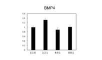
図3A、図3B、及び図3Cには、上皮系細胞、毛乳頭細胞、及び脂肪由来幹細胞の数の比を変えた共培養により形成された毛包原基のVersican遺伝子、BMP4遺伝子、及びALP遺伝子の発現量をそれぞれ解析した結果を示す。具体的に、図3A、図3B、及び図3Cには、脂肪由来幹細胞を用いることなく形成された比較例2-1の毛包原基(図中の「1:1:0」)、上皮系細胞:毛乳頭細胞:脂肪由来幹細胞=2:2:1の数比で形成された実施例2-1の毛包原基(図中の「2:2:1」)、上皮系細胞:毛乳頭細胞:脂肪由来幹細胞=4:4:1の数比で形成された実施例2-2の毛包原基(図中の「4:4:1」)、及び上皮系細胞:毛乳頭細胞:脂肪由来幹細胞=8:8:1の数比で形成された実施例2-3の毛包原基(図中の「8:8:1」)のそれぞれについて、当該比較例2-1における遺伝子発現量を「1」とした場合の、相対的な遺伝子発現量(図の縦軸)を示す。
図3A、図3B、及び図3Cには、上皮系細胞、毛乳頭細胞、及び脂肪由来幹細胞の数の比を変えた共培養により形成された毛包原基のVersican遺伝子、BMP4遺伝子、及びALP遺伝子の発現量をそれぞれ解析した結果を示す。具体的に、図3A、図3B、及び図3Cには、脂肪由来幹細胞を用いることなく形成された比較例2-1の毛包原基(図中の「1:1:0」)、上皮系細胞:毛乳頭細胞:脂肪由来幹細胞=2:2:1の数比で形成された実施例2-1の毛包原基(図中の「2:2:1」)、上皮系細胞:毛乳頭細胞:脂肪由来幹細胞=4:4:1の数比で形成された実施例2-2の毛包原基(図中の「4:4:1」)、及び上皮系細胞:毛乳頭細胞:脂肪由来幹細胞=8:8:1の数比で形成された実施例2-3の毛包原基(図中の「8:8:1」)のそれぞれについて、当該比較例2-1における遺伝子発現量を「1」とした場合の、相対的な遺伝子発現量(図の縦軸)を示す。
図3A及び図3Cに示すように、脂肪由来幹細胞を用いて形成された全ての毛包原基において、脂肪由来幹細胞を用いることなく形成された毛包原基(図中の「1:1:0」)に比べて、Versican遺伝子及びALP遺伝子の発現量が増加していた。特に、上皮系細胞:毛乳頭細胞:脂肪由来幹細胞=2:2:1の数比で形成された毛包原基(図中の「2:2:1」)におけるVersican遺伝子及びALP遺伝子の発現量は、顕著に大きかった。
また、図3Bに示すように、BMP4遺伝子については、実施例2-2(図中の「4:4:1」)及び実施例2-3(図中の「8:8:1」)において発現量の増加は認められなかったが、脂肪由来幹細胞の比率が比較的大きい実施例2-1(図中の「2:2:1」)においては、脂肪由来幹細胞を用いることなく形成された毛包原基(図中の「1:1:0」)に比べて、BMP4遺伝子の発現量が顕著に増加していた。
すなわち、上皮系細胞と毛乳頭細胞との共培養により形成される毛包原基において、脂肪由来幹細胞をさらに加えて当該共培養を行うことにより、当該毛包原基の発毛関連遺伝子の発現量(具体的には、当該毛包原基に含まれる毛乳頭細胞によるVersican遺伝子、BMP4遺伝子、及びALP遺伝子の発現量)を効果的に増加させることができることが確認された。
[マルチウェル培養容器の作製]
毛包原基スフェロイドを形成するための共培養に用いるマルチウェル培養容器を、上述した特許文献1と同様にして作製した。すなわち、まずCADソフト(V Carve Pro 6.5)を用いて、作製するマルチウェルのパターンをコンピューターで設計した。次いで、切削機を用いて、設計したパターン通りにオレフィン系樹脂基板を切削することで、当該パターンを有する凹鋳型を作製した。この鋳型にエポキシ樹脂(クリスタルリジン、日新レジン株式会社製)を流し込み、1日硬化させ、その後、離型することで、上述の設計されたパターンを有する凸鋳型を形成した。形成した凸鋳型を24ウェルプレートの底面に固定し、ポリジメチルシロキサン(PDMS)を流し込んで固化し、その後、離型することで、PDMS基板に規則的なパターンで形成されたマルチウェル(各ウェルの直径は1mm、深さは1mm)を有するマルチウェル培養容器(以下、「PDMSスフェロイドチップ」という。)を作製した。
毛包原基スフェロイドを形成するための共培養に用いるマルチウェル培養容器を、上述した特許文献1と同様にして作製した。すなわち、まずCADソフト(V Carve Pro 6.5)を用いて、作製するマルチウェルのパターンをコンピューターで設計した。次いで、切削機を用いて、設計したパターン通りにオレフィン系樹脂基板を切削することで、当該パターンを有する凹鋳型を作製した。この鋳型にエポキシ樹脂(クリスタルリジン、日新レジン株式会社製)を流し込み、1日硬化させ、その後、離型することで、上述の設計されたパターンを有する凸鋳型を形成した。形成した凸鋳型を24ウェルプレートの底面に固定し、ポリジメチルシロキサン(PDMS)を流し込んで固化し、その後、離型することで、PDMS基板に規則的なパターンで形成されたマルチウェル(各ウェルの直径は1mm、深さは1mm)を有するマルチウェル培養容器(以下、「PDMSスフェロイドチップ」という。)を作製した。
このPDMSスフェロイドチップは、酸素透過性に優れたPDMS製の基板にウェルが形成されているため、当該ウェル内で培養される細胞、及び細胞凝集塊には、培養期間を通じて、適切な量の酸素が供給される。
[上皮系細胞の採取]
上述の実施例1と同様に、胎齢18日のC57BL/6マウス胎児の皮膚組織から、上皮系細胞を単離した。
上述の実施例1と同様に、胎齢18日のC57BL/6マウス胎児の皮膚組織から、上皮系細胞を単離した。
[間葉系細胞の調製]
上述の実施例1と同様に、間葉系細胞として、毛乳頭細胞を調製した。
上述の実施例1と同様に、間葉系細胞として、毛乳頭細胞を調製した。
[間葉系幹細胞の調製]
上述の実施例1と同様に、間葉系幹細胞として、脂肪由来幹細胞を調製した。
上述の実施例1と同様に、間葉系幹細胞として、脂肪由来幹細胞を調製した。
[毛包原基の大量製造]
培養容器としては、上述のようにして作製したPDMSスフェロイドチップを用いた。そして、PDMSスフェロイドチップの各ウェルに、上皮系細胞及び毛乳頭細胞をそれぞれ4×10³個/ウェル、及び脂肪由来幹細胞を2×103個/ウェル(全細胞数が1×104個/ウェル)となるように混合して播種し、共培養を3日間行った。培養液としては、毛乳頭細胞培養培地(Follicle Dermal Papilla Cell Growth Medium kit、PromoCell社)と、HuMedia-KG2とを体積比1:1で混合して調製された混合培地を用いた。
培養容器としては、上述のようにして作製したPDMSスフェロイドチップを用いた。そして、PDMSスフェロイドチップの各ウェルに、上皮系細胞及び毛乳頭細胞をそれぞれ4×10³個/ウェル、及び脂肪由来幹細胞を2×103個/ウェル(全細胞数が1×104個/ウェル)となるように混合して播種し、共培養を3日間行った。培養液としては、毛乳頭細胞培養培地(Follicle Dermal Papilla Cell Growth Medium kit、PromoCell社)と、HuMedia-KG2とを体積比1:1で混合して調製された混合培地を用いた。
[結果]
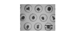
図4A及び図4Bには、それぞれ培養1日目及び培養3日目においてウェル内で形成されていた毛包原基の高倍率での位相差顕微鏡写真を示し、図4Cには、培養3日目においてウェル内で形成されていた毛包原基の低倍率での位相差顕微鏡写真を示す。
図4A及び図4Bには、それぞれ培養1日目及び培養3日目においてウェル内で形成されていた毛包原基の高倍率での位相差顕微鏡写真を示し、図4Cには、培養3日目においてウェル内で形成されていた毛包原基の低倍率での位相差顕微鏡写真を示す。
図4A~図4Cに示すように、PDMSスフェロイドチップの各ウェル内で上皮系細胞、毛乳頭細胞、及び脂肪由来幹細胞を共培養することにより、毛包原基が形成された。図4Bに示すように、毛包原基は、上皮系細胞が凝集して形成された上皮系細胞凝集部と、毛乳頭細胞が凝集して形成された毛乳頭細胞凝集部とを含み、脂肪由来幹細胞(図中の「ADSC」)の多くは、当該毛乳頭細胞凝集部に存在していた。また、図4Cに示すように、PDMSスフェロイドチップを用いることにより、サイズが均一な大量の毛包原基を同時に製造できることが確認された。
[上皮系細胞の採取]
上述の実施例1と同様に、胎齢18日のC57BL/6マウス胎児の皮膚組織から、上皮系細胞を単離した。
上述の実施例1と同様に、胎齢18日のC57BL/6マウス胎児の皮膚組織から、上皮系細胞を単離した。
[間葉系細胞の調製]
上述の実施例1と同様に、間葉系細胞として、毛乳頭細胞を調製した。
上述の実施例1と同様に、間葉系細胞として、毛乳頭細胞を調製した。
[間葉系幹細胞の調製]
上述の実施例1と同様に、間葉系幹細胞として、脂肪由来幹細胞を調製した。
上述の実施例1と同様に、間葉系幹細胞として、脂肪由来幹細胞を調製した。
[毛包原基の製造]
培養容器としては、上述の実施例1と同様に、96マルチウェルプレートを用いた。そして、上皮系細胞、毛乳頭細胞、及び脂肪由来幹細胞の数の比率を変えて、各ウェルで当該上皮系細胞、毛乳頭細胞、及び脂肪由来幹細胞の共培養を行った。培養液としては、毛乳頭細胞培養培地(Follicle Dermal Papilla Cell Growth Medium kit、PromoCell社)と、HuMedia-KG2とを体積比1:1で混合して調製された混合培地を用いた。
培養容器としては、上述の実施例1と同様に、96マルチウェルプレートを用いた。そして、上皮系細胞、毛乳頭細胞、及び脂肪由来幹細胞の数の比率を変えて、各ウェルで当該上皮系細胞、毛乳頭細胞、及び脂肪由来幹細胞の共培養を行った。培養液としては、毛乳頭細胞培養培地(Follicle Dermal Papilla Cell Growth Medium kit、PromoCell社)と、HuMedia-KG2とを体積比1:1で混合して調製された混合培地を用いた。
具体的に、実施例4-1として、上皮系細胞:毛乳頭細胞:脂肪由来幹細胞=2:2:1の数比で共培養を行った。すなわち、各ウェルに、上皮系細胞及び毛乳頭細胞をそれぞれ4×10³個/ウェル、及び脂肪由来幹細胞を2×103個/ウェル(全細胞数が1×104個/ウェル)となるように混合して播種し、共培養を3日間行った。
同様に、実施例4-2として、上皮系細胞:毛乳頭細胞:脂肪由来幹細胞=4:4:1の数比で共培養を行った。すなわち、各ウェルに、上皮系細胞及び毛乳頭細胞をそれぞれ4×10³個/ウェル、及び脂肪由来幹細胞を1×103個/ウェル(全細胞数が9×103個/ウェル)となるように混合して播種し、共培養を3日間行った。
一方、比較例4-1として、上皮系細胞:毛乳頭細胞:脂肪由来幹細胞=1:1:0の数比で共培養を行った。すなわち、脂肪由来幹細胞を用いることなく、各ウェルに、上皮系細胞及び毛乳頭細胞をそれぞれ4×10³個/ウェル(全細胞数が8×103個/ウェル)となるように混合して播種し、共培養を3日間行った。
[マウスへの移植]
上述のようにして形成した毛包原基を培養3日目に回収して、ヌードマウスの皮内に移植した。すなわち、ヌードマウスにイソフルラン吸引麻酔を施し、その背部をイソジンで消毒した。次いで、Vランスマイクロメス(日本アルコン)を用いて、皮膚の表皮層から真皮層下部に至る移植創を形成した。そして、この移植創に、毛包原基を20個ずつ注入した。なお、ヌードマウスの飼育及び移植実験は、横浜国立大学動物実験専門委員会の指針を遵守して行った。
上述のようにして形成した毛包原基を培養3日目に回収して、ヌードマウスの皮内に移植した。すなわち、ヌードマウスにイソフルラン吸引麻酔を施し、その背部をイソジンで消毒した。次いで、Vランスマイクロメス(日本アルコン)を用いて、皮膚の表皮層から真皮層下部に至る移植創を形成した。そして、この移植創に、毛包原基を20個ずつ注入した。なお、ヌードマウスの飼育及び移植実験は、横浜国立大学動物実験専門委員会の指針を遵守して行った。
[結果]
図5A、図5B、及び図5Cにはそれぞれ、脂肪由来幹細胞を用いることなく形成された比較例2-1の毛包原基(図5A中の「1:1:0」)、上皮系細胞:毛乳頭細胞:脂肪由来幹細胞=2:2:1の数比で形成された実施例4-1の毛包原基(図5B中の「2:2:1」)、上皮系細胞:毛乳頭細胞:脂肪由来幹細胞=4:4:1の数比で形成された実施例4-2の毛包原基(図5C中の「4:4:1」)、をマウスに移植してから30日後の時点で撮影された、当該毛包原基から再生した毛の写真を示す。
図5A、図5B、及び図5Cにはそれぞれ、脂肪由来幹細胞を用いることなく形成された比較例2-1の毛包原基(図5A中の「1:1:0」)、上皮系細胞:毛乳頭細胞:脂肪由来幹細胞=2:2:1の数比で形成された実施例4-1の毛包原基(図5B中の「2:2:1」)、上皮系細胞:毛乳頭細胞:脂肪由来幹細胞=4:4:1の数比で形成された実施例4-2の毛包原基(図5C中の「4:4:1」)、をマウスに移植してから30日後の時点で撮影された、当該毛包原基から再生した毛の写真を示す。
図5Aに示すように、脂肪由来幹細胞を含まない毛包原基からの発毛は、極僅かであった。また、図5Cに示すように、上皮系細胞:毛乳頭細胞:脂肪由来幹細胞=4:4:1の数比で形成された毛包原基からの発毛は、脂肪由来幹細胞を含まない毛包原基のそれに比べると多かった。さらに、図5Bに示すように、上皮系細胞:毛乳頭細胞:脂肪由来幹細胞=2:2:1の数比で形成された毛包原基からの発毛は特に顕著であり、明確な発毛が認められた。
すなわち、毛包原基を形成するための共培養において、上皮系細胞及び毛乳頭細胞に脂肪由来幹細胞をさらに加えることによって、当該毛包原基の発毛能力を向上させることができることが確認された。
Claims (12)
- 上皮系細胞と、間葉系細胞と、間葉系幹細胞とを含む、毛包原基。
- 前記間葉系幹細胞は、脂肪由来間葉系幹細胞である、請求項1に記載の毛包原基。
- 前記間葉系細胞は、毛乳頭細胞、及び/又は、毛球部毛根鞘細胞である、請求項1又は2に記載の毛包原基。
- 毛包原基スフェロイドである、請求項1乃至3のいずれかに記載の毛包原基。
- 前記上皮系細胞の数と前記間葉系細胞の数との合計に対する、前記間葉系幹細胞の数の比率が0.01以上である、請求項1乃至4のいずれかに記載の毛包原基。
- 前記上皮系細胞の数に対する、前記間葉系幹細胞の数の比率が0.02以上である、請求項1乃至5のいずれかに記載の毛包原基。
- 前記間葉系細胞の数に対する、前記間葉系幹細胞の数の比率が0.02以上である、請求項1乃至6のいずれかに記載の毛包原基。
- 上皮系細胞と、間葉系細胞と、間葉系幹細胞とを共培養することにより毛包原基を形成することを含む、毛包原基の製造方法。
- 前記上皮系細胞と、前記間葉系細胞と、前記間葉系幹細胞とを細胞非接着性表面上で共培養する、
請求項8に記載の毛包原基の製造方法。 - 前記共培養により形成された前記毛包原基は、前記間葉系幹細胞を用いない以外は同一の方法で形成された毛包原基に含まれる間葉系細胞に比べて、活性化された前記間葉系細胞を含む、
請求項8又は9に記載の毛包原基の製造方法。 - 前記活性化された間葉系細胞は、少なくとも一つの発毛関連遺伝子の発現量が、前記間葉系幹細胞を用いない以外は同一の方法で形成された毛包原基に含まれる間葉系細胞のそれより増加している、
請求項10に記載の毛包原基の製造方法。 - 上皮系細胞と間葉系細胞との共培養により形成される毛包原基において、
間葉系幹細胞をさらに加えて前記共培養を行うことにより、前記毛包原基に含まれる前記間葉系細胞を活性化する、
間葉系細胞の活性化方法。
Priority Applications (3)
| Application Number | Priority Date | Filing Date | Title |
|---|---|---|---|
| US17/276,628 US20220041983A1 (en) | 2018-11-08 | 2019-10-15 | Hair Follicle Germs, Method for Producing Hair Follicle Germs, and Method for Activating Cells Included in Hair Follicle Germs |
| CN201980059873.0A CN112703247A (zh) | 2018-11-08 | 2019-10-15 | 毛囊原基、毛囊原基的制造方法、以及毛囊原基中所含的细胞的活化方法 |
| EP19881986.4A EP3878946A4 (en) | 2018-11-08 | 2019-10-15 | HAIR FOLLICLE GERMS, METHOD FOR PRODUCING HAIR FOLLICLE GERMS, AND METHOD FOR ACTIVATING CELLS INCLUDED IN HAIR FOLLICLE GERMS |
Applications Claiming Priority (2)
| Application Number | Priority Date | Filing Date | Title |
|---|---|---|---|
| JP2018-210524 | 2018-11-08 | ||
| JP2018210524A JP7246595B2 (ja) | 2018-11-08 | 2018-11-08 | 毛包原基、毛包原基の製造方法、及び毛包原基に含まれる細胞の活性化方法 |
Publications (1)
| Publication Number | Publication Date |
|---|---|
| WO2020095631A1 true WO2020095631A1 (ja) | 2020-05-14 |
Family
ID=70612387
Family Applications (1)
| Application Number | Title | Priority Date | Filing Date |
|---|---|---|---|
| PCT/JP2019/040444 WO2020095631A1 (ja) | 2018-11-08 | 2019-10-15 | 毛包原基、毛包原基の製造方法、及び毛包原基に含まれる細胞の活性化方法 |
Country Status (5)
| Country | Link |
|---|---|
| US (1) | US20220041983A1 (ja) |
| EP (1) | EP3878946A4 (ja) |
| JP (1) | JP7246595B2 (ja) |
| CN (1) | CN112703247A (ja) |
| WO (1) | WO2020095631A1 (ja) |
Cited By (1)
| Publication number | Priority date | Publication date | Assignee | Title |
|---|---|---|---|---|
| WO2023171395A1 (ja) * | 2022-03-09 | 2023-09-14 | 株式会社 資生堂 | 間葉系細胞を含む細胞集団の毛髪再生能を評価する方法 |
Citations (4)
| Publication number | Priority date | Publication date | Assignee | Title |
|---|---|---|---|---|
| WO2008001938A1 (fr) * | 2006-06-27 | 2008-01-03 | Shiseido Company, Ltd. | Groupe de cellules renfermant des types diversifiés de cellules dérivées du soma, capables de former une structure primitive de type organique |
| JP2010534072A (ja) * | 2007-07-20 | 2010-11-04 | ドングク・ユニヴァーシティー・インダストリー−アカデミック・コーオペレーション・ファンデーション | 間葉系幹細胞を利用して毛乳頭組職を製造する方法 |
| WO2017073625A1 (ja) | 2015-10-30 | 2017-05-04 | 国立大学法人横浜国立大学 | 再生毛包原基の集合体の製造方法、毛包組織含有シート、及び毛包組織含有シートの製造方法 |
| US20180271913A1 (en) * | 2017-03-21 | 2018-09-27 | National Cheng Kung University | Pharmaceutical compositions for promoting hair follicle regeneration and methods for preparing the same |
Family Cites Families (2)
| Publication number | Priority date | Publication date | Assignee | Title |
|---|---|---|---|---|
| KR20160145778A (ko) * | 2014-07-07 | 2016-12-20 | 메디포스트(주) | 작은 크기 줄기세포의 발모 촉진능 및 이의 용도 |
| DE102015119880B4 (de) * | 2015-11-17 | 2018-05-24 | Technische Universität Berlin | Verfahren zur Herstellung von Haarfollikeln und de novo Papillen sowie deren Verwendung für in vitro Tests und in vivo Implantate |
-
2018
- 2018-11-08 JP JP2018210524A patent/JP7246595B2/ja active Active
-
2019
- 2019-10-15 CN CN201980059873.0A patent/CN112703247A/zh active Pending
- 2019-10-15 US US17/276,628 patent/US20220041983A1/en active Pending
- 2019-10-15 EP EP19881986.4A patent/EP3878946A4/en not_active Withdrawn
- 2019-10-15 WO PCT/JP2019/040444 patent/WO2020095631A1/ja unknown
Patent Citations (5)
| Publication number | Priority date | Publication date | Assignee | Title |
|---|---|---|---|---|
| WO2008001938A1 (fr) * | 2006-06-27 | 2008-01-03 | Shiseido Company, Ltd. | Groupe de cellules renfermant des types diversifiés de cellules dérivées du soma, capables de former une structure primitive de type organique |
| JP2013078344A (ja) | 2006-06-27 | 2013-05-02 | Shiseido Co Ltd | 体性に由来する複数の細胞種からなる原始的な器官様をなし得る細胞塊 |
| JP2010534072A (ja) * | 2007-07-20 | 2010-11-04 | ドングク・ユニヴァーシティー・インダストリー−アカデミック・コーオペレーション・ファンデーション | 間葉系幹細胞を利用して毛乳頭組職を製造する方法 |
| WO2017073625A1 (ja) | 2015-10-30 | 2017-05-04 | 国立大学法人横浜国立大学 | 再生毛包原基の集合体の製造方法、毛包組織含有シート、及び毛包組織含有シートの製造方法 |
| US20180271913A1 (en) * | 2017-03-21 | 2018-09-27 | National Cheng Kung University | Pharmaceutical compositions for promoting hair follicle regeneration and methods for preparing the same |
Non-Patent Citations (4)
| Title |
|---|
| KOH-EI TOYOSHIMA ET AL., NATURE COMMUNICATIONS, vol. 3, 2012, pages 784 |
| See also references of EP3878946A4 |
| TOYOSHIMA, K. ET AL.: "Fully functional hair follicle regeneration through the rearrangement of stem cells and their niches", NATURE COMMUNICATIONS, vol. 3, 2012, pages 784, XP0055557492 * |
| WON, C. H. ET AL.: "Hair growth promoting effects of adipose tissue-derived stem cells", JOURNAL OF DERMATOLOGICAL SCIENCE, vol. 57, 2010, pages 134 - 137, XP026874753 * |
Cited By (1)
| Publication number | Priority date | Publication date | Assignee | Title |
|---|---|---|---|---|
| WO2023171395A1 (ja) * | 2022-03-09 | 2023-09-14 | 株式会社 資生堂 | 間葉系細胞を含む細胞集団の毛髪再生能を評価する方法 |
Also Published As
| Publication number | Publication date |
|---|---|
| JP2020074718A (ja) | 2020-05-21 |
| US20220041983A1 (en) | 2022-02-10 |
| EP3878946A4 (en) | 2022-07-27 |
| CN112703247A (zh) | 2021-04-23 |
| JP7246595B2 (ja) | 2023-03-28 |
| EP3878946A1 (en) | 2021-09-15 |
Similar Documents
| Publication | Publication Date | Title |
|---|---|---|
| CN107012117B (zh) | 可从机体组织分离的多能干细胞 | |
| JP5887333B2 (ja) | 毛色が制御された移植用再生毛包原基の製造方法、当該移植用再生毛包原基を含む組成物、およびその移植方法 | |
| EP1954803B1 (en) | Multipotent adult stem cells having an ability of oct4 expression derived from umbilical cord blood and method for preparing the same | |
| US20100129771A1 (en) | Method for production of tooth, and tooth produced by the method | |
| JP2013078344A (ja) | 体性に由来する複数の細胞種からなる原始的な器官様をなし得る細胞塊 | |
| CN106661547B (zh) | 调控毛发生长的方法和组合物 | |
| JPWO2007013430A1 (ja) | 歯の再生方法 | |
| JP7444367B2 (ja) | 増幅毛包間葉系細胞の製造方法及びその使用 | |
| WO2020085023A1 (ja) | 間葉系細胞の培養方法、活性化間葉系細胞の製造方法、毛包原基の製造方法、間葉系細胞の活性化方法、及び上皮系細胞の活性化方法 | |
| WO2019220843A1 (ja) | 細胞含有ハイドロゲル体及びその製造方法 | |
| WO2020095631A1 (ja) | 毛包原基、毛包原基の製造方法、及び毛包原基に含まれる細胞の活性化方法 | |
| WO2023058429A1 (ja) | 毛髪再生能を有する細胞凝集塊の製造方法及びこれに関連する方法 | |
| JP7427180B2 (ja) | 毛包原基及びその製造方法 | |
| JP2020145990A (ja) | 培養容器、及び細胞−担体複合体の製造方法 | |
| JP2021073932A (ja) | ファイバー担体−細胞含有ゲル複合体及びその製造方法、並びにファイバー担体−細胞含有ゲル複合体製造用キット | |
| JP7158676B2 (ja) | 再生毛の色制御方法、毛の再生方法及び毛包原基の製造方法 | |
| JP5249789B2 (ja) | 毛髪の形成方法及び生物材料 | |
| Buttery et al. | A brief introduction to different cell types | |
| US20210380940A1 (en) | In vitro growth method for hair follicular epithelial stem cells | |
| Yamazaki et al. | Mari Dezawa, Kenichiro Tsuchiyama | |
| JP2023056592A (ja) | 移植用毛様組織及びこれに関連する方法 |
Legal Events
| Date | Code | Title | Description |
|---|---|---|---|
| 121 | Ep: the epo has been informed by wipo that ep was designated in this application |
Ref document number: 19881986 Country of ref document: EP Kind code of ref document: A1 |
|
| NENP | Non-entry into the national phase |
Ref country code: DE |
|
| ENP | Entry into the national phase |
Ref document number: 2019881986 Country of ref document: EP Effective date: 20210608 |