WO2020022494A1 - 消化管再生のためのシート状細胞培養物 - Google Patents
消化管再生のためのシート状細胞培養物 Download PDFInfo
- Publication number
- WO2020022494A1 WO2020022494A1 PCT/JP2019/029479 JP2019029479W WO2020022494A1 WO 2020022494 A1 WO2020022494 A1 WO 2020022494A1 JP 2019029479 W JP2019029479 W JP 2019029479W WO 2020022494 A1 WO2020022494 A1 WO 2020022494A1
- Authority
- WO
- WIPO (PCT)
- Prior art keywords
- sheet
- cell culture
- cells
- shaped cell
- present disclosure
- Prior art date
- Legal status (The legal status is an assumption and is not a legal conclusion. Google has not performed a legal analysis and makes no representation as to the accuracy of the status listed.)
- Ceased
Links
Images



Classifications
-
- A—HUMAN NECESSITIES
- A61—MEDICAL OR VETERINARY SCIENCE; HYGIENE
- A61L—METHODS OR APPARATUS FOR STERILISING MATERIALS OR OBJECTS IN GENERAL; DISINFECTION, STERILISATION OR DEODORISATION OF AIR; CHEMICAL ASPECTS OF BANDAGES, DRESSINGS, ABSORBENT PADS OR SURGICAL ARTICLES; MATERIALS FOR BANDAGES, DRESSINGS, ABSORBENT PADS OR SURGICAL ARTICLES
- A61L27/00—Materials for grafts or prostheses or for coating grafts or prostheses
- A61L27/36—Materials for grafts or prostheses or for coating grafts or prostheses containing ingredients of undetermined constitution or reaction products thereof, e.g. transplant tissue, natural bone, extracellular matrix
- A61L27/38—Materials for grafts or prostheses or for coating grafts or prostheses containing ingredients of undetermined constitution or reaction products thereof, e.g. transplant tissue, natural bone, extracellular matrix containing added animal cells
- A61L27/3839—Materials for grafts or prostheses or for coating grafts or prostheses containing ingredients of undetermined constitution or reaction products thereof, e.g. transplant tissue, natural bone, extracellular matrix containing added animal cells characterised by the site of application in the body
- A61L27/3882—Hollow organs, e.g. bladder, esophagus, urether, uterus
-
- A—HUMAN NECESSITIES
- A61—MEDICAL OR VETERINARY SCIENCE; HYGIENE
- A61K—PREPARATIONS FOR MEDICAL, DENTAL OR TOILETRY PURPOSES
- A61K35/00—Medicinal preparations containing materials or reaction products thereof with undetermined constitution
- A61K35/12—Materials from mammals; Compositions comprising non-specified tissues or cells; Compositions comprising non-embryonic stem cells; Genetically modified cells
- A61K35/28—Bone marrow; Haematopoietic stem cells; Mesenchymal stem cells of any origin, e.g. adipose-derived stem cells
-
- A—HUMAN NECESSITIES
- A61—MEDICAL OR VETERINARY SCIENCE; HYGIENE
- A61K—PREPARATIONS FOR MEDICAL, DENTAL OR TOILETRY PURPOSES
- A61K35/00—Medicinal preparations containing materials or reaction products thereof with undetermined constitution
- A61K35/12—Materials from mammals; Compositions comprising non-specified tissues or cells; Compositions comprising non-embryonic stem cells; Genetically modified cells
- A61K35/34—Muscles; Smooth muscle cells; Heart; Cardiac stem cells; Myoblasts; Myocytes; Cardiomyocytes
-
- A—HUMAN NECESSITIES
- A61—MEDICAL OR VETERINARY SCIENCE; HYGIENE
- A61L—METHODS OR APPARATUS FOR STERILISING MATERIALS OR OBJECTS IN GENERAL; DISINFECTION, STERILISATION OR DEODORISATION OF AIR; CHEMICAL ASPECTS OF BANDAGES, DRESSINGS, ABSORBENT PADS OR SURGICAL ARTICLES; MATERIALS FOR BANDAGES, DRESSINGS, ABSORBENT PADS OR SURGICAL ARTICLES
- A61L27/00—Materials for grafts or prostheses or for coating grafts or prostheses
- A61L27/14—Macromolecular materials
- A61L27/18—Macromolecular materials obtained otherwise than by reactions only involving carbon-to-carbon unsaturated bonds
-
- A—HUMAN NECESSITIES
- A61—MEDICAL OR VETERINARY SCIENCE; HYGIENE
- A61L—METHODS OR APPARATUS FOR STERILISING MATERIALS OR OBJECTS IN GENERAL; DISINFECTION, STERILISATION OR DEODORISATION OF AIR; CHEMICAL ASPECTS OF BANDAGES, DRESSINGS, ABSORBENT PADS OR SURGICAL ARTICLES; MATERIALS FOR BANDAGES, DRESSINGS, ABSORBENT PADS OR SURGICAL ARTICLES
- A61L27/00—Materials for grafts or prostheses or for coating grafts or prostheses
- A61L27/14—Macromolecular materials
- A61L27/22—Polypeptides or derivatives thereof, e.g. degradation products
- A61L27/222—Gelatin
-
- A—HUMAN NECESSITIES
- A61—MEDICAL OR VETERINARY SCIENCE; HYGIENE
- A61L—METHODS OR APPARATUS FOR STERILISING MATERIALS OR OBJECTS IN GENERAL; DISINFECTION, STERILISATION OR DEODORISATION OF AIR; CHEMICAL ASPECTS OF BANDAGES, DRESSINGS, ABSORBENT PADS OR SURGICAL ARTICLES; MATERIALS FOR BANDAGES, DRESSINGS, ABSORBENT PADS OR SURGICAL ARTICLES
- A61L27/00—Materials for grafts or prostheses or for coating grafts or prostheses
- A61L27/14—Macromolecular materials
- A61L27/22—Polypeptides or derivatives thereof, e.g. degradation products
- A61L27/225—Fibrin; Fibrinogen
-
- A—HUMAN NECESSITIES
- A61—MEDICAL OR VETERINARY SCIENCE; HYGIENE
- A61L—METHODS OR APPARATUS FOR STERILISING MATERIALS OR OBJECTS IN GENERAL; DISINFECTION, STERILISATION OR DEODORISATION OF AIR; CHEMICAL ASPECTS OF BANDAGES, DRESSINGS, ABSORBENT PADS OR SURGICAL ARTICLES; MATERIALS FOR BANDAGES, DRESSINGS, ABSORBENT PADS OR SURGICAL ARTICLES
- A61L27/00—Materials for grafts or prostheses or for coating grafts or prostheses
- A61L27/14—Macromolecular materials
- A61L27/22—Polypeptides or derivatives thereof, e.g. degradation products
- A61L27/24—Collagen
-
- A—HUMAN NECESSITIES
- A61—MEDICAL OR VETERINARY SCIENCE; HYGIENE
- A61L—METHODS OR APPARATUS FOR STERILISING MATERIALS OR OBJECTS IN GENERAL; DISINFECTION, STERILISATION OR DEODORISATION OF AIR; CHEMICAL ASPECTS OF BANDAGES, DRESSINGS, ABSORBENT PADS OR SURGICAL ARTICLES; MATERIALS FOR BANDAGES, DRESSINGS, ABSORBENT PADS OR SURGICAL ARTICLES
- A61L27/00—Materials for grafts or prostheses or for coating grafts or prostheses
- A61L27/36—Materials for grafts or prostheses or for coating grafts or prostheses containing ingredients of undetermined constitution or reaction products thereof, e.g. transplant tissue, natural bone, extracellular matrix
- A61L27/38—Materials for grafts or prostheses or for coating grafts or prostheses containing ingredients of undetermined constitution or reaction products thereof, e.g. transplant tissue, natural bone, extracellular matrix containing added animal cells
-
- A—HUMAN NECESSITIES
- A61—MEDICAL OR VETERINARY SCIENCE; HYGIENE
- A61L—METHODS OR APPARATUS FOR STERILISING MATERIALS OR OBJECTS IN GENERAL; DISINFECTION, STERILISATION OR DEODORISATION OF AIR; CHEMICAL ASPECTS OF BANDAGES, DRESSINGS, ABSORBENT PADS OR SURGICAL ARTICLES; MATERIALS FOR BANDAGES, DRESSINGS, ABSORBENT PADS OR SURGICAL ARTICLES
- A61L27/00—Materials for grafts or prostheses or for coating grafts or prostheses
- A61L27/36—Materials for grafts or prostheses or for coating grafts or prostheses containing ingredients of undetermined constitution or reaction products thereof, e.g. transplant tissue, natural bone, extracellular matrix
- A61L27/38—Materials for grafts or prostheses or for coating grafts or prostheses containing ingredients of undetermined constitution or reaction products thereof, e.g. transplant tissue, natural bone, extracellular matrix containing added animal cells
- A61L27/3804—Materials for grafts or prostheses or for coating grafts or prostheses containing ingredients of undetermined constitution or reaction products thereof, e.g. transplant tissue, natural bone, extracellular matrix containing added animal cells characterised by specific cells or progenitors thereof, e.g. fibroblasts, connective tissue cells, kidney cells
- A61L27/3826—Muscle cells, e.g. smooth muscle cells
-
- A—HUMAN NECESSITIES
- A61—MEDICAL OR VETERINARY SCIENCE; HYGIENE
- A61L—METHODS OR APPARATUS FOR STERILISING MATERIALS OR OBJECTS IN GENERAL; DISINFECTION, STERILISATION OR DEODORISATION OF AIR; CHEMICAL ASPECTS OF BANDAGES, DRESSINGS, ABSORBENT PADS OR SURGICAL ARTICLES; MATERIALS FOR BANDAGES, DRESSINGS, ABSORBENT PADS OR SURGICAL ARTICLES
- A61L27/00—Materials for grafts or prostheses or for coating grafts or prostheses
- A61L27/36—Materials for grafts or prostheses or for coating grafts or prostheses containing ingredients of undetermined constitution or reaction products thereof, e.g. transplant tissue, natural bone, extracellular matrix
- A61L27/38—Materials for grafts or prostheses or for coating grafts or prostheses containing ingredients of undetermined constitution or reaction products thereof, e.g. transplant tissue, natural bone, extracellular matrix containing added animal cells
- A61L27/3804—Materials for grafts or prostheses or for coating grafts or prostheses containing ingredients of undetermined constitution or reaction products thereof, e.g. transplant tissue, natural bone, extracellular matrix containing added animal cells characterised by specific cells or progenitors thereof, e.g. fibroblasts, connective tissue cells, kidney cells
- A61L27/3834—Cells able to produce different cell types, e.g. hematopoietic stem cells, mesenchymal stem cells, marrow stromal cells, embryonic stem cells
-
- A—HUMAN NECESSITIES
- A61—MEDICAL OR VETERINARY SCIENCE; HYGIENE
- A61L—METHODS OR APPARATUS FOR STERILISING MATERIALS OR OBJECTS IN GENERAL; DISINFECTION, STERILISATION OR DEODORISATION OF AIR; CHEMICAL ASPECTS OF BANDAGES, DRESSINGS, ABSORBENT PADS OR SURGICAL ARTICLES; MATERIALS FOR BANDAGES, DRESSINGS, ABSORBENT PADS OR SURGICAL ARTICLES
- A61L27/00—Materials for grafts or prostheses or for coating grafts or prostheses
- A61L27/36—Materials for grafts or prostheses or for coating grafts or prostheses containing ingredients of undetermined constitution or reaction products thereof, e.g. transplant tissue, natural bone, extracellular matrix
- A61L27/38—Materials for grafts or prostheses or for coating grafts or prostheses containing ingredients of undetermined constitution or reaction products thereof, e.g. transplant tissue, natural bone, extracellular matrix containing added animal cells
- A61L27/3839—Materials for grafts or prostheses or for coating grafts or prostheses containing ingredients of undetermined constitution or reaction products thereof, e.g. transplant tissue, natural bone, extracellular matrix containing added animal cells characterised by the site of application in the body
-
- A—HUMAN NECESSITIES
- A61—MEDICAL OR VETERINARY SCIENCE; HYGIENE
- A61L—METHODS OR APPARATUS FOR STERILISING MATERIALS OR OBJECTS IN GENERAL; DISINFECTION, STERILISATION OR DEODORISATION OF AIR; CHEMICAL ASPECTS OF BANDAGES, DRESSINGS, ABSORBENT PADS OR SURGICAL ARTICLES; MATERIALS FOR BANDAGES, DRESSINGS, ABSORBENT PADS OR SURGICAL ARTICLES
- A61L27/00—Materials for grafts or prostheses or for coating grafts or prostheses
- A61L27/50—Materials characterised by their function or physical properties, e.g. injectable or lubricating compositions, shape-memory materials, surface modified materials
- A61L27/52—Hydrogels or hydrocolloids
-
- A—HUMAN NECESSITIES
- A61—MEDICAL OR VETERINARY SCIENCE; HYGIENE
- A61L—METHODS OR APPARATUS FOR STERILISING MATERIALS OR OBJECTS IN GENERAL; DISINFECTION, STERILISATION OR DEODORISATION OF AIR; CHEMICAL ASPECTS OF BANDAGES, DRESSINGS, ABSORBENT PADS OR SURGICAL ARTICLES; MATERIALS FOR BANDAGES, DRESSINGS, ABSORBENT PADS OR SURGICAL ARTICLES
- A61L2430/00—Materials or treatment for tissue regeneration
- A61L2430/22—Materials or treatment for tissue regeneration for reconstruction of hollow organs, e.g. bladder, esophagus, urether, uterus
Definitions
- the present invention relates to a sheet-shaped cell culture for treating a luminal organ having a damaged portion, a method for producing the sheet-shaped cell culture, a method for treating a luminal organ using the sheet-shaped cell culture, and the like.
- Non-Patent Document 1 fetal cardiomyocytes, skeletal myoblasts, mesenchymal stem cells, cardiac stem cells, ES cells, iPS cells, etc. for repairing myocardial tissue damaged by ischemic heart diseases such as angina pectoris and myocardial infarction.
- Patent Document 1 a cell structure formed using a scaffold and a sheet-shaped cell culture in which cells are formed in a sheet shape have been developed (Patent Document 1, Non-Patent Document 2).
- sheet-shaped cell culture to treatment use of cultured epidermis sheet for skin damage due to burns, use of corneal epithelial sheet-shaped cell culture for corneal injury, oral mucosal sheet for endoscopic resection of esophageal cancer
- the use of dendritic cell cultures is being studied, and some of them are in the stage of clinical application.
- Patent Literature 2 discloses that a sheet-shaped cell culture containing mesenchymal stem cells is used to cure or prevent leakage from a damaged part of the digestive tract caused by a suture failure or the like.
- Non-Patent Documents 3 and 4 describe that a skeletal myoblast cell sheet can be used for healing in a model animal of pancreatic fistula or gastric perforation.
- ESD endoscopic submucosal dissection
- the present disclosure relates to a luminal organ having a damaged portion, particularly a sheet-shaped cell culture for regenerating duodenal tissue, a method for producing the sheet-shaped cell culture, and a luminal organ using the sheet-shaped cell culture. It is an object of the present invention to provide a method of reproducing a damaged portion.
- the present inventors have been intensively studying a method for reducing postoperative complications in duodenal ESD, and by applying a sheet-shaped cell culture of skeletal myoblasts to the outside of the ESD treatment site, the ulcer was evaluated. It was found for the first time that the continuity of the bottom was maintained and postoperative perforation could be prevented, and further research was conducted based on such findings, and as a result, the present invention was completed.
- the present invention relates to the following: [1] A sheet-shaped cell culture for promoting healing of a luminal organ having a damaged portion on at least one side of a lumen wall, wherein the sheet-shaped cell culture is a tube corresponding to a site where damage is present.
- the sheet-shaped cell culture wherein the sheet-shaped cell culture is applied to an opposite side of a cavity wall.
- the damage is on the inner side of the lumen wall and the sheet cell culture is applied to the corresponding outer side of the lumen wall, or the damage is on the outer side of the lumen wall and the sheet is The sheet-shaped cell culture of [1], wherein the cell-shaped cell culture is applied to a corresponding inner side of a lumen wall.
- [3] The sheet-shaped cell culture of [1], wherein the damage is penetrating damage or non-penetrating damage.
- [4] The sheet-shaped cell culture of [1] to [3], wherein the luminal organ is an organ of the digestive tract.
- [5] The sheet-shaped cell culture of [1] to [4], wherein the damage is due to detachment of the surface tissue of the lumen wall.
- [6] The sheet-shaped cell culture according to [1] to [5], further having a reinforcing layer containing a gel and / or a polymer.
- [7] The sheet-shaped cell culture of [1] to [6], which is used together with a pedicle.
- a method for preventing perforation of a luminal organ after a surgical procedure comprising a step of applying a sheet-shaped cell culture to a luminal organ, wherein the sheet-shaped cell culture is subjected to a treatment.
- [15] A method for treating a diseased part on a lumen wall surface, (A) removing the diseased part on the surface of the lumen wall (B) applying the sheet-shaped cell culture to the part detached in step (a) or the opposite side corresponding to the detached part;
- the above method comprising: [16] The method of [15], wherein the disease is cancer. [17] The method of [15] or [16], wherein the exfoliation of the diseased part on the surface of the lumen wall is exfoliation of the mucous membrane, mucosal muscularis and / or submucosa having the diseased part.
- the healing of the tissue is promoted by implanting a sheet-shaped cell culture on the opposite side corresponding to the site where the lesion exists.
- the damage is formed by a treatment such as ESD, it is effective, so that complications after these treatments can be prevented and the prognosis can be improved.
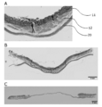
- FIG. 1 shows a tissue staining image of the sheet-shaped cell culture of the present disclosure.
- A is a sheet-shaped cell culture of porcine skeletal myoblasts
- B is a sheet-shaped cell culture of porcine skeletal myoblasts with a fibrin gel reinforcing layer formed
- C is a sheet-shaped cell culture of porcine mesenchymal stem cells. It is a stained image of an object.
- the sheet-shaped cell culture of skeletal myoblasts had a sheet thickness of about 100 ⁇ m, while the sheet-shaped cell culture of porcine mesenchymal stem cells had a thickness of less than 50 ⁇ m.
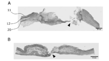
- FIG. 2 shows a tissue staining image of the gastrointestinal tract wall of the duodenum.
- A is a cross-sectional stained image showing the structure of the pig duodenal wall.
- 11 denotes a mucous membrane
- 12 denotes a submucosal layer (Brunner's gland)
- 20 denotes a muscular layer.
- B is a tissue staining image before the ESD treatment, and the dotted line portion is cut off.
- C is a stained image after the ESD treatment. It can be seen that the mucosa and submucosa have been removed, leaving the muscular layer.
- FIG. 3 shows a tissue-stained image of an ESD-treated site of a pig in a control group.
- A represents the object group 1 and B represents the object group 2.
- FIG. 4-1 shows a tissue staining image of an ESD-treated site of a pig in the sheet treatment group.
- a and B represent sheet treatment group 1
- C and D represent sheet treatment group 2
- E and F represent sheet treatment group 3
- G and H represent sheet treatment group 4, respectively.
- the locations indicated by the arrowheads of A, C and F are the locations where the sheet-shaped cell culture of porcine skeletal myoblasts was applied, the continuity of the ulcer garden was maintained, and histological prevention of postoperative perforation. You can see that it is done.
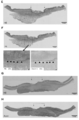
- FIG. 4-2 shows a tissue-stained image of an ESD-treated site of a pig in the sheet treatment group.
- a and B represent sheet treatment group 1
- C and D represent sheet treatment group 2
- E and F represent sheet treatment group 3
- G and H represent sheet treatment group 4, respectively.
- the locations indicated by the arrowheads of A, C, and F are the locations where the sheet-shaped cell culture of porcine skeletal myoblasts was applied, the continuity of the ulcer garden was maintained, and postoperative perforation was histologically prevented. You can see that it is done.
- the enlarged views of the portions surrounded by the dotted squares A and C are B and D, respectively, and the fibrin, which is the reinforcing layer of the attached sheet-shaped cell culture, was observed at the portion surrounded by the dotted ellipse B. it can.
- FIG. 5 shows data obtained by graphing the adhesion scores in Table 1.
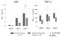
- FIG. 6 shows the transition of the inflammatory response. The increase or decrease in the inflammatory response was expressed as an increase or decrease in the expression of C-reactive protein (CRP) and TNF- ⁇ in plasma.
- CRP C-reactive protein
- the “sheet-shaped cell culture” refers to a cell in which cells are connected to each other to form a sheet.
- the cells may be connected to each other directly (including via a cellular element such as an adhesion molecule) and / or via an intermediary substance.
- the intervening substance is not particularly limited as long as it is a substance capable of at least physically (mechanically) connecting cells, and examples thereof include an extracellular matrix.
- the mediator is preferably of cell origin, in particular of the cells that make up the cell culture. Cells are at least physically (mechanically) linked, but may be further functionally linked, eg, chemically or electrically.
- the sheet-shaped cell culture may be composed of one cell layer (single layer) or composed of two or more cell layers (laminated (multilayer) body, for example, two layers, three layers, Four layers, five layers, six layers, etc.). Further, the sheet-shaped cell culture may have a three-dimensional structure having a thickness exceeding the thickness of one cell without the cells showing a clear layer structure. For example, in the vertical cross section of the sheet-shaped cell culture, the cells may not be uniformly arranged in the horizontal direction, but may be non-uniformly arranged (for example, in a mosaic).
- the sheet-shaped cell culture preferably does not contain a scaffold (support). Scaffolds are sometimes used in the art to attach cells on and / or to their surfaces and maintain the physical integrity of sheet cell cultures, such as polyvinylidene difluoride ( Although PVDF) membranes and the like are known, the sheet-shaped cell culture of the present disclosure can maintain its physical integrity without such a scaffold.
- the sheet-shaped cell culture of the present disclosure preferably includes only a substance derived from the cells constituting the sheet-shaped cell culture, and does not include other substances.
- the cells constituting the sheet-shaped cell culture are not particularly limited as long as they can form the sheet-shaped cell culture, and include, for example, adherent cells (adherent cells).
- Adherent cells include, for example, adherent somatic cells (eg, cardiomyocytes, fibroblasts, epithelial cells, endothelial cells, hepatocytes, pancreatic cells, kidney cells, adrenal cells, periodontal ligament cells, gingival cells, periosteal cells, skin Cells, synovial cells, chondrocytes, etc.) and stem cells (eg, tissue stem cells such as myoblasts, cardiac stem cells, embryonic stem cells, pluripotent stem cells such as iPS (induced pluripotent stem) cells, mesenchymal stem cells, etc.) Including.
- adherent somatic cells eg, cardiomyocytes, fibroblasts, epithelial cells, endothelial cells, hepatocytes, pancreatic cells, kidney cells, adrenal cells
- the somatic cells may be stem cells, particularly those differentiated from iPS cells (iPS cell-derived adherent cells).
- Non-limiting examples of cells constituting the sheet-shaped cell culture include, for example, myoblasts (eg, skeletal myoblasts), mesenchymal stem cells (eg, bone marrow, adipose tissue, peripheral blood, skin, hair root, Muscle tissue, endometrium, placenta, cord blood, etc.), cardiomyocytes, fibroblasts, cardiac stem cells, embryonic stem cells, iPS cells, synovial cells, chondrocytes, epithelial cells (eg, oral mucosal epithelial cells) , Retinal pigment epithelial cells, nasal mucosal epithelial cells, etc.), endothelial cells (eg, vascular endothelial cells, etc.), hepatocytes (eg, hepatic parenchymal cells, etc.), pancreatic cells (eg, pancreatic
- Non-limiting examples of iPS cell-derived adherent cells include iPS cell-derived cardiomyocytes, fibroblasts, epithelial cells, endothelial cells, hepatocytes, pancreatic cells, renal cells, adrenal cells, periodontal ligament cells, gingival cells, periosteal cells , Skin cells, synovial cells, chondrocytes and the like.
- skeletal myoblast means a myoblast existing in skeletal muscle.
- Skeletal myoblasts are well known in the art, and can be prepared from skeletal muscle by any known method (eg, the method described in JP-A-2007-89442), or commercially available. It is also available (eg, Lonza, Cat # CC-2580).
- Skeletal myoblasts include, but are not limited to, markers such as CD56, ⁇ 7 integrin, myosin heavy chain IIa, myosin heavy chain IIb, myosin heavy chain IId (IIx), MyoD, Myf5, Myf6, myogenin, desmin, PAX3, and the like.
- the skeletal myoblasts are CD56 positive.
- the skeletal myoblasts are CD56 positive and desmin positive.
- Skeletal myoblasts include any organism having skeletal muscle, including, but not limited to, humans, non-human primates, rodents (such as mice, rats, hamsters, guinea pigs), rabbits, dogs, cats, pigs, It may be derived from mammals such as horses, cows, goats and sheep.
- the skeletal myoblast is a mammalian skeletal myoblast.
- the skeletal myoblast is a human skeletal myoblast.
- Skeletal myoblasts can be collected from any skeletal muscle.
- the skeletal myoblasts of the present disclosure are skeletal myoblasts from the thigh, neck, and abdomen.
- the cells constituting the sheet-shaped cell culture can be derived from any organism that can be treated with the sheet-shaped cell culture. Such organisms include, without limitation, humans, non-human primates, dogs, cats, pigs, horses, goats, sheep, rodents (eg, mice, rats, hamsters, guinea pigs, etc.), rabbits, etc. Is included.
- the number of types of cells constituting the sheet-shaped cell culture is not particularly limited, and may be composed of only one type of cell, or may be a type using two or more types of cells.
- the content ratio (purity) of the most abundant cells at the end of the formation of the sheet-shaped cell culture is 50% or more, preferably 60% or more, more preferably Is at least 70%, more preferably at least 75%.
- the cell may be a cell derived from a different species or a cell derived from the same species.
- heterologous cell means a cell derived from an organism of a different species from the recipient when a sheet-shaped cell culture is used for transplantation.
- cells derived from monkeys and pigs correspond to xenogeneic cells.
- Allogeneic cell means a cell derived from an organism of the same species as the recipient.
- human cells correspond to cells derived from the same species.
- Allogeneic cells include autologous cells (also called autologous cells or autologous cells), that is, cells derived from the recipient and allogeneic non-autologous cells (also called allogeneic cells). Autologous cells are preferred in the present disclosure because rejection does not occur even when transplanted. However, it is also possible to use xenogeneic cells or allogeneic non-autologous cells. When xenogeneic cells or allogeneic non-autologous cells are used, immunosuppressive treatment may be necessary to suppress rejection.
- cells other than autologous cells that is, non-autologous cells of the same species as cells of xenogeneic origin may be collectively referred to as non-autologous cells.
- the cells are autologous cells or allogeneic cells. In one aspect of the present disclosure, the cells are autologous cells. In another aspect of the present disclosure, the cells are allogeneic cells.
- the sheet-shaped cell culture can be produced by any known method (for example, see Patent Document 1, JP-A-2010-081829, JP-A-2011-110368, etc.).
- a method for producing a sheet-shaped cell culture typically involves seeding cells on a culture substrate, forming the seeded cells into a sheet, and peeling the formed sheet-shaped cell culture from the culture substrate. Includes but is not limited to steps. Prior to the step of seeding the cells on the culture substrate, a step of freezing the cells and a step of thawing the cells may be performed. Further, a step of washing the cells may be performed after the step of thawing the cells. Each of these steps can be performed by any known technique suitable for producing a sheet-shaped cell culture.
- the production method of the present disclosure may include a step of producing a sheet-shaped cell culture, in which case, the step of producing the sheet-shaped cell culture is a step according to the method of producing the sheet-shaped cell culture as a sub-step. Or one or more of the following. In one embodiment, the method does not include the step of growing the cells after thawing the cells and before seeding the cells on a culture substrate.
- the culture substrate is not particularly limited as long as the cells can form a cell culture thereon, and includes, for example, containers of various materials, solid or semi-solid surfaces in the containers, and the like.
- the container is preferably made of a structure / material that does not allow the passage of a liquid such as a culture solution. Examples of such a material include, but are not limited to, polyethylene, polypropylene, Teflon (registered trademark), polyethylene terephthalate, polymethyl methacrylate, nylon 6,6, polyvinyl alcohol, cellulose, silicon, polystyrene, glass, polyacrylamide, and polydimethyl.
- Acrylamide, metal for example, iron, stainless steel, aluminum, copper, brass
- metal for example, iron, stainless steel, aluminum, copper, brass
- the container preferably has at least one flat surface.
- a culture container having a bottom surface formed of a culture substrate capable of forming a cell culture and a liquid impermeable side surface.
- culture vessels include, but are not limited to, cell culture dishes, cell culture bottles, and the like.
- the bottom of the container may be transparent or opaque. If the bottom surface of the container is transparent, observation and counting of cells can be performed from the back side of the container.
- the container may have a solid or semi-solid surface inside. Examples of the solid surface include plates and containers made of various materials as described above, and examples of the semi-solid surface include a gel and a soft polymer matrix.
- the culture substrate may be prepared using the above materials, or a commercially available substrate may be used.
- Preferred culture substrates include, but are not limited to, substrates having an adhesive surface, suitable for forming sheet-like cell cultures, for example.
- a substrate having a hydrophilic surface for example, corona discharge-treated polystyrene, a substrate having a hydrophilic compound such as a collagen gel or a hydrophilic polymer coated on the surface, further, collagen, fibronectin, laminin And extracellular matrices such as vitronectin, proteoglycan, and glycosaminoglycan, and substrates coated on the surface with cell adhesion factors such as cadherin family, selectin family, integrin family, and the like.
- substrates are commercially available (e.g., Corning (R) TC-Treated Culture Dish, Corning , etc.).
- the culture substrate may be entirely or partially transparent or opaque.
- the culture substrate may be coated on its surface with a material whose properties change in response to a stimulus, for example, temperature or light.
- materials include, but are not limited to, for example, (meth) acrylamide compounds, N-alkyl-substituted (meth) acrylamide derivatives (eg, N-ethylacrylamide, Nn-propylacrylamide, Nn-propylmethacrylamide, N-isopropylacrylamide, N-isopropylmethacrylamide, N-cyclopropylacrylamide, N-cyclopropylmethacrylamide, N-ethoxyethylacrylamide, N-ethoxyethylmethacrylamide, N-tetrahydrofurfurylacrylamide, N-tetrahydrofurfuryl methacryl Amide), N, N-dialkyl-substituted (meth) acrylamide derivatives (eg, N, N-dimethyl (meth) acrylamide, N, N-e
- a predetermined stimulus By applying a predetermined stimulus to these materials, their physical properties, for example, hydrophilicity or hydrophobicity, can be changed, and the detachment of the cell culture adhered on the materials can be promoted.
- Culture dishes coated with a temperature-responsive materials are commercially available (e.g., UpCell of CellSeed Inc. (R)), they can be used in the production method of the present disclosure.
- the culture substrate may be of various shapes, but is preferably flat.
- the area is not particularly limited, but may be, for example, about 1 cm 2 to about 200 cm 2 , about 2 cm 2 to about 100 cm 2 , about 3 cm 2 to about 50 cm 2 , and the like.
- a circular culture dish having a diameter of 10 cm is used as a culture substrate. In this case, the area is 56.7 cm 2 .
- the culture substrate may be coated (coated or coated) with serum. By using a culture substrate coated with serum, a higher-density sheet-shaped cell culture can be formed.
- “Coated with serum” means a state in which serum components are attached to the surface of the culture substrate. Such a state is not limited, and can be obtained, for example, by treating a culture substrate with serum.
- the treatment with serum includes contacting the serum with a culture substrate and, if necessary, incubating for a predetermined period.
- heterologous serum and / or allogeneic serum can be used.
- Xenogeneic serum means serum derived from an organism of a different species from the recipient when a sheet-shaped cell culture is used for transplantation.
- serum derived from cattle or horse such as fetal calf serum (FBS, FCS), calf serum (CS), horse serum (HS), etc.
- FBS, FCS fetal calf serum
- CS calf serum
- HS horse serum
- homologous serum means serum derived from an organism of the same species as the recipient.
- human serum corresponds to allogeneic serum.
- Allogeneic serum includes autologous serum (also called autologous serum), ie, serum from the recipient and allogeneic serum from an individual other than the recipient.
- autologous serum also called autologous serum
- sera other than autologous serum that is, heterologous serum and allogeneic serum may be collectively referred to as non-self serum.
- Serum for coating the culture substrate is commercially available or can be prepared by a routine method from blood collected from a desired organism. Specifically, for example, a method of leaving the collected blood at room temperature for about 20 minutes to about 60 minutes to coagulate, centrifuging the blood at about 1000 ⁇ g to about 1200 ⁇ g, and collecting the supernatant And the like.
- the serum When incubating on a culture substrate, the serum may be used as a stock solution or diluted. Dilution can be performed in any medium, such as, but not limited to, water, saline, various buffers (eg, PBS, HBSS, etc.), various liquid media (eg, DMEM, MEM, F12, DMEM / F12, DME, RPMI 1640, MCDB (MCDB 102, 104, 107, 120, 131, 153, 199, etc.), L15, SkBM, RITC 80-7, etc. can be used.
- the dilution concentration is not particularly limited as long as the serum component can adhere to the culture substrate. For example, about 0.5% to about 100% (v / v), preferably about 1% to about 60% (v / V), more preferably from about 5% to about 40% (v / v).
- the incubation time is not particularly limited as long as the serum component can adhere to the culture substrate. For example, about 1 hour to about 72 hours, preferably about 2 hours to about 48 hours, more preferably about 2 hours to about 48 hours. 24 hours, more preferably about 2 hours to about 12 hours.
- the incubation temperature is not particularly limited as long as the serum component can adhere to the culture substrate, and is, for example, about 0 ° C. to about 60 ° C., preferably about 4 ° C. to about 45 ° C., and more preferably room temperature to about 40 ° C. It is.
- the serum may be discarded after incubation.
- a conventional liquid discarding method such as suction with a pipette or decantation can be used.
- the culture substrate may be washed with a serum-free washing solution.
- the serum-free washing solution is not particularly limited as long as it is a liquid medium that does not contain serum and does not adversely affect serum components attached to the culture substrate, and includes, for example, without limitation, water, saline, and various buffers.
- Liquid eg, PBS, HBSS, etc.
- various liquid culture media eg, DMEM, MEM, F12, DMEM / F12, DME, RPMI1640, MCDB (MCDB102, 104, 107, 120, 131, 153, 199, etc.), L15 , SkBM, RITC80-7, etc.
- a washing method a conventional culture substrate washing method, for example, without limitation, a method in which a serum-free washing solution is added onto a culture substrate, stirred for a predetermined time (for example, about 5 seconds to about 60 seconds), and then discarded. Etc. can be used.
- the culture substrate may be coated with a growth factor.
- growth factor means any substance that promotes cell proliferation compared to the absence thereof, for example, epidermal growth factor (EGF), vascular endothelial growth factor (VEGF), fibroblast Cell growth factor (FGF) and the like.
- the dilution concentration during the incubation is, for example, about 0.0001 ⁇ g / mL to about 1 ⁇ g / mL, preferably about 0.0005 ⁇ g / mL to about 0.5 ⁇ g / mL. It is basically the same as serum except that it is from 05 ⁇ g / mL, more preferably from about 0.001 ⁇ g / mL to about 0.01 ⁇ g / mL.
- the culture substrate may be coated with a steroid agent.
- the “steroid agent” refers to a compound having a steroid nucleus that can have an adverse effect on the living body such as adrenocortical dysfunction and Cushing's syndrome.
- Such compounds include, but are not limited to, for example, cortisol, prednisolone, triamcinolone, dexamethasone, betamethasone, and the like.
- the dilution concentration at the time of incubation is, for example, about 0.1 ⁇ g / mL to about 100 ⁇ g / mL, preferably about 0.4 ⁇ g / mL, as dexamethasone. It is basically the same as serum except that it is about 40 ⁇ g / mL, more preferably about 1 ⁇ g / mL to about 10 ⁇ g / mL.
- the culture substrate may be coated with any one of serum, growth factor and steroid, or any combination thereof, ie, serum and growth factor, serum and steroid, serum and growth factor and steroid, Alternatively, it may be coated with a combination of a growth factor and a steroid. When coating with a plurality of components, these components may be mixed and coated simultaneously, or may be coated in separate steps.
- the culture substrate may be inoculated with cells immediately after being coated with serum or the like, or may be preserved after being coated, and then inoculated with cells.
- the coated substrate can be stored for a long period of time, for example, by keeping it at about 4 ° C. or lower, preferably about -20 ° C. or lower, more preferably about ⁇ 80 ° C. or lower.
- Seeding of the cells into the culture substrate can be performed by any known method and conditions. Seeding of the cells into the culture substrate may be performed, for example, by injecting a cell suspension in which the cells are suspended in a culture solution into the culture substrate (culture container).
- a device suitable for the operation of injecting the cell suspension such as a dropper or a pipette, can be used.
- Seeding is performed from about 7.1 ⁇ 10 5 / cm 2 to about 3.0 ⁇ 10 6 / cm 2 , about 7.3 ⁇ 10 5 / cm 2 to about 2.8 ⁇ 10 6 / cm 2 About 7.5 ⁇ 10 5 / cm 2 to about 2.5 ⁇ 10 6 / cm 2 , about 7.8 ⁇ 10 5 / cm 2 to about 2.3 ⁇ 10 6 / cm 2 , about 8.0 ⁇ 10 5 / cm 2 to about 2.0 ⁇ 10 6 / cm 2 , about 8.5 ⁇ 10 5 / cm 2 to about 1.8 ⁇ 10 6 / cm 2 , about 9. Performing at a density of 0 ⁇ 10 5 / cm 2 to about 1.6 ⁇ 10 6 / cm 2 , about 1.0 ⁇ 10 6 / cm 2 to about 1.6 ⁇ 10 6 / cm 2, etc. Can be.
- One aspect of the present disclosure is a sheet-shaped cell culture for promoting the healing of a luminal organ having a damaged portion on at least one side of a luminal wall, wherein the sheet-shaped cell culture is a site where damage is present.
- the sheet-shaped cell culture is applied to an opposite side of a lumen wall corresponding to the above.
- the sheet cell cultures of the present disclosure can promote healing by applying damaged tissue on at least one side (eg, the inner side) of the lumen wall from the opposite side (eg, the outer side) of the lumen wall. .
- one side of the lumen wall eg, the gastrointestinal tract wall
- the sheet-shaped cell culture of the present disclosure is damaged on at least one side of the lumen wall, by applying, for example, by sticking on the opposite side corresponding to the site where the damage is present, the damaged lumen can be applied. It promotes the healing of organ tissues and can regenerate.
- VEGF which is responsible for angiogenesis and protection and repair of cells at the site of injury
- cytokines such as HGF, collagen, and the like
- lumen organ refers to an organ having a lumen housed in a body cavity, that is, an organ having a tubular or bag-like structure, and includes, for example, a digestive tract system, a circulatory system, a urinary system, and a respiratory system. And organs of the female reproductive system. Typically, it is an organ of the digestive tract.
- lumen wall means an organ wall constituting a tube or a bag of a luminal organ. The lumen wall has an inner portion facing the lumen and an outer portion forming the outer surface of the organ.In the present disclosure, when referring to ⁇ one side of the lumen wall '', either the inside or the outside of the lumen wall is used.
- Gastrointestinal organs include the esophagus, stomach, duodenum, pancreas, gall bladder, bile duct, small intestine, large intestine, rectum and the like.
- the duodenum is most preferable because the lumen wall is thin and the risk of perforation is high, and the damage is likely to progress due to the severe environment exposed to various digestive fluids.
- the luminal organ is a gastrointestinal tract organ.
- the sectional structure of the lumen wall is roughly divided into a three-layer structure of a mucosal layer, a muscular layer, and a serosal layer from the lumen side.
- the mucosal layer is further classified into mucosa, mucosal muscle plate and submucosa, and the serosal layer is further classified into subserosal and serosal layers.
- lesions generally occur first on the inner mucous membrane, and the disease progresses by invading the serosal layer side, that is, outside the lumen wall.
- the disease can be treated by exfoliating and removing the mucosal layer, that is, the mucous membrane, mucosal muscularis and / or submucosal layer.
- a site where the muscular layer is exposed is generated on the lumen side of the luminal organ.
- the “damage” of the present disclosure includes a site where such a muscle layer is exposed.
- damage from the mucous membrane to the serosa for any reason such as, for example, the infiltration of the lesion reaches the serosa, it becomes omnipotent, ie, penetrating injury. If a penetrating injury occurs, a minor leak such as air leaks if the serosal side damage is minor, but a major leak such that the fluid in the lumen leaks out of the luminal organs if the damage is large Becomes
- damaged or “damaged part” refers to a state where at least a part of a lumen wall is damaged or a part thereof.
- "Having damage on one side” refers to a condition in which at least a portion of one side (eg, the inside) of the lumen wall is damaged, and the damage may be systemic, ie, penetrate through the lumen wall. It may be a penetrating damage that has been damaged in a form that causes damage, or may be a non-penetrating damage. If the damage is universal, ie, a penetrating damage, the damage on one side is greater than the damage on the other.
- the inner damaged portion is larger than the outer damaged portion.
- the damage is a non-penetrating damage.
- non-penetrating damage refers to a state where the damage has not penetrated the lumen wall. That is, in the non-penetrating damage on the inner side (lumen side), only the inner side of the lumen is damaged, and the damage does not reach the outer side (outer wall side).
- the outer non-penetrating damage is that only the outer wall side of the lumen is damaged, and the damage does not reach the lumen side.
- the injury is a non-penetrating injury where the mucosal layer of the lumen wall is damaged.
- the damage is a penetrating damage.
- the sheet-shaped cell culture of the present disclosure When the sheet-shaped cell culture of the present disclosure is applied to a penetrating injury, it may be applied as it is to the damaged portion or the opposite side corresponding to the damaged portion, or a hole of the penetrating injury by suturing the damaged portion with a thread, for example. May be applied after closing. Typically, if it is a minor leak among penetration damages, the sheet-shaped cell culture of the present disclosure is applied as it is, and if it is a major leak, the sheet-shaped cell culture of the present disclosure is applied after suturing the damaged portion. .
- the damage to the luminal organ may be caused by a wound caused by an external force, such as an incision, a tear, a split wound, a bruise, or a peeling wound, or by damage to the structure due to, for example, an ulcer. It may be a result.
- dissected wound means a wound caused by exfoliation of a part of the structure of a lumen organ wall for some reason.
- the injury of the present disclosure may be a wound, such as a cut wound, a dissected wound, and is preferably a dissected wound.
- the damage of the present disclosure may be caused by an operation performed on an inner wall of a gastrointestinal tract, such as, for example, endoscopic submucosal dissection (ESD), endoscopic mucosal dissection (EMR), or polypectomy. It may be.
- the damage of the present disclosure is damage caused by a dissection operation on an inner wall of a digestive tract, such as an endoscopic submucosal dissection (ESD) or an endoscopic mucosal dissection (EMR). .
- ESD endoscopic submucosal dissection
- EMR endoscopic mucosal dissection
- the damage is due to endoscopic submucosal dissection (ESD).
- the damage is due to duodenal ESD.
- the intestinal tract is narrow and bent, and the digestive tract wall is thin and within the limit of the reach of the endoscope, so it requires skill in ESD procedures and is exposed to pancreatic juice and bile Due to the harsh environment and the like, there is a particularly high risk of postoperative perforation at the treatment site as a complication after ESD treatment. In many cases, perforation of the stomach and colon due to ESD is conservatively cured by closing with a clip or the like.
- the sheet-shaped cell culture of the present disclosure is applied from the outer wall side to the treatment site of duodenal ESD, thereby promoting regeneration of the treatment site, reducing the risk of postoperative perforation, treating postoperative perforation, and / or treating postoperative perforation. Or it can be prevented. Since pancreatic juice and bile leaking into the abdominal cavity from the lumen wall are activated by mixing with the digestive juice and cause inflammation, the application of the sheet-shaped cell culture of the present disclosure makes it possible to perform surgical procedures such as ESD. It is possible to prevent inflammation such as peritonitis caused by postoperative perforation.
- the sheet-shaped cell culture of the present disclosure is applied to at least one of a damaged portion and an opposite side corresponding to the damaged portion (hereinafter, may be collectively referred to as “application site”).
- the sheet cell culture of the present disclosure is applied on the opposite side corresponding to the site where the lumen wall injury is present. That is, if the damage is on the inner side of the lumen wall, the sheet cell culture of the present disclosure is applied to the corresponding outer side of the lumen wall, and if the damage is on the outer side of the lumen wall, the present disclosure Is applied to the corresponding inside of the lumen wall.
- the sheet-shaped cell culture of the present disclosure is applied to a lesion so as to cover all of the lesion or the opposite side corresponding to the lesion.
- ⁇ To cover the damaged area or the opposite side corresponding to the damaged area '' means that the sheet-shaped cell culture is applied so as to cover the entire area where the damaged area exists or the corresponding opposite area. means. In this case, the whole area may be covered by applying one sheet-shaped cell culture larger than the area to cover the whole area, or by applying a plurality of sheet-shaped cell cultures without gaps. You may.
- the cells constituting the sheet-shaped cell culture of the present disclosure are as described in detail above.
- the sheet-shaped cell culture of the present disclosure does not need to include cells of a luminal organ to be applied, but may include cells that are not present in the organ.
- the sheet cell culture of the present disclosure includes ectopic cells, ie, cells that are not naturally present in the target organ.
- the sheet-shaped cell culture of the present disclosure is applied to damage to organs of the gastrointestinal tract (ie, smooth muscle).
- ectopic cells include, for example, skeletal cells.
- the sheet cell culture of the present disclosure comprises skeletal myoblasts.
- Skeletal myoblasts may be derived from striated muscles such as the thigh, neck, abdomen, and the like.
- Skeletal myoblasts refer to myoblasts present in skeletal muscle (striated muscle). Skeletal myoblasts are well known in the art, and can be prepared from skeletal muscle by any known method (eg, the method described in JP-A-2007-89442), or commercially available. It is also available (eg, Lonza, Cat # CC-2580). Skeletal myoblasts include, but are not limited to, markers such as CD56, ⁇ 7 integrin, myosin heavy chain IIa, myosin heavy chain IIb, myosin heavy chain IId (IIx), MyoD, Myf5, Myf6, myogenin, desmin, PAX3, and the like.
- markers such as CD56, ⁇ 7 integrin, myosin heavy chain IIa, myosin heavy chain IIb, myosin heavy chain IId (IIx), MyoD, Myf5, Myf6, myogenin, desmin, PAX3, and the like.
- the skeletal myoblasts are CD56 positive.
- the skeletal myoblasts are CD56 positive and desmin positive.
- Skeletal myoblasts can be any organism with skeletal muscle, including, but not limited to, humans, non-human primates, rodents (such as mice, rats, hamsters, guinea pigs), rabbits, dogs, cats, pigs, It may be derived from mammals such as horses, cows, goats and sheep.
- the skeletal myoblast is a mammalian skeletal myoblast.
- the skeletal myoblast is a human skeletal myoblast.
- the prepared cell population includes fibroblasts. If a cell population containing skeletal myoblasts prepared from striated muscle tissue is used when producing the sheet-shaped cell culture of the present disclosure, the cell population will contain a certain amount of fibroblasts. Fibroblasts are well known in the art and are known from TE-7 (eg, Rosendal et al., J Cell Sci. 1994; 107 (Pt 1): 29-37, Goodpaster et al., J Histochem Cytochem. 2008; 56 (4): 347-58).
- the cells forming the sheet-shaped cell culture of the present disclosure include skeletal myoblasts prepared from striated muscle tissue.
- cell populations used in the production of sheet cell cultures of the present disclosure may include skeletal myoblasts and fibroblasts.
- the cell population used in the production of the sheet cell culture of the present disclosure has a CD56 positive rate of 50% or more, 60% or more, 65% or more, 70% or more, 75% or more, 80% or more, 85% or more. %, 90% or more, preferably 60% or more.
- the cell population used in the production of the sheet-shaped cell culture of the present disclosure may contain fibroblasts, but if the content of fibroblasts is too high, the content of skeletal myoblasts decreases, which is not preferable. . Therefore, in one aspect, the cell population used in the production of the sheet-shaped cell culture of the present disclosure has a TE7 positive rate of 50% or less, 40% or less, 35% or less, 30% or less, 25% or less, 20% or less, It can be up to 15%, up to 10%, preferably up to 40%.
- the cell population used in the production of the sheet-shaped cell culture of the present disclosure may include cells other than skeletal myoblasts and fibroblasts, but the number of such cells is preferably as small as possible. Therefore, the total value of the CD56 positive rate and the TE7 positive rate is preferably higher, for example, 80% or more, 85% or more, 90% or more, 95% or more, 96% or more, 97% or more, 98% or more, 99% or more. And preferably 90% or more.
- the thickness of the sheet-shaped cell culture of the present disclosure is not particularly limited.
- the thickness usually has a thickness of one cell or more, and the thickness varies depending on the type of sheet-forming cells.
- the sheet-shaped cell culture has a thickness of 30 ⁇ m or more, and in a preferred embodiment, has a thickness of 50 ⁇ m or more.
- the range of the thickness of the sheet cell culture of the present disclosure includes, for example, 30 ⁇ m to 200 ⁇ m, preferably 50 ⁇ m to 150 ⁇ m, more preferably 60 ⁇ m to 100 ⁇ m.
- the thickness does not exceed the thickness of the single-layer sheet x the number of laminated layers.
- the thickness is 150 ⁇ m or more, preferably 250 ⁇ m or more, and the thickness is, for example, 150 ⁇ m to 1000 ⁇ m, preferably 250 ⁇ m to 750 ⁇ m.
- the thickness is 300 ⁇ m to 500 ⁇ m. Therefore, the thickness of the sheet-shaped cell culture of the present disclosure is, for example, 30 ⁇ m to 1000 ⁇ m, preferably 50 ⁇ m to 750 ⁇ m, 50 ⁇ m to 500 ⁇ m, and 60 ⁇ m to 500 ⁇ m.
- the sheet cell cultures of the present disclosure may be very fragile and difficult to handle. Therefore, the sheet-shaped cell culture of the present disclosure may further have a reinforcing layer for the purpose of simplifying handling and reducing the risk of breakage.
- the reinforcing layer may be any layer that can reinforce the structure without impairing the function of the sheet-shaped cell culture of the present disclosure, and may be, for example, a reinforcing layer containing gel and / or polymer. Since it is intended to be implanted in a living body, it is preferably a biocompatible reinforcing layer containing, for example, a biocompatible gel or polymer.
- the gel that can be used for the reinforcing layer of the present disclosure may be any gel that does not adversely affect the living body when introduced into the living body, and is not limited thereto.
- fibrin gel, fibrinogen gel, gelatin gel, collagen gel and the like can be mentioned.
- the polymer that can be used for the reinforcing layer of the present disclosure preferably a biocompatible polymer, may be any polymer that does not adversely affect a living body when introduced into a living body, and is not limited thereto.
- examples thereof include polylactic acid, polyglycolic acid, polydioxano, polyglycapro, and collagen.
- a method for forming the reinforcing layer containing the biocompatible gel a method known in the art can be used. Examples of such a method include, but are not limited to, a method of spraying a biocompatible gel or a polymer or a component serving as a material thereof on a sheet-shaped cell culture, and a method of spraying a sol-like biomaterial on a sheet-shaped cell culture.
- a method of laminating a compatible substance to form a gel a method of immersing the gel in a liquid gel, and then solidifying the gel, a method described in JP-A-2016-52271 and the like can be mentioned.
- the reinforcing layer is intended to simplify the handling of the sheet-shaped cell culture of the present disclosure and reduce the risk of breakage, and therefore preferably has a certain strength or more, and further has elasticity. Is preferred.
- Known units for evaluating the strength of a structure containing a gel or a polymer include, for example, jelly strength, and known units for evaluating the strength of a sheet-like structure include, for example, a tensile breaking load.
- the method of measuring the jelly strength is described in, for example, JIS @ K @ 6503.
- the tensile rupture load means the maximum load until both ends of a sheet-shaped cell culture or the like are pulled in the horizontal direction and ruptured, and a measuring method thereof is described in, for example, Japanese Patent Application No. 2014-179151.
- Examples of the reinforcing layer of the sheet-shaped cell culture of the present disclosure include, but are not limited to, a tensile breaking load of about 0.010 N or more, about 0.015 N or more, about 0.020 N or more, about 0.025 N or more.
- the above may be about 0.030 N or more, about 0.035 N or more, about 0.040 N or more, about 0.045 N or more, and about 0.010 N to about 0.200 N, about 0.015 N to about 0 N. .100N, about 0.020N to about 0.50N, and so on.
- the sheet-shaped cell culture having the reinforcing layer has a strength of about 1.5 times or more, about 2 times or more, about 3 times or more, about 4 times or more, as compared with the sheet cell culture without the reinforcing layer. It can be about 5 times or more, about 6 times or more, about 7 times or more, about 8 times or more, about 9 times or more, about 10 times or more, and about 1.5 times to about 20 times, about 2 times to about 15 times. Times, from about 2.5 times to about 10 times.
- a sheet-shaped cell culture having a reinforcing layer When a sheet-shaped cell culture having a reinforcing layer is applied, it is preferable to apply the cell layer culture so that the reinforcing layer does not directly contact the application site. That is, it is preferable that the application is performed so that the sheet-shaped cell culture is located between the application site and the reinforcing layer.
- Application of the sheet cell culture to the application site can be performed using any device and / or technique known in the art.
- the sheet-shaped cell culture of the present disclosure when it is applied to luminal tissue, it may be applied in combination with other compositions and / or implants that promote healing.
- Other compositions and / or grafts that promote healing include, but are not limited to, grafts containing pedicled vessels such as pedicled omentum, polyglycolic acid sheets, fibrin gels, Adspray (R) and the like.
- the sheet cell culture of the present disclosure is applied with a graft containing pedicled blood vessels.
- a typical example of a graft including a pedicled blood vessel includes a pedicle omentum.
- Other compositions and / or implants that promote such healing may be separate compositions or implants that are independent of the sheet cell cultures of the present disclosure, eg, sheet cell cultures or supplements. It may be incorporated in a layer or the like.
- compositions and / or implants may be applied prior to application of the sheet cell culture. Or may be applied after application. If other compositions and / or implants are applied prior to the application of the sheet cell culture, it is applied to be located between the point of application and the sheet cell culture. That is, after applying another composition and / or implant to the application site, a sheet-shaped cell culture (optionally including a reinforcing layer) is applied thereon. When applied after application of the sheet cell culture, it is applied over the sheet cell culture (optionally including a reinforcing layer) to the application site. That is, first, a sheet-shaped cell culture is applied to an application site, and another composition and / or an implant are applied thereon.
- the manufacturing method of the present disclosure includes: (I) seeding a cell population containing sheet-forming cells on a culture substrate, (Ii) sheeting the cell population seeded in step (i) in a sheeting medium to form a sheet-like cell culture; and (Iii) removing the sheet-shaped cell culture formed in step (ii) from the culture substrate.
- a cell population containing sheet-forming cells is seeded on a culture substrate.
- the sheet-forming cells are not particularly limited as long as they are the cells described above as cells that can constitute a sheet-shaped cell culture.
- the cell population includes at least one sheet-forming cell, but may include two or more sheet-forming cells, or may include cells other than the sheet-forming cells.
- at least one sheet forming cell included in the cell population is an ectopic cell that is not present in the target organ, and is preferably a skeletal myoblast.
- the cell population can further include fibroblasts. That is, a sheet-shaped cell culture containing skeletal myoblasts and fibroblasts as sheet-forming cells can be mentioned.
- at least one sheet forming cell included in the cell population is a mesenchymal stem cell.
- the cell population can further include vascular endothelial cells and cardiomyocytes.
- the cell density to be seeded is not particularly limited as long as it can form a sheet-shaped cell culture, but in a preferred embodiment, the cell population is seeded at a density that reaches confluence or higher.
- the “density that reaches confluence” refers to a density at which it is assumed that when the cells are seeded, the seeded cells cover the entire adhesive surface of the culture vessel without gaps.
- the density is such that cells are expected to come into contact with each other when seeded, the density at which contact inhibition occurs, or the density at which cell growth is substantially stopped by contact inhibition.
- the “density to reach confluence or higher” may vary depending on the type of sheet forming cells to be seeded. For example, in the case of skeletal myoblasts, it may be 3.0 ⁇ 10 5 cells / cm 2 or more, 3.5 ⁇ 10 5 cells / cm 2 or more, 1.0 ⁇ 10 6 cells / cm 2 or more.
- Non-limiting examples of seeding densities for cell populations are from about 7.1 ⁇ 10 5 cells / cm 2 to about 3.0 ⁇ 10 6 cells / cm 2 , from about 7.3 ⁇ 10 5 cells / cm 2 to about 2. 8 ⁇ 10 6 / cm 2 , about 7.5 ⁇ 10 5 / cm 2 to about 2.5 ⁇ 10 6 / cm 2 , about 7.5 ⁇ 10 5 / cm 2 to about 3.0 ⁇ 10 6 pieces / cm 2 , about 7.8 ⁇ 10 5 pieces / cm 2 to about 2.3 ⁇ 10 6 pieces / cm 2 , about 8.0 ⁇ 10 5 pieces / cm 2 to about 2.0 ⁇ 10 6 Pieces / cm 2 , about 8.5 ⁇ 10 5 pieces / cm 2 to about 1.8 ⁇ 10 6 pieces / cm 2 , about 9.0 ⁇ 10 5 pieces / cm 2 to about 1.6 ⁇ 10 6 pieces / cm 2 cm 2 , including densities from about 1.0 ⁇ 10 6 / cm 2 to about 1.6 ⁇ 10 6
- the seeding can be performed in a cell culture medium substantially free of growth factors at a density such that at least one sheet forming cell that can be included in the cell population does not substantially proliferate.
- other cells that may be included in the cell population may be at a density that allows them to proliferate while undergoing growth suppression.
- the culture substrate used in the method of the present disclosure is as described above.
- the culture substrate may be coated with serum.
- the culture substrate may be coated with a temperature-responsive material.
- the culture substrate may be coated with a temperature-responsive material and serum.
- the seeded cell population is incubated and sheeted in a sheeting medium to form a sheet cell culture.
- Sheeting of the seeded cells can be performed by any known method and conditions. Non-limiting examples of such techniques are described in, for example, Patent Document 1, WO2014 / 185517, and the like. It is believed that cell sheeting is achieved by cells adhering to each other via intercellular adhesion mechanisms such as adhesion molecules and extracellular matrix. Therefore, sheeting of the seeded cells can be achieved, for example, by culturing the cells under conditions that form cell-cell adhesion. Such conditions may be any as long as they can form cell-cell adhesion, but usually, cell-cell adhesion can be formed under the same conditions as general cell culture conditions.
- Such conditions include, for example, culture at about 37 ° C., 5% CO 2 .
- the cultivation can be performed under normal pressure (atmospheric pressure, non-pressurized).
- the culture can be performed in a container of any size and shape.
- the size and shape of the sheet-shaped cell culture can be adjusted by adjusting the size and shape of the cell attachment surface of the culture container, or by installing a mold of the desired size and shape on the cell attachment surface of the culture container, It can be adjusted arbitrarily by culturing the cells therein.
- the culture for forming the seeded cells into sheets may be referred to as “sheet culture”.
- the sheet culture reduces the thickness of the sheet-shaped cell culture on the culture substrate (in the culture vessel).
- the thickness of the cell layer on the culture substrate is reduced by subsequent sheeting, but the sheet-shaped cell culture contracts due to detachment from the culture substrate and increases in thickness again.
- the reduction in thickness due to sheeting is about 90% to about 70% when the thickness of the cell layer immediately after seeding is 100%.
- the incubation for sheeting be completed before the sheet-shaped cell culture is spontaneously detached.
- the time from the start of incubation to the start of spontaneous detachment can vary depending on the type of cells (particularly the type of sheet-forming cells) contained in the seeded cell population and the state of the cells. When a cell population containing myoblasts is seeded, spontaneous detachment often occurs in about 6 to 12 hours. Therefore, in one aspect of the present disclosure, the upper limit of the incubation time for sheeting is 12 hours, 11.5 hours, 11 hours, 10 hours, 9 hours, 8 hours, 7 hours, 6 hours, 5 hours, or 4 hours. possible.
- the incubation time for sheeting is 2 to 12 hours, 2 to 11.5 hours, 2 to 11 hours, 2 to 10 hours, 2 to 9 hours, 2 to 8 hours, It may be 77 hours, 2-6 hours, 2-5 hours or 2-4 hours, preferably 2-4 hours or 2-6 hours.
- the cell culture solution (sometimes referred to as “sheeting medium” or simply “culture solution” or “medium”) used for sheet culture is not particularly limited as long as it can maintain the survival of cells. Can be used mainly containing amino acids, vitamins and electrolytes.
- the culture solution is based on a basal medium for cell culture. Examples of such a basal medium include, but are not limited to, DMEM, MEM, F12, DMEM / F12, DME, RPMI1640, MCDB (MCDB102, 104, 107, 120, 131, 153, 199, etc.), L15, SkBM, RITC80. -7 and the like. Many of these basal media are commercially available and their compositions are also known.
- the basal medium may be used with its standard composition (for example, as it is commercially available), or its composition may be appropriately changed depending on the cell type and cell conditions. Therefore, the basal medium used in the present disclosure is not limited to those having a known composition, and includes those in which one or more components have been added, removed, increased or reduced in weight.
- Examples of amino acids contained in the basal medium include, but are not limited to, L-arginine, L-cystine, L-glutamine, glycine, L-histidine, L-isoleucine, L-leucine, L-lysine, L-methionine,
- vitamins include, but are not limited to, L-phenylalanine, L-serine, L-threonine, L-tryptophan, L-tyrosine, L-valine, and the like, for example, calcium D-pantothenate, choline chloride, folic acid, i Inositol, niacinamide, riboflavin, thiamine, pyridoxine, biotin, lipoic acid, vitamin B12, adenine, thymidine and the like, and without limitation, electrolytes such as, for example, CaCl 2 , KCl, MgSO 4 , NaCl, NaH 2 PO 4, NaHCO 3, Fe (
- the concentration of amino acids contained in the basal medium is about 63.2 mg / L to about 84 mg / L for L-arginine, about 35 mg / L to about 63 mg / L for L-cystine, and L-glutamine.
- the concentration of the vitamin agent contained in the basal medium is about 4 mg / L to about 12 mg / L for calcium D-pantothenate, about 4 mg / L to about 14 mg / L for choline chloride, and folic acid.
- the cell culture solution may contain one or more additives such as serum, growth factors, steroid components, and selenium components.
- these components are not of autologous origin, they can be undeniable impurities in the manufacturing process that can be a side effect factor such as anaphylactic shock to the recipient in clinical practice. It may be desirable to exclude the source component.
- the cell culture does not contain an effective amount of at least one of these non-autologous additives.
- the cell culture medium is substantially free of at least one of these non-autologous additives.
- the cell culture is substantially free of non-autologous additives.
- the cell culture may comprise only a basal medium.
- the cell culture medium is substantially free of serum.
- a cell culture medium substantially free of serum may be referred to herein as “serum-free medium”.
- substantially free of serum means that the serum content in the culture solution has no adverse effect when the sheet-shaped cell culture is applied to a living body (for example, in a sheet-shaped cell culture. Serum albumin content of less than about 50 ng), preferably not actively adding these substances to the culture.
- the cell culture solution in order to avoid side effects at the time of transplantation, preferably contains substantially no heterologous serum, and more preferably contains substantially no non-self serum.
- the cell culture medium contains serum.
- the serum may be a homologous serum or a heterologous serum.
- the cell culture comprises autologous serum.
- the serum contained in the cell culture solution (the serum used for culturing the cells) may be the same as the serum used for coating the culture substrate, but may be different. You may.
- the serum contained in the cell culture is the same as the serum used to coat the culture substrate, and in certain embodiments, the serum is autologous.
- Serum may be for use in the production method of the present disclosure.
- the serum may be for use in culturing cells or for coating a culture substrate.
- the cell culture medium does not contain an effective amount of a growth factor.
- the term "effective amount of growth factor” refers to the amount of a growth factor that significantly promotes cell growth as compared to the absence of a growth factor, or, for convenience, increases cell growth in the art. It means the amount usually added for the purpose. The significance of promoting cell growth can be appropriately evaluated by, for example, any statistical technique known in the art, for example, a t-test, and the usual amount of addition may be various values in the art. It can be known from known literature. Specifically, the effective amount of EGF in cell culture is, for example, about 0.005 ⁇ g / mL or more.
- “containing no effective amount of growth factor” means that the concentration of the growth factor in the culture solution in the present disclosure is less than such an effective amount.
- the concentration of EGF in the culture solution in cell culture is preferably less than about 0.005 ⁇ g / mL, more preferably less than about 0.001 ⁇ g / mL.
- the concentration of a growth factor in a culture solution is lower than a normal concentration in a living body.
- the concentration of EGF in the culture solution in the cell culture is preferably less than about 5.5 ng / mL, more preferably less than about 1.3 ng / mL, even more preferably about 0.5 ng / mL.
- the culture solution of the present disclosure is substantially free of growth factors.
- the term “substantially not contained” means that the content of the growth factor in the culture solution is such that it does not have an adverse effect when the sheet-shaped cell culture is applied to a living body. Means not actively added.
- the culture medium does not contain growth factors at concentrations higher than those contained in other components therein, such as serum.
- the cell culture solution is substantially free of steroid drug components.
- the “steroid agent component” refers to a compound having a steroid nucleus that can have an adverse effect on the living body such as adrenocortical dysfunction and Cushing's syndrome.
- Such compounds include, but are not limited to, for example, cortisol, prednisolone, triamcinolone, dexamethasone, betamethasone, and the like.
- substantially free of steroid agent components means that the content of these compounds in the culture solution is such that there is no adverse effect when the sheet-shaped cell culture is applied to a living body, preferably Not actively adding these compounds to the culture solution, that is, the culture solution does not contain other components therein, for example, steroid drug components at concentrations higher than those contained in serum or the like.
- the cell culture solution is substantially free of a selenium component.
- the “selenium component” includes a selenium molecule and a selenium-containing compound, in particular, a selenium-containing compound capable of releasing a selenium molecule in a living body, such as selenous acid. Therefore, “substantially free of selenium components” means that the content of these substances in the culture solution is such that there is no adverse effect when the sheet-shaped cell culture is applied to a living body, preferably the culture is performed. Not actively adding these substances to the solution, that is, the culture solution does not contain other components therein, for example, selenium components at concentrations higher than those contained in serum or the like.
- the selenium concentration in the culture medium is set to a normal value (for example, 10.6 ⁇ g / dL to 17.4 ⁇ g / dL) in human serum, It is lower than the product of the percentages (ie, if the human serum content is about 10%, the selenium concentration is, for example, from about 1.0 ⁇ g / dL to less than about 1.7 ⁇ g / dL).
- impurities derived from the production process such as a growth factor, a steroid agent component, and a heterologous serum component, which have been conventionally required when preparing a cell culture applied to a living body, are removed by washing or the like. No steps are required. Therefore, one embodiment of the method of the present disclosure does not include the step of removing impurities from the manufacturing process.
- “manufacturing process-derived impurities” typically include those listed below derived from each manufacturing process.
- those derived from a cell substrate eg, a host cell-derived protein, a host cell-derived DNA
- those derived from a cell culture solution eg, an inducer, an antibiotic, a medium component
- these include those derived from the extraction, separation, processing, and purification steps of a certain target substance (for example, see Pharmaceutical Inspection No. 571).
- the formed sheet-shaped cell culture is peeled from the culture substrate.
- the detachment of the sheet-shaped cell culture from the culture substrate is not particularly limited as long as the sheet-shaped cell culture can be at least partially released (peeled) from the culture substrate serving as a scaffold while maintaining the sheet structure.
- it can be performed by an enzymatic treatment with a protease (for example, trypsin or the like) and / or a mechanical treatment such as pipetting.
- a cell culture is formed by culturing cells on a culture substrate whose surface is coated with a material whose physical properties change in response to temperature or light, for example, a material
- the predetermined stimulus is applied.
- it can be released non-enzymatically.
- the temperature is set to be lower than the lower critical solution temperature (LCST) or higher than the upper critical solution temperature (UCST) of water of the temperature-responsive material.
- the temperature treatment can release the sheet-shaped cell culture non-enzymatically.
- a temperature treatment is not limited.
- a culture substrate to which the formed sheet-shaped cell culture is adhered is placed in a culture environment at a temperature higher than LCST (for example, in an incubator at a temperature of about 37 ° C.). It can be achieved by shifting to an environment below the LCST (for example, a room temperature environment outside the incubator). The transition to the environment below the LCST is not limited.
- a culture solution at a higher temperature than the LCST where the formed sheet-shaped cell culture is present is transferred to a medium at a temperature below the LCST (eg, a buffer solution (PBS, PBS, etc.)).
- a medium at a temperature below the LCST eg, a buffer solution (PBS, PBS, etc.)
- HBSS or the like a medium such as a culture solution
- a medium such as the above buffer solution can be used in the production method of the present disclosure to non-enzymatically release the sheet-shaped cell culture from the culture substrate.
- the sheet-shaped cell culture exfoliated in the step (iii) shrinks and has a smaller area than before the exfoliation.
- the sheet-shaped cell culture produced by the production method of the present disclosure is characterized by being less likely to shrink after peeling and having a larger area.
- the sheet-shaped cell culture after peeling is about 20% or more, for example, about 20% or more, based on the area of the sheet-shaped cell culture before peeling (that is, the area of the culture substrate).
- the production method of the present disclosure may include, before (i), a step of freezing the cells (cell population) and a step of thawing the frozen cells.
- Freezing of the cells can be performed by any known technique.
- a technique includes, but is not limited to, for example, subjecting the cells in the container to a freezing means, for example, a freezer, a deep freezer, or a low-temperature medium (for example, liquid nitrogen or the like).
- the temperature of the freezing means is not particularly limited as long as it can freeze a part, preferably the whole, of the cell population in the container, but is typically about 0 ° C. or lower, preferably about ⁇ 20 ° C. or lower, more preferably Is about ⁇ 40 ° C.
- the cooling rate in the freezing operation is not particularly limited as long as the viability and function of the cells after freezing and thawing are not significantly impaired, but typically, cooling is started at 4 ° C. until the temperature reaches about ⁇ 80 ° C. Cooling rates are in the order of hours to about 5 hours, preferably about 2 hours to about 4 hours, especially about 3 hours. Specifically, for example, the cooling can be performed at a rate of about 0.46 ° C./min. Such a cooling rate can be achieved by providing a container containing cells directly to a freezing means set to a desired temperature or by providing the container containing the cells in a freezing treatment container.
- the freezing treatment container may have a function of controlling the rate of temperature decrease in the container to a predetermined speed.
- Such freezing process vessel any known, for example, BICELL (R) (Nippon freezer), such as program freezer may be used.
- the freezing operation may be performed while the cells are immersed in a culture solution or physiological buffer, but a cryoprotectant to protect the cells from freezing and thawing operations may be added to the culture solution, or the culture solution may be cryoprotected. It may be performed after treatment such as replacement with a cryopreservation solution containing the agent. Therefore, the production method of the present disclosure including the freezing step may further include a step of adding a cryoprotectant to the culture solution, or a step of replacing the culture solution with a cryopreservation solution. When replacing the culture solution with a cryopreservation solution, if the liquid in which the cells are immersed during freezing contains an effective concentration of a cryoprotectant, virtually all of the culture solution is removed before adding the cryopreservation solution.
- the cryopreservation solution may be added while leaving a part of the culture solution.
- the “effective concentration” means that the cryoprotectant does not show toxicity and has a cryoprotective effect, for example, cell viability, vitality, and function after cryothawing as compared with the case where no cryoprotectant is used.
- Etc. means a concentration which exhibits a reduction suppressing effect. Such a concentration is known to those skilled in the art or can be appropriately determined by routine experiments and the like.
- the cryoprotective agent is not particularly limited as long as it exhibits a cryoprotective effect on cells, and examples thereof include dimethylsulfoxide (DMSO), glycerol, ethylene glycol, propylene glycol, sericin, propanediol, dextran, polyvinylpyrrolidone, Polyvinyl alcohol, hydroxyethyl starch, chondroitin sulfate, polyethylene glycol, formamide, acetamide, adonitol, perseitol, raffinose, lactose, trehalose, sucrose, mannitol and the like.
- the cryoprotectants may be used alone or in combination of two or more.
- the concentration of the cryoprotectant added to the culture solution or the concentration of the cryoprotectant in the cryopreservation solution is not particularly limited as long as it is an effective concentration as defined above. It is about 2% to about 20% (v / v) based on the whole storage solution. However, outside of this concentration range, alternative use concentrations known or experimentally determined for each cryoprotectant may be employed, and such concentrations are also within the scope of the present disclosure.
- the step of thawing the frozen cells can be performed by any known cell thawing technique, typically by, for example, thawing the frozen cells by a thawing means, such as a solid, liquid or gas at a temperature above the freezing temperature.
- a thawing means such as a solid, liquid or gas at a temperature above the freezing temperature.
- This can be achieved by subjecting the cells to a medium (eg, water), a water bath, an incubator, an incubator, or the like, or immersing the frozen cells in a medium (eg, a culture solution) at a temperature higher than the freezing temperature.
- a medium eg, water
- the temperature of the thawing means or the immersion medium is not particularly limited as long as the cells can be thawed within a desired time, but is typically about 4 ° C.
- the thawing time is not particularly limited as long as it does not significantly impair the viability and function of the cells after thawing, but is typically about 2 minutes or less, and particularly about 20 seconds or less. The decrease can be greatly suppressed.
- the thawing time can be adjusted by, for example, changing the temperature of the thawing means or the immersion medium, and the volume or composition of the culture solution or the cryopreservation solution at the time of freezing.
- Frozen cells include cells frozen by any technique, and non-limiting examples include, for example, cells frozen by the above-described cell freezing step.
- the frozen cells are cells that have been frozen in the presence of a cryoprotectant.
- the frozen cells are for use in the production methods of the present disclosure.
- the production method of the present disclosure is to wash the cells after the above-described step of thawing the frozen cells and before the step of forming a sheet-shaped cell culture, preferably before the step of seeding the cells on a culture substrate. Steps may be included. Washing of the cells can be performed by any known technique. Typically, for example, the cells are washed with or without a washing solution (eg, serum or serum components (such as serum albumin), and a culture solution (eg, a medium). Or by suspending in a physiological buffer (eg, PBS, HBSS, etc.), centrifuging, discarding the supernatant, and collecting the precipitated cells, but is not limited thereto.
- a washing solution eg, serum or serum components (such as serum albumin)
- a culture solution eg, a medium
- a physiological buffer eg, PBS, HBSS, etc.
- step of washing the cells such a cycle of suspension, centrifugation, and collection may be performed once or plural times (for example, 2, 3, 4, 5, etc.).
- step of washing the cells is performed immediately after the step of thawing the frozen cells.
- the production method of the present disclosure may further include a step of growing the cells before the step of freezing the cells.
- the step of growing the cells may be performed by any known technique, and those skilled in the art are familiar with culture conditions suitable for growing various cells.
- the production method of the present disclosure does not include a step of introducing a gene into a cell.
- the production method of the present disclosure includes a step of introducing a gene into a cell.
- the gene to be introduced is not particularly limited as long as it is useful for treating the disease of interest, and may be, for example, a cytokine such as HGF or VEGF.
- the gene can be introduced by any known method such as the calcium phosphate method, lipofection method, ultrasonic introduction method, electroporation method, particle gun method, adenovirus vector, method using a virus vector such as retrovirus vector, microinjection method, etc. It can be performed using: Introduction of a gene into a cell can be performed without limitation, for example, before the step of freezing the cell.
- the manufacturing method of the present disclosure includes all steps performed in vitro.
- the method of manufacture of the present disclosure includes the steps performed in vivo, including, but not limited to, the steps of, for example, cell or tissue from which a source of cells (eg, striated muscle tissue, especially skeletal muscle tissue) is obtained from a subject. Including the step of collecting.
- the production method of the present disclosure is performed under aseptic conditions in all the steps.
- the production method of the present disclosure is performed so that the finally obtained sheet-shaped cell culture is substantially sterile.
- the production method of the present disclosure is performed so that the finally obtained sheet-shaped cell culture is sterile.
- the manufacturing method of the present disclosure may optionally include, after the step (iii), a step of forming a reinforcing layer with a biocompatible gel on the detached sheet-shaped cell culture.
- Biocompatible gels that can be used to form the reinforcement layer are as described in detail above.
- a method for forming the reinforcing layer containing the biocompatible gel a method known in the art can be used. Examples of such a method include, but are not limited to, a method of spraying a biocompatible gel on a sheet-shaped cell culture, and a method of laminating a sol-shaped biocompatible substance on a sheet-shaped cell culture to form a gel.
- a method of solidifying the gel after being immersed in a liquid gel and a method described in JP-A-2016-52271.
- compositions eg, pharmaceutical compositions, etc.
- implants e.g., implants, medical products, etc.
- compositions, etc. comprising the sheet cell cultures of the present disclosure.
- the composition or the like of the present disclosure may be used in addition to the sheet-shaped cell culture of the present disclosure, as well as various additional components, such as a pharmaceutically acceptable carrier, and the viability, engraftability and / or viability of the sheet-shaped cell culture.
- it may contain a component that enhances the function and the like, another active component useful for regeneration of an applied organ, and / or a graft.
- composition and the like of the present disclosure include a component that enhances the viability, engraftment, and / or function of a sheet-shaped cell culture, and other active components that are useful for promoting regeneration and healing of an applied organ and / or Alternatively, it can be used in combination with a graft or the like.
- Another aspect of the present disclosure is a method of promoting the healing of a tissue of a luminal organ having a damaged portion on at least one side of a luminal wall in a subject, the method comprising using the sheet-like cell culture or the composition of the present disclosure to be effective.
- the method of promoting healing comprising applying an amount to the opposite side of the lumen wall corresponding to the site where the injury is present.
- Tissues, diseases, damages, and sheet cell cultures to be used in the method for promoting healing of the present disclosure are as described in detail in the description of the sheet cell culture of the present disclosure.
- a component that enhances the survival, engraftment, and / or function of a sheet-shaped cell culture, and other active components and / or grafts that are useful for promoting the healing of a target organ Can be used in combination with the sheet-shaped cell culture or composition or the like of the present disclosure.
- Another aspect of the present disclosure is a method of regenerating a tissue of a luminal organ having a lesion on at least one side of a luminal wall in a subject, comprising the steps of providing an effective amount of a sheet-shaped cell culture or a composition or the like of the present disclosure.
- the method (hereinafter, may be referred to as “the regenerating method of the present disclosure”), including applying the method to the opposite side of a lumen wall corresponding to a site where damage is present.
- Tissues, diseases, damages, and sheet cell cultures to be used in the regeneration method of the present disclosure are as described in detail in the description of the sheet cell culture of the present disclosure.
- a component that enhances the viability, engraftment, and / or function of the sheet-shaped cell culture, and other active components and / or grafts useful for regeneration of a target organ are used in the present invention. It can be used in combination with the disclosed sheet-shaped cell culture or composition or the like.
- the healing promoting method or the regeneration method of the present disclosure may further include a step of producing a sheet-shaped cell culture according to the producing method of the present disclosure.
- the method of promoting healing or the method of regeneration of the present disclosure includes, prior to the step of producing a sheet-shaped cell culture, collecting a cell or a tissue serving as a source of cells for producing a sheet-shaped cell culture from a subject. It may further include.
- the subject from which the cells or the tissue that serves as the source of the cells is collected is the same individual as the subject to which the sheet-shaped cell culture or the composition or the like is applied.
- the subject from which the cells or the tissue from which the cells are sourced is harvested is a homogenate separate from the subject to which the subject, such as a sheet cell culture or composition, is applied.
- the subject from which the cells or tissue from which the cells are sourced is harvested is an individual that is heterogeneous to the subject to which the sheeted cell culture or composition or the like is applied.
- the term “subject” means any living individual, preferably an animal, more preferably a mammal, more preferably a human individual.
- the effective amount is, for example, an amount capable of promoting healing or regeneration of a target organ (for example, the size and weight of a sheet-shaped cell culture, the number of sheets, and the like).
- Such an amount can be appropriately determined, for example, by a test in a laboratory animal such as a mouse, a rat, a dog or a pig, or a disease model animal, and such a test method is well known to those skilled in the art.
- the magnitude of damage to an organ to be promoted or regenerated can be an important index for determining an effective amount.
- Another aspect of the present disclosure is a method for preventing perforation of a luminal organ in a subject, the method comprising applying the sheet-shaped cell culture of the present disclosure to the luminal organ (hereinafter referred to as “the luminal organ”).
- the present disclosure may be referred to as "a method for preventing perforation of the present disclosure.”
- the term “perforation” refers to a state in which a hole is opened or in a state in which the contents of the lumen of a luminal organ are leaked, particularly among penetration damages.
- “preventing perforation” means preventing perforation at a location where there is a risk of perforation of a luminal organ.
- a place where there is a risk of perforation is a place where medically predicted that a perforation will occur at a high probability without any measures, but is not limited to this.
- a site damaged by an operation or the like and a lesion site such as an ulcer or a tumor may be mentioned.
- the method for preventing perforation of the present disclosure is particularly effective when surgical treatment is performed on a luminal organ, since the risk of postoperative puncture occurring at the treated site is increased. It can be suitably used to prevent postoperative perforation.
- Such surgical procedures include, but are not limited to, for example, laparotomy surgery, endoscopic surgery, and the like, preferably endoscopic surgery, especially mucous membranes such as ESD and EMR, mucosal muscularis and / or Submucosal dissection is included.
- the sheet-shaped cell culture is applied to the damaged portion of the lumen wall or the opposite side corresponding to the damaged portion.
- the lesion is a lesion that has occurred at a location treated by a surgical procedure. Therefore, in a preferred embodiment of the method for preventing perforation of the present disclosure, the application site is the site subjected to the surgical treatment or the opposite side corresponding to the site subjected to the surgical treatment. If the application site is the site where the surgical procedure was performed or the opposite side corresponding to the site where the surgical procedure was performed, a penetrating injury may occur in the surgical site during the surgical procedure, for example, the operation of the exfoliation procedure.
- the method for preventing perforation of the present disclosure can prevent postoperative perforation regardless of the presence or absence of perforation damage during surgery.For example, even when perforation damage occurs during surgical treatment, Perforation may not occur after a targeted procedure. Therefore, in the method for preventing perforation of the present disclosure, it is possible to prevent inflammation such as peritonitis caused by surgical treatment such as ESD and postoperative perforation by applying the sheet-shaped cell culture.
- the sheet-shaped cell culture is applied so as to cover all of the injury or the opposite side corresponding to the injury. Therefore, as described above, the present invention may be applied so as to be entirely covered with one sheet-shaped cell culture larger than a damaged portion, preferably a damaged portion generated at a site where a surgical treatment is performed, or may be applied to a plurality of sheet-shaped cell cultures. Applying the cell culture without gaps may cover all of the area.
- the sheet-shaped cell culture is delivered to an application site by a known delivery device or technique for delivering the sheet-shaped cell culture.
- a device or technique is for delivering sheet cell cultures preferably in a minimally invasive manner.
- various delivery devices have been developed for delivering a fragile implant such as a sheet-shaped cell culture into the body, and such a delivery device can be used in the method of the present disclosure.
- a delivery device for example, devices described in JP-A-2008-173333 and JP-A-2009-511 can be used, although not limited thereto.
- such a device has an operation section, a long section, and an application section from the proximal end side, holds the sheet-shaped cell culture in the application section, and inserts the application section and the long section into the body.
- the sheet-shaped cell culture can be delivered to the inside of the body, and the sheet-shaped cell culture can be applied to the application site in the body by operating the operation unit outside the body.
- the method for preventing perforation of the present disclosure may further include a step of producing a sheet-shaped cell culture according to the production method of the present disclosure.
- the method for preventing perforation of the present disclosure may further include, before the step of producing the sheet-shaped cell culture, collecting a cell or a tissue that is a source of the cells for producing the sheet-shaped cell culture from the subject. Good.
- the subject from which the cells or the tissue that serves as the source of the cells is collected is the same individual as the subject to which the sheet-shaped cell culture or the composition or the like is applied.
- the subject from which the cells or the tissue from which the cells are sourced is harvested is a homogenate separate from the subject to which the subject, such as a sheet cell culture or composition, is applied.
- the subject from which the cells or tissue from which the cells are sourced is harvested is an individual that is heterogeneous to the subject to which the sheeted cell culture or composition or the like is applied.
- one aspect of the present disclosure is a method of reducing the risk of developing complications of endoscopic submucosal dissection of the duodenum, wherein the sheet-shaped cell culture of the present disclosure is used for the duodenum corresponding to the submucosal dissection site.
- the method relates to applying the method to an outer wall.
- the injury treatment method of the present disclosure includes the following steps: (A) A step of applying a sheet-shaped cell culture to a luminal organ.
- the injury treatment method of the present disclosure may optionally include providing the sheet cell culture of the present disclosure.
- a sheet cell culture of the present disclosure is produced and provided.
- These steps are as described in detail in the method for producing a sheet-shaped cell culture of the present disclosure.
- the sheet-shaped cell culture of the present disclosure is applied to an application site of a luminal organ.
- the “application site” is, as described above, the injury site or the opposite side corresponding to the injury site.
- the “lumen organ” is as described in detail above, and is preferably a gastrointestinal organ. Gastrointestinal organs are also as described in detail above.
- At least one sheet-shaped cell culture is provided, but a plurality of sheets, for example, two, three, four, or five sheets may be provided.
- the damage is as described in detail above, and may be penetrating damage or non-penetrating damage, but is preferably non-penetrating damage.
- non-penetrating damage existing on the lumen side that is, damage existing only on the inner side, is preferable because treatment is difficult with the conventional method.
- the type of damage is not particularly limited and is as described above, and among them, exfoliated wounds are preferable.
- the damage is caused by peeling of a part of the lumen wall, and typically includes a peeling wound by a peeling operation such as ESD or EMR.
- the luminal organ is duodenum
- the injury is a penetrating injury.
- a preferred embodiment of the treatment method of the present disclosure is a method for treating penetration injury in the duodenum.
- the duodenum has a high risk of perforation due to the thin lumen wall, and the damage is likely to progress due to the severe environment exposed to various digestive juices, and it is difficult to treat the damage. According to the treatment method of the present disclosure, it is possible to suitably treat penetrating damage of the duodenum.
- the penetrating injury may be a non-penetrating injury such as a peeling wound or a penetrating injury caused by aggravation of a lesion such as an ulcer, or may be a hiatus caused during an operation such as a dissecting operation.
- the method of treatment of this aspect may optionally include suturing a penetrating injury before applying the sheet cell culture of the present disclosure to the application site.
- the sheet-shaped cell culture is applied so as to cover all the application sites.
- at least one sheet-shaped cell culture is provided, but one large sheet-shaped cell culture may cover the entire application area, or a plurality of sheet-shaped cell cultures may be applied. This may cover all the applied portions as a whole.
- the sheet cell culture of the present disclosure has a reinforcing layer containing a biocompatible gel such as fibrin gel.
- the reinforcing layer may be formed after the production of the sheet-shaped cell culture, that is, before the step (A), or may be formed after the application of the sheet-shaped cell culture, that is, after the step (A).
- the method for forming the reinforcing layer containing the biocompatible gel is as described in detail above.
- the method of this aspect may include, before or after step (A), further applying (A ') other compositions and / or implants that promote regeneration and / or healing.
- “Other compositions and / or implants that promote regeneration and / or healing" are as described in detail above.
- the step of applying such compositions and / or implants may be performed before or after step (A).
- other compositions and / or implants that promote regeneration and / or healing are applied directly to the site of application, upon which the sheet cell culture of the present disclosure is applied. Is done.
- other compositions and / or implants that promote regeneration and / or healing are applied over the applied sheet cell culture.
- the sheet cell culture may optionally have a reinforcing layer, in which case the reinforcing layer may be formed prior to applying other compositions and / or implants that promote regeneration and / or healing. After application of other compositions and / or implants that promote regeneration and / or healing, a reinforcing layer may be formed thereon, such as by spraying a biocompatible gel thereon.
- the cell culture is delivered to the application site using a delivery device and / or technique known in the art.
- a delivery device is as described in detail in the method for preventing perforation.
- Another aspect of the present disclosure relates to a method of improving the state of a luminal organ after the procedure (hereinafter, may be referred to as an “improvement method of the present disclosure”) in surgical treatment of the luminal organ.
- an “improvement method of the present disclosure” By applying the sheet-shaped cell culture of the present disclosure to the surgical treatment site or the opposite side corresponding to the surgical treatment site, the state of the luminal organ after the surgical treatment is compared with the case where no surgical treatment is applied. It is possible to improve.
- "improving the condition of a luminal organ after a surgical procedure” means that the condition of a luminal organ resulting from the surgical procedure is improved as compared to a case where the method of the present disclosure is not used.
- Complications include, but are not limited to, adhesion of luminal organs, occurrence of perforations, and the like.
- the normalization of the tissue includes, for example, regeneration of the tissue at the treatment site, continuity of the tissue at the treatment site, and the like.
- one preferable aspect of the improved method of the present disclosure relates to a method for preventing adhesion at a treatment site after a surgical operation in a surgical operation on a luminal organ.
- Another preferred embodiment relates to a method for maintaining continuity of a tissue at a treatment site, for example, an ulcer floor, in a surgical treatment of a luminal organ.
- the details of the sheet-shaped cell culture, the application method, and the like are as described in detail in each of the above aspects.
- the disease treatment methods of the present disclosure include: (A) exfoliating a diseased part on a lumen wall surface, and (B) applying the sheet-shaped cell culture to the part detached in step (a) or to the opposite side corresponding to the detached part.
- the diseased part on the surface of the lumen wall is exfoliated.
- “lumen wall surface” refers to a portion exposed to the outside on one side of the lumen wall, and typically means, for example, a mucosal layer portion in a digestive tract wall.
- the disease that can be treated by the disease treatment method of the present disclosure is not particularly limited as long as it is a disease that occurs on the surface of a luminal organ, and examples thereof include tumors such as cancer, ulcers, inflammation, and the like. Tumors or ulcers are preferred in that they include targeted removal.
- “tumor” includes benign tumors and malignant tumors (cancer, malignant neoplasms).
- Cancer includes epithelial malignancies (cancer) and non-epithelial malignancies (sarcomas).
- the luminal organ in the disease treatment method of the present disclosure is as described in detail in each of the above aspects. Therefore, in a preferred embodiment, the disease to be treated in the disease treatment method of the present disclosure is gastrointestinal cancer.
- the cancer be a cancer that stays in the mucosal layer of the inner wall of the digestive tract.
- inflammation refers to inflammation caused by, for example, activation of pancreatic juice or bile leaking into the abdominal cavity and mixing with digestive juice, and typically involves surgical treatment of the gastrointestinal tract and post-procedure. Peritonitis caused by gastrointestinal perforation.
- Such inflammation is typically accompanied by an increase in the expression of inflammatory cytokines such as TNF- ⁇ , IL-1, IL-6 in plasma, which causes an inflammatory response such as induction of C-reactive protein (CRP).
- CRP C-reactive protein
- the diseased part may be separated by a method known in the art.
- a diseased portion of the inner wall of the gastrointestinal tract preferably a mucosal layer (that is, mucous membrane, mucosal muscularis and / or submucosal layer) is peeled off and removed.
- a mucosal layer that is, mucous membrane, mucosal muscularis and / or submucosal layer
- ESD endoscopic submucosal dissection
- EMR endoscopic mucosal dissection
- polypectomy There is a method for removing a disease by peeling.
- the diseased part is detached by endoscopic submucosal dissection (ESD).
- the exfoliation wound due to exfoliation of the diseased part may be penetrating injury or non-penetrating injury, but is preferably non-penetrating injury.
- the method may further include suturing the penetrating injury before (b).
- the sheet-shaped cell culture of the present disclosure is applied to the detached portion or the opposite side corresponding to the detached portion (hereinafter, may be referred to as “application site” in the disease treatment method of the present disclosure).
- application site in the disease treatment method of the present disclosure.
- the sheet-shaped cell culture, the application method, and the like in the disease method of the present disclosure are as described in detail in the above aspects of the present disclosure.
- Example 1 Preparation of sheet cell culture (1) Sheet-shaped porcine skeletal myoblast culture The striated muscle of the lower leg of a pig was collected under general anesthesia, and the collected tissue was treated with an enzyme digestion solution containing collagenase and trypsin to disperse it into single cells. The cells were cultured in a MCDB131 medium containing 15% FBS under a condition of 37 ° C. and 5% CO 2 until the cells became confluent, and the cells were collected. Recovered cells are temperature-responsive culture dish 60mm (UpCell (R), Cellseed Ltd.) were seeded 2.2 ⁇ 10 7 cells in, and cultured for 12 hours in a containing 20% FBS DMEM / F12 medium.
- MCDB131 medium containing 15% FBS under a condition of 37 ° C. and 5% CO 2
- Sheet-shaped porcine fat-derived mesenchymal stem cell culture A sheet-like cell culture of adipose-derived mesenchymal stem cells was prepared using the method described in Example 1-1 of WO2017 / 130802. The obtained sheet-shaped pig fat-derived mesenchymal stem cell culture was stained with hematoxylin and eosin, and histologically analyzed (FIG. 1C).
- Example 2 Creation of a duodenal ESD pig model
- Example 1 The sheet prepared in Example 1 (1) so as to cover all the detached portions of the lumen surface on the outer serosal surface of the duodenum which corresponds to the ESD treatment site (the detached portion of the lumen surface of the duodenum) of the pig in the sheet treatment group Skeletal myoblast cultures were applied (applied). Furthermore, the sheet-shaped skeletal myoblast cell culture was covered with a pedunculated omentum so as to cover all detached portions of the lumen surface from above (outside). In the sheet treatment group, two patients underwent laparotomy under general anesthesia again on the 14th postoperative day and the other 3 postoperative days, observed the duodenal ESD treatment site, and excised the tissue.
- the sheet-like skeletal myoblast cell culture was not applied to the outer surface of the luminal organ at the detached portion of the luminal surface of the duodenum, but only covered with pedunculated omentum.
- one case was opened on the first day after the operation, another case was opened on the second day after the operation, and the rest was opened on the third day after the operation under general anesthesia again to observe the duodenal ESD treatment site. , The tissue was removed. The presence or absence of perforated peritonitis, adhesion score, and histological healing status were compared with those of a control group.
- the presence or absence of perforating peritonitis was examined as follows.
- the course of the inflammatory response was measured by increasing or decreasing the expression of C-reactive protein (CRP) and TNF- ⁇ .
- CRP C-reactive protein
- TNF- ⁇ TNF- ⁇ -reactive protein
- pig blood was collected at the time of transplantation (day 0), one day after transplantation (day 1), and three days after transplantation (day 3), and serum was obtained by centrifugation.
- the concentrations of CRP and TNF- ⁇ contained in their sera were measured using a commercially available ELISA kit for measuring CRP and TNF- ⁇ .
- FIG. 6 shows the result. As shown in FIG. 6, it can be seen that the inflammatory markers CRP and TNF- ⁇ are lower in the sheet transplant group than in the control group. Accordingly, in the control group, inflammation occurred due to leakage of the liquid due to a hole, but in the sheet treatment group, inflammation due to liquid leakage could be prevented by attaching the sheet (FIG. 6).
Landscapes
- Health & Medical Sciences (AREA)
- Life Sciences & Earth Sciences (AREA)
- Chemical & Material Sciences (AREA)
- Engineering & Computer Science (AREA)
- Biomedical Technology (AREA)
- Cell Biology (AREA)
- Epidemiology (AREA)
- Animal Behavior & Ethology (AREA)
- General Health & Medical Sciences (AREA)
- Public Health (AREA)
- Veterinary Medicine (AREA)
- Medicinal Chemistry (AREA)
- Dermatology (AREA)
- Transplantation (AREA)
- Oral & Maxillofacial Surgery (AREA)
- Zoology (AREA)
- Chemical Kinetics & Catalysis (AREA)
- Botany (AREA)
- Developmental Biology & Embryology (AREA)
- Urology & Nephrology (AREA)
- Immunology (AREA)
- Vascular Medicine (AREA)
- Biotechnology (AREA)
- Pharmacology & Pharmacy (AREA)
- Virology (AREA)
- Hematology (AREA)
- Cardiology (AREA)
- Dispersion Chemistry (AREA)
- Biophysics (AREA)
- Reproductive Health (AREA)
- Materials For Medical Uses (AREA)
- Micro-Organisms Or Cultivation Processes Thereof (AREA)
Priority Applications (5)
| Application Number | Priority Date | Filing Date | Title |
|---|---|---|---|
| EP19840412.1A EP3741399A4 (en) | 2018-07-27 | 2019-07-26 | Sheet-like cell-cultured product for regeneration of gastrointestinal tract |
| CN201980016021.3A CN111787959A (zh) | 2018-07-27 | 2019-07-26 | 用于消化道再生的片状细胞培养物 |
| SG11202007242UA SG11202007242UA (en) | 2018-07-27 | 2019-07-26 | Sheet-shaped cell culture for regeneration of digestive tract |
| JP2020532508A JPWO2020022494A1 (ja) | 2018-07-27 | 2019-07-26 | 消化管再生のためのシート状細胞培養物 |
| US17/007,475 US20200390937A1 (en) | 2018-07-27 | 2020-08-31 | Sheet-shaped cell culture for regeneration of digestive tract |
Applications Claiming Priority (2)
| Application Number | Priority Date | Filing Date | Title |
|---|---|---|---|
| JP2018-140977 | 2018-07-27 | ||
| JP2018140977 | 2018-07-27 |
Related Child Applications (1)
| Application Number | Title | Priority Date | Filing Date |
|---|---|---|---|
| US17/007,475 Continuation US20200390937A1 (en) | 2018-07-27 | 2020-08-31 | Sheet-shaped cell culture for regeneration of digestive tract |
Publications (1)
| Publication Number | Publication Date |
|---|---|
| WO2020022494A1 true WO2020022494A1 (ja) | 2020-01-30 |
Family
ID=69181591
Family Applications (1)
| Application Number | Title | Priority Date | Filing Date |
|---|---|---|---|
| PCT/JP2019/029479 Ceased WO2020022494A1 (ja) | 2018-07-27 | 2019-07-26 | 消化管再生のためのシート状細胞培養物 |
Country Status (6)
| Country | Link |
|---|---|
| US (1) | US20200390937A1 (https=) |
| EP (1) | EP3741399A4 (https=) |
| JP (1) | JPWO2020022494A1 (https=) |
| CN (1) | CN111787959A (https=) |
| SG (1) | SG11202007242UA (https=) |
| WO (1) | WO2020022494A1 (https=) |
Cited By (2)
| Publication number | Priority date | Publication date | Assignee | Title |
|---|---|---|---|---|
| JP2022055039A (ja) * | 2020-09-28 | 2022-04-07 | 国立大学法人信州大学 | 細胞移植材、その製造方法及び使用方法 |
| JPWO2022168984A1 (https=) * | 2021-02-08 | 2022-08-11 |
Citations (13)
| Publication number | Priority date | Publication date | Assignee | Title |
|---|---|---|---|---|
| JPH02211865A (ja) | 1989-02-10 | 1990-08-23 | Kao Corp | 細胞培養支持体材料 |
| JP2003033177A (ja) | 2001-07-24 | 2003-02-04 | Mitsuo Okano | 高密度細胞アレイ用基板、製造法、及びその利用方法 |
| JP2007089442A (ja) | 2005-09-28 | 2007-04-12 | Terumo Corp | 骨格筋芽細胞の分離方法 |
| JP2007528755A (ja) | 2003-08-01 | 2007-10-18 | 株式会社カルディオ | 三次元組織構造体 |
| JP2008173333A (ja) | 2007-01-19 | 2008-07-31 | Hoya Corp | 治療用物質の運搬投与器具 |
| JP2009000511A (ja) | 2007-05-22 | 2009-01-08 | Hoya Corp | 治療用物質の運搬投与器具 |
| WO2010016469A1 (ja) * | 2008-08-04 | 2010-02-11 | 片山化学工業株式会社 | Nell-1による組織・臓器異所再生制御技術 |
| JP2010081829A (ja) | 2008-09-30 | 2010-04-15 | Terumo Corp | 医療用細胞シートの製造方法 |
| JP2011110368A (ja) | 2009-11-30 | 2011-06-09 | Terumo Corp | シート状細胞培養物の製造方法 |
| JP2014179151A (ja) | 2013-03-15 | 2014-09-25 | Toshiba Corp | 半導体記憶装置 |
| WO2014185517A1 (ja) | 2013-05-17 | 2014-11-20 | テルモ株式会社 | シート状細胞培養物の製造方法 |
| JP2016052271A (ja) | 2014-09-03 | 2016-04-14 | テルモ株式会社 | シート状細胞培養物とフィブリンゲルとの積層体の製造方法 |
| WO2017130802A1 (ja) | 2016-01-29 | 2017-08-03 | 学校法人東京女子医科大学 | 間葉系幹細胞を含む細胞シート組成物、及び、それを用いた管腔臓器の治癒方法 |
Family Cites Families (3)
| Publication number | Priority date | Publication date | Assignee | Title |
|---|---|---|---|---|
| US9950094B2 (en) * | 2013-03-19 | 2018-04-24 | Tokyo Women's Medical University | Layered cell sheets containing myoblasts and method for producing same |
| WO2015054383A1 (en) * | 2013-10-09 | 2015-04-16 | The University Of Arizona | Muscle cell patches and uses therefor |
| JP6599089B2 (ja) * | 2014-09-03 | 2019-10-30 | テルモ株式会社 | 補強部を有するシート状細胞培養物とフィブリンゲルとの積層体 |
-
2019
- 2019-07-26 EP EP19840412.1A patent/EP3741399A4/en not_active Withdrawn
- 2019-07-26 SG SG11202007242UA patent/SG11202007242UA/en unknown
- 2019-07-26 CN CN201980016021.3A patent/CN111787959A/zh active Pending
- 2019-07-26 JP JP2020532508A patent/JPWO2020022494A1/ja active Pending
- 2019-07-26 WO PCT/JP2019/029479 patent/WO2020022494A1/ja not_active Ceased
-
2020
- 2020-08-31 US US17/007,475 patent/US20200390937A1/en not_active Abandoned
Patent Citations (13)
| Publication number | Priority date | Publication date | Assignee | Title |
|---|---|---|---|---|
| JPH02211865A (ja) | 1989-02-10 | 1990-08-23 | Kao Corp | 細胞培養支持体材料 |
| JP2003033177A (ja) | 2001-07-24 | 2003-02-04 | Mitsuo Okano | 高密度細胞アレイ用基板、製造法、及びその利用方法 |
| JP2007528755A (ja) | 2003-08-01 | 2007-10-18 | 株式会社カルディオ | 三次元組織構造体 |
| JP2007089442A (ja) | 2005-09-28 | 2007-04-12 | Terumo Corp | 骨格筋芽細胞の分離方法 |
| JP2008173333A (ja) | 2007-01-19 | 2008-07-31 | Hoya Corp | 治療用物質の運搬投与器具 |
| JP2009000511A (ja) | 2007-05-22 | 2009-01-08 | Hoya Corp | 治療用物質の運搬投与器具 |
| WO2010016469A1 (ja) * | 2008-08-04 | 2010-02-11 | 片山化学工業株式会社 | Nell-1による組織・臓器異所再生制御技術 |
| JP2010081829A (ja) | 2008-09-30 | 2010-04-15 | Terumo Corp | 医療用細胞シートの製造方法 |
| JP2011110368A (ja) | 2009-11-30 | 2011-06-09 | Terumo Corp | シート状細胞培養物の製造方法 |
| JP2014179151A (ja) | 2013-03-15 | 2014-09-25 | Toshiba Corp | 半導体記憶装置 |
| WO2014185517A1 (ja) | 2013-05-17 | 2014-11-20 | テルモ株式会社 | シート状細胞培養物の製造方法 |
| JP2016052271A (ja) | 2014-09-03 | 2016-04-14 | テルモ株式会社 | シート状細胞培養物とフィブリンゲルとの積層体の製造方法 |
| WO2017130802A1 (ja) | 2016-01-29 | 2017-08-03 | 学校法人東京女子医科大学 | 間葉系幹細胞を含む細胞シート組成物、及び、それを用いた管腔臓器の治癒方法 |
Non-Patent Citations (17)
| Title |
|---|
| AKIRA MATISUMOTO : "A preclinical study using large animals for a method of implantation of autologous myoblast sheet to prevent perforation after alimentary tract surgery", April 2019 (2019-04-01), pages 1198, XP009522469, Retrieved from the Internet <URL:https://www.micenavi.jp/jss119/search/detail_program/id:966> * |
| FUJIHARA S ET AL., WORLD J GASTROENTEROL, 2016 |
| GOODPASTER ET AL., J HISTOCHEM CYTOCHEM., vol. 56, no. 4, 2008, pages 347 - 58 |
| HARAGUCHI ET AL., STEM CELLS TRANSL MED., vol. 1, no. 2, February 2012 (2012-02-01), pages 136 - 41 |
| KANETAKA, KENGO ET AL.: "Development of medical care of autologous myoblast sheet for early duodenal cancer", REGENERATIVE MEDICINE, February 2019 (2019-02-01), pages 51, XP009522464, ISSN: 1347-7919 * |
| MAEKAWA S ET AL., SURG ENDSC, 2015 |
| MATTSUMOTO, AKIRA ET AL.: "Postoperative perforation prevention effect by implantation of autologous myoblast sheet using a duodenal mucosal separation model", PROGRAMS AND ABSTRACTS OF 18TH GENERAL MEETING OF THE JAPANESE SOCIETY FOR REGENERATIVE MEDICINE, 22 February 2019 (2019-02-22), pages 631, XP009522461, ISSN: 1347-7919 * |
| ROSENDAAL ET AL., J CELL SCI., vol. 107, 1994, pages 29 - 37 |
| SAWA ET AL., SURG TODAY, vol. 42, no. 2, January 2012 (2012-01-01), pages 181 - 4 |
| See also references of EP3741399A4 |
| TAKAYUKI TANAKA; TAMOTSU KUROKI; TOMOHIKO ADACHI; SHINICHIRO ONO; AMANE KITASATO; MASATAKA HIRABARU; MITSUHISA TAKATSUKI; SUSUMU E: "Development of a novel rat model with pancreatic fistula and the prevention of this complication using tissue-engineered myoblast sheets", JOURNAL OF GASTROENTERLOGY, vol. 48, no. 9, 30 September 2013 (2013-09-30), pages 1081 - 9, XP055680016, ISSN: 0944-1174, DOI: 10.1007/s00535-012-0706-9 * |
| TAKIMOTO K ET AL., DIG ENDOSCO, 2014 |
| TANAKA ET AL., J GASTROENTEROL., vol. 48, no. 9, September 2013 (2013-09-01), pages 1081 - 9 |
| TANAKA ET AL., SURG TODAY, vol. 47, no. 1, January 2017 (2017-01-01), pages 114 - 121 |
| TANAKA SHIRO; KANETAKA KENGO; FUJII MIO; ITO SHINICHIRO; SAKAI YUSUKE; KOBAYASHI SHINICHIRO; YAMANOUCHI KOSHO; FUJITA FUMIHIKO; KU: "Cell sheet technology for the regeneration of gastrointestinal tissue using a novel gastric perforation rat model", SURGERY TODAY, vol. 47, no. 1, 201660604, pages 114 - 21, XP036110590, ISSN: 0941-1291, DOI: 10.1007/s00595-016-1360-2 * |
| YASUHIRO MARUYA; NOBUO KANAI; SHINICHIRO KOBAYASHI; KURODO KOSHINO; TERUO OKANO; SUSUMU EGUCHI; MASAYUKI YAMATO: "Autologous adipose-derived stem cell sheets enhance the strength of intestinal anastomosis", REGEN REGENERATIVE THERAPY, vol. 7, 31 December 2017 (2017-12-31), pages 24 - 33, XP055680018, ISSN: 2352-3204, DOI: 10.1016/j.reth.2017.06.004 * |
| YUSUKE SAKAI, SUSUMU EGUCHI: "Tissue Repair Promotion and Functional Tissue Fabrication of Digestive Organ by Cell Sheets", MEMBRAME, vol. 43, no. 2, 28 February 2018 (2018-02-28), pages 34 - 39, XP009522353, ISSN: 0385-1036, DOI: 10.5360/membrane.43.34 * |
Cited By (4)
| Publication number | Priority date | Publication date | Assignee | Title |
|---|---|---|---|---|
| JP2022055039A (ja) * | 2020-09-28 | 2022-04-07 | 国立大学法人信州大学 | 細胞移植材、その製造方法及び使用方法 |
| JP7536235B2 (ja) | 2020-09-28 | 2024-08-20 | 国立大学法人信州大学 | 細胞移植材、その製造方法及び使用方法 |
| JPWO2022168984A1 (https=) * | 2021-02-08 | 2022-08-11 | ||
| WO2022168984A1 (ja) * | 2021-02-08 | 2022-08-11 | 国立大学法人 長崎大学 | 膵臓の切断面を覆うためのシート状細胞培養物 |
Also Published As
| Publication number | Publication date |
|---|---|
| SG11202007242UA (en) | 2020-08-28 |
| US20200390937A1 (en) | 2020-12-17 |
| CN111787959A (zh) | 2020-10-16 |
| EP3741399A1 (en) | 2020-11-25 |
| JPWO2020022494A1 (ja) | 2021-08-02 |
| EP3741399A4 (en) | 2021-05-05 |
Similar Documents
| Publication | Publication Date | Title |
|---|---|---|
| JP2024015176A (ja) | 臓器および組織を脱細胞化および再細胞化する方法 | |
| US10426802B2 (en) | Method for producing sheet-shaped cell culture | |
| JP2020141692A (ja) | 瘻の治療における脂肪組織由来間質幹細胞の使用 | |
| JP7585207B2 (ja) | 子宮瘢痕形成を予防するための間葉系幹細胞シートの使用 | |
| CN108697739A (zh) | 含有间充质干细胞的细胞片组合物、以及使用该细胞片组合物治愈管腔脏器的方法 | |
| JP6670040B2 (ja) | シート状細胞培養物の製造方法 | |
| US20200390937A1 (en) | Sheet-shaped cell culture for regeneration of digestive tract | |
| JP7262443B2 (ja) | 貫通構造を有するシート状細胞培養物 | |
| WO2021149830A1 (ja) | 臓器の表面への貼付のための生着シート状物 | |
| JP7307408B2 (ja) | シート状細胞培養物の製造方法 | |
| JP2019154363A (ja) | 医療用細胞培養物 | |
| EP4289937A1 (en) | Sheet-shaped cell culture for covering cut surface of pancreas | |
| Sabetkish et al. | Lung extracellular matrix as a platform for lung organ bioengineering: design and development of tissue engineered lung | |
| JPWO2019177135A1 (ja) | シート状細胞培養物の製造方法 | |
| US20200390936A1 (en) | Method for cryopreserving cells | |
| CN118525083A (zh) | 用于处置肝功能障碍的片状细胞培养物 | |
| JP2023171287A (ja) | 乾燥細胞シート及び乾燥細胞シートの作製方法 | |
| WO2020067434A1 (ja) | 細胞の凍結保存方法 | |
| HK1170662A (en) | Decellularization and recellularization of organs and tissues |
Legal Events
| Date | Code | Title | Description |
|---|---|---|---|
| 121 | Ep: the epo has been informed by wipo that ep was designated in this application |
Ref document number: 19840412 Country of ref document: EP Kind code of ref document: A1 |
|
| ENP | Entry into the national phase |
Ref document number: 2020532508 Country of ref document: JP Kind code of ref document: A |
|
| WWE | Wipo information: entry into national phase |
Ref document number: 19840412.1 Country of ref document: EP |
|
| ENP | Entry into the national phase |
Ref document number: 2019840412 Country of ref document: EP Effective date: 20200818 |
|
| NENP | Non-entry into the national phase |
Ref country code: DE |