WO2019064652A1 - 被曝線量表示装置 - Google Patents
被曝線量表示装置 Download PDFInfo
- Publication number
- WO2019064652A1 WO2019064652A1 PCT/JP2018/012913 JP2018012913W WO2019064652A1 WO 2019064652 A1 WO2019064652 A1 WO 2019064652A1 JP 2018012913 W JP2018012913 W JP 2018012913W WO 2019064652 A1 WO2019064652 A1 WO 2019064652A1
- Authority
- WO
- WIPO (PCT)
- Prior art keywords
- exposure
- exposure dose
- ray
- examination
- information
- Prior art date
- Legal status (The legal status is an assumption and is not a legal conclusion. Google has not performed a legal analysis and makes no representation as to the accuracy of the status listed.)
- Ceased
Links
Images



Classifications
-
- A—HUMAN NECESSITIES
- A61—MEDICAL OR VETERINARY SCIENCE; HYGIENE
- A61B—DIAGNOSIS; SURGERY; IDENTIFICATION
- A61B6/00—Apparatus or devices for radiation diagnosis; Apparatus or devices for radiation diagnosis combined with radiation therapy equipment
- A61B6/46—Arrangements for interfacing with the operator or the patient
- A61B6/461—Displaying means of special interest
- A61B6/463—Displaying means of special interest characterised by displaying multiple images or images and diagnostic data on one display
-
- G—PHYSICS
- G16—INFORMATION AND COMMUNICATION TECHNOLOGY [ICT] SPECIALLY ADAPTED FOR SPECIFIC APPLICATION FIELDS
- G16H—HEALTHCARE INFORMATICS, i.e. INFORMATION AND COMMUNICATION TECHNOLOGY [ICT] SPECIALLY ADAPTED FOR THE HANDLING OR PROCESSING OF MEDICAL OR HEALTHCARE DATA
- G16H30/00—ICT specially adapted for the handling or processing of medical images
- G16H30/20—ICT specially adapted for the handling or processing of medical images for handling medical images, e.g. DICOM, HL7 or PACS
-
- G—PHYSICS
- G01—MEASURING; TESTING
- G01T—MEASUREMENT OF NUCLEAR OR X-RADIATION
- G01T1/00—Measuring X-radiation, gamma radiation, corpuscular radiation, or cosmic radiation
- G01T1/02—Dosimeters
Definitions
- the present invention relates to an exposure dose display device for displaying the exposure dose of an operator such as an X-ray examination engineer or a doctor.
- Non-Patent Document 1 As described in Non-Patent Document 1, several devices have been proposed for monitoring by displaying the integrated value of the exposure dose measured by a personal dosimeter, etc. However, each exposure can be performed at any X-ray examination. It was not possible to recognize what was done. When information on which X-ray examination is being performed can be visualized, it is expected that the exposure dose can be reduced by improving the operation and the like in each examination.
- the present invention has been made to solve the above-mentioned problems, and it is an object of the present invention to provide an exposure dose display device capable of displaying an exposure dose for each X-ray examination.
- the invention according to claim 1 is an exposure dose data storage unit that stores exposure dose data indicating a relationship between exposure dose information and exposure time information, and an image captured by an X-ray imaging apparatus when an X-ray examination is performed.
- An X-ray image storage unit that stores an X-ray image together with imaging time information attached to the X-ray image; the exposure dose data stored in the exposure dose data storage unit; and the X-ray image storage unit
- An information connecting unit that connects the X-ray image with the exposure time information and the imaging time information, and the exposure dose data connected by the information connecting unit and the X-ray image
- the invention according to claim 2 relates to the invention according to claim 1, wherein the exposure dose data indicating the relationship between the exposure dose information and the exposure time information is an exposure dose measured by a personal dosimeter together with time information. Created by exposure measurement system to record.
- the exposure dose for each X-ray examination or for each X-ray image can be obtained by connecting the exposure dose data and the X-ray image using the exposure time information and the imaging time information. It becomes possible to display. This makes it possible to visualize the information as to which X-ray examination or X-ray image is being exposed, so that it is possible to reduce the exposure dose by improving the operation etc. in each examination.
- the exposure dose data created by the exposure measurement system that records the exposure dose measured by the personal dosimeter together with the time information is used as it is for each X-ray examination or X-ray image It becomes possible to easily display each exposure dose.
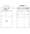
- FIG. 6 is a schematic view showing the structure of an X-ray image according to the Dicom standard created by the console unit 1 in the X-ray imaging apparatus, and the structure of exposure dose data created by the exposure measurement system 7;
- FIG. 6 is a schematic view showing data of an exposure dose for each X-ray examination created by the workstation 5;
- FIG. 1 is a schematic view of an X-ray imaging apparatus used for an exposure dose display apparatus according to the present invention.
- the X-ray imaging apparatus includes a console unit 1 and a high voltage apparatus 2 installed in an operation room 101 for an operator to perform an X-ray imaging operation, and an imaging room 100 for imaging a subject 20.
- the examination table 3 and the imaging unit 4 installed in The imaging room 100 and the operation room 101 are shut off by a partition 102.
- the console unit 1 installed in the operation room 101 includes a display unit 11 including a liquid crystal display and the like, and an operation unit 12 including a keyboard and a mouse for performing various operations, and performs X-ray imaging. It is for performing various operations for execution.
- the high voltage device 2 disposed in the partition 102 in the operation room 101 includes an operation panel having a display unit including a touch panel liquid crystal display and the like and various input buttons, and irradiation of X-rays. And a push button switch for starting.
- the high voltage device 2 is for setting irradiation conditions of X-rays such as a tube voltage and a tube current of an X-ray tube 42 described later, or an X-ray irradiation time.
- the examination table 3 installed in the imaging room 100 includes a top 31 on which the subject 20 is placed, an X-ray detection unit 33 in which a flat panel detector (FPD) as an X-ray detector is housed, and An elevator unit 34 for moving up and down the plate 31 and the X-ray detection unit 33 is provided.
- the X-ray detection unit 33 is horizontally movable in the left and right direction in FIG.
- the X-ray detection unit 33 can move up and down in the vertical direction in FIG. 1 together with the top 31.
- the imaging unit 4 installed in the imaging room 100 includes a base 61 movable in two directions orthogonal to the ceiling of the imaging room 100, a support 62 extending downward from the base 61, and the support A moving unit 63 which moves up and down and rotates with respect to 62, and a handle 41, an X-ray tube 42, and a collimator 43 integrally supported at the lower end of the moving unit 63 are provided.
- the X-ray detector 33 detects an X-ray emitted from the X-ray tube 42 to the subject 20 via the collimator 43.
- the X-ray image detected by the X-ray detection unit 33 is transmitted to the workstation 5 and the server 6 described later via the console unit 1. At this time, the examination start time and end time are transmitted together with the X-ray image.
- FIG. 2 is a block diagram showing a configuration of the exposure dose display device according to the present invention.
- the console unit 1 shown in FIG. 1 is connected to the workstation 5 and transmits an X-ray image to the workstation 5.
- This X-ray image is an image according to the Dicom standard.
- Dicom is an abbreviation of Digital Imaging and COmunications in Medicine, which is a format of medical images taken by CT, MRI, CR, etc. developed by the American Radiological Society (ACR) and the North American Electronic Equipment Industries Association (NEMA). This is a standard that defines a communication protocol between medical imaging devices that handle those images.
- the workstation 5 transmits this X-ray image and other data to the server 6.
- the server 6 is connected to a plurality of workstations 5 installed in a hospital, and is used to store various images captured in the hospital.
- the workstation 5 is connected to an exposure measurement system 7 which records the exposure dose measured by the individual dosimeter 8 worn by the operator together with time information.
- this exposure measurement system 7 includes a touch panel type screen installed in the imaging room 100, etc., and communicates with the personal dosimeter 8 within the communication range to realize real time operation.
- the radiation dose is displayed, and the radiation dose is stored.
- the individual dosimeter 8 is always worn by the operator on the clothes etc., and constantly measured the exposure dose at the time of daily examination and treatment.
- FIG. 3 is a schematic view showing the structure of an X-ray image according to the Dicom standard created by the console unit 1 in the X-ray imaging apparatus, and the structure of exposure dose data created by the exposure measurement system 7.
- An X-ray image conforming to the Dicom standard is composed of an information area in which various types of information such as imaging date and time, patient name, and imaging conditions are recorded, and an image area in which an image is recorded.
- the exposure dose data created by the exposure measurement system 7 has a structure in which the shooting date and time and the exposure dose at that time are connected.
- the workstation 5 shown in FIG. 3 uses the information of imaging date and time in the X-ray image according to the standard of Dicom, and the information of imaging date and time in the exposure dose data created by the exposure measurement system 7. Then, the X-ray image according to the Dicom standard and the exposure dose data created by the exposure measurement system 7 are linked. Then, the workstation 5 calculates an exposure dose for each X-ray image. In addition, the workstation 5 calculates an exposure dose for each X-ray examination based on the data of the examination start time and the examination end time received from the console unit 1.
- the workstation 5 functions as an information connection unit according to the present invention.
- FIG. 4 is a schematic view showing data of exposure dose for each X-ray examination created by the workstation 5.
- data including the examination time and photographing technique in each examination data including the information of the subject 20, and information indicating the exposure dose for the examination are configured as one data.
- This data is displayed on the display unit 51 of the workstation 5.
- the data may be displayed on the display unit 11 of the console unit 1 or the display unit 71 of the exposure measurement system 7.
- the data may be printed out and displayed.
- the display in this specification is a concept including such a form.
- the operator and the third party can grasp the exposure dose for each X-ray examination of each operator. For this reason, it becomes possible to reduce an exposure dose by visualizing the information of which X-ray inspection is exposed and improving the operation etc. in each inspection.
- the data of the exposure dose for each X-ray image may be displayed individually. In this case, it becomes possible to visualize information on what kind of X-ray exposure is being performed.
Landscapes
- Health & Medical Sciences (AREA)
- Life Sciences & Earth Sciences (AREA)
- Engineering & Computer Science (AREA)
- Medical Informatics (AREA)
- High Energy & Nuclear Physics (AREA)
- Molecular Biology (AREA)
- Physics & Mathematics (AREA)
- Nuclear Medicine, Radiotherapy & Molecular Imaging (AREA)
- General Health & Medical Sciences (AREA)
- Public Health (AREA)
- Radiology & Medical Imaging (AREA)
- Human Computer Interaction (AREA)
- Surgery (AREA)
- Veterinary Medicine (AREA)
- Spectroscopy & Molecular Physics (AREA)
- Animal Behavior & Ethology (AREA)
- Biophysics (AREA)
- Optics & Photonics (AREA)
- Pathology (AREA)
- Biomedical Technology (AREA)
- Heart & Thoracic Surgery (AREA)
- General Physics & Mathematics (AREA)
- Epidemiology (AREA)
- Primary Health Care (AREA)
- Apparatus For Radiation Diagnosis (AREA)
- Measurement Of Radiation (AREA)
Abstract
ワークステーション(5)は、このDicomの規格に従ったX線画像における撮影日・時刻の情報と、被曝測定システム(7)により作成された被曝線量データにおける撮影日・時刻の情報とを利用して、Dicomの規格に従ったX線画像と被曝測定システム(7)により作成された被曝線量データとを連結する。これにより、ワークステーション(5)は、X線画像毎の被曝線量を算出する。また、ワークステーション(5)は、コンソール部(1)より受け取った検査の開始時間と終了時間のデータに基づいて、X線検査毎の被曝線量を算出する。各検査における検査時間や撮影術式、また、被検者の情報を含むデータと、その検査に対する被曝線量を示す情報とが、一つのデータとして構成され、ワークステーション(5)における表示部(51)に表示される。
Description
この発明は、X線検査技師や医師等の術者の被曝線量を表示するための被曝線量表示装置に関する。
X線撮影装置を使用して検査や治療を行うときには、被検者の被曝だけではなく、その業務を日常的に実行するX線検査技師や医師等の術者の術者被曝を考慮する必要がある。術者被曝は、被曝しやすい利き手や、X線によるダメージを受けやすい両眼球付近、あるいは、甲状腺付近の被曝線量分布を可視化することが有効である。
このような被曝線量を表示するシステムとして、個人線量計により測定した被曝線量を時間情報とともに記録する被曝測定システムが提案されている。東洋メディック株式会社によるリアルタイム被曝測定システムである「RaySafe i2」は、検査室内に設置されたタッチパネル式のスクリーンを備え、通信範囲内にある個人線量計と通信して、リアルタイムの被曝線量を表示する構成を有する。「RaySafe i2」においては、各術者が装着した個人線量計毎に、現在受けている被曝線量の程度と積算線量値がタッチパネル式のスクリーンに表示される構成となっている(非特許文献1および非特許文献2参照)。
このような被曝測定システムにおいては、一般的に、被曝線量の情報と、その被曝を受けた被爆時間の情報は、記憶部に一旦記憶された上で、汎用のソフトウエアに対応した表形式のデータとして出力可能となっている。
東洋メディック株式会社:RaySafe i2:リアルタイム被曝測定システム:[平成29年9月14日検索]:インターネット<URL:http://www.toyo-medic.co.jp/seihin/catg03/raysafei2.html>
Unfors RaySafe社:RaySafe i2:Product Leaflet:[平成30年3月14日検索]:インターネット<URL:http://www.raysafe.com/Products/Staff/RaySafe%20i2#Downloads>
非特許文献1に記載されたように、個人線量計により測定した被曝線量の積算値等を表示することによりモニタリングする装置はいくつか提案されているが、それぞれの被曝がいずれのX線検査時になされたものであるのかを認識することはできなかった。いずれのX線検査で被曝しているのかの情報を可視化できたときには、各検査における操作等を改善することにより、被曝線量を低減することが可能となることが予想される。
この発明は上記課題を解決するためになされたものであり、X線検査毎の被曝線量を表示することが可能な被曝線量表示装置を提供することを目的とする。
請求項1に記載の発明は、被曝線量情報と被曝時間情報との関係を示す被曝線量データを記憶する被曝線量データ記憶部と、X線検査を実行するときにX線撮影装置により撮影されたX線画像を、前記X線画像に付与された撮影時間情報とともに記憶するX線画像記憶部と、前記被曝線量データ記憶部に記憶された前記被曝線量データと前記X線画像記憶部に記憶された前記X線画像とを、前記被曝時間情報と前記撮影時間情報とを利用して連結する情報連結部と、前記情報連結部により連結された前記被曝線量データと前記X線画像とに基づいて、前記X線検査毎の被曝線量、または、前記X線検査における前記X線画像毎の被曝線量を表示する被曝線量表示部と、を備えたことを特徴とする。
請求項2に記載の発明は、請求項1に記載の発明において、前記被曝線量情報と前記被曝時間情報との関係を示す前記被曝線量データは、個人線量計により測定した被曝線量を時間情報とともに記録する被曝測定システムにより作成される。
請求項1に記載の発明によれば、被曝線量データとX線画像とを被曝時間情報と撮影時間情報とを利用して連結することにより、X線検査毎またはX線画像毎の被曝線量を表示することが可能となる。これにより、いずれのX線検査あるいはX線画像の取得時に被曝しているのかという情報を可視化できることから、各検査における操作等を改善することにより被曝線量を低減することが可能となる
請求項2に記載の発明によれば、個人線量計により測定した被曝線量を時間情報とともに記録する被曝測定システムで作成された被曝線量データをそのまま利用することにより、X線検査毎あるいはX線画像毎の被曝線量を容易に表示することが可能となる。
以下、この発明の実施の形態を図面に基づいて説明する。図1は、この発明に係る被曝線量表示装置に使用するX線撮影装置の概要図である。
このX線撮影装置は、オペレータがX線撮影操作を実行するための操作室101に設置されたコンソール部1および高電圧装置2と、被検者20に対して撮影を行うための撮影室100に設置された検診台3および撮影部4とを備える。撮影室100と操作室101とは、隔壁102により遮断されている。
操作室101に設置されたコンソール部1は、液晶表示器等から構成される表示部11と、各種の操作を実行するためのキーボードやマウス等からなる操作部12とを備え、X線撮影を実行するための各種の操作を行うためのものである。また、操作室101内において隔壁102に配設された高電圧装置2は、タッチパネル式の液晶表示器等から構成される表示部と各種の入力ボタンとを有する操作パネルと、X線の照射を開始するための押しボタンスイッチとを備える。この高電圧装置2は、後述するX線管42の管電圧や管電流、あるいは、X線照射時間等のX線の照射条件を設定するためのものである。
撮影室100に設置された検診台3は、被検者20を載置する天板31と、内部にX線検出器としてのフラットパネルディテクタ(FPD)を収納したX線検出部33と、天板31およびX線検出部33を昇降するための昇降部34とを備える。X線検出部33は、図1における左右方向に水平移動可能となっている。また、X線検出部33は、天板31とともに、図1における上下方向に昇降可能となっている。また、撮影室100に設置された撮影部4は、撮影室100の天井に対して互いに直交する二方向に移動可能な基部61と、この基部61から下方に延びる支持部62と、この支持部62に対して昇降および回動する移動部63と、この移動部63の下端部に一体的に支持されるハンドル41、X線管42およびコリメータ43とを備える。
X線管42からコリメータ43を介して被検者20に照射されたX線は、X線検出部33により検出される。X線検出部33で検出されたX線画像は、コンソール部1を介して、後述するワークステーション5およびサーバ6に送信される。このときには、検査の開始時間と終了時間が、X線画像とともに送信される。
図2は、この発明に係る被曝量表示装置の構成を示すブロック図である。
図1に示すコンソール部1は、上述したように、ワークステーション5と接続されており、ワークステーション5に対してX線画像を送信する。このX線画像は、Dicomの規格に従った画像となっている。ここで、Dicomとは、Digital Imaging and COmmunications in Medicineの略で、米国放射線学会(ACR)と北米電子機器工業会(NEMA)が開発した、CTやMRI、CRなどで撮影した医用画像のフォーマットと、それらの画像を扱う医用画像機器間の通信プロトコルを定義した標準規格のことである。
ワークステーション5は、このX線画像と、その他のデータとを、サーバ6に送信する。このサーバ6は、病院内に設置された複数のワークステーション5と接続されており、病院内で撮影される各種の画像を保存するために使用される。また、ワークステーション5は、術者に装着された個人線量計8により測定した被曝線量を時間情報とともに記録する被曝測定システム7と接続されている。この被曝測定システム7は、例えば、非特許文献1に示すように、撮影室100内に設置されたタッチパネル式のスクリーン等を備え、通信範囲内にある個人線量計8と通信して、リアルタイムの被曝線量を表示するとともに、その被曝線量を記憶する構成を有する。なお、個人線量計8は、術者がその着衣等に常に装着し、日常の検査や治療時に、常時、被曝線量を測定する。
図3は、X線撮影装置におけるコンソール部1により作成されたDicomの規格に従ったX線画像の構造と、被曝測定システム7により作成された被曝線量データの構造とを示す模式図である。
Dicomの規格に従ったX線画像は、撮影日や時刻、患者名、撮影条件等の各種の情報が記録された情報領域と、画像が記録された画像領域とから構成される。一方、被曝測定システム7により作成された被曝線量データは、撮影日や時刻と、そのときの被曝線量とを連結した構造を有する。図3に示すワークステーション5は、このDicomの規格に従ったX線画像における撮影日・時刻の情報と、被曝測定システム7により作成された被曝線量データにおける撮影日・時刻の情報とを利用して、Dicomの規格に従ったX線画像と被曝測定システム7により作成された被曝線量データとを連結する。そして、ワークステーション5は、X線画像毎の被曝線量を算出する。また、ワークステーション5は、コンソール部1より受け取った検査の開始時間と終了時間のデータに基づいて、X線検査毎の被曝線量を算出する。このワークステーション5は、この発明に係る情報連結部として機能する。
図4は、ワークステーション5により作成されたX線検査毎の被曝線量のデータを示す模式図である。
この図に示すように、各検査における検査時間や撮影術式、また、被検者20の情報を含むデータと、その検査に対する被曝線量を示す情報とが、一つのデータとして構成される。このデータは、ワークステーション5における表示部51に表示される。なお、このデータを、コンソール部1における表示部11や、被曝測定システム7における表示部71にも表示するようにしてもよい。
また、このデータをワークステーション5における表示部51や、コンソール部1における表示部11、あるいは、被曝測定システム7における表示部71に表示するかわりに、プリントアウトして表示するようにしてもよい。この明細書における表示とは、このような形態をも含む概念である。
これにより、術者および第三者は、各術者のX線検査毎の被曝線量を把握することが可能となる。このため、いずれのX線検査で被曝しているかの情報を可視化し、各検査における操作等を改善することにより、被曝線量を低減することが可能となる。
なお、図4に示すようにX線検査毎の被曝線量のデータを表示する代わりに、X線画像毎の被曝線量のデータを個別に表示するようにしてもよい。この場合においては、どのようなX線撮影で被曝しているかの情報を可視化することが可能となる。
1 コンソール部
2 高電圧装置
3 検診台
4 撮影部
5 ワークステーション
6 サーバ
7 被曝測定システム
8 個別線量計
11 表示部
12 操作部
20 被検者
31 天板
33 X線検出部
42 X線管
43 コリメータ
51 表示部
71 表示部
100 撮影室
101 操作室
2 高電圧装置
3 検診台
4 撮影部
5 ワークステーション
6 サーバ
7 被曝測定システム
8 個別線量計
11 表示部
12 操作部
20 被検者
31 天板
33 X線検出部
42 X線管
43 コリメータ
51 表示部
71 表示部
100 撮影室
101 操作室
Claims (2)
- 被曝線量情報と被曝時間情報との関係を示す被曝線量データを記憶する被曝線量データ記憶部と、
X線検査を実行するときにX線撮影装置により撮影されたX線画像を、前記X線画像に付与された撮影時間情報とともに記憶するX線画像記憶部と、
前記被曝線量データ記憶部に記憶された前記被曝線量データと前記X線画像記憶部に記憶された前記X線画像とを、前記被曝時間情報と前記撮影時間情報とを利用して連結する情報連結部と、
前記情報連結部により連結された前記被曝線量データと前記X線画像とに基づいて、前記X線検査毎の被曝線量、または、前記X線検査における前記X線画像毎の被曝線量を表示する被曝線量表示部と、
を備えたことを特徴とする被曝線量表示装置。 - 請求項1に記載の被曝線量表示装置において、
前記被曝線量情報と前記被曝時間情報との関係を示す前記被曝線量データは、個人線量計により測定した被曝線量を時間情報とともに記録する被曝測定システムにより作成される被曝線量表示装置。
Priority Applications (3)
| Application Number | Priority Date | Filing Date | Title |
|---|---|---|---|
| CN201880040416.2A CN110770602B (zh) | 2017-09-27 | 2018-03-28 | 辐射曝光剂量显示装置 |
| US16/629,241 US11278252B2 (en) | 2017-09-27 | 2018-03-28 | Exposure-dose display device |
| JP2019544224A JP6891967B2 (ja) | 2017-09-27 | 2018-03-28 | 被曝線量表示装置 |
Applications Claiming Priority (2)
| Application Number | Priority Date | Filing Date | Title |
|---|---|---|---|
| JP2017-186486 | 2017-09-27 | ||
| JP2017186486 | 2017-09-27 |
Publications (1)
| Publication Number | Publication Date |
|---|---|
| WO2019064652A1 true WO2019064652A1 (ja) | 2019-04-04 |
Family
ID=65903244
Family Applications (1)
| Application Number | Title | Priority Date | Filing Date |
|---|---|---|---|
| PCT/JP2018/012913 Ceased WO2019064652A1 (ja) | 2017-09-27 | 2018-03-28 | 被曝線量表示装置 |
Country Status (4)
| Country | Link |
|---|---|
| US (1) | US11278252B2 (ja) |
| JP (1) | JP6891967B2 (ja) |
| CN (1) | CN110770602B (ja) |
| WO (1) | WO2019064652A1 (ja) |
Cited By (2)
| Publication number | Priority date | Publication date | Assignee | Title |
|---|---|---|---|---|
| WO2021234234A1 (fr) * | 2020-05-21 | 2021-11-25 | Fibermetrix | Procede de traitement des donnees relatives a un examen radiologique d'un patient |
| JP2023162794A (ja) * | 2022-04-27 | 2023-11-09 | キヤノンメディカルシステムズ株式会社 | X線撮影装置、医用情報処理装置、及び医用情報処理プログラム |
Families Citing this family (2)
| Publication number | Priority date | Publication date | Assignee | Title |
|---|---|---|---|---|
| IT201900005170A1 (it) * | 2019-04-05 | 2020-10-05 | Ubiquicom S R L | Sistema e metodo di monitoraggio dell’esposizione a radiazioni in ambito ospedaliero per un operatore |
| TWI906703B (zh) * | 2023-12-01 | 2025-12-01 | 宏碁智醫股份有限公司 | 資料處理方法與裝置 |
Citations (2)
| Publication number | Priority date | Publication date | Assignee | Title |
|---|---|---|---|---|
| JP2013113844A (ja) * | 2012-03-27 | 2013-06-10 | Techno Link Co Ltd | 個人線量計 |
| JP2015167695A (ja) * | 2014-03-06 | 2015-09-28 | 株式会社東芝 | 医用情報処理装置及び医用情報処理システム |
Family Cites Families (16)
| Publication number | Priority date | Publication date | Assignee | Title |
|---|---|---|---|---|
| JP2947830B2 (ja) * | 1989-09-11 | 1999-09-13 | 株式会社日立製作所 | 放射線作業支援方法及びその支援装置 |
| JP2003130956A (ja) * | 2001-10-24 | 2003-05-08 | Hitachi Ltd | 被ばく管理,放射線作業管理システムとその方法 |
| DE102004035740A1 (de) * | 2004-07-23 | 2006-03-16 | Siemens Ag | Verfahren zur Zwischenbildgenerierung bei der Bildgebung mit einer tomographischen bildgebenden Modalität |
| JP4250147B2 (ja) * | 2004-09-13 | 2009-04-08 | 日油技研工業株式会社 | 被ばく線量検量方法 |
| JP2007097909A (ja) * | 2005-10-05 | 2007-04-19 | Bio Arts:Kk | 放射線被曝線量管理システム及び記憶媒体 |
| US8581214B2 (en) * | 2007-05-07 | 2013-11-12 | Koninklijke Philips N.V. | Staff dose awareness indication |
| JP5386227B2 (ja) * | 2009-05-18 | 2014-01-15 | 株式会社日立メディコ | X線画像診断装置及び可搬記録媒体 |
| NZ588050A (en) * | 2010-09-17 | 2013-03-28 | Ind Res Ltd | Recording dose information about ionizing radiation absored and stored in a dosimeter |
| US8575554B2 (en) * | 2011-07-26 | 2013-11-05 | General Electric Company | Methods and systems for scatter estimation in positron emission tomography |
| JP5708827B2 (ja) * | 2011-12-27 | 2015-04-30 | 株式会社島津製作所 | 放射線撮影装置 |
| JP6109650B2 (ja) | 2013-06-06 | 2017-04-05 | 東芝メディカルシステムズ株式会社 | X線診断装置、被曝管理装置、散乱線線量分布形成方法、および散乱線線量分布形成プログラム |
| JP6364201B2 (ja) * | 2014-02-24 | 2018-07-25 | 株式会社日本環境調査研究所 | 空間線量当量率可視化システム |
| FR3023624B1 (fr) * | 2014-07-11 | 2022-07-15 | Conseils Et Etudes En Radioprotection | Procede et dispositif pour le suivi spatial et temporel de l’exposition a des risques |
| JP6749757B2 (ja) * | 2015-12-21 | 2020-09-02 | ジーイー・メディカル・システムズ・グローバル・テクノロジー・カンパニー・エルエルシー | 放射線断層撮影システム |
| US10422886B1 (en) * | 2016-11-17 | 2019-09-24 | Clinitraq | Real-time location aware radiation system and method for use thereof |
| JP6953199B2 (ja) * | 2017-06-26 | 2021-10-27 | キヤノンメディカルシステムズ株式会社 | 医用装置及びx線システム |
-
2018
- 2018-03-28 CN CN201880040416.2A patent/CN110770602B/zh active Active
- 2018-03-28 JP JP2019544224A patent/JP6891967B2/ja active Active
- 2018-03-28 WO PCT/JP2018/012913 patent/WO2019064652A1/ja not_active Ceased
- 2018-03-28 US US16/629,241 patent/US11278252B2/en active Active
Patent Citations (2)
| Publication number | Priority date | Publication date | Assignee | Title |
|---|---|---|---|---|
| JP2013113844A (ja) * | 2012-03-27 | 2013-06-10 | Techno Link Co Ltd | 個人線量計 |
| JP2015167695A (ja) * | 2014-03-06 | 2015-09-28 | 株式会社東芝 | 医用情報処理装置及び医用情報処理システム |
Cited By (4)
| Publication number | Priority date | Publication date | Assignee | Title |
|---|---|---|---|---|
| WO2021234234A1 (fr) * | 2020-05-21 | 2021-11-25 | Fibermetrix | Procede de traitement des donnees relatives a un examen radiologique d'un patient |
| FR3110708A1 (fr) * | 2020-05-21 | 2021-11-26 | Fibermetrix | Procede de traitement des donnees relatives a un examen radiologique d’un patient |
| US12276763B2 (en) | 2020-05-21 | 2025-04-15 | Fibermetrix | Method for processing data relating to a radiological examination of a patient |
| JP2023162794A (ja) * | 2022-04-27 | 2023-11-09 | キヤノンメディカルシステムズ株式会社 | X線撮影装置、医用情報処理装置、及び医用情報処理プログラム |
Also Published As
| Publication number | Publication date |
|---|---|
| JPWO2019064652A1 (ja) | 2020-03-26 |
| US11278252B2 (en) | 2022-03-22 |
| CN110770602B (zh) | 2023-06-23 |
| US20200214657A1 (en) | 2020-07-09 |
| JP6891967B2 (ja) | 2021-06-18 |
| CN110770602A (zh) | 2020-02-07 |
Similar Documents
| Publication | Publication Date | Title |
|---|---|---|
| US11224398B2 (en) | Wireless x-ray system | |
| US11129579B2 (en) | Dental treatment planning apparatus and method using matching of two-dimensional medical image and three-dimensional medical image | |
| JP6891967B2 (ja) | 被曝線量表示装置 | |
| JP7106718B2 (ja) | 放射線撮影装置、放射線撮影システム、放射線撮影方法、及びプログラム | |
| JP2014210049A (ja) | 支持器及びx線診断装置 | |
| Little et al. | COVID-19 and beyond: implications for dental radiography | |
| US20170329501A1 (en) | Image analysis system | |
| Tadinada et al. | The reliability of tablet computers in depicting maxillofacial radiographic landmarks | |
| JP5738000B2 (ja) | 放射線撮影システム及びその制御方法、並びに、プログラム | |
| JP2011005169A (ja) | 医用画像管理装置及びプログラム | |
| Ho et al. | Eye of the storm: the roles of a radiology department in the outbreak of severe acute respiratory syndrome | |
| JP2004275601A (ja) | 画像管理装置および画像表示装置 | |
| JP2024104629A (ja) | 画像処理装置、放射線撮影システム、画像処理方法及びプログラム | |
| JP4950461B2 (ja) | 照射線量異常分析装置および照射線量異常分析プログラム | |
| US12478339B2 (en) | Round radiography system, console for round radiography, and recording medium | |
| Alipio et al. | Image Repeat Analysis in Conventional Radiography in Mobile Clinics: A Retrospective Observational Study | |
| JP2025158400A (ja) | 画像処理装置および動態画像における角度計測方法 | |
| Singh et al. | Compliance of dentists with safety standards of dental radiology | |
| JP2013149267A (ja) | 診断支援システム | |
| US20240307012A1 (en) | Radiographic imaging support apparatus, radiographic imaging support method, and recording medium | |
| WO2019031696A1 (ko) | 방사선량 통합 관리 시스템 및 방법 | |
| JP2011189091A (ja) | 検像装置及び検像システム | |
| JP2022179672A (ja) | 解析処理システム | |
| Weir et al. | Fluoroscopy and Interventional Radiology | |
| JP6283877B2 (ja) | 医用画像表示装置 |
Legal Events
| Date | Code | Title | Description |
|---|---|---|---|
| 121 | Ep: the epo has been informed by wipo that ep was designated in this application |
Ref document number: 18862073 Country of ref document: EP Kind code of ref document: A1 |
|
| ENP | Entry into the national phase |
Ref document number: 2019544224 Country of ref document: JP Kind code of ref document: A |
|
| NENP | Non-entry into the national phase |
Ref country code: DE |
|
| 122 | Ep: pct application non-entry in european phase |
Ref document number: 18862073 Country of ref document: EP Kind code of ref document: A1 |