WO2019049957A1 - 幼若ブタ由来幹細胞およびその調製方法 - Google Patents
幼若ブタ由来幹細胞およびその調製方法 Download PDFInfo
- Publication number
- WO2019049957A1 WO2019049957A1 PCT/JP2018/033110 JP2018033110W WO2019049957A1 WO 2019049957 A1 WO2019049957 A1 WO 2019049957A1 JP 2018033110 W JP2018033110 W JP 2018033110W WO 2019049957 A1 WO2019049957 A1 WO 2019049957A1
- Authority
- WO
- WIPO (PCT)
- Prior art keywords
- cells
- stem cells
- cell
- msc
- juvenile
- Prior art date
Links
- 210000000130 stem cell Anatomy 0.000 title claims abstract description 108
- 238000002360 preparation method Methods 0.000 title abstract description 25
- 210000004027 cell Anatomy 0.000 claims description 217
- 230000000366 juvenile effect Effects 0.000 claims description 85
- 210000002901 mesenchymal stem cell Anatomy 0.000 claims description 84
- 210000001185 bone marrow Anatomy 0.000 claims description 65
- 238000007710 freezing Methods 0.000 claims description 25
- 230000008014 freezing Effects 0.000 claims description 25
- 238000000034 method Methods 0.000 claims description 24
- 210000005087 mononuclear cell Anatomy 0.000 claims description 19
- 238000002054 transplantation Methods 0.000 claims description 14
- 241000282887 Suidae Species 0.000 claims description 11
- 238000010899 nucleation Methods 0.000 claims description 11
- 230000003698 anagen phase Effects 0.000 claims description 5
- 238000002955 isolation Methods 0.000 claims description 5
- 230000002062 proliferating effect Effects 0.000 claims description 2
- 230000004069 differentiation Effects 0.000 abstract description 34
- 230000035755 proliferation Effects 0.000 abstract description 14
- 241000282898 Sus scrofa Species 0.000 description 85
- 239000002609 medium Substances 0.000 description 53
- 108010010803 Gelatin Proteins 0.000 description 28
- 229920000159 gelatin Polymers 0.000 description 28
- 239000008273 gelatin Substances 0.000 description 28
- 235000019322 gelatine Nutrition 0.000 description 28
- 235000011852 gelatine desserts Nutrition 0.000 description 28
- CIWBSHSKHKDKBQ-JLAZNSOCSA-N Ascorbic acid Chemical compound OC[C@H](O)[C@H]1OC(=O)C(O)=C1O CIWBSHSKHKDKBQ-JLAZNSOCSA-N 0.000 description 24
- 239000007640 basal medium Substances 0.000 description 21
- 239000006285 cell suspension Substances 0.000 description 15
- IAZDPXIOMUYVGZ-UHFFFAOYSA-N Dimethylsulphoxide Chemical compound CS(C)=O IAZDPXIOMUYVGZ-UHFFFAOYSA-N 0.000 description 14
- 239000000872 buffer Substances 0.000 description 13
- 238000004458 analytical method Methods 0.000 description 12
- 238000012360 testing method Methods 0.000 description 12
- 238000012258 culturing Methods 0.000 description 11
- 239000008188 pellet Substances 0.000 description 11
- 239000000243 solution Substances 0.000 description 11
- IJGRMHOSHXDMSA-UHFFFAOYSA-N Atomic nitrogen Chemical compound N#N IJGRMHOSHXDMSA-UHFFFAOYSA-N 0.000 description 10
- 108091003079 Bovine Serum Albumin Proteins 0.000 description 10
- 101000800116 Homo sapiens Thy-1 membrane glycoprotein Proteins 0.000 description 10
- 102100033523 Thy-1 membrane glycoprotein Human genes 0.000 description 10
- 239000012091 fetal bovine serum Substances 0.000 description 10
- 102100032912 CD44 antigen Human genes 0.000 description 9
- 101000868273 Homo sapiens CD44 antigen Proteins 0.000 description 9
- 210000001789 adipocyte Anatomy 0.000 description 9
- 210000001988 somatic stem cell Anatomy 0.000 description 9
- 238000010186 staining Methods 0.000 description 9
- ZZZCUOFIHGPKAK-UHFFFAOYSA-N D-erythro-ascorbic acid Natural products OCC1OC(=O)C(O)=C1O ZZZCUOFIHGPKAK-UHFFFAOYSA-N 0.000 description 8
- 229930003268 Vitamin C Natural products 0.000 description 8
- 238000005138 cryopreservation Methods 0.000 description 8
- 235000019154 vitamin C Nutrition 0.000 description 8
- 239000011718 vitamin C Substances 0.000 description 8
- 101710160107 Outer membrane protein A Proteins 0.000 description 7
- 210000001612 chondrocyte Anatomy 0.000 description 7
- 239000012530 fluid Substances 0.000 description 7
- 230000012010 growth Effects 0.000 description 7
- 239000001963 growth medium Substances 0.000 description 6
- 241000894007 species Species 0.000 description 6
- 230000035899 viability Effects 0.000 description 6
- 101000935043 Homo sapiens Integrin beta-1 Proteins 0.000 description 5
- 102100025304 Integrin beta-1 Human genes 0.000 description 5
- 102000004142 Trypsin Human genes 0.000 description 5
- 108090000631 Trypsin Proteins 0.000 description 5
- 238000005119 centrifugation Methods 0.000 description 5
- 239000011248 coating agent Substances 0.000 description 5
- 238000000576 coating method Methods 0.000 description 5
- 239000007788 liquid Substances 0.000 description 5
- 229910052757 nitrogen Inorganic materials 0.000 description 5
- 210000004409 osteocyte Anatomy 0.000 description 5
- 239000006228 supernatant Substances 0.000 description 5
- 230000004083 survival effect Effects 0.000 description 5
- 210000001519 tissue Anatomy 0.000 description 5
- 239000012588 trypsin Substances 0.000 description 5
- 235000005956 Cosmos caudatus Nutrition 0.000 description 4
- 235000010323 ascorbic acid Nutrition 0.000 description 4
- 239000011668 ascorbic acid Substances 0.000 description 4
- 210000002449 bone cell Anatomy 0.000 description 4
- 230000006698 induction Effects 0.000 description 4
- 239000003550 marker Substances 0.000 description 4
- 210000002894 multi-fate stem cell Anatomy 0.000 description 4
- 239000002777 nucleoside Substances 0.000 description 4
- 125000003835 nucleoside group Chemical group 0.000 description 4
- 229960005322 streptomycin Drugs 0.000 description 4
- 230000001225 therapeutic effect Effects 0.000 description 4
- 210000000689 upper leg Anatomy 0.000 description 4
- 102000004190 Enzymes Human genes 0.000 description 3
- 108090000790 Enzymes Proteins 0.000 description 3
- 241000588724 Escherichia coli Species 0.000 description 3
- 241000282412 Homo Species 0.000 description 3
- OKKJLVBELUTLKV-UHFFFAOYSA-N Methanol Chemical compound OC OKKJLVBELUTLKV-UHFFFAOYSA-N 0.000 description 3
- 208000010378 Pulmonary Embolism Diseases 0.000 description 3
- 238000003556 assay Methods 0.000 description 3
- 210000000988 bone and bone Anatomy 0.000 description 3
- 210000002798 bone marrow cell Anatomy 0.000 description 3
- 238000004113 cell culture Methods 0.000 description 3
- UREBDLICKHMUKA-CXSFZGCWSA-N dexamethasone Chemical compound C1CC2=CC(=O)C=C[C@]2(C)[C@]2(F)[C@@H]1[C@@H]1C[C@@H](C)[C@@](C(=O)CO)(O)[C@@]1(C)C[C@@H]2O UREBDLICKHMUKA-CXSFZGCWSA-N 0.000 description 3
- 229960003957 dexamethasone Drugs 0.000 description 3
- 201000010099 disease Diseases 0.000 description 3
- 208000037265 diseases, disorders, signs and symptoms Diseases 0.000 description 3
- 239000012997 ficoll-paque Substances 0.000 description 3
- 230000000977 initiatory effect Effects 0.000 description 3
- 210000004153 islets of langerhan Anatomy 0.000 description 3
- 239000011435 rock Substances 0.000 description 3
- 239000000725 suspension Substances 0.000 description 3
- 229930003231 vitamin Natural products 0.000 description 3
- 235000013343 vitamin Nutrition 0.000 description 3
- 239000011782 vitamin Substances 0.000 description 3
- 229940088594 vitamin Drugs 0.000 description 3
- 150000003722 vitamin derivatives Chemical class 0.000 description 3
- XLYOFNOQVPJJNP-UHFFFAOYSA-N water Substances O XLYOFNOQVPJJNP-UHFFFAOYSA-N 0.000 description 3
- HDTRYLNUVZCQOY-UHFFFAOYSA-N α-D-glucopyranosyl-α-D-glucopyranoside Natural products OC1C(O)C(O)C(CO)OC1OC1C(O)C(O)C(O)C(CO)O1 HDTRYLNUVZCQOY-UHFFFAOYSA-N 0.000 description 2
- 102000002260 Alkaline Phosphatase Human genes 0.000 description 2
- 108020004774 Alkaline Phosphatase Proteins 0.000 description 2
- OYPRJOBELJOOCE-UHFFFAOYSA-N Calcium Chemical compound [Ca] OYPRJOBELJOOCE-UHFFFAOYSA-N 0.000 description 2
- 229920002307 Dextran Polymers 0.000 description 2
- DHCLVCXQIBBOPH-UHFFFAOYSA-N Glycerol 2-phosphate Chemical compound OCC(CO)OP(O)(O)=O DHCLVCXQIBBOPH-UHFFFAOYSA-N 0.000 description 2
- 206010061218 Inflammation Diseases 0.000 description 2
- FYYHWMGAXLPEAU-UHFFFAOYSA-N Magnesium Chemical compound [Mg] FYYHWMGAXLPEAU-UHFFFAOYSA-N 0.000 description 2
- HDTRYLNUVZCQOY-WSWWMNSNSA-N Trehalose Natural products O[C@@H]1[C@@H](O)[C@@H](O)[C@@H](CO)O[C@@H]1O[C@@H]1[C@H](O)[C@@H](O)[C@@H](O)[C@@H](CO)O1 HDTRYLNUVZCQOY-WSWWMNSNSA-N 0.000 description 2
- 230000002293 adipogenic effect Effects 0.000 description 2
- 238000013019 agitation Methods 0.000 description 2
- HDTRYLNUVZCQOY-LIZSDCNHSA-N alpha,alpha-trehalose Chemical compound O[C@@H]1[C@@H](O)[C@H](O)[C@@H](CO)O[C@@H]1O[C@@H]1[C@H](O)[C@@H](O)[C@H](O)[C@@H](CO)O1 HDTRYLNUVZCQOY-LIZSDCNHSA-N 0.000 description 2
- 229940072107 ascorbate Drugs 0.000 description 2
- 229960005070 ascorbic acid Drugs 0.000 description 2
- 230000015572 biosynthetic process Effects 0.000 description 2
- 230000002308 calcification Effects 0.000 description 2
- 239000011575 calcium Substances 0.000 description 2
- 229910052791 calcium Inorganic materials 0.000 description 2
- 239000003153 chemical reaction reagent Substances 0.000 description 2
- 230000005757 colony formation Effects 0.000 description 2
- 238000012790 confirmation Methods 0.000 description 2
- 238000010586 diagram Methods 0.000 description 2
- 210000002950 fibroblast Anatomy 0.000 description 2
- 238000000684 flow cytometry Methods 0.000 description 2
- 238000005194 fractionation Methods 0.000 description 2
- 230000004957 immunoregulator effect Effects 0.000 description 2
- CGIGDMFJXJATDK-UHFFFAOYSA-N indomethacin Chemical compound CC1=C(CC(O)=O)C2=CC(OC)=CC=C2N1C(=O)C1=CC=C(Cl)C=C1 CGIGDMFJXJATDK-UHFFFAOYSA-N 0.000 description 2
- 230000004054 inflammatory process Effects 0.000 description 2
- 238000011081 inoculation Methods 0.000 description 2
- NOESYZHRGYRDHS-UHFFFAOYSA-N insulin Chemical compound N1C(=O)C(NC(=O)C(CCC(N)=O)NC(=O)C(CCC(O)=O)NC(=O)C(C(C)C)NC(=O)C(NC(=O)CN)C(C)CC)CSSCC(C(NC(CO)C(=O)NC(CC(C)C)C(=O)NC(CC=2C=CC(O)=CC=2)C(=O)NC(CCC(N)=O)C(=O)NC(CC(C)C)C(=O)NC(CCC(O)=O)C(=O)NC(CC(N)=O)C(=O)NC(CC=2C=CC(O)=CC=2)C(=O)NC(CSSCC(NC(=O)C(C(C)C)NC(=O)C(CC(C)C)NC(=O)C(CC=2C=CC(O)=CC=2)NC(=O)C(CC(C)C)NC(=O)C(C)NC(=O)C(CCC(O)=O)NC(=O)C(C(C)C)NC(=O)C(CC(C)C)NC(=O)C(CC=2NC=NC=2)NC(=O)C(CO)NC(=O)CNC2=O)C(=O)NCC(=O)NC(CCC(O)=O)C(=O)NC(CCCNC(N)=N)C(=O)NCC(=O)NC(CC=3C=CC=CC=3)C(=O)NC(CC=3C=CC=CC=3)C(=O)NC(CC=3C=CC(O)=CC=3)C(=O)NC(C(C)O)C(=O)N3C(CCC3)C(=O)NC(CCCCN)C(=O)NC(C)C(O)=O)C(=O)NC(CC(N)=O)C(O)=O)=O)NC(=O)C(C(C)CC)NC(=O)C(CO)NC(=O)C(C(C)O)NC(=O)C1CSSCC2NC(=O)C(CC(C)C)NC(=O)C(NC(=O)C(CCC(N)=O)NC(=O)C(CC(N)=O)NC(=O)C(NC(=O)C(N)CC=1C=CC=CC=1)C(C)C)CC1=CN=CN1 NOESYZHRGYRDHS-UHFFFAOYSA-N 0.000 description 2
- 230000007774 longterm Effects 0.000 description 2
- 239000011777 magnesium Substances 0.000 description 2
- 229910052749 magnesium Inorganic materials 0.000 description 2
- 238000012423 maintenance Methods 0.000 description 2
- 239000011259 mixed solution Substances 0.000 description 2
- 210000000056 organ Anatomy 0.000 description 2
- 210000000496 pancreas Anatomy 0.000 description 2
- 239000008363 phosphate buffer Substances 0.000 description 2
- 210000001778 pluripotent stem cell Anatomy 0.000 description 2
- 238000004321 preservation Methods 0.000 description 2
- 238000011160 research Methods 0.000 description 2
- 238000010079 rubber tapping Methods 0.000 description 2
- 230000028327 secretion Effects 0.000 description 2
- 210000002966 serum Anatomy 0.000 description 2
- DAEPDZWVDSPTHF-UHFFFAOYSA-M sodium pyruvate Chemical compound [Na+].CC(=O)C([O-])=O DAEPDZWVDSPTHF-UHFFFAOYSA-M 0.000 description 2
- 238000003860 storage Methods 0.000 description 2
- 238000010257 thawing Methods 0.000 description 2
- 210000002444 unipotent stem cell Anatomy 0.000 description 2
- APIXJSLKIYYUKG-UHFFFAOYSA-N 3 Isobutyl 1 methylxanthine Chemical compound O=C1N(C)C(=O)N(CC(C)C)C2=C1N=CN2 APIXJSLKIYYUKG-UHFFFAOYSA-N 0.000 description 1
- 239000012114 Alexa Fluor 647 Substances 0.000 description 1
- 208000023275 Autoimmune disease Diseases 0.000 description 1
- 208000020084 Bone disease Diseases 0.000 description 1
- 208000024172 Cardiovascular disease Diseases 0.000 description 1
- 102000019034 Chemokines Human genes 0.000 description 1
- 108010012236 Chemokines Proteins 0.000 description 1
- 102000004127 Cytokines Human genes 0.000 description 1
- 108090000695 Cytokines Proteins 0.000 description 1
- 208000009329 Graft vs Host Disease Diseases 0.000 description 1
- HTTJABKRGRZYRN-UHFFFAOYSA-N Heparin Chemical compound OC1C(NC(=O)C)C(O)OC(COS(O)(=O)=O)C1OC1C(OS(O)(=O)=O)C(O)C(OC2C(C(OS(O)(=O)=O)C(OC3C(C(O)C(O)C(O3)C(O)=O)OS(O)(=O)=O)C(CO)O2)NS(O)(=O)=O)C(C(O)=O)O1 HTTJABKRGRZYRN-UHFFFAOYSA-N 0.000 description 1
- 206010061216 Infarction Diseases 0.000 description 1
- 102100023915 Insulin Human genes 0.000 description 1
- 108090001061 Insulin Proteins 0.000 description 1
- ONIBWKKTOPOVIA-BYPYZUCNSA-N L-Proline Chemical compound OC(=O)[C@@H]1CCCN1 ONIBWKKTOPOVIA-BYPYZUCNSA-N 0.000 description 1
- JVTAAEKCZFNVCJ-UHFFFAOYSA-M Lactate Chemical compound CC(O)C([O-])=O JVTAAEKCZFNVCJ-UHFFFAOYSA-M 0.000 description 1
- -1 MCGS Chemical compound 0.000 description 1
- 206010029719 Nonspecific reaction Diseases 0.000 description 1
- ONIBWKKTOPOVIA-UHFFFAOYSA-N Proline Natural products OC(=O)C1CCCN1 ONIBWKKTOPOVIA-UHFFFAOYSA-N 0.000 description 1
- 208000001647 Renal Insufficiency Diseases 0.000 description 1
- 239000012891 Ringer solution Substances 0.000 description 1
- FHNINJWBTRXEBC-UHFFFAOYSA-N Sudan III Chemical compound OC1=CC=C2C=CC=CC2=C1N=NC(C=C1)=CC=C1N=NC1=CC=CC=C1 FHNINJWBTRXEBC-UHFFFAOYSA-N 0.000 description 1
- 241000700605 Viruses Species 0.000 description 1
- 239000002253 acid Substances 0.000 description 1
- 230000001464 adherent effect Effects 0.000 description 1
- 239000002870 angiogenesis inducing agent Substances 0.000 description 1
- 210000004102 animal cell Anatomy 0.000 description 1
- 239000003242 anti bacterial agent Substances 0.000 description 1
- 230000003510 anti-fibrotic effect Effects 0.000 description 1
- 230000003110 anti-inflammatory effect Effects 0.000 description 1
- 239000000427 antigen Substances 0.000 description 1
- 102000036639 antigens Human genes 0.000 description 1
- 108091007433 antigens Proteins 0.000 description 1
- 230000008901 benefit Effects 0.000 description 1
- 230000003115 biocidal effect Effects 0.000 description 1
- 230000037396 body weight Effects 0.000 description 1
- 239000012888 bovine serum Substances 0.000 description 1
- BPKIGYQJPYCAOW-FFJTTWKXSA-I calcium;potassium;disodium;(2s)-2-hydroxypropanoate;dichloride;dihydroxide;hydrate Chemical compound O.[OH-].[OH-].[Na+].[Na+].[Cl-].[Cl-].[K+].[Ca+2].C[C@H](O)C([O-])=O BPKIGYQJPYCAOW-FFJTTWKXSA-I 0.000 description 1
- 210000000845 cartilage Anatomy 0.000 description 1
- 230000010261 cell growth Effects 0.000 description 1
- 239000002771 cell marker Substances 0.000 description 1
- 230000004663 cell proliferation Effects 0.000 description 1
- 206010008118 cerebral infarction Diseases 0.000 description 1
- 208000026106 cerebrovascular disease Diseases 0.000 description 1
- 230000000295 complement effect Effects 0.000 description 1
- 238000004132 cross linking Methods 0.000 description 1
- 238000012136 culture method Methods 0.000 description 1
- 230000003247 decreasing effect Effects 0.000 description 1
- 238000011161 development Methods 0.000 description 1
- 230000018109 developmental process Effects 0.000 description 1
- 238000010790 dilution Methods 0.000 description 1
- 239000012895 dilution Substances 0.000 description 1
- 238000011156 evaluation Methods 0.000 description 1
- 210000001808 exosome Anatomy 0.000 description 1
- 238000002474 experimental method Methods 0.000 description 1
- 210000004700 fetal blood Anatomy 0.000 description 1
- 230000003176 fibrotic effect Effects 0.000 description 1
- 208000024908 graft versus host disease Diseases 0.000 description 1
- 230000005484 gravity Effects 0.000 description 1
- 239000003102 growth factor Substances 0.000 description 1
- 210000003958 hematopoietic stem cell Anatomy 0.000 description 1
- 238000007490 hematoxylin and eosin (H&E) staining Methods 0.000 description 1
- 229960002897 heparin Drugs 0.000 description 1
- 229920000669 heparin Polymers 0.000 description 1
- 230000028993 immune response Effects 0.000 description 1
- 229960000905 indomethacin Drugs 0.000 description 1
- 230000007574 infarction Effects 0.000 description 1
- 238000002347 injection Methods 0.000 description 1
- 239000007924 injection Substances 0.000 description 1
- 229940125396 insulin Drugs 0.000 description 1
- 230000005732 intercellular adhesion Effects 0.000 description 1
- 230000009545 invasion Effects 0.000 description 1
- 208000023589 ischemic disease Diseases 0.000 description 1
- 201000006370 kidney failure Diseases 0.000 description 1
- 208000019423 liver disease Diseases 0.000 description 1
- 210000004072 lung Anatomy 0.000 description 1
- 210000004698 lymphocyte Anatomy 0.000 description 1
- 210000001161 mammalian embryo Anatomy 0.000 description 1
- 238000005259 measurement Methods 0.000 description 1
- 210000003716 mesoderm Anatomy 0.000 description 1
- 244000309715 mini pig Species 0.000 description 1
- 238000012986 modification Methods 0.000 description 1
- 230000004048 modification Effects 0.000 description 1
- 210000001178 neural stem cell Anatomy 0.000 description 1
- 230000011164 ossification Effects 0.000 description 1
- 201000008482 osteoarthritis Diseases 0.000 description 1
- 244000052769 pathogen Species 0.000 description 1
- 238000007747 plating Methods 0.000 description 1
- 239000003761 preservation solution Substances 0.000 description 1
- 230000008569 process Effects 0.000 description 1
- 229960002429 proline Drugs 0.000 description 1
- 230000008929 regeneration Effects 0.000 description 1
- 238000011069 regeneration method Methods 0.000 description 1
- 238000007634 remodeling Methods 0.000 description 1
- 208000023504 respiratory system disease Diseases 0.000 description 1
- 229910052711 selenium Inorganic materials 0.000 description 1
- 239000011669 selenium Substances 0.000 description 1
- 238000000926 separation method Methods 0.000 description 1
- 229940054269 sodium pyruvate Drugs 0.000 description 1
- 239000007787 solid Substances 0.000 description 1
- 208000020431 spinal cord injury Diseases 0.000 description 1
- 210000001562 sternum Anatomy 0.000 description 1
- 239000013589 supplement Substances 0.000 description 1
- 238000004114 suspension culture Methods 0.000 description 1
- 210000003462 vein Anatomy 0.000 description 1
Images


Classifications
-
- C—CHEMISTRY; METALLURGY
- C12—BIOCHEMISTRY; BEER; SPIRITS; WINE; VINEGAR; MICROBIOLOGY; ENZYMOLOGY; MUTATION OR GENETIC ENGINEERING
- C12N—MICROORGANISMS OR ENZYMES; COMPOSITIONS THEREOF; PROPAGATING, PRESERVING, OR MAINTAINING MICROORGANISMS; MUTATION OR GENETIC ENGINEERING; CULTURE MEDIA
- C12N5/00—Undifferentiated human, animal or plant cells, e.g. cell lines; Tissues; Cultivation or maintenance thereof; Culture media therefor
- C12N5/06—Animal cells or tissues; Human cells or tissues
- C12N5/0602—Vertebrate cells
- C12N5/0652—Cells of skeletal and connective tissues; Mesenchyme
- C12N5/0662—Stem cells
- C12N5/0663—Bone marrow mesenchymal stem cells (BM-MSC)
-
- C—CHEMISTRY; METALLURGY
- C12—BIOCHEMISTRY; BEER; SPIRITS; WINE; VINEGAR; MICROBIOLOGY; ENZYMOLOGY; MUTATION OR GENETIC ENGINEERING
- C12N—MICROORGANISMS OR ENZYMES; COMPOSITIONS THEREOF; PROPAGATING, PRESERVING, OR MAINTAINING MICROORGANISMS; MUTATION OR GENETIC ENGINEERING; CULTURE MEDIA
- C12N5/00—Undifferentiated human, animal or plant cells, e.g. cell lines; Tissues; Cultivation or maintenance thereof; Culture media therefor
- C12N5/06—Animal cells or tissues; Human cells or tissues
- C12N5/0602—Vertebrate cells
- C12N5/0634—Cells from the blood or the immune system
-
- C—CHEMISTRY; METALLURGY
- C12—BIOCHEMISTRY; BEER; SPIRITS; WINE; VINEGAR; MICROBIOLOGY; ENZYMOLOGY; MUTATION OR GENETIC ENGINEERING
- C12N—MICROORGANISMS OR ENZYMES; COMPOSITIONS THEREOF; PROPAGATING, PRESERVING, OR MAINTAINING MICROORGANISMS; MUTATION OR GENETIC ENGINEERING; CULTURE MEDIA
- C12N5/00—Undifferentiated human, animal or plant cells, e.g. cell lines; Tissues; Cultivation or maintenance thereof; Culture media therefor
- C12N5/06—Animal cells or tissues; Human cells or tissues
- C12N5/0602—Vertebrate cells
- C12N5/0676—Pancreatic cells
- C12N5/0678—Stem cells; Progenitor cells; Precursor cells
-
- C—CHEMISTRY; METALLURGY
- C12—BIOCHEMISTRY; BEER; SPIRITS; WINE; VINEGAR; MICROBIOLOGY; ENZYMOLOGY; MUTATION OR GENETIC ENGINEERING
- C12N—MICROORGANISMS OR ENZYMES; COMPOSITIONS THEREOF; PROPAGATING, PRESERVING, OR MAINTAINING MICROORGANISMS; MUTATION OR GENETIC ENGINEERING; CULTURE MEDIA
- C12N2533/00—Supports or coatings for cell culture, characterised by material
- C12N2533/50—Proteins
- C12N2533/54—Collagen; Gelatin
Definitions
- the present invention relates to juvenile pig-derived stem cells and a method of preparing the same, and more particularly to juvenile pig-derived mesenchymal stem cells capable of differentiating into adipocytes, osteocytes and chondrocytes, and a method of preparing the same.
- Somatic stem cells include mesenchymal stem cells
- somatic stem cells have three major functions (multipotency, immunoregulatory ability, remodeling ability of extracellular environment), and are expected as cells for treating intractable diseases.
- the first pluripotency is the ability of somatic stem cells to differentiate directly into bone, cartilage, etc., and somatic stem cells administered complement the lost cells or substitute cells with insufficient function. It exerts a therapeutic effect by
- the second immunoregulatory ability acts on the patient's immunocompetent cells through secretion of anti-inflammatory cytokines, chemokines, exosomes, etc. from somatic stem cells, or through intercellular adhesion factors, etc., resulting in inflammation and transplantation. It exerts a therapeutic effect by suppressing the immune response such as one-to-host disease.
- the third ability to remodel the extracellular environment is, for the infarct site in ischemic disease, the fibrotic site caused by inflammation, etc., angiogenic factor from somatic stem cells, growth factor, antifibrotic factor It exerts a therapeutic effect by secretion of
- Mesenchymal stem cells are present in mammalian bone marrow, fat, pancreatic islets, umbilical cord blood, etc., are somatic stem cells derived from mesodermal tissue (mesenchyma), and have the ability to differentiate into cells belonging to the mesenchymal system .
- diseases such as graft versus host disease, cardiovascular disease, autoimmune disease, osteoarthritis, osteogenesis, bone disorder, liver disease, respiratory disease, spinal cord injury, cerebral infarction and renal failure.
- Mesenchymal stem cells are expected to have various clinical applications, but there are issues such as securing of donors, invasion to donors, security of safety such as virus negative test for each donor, and the like.
- the efficacy of the obtained mesenchymal stem cells largely varies depending on the conditions such as the donor and the age thereof, and securing stable quality of the therapeutic cells is also a major issue.
- a technique in which mesenchymal stem cells derived from a patient's bone marrow and the like are proliferated outside the body and treated with the cells in the same patient can be an alternative treatment for tissue / organ transplantation which is problematic due to donor shortage.
- Non-patent Document 2 there are individual differences in the proliferation ability and differentiation ability of cells, and cells from all patients do not show the same behavior.
- Non-patent Document 2 in order to prepare stem cells sufficient for treatment, securing of donors, confirmation of safety, and excellent proliferation and differentiation ability of stem cells are required.
- an object of the present invention is to provide a stem cell having excellent proliferation ability and differentiation ability, using a juvenile medical pig which can stably supply and manage pathogens as a donor source.
- mesenchymal stem cells prepared from the bone marrow of juvenile pigs have remarkable characteristics such as high proliferation rate and excellent proliferation ability and small cell size compared to conventional mesenchymal stem cells. And completed the present invention.
- the present invention relates to the following.
- Stem cells isolated from juvenile pigs. 2. The stem cell according to 1 above, which has an average diameter of 17 ⁇ m or less. 3. The stem cell according to 1 or 2, wherein the doubling time in the logarithmic growth phase is 36 hours or less. 4. The stem cell according to any one of the above 1 to 3, which is a mesenchymal stem cell. 5. The stem cell according to any one of the above 1 to 4, wherein the juvenile pig is a juvenile pig capable of cell transplantation into humans. 6. The stem cell according to any one of the above 1 to 5 isolated from bone marrow or islet of juvenile pig. 7. 7. The stem cell according to any one of the above 1 to 6, which is a stem cell for transplantation. 8. 7.
- the stem cell according to 7 above which is a human transplantation stem cell.
- a method of preparing stem cells comprising the step of isolating cells from juvenile pigs. 12.
- the method for preparing stem cells according to 11 above which comprises passaging the stem cells 3 to 12 days after seeding.
- the stem cells of the present invention have the advantage of being significantly faster in proliferation rate, superior in proliferation potential, and smaller in cell size, as compared to conventional stem cells. Since the stem cells of the present invention have a remarkably high proliferation rate, it is possible to obtain a large amount of stem cells used for applications such as transplantation and feeder cells in a short time and at low cost. Although administration of stem cells may block the lungs and cause pulmonary embolism, the stem cells of the present invention can prevent such pulmonary embolism formation due to the small cell size.
- FIG. 1A is a diagram showing the total cell mass in a specific culture period (day) when the stem cells of the present invention are cultured.
- FIG. 1B is a diagram showing the total cell proliferation rate in a specific culture period (day) when the stem cells of the present invention are cultured.
- dotted lines and solid circles indicate juvenile pig bone marrow-derived mesenchymal stem cells (npBM-MSC), and solid lines and white circles indicate human bone marrow-derived mesenchymal stem cells (hBM-MSC).
- npBM-MSC juvenile pig bone marrow-derived mesenchymal stem cells
- hBM-MSC human bone marrow-derived mesenchymal stem cells
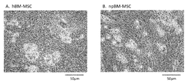
- FIGS. 2A and 2B show adipocyte differentiation of human bone marrow-derived mesenchymal stem cells (hBM-MSCs) and juvenile pig bone marrow-derived mesenchymal stem cells (npBM-MSCs), respectively.
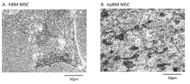
- FIGS. 3A and 3B show osteocyte differentiation of human bone marrow-derived mesenchymal stem cells (hBM-MSCs) and juvenile pig bone marrow-derived mesenchymal stem cells (npBM-MSCs), respectively.
- FIGS. 4A and 4B show osteocyte differentiation of human bone marrow-derived mesenchymal stem cells (hBM-MSCs) and juvenile pig bone marrow-derived mesenchymal stem cells (npBM-MSCs), respectively.
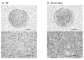
- FIGS. 5A and 5B show chondrocyte differentiation of juvenile pig bone marrow-derived mesenchymal stem cells (npBM-MSC).
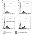
- FIG. 6 shows the results of cell surface antigen analysis of juvenile pig bone marrow-derived mesenchymal stem cells (npBM-MSC) using CD44, a marker of mesenchymal stem cells.
- FIG. 7 shows the results of cell surface antigen analysis of CD90 juvenile pig bone marrow-derived mesenchymal stem cells (npBM-MSC), which are markers of mesenchymal stem cells.
- FIGS. 5A and 5B show chondrocyte differentiation of juvenile pig bone marrow-derived mesenchymal stem cells (npBM-MSC).
- FIG. 6 shows the results of cell surface antigen analysis of juvenile pig bone marrow-derived mesenchymal stem cells (npBM-MSC) using CD44, a marker of mesenchymal stem cells.
- FIG. 7 shows the results of
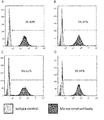
- FIGS. 9A to 9D show the results of cell surface antigen analysis of juvenile pig islet-derived mesenchymal stem cells (npISLET-MSC) using CD29, a marker of mesenchymal stem cells.
- 8A sample 11 (no freezing immediately after preparation of islet)
- FIG. 8B sample 12 (with freezing immediately after preparation of islet)
- FIG. 8C sample 13 (freezing on day 3 of culture after preparation of islet)
- FIG. The result of 14 (with freezing on the 3rd day of culture after islet preparation) is shown.
- FIGS. 9A to 9D show the results of cell surface antigen analysis of juvenile pig islet-derived mesenchymal stem cells (npISLET-MSC) using CD44 which is a marker of mesenchymal stem cells.
- FIG. 9A sample 11 (no freezing immediately after preparation of islet)
- FIG. 9B sample 12 (with freezing immediately after preparation of islet)
- FIG. 9C sample 13 (freezing on day 3 of culture after preparation of islet)
- FIG. The result of 14 (with freezing on the 3rd day of culture after islet preparation) is shown.
- 10A to 10D show the results of cell surface antigen analysis of juvenile pig islet-derived mesenchymal stem cells (npISLET-MSC) using CD90, a marker of mesenchymal stem cells.
- 10A sample 11 (no freezing immediately after preparation of islet)
- FIG. 10B sample 12 (with freezing immediately after preparation of islet)
- FIG. 10C sample 13 (freezing on day 3 of culture after preparation of islet);
- the result of 14 (with freezing on the 3rd day of culture after islet preparation) is shown.
- the stem cells of the present invention are stem cells isolated from juvenile pigs.
- the stem cell described in the Example mentioned later was isolate
- "juvenile pig” refers to a pig less than one month after birth, preferably less than 25 days after birth.
- the juvenile pig is preferably for medical use, and more preferably a juvenile pig capable of cell transplantation into humans.
- the type of pig is not particularly limited.
- landrace species eg, Denmark landrace species, American landrace species, British landrace species, Dutch landrace species, Swedish landrace species
- stem cells mean immature cells having self-replication ability and differentiation / proliferation ability.
- Stem cells include subpopulations such as pluripotent stem cells, multipotent stem cells, unipotent stem cells, etc., depending on differentiation ability.
- a pluripotent stem cell means a cell which can not be an individual per se, but has the ability to differentiate into all tissues and cells constituting a living body.
- Multipotent stem cells mean cells that have the ability to differentiate into multiple types of tissues and cells, but not all types.
- Unipotent stem cells mean cells having the ability to differentiate into specific tissues or cells.
- multipotent stem cells are preferable.
- multipotent stem cells include mesenchymal stem cells, hematopoietic stem cells, neural stem cells, somatic stem cells such as bone marrow stem cells and germ stem cells, and the like, with preference given to mesenchymal stem cells.
- the stem cell of the present invention is a stem cell isolated from juvenile pig, which is a primary culture cell, a cell obtained by subculturing the primary culture cell, and giving rise to various cells expressing various differentiation markers Possible stem cells are also included in the stem cells of the present invention.
- the stem cell of the present invention is a mesenchymal stem cell, it is preferable that both cell markers, CD44 and CD90, are 60% or more positive, more preferably 70% or more, still more preferably 80% or more. It is.
- the cell marker CD29 is preferably 60% or more positive, more preferably 70% or more, and still more preferably 80% or more.
- the stem cell of the present invention preferably has a doubling time of 36 hours or less, more preferably 32 hours or less, still more preferably 28 hours or less, particularly preferably 24 hours or less, and most preferably 20 hours or less. is there.
- the doubling time in the logarithmic growth phase is preferably 14 hours or more, and more preferably 16 hours or more.
- the stem cells of the present invention can be cultured in the logarithmic growth phase, for example, by seeding the stem cells of the present invention in a medium (for example, MSC medium) containing vitamin C described later and culturing at 37 ° C. in the presence of 5% CO 2 It can be carried out by culturing in an incubator. As the doubling time in the logarithmic growth phase is shorter, it is possible to prepare a large amount of stem cells in a short time and inexpensively.
- a medium for example, MSC medium
- the stem cells of the present invention preferably have an average diameter of 17 ⁇ m or less, more preferably 16.5 ⁇ m or less, still more preferably 16 ⁇ m or less, particularly preferably 15.5 ⁇ m or less, most preferably 15 ⁇ m or less It is.
- the average diameter is preferably 10 ⁇ m or more, more preferably 12 ⁇ m or more.
- the smaller mean diameter can prevent the formation of pulmonary embolism by administration of stem cells.
- the mean diameter can be measured, for example, using Nucleo Counter NC-200TM.
- the average means arithmetic mean.
- the differentiation of mesenchymal stem cells of the present invention into adipocytes includes, for example, mesenchymal stem cells of the present invention in the presence of insulin, MCGS (serum component, Mesenchymal Cell Growth Supplement), dexamethasone, indomethacin, isobutylmethylxanthine, etc. By culturing, differentiation into fat cells can be induced.
- a commercially available kit or medium may be used, for example, hMSC differentiation BulletKitTM-adipogeni (PT-3004) manufactured by Lonza Walkersville, hMSC adipogenic induction medium (Lonza Walkersville) PT-3102B), hMSC adipogenic maintenance medium (PT-3102B) manufactured by Lonza Walkersville, and the like.
- hMSC differentiation BulletKitTM-adipogeni PT-3004
- hMSC adipogenic induction medium Longza Walkersville
- PT-3102B hMSC adipogenic maintenance medium manufactured by Lonza Walkersville, and the like.
- the differentiation of mesenchymal stem cells into adipocytes can be confirmed using a commercially available kit, and examples include Lonza Adipo RedTM assay reagent.
- Differentiation of bone cells from mesenchymal stem cells of the present invention can be performed by, for example, culturing mesenchymal stem cells of the present invention in the presence of dexamethasone, ascorbate, MCGS, ⁇ -glycerophosphate, etc. It can be induced.
- a commercially available kit may be used, and examples thereof include hMSC differentiation BulletKit (trademark) -osteogenic, PT-3004 and the like manufactured by Lonza Walkersville.
- the differentiation of mesenchymal stem cells into bone cells may be confirmed by using a commercially available alkaline phosphatase staining kit (for example, Cosmo Bio Co., Ltd., etc.), a commercially available calcification staining kit (eg, Cosmo Bio Co., etc., etc.) Can.
- a commercially available alkaline phosphatase staining kit for example, Cosmo Bio Co., Ltd., etc.
- a commercially available calcification staining kit eg, Cosmo Bio Co., etc., etc.
- the differentiation of mesenchymal stem cells to chondrocytes is carried out, for example, in the presence of TGF-.beta.3, dexamethasone, insulin-transferrin-selenium acid (ITS), sodium pyruvate, proline, ascorbate.
- ITS insulin-transferrin-selenium acid
- a commercially available kit may be used, and examples thereof include hMSC differentiation BulletKit (trademark) -condrogenic manufactured by Lonza Walkersville, PT-3003 and the like.
- the differentiation of mesenchymal stem cells into chondrocytes can be confirmed by Alcian blue staining or the like.
- Transplantation of stem cells can be easily performed by injecting a suspension of stem cells into the host body.
- the injection can be performed to an organ to be regeneratively treated or in the vicinity thereof, or in a vein or the like.
- the number of stem cells to be injected is not particularly limited and may be appropriately selected depending on the condition, body weight of the host, administration method and the like, but it is usually about 10 2 to 10 10 .
- feeder cells refers to other cell types that serve as an adjunct to adjust the culture conditions of the cells of interest for which proliferation and differentiation are to occur.
- feeder cells When using as a feeder cell, it is preferable to process beforehand by gamma irradiation or an antibiotic so as not to normally grow.
- feeder cells for stem cells fibroblasts derived from mouse embryo are mainly used, but various cell types such as fibroblasts such as 3T3 and SNL are used as feeder cells depending on the purpose of the experiment and cells.
- the stem cells of the present invention can be used as feeder cells for stem cells for human transplantation, preferably by isolating them from juvenile pigs capable of cell transplantation into humans.
- the method for preparing stem cells of the present invention is characterized by comprising the step of isolating cells from juvenile pigs.
- One embodiment of the method for preparing a stem cell of the present invention includes, for example, a method comprising the following steps. (1) Step of Collecting Cells from Juvenile Pig (2) Step of Cultivating Cells Collected in Step (1) to Prepare Juvenile Pig-Derived Stem Cells Hereinafter, each step will be described.
- step (1) cells are collected from bone marrow, fat, skin, pancreas and the like of juvenile pig.
- bone marrow cells can be collected from the femur, iliac crest and sternum of the juvenile pig.
- a femur is collected from a young pig, cut at both ends, a needle is inserted, and flushed with a heparin-added physiological buffer (for example, phosphate buffer, hereinafter also referred to as PBS), and flows out from the opposite site.
- PBS heparin-added physiological buffer
- the fluid is collected as bone marrow fluid.
- the volume of the effluent is reduced, the bone is inverted, the needle is inserted on the opposite side, and rinsed again with PBS to prepare a cell-containing solution, bone marrow fluid.
- the juvenile pig-derived mononuclear cell fraction may be isolated by usually centrifuging the cell-containing solution prepared above.
- the cell-containing solution prepared above was diluted with PBS or the like and diluted onto the medium layer in a tube containing a medium for human lymphocyte separation (for example, Ficoll-Paque PLUS by GE Healthcare Life Sciences). Put in the cell-containing solution.
- a medium for human lymphocyte separation for example, Ficoll-Paque PLUS by GE Healthcare Life Sciences.
- the tube is centrifuged to separate layers, and a layer containing juvenile pig-derived mononuclear cells is collected.
- the collected solution is further centrifuged, and after removing the supernatant, it is diluted with PBS or the like and centrifuged again to isolate a mononuclear cell fraction.
- the cells of the mononuclear cell fraction thus isolated may be cryopreserved prior to culture.
- the temperature is preferably ⁇ 80 ° C. or less, more preferably ⁇ 150 ° C. or less.
- islet is collected from juvenile pig, and in some cases, it is used for adhesion culture for the purpose of preparing stem cells by suspending and culturing the islet. Prepare cell mass.
- the fat is collected from juvenile pig, finely chopped with scissors, and then subjected to the enzyme treatment. Filter with a cell strainer and centrifuge at low speed. The cells precipitated at the bottom of the tube are used for culture.
- the skin is collected from young pig and subjected to an enzyme treatment. After enzyme treatment, the hair is removed from the skin and the Bulge part is collected and used for culture.
- 3T3 feeder cells use 3T3 feeder cells.
- step (2) A step of culturing the cells collected in step (1) and preparing juvenile pig-derived stem cells
- the cells, cell fraction, or cell mass collected in step (1) above are not intended except for stem cells. It contains many cells.
- a culture method for removing these cells is used by using a vitamin C-free basal medium (for example, MSC basal medium described later), which is essential for the survival of these extraneous cells of interest.
- the cells, cell fractions or cell masses collected in the above step (1) are preferably brought to 35 to 39 ° C., more preferably 36 to 38 ° C., most preferably 37 ° C.
- a culture incubator in the presence of 4 to 6%, more preferably 4.5 to 5.5%, most preferably 5% of CO 2 in a culture incubator.
- the stem cells of the present invention are expanded while removing the
- a culture medium containing vitamin C (eg, as described later) is used instead of a vitamin C-free basal medium for culture to remove the above-mentioned non-target cells.
- the stem cells of the present invention can be prepared using only MSC medium).
- the medium is replaced with a medium containing vitamin C to proliferate the stem cells of the present invention.
- Stem cells can also be prepared.
- the stem cells of the present invention are cultured by the following method.
- a culture vessel coated with gelatin eg, a plate coated with 0.1% gelatin
- a culture vessel without gelatin coat eg, a plate
- a vitamin C-free basal medium eg, MSC basal described below
- 5.0 ⁇ 10 5 to 5.0 ⁇ 10 7 cells / 9.6 cm 2 are seeded using a culture medium) or a culture medium containing vitamin C (eg, MSC culture medium described later), for example
- the primary culture cells are obtained by incubating at 37 ° C. under 5% CO 2 and 90% humidity.
- the culture period for obtaining primary culture cells is preferably 3 to 12 days, more preferably 3 to 11 days, and most preferably 3 to 10 days after seeding.
- Primary culture cells may be passaged.
- the stem cells obtained by passage are also referred to as passage cells.
- the primary culture cells or subcultured cells may be passaged preferably 2 to 6 days, more preferably 2 to 5 days, still more preferably 2 to 4 days, most preferably 3 days after seeding the stem cells.
- Stem cells can be seeded using a culture vessel coated with gelatin (eg, a plate coated with 0.1% gelatin) or a culture vessel without gelatin coat (eg, a plate) using a culture medium containing vitamin C (eg, as described later) (MSc medium) is preferably used to inoculate 5.0 ⁇ 10 5 to 5.0 ⁇ 10 7 cells / 9.6 cm 2 .
- a culture medium containing vitamin C eg, as described later
- Stem cells are cultured, for example, at 37 ° C. under conditions of 5% CO 2 and 90% humidity.
- the medium is exchanged as needed to grow the stem cells of the present invention.
- MSC basal medium and MSC medium conventionally known ones can be used, and commercially available ones may be used.
- MSC basal medium for example, a medium obtained by adding 55 mL of Gibco Fetal bovine serum (FBS) and 5.5 mL of Sigma-Aldorich Penicillin-Streptomycin to 500 mL of MEMb (Nucleosides, no Ascorbic acid) from Gibco Can be mentioned.
- FBS Gibco Fetal bovine serum
- MEMb Nucleosides, no Ascorbic acid
- MSC medium for example, in 500 mL of Gibco MEM ⁇ (nucleosides), 55 mL of Gibco Fetal bovine serum (FBS), 5.5 mL of Sigma-Aldorich Penicillin-Streptomycin and 22.2 ⁇ L of Sigma- Aldorich FGF-Basic, recombinant, expressed in E.
- FBS Gibco Fetal bovine serum
- FBS Gibco Fetal bovine serum
- FBS Gibco Fetal bovine serum
- Sigma-Aldorich Penicillin-Streptomycin 5.5 mL of Sigma-Aldorich Penicillin-Streptomycin
- 22.2 ⁇ L of Sigma- Aldorich FGF-Basic recombinant, expressed in E.
- examples include media to which E. coli, suitable for cell culture (final concentration: 1 ng / mL) have been added.
- Passage is preferably performed at least once or more.
- the number of passages is not particularly limited as long as the stem cells of the present invention can be obtained, but it is preferably 1 to 3 times, more preferably 1 to 20 times.
- the stem cells of the present invention can be cryopreserved.
- the timing of the cryopreservation is not particularly limited, but preferably after passage 1 to 20, more preferably after 2 to 10 passages. Conventionally known methods can be used as the cryopreservation and thawing methods.
- cryopreservation of stem cells specifically, for example, it can be dispersed in a cryopreservation solution and cryopreserved in -80 ° C. or less or liquid nitrogen in a freezer until necessary.
- a cryopreservation solution for example, OPF-301 [lactate Ringer solution containing 3% trehalose and 5% dextran (WO 2014/208053)] and dimethyl sulfoxide (DMSO) in a ratio of 9: 1
- DMSO dimethyl sulfoxide
- a mixed solution, a serum-containing or serum-free preservation solution that can be used for cryopreservation of animal cells, or a commercially available cell cryopreservation reagent preferably, a cell banker such as Takara Bio Inc. CELLBANKER (registered trademark)] can be mentioned. .
- Bone marrow was collected from the femurs of young pigs. Femurs are collected from juvenile pigs (23 days old medical landrace pig), both ends are cut, 12 G needle is inserted, and 50 mL heparinized PBS (3 mL heparin (1000 U / mL), 47 mL The solution was flushed with PBS, and 50 mL of bone marrow outflow (hereinafter also referred to as bone marrow fluid) was collected from the opposite site. When the volume of effluent decreased, the bone was inverted and a needle was inserted on the opposite side and rinsed again with PBS to collect bone marrow fluid. A 50 ⁇ L sample was taken in 1950 ⁇ L PBS (40-fold dilution) in a 15 mL conical tube for counting, and the cell number was measured with a cell counter.
- the tube was centrifuged at 400 ⁇ g for 30 minutes at 20 ° C. and slowly accelerated without brake (1/3 of full speed) to form three different layers. Because the mononuclear cell fraction was placed in a floating white ring, the entire white ring was collected in a 50 mL tube (x 4) containing 25 mL PBS. The supernatant was removed by centrifugation at 400 ⁇ g for 7 minutes at room temperature. PBS was added to 40 mL and centrifuged again at 400 ⁇ g for 7 minutes at room temperature. When the number of cells was measured in the same manner as described above, 25 to 30% of the total bone marrow cells were isolated as (20 to 30) ⁇ 10 6 as a mononuclear cell fraction.
- npMNC juvenile pig-derived mononuclear cell fraction cells
- the cryovial was stored at ⁇ 20 ° C. for 1 hour, and subsequently stored at ⁇ 80 ° C. for 24 hours, and finally transferred to a liquid nitrogen tank for long-term storage.
- npMNC juvenile pig-derived mononuclear cell
- npBM-MSC juvenile pig bone marrow-derived mesenchymal stem cells
- the 6-well plate was coated with 0.1% gelatin and left in an incubator (37 ° C., 5% CO 2 ) for 10 to 15 minutes to remove gelatin before use.
- Add cell suspension to each of the prepared 0.1% gelatin coated 6-well plates and gently rock to disperse the cell suspension on the growth surface (gelatin coated) in 2 mL of MSC basal medium We seeded 2.09 ⁇ 10 6 cells / well.
- the cells were cultured in a CO 2 incubator at 37 ° C.
- npBM-MSC juvenile pig bone marrow-derived mesenchymal stem cells
- immature pig bone marrow-derived mesenchymal stem cells npBM-MSC
- npBM-MSC juvenile pig bone marrow-derived mesenchymal stem cells
- the cells were washed with 2 mL PBS (containing no calcium and magnesium), added with 320 ⁇ L of 0.25% trypsin per well, placed in an incubator for several minutes, and when cells were detached, neutralized with 1680 ⁇ L of MSC medium.
- the cell suspension was collected into a 50 mL tube using a 1 mL pipette, and 16 mL (8 mL ⁇ 2 wells) of MSC medium was added, followed by centrifugation at 500 ⁇ g for 5 minutes at room temperature. Using a pipette, the resulting pellet was gently resuspended in temperature-equilibrated MSC medium (2 mL).
- the total number of cells was 2.05 ⁇ 10 6
- the number of living cells was 2.02 ⁇ 10 6
- the survival rate was 98.5%.
- npBM-MSC juvenile pig bone marrow-derived mesenchymal stem cells
- npBM-MSC Freeze preservation of juvenile pig bone marrow-derived mesenchymal stem cells
- npBM-MSC early passage juvenile pig bone marrow-derived mesenchymal stem cells
- DMSO a mixed solution of DMSO and a desired concentration of CELLBANKER® 1 or OPF-301 [Lactated Ringer's solution (WO 2014/208053) containing 3% trehalose and 5% dextran] and DMSO at a ratio of 9: 1
- the trypsinized npBM-MSC pellet was resuspended to 1.5 ⁇ 10 6 cells / 1 mL / vial. After storing the vial in the bicell and storing at -80 ° C for 24 hours, the cells were transferred from -80 ° C to liquid nitrogen for long-term storage.
- npBM-MSC Young pig bone marrow-derived mesenchymal stem cells (npBM-MSC) (P2) are seeded at a density of 30 cells / cm 2 with 630 cells in a 21 cm 2 culture dish (without gelatin coating or 0.1% gelatin coating), The cells were cultured in MSC medium. MSC medium was changed every 3 days. After 6 days of culture, adherent cells were washed twice with 4 mL PBS and fixed with 4 mL ice cold methanol for 15 minutes at 4 ° C. To visualize colonies, cells were stained for 30 minutes with 4 mL of Giemsa diluted 1:19 with phosphate buffer, then washed at room temperature (RT) and washed twice with H 2 O.
- RT room temperature
- npBM-MSC juvenile pig bone marrow-derived mesenchymal stem cells
- Table 2 shows the results of measuring the average diameter of human bone marrow-derived mesenchymal stem cells (hBM-MSC, passage number P4) and the obtained juvenile pig bone marrow-derived mesenchymal stem cells (npBM-MSC). .
- npBM-MSC juvenile pig bone marrow-derived mesenchymal stem cells
- the obtained juvenile pig bone marrow-derived mesenchymal stem cells are compared to human bone marrow-derived mesenchymal stem cells in It was found that the growth rate was remarkably fast.
- hBM-MSC human bone marrow-derived mesenchymal stem cells
- npBM-MSC juvenile pig bone marrow-derived mesenchymal stem cells
- hMSC differentiation BulletKit trademark
- PT-3004 manufactured by Lonza Walkersville
- npBM-MSC juvenile pig bone marrow-derived mesenchymal stem cells
- hBM-MSC human bone marrow-derived mesenchymal stem cells
- npBM-MSC juvenile pig bone marrow-derived mesenchymal stem cells
- BulletKitTM-osteogenic PT-3002 manufactured by Lonza Walkersville
- the obtained juvenile pig bone marrow-derived mesenchymal stem cells are similar to human bone marrow-derived mesenchymal stem cells as bone cells. It turned out that it can differentiate.
- chondrocytes With respect to juvenile pig bone marrow-derived mesenchymal stem cells (npBM-MSC), differentiation into osteocytes was induced according to a protocol using hMSC differentiation BulletKit (trademark) -chondrogenic, PT-3003 (manufactured by Lonza Walkersville). The results of HE staining and Alcian blue staining on day 19 after initiation of induction are shown in FIGS. 5A and 5B, respectively.
- npBM-MSC juvenile pig bone marrow-derived mesenchymal stem cells
- Test example 2 [Culture of cells of juvenile pig-derived mononuclear cell (npMNC) fraction and preparation of juvenile pig bone marrow-derived mesenchymal stem cells (npBM-MSC)] MSC basal medium or MSC medium was allowed to stand in an incubator (37 ° C., 5% CO 2 ) for 10 to 15 minutes before use. Similar to Test Example 1, a cell suspension containing cells of a juvenile pig-derived mononuclear cell (npMNC) fraction which had been cryopreserved in a cryovial in a 37 ° C. water bath was quickly thawed. Using a micropipette, the thawed cell suspension was gently added to 30 mL of temperature equilibrated (37 ° C.) MSC basal medium and aliquoted 15 mL into two 50 mL tubes.
- npMNC juvenile pig-derived mononuclear cell
- npBM-MSC juvenile pig bone m
- the medium was changed with MSC medium three days and six days after seeding, cells were grown, and passaged eight days after seeding.
- npBM-MSCs juvenile pig bone marrow-derived mesenchymal stem cells
- the cells were washed with 2 mL PBS ( ⁇ ), added with 320 ⁇ L of 0.25% trypsin per well, allowed to stand in an incubator for several minutes, and neutralized with 1680 ⁇ L of MSC medium when the cells were detached.
- the cell suspension was collected in a 50 mL tube, 8 mL of MSC medium was added, and centrifuged at 500 ⁇ g for 5 minutes at room temperature.
- MSC medium Fifteen mL of MSC medium was added to a T75 flask (without gelatin coat), and cultured in an incubator in which juvenile pig bone marrow-derived mesenchymal stem cells (npBM-MSC) were replated so as to have the following cell number. These cells were taken as the first passage.
- MSC medium at the time of P0 inoculation viable cell number 3.3 ⁇ 10 5 / flask
- npBM-MSC young pig bone marrow-derived mesenchymal stem cells
- the temperature-equilibrated MSC medium (5 mL) was added to the obtained pellet and gently resuspended by pipetting up and down to measure the total cell number and viable cell number. The results are shown below.
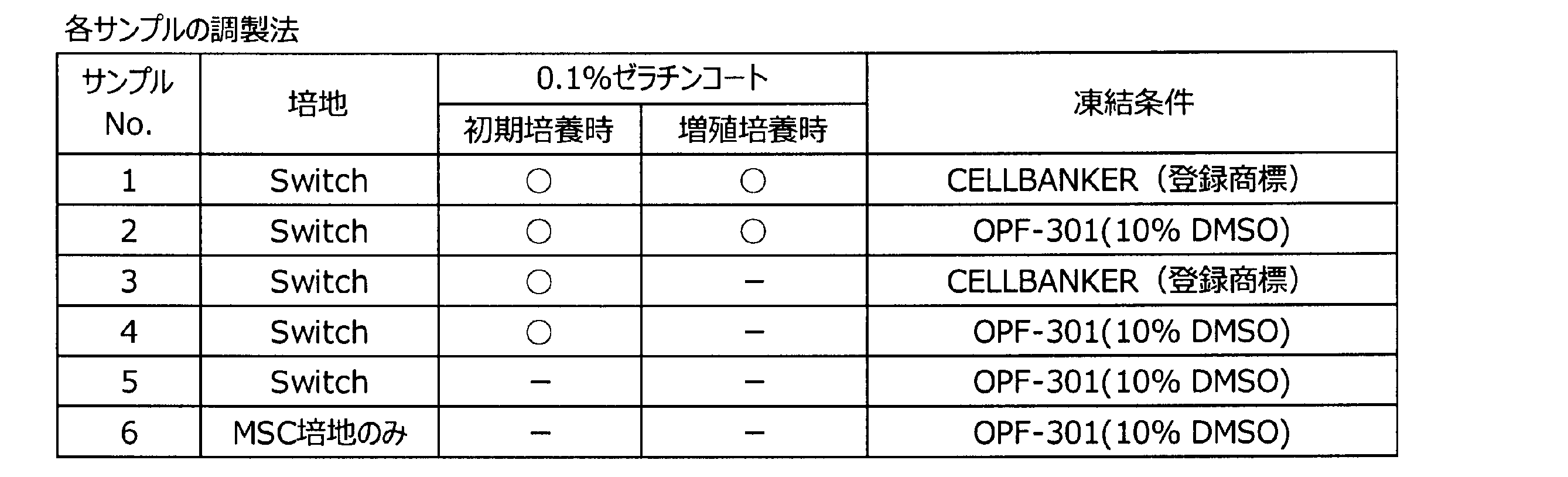
- Test Example 3 Cell surface antigens of juvenile pig-derived mononuclear cells (npMNC) prepared in Test Example 1 and Test Example 2 were analyzed. The preparation method of each sample used for analysis is shown in Table 5.
- “Switch” indicates that culture was performed using MSC basal medium (vitamin C free) at the time of initial culture, and changing to MSC medium (containing vitamin C) which is a growth medium at the time of growth culture.
- the cells were resuspended with 2 mL of Stain Buffer (manufactured by BD), and the number of viable cells was counted. Re-centrifugation (500 ⁇ g, 5 minutes, 4 ° C.) and resuspend in Stain Buffer (BD) to a cell count of 1 ⁇ 10 7 cells / mL, 20 ⁇ L (cell number: 2 ⁇ 10 5 ) Each aliquot was dispensed into 1.5 mL tubes, and a total of 4 tubes of non-stained control, CD44, CD90 and Isotype Control were prepared.
- Stain Buffer manufactured by BD
- markers of mesenchymal stem cells were positive. It is also considered that the target mesenchymal stem cells could be established without coating with gelatin at the time of initial culture. In any case, no nonspecific reaction was observed in the measurement of Isotype Control.
- Test Example 4 Preparation of young pig pancreatic islet-derived mesenchymal stem cells. After collecting islets from juvenile pigs and preparing a cell mass by suspension culture, it was frozen and stored in the same manner as in Test Example 1. Young pig islets were stored frozen in cryovials in a 37 ° C. water bath and thawed quickly.
- thawed islet suspension was gently added to 30 mL of MSC basal medium adjusted to temperature equilibrium (37 ° C.). Centrifuge at 210 ⁇ g for 1 minute at 4 ° C. In addition, when the islet was not frozen, the supernatant was removed after the islet was precipitated by gravity at room temperature. The pellet was resuspended in equilibrated MSC basal medium at a temperature of 4 mL and gently pipetted up and down.
- the cells are grown by culturing in a CO 2 incubator at 37 ° C. under 5% CO 2 and 90% humidity, and after 3 days, they are replaced with MSC medium to grow the cells, and then once every 3 days I made an exchange.
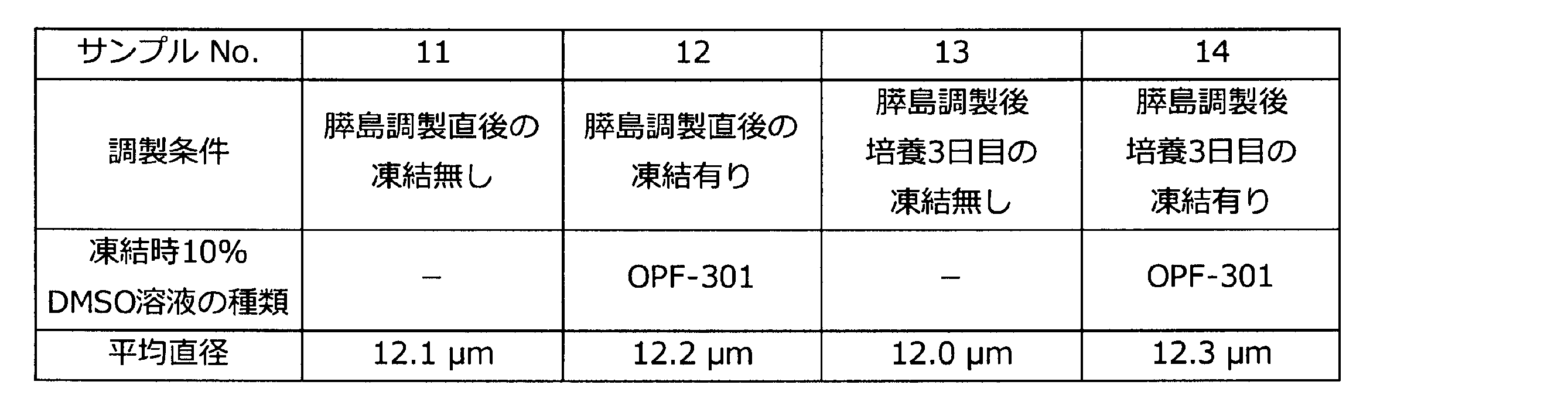
- Table 6 shows the preparation conditions of the sample. It reached 100% confluence 6 days after seeding, with or without initial freezing.
- the cells were washed with 2 mL PBS (containing no calcium and magnesium), added with 320 ⁇ L of 0.25% trypsin per well, allowed to stand in an incubator for several minutes, and neutralized with 1680 ⁇ L of MSC medium when the cells were detached.
- the cell suspension was collected into a 50 mL tube using a 1 mL pipette, and 16 mL (8 mL ⁇ 2 wells) of MSC medium was added, followed by centrifugation at 500 ⁇ g for 5 minutes at room temperature. Using a pipette, the resulting pellet was gently resuspended in temperature-equilibrated MSC medium (2 mL).
- juvenile pig islet-derived mesenchymal stem cells can be prepared regardless of freezing conditions in preparation of islets, and in the presence of freezing, the average diameter is the same regardless of the case of no freezing. I found it to be.
- the cells were resuspended with 2 mL of Stain Buffer (manufactured by BD), and the number of viable cells was counted. Re-centrifugation (500 ⁇ g, 5 minutes, 4 ° C.) and resuspend in Stain Buffer (BD) to a cell count of 1 ⁇ 10 7 cells / mL, 20 ⁇ L (cell number: 2 ⁇ 10 5 ) Each aliquot was dispensed into 1.5 mL tubes, and a total of four non-stained controls, CD29, CD44 and CD90 were prepared.
- Stain Buffer manufactured by BD
- FIGS. 8A-8D The results of CD29 are shown in FIGS. 8A-8D, the results of CD44 in FIGS. 9A-9D, and the results of CD90 in FIGS. 10A-10D.
- 8A, 9A and 10A Sample 11 (without freezing immediately after preparation of islet);
- FIG. 8B, 9B and 10B Sample 12 (with freezing immediately after preparation of islet);
- FIGS. 8C, 9C and 10C FIG. 8D, FIG. 9D, and FIG. 10D show the results of sample 14 (freezing on day 3 of culture after preparation of islet).
Landscapes
- Health & Medical Sciences (AREA)
- Engineering & Computer Science (AREA)
- Life Sciences & Earth Sciences (AREA)
- Biomedical Technology (AREA)
- Organic Chemistry (AREA)
- Chemical & Material Sciences (AREA)
- Bioinformatics & Cheminformatics (AREA)
- Genetics & Genomics (AREA)
- Biotechnology (AREA)
- Wood Science & Technology (AREA)
- Zoology (AREA)
- General Engineering & Computer Science (AREA)
- Biochemistry (AREA)
- Cell Biology (AREA)
- General Health & Medical Sciences (AREA)
- Microbiology (AREA)
- Developmental Biology & Embryology (AREA)
- Hematology (AREA)
- Immunology (AREA)
- Rheumatology (AREA)
- Micro-Organisms Or Cultivation Processes Thereof (AREA)
- Medicines Containing Material From Animals Or Micro-Organisms (AREA)
Abstract
Description
2つ目の免疫調節能は、体性幹細胞からの抗炎症性サイトカイン、ケモカイン、エクソソームなどの分泌を介し、あるいは、細胞間の接着因子などを介し、患者の免疫担当細胞に働きかけ、炎症や移植片対宿主病などの免疫反応を抑制することで治療効果を発揮する。
3つ目の細胞外環境のリモデリング能については、虚血性疾患における梗塞部位や、炎症によって引き起こされた線維化部位などに対し、体性幹細胞からの血管誘導因子、成長因子、抗線維化因子などの分泌により治療効果を発揮するものである。
1.幼若ブタより単離された、幹細胞。
2.平均直径が17μm以下である前記1に記載の幹細胞。
3.対数増殖期における倍加時間が36時間以下である前記1または2に記載の幹細胞。
4.間葉系幹細胞である前記1~3のいずれか1に記載の幹細胞。
5.幼若ブタがヒトへ細胞移植することができる幼若ブタである前記1~4のいずれか1に記載の幹細胞。
6.幼若ブタの骨髄または膵島より単離された前記1~5のいずれか1に記載の幹細胞。
7.移植用幹細胞である前記1~6のいずれか1に記載の幹細胞。
8.ヒト移植用幹細胞である前記7に記載の幹細胞。
9.フィーダー細胞用幹細胞である前記1~6のいずれか1に記載の幹細胞。
10.ヒト移植用幹細胞を増殖するためのフィーダー細胞用幹細胞である前記9に記載の幹細胞。
11.幼若ブタから細胞を単離する工程を含む、幹細胞の調製方法。
12.幹細胞を播種後3~12日後に継代する工程を含む、前記11に記載の幹細胞の調製方法。
13.幼若ブタから単離する細胞が単核球細胞画分の細胞である前記11または12に記載の幹細胞の調製方法。
14.幼若ブタからの細胞の単離が、幼若ブタの骨髄または膵島からの細胞の単離である前記11~13のいずれか1に記載の幹細胞の調製方法。
15.単離した単核球細胞画分の細胞を凍結する工程を含む、前記13または14に記載の幹細胞の調製方法。
(1)幼若ブタから細胞を採取する工程
(2)工程(1)において採取した細胞を培養し、幼若ブタ由来幹細胞を調製する工程
以下、各工程について説明する。
工程(1)では幼若ブタの骨髄、脂肪、皮膚、膵臓等から細胞を採取する。
具体的には例えば、幼若ブタの骨髄から細胞を採取する場合、幼若ブタの大腿骨、腸骨稜及び胸骨などから骨髄細胞を採取することができる。例えば、幼若ブタから大腿骨を回収し、両端を切断して針を挿入し、ヘパリンを添加した生理的緩衝液(例えば燐酸緩衝液、以後PBSとも言う)で洗い流し、反対側の場所から流出液を骨髄液として回収する。流出液の量が減少したら、骨を逆にして針を反対側に挿入し、PBSで再び洗い流して、細胞含有溶液である骨髄液を調製する。
上記工程(1)で採取した細胞、細胞画分、または細胞塊には、幹細胞以外の目的外の細胞が多く含まれる。通常、これらの目的外細胞の生存に必須である、ビタミンCを含まない基礎培地(例えば、後述のMSC基礎培地)を用いることで、これらの細胞を除去する培養方法が用いられている。
〔幼若ブタ由来骨髄細胞の回収〕
幼若ブタの大腿骨から骨髄を採取した。幼若ブタ(生後23日の医療用ランドレース種ブタ)から大腿骨を回収し、両端を切断して12G針を挿入し、50mLのヘパリン処理したPBS(3mLのヘパリン(1000U/mL)、47mLのPBS)で洗い流し、反対側の場所から50mLの骨髄の流出液(以下、骨髄液とも略す)を回収した。流出液の量が減少したら、骨を逆にして針を反対側に挿入し、PBSで再び洗い流して骨髄液を収集した。カウント用の15mLコニカルチューブで1950μLのPBS(40倍希釈)に50μLのサンプルを取り、セルカウンターで細胞数を測定した。
上記手順で得られた骨髄液を静かに再懸濁した。骨髄液全体を50mLチューブ4本に各10mLずつに分け、各々PBSで30mLに希釈し、細胞がチューブに付着していないことを確認してよく混合した。10mLのFicoll-Paque PLUS(GEヘルスケアライフサイエンス社製)を4本の新しい50mLチューブに加え、Ficoll-Paque PLUS層の上にPBSと混合した30mLの骨髄液を入れた。
単離された単核球細胞画分の細胞を、107細胞/mLのDMSOを混合したFBS(90%FBSと10%DMSO)を含むクライオバイアルに入れ、細胞懸濁液の全容量を1mlとした[細胞数/10×106=DMSOを混合したFBSの容量(mL)とした]。クライオバイアルを-20℃にて1時間保存し、続いて-80℃にて24時間保存後、最終的に長期保存用の液体窒素タンクに移した。
37℃の水浴でクライオバイアルに冷凍保存していた幼若ブタ由来単核球細胞(npMNCs)画分の細胞を含む細胞懸濁液を素早く解凍し、マイクロピペットを用いて、解凍した細胞懸濁液を30mLの温度平衡(37℃)に調整したMSC基礎培地[500mLのGibco社製MEMα(Nucleosides、no Ascorbic acid)に55mLのGibco社製Fetal bovine serum (FBS)及び5.5mLのSigma-Aldorich社製Penicillin-Streptomycinを添加した培地、以下同様]に静かに加えた。室温にて5分間、500×gで遠心分離し、ペレットを4mLの温度で平衡化したMSC基礎培地に再懸濁し、上下に穏やかにピペッティングした。総細胞数および生細胞数を計測した結果、総細胞数4.18×10⁶個、生細胞数6.6×10⁵個、生存率:15.8%であった。
幼若ブタ骨髄由来間葉系幹細胞(npBM-MSC)がほぼ100%コンフルエンスに達した後、2ウェルから細胞を回収し、0.1%ゼラチンコート有りまたは無しでT75フラスコにそれらを再播種した。
幼若ブタ骨髄由来間葉系幹細胞(npBM-MSC)がほぼ100%コンフルエンスに達した後、0.1%ゼラチンコートを含むまたは含まないT75フラスコの2つのフラスコから細胞を回収した。8mLのPBS(-)で細胞を洗浄し、1ウェルあたり0.25%のトリプシン2.4mLを加え、インキュベーターに数分間静置し、細胞が剥がれたら12.6mLのMSC培地で中和した。細胞懸濁液を50mLのチューブに集めて、室温にて5分間、500×gで遠心分離した。
0.1%ゼラチンコートされたフラスコ(×2)からの細胞:総細胞数1.62×10⁷個、生細胞数1.60×10⁷個、生存率:98.8%
ゼラチンコートなしのフラスコ(×2)からの細胞:総細胞数1.48×10⁷個、生細胞数1.46×10⁷個、生存率:98.6%
上記した培養とは別に、早期継代の幼若ブタ骨髄由来間葉系幹細胞(npBM-MSC)を凍結して細胞ストックを作製した。所望の濃度のCELLBANKER(登録商標)1またはOPF-301[3%トレハロース及び5%デキストランを含有する乳酸リンゲル液(国際公開第2014/208053号)]とDMSOを9:1の比率で混合した溶液中でトリプシン処理したnpBM-MSCペレットを再懸濁し、1.5×106細胞/1mL/バイアルとした。バイアルをバイセルに入れて-80℃にて24時間保存した後、細胞を-80℃から液体窒素に移して長期保存した。
幼若ブタ骨髄由来間葉系幹細胞(npBM-MSC)(P2)を、21cm2培養ディッシュ(ゼラチンコート無しまたは0.1%ゼラチンコート)に630細胞を30細胞/cm2の密度で播種し、MSC培地中で培養した。MSC培地は3日毎に交換した。6日間の培養後、接着細胞を4mLのPBSで2回洗浄し、4mLの氷冷メタノールで4℃にて15分間固定した。コロニーを可視化するために、リン酸緩衝液で1:19に希釈した4mLのギムザで30分間細胞を染色後、室温(RT)で洗浄し、H2Oで2回洗浄した。

ヒト骨髄由来間葉系幹細胞(hBM-MSC、継代回数P4)および得られた幼若ブタ骨髄由来間葉系幹細胞(npBM-MSC)について、細胞の平均直径を計測した結果を表2に示す。細胞の平均直径はNucleo Counter NC-200(商標)を用いて計測し、平均値(n=3)を算出した。

ヒト骨髄由来間葉系幹細胞(hBM-MSC)および幼若ブタ骨髄由来間葉系幹細胞(npBM-MSC)について、細胞をT25フラスコ中で5000細胞/cm2(1.25×105細胞/フラスコ)の密度で播種し、MSC培地を用いて培養した。MSC培地は3日毎に交換した。培養開始から1、2、4および8日後に生存可能な細胞および死んだ細胞の総数を数えた。結果を表3および表4、並びに図1Aおよび図1Bに示す。なお、表3および表4の値は平均値±SD(n=4)である。


ヒト骨髄由来間葉系幹細胞(hBM-MSC)および幼若ブタ骨髄由来間葉系幹細胞(npBM-MSC)について、hMSC differentiation BulletKit(商標)-adipogeni、PT-3004(Lonza Walkersville社製)を用いて、プロトコールに従って脂肪細胞への分化を誘導した。誘導開始後17日目にSigma-Aldorich社製Oil Redを用いて染色した結果をそれぞれ図2Aおよび図2Bに示す。
ヒト骨髄由来間葉系幹細胞(hBM-MSC)および幼若ブタ骨髄由来間葉系幹細胞(npBM-MSC)について、hMSC differentiation BulletKit(商標)-osteogenic、PT-3002(Lonza Walkersville社製)を用いて、プロトコールに従って骨細胞への分化を誘導した。誘導開始後14日目に、コスモ・バイオ社製アルカリフォスファターゼ染色キットを用いて染色した結果をそれぞれ図3Aおよび図3Bに、コスモ・バイオ社製石灰化染色キットを用いて染色した結果をそれぞれ図4Aおよび図4Bに示す。
幼若ブタ骨髄由来間葉系幹細胞(npBM-MSC)について、hMSC differentiation BulletKit(商標)-chondrogenic、PT-3003(Lonza Walkersville社製)を用いて、プロトコールに従って骨細胞への分化を誘導した。誘導開始後19日目に、HE染色した結果およびアルシアンブルー染色した結果をそれぞれ図5Aおよび図5Bに示す。
〔幼若ブタ由来単核球細胞(npMNC)画分の細胞の培養および幼若ブタ骨髄由来間葉系幹細胞(npBM-MSC)調製〕
MSC基礎培地またはMSC培地は、使用前にインキュベーター(37℃、5%CO2)中に10~15分間静置した。試験例1と同様に、37℃の水浴でクライオバイアルに冷凍保存していた幼若ブタ由来単核球細胞(npMNC)画分の細胞を含む細胞懸濁液を素早く解凍した。マイクロピペットを用いて、解凍した細胞懸濁液を30mLの温度平衡(37℃)MSC基礎培地に静かに加え、50mLのチューブ2本に15mLずつ分注した。
2mLのMSC基礎培地:総細胞数2.60×10⁶個、生細胞数4.8×10⁵個、生存率18.5%
2mLのMSC培地:総細胞数2.55×10個、生細胞数4.5×10⁵個、生存率17.6%
2mLのMSC基礎培地:2.60×10⁶個/1ウェルの細胞を播種
2mLのMSC培地:2.55×10⁶個/1ウェルの細胞を播種
幼若ブタ骨髄由来間葉系幹細胞(npBM-MSC)がほぼ50~60%コンフルエントに達した後、1ウェルから細胞を回収し、ゼラチンコートなしでT75フラスコにそれらを再播種した。
P0播種時にMSC基本培地の群:総細胞数5.0×10⁵個、生細胞数5.0×10⁵個、生存率:100%
P0播種時にMSC培地の群:総細胞数3.3×10⁵個、生細胞数3.3×10⁵個、生存率:100%
P0播種時にMSC基礎培地の群:生細胞数5.0×10⁵個/フラスコ
P0播種時にMSC培地の群:生細胞数3.3×10⁵個/フラスコ
前記手順により再播種した細胞がほぼ80~90%のコンフルエンスに達した後、T75フラスコ(ゼラチンコート無し)の1フラスコから細胞を集めた。8mLのPBS(-)で細胞を洗浄し、0.25mL/1ウェルのトリプシン2.4mLを加え、インキュベーターに数分間静置し、細胞が剥がれたら12.6mLのMSC培地で中和した。細胞懸濁液を50mLのチューブに集めて、室温にて5分間、500×gで遠心分離した。
1つのフラスコからの細胞(P0の播種後の3日間のMSC基礎培地):総細胞数5.12×10⁶個、生細胞数5.09×10⁶個、生存率:99.5%
1つのフラスコからの細胞(P0の播種時からMSC培地):総細胞数4.76×10⁶個、生細胞数4.73×10⁶個、生存率:99.4%
上記した培養とは別に、試験例1と同様にして、早期継代の細胞を凍結して細胞ストックを作製した。
試験例1および試験例2で調製した幼若ブタ由来単核球細胞(npMNC)の細胞表面抗原を解析した。解析に用いた各サンプルの調製方法について、表5に示す。表5において、「Switch」とは、初期培養時はMSC基礎培地(ビタミンCフリー)を用い、増殖培養時には増殖培地であるMSC培地(ビタミンC含有)に変更して培養したことを示す。
各細胞サンプルを液体窒素タンクより取出して蓋を緩めて圧を抜き、再びふたを閉め、37℃に予め加温しておいた恒温槽で1~2分間軽く撹拌しながら融解した。Stain Buffer(BD社製)5mLを入れた15mL遠沈管に融解させた各細胞を移し、4℃にて500×g、5分間遠心し、上清を取り除いた。5mLのStain Bufferを入れ、4℃にて500×g、5分間遠心し、2回洗浄した。
〔幼若ブタ膵島由来間葉系幹細胞の調製〕
幼若ブタから膵島を回収し、浮遊培養することにより細胞塊を調製した後、試験例1と同様にして、冷凍保存した。37℃の水浴でクライオバイアルに冷凍保存していた幼若ブタ膵島を素早く解凍した。

幼若ブタ膵島由来間葉系幹細胞(npISLET-MSC)が約80%~ほぼ95%コンフルエンスに達した後、2ウェルから細胞を回収し、ゼラチンコートなしでT75フラスコにそれらを再播種した。
20mLの前記MSC培地をゼラチンコート無しのT75フラスコに加えて再播種し、CO2インキュベーター中で、37℃にて5%CO2、90%湿度の条件下で培養した。これらの細胞を第1継代とした。第1継代を播種した3日後に、初期凍結の有無にかかわらず、100%コンフルエントに達した。このことから、幼若ブタの膵島から調製した間葉系幹細胞の増殖速度は、幼若ブタの骨髄から調製した間葉系幹細胞の増殖速度と同程度であることがわかった。得られた幼若ブタ膵島由来間葉系幹細胞の平均直径を表7に示す。
各細胞サンプルを液体窒素タンクより取出して蓋を緩めて圧を抜き、再びふたを閉め、37℃に予め加温しておいた恒温槽で1~2分間軽く撹拌しながら融解した。Stain Buffer(BD社製)5mLを入れた15mL遠沈管に融解させた各細胞を移し、4℃にて500×g、5分間遠心し、上清を取り除いた。5mLのStain Bufferを入れ、4℃にて500×g、5分間遠心し、2回洗浄した。
Claims (15)
- 幼若ブタより単離された、幹細胞。
- 平均直径が17μm以下である請求項1に記載の幹細胞。
- 対数増殖期における倍加時間が36時間以下である請求項1または2に記載の幹細胞。
- 間葉系幹細胞である請求項1~3のいずれか1項に記載の幹細胞。
- 幼若ブタがヒトへ細胞移植することができる幼若ブタである請求項1~4のいずれか1項に記載の幹細胞。
- 幼若ブタの骨髄または膵島より単離された請求項1~5のいずれか1項に記載の幹細胞。
- 移植用幹細胞である請求項1~6のいずれか1項に記載の幹細胞。
- ヒト移植用幹細胞である請求項7に記載の幹細胞。
- フィーダー細胞用幹細胞である請求項1~6のいずれか1項に記載の幹細胞。
- ヒト移植用幹細胞を増殖するためのフィーダー細胞用幹細胞である請求項9に記載の幹細胞。
- 幼若ブタから細胞を単離する工程を含む、幹細胞の調製方法。
- 幹細胞を播種後3~12日後に継代する工程を含む、請求項11に記載の幹細胞の調製方法。
- 幼若ブタから単離する細胞が単核球細胞画分の細胞である請求項11または12に記載の幹細胞の調製方法。
- 幼若ブタからの細胞の単離が、幼若ブタの骨髄または膵島からの細胞の単離である請求項11~13のいずれか1項に記載の幹細胞の調製方法。
- 単離した単核球細胞画分の細胞を凍結する工程を含む、請求項13または14に記載の幹細胞の調製方法。
Priority Applications (9)
| Application Number | Priority Date | Filing Date | Title |
|---|---|---|---|
| BR112020004517-9A BR112020004517A2 (pt) | 2017-09-08 | 2018-09-06 | células tronco derivadas de porco neonato e processo para sua preparação |
| SG11202002047QA SG11202002047QA (en) | 2017-09-08 | 2018-09-06 | Stem cell derived from neonatal pig and method for producing same |
| JP2019541010A JPWO2019049957A1 (ja) | 2017-09-08 | 2018-09-06 | 幼若ブタ由来幹細胞およびその調製方法 |
| KR1020207006812A KR102503086B1 (ko) | 2017-09-08 | 2018-09-06 | 어린 돼지 유래 줄기 세포 및 그 제조 방법 |
| AU2018329882A AU2018329882A1 (en) | 2017-09-08 | 2018-09-06 | Stem cells derived from young pig and preparation method therefor |
| EP18852821.0A EP3680324A4 (en) | 2017-09-08 | 2018-09-06 | STEM CELLS FROM A YOUNG PIG AND METHOD OF MANUFACTURING THEREFORE |
| US16/645,213 US20200291358A1 (en) | 2017-09-08 | 2018-09-06 | Stem cell derived from young pig and method for producing same |
| CN201880058519.1A CN111094550A (zh) | 2017-09-08 | 2018-09-06 | 来源于幼猪的干细胞及其制备方法 |
| CA3074582A CA3074582A1 (en) | 2017-09-08 | 2018-09-06 | Stem cell derived from neonatal pig and method for producing same |
Applications Claiming Priority (2)
| Application Number | Priority Date | Filing Date | Title |
|---|---|---|---|
| US201762555913P | 2017-09-08 | 2017-09-08 | |
| US62/555,913 | 2017-09-08 |
Publications (1)
| Publication Number | Publication Date |
|---|---|
| WO2019049957A1 true WO2019049957A1 (ja) | 2019-03-14 |
Family
ID=65634872
Family Applications (1)
| Application Number | Title | Priority Date | Filing Date |
|---|---|---|---|
| PCT/JP2018/033110 WO2019049957A1 (ja) | 2017-09-08 | 2018-09-06 | 幼若ブタ由来幹細胞およびその調製方法 |
Country Status (11)
| Country | Link |
|---|---|
| US (1) | US20200291358A1 (ja) |
| EP (1) | EP3680324A4 (ja) |
| JP (2) | JPWO2019049957A1 (ja) |
| KR (1) | KR102503086B1 (ja) |
| CN (1) | CN111094550A (ja) |
| AU (1) | AU2018329882A1 (ja) |
| BR (1) | BR112020004517A2 (ja) |
| CA (1) | CA3074582A1 (ja) |
| SG (1) | SG11202002047QA (ja) |
| TW (1) | TW201920658A (ja) |
| WO (1) | WO2019049957A1 (ja) |
Cited By (2)
| Publication number | Priority date | Publication date | Assignee | Title |
|---|---|---|---|---|
| EP3556849A4 (en) * | 2016-12-14 | 2020-08-19 | Otsuka Pharmaceutical Factory, Inc. | Mammalian Cryopreservation Fluid |
| WO2020217652A1 (ja) * | 2019-04-24 | 2020-10-29 | 学校法人福岡大学 | 医薬用組成物 |
Citations (7)
| Publication number | Priority date | Publication date | Assignee | Title |
|---|---|---|---|---|
| JP2006230316A (ja) * | 2005-02-25 | 2006-09-07 | Japan Health Science Foundation | 瘢痕のない創傷治癒能を有する細胞およびその調製方法 |
| JP2006522597A (ja) * | 2003-04-14 | 2006-10-05 | アヴェンティス ファーマ エス.エー. | ブタ組織から肥満細胞系統を得る方法およびヘパリンタイプの分子を製造する方法 |
| WO2007091409A1 (ja) * | 2006-02-08 | 2007-08-16 | Keio University | 組織幹細胞由来フィーダー細胞 |
| WO2014208053A1 (ja) | 2013-06-28 | 2014-12-31 | 株式会社大塚製薬工場 | トレハロース及びデキストラン含有哺乳動物細胞移植用溶液 |
| JP2016537414A (ja) * | 2013-10-29 | 2016-12-01 | ベスティオン、インク. | 心臓神経堤細胞、及びその使用方法 |
| WO2017040548A1 (en) * | 2015-08-31 | 2017-03-09 | I Peace, Inc. | Pluripotent stem cell manufacturing system and method for producing induced pluripotent stem cells |
| WO2017061392A1 (ja) * | 2015-10-05 | 2017-04-13 | メビオール株式会社 | 細胞の保存方法 |
Family Cites Families (5)
| Publication number | Priority date | Publication date | Assignee | Title |
|---|---|---|---|---|
| EP1303588B1 (en) * | 2000-07-26 | 2012-10-24 | Boston Scientific Limited | Therapeutic angiogenesis by bone marrow-derived cell transplantation in myocardial ischemic tissue and skeletal muscle ischemic tissue |
| US20090004661A1 (en) * | 2003-05-26 | 2009-01-01 | Reliance Life Sciences Pvt Ltd. | Method of growing mesenchymal stem cells from bone marrow |
| WO2008071074A1 (fr) * | 2006-12-13 | 2008-06-19 | Songling Wang | Utilisation de cellules souches mésenchymateuses, et procédé pour isoler des cellules couches dans des tissus humains et les conserver |
| CN105018430B (zh) * | 2015-02-06 | 2018-08-31 | 华南农业大学 | 一种长白仔猪骨髓间充质干细胞永生系的建立方法 |
| CN105441386A (zh) * | 2015-12-25 | 2016-03-30 | 江苏省苏北人民医院 | 一种猪极小胚胎样干细胞培养与鉴定方法 |
-
2018
- 2018-09-06 JP JP2019541010A patent/JPWO2019049957A1/ja active Pending
- 2018-09-06 SG SG11202002047QA patent/SG11202002047QA/en unknown
- 2018-09-06 WO PCT/JP2018/033110 patent/WO2019049957A1/ja unknown
- 2018-09-06 CA CA3074582A patent/CA3074582A1/en active Pending
- 2018-09-06 KR KR1020207006812A patent/KR102503086B1/ko active IP Right Grant
- 2018-09-06 BR BR112020004517-9A patent/BR112020004517A2/pt unknown
- 2018-09-06 EP EP18852821.0A patent/EP3680324A4/en active Pending
- 2018-09-06 AU AU2018329882A patent/AU2018329882A1/en active Pending
- 2018-09-06 CN CN201880058519.1A patent/CN111094550A/zh active Pending
- 2018-09-06 US US16/645,213 patent/US20200291358A1/en active Pending
- 2018-09-07 TW TW107131628A patent/TW201920658A/zh unknown
-
2023
- 2023-05-22 JP JP2023084176A patent/JP2023096192A/ja active Pending
Patent Citations (7)
| Publication number | Priority date | Publication date | Assignee | Title |
|---|---|---|---|---|
| JP2006522597A (ja) * | 2003-04-14 | 2006-10-05 | アヴェンティス ファーマ エス.エー. | ブタ組織から肥満細胞系統を得る方法およびヘパリンタイプの分子を製造する方法 |
| JP2006230316A (ja) * | 2005-02-25 | 2006-09-07 | Japan Health Science Foundation | 瘢痕のない創傷治癒能を有する細胞およびその調製方法 |
| WO2007091409A1 (ja) * | 2006-02-08 | 2007-08-16 | Keio University | 組織幹細胞由来フィーダー細胞 |
| WO2014208053A1 (ja) | 2013-06-28 | 2014-12-31 | 株式会社大塚製薬工場 | トレハロース及びデキストラン含有哺乳動物細胞移植用溶液 |
| JP2016537414A (ja) * | 2013-10-29 | 2016-12-01 | ベスティオン、インク. | 心臓神経堤細胞、及びその使用方法 |
| WO2017040548A1 (en) * | 2015-08-31 | 2017-03-09 | I Peace, Inc. | Pluripotent stem cell manufacturing system and method for producing induced pluripotent stem cells |
| WO2017061392A1 (ja) * | 2015-10-05 | 2017-04-13 | メビオール株式会社 | 細胞の保存方法 |
Non-Patent Citations (7)
| Title |
|---|
| BOSCH P. ET AL.: "Isolation, Characterization, Gene Modification, and Nuclear Reprogramming of Porcine Mesenchymal Stem Cells", BIOLOGY OF REPRODUCTION, vol. 74, 2006, pages 46 - 57, XP055582274 * |
| CAO H. ET AL.: "Characterizaton of immortalized mesenchymal stem cells derived from foetal porcine pancreas", CELL PROLIF, vol. 44, 2011, pages 19 - 32, XP055582268 * |
| CHU Y. ET AL.: "Construction of porcine insulin promoter reporter system and its application in detecting the induced differentiation of islet-derived mesenchymal stem cells", HEILONGJIANG ANIMAL SCIENCE AND VETERINARY MEDICINE, 2015, CHINA , pages 1 - 5, XP009519320, ISSN: 1004-7034, DOI: 10.13881/j.cnki.hljxmsy.2015.0001 * |
| HAJIME OHGUSHI, BIOCHEMISTRY, vol. 81, no. 2, February 2009 (2009-02-01), pages 99 - 104 |
| LEMOS NEDE ALMEIDA BRONDANI LDIETER CRHEINHEIMER JBOUCAS APBAUERMANN LEITAO CCRISPIM DBAUER AC, ISLETS, 5 July 2017 (2017-07-05), pages e1335842 |
| LIU L. ET AL.: "Porcine fetal bone marrow mesenchymal stem cells in cartilage tissue engineering", ZUZHI GONGCHENG YU CHONGJIAN WAIKE ZAZHI, vol. 9, no. 4, 2013, CHINA , pages 181 - 185, XP009519321, ISSN: 1673-0364, DOI: 10.3969/j.issn.1673-0364.2013.04.001 * |
| See also references of EP3680324A4 |
Cited By (3)
| Publication number | Priority date | Publication date | Assignee | Title |
|---|---|---|---|---|
| EP3556849A4 (en) * | 2016-12-14 | 2020-08-19 | Otsuka Pharmaceutical Factory, Inc. | Mammalian Cryopreservation Fluid |
| US11889829B2 (en) | 2016-12-14 | 2024-02-06 | Otsuka Pharmaceutical Factory, Inc. | Mammalian cell cryopreservation liquid |
| WO2020217652A1 (ja) * | 2019-04-24 | 2020-10-29 | 学校法人福岡大学 | 医薬用組成物 |
Also Published As
| Publication number | Publication date |
|---|---|
| JP2023096192A (ja) | 2023-07-06 |
| BR112020004517A2 (pt) | 2020-09-08 |
| JPWO2019049957A1 (ja) | 2020-10-15 |
| AU2018329882A1 (en) | 2020-03-19 |
| CN111094550A (zh) | 2020-05-01 |
| CA3074582A1 (en) | 2019-03-14 |
| KR20200047565A (ko) | 2020-05-07 |
| EP3680324A1 (en) | 2020-07-15 |
| EP3680324A4 (en) | 2021-06-16 |
| TW201920658A (zh) | 2019-06-01 |
| KR102503086B1 (ko) | 2023-02-23 |
| SG11202002047QA (en) | 2020-04-29 |
| US20200291358A1 (en) | 2020-09-17 |
Similar Documents
| Publication | Publication Date | Title |
|---|---|---|
| Araña et al. | Adipose tissue-derived mesenchymal stem cells: isolation, expansion, and characterization | |
| Chong et al. | Human peripheral blood derived mesenchymal stem cells demonstrate similar characteristics and chondrogenic differentiation potential to bone marrow derived mesenchymal stem cells | |
| JP5732011B2 (ja) | 非骨軟骨性の間葉組織由来の多能性細胞の同定および単離 | |
| Tsagias et al. | Isolation of mesenchymal stem cells using the total length of umbilical cord for transplantation purposes | |
| Debnath et al. | Standardization and quality assessment for clinical grade mesenchymal stem cells from human adipose tissue | |
| US20190367883A1 (en) | Regulating stem cells | |
| Minonzio et al. | Frozen adipose-derived mesenchymal stem cells maintain high capability to grow and differentiate | |
| MX2011010367A (es) | Aislamiento de celulas germinales mesenquimales derivadas de la sangre de cordon umbilical humano. | |
| Mohammadi et al. | Human platelet lysate as a xeno free alternative of fetal bovine serum for the in vitro expansion of human mesenchymal stromal cells | |
| JP2023096192A (ja) | 幼若ブタ由来幹細胞およびその調製方法 | |
| Wittig et al. | Viability and functionality of mesenchymal stromal cells loaded on collagen microspheres and incorporated into plasma clots for orthopaedic application: effect of storage conditions | |
| WO2020066991A1 (ja) | アカルボース又はスタキオースを含む哺乳動物細胞保存用液 | |
| Helmy et al. | A protocol for primary isolation and culture of adipose-derived stem cells and their phenotypic profile | |
| WO2020217652A1 (ja) | 医薬用組成物 | |
| US20080317719A1 (en) | Regulating stem cells | |
| KR20060125597A (ko) | 다분화능 줄기 세포들의 제조 및 그들의 용도 | |
| Dias et al. | Mesenchymal stem cells from sternum: the type of heart disease, ischemic or valvular, does not influence the cell culture establishment and growth kinetics | |
| Baptista et al. | Processing of lipoaspirate samples for optimal mesenchymal stem cells isolation | |
| Alm et al. | Clinical grade production of mesenchymal stromal cells | |
| EP1833962B1 (en) | Regulating stem cells | |
| Nagalikar et al. | Comparative analysis of isolation, characterization and differentiation of human mesenchymal stem cells derived from adipose tissue and umbilical cord blood: An in-vitro study. | |
| Blitterswijk et al. | A rapid and efficient method for expansion of human mesenchymal stem cells. | |
| Ingólfsdóttir | The effect of expanding mesenchymal stem cells in media supplemented with lysate manufactured from fresh or expired platelet concentrates on chondrogenic differentiation | |
| IL193947A (en) | Cells cultured as cd31brigi: it, a method for stimuli to differentiate into a congenital / precursor cell population (pcp), use of the same drug preparation population, and implantation device that includes it |
Legal Events
| Date | Code | Title | Description |
|---|---|---|---|
| 121 | Ep: the epo has been informed by wipo that ep was designated in this application |
Ref document number: 18852821 Country of ref document: EP Kind code of ref document: A1 |
|
| ENP | Entry into the national phase |
Ref document number: 2019541010 Country of ref document: JP Kind code of ref document: A |
|
| ENP | Entry into the national phase |
Ref document number: 3074582 Country of ref document: CA |
|
| NENP | Non-entry into the national phase |
Ref country code: DE |
|
| REG | Reference to national code |
Ref country code: BR Ref legal event code: B01A Ref document number: 112020004517 Country of ref document: BR |
|
| ENP | Entry into the national phase |
Ref document number: 2018329882 Country of ref document: AU Date of ref document: 20180906 Kind code of ref document: A |
|
| ENP | Entry into the national phase |
Ref document number: 2018852821 Country of ref document: EP Effective date: 20200408 |
|
| ENP | Entry into the national phase |
Ref document number: 112020004517 Country of ref document: BR Kind code of ref document: A2 Effective date: 20200306 |