WO2017034019A1 - 核酸検出用デバイス及び核酸検出方法 - Google Patents
核酸検出用デバイス及び核酸検出方法 Download PDFInfo
- Publication number
- WO2017034019A1 WO2017034019A1 PCT/JP2016/074953 JP2016074953W WO2017034019A1 WO 2017034019 A1 WO2017034019 A1 WO 2017034019A1 JP 2016074953 W JP2016074953 W JP 2016074953W WO 2017034019 A1 WO2017034019 A1 WO 2017034019A1
- Authority
- WO
- WIPO (PCT)
- Prior art keywords
- nucleic acid
- control
- target
- primer
- detection unit
- Prior art date
Links
Images


Classifications
-
- C—CHEMISTRY; METALLURGY
- C12—BIOCHEMISTRY; BEER; SPIRITS; WINE; VINEGAR; MICROBIOLOGY; ENZYMOLOGY; MUTATION OR GENETIC ENGINEERING
- C12Q—MEASURING OR TESTING PROCESSES INVOLVING ENZYMES, NUCLEIC ACIDS OR MICROORGANISMS; COMPOSITIONS OR TEST PAPERS THEREFOR; PROCESSES OF PREPARING SUCH COMPOSITIONS; CONDITION-RESPONSIVE CONTROL IN MICROBIOLOGICAL OR ENZYMOLOGICAL PROCESSES
- C12Q1/00—Measuring or testing processes involving enzymes, nucleic acids or microorganisms; Compositions therefor; Processes of preparing such compositions
- C12Q1/68—Measuring or testing processes involving enzymes, nucleic acids or microorganisms; Compositions therefor; Processes of preparing such compositions involving nucleic acids
- C12Q1/6876—Nucleic acid products used in the analysis of nucleic acids, e.g. primers or probes
-
- C—CHEMISTRY; METALLURGY
- C12—BIOCHEMISTRY; BEER; SPIRITS; WINE; VINEGAR; MICROBIOLOGY; ENZYMOLOGY; MUTATION OR GENETIC ENGINEERING
- C12Q—MEASURING OR TESTING PROCESSES INVOLVING ENZYMES, NUCLEIC ACIDS OR MICROORGANISMS; COMPOSITIONS OR TEST PAPERS THEREFOR; PROCESSES OF PREPARING SUCH COMPOSITIONS; CONDITION-RESPONSIVE CONTROL IN MICROBIOLOGICAL OR ENZYMOLOGICAL PROCESSES
- C12Q1/00—Measuring or testing processes involving enzymes, nucleic acids or microorganisms; Compositions therefor; Processes of preparing such compositions
- C12Q1/68—Measuring or testing processes involving enzymes, nucleic acids or microorganisms; Compositions therefor; Processes of preparing such compositions involving nucleic acids
- C12Q1/6813—Hybridisation assays
- C12Q1/6834—Enzymatic or biochemical coupling of nucleic acids to a solid phase
-
- C—CHEMISTRY; METALLURGY
- C12—BIOCHEMISTRY; BEER; SPIRITS; WINE; VINEGAR; MICROBIOLOGY; ENZYMOLOGY; MUTATION OR GENETIC ENGINEERING
- C12M—APPARATUS FOR ENZYMOLOGY OR MICROBIOLOGY; APPARATUS FOR CULTURING MICROORGANISMS FOR PRODUCING BIOMASS, FOR GROWING CELLS OR FOR OBTAINING FERMENTATION OR METABOLIC PRODUCTS, i.e. BIOREACTORS OR FERMENTERS
- C12M1/00—Apparatus for enzymology or microbiology
- C12M1/34—Measuring or testing with condition measuring or sensing means, e.g. colony counters
-
- C—CHEMISTRY; METALLURGY
- C12—BIOCHEMISTRY; BEER; SPIRITS; WINE; VINEGAR; MICROBIOLOGY; ENZYMOLOGY; MUTATION OR GENETIC ENGINEERING
- C12Q—MEASURING OR TESTING PROCESSES INVOLVING ENZYMES, NUCLEIC ACIDS OR MICROORGANISMS; COMPOSITIONS OR TEST PAPERS THEREFOR; PROCESSES OF PREPARING SUCH COMPOSITIONS; CONDITION-RESPONSIVE CONTROL IN MICROBIOLOGICAL OR ENZYMOLOGICAL PROCESSES
- C12Q1/00—Measuring or testing processes involving enzymes, nucleic acids or microorganisms; Compositions therefor; Processes of preparing such compositions
- C12Q1/68—Measuring or testing processes involving enzymes, nucleic acids or microorganisms; Compositions therefor; Processes of preparing such compositions involving nucleic acids
-
- G—PHYSICS
- G01—MEASURING; TESTING
- G01N—INVESTIGATING OR ANALYSING MATERIALS BY DETERMINING THEIR CHEMICAL OR PHYSICAL PROPERTIES
- G01N33/00—Investigating or analysing materials by specific methods not covered by groups G01N1/00 - G01N31/00
- G01N33/48—Biological material, e.g. blood, urine; Haemocytometers
- G01N33/50—Chemical analysis of biological material, e.g. blood, urine; Testing involving biospecific ligand binding methods; Immunological testing
- G01N33/53—Immunoassay; Biospecific binding assay; Materials therefor
- G01N33/543—Immunoassay; Biospecific binding assay; Materials therefor with an insoluble carrier for immobilising immunochemicals
-
- C—CHEMISTRY; METALLURGY
- C12—BIOCHEMISTRY; BEER; SPIRITS; WINE; VINEGAR; MICROBIOLOGY; ENZYMOLOGY; MUTATION OR GENETIC ENGINEERING
- C12N—MICROORGANISMS OR ENZYMES; COMPOSITIONS THEREOF; PROPAGATING, PRESERVING, OR MAINTAINING MICROORGANISMS; MUTATION OR GENETIC ENGINEERING; CULTURE MEDIA
- C12N15/00—Mutation or genetic engineering; DNA or RNA concerning genetic engineering, vectors, e.g. plasmids, or their isolation, preparation or purification; Use of hosts therefor
- C12N15/09—Recombinant DNA-technology
Definitions
- the present invention relates to a nucleic acid detection device and a nucleic acid detection method.
- a genetic test generally comprises a sample collection step, a nucleic acid preparation step from a sample, a nucleic acid amplification reaction step and / or a labeling reaction step, and an amplification and / or labeled nucleic acid detection step.
- the amplified and / or labeled nucleic acid can be captured and detected on a solid support, and the lateral flow type (Patent Document 1), dipstick type (Patent Document 2) and array type (Patent Document 3).
- Nucleic acid detection devices such as these have been developed and used. These nucleic acid detection devices detect target nucleic acid amplification and / or labeling reaction products, and target nucleic acid detection and amplification of control nucleic acid (internal standard) added as controls and / or labeling reaction products.
- a control detection unit is provided. The control detection unit is used to evaluate whether the amplification reaction and / or the labeling reaction has progressed normally. Specifically, by detecting a signal in the control detection unit, a reagent such as an enzyme is normally used. It is used to evaluate the reliability of the test results by evaluating that it has worked and that the experimental operation was performed properly without any problems.
- JP 2013-247968 Japanese Unexamined Patent Publication No. 2014-057565 JP 2004-502929 A
- the present inventors considered that there is a possibility that the signal that should be obtained in the control detection unit may be lost or decreased even though there is no problem in the reagent and the experimental operation.
- the inventors of the present invention for example, in multiplex PCR, when a large amount of target nucleic acid is present in the reaction system, the target nucleic acid is preferentially amplified, and the amplification reaction and / or labeling reaction of the control nucleic acid is performed. It was clarified that the signal of the control detection part may be lost or weakened due to inhibition.
- control detection part of the nucleic acid detection device captures and detects not only the control nucleic acid but also at least one type of target nucleic acid, thereby preventing the disappearance or decrease of the signal of the control detection part and providing a stable high signal. It was found that can be obtained. Furthermore, a nucleic acid detection device using this technique was produced to complete the present invention.
- the present invention is as follows.
- a nucleic acid detection device having at least one target detection unit and a control detection unit, wherein a probe for capturing the target nucleic acid is immobilized on the target detection unit, and the control A nucleic acid detection device, wherein a probe for capturing a control nucleic acid and one or more probes for capturing the target nucleic acid are immobilized on the detection unit.
- the target nucleic acid and the control nucleic acid are amplification reaction products that have undergone an amplification step.
- the probe is a nucleic acid or a protein.
- the device is a chromatographic device.
- nucleic acid detection device (5) The nucleic acid detection device according to (1) or the nucleic acid detection device according to a preferred embodiment according to any one of (2) to (4), a control template nucleic acid, a nucleic acid amplification enzyme, a nucleic acid amplification reaction reagent, A nucleic acid detection kit comprising a reagent for nucleic acid detection reaction.
- a nucleic acid detection method for detecting one or more target nucleic acids and a control nucleic acid (a) performing a nucleic acid amplification reaction simultaneously in the same reaction vessel using a primer, a template nucleic acid, and a nucleic acid amplification reaction reagent, and amplifying the one or more target nucleic acids and the control nucleic acid; (b) a step of binding the one or more target nucleic acids to a probe immobilized on a target detection unit on a solid phase carrier; (c) binding the one or more target nucleic acids and the control nucleic acid to one or more probes immobilized on a control detection unit on the solid phase carrier; (d) labeling the captured target nucleic acid and the control nucleic acid, and detecting signals derived from the target nucleic acid and the control nucleic acid;
- a nucleic acid detection method comprising:
- the probe is a nucleic acid or a protein.
- the present invention it is possible to prevent the disappearance or lowering of the signal of the control detection unit in the nucleic acid detection device and to obtain a stable high signal, so that the reliability of the test result can be easily confirmed.
- FIG. 6 is a diagram related to Example 2 of the present invention.
- FIG. 6 is a diagram related to Comparative Examples 2 and 8 of the present invention.
- FIG. 10 is a diagram related to Example 4 of the present invention.
- FIG. 10 is a diagram related to Comparative Example 4 of the present invention.
- FIG. 10 is a diagram related to Example 5 of the present invention.
- FIG. 10 is a diagram related to Comparative Example 5 of the present invention.
- FIG. 10 is a diagram related to Example 7 of the present invention. It is a figure which concerns on the comparative example 7 of this invention.
- FIG. 10 is a diagram related to Example 8 of the present invention. It is a figure (cross-sectional view) concerning the lateral flow type device of the present invention.
- sample collection process> There is no particular limitation on the specimen that is a target for detecting the presence of the target nucleic acid as long as it is a sample that may contain the nucleic acid.
- small cells derived from animal or plant cells tissues, whole blood, serum, lymph, bone marrow fluid, tissue fluid, urine, semen, vaginal fluid, amniotic fluid, tears, saliva, sweat, milk and other cells such as exosomes Cysts, feces, pharyngeal fluid, sputum, bacteria, viruses, viroids, and the like.
- tissue fluid whole blood, serum, lymph, bone marrow fluid, tissue fluid, urine, semen, vaginal fluid, amniotic fluid, tears, saliva, sweat, milk and other cells
- exosomes Cysts, feces, pharyngeal fluid sputum, bacteria, viruses, viroids, and the like.
- Various conventionally known methods can be used for collecting the specimen.
- the nucleic acid in the present invention is roughly classified into a natural nucleic acid and a non-natural nucleic acid.
- a “natural nucleic acid” is a basic unit of nucleotides, and each nucleotide is linked to a 3′-position and 5′-position carbon sugar. A polynucleotide linked by an acid diester bond.
- Natural nucleic acids include polymers of deoxyribonucleotides and ribonucleotides such as DNA and RNA.
- “Non-natural nucleic acid” refers to a nucleic acid containing a non-natural nucleotide in place of or in addition to the above-mentioned natural nucleotide.
- the non-natural nucleotide is an artificial modification of the base part of the nucleotide.
- the target nucleic acid in the present invention is a nucleic acid to be detected, and is not particularly limited as long as the target sequence is included.
- an amplification reaction product obtained by amplifying a target nucleic acid contained in a specimen may also be referred to as “target nucleic acid”.
- the reference nucleic acid in the present invention is a nucleic acid used as an internal standard for confirming that there is no problem in operation or reagent when detecting a target nucleic acid, and is particularly a nucleic acid that exhibits the function.
- an amplification reaction product obtained by amplifying a control template nucleic acid used as a template nucleic acid in a nucleic acid amplification reaction may also be referred to as a “control nucleic acid”.
- the step of preparing a nucleic acid to be used as a template nucleic acid from the sample in step a to be described later is not particularly limited as long as the nucleic acid can be extracted or separated / purified from the sample.
- extraction methods include physical disruption with heat, ultrasonic waves, etc., disruption with chemicals such as alkaline reagents, organic solvents, and surfactants, and disruption with enzymes such as lysozyme and proteinase K.
- separation / purification examples include the bind elut method, ion exchange resins such as zeolite, and centrifugation, etc., but using the prepared nucleic acid as a template nucleic acid, and subsequent biochemical reactions such as nucleic acid amplification reactions There is no particular limitation as long as the reaction proceeds without any problem. Further, this step can be omitted, and the sample can be directly subjected to a nucleic acid amplification reaction using the template nucleic acid as a source of template nucleic acid.
- step a of the method of the present invention the nucleic acid amplification reaction is simultaneously performed in the same reaction vessel using a primer, a template nucleic acid, and a nucleic acid amplification reaction reagent (nucleic acid amplification reagent). And amplifying the above.
- the template nucleic acid includes a nucleic acid that may contain the target nucleic acid (typically a nucleic acid derived from the sample) and a control nucleic acid.
- a control nucleic acid added as a template nucleic acid to a nucleic acid amplification reaction system may be referred to as a “control template nucleic acid”.
- Performing nucleic acid amplification reaction simultaneously in the same reaction vessel means performing the nucleic acid amplification reaction of the one or more target nucleic acids and the nucleic acid amplification reaction of the control nucleic acid together in one reaction vessel.
- the primers used in step a include a set of primers that can amplify each of the one or more target nucleic acids and a set of primers that can amplify the control nucleic acids.
- Step a is preferably a step in which a nucleic acid amplification reaction is performed using a set of tag-added primers as the primer to prepare an amplification reaction product to which two types of tags have been added.
- the tag-added primer represents a primer for nucleic acid amplification in which a tag is added to the primer, and can be used for the purpose of preparing an amplification reaction product to which a tag is added.
- the primer in the present invention is a single-stranded nucleic acid of 5 to 80 bases that specifically recognizes a target nucleic acid or a control nucleic acid and serves as a starting point for extension in a nucleic acid amplification reaction.
- the base sequence of each primer hybridizes with the 3 ′ end side of the target base sequence (amplification target base sequence) of the target nucleic acid or control nucleic acid, or the 3 ′ end side of the complementary base sequence of the target base sequence.
- it is a base sequence complementary to the base sequence on the 3 ′ end side of the target base sequence, or a base sequence complementary to the 3 ′ end side in the complementary base sequence of the target base sequence. is there.
- the “target base sequence” is the base sequence to be detected of the target nucleic acid or the control nucleic acid or its complementary base sequence.
- the target nucleic acid or the control nucleic acid is a double-stranded nucleic acid, a double-stranded nucleic acid
- the primer may have a base deletion or insertion and a mismatch site as long as it can specifically bind to the target nucleic acid or the control nucleic acid.
- the “3 ′ end side” of a predetermined base sequence is a partial base composed of a plurality of consecutive bases (typically 5 to 80 bases) including the 3 ′ end base of the predetermined base sequence. Refers to an array.
- the nucleotide sequence X can be “hybridized” to the nucleotide sequence Y when the polynucleotide (especially DNA) containing the nucleotide sequence X contains a nucleotide sequence Y (especially DNA) under stringent conditions. It means that it does not hybridize to a polynucleotide that does not contain the base sequence Y. That is, hybridizing means specifically hybridizing.
- the “stringent condition” means a condition in which a so-called specific hybrid is formed and a non-specific hybrid is not formed.
- Stringent conditions can be determined depending on, for example, the melting temperature Tm (° C.) between the primer of the present invention and its complementary strand, the salt concentration of the hybridization solution, and the like.
- Tm melting temperature
- stringent conditions can be set according to the temperature at the time of Southern hybridization and the salt concentration contained in the solution, and the temperature at the time of the washing step of Southern hybridization and the salt concentration contained in the solution.
- stringent conditions include, for example, in the hybridization step, the sodium concentration is 25 to 500 mM, preferably 25 to 300 mM, and the temperature is slightly lower than the Tm determined by the polynucleotide sequence ( For example, the temperature is 0 to about 5 ° C. lower than Tm), for example, 40 to 68 ° C., preferably 40 to 65 ° C. More specifically, hybridization can be performed at 1 to 7 ⁇ SSC, 0.02 to 3% SDS, and a temperature of 40 ° C. to 60 ° C. Further, a washing step may be performed after the hybridization, and the washing step can be performed, for example, at 0.1 to 2 ⁇ SSC, 0.1 to 0.3% SDS, and a temperature of 50 to 65 ° C.
- the primer for amplifying the target nucleic acid is configured so that a target nucleic acid to which a tag for labeling and a tag for binding to a solid phase carrier are bound can be generated as an amplification reaction product. .
- a pair of tagged primers can be used.
- the tag was added so that an amplification reaction product of the target nucleic acid containing the nucleotide to which the tag was added was generated by the amplification reaction.
- a reagent for nucleic acid amplification reaction containing a nucleotide source can be used.
- the primer for amplifying the control nucleic acid is configured so that a control nucleic acid in which a tag for labeling and a tag for binding to a solid phase carrier are bound can be generated as an amplification reaction product.
- a pair of tagged primers can be used.
- the tag was added so that the amplification reaction product of the control nucleic acid containing the nucleotide to which the tag was added was generated by the amplification reaction.
- a reagent for nucleic acid amplification reaction containing a nucleotide source can be used.
- the “tag for labeling” is a tag that can be combined with a probe in a labeled probe described later.
- the “tag for binding to a solid phase carrier” is a tag that can bind to a probe immobilized on a solid phase carrier to be described later.
- the tag represents a substance used for labeling the amplification reaction product or a substance used for binding the amplification reaction product to the solid phase and does not emit a signal by itself.
- examples include nucleic acids, biotin, haptens and sugar chains containing DIG (digoxigenin), FITC (fluorescein isothiocyanate), and the like.
- DIG digoxigenin
- FITC fluorescein isothiocyanate
- a spacer structure comprising a polymerase reaction-inhibiting region between the tag and the primer so as not to be double-stranded by the nucleic acid amplification reaction.
- the spacer structure composed of the polymerase reaction inhibition region is not particularly limited as long as it can inhibit the nucleic acid elongation reaction by polymerase and keep the tag in a single-stranded structure.
- various natural or non-natural modifications such as alkylene chain or polyoxyalkylene chain and inverted base modification can be used.
- the number of bases of the nucleic acid used as the tag is not particularly limited, but can be, for example, 5 to 80.
- Examples of the nucleotide source to which the tag is added include dNTP (dUTP, dCTP, etc.) to which biotin or the hapten is added.
- the nucleic acid amplification reaction in the present invention is represented by, for example, a PCR method and may be any reaction as long as it amplifies a specific nucleic acid sequence.
- LCR Lise-Chain Reaction
- SDA Strand-Displacement-Amplification
- RCA Rolling-Circle Amplification
- CPT Cycling-Probe-Technology
- Q-Beta-Replicase-Amplification-Technology method ICAN primer-initiated Amplification of Nucleic Acids
- LAMP Loop-Mediated Isothermal Amplificaton of DNA
- NASBA Nucleic acid Sequence-based Amplification method
- TMA Transcription mediated amplification method
- RPA Raster DNA mediated amplification
- known methods such as the SIBA (StrandStrInvasion Amplification) method.
- Q-Beta Replicase Amplification Technology method RCA method, NASBA method, SDA method, TMA method, LAMP method, ICAN method, RPA method, SIBA method, etc. are methods that perform amplification reaction at a constant temperature, other PCR methods and LCR
- the method is a method of performing an amplification reaction by temperature cycling.
- the nucleic acid amplification reaction reagent used for the nucleic acid amplification reaction contains a nucleic acid amplification enzyme and other components necessary for the nucleic acid amplification reaction.
- the enzyme used for the nucleic acid amplification reaction is not particularly limited, and a commercially available polymerase or the like can be suitably used.
- nucleic acid amplification enzymes include E.Icoli-derived DNA polymerase I, T4 DNA polymerase, T7 DNA polymerase, Taq DNA polymerase, KOD DNA polymerase, Pfu DNA polymerase, Bst DNA polymerase, Bsu DNA polymerase, Phi29 DNA polymerase, Bca BEST DNA Examples include, but are not limited to, polymerase, reverse transcriptase, SP6 RNA polymerase, T7 RNA polymerase, and T3 RNA polymerase.
- nucleic acid amplification reaction reagent Other components of the nucleic acid amplification reaction reagent will be described later.
- step a one or more target nucleic acids each having two kinds of tags added thereto (provided that the target nucleic acid is contained in the template nucleic acid) and a control nucleic acid are obtained as nucleic acid amplification products.
- the two kinds of tags in each target nucleic acid or control nucleic acid are the above-described tag for labeling and tag for binding to a solid phase carrier.
- the nucleic acid amplification product of each target nucleic acid and control nucleic acid obtained in step a should be a nucleic acid amplification product in which each target nucleic acid or control nucleic acid is double-stranded and to which the above two types of tags are added. It is normal.
- the nucleic acid detection method of the present invention includes the following steps b, c and d in addition to the above step a: (b) a step of binding the one or more target nucleic acids to a probe immobilized on a target detection unit on a solid phase carrier; (c) binding the one or more target nucleic acids and the control nucleic acid to one or more probes immobilized on a control detection unit on the solid phase carrier; (d) labeling the captured target nucleic acid and the control nucleic acid and detecting signals derived from the target nucleic acid and the control nucleic acid.
- steps b, c, and d performed after step a is not particularly limited, and some or all of them may be performed simultaneously.
- steps b, c, and d are collectively referred to as “nucleic acid detection step”.
- the nucleic acid detection step first, one of the amplification reaction products (target nucleic acid and control nucleic acid) to which two types of tags obtained in the amplification step are added is combined with a labeled probe. Then, the other tag is used to capture the complex onto a solid support, and a signal derived from the amplification reaction product is detected.
- a probe in the present invention is a substance that can specifically bind to a tag, an antibody against a hapten tag such as DIG or FITC, an avidin against a biotin tag (may be streptavidin), a single-stranded nucleic acid tag Examples include complementary strands, but are not limited to these as long as they specifically recognize and bind to the tag.
- the probe is preferably a protein such as an antibody or avidin, or a nucleic acid such as a complementary strand to a single-stranded nucleic acid tag.
- the number of bases of the nucleic acid used as the probe is not particularly limited, but can be, for example, 5 to 80.
- the labeled probe is a combination of a probe and a labeled substance that emits a detection signal.
- the labeling substance conventionally known substances can be appropriately selected and used. Examples thereof include fluorescent compounds, radioisotopes, electrochemically active compounds, metal colloid particles, colored particles, and colorants such as pigments and dyes.
- the signal detection in step d may be performed by a method corresponding to the labeling substance. The signal can be detected by a measuring instrument or by visual observation. For example, when gold colloidal particles are used as a labeling substance, red coloring associated with aggregation of gold colloidal particles is detected as a signal.
- the amplification reaction product (which may be a complex of the amplification reaction product and the labeled probe) is immobilized on the solid phase carrier and the other tag added to the amplification reaction product. This is done through binding to the activated probe.
- the method for immobilizing the probe on the solid phase carrier in the present invention is not particularly limited.
- a single-stranded nucleic acid probe may be immobilized on a carrier via its 3 ′ end, immobilized on a carrier via its 5 ′ end, or immobilized on a carrier other than each end, These portions may be immobilized on a carrier or may be immobilized on a carrier via a protein.
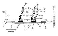
- FIG. 1 shows a schematic diagram of a lateral flow type device (100) as an embodiment of the nucleic acid detection device of the present invention (a plan view is shown in FIG. 1 and a sectional view is shown in FIG. 13).
- the shape of the device is not particularly limited as long as the tag-added amplification reaction product can be detected, and other forms include a dipstick type device (FIG.
- the nucleic acid detection devices of these embodiments are all examples of chromatographic devices.
- the chromatographic device refers to a nucleic acid detection device in the form of a chromatography carrier.
- the lateral flow device (100) in FIG. 1 is a conjugate in which a sample pad (1) for adding an amplification reaction product to which a tag is added and a labeled probe are arranged on a plastic substrate (110).
- a pad (2), a solid phase carrier (3), and an absorption pad (6) are sequentially stacked.
- the solid phase carrier (3) has one or more independent target detection units (4) and control detection units (5).
- the sample pad side is defined as upstream and the absorption pad side is defined as downstream for convenience.
- the target detection unit (4) refers to a region where a probe that specifically captures an amplification reaction product derived from a target nucleic acid to which a tag is added is immobilized.
- the control detection part (5) is one or more probes that specifically capture the amplification reaction product derived from the target nucleic acid. Indicates an area where is fixed.
- the type and number of amplification reaction products derived from the target nucleic acid captured by the control detection unit are not particularly limited.
- the position of the control detection unit in the present invention is not particularly limited, but in a lateral flow type device or a dipstick type device, it is preferably located downstream from the target detection unit.
- Sample pad (1), conjugate pad (2), solid support (3), and absorbent pad (6) are made of plastic, glass, cellulose, nitrocellulose, nylon, polyethersulfone, polyvinylidene fluoride, filter paper, etc. It is made of a material such as a porous body. These may be made of the same material, or may be made of different members.
- the shape of the carrier is not particularly limited, but is preferably a flat plate shape.
- an amplification reaction product to which two kinds of tags obtained by the nucleic acid amplification step are added is added to the sample pad (1).
- the reaction solution after the nucleic acid amplification reaction may be added dropwise as it is, or added after mixing with an appropriate developing solution (for example, phosphate buffer, Tris buffer, Good buffer, SSC buffer). May be.
- the developing solution can further contain a surfactant, salt, protein, sugar, nucleic acid, and the like, if necessary.
- the amplification reaction product to which the tag added to the sample pad (1) is added is developed by capillary action in the direction from the sample pad (1) to the absorption pad (6) in FIG.
- the amplification reaction product to which the two types of tags are added passes through the conjugate pad (2) containing the labeled probe, contacts the labeled probe, and binds to the labeled probe through one tag.
- the amplification reaction product bound to the labeled probe passes through the solid phase carrier (3), it is captured on the solid phase carrier through the binding between the other tag and the probe immobilized on the solid phase carrier. Is done.
- the amplification reaction product derived from the control nucleic acid is captured only by the control detection part (5) on the solid phase carrier, whereas the amplification reaction product derived from the target nucleic acid is captured by the target detection part (4) on the solid phase support and the control. Captured by both detectors (5).
- the target nucleic acid was preferentially amplified under conditions where a large amount of target nucleic acid was present in the reaction system, and the amplification reaction of the control nucleic acid was inhibited. Even in this case, the amplification reaction product derived from the target nucleic acid is captured not only in the target detection unit but also in the control detection unit and emits a signal, so that it is possible to prevent the disappearance or decrease of the signal in the control detection unit.
- the means for solving the problem of preventing the disappearance or decrease of the signal of the control detection unit is not limited to only means for providing a device in which two or more types of probes are immobilized on the control detection unit.
- the problem can be solved by using a device in which one type of probe is fixed to the control detection unit.
- a tagged primer is designed for each of the target nucleic acids having a tag that is specifically captured by the target detector and a target nucleic acid that has a tag that is specifically captured by the control detector. If a multiplex PCR system is constructed, even if a device in which one type of probe is immobilized on the control detection unit is used, one of the two types of amplification reaction products described above is captured by the detection unit. . Therefore, the means can solve the problem.
- the signal derived from the target nucleic acid and the control nucleic acid that has undergone amplification and / or labeling reaction is detected by a measuring instrument or visually, and the determination is made based on the obtained result Do.
- the labeling substance the coloration of red accompanying the aggregation of the gold colloidal particles is detected as a signal to make a determination.
- Examples of other shapes of the nucleic acid detection device in the present invention include the dipstick type nucleic acid detection device (100) shown in FIG. 2, the array type nucleic acid detection device (100) shown in FIG. Further, a plurality of array sections (4, 5) may be provided on the solid phase carrier (3) of the lateral flow type device (100) or the dipstick type device (100). In the plurality of array sections, the same probe may be immobilized, or separate probes may be immobilized.
- the shape of the nucleic acid detection device can also be set in consideration of the capture / detection mode.
- the nucleic acid detection device includes a detection kit containing reagents necessary for nucleic acid detection such as a control template nucleic acid, a nucleic acid amplification enzyme, a nucleic acid amplification reaction reagent, and a nucleic acid detection reaction reagent in addition to the nucleic acid detection device. It may be.
- the storage state of the control template nucleic acid contained in the kit may be any state of liquid, frozen, and dried products.
- the detection kit may further contain the above primers.
- the reagent for nucleic acid amplification reaction includes components such as a nucleic acid amplification enzyme, a substrate, and a buffer.
- the substrate include dATP, dTTP, dCTP, dGTP, dUTP, biotin-labeled dCTP, and biotin-labeled dUTP. It is not limited to these.
- As the buffer a buffer containing magnesium, potassium, a buffering agent, a surfactant, a reducing agent and the like is preferable.
- the storage state of the reagent for nucleic acid amplification reaction may be any state such as liquid, frozen, dried, lyophilized.
- the nucleic acid detection reaction reagent may be any reagent as long as it is used for detection, and examples thereof include the above-described developing solution, labeled antibody solution, chromogenic substrate solution, and fluorescent substrate solution. It is not limited.
- the storage state may be liquid or frozen.
- the nucleic acid detection device 100 having the form shown in FIG. 1 was created and used as the nucleic acid detection device.
- Example 1> (1) Preparation of labeled probe and conjugate pad 5.5 ml Gold Colloid (40 nm, 9.0 ⁇ 10 10 (number of particles / ml)) (British Biocell International) and 100 ⁇ M thiolated DNA (SEQ ID NO: 1) ) 60 ⁇ l was mixed and incubated at 50 ° C. for 16 hours. After 16 hours, 250 ⁇ l of 0.1 M phosphate buffer (pH 7.0) and 150 ⁇ l of 1 M NaCl were added and incubated at 50 ° C. for 24 hours.
- 0.1 M phosphate buffer pH 7.0
- the thiolated DNA used in this step is shown below.
- -Thiolated DNA 5'-CTATAAACCCAGTGAAAAATGTTGCCA-SH-3 '(SEQ ID NO: 1).
- (2) Production of solid phase carrier with immobilized probe The nucleic acid detection device 100 in this example has a target detection unit 4 and a control detection unit 5 installed in a line from the upstream on the solid phase carrier 3.
- Target detection unit 4 is a mixture of 200 ⁇ l of 100 ⁇ M probe A (SEQ ID NO: 2), 200 ⁇ l of 2.5 ⁇ mg / ml streptavidin, 100 ⁇ l of 1% BSA solution, and 500 ⁇ l of 5 ⁇ mM phosphate buffer.
- Control detection unit 5 is 100 ⁇ M probe A (SEQ ID NO: 2) and 100 ⁇ M probe B (SEQ ID NO: 3) 200 ⁇ l, 2.5 ⁇ mg / ml streptavidin 200 ⁇ l, 1% BSA solution 100 ⁇ l, 5 ⁇ mM, respectively.
- Probe A 5'-ATCACACATTAGCTGTCACTCGATGCA-Biotin-3 '(SEQ ID NO: 2)
- Probe B 5'-TCAAAGTCATTGTAAGTCCGTACTAG-Biotin-3 '(SEQ ID NO: 3)
- the substrate 110 made of a backing sheet is coated with the nitrocellulose membrane 3 which is a solid phase carrier produced in this example (2) and the conjugate pad 2 produced in this example (1).
- the sample addition part 1, which is a sample addition part, and the absorbent pad 6 for absorbing the developed sample and the labeled substance were bonded together to produce a nucleic acid detection device 100 capable of detecting the amplification reaction product after the amplification process.
- ⁇ Comparative Example 1> Line the control detector 5 with a mixture of 200 ⁇ l of 100 ⁇ M probe B (SEQ ID NO: 3), 200 ⁇ l of 2.5 mg / ml streptavidin, 100 ⁇ l of 1% BSA solution, and 300 ⁇ l of 5 mM phosphate buffer.
- a nucleic acid detection device 100 was produced in the same manner as in Example 1 except that the device was applied in the form of a thin film.
- the nucleic acid detection device 100 of Comparative Example 1 has the same structure as the nucleic acid detection device 100 of Example 1 except that the probes arranged in the control detection unit 5 are different.
- pUC19 described in SEQ ID NO: 4 (manufactured by Takara Bio Inc.) was set as the target nucleic acid to be detected
- ⁇ phage DNA described in SEQ ID NO: 5 (manufactured by Eurofin Genomics) was set as the control nucleic acid.
- the amplification reaction product is applied to the device 100 prepared in Example 1, and the color emitted from the complex in which the amplification reaction product and the labeled probe are bound A signal was detected.
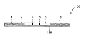
- the detection form is shown in FIG.
- a plan view of the nucleic acid detection device 100 is shown on the right side, and a schematic view of a cross section along the line XX ′ of the plan view is shown on the left side. 110 is omitted, and the ratio of the dimensions of the sample pad 1, the conjugate pad 2, the solid phase carrier 3, the target detection unit 4, the control detection unit 5 and the absorption pad 6 is appropriately changed for convenience of description.
- (1) Design of single-stranded nucleic acid tag-added primer The single-stranded nucleic acid tag-added primer used in this study consists of a primer body that binds to each nucleic acid and a polymerase reaction inhibition region (X) on the 5 'end side. It consists of a single-stranded nucleic acid tag added via
- PUC19 forward primer (SEQ ID NO: 6) and pUC19 reverse primer (SEQ ID NO: 7) were designed as primer bodies for amplifying pUC19.
- a ⁇ phage DNA forward primer (SEQ ID NO: 12) and a ⁇ phage DNA reverse primer (SEQ ID NO: 13) were designed as primer main bodies for amplifying ⁇ phage DNA.
- a single-stranded nucleic acid tag-added primer was designed by adding a single-stranded nucleic acid tag to the 5 ′ end side of each primer main body via azobenzene as a polymerase reaction inhibition region (X).
- a primer T1-X-F1 (Sequence No. 8) was added to the 5 ′ end of the pUC19 forward primer via azobenzene. No. 10) and a primer T2-X-R1 (SEQ ID NO: 11) in which a single-stranded nucleic acid tag T2 (SEQ ID NO: 9) was added to the pUC19 reverse primer were designed.
- a primer T3-X-F2 obtained by adding a single-stranded nucleic acid tag T3 (SEQ ID NO: 14) to a ⁇ phage DNA forward primer
- ⁇ Primer T2-X-R2 was designed by adding a single-stranded nucleic acid tag T2 (SEQ ID NO: 9) to a phage DNA reverse primer.
- PCR using single-stranded nucleic acid tag-added primers PCR was performed using the primer set described above. Place the primer set of SEQ ID NOs: 10 and 11, 15 pmol each of the primer sets of SEQ ID NOs: 15 and 16, and 10 pg of pUC19 and 1 pg of ⁇ phage DNA into a 0.2 ml PCR tube, and add TaKaRa Ex Taq (registered trademark) 100 ⁇ l of a PCR sample solution (A) was prepared according to the instructions (manufactured by Takara Bio Inc.).
- a PCR sample solution (B) containing the primer set and 100 pg pUC19, 1 pg ⁇ phage DNA, and a PCR sample solution containing the primer set and 1000 pg pUC19, 1 pg ⁇ phage DNA ( C) was prepared. Then, each tube was set in a thermal cycler (GeneAmp PCR System 9700 (Applied Biosystems)), heat-treated at 95 ° C for 5 minutes, 95 ° C for 30 seconds, 55 ° C for 30 seconds, 72 ° C for 30 seconds. The amplification reaction product was obtained by performing 35 cycles.
- a solution containing sterile water and 1 pg of ⁇ phage DNA instead of pUC19 was prepared and PCR was performed in the same manner as a negative control (D).
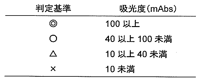
- the amplification reaction products (A) to (D) prepared in Example (2) were prepared in Example 1 without being denatured by heating. The sample was applied to the sample pad 1 of the nucleic acid detection device 100 and a detection test was performed. The result was judged by measuring the color intensity of the line with a chromato reader C10066-10 (manufactured by Hamamatsu Photonics) 10 minutes after the application. The results are shown in Table 1. Table 2 shows the criteria for determination of the chromatoreader measurement values.
- ⁇ indicates that the color is very dark and easy to visually check
- ⁇ indicates that the color is lighter than ⁇ , but easy to check visually
- ⁇ indicates that the color is light and visible but is somewhat difficult
- ⁇ To the extent that visual confirmation is impossible without coloring.
- “ ⁇ ” may be displayed as “4”, “ ⁇ ” as “3”, ⁇ as “2”, and “ ⁇ ” as “1”.
- the target detection unit 4 A signal derived from the target nucleic acid to be detected is also detected by the control detection unit 5. This prevents the signal from the control detection unit 5 from being lowered due to preferential amplification of the target nucleic acid, and the signal from the control detection unit 5 can be easily detected even when a large amount of target nucleic acid is present, such as 1000 pg. It has been shown.
- nucleic acid detection device 100 was produced in the same manner as in Example 1 (3).
- ⁇ Comparative Example 3> Apply 200 ⁇ l of 100 ⁇ M probe B (SEQ ID NO: 3), 200 ⁇ l of 1 M NaCl, 100 ⁇ l of 1% BSA solution, and 300 ⁇ l of 5 mM phosphate buffer to control detector 5 in a line.
- a nucleic acid detection device 100 was produced in the same manner as in Example 3 except that.
- Example 4 Similarly to Example 2, pUC19 (manufactured by Takara Bio Inc.) was set as the target nucleic acid to be detected, and ⁇ phage DNA (manufactured by Eurofin Genomics) was set as the control nucleic acid. After performing PCR using the single-stranded nucleic acid tag-added forward primer and biotin-modified reverse primer using each as a template, the amplification reaction product was applied to the device 100 prepared in Example 3, and the amplification reaction product and the labeled probe were combined. A chromogenic signal emanating from the bound complex was detected. The detection form is shown in FIG.
- Example 3 Detection of amplification reaction products using a nucleic acid detection device
- the amplification reaction products (A) to (D) prepared in Example (2) were prepared in Example 3 without being denatured by heating.
- the sample was applied to the sample pad 1 of the nucleic acid detection device 100 and a detection test was performed.
- the result was judged by measuring the color intensity of the line with a chromato reader C10066-10 (manufactured by Hamamatsu Photonics) 10 minutes after the application. The results are shown in Table 3.
- the target detection unit 4 A signal derived from the target nucleic acid to be detected is also detected by the control detection unit 5. This prevents the signal from the control detection unit 5 from being lowered due to preferential amplification of the target nucleic acid, and the signal from the control detection unit 5 can be easily detected even when a large amount of target nucleic acid such as 1000 pg is present. It has been shown.
- Example 5 Similarly to Example 2, pUC19 (manufactured by Takara Bio Inc.) was set as the target nucleic acid, and ⁇ phage DNA (manufactured by Eurofin Genomics) was set as the control nucleic acid.
- the amplification reaction product was applied to the device 100 prepared in Example 3, A chromogenic signal emitted from the complex in which the amplification reaction product and the labeled probe were bound was detected.
- a detection form using the device 100 produced in Example 3 is shown in FIG. (1) Selection of single-stranded nucleic acid tag-added primer The primer of SEQ ID NO: 10 was selected as the pUC19 forward primer, and the primer of SEQ ID NO: 7 was selected as the pUC19 reverse primer.
- the primer of SEQ ID NO: 15 was selected as the ⁇ phage DNA forward primer, and the primer of SEQ ID NO: 13 was selected as the ⁇ phage DNA reverse primer.
- (2) PCR using single-stranded nucleic acid tag-added primer and biotin-labeled 16-dUTP PCR was performed using the primer set described above.
- samples (A) to (D) were prepared according to the preparation method described in Example 2 (2), and PCR was performed under the same conditions as in Example 2 (2).
- PCR was performed under the same conditions as in Example 2 (2).
- Detection of amplification reaction products using a nucleic acid detection device The amplification reaction products (A) to (D) prepared in Example (2) were prepared in Example 3 without being denatured by heating or the like.
- the target detection unit 4 A signal derived from the target nucleic acid to be detected is also detected by the control detection unit 5. As a result, it is possible to prevent the signal from the control detection unit 5 from being lowered due to the preferential increase in the target nucleic acid. It was shown to be detectable.
- the device 100 prepared in Comparative Example 3 is used, if a large amount of target nucleic acid such as 1000 pg is present, the signal of the control detection unit 5 disappears, and it is determined whether PCR has been performed appropriately. I could't.
- Example 6 (1) Preparation of solid phase carrier with immobilized probe
- a solid phase carrier 3 on which a probe was immobilized was prepared in the same manner as in Example 1 (2).
- nucleic acid detection device 100 was produced in the same manner as in Example 1 (3).
- a probe mixed solution used for the control detection unit 5 a mixed solution containing 2.0 mg / ml anti-FITC antibody (Roche Applied Science), 2.5% sucrose, 20 mM TBS (pH 8.0) was applied in a line.
- a nucleic acid detection device 100 was produced in the same manner as in Example 6 except for the above.
- Example 7 Similarly to Example 2, pUC19 (manufactured by Takara Bio Inc.) was set as the target nucleic acid, and ⁇ phage DNA (manufactured by Eurofin Genomics) was set as the control nucleic acid. After performing PCR using each template and primer set, the amplification reaction product was applied to the device 100 prepared in Example 6, and a color signal emitted from the complex in which the amplification reaction product and the labeled probe were bound was detected. A detection form using the device 100 produced in Example 6 is shown in FIG.
- DIG-modified primers obtained by consignment synthesis at Tsukuba Oligo Service Co., Ltd.
- Primer DIG-F1 5'-DIG-GGAAACAGCTATGACCATGA-3 '(SEQ ID NO: 19)
- Primer FITC-F2 5'-FITC- AAGTTCTCGCTGGAAGAGGT-3 '(SEQ ID NO: 20)
- Samples (A) to (D) were prepared according to the preparation method described in Example 2 (2) using the primer sets of SEQ ID NOS: 11 and 19 and the primer sets of SEQ ID NOs: 16 and 20, and Example 2 ( PCR was performed under the same conditions as in 2).
- (3) Detection of amplification reaction products using a nucleic acid detection device The amplification reaction products (A) to (D) prepared in Example (2) were prepared in Example 6 without being denatured by heating. The sample was applied to the sample pad 1 of the nucleic acid detection device 100 and a detection test was performed. The result was judged by measuring the color intensity of the line with a chromato reader C10066-10 (manufactured by Hamamatsu Photonics) 10 minutes after the application. The results are shown in Table 5.
- the target detection unit 4 A signal derived from the target nucleic acid to be detected is also detected by the control detection unit 5. As a result, it is possible to prevent the signal from the control detection unit 5 from being lowered due to the preferential increase in the target nucleic acid. It was shown to be detectable. On the other hand, when the device 100 prepared in Comparative Example 6 is used, if a large amount of target nucleic acid such as 1000 pg is present, the signal from the control detection unit 5 disappears, and it is determined whether PCR has been performed appropriately. I could't.
- This embodiment is not a method of immobilizing two types of probes for capturing a control nucleic acid and a target nucleic acid in the control detection unit 5 as in the previous examples, but two types of target nucleic acids with different tag types.
- a tag-added primer is used to amplify a target nucleic acid having a tag specifically captured by the target detection unit 4 and a target nucleic acid having a tag specifically captured by the control detection unit 5.
- the design was such that one of the two types of target nucleic acid was captured by the control detection unit 5.
- FIG. 12 shows the detection form of this example. As shown in FIG. 12, one of the two types of target nucleic acids described above is captured by the control detection unit 5.
- Example 2 Similarly to Example 2, pUC19 (manufactured by Takara Bio Inc.) was set as the target nucleic acid, and ⁇ phage DNA (manufactured by Eurofin Genomics) was set as the control nucleic acid. After PCR, the amplification reaction product was applied to the device 100 prepared in Comparative Example 1, and a color signal emitted from the complex in which the amplification reaction product and the labeled probe were bound was detected.
- (1) Design and selection of single-stranded nucleic acid tag-added primers In this study, two types of primer sets were prepared as pUC19 primer sets. First, primers of SEQ ID NOs: 10 and 11 were selected as the first primer set.
- primer T3-X-F1 (SEQ ID NO: 14) having a single-stranded nucleic acid tag T3 (SEQ ID NO: 14) added to the 5 ′ end of the forward primer of SEQ ID NO: 6 via azobenzene as a polymerase reaction inhibition region (X) No. 21) was designed, and the set of SEQ ID Nos. 11 and 21 was used as the second primer set.
- the primer sets of SEQ ID NOs: 15 and 16 were selected as the ⁇ phage DNA primer set.
- Example (3) Detection of amplification reaction products using a nucleic acid detection device
- the amplification reaction products (A) to (D) prepared in Example (2) were prepared in Comparative Example 1 without being denatured by heating.
- the sample was applied to the sample pad 1 of the nucleic acid detection device 100 and a detection test was performed. The result was judged by measuring the color intensity of the line with a chromato reader C10066-10 (manufactured by Hamamatsu Photonics) 10 minutes after the application. The results are shown in Table 6.
- Example 8 two kinds of target nucleic acids having a tag specifically captured by the target detection unit 4 and a target nucleic acid having a tag specifically captured by the control detection unit 5 are prepared, and the detection device A device in which one type of probe was immobilized on both the target detection unit 4 and the control detection unit 5 was used as 100. Also in this example, since the target nucleic acid is detected not only by the target detection unit 4 but also by the control detection unit 5, as shown in Table 6, under conditions where 1000 ⁇ pg of target nucleic acid and 1 ⁇ pg of control nucleic acid exist. Even in this case, the control detection unit 5 was able to detect a signal derived from the target nucleic acid.
- Detection method using dipstick type device 100 and array type device 100 method of detecting amplified and / or labeled product in a single-stranded state by heat treatment, and immobilizing sugar chain on tag to carrier
- This embodiment can also be implemented by a method using a lectin as a probe.
Abstract
本発明は核酸検出用デバイスを用いた遺伝子検査において、対照検出部で得られるはずのシグナルの消失または低下の防止を課題とする。 対照検出部に、対照核酸と一種以上の標的核酸を捕捉するためのプローブが固定されていることを特徴とする、核酸検出用デバイス。
Description
本発明は、核酸検出用デバイスおよび核酸検出方法に関する。
遺伝子工学の発展に伴い、病原体やがん細胞の検出、細胞動態の解析、体質検査、医薬品の奏効検査などへ遺伝子検査が利用されるようになった。遺伝子検査は一般的に、検体の採取工程、検体からの核酸の調製工程、核酸の増幅反応工程および/または標識反応工程、増幅および/または標識された核酸の検出工程から構成される。
検出工程においては、増幅および/または標識された核酸を固相担体上で捕捉・検出可能な、ラテラルフロー型(特許文献1)、ディップスティック型(特許文献2)やアレイ型(特許文献3)などの核酸検出用デバイスが開発され利用されている。これらの核酸検出用デバイスには、標的核酸の増幅および/または標識反応産物を検出する標的検出部と、コントロールとして予め添加される対照核酸(内部標準物質)の増幅および/または標識反応産物を検出するための対照検出部を設けるのが一般的である。対照検出部は、増幅反応および/または標識反応が正常に進行したかどうかを評価するために用いられ、具体的には、対照検出部におけるシグナルを検出することで、酵素などの試薬が正常に機能したこと、および実験操作が問題なく適切に行われたことを評価し、検査結果の信頼性を確認するために利用する。
従来の核酸検出用デバイスにおいては、試薬や実験操作に問題がないにもかかわらず、対照検出部で得られるはずのシグナルが消失または低下する可能性があると、本発明者らは考えた。
本発明者らは、驚くべきことに、例えばマルチプレックスPCRなどにおいて、反応系内に標的核酸が多量に存在すると、標的核酸が優先的に増幅し、対照核酸の増幅反応および/または標識反応が阻害され、対照検出部のシグナルが消失あるいは微弱となる場合があることを解明した。
さらに、核酸検出用デバイスの対照検出部において、対照核酸のみならず少なくとも1種類の標的核酸を併せて捕捉・検出することで、対照検出部のシグナルの消失または低下を防ぎ、安定して高いシグナルが得られることを見出した。さらには、本手法を利用した核酸検出用デバイスを作製し、本発明を完成させるに至った。
すなわち、本発明は以下の通りである。
(1)一つ以上の標的検出部と、対照検出部を有する核酸検出用デバイスであって、前記標的検出部には標的核酸を捕捉するためのプローブが固定化されており、かつ、前記対照検出部には対照核酸を捕捉するためのプローブと前記標的核酸を捕捉するための一種以上のプローブとが固定化されていることを特徴とする、核酸検出用デバイス。
(2)(1)に記載の核酸検出用デバイスの、より好ましい態様では、前記標的核酸と前記対照核酸が、増幅工程を経た増幅反応産物である。
(3)(1)に記載の核酸検出用デバイスまたは(2)に記載の好ましい態様の核酸検出用デバイスの、より好ましい態様では、前記プローブが、核酸またはタンパク質である。
(4)(1)に記載の核酸検出用デバイスまたは(2)または(3)に記載の好ましい態様の核酸検出用デバイスの、より好ましい態様では、前記デバイスが、クロマト型デバイスである。
(5)(1)に記載の核酸検出用デバイスまたは(2)~(4)のいずれかに記載の好ましい態様の核酸検出用デバイス、対照用鋳型核酸、核酸増幅酵素、核酸増幅反応用試薬、核酸検出反応用試薬を含む、核酸検出用キット。
(6)一種以上の標的核酸と対照核酸とを検出する核酸検出方法であって、
(a) プライマーと鋳型核酸、核酸増幅反応用試薬を用いて、同一反応容器内で同時に核酸増幅反応を行い、前記一種以上の標的核酸と前記対照核酸とを増幅する工程、
(b) 前記一種以上の標的核酸と、固相担体上の標的検出部に固定化されたプローブとを結合させる工程、
(c) 前記一種以上の標的核酸と前記対照核酸を、前記固相担体上の対照検出部に固定化された一種以上のプローブと結合させる工程、
(d) 捕捉された前記標的核酸と前記対照核酸を標識し、前記標的核酸および前記対照核酸に由来するシグナルを検出する工程、
を含む、核酸検出方法。
(a) プライマーと鋳型核酸、核酸増幅反応用試薬を用いて、同一反応容器内で同時に核酸増幅反応を行い、前記一種以上の標的核酸と前記対照核酸とを増幅する工程、
(b) 前記一種以上の標的核酸と、固相担体上の標的検出部に固定化されたプローブとを結合させる工程、
(c) 前記一種以上の標的核酸と前記対照核酸を、前記固相担体上の対照検出部に固定化された一種以上のプローブと結合させる工程、
(d) 捕捉された前記標的核酸と前記対照核酸を標識し、前記標的核酸および前記対照核酸に由来するシグナルを検出する工程、
を含む、核酸検出方法。
(7)(6)に記載の核酸検出方法の、より好ましい態様では、前記プローブが、核酸またはタンパク質である。
本明細書は本願の優先権の基礎となる日本国特許出願番号2015-167198号の開示内容を包含する。
本発明によれば、核酸検出用デバイスにおける対照検出部のシグナルの消失または低下を防ぎ、安定して高いシグナルを得ることができるため、検査結果の信頼性を容易に確認することができる。
本発明の核酸検出方法に用いることができる、検体の採取、検体からの核酸の調製、核酸の増幅、及び核酸の検出の好ましい態様について以下に説明する。
<検体の採取工程>
標的核酸の存在を検出する対象となる検体は、核酸を含む可能性のある試料であれば特に制限はない。例えば、動物または植物の細胞、組織、全血、血清、リンパ液、骨髄液、組織液、尿、精液、膣液、羊水、涙、唾液、汗、乳汁などの体液、エキソソームなどの細胞に由来する小胞、糞便、咽頭液、痰、細菌、ウイルス、ウイロイドなどが挙げられる。検体の採取に関しては、従来公知の各種方法が利用できる。
<検体の採取工程>
標的核酸の存在を検出する対象となる検体は、核酸を含む可能性のある試料であれば特に制限はない。例えば、動物または植物の細胞、組織、全血、血清、リンパ液、骨髄液、組織液、尿、精液、膣液、羊水、涙、唾液、汗、乳汁などの体液、エキソソームなどの細胞に由来する小胞、糞便、咽頭液、痰、細菌、ウイルス、ウイロイドなどが挙げられる。検体の採取に関しては、従来公知の各種方法が利用できる。
本発明における核酸とは、大きく天然型核酸と非天然型核酸に分けられ、「天然型核酸」とは、ヌクレオチドを基本単位とし、各ヌクレオチド間が糖の3'位と5'位炭素のリン酸ジエステル結合で結ばれたポリヌクレオチドをいう。天然型核酸としては、DNAやRNAといったデオキシリボヌクレオチドやリボヌクレオチドの重合体が挙げられる。「非天然型核酸」とは、上記天然のヌクレオチドにかえて、又は加えて、非天然のヌクレオチドを含む核酸をいい、非天然のヌクレオチドとは、ヌクレオチドの塩基部分等に人工的な改変がなされたヌクレオチド、又は人工的に作られたヌクレオチドに類似する性質を有するヌクレオチド類似体を指し、例えば、キサントシン類及びジアミノピリミジン類等が挙げられる。
本発明における標的核酸とは、検出対象となる核酸のことであり、標的配列が含まれていれば特に限定されず、上述した検体に由来するゲノムDNA、プラスミドDNA、ctDNA(cfDNA)、mRNA、miRNA、lncRNAやアンプリコンなどが例示される。本明細書では、検体に含まれる標的核酸を増幅した増幅反応産物も「標的核酸」と称する場合がある。
本発明における対照核酸とは、標的核酸を検出する際に、操作や試薬に問題がないことを確認するための、内部標準として用いる核酸のことであり、当該機能を発揮する核酸であれば特に限定はされない。本発明では、核酸増幅反応において鋳型核酸として用いる対照用鋳型核酸を増幅した増幅反応産物も「対照核酸」と称する場合がある。
<検体からの核酸の調製工程>
後述する工程aにおいて鋳型核酸として用いる核酸を検体から調製する工程は、検体から核酸を抽出あるいは分離・精製できればよく、その手法は特に限定されない。抽出法の一例としては、熱や超音波などによる物理的な破砕や、アルカリ試薬、有機溶剤、界面活性剤などの薬品による破砕、リゾチームやプロテイナーゼKなどの酵素による破砕が挙げられる。分離・精製の一例としては、バインドエリュート法、ゼオライトなどのイオン交換樹脂、遠心分離などが例示されるが、調製後の核酸を鋳型核酸として用いて、その後の核酸増幅反応などの生化学的反応が問題なく進行する手法であれば特に限定されない。また、本工程を省略し、直接検体を鋳型核酸の供給源として核酸増幅反応に供することもできる。
<核酸の増幅工程(工程a)>
本発明の方法の工程aは、プライマーと鋳型核酸、核酸増幅反応用試薬(核酸増幅試薬)を用いて、同一反応容器内で同時に核酸増幅反応を行い、前記一種以上の標的核酸と前記対照核酸とを増幅する工程である。
工程aにおいて鋳型核酸は、標的核酸を含む可能性のある核酸(典型的には前記検体に由来する核酸)と、対照核酸とを含む。鋳型核酸として核酸増幅反応系に添加する対照核酸を「対照用鋳型核酸」と称する場合がある。
「同一反応容器内で同時に核酸増幅反応を行い」とは、前記一種以上の標的核酸の核酸増幅反応と、前記対照核酸の核酸増幅反応を、1つの反応容器内で一緒に行うことを指す。このためには、工程aに用いるプライマーは、1以上の標的核酸の各々を増幅することができるプライマーのセットと、対照核酸を増幅することができるプライマーのセットとを含む。
工程aは、好ましくは、前記プライマーとしてタグ付加プライマーのセットを用いて、核酸増幅反応を行い、二種類のタグが付加された増幅反応産物を調製する工程である。
タグ付加プライマーとは、プライマーにタグが付加された核酸増幅用プライマーのことを表し、タグの付加された増幅反応産物を調製する目的で使用することができる。
本発明におけるプライマーとは、標的核酸または対照核酸を特異的に認識し、核酸増幅反応における伸長の起点となる5~80塩基の一本鎖核酸である。具体的には、各プライマーの塩基配列は、標的核酸または対照核酸の標的塩基配列(増幅対象塩基配列)における3’末端側、または、標的塩基配列の相補塩基配列における3’末端側とハイブリダイズしうる配列であり、一般的には、標的塩基配列の3’末端側の塩基配列と相補的な塩基配列、または、標的塩基配列の相補塩基配列における3’末端側と相補的な塩基配列である。ここで「標的塩基配列」は、標的核酸または対照核酸の、検出しようとする塩基配列又はその相補塩基配列であり、標的核酸または対照核酸が二本鎖核酸である場合には、二本鎖核酸のいずれか一方の鎖の核酸の塩基配列を指す。プライマーは、標的核酸または対照核酸と特異的に結合可能であれば、塩基欠損や挿入、およびミスマッチ部位を有していてもよい。ここで所定の塩基配列の「3’末端側」とは、該所定の塩基配列の3'末端の塩基を含む、連続した複数の塩基(典型的には5~80の塩基)からなる部分塩基配列を指す。
本発明において、塩基配列Xが塩基配列Yに「ハイブリダイズしうる」とは、塩基配列Xを含むポリヌクレオチド(特にDNA)が、ストリンジェント条件で、塩基配列Yを含むポリヌクレオチド(特にDNA)にハイブリダイズし、塩基配列Yを含まないポリヌクレオチドにはハイブリダイズしないことを意味する。すなわちハイブリダイズするとは、特異的にハイブリダイズすることを指す。ここで、「ストリンジェントな条件」とは、いわゆる特異的なハイブリッドが形成され、非特異的なハイブリッドが形成されない条件を意味する。ストリンジェントな条件は、例えば、本発明中のプライマーとその相補鎖との融解温度Tm(℃)およびハイブリダイゼーション溶液の塩濃度などに依存して決定することができ、例えばGreen and Sambrook, Molecular Cloning, 4th Ed (2012), Cold Spring Harbor Laboratory Press を参照することができる。具体的には、サザンハイブリダイゼーションの際の温度や溶液に含まれる塩濃度、及びサザンハイブリダイゼーションの洗浄工程の際の温度や溶液に含まれる塩濃度によりストリンジェントな条件を設定することができる。より詳細には、ストリンジェントな条件としては、例えば、ハイブリダイゼーション工程では、ナトリウム濃度が25~500mM、好ましくは25~300mMであり、温度がポリヌクレオチド配列によって決定されるTmよりわずかに低い温度(例えば、Tmよりも0~約5℃低い温度)、例えば40~68℃、好ましくは40~65℃である。より具体的には、ハイブリダイゼーションは、1~7×SSC、0.02~3%SDS、温度40℃~60℃で行うことができる。また、ハイブリダイゼーションの後に洗浄工程を行っても良く、洗浄工程は、例えば0.1~2×SSC、0.1~0.3%SDS、温度50~65℃で行うことができる。
標的核酸を増幅するためのプライマーは、標識するためのタグと、固相担体への結合のためのタグとが結合した標的核酸を、増幅反応産物として生成することができるように構成されている。このためには一対のタグ付加プライマーを使用することができる。また、一対のプライマーの一方又は両方にタグが付加されていない場合には、増幅反応により、タグが付加されたヌクレオチドを含む標的核酸の増幅反応産物が生成されるように、タグが付加されたヌクレオチド源を含む核酸増幅反応用試薬を用いることができる。
同様に、対照核酸を増幅するためのプライマーは、標識するためのタグと、固相担体への結合のためのタグとが結合した対照核酸を、増幅反応産物として生成することができるように構成されている。このためには一対のタグ付加プライマーを使用することができる。また、一対のプライマーの一方又は両方にタグが付加されていない場合には、増幅反応により、タグが付加されたヌクレオチドを含む対照核酸の増幅反応産物が生成されるように、タグが付加されたヌクレオチド源を含む核酸増幅反応用試薬を用いることができる。
ここで「標識するためのタグ」とは、後述する標識プローブにおけるプローブと結合することができるタグである。また「固相担体への結合のためのタグ」とは、後述する固相担体に固定化されたプローブと結合することができるタグである。
タグとは、増幅反応産物を標識するために用いる物質又は増幅反応産物を固相に結合するために用いる物質であって、それ自体ではシグナルを発しない物質のことを表す。例えば、核酸、ビオチン、DIG(ジゴキシゲニン)やFITC(フルオレセインイソチオシアネート)などを含むハプテン、糖鎖などが挙げられる。一本鎖の核酸をタグとした場合、核酸増幅反応で二本鎖化されないようタグとプライマーとの間にポリメラーゼ反応阻害領域からなるスペーサー構造を挿入することが好ましい。ポリメラーゼ反応阻害領域からなるスペーサー構造は、ポリメラーゼによる核酸伸長反応を阻害し、タグを一本鎖構造に保つことが可能なら特に限定はされない。例えばアゾベンゼン修飾の他、アルキレン鎖又はポリオキシアルキレン鎖や逆位塩基修飾など種々の天然あるいは非天然型修飾の挿入などが挙げられる。タグとして用いる核酸の塩基数は特に限定されないが例えば5~80であることができる。タグが付加されたヌクレオチド源としては、ビオチン又は前記ハプテンが付加されたdNTP(dUTP, dCTP等)が例示できる。
本発明における核酸増幅反応とは、例えばPCR法などに代表され、特定の核酸配列を増幅するものであればどのような反応でもかまわない。PCR法以外に、LCR(Ligase Chain Reaction)法 、SDA(Strand Displacement Amplification)法、RCA (Rolling Circle Amplification)法、CPT(Cycling Probe Technology) 法、Q-Beta Replicase Amplification Technology法、ICAN (Isothermal and Chimeric primer-initiated Amplification of Nucleic Acids)法 、LAMP(Loop-Mediated Isothermal Amplificaton of DNA)法、NASBA(Nucleic acid Sequence-based Amplification method) 法、及びTMA(Transcription mediated amplification method)法、RPA(Recombinase Polymerase Amplification)法、SIBA(Strand Invasion Based Amplification)法などの公知の方法が例示できる。Q-Beta Replicase Amplification Technology法、RCA法、NASBA法、SDA法、TMA法、LAMP法、ICAN法、RPA法、SIBA法などは一定温度で増幅反応を行う方法であり、その他のPCR法やLCR法などは温度サイクリングで増幅反応を行う方法である。
核酸増幅反応に使用する核酸増幅反応用試薬は、核酸増幅酵素と、核酸増幅反応に必要な他の成分とを含む。
核酸増幅反応に使用する酵素としては、特に限定されるものではなく、市販のポリメラーゼなどを好適に使用しうる。核酸増幅酵素の一例として、E. coli由来DNAポリメラーゼI、T4 DNAポリメラーゼ、T7 DNAポリメラーゼ、Taq DNAポリメラーゼ、KOD DNAポリメラーゼ、Pfu DNAポリメラーゼ、Bst DNAポリメラーゼ、Bsu DNAポリメラーゼ、Phi29 DNAポリメラーゼ、Bca BEST DNAポリメラーゼ、逆転写酵素、SP6 RNAポリメラーゼ、T7 RNAポリメラーゼ、T3 RNAポリメラーゼなどが挙げられるがこれらに限定されるものではない。
核酸増幅反応用試薬の他の成分については後述する。
上記の工程aにより、各々に二種のタグが付加した一種以上の標的核酸(ただし鋳型核酸に標的核酸が含まれる場合)と対照核酸が、核酸増幅産物として得られる。各標的核酸または対照核酸において前記二種のタグは、上述の、標識するためのタグと、固相担体への結合のためのタグである。工程aで得られる各標的核酸及び対照核酸の核酸増幅産物は、それぞれ、各標的核酸又は対照核酸が二本鎖化され、且つ、前記の二種のタグが付加した核酸増幅産物であることが通常である。
<核酸の検出工程(工程b,c,d)>
本発明の核酸検出方法は、上記の工程aに加えて下記の工程b,c,d:
(b) 前記一種以上の標的核酸と、固相担体上の標的検出部に固定化されたプローブとを結合させる工程、
(c) 前記一種以上の標的核酸と前記対照核酸を、前記固相担体上の対照検出部に固定化された一種以上のプローブと結合させる工程、
(d) 捕捉された前記標的核酸と前記対照核酸を標識し、前記標的核酸および前記対照核酸に由来するシグナルを検出する工程
を含む。
<核酸の検出工程(工程b,c,d)>
本発明の核酸検出方法は、上記の工程aに加えて下記の工程b,c,d:
(b) 前記一種以上の標的核酸と、固相担体上の標的検出部に固定化されたプローブとを結合させる工程、
(c) 前記一種以上の標的核酸と前記対照核酸を、前記固相担体上の対照検出部に固定化された一種以上のプローブと結合させる工程、
(d) 捕捉された前記標的核酸と前記対照核酸を標識し、前記標的核酸および前記対照核酸に由来するシグナルを検出する工程
を含む。
工程aの後に行うこれらの工程b, c, dの順序は特に限定されず、一部又は全部を同時に行ってもよい。
本明細書では、工程b, c, dをまとめて「核酸の検出工程」と称する。
核酸の検出工程は、典型的には、まず、上記増幅工程で得られた二種類のタグが付加された増幅反応産物(標的核酸及び対照核酸)の一方のタグと標識プローブとの結合により複合体を形成し、次いで、もう一方のタグを利用して当該複合体を固相担体上へ捕捉し、増幅反応産物に由来するシグナルを検出する。
本発明におけるプローブとは、タグと特異的に結合することができる物質であり、DIGやFITCなどのハプテンタグに対する抗体、ビオチンタグに対するアビジン(ストレプトアビジンであってもよい)、一本鎖核酸タグに対する相補鎖などが例示されるが、タグを特異的に認識し結合するものであればこれらに限定されない。本発明においてプローブは、好ましくは、抗体、アビジン等のタンパク質、又は、一本鎖核酸タグに対する相補鎖等の核酸である。プローブとして用いる核酸の塩基数は特に限定されないが例えば5~80であることができる。
本発明における標識プローブとは、プローブと検出用のシグナルを発する標識物質とが結合したものである。標識物質には、従来公知のものを適宜選んで使用することができる。例えば、蛍光化合物、放射性同位元素、電気化学活性化合物、金属コロイド粒子、着色粒子、顔料や染料といった着色剤などが挙げられる。工程dにおけるシグナルの検出は、標識物質に応じた方法により行えばよい。シグナルの検出は、測定器または目視により行うことができる。例えば、標識物質として金コロイド粒子を利用した場合、金コロイド粒子の凝集に伴う赤色の着色をシグナルとして検出する。
本発明における増幅反応産物(増幅反応産物と標識プローブとの複合体であってもよい)の固相担体上への捕捉は、増幅反応産物に付加されたもう一方のタグと固相担体に固定化されたプローブとの結合を介して行われる。本発明における固相担体上へのプローブの固定化方法は特に限定されない。例えば、一本鎖核酸プローブは、その3’末端を介して担体に固定化しても、5’末端を介して担体に固定化しても、各末端部以外で担体に固定化しても、一以上の部分で担体に固定化しても、タンパク質を介して担体に固定化してもよい。このようなタンパク質を介した固定化法としては、固相担体上にストレプトアビジンをコートし、ビオチン修飾プローブを固定化する、いわゆるビオチン-アビジン反応を利用した方法が例示される。固相担体に固定化されたプローブは、担体の表面に対して適当なスペーサーを備えていてもよい。
<核酸検出用デバイス>
本発明における核酸検出用デバイスの一実施形態としてラテラルフロー型デバイス(100)の模式図(平面図が図1、断面図が図13)を示す。タグの付加された増幅反応産物を検出できればデバイス形状は特に限定されず、他の形態としてディップスティック型デバイス(図2)、アレイ型デバイス(図3)などが例示される。これらの実施形態の核酸検出用デバイスは、いずれも、クロマト型デバイスの例である。ここでクロマト型デバイスとは、クロマトグラフィー担体の形態の核酸検出用デバイスを指す。
図1のラテラルフロー型デバイス(100)は、プラスチック製の基材(110)の上に、タグが付加された増幅反応産物を添加するためのサンプルパッド(1)、標識プローブを配置したコンジュゲートパッド(2)、固相担体(3)、および吸収パッド(6)を順次重ねて配置してなる。固相担体(3)には、それぞれ独立した一つ以上の標的検出部(4)と対照検出部(5)を有する。本発明においては、便宜上サンプルパッド側を上流、吸収パッド側を下流と定義する。
本発明において標的検出部(4)とは、タグが付加された標的核酸由来の増幅反応産物を特異的に捕捉するプローブが固定化されている領域を示す。また、対照検出部(5)とは、タグが付加された対照核酸由来の増幅反応産物を特異的に捕捉するプローブに加え、標的核酸由来の増幅反応産物を特異的に捕捉する一種以上のプローブが固定化されている領域を示す。対照検出部にて捕捉される標的核酸由来の増幅反応産物の種類および数は特に限定されない。
本発明における対照検出部の位置は特に限定されないが、ラテラルフロー型デバイスや、ディップスティック型デバイスにおいては、標的検出部より下流に位置することが好ましい。
サンプルパッド(1)、コンジュゲートパッド(2)、固相担体(3)、及び吸収パッド(6)は、プラスチックやガラス、セルロース、ニトロセルロース、ナイロン、ポリエーテルスルホン、ポリフッ化ビニリデン及びろ紙などの多孔質体などの材質よりなる。これらは同一の材質より構成されていてもよいし、異なる部材より構成されていてもよい。また、担体の形状は特に限定されないが、平板状であることが好ましい。
核酸の増幅工程により得られた二種類のタグが付加された増幅反応産物はサンプルパッド(1)に添加される。添加方法としては、核酸増幅反応後の反応液をそのまま滴下してもよいし、適当な展開溶液(例えば、リン酸緩衝液、Tris緩衝液、グッド緩衝液、SSC緩衝液)と混合後に滴下してもよい。展開溶液には必要に応じて界面活性剤、塩、タンパク質、糖、核酸などをさらに含めることができる。サンプルパッド(1)に添加されたタグが付加された増幅反応産物は、図1中のサンプルパッド(1)から吸収パッド(6)の方向に毛細管現象により展開される。
二種類のタグが付加された増幅反応産物は標識プローブを含むコンジュゲートパッド(2)を通過する際に、標識プローブと接触し、一方のタグを介して標識プローブと結合する。
次いで、標識プローブと結合した増幅反応産物は、固相担体(3)を通過する際に、もう一方のタグと固相担体に固定化されたプローブとの結合を介して固相担体上に捕捉される。対照核酸由来の増幅反応産物は固相担体上の対照検出部(5)のみに捕捉されるのに対し、標的核酸由来の増幅反応産物は、固相担体上の標的検出部(4)および対照検出部(5)の両方に捕捉される。これにより、マルチプレックスPCRを利用した核酸検出用デバイスによる検出において、反応系内に標的核酸が多量に存在する条件下で、標的核酸が優先的に増幅し、対照核酸の増幅反応が阻害された場合においても、標的核酸由来の増幅反応産物が、標的検出部だけでなく、対照検出部においても捕捉されシグナルを発するため、対照検出部のシグナルの消失または低下を防止することが可能となる。
なお、対照検出部のシグナルの消失または低下を防止するという課題を解決する手段であれば、対照検出部にプローブが二種以上固定化されたデバイスを提供する手段のみに限定されない。例えば、対照検出部に一種類のプローブが固定化されたデバイスを用いても該課題を解決することができる。標的検出部で特異的に捕捉されるタグを有する標的核酸と、対照検出部で特異的に捕捉されるタグを有する標的核酸の二種類が増幅するよう、それぞれに対してタグ付加プライマーを設計し、マルチプレックスPCRの系を構築すれば、対照検出部に一種類のプローブが固定化されたデバイスを用いたとしても、該検出部で上述した二種類のうち片方の増幅反応産物が捕捉される。したがって該手段により該課題を解決することが可能となる。
本発明における核酸検出用デバイスを用いた結果判定は、増幅および/または標識反応を経た標的核酸および対照核酸に由来するシグナルを測定器または目視により検出し、得られた結果をもとに判定を行う。例えば、標識物質として金コロイド粒子を利用した場合、金コロイド粒子の凝集に伴う赤色の着色をシグナルとして検出し、判定を行う。
本発明における核酸検出用デバイスのその他の形状としては、図2に示すディップスティック型の核酸検出用デバイス(100)や、図3に示すアレイ型の核酸検出用デバイス(100)などが挙げられる。また、ラテラルフロー型デバイス(100)、ディップスティック型デバイス(100)の固相担体(3)上に複数のアレイ区画(4,5)を備えていてもよい。これらの複数のアレイ区画は、それぞれ同一のプローブが固定化されていてもよいし、それぞれ別個のプローブが固定化されていてもよい。核酸検出用デバイスの形状は、捕捉・検出の様態を考慮して設定することもできる。例えば、ディップスティック型デバイス(100)の場合、一般的に用いられているマイクロチューブに供給される増幅および/または標識産物の溶液に対してデバイスの先端部(図示する例ではサンプルパッド(1)の側の端部)が浸漬可能な幅及び形状を備えていることが好ましい。
<核酸検出用キット>
本発明における核酸検出用デバイスは、核酸検出用デバイスに加えて対照用鋳型核酸、核酸増幅酵素、核酸増幅反応用試薬、核酸検出反応用試薬など、核酸検出に必要な試薬類を含んだ検出キットであってもよい。本キットに含まれる対照用鋳型核酸の保存状態は液体、冷凍、乾燥品いかなる状態であってもよい。検出キットは更に上記のプライマーを含んでもよい。
<核酸検出用キット>
本発明における核酸検出用デバイスは、核酸検出用デバイスに加えて対照用鋳型核酸、核酸増幅酵素、核酸増幅反応用試薬、核酸検出反応用試薬など、核酸検出に必要な試薬類を含んだ検出キットであってもよい。本キットに含まれる対照用鋳型核酸の保存状態は液体、冷凍、乾燥品いかなる状態であってもよい。検出キットは更に上記のプライマーを含んでもよい。
核酸増幅反応用試薬には、核酸増幅酵素、基質、バッファー等の成分が含まれ、基質の一例としては、dATP、dTTP、dCTP、dGTP、dUTP、ビオチン標識dCTP、ビオチン標識dUTPなどが挙げられるがこれらに限定されるものではない。バッファーとしては、マグネシウム・カリウム・緩衝剤・界面活性剤・還元剤などを含むバッファーが好ましい。核酸増幅反応用試薬の保存状態は、液体、冷凍、乾燥、凍結乾燥などいかなる状態であってもよい。
核酸検出反応用試薬(核酸検出用試薬)は、検出に用いる試薬であればいかなるものでもよく、例えば、上述した展開溶液、標識抗体溶液、発色基質溶液、蛍光基質溶液などが挙げられるがこれらに限定されるものではない。保存状態は、液体、冷凍いかなる状態であってもよい。
以下、本発明を実施例を挙げて具体的に説明する。但し、本発明はこれらの実施例にその技術的範囲が限定されるものではない。
以下の実施例及び比較例では、核酸検出用デバイスとして、図1に示す形態の核酸検出用デバイス100を作成し用いた。
<実施例1>
(1)標識プローブおよびコンジュゲートパッドの作製
5.5 mlのGold Colloid (40 nm, 9.0×10¹⁰(粒子数/ml))(British Biocell International社製)と、100 μMのチオール化DNA(配列番号1)60 μlを混合し、50℃にて16時間インキュベートした。16時間後、250 μlの0.1 Mリン酸バッファー(pH 7.0)、150 μlの1 M NaClを添加し、50℃にて24時間インキュベートした。24時間後、遠心(5000 G、15℃、20分)し、上清を除いた。6 mlの5 mMリン酸バッファー(pH 7.0)を添加し、転倒混和後、再度遠心(5000 G、15℃、20分)した。6 mlの上清を除き1.5 mlの5 mMリン酸バッファー(pH 7.0)を加えた。この溶液をチオール化DNAと金コロイド粒子が結合した標識プローブ溶液とした。調製した溶液をグラスファイバー製パッドに均一になるように添加した後、真空乾燥機にて乾燥させ、これを標識プローブを含むコンジュゲートパッド2とした。
以下に本工程で用いたチオール化DNAを示す。
・チオール化DNA:5’-CTATAAACCCAGTGAAAAATGTTGCCA-SH-3’(配列番号1)。
(2)プローブを固定化した固相担体の作製
本実施例における核酸検出用デバイス100は固相担体3に上流からライン状に設置された標的検出部4と対照検出部5を有する。
・チオール化DNA:5’-CTATAAACCCAGTGAAAAATGTTGCCA-SH-3’(配列番号1)。
(2)プローブを固定化した固相担体の作製
本実施例における核酸検出用デバイス100は固相担体3に上流からライン状に設置された標的検出部4と対照検出部5を有する。
標的検出部4は、100 μMプローブA(配列番号2)を200 μl、2.5 mg/mlストレプトアビジンを200 μl、1% BSA溶液を100 μl、5 mMリン酸バッファーを500 μl加えた混合溶液を、対照検出部5は100 μMプローブA(配列番号2)および100 μM プローブB(配列番号3)をそれぞれ200 μl、2.5 mg/mlストレプトアビジンを200 μl、1% BSA溶液を100 μl、5 mMリン酸バッファーを300 μl加えた混合溶液を、固相担体であるニトロセルロースメンブレン(商品名:Hi-Flow 180、ミリポア社製)3上の二箇所にディスペンサーを用いてライン状に塗布後、40℃で30分間風乾乾燥することにより作製した。
以下に本工程で用いたプローブを示す。
・プローブA:5’-ATCACACATTAGCTGTCACTCGATGCA-Biotin-3’(配列番号2)
・プローブB:5’-TCAAAGTCATTGTAAGTCCGTACTAG-Biotin-3’(配列番号3)
(3)核酸検出用デバイスの作製
バッキングシートから成る基材110に、本実施例(2)で作製した固相担体であるニトロセルロースメンブレン3、本実施例(1)で作製したコンジュゲートパッド2、試料添加部である汎用性のサンプルパッド1、展開した試料や標識物質を吸収するための吸収パッド6を貼り合わせ、増幅工程を経た増幅反応産物を検出可能な核酸検出用デバイス100を作製した。
<比較例1>
対照検出部5に100 μM プローブB(配列番号3)を200 μl、2.5 mg/mlストレプトアビジンを200 μl、1% BSA溶液を100 μl、5 mMリン酸バッファーを300 μl加えた混合溶液をライン状に塗布した以外は、実施例1と同様の手法で核酸検出用デバイス100を作製した。比較例1の核酸検出用デバイス100は、実施例1の核酸検出デバイス100と、対照検出部5に配置されたプローブが異なる以外は同様の構造を有する。
<実施例2>
本実施例では、検出対象の標的核酸として配列番号4に記載のpUC19(タカラバイオ社製)を、対照核酸として配列番号5に記載のλファージDNA(ユーロフィンジェノミクス社製)を設定した。それぞれを鋳型として一本鎖核酸タグ付加プライマーを用いてPCRを実施後、増幅反応産物を実施例1で作製したデバイス100にアプライし、増幅反応産物と標識プローブとが結合した複合体から発する発色シグナルを検出した。その検出形態を図4に示す。以下の説明で用いる図4~12では、それぞれ、核酸検出デバイス100の平面図を右側に示し、平面図のX-X'線断面の模式図を左側に示すが、各断面模式図では基材110を省略するとともに、サンプルパッド1、コンジュゲートパッド2、固相担体3、標的検出部4、対照検出部5及び吸収パッド6の寸法の比率は説明の便宜上適宜変更して描写している。
(1)一本鎖核酸タグ付加プライマーの設計
本検討に用いる一本鎖核酸タグ付加プライマーは、各核酸に結合するプライマー本体部と、その5’末端側に、ポリメラーゼ反応阻害領域(X)を介して付加された一本鎖核酸タグからなる。
pUC19を増幅するためのプライマー本体部として、pUC19フォワードプライマー(配列番号6)およびpUC19リバースプライマー(配列番号7)を設計した。また同様に、λファージDNAを増幅するためのプライマー本体部として、λファージDNAフォワードプライマー(配列番号12)およびλファージDNAリバースプライマー(配列番号13)を設計した。
さらに、各プライマー本体部の5’末端側に、ポリメラーゼ反応阻害領域(X)としてのアゾベンゼンを介して一本鎖核酸タグを付加し、一本鎖核酸タグ付加プライマーを設計した。pUC19用の一本鎖核酸タグ付加プライマーとしては、pUC19フォワードプライマーの5’末端側に、アゾベンゼンを介して一本鎖核酸タグT1(配列番号8)を付加した、プライマーT1-X-F1(配列番号10)および、pUC19リバースプライマーに一本鎖核酸タグT2(配列番号9)を付加した、プライマーT2-X-R1(配列番号11)を設計した。λファージDNA用の一本鎖核酸タグ付加プライマーとしては、λファージDNAフォワードプライマーに一本鎖核酸タグT3(配列番号14)を付加した、プライマーT3-X-F2(配列番号15)と、λファージDNAリバースプライマーに一本鎖核酸タグT2(配列番号9)を付加した、プライマーT2-X-R2(配列番号16)を設計した。
以下に、本検討で設計したプライマーの配列を示す。
・pUC19フォワードプライマー:5’-GGAAACAGCTATGACCATGA-3’(配列番号6)
・pUC19リバースプライマー:5’-tCTATGCGGCATCAGAGCAG-3’(配列番号7)
・タグ配列T1:5’-TCGAGTGACAGCTAATGTGTGATT-3’ (配列番号8)
・タグ配列T2:5’-ATTTTTCACTGGGTTTATAGT-3’ (配列番号9)
・プライマーT1-X-F1:5’-TCGAGTGACAGCTAATGTGTGATT X GGAAACAGCTATGACCATGA -3’ (配列番号10)
・プライマーT2-X-R1:5’-ATTTTTCACTGGGTTTATAGT X tCTATGCGGCATCAGAGCAG-3’ (配列番号11)
・λファージDNAフォワードプライマー:5’-AAGTTCTCGCTGGAAGAGGT-3’(配列番号12)
・λファージDNAリバースプライマー:5’-AGGATTAGAAGGTCGAACCGT-3’(配列番号13)
・タグ配列T3:5’-GTACGGACTTACAATGACTTTGAT-3’ (配列番号14)
・プライマーT3-X-F2:5’-GTACGGACTTACAATGACTTTGAT X AAGTTCTCGCTGGAAGAGGT -3’ (配列番号15)
・プライマーT2-X-R2:5’-ATTTTTCACTGGGTTTATAGT X AGGATTAGAAGGTCGAACCGT-3’ (配列番号16)
なお、Xは次式(X)で示される。

(2)一本鎖核酸タグ付加プライマーを用いたPCR
前記したプライマーセットを用いてPCRを行った。配列番号10、11のプライマーセット、配列番号15、16のプライマーセット各15 pmolと、10 pgのpUC19と1 pgのλファージDNAを0.2 mlのPCR用チューブに入れ、TaKaRa Ex Taq(登録商標)(タカラバイオ社製)の説明書に従い、100 μlのPCRサンプル液(A)を調製した。同様に前記プライマーセットと100 pgのpUC19、1 pgのλファージDNAを含んだPCRサンプル液(B)、および前記プライマーセットと1000 pgのpUC19、1 pgのλファージDNAを含んだPCRサンプル液(C)を調製した。その後、各チューブをサーマルサイクラー(GeneAmp PCR System 9700 (アプライドバイオシステム社製))にセットし、95℃で5分間熱処理後、95℃で30秒、55℃で30秒、72℃で30秒のサイクルを35回行い増幅反応産物を得た。また、pUC19の代わりに滅菌水、1 pgのλファージDNAを含んだ溶液を調製し同様にPCRを行い、ネガティブコントロール(D)とした。
(3)核酸検出用デバイスによる増幅反応産物の検出
本実施例(2)で調製した(A)~(D)の増幅反応産物を、それぞれ加温などにより変性することなく、実施例1で作製した核酸検出用デバイス100のサンプルパッド1にアプライし、検出試験を行った。結果の判定は、アプライ後、10分後にクロマトリーダーC10066-10(浜松ホトニクス社製)によりラインの着色強度を測定し判定した。本結果を表1に示す。また、表2にクロマトリーダー測定値の判定基準を示す。表2中の◎は極めて着色が濃く目視確認が容易な程度、○は◎よりも着色が薄いものの目視確認は容易な程度、△は着色が薄く目視確認は可能だがやや困難な程度、×は着色せず目視確認が不可能な程度である。本明細書において◎は「4」、○は「3」、△は「2」、×は「1」と表示してもよい。

<比較例2>
比較例1で作製したデバイス100を用いる以外は、実施例2と同様の手法で発色シグナルを検出した。その検出形態を図5に、判定結果を表1に示す。
比較例1で作製したデバイス100を用いる以外は、実施例2と同様の手法で発色シグナルを検出した。その検出形態を図5に、判定結果を表1に示す。
実施例1で作製したデバイス100の対照検出部5には、標的検出部4に固定化されたプローブ8を含んだ二種類のプローブ8,9が固定化されているため、標的検出部4で検出される標的核酸に由来するシグナルが、対照検出部5でも検出されるようになった。これにより、標的核酸の優先的な増幅による対照検出部5のシグナル低下を防止することができ、1000 pgのように標的核酸が多量に存在する場合でも対照検出部5のシグナルを容易に検出できることが示された。一方、比較例1で作製したデバイス100を用いた場合、1000 pgのような多量の標的核酸が存在する場合、対照検出部5のシグナルが消失し、PCRが適切に行われたのか判断することが出来なかった。
<実施例3>
(1)コンジュゲートパッドの作製
1% BSA溶液で8倍希釈したGold Colloid( ストレプトアビジンコンジュゲート、80nm、9.0×10¹⁰(粒子数/ml))(フナコシ社製)溶液を用いて実施例1(1)の手法と同様の手法でコンジュゲートパッド2を作製した。
(2)プローブを固定化した固相担体の作製
標的検出部4には、100 μMプローブA(配列番号2)を200 μl、1 M NaClを200 μl 、1% BSA溶液を100 μl、5 mMリン酸バッファーを500 μl加えた混合溶液を、対照検出部5には100 μMプローブA(配列番号2)および100 μM プローブB(配列番号3)をそれぞれ200 μl、1 M NaClを200 μl、1% BSA溶液を100 μl、5 mMリン酸バッファーを300 μl加えた混合溶液を用いて実施例1(2)と同様の手法でプローブを固定化した固相担体3を作製した。
(3)核酸検出用デバイスの作製
バッキングシートから成る基材110、実施例1(2)で作製したプローブを固定化したニトロセルロースメンブレン3および本実施例(1)で作製したコンジュゲートパッド2を使用して実施例1(3)と同様の手法で核酸検出用デバイス100を作製した。
<比較例3>
対照検出部5に100 μM プローブB(配列番号3)を200 μl、1 M NaClを200 μl、1% BSA溶液を100 μl、5 mMリン酸バッファーを300 μl加えた混合溶液をライン状に塗布した以外は、実施例3と同様の手法で核酸検出用デバイス100を作製した。
<実施例4>
実施例2と同様に検出対象の標的核酸としてpUC19(タカラバイオ社製)を、対照核酸としてλファージDNA(ユーロフィンジェノミクス社製)を設定した。それぞれを鋳型として、一本鎖核酸タグ付加フォワードプライマーおよびビオチン修飾リバースプライマーを用いてPCRを実施後、増幅反応産物を実施例3で作製したデバイス100にアプライし、増幅反応産物と標識プローブとが結合した複合体から発する発色シグナルを検出した。その検出形態を図6に示す。
(1)ビオチン修飾プライマーの設計、一本鎖核酸タグ付加プライマーの選択
pUC19フォワードプライマーとして、実施例2(1)で設計した配列番号10のプライマーを選択した。またpUC19リバースプライマーとしてプライマー本体部の5’末端側をビオチン修飾したプライマーBiotinylated-R1(配列番号17)を設計した。λファージDNAフォワードプライマーとして実施例2(1)で設計した配列番号15のプライマーを選択した。また、λファージDNAリバースプライマーとしてプライマー本体部の5’末端側をビオチン修飾したプライマーBiotinylated-R2(配列番号18)を設計した。
これらの一本鎖核酸タグ付加プライマーおよびビオチン修飾プライマーはつくばオリゴサービス株式会社にて委託合成し入手した。
以下に、本検討で設計したプライマーセットを示す。
・プライマーBiotinylated-R1:5’-Biotin- tCTATGCGGCATCAGAGCAG -3‘(配列番号17)
・プライマーBiotinylated-R2: 5-Biotin- AGGATTAGAAGGTCGAACCGT -3’(配列番号18)
(2)ビオチン修飾プライマー、一本鎖核酸タグ付加プライマーを用いたPCR
(1)で設計した配列番号10、17のプライマーセット、配列番号15、18のプライマーセットを用いて、実施例2(2)に記載の調製法に従い(A)~(D)のサンプルを調製し、実施例2(2)と同様の条件でPCRを実施した。
(3)核酸検出用デバイスによる増幅反応産物の検出
本実施例(2)で調製した(A)~(D)の増幅反応産物を、それぞれ加温などにより変性することなく、実施例3で作製した核酸検出用デバイス100のサンプルパッド1にアプライし、検出試験を行った。結果の判定は、アプライ後、10分後にクロマトリーダーC10066-10(浜松ホトニクス社製)によりラインの着色強度を測定し判定した。本結果を表3に示す。
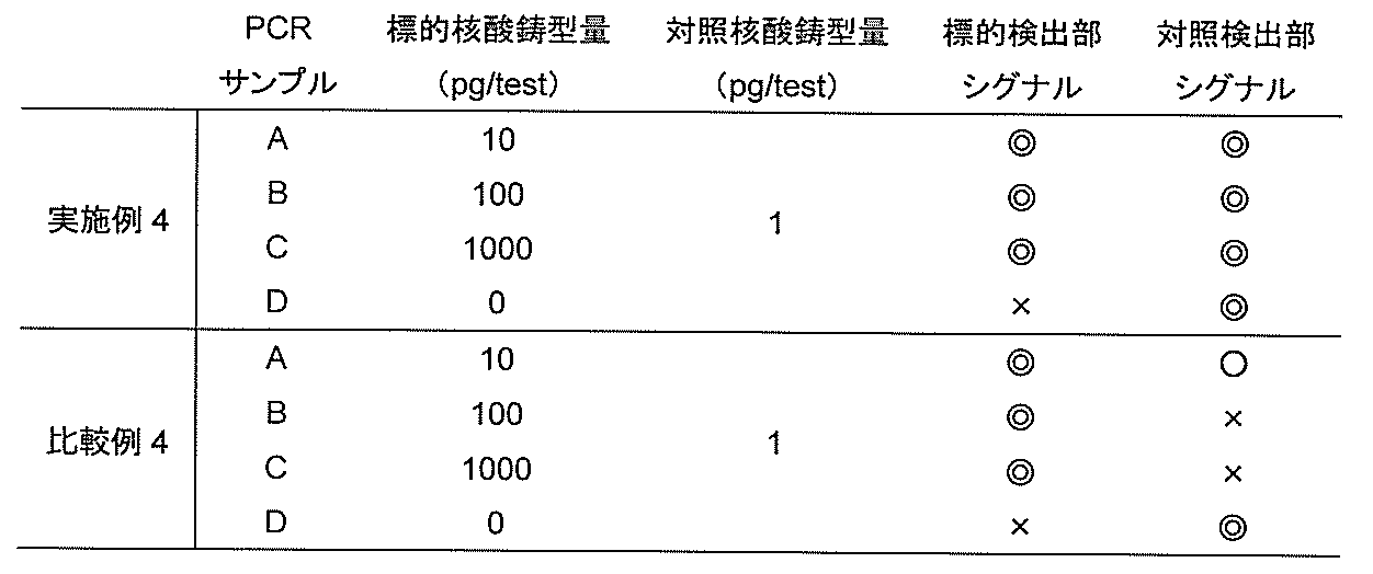
<比較例4>
比較例3で作製したデバイス100を用いる以外は、実施例4と同様の手法で発色シグナルを検出した。その検出形態を図7に、結果を表3に示す。
比較例3で作製したデバイス100を用いる以外は、実施例4と同様の手法で発色シグナルを検出した。その検出形態を図7に、結果を表3に示す。
実施例3で作製したデバイス100の対照検出部5には、標的検出部4に固定化されたプローブ8を含んだ二種類のプローブ8,9が固定化されているため、標的検出部4で検出される標的核酸に由来するシグナルが、対照検出部5でも検出されるようになった。これにより、標的核酸の優先的な増幅による対照検出部5のシグナル低下を防止することができ、1000 pgのような標的核酸が多量に存在する場合でも対照検出部5のシグナルを容易に検出できることが示された。一方、比較例3で作製したデバイス100を用いた場合、1000 pgのような多量の標的核酸が存在する場合、対照検出部5のシグナルが消失し、PCRが適切に行われたのか判断することが出来なかった。
<実施例5>
実施例2と同様に標的核酸としてpUC19(タカラバイオ社製)を、対照核酸としてλファージDNA(ユーロフィンジェノミクス社製)を設定した。それぞれの鋳型、プライマーセット、反応基質としてビオチン標識16-dUTP(ロシュアプライドサイエンス社製)を含んだdNTPを用いてPCRを実施後、増幅反応産物を実施例3で作製したデバイス100にアプライし、増幅反応産物と標識プローブとが結合した複合体から発する発色シグナルを検出した。実施例3で作製したデバイス100を使用した検出形態を図8に示す。
(1)一本鎖核酸タグ付加プライマーの選択
pUC19フォワードプライマーとして配列番号10のプライマーを、pUC19リバースプライマーとして配列番号7のプライマーを選択した。λファージDNAフォワードプライマーとして配列番号15のプライマーを、λファージDNAリバースプライマーとして配列番号13のプライマーを選択した。
(2)一本鎖核酸タグ付加プライマーおよびビオチン標識16-dUTPを用いたPCR
前記したプライマーセットを用いたPCRを行った。配列番号7、10のプライマーセット、配列番号13、15のプライマーセット、TaKaRa Ex Taq(登録商標)(タカラバイオ社製)に付属のdNTPに加えてビオチン標識16-dUTP(ロシュアプライドサイエンス社製)を用いて、実施例2(2)に記載の調製法に従い(A)~(D)のサンプルを調製し、実施例2(2)と同様の条件でPCRを実施した。
(3)核酸検出用デバイスによる増幅反応産物の検出
本実施例(2)で調製した(A)~(D)の増幅反応産物をそれぞれ加温などにより変性することなく、実施例3で作製した核酸検出用デバイス100のサンプルパッド1にアプライし、検出試験を行った。結果の判定は、アプライ後、10分後にクロマトリーダーC10066-10(浜松ホトニクス社製)によりラインの着色強度を測定し判定した。結果の判定は、アプライ後、10分後にクロマトリーダーC10066-10(浜松ホトニクス社製)によりラインの着色強度を測定し判定した。本結果を表4に示す。

<比較例5>
比較例3で作製したデバイス100を用いる以外は、実施例5と同様の手法で発色シグナルを検出した。その検出形態を図9に、結果を表4に示す。
比較例3で作製したデバイス100を用いる以外は、実施例5と同様の手法で発色シグナルを検出した。その検出形態を図9に、結果を表4に示す。
実施例3で作製したデバイス100の対照検出部5には、標的検出部4に固定化されたプローブ8を含んだ二種類のプローブ8,9が固定化されているため、標的検出部4で検出される標的核酸に由来するシグナルが、対照検出部5でも検出されるようになった。これにより、標的核酸の優先的な増加に起因する対照検出部5のシグナル低下を防止することができ、1000 pgのような標的核酸が多量に存在する場合でも対照検出部5のシグナルを容易に検出できることが示された。一方、比較例3で作製したデバイス100を用いた場合、1000 pgのような多量の標的核酸が存在する場合、対照検出部5のシグナルが消失し、PCRが適切に行われたのか判断することが出来なかった。
<実施例6>
(1)プローブを固定化した固相担体の作製
標的検出部4に使用するプローブ混合溶液として、2.0 mg/mlの抗DIG抗体(ロシュアプライドサイエンス社製)、2.5%スクロース、20 mM TBS(pH 8.0)を含む溶液を調製した。対照検出部5に使用するプローブ混合溶液として、1.0 mg/mlの抗DIG抗体(ロシュアプライドサイエンス社製)および抗FITC抗体(ロシュアプライドサイエンス社製)、2.5%スクロース、20 mM TBS(pH 8.0)を含む溶液を調製した。それぞれの溶液を用いて、実施例1(2)と同様の手法でプローブを固定化した固相担体3を作製した。
(2)核酸検出用デバイスの作製
バッキングシートから成る基材110、本実施例(1)で作製した固相担体であるニトロセルロースメンブレン3および実施例1(1)で作製したコンジュゲートパッド2を使用して実施例1(3)と同様の手法で核酸検出用デバイス100を作製した。
<比較例6>
対照検出部5に使用するプローブ混合溶液として、2.0 mg/mlの抗FITC抗体(ロシュアプライドサイエンス社製)、2.5%スクロース、20 mM TBS(pH 8.0)を加えた混合溶液をライン状に塗布した以外は実施例6と同様の手法で核酸検出用デバイス100を作製した。
<実施例7>
実施例2と同様に標的核酸としてpUC19(タカラバイオ社製)を、対照核酸としてλファージDNA(ユーロフィンジェノミクス社製)を設定した。それぞれの鋳型、プライマーセットを用いてPCRを実施後、増幅反応産物を実施例6で作製したデバイス100にアプライし、増幅反応産物と標識プローブとが結合した複合体から発する発色シグナルを検出した。実施例6で作製したデバイス100を使用した検出形態を図10に示す。
(1)DIG修飾プライマー、FITC修飾プライマーの設計および一本鎖核酸タグ付加プライマーの選択
pUC19フォワードプライマーとしてプライマー本体部の5’末端側をDIG修飾したプライマーDIG-F1(配列番号19)を設計した。また、pUC19リバースプライマーとして配列番号11のプライマーを選択した。λファージDNAフォワードプライマーとしてプライマー本体部の5’末端側をFITC修飾したプライマーFITC-F2(配列番号20)を設計した。また、λファージDNAリバースプライマーとして配列番号16のプライマーを選択した。
これらのDIG修飾プライマー、FITC修飾プライマー、一本鎖核酸タグ付加プライマーはつくばオリゴサービス株式会社にて委託合成し入手した。
以下に本検討で設計したプライマーセットを示す。
・プライマーDIG-F1:5’-DIG- GGAAACAGCTATGACCATGA-3’(配列番号19)
・プライマーFITC-F2: 5’-FITC- AAGTTCTCGCTGGAAGAGGT-3’(配列番号20)
(2)DIG修飾プライマー、FITC修飾プライマー、一本鎖核酸タグ付加プライマーを用いたPCR
前記したプライマーセットを用いたPCRを行った。配列番号11, 19のプライマーセット、配列番号16, 20のプライマーセットを用いて、実施例2(2)に記載の調製法に従い(A)~(D)のサンプルを調製し、実施例2(2)と同様の条件でPCRを実施した。
(3)核酸検出用デバイスによる増幅反応産物の検出
本実施例(2)で調製した(A)~(D)の増幅反応産物を、それぞれ加温などにより変性することなく、実施例6で作製した核酸検出用デバイス100のサンプルパッド1にアプライし、検出試験を行った。結果の判定は、アプライ後、10分後にクロマトリーダーC10066-10(浜松ホトニクス社製)によりラインの着色強度を測定し判定した。本結果を表5に示す。

<比較例7>
比較例6で作製したデバイス100を用いる以外は、実施例6と同様の手法で発色シグナルを検出した。その検出形態を図11に、結果を表5に示す。
比較例6で作製したデバイス100を用いる以外は、実施例6と同様の手法で発色シグナルを検出した。その検出形態を図11に、結果を表5に示す。
実施例6で作製したデバイス100の対照検出部5には、標的検出部4に固定化されたプローブ21を含んだ二種類のプローブ21,22が固定化されているため、標的検出部4で検出される標的核酸に由来するシグナルが、対照検出部5でも検出されるようになった。これにより、標的核酸の優先的な増加に起因する対照検出部5のシグナル低下を防止することができ、1000 pgのような標的核酸が多量に存在する場合でも対照検出部5のシグナルを容易に検出可能なことが示された。一方、比較例6で作製したデバイス100を用いた場合、1000 pgのような多量の標的核酸が存在する場合、対照検出部5のシグナルが消失し、PCRが適切に行われたのか判断することが出来なかった。
<実施例8>
本実施例では、これまでの実施例のように対照検出部5に対照核酸捕捉用と標的核酸捕捉用の二種類のプローブを固定化する方法ではなく、タグの種類が異なる二種類の標的核酸が増幅するような増幅反応系を構築することで対照検出部5のシグナルの低下を防止した。具体的には、標的検出部4で特異的に捕捉されるタグを有する標的核酸と、対照検出部5で特異的に捕捉されるタグを有する標的核酸の二種類が増幅するようタグ付加プライマーを設計し、二種類のうち片方の標的核酸が対照検出部5で捕捉されるような設計にした。本実施例の検出形態を図12に示す。図12に示すように上述した二種類の標的核酸のうち片方の増幅反応産物が対照検出部5で捕捉される。
実施例2と同様に、標的核酸としてpUC19(タカラバイオ社製)を、対照核酸としてλファージDNA(ユーロフィンジェノミクス社製)を設定した。PCR実施後、増幅反応産物を比較例1で作製したデバイス100にアプライし、増幅反応産物と標識プローブとが結合した複合体から発する発色シグナルを検出した。
(1)一本鎖核酸タグ付加プライマーの設計および選択
本検討では、pUC19プライマーセットとして二種類のプライマーセットを準備した。まず一つ目のプライマーセットとして配列番号10、11のプライマーを選択した。また、配列番号6のフォワードプライマーの5’末端側に、ポリメラーゼ反応阻害領域(X)としてのアゾベンゼンを介して一本鎖核酸タグT3(配列番号14)を付加したプライマーT3-X-F1(配列番号21)を設計し、配列番号11、21のセットを2つ目のプライマーセットとした。λファージDNAプライマーセットとして配列番号15、16のプライマーセットを選択した。これらの一本鎖核酸タグ付加プライマーは、つくばオリゴサービス株式会社に委託合成し入手した。
以下に本検討で設計したプライマーを示す。
・プライマーT3-X-F1:5’-GTACGGACTTACAATGACTTTGAT X GGAAACAGCTATGACCATGA -3’ (配列番号21)
(2)一本鎖核酸タグ付加プライマーを用いたPCR
本実施例(1)で設計した配列番号10、11のプライマーセット、配列番号11、21のプライマーセットおよび配列番号15、16のプライマーセットの三種を用いて、実施例2(2)に記載の調製法に従い(A)~(D)のサンプルを調製した。これらサンプルを用いて、実施例2(2)と同様の条件でPCRを実施した。
(3)核酸検出用デバイスによる増幅反応産物の検出
本実施例(2)で調製した(A)~(D)の増幅反応産物を、それぞれ加温などにより変性することなく、比較例1で作製した核酸検出用デバイス100のサンプルパッド1にアプライし、検出試験を行った。結果の判定は、アプライ後、10分後にクロマトリーダーC10066-10(浜松ホトニクス社製)によりラインの着色強度を測定し判定した。本結果を表6に示す。

<比較例8>
比較例2と同様の手法で発色シグナルを検出した。その検出形態を図5に、結果を表6に示す。
比較例2と同様の手法で発色シグナルを検出した。その検出形態を図5に、結果を表6に示す。
実施例8では、標的検出部4で特異的に捕捉されるタグを有した標的核酸と対照検出部5で特異的に捕捉されるタグを有した標的核酸の二種類を調製し、検出用デバイス100として標的検出部4、対照検出部5共に一種類のプローブが固定化されたデバイスを使用した。本実施例においても、標的核酸が標的検出部4のみならず対照検出部5でも検出されるため、表6に示すように、1000 pgの標的核酸と1 pgの対照核酸が存在する条件下であっても、対照検出部5において標的核酸に由来するシグナルを検出できた。ゆえに、二種類のタグを有する標的核酸を調製することで、対照検出部5のシグナルの消失または低下を防止することができることが示された。一方、比較例8では、1000 pgのような多量の標的核酸が存在する場合、対照検出部5のシグナルが消失し、PCRが適切に行われたのか判断することが出来なかった。
以上、実施形態および比較形態を参照し本願発明を説明したが、本願発明は上記実施形態、比較形態に限定されるものではない。ディップスティック型デバイス100、アレイ型デバイス100を用いて検出する方法や、増幅および/または標識した産物を熱処理し一本鎖の状態で検出する方法、さらにタグに糖鎖を、担体に固定化するプローブにレクチンを用いる方法でも本実施形態を実施することはできる。
1.サンプルパッド
2.コンジュゲートパッド
3.プローブを保持した担体
4.標的検出部
5.対照検出部
6.吸収パッド
7.サンプル入りチューブ
8.標的核酸捕捉用プローブ
9.対照核酸捕捉用プローブ
10.配列番号8に記載のタグ配列
11.標的核酸増幅反応産物
12.配列番号1に記載のチオール化DNA
13.配列番号9に記載のタグ配列
14.金コロイド粒子
15.対照核酸増幅反応産物
16.配列番号14に記載のタグ配列
17.ビオチン
18.ストレプトアビジン
19.5’末端結合DIG
20.5’末端結合FITC
21.標的核酸捕捉用抗DIG抗体
22.対照核酸捕捉用抗FITC抗体
100. デバイス
110. バッキングシート
本明細書で引用した全ての刊行物、特許及び特許出願はそのまま引用により本明細書に組み入れられるものとする。
Claims (7)
- 一つ以上の標的検出部と、対照検出部を有する核酸検出用デバイスであって、前記標的検出部には標的核酸を捕捉するためのプローブが固定化されており、かつ、前記対照検出部には対照核酸を捕捉するためのプローブと前記標的核酸を捕捉するための一種以上のプローブとが固定化されていることを特徴とする、核酸検出用デバイス。
- 前記標的核酸と前記対照核酸が、増幅工程を経た増幅反応産物である、請求項1に記載の核酸検出用デバイス。
- 前記プローブが、核酸またはタンパク質であることを特徴とする、請求項1または請求項2に記載の核酸検出用デバイス。
- 前記デバイスが、クロマト型デバイスであることを特徴とする、請求項1~3のいずれかに記載の核酸検出用デバイス。
- 請求項1~4のいずれかに記載の核酸検出用デバイス、対照用鋳型核酸、核酸増幅酵素、核酸増幅反応用試薬、核酸検出反応用試薬を含む、核酸検出用キット。
- 一種以上の標的核酸と対照核酸とを検出する核酸検出方法であって、
(a) プライマーと鋳型核酸、核酸増幅反応用試薬を用いて、同一反応容器内で同時に核酸増幅反応を行い、前記一種以上の標的核酸と前記対照核酸とを増幅する工程、
(b) 前記一種以上の標的核酸と、固相担体上の標的検出部に固定化されたプローブとを結合させる工程、
(c) 前記一種以上の標的核酸と前記対照核酸を、前記固相担体上の対照検出部に固定化された一種以上のプローブと結合させる工程、
(d) 捕捉された前記標的核酸と前記対照核酸を標識し、前記標的核酸および前記対照核酸に由来するシグナルを検出する工程、
を含む、核酸検出方法。 - 前記プローブが、核酸またはタンパク質であることを特徴とする、請求項6に記載の核酸検出方法。
Priority Applications (4)
| Application Number | Priority Date | Filing Date | Title |
|---|---|---|---|
| CN201680049370.1A CN107922908A (zh) | 2015-08-26 | 2016-08-26 | 核酸检测用设备和核酸检测方法 |
| JP2017536487A JP6829198B2 (ja) | 2015-08-26 | 2016-08-26 | 核酸検出用デバイス及び核酸検出方法 |
| EP16839366.8A EP3342851B1 (en) | 2015-08-26 | 2016-08-26 | Nucleic acid detection device and nucleic acid detection method |
| US15/905,497 US20180251835A1 (en) | 2015-08-26 | 2018-02-26 | Nucleic acid detection device and nucleic acid detection method |
Applications Claiming Priority (2)
| Application Number | Priority Date | Filing Date | Title |
|---|---|---|---|
| JP2015-167198 | 2015-08-26 | ||
| JP2015167198 | 2015-08-26 |
Related Child Applications (1)
| Application Number | Title | Priority Date | Filing Date |
|---|---|---|---|
| US15/905,497 Continuation US20180251835A1 (en) | 2015-08-26 | 2018-02-26 | Nucleic acid detection device and nucleic acid detection method |
Publications (1)
| Publication Number | Publication Date |
|---|---|
| WO2017034019A1 true WO2017034019A1 (ja) | 2017-03-02 |
Family
ID=58100562
Family Applications (1)
| Application Number | Title | Priority Date | Filing Date |
|---|---|---|---|
| PCT/JP2016/074953 WO2017034019A1 (ja) | 2015-08-26 | 2016-08-26 | 核酸検出用デバイス及び核酸検出方法 |
Country Status (5)
| Country | Link |
|---|---|
| US (1) | US20180251835A1 (ja) |
| EP (1) | EP3342851B1 (ja) |
| JP (1) | JP6829198B2 (ja) |
| CN (1) | CN107922908A (ja) |
| WO (1) | WO2017034019A1 (ja) |
Citations (3)
| Publication number | Priority date | Publication date | Assignee | Title |
|---|---|---|---|---|
| JP2006201062A (ja) * | 2005-01-21 | 2006-08-03 | Kainosu:Kk | 核酸の検出あるいは定量方法 |
| JP2012528583A (ja) * | 2009-06-04 | 2012-11-15 | キアゲン ゲゼルシャフト ミット ベシュレンクテル ハフツング | 複合核酸の増幅 |
| JP2013106549A (ja) * | 2011-11-18 | 2013-06-06 | Ngk Insulators Ltd | 標的ポリヌクレオチドの検出用剤及びその利用 |
Family Cites Families (11)
| Publication number | Priority date | Publication date | Assignee | Title |
|---|---|---|---|---|
| CN1136319C (zh) * | 1999-08-23 | 2004-01-28 | 谢佐福 | 一种基因快速诊断金标试条及其制备方法 |
| CN105087777A (zh) * | 2004-04-07 | 2015-11-25 | 安克塞斯生物公司 | 核酸检测系统 |
| EP2007903A2 (en) * | 2006-04-10 | 2008-12-31 | 3M Innovative Properties Company | Nucleic acid detection using lateral flow methods |
| WO2009027932A1 (en) * | 2007-08-27 | 2009-03-05 | Koninklijke Philips Electronics N.V. | Assay substrate and a method for preparation |
| US9121054B2 (en) * | 2009-12-08 | 2015-09-01 | Biohelix Corporation | Detection of nucleic acid amplification products in the presence of an internal control sequence on an immunochromatographic strip |
| CN101818200A (zh) * | 2010-03-04 | 2010-09-01 | 宁波博奥生物工程有限公司 | 创伤弧菌核酸定量检测试纸条 |
| MY150966A (en) * | 2010-03-08 | 2014-03-31 | Universiti Sains Malaysia Usm | Lateral flow device and method of detection of nucleic acid sequence |
| CN103228785B (zh) * | 2010-11-24 | 2016-09-07 | 株式会社钟化 | 扩增核酸的检测方法和检测装置 |
| WO2013122453A1 (en) * | 2012-02-15 | 2013-08-22 | Universiti Sains Malaysia | Device and method for detection of nucleic acid(s) |
| DE102013000682B3 (de) * | 2013-01-08 | 2013-10-31 | Rainer Hintsche | Verfahren zur Detektion von Molekülen |
| CN104099413B (zh) * | 2014-06-18 | 2016-03-09 | 苏州市外来有害生物防控技术中心 | 美国白蛾恒温核酸检测试剂盒及其检测方法 |
-
2016
- 2016-08-26 JP JP2017536487A patent/JP6829198B2/ja active Active
- 2016-08-26 WO PCT/JP2016/074953 patent/WO2017034019A1/ja unknown
- 2016-08-26 CN CN201680049370.1A patent/CN107922908A/zh active Pending
- 2016-08-26 EP EP16839366.8A patent/EP3342851B1/en active Active
-
2018
- 2018-02-26 US US15/905,497 patent/US20180251835A1/en not_active Abandoned
Patent Citations (3)
| Publication number | Priority date | Publication date | Assignee | Title |
|---|---|---|---|---|
| JP2006201062A (ja) * | 2005-01-21 | 2006-08-03 | Kainosu:Kk | 核酸の検出あるいは定量方法 |
| JP2012528583A (ja) * | 2009-06-04 | 2012-11-15 | キアゲン ゲゼルシャフト ミット ベシュレンクテル ハフツング | 複合核酸の増幅 |
| JP2013106549A (ja) * | 2011-11-18 | 2013-06-06 | Ngk Insulators Ltd | 標的ポリヌクレオチドの検出用剤及びその利用 |
Non-Patent Citations (1)
| Title |
|---|
| See also references of EP3342851A4 * |
Also Published As
| Publication number | Publication date |
|---|---|
| CN107922908A (zh) | 2018-04-17 |
| EP3342851A4 (en) | 2019-04-03 |
| US20180251835A1 (en) | 2018-09-06 |
| JPWO2017034019A1 (ja) | 2018-06-07 |
| EP3342851A1 (en) | 2018-07-04 |
| EP3342851B1 (en) | 2022-11-30 |
| JP6829198B2 (ja) | 2021-02-10 |
Similar Documents
| Publication | Publication Date | Title |
|---|---|---|
| RU2437939C2 (ru) | Детекция нуклеиновых кислот способом, основанным на связывании мишенеспецифичного гибрида | |
| US9617589B2 (en) | Microfluidic devices, solid supports for reagents and related methods | |
| US7851148B2 (en) | Method and kit for primer based multiplex amplification of nucleic acids employing primer binding tags | |
| JP4268944B2 (ja) | 核酸の検出あるいは定量方法 | |
| CA2430373A1 (en) | Nucleic acid amplification methods | |
| JP2005511030A (ja) | 核酸の増幅方法 | |
| EP2248915A1 (en) | Detection of multiple nucleic acid sequences in a reaction cartridge | |
| WO2006086669A2 (en) | Aptamer regulated nucleic acids and uses thereof | |
| JP2020114225A (ja) | サンプル中の複数の核酸を検出するための方法 | |
| JP2003210199A (ja) | 高度ダイナミックレンジを有する定量的多重pcr | |
| JP6387606B2 (ja) | 核酸の検出方法 | |
| AU2004304765A1 (en) | Nucleic acid amplification methods | |
| US9121054B2 (en) | Detection of nucleic acid amplification products in the presence of an internal control sequence on an immunochromatographic strip | |
| US9090926B2 (en) | Method for cell lysis and PCR within the same reaction chamber | |
| JP5798631B2 (ja) | Rt−pcr反応緩衝液中での細胞溶解のための方法 | |
| WO2017034019A1 (ja) | 核酸検出用デバイス及び核酸検出方法 | |
| US20230101834A1 (en) | Padlock probe-based rolling circle amplification paired with nuclease protection for point-of-need nucleic acid detection | |
| US20190203269A1 (en) | Tri-nucleotide rolling circle amplification | |
| JP2017131166A (ja) | 標的核酸の検出方法 | |
| WO2017006859A1 (ja) | 標的核酸の検出法 | |
| JP6468555B2 (ja) | 検出キット,及び検出方法 | |
| WO2020017632A1 (ja) | 標的核酸の核酸クロマトグラフィーによる検出方法、及び標的核酸を検出するためのキット | |
| JP2007075023A (ja) | 蛍光共鳴エネルギー移動法を用いた遺伝子多型検出法及びキット | |
| JPWO2016129609A1 (ja) | 標的核酸の検出方法 | |
| WO2024084249A1 (en) | Clonal amplification |
Legal Events
| Date | Code | Title | Description |
|---|---|---|---|
| 121 | Ep: the epo has been informed by wipo that ep was designated in this application |
Ref document number: 16839366 Country of ref document: EP Kind code of ref document: A1 |
|
| ENP | Entry into the national phase |
Ref document number: 2017536487 Country of ref document: JP Kind code of ref document: A |
|
| NENP | Non-entry into the national phase |
Ref country code: DE |