WO2016144137A1 - 순환 유리 핵산의 분리 방법 - Google Patents
순환 유리 핵산의 분리 방법 Download PDFInfo
- Publication number
- WO2016144137A1 WO2016144137A1 PCT/KR2016/002462 KR2016002462W WO2016144137A1 WO 2016144137 A1 WO2016144137 A1 WO 2016144137A1 KR 2016002462 W KR2016002462 W KR 2016002462W WO 2016144137 A1 WO2016144137 A1 WO 2016144137A1
- Authority
- WO
- WIPO (PCT)
- Prior art keywords
- salt
- kit
- nucleic acid
- peg
- cfdna
- Prior art date
- Legal status (The legal status is an assumption and is not a legal conclusion. Google has not performed a legal analysis and makes no representation as to the accuracy of the status listed.)
- Ceased
Links
Images





Classifications
-
- C—CHEMISTRY; METALLURGY
- C12—BIOCHEMISTRY; BEER; SPIRITS; WINE; VINEGAR; MICROBIOLOGY; ENZYMOLOGY; MUTATION OR GENETIC ENGINEERING
- C12N—MICROORGANISMS OR ENZYMES; COMPOSITIONS THEREOF; PROPAGATING, PRESERVING, OR MAINTAINING MICROORGANISMS; MUTATION OR GENETIC ENGINEERING; CULTURE MEDIA
- C12N15/00—Mutation or genetic engineering; DNA or RNA concerning genetic engineering, vectors, e.g. plasmids, or their isolation, preparation or purification; Use of hosts therefor
- C12N15/09—Recombinant DNA-technology
- C12N15/10—Processes for the isolation, preparation or purification of DNA or RNA
-
- C—CHEMISTRY; METALLURGY
- C12—BIOCHEMISTRY; BEER; SPIRITS; WINE; VINEGAR; MICROBIOLOGY; ENZYMOLOGY; MUTATION OR GENETIC ENGINEERING
- C12Q—MEASURING OR TESTING PROCESSES INVOLVING ENZYMES, NUCLEIC ACIDS OR MICROORGANISMS; COMPOSITIONS OR TEST PAPERS THEREFOR; PROCESSES OF PREPARING SUCH COMPOSITIONS; CONDITION-RESPONSIVE CONTROL IN MICROBIOLOGICAL OR ENZYMOLOGICAL PROCESSES
- C12Q1/00—Measuring or testing processes involving enzymes, nucleic acids or microorganisms; Compositions therefor; Processes of preparing such compositions
- C12Q1/68—Measuring or testing processes involving enzymes, nucleic acids or microorganisms; Compositions therefor; Processes of preparing such compositions involving nucleic acids
Definitions
- the present invention relates to a method for separating circulating free nucleic acid from a sample comprising circulating cell-free nucleic acid, a circulating free nucleic acid separation kit and a lysis buffer for circulating free nucleic acid separation.
- Circulating cell-free nucleic acid such as cancer-specific extracellular DNA fragments and mRNA present in the blood or fetal nucleic acid present in the maternal blood, may be serum or It is known to exist in plasma in the form of DNA of 1000 bp or less, or RNA of 100 nt or less. In particular, it is known that nucleic acids liberated by necrosis or killed cells of cancer during the development of cancer are present in the plasma. Kamat et al (Cancer Biol Ther.
- kits and methods for separating circulating free nucleic acids using columns are known (QIAamp Circulating Nucleic Acid Kit).
- QIAamp Circulating Nucleic Acid Kit As a method for separating circulating free nucleic acids, kits and methods for separating circulating free nucleic acids using columns are known (QIAamp Circulating Nucleic Acid Kit).
- QIAamp Circulating Nucleic Acid Kit there is still a need for development of kits and methods for the separation of circulating free nucleic acids that are improved over previously known methods.
- the present inventors have made diligent efforts to develop kits and methods that can effectively obtain circulating free nucleic acids. As a result, the inventors dissolve samples containing circulating free nucleic acids, and add solutions and magnetic beads containing salts and PEG to circulate them. A method for obtaining circulating free nucleic acid from magnetic beads to which free nucleic acid is bound has been developed. Using the method developed above, it was confirmed that the circulating free nucleic acid can be effectively separated from a sample containing the circulating free nucleic acid, and the present invention has been completed.
- One object of the present invention is to provide a method for separating circulating free nucleic acid from a sample comprising circulating cell-free nucleic acid.
- Another object of the invention is a lysis buffer; A solution comprising a salt and PEG; And it provides a circulating free nucleic acid separation kit comprising magnetic beads.
- a circulating free nucleic acid can be obtained in a high yield in a short time from a sample including the circulating free nucleic acid separation, particularly a plasma or serum sample.
- FIG. 1 graphically depicts the amount of cfDNA obtained from 200 ⁇ l of plasma in normal and lung cancer patients.
- the gray rhombus represents the experimental results for the plasma isolated from the normal person
- the black rhombus represents the experimental results for the plasma isolated from the lung cancer patients.
- the y axis shows the amount of cfDNA (ng).
- Figure 2 shows the results of confirming the reproducibility after performing cfDNA Prep twice using plasma isolated from lung cancer patients.
- Plasma cfDNA is known to be fragmented in nucleosome units, and the size of the cfDNA obtained according to the method of the present invention is fragmented to approximately 180 bp, which is a nucleosome unit. Suggested.
- FIG. 4 is a result confirming whether or not PCR products of ⁇ 100BP and 237BP size are amplified using the cfDNA isolated according to the method of the present invention as a template. As shown in Figure 4, the PCR product was amplified well, indicating that the purified cfDNA according to the method of the present invention has a high purity, does not affect the enzymatic reaction of the DNA polymerase.
- Figure 5a shows the results of confirming the amount of DNA obtained by separating the plasma from the blood of normal people by a one-step centrifugation method and a two-step centrifugation method, and then performing cfDNA PREP.
- the x-axis represents the number of each plasma sample and the y-axis represents the amount of cfDNA in ng.
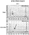
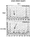
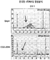
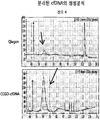
- FIG. 5B shows the results of plasma separation from normal blood by one-step centrifugation and two-step centrifugation, followed by cfDNA PREP, and analysis of the separated cfDNA with a Bioanalyzer.
- FIG. 6 is a diagram showing the results of separating cfDNA using the method according to the present invention (named CCGD_cfDNA) and the Qiagen Kit, respectively.
- the numbers shown above the bar graphs indicate the amount of DNA in ng.
- Figure 9 shows the results obtained by using the Picogreen quantitative analysis (PG) and RQ-PCR quantitative analysis of the yield of cfDNA isolated using a method according to the present invention (named CCGD_cfDNA) and Qiagen Kit in plasma separated from 10 mothers, respectively Indicates.
- Figure 10 shows the results of analyzing the correlation coefficient of Picogreen quantitative value (PG) and RQ-PCR quantitative value.
- FIG. 11 shows the quality of cfDNA isolated from the plasma of 10 mothers using the method according to the invention (named CCGD_cfDNA) and Qiagen Kit, respectively.
- FIG. 12 shows the purity of cfDNA isolated using a method according to the invention (named CCGD_cfDNA) and Qiagen Kit, respectively, from plasma isolated from mother.
- the method of separating the nucleic acid may also be called PREP.
- the term "circulating cell-free nucleic acid” refers to DNA that circulates and is present in the blood.
- the circulating free nucleic acid specifically includes circulating free DNA (cfDNA), circulating free RNA, and the like, and specifically, may be circulating free DNA, but is not limited thereto.
- the circulating free nucleic acid generally has a length of 1000 bp or less (DNA), 100 nt or less (RNA) in plasma or serum, but is not limited thereto.
- the circulating free nucleic acid is present in a small amount in the blood has been required to develop a method for obtaining it in high yield.
- the sample containing the circulating free nucleic acid is magnetic beads; And a method for reacting with a solution comprising a salt and PEG and isolating circulating free nucleic acids bound to magnetic beads.
- the magnetic beads can more efficiently capture the circulating free nucleic acid, thereby producing a circulating free nucleic acid with high yield. It was confirmed that it can be obtained.
- lysis buffer and plasma preparation were also optimized in the above method to develop a technique capable of effectively separating circulating free nucleic acids with reproducibility.
- step (a) is a step of adding a solution containing salt and PEG, and magnetic beads to an isolated sample containing circulating free nucleic acid to bind the circulating free nucleic acid to the magnetic beads.
- the separated sample containing the circulating free nucleic acid is not particularly limited as long as it is an isolated sample containing the circulating free nucleic acid, but may be plasma or serum.
- plasma or serum isolated from an individual in need of analysis or obtaining circulating free nucleic acids may be plasma, but is not limited thereto.
- the separated sample containing the circulating free nucleic acid may be prepared by dissolving plasma or serum in a lysis buffer.
- the method of the present invention comprises the steps of adding a lysis buffer to an isolated plasma or serum sample comprising circulating free nucleic acid; Adding a magnetic beads and a solution containing salt and PEG to the sample containing circulating free nucleic acid dissolved in the first step; And a third step of obtaining the magnetic beads from the sample to which the magnetic beads are added and separating the circulating free nucleic acid therefrom, but is not particularly limited thereto.
- the lysis buffer used for lysis of plasma or serum may specifically include a chaotropic salt, a chelating agent, a nonionic surfactant, and Tris-Cl, This is not restrictive.
- the use of the lysis buffer having the above composition compared to the case of using other buffers including lysis buffer consisting of chelating agent EDTA, chaotropic salt SDS, and Tris-Cl, It was confirmed that the separation was easy and / or the yield was high.
- the chaotropic salt includes a substance capable of interfering hydrogen bonds between water molecules.
- the chaotropic salt causes a structural change of the protein, thereby weakening the binding force between the protein and the DNA, thereby purifying DNA. To increase the degree of separation.
- chaotropic salts examples include, but are not limited to, guanidinium salts, lithium salts, magnesium salts, sodium dodecyl sulfate (SDS), thiourea, urea, butanol or ethanol, It is not limited.
- guanidinium salt examples include guanidinium chloride
- examples of the lithium salt include lithium perchlorate, lithium acetate
- magnesium salt may include magnesium chloride, but is not limited thereto. It doesn't happen.
- guanidinium chloride was used as the chaotropic salt.
- the chelating agent is Mg 2+ and Ca can be used to isolate the divalent cation, such as + 2, and therefore can protect the DNA from the enzyme to decompose it, but is not limited thereto.
- chelating agent may be selected from the group consisting of diethylenetriaminepentaacetic acid (DTPA), ethylenediaminetetraacetic acid (EDTA), ethylene glycol tetraacetic acid (EGTA), and NTA (N, N-bis (carboxymethyl) glycine). It is not limited. In one embodiment of the present invention, EDTA was used as a chelating agent.
- DTPA diethylenetriaminepentaacetic acid
- EDTA ethylenediaminetetraacetic acid
- EGTA ethylene glycol tetraacetic acid
- NTA N, N-bis (carboxymethyl) glycine
- the EDTA may represent an EDTA portion of an EDTA compound (eg, K 2 EDTA, K 3 EDTA, or Na 2 EDTA), but is not limited thereto.
- an EDTA compound eg, K 2 EDTA, K 3 EDTA, or Na 2 EDTA
- a nonionic surfactant is a material having both hydrophilic and hydrophobic moieties in a molecule and exhibits nonionic properties when dissociated.
- nonionic surfactants examples include, but are not limited to, Triton X-100.
- the lysis buffer comprises 2-6M chaotropic salts; 1 to 50 mM chelating agent; 0.1 to 5% (w / v) nonionic surfactant; Or / and 10 to 100 mM Tris-Cl, but is not limited thereto.
- the pH of the lysis buffer may be pH 7.0 to 8.5.
- the lysis buffer may include, but is not limited to, guanidinium chloride, EDTA, Triton X-100, and Tris-Cl.
- protease when the plasma or serum sample is dissolved in the lysis buffer, protease may be added together.
- protease may be used a variety of protease commonly used for nucleic acid separation, for example, can be used protease K (proteinase K).
- the separated plasma sample is a first centrifugation to separate the plasma from the separated blood sample; And separating the plasma from the first centrifuged sample, and separating the plasma from the second centrifuged sample, but the present invention is not limited thereto.
- the first centrifugation may be performed under the conditions of 1900 to 2000 x g
- the second centrifugation may be performed under the conditions of 12000 to 18000 x g, but is not limited thereto.
- the centrifugation described above may be performed at a temperature condition of about 4 °C, but is not limited thereto.
- the addition of the solution containing the magnetic beads, salt and PEG to the sample may be performed sequentially or simultaneously, but is not limited thereto.
- a solution containing PEG and salts added with the magnetic beads is not particularly limited, but may help capture small nucleic acid fragments of 100 bp or less than 100 nt.
- the salt in the solution containing the salt and polyethylene glycol (PEG) is not particularly limited in kind, but may be, for example, sodium chloride.
- PEG of the solution may be PEG having an average molecular weight of 6,000 to 10,000 Da, but is not limited thereto.
- the solution containing the salt and PEG may be one containing 1 to 4M salt and 10 to 60% (w / v) PEG, but is not limited thereto.
- magnetic beads refer to particles or beads that react to a magnetic field.
- magnetic beads do not have a magnetic field, but refer to a material forming a magnetic dipole exposed to the magnetic field.
- it refers to a substance that can be magnetized in a magnetic field, but does not have magnetism in the absence of a magnetic field.
- the magnetism used in the present invention includes, but is not limited to, both paramagnetic and superparamagnetic materials.
- the magnetic beads are preferably beads having a property of binding to a nucleic acid.
- the magnetic beads may be in a form having a functional group that binds to a nucleic acid, for example, a -COOH group, but is not limited thereto.
- step (b) is a step of obtaining magnetic beads from the sample to which the magnetic beads are added, and separating circulating free nucleic acid therefrom.
- step (b) is a step of separating the circulating free nucleic acid attached to the magnetic beads added in the step (a) from the magnetic beads.
- the method may include washing the magnetic beads before obtaining the magnetic beads from the sample to which the magnetic beads have been added.
- the washing of the magnetic beads may be performed using 50 to 95% (v / v) ethanol solution, specifically, 80 to 90% (v / v) ethanol solution.
- This washing process can be carried out by placing the magnetic beads under a magnetic stand, collecting the magnetic beads, removing the supernatant, and adding the wash buffer thereto to wash. In addition, this washing step may be performed one or more times.
- This washing procedure may optionally be followed, followed by separation of the magnetic beads and separation of the circulating free nucleic acid from the magnetic beads by addition of an elution buffer to the separated magnetic beads.
- lysis buffers A solution comprising a salt and PEG; And circulating free nucleic acid separation kit, including magnetic beads.
- Lysis buffers solutions comprising salts and PEG, and magnetic beads are as described above.
- the circulating free nucleic acid separation kit of the present invention is not limited to its origin, and can separate cfDNA, so that cfDNA can be extracted from cancer patients or mothers. Therefore, cfDNA extracted from the blood of cancer patients can be used to diagnose specific genetic mutations of the cancer patients. In addition, since there is a large amount of cfDNA derived from the fetus in the mother's plasma, cfDNA extracted from the mother's blood can be used for non-invasive prenatal diagnosis of the fetus. That is, the circulating free nucleic acid separation kit of the present invention can be used for cancer diagnosis or antenatal diagnosis, and the like, and is not particularly limited as long as it can utilize cfDNA.
- kit may further include other instruments, solutions, and the like, which are generally used for nucleic acid separation, and may include instructions for circulating free nucleic acid, but are not limited thereto.
- kit may further include, but is not limited to, magnetic bead wash buffer and magnetic bead separation mechanism (eg, magnetic stand).
- Another specific embodiment of the present invention is a lysis buffer for circulating free nucleic acid separation, comprising chaotropic salts, chelating agents, nonionic surfactants and Tris-Cl.
- Chaotropic salts, chelating agents, nonionic surfactants, circulating free nucleic acids and lysis buffers are as described above.
- Fresh blood samples were centrifuged at 1,900 ⁇ g for 10 minutes at 4 ° C. to separate plasma. Then, the separated plasma was again centrifuged at 16,000 x g for 10 minutes at 4 °C, the supernatant was separated. The plasma samples thus obtained were then stored at ⁇ 20 ° C. before DNA isolation. Except where specifically stated, the following examples used plasma prepared using the above method.
- the frozen plasma sample was dissolved before DNA separation, and then 200 ⁇ l of the plasma sample was placed in a tube. Next, 400 ⁇ l of plasma lysis buffer (5.5 M guanidine HCl, 50 mM Tris-Cl, pH 8.0, 20 mM EDTA, pH 8.0, 1.3% Triton X-100) and 10 ⁇ l of proteinase K were added to the sample tube. Was added and it was mixed.
- plasma lysis buffer 5.5 M guanidine HCl, 50 mM Tris-Cl, pH 8.0, 20 mM EDTA, pH 8.0, 1.3% Triton X-100
- wash buffer 85% EtOH
- the beads were then dried and the reaction tube was removed from the magnetic stand and then elution buffer (pure separated tertiary distilled water or 10 mM TE buffer) was added. Then, the reaction tube was placed on a magnetic stand, and the eluate was separated to obtain a cfDNA sample.
- elution buffer pure separated tertiary distilled water or 10 mM TE buffer
- Example 3 from plasma of normal and lung cancer patients cfDNA Sample acquisition
- Example 2 Using the cfDNA separation method of Example 2 was carried out an experiment to compare the amount of cfDNA extraction between normal people and patients.
- cfDNA was isolated using 200 ⁇ l of plasma obtained from 31 samples of normal subjects and 49 samples of lung cancer patients, and the results are shown in FIG. 1.
- the samples were sold at Asan Medical Center, BRC (Bio Resource Center).
- the amount of cfDNA obtained in 200 ⁇ l plasma samples from normal individuals was median of 4.4 ng (maximum: 9.2 ng, minimum: 0.8 ng), and the amount of cfDNA obtained in 200 ⁇ l plasma samples of lung cancer patients was medium. 32.0 ng (maximum value: 268.0 ng, minimum value: 4.78 ng) was confirmed, and it was confirmed that cfDNA can be effectively separated even in 200 ⁇ l plasma sample.
- Example 4 Example 2 of cfDNA Confirm reproducibility of PREP method
- Example 5 Example 2 of cfDNA Of samples obtained by the PREP method cfDNA Pattern analysis
- a size of about 180 bp is the minimum unit, and a size of 180 * 2, 180 * 3, and 180 * 4 bp sizes of 2, 3, and 4 nucleosome units Also shown (FIG. 3B).
- Example 6 Example 2 of cfDNA Obtained by the PREP method cfDNA's Quality analysis
- PCR was performed using a primer pair capable of amplifying the product of 100 bp and 237 bp.
- template DNA 1 to 5 cfDNA samples isolated from samples of lung cancer patients and two control gDNAs (Beas2B, genomic DNA PREP from H1975 cell line) were used. The results are shown in Figure 4 (in Figure 4, cfDNA samples are represented by 1 to 5).
- the primer information used is as follows.
- Primer name Sequence (5 '-> 3') SEQ ID NO: Amplification Product Size (bp) rs1952966-F GGCTCTGGTTACAACAGCTT One 100 rs1952966-R AGAAGTTTGCTTGGCTGAAG 2 RASSF1A-F GTGGGGACCCTCTTCCTCTA 3 237 RASSF1A-R GGAAGGAGCTGAGGAGAGC 4
- the preparation of plasma used in the cfDNA PREP method of the present invention was optimized as follows.
- the effect of preparing plasma by performing two-step centrifugation in cfDNA PREP was compared with that of one-step centrifugation.
- the one-step centrifugation method is a method of centrifuging fresh blood obtained from the subject at 1,900 xg and 4 ° C. for 10 minutes to obtain a plasma sample. After centrifugation for 10 minutes, centrifugation at 16,000 xg and 4 °C for 10 minutes to obtain a plasma sample.
- Plasma samples obtained using the one-step centrifugation method and the two-step centrifugation method were separated by the method of Example 2, and the amounts and properties thereof were compared. The results are shown in FIGS. 5A, 2 and 5B, respectively.
- the amount of cfDNA separated from the plasma obtained using the two-step centrifugation method is about one third the amount of cfDNA separated from the plasma obtained using the one-step centrifugation method. Indicated. In the one-step centrifugation method, the amount of extracted cfDNA was large and more than expected.
- Healthy plasma 1 step method (1,900 g, 4 °C , 10min ), total ng 2 step method (1,900 g, 4 °C , 10min -> 16,000g, 4 °C, 10min ), total ng Ratio between 1 step / 2 step One 17.05 6.26 2.72 2 7.11 2.91 2.45 3 10.76 2.75 3.92 4 10.64 6.26 1.70 5 16.98 5.26 3.23 6 21.76 2.95 7.36 7 31.87 4.52 7.06 8 9.66 3.68 2.63
- the characteristics of the cfDNA obtained from the plasma prepared by using a two-step centrifugation method and a one-step centrifugation method the DNA obtained by the two-step centrifugation method, typical It was confirmed that the cfDNA characterizes the minimum nucleosome unit size of about 180bp.
- the DNA extracted by the one-step centrifugation method showed a pattern in which genomic DNA (gDNA) was mixed. That is, when loading BioAnalyzer, the intact gDNA larger than the upper marker (upper marker) and some fragments (fragmentation) showed a gDNA appearing as a smear pattern.
- cfDNA was separated using each method.
- the amount of cfDNA was about 4 to 5 times higher than that of the Qiagen Kit. It was confirmed that it can be obtained.
- the optimal conditions of the lysis buffer that can be effectively used in the cfDNA PREP method of the present invention were identified as follows.
- Example 2 a buffer containing chaotropic salt, chelating agent and Tris-Cl (comparative buffer 1) and a buffer containing chaotropic salt, chelating agent, nonionic surfactant, and Tris-Cl (execution buffer), respectively, were used.
- CfDNA was isolated by the method of Example 2. At this time, the experiment was performed using the plasma of the ovarian cancer patient, and the sample subjected to two centrifugation as in Example 1 was used.
- the cfDNA prep method of the present invention can obtain cfDNA not only in lung cancer patients but also in normal individuals. That is, cfDNA can be obtained from isolated plasma without limiting the condition of the individual.
- cfDNA was separated from the mother and analyzed.
- 200 ⁇ l of plasma sample was used, but the separation conditions were appropriately changed as follows to apply the cfDNA prep method of the present invention to a larger volume of sample (Table 8).
- Plasma volume
- Lysis buffer volume
- Protease K Solution A Magnetic beads Furtherance volume existing 200 ⁇ l 400 ⁇ l 20 ⁇ l 20% PEG / 2.5 M Nacl 1000 ⁇ l 200 ⁇ l change 500 ⁇ l 1000 ⁇ l 20 ⁇ l 40% PEG / 2.5 M Nacl 1100 ⁇ l 400 ⁇ l
- Example 11 Yield Comparison Between the cfDNA PREP Method of Example 10 and a Commercially Available Kit
- the cfDNA prep method of the present invention was confirmed to appear in a similar or higher yield compared to the case using the Qiagen kit.
- the cfDNA prep method of the present invention has a very good effect in yield compared to the Qiagen kit.
- Example 12 Example 10 cfDNA Commercialized with the PREP Law As a kit Separated cfDNA's Quality and Purity Comparison
- CfDNA extracted from three mothers was subjected to RQ-PCR reaction to amplify amplicon having a size of 110 bp.
- RQ-PCR was performed while increasing the amount of cfDNA extracted from each mother by 1, 2, and 4 ⁇ l to confirm the Ct value, and then converted to a relative amount (1/2 ⁇ Ct) to determine the amount of cfDNA used.
- the purity of the extracted cfDNA was reconfirmed by checking the proportionality of the relative amount confirmed by RQ-PCR according to the amount.
Landscapes
- Chemical & Material Sciences (AREA)
- Life Sciences & Earth Sciences (AREA)
- Health & Medical Sciences (AREA)
- Engineering & Computer Science (AREA)
- Organic Chemistry (AREA)
- Genetics & Genomics (AREA)
- Wood Science & Technology (AREA)
- Zoology (AREA)
- Biotechnology (AREA)
- General Engineering & Computer Science (AREA)
- Bioinformatics & Cheminformatics (AREA)
- Biomedical Technology (AREA)
- Biochemistry (AREA)
- Physics & Mathematics (AREA)
- Molecular Biology (AREA)
- Microbiology (AREA)
- General Health & Medical Sciences (AREA)
- Biophysics (AREA)
- Analytical Chemistry (AREA)
- Proteomics, Peptides & Aminoacids (AREA)
- Crystallography & Structural Chemistry (AREA)
- Plant Pathology (AREA)
- Immunology (AREA)
- Measuring Or Testing Involving Enzymes Or Micro-Organisms (AREA)
- Chemical Kinetics & Catalysis (AREA)
Abstract
본 발명은 순환 유리 핵산(circulating cell-free nucleic acid)을 포함하는 시료로부터 순환 유리 핵산을 분리하는 방법, 순환 유리 핵산 분리 키트 및 순환 유리 핵산 분리를 위한 용해 완충액에 관한 것이다.
Description
본 발명은 순환 유리 핵산(circulating cell-free nucleic acid)을 포함하는 시료로부터 순환 유리 핵산을 분리하는 방법, 순환 유리 핵산 분리 키트 및 순환 유리 핵산 분리를 위한 용해 완충액에 관한 것이다.
혈액에 존재하는 암 특이적 세포 외 DNA 단편 및 mRNA, 또는 모체혈(materanal blood)에 존재하는 태아의 핵산(fetal nucleic acid)과 같은 순환 유리 핵산(circulating cell-free nucleic acid, cfNA)이 혈청 또는 혈장 내에 1000bp 이하의 DNA, 또는 100nt 이하의 RNA 형태로 존재하는 것이 알려져 있다. 특히, 암의 발달 과정 중 암의 괴사 혹은 사멸된 세포에 의해 유리된 핵산이 혈장 내에 존재하는 것이 알려져 있다. Kamat et al(Cancer Biol Ther. 2006 Oct;5(10):1369-74.)은 누드 마우스에 암세포를 주입하고 이후 cfDNA의 양을 측정하였을 때, cfDNA의 양이 암의 크기를 잘 반영하고, 이후의 치료에 대한 반응도 잘 반영한다고 보고한 바 있다. 이렇듯 cfDNA는 다양한 위치의 암에서 다양하게 유리되어 암의 발달, 진행 및 전이된 암의 특성을 나타내는 것으로 알려져 있어, 이를 암의 연구 및 진단에 이용하고자 순환 유리 핵산의 효과적인 분리에 대한 필요성이 강조되고 있다.
순환 유리 핵산의 분리 방법으로, 컬럼을 이용하여 순환 유리 핵산을 분리하는 키트 및 방법 등이 알려져 있다(QIAamp Circulating Nucleic Acid Kit). 그러나, 기존에 알려진 방법에 비하여 개선된 순환 유리 핵산의 분리 키트 및 방법의 개발이 여전히 요구된다.
본 발명자들은 순환 유리 핵산을 효과적으로 수득할 수 있는 키트 및 방법의 개발을 위하여 예의 노력한 결과, 순환 유리 핵산을 포함하는 시료를 용해시키고, 여기에 염 및 PEG를 포함하는 용액과 자성 비드를 첨가하여 순환 유리 핵산이 결합된 자성 비드로부터 순환 유리 핵산을 획득하는 방법을 개발하였다. 상기 개발된 방법을 사용하여 순환 유리 핵산을 포함하는 시료에서 순환 유리 핵산을 효과적으로 분리할 수 있음을 확인하였고, 본 발명을 완성하였다.
본 발명의 하나의 목적은 순환 유리 핵산(circulating cell-free nucleic acid)을 포함하는 시료로부터 순환 유리 핵산을 분리하는 방법을 제공하는 것이다.
본 발명의 다른 목적은 용해 완충액(lysis buffer); 염 및 PEG를 포함하는 용액; 및 자성 비드를 포함하는, 순환 유리 핵산 분리 키트를 제공하는 것이다.
본 발명의 또 다른 목적은 카오트로픽 염(chaotropic salt), 킬레이트제(chelating agent), 비이온성 계면활성제(nonionic surfactant) 및 Tris-Cl을 포함하는, 순환 유리 핵산 분리를 위한 용해 완충액을 제공하는 것이다.
본 발명의 순환 유리 핵산 분리 키트 및 방법에 따르면 순환 유리 핵산 분리를 포함하는 시료, 특히 혈장 또는 혈청 시료에서 순환 유리 핵산을 단기간 내에 고수율로 수득할 수 있는 효과를 가진다.
도 1은, 정상인 및 폐암 환자의 혈장 200 ㎕로부터 수득한 cfDNA의 양을 그래프로 나타낸 것이다. 여기서, 회색 마름모는 정상인로부터 분리한 혈장에 대한 실험 결과를, 검은색 마름모는 폐암 환자로부터 분리한 혈장에 대한 실험 결과를 나타낸다. y 축은 cfDNA의 양(ng)을 나타낸다.
도 2는, 폐암 환자로부터 분리한 혈장을 사용하여 cfDNA Prep을 2번 수행한 후, 재현성을 확인한 결과를 나타낸 것이다.
도 3a 및 b는, 본 발명의 방법에 따라 난소암 환자의 혈장에서 분리한 cfDNA의 패턴을 Bioanalyzer로 분석한 결과를 나타낸 것이다. 혈장 cfDNA는 뉴클레오좀 단위로 단편화되어 있는 것으로 알려져 있는데, 본 발명의 방법에 따라 수득한 cfDNA의 크기가 뉴클레오좀 단위인 대략 180bp 로 단편화 되어 있는 결과를 나타내어, 본 발명의 방법이 cfDNA 분리에 적합함을 시사하였다.
도 4는, 본 발명의 방법에 따라 분리한 cfDNA를 주형으로 사용하여 ~100BP와 237BP 크기의 PCR 산물이 증폭되는지 여부를 확인한 결과이다. 도 4에 나타난 바와 같이, 상기 PCR 산물이 잘 증폭되어, 본 발명의 방법에 따라 정제된 cfDNA가 DNA 중합효소의 효소 반응에 영향을 주지 않는, 높은 순도를 가지고 있음을 나타내었다.
도 5a는, 1 단계 원심분리 방법 및 2 단계 원심분리 방법으로 정상인의 혈액으로부터 혈장을 분리한 다음, cfDNA PREP을 수행하여 수득된 DNA 양을 확인한 결과를 각각 나타낸 것이다. x 축은 각 혈장 시료의 번호를, y 축은 cfDNA 양(단위: ng)을 나타낸다.
도 5b는, 1 단계 원심분리 방법 및 2 단계 원심분리 방법으로 정상인의 혈액으로부터 혈장을 분리한 다음, cfDNA PREP을 수행한 다음, 분리한 cfDNA를 Bioanalyzer로 분석한 결과를 나타낸다.
도 6은, 본 발명에 따른 방법(CCGD_cfDNA로 명명)과 Qiagen Kit를 각각 사용하여 cfDNA를 분리한 결과를 나타내는 도이다. 막대 그래프 위에 표시된 숫자는 DNA 양(단위: ng)을 나타낸다.
도 7은, 비교 완충액 1(카오트로픽 염, 킬레이트 제 및 Tris-Cl을 포함) 및 본 발명에 따른 실시 완충액을 사용하여 cfDNA PREP을 수행한 결과를 나타낸 도이다. 막대 그래프 위에 표시된 숫자는 DNA 양(단위: ng)을 나타낸다.
도 8은, 비교 완충액 2(킬레이트 제, 비이온성 계면활성제 및 Tris-Cl을 포함) 및 본 발명에 따른 실시 완충액을 사용하여 cfDNA PREP을 수행한 결과를 나타낸 도이다. 막대 그래프 위에 표시된 숫자는 DNA 양(단위: ng)을 나타낸다.
도 9는 10 명의 산모로부터 분리된 혈장에서 본 발명에 따른 방법(CCGD_cfDNA로 명명)과 Qiagen Kit를 각각 사용하여 분리한 cfDNA의 수율을 Picogreen 정량법 (PG) 및 RQ-PCR 정량법을 이용하여 확인한 결과를 나타낸다.
도 10은 Picogreen 정량값 (PG) 및 RQ-PCR 정량값의 상관 계수를 분석한 결과를 나타낸다.
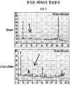
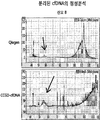
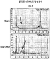
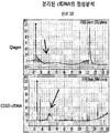
도 11은 10 명의 산모로부터 분리된 혈장에서 본 발명에 따른 방법(CCGD_cfDNA로 명명)과 Qiagen Kit를 각각 사용하여 분리한 cfDNA의 품질을 나타낸다.
도 12는 산모로부터 분리된 혈장에서 본 발명에 따른 방법(CCGD_cfDNA로 명명)과 Qiagen Kit를 각각 사용하여 분리한 cfDNA의 순도를 나타낸다.
상기의 과제를 해결하기 위한 본 발명의 구체적인 하나의 양태는
(a) 순환 유리 핵산을 포함하는 분리된 시료에 염 및 PEG(polyethylene glycol)를 포함하는 용액 및 자성 비드를 첨가하는 단계; 및
(b) 상기 자성 비드가 첨가된 시료에서 자성 비드를 획득하고, 이로부터 순환 유리 핵산을 분리하는 단계를 포함하는, 순환 유리 핵산을 포함하는 시료로부터 순환 유리 핵산을 분리하는 방법이다. 상기 핵산을 분리하는 방법은 PREP으로도 불릴 수 있다.
본 발명에서 용어, "순환 유리 핵산(circulating cell-free nucleic acid)"은 혈액에서 순환하며 존재하는 DNA를 말한다. 상기 순환 유리 핵산은 구체적으로 순환 유리 DNA(circulating cell-free DNA, cfDNA), 순환 유리 RNA 등을 포함하며, 구체적으로는 순환 유리 DNA일 수 있으나, 이에 제한되지 않는다. 상기 순환 유리 핵산은 일반적으로 혈장 또는 혈청에 1000bp 이하(DNA), 100nt 이하(RNA)의 길이를 나타내나, 이에 제한되지 않는다. 상기 순환 유리 핵산은 혈액 내에 소량으로 존재하여 이를 높은 수율로 획득하는 방법의 개발이 요구되어 왔다.
본 발명에서는, 순환 유리 핵산을 포함하는 시료를 자성 비드; 및 염 및 PEG를 포함하는 용액과 반응시키고, 자성 비드에 결합된 순환 유리 핵산을 분리하는 방법을 개발하였다. 자성 비드와 순환 유리 핵산을 반응시킴에 있어서, 염 및 PEG를 포함하는 용액을 모두 포함하는 용액을 사용하는 경우, 자성 비드가 순환 유리 핵산을 보다 효과적으로 포획할 수 있어, 고수율로 순환 유리 핵산을 수득할 수 있음을 확인하였다. 나아가, 본 발명에서는 상기 방법에 있어 용해 완충액 및 혈장 제조 역시 최적화하여 재현성있게 순환 유리 핵산을 효과적으로 분리할 수 있는 기술을 개발하였다.
이하 본 발명의 단계 및 각 구성을 보다 상세히 설명한다.
본 발명의 방법에서 (a) 단계는 순환 유리 핵산을 포함하는 분리된 시료에 염 및 PEG를 포함하는 용액, 및 자성 비드를 첨가하여 순환 유리 핵산이 자성 비드에 결합하도록 하는 단계이다.
본 발명에서, 상기 순환 유리 핵산을 포함하는 분리된 시료는 순환 유리 핵산을 포함하는 분리된 시료라면 그 종류는 특별히 제한되지 않으나, 혈장 또는 혈청일 수 있다. 구체적으로, 순환 유리 핵산의 분석 또는 수득이 필요한 개체로부터 분리된 혈장 또는 혈청, 보다 구체적으로는 혈장일 수 있으나, 이에 제한되는 것은 아니다.
상기 순환 유리 핵산을 포함하는 분리된 시료는 혈장 또는 혈청을 용해 완충액(lysis buffer)으로 용해시켜 제조한 것일 수 있다.
이에 따라 본 발명의 방법은 순환 유리 핵산을 포함하는 분리된 혈장 또는 혈청 시료에 용해 완충액을 첨가하는 제1 단계; 상기 제1 단계에서 용해된, 순환 유리 핵산을 포함하는 상기 시료에 염 및 PEG를 포함하는 용액 및 자성 비드를 첨가하는 제2 단계; 및 상기 자성 비드가 첨가된 시료에서 자성 비드를 획득하고, 이로부터 순환 유리 핵산을 분리하는 제3 단계를 포함할 수 있으나, 특별히 이에 제한되지 않는다.
혈장 또는 혈청의 용해를 위해 사용되는 상기 용해 완충액은 구체적으로, 카오트로픽 염(chaotropic salt), 킬레이트제(chelating agent), 비이온성 계면활성제(nonionic surfactant) 및 Tris-Cl을 포함하는 것일 수 있으나, 이에 제한되지 않는다.
본 발명의 일 실시양태에 따르면, 상기 조성을 가진 용해 완충액을 사용하는 경우, 킬레이트제인 EDTA, 카오트로픽염인 SDS, 및 Tris-Cl로 구성된 용해 완충액을 비롯한 다른 완충액을 사용한 경우에 비하여, 자성 비드의 분리가 용이하거나/하며 수율이 높음을 확인하였다.
본 발명에서 카오트로픽 염은, 물 분자 간의 수소 결합을 방해할 수 있는 물질을 포함한다 또한, 상기 카오트로픽 염은 단백질의 구조 변화를 야기하여 상기 단백질과 DNA 간의 결합력을 약화시키고, 이에 따라 DNA를 순수하게 분리하는 정도를 높이는 역할을 수행할 수 있다.
상기 카오트로픽 염의 예로서, 구아니디늄(guanidinium) 염, 리튬(lithium) 염, 마그네슘 염, SDS(Sodium dodecyl sulfate), 티오우레아(thiourea), 우레아(urea), 부탄올 또는 에탄올을 포함하나, 이에 제한되지 않는다.
상기 구아니디늄 염의 예로는, 구아니디늄 클로라이드를 들 수 있으며, 상기 리튬 염의 예로는 리튬 퍼클로레이트(lithium perchlorate), 리튬 아세테이트(lithium acetate)를, 마그네슘 염의 예로는 마그네슘 클로라이드를 들 수 있으나, 이에 제한되는 것은 아니다. 본 발명의 일 실시예에서는 구아니디늄 클로라이드를 카오트로픽 염으로서 사용하였다.
본 발명에서, 킬레이트 제는 Mg2
+ 및 Ca2
+와 같은 이가 양이온을 격리시키기 위하여 사용될 수 있으며, 이에 따라 DNA를 이를 분해하는 효소들로부터 보호할 수 있으나, 이에 제한되지 않는다.
상기 킬레이트 제의 예로는, DTPA(diethylenetriaminepentaacetic acid), EDTA(ethylenediaminetetraacetic acid), EGTA(ethylene glycol tetraacetic acid), 및 NTA(N,N-bis(carboxymethyl)glycine)로 이루어진 군으로부터 선택된 것일 수 있으나, 이에 제한되지 않는다. 본 발명의 일 실시예에서는 EDTA를 킬레이트제로 사용하였다.
상기 EDTA는 EDTA 화합물(예, K2EDTA,K3EDTA,또는 Na2EDTA)중 EDTA 부분을 나타내는 것일 수 있으나, 이에 제한되지 않는다.
본 발명에서, 비이온성 계면활성제(nonionic surfactant)는 한 분자 내에 친수성과 소수성 부분을 함께 가지고 있는 물질로서, 해리 시 비이온성인 특성을 나타내는 물질이다.
본 발명에서 사용될 수 있는 상기 비이온성 계면활성제의 예로는, 트리톤(triton) X-100을 들 수 있으나, 이에 제한되지 않는다.
상기 용해 완충액은 2 내지 6M 카오트로픽염; 1 내지 50mM 킬레이트제; 0.1 내지 5% (w/v) 비이온성 계면활성제; 또는/및 10 내지 100mM Tris-Cl을 포함하는 것일 수 있으나, 이에 제한되지 않는다.
여기서, 상기 용해 완충액의 pH는 pH 7.0 내지 8.5일 수 있다.
보다 구체적으로, 상기 용해 완충액은 구아니디늄 클로라이드, EDTA, 트리톤 X-100 및 Tris-Cl을 포함하는 것일 수 있으나, 이에 제한되지 않는다.
또한, 상기 용해 완충액으로 혈장 또는 혈청 시료를 용해시킬 때, 단백질분해효소를 함께 첨가할 수 있다.
상기 단백질분해효소는 핵산 분리에 통상적으로 사용되는 다양한 단백질분해효소가 사용될 수 있으나, 그 예로 단백질분해효소 K(proteinase K)를 사용할 수 있다.
한편, 상기 분리된 혈장 시료는 분리된 혈액 시료에서 혈장을 분리하기 위해 제1 원심분리하는 단계; 및 제1 원심분리된 시료에서 혈장을 분리하고, 분리된 혈장을 제2 원심분리하는 단계를 포함하는 방법으로 수득된 것일 수 있으나, 이에 제한되지 않는다.
본 발명에서는, 혈액 시료에서 혈장 시료를 제조하는 경우에 있어서 저속도에서 1차 원심분리를 수행하고, 이후 고속에서 2차 원심분리를 수행하는 경우, 백혈구(WBC) 등에서 유래된 DNA의 오염 없이 cfDNA를 보다 순수하게 분리할 수 있음을 확인하였다.
구체적으로, 상기 제1 원심분리는 1900 내지 2000 x g의 조건에서 원심분리하고, 제2 원심분리는 12000 내지 18000 x g의 조건에서 수행되는 것일 수 있으나, 이에 제한되는 것은 아니다. 또한, 상기 기술한 원심분리는 4 ℃ 내외의 온도 조건에서 수행되는 것일 수 있으나, 이에 제한되지 않는다.
본 발명에서, 상기 자성 비드와 염 및 PEG를 포함하는 용액을 상기 시료에 첨가하는 것은 순차적으로, 또는 동시에 수행될 수 있으나, 이에 제한되지 않는다.
순차적으로 수행하는 경우, 자성 비드의 첨가 후 상기 용액을 첨가하거나, 상기 용액을 첨가한 다음, 자성 비드를 첨가하는 방식 모두 수행될 수 있다.
본 발명에서, 상기 자성 비드와 함께 첨가되는 염 및 PEG를 포함하는 용액은 특별히 이에 제한되지는 않으나, 100bp 혹은 100nt 이하의 작은 핵산 단편을 포획하는 것에 도움을 줄 수 있다.
상기 염 및 PEG(polyethylene glycol)를 포함하는 용액에서 상기 염은 그 종류가 특별히 제한되지 않으나, 그 예로 염화나트륨일 수 있다.
상기 용액의 PEG는 평균 분자량이 6,000 내지 10,000 Da인 PEG일 수 있으나, 이에 제한되지 않는다.
보다 구체적으로, 상기 염 및 PEG를 포함하는 용액은 1 내지 4M 염 및 10 내지 60 %(w/v)의 PEG를 포함하는 것일 수 있으나, 이에 제한되지 않는다.
본 발명에서 자성 비드는, 자기장에 반응하는 입자 또는 비드를 말한다. 일반적으로, 자성 비드는 자기장을 가지지 않으나, 자기장에 노출되는 자기 쌍극자를 형성하는 물질을 말한다. 예컨대, 자기장 하에서 자화될 수 있으나, 자기장의 부재 하에서는 스스로는 자성을 가지지 않는 물질을 말한다. 본 발명에서 사용되는 자성은 상자성(paramagnetic) 또는 초상자성(superparamagnetic) 물질을 모두 포함하나, 이에 제한되지 않는다.
본 발명의 목적상 상기 자성 비드는 핵산에 결합하는 성질을 가지는 비드임이 바람직하고, 그 예로 핵산에 결합하는 관능기, 예컨대 -COOH기를 가지는 형태일 수 있으나, 이에 제한되지 않는다.
본 발명에서 상기 (b) 단계는 상기 자성 비드가 첨가된 시료에서 자성 비드를 획득하고, 이로부터 순환 유리 핵산을 분리하는 단계이다.
구체적으로, 상기 (b) 단계는 상기 (a) 단계에서 첨가된 자성 비드에 부착되어 있는 순환 유리 핵산을 자성 비드로부터 분리하는 단계이다.
상기 자성 비드가 첨가된 시료에서 자성 비드를 획득하기 전, 자성 비드를 세척하는 단계를 포함할 수 있다.
이때, 상기 자성 비드의 세척은 50 내지 95%(v/v) 에탄올 용액, 구체적으로는 80 내지 90 %(v/v) 에탄올 용액을 사용하여 수행되는 것일 수 있다.
이와 같은 세척 과정은 자성 비드를 자성 스탠드 하에 두어, 자성 비드를 모은 다음, 상층액을 제거하고, 여기에 세척 완충액을 첨가하여 세척하는 방식으로 수행할 수 있다. 또한, 이러한 세척 단계는 1회 이상 수행할 수 있다.
이와 같은 세척 과정을 선택적으로 수행한 다음, 자성 비드를 분리하고, 분리된 자성 비드에 용출 완충액(elution buffer)을 첨가하는 등의 방식으로 자성 비드로부터 순환 유리 핵산을 분리할 수 있다.
본 발명의 구체적인 다른 양태는, 용해 완충액; 염 및 PEG를 포함하는 용액; 및 자성 비드를 포함하는, 순환 유리 핵산 분리 키트이다.
용해 완충액, 염 및 PEG를 포함하는 용액, 및 자성 비드에 대해서는 앞서 설명한 바와 같다.
본 발명의 순환 유리 핵산 분리 키트는 그 유래에 제한되지 않고 cfDNA를 분리할 수 있으므로, 암 환자 또는 산모에서 cfDNA를 추출할 수 있다. 따라서, 암 환자의 혈액에서 추출한 cfDNA를 이용하여 해당 암 환자 특이적인 유전변이를 진단할 수 있다. 또한, 산모의 혈장 내에는 태아로부터 유래된 cfDNA가 다량 존재하므로, 산모 혈액에서 추출한 cfDNA를 이용하여, 태아의 비침습적 산전 진단에 활용할 수 있다. 즉, 본 발명의 순환 유리 핵산 분리 키트는 암 진단용 또는 산전 진단용 등으로 사용될 수 있으며, cfDNA를 활용할 수 있는 분야라면 그 용도에 특별히 제한되지 않는다.
또한, 상기 키트는 핵산 분리에 일반적으로 사용되는 다른 기구, 용액 등을 추가로 포함할 수 있으며, 순환 유리 핵산 분리를 위한 지시서 등을 포함할 수 있으나, 이에 제한되지 않는다.
또한, 상기 키트는 추가로 자성비드 세척 완충액, 및 자성비드 분리 기구(예, 자성 스탠드(magnetic stand))를 추가로 포함할 수 있으나, 이에 제한되지 않는다.
본 발명의 구체적인 다른 양태는, 카오트로픽 염, 킬레이트제, 비이온성 계면활성제 및 Tris-Cl을 포함하는, 순환 유리 핵산 분리를 위한 용해 완충액이다.
카오트로픽 염, 킬레이트제, 비이온성 계면활성제, 순환 유리 핵산 및 용해 완충액에 대해서는 앞서 설명한 바와 같다.
이하 본 발명을 하기 예에 의해 상세히 설명한다. 다만, 하기 예는 본 발명을 예시하기 위한 것일 뿐, 하기 예에 의해 본 발명의 범위가 제한되지는 않는다.
실시예
1: 혈장 시료의 제조
신선한 혈액 시료를 4 ℃에서 1,900 x g로 10분간 원심분리하여 혈장을 분리하였다. 그 다음, 분리된 혈장을 다시 4 ℃에서 16,000 x g로 10분간 원심분리하고, 상층액을 분리하였다. 그 다음, 이렇게 수득한 혈장 시료는 DNA 분리 전에 -20℃에서 저장하였다. 특별히 기술된 경우를 제외하고는 하기 실시예에서는 상기 방법을 이용하여 제조된 혈장을 사용하였다.
실시예
2:
cfDNA의
분리
동결된 혈장 시료는 DNA 분리 전에 녹인 후, 튜브에 혈장 시료 200㎕를 담았다. 그 다음, 상기 시료 튜브에 혈장 용해 완충액(5.5M 구아니딘 HCl, 50mM Tris-Cl, pH 8.0, 20mM EDTA, pH 8.0, 1.3% Triton X-100) 400㎕ 및 단백질 분해효소 K(proteinase K) 10㎕를 첨가하고, 이를 혼합하였다.
그 다음, 상기 튜브에 자성 비드(AMPure XP, Backman Coulter 사) 100㎕ 및 용액 A(2.5M NaCl, 20% PEG-8000) 1100㎕를 각 튜브에 첨가하고, 이를 혼합하였다. 그 다음, 자성 스탠드(magetic stand)에서 상기 튜브를 인큐베이션하고, 상기 튜브의 상층액을 제거하였다.
그 다음, 세척 완충액(85% EtOH) 1mL를 첨가하고 반응시킨 후, 자성 스탠드 위에서 에탄올 상층액을 제거하였고, 상기 세척 과정을 수회 반복하였다.
그 다음, 비드를 건조시켰고, 자성 스탠드에서 반응 튜브를 빼어낸 다음, 용출 완충액(순수 분리된 3차 증류수 또는 10 mM TE 완충액)을 첨가하였다. 그 다음, 자성 스탠드 위에서 반응 튜브를 둔 다음, 용출액을 분리하여, cfDNA 시료를 수득하였다.
실시예
3: 정상인 및 폐암 환자의 혈장으로부터의
cfDNA
시료 수득
상기 실시예 2의 cfDNA 분리 방법을 이용하여 정상인과 환자 간의 cfDNA 추출 양을 비교하는 실험을 진행하였다.
구체적으로, 정상 개체의 시료 31개, 폐암 환자 시료 49개에서 얻은 혈장을 각각 200㎕ 이용하여 cfDNA를 분리하였고, 그 결과를 도 1에 나타내었다. 상기 시료는 서울 아산병원 BRC (Bio Resource Center)에서 분양 받았다.
그 결과, 정상 개체와 폐암 환자에서 얻은 cfDNA의 양을 비교하였을 때, 폐암 환자에서 훨씬 많은 cfDNA를 얻을 수 있었고, 이는 당업계에 공지된 내용으로부터 예상된 사실이었다. 특히, 정상 개체의 200㎕ 혈장 시료에서 수득된 cfDNA의 양은 중간 값이 4.4ng (최대값: 9.2ng, 최소값: 0.8ng)이었고, 폐암 환자의 200㎕ 혈장 시료에서 수득된 cfDNA의 양은 중간 값이 32.0ng (최대값: 268.0 ng, 최소값: 4.78 ng)을 나타내어, 200㎕ 혈장 시료에서도 효과적으로 cfDNA를 분리할 수 있음을 확인하였다.
실시예
4:
실시예
2의
cfDNA
PREP 방법의 재현가능성 확인
상기 실시예 2의 cfDNA 분리 방법의 재현성을 조사하였다.
구체적으로, 폐암 환자 시료 49개에서 얻은 혈장을 각각 200㎕ 이용하여 두 번의 실험을 반복 수행한 후, 각각의 실험에서 얻은 cfDNA의 양을 비교하였고, 그 결과를 도 2에 나타내었다.
그 결과, 도 2에 나타낸 바와 같이, 두 값 간의 상관 관계가 1에 가까울 정도로 재현성이 우수한 결과를 나타내었다.
실시예
5:
실시예
2의
cfDNA
PREP 방법으로 수득한 시료의
cfDNA
패턴 분석
실시예 2의 cfDNA PREP 방법을 이용하여 PREP한 cfDNA의 특성을 BioAnalyzer를 이용하여 분석하였다
먼저, 폐암 환자의 혈장 시료로부터 PREP한 cfDNA의 크기를 확인하여 도 3a 및 b에 그 결과를 나타내었다.
크기를 확인한 결과, 세포사멸(Apoptosis)에 의해 생성되는 cfDNA의 특성으로 잘 알려진, DNA Ladder 크기 형태의 패턴으로 나타남을 확인할 수 있었다.
또한, cfDNA는 뉴클레오좀(nucleosome) 단위로 잘려져 있기 때문에, 대략 180bp 정도의 크기가 최소 단위이며, 2, 3, 4 뉴클레오좀 단위 크기인 180*2, 180*3, 180*4bp 크기 형태도 보였다(도 3b).
하지만, 기대보다 큰 크기의 DNA도 보였는데, 이는 혈액에서 혈장을 제조하는 경우에 DNA가 완전히 제거되지 않았거나, 용혈된 백혈구(WBC)에서 나온 gDNA가 함께 추출되어 나온 것이었다. 이는, 오래 전에 준비되어 보관되었던 폐암환자의 혈장을 이용하여 실험을 진행하였기 때문으로 여겨지며, 신선한 혈액을 이용하여, 실시예 1과 같이 2 단계 원심분리 법으로 혈장을 준비하면 해결될 수 있음을 확인하였다.
실시예
6:
실시예
2의
cfDNA
PREP 방법으로 수득한
cfDNA의
품질 분석
또한, 실시예 2의 cfDNA PREP 방법을 이용하여 PREP한 cfDNA의 품질(quality)를 확인하기 위하여, 100bp 및 237bp 크기의 생성 산물을 증폭할 수 있는 프라이머쌍을 이용하여 PCR을 수행하였다. 이때, 주형 DNA로는 폐암 환자의 시료로부터 분리한 1 내지 5번의 cfDNA 시료와 2개의 대조군 gDNA(Beas2B, H1975 세포주에서 PREP한 게노믹 DNA)를 사용하였다. 그 결과를 도 4에 나타내었다 (도 4에서, cfDNA 시료는 1 내지 5로 표시함).
PCR 수행 조건은 하기와 같았다.
구체적으로, 순수분리한 cfDNA 2 ㎕, 표 1에 표기된 프라이머쌍 (rs1952996 프라이머쌍 또는 RASSF1A 프라이머쌍 (각 프라이머쌍의 농도는 해당 F와 R 모두 각각 5 μM) 1 ㎕, dNTP mix (2.5 mM) 1 ㎕, Qiagen 사의 HotStar Taq DNA polymerase (5 U/㎕) 0.1 ㎕, 10x PCR buffer 1 ㎕, 최종 10 ㎕가 되도록 3차 증류수를 첨가한 다음, 94 ℃에서 15분 반응시켜 HotStarTaq polymerase를 활성화한 후, [94 ℃(20초)/60 ℃(30초)/72 ℃(30초)] 반응을 35회 반복한 후, 72 ℃에서 3분 반응시켜 증폭반응 종료하였다.
사용한 프라이머 정보는 하기와 같다.
| Primer 이름 | 염기서열(5'->3') | 서열번호 | 증폭산물 크기 (bp) |
| rs1952966-F | GGCTCTGGTTACAACAGCTT | 1 | 100 |
| rs1952966-R | AGAAGTTTGCTTGGCTGAAG | 2 | |
| RASSF1A-F | GTGGGGACCCTCTTCCTCTA | 3 | 237 |
| RASSF1A-R | GGAAGGAGCTGAGGAGAGC | 4 |
그 결과, 사용한 모든 cfDNA에서 PCR 산물 증폭이 잘 되었음을 확인하였다. 즉, PREP한 cfDNA의 품질에 문제가 없음을 확인하였다.
실시예 7: 혈장 제조 과정의 최적화
본 발명의 cfDNA PREP 방법에 사용되는 혈장의 제조 과정을 하기와 같이 최적화하였다.
구체적으로, cfDNA PREP 시, 2 단계 원심분리를 수행하여 혈장을 준비하는 경우의 효과를 1 단계 원심분리를 수행한 경우의 효과와 서로 비교하였다. 이를 위하여 정상 개체로부터 얻은 신선한 혈액을 이용하였다. 1 단계 원심분리 방법은 개체로부터 얻은 신선한 혈액을 1,900 x g 및 4 ℃에서 10분간 원심분리하여 혈장 시료를 수득하는 방법이고, 2 단계 원심분리 방법은 개체로부터 얻은 신선한 혈액을 1,900 x g 및 4 ℃에서 10분간 원심분리한 다음에 16,000 x g 및 4 ℃에서 10분간 원심분리하여 혈장 시료를 수득하는 방법이다.
상기 1 단계 원심분리 방법 및 2 단계 원심분리 방법을 사용하여 수득한 혈장 시료를 실시예 2의 방법으로 cfDNA를 분리하고, 그 양과 특성을 비교하였다. 상기 결과를 도 5a, 표 2 및 5b에 각각 나타내었다.
도 5a 및 하기 표 2에 나타낸 바와 같이 2 단계 원심분리 방법을 사용하여 획득한 혈장에서 분리한 cfDNA의 양이, 1 단계 원심분리 방법을 사용하여 획득한 혈장에서 분리한 cfDNA 양의 1/3 정도를 나타내었다. 1 단계 원심분리 방법의 경우, 추출된 cfDNA의 양에 편차가 크고 예상보다 양이 많았다.
| Healthy plasma | 1 step method (1,900g, 4 ℃ , 10min ), total ng | 2 step method (1,900g, 4 ℃ , 10min --> 16,000g, 4 ℃, 10min ), total ng | Ratio between 1 step / 2 step |
| 1 | 17.05 | 6.26 | 2.72 |
| 2 | 7.11 | 2.91 | 2.45 |
| 3 | 10.76 | 2.75 | 3.92 |
| 4 | 10.64 | 6.26 | 1.70 |
| 5 | 16.98 | 5.26 | 3.23 |
| 6 | 21.76 | 2.95 | 7.36 |
| 7 | 31.87 | 4.52 | 7.06 |
| 8 | 9.66 | 3.68 | 2.63 |
또한, 도 5b에 나타낸 바와 같이, 2 단계 원심분리 방법 및 1 단계 원심분리 방법을 이용하여 제조한 혈장으로부터 수득한 cfDNA의 특성을 분석한 결과, 2 단계 원심분리 방법으로 획득된 DNA의 경우, 전형적인 cfDNA 특징인 180bp 정도의 최소 뉴클레오좀 단위 크기를 나타냄을 확인할 수 있었다. 반면, 1 단계 원심분리 방법으로 추출된 DNA의 경우, 게노믹 DNA (gDNA)가 섞인 형태의 패턴을 나타내었다. 즉, BioAnalyzer 로딩 시, 상위 마커(upper marker) 보다 큰 intact gDNA 및 일부 단편화(fragmentation)되어 smear 패턴으로 보이는 gDNA를 나타내었다.
실시예 8: 실시예 2의 cfDNA PREP 법과 상용화된 키트 간의 비교
본 발명에 따른 cfDNA PREP 법의 효과를 기존 상용화된 키트와 비교하기 위하여 하기와 같은 실험을 수행하였다.
구체적으로, 현재 cfDNA PREP에 가장 많이 이용되고 있는 Qiagen circulating tumor DNA prep 키트와 성능을 비교하였다. 이를 위하여, 실제 난소암 환자의 혈장을 이용하여 실험을 진행하였고, 그 결과를 하기 표 3 및 도 6에 나타내었다. 도 6에서 본 발명에 따른 cfDNA PREP(실시예 2)의 방법을 'CCGD_cfDNA'로 표시하였다. 상기 혈장은 서울 아산병원 BRC에서 분양 받았다.
그 결과, 하기 표 3 및 도 6에 나타낸 바와 같이, 각 방법을 사용하여 cfDNA를 분리한 결과, 본 발명의 방법을 이용하는 경우에 Qiagen Kit를 사용한 경우에 비하여 4 내지 5배 가량 높은 양의 cfDNA를 수득할 수 있음을 확인하였다.
| 샘플번호 | Qiagen Kit | CCGD_cfDNA | ||
| 농도 (ng/㎕) | 수득한 양 (ng) | 농도 (ng/㎕) | 수득한 양 (ng) | |
| 2395 | 0.02 | 0.8 | 0.20 | 4.0 |
| 2397 | 0.03 | 1.5 | 0.22 | 4.5 |
| 2672 | 0.01 | 1.0 | 0.20 | 3.9 |
| 2722 | 0.02 | 1.3 | 0.33 | 6.7 |
| 2772 | 0.02 | 1.5 | 0.37 | 7.5 |
| 2799 | 0.02 | 1.5 | 0.22 | 4.3 |
| 2834 | 0.23 | 2.3 | 0.26 | 5.1 |
| 3030 | 0.08 | 0.9 | 0.28 | 5.5 |
| 3051 | 0.05 | 0.6 | 0.17 | 3.4 |
| 3052 | 0.13 | 1.4 | 0.38 | 7.6 |
| * 2차 원심 분리 후 수득한 200㎕의 혈장을 사용함 | ||||
실시예 9: 완충액의 종류에 따른 효과 비교
본 발명의 cfDNA PREP 방법에 효과적으로 사용될 수 있는 용해 완충액의 최적 조건을 하기와 같이 규명하였다.
먼저, 카오트로픽 염, 킬레이트 제 및 Tris-Cl을 포함하는 완충액(비교 완충액 1)과 카오트로픽 염, 킬레이트 제, 비이온성 계면활성제, 및 Tris-Cl을 포함하는 완충액(실시 완충액)을 각각 사용하여 실시예 2의 방법으로 cfDNA를 분리하였다. 이때, 난소암 환자의 혈장을 이용하여 실험을 진행하였고, 실시예 1과 같이 2번의 원심분리를 거친 시료를 사용하였다.
사용한 비교 완충액 1 및 실시 완충액의 구체적인 조성은 하기와 같았다.
| 비교 완충액 1 | 실시 완충액 |
| 10mM Tris-Cl (pH 8.0)0.1mM EDTA(pH 8.0)0.5% SDS | 5.5M Guanidine-HCl50mM Tris-Cl(pH 8.0)20mM EDTA(pH 8.0)1.3% Triton X-100 |
그 결과, 하기 표 5 및 도 7에 나타낸 바와 같이, 본 발명에 따른 실시 완충액을 사용하는 경우, 비교 완충액 1을 사용하는 경우에 비하여 훨씬 더 많은 양의 cfDNA를 얻을 수 있음을 확인하였다.
| 샘플 번호 | 비교 완충액 1 | 실시 완충액 | ||
| 농도 (ng/㎕) | 수득한 양 (ng) | 농도 (ng/㎕) | 수득한 양 (ng) | |
| 2395 | 0.08 | 1.6 | 0.20 | 4.0 |
| 2397 | 0.08 | 1.6 | 0.22 | 4.5 |
| 2672 | 0.12 | 2.5 | 0.20 | 3.9 |
| 2722 | 0.08 | 1.6 | 0.33 | 6.7 |
| 2772 | 0.11 | 2.1 | 0.37 | 7.5 |
| 2799 | 0.11 | 2.2 | 0.22 | 4.3 |
| 2834 | 0.10 | 2.0 | 0.26 | 5.1 |
| 3030 | 0.07 | 1.4 | 0.28 | 5.5 |
| 3051 | 0.07 | 1.3 | 0.17 | 3.4 |
| 3052 | 0.14 | 2.8 | 0.38 | 7.6 |
| * 2차 원심 분리 후 수득한 200㎕의 혈장을 사용함 | ||||
또한, 용해 완충액의 카오트로픽 염의 포함 유무에 따른 cfDNA PREP 효과를 비교하였다. 구체적으로 하기 [표 6]의 조성을 가지는 본 발명에 따른 실시 완충액 및 비교 완충액 2를 각각 사용하여 실시예 2의 방법으로 DNA를 분리하였다. 이 때, 2명의 정상인 개체로부터 얻은 혈장에 200ng의 Highly degraded gDNA를 첨가하여 진행하여, 용해 완충액의 조건에 따른 DNA 획득률을 서로 비교하였다. 그 결과를 하기 표 7 및 도 8에 나타내었다.
| 실시 완충액 | 비교 완충액 2 | |
| Guanidine-HCl | 5.5M | - |
| Tris-Cl (pH8.0) | 50mM | 66.7mM |
| EDTA (pH8.0) | 20mM | 26.7mM |
| Triton X-100 | 1.30% | 1.74% |
| 샘플번호 | 실시 완충액 | 비교 완충액 2 | |
| Healthy control-1 | AppliedgDNA(ng) | 200 | 200 |
| Total recovered amount (ng) | 32.5 | 24.7 | |
| Recoveryratio(%) | 16.2 | 12.4 | |
| Healthy control-2 | Applied FFPE gDNA(ng) | 200 | 200 |
| Total recovered amount (ng) | 48.8 | 19.4 | |
| Recoveryratio(%) | 24.4 | 9.7 |
그 결과, 두 명의 정상 개체의 혈장을 이용한 결과 모두에서, 본원발명에 따른 실시 완충액을 이용한 경우, 가장 높은 회수율을 나타내었다.
실시예 10: 산모의 혈장으로부터 cfDNA의 분리
본 발명의 cfDNA prep 방법은 폐암 환자뿐만 아니라, 정상 개체에서도 cfDNA를 수득할 수 있다. 즉, 개체의 상태에 제한 없이 분리된 혈장으로부터 cfDNA를 수득할 수 있다. 이에, 본 발명의 cfDNA prep 방법의 특성을 더욱 분석하기 위하여 이하의 실시예에서는 산모로부터 cfDNA를 분리하여 분석을 수행하였다. 나아가, 상기 실시예 2에서는 200 ㎕의 혈장 시료를 사용하였으나, 본 발명의 cfDNA prep 방법을 더 큰 부피의 시료에 적용하기 위해 분리 조건을 하기와 같이 적절히 변경하였다 (표 8).
| 혈장(부피) | 용해 완충액(부피) | 단백질 분해효소 K | 용액 A | 자성 비드 | ||
| 조성 | 부피 | |||||
| 기존 | 200 ㎕ | 400 ㎕ | 20 ㎕ | 20 % PEG/2.5 M Nacl | 1000 ㎕ | 200 ㎕ |
| 변경 | 500 ㎕ | 1000 ㎕ | 20 ㎕ | 40 % PEG/2.5 M Nacl | 1100 ㎕ | 400 ㎕ |
구체적으로, 총 10 명의 산모로부터 채취한 신선한 혈액 시료를 4 ℃에서 1,900 x g로 10 분간 원심분리하여 혈장을 분리하였다. 그 다음, 분리된 혈장을 다시 4 ℃에서 16,000 x g로 10 분간 원심분리하고, 상층액을 분리하였다. 그 다음, 분리된 혈장 500 ㎕를 이용하여, 본 발명의 cfDNA prep 방법과 기존 상용화된 키트인 Qiagen circulating tumor DNA prep 키트와의 비교를 실시하였다.
실시예 11: 실시예 10의 cfDNA PREP 법과 상용화된 키트 간의 수율 비교
본 발명의 cfDNA prep 방법 (CCGD_cfDNA) 및 Qiagen 키트로 분리된 cfDNA의 수율을 Picogreen 정량법 (PG) 및 RQ-PCR 정량법을 이용하여 비교하였다 (도 9).
그 결과, 정량법에 관계 없이 본 발명의 cfDNA prep 방법은 Qiagen 키트를 이용한 경우와 비교하여 유사하거나 높은 수율로 나타나는 것을 확인하였다. 특히 산모 4 및 산모 6으로부터 분리된 샘플의 경우 2 배 이상의 수율이 확인되어, 본 발명의 cfDNA prep 방법이 Qiagen 키트에 비해 수율 면에서 매우 우수한 효과를 가지는 것을 확인하였다.
한편, 각각의 정량법을 비교하였을 때, Qiagen 키트를 이용한 경우 PG 정량값과 RQ-PCR 정량값에서 큰 차이가 나는 것을 확인할 수 있었던 반면, 본 발명의 cfDNA prep 방법은 상기 두 정량법에서 큰 차이를 보이지 않았다. 이를 더욱 명확히 하기 위하여 PG 정량값과 RQ-PCR 정량값 간의 일치율을 확인하였다 (도 10).
그 결과, Qiagen 키트를 이용한 경우, 상기 도 9에서도 확인한 것과 같이 농도가 높은 2 샘플을 제외하고는 모두 아주 낮은 농도로 존재하는 것을 확인할 수 있었다. 또한, PG 정량값과 RQ-PCR 정량값 간의 일치율에서 상관성 계수가, Qiagen 키트를 이용한 경우 0.9245, 본 발명의 cfDNA prep 방법의 경우 0.9676으로 나타나 본 발명의 cfDNA prep 방법이 더 높은 상관성 계수를 가지는 것을 확인하였다.
Qiagen 키트의 경우, 작은 조각으로 존재하는 cfDNA의 추출 효율을 높이기 위해 키트 안에 DNA 운반체로 tRNA를 이용하였기 때문에, 추출이 완료된 cfDNA에는 많은 양의 tRNA가 함께 존재하고 있다. 따라서, 이로 인해 Qiagen 키트를 이용해 cfDNA를 분리하는 경우 Picogreen을 이용한 정량값에서는 tRNA로 인한 과측정이 나타나며, 이로 인해 상기와 같은 낮은 상관성 계수가 나타난 것으로 파악된다. 즉, 10 ng 정도의 수율로 나타나는 8 개의 샘플은 많은 부분이 tRNA 오염에 의해 측정된 값으로 예상된다. 반면 본 발명의 경우 tRNA를 사용하지 않기 때문에 상기 두 종류의 정량법에서 높은 수준의 상관성 계수가 나타났다.
실시예
12:
실시예
10의
cfDNA
PREP 법과 상용화된
키트로
분리한
cfDNA의
품질 및 순도 비교
10 명의 산모로부터 본 발명의 cfDNA prep 방법 및 Qiagen 키트를 이용해 추출된 cfDNA의 품질을 분석하기 위해, Bioanalyzer를 이용하여 추출된 cfDNA의 크기 분석을 수행하였다. 그 결과, 예상한 바와 같이 거의 모든 표본에서 cfDNA 크기 (~180 bp)로 보이는 피크 신호가 Qiagen 키트를 이용한 경우에 비해 본 발명의 cfDNA prep을 이용한 경우 매우 높게 나타나는 것을 확인하였다 (도 11).
다음으로, 상기 두 방법으로 추출된 cfDNA의 순도가 효소반응이 가능한 수준으로 우수한지를 확인하고자 하였다. 3 명의 산모 (산모 8, 산모 9 및 산모 10)로부터 추출된 cfDNA를 대상으로, 110 bp 크기를 가지는 amplicon을 증폭시키는 RQ-PCR 반응을 수행하였다. 동일 조건에서, 각 산모로부터 추출한 cfDNA의 양을 1, 2 및 4 ㎕씩 증가시키면서 RQ-PCR을 수행하여 Ct 값을 확인한 후 이를 상대량 (1/2^Ct)으로 변환하여, 사용된 cfDNA의 양에 따른 RQ-PCR로 확인된 상대량의 비례 정도를 확인함으로써, 추출된 cfDNA의 순도를 재확인하였다.
그 결과, 세 명의 산모로부터 추출한 cfDNA 모두, 사용된 cfDNA 양에 비례하여 상대량 (RQ-PCR 값, 1/2^Ct)이 증가하는 것으로 확인되었으며, 두 값 사이의 일치율 상관성 계수가, Qiagen 키트를 이용한 경우 0.9574, 본 발명의 cfDNA prep 방법의 경우 0.9983으로 나타나 본 발명의 cfDNA prep 방법이 더 높은 상관성 계수를 가지는 것을 확인하였다 (도 12).
이상의 설명으로부터, 본 발명이 속하는 기술 분야의 당업자는 본 발명이 그 기술적 사상이나 필수적 특징을 변경하지 않고서 다른 구체적인 형태로 실시될 수 있다는 것을 이해할 수 있을 것이다. 이와 관련하여, 이상에서 기술한 실시 예들은 모든 면에서 예시적인 것이며 한정적인 것이 아닌 것으로서 이해해야만 한다. 본 발명의 범위는 상기 상세한 설명보다는 후술하는 특허 청구범위의 의미 및 범위 그리고 그 등가 개념으로부터 도출되는 모든 변경 또는 변형된 형태가 본 발명의 범위에 포함되는 것으로 해석되어야 한다.
Claims (37)
- (a) 순환 유리 핵산(circulating cell-free nucleic acid)을 포함하는 분리된 시료에 염 및 PEG(polyethylene glycol)를 포함하는 용액 및 자성 비드를 첨가하는 단계; 및(b) 상기 자성 비드가 첨가된 시료에서 자성 비드를 획득하고, 이로부터 순환 유리 핵산을 분리하는 단계를 포함하는, 순환 유리 핵산을 포함하는 시료로부터 순환 유리 핵산을 분리하는 방법.
- 제1항에 있어서, 상기 순환 유리 핵산을 포함하는 분리된 시료는 분리된 혈장 또는 혈청인 것인 방법.
- 제2항에 있어서, 순환 유리 핵산을 포함하는 분리된 시료는 혈장 또는 혈청을 용해 완충액(lysis buffer)으로 용해시켜 제조된 것인, 방법.
- 제3항에 있어서,상기 용해 완충액은 카오트로픽 염(chaotropic salt), 킬레이트제(chelating agent), 비이온성 계면활성제(nonionic surfactant) 및 Tris-Cl을 포함하는 것인, 방법.
- 제3항에 있어서,상기 용해 완충액은 2 내지 6M 카오트로픽염; 1 내지 50mM 킬레이트제; 0.1 내지 5% (w/v) 비이온성 계면활성제; 및 10 내지 100mM Tris-Cl을 포함하는 것인, 방법.
- 제3항 내지 제5항 중 어느 한 항에 있어서, 상기 용해 완충액의 pH는 pH 7.0 내지 8.5인 것인, 방법.
- 제4항에 있어서,상기 카오트로픽 염은, 구아니디늄(guanidinium) 염, 리튬(lithium) 염, 마그네슘 염, SDS(Sodium dodecyl sulfate), 티오우레아(thiourea), 우레아(urea), 부탄올 및 에탄올로 이루어진 군으로부터 선택되는 것인, 방법.
- 제7항에 있어서, 상기 구아니디늄 염은 구아니디늄 클로라이드인, 방법.
- 제4항에 있어서, 상기 킬레이트제(chelating agent)는 DTPA(diethylenetriaminepentaacetic acid), EDTA(ethylenediaminetetraacetic acid), EGTA(ethylene glycol tetraacetic acid), 및 NTA(N,N-bis(carboxymethyl)glycine)로 이루어진 군으로부터 선택된 것인, 방법.
- 제4항에 있어서,상기 비이온성 계면활성제(nonionic surfactant)는 트리톤(triton) X-100인 것인, 방법.
- 제3항에 있어서, 상기 용해 완충액은 구아니디늄 클로라이드, EDTA, 트리톤 X-100 및 Tris-Cl을 포함하는 것인, 방법.
- 제1항에 있어서, 상기 염 및 PEG를 포함하는 용액은 염화나트륨 및 PEG를 포함하는 것인, 방법.
- 제1항에 있어서, 상기 PEG(polyethylene glycol)는 평균 분자량이 6,000 내지 10,000Da인 PEG인 것인, 방법.
- 제1항에 있어서, 상기 염 및 PEG를 포함하는 용액은 1 내지 4M 염 및 10 내지 60%(w/v)의 PEG를 포함하는 것인, 방법.
- 제3항에 있어서, 용해 완충액과 함께 단백질분해효소를 혈장 또는 혈청에 첨가하는 것을 포함하는 것인, 방법.
- 제1항에 있어서, (b) 단계는 자성 비드가 첨가된 시료에서 자성 비드를 세척한 후, 자성 비드를 획득하고, 이로부터 순환 유리 핵산을 분리하는 것인, 방법.
- 제16항에 있어서, 상기 세척은 50 내지 95%(v/v) 에탄올 용액을 사용하여 수행되는 것인, 방법.
- 제2항에 있어서, 상기 분리된 혈장 시료는분리된 혈액 시료에서 혈장을 분리하기 위해 제1 원심분리하는 단계; 및제1 원심분리된 시료에서 혈장을 분리하고, 분리된 혈장을 제2 원심분리하는 단계를 포함하는 방법으로 수득된 것인, 방법.
- 제18항에 있어서, 상기 제1 원심분리는 1,900 내지 2,000 x g의 조건에서 원심분리하고, 제2 원심분리는 12,000 내지 18,000 x g의 조건에서 원심분리하는 것인, 방법.
- 제1항에 있어서,상기 순환 유리 핵산은 순환 유리 DNA(circulating cell-free DNA)인, 방법.
- 용해 완충액(lysis buffer); 염 및 PEG(polyethylene glycol)를 포함하는 용액; 및 자성 비드를 포함하는, 순환 유리 핵산(circulating cell-free nucleic acid) 분리 키트.
- 제21항에 있어서,상기 용해 완충액은 카오트로픽 염(chaotropic salt), 킬레이트제(chelating agent), 비이온성 계면활성제(nonionic surfactant) 및 Tris-Cl을 포함하는 것인, 키트.
- 제21항에 있어서,상기 용해 완충액은 2 내지 6M 카오트로픽염; 1 내지 50mM 킬레이트제; 0.1 내지 5% (w/v)비이온성 계면활성제; 및 10 내지 100mM Tris-Cl을 포함하는 것인, 키트.
- 제21항 내지 제23항 중 어느 한 항에 있어서, 상기 용해 완충액의 pH는 pH 7.0 내지 8.5인 것인, 키트.
- 제22항에 있어서,상기 카오트로픽 염은, 구아니디늄(guanidinium) 염, 리튬(lithium) 염, 마그네슘 염, SDS(Sodium dodecyl sulfate), 티오우레아(thiourea), 우레아(urea), 부탄올 및 에탄올로 이루어진 군으로부터 선택되는 것인, 키트.
- 제25항에 있어서, 상기 구아니디늄 염은 구아니디늄 클로라이드인, 키트.
- 제22항에 있어서,상기 킬레이트제(chelating agent)는 DTPA(diethylenetriaminepentaacetic acid), EDTA(ethylenediaminetetraacetic acid), EGTA(ethylene glycol tetraacetic acid), 및 NTA(N,N-bis(carboxymethyl)glycine)로 이루어진 군으로부터 선택된 것인, 키트.
- 제22항에 있어서,상기 비이온성 계면활성제(nonionic surfactant)는 트리톤(triton) X-100인 것인, 키트.
- 제21항에 있어서, 상기 용해 완충액은 구아니디늄 클로라이드, EDTA, 트리톤 X-100 및 Tris-Cl을 포함하는 것인, 키트.
- 제21항에 있어서, 상기 염 및 PEG(polyethylene glycol)를 포함하는 용액은 염화나트륨 및 PEG를 포함하는 것인, 키트.
- 제21항 또는 제30항에 있어서, 상기 PEG(polyethylene glycol)는 평균 분자량이 6,000 내지 10,000 Da인 PEG인 것인, 키트.
- 제21항 또는 제30항에 있어서, 상기 염 및 PEG를 포함하는 용액은 1 내지 4M 염 및 10 내지 60%(w/v)의 PEG를 포함하는 것인, 키트.
- 제21항에 있어서, 상기 키트는 추가로 자성비드 세척 완충액, 자성비드 분리 기구, 또는 둘 다를 추가로 포함하는 것인, 키트.
- 제33항에 있어서, 상기 자성비드 세척 완충액은 50 내지 95%(v/v) 에탄올 용액인 것인, 키트.
- 제33항에 있어서, 상기 자성비드 분리 기구는 자성 스탠드(magnetic stand)인 것인, 키트.
- 제21항에 있어서, 상기 순환 유리 핵산은 순환 유리 DNA(circulating cell-free DNA)인, 키트.
- 카오트로픽 염(chaotropic salt), 킬레이트제(chelating agent), 비이온성 계면활성제(nonionic surfactant) 및 Tris-Cl을 포함하는, 순환 유리 핵산(circulating cell-free nucleic acid) 분리를 위한 용해 완충액.
Applications Claiming Priority (2)
| Application Number | Priority Date | Filing Date | Title |
|---|---|---|---|
| KR10-2015-0034686 | 2015-03-12 | ||
| KR20150034686 | 2015-03-12 |
Publications (1)
| Publication Number | Publication Date |
|---|---|
| WO2016144137A1 true WO2016144137A1 (ko) | 2016-09-15 |
Family
ID=56879508
Family Applications (1)
| Application Number | Title | Priority Date | Filing Date |
|---|---|---|---|
| PCT/KR2016/002462 Ceased WO2016144137A1 (ko) | 2015-03-12 | 2016-03-11 | 순환 유리 핵산의 분리 방법 |
Country Status (2)
| Country | Link |
|---|---|
| KR (1) | KR101967878B1 (ko) |
| WO (1) | WO2016144137A1 (ko) |
Cited By (7)
| Publication number | Priority date | Publication date | Assignee | Title |
|---|---|---|---|---|
| CN110144345A (zh) * | 2019-05-10 | 2019-08-20 | 上海交通大学 | 一种从卵泡液中提取cfDNA的方法 |
| CN112322615A (zh) * | 2020-11-18 | 2021-02-05 | 威高集团有限公司 | 核酸保存液、核酸提取保存液、采血管和提取核酸的方法 |
| CN112410327A (zh) * | 2020-12-11 | 2021-02-26 | 福建和瑞基因科技有限公司 | 一种提取rna的试剂盒及其方法 |
| CN113621606A (zh) * | 2021-04-25 | 2021-11-09 | 北京全式金生物技术有限公司 | 一种提取血浆游离dna的试剂盒和方法 |
| CN114457069A (zh) * | 2022-03-07 | 2022-05-10 | 江苏迅睿生物技术有限公司 | 一种磁珠法病原体核酸提取试剂盒及使用方法 |
| WO2025112952A1 (zh) * | 2023-11-28 | 2025-06-05 | 上海微荷医学检验实验室有限公司 | 提取血浆中游离dna的试剂组合、试剂盒和方法 |
| CN120400128A (zh) * | 2025-07-03 | 2025-08-01 | 上海金福康制药工程技术有限公司 | 一种cfDNA提取试剂盒及提取方法 |
Families Citing this family (1)
| Publication number | Priority date | Publication date | Assignee | Title |
|---|---|---|---|---|
| CN118256489B (zh) * | 2024-05-31 | 2024-08-27 | 天根生化科技(北京)有限公司 | 一种循环核酸的提取试剂盒及循环核酸的提取方法 |
Citations (5)
| Publication number | Priority date | Publication date | Assignee | Title |
|---|---|---|---|---|
| US20060078923A1 (en) * | 2003-04-02 | 2006-04-13 | Mckernan Kevin | Method for isolating nucleic acids |
| US20060141450A1 (en) * | 2002-12-09 | 2006-06-29 | Xu Zhang | Magnetism based rapid cell separation |
| US7527929B2 (en) * | 2004-07-30 | 2009-05-05 | Agencourt Bioscience Corporation | Methods of isolating nucleic acids using multifunctional group-coated solid phase carriers |
| US20120171675A1 (en) * | 2009-06-18 | 2012-07-05 | Qiagen Gmbh | Method for isolating nucleic acids |
| US20140274740A1 (en) * | 2013-03-15 | 2014-09-18 | Verinata Health, Inc. | Generating cell-free dna libraries directly from blood |
Family Cites Families (3)
| Publication number | Priority date | Publication date | Assignee | Title |
|---|---|---|---|---|
| US20060105372A1 (en) * | 2004-11-05 | 2006-05-18 | Bair Robert J | Compositions and methods for purifying nucleic acids from stabilization reagents |
| JP2009033995A (ja) * | 2007-07-31 | 2009-02-19 | Hitachi Metals Ltd | 高磁化磁気ビーズを用いる血液試料からの核酸抽出方法 |
| KR101432034B1 (ko) * | 2007-11-16 | 2014-08-21 | 삼성전자주식회사 | 코스모트로픽 염을 이용한 고체 지지체에서의 작은 rna분리 방법 |
-
2016
- 2016-03-11 KR KR1020160029622A patent/KR101967878B1/ko active Active
- 2016-03-11 WO PCT/KR2016/002462 patent/WO2016144137A1/ko not_active Ceased
Patent Citations (5)
| Publication number | Priority date | Publication date | Assignee | Title |
|---|---|---|---|---|
| US20060141450A1 (en) * | 2002-12-09 | 2006-06-29 | Xu Zhang | Magnetism based rapid cell separation |
| US20060078923A1 (en) * | 2003-04-02 | 2006-04-13 | Mckernan Kevin | Method for isolating nucleic acids |
| US7527929B2 (en) * | 2004-07-30 | 2009-05-05 | Agencourt Bioscience Corporation | Methods of isolating nucleic acids using multifunctional group-coated solid phase carriers |
| US20120171675A1 (en) * | 2009-06-18 | 2012-07-05 | Qiagen Gmbh | Method for isolating nucleic acids |
| US20140274740A1 (en) * | 2013-03-15 | 2014-09-18 | Verinata Health, Inc. | Generating cell-free dna libraries directly from blood |
Cited By (10)
| Publication number | Priority date | Publication date | Assignee | Title |
|---|---|---|---|---|
| CN110144345A (zh) * | 2019-05-10 | 2019-08-20 | 上海交通大学 | 一种从卵泡液中提取cfDNA的方法 |
| CN110144345B (zh) * | 2019-05-10 | 2022-07-15 | 上海交通大学 | 一种从卵泡液中提取cfDNA的方法 |
| CN112322615A (zh) * | 2020-11-18 | 2021-02-05 | 威高集团有限公司 | 核酸保存液、核酸提取保存液、采血管和提取核酸的方法 |
| CN112410327A (zh) * | 2020-12-11 | 2021-02-26 | 福建和瑞基因科技有限公司 | 一种提取rna的试剂盒及其方法 |
| CN113621606A (zh) * | 2021-04-25 | 2021-11-09 | 北京全式金生物技术有限公司 | 一种提取血浆游离dna的试剂盒和方法 |
| CN113621606B (zh) * | 2021-04-25 | 2023-12-12 | 北京全式金生物技术股份有限公司 | 一种提取血浆游离dna的试剂盒和方法 |
| CN114457069A (zh) * | 2022-03-07 | 2022-05-10 | 江苏迅睿生物技术有限公司 | 一种磁珠法病原体核酸提取试剂盒及使用方法 |
| WO2025112952A1 (zh) * | 2023-11-28 | 2025-06-05 | 上海微荷医学检验实验室有限公司 | 提取血浆中游离dna的试剂组合、试剂盒和方法 |
| CN120400128A (zh) * | 2025-07-03 | 2025-08-01 | 上海金福康制药工程技术有限公司 | 一种cfDNA提取试剂盒及提取方法 |
| CN120400128B (zh) * | 2025-07-03 | 2025-10-03 | 上海金福康制药工程技术有限公司 | 一种cfDNA提取试剂盒及提取方法 |
Also Published As
| Publication number | Publication date |
|---|---|
| KR20160110902A (ko) | 2016-09-22 |
| KR101967878B1 (ko) | 2019-04-11 |
Similar Documents
| Publication | Publication Date | Title |
|---|---|---|
| WO2016144137A1 (ko) | 순환 유리 핵산의 분리 방법 | |
| WO2015183026A1 (ko) | 불활성화된 표적 특이적 뉴클레아제를 이용한 표적 dna의 분리 방법 | |
| EP3194578A1 (en) | Immune-compatible cells created by nuclease-mediated editing of genes encoding hla | |
| WO2017122896A1 (ko) | 수산생물전염병 원인바이러스의 판별 및 검출용 유전자 마커,및 이를 이용한 원인바이러스의 판별 및 검출 방법 | |
| WO2016167408A1 (ko) | 차세대 염기서열 분석기법을 이용한 장기 이식 거부 반응 예측 방법 | |
| WO2020022821A1 (ko) | 실리카 코팅 자성 비드를 이용한 바이러스 rna 추출용 조성물 및 이를 이용한 바이러스 rna 추출 방법 | |
| WO2015126078A1 (ko) | 핵산과 신호 프로브의 비대칭 등온증폭을 이용한 핵산의 검출방법 | |
| WO2016144136A1 (ko) | Ffpe 조직에서 핵산의 분리 방법 | |
| JP6583796B2 (ja) | 核酸の増幅方法 | |
| WO2017074094A1 (en) | A method for prenatal diagnosis using digital pcr. | |
| WO2017122897A1 (ko) | 참돔 이리도바이러스병 원인바이러스의 검출용 유전자 마커, 및 이를 이용한 원인바이러스의 검출 방법 | |
| WO2023163458A1 (ko) | Crispr-cas 기반의 살모넬라균 검출용 조성물 및 이를 이용한 살모넬라균 검출방법 | |
| WO2023063562A1 (ko) | 세포 유리 dna를 이용한 대장암 진단을 위한 메틸화 마커 유전자 및 이의 용도 | |
| WO2023106868A1 (ko) | 단일가닥 생성 방법 및 이를 이용한 변이 검출 방법 | |
| WO2013105801A1 (ko) | 만성 골수성 백혈병 융합 유전자형 타이핑용 프로브, 프라이머 및 이의 이용방법 | |
| WO2022097844A1 (ko) | 유전자 복제수 변이 정보를 이용하여 췌장암 환자의 생존 예후를 예측하는 방법 | |
| WO2020159069A1 (ko) | 압타머를 이용한 다중 multiplex pcr 방법 | |
| WO2024096536A1 (ko) | 폐암 진단용 dna 메틸화 마커 및 이의 용도 | |
| EP0989192A2 (en) | Method for synthesis of nucleic acids | |
| WO2024158203A1 (ko) | 간암 발병의 위험성 예측용 조성물 | |
| WO2023014009A1 (ko) | 세포 용해 및 핵산 추출용 조성물, 이를 이용한 핵산 추출 방법 및 이를 이용한 분자진단방법 | |
| WO2018194280A1 (ko) | SDC2(Syndecan 2) 유전자의 메틸화 검출방법 | |
| WO2011142646A9 (ko) | 인유두종바이러스 검출 및 유전형 확인 방법 | |
| EP3368690A1 (en) | A method for prenatal diagnosis using digital pcr. | |
| Baker et al. | Isolation of genomic DNA from blood using a novel filter-based DNA purification technology |
Legal Events
| Date | Code | Title | Description |
|---|---|---|---|
| 121 | Ep: the epo has been informed by wipo that ep was designated in this application |
Ref document number: 16762024 Country of ref document: EP Kind code of ref document: A1 |
|
| NENP | Non-entry into the national phase |
Ref country code: DE |
|
| 122 | Ep: pct application non-entry in european phase |
Ref document number: 16762024 Country of ref document: EP Kind code of ref document: A1 |