WO2016084467A1 - Endoscope encapsulé et système d'endoscope encapsulé - Google Patents
Endoscope encapsulé et système d'endoscope encapsulé Download PDFInfo
- Publication number
- WO2016084467A1 WO2016084467A1 PCT/JP2015/077074 JP2015077074W WO2016084467A1 WO 2016084467 A1 WO2016084467 A1 WO 2016084467A1 JP 2015077074 W JP2015077074 W JP 2015077074W WO 2016084467 A1 WO2016084467 A1 WO 2016084467A1
- Authority
- WO
- WIPO (PCT)
- Prior art keywords
- information
- image
- capsule endoscope
- brightness information
- acquired
- Prior art date
Links
Images


Classifications
-
- A—HUMAN NECESSITIES
- A61—MEDICAL OR VETERINARY SCIENCE; HYGIENE
- A61B—DIAGNOSIS; SURGERY; IDENTIFICATION
- A61B1/00—Instruments for performing medical examinations of the interior of cavities or tubes of the body by visual or photographical inspection, e.g. endoscopes; Illuminating arrangements therefor
- A61B1/04—Instruments for performing medical examinations of the interior of cavities or tubes of the body by visual or photographical inspection, e.g. endoscopes; Illuminating arrangements therefor combined with photographic or television appliances
- A61B1/041—Capsule endoscopes for imaging
-
- A—HUMAN NECESSITIES
- A61—MEDICAL OR VETERINARY SCIENCE; HYGIENE
- A61B—DIAGNOSIS; SURGERY; IDENTIFICATION
- A61B1/00—Instruments for performing medical examinations of the interior of cavities or tubes of the body by visual or photographical inspection, e.g. endoscopes; Illuminating arrangements therefor
- A61B1/00002—Operational features of endoscopes
- A61B1/00004—Operational features of endoscopes characterised by electronic signal processing
- A61B1/00009—Operational features of endoscopes characterised by electronic signal processing of image signals during a use of endoscope
-
- A—HUMAN NECESSITIES
- A61—MEDICAL OR VETERINARY SCIENCE; HYGIENE
- A61B—DIAGNOSIS; SURGERY; IDENTIFICATION
- A61B1/00—Instruments for performing medical examinations of the interior of cavities or tubes of the body by visual or photographical inspection, e.g. endoscopes; Illuminating arrangements therefor
- A61B1/00002—Operational features of endoscopes
- A61B1/00011—Operational features of endoscopes characterised by signal transmission
- A61B1/00016—Operational features of endoscopes characterised by signal transmission using wireless means
-
- A—HUMAN NECESSITIES
- A61—MEDICAL OR VETERINARY SCIENCE; HYGIENE
- A61B—DIAGNOSIS; SURGERY; IDENTIFICATION
- A61B1/00—Instruments for performing medical examinations of the interior of cavities or tubes of the body by visual or photographical inspection, e.g. endoscopes; Illuminating arrangements therefor
- A61B1/00002—Operational features of endoscopes
- A61B1/0002—Operational features of endoscopes provided with data storages
-
- A—HUMAN NECESSITIES
- A61—MEDICAL OR VETERINARY SCIENCE; HYGIENE
- A61B—DIAGNOSIS; SURGERY; IDENTIFICATION
- A61B1/00—Instruments for performing medical examinations of the interior of cavities or tubes of the body by visual or photographical inspection, e.g. endoscopes; Illuminating arrangements therefor
- A61B1/00002—Operational features of endoscopes
- A61B1/00025—Operational features of endoscopes characterised by power management
- A61B1/00036—Means for power saving, e.g. sleeping mode
-
- A—HUMAN NECESSITIES
- A61—MEDICAL OR VETERINARY SCIENCE; HYGIENE
- A61B—DIAGNOSIS; SURGERY; IDENTIFICATION
- A61B1/00—Instruments for performing medical examinations of the interior of cavities or tubes of the body by visual or photographical inspection, e.g. endoscopes; Illuminating arrangements therefor
- A61B1/04—Instruments for performing medical examinations of the interior of cavities or tubes of the body by visual or photographical inspection, e.g. endoscopes; Illuminating arrangements therefor combined with photographic or television appliances
- A61B1/045—Control thereof
-
- A—HUMAN NECESSITIES
- A61—MEDICAL OR VETERINARY SCIENCE; HYGIENE
- A61B—DIAGNOSIS; SURGERY; IDENTIFICATION
- A61B1/00—Instruments for performing medical examinations of the interior of cavities or tubes of the body by visual or photographical inspection, e.g. endoscopes; Illuminating arrangements therefor
- A61B1/06—Instruments for performing medical examinations of the interior of cavities or tubes of the body by visual or photographical inspection, e.g. endoscopes; Illuminating arrangements therefor with illuminating arrangements
- A61B1/0661—Endoscope light sources
-
- H—ELECTRICITY
- H04—ELECTRIC COMMUNICATION TECHNIQUE
- H04N—PICTORIAL COMMUNICATION, e.g. TELEVISION
- H04N23/00—Cameras or camera modules comprising electronic image sensors; Control thereof
-
- H—ELECTRICITY
- H04—ELECTRIC COMMUNICATION TECHNIQUE
- H04N—PICTORIAL COMMUNICATION, e.g. TELEVISION
- H04N23/00—Cameras or camera modules comprising electronic image sensors; Control thereof
- H04N23/60—Control of cameras or camera modules
- H04N23/66—Remote control of cameras or camera parts, e.g. by remote control devices
- H04N23/661—Transmitting camera control signals through networks, e.g. control via the Internet
-
- H—ELECTRICITY
- H04—ELECTRIC COMMUNICATION TECHNIQUE
- H04N—PICTORIAL COMMUNICATION, e.g. TELEVISION
- H04N23/00—Cameras or camera modules comprising electronic image sensors; Control thereof
- H04N23/70—Circuitry for compensating brightness variation in the scene
- H04N23/71—Circuitry for evaluating the brightness variation
-
- H—ELECTRICITY
- H04—ELECTRIC COMMUNICATION TECHNIQUE
- H04N—PICTORIAL COMMUNICATION, e.g. TELEVISION
- H04N23/00—Cameras or camera modules comprising electronic image sensors; Control thereof
- H04N23/70—Circuitry for compensating brightness variation in the scene
- H04N23/76—Circuitry for compensating brightness variation in the scene by influencing the image signals
-
- A—HUMAN NECESSITIES
- A61—MEDICAL OR VETERINARY SCIENCE; HYGIENE
- A61B—DIAGNOSIS; SURGERY; IDENTIFICATION
- A61B1/00—Instruments for performing medical examinations of the interior of cavities or tubes of the body by visual or photographical inspection, e.g. endoscopes; Illuminating arrangements therefor
- A61B1/00002—Operational features of endoscopes
- A61B1/00004—Operational features of endoscopes characterised by electronic signal processing
- A61B1/00006—Operational features of endoscopes characterised by electronic signal processing of control signals
-
- H—ELECTRICITY
- H04—ELECTRIC COMMUNICATION TECHNIQUE
- H04N—PICTORIAL COMMUNICATION, e.g. TELEVISION
- H04N23/00—Cameras or camera modules comprising electronic image sensors; Control thereof
- H04N23/50—Constructional details
- H04N23/555—Constructional details for picking-up images in sites, inaccessible due to their dimensions or hazardous conditions, e.g. endoscopes or borescopes
Definitions
- the present invention relates to a capsule endoscope and a capsule endoscope system that are introduced into a subject and can acquire in-vivo information.
- Endoscopes in the medical field are conventionally used in applications such as in vivo observation.
- a subject is placed in a body cavity by swallowing, and an image of the subject is captured while moving in the body cavity in accordance with a peristaltic motion, and the captured image of the subject is captured as an imaging signal.
- capsule endoscopes capable of wireless transmission to the outside have been proposed.
- Such capsule endoscopes usually take 2 frames per second.
- the reason for setting 2 frames per second is that the time when the capsule endoscope is located in the body cavity is very long, but the battery capacity built into the capsule endoscope is limited. It is.
- Japanese Patent Laid-Open No. 2005-20755 compares a captured image with a previously transmitted image. For example, only a captured image that is substantially different from the previously captured image is transmitted to an external receiving device. A capsule endoscope that saves energy consumed by transmission has been proposed.
- an object of the present invention is to provide a capsule endoscope and a capsule endoscope system capable of preventing useless image capturing and reducing power consumption.
- the capsule endoscope of one embodiment of the present invention includes an imaging unit, a storage unit that stores brightness information acquired by the imaging unit, brightness information acquired by the imaging unit, and the brightness information. Is compared with the brightness information acquired by the imaging unit and stored in the storage unit immediately before acquiring the brightness information, and if the comparison result is within a predetermined threshold, the brightness information is further acquired by the imaging unit and acquired. If the comparison result is greater than the predetermined threshold value, the brightness information obtained by the imaging unit immediately before is further compared with the brightness information stored in the storage unit.
- a control unit that performs imaging and stores the captured image in the storage unit or controls the image transmission unit provided in the capsule endoscope to transmit the image to the outside of the capsule endoscope And comprising.
- the capsule endoscope system includes an imaging unit, a storage unit that stores brightness information acquired by the imaging unit, brightness information acquired by the imaging unit, Immediately before acquiring brightness information, the brightness information acquired by the imaging unit and compared with the brightness information stored in the storage unit is compared. When the comparison result is within a predetermined threshold, the brightness information is further acquired by the imaging unit.
- the imaging unit Control to store the captured image in the storage unit or to transmit the image to the outside of the capsule endoscope by an image transmission unit provided in the capsule endoscope
- FIG. 1 is a diagram for explaining a usage pattern of the endoscope system according to the first embodiment
- FIG. 2 is a detailed configuration of the capsule endoscope and the receiving apparatus according to the first embodiment
- 3 is a diagram for explaining a pre-exposure area for performing pre-exposure
- FIG. 4 is a diagram for explaining exposure timing
- FIGS. 5A and 5B are diagrams for explaining exposure timing. It is a figure for demonstrating the space
- an endoscope system 1 that is a capsule endoscope system includes a capsule endoscope 10 and a receiving device 20 as an external device.
- the capsule endoscope 10 is introduced into the digestive organ lumen when the subject 2 swallows.
- the capsule endoscope 10 includes a battery 11, an illumination unit 12 that illuminates a subject, an imaging element 13 as an imaging unit that images the subject, and a captured imaging signal (internal An image transmission unit 14 that wirelessly transmits an endoscopic image), a memory 15 as a storage unit that temporarily stores a photometric result to be described later, and a control unit 16 that controls the entire capsule endoscope 10. These are housed in a housing to constitute a main part.
- the receiving device 20 disposed outside the body of the subject 2 includes an antenna unit 21 that receives an imaging signal from the capsule endoscope 10, and a main body 25 that the subject 2 wears on, for example, the waist.
- the main body 25 includes an image receiving unit 22 that receives an imaging signal (endoscopic image) wirelessly transmitted from the capsule endoscope 10 via the antenna unit 21, and the antenna unit 21.
- An external memory 23 as an external storage unit for storing an endoscopic image received by the image receiving unit 22 via the endoscope, and an endoscopic image received by the image receiving unit 22 or an endoscopic image stored in the external memory 23
- a display unit 24 for displaying the.
- the capsule endoscope 10 performs photometry by performing pre-exposure before capturing an image. That is, the control unit 16 emits light from the illumination unit 12 provided in the capsule endoscope 10 and performs imaging with the imaging element 13. In this imaging, the control unit 16 does not use all the pixel information of the image sensor 13, but acquires image brightness information from only a part of pixel information in a pre-exposure area described later (that is, performs photometry). ) In the following description, photometry performed using pixel information of the pre-exposure area is also referred to as pre-photometry.
- the pre-exposure area 13a is provided so as to include the center pixel of the image sensor 13, and the control unit 16 is based on this partial pixel information, that is, information obtained from the pre-exposure area 13a. Get brightness information.
- the pre-exposure area provided in the image pick-up element 13 is not limited to the pre-exposure area 13a provided so that the center pixel may be included.
- pre-exposure areas 13b are provided in the vicinity of the four corners of the surface on which the image sensor 13 is disposed, and the control unit 16 obtains brightness information based on information obtained from these four pre-exposure areas 13b. You may make it acquire.
- pre-exposure areas 13c are provided in positions around the pixel region 13d used for display among the pixels of the image sensor 13, and are not reproduced and displayed on the monitor screen.
- the control unit 16 controls the four pre-exposure areas 13c.
- Brightness information may be acquired based on information obtained from the exposure area 13c. That is, the pre-exposure area 13c is provided in an area called optical black for detecting black, which is provided outside the pixel area 13d used for display.
- control unit 16 is not limited to acquiring brightness information based on information obtained from the pre-exposure areas 13a, 13b, or 13c.
- pre-exposure area 13a and the four pre-exposure areas 13b In combination, brightness information may be acquired based on information obtained from these pre-exposure areas 13a and 13b.
- the control unit 16 temporarily stores the brightness information (pre-photometry result) thus acquired in the memory 15. Then, as shown in FIG. 4, when the pre-photometry operation is completed, the control unit 16 performs the next pre-exposure and pre-photometry operations.
- the pre-exposure and pre-photometry operations have the above-described contents, and the same pre-exposure and pre-photometry operations are performed.
- the pre-exposure interval will be described with reference to FIGS. 5A and 5B.
- the following two patterns are assumed for the pre-exposure interval, but the present invention is not limited to this, and other patterns may be used.
- the first pattern is a pattern that minimizes the pre-exposure interval, that is, as short as possible.
- the pre-exposure interval it is possible to avoid missing the movement of the capsule endoscope 10.
- the conventional capsule endoscope acquires a captured image of 2 frames per second, but in FIG. 5A, the capsule endoscope 10 moves by minimizing the pre-exposure interval. It is also possible to acquire a captured image of 3 frames or more per second, and prevent oversight of a lesioned part or the like.
- the second pattern is a pattern in which the pre-exposure interval is a predetermined interval as shown in FIG. 5B.
- the pre-exposure interval is 2 fps, but the present invention is not limited to this and may be another interval.
- the capsule endoscope 10 when the capsule endoscope 10 has movement, a captured image of 2 frames is acquired per second as in the conventional case, but when the capsule endoscope 10 has no movement, 1 second. Since one frame of the captured image is acquired or no captured image is acquired at all, the power consumption of the capsule endoscope 10 can be reduced as compared with the conventional case.
- the control unit 16 compares the brightness information obtained in the second pre-photometry with the previous brightness information stored in the memory 15. Specifically, for example, the control unit 16 calculates the difference between the brightness information obtained in the second pre-exposure and the previous brightness information stored in the memory 15. If the comparison result is within a predetermined threshold, the control unit 16 determines that the capsule endoscope 10 is not moving (stayed) between the two photometric operations. When the control unit 16 determines that the capsule endoscope 10 is not moving, the control unit 16 further proceeds to the next pre-exposure and pre-photometry operation. If the comparison result between the latest brightness information and the previous brightness information becomes larger than a predetermined threshold while repeating such pre-exposure and pre-photometry operations, the control unit 16 may It is determined that the mirror 10 has moved.
- the control unit 16 determines the movement of the capsule endoscope 10 from the change in the brightness information of each pre-exposure area.
- the control unit 16 determines that the capsule endoscope 10 has moved when the brightness information in each pre-exposure area has changed at least at one location. Note that the control unit 16 may determine that the capsule endoscope 10 has moved when all of the brightness information in each pre-exposure area has changed.
- the control unit 16 selects the capsule type. It is determined that the endoscope 10 has moved, and after the pre-photometry operation, image capturing, that is, main exposure is performed. For image capturing, all the pixel information of the image sensor 13, here, substantially all the pixel information of the image sensor 13 including all the pixels to be displayed on the monitor are acquired.
- the control unit 16 outputs an image signal obtained by performing predetermined signal processing on the pixel information acquired by the image sensor 13 to the image transmission unit 14.
- the image transmission unit 14 wirelessly transmits the image signal that has been subjected to predetermined signal processing by the control unit 16 to the external reception device 20. Note that the image transmission unit 14 transmits the image signal to the external reception device 20, but the image signal may be stored in the memory 15 in the capsule endoscope 10 via the control unit 16.
- the image signal transmitted wirelessly is received by the image receiving unit 22 via the external antenna unit 21.
- the image receiving unit 22 stores the received image signal in the external memory 23 or outputs it to the display unit 24 to display an endoscopic image.
- the receiving device 20 may add a time stamp to the endoscopic image and store it in the external memory 23 when receiving an image signal (endoscopic image) from the capsule endoscope 10. .
- the control unit 16 starts pre-exposure and pre-photometry operations again, and repeats the above-described processing.
- the brightness information obtained as a result of the pre-photometry is the brightness information stored in the memory 15.
- the control unit 16 captures the image. It is compared with the determined latest brightness information to determine whether or not the capsule endoscope 10 is moving.
- the control unit 16 continues the pre-exposure and the pre-photometry operation, and when it is determined that the capsule endoscope 10 is moving, the main exposure exposes the image. Imaging is performed, and data transmission of the obtained image signal is performed.
- FIG. 6 is a flowchart for explaining the operation of the capsule endoscope 10 according to the first embodiment.
- a target value and area for light control are set (step S1), and a pre-exposure area is set (step S2).
- a light control target value is calculated from the light control target value and area and the pre-exposure area (step S3).
- pre-exposure is executed (step S4), and pre-photometry is executed (step S5).
- step S6 It is determined whether or not the difference between the pre-photometric value and the immediately preceding pre-photometric value is within a predetermined threshold (step S6). If the difference between the pre-photometric value and the previous pre-photometric value is within a predetermined threshold, the determination is YES, the process returns to step S4, and the same processing is repeated. On the other hand, when the difference between the pre-photometric value and the previous pre-photometric value is larger than a predetermined threshold value, the result is NO, the difference between the pre-photometric value and the target value of light control is calculated, and the exposure amount is calculated and set ( Step S7).
- step S8 the main exposure is executed with the set exposure amount (step S8), the endoscope image data acquired in step S8 is transmitted (step S9), the process returns to step S4, and the same processing is repeated.
- the capsule endoscope 10 when it is determined that the capsule endoscope 10 is moving, images are continuously captured at predetermined intervals, and it is determined that the capsule endoscope 10 is not moving. In such a case, a state where an image is not captured continues. As a result, the image group captured by the capsule endoscope 10 is captured at random time intervals without being constrained by the concept of two frames per second, for example.
- the capsule endoscope 10 of the present embodiment measures the brightness by performing pre-exposure and pre-photometry, and compares this brightness information with the previously measured brightness information. An image is taken after detecting the movement of the endoscope 10. Therefore, the capsule endoscope 10 can reduce power consumption without taking a useless image.
- the capsule endoscope 10 performs pre-exposure in the pre-exposure area 13a, which is a partial area of the image sensor 13, thereby reducing the exposure time and reducing the exposure area. Can be shortened. Further, since pre-exposure and pre-photometry can be performed in a shorter time than the main exposure, the movement of the capsule endoscope 10 can be detected at a higher speed and an image can be taken at an optimal timing.
- the capsule endoscope and the capsule endoscope system of the present embodiment it is possible to prevent useless image capturing and to reduce power consumption.
- the capsule endoscope 10 for example, when the range from when the target region to be imaged is dark to when it is bright, that is, when the amount of brightness is very small or very large, The photometric value may be very small or very large, and accurate brightness information may not be acquired. Therefore, in the second embodiment, a capsule endoscope 10 that can accurately acquire brightness information even when the target region to be imaged is wide from when it is dark to when it is bright will be described.
- the overall configuration of the capsule endoscope 10 of the second embodiment is the same as that of the first embodiment, and only the configuration different from that of the first embodiment will be described.
- a plurality of pixel areas are provided in the pre-exposure area.
- the pre-exposure area 13a of FIG. 3 will be described, but the pre-exposure areas 13b and 13c can be similarly configured.
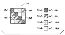
- FIG. 7 is an enlarged view of the pre-exposure area 13a.
- the pre-exposure area 13a includes four pixel areas 13a1, 13a2, 13a3, and 13a4. These pixel areas 13a1, 13a2, 13a3, and 13a4 are mosaics. Arranged in a shape.
- the pre-exposure area 13a has four pixel areas 13a1, 13a2, 13a3, and 13a4, but is not limited to this, and has at least two or more pixel areas. Just do it.
- the pixel areas 13a1, 13a2, 13a3, and 13a4 are not limited to the mosaic arrangement, and may be arranged in parallel, for example.
- the pixel areas 13a1, 13a2, 13a3, and 13a4 are set with different gains.
- the pixel area 13a1 is an area with a gain of 0db where no gain is increased, and the pixel area 13a2 is an area where the gain is increased by 6db.
- the pixel area 13a3 is an area for gain increase of 12 db, and the pixel area 13a4 is an area for gain increase of 18 db.
- the gain increase amount is not limited to the above-described 6 db, 12 db, and 18 db, and may be other gain increase amounts.
- the control unit 16 compares the photometric value acquired in each of the pixel areas 13a1 to 13a4 with the photometric value acquired in each of the pixel areas 13a1 to 13a4 by the previous pre-exposure, and the comparison result indicates which pixel area 13a1 to Whether or not 13a4 is within a predetermined threshold is determined, and the movement of the capsule endoscope 10 is detected.
- the control unit 16 determines that the capsule endoscope 10 is not moving when the comparison result is within a predetermined threshold value in any of the pixel areas 13a1 to 13a4, and if any one of the comparison results is larger than the predetermined threshold value, It is determined that the capsule endoscope 10 is moving.
- FIG. 8 is a flowchart for explaining the operation of the capsule endoscope 10 according to the second embodiment.
- the same processes as those in FIG. 6 are denoted by the same reference numerals and description thereof is omitted.
- pre-photometry is executed for each of the pixel areas 13a1 to 13a4 (step S11). It is determined whether or not the difference between the pre-photometric value of each pixel area 13a1 to 13a4 and the pre-photometric value of each previous pixel area 13a1 to 13a4 is within a predetermined threshold (step S12).
- step S4 If it is determined that the difference between the pre-photometric values of the pixel areas 13a1 to 13a4 and the pre-photometric values of the immediately preceding pixel areas 13a1 to 13a4 is within a predetermined threshold value, the determination is YES, the process returns to step S4, and the same processing is repeated. . On the other hand, if any of the differences between the pre-photometric values of the pixel areas 13a1 to 13a4 and the pre-photometric values of the immediately preceding pixel areas 13a1 to 13a4 is larger than a predetermined threshold value, the determination is NO and the process proceeds to step S7. And the target value of light control are calculated, and the exposure amount is calculated and set. Thereafter, in step S8, the main exposure is executed with the set exposure amount. In step S9, the endoscope image data acquired in step S8 is transmitted, and then the process returns to step S4 to repeat the same processing. .
- the pixel areas 13a1 to 13a4 having different gains are provided in the pre-exposure area 13a.
- the brightness amount is very small in the pixel area 13a1 with a gain of 0 db, so the photometric value becomes very small and accurate brightness information cannot be acquired.
- the pixel areas 13a2, 13a3, and 13a4 that increase the gain of 6db, 12db, and 18db in a mosaic shape sufficient photometric information (brightness information) is obtained in the pixel that has been increased in gain to 12db, for example. be able to.
- the brightness information becomes very large in the pixel areas 13a2, 13a3, and 13a4 that perform gain increase of 6 db, 12 db, and 18 db.
- the photometric value becomes very large and accurate brightness information cannot be acquired, by providing the pixel area 13a1 having a gain of 0 db, sufficient photometric information can be obtained in pixels where gain is not increased.
- the capsule endoscope 10 of the present embodiment has an effect that the dynamic range can be widened compared to the first embodiment without changing or lengthening the pre-exposure time. .
Abstract
La présente invention concerne un endoscope encapsulé 10 qui comporte un élément de capture d'image 13, une mémoire 15 pour stocker des informations de luminosité acquises par l'élément de capture d'image 13, et une unité de commande 16 pour comparer les informations de luminosité acquises par l'élément de capture d'image 13 et les informations de luminosité acquises par l'élément de capture d'image 13 avant l'acquisition des informations de luminosité et stockées dans la mémoire 15. Lorsque le résultat de comparaison est égal ou inférieur à une valeur de seuil prédéterminée, des informations de luminosité supplémentaires sont acquises au moyen de l'élément de capture d'image 13, et une comparaison supplémentaire est effectuée avec des informations de luminosité acquises par l'élément de capture d'image 13 et stockées dans la mémoire 15 avant l'acquisition des informations de luminosité acquises. Lorsque le résultat de la comparaison est supérieur à la valeur de seuil prédéterminée, une image est capturée au moyen de l'élément de capture d'image 13 et l'image capturée est stockée dans la mémoire 15; en variante, l'unité de commande 16 exécute une commande de sorte que l'image capturée soit transmise hors de l'endoscope encapsulé 10 au moyen d'une unité de transmission d'image 14 disposée au niveau de l'endoscope encapsulé 10.
Priority Applications (4)
| Application Number | Priority Date | Filing Date | Title |
|---|---|---|---|
| EP15863528.4A EP3231349A4 (fr) | 2014-11-27 | 2015-09-25 | Endoscope encapsulé et système d'endoscope encapsulé |
| JP2016520117A JP5977907B1 (ja) | 2014-11-27 | 2015-09-25 | カプセル型内視鏡及びカプセル型内視鏡システム |
| CN201580020012.3A CN106255444B (zh) | 2014-11-27 | 2015-09-25 | 胶囊型内窥镜以及胶囊型内窥镜系统 |
| US15/291,190 US9993143B2 (en) | 2014-11-27 | 2016-10-12 | Capsule endoscope and capsule endoscope system |
Applications Claiming Priority (2)
| Application Number | Priority Date | Filing Date | Title |
|---|---|---|---|
| JP2014240323 | 2014-11-27 | ||
| JP2014-240323 | 2014-11-27 |
Related Child Applications (1)
| Application Number | Title | Priority Date | Filing Date |
|---|---|---|---|
| US15/291,190 Continuation US9993143B2 (en) | 2014-11-27 | 2016-10-12 | Capsule endoscope and capsule endoscope system |
Publications (1)
| Publication Number | Publication Date |
|---|---|
| WO2016084467A1 true WO2016084467A1 (fr) | 2016-06-02 |
Family
ID=56074046
Family Applications (1)
| Application Number | Title | Priority Date | Filing Date |
|---|---|---|---|
| PCT/JP2015/077074 WO2016084467A1 (fr) | 2014-11-27 | 2015-09-25 | Endoscope encapsulé et système d'endoscope encapsulé |
Country Status (5)
| Country | Link |
|---|---|
| US (1) | US9993143B2 (fr) |
| EP (1) | EP3231349A4 (fr) |
| JP (1) | JP5977907B1 (fr) |
| CN (1) | CN106255444B (fr) |
| WO (1) | WO2016084467A1 (fr) |
Families Citing this family (4)
| Publication number | Priority date | Publication date | Assignee | Title |
|---|---|---|---|---|
| US8952312B2 (en) | 2011-05-12 | 2015-02-10 | Olive Medical Corporation | Image sensor for endoscopic use |
| CA2878512A1 (fr) * | 2012-07-26 | 2014-01-30 | Olive Medical Corporation | Systeme de camera a capteur d'image cmos monolithique a surface minimale |
| BR112015023206A2 (pt) | 2013-03-15 | 2017-08-22 | Olive Medical Corp | Sincronização de sensor de imagem sem temporizador de entrada e temporizador de transmissão de dados |
| WO2014145248A1 (fr) | 2013-03-15 | 2014-09-18 | Olive Medical Corporation | Minimisation du nombre d'entrée/de sortie et de conducteur d'un capteur d'image dans des applications endoscopes |
Citations (3)
| Publication number | Priority date | Publication date | Assignee | Title |
|---|---|---|---|---|
| JP2005020755A (ja) * | 2003-06-26 | 2005-01-20 | Given Imaging Ltd | 生体内で撮影された画像ストリームの伝送を減らすための方法、画像データを伝送するための生体内装置、および生体内画像を表示するためのシステム |
| JP2008264539A (ja) * | 2007-04-17 | 2008-11-06 | Gyrus Acmi Inc | 医療機器 |
| JP2013511320A (ja) * | 2009-11-20 | 2013-04-04 | ギブン イメージング リミテッド | 生体内デバイスの電力消費を制御するシステムおよび方法 |
Family Cites Families (9)
| Publication number | Priority date | Publication date | Assignee | Title |
|---|---|---|---|---|
| IL122602A0 (en) * | 1997-12-15 | 1998-08-16 | Tally Eitan Zeev Pearl And Co | Energy management of a video capsule |
| EP2301434B1 (fr) * | 2005-02-25 | 2013-01-23 | Olympus Corporation | Appareil insérable dans le corps |
| US7983458B2 (en) * | 2005-09-20 | 2011-07-19 | Capso Vision, Inc. | In vivo autonomous camera with on-board data storage or digital wireless transmission in regulatory approved band |
| JP4830652B2 (ja) * | 2006-06-12 | 2011-12-07 | 日産自動車株式会社 | 画像処理装置及び画像処理方法 |
| US8213698B2 (en) * | 2006-09-19 | 2012-07-03 | Capso Vision Inc. | Systems and methods for capsule camera control |
| EP2149328B1 (fr) * | 2007-05-22 | 2016-07-20 | Olympus Corporation | Dispositif médical encapsulé et système médical encapsulé |
| CN101179725A (zh) * | 2007-12-12 | 2008-05-14 | 北京中星微电子有限公司 | 一种运动检测方法与装置 |
| JP4875691B2 (ja) * | 2007-12-17 | 2012-02-15 | オリンパスメディカルシステムズ株式会社 | 撮像装置、画像表示装置、および画像表示システム |
| JP5385469B2 (ja) * | 2011-01-20 | 2014-01-08 | オリンパスメディカルシステムズ株式会社 | カプセル型内視鏡 |
-
2015
- 2015-09-25 JP JP2016520117A patent/JP5977907B1/ja active Active
- 2015-09-25 EP EP15863528.4A patent/EP3231349A4/fr not_active Withdrawn
- 2015-09-25 WO PCT/JP2015/077074 patent/WO2016084467A1/fr active Application Filing
- 2015-09-25 CN CN201580020012.3A patent/CN106255444B/zh active Active
-
2016
- 2016-10-12 US US15/291,190 patent/US9993143B2/en active Active
Patent Citations (3)
| Publication number | Priority date | Publication date | Assignee | Title |
|---|---|---|---|---|
| JP2005020755A (ja) * | 2003-06-26 | 2005-01-20 | Given Imaging Ltd | 生体内で撮影された画像ストリームの伝送を減らすための方法、画像データを伝送するための生体内装置、および生体内画像を表示するためのシステム |
| JP2008264539A (ja) * | 2007-04-17 | 2008-11-06 | Gyrus Acmi Inc | 医療機器 |
| JP2013511320A (ja) * | 2009-11-20 | 2013-04-04 | ギブン イメージング リミテッド | 生体内デバイスの電力消費を制御するシステムおよび方法 |
Non-Patent Citations (1)
| Title |
|---|
| See also references of EP3231349A4 * |
Also Published As
| Publication number | Publication date |
|---|---|
| EP3231349A4 (fr) | 2018-08-22 |
| CN106255444A (zh) | 2016-12-21 |
| JPWO2016084467A1 (ja) | 2017-04-27 |
| US9993143B2 (en) | 2018-06-12 |
| US20170027425A1 (en) | 2017-02-02 |
| EP3231349A1 (fr) | 2017-10-18 |
| JP5977907B1 (ja) | 2016-08-24 |
| CN106255444B (zh) | 2018-04-13 |
Similar Documents
| Publication | Publication Date | Title |
|---|---|---|
| JP5377888B2 (ja) | 撮像装置および被検体内画像取得装置 | |
| JP5388657B2 (ja) | 画像処理装置、画像処理装置の作動方法、およびシステム | |
| JP6177083B2 (ja) | データ受信装置、カプセル内視鏡システム、データ受信方法、及びプログラム | |
| JP5977907B1 (ja) | カプセル型内視鏡及びカプセル型内視鏡システム | |
| JP2009172287A (ja) | カプセル内視鏡、およびカプセル内視鏡の動作制御方法、並びに情報管理装置 | |
| JP2008237639A (ja) | カプセル内視鏡システム、およびカプセル内視鏡の動作制御方法 | |
| US20190090721A1 (en) | Receiving apparatus and radio wave interference determination method | |
| US8419632B2 (en) | Body-insertable apparatus having light adjustment control unit and in-vivo information acquisition system | |
| US10932648B2 (en) | Image processing apparatus, image processing method, and computer-readable recording medium | |
| WO2015182185A1 (fr) | Appareil du type vidéocapsule endoscopique | |
| US8830310B2 (en) | Capsule endoscope | |
| US10201266B2 (en) | Single image sensor control for capturing mixed mode images | |
| US20200196845A1 (en) | Capsule endoscope system, capsule endoscope, and receiving device | |
| JP2006288808A (ja) | カプセル内視鏡を備えた内視鏡システム | |
| JP2006288831A (ja) | 被検体内導入装置 | |
| JPWO2019171616A1 (ja) | 受信装置及び受信方法 | |
| JP2019201757A (ja) | カプセル型内視鏡、カプセル型内視鏡システム及びカプセル型内視鏡の送信方法 | |
| JP2016077683A (ja) | 受信装置およびカプセル型内視鏡システム | |
| JP6275344B1 (ja) | 動き判定装置、被検体内導入装置、動き判定方法及びプログラム | |
| JP5896877B2 (ja) | 調光装置 | |
| JP5815166B1 (ja) | カプセル型内視鏡装置 |
Legal Events
| Date | Code | Title | Description |
|---|---|---|---|
| ENP | Entry into the national phase |
Ref document number: 2016520117 Country of ref document: JP Kind code of ref document: A |
|
| 121 | Ep: the epo has been informed by wipo that ep was designated in this application |
Ref document number: 15863528 Country of ref document: EP Kind code of ref document: A1 |
|
| REEP | Request for entry into the european phase |
Ref document number: 2015863528 Country of ref document: EP |
|
| NENP | Non-entry into the national phase |
Ref country code: DE |