WO2015112900A1 - Antibody molecules to pd-1 and uses thereof - Google Patents
Antibody molecules to pd-1 and uses thereof Download PDFInfo
- Publication number
- WO2015112900A1 WO2015112900A1 PCT/US2015/012754 US2015012754W WO2015112900A1 WO 2015112900 A1 WO2015112900 A1 WO 2015112900A1 US 2015012754 W US2015012754 W US 2015012754W WO 2015112900 A1 WO2015112900 A1 WO 2015112900A1
- Authority
- WO
- WIPO (PCT)
- Prior art keywords
- seq
- amino acid
- acid sequence
- antibody molecule
- bap049
- Prior art date
Links
Classifications
-
- C—CHEMISTRY; METALLURGY
- C07—ORGANIC CHEMISTRY
- C07K—PEPTIDES
- C07K16/00—Immunoglobulins [IGs], e.g. monoclonal or polyclonal antibodies
- C07K16/18—Immunoglobulins [IGs], e.g. monoclonal or polyclonal antibodies against material from animals or humans
- C07K16/28—Immunoglobulins [IGs], e.g. monoclonal or polyclonal antibodies against material from animals or humans against receptors, cell surface antigens or cell surface determinants
- C07K16/2803—Immunoglobulins [IGs], e.g. monoclonal or polyclonal antibodies against material from animals or humans against receptors, cell surface antigens or cell surface determinants against the immunoglobulin superfamily
- C07K16/2818—Immunoglobulins [IGs], e.g. monoclonal or polyclonal antibodies against material from animals or humans against receptors, cell surface antigens or cell surface determinants against the immunoglobulin superfamily against CD28 or CD152
-
- A—HUMAN NECESSITIES
- A61—MEDICAL OR VETERINARY SCIENCE; HYGIENE
- A61K—PREPARATIONS FOR MEDICAL, DENTAL OR TOILETRY PURPOSES
- A61K39/00—Medicinal preparations containing antigens or antibodies
- A61K39/395—Antibodies; Immunoglobulins; Immune serum, e.g. antilymphocytic serum
- A61K39/39533—Antibodies; Immunoglobulins; Immune serum, e.g. antilymphocytic serum against materials from animals
- A61K39/3955—Antibodies; Immunoglobulins; Immune serum, e.g. antilymphocytic serum against materials from animals against proteinaceous materials, e.g. enzymes, hormones, lymphokines
-
- A—HUMAN NECESSITIES
- A61—MEDICAL OR VETERINARY SCIENCE; HYGIENE
- A61K—PREPARATIONS FOR MEDICAL, DENTAL OR TOILETRY PURPOSES
- A61K45/00—Medicinal preparations containing active ingredients not provided for in groups A61K31/00 - A61K41/00
- A61K45/06—Mixtures of active ingredients without chemical characterisation, e.g. antiphlogistics and cardiaca
-
- A—HUMAN NECESSITIES
- A61—MEDICAL OR VETERINARY SCIENCE; HYGIENE
- A61P—SPECIFIC THERAPEUTIC ACTIVITY OF CHEMICAL COMPOUNDS OR MEDICINAL PREPARATIONS
- A61P1/00—Drugs for disorders of the alimentary tract or the digestive system
- A61P1/04—Drugs for disorders of the alimentary tract or the digestive system for ulcers, gastritis or reflux esophagitis, e.g. antacids, inhibitors of acid secretion, mucosal protectants
-
- A—HUMAN NECESSITIES
- A61—MEDICAL OR VETERINARY SCIENCE; HYGIENE
- A61P—SPECIFIC THERAPEUTIC ACTIVITY OF CHEMICAL COMPOUNDS OR MEDICINAL PREPARATIONS
- A61P1/00—Drugs for disorders of the alimentary tract or the digestive system
- A61P1/16—Drugs for disorders of the alimentary tract or the digestive system for liver or gallbladder disorders, e.g. hepatoprotective agents, cholagogues, litholytics
-
- A—HUMAN NECESSITIES
- A61—MEDICAL OR VETERINARY SCIENCE; HYGIENE
- A61P—SPECIFIC THERAPEUTIC ACTIVITY OF CHEMICAL COMPOUNDS OR MEDICINAL PREPARATIONS
- A61P1/00—Drugs for disorders of the alimentary tract or the digestive system
- A61P1/18—Drugs for disorders of the alimentary tract or the digestive system for pancreatic disorders, e.g. pancreatic enzymes
-
- A—HUMAN NECESSITIES
- A61—MEDICAL OR VETERINARY SCIENCE; HYGIENE
- A61P—SPECIFIC THERAPEUTIC ACTIVITY OF CHEMICAL COMPOUNDS OR MEDICINAL PREPARATIONS
- A61P11/00—Drugs for disorders of the respiratory system
-
- A—HUMAN NECESSITIES
- A61—MEDICAL OR VETERINARY SCIENCE; HYGIENE
- A61P—SPECIFIC THERAPEUTIC ACTIVITY OF CHEMICAL COMPOUNDS OR MEDICINAL PREPARATIONS
- A61P13/00—Drugs for disorders of the urinary system
- A61P13/08—Drugs for disorders of the urinary system of the prostate
-
- A—HUMAN NECESSITIES
- A61—MEDICAL OR VETERINARY SCIENCE; HYGIENE
- A61P—SPECIFIC THERAPEUTIC ACTIVITY OF CHEMICAL COMPOUNDS OR MEDICINAL PREPARATIONS
- A61P13/00—Drugs for disorders of the urinary system
- A61P13/12—Drugs for disorders of the urinary system of the kidneys
-
- A—HUMAN NECESSITIES
- A61—MEDICAL OR VETERINARY SCIENCE; HYGIENE
- A61P—SPECIFIC THERAPEUTIC ACTIVITY OF CHEMICAL COMPOUNDS OR MEDICINAL PREPARATIONS
- A61P15/00—Drugs for genital or sexual disorders; Contraceptives
-
- A—HUMAN NECESSITIES
- A61—MEDICAL OR VETERINARY SCIENCE; HYGIENE
- A61P—SPECIFIC THERAPEUTIC ACTIVITY OF CHEMICAL COMPOUNDS OR MEDICINAL PREPARATIONS
- A61P17/00—Drugs for dermatological disorders
-
- A—HUMAN NECESSITIES
- A61—MEDICAL OR VETERINARY SCIENCE; HYGIENE
- A61P—SPECIFIC THERAPEUTIC ACTIVITY OF CHEMICAL COMPOUNDS OR MEDICINAL PREPARATIONS
- A61P31/00—Antiinfectives, i.e. antibiotics, antiseptics, chemotherapeutics
-
- A—HUMAN NECESSITIES
- A61—MEDICAL OR VETERINARY SCIENCE; HYGIENE
- A61P—SPECIFIC THERAPEUTIC ACTIVITY OF CHEMICAL COMPOUNDS OR MEDICINAL PREPARATIONS
- A61P35/00—Antineoplastic agents
-
- A—HUMAN NECESSITIES
- A61—MEDICAL OR VETERINARY SCIENCE; HYGIENE
- A61P—SPECIFIC THERAPEUTIC ACTIVITY OF CHEMICAL COMPOUNDS OR MEDICINAL PREPARATIONS
- A61P35/00—Antineoplastic agents
- A61P35/02—Antineoplastic agents specific for leukemia
-
- A—HUMAN NECESSITIES
- A61—MEDICAL OR VETERINARY SCIENCE; HYGIENE
- A61P—SPECIFIC THERAPEUTIC ACTIVITY OF CHEMICAL COMPOUNDS OR MEDICINAL PREPARATIONS
- A61P35/00—Antineoplastic agents
- A61P35/04—Antineoplastic agents specific for metastasis
-
- A—HUMAN NECESSITIES
- A61—MEDICAL OR VETERINARY SCIENCE; HYGIENE
- A61P—SPECIFIC THERAPEUTIC ACTIVITY OF CHEMICAL COMPOUNDS OR MEDICINAL PREPARATIONS
- A61P37/00—Drugs for immunological or allergic disorders
- A61P37/02—Immunomodulators
-
- A—HUMAN NECESSITIES
- A61—MEDICAL OR VETERINARY SCIENCE; HYGIENE
- A61P—SPECIFIC THERAPEUTIC ACTIVITY OF CHEMICAL COMPOUNDS OR MEDICINAL PREPARATIONS
- A61P37/00—Drugs for immunological or allergic disorders
- A61P37/02—Immunomodulators
- A61P37/04—Immunostimulants
-
- A—HUMAN NECESSITIES
- A61—MEDICAL OR VETERINARY SCIENCE; HYGIENE
- A61P—SPECIFIC THERAPEUTIC ACTIVITY OF CHEMICAL COMPOUNDS OR MEDICINAL PREPARATIONS
- A61P43/00—Drugs for specific purposes, not provided for in groups A61P1/00-A61P41/00
-
- A—HUMAN NECESSITIES
- A61—MEDICAL OR VETERINARY SCIENCE; HYGIENE
- A61P—SPECIFIC THERAPEUTIC ACTIVITY OF CHEMICAL COMPOUNDS OR MEDICINAL PREPARATIONS
- A61P5/00—Drugs for disorders of the endocrine system
- A61P5/14—Drugs for disorders of the endocrine system of the thyroid hormones, e.g. T3, T4
-
- A—HUMAN NECESSITIES
- A61—MEDICAL OR VETERINARY SCIENCE; HYGIENE
- A61P—SPECIFIC THERAPEUTIC ACTIVITY OF CHEMICAL COMPOUNDS OR MEDICINAL PREPARATIONS
- A61P7/00—Drugs for disorders of the blood or the extracellular fluid
-
- C—CHEMISTRY; METALLURGY
- C07—ORGANIC CHEMISTRY
- C07K—PEPTIDES
- C07K16/00—Immunoglobulins [IGs], e.g. monoclonal or polyclonal antibodies
- C07K16/18—Immunoglobulins [IGs], e.g. monoclonal or polyclonal antibodies against material from animals or humans
- C07K16/28—Immunoglobulins [IGs], e.g. monoclonal or polyclonal antibodies against material from animals or humans against receptors, cell surface antigens or cell surface determinants
- C07K16/2896—Immunoglobulins [IGs], e.g. monoclonal or polyclonal antibodies against material from animals or humans against receptors, cell surface antigens or cell surface determinants against molecules with a "CD"-designation, not provided for elsewhere
-
- C—CHEMISTRY; METALLURGY
- C07—ORGANIC CHEMISTRY
- C07K—PEPTIDES
- C07K16/00—Immunoglobulins [IGs], e.g. monoclonal or polyclonal antibodies
- C07K16/18—Immunoglobulins [IGs], e.g. monoclonal or polyclonal antibodies against material from animals or humans
- C07K16/28—Immunoglobulins [IGs], e.g. monoclonal or polyclonal antibodies against material from animals or humans against receptors, cell surface antigens or cell surface determinants
- C07K16/30—Immunoglobulins [IGs], e.g. monoclonal or polyclonal antibodies against material from animals or humans against receptors, cell surface antigens or cell surface determinants from tumour cells
-
- G—PHYSICS
- G01—MEASURING; TESTING
- G01N—INVESTIGATING OR ANALYSING MATERIALS BY DETERMINING THEIR CHEMICAL OR PHYSICAL PROPERTIES
- G01N33/00—Investigating or analysing materials by specific methods not covered by groups G01N1/00 - G01N31/00
- G01N33/48—Biological material, e.g. blood, urine; Haemocytometers
- G01N33/50—Chemical analysis of biological material, e.g. blood, urine; Testing involving biospecific ligand binding methods; Immunological testing
- G01N33/53—Immunoassay; Biospecific binding assay; Materials therefor
- G01N33/566—Immunoassay; Biospecific binding assay; Materials therefor using specific carrier or receptor proteins as ligand binding reagents where possible specific carrier or receptor proteins are classified with their target compounds
-
- G—PHYSICS
- G01—MEASURING; TESTING
- G01N—INVESTIGATING OR ANALYSING MATERIALS BY DETERMINING THEIR CHEMICAL OR PHYSICAL PROPERTIES
- G01N33/00—Investigating or analysing materials by specific methods not covered by groups G01N1/00 - G01N31/00
- G01N33/48—Biological material, e.g. blood, urine; Haemocytometers
- G01N33/50—Chemical analysis of biological material, e.g. blood, urine; Testing involving biospecific ligand binding methods; Immunological testing
- G01N33/53—Immunoassay; Biospecific binding assay; Materials therefor
- G01N33/574—Immunoassay; Biospecific binding assay; Materials therefor for cancer
- G01N33/57484—Immunoassay; Biospecific binding assay; Materials therefor for cancer involving compounds serving as markers for tumor, cancer, neoplasia, e.g. cellular determinants, receptors, heat shock/stress proteins, A-protein, oligosaccharides, metabolites
-
- G—PHYSICS
- G01—MEASURING; TESTING
- G01N—INVESTIGATING OR ANALYSING MATERIALS BY DETERMINING THEIR CHEMICAL OR PHYSICAL PROPERTIES
- G01N33/00—Investigating or analysing materials by specific methods not covered by groups G01N1/00 - G01N31/00
- G01N33/48—Biological material, e.g. blood, urine; Haemocytometers
- G01N33/50—Chemical analysis of biological material, e.g. blood, urine; Testing involving biospecific ligand binding methods; Immunological testing
- G01N33/68—Chemical analysis of biological material, e.g. blood, urine; Testing involving biospecific ligand binding methods; Immunological testing involving proteins, peptides or amino acids
- G01N33/6854—Immunoglobulins
-
- C—CHEMISTRY; METALLURGY
- C07—ORGANIC CHEMISTRY
- C07K—PEPTIDES
- C07K2317/00—Immunoglobulins specific features
- C07K2317/20—Immunoglobulins specific features characterized by taxonomic origin
- C07K2317/24—Immunoglobulins specific features characterized by taxonomic origin containing regions, domains or residues from different species, e.g. chimeric, humanized or veneered
-
- C—CHEMISTRY; METALLURGY
- C07—ORGANIC CHEMISTRY
- C07K—PEPTIDES
- C07K2317/00—Immunoglobulins specific features
- C07K2317/30—Immunoglobulins specific features characterized by aspects of specificity or valency
- C07K2317/31—Immunoglobulins specific features characterized by aspects of specificity or valency multispecific
-
- C—CHEMISTRY; METALLURGY
- C07—ORGANIC CHEMISTRY
- C07K—PEPTIDES
- C07K2317/00—Immunoglobulins specific features
- C07K2317/50—Immunoglobulins specific features characterized by immunoglobulin fragments
- C07K2317/56—Immunoglobulins specific features characterized by immunoglobulin fragments variable (Fv) region, i.e. VH and/or VL
- C07K2317/565—Complementarity determining region [CDR]
-
- C—CHEMISTRY; METALLURGY
- C07—ORGANIC CHEMISTRY
- C07K—PEPTIDES
- C07K2317/00—Immunoglobulins specific features
- C07K2317/50—Immunoglobulins specific features characterized by immunoglobulin fragments
- C07K2317/56—Immunoglobulins specific features characterized by immunoglobulin fragments variable (Fv) region, i.e. VH and/or VL
- C07K2317/569—Single domain, e.g. dAb, sdAb, VHH, VNAR or nanobody®
-
- C—CHEMISTRY; METALLURGY
- C07—ORGANIC CHEMISTRY
- C07K—PEPTIDES
- C07K2317/00—Immunoglobulins specific features
- C07K2317/70—Immunoglobulins specific features characterized by effect upon binding to a cell or to an antigen
- C07K2317/74—Inducing cell proliferation
-
- C—CHEMISTRY; METALLURGY
- C07—ORGANIC CHEMISTRY
- C07K—PEPTIDES
- C07K2317/00—Immunoglobulins specific features
- C07K2317/70—Immunoglobulins specific features characterized by effect upon binding to a cell or to an antigen
- C07K2317/76—Antagonist effect on antigen, e.g. neutralization or inhibition of binding
-
- C—CHEMISTRY; METALLURGY
- C07—ORGANIC CHEMISTRY
- C07K—PEPTIDES
- C07K2317/00—Immunoglobulins specific features
- C07K2317/90—Immunoglobulins specific features characterized by (pharmaco)kinetic aspects or by stability of the immunoglobulin
- C07K2317/92—Affinity (KD), association rate (Ka), dissociation rate (Kd) or EC50 value
-
- G—PHYSICS
- G01—MEASURING; TESTING
- G01N—INVESTIGATING OR ANALYSING MATERIALS BY DETERMINING THEIR CHEMICAL OR PHYSICAL PROPERTIES
- G01N2333/00—Assays involving biological materials from specific organisms or of a specific nature
- G01N2333/435—Assays involving biological materials from specific organisms or of a specific nature from animals; from humans
- G01N2333/705—Assays involving receptors, cell surface antigens or cell surface determinants
- G01N2333/70596—Molecules with a "CD"-designation not provided for elsewhere in G01N2333/705
-
- Y—GENERAL TAGGING OF NEW TECHNOLOGICAL DEVELOPMENTS; GENERAL TAGGING OF CROSS-SECTIONAL TECHNOLOGIES SPANNING OVER SEVERAL SECTIONS OF THE IPC; TECHNICAL SUBJECTS COVERED BY FORMER USPC CROSS-REFERENCE ART COLLECTIONS [XRACs] AND DIGESTS
- Y02—TECHNOLOGIES OR APPLICATIONS FOR MITIGATION OR ADAPTATION AGAINST CLIMATE CHANGE
- Y02A—TECHNOLOGIES FOR ADAPTATION TO CLIMATE CHANGE
- Y02A50/00—TECHNOLOGIES FOR ADAPTATION TO CLIMATE CHANGE in human health protection, e.g. against extreme weather
- Y02A50/30—Against vector-borne diseases, e.g. mosquito-borne, fly-borne, tick-borne or waterborne diseases whose impact is exacerbated by climate change
Definitions
- T cells The ability of T cells to mediate an immune response against an antigen requires two distinct signaling interactions (Viglietta, V. et al. (2007) Neurotherapeutics 4:666-675; Korman, A. J. et al. (2007) Adv. Immunol. 90:297-339).
- APC antigen-presenting cells
- TCR T cell receptor
- the immune system is tightly controlled by a network of costimulatory and co-inhibitory ligands and receptors. These molecules provide the second signal for T cell activation and provide a balanced network of positive and negative signals to maximize immune responses against infection, while limiting immunity to self (Wang, L. et al. (Epub Mar. 7, 2011) J. Exp. Med. 208(3):577-92; Lepenies, B. et al. (2008) Endocrine, Metabolic & Immune Disorders-- Drug Targets 8:279-288).
- costimulatory signals include the binding between the B7.1 (CD80) and B7.2 (CD86) ligands of the APC and the CD28 and CTLA-4 receptors of the CD4 + T-lymphocyte (Sharpe, A. H. et al. (2002) Nature Rev. Immunol. 2:116-126; Lindley, P. S.
- Binding of B7.1 or B7.2 to CD28 stimulates T cell activation, whereas binding of B7.1 or B7.2 to CTLA-4 inhibits such activation (Dong, C. et al. (2003) Immunolog. Res. 28(1):39-48; Greenwald, R. J. et al. (2005) Ann. Rev. Immunol. 23:515- 548).
- CD28 is constitutively expressed on the surface of T cells (Gross, J., et al. (1992) J.
- CTLA-4 expression is rapidly up-regulated following T-cell activation (Linsley, P. et al. (1996) Immunity 4:535-543).
- B7 Superfamily a group of related B7 molecules, also known as the "B7 Superfamily" (Coyle, A. J. et al. (2001) Nature Immunol. 2(3):203-209; Sharpe, A. H. et al. (2002) Nature Rev. Immunol. 2:116-126; Collins, M. et al. (2005) Genome Biol. 6:223.1- 223.7; Korman, A. J. et al. (2007) Adv. Immunol. 90:297-339).
- B7 Superfamily Several members of the B7 Superfamily are known, including B7.1 (CD80), B7.2 (CD86), the inducible co-stimulator ligand (ICOS-L), the programmed death-1 ligand (PD-L1; B7-H1), the programmed death-2 ligand (PD-L2; B7-DC), B7-H3, B7-H4 and B7-H6 (Collins, M. et al. (2005) Genome Biol. 6:223.1- 223.7).
- B7.1 CD80
- B7.2 the inducible co-stimulator ligand
- PD-L1 programmed death-1 ligand
- PD-L2 programmed death-2 ligand
- B7-DC B7-H3, B7-H4 and B7-H6
- the Programmed Death 1 (PD-1) protein is an inhibitory member of the extended CD28/CTLA-4 family of T cell regulators (Okazaki et al. (2002) Curr Opin Immunol 14:
- CD28 CD28
- CTLA-4 CTLA-4
- ICOS BTLA
- PD-1 is suggested to exist as a monomer, lacking the unpaired cysteine residue characteristic of other CD28 family members. PD-1 is expressed on activated B cells, T cells, and monocytes.
- the PD-1 gene encodes a 55 kDa type I transmembrane protein (Agata et al. (1996) Int Immunol. 8:765-72). Although structurally similar to CTLA-4, PD-1 lacks the MYPPY motif (SEQ ID NO: 236) that is important for B7-1 and B7-2 binding.
- Two ligands for PD-1 have been identified, PD-L1 (B7-H1) and PD-L2 (B7-DC), that have been shown to downregulate T cell activation upon binding to PD-1 (Freeman et al. (2000) J. Exp. Med. 192:1027-34; Carter et al. (2002) Eur. J. Immunol. 32:634-43).
- Both PD-L1 and PD-L2 are B7 homologs that bind to PD-1, but do not bind to other CD28 family members.
- PD-L1 is abundant in a variety of human cancers (Dong et al. (2002) Nat. Med. 8:787-9).
- PD-1 is known as an immunoinhibitory protein that negatively regulates TCR signals (Ishida, Y. et al. (1992) EMBO J. 11:3887-3895; Blank, C. et al. (Epub 2006 Dec. 29) Immunol. Immunother. 56(5):739-745).
- the interaction between PD-1 and PD-L1 can act as an immune checkpoint, which can lead to, e.g., a decrease in tumor infiltrating lymphocytes, a decrease in T- cell receptor mediated proliferation, and/or immune evasion by cancerous cells (Dong et al. (2003) J. Mol. Med. 81:281-7; Blank et al. (2005) Cancer Immunol. Immunother.
- Immune suppression can be reversed by inhibiting the local interaction of PD-1 with PD-L1 or PD-L2; the effect is additive when the interaction of PD-1 with PD-L2 is blocked as well (Iwai et al. (2002) Proc. Nat'l. Acad. Sci. USA 99:12293-7; Brown et al. (2003) J. Immunol. 170:1257-66).
- antibody molecules e.g., humanized antibody molecules
- PD-1 antibody molecules that bind to Programmed Death 1 (PD-1) with high affinity and specificity.
- the anti- PD-1 antibody molecules comprise a novel combination of framework regions (e.g., FW1, FW2, FW3 and/or FW4), e.g., novel combinations of a heavy chain framework regions and/or light chain framework regions.
- Nucleic acid molecules encoding the antibody molecules, expression vectors, host cells and methods for making the antibody molecules are also provided.
- Immunoconjugates multi- or bispecific antibody molecules and pharmaceutical compositions comprising the antibody molecules are also provided.
- the anti-PD-1 antibody molecules disclosed herein can be used (alone or in combination with other agents or therapeutic modalities) to treat, prevent and/or diagnose disorders, such as cancerous disorders (e.g., solid and soft-tissue tumors), as well as infectious diseases (e.g., chronic infectious disorders or sepsis).
- cancerous disorders e.g., solid and soft-tissue tumors
- infectious diseases e.g., chronic infectious disorders or sepsis.
- compositions and methods for detecting PD-1, as well as methods for treating various disorders including cancer and/or infectious diseases, using the anti-PD-1 antibody molecules are disclosed herein.
- the invention features an antibody molecule (e.g., an isolated or recombinant antibody molecule) having one or more of the following properties: (i) binds to PD-1, e.g., human PD-1, with high affinity, e.g., with an affinity constant of at least about 10 7 M -1 , typically about 10 8 M -1 , and more typically, about 10 9 M -1 to 10 10 M -1 or stronger;
- PD-1 e.g., human PD-1
- high affinity e.g., with an affinity constant of at least about 10 7 M -1 , typically about 10 8 M -1 , and more typically, about 10 9 M -1 to 10 10 M -1 or stronger
- PD-1 ligand e.g., PD-L1 or PD-L2, or both
- binds specifically to an epitope on PD-1 e.g., the same or similar epitope as the epitope recognized by murine monoclonal antibody BAP049 or a chimeric antibody BAP049, e.g., BAP049-chi or BAP049-chi-Y;
- (v) shows the same or similar binding affinity or specificity, or both, as any of BAP049- hum01, BAP049-hum02, BAP049-hum03, BAP049-hum04, BAP049-hum05, BAP049-hum06, BAP049-hum07, BAP049-hum08, BAP049-hum09, BAP049-hum10, BAP049-hum11,
- BAP049-Clone-A BAP049-Clone-B, BAP049-Clone-C, BAP049-Clone-D, or BAP049-Clone- E;
- (vii) shows the same or similar binding affinity or specificity, or both, as an antibody molecule (e.g., an heavy chain variable region and light chain variable region) having an amino acid sequence shown in Table 1;
- an antibody molecule e.g., an heavy chain variable region and light chain variable region having an amino acid sequence shown in Table 1;
- (viii) shows the same or similar binding affinity or specificity, or both, as an antibody molecule (e.g., an heavy chain variable region and light chain variable region) encoded by the nucleotide sequence shown in Table 1;
- an antibody molecule e.g., an heavy chain variable region and light chain variable region
- (ix) inhibits, e.g., competitively inhibits, the binding of a second antibody molecule to PD-1, wherein the second antibody molecule is an antibody molecule described herein, e.g., an antibody molecule chosen from, e.g., any of BAP049-hum01, BAP049-hum02, BAP049-hum03, BAP049-hum04, BAP049-hum05, BAP049-hum06, BAP049-hum07, BAP049-hum08,
- (xi) competes for binding, and/or binds the same epitope, with a second antibody molecule to PD-1, wherein the second antibody molecule is an antibody molecule described herein, e.g., an antibody molecule chosen from, e.g., any of BAP049-hum01, BAP049-hum02, BAP049-hum03, BAP049-hum04, BAP049-hum05, BAP049-hum06, BAP049-hum07,
- (xii) has one or more biological properties of an antibody molecule described herein, e.g., an antibody molecule chosen from, e.g., any of BAP049-hum01, BAP049-hum02, BAP049- hum03, BAP049-hum04, BAP049-hum05, BAP049-hum06, BAP049-hum07, BAP049-hum08, BAP049-hum09, BAP049-hum10, BAP049-hum11, BAP049-hum12, BAP049-hum13,
- an antibody molecule chosen from, e.g., any of BAP049-hum01, BAP049-hum02, BAP049- hum03, BAP049-hum04, BAP049-hum05, BAP049-hum06, BAP049-hum07, BAP049-hum08, BAP049-hum09, BAP049-hum10, BAP049-hum11, BAP
- (xiii) has one or more pharmacokinetic properties of an antibody molecule described herein, e.g., an antibody molecule chosen from, e.g., any of BAP049-hum01, BAP049-hum02, BAP049-hum03, BAP049-hum04, BAP049-hum05, BAP049-hum06, BAP049-hum07,
- (xiv) inhibits one or more activities of PD-1, e.g., results in one or more of: an increase in tumor infiltrating lymphocytes, an increase in T-cell receptor mediated proliferation, or a decrease in immune evasion by cancerous cells;
- (xv) binds human PD-1 and is cross-reactive with cynomolgus PD-1; (xvi) binds to one or more residues within the C strand, CC’ loop, C’ strand, or FG loop of PD-1, or a combination two, three or all of the C strand, CC’ loop, C’ strand or FG loop of PD-1, e.g., wherein the binding is assayed using ELISA or Biacore; or
- (xvii) has a VL region that contributes more to binding to PD-1 than a VH region.
- the antibody molecule binds to PD-1 with high affinity, e.g., with a K D that is about the same, or at least about 10%, 20%, 30%, 40%, 50%, 60%, 70%, 80% or 90% higher or lower than the K D of a murine or chimeric anti-PD-1 antibody molecule, e.g., a murine or chimeric anti-PD-1 antibody molecule described herein.
- the K D of the murine or chimeric anti-PD-1 antibody molecule is less than about 0.4, 0.3, 0.2, 0.1, or 0.05 nM, e.g., measured by a Biacore method.
- the K D of the murine or chimeric anti-PD-1 antibody molecule is less than about 0.2 nM, e.g., about 0.135 nM. In other embodiments, the K D of the murine or chimeric anti PD-1 antibody molecule is less than about 10, 5, 3, 2, or 1 nM, e.g., measured by binding on cells expressing PD-1 (e.g., 300.19 cells). In some embodiments, the K D of the murine or chimeric anti PD-1 antibody molecule is less than about 5 nM, e.g., about 4.60 nM (or about 0.69 ⁇ g/mL).
- the anti-PD-1 antibody molecule binds to PD-1 with a K off slower than 1° 10 -4 , 5° 10 -5 , or 1° 10 -5 s -1 , e.g., about 1.65° 10 -5 s -1 . In some embodiments, the the anti-PD-1 antibody molecule binds to PD-1 with a K on faster than 1° 10 4 , 5° 10 4 , 1° 10 5 , or 5° 10 5 M -1 s -1 , e.g., about 1.23° 10 5 M -1 s -1 .
- the expression level of the antibody molecule is higher, e.g., at least about 0.5, 1, 2, 3, 4, 5, 6, 7, 8, 9 or 10-fold higher, than the expression level of a murine or chimeric antibody molecule, e.g., a murine or chimeric anti-PD-1 antibody molecule described herein.
- the antibody molecule is expressed in CHO cells.
- the anti-PD-1 antibody molecule reduces one or more PD-1- associated activities with an IC 50 (concentration at 50% inhibition) that is about the same or lower, e.g., at least about 10%, 20%, 30%, 40%, 50%, 60%, 70%, 80% or 90% lower, than the IC 50 of a murine or chimeric anti-PD-1 antibody molecule, e.g., a murine or chimeric anti-PD-1 antibody molecule described herein.
- the IC 50 of the murine or chimeric anti-PD-1 antibody molecule is less than about 6, 5, 4, 3, 2, or 1 nM, e.g., measured by binding on cells expressing PD-1 (e.g., 300.19 cells).
- the IC 50 of the murine or chimeric anti-PD-1 antibody molecule is less than about 4 nM, e.g., about 3.40 nM (or about 0.51 ⁇ g/mL).
- the PD-1-associated activity reduced is the binding of PD- L1 and/or PD-L2 to PD-1.
- the anti-PD-1 antibody molecule binds to peripheral blood mononucleated cells (PBMCs) activated by Staphylococcal enterotoxin B (SEB).
- SEB Staphylococcal enterotoxin B
- the anti-PD-1 antibody molecule increases the expression of IL-2 on whole blood activated by SEB.
- the anti-PD-1 antibody increases the expression of IL-2 by at least about 2, 3, 4, or 5-fold, compared to the expression of IL-2 when an isotype control (e.g., IgG4) is used.
- the anti-PD-1 antibody molecule has improved stability, e.g., at least about 0.5, 1, 2, 3, 4, 5, 6, 7, 8, 9 or 10-fold more stable in vivo or in vitro, than a murine or chimeric anti-PD-1 antibody molecule, e.g., a murine or chimeric anti-PD-1 antibody molecule described herein.
- the anti PD-1 antibody molecule is a humanized antibody molecule and has a risk score based on T cell epitope analysis of 300 to 700, 400 to 650, 450 to 600, or a risk score as described herein.
- the anti-PD-1 antibody molecule comprises at least one antigen- binding region, e.g., a variable region or an antigen-binding fragment thereof, from an antibody described herein, e.g., an antibody chosen from any of BAP049-hum01, BAP049-hum02, BAP049-hum03, BAP049-hum04, BAP049-hum05, BAP049-hum06, BAP049-hum07,
- the anti-PD-1 antibody molecule comprises at least one, two, three or four variable regions from an antibody described herein, e.g., an antibody chosen from any of BAP049-hum01, BAP049-hum02, BAP049-hum03, BAP049-hum04, BAP049-hum05, BAP049-hum06, BAP049-hum07, BAP049-hum08, BAP049-hum09, BAP049-hum10,
- BAP049-hum16 BAP049-Clone-A, BAP049-Clone-B, BAP049-Clone-C, BAP049-Clone-D, or BAP049-Clone-E; or as described in Table 1, or encoded by the nucleotide sequence in Table 1; or a sequence substantially identical (e.g., at least 80%, 85%, 90%, 92%, 95%, 97%, 98%, 99% or higher identical) to any of the aforesaid sequences.
- the anti-PD-1 antibody molecule comprises at least one or two heavy chain variable regions from an antibody described herein, e.g., an antibody chosen from any of BAP049-hum01, BAP049-hum02, BAP049-hum03, BAP049-hum04, BAP049- hum05, BAP049-hum06, BAP049-hum07, BAP049-hum08, BAP049-hum09, BAP049-hum10, BAP049-hum11, BAP049-hum12, BAP049-hum13, BAP049-hum14, BAP049-hum15,
- an antibody chosen from any of BAP049-hum01, BAP049-hum02, BAP049-hum03, BAP049-hum04, BAP049- hum05, BAP049-hum06, BAP049-hum07, BAP049-hum08, BAP049-hum09, BAP049-hum10, BAP049-hum11
- BAP049-hum16 BAP049-Clone-A, BAP049-Clone-B, BAP049-Clone-C, BAP049-Clone-D, or BAP049-Clone-E; or as described in Table 1, or encoded by the nucleotide sequence in Table 1; or a sequence substantially identical (e.g., at least 80%, 85%, 90%, 92%, 95%, 97%, 98%, 99% or higher identical) to any of the aforesaid sequences.
- the anti-PD-1 antibody molecule comprises at least one or two light chain variable regions from an antibody described herein, e.g., an antibody chosen from any of BAP049-hum01, BAP049-hum02, BAP049-hum03, BAP049-hum04, BAP049-hum05, BAP049-hum06, BAP049-hum07, BAP049-hum08, BAP049-hum09, BAP049-hum10,
- BAP049-hum16 BAP049-Clone-A, BAP049-Clone-B, BAP049-Clone-C, BAP049-Clone-D, or BAP049-Clone-E; or as described in Table 1, or encoded by the nucleotide sequence in Table 1; or a sequence substantially identical (e.g., at least 80%, 85%, 90%, 92%, 95%, 97%, 98%, 99% or higher identical) to any of the aforesaid sequences.
- the anti-PD-1 antibody molecule includes a heavy chain constant region for an IgG4, e.g., a human IgG4.
- the human IgG4 includes a substitution at position 228 according to EU numbering (e.g., a Ser to Pro substitution).
- the anti-PD-1 antibody molecule includes a heavy chain constant region for an IgG1, e.g., a human IgG1.
- the human IgG1 includes a substitution at position 297 according to EU numbering (e.g., an Asn to Ala substitution).
- the human IgG1 includes a substitution at position 265 according to EU numbering, a
- the human IgG1 includes a substitution at position 234 according to EU numbering, a substitution at position 235 according to EU numbering, or both (e.g., a Leu to Ala substitution at position 234 and/or a Leu to Ala substitution at position 235).
- the heavy chain constant region comprises an amino sequence set forth in Table 3, or a sequence substantially identical (e.g., at least 80%, 85%, 90%, 92%, 95%, 97%, 98%, 99% or higher identical) thereto.
- the anti-PD-1 antibody molecule includes a kappa light chain constant region, e.g., a human kappa light chain constant region.
- the light chain constant region comprises an amino sequence set forth in Table 3, or a sequence substantially identical (e.g., at least 80%, 85%, 90%, 92%, 95%, 97%, 98%, 99% or higher identical) thereto.
- the anti-PD-1 antibody molecule includes a heavy chain constant region for an IgG4, e.g., a human IgG4, and a kappa light chain constant region, e.g., a human kappa light chain constant region, e.g., a heavy and light chain constant region comprising an amino sequence set forth in Table 3, or a sequence substantially identical (e.g., at least 80%, 85%, 90%, 92%, 95%, 97%, 98%, 99% or higher identical) thereto.
- the human IgG4 includes a substitution at position 228 according to EU numbering (e.g., a Ser to Pro substitution).
- the anti-PD-1 antibody molecule includes a heavy chain constant region for an IgG1, e.g., a human IgG1, and a kappa light chain constant region, e.g., a human kappa light chain constant region, e.g., a heavy and light chain constant region comprising an amino sequence set forth in Table 3, or a sequence substantially identical (e.g., at least 80%, 85%, 90%, 92%, 95%, 97%, 98%, 99% or higher identical) thereto.
- the human IgG1 includes a substitution at position 297 according to EU numbering (e.g., an Asn to Ala substitution).
- the human IgG1 includes a substitution at position 265 according to EU numbering, a substitution at position 329 according to EU numbering, or both (e.g., an Asp to Ala substitution at position 265 and/or a Pro to Ala substitution at position 329).
- the human IgG1 includes a substitution at position 234 according to EU numbering, a substitution at position 235 according to EU numbering, or both (e.g., a Leu to Ala substitution at position 234 and/or a Leu to Ala substitution at position 235).
- the anti-PD-1 antibody molecule includes a heavy chain variable domain and a constant region, a light chain variable domain and a constant region, or both, comprising the amino acid sequence of BAP049-Clone-A, BAP049-Clone-B, BAP049-Clone-C, BAP049-Clone-D, or BAP049-Clone-E; or as described in Table 1, or encoded by the nucleotide sequence in Table 1; or a sequence substantially identical (e.g., at least 80%, 85%, 90%, 92%, 95%, 97%, 98%, 99% or higher identical) to any of the aforesaid sequences.
- the anti-PD-1 antibody molecule optionally, comprises a leader sequence from a heavy chain, a light chain, or both, as showin in Table 4; or a sequence substantially identical thereto.
- the anti-PD-1 antibody molecule includes at least one, two, or three complementarity determining regions (CDRs) from a heavy chain variable region of an antibody described herein, e.g., an antibody chosen from any of BAP049-hum01, BAP049- hum02, BAP049-hum03, BAP049-hum04, BAP049-hum05, BAP049-hum06, BAP049-hum07, BAP049-hum08, BAP049-hum09, BAP049-hum10, BAP049-hum11, BAP049-hum12,
- CDRs complementarity determining regions
- the anti-PD-1 antibody molecule includes at least one, two, or three CDRs (or collectively all of the CDRs) from a heavy chain variable region comprising an amino acid sequence shown in Table 1, or encoded by a nucleotide sequence shown in Table 1.
- one or more of the CDRs (or collectively all of the CDRs) have one, two, three, four, five, six or more changes, e.g., amino acid substitutions or deletions, relative to the amino acid sequence shown in Table 1, or encoded by a nucleotide sequence shown in Table 1.
- the anti-PD-1 antibody molecule includes at least one, two, or three CDRs from a light chain variable region of an antibody described herein, e.g., an antibody chosen from any of BAP049-hum01, BAP049-hum02, BAP049-hum03, BAP049- hum04, BAP049-hum05, BAP049-hum06, BAP049-hum07, BAP049-hum08, BAP049-hum09, BAP049-hum10, BAP049-hum11, BAP049-hum12, BAP049-hum13, BAP049-hum14,
- the anti-PD-1 antibody molecule includes at least one, two, or three CDRs (or collectively all of the CDRs) from a light chain variable region comprising an amino acid sequence shown in Table 1, or encoded by a nucleotide sequence shown in Table 1.
- one or more of the CDRs (or collectively all of the CDRs) have one, two, three, four, five, six or more changes, e.g., amino acid substitutions or deletions, relative to the amino acid sequence shown in Table 1, or encoded by a nucleotide sequence shown in Table 1.
- the anti-PD-1 antibody molecule includes a substitution in a light chain CDR, e.g., one or more substitutions in a CDR1, CDR2 and/or CDR3 of the light chain.
- the anti-PD-1 antibody molecule includes a substitution in the light chain CDR3 at position 102 of the light variable region, e.g., a substitution of a cysteine to tyrosine, or a cysteine to serine residue, at position 102 of the light variable region according to Table 1 (e.g., SEQ ID NO: 16 or 24 for murine or chimeric, unmodified; or any of SEQ ID NOs: 34, 42, 46, 54, 58, 62, 66, 70, 74, or 78 for a modified sequence).
- Table 1 e.g., SEQ ID NO: 16 or 24 for murine or chimeric, unmodified; or any of SEQ ID NOs: 34, 42, 46, 54, 58, 62, 66, 70, 74
- the anti-PD-1 antibody molecule includes at least one, two, three, four, five or six CDRs (or collectively all of the CDRs) from a heavy and light chain variable region comprising an amino acid sequence shown in Table 1, or encoded by a nucleotide sequence shown in Table 1.
- one or more of the CDRs (or collectively all of the CDRs) have one, two, three, four, five, six or more changes, e.g., amino acid substitutions or deletions, relative to the amino acid sequence shown in Table 1, or encoded by a nucleotide sequence shown in Table 1.
- the anti-PD-1 antibody molecule includes all six CDRs from an antibody described herein, e.g., an antibody chosen from any of BAP049-hum01, BAP049- hum02, BAP049-hum03, BAP049-hum04, BAP049-hum05, BAP049-hum06, BAP049-hum07, BAP049-hum08, BAP049-hum09, BAP049-hum10, BAP049-hum11, BAP049-hum12,
- the anti-PD-1 antibody molecule may include any CDR described herein.
- the anti-PD-1 antibody molecule includes a substitution in a light chain CDR, e.g., one or more substitutions in a CDR1, CDR2 and/or CDR3 of the light chain.
- the anti-PD-1 antibody molecule includes a substitution in the light chain CDR3 at position 102 of the light variable region, e.g., a substitution of a cysteine to tyrosine, or a cysteine to serine residue, at position 102 of the light variable region according to Table 1 (e.g., SEQ ID NO: 16 or 24 for murine or chimeric, unmodified; or any of SEQ ID NOs: 34, 42, 46, 54, 58, 62, 66, 70, 74, or 78 for a modified sequence).
- Table 1 e.g., SEQ ID NO: 16 or 24 for murine or chimeric, unmodified; or any of SEQ ID NOs: 34, 42, 46, 54, 58, 62, 66, 70, 74, or 78 for a modified sequence.
- the anti-PD-1 antibody molecule includes at least one, two, or three CDRs according to Kabat et al. (e.g., at least one, two, or three CDRs according to the Kabat definition as set out in Table 1) from a heavy chain variable region of an antibody described herein, e.g., an antibody chosen from any of BAP049-hum01, BAP049-hum02, BAP049-hum03, BAP049-hum04, BAP049-hum05, BAP049-hum06, BAP049-hum07,
- the anti-PD-1 antibody molecule includes at least one, two, or three CDRs according to Kabat et al. (e.g., at least one, two, or three CDRs according to the Kabat definition as set out in Table 1) from a light chain variable region of an antibody described herein, e.g., an antibody chosen from any of BAP049-hum01, BAP049-hum02, BAP049-hum03, BAP049-hum04, BAP049-hum05, BAP049-hum06, BAP049-hum07, BAP049-hum08,
- the anti-PD-1 antibody molecule includes at least one, two, three, four, five, or six CDRs according to Kabat et al. (e.g., at least one, two, three, four, five, or six CDRs according to the Kabat definition as set out in Table 1) from the heavy and light chain variable regions of an antibody described herein, e.g., an antibody chosen from any of BAP049- hum01, BAP049-hum02, BAP049-hum03, BAP049-hum04, BAP049-hum05, BAP049-hum06, BAP049-hum07, BAP049-hum08, BAP049-hum09, BAP049-hum10, BAP049-hum11,
- the anti-PD-1 antibody molecule includes all six CDRs according to Kabat et al. (e.g., all six CDRs according to the Kabat definition as set out in Table 1) from the heavy and light chain variable regions of an antibody described herein, e.g., an antibody chosen from any of BAP049-hum01, BAP049-hum02, BAP049-hum03, BAP049- hum04, BAP049-hum05, BAP049-hum06, BAP049-hum07, BAP049-hum08, BAP049-hum09, BAP049-hum10, BAP049-hum11, BAP049-hum12, BAP049-hum13, BAP049-hum14,
- the anti-PD-1 antibody molecule may include any CDR described herein.
- the anti-PD-1 antibody molecule includes at least one, two, or three Chothia hypervariable loops (e.g., at least one, two, or three hypervariable loops according to the Chothia definition as set out in Table 1) from a heavy chain variable region of an antibody described herein, e.g., an antibody chosen from any of BAP049-hum01, BAP049-hum02, BAP049-hum03, BAP049-hum04, BAP049-hum05, BAP049-hum06, BAP049-hum07,
- alterations e.g., substitutions, deletions, or insertions, e.g., conservative substitutions
- the anti-PD-1 antibody molecule includes at least one, two, or three Chothia hypervariable loops (e.g., at least one, two, or three hypervairalbe loops according to the Chothia definition as set out in Table 1) of a light chain variable region of an antibody described herein, e.g., an antibody chosen from any of BAP049-hum01, BAP049-hum02, BAP049-hum03, BAP049-hum04, BAP049-hum05, BAP049-hum06, BAP049-hum07,
- alterations e.g., substitutions, deletions, or insertions, e.g., conservative substitutions
- the anti-PD-1 antibody molecule includes at least one, two, three, four, five, or six hypervariable loops (e.g., at least one, two, three, four, five, or six hypervariable loops according to the Chothia definition as set out in Table 1) from the heavy and light chain variable regions of an antibody described herein, e.g., an antibody chosen from any of BAP049-hum01, BAP049-hum02, BAP049-hum03, BAP049-hum04, BAP049-hum05,
- BAP049-hum16 BAP049-Clone-A, BAP049-Clone-B, BAP049-Clone-C, BAP049-Clone-D, or BAP049-Clone-E; or as described in Table 1, or encoded by the nucleotide sequence in Table 1; or at least the amino acids from those hypervariable loops that contact PD-1; or which have at least one amino acid alteration, but not more than two, three or four alterations (e.g.,
- substitutions, deletions, or insertions e.g., conservative substitutions
- substitutions, deletions, or insertions e.g., conservative substitutions
- the anti-PD-1 antibody molecule includes all six hypervariable loops (e.g., all six hypervariable loops according to the Chothia definition as set out in Table 1) of an antibody described herein, e.g., an antibody chosen from any of BAP049-hum01, BAP049- hum02, BAP049-hum03, BAP049-hum04, BAP049-hum05, BAP049-hum06, BAP049-hum07, BAP049-hum08, BAP049-hum09, BAP049-hum10, BAP049-hum11, BAP049-hum12,
- an antibody chosen from any of BAP049-hum01, BAP049- hum02, BAP049-hum03, BAP049-hum04, BAP049-hum05, BAP049-hum06, BAP049-hum07, BAP049-hum08, BAP049-hum09, BAP049-hum10, BAP049-hum11
- the anti-PD-1 antibody molecule may include any hypervariable loop described herein.
- the anti-PD-1 antibody molecule includes at least one, two, or three hypervariable loops that have the same canonical structures as the corresponding hypervariable loop of an antibody described herein, e.g., an antibody chosen from any of BAP049-hum01, BAP049-hum02, BAP049-hum03, BAP049-hum04, BAP049-hum05,
- BAP049-hum16, BAP049-Clone-A, BAP049-Clone-B, BAP049-Clone-C, BAP049-Clone-D, or BAP049-Clone-E e.g., the same canonical structures as at least loop 1 and/or loop 2 of the heavy and/or light chain variable domains of an antibody described herein. See, e.g., Chothia et al., (1992) J. Mol. Biol. 227:799-817; Tomlinson et al., (1992) J. Mol. Biol. 227:776-798 for descriptions of hypervariable loop canonical structures. These structures can be determined by inspection of the tables described in these references.
- the anti-PD-1 antibody molecule includes a combination of CDRs or hypervariable loops defined according to the Kabat et al. and Chothia et al.
- the anti-PD-1 antibody molecule includes at least one, two or three CDRs or hypervariable loops from a heavy chain variable region of an antibody described herein, e.g., an antibody chosen from any of BAP049-hum01, BAP049-hum02, BAP049-hum03, BAP049-hum04, BAP049-hum05, BAP049-hum06, BAP049-hum07, BAP049-hum08,
- the anti-PD-1 antibody molecule can include VH CDR1 according to Kabat et al. or VH hypervariable loop 1 according to Chothia et al., or a combination thereof, e.g., as shown in Table 1.
- the combination of Kabat and Chothia CDR of VH CDR1 comprises the amino acid sequence GYTFTTYWMH (SEQ ID NO: 224), or an amino acid sequence substantially identical thereto (e.g., having at least one amino acid alteration, but not more than two, three or four alterations (e.g., substitutions, deletions, or insertions, e.g., conservative substitutions)).
- the anti-PD-1 antibody molecule can further include, e.g., VH CDRs 2-3 according to Kabat et al. and VL CDRs 1-3 according to Kabat et al., e.g., as shown in Table 1. Accordingly, in some embodiments, framework regions are defined based on a combination of CDRs defined according to Kabat et al. and hypervariable loops defined according to Chothia et al.
- the anti-PD-1 antibody molecule can include VH FR1 defined based on VH hypervariable loop 1 according to Chothia et al. and VH FR2 defined based on VH CDRs 1-2 according to Kabat et al., e.g., as shown in Table 1.
- the anti-PD-1 antibody molecule can further include, e.g., VH FRs 3-4 defined based on VH CDRs 2-3 according to Kabat et al. and VL FRs 1-4 defined based on VL CDRs 1-3 according to Kabat et al.
- the anti-PD-1 antibody molecule can contain any combination of CDRs or hypervariable loops according to the Kabat and Chothia definitions.
- the anti-PD-1 antibody molecule includes at least one, two or three CDRs from a light chain variable region of an antibody described herein, e.g., an antibody chosen from any of BAP049-hum01, BAP049- hum02, BAP049-hum03, BAP049-hum04, BAP049-hum05, BAP049-hum06, BAP049-hum07, BAP049-hum08, BAP049-hum09, BAP049-hum10, BAP049-hum11, BAP049-hum12,
- the antibody molecule is a monospecific antibody molecule, a bispecific antibody molecule, or is an antibody molecule that comprises an antigen binding fragment of an antibody, e.g., a half antibody or antigen binding fragment of a half antibody.
- the antibody molecule is a bispecific antibody molecule having a first binding specificity for PD-1 and a second binding specificity for TIM-3, LAG-3, CEACAM (e.g., CEACAM-1 and/or CEACAM-5), PD-L1 or PD- L2.
- the anti-PD-1 antibody molecule includes:
- VH heavy chain variable region
- VL light chain variable region
- VH comprising a VHCDR1 amino acid sequence chosen from SEQ ID NO: 1; a VHCDR2 amino acid sequence of SEQ ID NO: 2; and a VHCDR3 amino acid sequence of SEQ ID NO: 3; and a VL comprising a VLCDR1 amino acid sequence of SEQ ID NO: 10, a
- the anti-PD-1 antibody molecule comprises a VH comprising a VHCDR1 amino acid sequence of SEQ ID NO: 4, a VHCDR2 amino acid sequence of SEQ ID NO: 5, and a VHCDR3 amino acid sequence of SEQ ID NO: 3; and a VL comprising a
- VLCDR1 amino acid sequence of SEQ ID NO: 13 a VLCDR2 amino acid sequence of SEQ ID NO: 14, and a VLCDR3 amino acid sequence of SEQ ID NO: 33.
- the anti-PD-1 antibody molecule comprises a VH comprising a VHCDR1 amino acid sequence of SEQ ID NO: 1 ; a VHCDR2 amino acid sequence of SEQ ID NO: 2; and a VHCDR3 amino acid sequence of SEQ ID NO: 3; and a VL comprising a
- VLCDR1 amino acid sequence of SEQ ID NO: 10 a VLCDR2 amino acid sequence of SEQ ID NO: 11, and a VLCDR3 amino acid sequence of SEQ ID NO: 32.
- the anti-PD-1 antibody molecule comprises a VH comprising a VHCDR1 amino acid sequence of SEQ ID NO: 224, a VHCDR2 amino acid sequence of SEQ ID NO: 5, and a VHCDR3 amino acid sequence of SEQ ID NO: 3; and a VL comprising a VLCDR1 amino acid sequence of SEQ ID NO: 13, a VLCDR2 amino acid sequence of SEQ ID NO: 14, and a VLCDR3 amino acid sequence of SEQ ID NO: 33.
- the anti-PD-1 antibody molecule comprises a VH comprising a VHCDR1 amino acid sequence of SEQ ID NO: 224; a VHCDR2 amino acid sequence of SEQ ID NO: 2; and a VHCDR3 amino acid sequence of SEQ ID NO: 3; and a VL comprising a VLCDR1 amino acid sequence of SEQ ID NO: 10, a VLCDR2 amino acid sequence of SEQ ID NO: 11, and a VLCDR3 amino acid sequence of SEQ ID NO: 32.
- the antibody molecule is a humanized antibody molecule. In another embodiment, the antibody molecule is a monospecific antibody molecule. In yet another embodiment, the antibody molecule is a bispecific antibody molecule. In one embodiment, the anti-PD-1 antibody molecule includes:
- VH heavy chain variable region
- VL light chain variable region
- the anti-PD-1 antibody molecule includes:
- VH heavy chain variable region
- VL light chain variable region
- the anti-PD-1 antibody molecule comprises the VHCDR1 amino acid sequence of SEQ ID NO: 1. In another embodiment, the anti-PD-1 antibody molecule comprises the VHCDR1 amino acid sequence of SEQ ID NO: 4. In yet another embodiment, the anti-PD-1 antibody molecule comprises the VHCDR1 amino acid sequence of SEQ ID NO: 224.
- the light or the heavy chain variable framework (e.g., the region encompassing at least FR1, FR2, FR3, and optionally FR4) of the anti-PD-1 antibody molecule can be chosen from: (a) a light or heavy chain variable framework including at least 80%, 85%, 87% 90%, 92%, 93%, 95%, 97%, 98%, or preferably 100% of the amino acid residues from a human light or heavy chain variable framework, e.g., a light or heavy chain variable framework residue from a human mature antibody, a human germline sequence, or a human consensus sequence; (b) a light or heavy chain variable framework including from 20% to 80%, 40% to 60%, 60% to 90%, or 70% to 95% of the amino acid residues from a human light or heavy chain variable framework, e.g., a light or heavy chain variable framework residue from a human mature antibody, a human germline sequence, or a human consensus sequence; (c) a non-human framework (e.g., a rodent framework); or
- the light or heavy chain variable framework region (particularly FR1, FR2 and/or FR3) includes a light or heavy chain variable framework sequence at least 70, 75, 80, 85, 87, 88, 90, 92, 94, 95, 96, 97, 98, 99% identical or identical to the frameworks of a VL or VH segment of a human germline gene.
- the anti-PD-1 antibody molecule comprises a heavy chain variable domain having at least one, two, three, four, five, six, seven, ten, fifteen, twenty or more changes, e.g., amino acid substitutions or deletions, from an amino acid sequence of BAP049- chi-HC, e.g., the amino acid sequence of the FR region in the entire variable region, e.g., shown in FIGs. 9A-9B, or SEQ ID NO: 18, 20, 22 or 30.
- the anti-PD-1 antibody molecule comprises a heavy chain variable domain having one or more of: E at position 1, V at position 5, A at position 9, V at position 11, K at position 12, K at position 13, E at position 16, L at position 18, R at position 19, I or V at position 20, G at position 24, I at position 37, A or S at position 40, T at position 41, S at position 42, R at position 43, M or L at position 48, V or F at position 68, T at position 69, I at position 70, S at position 71, A or R at position 72, K or N at position 74, T or K at position 76, S or N at position 77, L at position 79, L at position 81, E or Q at position 82, M at position 83, S or N at position 84, R at position 87, A at position 88, or T at position 91 of amino acid sequence of BAP049-chi-HC, e.g., the amino acid sequence of the FR in the entire variable region, e.g., shown in
- the anti-PD-1 antibody molecule comprises a light chain variable domain having at least one, two, three, four, five, six, seven, ten, fifteen, twenty or more amino acid changes, e.g., amino acid substitutions or deletions, from an amino acid sequence of BAP049- chi-LC, e.g., the amino acid sequence shown in FIGs. 10A-10B, or SEQ ID NO: 24 or 26.
- the anti-PD-1 antibody molecule comprises a heavy chain variable domain having one or more of: E at position 1, V at position 2, Q at position 3, L at position 4, T at position 7, D or L or A at position 9, F or T at position 10, Q at position 11, S or P at position 12, L or A at position 13, S at position 14, P or L or V at position 15, K at position 16, Q or D at position 17, R at position 18, A at position 19, S at position 20, I or L at position 21, T at position 22, L at position 43, K at position 48, A or S at position 49, R or Q at position 51, Y at position 55, I at position 64, S or P at position 66, S at position 69, Y at position 73, G at position 74, E at position 76, F at position 79, N at position 82, N at position 83, L or I at position 84, E at position 85, S or P at position 86, D at position 87, A or F or I at position 89, T or Y at position 91, F at position
- the anti-PD-1 antibody molecule includes one, two, three, or four heavy chain framework regions (e.g., a VHFW amino acid sequence shown in Table 2, or encoded by the nucleotide sequence shown in Table 2), or a sequence substantially identical thereto.
- heavy chain framework regions e.g., a VHFW amino acid sequence shown in Table 2, or encoded by the nucleotide sequence shown in Table 2
- the anti-PD-1 antibody molecule includes one, two, three, or four light chain framework regions (e.g., a VLFW amino acid sequence shown in Table 2, or encoded by the nucleotide sequence shown in Table 2), or a sequence substantially identical thereto.
- light chain framework regions e.g., a VLFW amino acid sequence shown in Table 2, or encoded by the nucleotide sequence shown in Table 2
- the anti-PD-1 antibody molecule includes one, two, three, or four heavy chain framework regions (e.g., a VHFW amino acid sequence shown in Table 2, or encoded by the nucleotide sequence shown in Table 2), or a sequence substantially identical thereto; and one, two, three, or four light chain framework regions (e.g., a VLFW amino acid equence shown in Table 2, or encoded by the nucleotide sequence shown in Table 2), or a sequence substantially identical thereto.
- heavy chain framework regions e.g., a VHFW amino acid sequence shown in Table 2, or encoded by the nucleotide sequence shown in Table 2
- light chain framework regions e.g., a VLFW amino acid equence shown in Table 2, or encoded by the nucleotide sequence shown in Table 2
- the anti-PD-1 antibody molecule comprises the heavy chain framework region 1 (VHFW1) of BAP049-hum01, BAP049-hum02, BAP049-hum03, BAP049- hum04, BAP049-hum05, BAP049-hum06, BAP049-hum07, BAP049-hum08, BAP049-hum09, BAP049-hum10, BAP049-hum11, BAP049-hum12, BAP049-hum13, BAP049-hum15,
- VHFW1 heavy chain framework region 1
- the antibody molecule comprises the heavy chain framework region 1 (VHFW1) of BAP049-hum14 or BAP049-hum15 (e.g., SEQ ID NO: 151).
- the anti-PD-1 antibody molecule comprises the heavy chain framework region 2 (VHFW2) of BAP049-hum01, BAP049-hum02, BAP049-hum05, BAP049- hum06, BAP049-hum07, BAP049-hum09, BAP049-hum11, BAP049-hum12, BAP049-hum13, BAP049-Clone-A, BAP049-Clone-B, BAP049-Clone-C, or BAP049-Clone-E (e.g., SEQ ID NO: 153).
- VHFW2 heavy chain framework region 2
- the antibody molecule comprises the heavy chain framework region 2 (VHFW2) of BAP049-hum03, BAP049-hum04, BAP049-hum08, BAP049-hum10, BAP049-hum14, BAP049-hum15, or BAP049-Clone-D (e.g., SEQ ID NO: 157).
- the antibody molecule comprises the heavy chain framework region 2 (VHFW2) of BAP049-hum16 (e.g., SEQ ID NO: 160).
- the anti-PD-1 antibody molecule comprises the heavy chain framework region 3 (VHFW3) of BAP049-hum01, BAP049-hum02, BAP049-hum05, BAP049- hum06, BAP049-hum07, BAP049-hum09, BAP049-hum11, BAP049-hum12, BAP049-hum13, BAP049-Clone-A, BAP049-Clone-B, BAP049-Clone-C, or BAP049-Clone-E (e.g., SEQ ID NO: 162).
- VHFW3 heavy chain framework region 3
- the antibody molecule comprises the heavy chain framework region 3 (VHFW3) of BAP049-hum03, BAP049-hum04, BAP049-hum08, BAP049-hum10, BAP049-hum14, BAP049-hum15, BAP049-hum16, or BAP049-Clone-D (e.g., SEQ ID NO: 166).
- VHFW3 heavy chain framework region 3
- the anti-PD-1 antibody molecule comprises the heavy chain framework region 4 (VHFW4) of BAP049-hum01, BAP049-hum02, BAP049-hum03, BAP049- hum04, BAP049-hum05, BAP049-hum06, BAP049-hum07, BAP049-hum08, BAP049-hum09, BAP049-hum10, BAP049-hum11, BAP049-hum12, BAP049-hum13, BAP049-hum14,
- VHFW4 heavy chain framework region 4
- BAP049-hum15, BAP049-hum16, BAP049-Clone-A, BAP049-Clone-B, BAP049-Clone-C, BAP049-Clone-D, or BAP049-Clone-E (e.g., SEQ ID NO: 169).
- the anti-PD-1 antibody molecule comprises the light chain framework region 1 (VLFW1) of BAP049-hum08, BAP049-hum09, BAP049-hum15, BAP049- hum16, or BAP049-Clone-C (e.g., SEQ ID NO: 174).
- VLFW1 light chain framework region 1
- the antibody molecule comprises the light chain framework region 1 (VLFW1) of BAP049-hum01, BAP049- hum04, BAP049-hum05, BAP049-hum07, BAP049-hum10, BAP049-hum11, BAP049-hum14, BAP049-Clone-A, BAP049-Clone-B, BAP049-Clone-D, or BAP049-Clone-E (e.g., SEQ ID NO: 177).
- the antibody molecule comprises the light chain framework region 1 (VLFW1) of BAP049-hum06 (e.g., SEQ ID NO: 181).
- the antibody molecule comprises the light chain framework region 1 (VLFW1) of BAP049-hum13 (e.g., SEQ ID NO: 183). In some embodiments, the antibody molecule comprises the light chain framework region 1 (VLFW1) of BAP049-hum02, BAP049-hum03, or BAP049-hum12 (e.g., SEQ ID NO: 185).
- the anti-PD-1 antibody molecule comprises the light chain framework region 2 (VLFW2) of BAP049-hum01, BAP049-hum02, BAP049-hum03, BAP049- hum06, BAP049-hum08, BAP049-hum09, BAP049-hum10, BAP049-hum11, BAP049-hum14, BAP049-hum15, BAP049-hum16, BAP049-Clone-A, BAP049-Clone-B, BAP049-Clone-D, or BAP049-Clone-E (e.g., SEQ ID NO: 187).
- VLFW2 light chain framework region 2
- the antibody molecule comprises the light chain framework region 2 (VLFW2) of BAP049-hum04, BAP049-hum05, BAP049-hum07, BAP049-hum13, or BAP049-Clone-C (e.g., SEQ ID NO: 191).
- the antibody molecule comprises the light chain framework region 2 (VLFW2) of BAP049-hum12 (e.g., SEQ ID NO: 194).
- the anti-PD-1 antibody molecule comprises the light chain framework region 3 (VLFW3) of BAP049-hum06, BAP049-hum07, BAP049-hum08, BAP049- hum09, BAP049-hum10, BAP049-hum11, BAP049-hum12, BAP049-hum13, BAP049-hum14, BAP049-hum15, BAP049-hum16, BAP049-Clone-C, BAP049-Clone-D, or BAP049-Clone-E (e.g., SEQ ID NO: 196).
- VLFW3 light chain framework region 3
- the antibody molecule comprises the light chain framework region 3 (VLFW3) of BAP049-hum02 or BAP049-hum03 (e.g., SEQ ID NO: 200). In some embodiments, the antibody molecule comprises the light chain framework region 3 (VLFW3) of BAP049-hum01 or BAP049-Clone-A (e.g., SEQ ID NO: 202). In some
- the antibody molecule comprises the light chain framework region 3 (VLFW3) of BAP049-hum04, BAP049-hum05, or BAP049-Clone-B (e.g., SEQ ID NO: 205).
- VLFW3 light chain framework region 3 of BAP049-hum04, BAP049-hum05, or BAP049-Clone-B (e.g., SEQ ID NO: 205).
- the anti-PD-1 antibody molecule comprises the light chain framework region 4 (VLFW4) of BAP049-hum01, BAP049-hum02, BAP049-hum03, BAP049- hum04, BAP049-hum05, BAP049-hum06, BAP049-hum07, BAP049-hum08, BAP049-hum09, BAP049-hum10, BAP049-hum11, BAP049-hum12, BAP049-hum13, BAP049-hum14,
- VLFW4 light chain framework region 4
- BAP049-hum15, BAP049-hum16, BAP049-Clone-A, BAP049-Clone-B, BAP049-Clone-C, BAP049-Clone-D, or BAP049-Clone-E (e.g., SEQ ID NO: 208).
- the anti-PD-1 antibody molecule comprises the heavy chain framework regions 1-3 of BAP049-hum01, BAP049-hum02, BAP049-hum05, BAP049-hum06, BAP-hum07, BAP049-hum09, BAP049-hum11, BAP049-hum12, BAP049-hum13, BAP049- Clone-A, BAP049-Clone-B, BAP049-Clone-C, or BAP049-Clone-E (e.g., SEQ ID NO: 147 (VHFW1), SEQ ID NO: 153 (VHFW2), and SEQ ID NO: 162 (VHFW3)).
- VHFW1 VHFW1
- VHFW2 VHFW3
- the antibody molecule comprises the heavy chain framework regions 1-3 of BAP049-hum03, BAP049-hum04, BAP049-hum08, BAP049-hum10, or BAP049-Clone-D (e.g., SEQ ID NO: 147 (VHFW1), SEQ ID NO: 157 (VHFW2), and SEQ ID NO: 166 (VHFW3)).
- the antibody molecule comprises the heavy chain framework regions 1-3 of BAP049-hum14 or BAP049-hum15 (e.g., SEQ ID NO: 151 (VHFW1), SEQ ID NO: 157 (VHFW2), and SEQ ID NO: 166 (VHFW3)).
- the antibody molecule comprises the heavy chain framework regions 1-3 of BAP049-hum16 (e.g., SEQ ID NO: 147 (VHFW1), SEQ ID NO: 160 (VHFW2), and SEQ ID NO: 166 (VHFW3)).
- VHFW1 SEQ ID NO: 147
- VHFW2 SEQ ID NO: 160
- VHFW3 SEQ ID NO: 166
- the antibody molecule further comprises the heavy chain framework region 4 (VHFW4) of BAP049-hum01, BAP049-hum02, BAP049-hum03, BAP049-hum04, BAP049- hum05, BAP049-hum06, BAP049-hum07, BAP049-hum08, BAP049-hum09, BAP049-hum10, BAP049-hum11, BAP049-hum12, BAP049-hum13, BAP049-hum14, BAP049-hum15,
- VHFW4 heavy chain framework region 4
- BAP049-hum16 BAP049-Clone-A, BAP049-Clone-B, BAP049-Clone-C, BAP049-Clone-D, or BAP049-Clone-E (e.g., SEQ ID NO: 169).
- the anti-PD-1 antibody molecule comprises the light chain framework regions 1-3 of BAP049-hum01 or BAP049-Clone-A (e.g., SEQ ID NO: 177
- VLFW1 SEQ ID NO: 187 (VLFW2), and SEQ ID NO: 202 (VLFW3)).
- the antibody molecule comprises the light chain framework regions 1-3 of BAP049-hum02 or BAP049-hum03 (e.g., SEQ ID NO: 185 (VLFW1), SEQ ID NO: 187 (VLFW2), and SEQ ID NO: 200 (VLFW3)).
- the antibody molecule comprises the light chain framework regions 1-3 of BAP049-hum04, BAP049-hum05, or BAP049-Clone-B (e.g., SEQ ID NO: 177 (VLFW1), SEQ ID NO: 191 (VLFW2), and SEQ ID NO: 205 (VLFW3)).
- the antibody molecule comprises the light chain framework regions 1-3 of BAP049-hum06 (e.g., SEQ ID NO: 181 (VLFW1), SEQ ID NO: 187 (VLFW2), and SEQ ID NO: 196 (VLFW3)).
- the antibody molecule comprises the light chain framework regions 1-3 of BAP049-hum07 (e.g., SEQ ID NO: 177 (VLFW1), SEQ ID NO: 191 (VLFW2), and SEQ ID NO: 196 (VLFW3)).
- the antibody molecule comprises the light chain framework regions 1-3 of BAP049-hum08, BAP049-hum09, BAP049-hum15, BAP049-hum16, or BAP049-Clone-C (e.g., SEQ ID NO: 174 (VLFW1), SEQ ID NO: 187 (VLFW2), and SEQ ID NO: 196 (VLFW3)).
- BAP049-hum08 e.g., SEQ ID NO: 174 (VLFW1), SEQ ID NO: 187 (VLFW2), and SEQ ID NO: 196 (VLFW3).
- the antibody molecule comprises the light chain framework regions 1-3 of BAP049-hum10, BAP049-hum11, BAP049-hum14, BAP049-Clone-D, or BAP049-Clone-E (e.g., SEQ ID NO: 177 (VLFW1), SEQ ID NO: 187 (VLFW2), and SEQ ID NO: 196
- the antibody molecule comprises the light chain framework regions 1-3 of BAP049-hum12 (e.g., SEQ ID NO: 185 (VLFW1), SEQ ID NO: 194 (VLFW2), and SEQ ID NO: 196 (VLFW3)). In some embodiments, the antibody molecule comprises the light chain framework regions 1-3 of BAP049-hum13 (e.g., SEQ ID NO: 183 (VLFW1), SEQ ID NO: 191 (VLFW2), and SEQ ID NO: 196 (VLFW3)).
- BAP049-hum12 e.g., SEQ ID NO: 185 (VLFW1), SEQ ID NO: 194 (VLFW2), and SEQ ID NO: 196 (VLFW3).
- the antibody molecule further comprises the light chain framework region 4 (VLFW4) of BAP049-hum01, BAP049-hum02, BAP049-hum03, BAP049-hum04, BAP049-hum05, BAP049-hum06, BAP049-hum07, BAP049-hum08, BAP049-hum09, BAP049-hum10, BAP049-hum11, BAP049-hum12, BAP049-hum13, BAP049-hum14, BAP049-hum15, BAP049-hum16, BAP049-Clone-A, BAP049-Clone-B, BAP049-Clone-C, BAP049-Clone-D, or BAP049-Clone-E (e.g., SEQ ID NO: 208).
- VLFW4 light chain framework region 4
- the anti-PD-1 antibody molecule comprises the heavy chain framework regions 1-3 of BAP049-hum01 or BAP049-Clone-A (e.g., SEQ ID NO: 147
- VHFW1 VHFW1
- VHFW2 SEQ ID NO: 153
- VHFW3 SEQ ID NO: 162
- VHFW3 the light chain framework regions 1-3 of BAP049-hum01 or BAP049-Clone-A
- VLFW1 SEQ ID NO: 187 (VLFW2), and SEQ ID NO: 202 (VLFW3)).
- the anti-PD-1 antibody molecule comprises the heavy chain framework regions 1-3 of BAP049-hum02 (e.g., SEQ ID NO: 147 (VHFW1), SEQ ID NO: 153 (VHFW2), and SEQ ID NO: 162 (VHFW3)) and the light chain framework regions 1-3 of BAP049-hum02 (e.g., SEQ ID NO: 185 (VLFW1), SEQ ID NO: 187 (VLFW2), and SEQ ID NO: 200 (VLFW3)).
- BAP049-hum02 e.g., SEQ ID NO: 147 (VHFW1), SEQ ID NO: 153 (VHFW2), and SEQ ID NO: 162 (VHFW3)
- VHFW3 the heavy chain framework regions 1-3 of BAP049-hum02
- VLFW1 SEQ ID NO: 185
- VLFW2 SEQ ID NO: 187
- VLFW3 SEQ ID NO: 200
- the anti-PD-1 antibody molecule comprises the heavy chain framework regions 1-3 of BAP049-hum03 (e.g., SEQ ID NO: 147 (VHFW1), SEQ ID NO: 157 (VHFW2), and SEQ ID NO: 166 (VHFW3)) and the light chain framework regions 1-3 of BAP049-hum03 (e.g., SEQ ID NO: 185 (VLFW1), SEQ ID NO: 187 (VLFW2), and SEQ ID NO: 200 (VLFW3)).
- BAP049-hum03 e.g., SEQ ID NO: 147 (VHFW1), SEQ ID NO: 157 (VHFW2), and SEQ ID NO: 166 (VHFW3)
- VHFW3 the heavy chain framework regions 1-3 of BAP049-hum03
- VLFW1 SEQ ID NO: 185
- VLFW2 SEQ ID NO: 187
- SEQ ID NO: 200 VLFW3
- the anti-PD-1 antibody molecule comprises the heavy chain framework regions 1-3 of BAP049-hum04 (e.g., SEQ ID NO: 147 (VHFW1), SEQ ID NO: 157 (VHFW2), and SEQ ID NO: 166 (VHFW3)) and the light chain framework regions 1-3 of BAP049-hum04 (e.g., SEQ ID NO: 177 (VLFW1), SEQ ID NO: 191 (VLFW2), and SEQ ID NO: 205 (VLFW3)).
- BAP049-hum04 e.g., SEQ ID NO: 147 (VHFW1), SEQ ID NO: 157 (VHFW2), and SEQ ID NO: 166 (VHFW3)
- VHFW3 the heavy chain framework regions 1-3 of BAP049-hum04
- VLFW1 SEQ ID NO: 177
- SEQ ID NO: 191 VLFW2
- SEQ ID NO: 205 VLFW3
- the anti-PD-1 antibody molecule comprises the heavy chain framework regions 1-3 of BAP049-hum05 or BAP049-Clone-B (e.g., SEQ ID NO: 147 (VHFW1), SEQ ID NO: 153 (VHFW2), and SEQ ID NO: 162 (VHFW3)) and the light chain framework regions 1-3 of BAP049-hum05 or BAP049-Clone-B (e.g., SEQ ID NO: 177 (VLFW1), SEQ ID NO: 191 (VLFW2), and SEQ ID NO: 205 (VLFW3)).
- the anti-PD-1 antibody molecule comprises the heavy chain framework regions 1-3 of BAP049-hum06 (e.g., SEQ ID NO: 147 (VHFW1), SEQ ID NO: 153 (VHFW2), and SEQ ID NO: 162 (VHFW3)) and the light chain framework regions 1-3 of BAP049-hum06 (e.g., SEQ ID NO: 181 (VLFW1), SEQ ID NO: 187 (VLFW2), and SEQ ID NO: 196 (VLFW3)).
- BAP049-hum06 e.g., SEQ ID NO: 147 (VHFW1), SEQ ID NO: 153 (VHFW2), and SEQ ID NO: 162 (VHFW3)
- VHFW3 the heavy chain framework regions 1-3 of BAP049-hum06
- VLFW1 SEQ ID NO: 181
- VLFW2 SEQ ID NO: 187
- VLFW3 SEQ ID NO: 196
- the anti-PD-1 antibody molecule comprises the heavy chain framework regions 1-3 of BAP049-hum07 (e.g., SEQ ID NO: 147 (VHFW1), SEQ ID NO: 153 (VHFW2), and SEQ ID NO: 162 (VHFW3)) and the light chain framework regions 1-3 of BAP049-hum07 (e.g., SEQ ID NO: 177 (VLFW1), SEQ ID NO: 191 (VLFW2), and SEQ ID NO: 196 (VLFW3)).
- BAP049-hum07 e.g., SEQ ID NO: 147 (VHFW1), SEQ ID NO: 153 (VHFW2), and SEQ ID NO: 162 (VHFW3)
- VHFW3 the heavy chain framework regions 1-3 of BAP049-hum07
- VLFW1 SEQ ID NO: 177
- SEQ ID NO: 191 VLFW2
- VLFW3 SEQ ID NO: 196
- the anti-PD-1 antibody molecule comprises the heavy chain framework regions 1-3 of BAP049-hum08 (e.g., SEQ ID NO: 147 (VHFW1), SEQ ID NO: 157 (VHFW2), and SEQ ID NO: 166 (VHFW3)) and the light chain framework regions 1-3 of BAP049-hum08 (e.g., SEQ ID NO: 174 (VLFW1), SEQ ID NO: 187 (VLFW2), and SEQ ID NO: 196 (VLFW3)).
- BAP049-hum08 e.g., SEQ ID NO: 147 (VHFW1), SEQ ID NO: 157 (VHFW2), and SEQ ID NO: 166 (VHFW3)
- VHFW3 the heavy chain framework regions 1-3 of BAP049-hum08
- VLFW1 SEQ ID NO: 174
- VLFW2 SEQ ID NO: 187
- VLFW3 SEQ ID NO: 196
- the anti-PD-1 antibody molecule comprises the heavy chain framework regions 1-3 of BAP049-hum09 or BAP049-Clone-C (e.g., SEQ ID NO: 147 (VHFW1), SEQ ID NO: 153 (VHFW2), and SEQ ID NO: 162 (VHFW3)) and the light chain framework regions 1-3 of BAP049-hum09 or BAP049-Clone-C (e.g., SEQ ID NO: 174 (VLFW1), SEQ ID NO: 187 (VLFW2), and SEQ ID NO: 196 (VLFW3)).
- the anti-PD-1 antibody molecule comprises the heavy chain framework regions 1-3 of BAP049-hum10 or BAP049-Clone-D (e.g., SEQ ID NO: 147 (VHFW1), SEQ ID NO: 157 (VHFW2), and SEQ ID NO: 166 (VHFW3)) and the light chain framework regions 1-3 of BAP049-hum10 or BAP049-Clone-D (e.g., SEQ ID NO: 177 (VLFW1), SEQ ID NO: 187 (VLFW2), and SEQ ID NO: 196 (VLFW3)).
- VHFW1 VHFW1
- VHFW2 SEQ ID NO: 157
- VHFW3 SEQ ID NO: 166
- the anti-PD-1 antibody molecule comprises the heavy chain framework regions 1-3 of BAP049-hum11 or BAP049-Clone-E (e.g., SEQ ID NO: 147 (VHFW1), SEQ ID NO: 153 (VHFW2), and SEQ ID NO: 162 (VHFW3)) and the light chain framework regions 1-3 of BAP049-hum11 or BAP049-Clone-E (e.g., SEQ ID NO: 177 (VLFW1), SEQ ID NO: 187 (VLFW2), and SEQ ID NO: 196 (VLFW3)).
- the anti-PD-1 antibody molecule comprises the heavy chain framework regions 1-3 of BAP049-hum12 (e.g., SEQ ID NO: 147 (VHFW1), SEQ ID NO: 153 (VHFW2), and SEQ ID NO: 162 (VHFW3)) and the light chain framework regions 1-3 of BAP049-hum12 (e.g., SEQ ID NO: 185 (VLFW1), SEQ ID NO: 194 (VLFW2), and SEQ ID NO: 196 (VLFW3)).
- BAP049-hum12 e.g., SEQ ID NO: 147 (VHFW1), SEQ ID NO: 153 (VHFW2), and SEQ ID NO: 162 (VHFW3)
- VHFW3 the heavy chain framework regions 1-3 of BAP049-hum12
- VLFW1 SEQ ID NO: 185
- VLFW2 SEQ ID NO: 194
- VLFW3 SEQ ID NO: 196
- the anti-PD-1 antibody molecule comprises the heavy chain framework regions 1-3 of BAP049-hum13 (e.g., SEQ ID NO: 147 (VHFW1), SEQ ID NO: 153 (VHFW2), and SEQ ID NO: 162 (VHFW3)) and the light chain framework regions 1-3 of BAP049-hum13 (e.g., SEQ ID NO: 183 (VLFW1), SEQ ID NO: 191 (VLFW2), and SEQ ID NO: 196 (VLFW3)).
- BAP049-hum13 e.g., SEQ ID NO: 147 (VHFW1), SEQ ID NO: 153 (VHFW2), and SEQ ID NO: 162 (VHFW3)
- VHFW3 the heavy chain framework regions 1-3 of BAP049-hum13
- VLFW1 SEQ ID NO: 183
- VLFW2 SEQ ID NO: 191
- VLFW3 SEQ ID NO: 196
- the anti-PD-1 antibody molecule comprises the heavy chain framework regions 1-3 of BAP049-hum14 (e.g., SEQ ID NO: 151 (VHFW1), SEQ ID NO: 157 (VHFW2), and SEQ ID NO: 166 (VHFW3)) and the light chain framework regions 1-3 of BAP049-hum14 (e.g., SEQ ID NO: 177 (VLFW1), SEQ ID NO: 187 (VLFW2), and SEQ ID NO: 196 (VLFW3)).
- BAP049-hum14 e.g., SEQ ID NO: 151 (VHFW1), SEQ ID NO: 157 (VHFW2), and SEQ ID NO: 166 (VHFW3)
- VHFW3 the heavy chain framework regions 1-3 of BAP049-hum14
- VLFW1 SEQ ID NO: 177
- SEQ ID NO: 187 VLFW2
- SEQ ID NO: 196 VLFW3
- the anti-PD-1 antibody molecule comprises the heavy chain framework regions 1-3 of BAP049-hum15 (e.g., SEQ ID NO: 151 (VHFW1), SEQ ID NO: 157 (VHFW2), and SEQ ID NO: 166 (VHFW3)) and the light chain framework regions 1-3 of BAP049-hum15 (e.g., SEQ ID NO: 174 (VLFW1), SEQ ID NO: 187 (VLFW2), and SEQ ID NO: 196 (VLFW3)).
- BAP049-hum15 e.g., SEQ ID NO: 151 (VHFW1), SEQ ID NO: 157 (VHFW2), and SEQ ID NO: 166 (VHFW3)
- VHFW3 the heavy chain framework regions 1-3 of BAP049-hum15
- VLFW1 SEQ ID NO: 174
- VLFW2 SEQ ID NO: 187
- VLFW3 SEQ ID NO: 196
- the anti-PD-1 antibody molecule comprises the heavy chain framework regions 1-3 of BAP049-hum16 (e.g., SEQ ID NO: 147 (VHFW1), SEQ ID NO: 160 (VHFW2), and SEQ ID NO: 166 (VHFW3)) and the light chain framework regions 1-3 of BAP049-hum16 (e.g., SEQ ID NO: 174 (VLFW1), SEQ ID NO: 187 (VLFW2), and SEQ ID NO: 196 (VLFW3)).
- BAP049-hum16 e.g., SEQ ID NO: 147 (VHFW1), SEQ ID NO: 160 (VHFW2), and SEQ ID NO: 166 (VHFW3)
- VHFW3 the heavy chain framework regions 1-3 of BAP049-hum16
- VLFW1 SEQ ID NO: 174
- VLFW2 SEQ ID NO: 187
- VLFW3 SEQ ID NO: 196
- the anti-PD-1 antibody molecule further comprises the heavy chain framework region 4 (VHFW4) of BAP049-hum01, BAP049-hum02, BAP049-hum03, BAP049-hum04, BAP049-hum05, BAP049-hum06, BAP049-hum07, BAP049-hum08, BAP049-hum09, BAP049-hum10, BAP049-hum11, BAP049-hum12, BAP049-hum13, BAP049-hum14, BAP049-hum15, BAP049-hum16, BAP049-Clone-A, BAP049-Clone-B, BAP049-Clone-C, BAP049-Clone-D, or BAP049-Clone-E (e.g., SEQ ID NO: 169) and the light chain framework region 4 (VLFW4) of BAP049-hum01, BAP049-hum02, BAP049-hum03
- BAP049-hum14, BAP049-hum15, BAP049-hum16, BAP049-Clone-A, BAP049-Clone-B, BAP049-Clone-C, BAP049-Clone-D, or BAP049-Clone-E (e.g., SEQ ID NO: 208).
- the anti-PD-1 antibody molecule comprises a heavy chain framework region having a combination of framework regions FW1, FW2 and FW3 as showin in FIGs. 5 or 7.
- the antibody molecule comprises a light chain framework region having a combination of framework regions FW1, FW2 and FW3 as showin in FIGs. 5 or 7.
- the antibody molecule comprises a heavy chain framework region having a combination of framework regions FW1, FW2 and FW3 as showin in FIGs. 5 or 7, and a light chain framework region having a combination of framework regions FW1, FW2 and FW3 as showin in FIGs. 5 or 7.
- the heavy or light chain variable domain, or both, of the anti-PD-1 antibody molecule includes an amino acid sequence, which is substantially identical to an amino acid disclosed herein, e.g., at least 80%, 85%, 90%, 92%, 95%, 97%, 98%, 99% or higher identical to a variable region of an antibody described herein, e.g., an antibody chosen from any of BAP049-hum01, BAP049-hum02, BAP049-hum03, BAP049-hum04, BAP049-hum05, BAP049-hum06, BAP049-hum07, BAP049-hum08, BAP049-hum09, BAP049-hum10,
- the heavy or light chain variable region, or both, of the anti-PD-1 antibody molecule includes an amino acid sequence encoded by a nucleic acid sequence described herein or a nucleic acid that hybridizes to a nucleic acid sequence described herein (e.g., a nucleic acid sequence as shown in Tables 1 and 2) or its complement, e.g., under low stringency, medium stringency, or high stringency, or other hybridization condition described herein.
- the anti-PD-1 antibody molecule comprises at least one, two, three, or four antigen-binding regions, e.g., variable regions, having an amino acid sequence as set forth in Table 1, or a sequence substantially identical thereto (e.g., a sequence at least about 85%, 90%, 95%, 99% or more identical thereto, or which differs by no more than 1, 2, 5, 10, or 15 amino acid residues from the sequences shown in Table 1.
- antigen-binding regions e.g., variable regions, having an amino acid sequence as set forth in Table 1, or a sequence substantially identical thereto (e.g., a sequence at least about 85%, 90%, 95%, 99% or more identical thereto, or which differs by no more than 1, 2, 5, 10, or 15 amino acid residues from the sequences shown in Table 1.
- the anti- PD-1 antibody molecule includes a VH and/or VL domain encoded by a nucleic acid having a nucleotide sequence as set forth in Table 1, or a sequence substantially identical thereto (e.g., a sequence at least about 85%, 90%, 95%, 99% or more identical thereto, or which differs by no more than 3, 6, 15, 30, or 45 nucleotides from the sequences shown in Table 1.
- the anti-PD-1 antibody molecule comprises at least one, two, or three CDRs from a heavy chain variable region having an amino acid sequence as set forth in Table 1, or a sequence substantially homologous thereto (e.g., a sequence at least about 85%, 90%, 95%, 99% or more identical thereto, and/or having one, two, three or more substitutions, insertions or deletions, e.g., conserved substitutions).
- the anti-PD-1 antibody molecule comprises at least one, two, or three CDRs from a light chain variable region having an amino acid sequence as set forth in Table 1, or a sequence substantially homologous thereto (e.g., a sequence at least about 85%, 90%, 95%, 99% or more identical thereto, and/or having one, two, three or more substitutions, insertions or deletions, e.g., conserved
- the anti-PD-1 antibody molecule comprises at least one, two, three, four, five or six CDRs from heavy and light chain variable regions having an amino acid sequence as set forth in Table 1), or a sequence substantially homologous thereto (e.g., a sequence at least about 85%, 90%, 95%, 99% or more identical thereto, and/or having one, two, three or more substitutions, insertions or deletions, e.g., conserved substitutions).
- the anti-PD-1 antibody molecule comprises at least one, two, or three CDRs and/or hypervariable loops from a heavy chain variable region having an amino acid sequence of an antibody described herein, e.g., an antibody chosen from any of BAP049-hum01, BAP049-hum02, BAP049-hum03, BAP049-hum04, BAP049-hum05, BAP049-hum06,
- a sequence substantially identical thereto e.g., a sequence at least about 85%, 90%, 95%, 99% or more identical thereto, and/or having one, two, three or more substitutions, insertions or deletions, e.g., conserved substitutions.
- the anti-PD-1 antibody molecule comprises at least one, two, or three CDRs and/or hypervariable loops from a light chain variable region having an amino acid sequence of of an antibody described herein, e.g., an antibody chosen from any of BAP049-hum01, BAP049- hum02, BAP049-hum03, BAP049-hum04, BAP049-hum05, BAP049-hum06, BAP049-hum07, BAP049-hum08, BAP049-hum09, BAP049-hum10, BAP049-hum11, BAP049-hum12,
- the anti-PD-1 antibody molecule comprises all six CDRs and/or hypervariable loops described herein, e.g., described in Table 1.
- the anti-PD-1 antibody molecule has a variable region that is identical in sequence, or which differs by 1, 2, 3, or 4 amino acids from a variable region described herein (e.g., an FR region disclosed herein).
- the anti-PD-1 antibody molecule is a full antibody or fragment thereof (e.g., a Fab, F(ab') 2 , Fv, or a single chain Fv fragment (scFv)).
- the anti-PD-1 antibody molecule is a monoclonal antibody or an antibody with single specificity.
- the anti-PD-1 antibody molecule can also be a humanized, chimeric, camelid, shark, or an in vitro-generated antibody molecule.
- the anti-PD-1 antibody molecule thereof is a humanized antibody molecule.
- the heavy and light chains of the anti-PD-1 antibody molecule can be full-length (e.g., an antibody can include at least one, and preferably two, complete heavy chains, and at least one, and preferably two, complete light chains) or can include an antigen-binding fragment (e.g., a Fab, F(ab') 2 , Fv, a single chain Fv fragment, a single domain antibody, a diabody (dAb), a bivalent antibody, or bispecific antibody or fragment thereof, a single domain variant thereof, or a camelid antibody).
- the anti-PD-1 antibody molecule is in the form of a bispecific or a multispecific antibody molecule.
- the bispecific antibody molecule has a first binding specificity for PD-1 and a second binding specifity for TIM-3, LAG-3, CEACAM (e.g., CEACAM-1, CEACAM-3, and/or CEACAM-5), PD-L1 or PD-L2.
- the bispecific antibody molecule binds to PD-1 and TIM-3.
- the bispecific antibody molecule binds to PD-1 and LAG-3.
- the bispecific antibody molecule binds to PD-1 and CEACAM (e.g., CEACAM-1, CEACAM-3, and/or CEACAM-5).
- the bispecific antibody molecule binds to PD-1 and CEACAM-1.
- the bispecific antibody molecule binds to PD-1 and CEACAM-5. In another embodiment, the bispecific antibody molecule binds to PD-1 and PD- L1. In yet another embodiment, the bispecific antibody molecule binds to PD-1 and PD-L2. Any combination of the aforesaid molecules can be made in a multispecific antibody molecule, e.g., a trispecific antibody that includes a first binding specificity to PD-1, and a second and third binding specificity to one or more of: TIM-3, LAG-3, CEACAM (e.g., CEACAM-1, CEACAM- 3, or CEACAM-5), PD-L1 or PD-L2.
- a multispecific antibody molecule e.g., a trispecific antibody that includes a first binding specificity to PD-1, and a second and third binding specificity to one or more of: TIM-3, LAG-3, CEACAM (e.g., CEACAM-1, CEACAM- 3, or CEACAM-5),
- the anti-PD-1 antibody molecule is used in combination with a bispecific molecule comprising one or more of: TIM-3, LAG-3, CEACAM (e.g., CEACAM-1, CEACAM-3, or CEACAM-5), PD-L1 or PD-L2.
- the bispecific antibody molecule used in combination binds to CEACAM (e.g., CEACAM-1, CEACAM-3, and/or CEACAM-5) and LAG-3.
- the bispecific antibody molecule used in combination binds to CEACAM (e.g., CEACAM-1, CEACAM-3, and/or CEACAM-5) and TIM-3.
- the bispecific antibody molecule used in combination binds to LAG-3 and TIM-3.
- the anti-PD-1 antibody molecule has a heavy chain constant region (Fc) chosen from, e.g., the heavy chain constant regions of IgG1, IgG2, IgG3, IgG4, IgM, IgA1, IgA2, IgD, and IgE; particularly, chosen from, e.g., the heavy chain constant regions of IgG1, IgG2, IgG3, and IgG4, more particularly, the heavy chain constant region of IgG1 or IgG2 (e.g., human IgG1, IgG2 or IgG4).
- the heavy chain constant region is human IgG1.
- the anti-PD-1 antibody molecule has a light chain constant region chosen from, e.g., the light chain constant regions of kappa or lambda, preferably kappa (e.g., human kappa).
- the constant region is altered, e.g., mutated, to modify the properties of the anti-PD-1 antibody molecule (e.g., to increase or decrease one or more of: Fc receptor binding, antibody glycosylation, the number of cysteine residues, effector cell function, or complement function).
- the constant region is mutated at positions 296 (M to Y), 298 (S to T), 300 (T to E), 477 (H to K) and 478 (N to F) to alter Fc receptor binding (e.g., the mutated positions correspond to positions 132 (M to Y), 134 (S to T), 136 (T to E), 313 (H to K) and 314 (N to F) of SEQ ID NOs: 212 or 214; or positions 135 (M to Y), 137 (S to T), 139 (T to E), 316 (H to K) and 317 (N to F) of SEQ ID NOs: 215, 216, 217 or 218).
- the mutated positions correspond to positions 132 (M to Y), 134 (S to T), 136 (T to E), 313 (H to K) and 314 (N to F) of SEQ ID NOs: 212 or 214; or positions 135 (M to Y), 137 (S to T), 139 (T to
- the heavy chain constant region of an IgG4, e.g., a human IgG4, is mutated at position 228 according to EU numbering (e.g., S to P), e.g., as shown in Table 3.
- the anti-PD-1 antibody molecules comprises a human IgG4 mutated at position 228 according to EU numbering (e.g., S to P), e.g., as shown in Table 3; and a kappa light chain constant region, e.g., as shown in Table 3.
- the heavy chain constant region of an IgG1 is mutated at one or more of position 297 according to EU numbering (e.g., N to A), position 265 according to EU numbering (e.g., D to A), position 329 according to EU numbering (e.g., P to A), position 234 according to EU numbering (e.g., L to A), or position 235 according to EU numbering (e.g., L to A), e.g., as shown in Table 3.
- the anti-PD-1 antibody molecules comprises a human IgG1 mutated at one or more of the aforesaid positions, e.g., as shown in Table 3; and a kappa light chain constant region, e.g., as shown in Table 3.
- the anti-PD-1 antibody molecule is isolated or recombinant.
- the anti-PD-1 antibody molecule is a humanized antibody molecule. In one embodiment, the anti-PD-1 antibody molecule has a risk score based on T cell epitope analysis of less than 700, 600, 500, 400 or less.
- the anti-PD-1 antibody molecue is a humanized antibody molecule and has a risk score based on T cell epitope analysis of 300 to 700, 400 to 650, 450 to 600, or a risk score as described herein.
- the invention also features a nucleic acid molecule that comprise one or both nucleotide sequences that encode heavy and light chain variable regions, CDRs, hypervariable loops, framework regions of the anti-PD-1 antibody molecules, as described herein.
- the nucleotide sequence that encodes the anti-PD-1 antibody molecule is codon optimized.
- the invention features a first and second nucleic acid encoding heavy and light chain variable regions, respectively, of an anti-PD-1 antibody molecule chosen from one or more of, e.g., any of BAP049-hum01, BAP049-hum02, BAP049-hum03, BAP049- hum04, BAP049-hum05, BAP049-hum06, BAP049-hum07, BAP049-hum08, BAP049-hum09, BAP049-hum10, BAP049-hum11, BAP049-hum12, BAP049-hum13, BAP049-hum14,
- an anti-PD-1 antibody molecule chosen from one or more of, e.g., any of BAP049-hum01, BAP049-hum02, BAP049-hum03, BAP049- hum04, BAP049-hum05, BAP049-hum06, BAP049-hum07, BAP049-hum08, BAP0
- the nucleic acid can comprise a nucleotide sequence as set forth in Tables 1 and 2, or a sequence substantially identical thereto (e.g., a sequence at least about 85%, 90%, 95%, 99% or more identical thereto, or which differs by no more than 3, 6, 15, 30, or 45 nucleotides from the sequences shown in Tables 1 and 2).
- the nucleic acid molecule comprises a nucleotide sequence that encodes a heavy chain variable domain and/or a heavy chain constant region comprising the amino acid sequence of BAP049-Clone-A, BAP049-Clone-B, BAP049-Clone-C, BAP049- Clone-D, or BAP049-Clone-E; or as described in Table 1; or the nucleotide sequence in Table 1; or a sequence substantially identical (e.g., a sequence at least about 85%, 90%, 95%, 99% or more identical) to any of the aforesaid sequences.
- the nucleic acid molecule comprises a nucleotide sequence that encodes a light chain variable domain and/or a light chain constant region comprising the amino acid sequence of BAP049-Clone-A, BAP049-Clone-B, BAP049-Clone-C, BAP049-Clone-D, or BAP049-Clone-E; or as described in Table 1; or the nucleotide sequence in Table 1; or a sequence substantially identical (e.g., a sequence at least about 85%, 90%, 95%, 99% or more identical) to any of the aforesaid sequences.
- nucleic acid molecules comprise a nucleotide sequence encoding a leader sequence, e.g., a leader sequence as shown in Table 4, or a sequence substantially identitical thereto.
- the nucleic acid molecule comprises a nucleotide sequence encoding at least one, two, or three CDRs, or hypervariable loops, from a heavy chain variable region having an amino acid sequence as set forth in Table 1, or a sequence substantially homologous thereto (e.g., a sequence at least about 85%, 90%, 95%, 99% or more identical thereto, and/or having one, two, three or more substitutions, insertions or deletions, e.g., conserved substitutions).
- the nucleic acid molecule comprises a nucleotide sequence encoding at least one, two, or three CDRs, or hypervariable loops, from a light chain variable region having an amino acid sequence as set forth in Table 1, or a sequence substantially homologous thereto (e.g., a sequence at least about 85%, 90%, 95%, 99% or more identical thereto, and/or having one, two, three or more substitutions, insertions or deletions, e.g., conserved substitutions).
- the nucleic acid molecule comprises a nucleotide sequence encoding at least one, two, three, four, five, or six CDRs, or hypervariable loops, from heavy and light chain variable regions having an amino acid sequence as set forth in Table 1, or a sequence substantially homologous thereto (e.g., a sequence at least about 85%, 90%, 95%, 99% or more identical thereto, and/or having one, two, three or more substitutions, insertions or deletions, e.g., conserved substitutions).
- the nucleic acid molecule includes a nucleotide sequence encoding an anti-PD-1 antibody molecule that includes a substitution in the light chain CDR3 at position 102 of the light variable region, e.g., a substitution of a cysteine to tyrosine, or a cysteine to serine residue, at position 102 of the light variable region according to Table 1 (e.g., SEQ ID NO: 16 or 24 for murine or chimeric, unmodified; or any of SEQ ID NOs: 34, 42, 46, 54, 58, 62, 66, 70, 74, or 78 for a modified sequence).
- Table 1 e.g., SEQ ID NO: 16 or 24 for murine or chimeric, unmodified; or any of SEQ ID NOs: 34, 42, 46, 54, 58, 62, 66, 70, 74, or 78 for a modified sequence.
- the nucleic acid molecule includes one or more heavy chain framework region (e.g., any of VHFW1 (type a), VHFW1 (type b), VHFW2 (type a), VHFW2 (type b), VHFW2 (type c), VHFW3 (type a), VHFW3 (type b), or VHFW4, or any combination thereof, e.g., a framework combination as described herein) for any of BAP049-hum01,
- one or more heavy chain framework region e.g., any of VHFW1 (type a), VHFW1 (type b), VHFW2 (type a), VHFW2 (type b), VHFW2 (type c), VHFW3 (type a), VHFW3 (type b), or VHFW4, or any combination thereof, e.g., a framework combination as described herein) for any of BAP049-hum01,
- the nucleic acid molecule can comprise a nucleotide sequence as set forth in Tables 1 and 2, or a sequence substantially identical thereto (e.g., a sequence at least about 85%, 90%, 95%, 99% or more identical thereto, or which differs by no more than 3, 6, 15, 30, or 45 nucleotides from the sequences shown in Tables 1 and 2).
- the nucleic acid molecule includes one or more light chain framework region (e.g., any of VLFW1 (type a), VLFW1 (type b), VLFW1 (type c), VLFW1 (type d), VLFW1 (type e), VLFW2 (type a), VLFW2 (type b), VLFW2 (type c), VLFW3 (type a), VLFW3 (type b), VLFW3 (type c), VLFW3 (type d), or VLFW4, or any combination thereof, e.g., a framework combination as described herein) for any of BAP049-hum01, BAP049-hum02, BAP049-hum03, BAP049-hum04, BAP049-hum05, BAP049-hum06, BAP049-hum07,
- a framework combination as described herein for any of BAP049-hum01, BAP049-hum02, BAP049-hum03, BAP
- the nucleic acid molecule can comprise a nucleotide sequence as set forth in Tables 1 and 2, or a sequence substantially identical thereto (e.g., a sequence at least about 85%, 90%, 95%, 99% or more identical thereto, or which differs by no more than 3, 6, 15, 30, or 45 nucleotides from the sequences shown in Tables 1 and 2).
- the nucleic acid molecule includes one or more heavy chain framework region and one or more light chain framework region as described herein.
- the heavy and light chain framework regions may be present in the same vector or separate vectors.
- the application features host cells and vectors containing the nucleic acids described herein.
- the nucleic acids may be present in a single vector or separate vectors present in the same host cell or separate host cell.
- the host cell can be a eukaryotic cell, e.g., a mammalian cell, an insect cell, a yeast cell, or a prokaryotic cell, e.g., E. coli.
- the mammalian cell can be a cultured cell or a cell line.
- Exemplary mammalian cells include lymphocytic cell lines (e.g., NSO), Chinese hamster ovary cells (CHO), COS cells, oocyte cells, and cells from a transgenic animal, e.g., mammary epithelial cell.
- lymphocytic cell lines e.g., NSO
- CHO Chinese hamster ovary cells
- COS cells e.g., COS
- oocyte cells e.g., oocyte cells
- the invention features a method of providing an antibody molecule described herein.
- the method includes: providing a PD-1 antigen (e.g., an antigen comprising at least a portion of a PD-1 epitope); obtaining an antibody molecule that specifically binds to the PD-1 polypeptide; and evaluating if the antibody molecule specifically binds to the PD-1 polypeptide, or evaluating efficacy of the antibody molecule in modulating, e.g., inhibiting, the activity of the PD-1.
- the method can further include administering the antibody molecule to a subject, e.g., a human or non-human animal.
- the invention provides, compositions, e.g., pharmaceutical
- compositions which include a pharmaceutically acceptable carrier, excipient or stabilizer, and at least one of the anti-PD-1 antibody molecules described herein.
- the composition e.g., the pharmaceutical composition, includes a combination of the antibody molecule and one or more agents, e.g., a therapeutic agent or other antibody molecule, as described herein.
- the antibody molecule is conjugated to a label or a therapeutic agent.
- the anti-PD-1 antibody molecules disclosed herein can inhibit, reduce or neutralize one or more activities of PD-1, resulting in blockade or reduction of an immune checkpoint.
- the antibody molecule results in one or more of: an increase in tumor infiltrating lymphocytes, an increase in T-cell receptor mediated proliferation, a decrease in immune evasion by cancerous cells, restoration of effector cell function (e.g., one or more of T cell proliferation, IFN- ⁇ secretion or cytolytic function), inhibition of regulatory T cell function, or an effect on the activity of multiple cell types, such as regulatory T cell, effector T cells and NK cells).
- effector cell function e.g., one or more of T cell proliferation, IFN- ⁇ secretion or cytolytic function
- a method of modulating an immune response in a subject comprises administering to the subject an anti-PD-1 antibody molecule disclosed herein (e.g., a therapeutically effective amount of an anti-PD-1 antibody molecule), alone or in combination with one or more agents or procedures, such that the immune response in the subject is modulated.
- the antibody molecule enhances, stimulates or increases the immune response in the subject.
- the subject can be a mammal, e.g., a primate, preferably a higher primate, e.g., a human (e.g., a patient having, or at risk of having, a disorder described herein).
- the subject is in need of enhancing an immune response.
- the subject has, or is at risk of, having a disorder described herein, e.g., a cancer or an infectious disorder as described herein.
- the subject is, or is at risk of being, immunocompromised.
- the subject is undergoing or has undergone a chemotherapeutic treatment and/or radiation therapy.
- the subject is, or is at risk of being, immunocompromised as a result of an infection.
- a method of treating e.g., one or more of reducing, inhibiting, or delaying progression
- the method comprises administering to the subject an anti-PD-1 antibody molecule described herein, e.g., a therapeutically effective amount of an anti-PD-1 antibody molecule, alone or in combination with one or more agents or procedures.
- the anti-PD-1 antibody molecule is administered in combination with a modulator of a costimulatory molecule (e.g., an agonist of a costimulatory molecule) or a modulator of an inhibitory molecule (e.g., an inhibitor of an immune checkpoint inhibitor), e.g., as described herein.
- the cancer treated with the anti-PD-1 antibody molecule includes but is not limited to, a solid tumor, a hematological cancer (e.g., leukemia, lymphoma, myeloma, e.g., multiple myeloma), and a metastatic lesion.
- a hematological cancer e.g., leukemia, lymphoma, myeloma, e.g., multiple myeloma
- a metastatic lesion e.g., the cancer is a solid tumor.
- solid tumors include malignancies, e.g., sarcomas and carcinomas, e.g., adenocarcinomas of the various organ systems, such as those affecting the lung, breast, ovarian, lymphoid, gastrointestinal (e.g., colon), anal, genitals and genitourinary tract (e.g., renal, urothelial, bladder cells, prostate), pharynx, CNS (e.g., brain, neural or glial cells), head and neck, skin (e.g., melanoma), and pancreas, as well as adenocarcinomas which include
- malignancies such as colon cancers, rectal cancer, renal-cell carcinoma, liver cancer, non-small cell lung cancer, cancer of the small intestine and cancer of the esophagus.
- the cancer may be at an early, intermediate, late stage or metastatic cancer.
- the cancer is chosen from a lung cancer (e.g., a non-small cell lung cancer (NSCLC) (e.g., a NSCLC with squamous and/or non-squamous histology, or a NSCLC adenocarcinoma)), a melanoma (e.g., an advanced melanoma), a renal cancer (e.g., a renal cell carcinoma), a liver cancer, a myeloma (e.g., a multiple myeloma), a prostate cancer, a breast cancer (e.g., a breast cancer that does not express one, two or all of estrogen receptor, progesterone receptor, or Her2/neu, e.g., a triple negative breast cancer), a colorectal cancer, a pancreatic cancer, a head and neck cancer (e.g., head and neck squamous cell carcinoma (HNSCC), anal cancer, gastro-esophageal cancer,
- T-cell lymphoma T-cell lymphoma
- B-cell lymphoma B-cell lymphoma
- non-Hogdkin lymphoma a leukemia (e.g., a myeloid leukemia or a lymphoid leukemia).
- a leukemia e.g., a myeloid leukemia or a lymphoid leukemia.
- the cancer is chosen form a carcinoma (e.g., advanced or metastatic carcinoma), melanoma or a lung carcinoma, e.g., a non-small cell lung carcinoma.
- a carcinoma e.g., advanced or metastatic carcinoma
- melanoma e.g., a non-small cell lung carcinoma.
- the cancer is a lung cancer, e.g., a non-small cell lung cancer or small cell lung cancer.
- the cancer is a melanoma, e.g., an advanced melanoma. In one embodiment, the cancer is an advanced or unresectable melanoma that does not respond to other therapies. In other embodiments, the cancer is a melanoma with a BRAF mutation (e.g., a BRAF V600 mutation). In yet other embodiments, the anti-PD-1 antibody molecule is administered after treatment with an anti-CTLA4 antibody (e.g., ipilimumab) with or without a BRAF inhibitor (e.g., vemurafenib or dabrafenib).
- an anti-CTLA4 antibody e.g., ipilimumab
- a BRAF inhibitor e.g., vemurafenib or dabrafenib.
- the cancer is a hepatocarcinoma, e.g., an advanced hepatocarcinoma
- hepatocarcinoma with or without a viral infection, e.g., a chronic viral hepatitis.
- the cancer is a prostate cancer, e.g., an advanced prostate cancer.
- the cancer is a myeloma, e.g., multiple myeloma.
- the cancer is a renal cancer, e.g., a renal cell carcinoma (RCC) (e.g., a metastatic RCC or clear cell renal cell carcinoma (CCRCC)).
- RCC renal cell carcinoma
- CCRCC clear cell renal cell carcinoma
- the cancer microenvironment has an elevated level of PD-L1 expression.
- the cancer microenvironment can have increased IFN ⁇ and/or CD8 expression.
- the subject has, or is identified as having, a tumor that has one or more of high PD-L1 level or expression, or as being Tumor Infiltrating Lymphocyte (TIL)+ (e.g., as having an increased number of TILs), or both.
- TIL Tumor Infiltrating Lymphocyte
- the subject has, or is identified as having, a tumor that has high PD-L1 level or expression and that is TIL+.
- the methods described herein further include identifying a subject based on having a tumor that has one or more of high PD-L1 level or expression, or as being TIL+, or both.
- the methods described herein further include identifying a subject based on having a tumor that has high PD-L1 level or expression and as being TIL+.
- tumors that are TIL+ are positive for CD8 and IFN ⁇ .
- the subject has, or is identified as having, a high percentage of cells that are positive for one, two or more of PD-L1, CD8, and/or IFN ⁇ .
- the subject has or is identified as having a high percentage of cells that are positive for all of PD-L1, CD8, and IFN ⁇ .
- the methods described herein further include identifying a subject based on having a high percentage of cells that are positive for one, two or more of PD-L1, CD8, and/or IFN ⁇ . In certain embodiments, the methods described herein further include identifying a subject based on having a high percentage of cells that are positive for all of PD-L1, CD8, and IFN ⁇ .
- the subject has, or is identified as having, one, two or more of PD- L1, CD8, and/or IFN ⁇ , and one or more of a lung cancer, e.g., squamous cell lung cancer or lung adenocarcinoma; a head and neck cancer; a squamous cell cervical cancer; a stomach cancer; an esophageal cancer; a thyroid cancer; a melanoma, and/or a nasopharyngeal cancer (NPC).
- a lung cancer e.g., squamous cell lung cancer or lung adenocarcinoma
- a head and neck cancer e.g., squamous cell lung cancer or lung adenocarcinoma
- a head and neck cancer e.g., squamous cell cervical cancer or lung adenocarcinoma
- a stomach cancer e.g., squamous cell cervical cancer
- an esophageal cancer
- the methods described herein further describe identifying a subject based on having one, two or more of PD-L1, CD8, and/or IFN ⁇ , and one or more of a lung cancer, e.g., squamous cell lung cancer or lung adenocarcinoma; a head and neck cancer; a squamous cell cervical cancer; a stomach cancer; a thyroid cancer; a melanoma, and or a nasopharyngeal cancer.
- a lung cancer e.g., squamous cell lung cancer or lung adenocarcinoma
- a head and neck cancer e.g., squamous cell lung cancer or lung adenocarcinoma
- a head and neck cancer e.g., squamous cell lung cancer or lung adenocarcinoma
- a head and neck cancer e.g., squamous cell lung cancer or lung adenocarcinoma
- Methods and compositions disclosed herein are useful for treating metastatic lesions associated with the aforementioned cancers.
- the invention provides a method of treating an infectious disease in a subject, comprising administering to a subject a therapeutically effective amount of an anti-PD-1 antibody molecule described herein, alone or in combination with one or more agents or procedures.
- the infection disease is chosen from hepatitis (e.g., hepatis C infection), or sepsis.
- the invention provides a method of enhancing an immune response to an antigen in a subject, comprising administering to the subject: (i) the antigen; and (ii) an anti-PD- 1 antibody molecule, such that an immune response to the antigen in the subject is enhanced.
- the antigen can be, for example, a tumor antigen, a viral antigen, a bacterial antigen or an antigen from a pathogen.
- the anti-PD-1 antibody molecule can be administered to the subject systemically (e.g., orally, parenterally, subcutaneously, intravenously, rectally, intramuscularly, intraperitoneally, intranasally, transdermally, or by inhalation or intracavitary installation), topically, or by application to mucous membranes, such as the nose, throat and bronchial tubes.
- the anti-PD-1 antibody molecule is administered by injection (e.g., subcutaneously or intravenously) at a dose of about 1 to 30 mg/kg, e.g., about 5 to 25 mg/kg, about 10 to 20 mg/kg, about 1 to 5 mg/kg, or about 3 mg/kg.
- the dosing schedule can vary from e.g., once a week to once every 2, 3, or 4 weeks.
- the anti-PD-1 antibody molecule is administered at a dose from about 10 to 20 mg/kg every other week.
- the methods described herein can be used in combination with other agents or therapeutic modalities.
- the methods described herein include administering to the subject an anti-PD-1 antibody molecule as described herein, in combination with an agent or therapeutic procedure or modality, in an amount effective to treat or prevent a disorder.
- the anti-PD-1 antibody molecule and the agent or therapeutic procedure or modality can be administered simultaneously or sequentially in any order. Any combination and sequence of the anti-PD-1 antibody molecules and other therapeutic agents, procedures or modalities (e.g., as described herein) can be used.
- the antibody molecule and/or other therapeutic agents, procedures or modalities can be administered during periods of active disorder, or during a period of remission or less active disease.
- the antibody molecule can be administered before the other treatment, concurrently with the treatment, post-treatment, or during remission of the disorder.
- the methods and compositions described herein are administered in combination with one or more of other antibody molecules, chemotherapy, other anti-cancer therapy (e.g., targeted anti-cancer therapies, gene therapy, viral therapy, RNA therapy bone marrow transplantation, nanotherapy, or oncolytic drugs), cytotoxic agents, immune-based therapies (e.g., cytokines or cell-based immune therapies), surgical procedures (e.g., lumpectomy or mastectomy) or radiation procedures, or a combination of any of the foregoing.
- the additional therapy may be in the form of adjuvant or neoadjuvant therapy.
- the additional therapy is an enzymatic inhibitor (e.g., a small molecule enzymatic inhibitor) or a metastatic inhibitor.
- Exemplary cytotoxic agents that can be administered in combination with include antimicrotubule agents, topoisomerase inhibitors, anti-metabolites, mitotic inhibitors, alkylating agents, anthracyclines, vinca alkaloids, intercalating agents, agents capable of interfering with a signal transduction pathway, agents that promote apoptosis, proteosome inhibitors, and radiation (e.g., local or whole body irradiation (e.g., gamma irradiation).
- the additional therapy is surgery or radiation, or a combination thereof.
- the additional therapy is a therapy targeting one or more of PI3K/AKT/mTOR pathway, an HSP90 inhibitor, or a tubulin inhibitor.
- the methods and compositions described herein can be administered in combination with one or more of: an immunomodulator (e.g., an activator of a costimulatory molecule or an inhibitor of an inhibitory molecule, e.g., an immune checkpoint molecule); a vaccine, e.g., a therapeutic cancer vaccine; or other forms of cellular immunotherapy.
- an immunomodulator e.g., an activator of a costimulatory molecule or an inhibitor of an inhibitory molecule, e.g., an immune checkpoint molecule
- a vaccine e.g., a therapeutic cancer vaccine
- Exemplary non-limiting combinations and uses of the anti-PD-1 antibody molecules include the following.
- the anti-PD-1 antibody molecule is administered in combination with a modulator of a costimulatory molecule or an inhibitory molecule, e.g., a co-inhibitory ligand or receptor.
- the anti-PD-1 antibody molecule is administered in combination with a modulator, e.g., agonist, of a costimulatory molecule.
- a modulator e.g., agonist
- the agonist of the costimulatory molecule is chosen from an agonist (e.g., an agonistic antibody or antigen-binding fragment thereof, or a soluble fusion) of OX40, CD2, CD27, CDS, ICAM-1, LFA-1
- CD11a/CD18 CD11a/CD18
- ICOS CD278
- 4-1 BB CD137
- GITR CD30, CD40, BAFFR, HVEM, CD7, LIGHT, NKG2C, SLAMF7, NKp80, CD160, B7-H3 or CD83 ligand.
- the anti-PD-1 antibody molecule is administered in combination with an inhibitor of an inhibitory (or immune checkpoint) molecule chosen from PD-L1, PD-L2, CTLA-4, TIM-3, LAG-3, CEACAM (e.g., CEACAM-1, CEACAM-3, and/or CEACAM-5), VISTA, BTLA, TIGIT, LAIR1, CD160, 2B4 and/or TGFR beta.
- an inhibitory nucleic acid e.g., a dsRNA, siRNA or shRNA
- the inhibitor of an inhibitory signal is, a polypeptide e.g., a soluble ligand, or an antibody or antigen-binding fragment thereof, that binds to the inhibitory molecule.
- the inhibitor is a soluble ligand (e.g., a CTLA-4- Ig), or an antibody or antibody fragment that binds to PD-L1, PD-L2 or CTLA-4.
- the anti-PD-1 antibody molecule can be administered in combination with an anti-CTLA-4 antibody, e.g., ipilimumab, for example, to treat a cancer (e.g., a cancer chosen from: a melanoma, e.g., a metastatic melanoma; a lung cancer, e.g., a non-small cell lung carcinoma; or a prostate cancer).
- a cancer e.g., a cancer chosen from: a melanoma, e.g., a metastatic melanoma; a lung cancer, e.g., a non-small cell lung carcinoma; or a prostate cancer.
- the anti-PD-1 antibody molecule is administered after treatment with an anti-CTLA-4 antibody (e.g., ipilimumab) with or without a BRAF inhibitor (e.g., vemurafenib or dabrafenib).
- the anti-PD-1 antibody molecule is administered in combination with an anti-LAG-3 antibody or antigen-binding fragment thereof.
- the anti-PD-1 antibody molecule is administered in combination with an anti-TIM-3 antibody or antigen-binding fragment thereof.
- the anti-PD-1 antibody molecule is administered in
- the anti-PD-1 antibody molecule is administered in combination with a CEACAM inhibitor (e.g., CEACAM-1 and/or CEACAM-5 inhibitor), e.g., an anti- CEACAM antibody molecule.
- a CEACAM inhibitor e.g., CEACAM-1 and/or CEACAM-5 inhibitor
- the anti-PD-1 antibody molecule is administered in combination with a CEACAM-1 inhibitor, e.g., an anti- CEACAM-1 antibody molecule.
- the anti-PD-1 antibody molecule is administered in combination with a CEACAM-5 inhibitor, e.g., an anti- CEACAM-5 antibody molecule.
- the combination of antibodies recited herein can be administered separately, e.g., as separate antibodies or antigen-binding fragments thereof, or linked, e.g., as a bispecific or trispecific antibody molecule.
- a bispecific antibody that includes an anti- PD-1 antibody molecule and an anti-TIM-3, anti-CEACAM (e.g., anti-CEACAM-1, CEACAM- 3, and/or anti-CEACAM-5), or anti-LAG-3 antibody, or an antigen-binding fragment thereof, is administered.
- the combination of antibodies recited herein is used to treat a cancer, e.g., a cancer as described herein (e.g., a solid tumor or a hematologic malignancy).
- the anti-PD-1 antibody molecule is administered in combination with a cytokine.
- the cytokine can be administered as a fusion molecule to the anti-PD-1 antibody molecule, or as separate compositions.
- the anti-PD-1 antibody is administered in combination with one, two, three or more cytokines, e.g., as a fusion molecule or as separate compositions.
- the cytokine is an interleukin (IL) chosen from one, two, three or more of IL-1, IL-2, IL-12, IL-15 or IL-21.
- IL interleukin
- a bispecific antibody molecule has a first binding specificity to a first target (e.g., to PD-1), a second binding specificity to a second target (e.g., LAG-3 or TIM-3), and is optionally linked to an interleukin (e.g., IL-12) domain e.g., full length IL-12 or a portion thereof.
- a first target e.g., to PD-1
- a second binding specificity to a second target e.g., LAG-3 or TIM-3
- an interleukin e.g., IL-12 domain
- the combination of anti-PD-1 antibody molecule and the cytokine described herein is used to treat a cancer, e.g., a cancer as described herein (e.g., a solid tumor).
- the anti-PD-1 antibody molecule is administered in combination with an antibody specific against an HLA C, e.g., an antibody specific to Killer-cell
- Immunoglobulin-like Receptors also referred to herein as an“anti-KIR antibody”.
- a cancer e.g., a cancer as described herein (e.g., a solid tumor, e.g., an advanced solid tumor).
- the anti-PD-1 antibody molecule is administered in combination with a cellular immunotherapy (e.g., Provenge® (e.g., Sipuleucel-T)), and optionally in combination with cyclophosphamide.
- a cellular immunotherapy e.g., Provenge® (e.g., Sipuleucel-T)
- cyclophosphamide optionally in combination with cyclophosphamide.
- the combination of anti-PD-1 antibody molecule, Provenge® and/or cyclophosphamide is used to treat a cancer, e.g., a cancer as described herein (e.g., a prostate cancer, e.g., an advanced prostate cancer).
- the anti-PD-1 antibody molecule is administered in combination with a vaccine, e.g., a cancer vaccine, (e.g., a dendritic cell renal carcinoma (DC-RCC) vaccine).
- a vaccine e.g., a cancer vaccine, (e.g., a dendritic cell renal carcinoma (DC-RCC) vaccine).
- the vaccine is peptide-based, DNA-based, RNA-based, or antigen-based, or a combination thereof.
- the vaccine comprises one or more peptides, nucleic acids (e.g., DNA or RNA), antigens, or a combination thereof.
- the combination of anti-PD-1 antibody molecule and the DC-RCC vaccine is used to treat a cancer, e.g., a cancer as described herein (e.g., a renal carcinoma, e.g., metastatic renal cell carcinoma (RCC) or clear cell renal cell carcinoma (CCRCC)).
- a cancer e.g., a cancer as described herein (e.g., a renal carcinoma, e.g., metastatic renal cell carcinoma (RCC) or clear cell renal cell carcinoma (CCRCC)).
- a cancer as described herein e.g., a renal carcinoma, e.g., metastatic renal cell carcinoma (RCC) or clear cell renal cell carcinoma (CCRCC)
- the anti-PD-1 antibody molecule is administered in combination with an adjuvant.
- the anti-PD-1 antibody molecule is administered in combination with chemotherapy, and/or immunotherapy.
- the anti-PD-1 antibody molecule can be used to treat a myeloma, alone or in combination with one or more of:
- the anti-PD-1 antibody molecule is used in combination with an anti- TIM-3 antibody to treat a myeloma, e.g., a multiple myeloma.
- the anti-PD-1 antibody molecule is used in combination with chemotherapy to treat a lung cancer, e.g., non-small cell lung cancer.
- a lung cancer e.g., non-small cell lung cancer.
- the anti-PD-1 antibody molecule is used with standard lung, e.g., NSCLC, chemotherapy, e.g., platinum doublet therapy, to treat lung cancer.
- the anti-PD-1 antibody molecule is used in combination with an indoleamine-pyrrole 2,3-dioxygenase (IDO) inhibitor (e.g., (4E)-4-[(3-chloro-4-fluoroanilino)-nitrosomethylidene]-1,2,5-oxadiazol-3-amine (also known as INCB24360), indoximod (1-methyl-D-tryptophan), ⁇ -cyclohexyl-5H-Imidazo[5,1- a]isoindole-5-ethanol (also known as NLG919), etc.) in a subject with advanced or metastatic cancer (e.g., a patient with metastic and recurrent NSCL cancer).
- IDO indoleamine-pyrrole 2,3-dioxygenase
- the anti-PD-1 antibody molecule is used in combination with one or more of: an immune-based strategy (e.g., interleukin-2 or interferon- ⁇ ), a targeting agent (e.g., a VEGF inhibitor such as a monoclonal antibody to VEGF); a VEGF tyrosine kinase inhibitor such as sunitinib, sorafenib, axitinib and pazopanib; an RNAi inhibitor; or an inhibitor of a downstream mediator of VEGF signaling, e.g., an inhibitor of the mammalian target of rapamycin (mTOR), e.g., everolimus and temsirolimus.
- mTOR mammalian target of rapamycin
- Any of such combinations can be used to treat a renal cancer, e.g., renal cell carcinoma (RCC) (e.g., clear cell renal cell carcinoma (CCRCC)) or metastatic RCC.
- RCC renal cell carcinoma
- the anti-PD-1 antibody molecule e.g., the anti-PD-1 antibody molecule described herein, is used in combination with a MEK inhibitor (e.g., a MEK inhibitor as described herein).
- the combination of the anti-PD-1 antibody and the MEK inhibitor is used to treat a cancer (e.g., a cancer described herein).
- the cancer treated with the combination is chosen from a melanoma, a colorectal cancer, a non- small cell lung cancer, an ovarian cancer, a breast cancer, a prostate cancer, a pancreatic cancer, a hematological malignancy or a renal cell carcinoma.
- the cancer includes a BRAF mutation (e.g., a BRAF V600E mutation), a BRAF wildtype, a KRAS wildtype or an activating KRAS mutation.
- the cancer may be at an early, intermediate or late stage.
- the anti-PD-1 antibody molecule is used in combination with one, two or all of oxaliplatin, leucovorin or 5-FU (e.g., a FOLFOX co-treatment).
- combination further includes a VEGF inhibitor (e.g., a VEGF inhibitor as disclosed herein).
- the combination of the anti-PD-1 antibody, the FOLFOX co-treatment, and the VEGF inhibitor is used to treat a cancer (e.g., a cancer described herein).
- the cancer treated with the combination is chosen from a melanoma, a colorectal cancer, a non-small cell lung cancer, an ovarian cancer, a breast cancer, a prostate cancer, a pancreatic cancer, a hematological malignancy or a renal cell carcinoma.
- the cancer may be at an early, intermediate or late stage.
- the anti-PD-1 antibody molecule is administered with a tyrosine kinase inhibitor (e.g., axitinib) to treat renal cell carcinoma and other solid tumors.
- a tyrosine kinase inhibitor e.g., axitinib
- the anti-PD-1 antibody molecule is administered with a 4-1BB receptor targeting agent (e.g., an antibody that stimulates signaling through 4-1BB (CD-137), e.g., PF-2566).
- a 4-1BB receptor targeting agent e.g., an antibody that stimulates signaling through 4-1BB (CD-137), e.g., PF-2566.
- the anti-PD-1 antibody molecule is administered in combination with a tyrosine kinase inhibitor (e.g., axitinib) and a 4-1BB receptor targeting agent.
- the anti-PD-1 antibody molecule can be bound to a substance, e.g., a cytotoxic agent or moiety (e.g., a therapeutic drug; a compound emitting radiation; molecules of plant, fungal, or bacterial origin; or a biological protein (e.g., a protein toxin) or particle (e.g., a recombinant viral particle, e.g., via a viral coat protein).
- a cytotoxic agent or moiety e.g., a therapeutic drug; a compound emitting radiation; molecules of plant, fungal, or bacterial origin; or a biological protein (e.g., a protein toxin) or particle (e.g., a recombinant viral particle, e.g., via a viral coat protein).
- the antibody can be coupled to a radioactive isotope such as an ⁇ -, ⁇ -, or ⁇ -emitter, or a ⁇ -and ⁇ -emitter.
- any combination and sequence of the anti-PD-1 antibody molecules and other therapeutic agents, procedures or modalities can be used.
- the antibody molecule and/or other therapeutic agents, procedures or modalities can be administered during periods of active disorder, or during a period of remission or less active disease.
- the antibody molecule can be administered before the other treatment, concurrently with the treatment, post-treatment, or during remission of the disorder. Additional Combination Therapies
- compositions described herein can be used in combination with other agents or therapeutic modalities, e.g., a second therapeutic agent chosen from one or more of the agents listed in Table 7.
- the methods described herein include administering to the subject an anti-PD-1 antibody molecule as described herein (optionally in combination with one or more inhibitors of PD-L1, LAG-3, TIM-3, CEACAM (e.g., CEACAM-1 and/or CEACAM-5), or CTLA-4)), further include administration of a second therapeutic agent chosen from one or more of the agents listed in Table 7, in an amount effective to treat or prevent a disorder, e.g., a disorder as described herein, e.g., a cancer.
- a disorder e.g., a disorder as described herein, e.g., a cancer.
- the anti-PD-1 antibody molecule, the additional agent (e.g., second or third agent), or all can be administered in an amount or dose that is higher, lower or the same than the amount or dosage of each agent used individually, e.g., as a monotherapy.
- the administered amount or dosage of the anti-PD-1 antibody, the additional agent (e.g., second or third agent), or all is lower (e.g., at least 20%, at least 30%, at least 40%, or at least 50%) than the amount or dosage of each agent used individually, e.g., as a monotherapy.
- the amount or dosage of the anti-PD-1 antibody, the additional agent (e.g., second or third agent), or all, that results in a desired effect is lower (e.g., at least 20%, at least 30%, at least 40%, or at least 50% lower).
- the second therapeutic agent is chosen from one or more of the agents listed in Table 7.
- the cancer is chosen from a lung cancer (e.g., a non-small cell lung cancer (NSCLC) (e.g., a NSCLC with squamous and/or non-squamous histology, or a NSCLC adenocarcinoma), or disclosed in a publication listed in Table 7.
- HSP90 heat shock protein 90
- PI3K phosphoinositide 3-kinase
- mTOR target of rapamycin
- an inhibitor of cytochrome P450 e.g., a CYP17 inhibitor or a 17alpha-Hydroxylase/C17-20 Lyase inhibitor
- an iron chelating agent e.g., an iron chelating agent
- an aromatase inhibitor e.g., an inhibitor of a p53/Mdm2 interaction
- the second therapeutic agent is chosen from one or more of:
- the second therapeutic agent is chosen from one or more of:
- the second therapeutic agent is chosen from one or more of:
- the second therapeutic agent is administered at a therapeutic or lower- than therapeutic dose.
- the concentration of the second therapeutic agent that is required to achieve inhibition, e.g., growth inhibition is lower when the second therapeutic agent is administered in combination with the anti-PD-1 antibody molecule than when the second therapeutic agent is administered individually.
- the concentration of the anti-PD-1 antibody molecule that is required to achieve inhibition, e.g., growth inhibition is lower when the anti-PD-1 antibody molecule is administered in combination with the second therapeutic agent than when the anti-PD-1 antibody molecule is administered individually.
- the concentration of the second therapeutic agent that is required to achieve inhibition, e.g., growth inhibition, is lower than the therapeutic dose of the second therapeutic agent as a monotherapy, e.g., 10-20%, 20-30%, 30-40%, 40-50%, 50-60%, 60-70%, 70-80%, or 80-90% lower.
- the concentration of the second therapeutic agent that is required to achieve inhibition is lower than the therapeutic dose of the second therapeutic agent as a monotherapy, e.g., 10-20%, 20-30%, 30-40%, 40-50%, 50-60%, 60-70%, 70-80%, or 80-90% lower.
- the concentration of the anti-PD-1 antibody molecule that is required to achieve inhibition, e.g., growth inhibition, is lower than the therapeutic dose of the anti-PD-1 antibody molecule as a monotherapy, e.g., 10-20%, 20-30%, 30-40%, 40-50%, 50- 60%, 60-70%, 70-80%, or 80-90% lower.
- the invention features methods for detecting the presence of PD-1 in a sample, e.g., in vitro or in vivo (e.g., a biological sample, e.g., serum, semen or urine, or a tissue biopsy, e.g., from a hyperproliferative or cancerous lesion).
- a sample e.g., in vitro or in vivo
- the subject method can be used to evaluate (e.g., monitor treatment or progression of, diagnose and/or stage a disorder described herein, e.g., a hyperproliferative or cancerous disorder, in a subject).
- the method includes: (i) contacting the sample with (and optionally, a reference, e.g., a control sample), or administering to the subject, an antibody molecule as described herein, under conditions that allow interaction to occur, and (ii) detecting formation of a complex between the antibody molecule, and the sample (and optionally, the reference, e.g., control, sample). Formation of the complex is indicative of the presence of PD-1, and can indicate the suitability or need for a treatment described herein.
- the method can involve an immunohistochemistry, immunocytochemistry, FACS, antibody molecule complexed magnetic beads, ELISA assays, PCR-techniques (e.g., RT- PCR).
- the antibody molecule used in the in vivo and in vitro diagnostic methods is directly or indirectly labeled with a detectable substance to facilitate detection of the bound or unbound binding agent.
- detectable substances include various biologically active enzymes, prosthetic groups, fluorescent materials, luminescent materials, paramagnetic (e.g., nuclear magnetic resonance active) materials, and radioactive materials.
- Additional embodiments provide a method of treating a cancer, comprising: identifying in a subject or a sample (e.g., a subject’s sample comprising cancer cells and optionally immune cells such as TILs) the presence of one, two or all of PD-L1, CD8, or IFN- ⁇ , thereby providing a value for one, two or all of PD-L1, CD8, and IFN- ⁇ .
- the method can further include comparing the PD-L1, CD8, and/or IFN- ⁇ values to a reference value, e.g., a control value.
- the cancer may be, e.g., a cancer described herein, such as lung cancer (squamous), lung cancer (adenocarcinoma), head and neck cancer, cervical cancer (squamous), stomach cancer, thyroid cancer, melanoma, nasopharyngeal cancer, or breast cancer, e.g., TN breast cancer, e.g., IM-TN breast cancer.
- the cancer is ER+ breast cancer or pancreatic cancer.
- Also provided is a method of treating a cancer comprising: testing a subject or a sample (e.g., a subject’s sample comprising cancer cells) for the presence of PD-L1, thereby identifying a PD-L1 value, comparing the PD-L1 value to a control value, and if the PD-L1 value is greater than the control value, administering a therapeutically effective amount of an anti-PD-1 antibody (e.g., an anti-PD-1 antibody described herein) to the subject, optionally in combination with one or more other agents, thereby treating the cancer.
- an anti-PD-1 antibody e.g., an anti-PD-1 antibody described herein
- the cancer may be, e.g., a cancer as described herein, such as cancer is non-small cell lung (NSCLC) adenocarcinoma (ACA), NSCLC squamous cell carcinoma (SCC), or hepatocellular carcinoma (HCC).
- NSCLC non-small cell lung
- ACA adenocarcinoma
- SCC NSCLC squamous cell carcinoma
- HCC hepatocellular carcinoma
- the invention features diagnostic or therapeutic kits that include the antibody molecules described herein and instructions for use.
- Figure 1 depicts the amino acid sequences of the light and heavy chain variable regions of murine anti-PD-1 mAb BAP049.
- the upper and lower sequences were from two independent analyses.
- the light and heavy chain CDR sequences based on Kabat numbering are underlined.
- the light heavy chain CDR sequences based on Chothia numbering are shown in bold italics.
- the unpaired Cys residue at position 102 of the light chain sequence is boxed. Sequences are disclosed as SEQ ID NOs: 8, 228, 16 and 229, respectively, in order of appearance.
- Figure 2A depicts the amino acid sequences of the light and heavy chain variable regions of murine anti-PD-1 mAb BAP049 aligned with the germline sequences.
- the upper and lower sequences are the germline (GL) and BAP049 (Mu mAb) sequences, respectively.
- the light and heavy chain CDR sequences based on Kabat numbering are underlined.
- the light heavy chain CDR sequences based on Chothia numbering are shown in bold italics.“-” means identical amino acid residue. Sequences disclosed as SEQ ID NOs: 230, 8, 231 and 16, respectively, in order of appearance.
- Figure 2B depicts the sequence of murine excellent J2 gene and the corresponding mutation in murine anti-PD-1 mAb BAP049.“-” means identical nucleotide residue. Sequences disclosed as SEQ ID NOs: 233, 232, 234 and 235, respectively, in order of appearance.
- Figures 3A-3B depict the competition binding between fluorescently labeled murine anti-PD-1 mAb BAP049 (Mu mAb) and three chimeric versions of BAP049 (Chi mAb).
- the three chimeric BAP049 antibodies (Chi mAb (Cys), Chi mAb (Tyr) and Chi mAb (Ser)) have Cys, Tyr and Ser residue at position 102 of the light chain variable region, respectively.
- Chi mAb (Cys), Chi mAb (Tyr) and Chi mAb (Ser) are also known as BAP049-chi, BAP049- chi-Y, and BAP049-chi-S, respectively.
- Figure 4 is a bar graph showing the results of FACS binding analysis for the sixteen humanized BAP049 clones (BAP049-hum01 to BAP049-hum16).
- the antibody concentrations are 200, 100, 50, 25 and 12.5 ng/ml from the leftmost bar to the rightmost bar for each tested mAb.
- Figure 5 depicts the structural analysis of the humanized BAP049 clones (a, b, c, d and e represent various types of framework region sequences). The concentrations of the mAbs in the samples are also shown.
- Figure 6A-6B depicts the binding affinity and specificity of humanized BAP049 mAbs measured in a competition binding assay using a constant concentration of Alexa 488-labeled murine mAb BAP049, serial dilutions of the test antibodies, and PD-1-expressing 300.19 cells. Experiment was performed twice, and the results are shown in Figures 6A and 6B, respectively.
- Figure 7 depicts the ranking of humanized BAP049 clones based on FACS data, competition binding and structural analysis. The concentrations of the mAbs in the samples are also shown.
- Figures 8A-8B depict blocking of ligand binding to PD-1 by selected humanized BAP049 clones. Blocking of PD-L1-Ig and PD-L2-Ig binding to PD-1 is shown in Figire 8A. Blocking of PD-L2-Ig binding to PD-1 is shown in Figire 8B. BAP049-hum01, BAP049- hum05, BAP049-hum08, BAP049-hum09, BAP049-hum10, and BAP049-hum11 were evaluated. Murine mAb BAP049 and chimeric mAb having Tyr at position 102 of the light chain variable region were also included in the analyses.
- Figures 9A-9B depict the alignment of heavy chain variable domain sequences for the sixteen humanized BAP049 clones and BAP049 chimera (BAP049-chi).
- Figure 9A all of the sequences are shown (SEQ ID NOs: 22, 38, 38, 38, 38, 38, 38, 38, 38, 38, 50, 50, 50, 50, 82, 82 and 86, respectively, in order of appearance).
- Figure 9B only amino acid sequences that are different from mouse sequence are shown (SEQ ID NOs: 22, 38, 38, 38, 38, 38, 38, 38, 38, 38, 38, 38, 38, 38, 38, 50, 50, 50, 50, 82, 82 and 86, respectively, in order of appearance).
- Figures 10A-10B depict the alignment of light chain variable domain sequences for the sixteen humanized BAP049 clones and BAP049 chimera (BAP049-chi).
- Figure 10A all of the sequences are shown (SEQ ID NOs: 24, 66, 66, 66, 66, 70, 70, 70, 58, 62, 78, 74, 46, 46, 42, 54 and 54, respectively, in order of appearance).
- Figure 10B only amino acid sequences that are different from mouse sequence are shown (SEQ ID NOs: 24, 66, 66, 66, 66, 70, 70, 70, 58, 62, 78, 74, 46, 46, 42, 54 and 54, respectively, in order of appearance).
- Figure 11 shows exemplary cancers having relatively high proportions of patients that are triple-positive for PD-L1/CD8/IFN- ⁇ .
- Figure 12 shows exemplary ER+ breast cancer and pancreatic cancer having relatively low proportions for patients that are triple positive for PD-L1/CD8/IFN- ⁇ .
- Figure 13 shows the proportion of exemplary breast cancer patients that are triple positive for PD-L1/CD8/IFN- ⁇ .
- Figure 14 shows the proportion of exemplary colon cancer patients that are triple positive for PD-L1/CD8/IFN- ⁇ .
- FIG. 15 shows a graphical representation of flow cytometry of PD-L1 surface expression in EBC-1 cells in vitro with or without Compound A17 treatment.
- EBC-1 cells are non-small cell lung cancer cells with a cMET amplification.
- Figure 16 shows a graphical representation of PD-L1 mRNA expression in Hs.746.T cells in a tumor xenograft model with or without Compound A17 treatment.
- Hs.746.T cells are gastric cancer cells with a c-MET amplification and a c-MET mutation.
- FIG 17 shows a graphical representation of PD-L1 mRNA expression in H3122 cells in vitro with or without Compound A23.
- H3122 cells are non-small cell lung cancer (NSCLC) cells with an ALK translocation.
- NSCLC non-small cell lung cancer
- Figure 18 shows a graphical representation of PD-L1 mRNA expression in LOXIMV1 cells (BRAF mutant melanoma cells) in a tumor xenograft model with or without Compound A29 treatment.
- Figure 19 shows a graphical representation of PD-L1 mRNA expression in HEYA8 cells (KRAS mutant ovarian cancer cells) in a tumor xenograft model with or without Compound A34 treatment.
- Figure 20 shows a graphical representation of PD-L1 mRNA expression in UKE-1 cells (JAK2 V617F mutant myeloproliferative neoplasm cells) in a tumor xenograft model with or without Compound A18 treatment.
- Table 1 is a summary of the amino acid and nucleotide sequences for the murine, chimeric and humanized anti-PD-1 antibody molecules.
- the antibody molecules include murine mAb BAP049, chimeric mAbs BAP049-chi and BAP049-chi-Y, and humanized mAbs BAP049- hum01 to BAP049-hum16 and BAP049-Clone-A to BAP049-Clone-E.
- the amino acid and nucleotide sequences of the heavy and light chain CDRs, the amino acid and nucleotide sequences of the heavy and light chain variable regions, and the amino acid and nucleotide sequences of the heavy and light chains are shown in this Table.
- Table 2 depicts the amino acid and nucleotide sequences of the heavy and light chain framework regions for humanized mAbs BAP049-hum01 to BAP049-hum16 and BAP049- Clone-A to BAP049-Clone-E.
- Table 3 depicts the constant region amino acid sequences of human IgG heavy chains and human kappa light chain.
- Table 4 shows the amino acid sequences of the heavy and light chain leader sequences for humanized mAbs BAP049-Clone-A to BAP049-Clone-E.
- Table 5 is a summary of yield, titre, monomer content and endotoxin levels for selected humanized BAP049 mAbs expressed in CHO cells.
- Table 6 shows the charge isoforms as detected by Novex IEF analysis for selected humanized BAP049 mAbs expressed in CHO cells.
- Table 7 is a summary of selected therapeutic agents that can be administered in combination with the anti-PD-1 antibody molecules and other immunomodulators (e.g., one or more of: an activator of a costimulatory molecule and/or an inhibitor of an immune checkpoint molecule) described herein.
- Table 7 provides from left to right the following: the Compound Designation of the second therapeutic agent, the Compound structure, and Patent publication(s) disclosing the Compound. DETAILED DESCRIPTION
- PD-1 Programmed Death 1
- PD-1 is a CD28/CTLA-4 family member expressed on activated CD4 + and CD8 + T cells, T regs , and B cells. It negatively regulates effector T cell signaling and function.
- PD-1 is induced on tumor-infiltrating T cells, resulting in functional exhaustion or dysfunction (Keir et al. (2008) Annu. Rev. Immunol. 26:677-704; Pardoll et al. (2012) Nat Rev Cancer 12(4):252-64).
- PD-1 delivers a coinhibitory signal upon binding to either of its two ligands, Programmed Death-Ligand 1 (PD-L1) or Programmed Death-Ligand 2 (PD-L2).
- PD-L1 is expressed on T cells, natural killer (NK) cells, macrophages, dendritic cells (DCs), B cells, epithelial cells, vascular endothelial cells, as well as many types of tumors.
- High expression of PD-L1 on murine and human tumors has been linked to poor clinical outcomes in a variety of cancers (Keir et al. (2008) Annu. Rev. Immunol. 26:677-704; Pardoll et al. (2012) Nat Rev Cancer 12(4):252- 64).
- Blockade of the PD-1 pathway has been pre-clinically and clinically validated for cancer immunotherapy. Both preclinical and clinical studies have demonstrated that anti-PD-1 blockade restores activity of effector T cells and results in robust anti-tumor response. For example, blockade of PD-1 pathway restores exhausted/dysfunctional effector T cell function (e.g., proliferation, IFN- ⁇ secretion, or cytolytic function) and inhibits T reg cell function (Keir et al. (2008) Annu. Rev. Immunol. 26:677-704; Pardoll et al. (2012) Nat Rev Cancer 12(4):252-64).
- exhausted/dysfunctional effector T cell function e.g., proliferation, IFN- ⁇ secretion, or cytolytic function
- the present invention provides, at least in part, antibody molecules (e.g., humanized antibody molecules) that bind to Programmed Death 1 (PD-1) with high affinity and specificity.
- humanized antibodies against PD-1 are disclosed, which show a surprisingly low immunogenicity.
- humanized BAP049 antibodies were found to have a risk score of less than 650, 600, 550, or less than 500, according to the T cell epitope assays described herein.
- selected combination of framework regions e.g., as shown in FIGs. 5 and 7, were shown to have distinct production efficiencies and binding properties.
- Additional aspects of the invention include nucleic acid molecules encoding the antibody molecules, expression vectors, host cells and methods for making the antibody molecules.
- Immunoconjugates, multi- or bispecific molecules and pharmaceutical compositions comprising the antibody molecules are also provided.
- the anti-PD-1 antibody molecules disclosed herein can be used to treat, prevent and/or diagnose cancerous or malignant disorders (e.g., solid and soft-tissue tumors; melanoma, e.g., advanced melanoma; hepatocellular carcinoma; pancreatic cancer; renal cell carcinoma (RCC), e.g., metastatic RCC or clear cell RCC; gliomas or glioblastomas; multiple myeloma; colorectal cancer; and lung cancer, e.g., non-small cell carcinoma), as well as infectious diseases (e.g., infectious disorders such as hepatitis, e.g., hepatitis C (e.g., chronic viral hepatitis); sepsis).
- infectious diseases e.g., infectious disorders such as hepatitis, e.g., hepatitis C (e.g
- PD-1 Programmed Death 1
- isoforms mammalian, e.g., human PD-1, species homologs of human PD-1, and analogs comprising at least one common epitope with PD-1.
- the amino acid sequence of PD-1, e.g., human PD-1 is known in the art, e.g., Shinohara T et al. (1994) Genomics 23(3):704-6; Finger LR, et al. Gene (1997) 197(1-2):177-87. Additional terms are defined below and throughout the application.
- the articles “a” and “an” refer to one or to more than one (e.g., to at least one) of the grammatical object of the article.
- “About” and “approximately” shall generally mean an acceptable degree of error for the quantity measured given the nature or precision of the measurements. Exemplary degrees of error are within 20 percent (%), typically, within 10%, and more typically, within 5% of a given value or range of values.
- compositions and methods of the present invention encompass polypeptides and nucleic acids having the sequences specified, or sequences substantially identical or similar thereto, e.g., sequences at least 85%, 90%, 95% identical or higher to the sequence specified.
- substantially identical is used herein to refer to a first amino acid that contains a sufficient or minimum number of amino acid residues that are i) identical to, or ii) conservative substitutions of aligned amino acid residues in a second amino acid sequence such that the first and second amino acid sequences can have a common structural domain and/or common functional activity.
- amino acid sequences that contain a common structural domain having at least about 85%, 90%. 91%, 92%, 93%, 94%, 95%, 96%, 97%, 98% or 99% identity to a reference sequence, e.g., a sequence provided herein.
- nucleotide sequence in the context of nucleotide sequence, the term "substantially identical" is used herein to refer to a first nucleic acid sequence that contains a sufficient or minimum number of nucleotides that are identical to aligned nucleotides in a second nucleic acid sequence such that the first and second nucleotide sequences encode a polypeptide having common functional activity, or encode a common structural polypeptide domain or a common functional polypeptide activity.
- “functional variant” refers to polypeptides that have a substantially identical amino acid sequence to the naturally-occurring sequence, or are encoded by a substantially identical nucleotide sequence, and are capable of having one or more activities of the naturally- occurring sequence. Calculations of homology or sequence identity between sequences (the terms are used interchangeably herein) are performed as follows.
- the sequences are aligned for optimal comparison purposes (e.g., gaps can be introduced in one or both of a first and a second amino acid or nucleic acid sequence for optimal alignment and non-homologous sequences can be disregarded for comparison purposes).
- the length of a reference sequence aligned for comparison purposes is at least 30%, preferably at least 40%, more preferably at least 50%, 60%, and even more preferably at least 70%, 80%, 90%, 100% of the length of the reference sequence.
- the amino acid residues or nucleotides at corresponding amino acid positions or nucleotide positions are then compared.
- amino acid or nucleic acid “identity” is equivalent to amino acid or nucleic acid “homology”
- the percent identity between the two sequences is a function of the number of identical positions shared by the sequences, taking into account the number of gaps, and the length of each gap, which need to be introduced for optimal alignment of the two sequences.
- the comparison of sequences and determination of percent identity between two sequences can be accomplished using a mathematical algorithm.
- the percent identity between two amino acid sequences is determined using the Needleman and Wunsch ((1970) J. Mol. Biol. 48:444-453 ) algorithm which has been incorporated into the GAP program in the GCG software package (available at http://www.gcg.com), using either a
- Blossum 62 matrix or a PAM250 matrix and a gap weight of 16, 14, 12, 10, 8, 6, or 4 and a length weight of 1, 2, 3, 4, 5, or 6.
- the percent identity between two nucleotide sequences is determined using the GAP program in the GCG software package (available at http://www.gcg.com), using a NWSgapdna.CMP matrix and a gap weight of 40, 50, 60, 70, or 80 and a length weight of 1, 2, 3, 4, 5, or 6.
- a particularly preferred set of parameters are a Blossum 62 scoring matrix with a gap penalty of 12, a gap extend penalty of 4, and a frameshift gap penalty of 5.
- the percent identity between two amino acid or nucleotide sequences can be determined using the algorithm of E. Meyers and W. Miller ((1989) CABIOS, 4:11-17) which has been incorporated into the ALIGN program (version 2.0), using a PAM120 weight residue table, a gap length penalty of 12 and a gap penalty of 4.
- nucleic acid and protein sequences described herein can be used as a "query sequence" to perform a search against public databases to, for example, identify other family members or related sequences.
- Such searches can be performed using the NBLAST and XBLAST programs (version 2.0) of Altschul, et al. (1990) J. Mol. Biol. 215:403-10.
- Gapped BLAST can be utilized as described in Altschul et al., (1997) Nucleic Acids Res. 25:3389-3402.
- the default parameters of the respective programs e.g., XBLAST and NBLAST
- XBLAST and NBLAST See http://www.ncbi.nlm.nih.gov.
- hybridizes under low stringency, medium stringency, high stringency, or very high stringency conditions describes conditions for hybridization and washing.
- Guidance for performing hybridization reactions can be found in Current Protocols in Molecular Biology, John Wiley & Sons, N.Y. (1989), 6.3.1-6.3.6, which is incorporated by reference. Aqueous and nonaqueous methods are described in that reference and either can be used.
- hybridization conditions referred to herein are as follows: 1) low stringency hybridization conditions in 6X sodium chloride/sodium citrate (SSC) at about 45°C, followed by two washes in 0.2X SSC, 0.1% SDS at least at 50°C (the temperature of the washes can be increased to 55°C for low stringency conditions); 2) medium stringency hybridization conditions in 6X SSC at about 45°C, followed by one or more washes in 0.2X SSC, 0.1% SDS at 60°C; 3) high stringency hybridization conditions in 6X SSC at about 45°C, followed by one or more washes in 0.2X SSC, 0.1% SDS at 65°C; and preferably 4) very high stringency hybridization conditions are 0.5M sodium phosphate, 7% SDS at 65°C, followed by one or more washes at 0.2X SSC, 1% SDS at 65°C. Very high stringency conditions (4) are the preferred conditions and the ones that should be used unless otherwise specified. It is understood that the molecules of
- amino acid is intended to embrace all molecules, whether natural or synthetic, which include both an amino functionality and an acid functionality and capable of being included in a polymer of naturally-occurring amino acids.
- exemplary amino acids include naturally-occurring amino acids; analogs, derivatives and congeners thereof; amino acid analogs having variant side chains; and all stereoisomers of any of any of the foregoing.
- amino acid includes both the D- or L- optical isomers and peptidomimetics.
- a “conservative amino acid substitution” is one in which the amino acid residue is replaced with an amino acid residue having a similar side chain.
- Families of amino acid residues having similar side chains have been defined in the art. These families include amino acids with basic side chains (e.g., lysine, arginine, histidine), acidic side chains (e.g., aspartic acid, glutamic acid), uncharged polar side chains (e.g., glycine, asparagine, glutamine, serine, threonine, tyrosine, cysteine), nonpolar side chains (e.g., alanine, valine, leucine, isoleucine, proline, phenylalanine, methionine, tryptophan), beta-branched side chains (e.g., threonine, valine, isoleucine) and aromatic side chains (e.g., tyrosine, phenylalanine, tryptophan, histidine).
- polymers of amino acids of any length may be linear or branched, it may comprise modified amino acids, and it may be interrupted by non- amino acids.
- the terms also encompass an amino acid polymer that has been modified; for example, disulfide bond formation, glycosylation, lipidation, acetylation, phosphorylation, or any other manipulation, such as conjugation with a labeling component.
- the polypeptide can be isolated from natural sources, can be a produced by recombinant techniques from a eukaryotic or prokaryotic host, or can be a product of synthetic procedures.
- nucleic acid refers to any organic acid sequence.
- nucleotide sequence refers to any organic acid sequence.
- polynucleotide sequence and “polynucleotide” are used interchangeably. They refer to a polymeric form of nucleotides of any length, either deoxyribonucleotides or ribonucleotides, or analogs thereof.
- the polynucleotide may be either single-stranded or double-stranded, and if single-stranded may be the coding strand or non-coding (antisense) strand.
- a polynucleotide may comprise modified nucleotides, such as methylated nucleotides and nucleotide analogs.
- the sequence of nucleotides may be interrupted by non-nucleotide components.
- a polynucleotide may be further modified after polymerization, such as by conjugation with a labeling component.
- the nucleic acid may be a recombinant polynucleotide, or a polynucleotide of genomic, cDNA, semisynthetic, or synthetic origin which either does not occur in nature or is linked to another polynucleotide in a nonnatural arrangement.
- isolated refers to material that is removed from its original or native environment (e.g., the natural environment if it is naturally occurring).
- a naturally-occurring polynucleotide or polypeptide present in a living animal is not isolated, but the same polynucleotide or polypeptide, separated by human intervention from some or all of the co-existing materials in the natural system, is isolated.
- Such polynucleotides could be part of a vector and/or such polynucleotides or polypeptides could be part of a composition, and still be isolated in that such vector or composition is not part of the environment in which it is found in nature.
- the antibody molecule binds to a mammalian, e.g., human, PD-1.
- the antibody molecule binds specifically to an epitope, e.g., linear or
- conformational epitope (e.g., an epitope as described herein) on PD-1.
- antibody molecule refers to a protein, e.g., an immunoglobulin chain or fragment thereof, comprising at least one immunoglobulin variable domain sequence.
- the term“antibody molecule” includes, for example, a monoclonal antibody (including a full length antibody which has an immunoglobulin Fc region).
- an antibody molecule comprises a full length antibody, or a full length immunoglobulin chain.
- an antibody molecule comprises an antigen binding or functional fragment of a full length antibody, or a full length immunoglobulin chain.
- an antibody molecule is a monospecific antibody molecule and binds a single epitope.
- a monospecific antibody molecule having a plurality of immunoglobulin variable domain sequences, each of which binds the same epitope.
- an antibody molecule is a multispecific antibody molecule, e.g., it comprises a plurality of immunoglobulin variable domains sequences, wherein a first
- a multispecific antibody molecule comprises a third, fourth or fifth immunoglobulin variable domain.
- a multispecific antibody molecule is a bispecific antibody molecule, a trispecific antibody molecule, or tetraspecific antibody molecule,
- a multispecific antibody molecule is a bispecific antibody molecule.
- a bispecific antibody has specificity for no more than two antigens.
- a bispecific antibody molecule is characterized by a first immunoglobulin variable domain sequence which has binding specificity for a first epitope and a second immunoglobulin variable domain sequence that has binding specificity for a second epitope.
- the first and second epitopes are on the same antigen, e.g., the same protein (or subunit of a multimeric protein).
- the first and second epitopes overlap.
- the first and second epitopes do not overlap.
- first and second epitopes are on different antigens, e.g., the different proteins (or different subunits of a multimeric protein).
- a bispecific antibody molecule comprises a heavy chain variable domain sequence and a light chain variable domain sequence which have binding specificity for a first epitope and a heavy chain variable domain sequence and a light chain variable domain sequence which have binding specificity for a second epitope.
- a bispecific antibody molecule comprises a half antibody having binding specificity for a first epitope and a half antibody having binding specificity for a second epitope.
- a bispecific antibody molecule comprises a half antibody, or fragment thereof, having binding specificity for a first epitope and a half antibody, or fragment thereof, having binding specificity for a second epitope.
- a bispecific antibody molecule comprises a scFv, or fragment thereof, have binding specificity for a first epitope and a scFv, or fragment thereof, have binding specificity for a second epitope.
- the first epitope is located on PD-1 and the second epitope is located on a TIM-3, LAG-3, CEACAM (e.g., CEACAM-1 and/or CEACAM-5), PD-L1, or PD-L2.
- an antibody molecule comprises a diabody, and a single-chain molecule, as well as an antigen-binding fragment of an antibody (e.g., Fab, F(ab’) 2 , and Fv).
- an antibody molecule can include a heavy (H) chain variable domain sequence
- an antibody molecule comprises or consists of a heavy chain and a light chain (referred to herein as a half antibody.
- an antibody molecule in another example, includes two heavy (H) chain variable domain sequences and two light (L) chain variable domain sequence, thereby forming two antigen binding sites, such as Fab, Fab’, F(ab’) 2 , Fc, Fd, Fd’, Fv, single chain antibodies (scFv for example), single variable domain antibodies, diabodies (Dab) (bivalent and bispecific), and chimeric (e.g., humanized) antibodies, which may be produced by the modification of whole antibodies or those synthesized de novo using recombinant DNA technologies. These functional antibody fragments retain the ability to selectively bind with their respective antigen or receptor.
- Antibodies and antibody fragments can be from any class of antibodies including, but not limited to, IgG, IgA, IgM, IgD, and IgE, and from any subclass (e.g., IgG1, IgG2, IgG3, and IgG4) of antibodies.
- the a preparation of antibody molecules can be monoclonal or polyclonal.
- An antibodymolecule can also be a human, humanized, CDR- grafted, or in vitro generated antibody.
- the antibody can have a heavy chain constant region chosen from, e.g., IgG1, IgG2, IgG3, or IgG4.
- the antibody can also have a light chain chosen from, e.g., kappa or lambda.
- the term“immunoglobulin” (Ig) is used interchangeably with the term“antibody” herein.
- antigen-binding fragments of an antibody molecule include: (i) a Fab fragment, a monovalent fragment consisting of the VL, VH, CL and CH1 domains; (ii) a F(ab')2 fragment, a bivalent fragment comprising two Fab fragments linked by a disulfide bridge at the hinge region; (iii) a Fd fragment consisting of the VH and CH1 domains; (iv) a Fv fragment consisting of the VL and VH domains of a single arm of an antibody, (v) a diabody (dAb) fragment, which consists of a VH domain; (vi) a camelid or camelized variable domain; (vii) a single chain Fv (scFv), see e.g., Bird et al.
- a Fab fragment a monovalent fragment consisting of the VL, VH, CL and CH1 domains
- a F(ab')2 fragment a bivalent fragment comprising two Fab fragment
- antibody includes intact molecules as well as functional fragments thereof. Constant regions of the antibodies can be altered, e.g., mutated, to modify the properties of the antibody (e.g., to increase or decrease one or more of: Fc receptor binding, antibody
- glycosylation the number of cysteine residues, effector cell function, or complement function.
- Antibody molecules can also be single domain antibodies.
- Single domain antibodies can include antibodies whose complementary determining regions are part of a single domain polypeptide. Examples include, but are not limited to, heavy chain antibodies, antibodies naturally devoid of light chains, single domain antibodies derived from conventional 4-chain antibodies, engineered antibodies and single domain scaffolds other than those derived from antibodies.
- Single domain antibodies may be any of the art, or any future single domain antibodies.
- Single domain antibodies may be derived from any species including, but not limited to mouse, human, camel, llama, fish, shark, goat, rabbit, and bovine.
- a single domain antibody is a naturally occurring single domain antibody known as heavy chain antibody devoid of light chains. Such single domain antibodies are disclosed in WO 9404678, for example.
- variable domain derived from a heavy chain antibody naturally devoid of light chain is known herein as a VHH or nanobody to distinguish it from the conventional VH of four chain immunoglobulins.
- VHH molecule can be derived from antibodies raised in Camelidae species, for example in camel, llama, dromedary, alpaca and guanaco. Other species besides Camelidae may produce heavy chain antibodies naturally devoid of light chain; such VHHs are within the scope of the invention.
- VH and VL regions can be subdivided into regions of hypervariability, termed “complementarity determining regions” (CDR), interspersed with regions that are more conserved, termed “framework regions” (FR or FW).
- CDR complementarity determining regions
- FR framework regions
- CDR complementarity determining region
- HCDR1, HCDR2, HCDR3 three CDRs in each heavy chain variable region
- LCDR1, LCDR2, LCDR3 three CDRs in each light chain variable region
- the precise amino acid sequence boundaries of a given CDR can be determined using any of a number of well-known schemes, including those described by Kabat et al. (1991), “Sequences of Proteins of Immunological Interest,” 5th Ed. Public Health Service, National Institutes of Health, Bethesda, MD (“Kabat” numbering scheme), Al-Lazikani et al., (1997) JMB 273,927-948 (“Chothia” numbering scheme). As used herein, the CDRs defined according the “Chothia” number scheme are also sometimes referred to as“hypervariable loops.”
- the CDR amino acid residues in the heavy chain variable domain (VH) are numbered 31-35 (HCDR1), 50-65 (HCDR2), and 95-102 (HCDR3); and the CDR amino acid residues in the light chain variable domain (VL) are numbered 24-34 (LCDR1), 50-56 (LCDR2), and 89-97 (LCDR3).
- the CDR amino acids in the VH are numbered 26-32 (HCDR1), 52-56 (HCDR2), and 95-102 (HCDR3); and the amino acid residues in VL are numbered 26-32 (LCDR1), 50-52 (LCDR2), and 91-96 (LCDR3).
- the CDRs consist of amino acid residues 26-35 (HCDR1), 50-65 (HCDR2), and 95-102 (HCDR3) in human VH and amino acid residues 24-34 (LCDR1), 50-56 (LCDR2), and 89-97 (LCDR3) in human VL.
- the anti-PD-1 antibody molecules can include any combination of one or more Kabat CDRs and/or Chothia hypervariable loops, e.g., described in Table 1.
- the following definitions are used for the anti-PD-1 antibody molecules described in Table 1: HCDR1 according to the combined CDR definitions of both Kabat and Chothia, and HCCDRs 2-3 and LCCDRs 1-3 according the CDR definition of Kabat.
- each VH and VL typically includes three CDRs and four FRs, arranged from amino-terminus to carboxy-terminus in the following order: FR1, CDR1, FR2, CDR2, FR3, CDR3, FR4.
- an“immunoglobulin variable domain sequence” refers to an amino acid sequence which can form the structure of an immunoglobulin variable domain.
- the sequence may include all or part of the amino acid sequence of a naturally-occurring variable domain.
- the sequence may or may not include one, two, or more N- or C-terminal amino acids, or may include other alterations that are compatible with formation of the protein structure.
- antigen-binding site refers to the part of an antibody molecule that comprises determinants that form an interface that binds to the PD-1 polypeptide, or an epitope thereof.
- the antigen-binding site typically includes one or more loops (of at least four amino acids or amino acid mimics) that form an interface that binds to the PD-1 polypeptide.
- the antigen-binding site of an antibody molecule includes at least one or two CDRs and/or hypervariable loops, or more typically at least three, four, five or six CDRs and/or hypervariable loops.
- the terms“compete” or“cross-compete” are used interchangeably herein to refer to the ability of an antibody molecule to interfere with binding of an anti-PD-1 antibody molecule, e.g., an anti-PD-1 antibody molecule provided herein, to a target, e.g., human PD-1.
- the interference with binding can be direct or indirect (e.g., through an allosteric modulation of the antibody molecule or the target).
- the extent to which an antibody molecule is able to interfere with the binding of another antibody molecule to the target, and therefore whether it can be said to compete can be determined using a competition binding assay, for example, a FACS assay, an ELISA or BIACORE assay.
- a competition binding assay is a quantitative competition assay.
- a first anti-PD-1 antibody molecule is said to compete for binding to the target with a second anti-PD-1 antibody molecule when the binding of the first antibody molecule to the target is reduced by 10% or more, e.g., 20% or more, 30% or more, 40% or more, 50% or more, 55% or more, 60% or more, 65% or more, 70% or more, 75% or more, 80% or more, 85% or more, 90% or more, 95% or more, 98% or more, 99% or more in a competition binding assay (e.g., a competition assay described herein).
- a competition binding assay e.g., a competition assay described herein.
- monoclonal antibody or “monoclonal antibody composition” as used herein refer to a preparation of antibody molecules of single molecular composition.
- a monoclonal antibody composition displays a single binding specificity and affinity for a particular epitope.
- a monoclonal antibody can be made by hybridoma technology or by methods that do not use hybridoma technology (e.g., recombinant methods).
- An“effectively human” protein is a protein that does not evoke a neutralizing antibody response, e.g., the human anti-murine antibody (HAMA) response.
- HAMA can be problematic in a number of circumstances, e.g., if the antibody molecule is administered repeatedly, e.g., in treatment of a chronic or recurrent disease condition.
- a HAMA response can make repeated antibody administration potentially ineffective because of an increased antibody clearance from the serum (see, e.g., Saleh et al., Cancer Immunol. Immunother., 32:180-190 (1990)) and also because of potential allergic reactions (see, e.g., LoBuglio et al., Hybridoma, 5:5117-5123 (1986)).
- the antibody molecule can be a polyclonal or a monoclonal antibody.
- the antibody can be recombinantly produced, e.g., produced by phage display or by combinatorial methods.
- Phage display and combinatorial methods for generating antibodies are known in the art (as described in, e.g., Ladner et al. U.S. Patent No. 5,223,409; Kang et al. International
- the antibody is a fully human antibody (e.g., an antibody made in a mouse which has been genetically engineered to produce an antibody from a human
- a non-human antibody e.g., a rodent (mouse or rat), goat, primate (e.g., monkey), camel antibody.
- the non-human antibody is a rodent (mouse or rat antibody).
- rodent antibodies Methods of producing rodent antibodies are known in the art. Human monoclonal antibodies can be generated using transgenic mice carrying the human immunoglobulin genes rather than the mouse system. Splenocytes from these transgenic mice immunized with the antigen of interest are used to produce hybridomas that secrete human mAbs with specific affinities for epitopes from a human protein (see, e.g., Wood et al.
- An antibody can be one in which the variable region, or a portion thereof, e.g., the CDRs, are generated in a non-human organism, e.g., a rat or mouse. Chimeric, CDR-grafted, and humanized antibodies are within the invention. Antibodies generated in a non-human organism, e.g., a rat or mouse, and then modified, e.g., in the variable framework or constant region, to decrease antigenicity in a human are within the invention.
- Chimeric antibodies can be produced by recombinant DNA techniques known in the art (see Robinson et al., International Patent Publication PCT/US86/02269; Akira, et al., European Patent Application 184,187; Taniguchi, M., European Patent Application 171,496; Morrison et al., European Patent Application 173,494; Neuberger et al., International Application WO 86/01533; Cabilly et al. U.S. Patent No. 4,816,567; Cabilly et al., European Patent Application 125,023; Better et al. (1988 Science 240:1041-1043); Liu et al.
- a humanized or CDR-grafted antibody will have at least one or two but generally all three recipient CDRs (of heavy and or light immuoglobulin chains) replaced with a donor CDR.
- the antibody may be replaced with at least a portion of a non-human CDR or only some of the CDRs may be replaced with non-human CDRs. It is only necessary to replace the number of CDRs required for binding of the humanized antibody to PD-1.
- the donor will be a rodent antibody, e.g., a rat or mouse antibody
- the recipient will be a human framework or a human consensus framework.
- the immunoglobulin providing the CDRs is called the "donor” and the immunoglobulin providing the framework is called the “acceptor.”
- the donor immunoglobulin is a non-human (e.g., rodent).
- the acceptor framework is a naturally-occurring (e.g., a human) framework or a consensus framework, or a sequence about 85% or higher, preferably 90%, 95%, 99% or higher identical thereto.
- Consensus sequence refers to the sequence formed from the most frequently occurring amino acids (or nucleotides) in a family of related sequences (See e.g., Winnaker, From Genes to Clones (Verlagsgesellschaft, Weinheim, Germany 1987). In a family of proteins, each position in the consensus sequence is occupied by the amino acid occurring most frequently at that position in the family. If two amino acids occur equally frequently, either can be included in the consensus sequence.
- a “consensus framework” refers to the framework region in the consensus immunoglobulin sequence.
- An antibody can be humanized by methods known in the art (see e.g., Morrison, S. L., 1985, Science 229:1202-1207, by Oi et al., 1986, BioTechniques 4:214, and by Queen et al. US 5,585,089, US 5,693,761 and US 5,693,762, the contents of all of which are hereby incorporated by reference).
- Humanized or CDR-grafted antibodies can be produced by CDR-grafting or CDR substitution, wherein one, two, or all CDRs of an immunoglobulin chain can be replaced.
- CDR-grafting or CDR substitution wherein one, two, or all CDRs of an immunoglobulin chain can be replaced.
- U.S. Patent 5,225,539 Jones et al. 1986 Nature 321:552-525; Verhoeyan et al. 1988 Science 239:1534; Beidler et al. 1988 J. Immunol. 141:4053-4060; Winter US 5,225,539, the contents of all of which are hereby expressly incorporated by reference.
- Winter describes a CDR-grafting method which may be used to prepare the humanized antibodies of the present invention (UK Patent Application GB 2188638A, filed on March 26, 1987; Winter US
- humanized antibodies in which specific amino acids have been substituted, deleted or added. Criteria for selecting amino acids from the donor are described in US 5,585,089, e.g., columns 12-16 of US 5,585,089, e.g., columns 12-16 of US 5,585,089, the contents of which are hereby incorporated by reference. Other techniques for humanizing antibodies are described in Padlan et al. EP 519596 A1, published on December 23, 1992.
- the antibody molecule can be a single chain antibody.
- a single-chain antibody (scFV) may be engineered (see, for example, Colcher, D. et al. (1999) Ann N Y Acad Sci 880:263-80; and Reiter, Y. (1996) Clin Cancer Res 2:245-52).
- the single chain antibody can be dimerized or multimerized to generate multivalent antibodies having specificities for different epitopes of the same target protein.
- the antibody molecule has a heavy chain constant region chosen from, e.g., the heavy chain constant regions of IgG1, IgG2, IgG3, IgG4, IgM, IgA1, IgA2, IgD, and IgE; particularly, chosen from, e.g., the (e.g., human) heavy chain constant regions of IgG1, IgG2, IgG3, and IgG4.
- the antibody molecule has a light chain constant region chosen from, e.g., the (e.g., human) light chain constant regions of kappa or lambda.
- the constant region can be altered, e.g., mutated, to modify the properties of the antibody (e.g., to increase or decrease one or more of: Fc receptor binding, antibody glycosylation, the number of cysteine residues, effector cell function, and/or complement function).
- the antibody has: effector function; and can fix complement.
- the antibody does not; recruit effector cells; or fix complement.
- the antibody has reduced or no ability to bind an Fc receptor. For example, it is a isotype or subtype, fragment or other mutant, which does not support binding to an Fc receptor, e.g., it has a mutagenized or deleted Fc receptor binding region.
- Antibodies with altered function e.g. altered affinity for an effector ligand, such as FcR on a cell, or the C1 component of complement can be produced by replacing at least one amino acid residue in the constant portion of the antibody with a different residue (see e.g., EP 388,151 A1, U.S. Pat. No. 5,624,821 and U.S. Pat. No.5,648,260, the contents of all of which are hereby incorporated by reference). Similar type of alterations could be described which if applied to the murine, or other species immunoglobulin would reduce or eliminate these functions.
- an antibody molecule can be derivatized or linked to another functional molecule (e.g., another peptide or protein).
- a "derivatized" antibody molecule is one that has been modified. Methods of derivatization include but are not limited to the addition of a fluorescent moiety, a radionucleotide, a toxin, an enzyme or an affinity ligand such as biotin. Accordingly, the antibody molecules of the invention are intended to include derivatized and otherwise modified forms of the antibodies described herein, including immunoadhesion molecules.
- an antibody molecule can be functionally linked (by chemical coupling, genetic fusion, noncovalent association or otherwise) to one or more other molecular entities, such as another antibody (e.g., a bispecific antibody or a diabody), a detectable agent, a cytotoxic agent, a pharmaceutical agent, and/or a protein or peptide that can mediate association of the antibody or antibody portion with another molecule (such as a streptavidin core region or a polyhistidine tag).
- another antibody e.g., a bispecific antibody or a diabody
- detectable agent e.g., a detectable agent, a cytotoxic agent, a pharmaceutical agent, and/or a protein or peptide that can mediate association of the antibody or antibody portion with another molecule (such as a streptavidin core region or a polyhistidine tag).
- One type of derivatized antibody molecule is produced by crosslinking two or more antibodies (of the same type or of different types, e.g., to create bispecific antibodies).
- Suitable crosslinkers include those that are heterobifunctional, having two distinctly reactive groups separated by an appropriate spacer (e.g., m-maleimidobenzoyl-N-hydroxysuccinimide ester) or homobifunctional (e.g., disuccinimidyl suberate).
- an appropriate spacer e.g., m-maleimidobenzoyl-N-hydroxysuccinimide ester
- homobifunctional e.g., disuccinimidyl suberate
- Exemplary fluorescent detectable agents include fluorescein, fluorescein isothiocyanate, rhodamine, 5dimethylamine-1 -napthalenesulfonyl chloride, phycoerythrin and the like.
- An antibody may also be derivatized with detectable enzymes, such as alkaline phosphatase, horseradish peroxidase, ⁇ -galactosidase, acetylcholinesterase, glucose oxidase and the like.
- detectable enzymes such as alkaline phosphatase, horseradish peroxidase, ⁇ -galactosidase, acetylcholinesterase, glucose oxidase and the like.
- detectable enzymes such as alkaline phosphatase, horseradish peroxidase, ⁇ -galactosidase, acetylcholinesterase, glucose oxidase and the like.
- an antibody is derivatized with a detectable enzyme, it is detected by adding additional reagents that the enzyme uses to produce a detectable reaction product.
- the detectable agent horseradish peroxidase is present, the addition of hydrogen peroxide and diaminobenzidine leads to a
- an antibody may be derivatized with biotin, and detected through indirect measurement of avidin or streptavidin binding.
- suitable fluorescent materials include umbelliferone, fluorescein, fluorescein isothiocyanate, rhodamine, dichlorotriazinylamine fluorescein, dansyl chloride or phycoerythrin; an example of a luminescent material includes luminol; and examples of bioluminescent materials include luciferase, luciferin, and aequorin.
- Labeled antibody molecule can be used, for example, diagnostically and/or
- An antibody molecules may be conjugated to another molecular entity, typically a label or a therapeutic (e.g., a cytotoxic or cytostatic) agent or moiety.
- Radioactive isotopes can be used in diagnostic or therapeutic applications. Radioactive isotopes that can be coupled to the anti-PD- 1 antibodies include, but are not limited to ⁇ -, ⁇ -, or ⁇ -emitters, or ⁇ -and ⁇ -emitters.
- radioactive isotopes include, but are not limited to iodine ( 131 I or 125 I), yttrium ( 90 Y), lutetium ( 177 Lu), actinium ( 225 Ac), praseodymium, astatine ( 211 At), rhenium ( 186 Re), bismuth ( 212 Bi or 213 Bi), indium ( 111 In), technetium ( 99 mTc), phosphorus ( 32 P), rhodium ( 188 Rh), sulfur ( 35 S) , carbon ( 14 C), tritium ( 3 H), chromium ( 51 Cr), chlorine ( 36 Cl), cobalt ( 57 Co or 58 Co), iron ( 59 Fe), selenium ( 75 Se), or gallium ( 67 Ga).
- Radioisotopes useful as therapeutic agents include yttrium ( 90 Y), lutetium ( 177 Lu), actinium ( 225 Ac), praseodymium, astatine ( 211 At), rhenium ( 186 Re), bismuth ( 212 Bi or 213 Bi), and rhodium ( 188 Rh).
- Radioisotopes useful as labels include iodine ( 131 I or 125 I), indium ( 111 In), technetium ( 99 mTc), phosphorus ( 32 P), carbon ( 14 C), and tritium ( 3 H), or one or more of the therapeutic isotopes listed above.
- the invention provides radiolabeled antibody molecules and methods of labeling the same.
- a method of labeling an antibody molecule is disclosed. The method includes contacting an antibody molecule, with a chelating agent, to thereby produce a conjugated antibody.
- the conjugated antibody is radiolabeled with a radioisotope, e.g.,
- the antibody molecule can be conjugated to a therapeutic agent.
- therapeutically active radioisotopes have already been mentioned.
- examples of other therapeutic agents include taxol, cytochalasin B, gramicidin D, ethidium bromide, emetine, mitomycin, etoposide, tenoposide, vincristine, vinblastine, colchicine, doxorubicin,
- daunorubicin dihydroxy anthracin dione, mitoxantrone, mithramycin, actinomycin D, 1 - dehydrotestosterone, glucocorticoids, procaine, tetracaine, lidocaine, propranolol, puromycin, maytansinoids, e.g., maytansinol (see U.S. Pat. No. 5,208,020), CC-1065 (see U.S. Pat. Nos. 5,475,092, 5,585,499, 5,846, 545) and analogs or homologs thereof.
- Therapeutic agents include, but are not limited to, antimetabolites (e.g., methotrexate, 6-mercaptopurine, 6-thioguanine, cytarabine, 5-fluorouracil decarbazine), alkylating agents (e.g., mechlorethamine, thioepa chlorambucil, CC-1065, melphalan, carmustine (BSNU) and lomustine (CCNU),
- antimetabolites e.g., methotrexate, 6-mercaptopurine, 6-thioguanine, cytarabine, 5-fluorouracil decarbazine
- alkylating agents e.g., mechlorethamine, thioepa chlorambucil, CC-1065, melphalan, carmustine (BSNU) and lomustine (CCNU)
- cyclothosphamide busulfan, dibromomannitol, streptozotocin, mitomycin C, and cis- dichlorodiamine platinum (II) (DDP) cisplatin), anthracyclinies (e.g., daunorubicin (formerly daunomycin) and doxorubicin), antibiotics (e.g., dactinomycin (formerly actinomycin), bleomycin, mithramycin, and anthramycin (AMC)), and anti-mitotic agents (e.g., vincristine, vinblastine, taxol and maytansinoids).
- anthracyclinies e.g., daunorubicin (formerly daunomycin) and doxorubicin
- antibiotics e.g., dactinomycin (formerly actinomycin), bleomycin, mithramycin, and anthramycin (AMC)
- anti-mitotic agents e.
- the invention features a method of providing a target binding molecule that specifically binds to a PD-1 receptor.
- the target binding molecule is an antibody molecule.
- the method includes: providing a target protein that comprises at least a portion of non-human protein, the portion being homologous to (at least 70, 75, 80, 85, 87, 90, 92, 94, 95, 96, 97, 98% identical to) a corresponding portion of a human target protein, but differing by at least one amino acid (e.g., at least one, two, three, four, five, six, seven, eight, or nine amino acids); obtaining an antibody molecule that specifically binds to the antigen; and evaluating efficacy of the binding agent in modulating activity of the target protein.
- the method can further include administering the binding agent (e.g., antibody molecule) or a derivative (e.g., a humanized antibody molecule) to a human subject.
- the antibody molecule is a multi-specific (e.g., a bispecific or a trispecific) antibody molecule.
- Protocols for generating bispecific or heterodimeric antibody molecules are known in the art; including but not limited to, for example, the“knob in a hole” approach described in, e.g., US 5731168; the electrostatic steering Fc pairing as described in, e.g., WO 09/089004, WO 06/106905 and WO 2010/129304; Strand Exchange Engineered Domains (SEED) heterodimer formation as described in, e.g., WO 07/110205; Fab arm exchange as described in, e.g., WO 08/119353, WO 2011/131746, and WO 2013/060867; double antibody conjugate, e.g., by antibody cross-linking to generate a bi-specific structure using a
- heterobifunctional reagent having an amine-reactive group and a sulfhydryl reactive group as described in, e.g., US 4433059; bispecific antibody determinants generated by recombining half antibodies (heavy-light chain pairs or Fabs) from different antibodies through cycle of reduction and oxidation of disulfide bonds between the two heavy chains, as described in, e.g., US 4433059; bispecific antibody determinants generated by recombining half antibodies (heavy-light chain pairs or Fabs) from different antibodies through cycle of reduction and oxidation of disulfide bonds between the two heavy chains, as described in, e.g., US
- trifunctional antibodies e.g., three Fab' fragments cross-linked through sulfhdryl reactive groups, as described in, e.g., US5273743; biosynthetic binding proteins, e.g., pair of scFvs cross-linked through C-terminal tails preferably through disulfide or amine-reactive chemical cross-linking, as described in, e.g., US5534254; bifunctional antibodies, e.g., Fab fragments with different binding specificities dimerized through leucine zippers (e.g., c-fos and c-jun) that have replaced the constant domain, as described in, e.g., US5582996; bispecific and oligospecific mono-and oligovalent receptors, e.g., VH-CH1 regions of two antibodies (two Fab fragments) linked through a polypeptide spacer between the CH1 region of one antibody and the VH region of the other antibody typically with associated light
- multivalent and multispecific binding proteins e.g., dimer of polypeptides having first domain with binding region of Ig heavy chain variable region, and second domain with binding region of Ig light chain variable region, generally termed diabodies (higher order structures are also disclosed creating bispecifc, trispecific, or tetraspecific molecules, as described in, e.g.,
- minibody constructs with linked VL and VH chains further connected with peptide spacers to an antibody hinge region and CH3 region, which can be dimerized to form
- bispecific/multivalent molecules as described in, e.g., US5837821; VH and VL domains linked with a short peptide linker (e.g., 5 or 10 amino acids) or no linker at all in either orientation, which can form dimers to form bispecific diabodies; trimers and tetramers, as described in, e.g., US5844094; String of VH domains (or VL domains in family members) connected by peptide linkages with crosslinkable groups at the C-terminus futher associated with VL domains to form a series of FVs (or scFvs), as described in, e.g., US5864019; and single chain binding polypeptides with both a VH and a VL domain linked through a peptide linker are combined into multivalent structures through non-covalent or chemical crosslinking to form, e.g.,
- the anti-PD-1 antibody molecule (e.g., a monospecific, bispecific, or multispecific antibody molecule) is covalently linked, e.g., fused, to another partner e.g., a protein e.g., one, two or more cytokines, e.g., as a fusion molecule for example a fusion protein.
- the fusion molecule comprises one or more proteins, e.g., one, two or more cytokines.
- the cytokine is an interleukin (IL) chosen from one, two, three or more of IL-1, IL-2, IL-12, IL-15 or IL-21.
- IL interleukin
- a bispecific antibody molecule has a first binding specificity to a first target (e.g., to PD-1), a second binding specificity to a second target (e.g., LAG-3 or TIM-3), and is optionally linked to an interleukin (e.g., IL-12) domain e.g., full length IL-12 or a portion thereof.
- a first target e.g., to PD-1
- a second binding specificity to a second target e.g., LAG-3 or TIM-3
- an interleukin e.g., IL-12 domain e.g., full length IL-12 or a portion thereof.
- A“fusion protein” and a“fusion polypeptide” refer to a polypeptide having at least two portions covalently linked together, where each of the portions is a polypeptide having a different property.
- the property may be a biological property, such as activity in vitro or in vivo.
- the property can also be simple chemical or physical property, such as binding to a target molecule, catalysis of a reaction, etc.
- the two portions can be linked directly by a single peptide bond or through a peptide linker, but are in reading frame with each other.
- This invention provides an isolated nucleic acid molecule encoding the above antibody molecule, vectors and host cells thereof.
- the nucleic acid molecule includes but is not limited to RNA, genomic DNA and cDNA.
- the anti-PD-1 antibody molecule includes:
- VH heavy chain variable region
- VL light chain variable region
- VH comprising a VHCDR1 amino acid sequence chosen from SEQ ID NO: 1; a VHCDR2 amino acid sequence of SEQ ID NO: 2; and a VHCDR3 amino acid sequence of SEQ ID NO: 3; and a VL comprising a VLCDR1 amino acid sequence of SEQ ID NO: 10, a
- VLCDR2 amino acid sequence of SEQ ID NO: 11 and a VLCDR3 amino acid sequence of SEQ ID NO: 32;
- a VH comprising a VHCDR1 amino acid sequence of SEQ ID NO: 224, a VHCDR2 amino acid sequence of SEQ ID NO: 5, and a VHCDR3 amino acid sequence of SEQ ID NO: 3; and a VL comprising a VLCDR1 amino acid sequence of SEQ ID NO: 13, a VLCDR2 amino acid sequence of SEQ ID NO: 14, and a VLCDR3 amino acid sequence of SEQ ID NO: 33; or (d) a VH comprising a VHCDR1 amino acid sequence of SEQ ID NO: 224; a VHCDR2 amino acid sequence of SEQ ID NO: 2; and a VHCDR3 amino acid sequence of SEQ ID NO: 3; and a VL comprising a VLCDR1 amino acid sequence of SEQ ID NO: 10, a VLCDR2 amino acid sequence of SEQ ID NO: 11, and a VLCDR3 amino acid sequence of SEQ ID NO: 32.
- the anti-PD-1 antibody molecule comprises:
- VH heavy chain variable region
- the anti-PD-1 antibody molecule comprises:
- VH heavy chain variable region
- VL light chain variable region
- the VHCDR1 comprises the amino acid sequence of SEQ ID NO: 1. In other embodiments, the VHCDR1 comprises the amino acid sequence of SEQ ID NO: 4. In yet other embodiments, the VHCDR1 amino acid sequence of SEQ ID NO: 224.
- the aforesaid antibody molecules have a heavy chain variable region comprising at least one framework (FW) region comprising the amino acid sequence of any of SEQ ID NOs: 147, 151, 153, 157, 160, 162, 166, or 169, or an amino acid sequence at least 90% identical thereto, or having no more than two amino acid substitutions, insertions or deletions compared to the amino acid sequence of any of SEQ ID NOs: 147, 151, 153, 157, 160, 162, 166, or 169.
- FW framework
- the aforesaid antibody molecules have a heavy chain variable region comprising at least one framework region comprising the amino acid sequence of any of SEQ ID NOs: 147, 151, 153, 157, 160, 162, 166, or 169.
- the aforesaid antibody molecules have a heavy chain variable region comprising at least two, three, or four framework regions comprising the amino acid sequences of any of SEQ ID NOs: 147, 151, 153, 157, 160, 162, 166, or 169.
- the aforesaid antibody molecules comprise a VHFW1 amino acid sequence of SEQ ID NO: 147 or 151, a VHFW2 amino acid sequence of SEQ ID NO: 153, 157, or 160, and a VHFW3 amino acid sequence of SEQ ID NO: 162 or 166, and, optionally, further comprising a VHFW4 amino acid sequence of SEQ ID NO: 169.
- the aforesaid antibody molecules have a light chain variable region comprising at least one framework region comprising the amino acid sequence of any of SEQ ID NOs: 174, 177, 181, 183, 185, 187, 191, 194, 196, 200, 202, 205, or 208, or an amino acid sequence at least 90% identical thereto, or having no more than two amino acid substitutions, insertions or deletions compared to the amino acid sequence of any of 174, 177, 181, 183, 185, 187, 191, 194, 196, 200, 202, 205, or 208.
- the aforesaid antibody molecules have a light chain variable region comprising at least one framework region comprising the amino acid sequence of any of SEQ ID NOs: 174, 177, 181, 183, 185, 187, 191, 194, 196, 200, 202, 205, or 208.
- the aforesaid antibody molecules have a light chain variable region comprising at least two, three, or four framework regions comprising the amino acid sequences of any of SEQ ID NOs: 174, 177, 181, 183, 185, 187, 191, 194, 196, 200, 202, 205, or 208.
- the aforesaid antibody molecules comprise a VLFW1 amino acid sequence of SEQ ID NO: 174, 177, 181, 183, or 185, a VLFW2 amino acid sequence of SEQ ID NO: 187, 191, or 194, and a VLFW3 amino acid sequence of SEQ ID NO: 196, 200, 202, or 205, and, optionally, further comprising a VLFW4 amino acid sequence of SEQ ID NO: 208.
- the aforesaid antibodies comprise a heavy chain variable domain comprising an amino acid sequence at least 85% identical to any of SEQ ID NOs: 38, 50, 82, or 86.
- the aforesaid antibody molecules comprise a heavy chain variable domain comprising the amino acid sequence of SEQ ID NO: 38, 50, 82, or 86.
- the aforesaid antibody molecules comprise a light chain variable domain comprising an amino acid sequence at least 85% identical to any of SEQ ID NOs: 42, 46, 54, 58, 62, 66, 70, 74, or 78.
- the aforesaid antibody molecules comprise a light chain variable domain comprising the amino acid sequence of SEQ ID NO: 42, 46, 54, 58, 62, 66, 70, 74, or 78.
- the aforesaid antibody molecules comprise a heavy chain variable domain comprising the amino acid sequence of SEQ ID NO: 38.
- the aforesaid antibody molecules comprise a heavy chain comprising the amino acid sequence of SEQ ID NO: 40.
- the aforesaid antibody molecules comprise a heavy chain comprising the amino acid sequence of SEQ ID NO: 91.
- the aforesaid antibody molecules comprise a heavy chain variable domain comprising the amino acid sequence of SEQ ID NO: 50. In other embodiments, the aforesaid antibody molecules comprise a heavy chain comprising the amino acid sequence of SEQ ID NO: 52 or SEQ ID NO: 102.
- the aforesaid antibody molecules comprise a heavy chain variable domain comprising the amino acid sequence of SEQ ID NO: 82.
- the aforesaid antibody molecules comprise a heavy chain comprising the amino acid sequence of SEQ ID NO: 84.
- the aforesaid antibody molecules comprise a heavy chain variable domain comprising the amino acid sequence of SEQ ID NO: 86.
- the aforesaid antibody molecules comprise a heavy chain comprising the amino acid sequence of SEQ ID NO: 88.
- the aforesaid antibody molecules comprise a light chain variable domain comprising the amino acid sequence of SEQ ID NO: 42.
- the aforesaid antibody molecules comprise a light chain comprising the amino acid sequence of SEQ ID NO: 44.
- the aforesaid antibody molecules comprise a light chain variable domain comprising the amino acid sequence of SEQ ID NO: 46.
- the aforesaid antibody molecules comprise a light chain comprising the amino acid sequence of SEQ ID NO: 48.
- the aforesaid antibody molecules comprise a light chain variable domain comprising the amino acid sequence of SEQ ID NO: 54.
- the aforesaid antibody molecules comprise a light chain comprising the amino acid sequence of SEQ ID NO: 56.
- the aforesaid antibody molecules comprise a light chain variable domain comprising the amino acid sequence of SEQ ID NO: 58.
- the aforesaid antibody molecules comprise a light chain comprising the amino acid sequence of SEQ ID NO: 60.
- the aforesaid antibody molecules comprise a light chain variable domain comprising the amino acid sequence of SEQ ID NO: 62.
- the aforesaid antibodies comprise a light chain comprising the amino acid sequence of SEQ ID NO: 64. In other embodiments, the aforesaid antibody molecules comprise a light chain variable domain comprising the amino acid sequence of SEQ ID NO: 66.
- the aforesaid antibody molecules comprise a light chain comprising the amino acid sequence of SEQ ID NO: 68.
- the aforesaid antibody molecules comprise a light chain variable domain comprising the amino acid sequence of SEQ ID NO: 70.
- the aforesaid antibody molecules comprise a light chain comprising the amino acid sequence of SEQ ID NO: 72.
- the aforesaid antibody molecules comprise a light chain variable domain comprising the amino acid sequence of SEQ ID NO: 74.
- the aforesaid antibody molecules comprise a light chain comprising the amino acid sequence of SEQ ID NO: 76.
- the aforesaid antibody molecules comprise a light chain variable domain comprising the amino acid sequence of SEQ ID NO: 78.
- the aforesaid antibody molecules comprise a light chain comprising the amino acid sequence of SEQ ID NO: 80.
- the aforesaid antibody molecules comprise a heavy chain variable domain comprising the amino acid sequence of SEQ ID NO: 38 and a light chain variable domain comprising the amino acid sequence of SEQ ID NO: 42.
- the aforesaid antibody molecules comprise a heavy chain variable domain comprising the amino acid sequence of SEQ ID NO: 38 and a light chain variable domain comprising the amino acid sequence of SEQ ID NO: 66.
- the aforesaid antibody molecules comprise a heavy chain variable domain comprising the amino acid sequence of SEQ ID NO: 38 and a light chain variable domain comprising the amino acid sequence of SEQ ID NO: 70.
- the aforesaid antibody molecules comprise a heavy chain variable domain comprising the amino acid sequence of SEQ ID NO: 50 and a light chain variable domain comprising the amino acid sequence of SEQ ID NO: 70.
- the aforesaid antibody molecules comprise a heavy chain variable domain comprising the amino acid sequence of SEQ ID NO: 38 and a light chain variable domain comprising the amino acid sequence of SEQ ID NO: 46. In other embodiments, the aforesaid antibody molecules comprise a heavy chain variable domain comprising the amino acid sequence of SEQ ID NO: 50 and a light chain variable domain comprising the amino acid sequence of SEQ ID NO: 46.
- the aforesaid antibody molecules comprise a heavy chain variable domain comprising the amino acid sequence of SEQ ID NO: 50 and a light chain variable domain comprising the amino acid sequence of SEQ ID NO: 54.
- the aforesaid antibody molecules comprise a heavy chain variable domain comprising the amino acid sequence of SEQ ID NO: 38 and a light chain variable domain comprising the amino acid sequence of SEQ ID NO: 54.
- the aforesaid antibody molecules comprise a heavy chain variable domain comprising the amino acid sequence of SEQ ID NO: 38 and a light chain variable domain comprising the amino acid sequence of SEQ ID NO: 58.
- the aforesaid antibody molecules comprise a heavy chain variable domain comprising the amino acid sequence of SEQ ID NO: 38 and a light chain variable domain comprising the amino acid sequence of SEQ ID NO: 62.
- the aforesaid antibody molecules comprise a heavy chain variable domain comprising the amino acid sequence of SEQ ID NO: 50 and a light chain variable domain comprising the amino acid sequence of SEQ ID NO: 66.
- the aforesaid antibody molecules comprise a heavy chain variable domain comprising the amino acid sequence of SEQ ID NO: 38 and a light chain variable domain comprising the amino acid sequence of SEQ ID NO: 74.
- the aforesaid antibody molecules comprise a heavy chain variable domain comprising the amino acid sequence of SEQ ID NO: 38 and a light chain variable domain comprising the amino acid sequence of SEQ ID NO: 78.
- the aforesaid antibody molecules comprise a heavy chain variable domain comprising the amino acid sequence of SEQ ID NO: 82 and a light chain variable domain comprising the amino acid sequence of SEQ ID NO: 70.
- the aforesaid antibody molecules comprise a heavy chain variable domain comprising the amino acid sequence of SEQ ID NO: 82 and a light chain variable domain comprising the amino acid sequence of SEQ ID NO: 66. In other embodiments, the aforesaid antibody molecules comprise a heavy chain variable domain comprising the amino acid sequence of SEQ ID NO: 86 and a light chain variable domain comprising the amino acid sequence of SEQ ID NO: 66.
- the aforesaid antibody molecules comprise a heavy chain comprising the amino acid sequence of SEQ ID NO: 91 and a light chain comprising the amino acid sequence of SEQ ID NO: 44.
- the aforesaid antibody molecules comprise a heavy chain comprising the amino acid sequence of SEQ ID NO: 91 and a light chain comprising the amino acid sequence of SEQ ID NO: 56.
- the aforesaid antibody molecules comprise a heavy chain comprising the amino acid sequence of SEQ ID NO: 91 and a light chain comprising the amino acid sequence of SEQ ID NO: 68.
- the aforesaid antibody molecules comprise a heavy chain comprising the amino acid sequence of SEQ ID NO: 91 and a light chain comprising the amino acid sequence of SEQ ID NO: 72.
- the aforesaid antibody molecules comprise a heavy chain comprising the amino acid sequence of SEQ ID NO: 102 and a light chain comprising the amino acid sequence of SEQ ID NO: 72.
- the aforesaid antibody molecules comprise a heavy chain comprising the amino acid sequence of SEQ ID NO: 40 and a light chain comprising the amino acid sequence of SEQ ID NO: 44.
- the aforesaid antibody molecules comprise a heavy chain comprising the amino acid sequence of SEQ ID NO: 40 and a light chain comprising the amino acid sequence of SEQ ID NO: 48.
- the aforesaid antibody molecules comprise a heavy chain comprising the amino acid sequence of SEQ ID NO: 52 and a light chain comprising the amino acid sequence of SEQ ID NO: 48.
- the aforesaid antibody molecules comprise a heavy chain comprising the amino acid sequence of SEQ ID NO: 52 and a light chain comprising the amino acid sequence of SEQ ID NO: 56. In other embodiments, the aforesaid antibody molecules comprise a heavy chain comprising the amino acid sequence of SEQ ID NO: 40 and a light chain comprising the amino acid sequence of SEQ ID NO: 56.
- the aforesaid antibodies comprise a heavy chain comprising the amino acid sequence of SEQ ID NO: 40 and a light chain comprising the amino acid sequence of SEQ ID NO: 60.
- the aforesaid antibody molecules comprise a heavy chain comprising the amino acid sequence of SEQ ID NO: 40 and a light chain comprising the amino acid sequence of SEQ ID NO: 64.
- the aforesaid antibody molecules comprise a heavy chain comprising the amino acid sequence of SEQ ID NO: 52 and a light chain comprising the amino acid sequence of SEQ ID NO: 68.
- the aforesaid antibody molecules comprise a heavy chain comprising the amino acid sequence of SEQ ID NO: 40 and a light chain comprising the amino acid sequence of SEQ ID NO: 68.
- the aforesaid antibody molecules comprise a heavy chain comprising the amino acid sequence of SEQ ID NO: 52 and a light chain comprising the amino acid sequence of SEQ ID NO: 72.
- the aforesaid antibody molecules comprise a heavy chain comprising the amino acid sequence of SEQ ID NO: 40 and a light chain comprising the amino acid sequence of SEQ ID NO: 72.
- the aforesaid antibody molecules comprise a heavy chain comprising the amino acid sequence of SEQ ID NO: 40 and a light chain comprising the amino acid sequence of SEQ ID NO: 76.
- the aforesaid antibody molecules comprise a heavy chain comprising the amino acid sequence of SEQ ID NO: 40 and a light chain comprising the amino acid sequence of SEQ ID NO: 80.
- the aforesaid antibody molecules comprise a heavy chain comprising the amino acid sequence of SEQ ID NO: 84 and a light chain comprising the amino acid sequence of SEQ ID NO: 72. In other embodiments, the aforesaid antibodies comprise a heavy chain comprising the amino acid sequence of SEQ ID NO: 84 and a light chain comprising the amino acid sequence of SEQ ID NO: 68.
- the aforesaid antibody molecules comprise a heavy chain comprising the amino acid sequence of SEQ ID NO: 88 and a light chain comprising the amino acid sequence of SEQ ID NO: 68.
- the aforesaid antibody molecules are chosen from a Fab, F(ab')2, Fv, or a single chain Fv fragment (scFv).
- the aforesaid antibody molecules comprise a heavy chain constant region selected from IgG1, IgG2, IgG3, and IgG4.
- the aforesaid antibody molecules comprise a light chain constant region chosen from the light chain constant regions of kappa or lambda.
- the aforesaid antibody molecules comprise a human IgG4 heavy chain constant region with a mutation at position 228 according to EU numbering or position 108 of SEQ ID NO: 212 or 214 and a kappa light chain constant region.
- the aforesaid antibody molecules comprise a human IgG4 heavy chain constant region with a Serine to Proline mutation at position 228 according to EU numbering or position 108 of SEQ ID NO: 212 or 214 and a kappa light chain constant region.
- the aforesaid antibody molecules comprise a human IgG1 heavy chain constant region with an Asparagine to Alanine mutation at position 297 according to EU numbering or position 180 of SEQ ID NO: 216 and a kappa light chain constant region.
- the aforesaid antibody molecules comprise a human IgG1 heavy chain constant region with an Aspartate to Alanine mutation at position 265 according to EU numbering or position 148 of SEQ ID NO: 217, and Proline to Alanine mutation at position 329 according to EU numbering or position 212 of SEQ ID NO: 217 and a kappa light chain constant region.
- the aforesaid antibody molecules comprise a human IgG1 heavy chain constant region with a Leucine to Alanine mutation at position 234 according to EU numbering or position 117 of SEQ ID NO: 218, and Leucine to Alanine mutation at position 235 according to EU numbering or position 118 of SEQ ID NO: 218 and a kappa light chain constant region.
- the aforesaid antibody molecules are capable of binding to human PD-1 with a dissociation constant (K D ) of less than about 0.2 nM.
- the aforesaid antibody molecules bind to human PD-1 with a K D of less than about 0.2 nM, 0.15 nM, 0.1 nM, 0.05 nM, or 0.02 nM, e.g., about 0.13 nM to 0.03 nM, e.g., about 0.077 nM to 0.088 nM, e.g., about 0.083 nM, e.g., as measured by a Biacore method.
- the aforesaid antibody molecules bind to cynomolgus PD-1 with a K D of less than about 0.2 nM, 0.15 nM, 0.1 nM, 0.05 nM, or 0.02 nM, e.g., about 0.11 nM to 0.08 nM, e.g., about 0.093 nM, e.g., as measured by a Biacore method.
- the aforesaid antibody molecules bind to both human PD-1 and cynomolgus PD-1 with similar K D , e.g., in the nM range, e.g., as measured by a Biacore method. In some embodiments, the aforesaid antibody molecules bind to a human PD-1-Ig fusion protein with a K D of less than about 0.1 nM, 0.075 nM, 0.05 nM, 0.025 nM, or 0.01 nM, e.g., about 0.04 nM, e.g., as measured by ELISA.
- the aforesaid antibody molecules bind to Jurkat cells that express human PD-1 (e.g., human PD-1-transfected Jurkat cells) with a K D of less than about 0.1 nM, 0.075 nM, 0.05 nM, 0.025 nM, or 0.01 nM, e.g., about 0.06 nM, e.g., as measured by FACS analysis.
- human PD-1 e.g., human PD-1-transfected Jurkat cells
- the aforesaid antibody molecules bind to cynomolgus T cells with a K D of less than about 1nM, 0.75 nM, 0.5 nM, 0.25 nM, or 0.1 nM, e.g., about 0.4 nM, e.g., as measured by FACS analysis.
- the aforesaid antibody molecules bind to cells that express cynomolgus PD-1 (e.g., cells transfected with cynomolgus PD-1) with a K D of less than about 1nM, 0.75 nM, 0.5 nM, 0.25 nM, or 0.01 nM, e.g., about 0.6 nM, e.g., as measured by FACS analysis.
- cynomolgus PD-1 e.g., cells transfected with cynomolgus PD-1
- K D a K D of less than about 1nM, 0.75 nM, 0.5 nM, 0.25 nM, or 0.01 nM, e.g., about 0.6 nM, e.g., as measured by FACS analysis.
- the aforesaid antibody molecules are not cross-reactive with mouse or rat PD-1.
- the aforesaid antibodies are cross-reactive with rhesus PD-1.
- the cross-reactivity can be measured by a Biacore method or a binding assay using cells that expresses PD-1 (e.g., human PD-1 -expressing 300.19 cells).
- PD-1 e.g., human PD-1 -expressing 300.19 cells.
- the aforesaid antibody molecules bind an extracellular Ig-like domain of PD-1.
- the aforesaid antibody molecules are capable of reducing binding of PD-1 to PD-L1, PD-L2, or both, or a cell that expresses PD-L1, PD-L2, or both.
- the aforesaid antibody molecules reduce (e.g., block) PD-L1 binding to a cell that expresses PD-1 (e.g., human PD-1-expressing 300.19 cells) with an IC50 of less than about 1.5 nM, 1 nM, 0.8 nM, 0.6 nM, 0.4 nM, 0.2 nM, or 0.1 nM, e.g., between about 0.79 nM and about 1.09 nM, e.g., about 0.94 nM, or about 0.78 nM or less, e.g., about 0.3 nM.
- PD-1 e.g., human PD-1-expressing 300.19 cells
- an IC50 of less than about 1.5 nM, 1 nM, 0.8 nM, 0.6 nM, 0.4 nM, 0.2 nM, or 0.1 nM, e.g., between about 0.79 nM and about 1.09 nM, e
- the aforesaid antibodies reduce (e.g., block) PD-L2 binding to a cell that expresses PD-1 (e.g., human PD-1-expressing 300.19 cells) with an IC50 of less than about 2 nM, 1.5 nM, 1 nM, 0.5 nM, or 0.2 nM, e.g., between about 1.05 nM and about 1.55 nM, or about 1.3 nM or less, e.g., about 0.9 nM.
- PD-1 e.g., human PD-1-expressing 300.19 cells
- an IC50 of less than about 2 nM, 1.5 nM, 1 nM, 0.5 nM, or 0.2 nM, e.g., between about 1.05 nM and about 1.55 nM, or about 1.3 nM or less, e.g., about 0.9 nM.
- the aforesaid antibody molecules are capable of enhancing an antigen-specific T cell response.
- the antibody molecule is a monospecific antibody molecule or a bispecific antibody molecule. In embodiments, the antibody molecule has a first binding specificity for PD-1 and a second binding specifity for TIM-3, LAG-3, CEACAM (e.g.,
- the antibody molecule comprises an antigen binding fragment of an antibody, e.g., a half antibody or antigen binding fragment of a half antibody.
- the aforesaid antibody molecules increase the expression of IL-2 from cells activated by Staphylococcal enterotoxin B (SEB) (e.g., at 25 ⁇ g/mL) by at least about 2, 3, 4, 5-fold, e.g., about 2 to 3-fold, e.g., about 2 to 2.6-fold, e.g., about 2.3-fold, compared to the expression of IL-2 when an isotype control (e.g., IgG4) is used, e.g., as measured in a SEB T cell activation assay or a human whole blood ex vivo assay.
- SEB Staphylococcal enterotoxin B
- the aforesaid antibody molecules increase the expression of IFN- ⁇ from T cells stimulated by anti-CD3 (e.g., at 0.1 ⁇ g/mL) by at least about 2, 3, 4, 5-fold, e.g., about 1.2 to 3.4-fold, e.g., about 2.3-fold, compared to the expression of IFN- ⁇ when an isotype control (e.g., IgG4) is used, e.g., as measured in an IFN- ⁇ activity assay.
- an isotype control e.g., IgG4
- the aforesaid antibody molecules increase the expression of IFN- ⁇ from T cells activated by SEB (e.g., at 3 pg/mL) by at least about 2, 3, 4, 5-fold, e.g., about 0.5 to 4.5-fold, e.g., about 2.5-fold, compared to the expression of IFN- ⁇ when an isotype control (e.g., IgG4) is used, e.g., as measured in an IFN- ⁇ activity assay.
- an isotype control e.g., IgG4
- the aforesaid antibody molecules increase the expression of IFN- ⁇ from T cells activated with an CMV peptide by at least about 2, 3, 4, 5-fold, e.g., about 2 to 3.6- fold, e.g., about 2.8-fold, compared to the expression of IFN- ⁇ when an isotype control (e.g., IgG4) is used, e.g., as measured in an IFN- ⁇ activity assay.
- an isotype control e.g., IgG4
- an isotype control e.g., IgG4
- the aforesaid antibody molecules has a Cmax between about 100 ⁇ g/mL and about 500 ⁇ g/mL, between about 150 ⁇ g/mL and about 450 ⁇ g/mL, between about 250 ⁇ g/mL and about 350 ⁇ g/mL, or between about 200 ⁇ g/mL and about 400 ⁇ g/mL, e.g., about 292.5 ⁇ g/mL, e.g., as measured in monkey.
- the aforesaid antibody molecules has a T 1/2 between about 250 hours and about 650 hours, between about 300 hours and about 600 hours, between about 350 hours and about 550 hours, or between about 400 hours and about 500 hours, e.g., about 465.5 hours, e.g., as measured in monkey.
- the aforesaid antibody molecules bind to PD-1 with a Kd slower than 5° 10 -4 , 1° 10 -4 , 5° 10 -5 , or 1° 10 -5 s -1 , e.g., about 2.13° 10 -4 s -1 , e.g., as measured by a Biacore method.
- the aforesaid antibody molecules bind to PD-1 with a Ka faster than 1° 10 4 , 5° 10 4 , 1° 10 5 , or 5° 10 5 M -1 s -1 , e.g., about 2.78° 10 5 M -1 s -1 , e.g., as measured by a Biacore method.
- the aforesaid anti-PD-1 antibody molecules bind to one or more residues within the C strand, CC’ loop, C’ strand and FG loop of PD-1.
- the domain structure of PD-1 is described, e.g., in Cheng et al.,“Structure and Interactions of the Human Programmed Cell Death 1 Receptor” J. Biol. Chem. 2013, 288:11771-11785. As described in Cheng et.
- an anti-PD-1 antibody as described herein binds to at least one residue in one or more of the ranges F43-M50, S51-N54, Q55-F62, and L108-I114 of PD-1.
- an anti-PD-1 antibody as described herein binds to at least one residue in two, three, or all four of the ranges F43-M50, S51-N54, Q55-F62, and L108-I114 of PD-1. In some embodiments, the anti-PD-1 antibody binds to a residue in PD-1 that is also part of a binding site for one or both of PD-L1 and PD-L2.
- the invention provides an isolated nucleic acid molecule encoding any of the aforesaid antibody molecules, vectors and host cells thereof.
- An isolated nucleic acid encoding the antibody heavy chain variable region or light chain variable region, or both, of any the aforesaid antibody molecules is also provided.
- the isolated nucleic acid encodes heavy chain CDRs 1-3, wherein said nucleic acid comprises a nucleotide sequence of SEQ ID NO: 108-112, 223, 122-126, 133- 137, or 144-146.
- the isolated nucleic acid encodes light chain CDRs 1-3, wherein said nucleic acid comprises a nucleotide sequence of SEQ ID NO: 113-120, 127-132, or 138- 143.
- the aforesaid nucleic acid further comprises a nucleotide sequence encoding a heavy chain variable domain, wherein said nucleotide sequence is at least 85% identical to any of SEQ ID NO: 39, 51, 83, 87, 90, 95, or 101.
- the aforesaid nucleic acid further comprises a nucleotide sequence encoding a heavy chain variable domain, wherein said nucleotide sequence comprises any of SEQ ID NO: 39, 51, 83, 87, 90, 95, or 101.
- the aforesaid nucleic acid further comprises a nucleotide sequence encoding a heavy chain, wherein said nucleotide sequence is at least 85% identical to any of SEQ ID NO: 41, 53, 85, 89, 92, 96, or 103.
- the aforesaid nucleic acid further comprises a nucleotide sequence encoding a heavy chain, wherein said nucleotide sequence comprises any of SEQ ID NO: 41, 53, 85, 89, 92, 96, or 103.
- the aforesaid nucleic acid further comprises a nucleotide sequence encoding a light chain variable domain, wherein said nucleotide sequence is at least 85% identical to any of SEQ ID NO: 45, 49, 57, 61, 65, 69, 73, 77, 81, 94, 98, 100, 105, or 107.
- the aforesaid nucleic acid further comprises a nucleotide sequence encoding a light chain variable domain, wherein said nucleotide sequence comprises any of SEQ ID NO: 45, 49, 57, 61, 65, 69, 73, 77, 81, 94, 98, 100, 105, or 107.
- the aforesaid nucleic acid further comprises a nucleotide sequence encoding a light chain, wherein said nucleotide sequence is at least 85% identical to any of SEQ ID NO: 45, 49, 57, 61, 65, 69, 73, 77, 81, 94, 98, 100, 105 or 107.
- the aforesaid nucleic acid further comprises a nucleotide sequence encoding a light chain, wherein said nucleotide sequence comprises any of SEQ ID NO: 45, 49, 57, 61, 65, 69, 73, 77, 81, 94, 98, 100, 105 or 107.
- one or more expression vectors and host cells comprising the aforesaid nucleic acids are provided.
- a method of producing an antibody molecule or fragment thereof, comprising culturing the host cell as described herein under conditions suitable for gene expression is also provided.
- compositions e.g., pharmaceutically acceptable compositions, which include an antibody molecule described herein, formulated together with a pharmaceutically acceptable carrier.
- pharmaceutically acceptable carrier includes any and all solvents, dispersion media, isotonic and absorption delaying agents, and the like that are physiologically compatible.
- the carrier can be suitable for intravenous, intramuscular, subcutaneous, parenteral, rectal, spinal or epidermal administration (e.g. by injection or infusion).
- compositions of this invention may be in a variety of forms. These include, for example, liquid, semi-solid and solid dosage forms, such as liquid solutions (e.g., injectable and infusible solutions), dispersions or suspensions, liposomes and suppositories.
- liquid solutions e.g., injectable and infusible solutions
- dispersions or suspensions e.g., dispersions or suspensions
- liposomes e.g., liposomes and suppositories.
- the preferred form depends on the intended mode of administration and therapeutic application. Typical preferred compositions are in the form of injectable or infusible solutions.
- the preferred mode of administration is parenteral (e.g., intravenous, subcutaneous, intraperitoneal, intramuscular).
- the antibody is administered by intravenous infusion or injection.
- the antibody is administered by intramuscular or subcutaneous injection.
- parenteral administration and “administered parenterally” as used herein means modes of administration other than enteral and topical administration, usually by injection, and includes, without limitation, intravenous, intramuscular, intraarterial, intrathecal, intracapsular, intraorbital, intracardiac, intradermal, intraperitoneal, transtracheal, subcutaneous, subcuticular, intraarticular, subcapsular, subarachnoid, intraspinal, epidural and intrasternal injection and infusion.
- compositions typically should be sterile and stable under the conditions of manufacture and storage.
- the composition can be formulated as a solution, microemulsion, dispersion, liposome, or other ordered structure suitable to high antibody concentration.
- Sterile injectable solutions can be prepared by incorporating the active compound (i.e., antibody or antibody portion) in the required amount in an appropriate solvent with one or a combination of ingredients enumerated above, as required, followed by filtered sterilization.
- dispersions are prepared by incorporating the active compound into a sterile vehicle that contains a basic dispersion medium and the required other ingredients from those enumerated above.
- the preferred methods of preparation are vacuum drying and freeze-drying that yields a powder of the active ingredient plus any additional desired ingredient from a previously sterile-filtered solution thereof.
- the proper fluidity of a solution can be maintained, for example, by the use of a coating such as lecithin, by the maintenance of the required particle size in the case of dispersion and by the use of surfactants.
- Prolonged absorption of injectable compositions can be brought about by including in the composition an agent that delays absorption, for example, monostearate salts and gelatin.
- the antibody molecules can be administered by a variety of methods known in the art, although for many therapeutic applications, the preferred route/mode of administration is intravenous injection or infusion.
- the antibody molecules can be administered by intravenous infusion at a rate of more than 20 mg/min, e.g., 20-40 mg/min, and typically greater than or equal to 40 mg/min to reach a dose of about 35 to 440 mg/m 2 , typically about 70 to 310 mg/m 2 , and more typically, about 110 to 130 mg/m 2 .
- the antibody molecules can be administered by intravenous infusion at a rate of less than 10mg/min; preferably less than or equal to 5 mg/min to reach a dose of about 1 to 100 mg/m 2 , preferably about 5 to 50 mg/m 2 , about 7 to 25 mg/m 2 and more preferably, about 10 mg/m 2 .
- the route and/or mode of administration will vary depending upon the desired results.
- the active compound may be prepared with a carrier that will protect the compound against rapid release, such as a controlled release formulation, including implants, transdermal patches, and microencapsulated delivery systems.
- Biodegradable, biocompatible polymers can be used, such as ethylene vinyl acetate, polyanhydrides, polyglycolic acid, collagen, polyorthoesters, and polylactic acid. Many methods for the preparation of such formulations are patented or generally known to those skilled in the art. See, e.g., Sustained and Controlled Release Drug Delivery Systems, J. R. Robinson, ed., Marcel Dekker, Inc., New York, 1978.
- an antibody molecule can be orally administered, for example, with an inert diluent or an assimilable edible carrier.
- the compound (and other ingredients, if desired) may also be enclosed in a hard or soft shell gelatin capsule, compressed into tablets, or incorporated directly into the subject's diet.
- the compounds may be incorporated with excipients and used in the form of ingestible tablets, buccal tablets, troches, capsules, elixirs, suspensions, syrups, wafers, and the like.
- To administer a compound of the invention by other than parenteral administration it may be necessary to coat the compound with, or co-administer the compound with, a material to prevent its inactivation.
- Therapeutic compositions can also be administered with medical devices known in the art.
- Dosage regimens are adjusted to provide the optimum desired response (e.g., a therapeutic response). For example, a single bolus may be administered, several divided doses may be administered over time or the dose may be proportionally reduced or increased as indicated by the exigencies of the therapeutic situation. It is especially advantageous to formulate parenteral compositions in dosage unit form for ease of administration and uniformity of dosage.
- Dosage unit form as used herein refers to physically discrete units suited as unitary dosages for the subjects to be treated; each unit contains a predetermined quantity of active compound calculated to produce the desired therapeutic effect in association with the required
- an exemplary, non-limiting range for a therapeutically or prophylactically effective amount of an antibody molecule is 0.1-30 mg/kg, more preferably 1-25 mg/kg. Dosages and therapeutic regimens of the anti-PD-1 antibody molecule can be determined by a skilled artisan.
- the anti-PD-1 antibody molecule is administered by injection (e.g., subcutaneously or intravenously) at a dose of about 1 to 40 mg/kg, e.g., 1 to 30 mg/kg, e.g., about 5 to 25 mg/kg, about 10 to 20 mg/kg, about 1 to 5 mg/kg, 1 to 10 mg/kg, 5 to 15 mg/kg, 10 to 20 mg/kg, 15 to 25 mg/kg, or about 3 mg/kg.
- the dosing schedule can vary from e.g., once a week to once every 2, 3, or 4 weeks.
- the anti-PD-1 antibody molecule is administered at a dose from about 10 to 20 mg/kg every other week.
- the antibody molecule can be administered by intravenous infusion at a rate of more than 20 mg/min, e.g., 20-40 mg/min, and typically greater than or equal to 40 mg/min to reach a dose of about 35 to 440 mg/m 2 , typically about 70 to 310 mg/m 2 , and more typically, about 110 to 130 mg/m 2 .
- the infusion rate of about 110 to 130 mg/m 2 achieves a level of about 3 mg/kg.
- the antibody molecule can be administered by intravenous infusion at a rate of less than 10 mg/min, e.g., less than or equal to 5 mg/min to reach a dose of about 1 to 100 mg/m 2 , e.g., about 5 to 50 mg/m 2 , about 7 to 25 mg/m 2 , or, about 10 mg/m 2 .
- the antibody is infused over a period of about 30 min. It is to be noted that dosage values may vary with the type and severity of the condition to be alleviated.
- compositions of the invention may include a "therapeutically effective amount” or a “prophylactically effective amount” of an antibody or antibody portion of the invention.
- a “therapeutically effective amount” refers to an amount effective, at dosages and for periods of time necessary, to achieve the desired therapeutic result.
- a therapeutically effective amount of the modified antibody or antibody fragment may vary according to factors such as the disease state, age, sex, and weight of the individual, and the ability of the antibody or antibody portion to elicit a desired response in the individual.
- a therapeutically effective amount is also one in which any toxic or detrimental effects of the modified antibody or antibody fragment is outweighed by the therapeutically beneficial effects.
- a "therapeutically effective dosage” preferably inhibits a measurable parameter, e.g., tumor growth rate by at least about 20%, more preferably by at least about 40%, even more preferably by at least about 60%, and still more preferably by at least about 80% relative to untreated subjects.
- a measurable parameter e.g., tumor growth rate
- the ability of a compound to inhibit a measurable parameter, e.g., cancer, can be evaluated in an animal model system predictive of efficacy in human tumors. Alternatively, this property of a composition can be evaluated by examining the ability of the compound to inhibit, such inhibition in vitro by assays known to the skilled practitioner.
- prophylactically effective amount refers to an amount effective, at dosages and for periods of time necessary, to achieve the desired prophylactic result. Typically, since a prophylactic dose is used in subjects prior to or at an earlier stage of disease, the prophylactically effective amount will be less than the therapeutically effective amount.
- kits comprising an antibody molecule described herein.
- the kit can include one or more other elements including: instructions for use; other reagents, e.g., a label, a therapeutic agent, or an agent useful for chelating, or otherwise coupling, an antibody to a label or therapeutic agent, or a radioprotective composition; devices or other materials for preparing the antibody for administration; pharmaceutically acceptable carriers; and devices or other materials for administration to a subject.
- other reagents e.g., a label, a therapeutic agent, or an agent useful for chelating, or otherwise coupling, an antibody to a label or therapeutic agent, or a radioprotective composition
- devices or other materials for preparing the antibody for administration e.g., a label, a therapeutic agent, or an agent useful for chelating, or otherwise coupling, an antibody to a label or therapeutic agent, or a radioprotective composition
- devices or other materials for preparing the antibody for administration e.g., a label, a therapeutic agent, or an agent useful
- anti-PD-1 antibody molecules disclosed herein have in vitro and in vivo diagnostic, as well as therapeutic and prophylactic utilities.
- these molecules can be
- a subject e.g., a human subject, to treat, prevent, and/or diagnose a variety of disorders, such as cancers and infectious disorders.
- the invention provides a method of modifying an immune response in a subject comprising administering to the subject the antibody molecule described herein, such that the immune response in the subject is modified.
- the immune response is enhanced, stimulated or up-regulated.
- the antibody molecules enhance an immune response in a subject by blockade of PD-1.
- the term "subject” is intended to include human and non-human animals.
- the subject is a human subject, e.g., a human patient having a disorder or condition characterized by abnormal PD-1 functioning.
- non-human animals includes mammals and non-mammals, such as non-human primates.
- the subject is a human.
- the subject is a human patient in need of enhancement of an immune response.
- the subject is immunocompromised, e.g., the subject is undergoing, or has undergone a chemotherapeutic or radiation therapy. Alternatively, or in combination, the subject is, or is at risk of being, immunocompromised as a result of an infection.
- the methods and compositions described herein are suitable for treating human patients having a disorder that can be treated by augmenting the T-cell mediated immune response.
- the methods and compositions described herein can enhance a number of immune activities.
- the subject has increased number or activity of tumour- infiltrating T lymphocytes (TILs).
- TILs tumour- infiltrating T lymphocytes
- IFN- ⁇ interferon-gamma
- the subject has decreased PD-L1 expression or activity.
- Blockade of PD-1 can enhance an immune response to cancerous cells in a subject.
- the ligand for PD-1, PD-L1 is not expressed in normal human cells, but is abundant in a variety of human cancers (Dong et al. (2002) Nat Med 8:787-9).
- the interaction between PD-1 and PD-L1 can result in a decrease in tumor infiltrating lymphocytes, a decrease in T-cell receptor mediated proliferation, and/or immune evasion by the cancerous cells (Dong et al. (2003) J Mol Med 81:281-7; Blank et al. (2005) Cancer Immunol. Immunother. 54:307-314; Konishi et al. (2004) Clin. Cancer Res. 10:5094-100).
- Immune suppression can be reversed by inhibiting the local interaction of PD-1 to PD-L1; the effect is additive when the interaction of PD-1 to PD-L2 is blocked as well (Iwai et al. (2002) PNAS 99:12293-7; Brown et al. (2003) J. Immunol.
- the invention relates to treatment of a subject in vivo using an anti-PD-1 antibody molecule such that growth of cancerous tumors is inhibited or reduced.
- An anti-PD-1 antibody may be used alone to inhibit the growth of cancerous tumors.
- an anti- PD-1 antibody may be used in combination with one or more of: a standard of care treatment (e.g., for cancers or infectious disorders), another antibody or antigen-binding fragment thereof, an immunomodulator (e.g., an activator of a costimulatory molecule or an inhibitor of an inhibitory molecule); a vaccine, e.g., a therapeutic cancer vaccine; or other forms of cellular immunotherapy, as described below.
- the invention provides a method of inhibiting growth of tumor cells in a subject, comprising administering to the subject a therapeutically effective amount of an anti-PD-1 antibody molecule described herein.
- the methods are suitable for the treatment of cancer in vivo.
- the anti-PD-1 antibody molecule can be administered together with an antigen of interest.
- the combination can be administered in either order or simultaneously.
- a method of treating a subject e.g., reducing or ameliorating, a hyperproliferative condition or disorder (e.g., a cancer), e.g., solid tumor, a hematological cancer, soft tissue tumor, or a metastatic lesion, in a subject is provided.
- the method includes administering to the subject one or more anti-PD-1 antibody molecules described herein, alone or in combination with other agents or therapeutic modalities.
- cancer is meant to include all types of cancerous growths or oncogenic processes, metastatic tissues or malignantly transformed cells, tissues, or organs, irrespective of histopathologic type or stage of invasiveness.
- cancerous disorders include, but are not limited to, solid tumors, hematological cancers, soft tissue tumors, and metastatic lesions.
- solid tumors include malignancies, e.g., sarcomas, and carcinomas (including adenocarcinomas and squamous cell carcinomas), of the various organ systems, such as those affecting liver, lung, breast, lymphoid, gastrointestinal (e.g., colon), genitourinary tract (e.g., renal, urothelial cells), prostate and pharynx.
- Adenocarcinomas include malignancies such as most colon cancers, rectal cancer, renal-cell carcinoma, liver cancer, non- small cell carcinoma of the lung, cancer of the small intestine and cancer of the esophagus.
- Squamous cell carcinomas include malignancies, e.g., in the lung, esophagus, skin, head and neck region, oral cavity, anus, and cervix.
- the cancer is a melanoma, e.g., an advanced stage melanoma. Metastatic lesions of the aforementioned cancers can also be treated or prevented using the methods and compositions of the invention.
- Exemplary cancers whose growth can be inhibited using the antibodies molecules disclosed herein include cancers typically responsive to immunotherapy.
- preferred cancers for treatment include melanoma (e.g., metastatic malignant melanoma), renal cancer (e.g., clear cell carcinoma), prostate cancer (e.g., hormone refractory prostate adenocarcinoma), breast cancer, colon cancer and lung cancer (e.g., non-small cell lung cancer).
- melanoma e.g., metastatic malignant melanoma
- renal cancer e.g., clear cell carcinoma
- prostate cancer e.g., hormone refractory prostate adenocarcinoma
- breast cancer e.g., colon cancer
- lung cancer e.g., non-small cell lung cancer.
- refractory or recurrent malignancies can be treated using the antibody molecules described herein.
- cancers examples include bone cancer, pancreatic cancer, skin cancer, cancer of the head or neck, cutaneous or intraocular malignant melanoma, uterine cancer, ovarian cancer, rectal cancer, anal cancer, gastro-esophageal, stomach cancer, testicular cancer, uterine cancer, carcinoma of the fallopian tubes, carcinoma of the endometrium, carcinoma of the cervix, carcinoma of the vagina, carcinoma of the vulva, Merkel cell cancer, Hodgkin lymphoma, non-Hodgkin lymphoma, cancer of the esophagus, cancer of the small intestine, cancer of the endocrine system, cancer of the thyroid gland, cancer of the parathyroid gland, cancer of the adrenal gland, sarcoma of soft tissue, cancer of the urethra, cancer of the penis, chronic or acute leukemias including acute myeloid leukemia, chronic myeloid leukemia, acute lymphoblastic leukemia, chronic lymphocytic leukemia
- metastatic cancers e.g., metastatic cancers that express PD-L1 (Iwai et al. (2005) Int. Immunol. 17:133-144) can be effected using the antibody molecules described herein.
- the cancer expresses an elevated level of PD-L1, IFN ⁇ and /or CD8.
- a patient is more likely to respond to treatment with an immunomodulator (optionally in combination with one or more agents as described herein) if the patient has a cancer that highly expresses PD-L1, and/or the cancer is infiltrated by anti-tumor immune cells, e.g., TILs.
- the anti-tumor immunce cells may be positive for CD8, PD-L1, and/or IFN- ⁇ ; thus levels of CD8, PD-L1, and/or IFN- ⁇ can serve as a readout for levels of TILs in the microenvironment.
- the cancer microenvironment is referred to as triple-positive for PD-L1/CD8/IFN- ⁇ .
- this application provides methods of determining whether a tumor sample is positive for one or more of PD-L1, CD8, and IFN- ⁇ , and if the tumor sample is positive for one or more, e.g., two, or all three, of the markers, then administering to the patient a therapeutically effective amount of an anti-PD-1 antibody molecule, optionally in combination with one or more other immunnomodulators or anti-cancer agents.
- PD- L1/CD8/IFN- ⁇ Lung cancer (squamous); lung cancer (adenocarcinoma); head and neck cancer; stomach cancer; NSCLC; HNSCC; gastric cancers (e.g., MSIhi and/or EBV+); CRC (e.g., MSIhi); nasopharyngeal cancer (NPC); cervical cancer (e.g., squamous); thyroid cancer e.g., papillary thyroid; melanoma; TN breast cancer; and DLBCL (Diffuse Large B-Cell Lymphoma).
- screening the patients for these markers allows one to identify a fraction of patients that has an especially high likelihood of responding favorably to therapy with a PD-1 antibody (e.g., a blocking PD-1 antibody), optionally in combination with one or more other immunomodulators (e.g., an anti-TIM-3 antibody molecule, an anti-LAG-3 antibody molecule, or an anti-PD-L1 antibody molecule) and/or anti-cancer agents, e.g., those listed in Table 7 and disclosed in the publications listed in Table 7.
- a PD-1 antibody e.g., a blocking PD-1 antibody
- immunomodulators e.g., an anti-TIM-3 antibody molecule, an anti-LAG-3 antibody molecule, or an anti-PD-L1 antibody molecule
- anti-cancer agents e.g., those listed in Table 7 and disclosed in the publications listed in Table 7.
- the cancer sample is classified as triple-positive for PD- L1/CD8/IFN- ⁇ . This measurement can roughly be broken down into two thresholds: whether an individual cell is classified as positive, and whether the sample as a whole is classified as positive. First, one can measure, within an individual cell, the level of PD-L1, CD8, and/or IFN- ⁇ . In some embodiments, a cell that is positive for one or more of these markers is a cell that has a higher level of the marker compared to a control cell or a reference value. For example, in some embodiments, a high level of PD-L1 in a given cell is a level higher than the level of PD- L1 in a corresponding non-cancerous tissue in the patient.
- a high level of CD8 or IFN- ⁇ in a given cell is a level of that protein typically seen in a TIL.
- a triple positive sample is one that has a high percentage of cells, e.g., higher than a reference value or higher than a control sample, that are positive for these markers.
- a high level of CD8 or IFN- ⁇ in the sample can be the level of that protein typically seen in a tumor infiltrated with TIL.
- a high level of PD-L1 can be the level of that protein typically seen in a tumor sample, e.g., a tumor microenvironment.
- IM-TN breast cancer is described in, e.g., Brian D. Lehmann et al.,“Identification of human triple-negative breast cancer subtypes and preclinical models for selection of targeted therapies”, J Clin Invest. Jul 1, 2011; 121(7): 2750–2767.
- Triple-negative breast cancers are those that do not express estrogen receptor (ER), progesterone receptor (PR) and Her2/neu. These cancers are difficult to treat because they are typically not responsive to agents that target ER, PR, and Her2/neu. Triple- negative breast cancers can be further subdivided into different classes, one of which is immunomodulatory.
- IM-TN breast cancer is enriched for factors involved in immune cell processes, for example, one or more of immune cell signaling (e.g., TH1/TH2 pathway, NK cell pathway, B cell receptor signaling pathway, DC pathway, and T cell receptor signaling), cytokine signaling (e.g., cytokine pathway, IL-12 pathway, and IL-7 pathway), antigen processing and presentation, signaling through core immune signal transduction pathways (e.g., NFKB, TNF, and JAK/STAT signaling), genes involved in T-cell function, immune transcription, interferon (IFN) response and antigen processing.
- immune cell signaling e.g., TH1/TH2 pathway, NK cell pathway, B cell receptor signaling pathway, DC pathway, and T cell receptor signaling
- cytokine signaling e.g., cytokine pathway, IL-12 pathway, and IL-7 pathway
- antigen processing and presentation e.g., signaling through core immune signal transduction pathways (e.g., NFKB
- the cancer treated is a cancer that is, or is determined to be, positive for one or more marker of IM-TN breast cancer, e.g., a factor that promotes one or more of immune cell signaling (e.g., TH1/TH2 pathway, NK cell pathway, B cell receptor signaling pathway, DC pathway, and T cell receptor signaling), cytokine signaling (e.g., cytokine pathway, IL-12 pathway, and IL-7 pathway), antigen processing and presentation, signaling through core immune signal transduction pathways (e.g., NFKB, TNF, and JAK/STAT signaling), genes involved in T-cell function, immune transcription, interferon (IFN) response and antigen processing.
- a factor that promotes one or more of immune cell signaling e.g., TH1/TH2 pathway, NK cell pathway, B cell receptor signaling pathway, DC pathway, and T cell receptor signaling
- cytokine signaling e.g., cytokine pathway, IL-12 pathway, and IL-7
- a PD-1 antibody e.g., a PD-1 antibody as described herein, (optionally in combination with one or more immunomodulators such as a LAG-3 antibody, TIM-3 antibody, or PD-L1 antibody, and one or more anti-cancer agents, e.g., an anti-cancer agent described in Table 7 or in a publication in Table 7) is administered to a patient who has, or who is identified as having, colon cancer with high MSI, thereby treating the cancer.
- a cell with high MSI is a cell having MSI at a level higher than a reference value or a control cell, e.g., a non-cancerous cell of the same tissue type as the cancer.
- a subset of gastric cancer patients having high MSI, and/or which is EBV+ is also triple-positive for PD-L1/CD8/IFN- ⁇ .
- a PD-1 antibody e.g., a PD-1 antibody as described herein, (optionally in combination with one or more immunomodulators such as a LAG-3 antibody, TIM-3 antibody, or PD-L1 antibody, and one or more anti-cancer agents, e.g., an anti-cancer agent described in Table 7 or in a publication in Table 7) is administered to a patient who has, or who is identified as having, gastric cancer with high MSI and/or EBV+, thereby treating the cancer.
- a cell with high MSI is a cell having MSI at a level higher than a reference value or a control cell, e.g., a non-cancerous cell of the same tissue type as the cancer.
- a cancer sample can be assayed for PD-L1 protein levels or mRNA levels.
- a sample having levels of PD-L1 (protein or mRNA) higher than a reference value or a control cell (e.g., a non-cancerous cell) can be classified as PD-L1 positive.
- a PD-1 antibody e.g., a PD-1 antibody as described herein, (optionally in combination with one or more anti-cancer agents) is administered to a patient who has, or who is identified as having, a cancer that is PD- L1 positive.
- the cancer may be, e.g., non-small cell lung (NSCLC) adenocarcinoma (ACA), NSCLC squamous cell carcinoma (SCC), or hepatocellular carcinoma (HCC).
- NSCLC non-small cell lung
- SCC NSCLC squamous cell carcinoma
- HCC hepatocellular carcinoma
- the methods herein involve using a PD-1 antibody, e.g., a PD-1 antibody as described herein, e.g., as a monotherapy, for treating a cancer that is (or is identified as being) positive for PD-L1.
- the cancer is colorectal cancer (e.g., MSI- high), gastric cancer (e.g., MSI-high and/or EBV+), NPC, cervical cancer, breast cancer (e.g., TN breast cancer), and ovarian cancer.
- the cancer is NSCLC, melanoma, or HNSCC.
- the PD-1 antibody is administered at a dose of, e.g., 1, 3, 10, or 20 mg/kg.
- a cancer can be treated with an anti-PD-1 antibody molecule (optionally in combination with one or more immunomodulators, e.g., an anti-LAG-3 antibody molecule, an anti-TIM-3 antibody molecule, or an anti-PD-L1 antibody molecule) and an agent that inhibits PIK3CA.
- immunomodulators e.g., an anti-LAG-3 antibody molecule, an anti-TIM-3 antibody molecule, or an anti-PD-L1 antibody molecule
- agents in this category are described in Stein RC (September 2001).
- CRC e.g., a patient that has (or is identified as having) MSI-high CRC may be treated with a PD-1 antibody, optionally in combination with a therapeutic that targets one or more of LAG-3, RNF43, and BRAF.
- these cancers may be treated with a PD-1 antibody, optionally in combination with one or more therapeutics that target one or more of LAG-3, PD-1, RNF43, and BRAF.
- the one or more therapeutics include an immunomodulators such as an anti-LAG-3 antibody molecule, and an anti-cancer agent described in Table 7 or a publication listed in Table 7.
- LAG-3 inhibitors e.g., antibodies, are described herein.
- RNF43 can be inhibited, e.g., with an antibody, small molecule (e.g., 2-(2',3-dimethyl-[2,4'-bipyridin]-5-yl)-N-(5-(pyrazin-2-yl)pyridin-2-yl)acetamide
- an antibody small molecule (e.g., 2-(2',3-dimethyl-[2,4'-bipyridin]-5-yl)-N-(5-(pyrazin-2-yl)pyridin-2-yl)acetamide
- BRAF inhibitors e.g., vemurafenib or dabrafenib are described herein.
- a patient that has (or is identified as having) a squamous cell lung cancer may be treated with a PD-1 antibody molecule in combination with a therapeutic that targets LAG-3, e.g., a LAG-3 antibody molecule, and optionally with one or more anti- cancer agents, e.g., an anti-cancer agent described in Table 7 or in a publication in Table 7.
- a PD-1 antibody molecule in combination with a therapeutic that targets LAG-3, e.g., a LAG-3 antibody molecule
- anti- cancer agents e.g., an anti-cancer agent described in Table 7 or in a publication in Table 7.
- a subject that has (or is identified as having) a squamous cell lung cancer may be treated with a PD-1 antibody, optionally in combination with a therapeutic that targets TIM-3, e.g., a TIM-3 antibody.
- TIM-3 inhibitors e.g., antibodies, are described herein.
- a patient that has (or is identified as having) a thyroid cancer may be treated with a PD-1 antibody molecule, optionally in combination with a therapeutic that targets BRAF, and optionally in combination with one or more
- immunomodulators e.g., an anti-LAG-3 antibody molecule, an anti-TIM-3 antibody molecule, and an anti-PD-L1 antibody molecule.
- BRAF inhibitors e.g., vemurafenib or dabrafenib
- Table 7 e.g., in Table 7 and the publications listed in Table 7.
- the therapies here can be used to treat a patient that has (or is identified as having) a cancer associated with an infection, e.g., a viral or bacterial infection.
- a cancer associated with an infection e.g., a viral or bacterial infection.
- Exemplary cancers include cervical cancer, anal cancer, HPV-associated head and neck squamous cell cancer, HPV-associated esophageal papillomas, HHV6-associated lymphomas, EBV-associated lymphomas (including Burkitt lymphoma), Gastric MALT lymphoma, other infection-associated MALT lymphomas, HCC, and Kaposi’s sarcoma.
- the cancer is a hematological malignancy or cancer including but is not limited to a leukemia or a lymphoma.
- the anti-PD-1 antibody molecule can be used to treat cancers and malignancies including, but not limited to, e.g., acute leukemias including but not limited to, e.g., B-cell acute lymphoid leukemia (“BALL”), T-cell acute lymphoid leukemia (“TALL”), acute lymphoid leukemia (ALL); one or more chronic leukemias including but not limited to, e.g., chronic myelogenous leukemia (CML), chronic lymphocytic leukemia (CLL); additional hematologic cancers or hematologic conditions including, but not limited to, e.g., B cell prolymphocytic leukemia, blastic plasmacytoid dendritic cell neoplasm, Burkitt's lymphoma, diffuse large B cell lymphoma, Folli
- BALL B
- myelodysplasia and myelodysplastic syndrome non-Hodgkin lymphoma, plasmablastic lymphoma, plasmacytoid dendritic cell neoplasm, Waldenstrom macroglobulinemia, and “preleukemia” which are a diverse collection of hematological conditions united by ineffective production (or dysplasia) of myeloid blood cells, and the like.
- the cancer is chosen from a lung cancer (e.g., a non-small cell lung cancer (NSCLC) (e.g., a NSCLC with squamous and/or non-squamous histology, or a NSCLC adenocarcinoma)), a melanoma (e.g., an advanced melanoma), a renal cancer (e.g., a renal cell carcinoma, e.g., clear cell renal cell carcinoma), a liver cancer, a myeloma (e.g., a multiple myeloma), a prostate cancer, a breast cancer (e.g., a breast cancer that does not express one, two or all of estrogen receptor, progesterone receptor, or Her2/neu, e.g., a triple negative breast cancer), a colorectal cancer, a pancreatic cancer, a head and neck cancer (e.g., head and neck squamous cell carcinoma (HNSCC), an NSCLC
- the cancer is chosen form a carcinoma (e.g., advanced or metastatic carcinoma), melanoma or a lung carcinoma, e.g., a non-small cell lung carcinoma.
- a carcinoma e.g., advanced or metastatic carcinoma
- melanoma e.g., a non-small cell lung carcinoma.
- the cancer is a lung cancer, e.g., a non-small cell lung cancer.
- the cancer is a hepatocarcinoma, e.g., an advanced hepatocarcinoma
- hepatocarcinoma with or without a viral infection, e.g., a chronic viral hepatitis.
- the cancer is a prostate cancer, e.g., an advanced prostate cancer.
- the cancer is a myeloma, e.g., multiple myeloma.
- the cancer is a renal cancer, e.g., a renal cell carcinoma (RCC) (e.g., a metastatic RCC or clear cell renal cell carcinoma).
- RCC renal cell carcinoma
- the cancer is a melanoma, e.g., an advanced melanoma. In one embodiment, the cancer is an advanced or unresectable melanoma that does not respond to other therapies. In other embodiments, the cancer is a melanoma with a BRAF mutation (e.g., a BRAF V600 mutation). In yet other embodiments, the anti-PD-1 antibody molecule is administered after treatment with an anti-CTLA-4 antibody (e.g., ipilimumab) with or without a BRAF inhibitor (e.g., vemurafenib or dabrafenib).
- an anti-CTLA-4 antibody e.g., ipilimumab
- a BRAF inhibitor e.g., vemurafenib or dabrafenib.
- Antibody molecules to PD-1 can be combined with an immunogenic agent, such as cancerous cells, purified tumor antigens (including recombinant proteins, peptides, and carbohydrate molecules), cells, and cells transfected with genes encoding immune stimulating cytokines (He et al. (2004) J. Immunol. 173:4919-28).
- tumor vaccines include peptides of melanoma antigens, such as peptides of gp100, MAGE antigens, Trp-2, MART1 and/or tyrosinase, tumor cells transfected to express the cytokine GM- CSF, DNA-based vaccines, RNA-based vaccines, and viral transduction-based vaccines.
- the cancer vaccine may be prophylactic or therapeutic.
- PD-1 blockade can be combined with a vaccination protocol.
- Many experimental strategies for vaccination against tumors have been devised (see Rosenberg, S., 2000,
- a vaccine is prepared using autologous or allogeneic tumor cells. These cellular vaccines have been shown to be most effective when the tumor cells are transduced to express GM-CSF. GM-CSF has been shown to be a potent activator of antigen presentation for tumor vaccination (Dranoff et al.
- PD-1 blockade can be used in conjunction with a collection of recombinant proteins and/or peptides expressed in a tumor in order to generate an immune response to these proteins.
- proteins are normally viewed by the immune system as self antigens and are therefore tolerant to them.
- the tumor antigen may also include the protein telomerase, which is required for the synthesis of telomeres of chromosomes and which is expressed in more than 85% of human cancers and in only a limited number of somatic tissues (Kim, N et al. (1994) Science 266: 2011-2013). (These somatic tissues may be protected from immune attack by various means).
- Tumor antigen may also be "neo-antigens" expressed in cancer cells because of somatic mutations that alter protein sequence or create fusion proteins between two unrelated sequences (ie. bcr-abl in the Philadelphia chromosome), or idiotype from B cell tumors.
- tumor vaccines may include the proteins from viruses implicated in human cancers such a Human Papilloma Viruses (HPV), Hepatitis Viruses (HBV and HCV), Kaposi's Herpes Sarcoma Virus (KHSV), and Epstein–Barr virus (EBV).
- HPV Human Papilloma Viruses
- HBV and HCV Hepatitis Viruses
- KHSV Kaposi's Herpes Sarcoma Virus
- EBV Epstein–Barr virus
- Another form of tumor specific antigen which may be used in conjunction with PD-1 blockade is purified heat shock proteins (HSP) isolated from the tumor tissue itself. These heat shock proteins contain fragments of proteins from the tumor cells and these HSPs are highly efficient at delivery to antigen presenting cells for eliciting tumor immunity (Suot, R & Srivastava, P (1995) Science 269:1585-1588; Tamura, Y. et al. (1997) Science 278:117-
- DC Dendritic cells
- DC's can be produced ex vivo and loaded with various protein and peptide antigens as well as tumor cell extracts (Nestle, F. et al. (1998) Nature Medicine 4: 328-332). DCs may also be transduced by genetic means to express these tumor antigens as well. DCs have also been fused directly to tumor cells for the purposes of immunization (Kugler, A. et al. (2000) Nature Medicine 6:332-336). As a method of vaccination, DC immunization may be effectively combined with PD-1 blockade to activate more potent anti-tumor responses.
- the combination further includes an inhibitor or activator of an immune checkpoint modulator (e.g., a LAG-3 inhibitor (e.g., an anti-LAG-3 antibody molecule), a PD-L1 inhibitor (e.g., an anti-PD-L1 antibody molecule), a TIM-3 modulator (e.g., a TIM-3 activator or inhibitor, e.g., an anti-TIM-3 antibody molecule), or a CTLA-4 inhibitor (e.g., an anti-CTLA-4 antibody), or any combination thereof.
- an immune checkpoint modulator e.g., a LAG-3 inhibitor (e.g., an anti-LAG-3 antibody molecule), a PD-L1 inhibitor (e.g., an anti-PD-L1 antibody molecule), a TIM-3 modulator (e.g., a TIM-3 activator or inhibitor, e.g., an anti-TIM-3 antibody molecule), or a CTLA-4 inhibitor (e.g., an anti-CTLA-4 antibody),
- PD-1 blockade may also be combined with a standard cancer treatment. PD-1 blockade may be effectively combined with chemotherapeutic regimes. In these instances, it may be possible to reduce the dose of chemotherapeutic reagent administered (Mokyr, M. et al. (1998) Cancer Research 58: 5301-5304).
- the methods and compositions described herein are administered in combination with one or more of other antibody molecules, chemotherapy, other anti-cancer therapy (e.g., targeted anti-cancer therapies, or oncolytic drugs), cytotoxic agents, immune-based therapies (e.g., cytokines), surgical and/or radiation procedures.
- cytotoxic agents that can be administered in combination with include
- antimicrotubule agents topoisomerase inhibitors, anti-metabolites, mitotic inhibitors, alkylating agents, anthracyclines, vinca alkaloids, intercalating agents, agents capable of interfering with a signal transduction pathway, agents that promote apoptosis, proteosome inhibitors, and radiation (e.g., local or whole body irradiation).
- the methods and compositions described herein can be administered in combination with one or more of: an immunomodulator (e.g., an activator of a costimulatory molecule or an inhibitor of an inhibitory molecule); a vaccine, e.g., a therapeutic cancer vaccine; or other forms of cellular
- an immunomodulator e.g., an activator of a costimulatory molecule or an inhibitor of an inhibitory molecule
- a vaccine e.g., a therapeutic cancer vaccine
- anti-PD-1 antibody molecules include the following.
- the anti-PD-1 antibody molecule is administered in combination with a modulator of a costimulatory molecule or an inhibitory molecule, e.g., a co-inhibitory ligand or receptor.
- the anti-PD-1 antibody molecule is administered in combination with a modulator, e.g., agonist, of a costimulatory molecule.
- a modulator e.g., agonist
- the agonist of the costimulatory molecule is chosen from an agonist (e.g., an agonistic antibody or antigen-binding fragment thereof, or soluble fusion) of OX40, CD2, CD27, CDS, ICAM-1, LFA-1
- CD11a/CD18 CD11a/CD18
- ICOS CD278
- 4-1 BB CD137
- GITR CD30, CD40, BAFFR, HVEM, CD7, LIGHT, NKG2C, SLAMF7, NKp80, CD160, B7-H3 or CD83 ligand.
- the anti-PD-1 antibody molecule is used in combination with a costimulatory molecule, e.g., an agonist associated with a positive signal that includes a costimulatory domain of CD28, CD27, ICOS and GITR.
- a costimulatory molecule e.g., an agonist associated with a positive signal that includes a costimulatory domain of CD28, CD27, ICOS and GITR.
- Exemplary GITR agonists include, e.g., GITR fusion proteins and anti-GITR antibodies (e.g., bivalent anti-GITR antibodies), such as, a GITR fusion protein described in U.S. Patent No.: 6,111,090, European Patent No.: 090505B1, U.S Patent No.: 8,586,023, PCT Publication Nos.: WO 2010/003118 and 2011/090754, or an anti-GITR antibody described, e.g., in U.S. Patent No.: 7,025,962, European Patent No.: 1947183B1, U.S. Patent No.: 7,812,135, U.S.
- Patent No.: 8,388,967 U.S. Patent No.: 8,591,886, European Patent No.: EP 1866339
- PCT Publication No.: WO 2011/028683 European Patent No.: EP 1866339
- PCT Publication No.: WO 2011/028683 European Patent No.: EP 1866339
- PCT Publication No.: WO 2011/028683 European Patent No.: EP 1866339
- PCT Publication No.: WO 2011/028683 PCT Publication No.:WO 2013/039954
- PCT Publication No.: WO2005/007190 PCT Publication No.: WO 2007/133822
- the anti-PD-1 antibody molecule is administered in combination with an inhibitor of an inhibitory molecule of an immune checkpoint molecule.
- an inhibitory molecule of an immune checkpoint molecule means a group of molecules on the cell surface of CD4 and CD8 T cells. These molecules can effectively serve as “brakes” to down-modulate or inhibit an anti-tumor immune response.
- Immune checkpoint molecules include, but are not limited to, Programmed Death 1 (PD-1 ), Cytotoxic T-Lymphocyte Antigen 4 (CTLA-4), B7H1, B7H4, OX-40, CD137, CD40, LAG-3 and TIM-3, which directly inhibit immune cells.
- Immunotherapeutic agents which can act as immune checkpoint inhibitors useful in the methods of the present invention, include, but are not limited to, inhibitors of PD- L1, PD-L2, CTLA4, TIM-3, LAG-3, VISTA, BTLA, TIGIT, LAIR1, CD160, 2B4, CEACAM (e.g., CEACAM-1 and/or CEACAM-5), and/or TGFR beta.
- Inhibition of an inhibitory molecule can be performed by inhibition at the DNA, RNA or protein level.
- an inhibitory nucleic acid e.g., a dsRNA, siRNA or shRNA
- the inhibitor of an inhibitory signal is, a polypeptide e.g., a soluble ligand, or an antibody or antigen-binding fragment thereof, that binds to the inhibitory molecule.
- the inhibitor is a soluble ligand (e.g., a CTLA-4-Ig or a TIM-3-Ig), or an antibody or antibody fragment that binds to PD-L1, PD-L2 or CTLA4.
- the anti-PD-1 antibody molecule can be administered in combination with an anti-CTLA-4 antibody, e.g., ipilimumab, for example, to treat a cancer (e.g., a cancer chosen from: a melanoma, e.g., a metastatic melanoma; a lung cancer, e.g., a non-small cell lung carcinoma; or a prostate cancer).
- a cancer e.g., a cancer chosen from: a melanoma, e.g., a metastatic melanoma; a lung cancer, e.g., a non-small cell lung carcinoma; or a prostate cancer.
- anti-CTLA4 antibodies include Tremelimumab (IgG2 monoclonal antibody available from Pfizer, formerly known as ticilimumab, CP-675,206); and Ipilimumab (CTLA-4 antibody, also known as MDX-010, CAS No. 477202-00-9).
- the anti-PD-1 antibody molecule is administered after treatment, e.g., after treatment of a melanoma, with an anti- CTLA4 antibody (e.g., ipilimumab) with or without a BRAF inhibitor (e.g., vemurafenib or dabrafenib).
- Exemplary doses that can be use include a dose of anti-PD-1 antibody molecule of about 1 to 10 mg/kg, e.g., 3 mg/kg, and a dose of an anti-CTLA-4 antibody, e.g., ipilimumab, of about 3 mg/kg.
- Immune inhibitory molecules can regulate, e.g., synergistically regulate, T-cell function to promote tumoral immune escape.
- the anti- PD-1 antibody molecule is administered in combination with an anti-LAG-3 antibody or an antigen-binding fragment thereof.
- the anti-PD-1 antibody molecule is administered in combination with an anti-TIM-3 antibody or antigen-binding fragment thereof.
- the anti-PD-1 antibody molecule is administered in combination with an anti-LAG-3 antibody and an anti-TIM-3 antibody, or antigen-binding fragments thereof.
- the combination of antibodies recited herein can be administered separately, e.g., as separate antibodies, or linked, e.g., as a bispecific or trispecific antibody molecule.
- the anti-PD-1 antibody molecule is administered in combination with a CEACAM inhibitor (e.g., CEACAM-1, CEACAM-3, and/or CEACAM-5 inhibitor), e.g., an anti-CEACAM antibody molecule.
- the anti-PD-1 antibody molecule is administered in combination with a CEACAM-1 inhibitor, e.g., an anti- CEACAM-1 antibody molecule.
- the anti-PD-1 antibody molecule is administered in combination with a CEACAM-5 inhibitor, e.g., an anti- CEACAM-5 antibody molecule.
- a bispecific antibody that includes an anti-PD-1 antibody molecule and an anti-TIM-3 or anti- LAG-3 antibody, or antigen-binding fragment thereof is administered.
- the combination of antibodies recited herein is used to treat a cancer, e.g., a cancer as described herein (e.g., a solid tumor).
- a cancer e.g., a cancer as described herein (e.g., a solid tumor).
- the efficacy of the aforesaid combinations can be tested in animal models known in the art. For example, the animal models to test the synergistic effect of anti- PD-1 and anti-LAG-3 are described, e.g., in Woo et al. (2012) Cancer Res. 72(4):917-27).
- the inhibitor of CEACAM is an anti-CEACAM antibody molecule.
- CEACAM-1 has been described as a ligand and partner of TIM-3 (see e.g., WO 2014/022332).
- Synergistic in vivo effect of the combination of anti-TIM-3 and anti-CEACAM-1 antibodies have been detected in xenograft cancer models (see e.g., WO 2014/022332).
- Tumors are believed to use CEACAM-1 or CEACAM-5 to inhibit the immune system, as described in, e.g., Markel et al. J Immunol.
- CEACAM inhibitors can be used with the other immunomodulators described herein (e.g., anti-PD-1 or anti-TIM-3 inhibitors) to enhance an immune response against a cancer, e.g., melanoma, lung cancer (e.g., NSCLC), bladder, colon or ovarian cancer, or other cancers as described herein.
- the inhibitor of CEACAM is an anti- CEACAM-1 antibody as described in WO 2010/125571, WO 2013/82366 and WO
- 2014/022332 e.g., a monoclonal antibody 34B1, 26H7, and 5F4 or a recombinant form thereof, as described in, e.g., US 2004/0047858, US 7,132,255 and WO 99/52552.
- a monoclonal antibody 34B1, 26H7, and 5F4 or a recombinant form thereof, as described in, e.g., US 2004/0047858, US 7,132,255 and WO 99/52552.
- the anti-CEACAM antibody is an anti-CEACAM-1 and/or anti-CEACAM-5 antibody molecule as described in, e.g., WO 2010/125571, WO 2013/054331 and US 2014/0271618.
- the PD-1 and LAG-3 immune inhibitory molecules are administered in combination with each other, e.g., to treat cancer.
- the patient is a patient who progressed (e.g., experienced tumor growth) during therapy with a PD-1 inhibitor (e.g., an antibody molecule as described herein) and/or a PD-L1 inhibitor (e.g., antibody molecule).
- a PD-1 inhibitor e.g., an antibody molecule as described herein
- a PD-L1 inhibitor e.g., antibody molecule
- therapy with the PD-1 antibody molecule and/or PD-L1 antibody molecule is continued, and a LAG-3 immune inhibitory molecule (e.g., antibody) is added to the therapy.
- the PD-1 and TIM-3 immune inhibitory molecules are administered in combination with each other, e.g., to treat cancer.
- the patient is a patient who progressed (e.g., experienced tumor growth) during therapy with a PD-1 inhibitor (e.g., an antibody molecule as described herein) and/or a PD-L1 inhibitor (e.g., antibody molecule).
- a PD-1 inhibitor e.g., an antibody molecule as described herein
- a PD-L1 inhibitor e.g., antibody molecule
- therapy with the PD-1 antibody molecule and/or PD-L1 antibody molecule is continued, and a TIM-3 immune inhibitory molecule (e.g., antibody) is added to the therapy.
- the anti-PD-1 antibody molecule is administered in combination with a cytokine, e.g., interleukin-21, interleukin-2, interleukin-12, or interleukin-15.
- a cytokine e.g., interleukin-21, interleukin-2, interleukin-12, or interleukin-15.
- the combination of anti-PD-1 antibody molecule and cytokine described herein is used to treat a cancer, e.g., a cancer as described herein (e.g., a solid tumor or melanoma).
- immunomodulators that can be used in combination with anti-PD-1 antibody molecules include, but are not limited to, e.g., afutuzumab (available from Roche®);
- cytokines e.g., IL-21 or IRX-2 (mixture of human cytokines including interleukin 1, interleukin 2, and interferon ⁇ , CAS 951209-71-5, available from IRX
- the anti-PD-1 antibody molecule is used in combination with an indoleamine-pyrrole 2,3-dioxygenase (IDO) inhibitor (e.g., INCB24360) in a subject with advanced or metastatic cancer (e.g., a patient with metastic and recurrent NSCL cancer).
- IDO indoleamine-pyrrole 2,3-dioxygenase
- the anti-PD-1 antibody molecules are administered to a subject in conjunction with (e.g., before, simultaneously or following) one or more of: bone marrow transplantation, T cell ablative therapy using chemotherapy agents such as, fludarabine, external- beam radiation therapy (XRT), cyclophosphamide, and/or antibodies such as OKT3 or
- the anti-PD-1 antibody molecules are administered following B-cell ablative therapy such as agents that react with CD20, e.g., Rituxan.
- B-cell ablative therapy such as agents that react with CD20, e.g., Rituxan.
- subjects may undergo standard treatment with high dose chemotherapy followed by peripheral blood stem cell transplantation.
- subjects receive the anti-PD-1 antibody molecules.
- the anti-PD-1 antibody molecules are administered before or following surgery.
- Another example of a combination is an anti-PD-1 antibody in combination with decarbazine for the treatment of melanoma.
- PD-1 blockade and chemotherapy are believed to be facilitated by cell death, that is a consequence of the cytotoxic action of most chemotherapeutic compounds, which can result in increased levels of tumor antigen in the antigen presentation pathway.
- Other combination therapies that may result in synergy with PD-1 blockade through cell death are radiation, surgery, and hormone deprivation. Each of these protocols creates a source of tumor antigen in the host.
- Angiogenesis inhibitors may also be combined with PD-1 blockade. Inhibition of angiogenesis leads to tumor cell death which may feed tumor antigen into host antigen presentation pathways.
- PD-1 blocking antibodies can also be used in combination with bispecific antibodies.
- Bispecific antibodies can be used to target two separate antigens.
- anti-Fc receptor/anti tumor antigen e.g., Her-2/neu
- antigen may be delivered directly to DCs by the use of bispecific antibodies which bind to tumor antigen and a dendritic cell specific cell surface marker.
- Tumors evade host immune surveillance by a large variety of mechanisms. Many of these mechanisms may be overcome by the inactivation of proteins which are expressed by the tumors and which are immunosuppressive. These include among others TGF-beta (Kehrl, J. et al. (1986) J. Exp. Med. 163: 1037-1050), IL-10 (Howard, M. & O'Garra, A. (1992) Immunology Today 13: 198-200), and Fas ligand (Hahne, M. et al. (1996) Science 274: 1363-1365).
- Antibodies or antigen-binding fragments thereof to each of these entities may be used in combination with anti-PD-1 to counteract the effects of the immunosuppressive agent and favor tumor immune responses by the host.
- Anti-CD40 antibodies are able to substitute effectively for T cell helper activity (Ridge, J. et al. (1998) Nature 393: 474-478) and can be used in conjunction with PD-1 antibodies (Ito, N. et al. (2000) Immunobiology 201 (5) 527-40).
- Antibodies to T cell costimulatory molecules such as CTLA-4 (e.g., U.S. Pat. No. 5,811,097), OX-40 (Weinberg, A. et al.
- PD-1 blockade can be combined with other forms of immunotherapy such as cytokine treatment (e.g., interferons, GM-CSF, G-CSF, IL-2, IL-21), or bispecific antibody therapy, which provides for enhanced presentation of tumor antigens (see e.g., Holliger (1993) Proc. Natl. Acad. Sci. USA 90:6444-6448; Poljak (1994) Structure 2:1121- 1123).
- cytokine treatment e.g., interferons, GM-CSF, G-CSF, IL-2, IL-21
- bispecific antibody therapy which provides for enhanced presentation of tumor antigens
- the anti-PD-1 antibody molecule is administered by injection (e.g., subcutaneously or intravenously) at a dose of about 1 to 30 mg/kg, e.g., about 5 to 25 mg/kg, about 10 to 20 mg/kg, about 1 to 5 mg/kg, or about 3 mg/kg.
- the anti-PD-1 antibody molecule is administered at a dose of about 1 mg/kg, about 3 mg/kg, or 10 mg/kg, about 20 mg/kg, about 30 mg/kg, or about 40 mg/kg. In some embodiments, the anti-PD-1 antibody molecule is administered at a dose of about 1-3 mg/kg, or about 3-10 mg/kg. In some embodiments, the anti-PD-1 antibody molecule is administered at a dose of about 0.5-2, 2-4, 2-5, 5-15, or 5-20 mg/kg. The dosing schedule can vary from e.g., once a week to once every 2, 3, or 4 weeks. In one embodiment, the anti-PD-1 antibody molecule is administered at a dose from about 10 to 20 mg/kg every other week.
- the antibody molecules can be used in unconjugated forms or conjugated to a second agent, e.g., a cytotoxic drug, radioisotope, or a protein, e.g., a protein toxin or a viral protein.
- a second agent e.g., a cytotoxic drug, radioisotope, or a protein, e.g., a protein toxin or a viral protein.
- This method includes: administering the antibody molecule, alone or conjugated to a cytotoxic drug, to a subject requiring such treatment.
- the antibody molecules can be used to deliver a variety of therapeutic agents, e.g., a cytotoxic moiety, e.g., a therapeutic drug, a radioisotope, molecules of plant, fungal, or bacterial origin, or biological proteins (e.g., protein toxins) or particles (e.g., a recombinant viral particles, e.g.; via a viral coat protein), or mixtures thereof.
- a cytotoxic moiety e.g., a therapeutic drug, a radioisotope
- molecules of plant, fungal, or bacterial origin or biological proteins (e.g., protein toxins) or particles (e.g., a recombinant viral particles, e.g.; via a viral coat protein), or mixtures thereof.
- biological proteins e.g., protein toxins
- particles e.g., a recombinant viral particles, e.g.; via a viral coat protein
- the anti-PD-1 antibody molecule can be used in combination with other therapies.
- the combination therapy can include a composition of the present invention co- formulated with, and/or co-administered with, one or more additional therapeutic agents, e.g., one or more anti-cancer agents, cytotoxic or cytostatic agents, hormone treatment, vaccines, and/or other immunotherapies.
- the antibody molecules are administered in combination with other therapeutic treatment modalities, including surgery, radiation, cryosurgery, and/or thermotherapy.
- Such combination therapies may advantageously utilize lower dosages of the administered therapeutic agents, thus avoiding possible toxicities or complications associated with the various monotherapies.
- the anti-PD-1 antibody molecules can be administered concurrently with, prior to, or subsequent to, one or more other additional therapies or therapeutic agents.
- the anti-PD-1 antibody molecule and the other agent or therapeutic protocol can be administered in any order. In general, each agent will be administered at a dose and/or on a time schedule determined for that agent.
- the additional therapeutic agent utilized in this combination may be administered together in a single composition or administered separately in different compositions. In general, it is expected that additional therapeutic agents utilized in combination be utilized at levels that do not exceed the levels at which they are utilized individually.
- the levels utilized in combination will be lower than those utilized individually.
- the anti-PD-1 molecules described herein are administered in combination with one or more other inhibitors of PD-1, PD-L1 and/or PD-L2 known in the art.
- the antagonist may be an antibody, an antigen binding fragment thereof, an
- the other anti-PD-1 antibody is chosen from MDX-1106, Merck 3475 or CT-011.
- the PD-1 inhibitor is an immunoadhesin (e.g., an immunoadhesin comprising an extracellular or PD-1 binding portion of PD-Ll or PD-L2 fused to a constant region (e.g., an Fc region of an immunoglobulin sequence).
- the PD-1 inhibitor is AMP-224.
- the PD-Ll inhibitor is anti-PD-Ll antibody.
- the anti-PD-Ll binding antagonist is chosen from YW243.55.S70, MPDL3280A, MEDI-4736, MSB-0010718C, or MDX-1105.
- MDX-1105 also known as BMS-936559, is an anti-PD-Ll antibody described in WO2007/005874.
- Antibody YW243.55.S70 (heavy and light chain variable region sequences shown in SEQ ID Nos. 20 and 21, respectively) is an anti-PD-Ll described in WO 2010/077634.
- MDX-1106 also known as MDX-1106-04, ONO-4538 or BMS-936558, is an anti-PD-1 antibody described in WO2006/121168.
- Merck 3745 also known as MK-3475 or SCH-900475, is an anti-PD-1 antibody described in WO2009/114335.
- Pidilizumab (CT-011; Cure Tech) is a humanized IgG1k monoclonal antibody that binds to PD-1. Pidilizumab and other humanized anti-PD-1 monoclonal antibodies are disclosed in WO2009/101611. In other embodiments, the anti-PD-1 antibody is pembrolizumab.
- Pembrolizumab (Trade name Keytruda formerly lambrolizumab also known as MK-3475) disclosed, e.g., in Hamid, O. et al. (2013) New England Journal of Medicine 369 (2): 134–44.
- AMP-224 (B7-DCIg; Amplimmune; e.g., disclosed in WO2010/027827 and WO2011/066342), is a PD-L2 Fc fusion soluble receptor that blocks the interaction between PD-1 and B7-H1.
- Other anti-PD-1 antibodies include AMP 514
- the other anti-PD-1 antibody is MDX-1106.
- MDX- 1106 include MDX-1106-04, ONO-4538, BMS-936558 or Nivolumab.
- the anti-PD-1 antibody is Nivolumab (CAS Registry Number: 946414-94-4).
- Nivolumab also referred to as BMS-936558 or MDX1106; Bristol-Myers Squibb
- Nivolumab is a fully human IgG4 monoclonal antibody which specifically blocks PD-1.
- Nivolumab (clone 5C4) and other human monoclonal antibodies that specifically bind to PD-1 are disclosed in US 8,008,449 and WO2006/121168.
- Lambrolizumab (also referred to as pembrolizumab or MK03475; Merck) is a humanized IgG4 monoclonal antibody that binds to PD-1.
- Pembrolizumab and other humanized anti-PD-1 antibodies are disclosed in US 8,354,509 and WO2009/114335.
- MDPL3280A (Genentech / Roche) is a human Fc optimized IgG1 monoclonal antibody that binds to PD-L1.
- MDPL3280A and other human monoclonal antibodies to PD-L1 are disclosed in U.S. Patent No.: 7,943,743 and U.S Publication No.: 20120039906.
- Other anti-PD-L1 binding agents include YW243.55.S70 (heavy and light chain variable regions are shown in SEQ ID NOs 20 and 21 in WO2010/077634) and MDX-1105 (also referred to as BMS-936559, and, e.g., anti- PD-L1 binding agents disclosed in WO2007/005874).
- Exemplary combinations of anti-PD-1 antibody molecules (alone or in combination with other stimulatory agents) and standard of care for cancer include at least the following.
- the anti-PD-1 antibody molecule e.g., the anti-PD-1 antibody molecule described herein, is used in combination with a standard of cancer care chemotherapeutic agent including, but not limited to, anastrozole (Arimidex ® ), bicalutamide (Casodex ® ), bleomycin sulfate (Blenoxane ® ), busulfan (Myleran ® ), busulfan injection (Busulfex ® ), capecitabine (Xeloda ® ), N4-pentoxycarbonyl-5-deoxy-5-fluorocytidine, carboplatin (Paraplatin ® ), carmustine (BiCNU ® ), chlorambucil (Leukeran ® ), cisplatin (Platinol ® ), cladribine
- a standard of cancer care chemotherapeutic agent including, but not limited to, anastrozole (Arimidex ® ),
- Hycamptin ® vinblastine (Velban ® ), vincristine (Oncovin ® ), vinorelbine (Navelbine ® ), Ibrutinib, idelalisib, and brentuximab vedotin.
- alkylating agents include, without limitation, nitrogen mustards, ethylenimine derivatives, alkyl sulfonates, nitrosoureas and triazenes): uracil mustard (Aminouracil Mustard®, Chlorethaminacil®, Demethyldopan®, Desmethyldopan®, Haemanthamine®, Nordopan®, Uracil nitrogen mustard®, Uracillost®, Uracilmostaza®, Uramustin®, Uramustine®), chlormethine (Mustargen®), cyclophosphamide (Cytoxan®, Neosar®, Clafen®, Endoxan®, Procytox®, Revimmune TM ), ifosfamide (Mitoxana®), melphalan (Alkeran®), Chlorambucil (Leukeran®), pipobroman (Amedel®, Vercyte®), triethylenemelamine (Hemel®, Hexal
- Dactinomycin also known as actinomycin-D, Cosmegen®
- Melphalan also known as L-PAM, L-sarcolysin, and phenylalanine mustard, Alkeran®
- Altretamine also known as
- HMM hexamethylmelamine
- Hexalen® Carmustine
- BiCNU® Bendamustine
- Busulfan Busulfan
- Carboplatin Paraplatin®
- Lomustine also known as CCNU, CeeNU®
- Cisplatin also known as CDDP, Platinol® and Platinol®-AQ
- Chlorambucil (Leukeran®); Cyclophosphamide (Cytoxan® and Neosar®); dacarbazine (also known as DTIC, DIC and imidazole carboxamide, DTIC-Dome®); Altretamine (also known as hexamethylmelamine (HMM), Hexalen®); Ifosfamide (Ifex®); Prednumustine; Procarbazine (Matulane®); Mechlorethamine (also known as nitrogen mustard, mustine and
- mechloroethamine hydrochloride Mustargen®
- Streptozocin Zanosar®
- Thiotepa also known as thiophosphoamide, TESPA and TSPA, Thioplex®
- Cyclophosphamide Endoxan®, Cytoxan®, Neosar®, Procytox®, Revimmune®
- Bendamustine HCl Tereanda®
- anthracyclines include, e.g., doxorubicin (Adriamycin® and Rubex®);
- bleomycin lenoxane®
- daunorubicin daunorubicin hydrochloride, daunomycin
- rubidomycin hydrochloride Cerubidine®
- daunorubicin liposomal daunorubicin citrate liposome, DaunoXome®
- mitoxantrone DHAD, Novantrone®
- epirubicin EllenceTM
- idarubicin Idamycin®, Idamycin PFS®
- mitomycin C Mutamycin®
- geldanamycin
- Exemplary vinca alkaloids that can be used in combination with the anti-PD-1 antibody molecules, alone or in combination with another immunomodulator include, but ate not limited to, vinorelbine tartrate (Navelbine®), Vincristine (Oncovin®), and Vindesine (Eldisine®)); vinblastine (also known as vinblastine sulfate, vincaleukoblastine and VLB, Alkaban-AQ® and Velban®); and vinorelbine (Navelbine®).
- Exemplary proteosome inhibitors that can be used in combination with the anti-PD-1 antibody molecules, alone or in combination with another immunomodulator (e.g., an anti-LAG- 3, anti-PD-L1 or anti-TIM-3 antibody molecule), include, but ate not limited to, bortezomib (Velcade®); carfilzomib (PX-171-007, (S)-4-Methyl-N-((S)-1-(((S)-4-methyl-1-((R)-2- methyloxiran-2-yl)-1-oxopentan-2-yl)amino)-1-oxo-3-phenylpropan-2-yl)-2-((S)-2-(2- morpholinoacetamido)-4-phenylbutanamido)-pentanamide); marizomib (NPI-0052); ixazomib citrate (MLN-9708); delanzomib (CEP-18770); and O-Meth
- the anti-PD-1 antibody molecule e.g., the anti-PD-1 antibody molecule described herein, is used, alone or in combination with another immunomodulator (e.g., an anti-LAG-3, anti-PD-L1 or anti-TIM-3 antibody molecule), in combination with a tyrosine kinase inhibitor (e.g., a receptor tyrosine kinase (RTK) inhibitor).
- another immunomodulator e.g., an anti-LAG-3, anti-PD-L1 or anti-TIM-3 antibody molecule
- a tyrosine kinase inhibitor e.g., a receptor tyrosine kinase (RTK) inhibitor
- Exemplary tyrosine kinase inhibitor include, but are not limited to, an epidermal growth factor (EGF) pathway inhibitor (e.g., an epidermal growth factor receptor (EGFR) inhibitor), a vascular endothelial growth factor (VEGF) pathway inhibitor (e.g., a vascular endothelial growth factor receptor (VEGFR) inhibitor (e.g., a VEGFR-1 inhibitor, a VEGFR-2 inhibitor, a VEGFR-3 inhibitor)), a platelet derived growth factor (PDGF) pathway inhibitor (e.g., a platelet derived growth factor receptor (PDGFR) inhibitor (e.g., a PDGFR-ß inhibitor)), a RAF-1 inhibitor, a KIT inhibitor and a RET inhibitor.
- EGF epidermal growth factor
- VEGF vascular endothelial growth factor
- VEGFR-1 inhibitor vascular endothelial growth factor receptor (VEGFR) inhibitor
- VEGFR-1 inhibitor vascular endothelial
- the anti-cancer agent used in combination with the hedgehog inhibitor is selected from the group consisting of: axitinib (AG013736), bosutinib (SKI-606), cediranib (RECENTIN TM , AZD2171), dasatinib (SPRYCEL®, BMS-354825), erlotinib (TARCEVA®), gefitinib (IRESSA®), imatinib (Gleevec®, CGP57148B, STI-571), lapatinib (TYKERB®, TYVERB®), lestaurtinib (CEP-701), neratinib (HKI-272), nilotinib (TASIGNA®), semaxanib (semaxinib, SU5416), sunitinib (SUTENT®, SU11248), toceranib (PALLADIA®), vandetanib (ZACTIMA®, ZD6474), vatal
- the anti-PD-1 antibody molecule e.g., the anti-PD-1 antibody molecule described herein, is used, alone or in combination with another immunomodulator (e.g., an anti-LAG-3, anti-PD-L1 or anti-TIM-3 antibody molecule), in combination with a Vascular Endothelial Growth Factor (VEGF) receptor inhibitors, including but not limited to, Bevacizumab (Avastin®), axitinib (Inlyta®); Brivanib alaninate (BMS-582664, (S)-((R)-1-(4- (4-Fluoro-2-methyl-1H-indol-5-yloxy)-5-methylpyrrolo[2,1-f][1,2,4]triazin-6-yloxy)propan-2- yl)2-aminopropanoate); Sorafenib (Nexavar®); Pazopanib (Votrient®); Sunitinib malate (VEGF) receptor inhibitor
- Vatalanib dihydrochloride (PTK787, CAS 212141-51-0); Brivanib (BMS-540215, CAS 649735- 46-6); Vandetanib (Caprelsa® or AZD6474); Motesanib diphosphate (AMG706, CAS 857876- 30-3, N-(2,3-dihydro-3,3-dimethyl-1H-indol-6-yl)-2-[(4-pyridinylmethyl)amino]-3- pyridinecarboxamide, described in PCT Publication No.
- anti-VEGF antibodies include, but are not limited to, a monoclonal antibody that binds to the same epitope as the monoclonal anti-VEGF antibody A4.6.1 produced by hybridoma ATCC HB 10709; a recombinant humanized anti-VEGF monoclonal antibody generated according to Presta et al. (1997) Cancer Res. 57:4593-4599.
- the anti-VEGF antibody is Bevacizumab (BV), also known as rhuMAb VEGF or AVASTIN®.
- antibodies include those that bind to a functional epitope on human VEGF comprising of residues F17, Ml 8, D19, Y21, Y25, Q89, 191 , Kl 01, El 03, and C104 or, alternatively, comprising residues F17, Y21, Q22, Y25, D63, 183 and Q89.
- the anti-PD-1 antibody molecule e.g., the anti-PD-1 antibody molecule described herein, is used, alone or in combination with another immunomodulator (e.g., an anti-LAG-3, anti-PD-L1 or anti-TIM-3 antibody molecule), in combination with a PI3K inhibitor.
- the PI3K inhibitor is an inhibitor of delta and gamma isoforms of PI3K.
- PI3K inhibitors that can be used in combination are described in, e.g., WO 2010/036380, WO 2010/006086, WO 09/114870, WO 05/113556, GSK 2126458, GDC-0980, GDC-0941, Sanofi XL147, XL756, XL147, PF-46915032, BKM 120, CAL-101, CAL 263, SF1126, PX-886, and a dual PI3K inhibitor (e.g., Novartis BEZ235).
- the anti-PD-1 antibody molecules described herein is used, alone or in combination with another immunomodulator (e.g., an anti-LAG-3, anti-PD-L1 or anti-TIM- 3 antibody molecule), in combination with a mTOR inhibitor, e.g., one or more mTOR inhibitors chosen from one or more of rapamycin, temsirolimus (TORISEL®), AZD8055, BEZ235, BGT226, XL765, PF-4691502, GDC0980, SF1126, OSI-027, GSK1059615, KU-0063794, WYE-354, Palomid 529 (P529), PF-04691502, or PKI-587.
- ridaforolimus (formally known as deferolimus, (1R,2R,4S)-4-[(2R)-2 [(1R,9S,12S,15R,16E,18R,19R,21R,
- WO 03/064383 everolimus (Afinitor® or RAD001); rapamycin (AY22989, Sirolimus®); simapimod (CAS 164301-51-3); emsirolimus, (5- ⁇ 2,4-Bis[(3S)-3-methylmorpholin-4-yl]pyrido[2,3-d]pyrimidin-7-yl ⁇ -2- methoxyphenyl)methanol (AZD8055); 2-Amino-8-[trans-4-(2-hydroxyethoxy)cyclohexyl]-6-(6- methoxy-3-pyridinyl)-4-methyl-pyrido[2,3-d]pyrimidin-7(8H)-one (PF04691502, CAS 1013101- 36-4); and N 2 -[1,4-dioxo-4-[[4-(4-oxo-8-phenyl-4H-1-benzopyran-2-yl)morpholinium-4- yl]methoxy]butyl
- the anti-PD-1 antibody molecule e.g., the anti-PD-1 antibody molecule described herein, is used, alone or in combination with another immunomodulator (e.g., an anti-LAG-3, anti-PD-L1 or anti-TIM-3 antibody molecule), in combination with a BRAF inhibitor, e.g., GSK2118436, RG7204, PLX4032, GDC-0879, PLX4720, and sorafenib tosylate (Bay 43-9006).
- another immunomodulator e.g., an anti-LAG-3, anti-PD-L1 or anti-TIM-3 antibody molecule
- a BRAF inhibitor e.g., GSK2118436, RG7204, PLX4032, GDC-0879, PLX4720, and sorafenib tosylate (Bay 43-9006).
- the anti-PD-1 antibody molecule e.g., the anti-PD-1 antibody molecule described herein, is used, alone or in combination with another immunomodulator (e.g., an anti-LAG-3, anti-PD-L1 or anti-TIM-3 antibody molecule), in combination with a MEK inhibitor.
- another immunomodulator e.g., an anti-LAG-3, anti-PD-L1 or anti-TIM-3 antibody molecule
- the combination of the anti-PD-1 antibody and the MEK inhibitor is used to treat a cancer (e.g., a cancer described herein).
- the cancer treated with the combination is chosen from a melanoma, a colorectal cancer, a non-small cell lung cancer, an ovarian cancer, a breast cancer, a prostate cancer, a pancreatic cancer, a hematological malignancy or a renal cell carcinoma.
- the cancer includes a BRAF mutation (e.g., a BRAF V600E mutation), a BRAF wildtype, a KRAS wildtype or an activating KRAS mutation.
- the cancer may be at an early, intermediate or late stage.
- MEK inhibitor can be used in combination including, but not limited to, ARRY-142886, G02442104 (also known as GSK1120212), RDEA436, RDEA119/BAY 869766, AS703026, G00039805 (also known as AZD-6244 or selumetinib), BIX 02188, BIX 02189, CI-1040 (PD- 184352), PD0325901, PD98059, U0126, GDC-0973 (Methanone, [3,4-difluoro-2-[(2-fluoro-4- iodophenyl)amino]phenyl][3- hydroxy-3-(25)-2-piperidinyl- l -azetidinyl]-), G-38963,
- G02443714 also known as AS703206
- WO 2013/019906 discloses WO 2013/019906
- the anti-PD-1 antibody molecule e.g., the anti-PD-1 antibody molecule described herein, is used, alone or in combination with another immunomodulator (e.g., an anti-LAG-3, anti-PD-L1 or anti-TIM-3 antibody molecule), in combination with a JAK2 inhibitor, e.g., CEP-701, INCB18424, CP-690550 (tasocitinib).
- another immunomodulator e.g., an anti-LAG-3, anti-PD-L1 or anti-TIM-3 antibody molecule
- a JAK2 inhibitor e.g., CEP-701, INCB18424, CP-690550 (tasocitinib).
- the pharmaceutical composition described herein is used, alone or in combination with another immunomodulator (e.g., an anti-LAG-3, anti-PD-L1 or anti-TIM-3 antibody molecule), in combination with paclitaxel or a paclitaxel agent, e.g., TAXOL®, protein-bound paclitaxel (e.g., ABRAXANE®).
- another immunomodulator e.g., an anti-LAG-3, anti-PD-L1 or anti-TIM-3 antibody molecule
- paclitaxel or a paclitaxel agent e.g., TAXOL®
- protein-bound paclitaxel e.g., ABRAXANE®
- Exemplary paclitaxel agents include, but are not limited to, nanoparticle albumin-bound paclitaxel (ABRAXANE, marketed by Abraxis Bioscience), docosahexaenoic acid bound-paclitaxel (DHA-paclitaxel, Taxoprexin, marketed by Protarga), polyglutamate bound-paclitaxel (PG-paclitaxel, paclitaxel poliglumex, CT-2103, XYOTAX, marketed by Cell Therapeutic), the tumor-activated prodrug (TAP), ANG105 (Angiopep-2 bound to three molecules of paclitaxel, marketed by ImmunoGen), paclitaxel-EC-1 (paclitaxel bound to the erbB2-recognizing peptide EC-1; see Li et al., Biopolymers (2007) 87:225-230), and glucose-conjugated paclitaxel (e.g., 2'-paclitaxel methyl 2-glucopyranosyl
- Radiation therapy can be administered through one of several methods, or a combination of methods, including without limitation external-beam therapy, internal radiation therapy, implant radiation, stereotactic radiosurgery, systemic radiation therapy, radiotherapy and permanent or temporary interstitial brachytherapy.
- brachytherapy refers to radiation therapy delivered by a spatially confined radioactive material inserted into the body at or near a tumor or other proliferative tissue disease site.
- the term is intended without limitation to include exposure to radioactive isotopes (e.g., At-211, I-131, I-125, Y-90, Re-186, Re-188, Sm-153, Bi- 212, P-32, and radioactive isotopes of Lu).
- Suitable radiation sources for use as a cell conditioner of the present invention include both solids and liquids.
- the radiation source can be a radionuclide, such as I-125, I-131, Yb-169, Ir-192 as a solid source, I-125 as a solid source, or other radionuclides that emit photons, beta particles, gamma radiation, or other therapeutic rays.
- the radioactive material can also be a fluid made from any solution of radionuclide(s), e.g., a solution of I-125 or I-131, or a radioactive fluid can be produced using a slurry of a suitable fluid containing small particles of solid radionuclides, such as Au-198, Y-90.
- the radionuclide(s) can be embodied in a gel or radioactive micro spheres.
- Anti-PD-1 antibody molecules alone or in combination with another immunomodulator (e.g., an anti-LAG-3, anti-PD-L1 or anti-TIM-3 antibody molecule), can be administered in combination with one or more of the existing modalities for treating cancers, including, but not limited to: surgery; radiation therapy (e.g., external-beam therapy which involves three dimensional, conformal radiation therapy where the field of radiation is designed, local radiation (e.g., radition directed to a preselected target or organ), or focused radiation). Focused radiation can be selected from the group consisting of stereotactic radiosurgery, fractionated stereotactic radiosurgery, and intensity-modulated radiation therapy.
- radiation therapy e.g., external-beam therapy which involves three dimensional, conformal radiation therapy where the field of radiation is designed, local radiation (e.g., radition directed to a preselected target or organ), or focused radiation.
- Focused radiation can be selected from the group consisting of stereotactic radiosurgery, fraction
- the focused radiation can have a radiation source selected from the group consisting of a particle beam (proton), cobalt-60 (photon), and a linear accelerator (x-ray), e.g., as decribed in WO 2012/177624.
- the anti-PD-1 antibody molecule alone or in combination with another immunomodulator (e.g., an anti-LAG-3, anti-PD-L1 or anti-TIM-3 antibody molecule), is administered in combination with an antibody against a Killer-cell Immunoglobulin-like Receptors (also referred to herein as an“anti-KIR antibody”), a pan-KIR antibody, an anti- NKG2D antibody, and an anti-MICA antibody.
- an“anti-KIR antibody” also referred to herein as an“anti-KIR antibody”
- a pan-KIR antibody an anti- NKG2D antibody
- an anti-MICA antibody anti-MICA antibody
- the combination of anti-PD-1 antibody molecule and anti-KIR antibody, pan-KIR antibody, or an anti-NKG2D antibody described herein is used to treat a cancer, e.g., a cancer as described herein (e.g., a solid tumor, e.g., an advanced solid tumor).
- a cancer e.g., a cancer as described herein (e.g., a solid tumor, e.g., an advanced solid tumor).
- the anti-PD-1 antibody molecule alone or in combination with another immunomodulator (e.g., an anti-LAG-3, anti-PD-L1 or anti-TIM-3 antibody molecule), is administered in combination with a cellular immunotherapy (e.g., Provenge (e.g., Sipuleucel)), and optionally in combination with cyclophosphamide.
- a cellular immunotherapy e.g., Provenge (e.g., Sipuleucel)
- cyclophosphamide e.g., the combination of anti-PD-1 antibody molecule, Provenge and/or cyclophosphamide is used to treat a cancer, e.g., a cancer as described herein (e.g., a prostate cancer, e.g., an advanced prostate cancer).
- the anti-PD-1 antibody molecule alone or in combination with another immunomodulator (e.g., an anti-LAG-3, anti-PD-L1 or anti-TIM-3 antibody molecule), is administered in combination with a vaccine, e.g., a dendritic cell renal carcinoma (DC-RCC) vaccine.
- a vaccine e.g., a dendritic cell renal carcinoma (DC-RCC) vaccine.
- DC-RCC dendritic cell renal carcinoma
- the combination of anti-PD-1 antibody molecule and the DC- RCC vaccine is used to treat a cancer, e.g., a cancer as described herein (e.g., a renal carcinoma, e.g., metastatic renal cell carcinoma (RCC) or clear cell renal cell carcinoma (CCRCC)).
- a cancer e.g., a cancer as described herein (e.g., a renal carcinoma, e.g., metastatic renal cell carcinoma (RCC) or clear cell renal cell carcinoma (CCRCC)).
- the anti-PD-1 antibody molecule is administered in combination with chemotherapy, and/or immunotherapy.
- another immunomodulator e.g., an anti-LAG-3, anti-PD-L1 or anti-TIM-3 antibody molecule
- the anti-PD-1 antibody molecule can be used to treat a myeloma, alone or in combination with one or more of: chemotherapy or other anti-cancer agents (e.g., thalidomide analogs, e.g., lenalidomide), an anti-TIM-3 antibody, tumor antigen-pulsed dendritic cells, fusions (e.g., electrofusions) of tumor cells and dendritic cells, or vaccination with
- chemotherapy or other anti-cancer agents e.g., thalidomide analogs, e.g., lenalidomide
- an anti-TIM-3 antibody e.g., tumor antigen-pulsed dendritic cells
- fusions e.g., electrofusions
- the anti-PD- 1 antibody molecule is used in combination with an anti-TIM-3 antibody to treat a myeloma, e.g., a multiple myeloma.
- the anti-PD-1 antibody molecule alone or in combination with another immunomodulator (e.g., an anti-LAG-3, anti-PD-L1 or anti-TIM-3 antibody molecule), is used in combination with chemotherapy to treat a lung cancer, e.g., non-small cell lung cancer.
- the anti-PD-1 antibody molecule is used with platinum doublet therapy to treat lung cancer.
- the anti-PD-1 antibody molecule alone or in combination with another immunomodulator (e.g., an anti-LAG-3, anti-PD-L1 or anti-TIM-3 antibody molecule), is used to treat a renal cancer, e.g., renal cell carcinoma (RCC) (e.g., clear cell renal cell carcinoma (CCRCC) or metastatic RCC.
- RCC renal cell carcinoma
- CCRCC clear cell renal cell carcinoma
- metastatic RCC metastatic RCC
- the anti-PD-1 antibody molecule can be administered in combination with one or more of: an immune-based strategy (e.g., interleukin-2 or interferon- ⁇ ), a targeted agent (e.g., a VEGF inhibitor such as a monoclonal antibody to VEGF); a VEGF tyrosine kinase inhibitor such as sunitinib, sorafenib, axitinib and pazopanib; an RNAi inhibitor), or an inhibitor of a downstream mediator of VEGF signaling, e.g., an inhibitor of the mammalian target of rapamycin (mTOR), e.g., everolimus and temsirolimus.
- an immune-based strategy e.g., interleukin-2 or interferon- ⁇
- a targeted agent e.g., a VEGF inhibitor such as a monoclonal antibody to VEGF
- a VEGF tyrosine kinase inhibitor
- a chemotherapeutic agent e.g., paclitaxel or a paclitaxel agent
- a paclitaxel formulation such as TAXOL
- an albumin-stabilized nanoparticle paclitaxel formulation e.g., ABRAXANE
- gemcitabine e.g., gemcitabine alone or in combination with AXP107-11
- other chemotherapeutic agents such as oxaliplatin, 5-fluorouracil, capecitabine, rubitecan, epirubicin hydrochloride, NC-6004, cisplatin, docetaxel (e.g., TAXOTER
- HSP90 inhibitor e.g., tanespimycin, STA-9090
- rIL-2 e.g., denileukin diftitox
- topoisomerase 1 inhibitor e.g., irinotecan, PEP02
- statin e.g., simvastatin
- Factor VIIa inhibitor e.g., PCI-27483
- AKT inhibitor e.g., RX-0201
- hypoxia-activated prodrug e.g., TH- 302
- metformin hydrochloride, gamma-secretase inhibitor e.g., RO4929097
- ribonucleotide reductase inhibitor e.g., 3-AP
- immunotoxin e.g., HuC242-DM4
- PARP inhibitor e.g., KU- 0059436, veliparib
- CTLA-4 inhbitor e.g., CP-675,206, ipilimumab
- AdV-tk therapy e.g., AdV-tk therapy
- proteasome inhibitor e.g., bortezomib (Velcade), NPI-0052); thiazolidinedione (e.g., pioglitazone); NPC-1C; Aurora kinase inhibitor (e.g., R763/AS703569), CTGF inhibitor (e.g., FG-3019); siG12D LODER; and radiation therapy (e.g., tomotherapy, stereotactic radiation, proton therapy), surgery, and a combination thereof.
- a combination of paclitaxel or a paclitaxel agent, and gemcitabine can be used with the anti-PD-1 antibody molecules described herein.
- a chemotherapeutic agent e.g., etoposide, carboplatin, cisplatin, oxaliplatin, irinotecan, topotecan, gemcitabine, liposomal SN-38, bendamustine, temozolomide, belotecan, NK012, FR901228, flavopiridol
- tyrosine kinase inhibitor e.g., EGFR inhibitor (e.g., erlotinib, gefitinib, cetuximab, panitumumab); multikinase inhibitor (e.g., sorafenib, sunitinib); VEGF inhibitor (e.
- a chemotherapeutic agent e.g., vinorelbine, cisplatin, docetaxel, pemetrexed disodium, etoposide, gemcitabine, carboplatin, liposomal SN-38, TLK286, temozolomide, topotecan, pemetrexed disodium, azacitidine, irinotecan, tegafur-gimeracil-oteracil potassium, sapacitabine); tyrosine kinase inhibitor (e.g., EGFR inhibitor (e.g., erlotinib, gefitinib, cetuximab, panitumumab, necitumuma
- EGFR inhibitor e.g., erlotinib, gefitinib, cetuximab, panitumumab, necitumuma
- a chemotherapeutic agent e.g., paclitaxel or a paclitaxel agent; docetaxel; carboplatin; gemcitabine; doxorubicin; topotecan; cisplatin; irinotecan, TLK286, ifosfamide, olaparib, oxaliplatin, melphalan, pemetrexed disodium, SJG-136, cyclophosphamide, etoposide, decitabine); ghrelin antagonist (e.g., AEZS-130), immunotherapy (e.g., APC8024, oregovomab, OPT-821), tyrosine kinas
- a chemotherapeutic agent e.g., paclitaxel or a paclitaxel agent; docetaxel; carboplatin; gemcitabine; doxorubicin; topotecan; cisplatin; irinotecan, T
- the anti-PD-1 antibody molecule alone or in combination with another immunomodulator (e.g., an anti-LAG-3, anti-PD-L1 or anti-TIM-3 antibody molecule), is used to treat a myeloma, alone or in combination with one or more of:
- another immunomodulator e.g., an anti-LAG-3, anti-PD-L1 or anti-TIM-3 antibody molecule
- chemotherapy or other anti-cancer agents e.g., thalidomide analogs, e.g., lenalidomide
- HSCT Human sarcoma
- an anti-TIM-3 antibody Hallett, WHD et al. (2011) J of American Society for Blood and Marrow Transplantation 17(8):1133- 145
- tumor antigen-pulsed dendritic cells fusions (e.g., electrofusions) of tumor cells and dendritic cells, or vaccination with immunoglobulin idiotype produced by malignant plasma cells (reviewed in Yi, Q. (2009) Cancer J. 15(6):502-10).
- the anti-PD-1 antibody molecule alone or in combination with another immunomodulator (e.g., an anti-LAG-3, anti-PD-L1 or anti-TIM-3 antibody molecule), is used to treat a renal cancer, e.g., renal cell carcinoma (RCC) or metastatic RCC.
- the anti-PD-1 antibody molecule can be administered in combination with one or more of: an immune-based strategy (e.g., interleukin-2 or interferon- ⁇ ), a targeted agent (e.g., a VEGF inhibitor such as a monoclonal antibody to VEGF, e.g., bevacizumab (Rini, B.I. et al. (2010) J. Clin. Oncol.
- VEGF tyrosine kinase inhibitor such as sunitinib, sorafenib, axitinib and pazopanib (reviewed in Pal. S.K. et al. (2014) Clin. Advances in Hematology & Oncology 12(2):90-99)
- an RNAi inhibitor or an inhibitor of a downstream mediator of VEGF signaling, e.g., an inhibitor of the mammalian target of rapamycin (mTOR), e.g., everolimus and temsirolimus (Hudes, G. et al. (2007) N. Engl. J. Med. 356(22):2271-2281 , Motzer, R.J. et al. (2008) Lancet 372: 449-456).
- mTOR mammalian target of rapamycin
- Suitable therapeutics for use in combination with the anti-PD-1 antibody molecules described herein, alone or in combination with another immunomodulator e.g., an anti-LAG-3, anti-PD-L1 or anti-TIM-3 antibody molecule
- another immunomodulator e.g., an anti-LAG-3, anti-PD-L1 or anti-TIM-3 antibody molecule
- myelogenous leukemia includes, but is not limited to, a chemotherapeutic (e.g., cytarabine, hydroxyurea, clofarabine, melphalan, thiotepa, fludarabine, busulfan, etoposide, cordycepin, pentostatin, capecitabine, azacitidine, cyclophosphamide, cladribine, topotecan), tyrosine kinase inhibitor (e.g., BCR/ABL inhibitor (e.g., imatinib, nilotinib), ON 01910.Na, dual inhibitor (e.g., dasatinib, bosutinib), multikinase inhibitor (e.g., DCC-2036, ponatinib, sorafenib, sunitinib, RGB-286638)), interferon alfa, steroids, apoptotic agent
- mTOR inhibitor e.g., everolimus
- SMO antagonist e.g., BMS 833923
- ribonucleotide reductase inhibitor e.g., 3-AP
- JAK-2 inhibitor e.g., INCB018424
- HDAC inhibitor e.g., belinostat, vorinostat, JNJ-26481585)
- PARP inhibitor e.g., veliparib
- MDM2 antagonist e.g., RO5045337
- Aurora B kinase inhibitor e.g., TAK-901
- radioimmunotherapy e.g., actinium-225-labeled anti-CD33 antibody HuM195
- Hedgehog inhibitor e.g.
- chemotherapeutic agent e.g., fludarabine, cyclophosphamide, doxorubicin, vincristine, chlorambucil, bendamustine, chlorambucil, busulfan, gemcitabine, melphalan, pentostatin, mitoxantrone, 5-azacytidine, pemetrexed disodium
- tyrosine kinase inhibitor e.g., EGFR inhibitor (e.g., erlotinib)
- BTK inhibitor e.g., PCI-32765
- multikinase inhibitor e.g., MGCD265, RGB-286638
- chemotherapeutic agent e.g., prednisolone, dexamethasone, vincristine, asparaginase, daunorubicin, cyclophosphamide, cytarabine, etoposide, thioguanine, mercaptopurine, clofarabine, liposomal annamycin, busulfan, etoposide, capecitabine, decitabine, azacitidine, topotecan, temozolomide), tyrosine kinase inhibitor (e.g., BCR/ABL inhibitor (e.g., imatinib, nilotini
- BCR/ABL inhibitor e.g., imatinib, nilotini
- epratuzumab epratuzumab, inotuzumab
- immunotherapy e.g., autologous cytokine induced killer cells (CIK), AHN-12), blinatumomab, cyclin-dependent kinase inhibitor (e.g., UCN-01), CD45 targeting agent (e.g., BC8), MDM2 antagonist (e.g., RO5045337), immunotoxin (e.g., CAT-8015, DT2219ARL), HDAC inhibitor (e.g., JNJ-26481585), JVRS-100, paclitaxel or a paclitaxel agent, STAT3 inhibitor (e.g., OPB-31121), PARP inhibitor (e.g., veliparib), EZN-2285, radiation therapy, steroid, bone marrow transplantation, stem cell transplantation, or a combination thereof.
- immunotherapy e.g., autologous cytokine
- An example of suitable therapeutics for use in combination with the anti-PD-1 antibody molecules described herein, alone or in combination with another immunomodulator (e.g., an anti-LAG-3, anti-PD-L1 or anti-TIM-3 antibody molecule), for treatment of acute myeloid leukemia (AML) includes, but is not limited to, a chemotherapeutic agent (e.g., cytarabine, daunorubicin, idarubicin, clofarabine, decitabine, vosaroxin, azacitidine, clofarabine, ribavirin, CPX-351, treosulfan, elacytarabine, azacitidine), tyrosine kinase inhibitor (e.g., BCR/ABL inhibitor (e.g., imatinib, nilotinib), ON 01910.Na, multikinase inhibitor (e.g., midostaurin, SU 11248, quizartinib
- immunotherapy e.g., AHN-12
- histamine dihydrochloride e.g., AHN-12
- radiation therapy e.g., radiation therapy, bone marrow transplantation, stem cell transplantation, and a combination thereof.
- a chemotherapeutic agent e.g., melphalan, amifostine, cyclophosphamide, doxorubicin, clofarabine, bendamustine, fludarabine, adriamycin, SyB L- 0501
- thalidomide lenalidomide
- dexamethasone prednisone
- pomalidomide proteasome inhibitor
- cancer vaccine e.g., GVAX
- CD-40 targeting agent e.g., SGN-40, CHIR-12.12
- a chemotherapeutic agent e.g., docetaxel, carboplatin, fludarabine
- abiraterone hormonal therapy (e.g., flutamide, bicalutamide, nilutamide, cyproterone acetate, ketoconazole, aminoglutethimide, abarelix, degarelix, leuprolide, goserelin, triptorelin, buserelin), tyrosine kinase inhibitor (e.g., dual kinase inhibitor (e.g., lapatanib), multikinase inhibitor (e.g., sorafenib, sunitinib)), VEGF inhibitor (e.g.,
- Suitable therapeutics for use in combination with the anti-PD-1 antibody molecules, alone or in combination with another immunomodulator e.g., an anti-LAG-3, anti- PD-L1 or anti-TIM-3 antibody molecule
- another immunomodulator e.g., an anti-LAG-3, anti- PD-L1 or anti-TIM-3 antibody molecule
- HNSCC HNSCC
- the therapeutic e.g., the Compound A8 or compound related to A8
- the therapeutic is a PI3K modulator, e.g., a PI3K inhibitor.
- the therapeutic e.g., cetuximab
- the cancer has, or is identified as having, elevated levels or activity of PI3K or EGFR compared to a control cell or reference value.
- an example of suitable therapeutics for use in combination with the anti-PD-1 antibody molecules, alone or in combination with another immunomodulator (e.g., an anti-LAG-3, anti- PD-L1 or anti-TIM-3 antibody molecule), for treatment of gastric cancer includes, but is not limited to, Compound A8 as described herein (or a compound described in PCT Publication No. WO2010/029082).
- the therapeutic e.g., the Compound A8 or compound related to A8
- the cancer has, or is identified as having, elevated levels or activity of PI3K compared to a control cell or reference value.
- an example of suitable therapeutics for use in combination with the anti-PD-1 antibody molecules, alone or in combination with another immunomodulator (e.g., an anti-LAG-3, anti- PD-L1 or anti-TIM-3 antibody molecule), for treatment of gastric cancer includes, but is not limited to, Compound A28 as described herein (or a compound described in PCT Publication No. WO2010/101849).
- the therapeutic e.g., the Compound A28 or compound related to A28
- the cancer has, or is identified as having, elevated levels or activity of porcupine compared to a control cell or reference value.
- an example of suitable therapeutics for use in combination with the anti-PD-1 antibody molecules, alone or in combination with another immunomodulator (e.g., an anti-LAG-3, anti- PD-L1 or anti-TIM-3 antibody molecule), for treatment of GI stromal tumor (GIST), includes, but is not limited to, Compound A16 as described herein (or a compound described in PCT Publication No. WO1999/003854).
- the therapeutic e.g., the Compound A16 or compound related to A16
- the cancer has, or is determined to have, elevated levels or activity of a tyrosine kinase compared to a control cell or reference value.
- An example of suitable therapeutics for use in combination with the anti-PD-1 antibody molecules, alone or in combination with another immunomodulator (e.g., an anti-LAG-3, anti- PD-L1 or anti-TIM-3 antibody molecule), for treatment of NSCLC, e.g., squamous or adenocarcinoma, includes, but is not limited to, one or both of Compound A17 as described herein (or a compound described in US Patent No. 7,767,675 and 8,420,645) and Compound A23 as described herein (or a compound described in PCT Publication No. WO2003/077914).
- the compound modulates, e.g., inhibits, c-MET.
- the compound e.g., the Compound A23 or compound related to A23 modulates, e.g., inhibits, Alk.
- the cancer has, or is determined to have, elevated levels or activity of one or both of c-MET or Alk compared to a control cell or reference value.
- the cancer has, or is identified as having, a mutation in EGFR.
- the compound modulates, e.g., inhibits, one or more of JAK and CDK4/6.
- the compound e.g., the Compound A34 or compound related to A34
- the cancer has, or is identified as having, elevated levels or activity of one or more of JAK, CDK4/6, and MEK compared to a control cell or reference value.
- метод ⁇ melanoma e.g., NRAS melanoma
- melanoma e.g., NRAS melanoma
- melanoma e.g., NRAS melanoma
- the compound e.g., the Compound A29 or compound related to A29
- the compound e.g., the Compound A34 or compound related to A34 modulates, e.g., inhibits, MEK.
- the cancer has, or is identified as having, elevated levels or activity of one or both of BRAF and MEK compared to a control cell or reference value.
- Suitable therapeutics for use in combination with the anti-PD-1 antibody molecules, alone or in combination with another immunomodulator e.g., an anti-LAG-3, anti- PD-L1 or anti-TIM-3 antibody molecule
- another immunomodulator e.g., an anti-LAG-3, anti- PD-L1 or anti-TIM-3 antibody molecule
- for treatment of squamous NSCLC includes, but is not limited to, Compound A5 as described herein (or a compound described in US Patent No.
- the compound e.g., the Compound A5 or compound related to A5 modulates, e.g., inhibits, FGFR.
- the cancer has, or is identified as having, elevated levels or activity of FGFR compared to a control cell or reference value.
- an example of suitable therapeutics for use in combination with the anti-PD-1 antibody molecules, alone or in combination with another immunomodulator (e.g., an anti-LAG-3, anti- PD-L1 or anti-TIM-3 antibody molecule), for treatment of colorectal cancer includes, but is not limited to, one or both of Compound A29 as described herein (or a compound PCT Publication No. WO2011/025927) and cetuximab (e.g., Erbitux, marketed by BMS).
- the therapeutic e.g., the Compound A29 or compound related to A29
- the therapeutic e.g., cetuximab
- the cancer has, or is identified as having, elevated levels or activity of BRAF or EGFR compared to a control cell or reference value.
- This disclosure also provides a method of treating cancer with Compound A8, cetuximab, and a PD-1 antibody molecule (optionally in combination with a TIM-3 antibody molecule or LAG-3 antibody molecule).
- the patient is first treated with Compound A8 and cetuximab. This treatment continues for an amount of time, e.g., a predetermined amount of time, e.g., about 1, 2, 4, 6, 8, 10, or 12 months.
- the PD-1 antibody molecule (optionally in combination with a TIM-3 antibody molecule or LAG-3 antibody molecule) is administered.
- the PD-1 antibody can optionally be administered in combination with cetuximab.
- the patient is first treated with all three of Compound A8, cetuximab, and a PD-1 antibody molecule (optionally in combination with a TIM-3 antibody molecule or LAG-3 antibody molecule).
- This treatment continues for an amount of time, e.g., a predetermined amount of time, e.g., about 6, 8, 10, or 12 months.
- the Compound A8 and/or cetuximab can be tapered off, so that the maintenance phase involves treatment with the PD-1 antibody molecule (e.g., as a monotherapy, or in combination with a TIM-3 antibody molecule or LAG-3 antibody molecule) but not Compound A8 or cetuximab.
- the three compounds (Compound A8, cetuximab, and a PD-1 antibody molecule, optionally in combination with a TIM-3 antibody molecule or LAG-3 antibody molecule) are given sequentially at the outset of the treatment. For instance,
- Compound A8 and cetuximab can be given first, as described above. Next, the PD-1 antibody molecule (optionally in combination with a TIM-3 antibody molecule or LAG-3 antibody molecule) is added to the regimen. Next, the Compound A8 and/or cetuximab can be tapered off as described above.
- Exemplary doses for the three (or more) agent regimens are as follows.
- the PD-1 antibody molecule can be administered, e.g., at a dose of about 1 to 40 mg/kg, e.g., 1 to 30 mg/kg, e.g., about 5 to 25 mg/kg, about 10 to 20 mg/kg, about 1 to 5 mg/kg, or about 3 mg/kg.
- the Compound A8 is administered at a dose of approximately 200-300, 300-400, or 200-400 mg.
- the cetuximab is administered at a 400 mg/m2 initial dose as a 120-minute intravenous infusion followed by 250 mg/m2 weekly infused over 60 minutes.
- one or more of the Compound A8, cetuximab, and PD-1 antibody molecule is administered at a dose that is lower than the dose at which that agent is typically administered as a monotherapy, e.g., about 0-10%, 10-20%, 20-30%, 30-40%, 40-50%, 50-60%, 60-70%, 70-80%, or 80-90% lower than the dose at which that agent is typically administered as a monotherapy.
- the one or more of the Compound A8, cetuximab, and PD-1 antibody molecule is administered at a dose that is lower than the dose of that agent recited in this paragraph, e.g., about 0-10%, 10-20%, 20-30%, 30-40%, 40-50%, 50-60%, 60-70%, 70- 80%, or 80-90% lower than the dose of that agent recited in this paragraph.
- the concentration of the Compound A8 that is required to achieve inhibition, e.g., growth inhibition is lower when the Compound A8 is administered in combination with one or both of the cetuximab and PD-1 antibody molecule than when the Compound A8 is administered individually.
- the concentration of the cetuximab that is required to achieve inhibition, e.g., growth inhibition, is lower when the cetuximab is administered in combination with one or both of the Compound A8 and PD-1 antibody molecule than when the cetuximab is administered individually.
- the concentration of the PD-1 antibody molecule that is required to achieve inhibition, e.g., growth inhibition is lower when the PD-1 antibody molecule is administered in combination with one or both of the cetuximab and Compound A8 than when the PD-1 antibody molecule is administered individually.
- the anti-PD-1 antibody molecules alone or in combination with another immunomodulator (e.g., an anti-LAG-3, anti- PD-L1 or anti-TIM-3 antibody molecule), and a targeted anti-cancer agent, e.g., an agent that targets one or more proteins.
- another immunomodulator e.g., an anti-LAG-3, anti- PD-L1 or anti-TIM-3 antibody molecule
- a targeted anti-cancer agent e.g., an agent that targets one or more proteins.
- the anti-PD-1 antibody molecule (and optionally other immunomodulator(s)) are administered first, and the targeted anti-cancer agent is administered second.
- the length of time between administration of the anti-PD-1 antibody molecule and the targeted anti-cancer agent can be, e.g., 10, 20, or 30 minutes, 1, 2, 4, 6, or 12 hours, or 1, 2, 3, 4, 5, 6, or 7 days, or any span of time within this range.
- the anti-PD-1 antibody molecule is administered repeatedly over a period of time (e.g., 1, 2, 3, 4, 5, or 6 days, or 1, 2, 4, 8, 12, 16, or 20 weeks, or any span of time within this range) before the targeted anti-cancer agent is administered.
- the anti-PD- 1 antibody molecule and the targeted anti-cancer agent are administered at substantially the same time.
- Another aspect of the invention provides a method of treating an infectious disease in a subject comprising administering to the subject an anti-PD-1 antibody molecule, such that the subject is treated for the infectious disease.
- administering can be combined with conventional treatments in addition to or in lieu of stimulating natural host immune defenses to infection.
- Natural host immune defenses to infection include, but are not limited to inflammation, fever, antibody-mediated host defense, T- lymphocyte-mediated host defenses, including lymphokine secretion and cytotoxic T-cells (especially during viral infection), complement mediated lysis and opsonization (facilitated phagocytosis), and phagocytosis.
- the ability of the anti-PD-1 antibody molecules to reactivate dysfunctional T-cells would be useful to treat chronic infections, in particular those in which cell-mediated immunity is important for complete recovery.
- antibody mediated PD-1 blockade can be used alone, or as an adjuvant, in combination with vaccines, to stimulate the immune response to pathogens, toxins, and self-antigens.
- pathogens for which this therapeutic approach may be particularly useful include pathogens for which there is currently no effective vaccine, or pathogens for which conventional vaccines are less than completely effective. These include, but are not limited to HIV, Hepatitis (A, B, & C), Influenza, Herpes, Giardia, Malaria, Leishmania, Staphylococcus aureus, Pseudomonas Aeruginosa.
- PD-1 blockade is particularly useful against established infections by agents such as HIV that present altered antigens over the course of the infections. These novel epitopes are recognized as foreign at the time of anti-human PD-1 administration, thus provoking a strong T cell response that is not dampened by negative signals through PD-1. Viruses
- the anti-PD-1 antibody molecules can be combined by application simultaneous with, prior to or subsequent to application of standard therapies for treating viral infections.
- standard therapies vary depending upon type of virus, although in almost all cases, administration of human serum containing antibodies (e.g., IgA, IgG) specific to the virus can be effective.
- pathogenic viruses causing infections treatable by methods include HIV, hepatitis (A, B, or C), herpes virus (e.g., VZV, HSV-1, HAV-6, HSV-II, and CMV, Epstein Barr virus), adenovirus, influenza virus, flaviviruses, echovirus, rhinovirus, coxsackie virus, cornovirus, respiratory syncytial virus, mumps virus, rotavirus, measles virus, rubella virus, parvovirus, vaccinia virus, HTLV virus, dengue virus, papillomavirus, molluscum virus, poliovirus, rabies virus, JC virus and arboviral encephalitis virus.
- herpes virus e.g., VZV, HSV-1, HAV-6, HSV-II, and CMV, Epstein Barr virus
- adenovirus e.g., influenza virus, flaviviruses, echovirus, rhinovirus, coxsackie virus, corn
- the infection is an influenza infection.
- Influenza infection can result in fever, cough, myalgia, headache and malaise, which often occur in seasonal epidemics.
- Influenza is also associated with a number of postinfectious disorders, such as encephalitis, myopericarditis, Goodpasture's syndrome, and Reye's syndrome. Influenza infection also suppresses normal pulmonary antibacterial defenses, such that patient's recovering from influenza have an increased risk of developing bacterial pneumonia. Influenza viral surface proteins show marked antigenic variation, resulting from mutation and recombination. Thus, cytolytic T lymphocytes are the host's primary vehicle for the elimination of virus after infection. Influenza is classified into three primary types: A, B and C. Influenza A is unique in that it infects both humans and many other animals (e.g., pigs, horses, birds and seals) and is the principal cause of pandemic influenza.
- A is unique in that it infects both humans and many other animals (e.g., pigs, horses, birds and seals) and is the principal cause of pandemic influenza.
- influenza B does not replicate in animals and thus has less genetic variation and influenza C has only a single serotype.
- Influenza A may be treated both clinically and prophylactically by the administration of the cyclic amines inhibitors amantadine and rimantadine, which inhibit viral replication.
- the clinical utility of these drugs is limited due to the relatively high incidence of adverse reactions, their narrow anti-viral spectrum (influenza A only), and the propensity of the virus to become resistant.
- the most effective current treatment for influenza is vaccination with the administration of virus inactivated with formalin or ⁇ -propiolactone.
- the infection is a hepatitis infection, e.g., a Hepatitis B or C infection.
- Hepatitis B virus is the most infectious known bloodborne pathogen. It is a major cause of acute and chronic heptatis and hepatic carcinoma, as well as life-long, chronic infection. Following infection, the virus replicates in hepatocytes, which also then shed the surface antigen HBsAg. The detection of excessive levels of HBsAg in serum is used a standard method for diagnosing a hepatitis B infection. An acute infection may resolve or it can develop into a chronic persistent infection. Current treatments for chronic HBV include ⁇ -interferon, which increases the expression of class I human leukocyte antigen (HLA) on the surface of hepatocytes, thereby facilitating their recognition by cytotoxic T lymphocytes.
- HLA human leukocyte antigen
- nucleoside analogs ganciclovir, famciclovir and lamivudine have also shown some efficacy in the treatment of HBV infection in clinical trials. Additional treatments for HBV include pegylated a-interferon, adenfovir, entecavir and telbivudine. While passive immunity can be conferred through parental administration of anti-HBsAg serum antibodies, vaccination with inactivated or recombinant HBsAg also confers resistance to infection.
- the anti-PD-1 antibody molecules may be combined with conventional treatments for hepatitis B infections for therapeutic advantage.
- Hepatitis C virus (HC-V) infection may lead to a chronic form of hepatitis, resulting in cirrosis. While symptoms are similar to infections resulting from Hepatitis B, in distinct contrast to HB-V, infected hosts can be asymptomatic for 10-20 years.
- the anti-PD-1 antibody molecule can be administered as a monotherapy, or combined with the standard of care for hepatitis C infection.
- the anti-PD-1 antibody molecule can be administered with one or more of Sovaldi (sofosbuvir) Olysio (simeprevir), plus ribavirin or pegylated interferon.
- HC-V infection includes the administration of a combination of ⁇ -interferon and ribavirin.
- a promising potential therapy for HC-V infection is the protease inhibitor telaprevir (VX-960).
- Additional treatments include: anti-PD-1 antibody (MDX-1106, Medarex), bavituximab (an antibody that binds anionic phospholipid phosphatidylserine in a B2- glycoprotein I dependent manner, Peregrine Pharmaceuticals), anti-HPV viral coat protein E2 antibod(y)(ies) (e.g., ATL 6865 ⁇ Ab68+Ab65, XTL Pharmaceuticals) and Civacir® (polyclonal anti-HCV human immune globulin).
- anti-PD-1 antibody MDX-1106, Medarex
- bavituximab an antibody that binds anionic phospholipid phosphatidylserine in a B2- glycoprotein I dependent manner, Peregrine Pharmaceuticals
- the anti-PD-L1 antibodies of the invention may be combined with one or more of these treatments for hepatitis C infections for therapeutic advantage.
- Protease, polymerase and NS5A inhibitors which may be used in combination with the anti-PD-1 antibody molecules to specifically treat Hepatitis C infection include those described in US 2013/0045202, incorporated herein by reference.
- the infection is a measles virus. After an incubation of 9-11 days, hosts infected with the measles virus develop fever, cough, coryza and conjunctivitis.
- an erythematous, maculopapular rash develop, which quickly spreads over the entire body. Because infection also suppresses cellular immunity, the host is at greater risk for developing bacterial superinfections, including otitis media, pneumonia and postinfectious encephalomyelitis. Acute infection is associated with significant morbidity and mortality, especially in malnourished adolescents.
- Treatment for measles includes the passive administration of pooled human IgG, which can prevent infection in non-immune subjects, even if given up to one week after exposure.
- SSPE is a chronic progressive neurologic disorder resulting from a persistent infection of the central nervous system.
- SSPE is caused by clonal variants of measles virus with defects that interfere with virion assembly and budding.
- reactivation of T-cells with the anti-PD-1 antibody molecules so as to facilitate viral clearance would be desirable.
- the infection is HIV.
- HIV attacks CD4 + cells, including T- lymphocytes, monocyte-macrophages, follicular dendritic cells and Langerhan's cells, and CD4 + helper/inducer cells are depleted.
- CD4 + helper/inducer cells are depleted.
- the host acquires a severe defect in cell-mediated immunity.
- Infection with HIV results in AIDS in at least 50% of individuals, and is transmitted via sexual contact, administration of infected blood or blood products, artificial insemination with infected semen, exposure to blood-containing needles or syringes and transmission from an infected mother to infant during childbirth.
- a host infected with HIV may be asymptomatic, or may develop an acute illness that resembling mononucleosis fever, headache, sore throat, malaise and rash. Symptoms can progress to progressive immune dysfunction, including persistent fever, night sweats, weight loss, unexplained diarrhea, eczema, psoriasis, seborrheic dermatitis, herpes zoster, oral candidiasis and oral hairy leukoplakia. Opportunistic infections by a host of parasites are common in patients whose infections develop into AIDS.
- Treatments for HIV include antiviral therapies including nucleoside analogs, zidovudine (AST) either alone or in combination with didanosine or zalcitabine, dideoxyinosine, dideoxycytidine, lamidvudine, stavudine; reverse transcriptive inhibitors such as delavirdine, nevirapine, loviride, and proteinase inhibitors such as saquinavir, ritonavir, indinavir and nelfinavir.
- the anti-PD-1 antibody molecules may be combined with conventional treatments for HIV infections for therapeutic advantage.
- the infection is a Cytomegalovirus (CMV).
- CMV infection is often associated with persistent, latent and recurrent infection. CMV infects and remains latent in monocytes and granulocyte-monocyte progenitor cells.
- the clinical symptoms of CMV include mononucleosis-like symptoms (i.e., fever, swollen glands, malaise), and a tendancy to develop allergic skin rashes to antibiotics.
- the virus is spread by direct contact. The virus is shed in the urine, saliva, semen and to a lesser extent in other body fluids. Transmission can also occur from an infected mother to her fetus or newborn and by blood transfusion and organ transplants.
- CMV infection results in general impairment of cellular immunity, characterized by impaired blastogenic responses to nonspecific mitogens and specific CMV antigens, diminished cytotoxic ability and elevation of CD8 lymphocyte number of CD4 + lymphocytes.
- Treatments of CMV infection include the anti-virals ganciclovir, foscarnet and cidovir, but these druges are typically only prescribed in immunocompromised patients.
- the anti-PD-1 antibody molecules may be combined with conventional treatments for cytomegalovirus infections for therapeutic advantage.
- the infection is Epstein-Barr virus (EBV).
- EBV Epstein-Barr virus
- EBV can establish persistent and latent infections and primarily attacks B cells. Infection with EBV results in the clinical condition of infectious mononucleosis, which includes fever, sore throat, often with exudate, generalized lymphadenopathy and splenomegaly. Hepatitis is also present, which can develop into jaundice. While typical treatments for EBV infections are palliative of symptoms, EBV is associated with the development of certain cancers such as Burkitt's lymphoma and
- the anti-PD-1 antibody molecules may be combined with
- the infection is Herpes simplex virus (HSV).
- HSV Herpes simplex virus
- a direct infection may be asymptomatic, but typically result in blisters containing infectious particles.
- the disease manifests as cycles of active periods of disease, in which lesions appear and disappear as the viral latently infect the nerve ganglion for subsequent outbreaks. Lesions may be on the face, genitals, eyes and/or hands. In some case, an infection can also cause encephalitis.
- Treatments for herpes infections are directed primarily to resolving the symptomatic outbreaks, and include systemic antiviral medicines such as: acyclovir (e.g., Zovirax®), valaciclovir, famciclovir, penciclovir, and topical medications such as docosanol (Abreva®), tromantadine and zilactin.
- acyclovir e.g., Zovirax®
- valaciclovir e.g., valaciclovir
- famciclovir famciclovir
- penciclovir e.g., penciclovir
- topical medications such as docosanol (Abreva®), tromantadine and zilactin.
- the clearance of latent infections of herpes would be of great clinical benefit.
- the anti-PD-1 antibody molecules may be combined with conventional treatments for herpes virus infections for therapeutic advantage.
- the infection is Human T-lymphotrophic virus (HTLV-1, HTLV- 2).
- HTLV is transmitted via sexual contact, breast feeding or exposure to contaminated blood.
- the virus activates a subset of T H cells called Th1 cells, resulting in their overproliferation and overproduction of Th1 related cytokines (e.g., IFN- ⁇ and TNF- ⁇ ).
- Th2 lymphocytes e.g., IL-4, IL-5, IL- 10 and IL-13
- Th2 cytokine production e.g., IL-4, IL-5, IL- 10 and IL-13
- HTLV infections cause lead to opportunistic infections resulting in bronchiectasis, dermatitis and superinfections with Staphylococcus spp. and Strongyloides spp. resulting in death from polymicrobial sepsis.
- HTLV infection can also lead directly to adult T-cell
- HAM/TSP leukemia/lymphoma and progressive demyelinating upper motor neuron disease known as HAM/TSP.
- the clearance of HTLV latent infections would be of great clinical benefit.
- the anti- PD-1 antibody molecules may be combined with conventional treatments for HTLV infections for therapeutic advantage.
- the infection is Human papilloma virus (HPV).
- HPV Human papilloma virus
- cutaneous and genital HPV transmission is believed to occur through direct contact and/or sexual activity.
- Both cutaneous and genital HPV infection can result in warts and latent infections and sometimes recurring infections, which are controlled by host immunity which controls the symptoms and blocks the appearance of warts, but leaves the host capable of transmitting the infection to others.
- HPV human epithelial sarcoma
- HPV latent infections There are no known cures for HPV infection, but current treatment is topical application of Imiquimod, which stimulates the immune system to attack the affected area. The clearance of HPV latent infections would be of great clinical benefit.
- the anti-PD-L1 antibodies of the invention may be combined with conventional treatments for HPV infections for therapeutic advantage.
- pathogenic bacteria causing infections treatable by methods of the invention include syphilis, chlamydia, rickettsial bacteria, mycobacteria, staphylococci, streptococci, pneumonococci, meningococci and conococci, klebsiella, proteus, serratia, pseudomonas, legionella, diphtheria, salmonella, bacilli, cholera, tetanus, botulism, anthrax, plague, leptospirosis, and Lymes disease bacteria.
- the anti-PD-1 antibody molecules can be used in combination with existing treatment modalities for the aforesaid infections.
- Treatments for syphilis include penicillin (e.g., penicillin G.), tetracycline,
- Lyme disease caused by Borrelia burgdorferi is transmitted into humans through tick bites.
- the disease manifests initially as a localized rash, followed by flu-like symptoms including malaise, fever, headache, stiff neck and arthralgias. Later manifestations can include migratory and polyarticular arthritis, neurologic and cardiac involvement with cranial nerve palsies and radiculopathy, myocarditis and arrhythmias.
- Some cases of Lyme disease become persistent, resulting in irreversible damage analogous to tertiary syphilis.
- Current therapy for Lyme disease includes primarily the administration of antibiotics.
- Antibiotic-resistant strains may be treated with hydroxychloroquine or methotrexate.
- Antibiotic refractory patients with neuropathic pain can be treated with gabapentin.
- Minocycline may be helpful in late/chronic Lyme disease with neurological or other inflammatory manifestations.
- borreliois such as those resulting from B. recurentis, B. hermsii, B.
- pathogenic fungi causing infections treatable by methods of the invention include Candida (albicans, krusei, glabrata, tropicalis, etc.), Cryptococcus
- neoformans Aspergillus (fumigatus, niger, etc.), Genus Mucorales (mucor, absidia, rhizophus), Sporothrix schenkii, Blastomyces dermatitidis, Paracoccidioides brasiliensis, Coccidioides immitis and Histoplasma capsulatum.
- pathogenic parasites causing infections treatable by methods described herein include Entamoeba histolytica, Balantidium coli, Naegleriafowleri, Acanthamoeba sp., Giardia lambia, Cryptosporidium sp., Pneumocystis carinii, Plasmodium vivax, Babesia microti, Trypanosoma brucei, Trypanosoma cruzi, Leishmania donovani, Toxoplasma gondi, and Nippostrongylus brasiliensis. Additional Combination Therapies
- Combinations of PD-1 antibody molecules with one or more second therapeutics are provided herein. Many of the combinations in this section are useful in treating cancer, but other indications are also described. This section focuses on combinations of anti-PD-1 antibody molecules, optionally in combination with one or more immunomodulators (e.g., an anti-TIM-3 antibody molecule, an anti-LAG-3 antibody molecule, or an anti-PD-L1 antibody molecule), with one or more of the agents described in Table 7.
- the anti-PD-1 antibody molecule includes:
- VH heavy chain variable region
- VL light chain variable region
- VH comprising a VHCDR1 amino acid sequence chosen from SEQ ID NO: 1; a VHCDR2 amino acid sequence of SEQ ID NO: 2; and a VHCDR3 amino acid sequence of SEQ ID NO: 3; and a VL comprising a VLCDR1 amino acid sequence of SEQ ID NO: 10, a
- VLCDR2 amino acid sequence of SEQ ID NO: 11 and a VLCDR3 amino acid sequence of SEQ ID NO: 32;
- a VH comprising a VHCDR1 amino acid sequence of SEQ ID NO: 224, a VHCDR2 amino acid sequence of SEQ ID NO: 5, and a VHCDR3 amino acid sequence of SEQ ID NO: 3; and a VL comprising a VLCDR1 amino acid sequence of SEQ ID NO: 13, a VLCDR2 amino acid sequence of SEQ ID NO: 14, and a VLCDR3 amino acid sequence of SEQ ID NO: 33; or (d) a VH comprising a VHCDR1 amino acid sequence of SEQ ID NO: 224; a VHCDR2 amino acid sequence of SEQ ID NO: 2; and a VHCDR3 amino acid sequence of SEQ ID NO: 3; and a VL comprising a VLCDR1 amino acid sequence of SEQ ID NO: 10, a VLCDR2 amino acid sequence of SEQ ID NO: 11, and a VLCDR3 amino acid sequence of SEQ ID NO: 32.
- the anti-PD-1 antibody molecule comprises (i) a heavy chain variable region (VH) comprising a VHCDR1 amino acid sequence chosen from SEQ ID NO: 1, SEQ ID NO: 4, or SEQ ID NO: 224; a VHCDR2 amino acid sequence of SEQ ID NO: 2 or SEQ ID NO: 5; and a VHCDR3 amino acid sequence of SEQ ID NO: 3; and (ii) a light chain variable region (VL) comprising a VLCDR1 amino acid sequence of SEQ ID NO: 10 or SEQ ID NO: 13, a VLCDR2 amino acid sequence of SEQ ID NO: 11 or SEQ ID NO: 14, and a VLCDR3 amino acid sequence of SEQ ID NO: 32 or SEQ ID NO: 33.
- VH heavy chain variable region
- VL light chain variable region
- the anti-PD-1 antibody molecule e.g., an anti-PD-1 antibody molecule as described herein, alone or in combination with one or more other
- immunomodulators is used in combination with a PKC inhibitor, Sotrastaurin (Compound A1), or a compound disclosed in PCT Publication No. WO 2005/039549, to treat a disorder, e.g., a disorder described herein.
- the PKC inhibitor is Sotrastaurin (Compound A1) or a compound disclosed in PCT Publication No. WO 2005/039549.
- a PD-1 antibody molecule is used in combination with Sotrastaurin (Compound A1), or a compound as described in PCT Publication No.
- WO 2005/039549 to treat a disorder such as a cancer, a melanoma, a non-Hodgkin lymphoma, an inflammatory bowel disease, transplant rejection, an ophthalmic disorder, or psoriasis.
- Sotrastaurin (Compound A1) is administered at a dose of about 20 to 600 mg, e.g., about 200 to about 600 mg, about 50 mg to about 450 mg, about 100 mg to 400 mg, about 150 mg to 350 mg, or about 200 mg to 300 mg, e.g., about 50 mg, 100 mg, 150mg, 200 mg, 300 mg, 400 mg, 500 mg, or 600 mg.
- the dosing schedule can vary from e.g., every other day to daily, twice or three times a day.
- the anti-PD-1 antibody molecule e.g., an anti-PD-1 antibody molecule as described herein, alone or in combination with one or more other
- immunomodulators is used in combination with a BCR-ABL inhibitor, TASIGNA (Compound A2), or a compound disclosed in PCT Publication No. WO 2004/005281, to treat a disorder, e.g., a disorder described herein.
- the BCR-ABL inhibitor is TASIGNA, or a compound disclosed in PCT Publication No. WO 2004/005281.
- a PD-1 antibody molecule is used in combination with TASIGNA (Compound A2), or a compound as described in PCT Publication No.
- WO 2004/005281 to treat a disorder such as a lymphocytic leukemia, Parkinson’s Disease, a neurologic cancer, a melanoma, a digestive/gastrointestinal cancer, a colorectal cancer, a myeloid leukemia, a head and neck cancer, or pulmonary hypertension.
- a disorder such as a lymphocytic leukemia, Parkinson’s Disease, a neurologic cancer, a melanoma, a digestive/gastrointestinal cancer, a colorectal cancer, a myeloid leukemia, a head and neck cancer, or pulmonary hypertension.
- the BCR-ABL inhibitor or TASIGNA is administered at a dose of about 300 mg (e.g., twice daily, e.g., for newly diagnosed Ph+ CML-CP), or about 400 mg, e.g., twice daily, e.g., for resistant or intolerant Ph+ CML-CP and CML-AP).
- BCR-ABL inhibitor or a Compound A2 is administered at a dose of about 300-400 mg.
- the anti-PD-1 antibody molecule e.g., an anti-PD-1 antibody molecule as described herein, alone or in combination with one or more other
- immunomodulators is used in combination with an HSP90 inhibitor, such as 5-(2,4-dihydroxy- 5-isopropylphenyl)-N-ethyl-4-(4-(morpholinomethyl)phenyl)isoxazole-3-carboxamide
- HSP90 inhibitor such as 5-(2,4-dihydroxy- 5-isopropylphenyl)-N-ethyl-4-(4-(morpholinomethyl)phenyl)isoxazole-3-carboxamide
- the HSP90 inhibitor is 5-(2,4-dihydroxy-5-isopropylphenyl)-N-ethyl-4-(4- (morpholinomethyl)phenyl)isoxazole-3-carboxamide (Compound A3), or a compound disclosed in PCT Publication No. WO 2010/060937 or WO 2004/072051.
- a PD-1 antibody molecule is used in combination with 5-(2,4-dihydroxy-5-isopropylphenyl)-N-ethyl-4- (4-(morpholinomethyl)phenyl)isoxazole-3-carboxamide (Compound A3), or a compound as described in PCT Publication No. WO 2010/060937 or WO 2004/072051, to treat a disorder such as a cancer, a multiple myeloma, a non-small cell lung cancer, a lymphoma, a gastric cancer, a breast cancer, a digestive/gastrointestinal cancer, a pancreatic cancer, a colorectal cancer, a solid tumor, or a hematopoiesis disorder.
- a disorder such as a cancer, a multiple myeloma, a non-small cell lung cancer, a lymphoma, a gastric cancer, a breast cancer, a digestive/gastrointestinal cancer, a pancreatic cancer, a colorectal cancer,
- the anti-PD-1 antibody molecule e.g., an anti-PD-1 antibody molecule as described herein, alone or in combination with one or more other
- immunomodulators is used in combination with an inhibitor of PI3K and/or mTOR, Dactolisib (Compound A4) or 8-(6-Methoxy-pyridin-3-yl)-3-methyl-1-(4-piperazin-1-yl-3-trifluoromethyl- phenyl)-1,3-dihydro-imidazo[4,5-c]quinolin-2-one (Compound A41), or a compound disclosed in PCT Publication No. WO 2006/122806, to treat a disorder, e.g., a disorder described herein.
- a disorder e.g., a disorder described herein.
- the PI3K and/or mTOR inhibitor is Dactolisib (Compound A4), 8-(6- Methoxy-pyridin-3-yl)-3-methyl-1-(4-piperazin-1-yl-3-trifluoromethyl-phenyl)-1,3-dihydro- imidazo[4,5-c]quinolin-2-one (Compound A41), or a compound disclosed in PCT Publication No. WO 2006/122806.
- a PD-1 antibody molecule is used in combination with Dactolisib (Compound A4), 8-(6-Methoxy-pyridin-3-yl)-3-methyl-1-(4-piperazin-1-yl-3- trifluoromethyl-phenyl)-1,3-dihydro-imidazo[4,5-c]quinolin-2-one (Compound A41), or a compound described in PCT Publication No.
- WO 2006/122806 to treat a disorder such as a cancer, a prostate cancer, a leukemia (e.g., lymphocytic leukemia), a breast cancer, a brain cancer, a bladder cancer, a pancreatic cancer, a renal cancer, a solid tumor, or a liver cancer.
- a leukemia e.g., lymphocytic leukemia
- a breast cancer e.g., a breast cancer
- a brain cancer e.g., a bladder cancer, a pancreatic cancer, a renal cancer, a solid tumor, or a liver cancer.
- the anti-PD-1 antibody molecule e.g., an anti-PD-1 antibody molecule as described herein, alone or in combination with one or more other
- immunomodulators is used in combination with an FGFR inhibitor, 3-(2,6-dichloro-3,5- dimethoxyphenyl)-1-(6-((4-(4-ethylpiperazin-1-yl)phenyl)amino)pyrimidin-4-yl)-1-methylurea (Compound A5) or a compound disclosed in US Patent 8,552,002, to treat a disorder, e.g., a disorder described herein.
- the FGFR inhibitor is 3-(2,6-dichloro-3,5- dimethoxyphenyl)-1-(6-((4-(4-ethylpiperazin-1-yl)phenyl)amino)pyrimidin-4-yl)-1-methylurea (Compound A5) or a compound disclosed in US Patent 8,552,002.
- a PD-1 antibody molecule is used in combination with Compound A5, or a compound as described in US 8,552,002, to treat a disorder such as a digestive/gastrointestinal cancer, a hematological cancer, or a solid tumor.
- the FGFR inhibitor or 3-(2,6-dichloro-3,5-dimethoxyphenyl)-1-(6- ((4-(4-ethylpiperazin-1-yl)phenyl)amino)pyrimidin-4-yl)-1-methylurea is administered at a dose of about 100-125 mg (e.g., per day), e.g., about 100 mg or about 125 mg.
- the anti-PD-1 antibody molecule e.g., an anti-PD-1 antibody molecule as described herein, alone or in combination with one or more other
- immunomodulators is used in combination with a PI3K inhibitor, Buparlisib (Compound A6), or a compound disclosed in PCT Publication No. WO 2007/084786, to treat a disorder, e.g., a disorder described herein.
- the PI3K inhibitor is Buparlisib (Compound A6) or a compound disclosed in PCT Publication No. WO 2007/084786.
- a PD-1 antibody molecule is used in combination with Buparlisib (Compound A6), or a compound disclosed in PCT Publication No.
- WO 2007/084786 to treat a disorder such as, a prostate cancer, a non-small cell lung cancer, an endocrine cancer, a leukemia, an ovarian cancer, a melanoma, a bladder cancer, a breast cancer, a female reproductive system cancer, a digestive/gastrointestinal cancer, a colorectal cancer, a glioblastoma multiforme, a solid tumor, a non-Hodgkin lymphoma, a hematopoiesis disorder, or a head and neck cancer.
- a disorder such as, a prostate cancer, a non-small cell lung cancer, an endocrine cancer, a leukemia, an ovarian cancer, a melanoma, a bladder cancer, a breast cancer, a female reproductive system cancer, a digestive/gastrointestinal cancer, a colorectal cancer, a glioblastoma multiforme, a solid tumor, a non-Hodgkin lymphoma, a hem
- the PI3K inhibitor or Buparlisib (Compound A6) is administered at a dose of about 100 mg (e.g., per day).
- the anti-PD-1 antibody molecule e.g., an anti-PD-1 antibody molecule as described herein, alone or in combination with one or more other
- immunomodulators is used in combination with an FGFR inhibitor, 8-(2,6-difluoro-3,5- dimethoxyphenyl)-N-(4-((dimethylamino)methyl)-1H-imidazol-2-yl)quinoxaline-5-carboxamide (Compound A7) or a compound disclosed in PCT Publication No. WO 2009/141386 to treat a disorder, e.g., a disorder described herein.
- the FGFR inhibitor is 8-(2,6- difluoro-3,5-dimethoxyphenyl)-N-(4-((dimethylamino)methyl)-1H-imidazol-2-yl)quinoxaline-5- carboxamide(Compound A7) or a compound disclosed in a PCT Publication No. WO
- the FGFR inhibitor is 8-(2,6-difluoro-3,5-dimethoxyphenyl)- N-(4-((dimethylamino)methyl)-1H-imidazol-2-yl)quinoxaline-5-carboxamide(Compound A7).
- a PD-1 antibody molecule is used in combination with 8-(2,6-difluoro-3,5- dimethoxyphenyl)-N-(4-((dimethylamino)methyl)-1H-imidazol-2-yl)quinoxaline-5- carboxamide(Compound A7), or a compound disclosed in PCT Publication No. WO 2009/141386, to treat a disorder such as a cancer characterized by angiogenesis.
- the FGFR inhibitor or 8-(2,6-difluoro-3,5-dimethoxyphenyl)-N-(4- ((dimethylamino)methyl)-1H-imidazol-2-yl)quinoxaline-5-carboxamide is administered at a dose of e.g., from approximately 3 mg to approximately 5 g, more preferably from approximately 10 mg to approximately 1.5 g per person per day, optionally divided into 1 to 3 single doses which may, for example, be of the same size.
- the anti-PD-1 antibody molecule e.g., an anti-PD-1 antibody molecule as described herein, alone or in combination with one or more other
- immunomodulators is used in combination with a PI3K inhibitor, (S)-N1 -(4-methyl-5-(2-(1,1,1- trifluoro-2-methylpropan-2-yl)pyridin-4-yl)thiazol-2-yl)pyrrolidine-1,2-dicarboxamide
- the PI3K inhibitor is (S)-N1-(4- methyl-5-(2-(1,1,1-trifluoro-2-methylpropan-2-yl)pyridin-4-yl)thiazol-2-yl)pyrrolidine-1,2- dicarboxamide (Compound A8) or a compound disclosed PCT Publication No. WO 2010/029082 to treat a disorder, e.g., a disorder described herein.
- the PI3K inhibitor is (S)-N1-(4- methyl-5-(2-(1,1,1-trifluoro-2-methylpropan-2-yl)pyridin-4-yl)thiazol-2-yl)pyrrolidine-1,2- dicarboxamide (Compound A8) or a compound disclosed PCT Publication No. WO
- a PD-1 antibody molecule is used in combination with (S)- N1-(4-methyl-5-(2-(1,1,1 -trifluoro-2-methylpropan-2-yl)pyridin-4-yl)thiazol-2-yl)pyrrolidine- 1,2-dicarboxamide (Compound A8), or a compound disclosed PCT Publication No. WO 2010/029082.
- a PD-1 antibody molecule is used in combination with (S)- N1-(4-methyl-5-(2-(1,1,1 -trifluoro-2-methylpropan-2-yl)pyridin-4-yl)thiazol-2-yl)pyrrolidine- 1,2-dicarboxamide (Compound A8), or a compound disclosed PCT Publication No. WO
- a disorder such as a gastric cancer, a breast cancer, a pancreatic cancer, a digestive/ gastrointestinal cancer, a solid tumor, and a head and neck cancer.
- the PI3K inhibitor or (S)-N1 -(4-methyl-5-(2-(1,1,1 -trifluoro-2- methylpropan-2-yl)pyridin-4-yl)thiazol-2-yl)pyrrolidine-1,2-dicarboxamide is administered at a dose of about 150-300, 200-300, 200-400, or 300-400 mg (e.g., per day), e.g., about 200, 300, or 400 mg.
- the anti-PD-1 antibody molecule e.g., an anti-PD-1 antibody molecule as described herein, alone or in combination with one or more other
- cytochrome P450 e.g., a CYP17 inhibitor
- a compound disclosed in PCT Publication No. WO 2010/149755 to treat a disorder, e.g., a disorder described herein.
- the cytochrome P450 inhibitor e.g., the CYP17 inhibitor
- a PD-1 antibody molecule is used in combination with a compound disclosed in PCT Publication No. WO 2010/149755, to treat prostate cancer.
- the anti-PD-1 antibody molecule e.g., an anti-PD-1 antibody molecule as described herein, alone or in combination with one or more other
- immunomodulators is used in combination with an HDM2 inhibitor, (S)-1-(4-chlorophenyl)-7- isopropoxy-6-methoxy-2-(4-(methyl(((1r,4S)-4-(4-methyl-3-oxopiperazin-1- yl)cyclohexyl)methyl)amino)phenyl)-1,2-dihydroisoquinolin-3(4H)-one(Compound A10) or a compound disclosed in PCT Publication No. WO 2011/076786 to treat a disorder, e.g., a disorder described herein).
- the HDM2 inhibitor is (S)-1-(4-chlorophenyl)-7- isopropoxy-6-methoxy-2-(4-(methyl(((1r,4S)-4-(4-methyl-3-oxopiperazin-1- yl)cyclohexyl)methyl)amino)phenyl)-1,2-dihydroisoquinolin-3(4H)-one (Compound A10) or a compound disclosed in PCT Publication No. WO 2011/076786.
- a PD-1 antibody molecule is used in combination with (S)-1-(4-chlorophenyl)-7-isopropoxy-6-methoxy- 2-(4-(methyl(((1r,4S)-4-(4-methyl-3-oxopiperazin-1-yl)cyclohexyl)methyl)amino)phenyl)-1,2- dihydroisoquinolin-3(4H)-one (Compound A10), or a compound disclosed in PCTPublication No. WO 2011/076786, to treat a disorder such as a solid tumor.
- the HDM2 inhibitor or (S)-1 -(4-chlorophenyl)-7-isopropoxy-6- methoxy-2-(4-(methyl(((1r,4S)-4-(4-methyl-3-oxopiperazin-1- yl)cyclohexyl)methyl)amino)phenyl)-1,2-dihydroisoquinolin-3(4H)-one is administered at a dose of about 400 to 700 mg, e.g., administered three times weekly, 2 weeks on and one week off. In some embodiments, the dose is about 400, 500, 600, or 700 mg; about 400- 500, 500-600, or 600-700 mg, e.g., administered three times weekly.
- the anti-PD-1 antibody molecule e.g., an anti-PD-1 antibody molecule as described herein, alone or in combination with one or more other
- immunomodulators is used in combination with an iron chelating agent, Deferasirox (also known as EXJADE; Compound A11), or a compound disclosed in PCT Publication No. WO 1997/049395 to treat a disorder, e.g., a disorder described herein.
- the iron chelating agent is Deferasirox or a compound disclosed in PCT Publication No. WO
- the iron chelating agent is Deferasirox (Compound A11).
- a PD-1 antibody molecule is used in combination with Deferasirox (Compound A11), or a compound disclosed in PCT Publication No. WO 1997/049395, to treat iron overload, hemochromatosis, or myelodysplasia.
- the anti-PD-1 antibody molecule e.g., an anti-PD-1 antibody molecule as described herein, alone or in combination with one or more other
- an aromatase inhibitor such as FEMARA; Compound A12
- a PD-1 antibody molecule is used in combination with Letrozole (Compound A12), or a compound disclosed in US Patent 4,978,672, to treat a disorder such as a cancer, a leiomyosarcoma, an endometrium cancer, a breast cancer, a female reproductive system cancer, or a hormone deficiency.
- the anti-PD-1 antibody molecule e.g., an anti-PD-1 antibody molecule as described herein, alone or in combination with one or more other
- PI3K inhibitor e.g., a pan-PI3K inhibitor, (4S,5R)-3-(2'-amino-2-morpholino-4'-(trifluoromethyl)-[4,5'-bipyrimidin]-6-yl)-4- (hydroxymethyl)-5-methyloxazolidin-2-one (Compound A13) or a compound disclosed in PCT Publication No. WO2013/124826 to treat a disorder, e.g., a disorder described herein.
- a PI3K inhibitor e.g., a pan-PI3K inhibitor, (4S,5R)-3-(2'-amino-2-morpholino-4'-(trifluoromethyl)-[4,5'-bipyrimidin]-6-yl)-4- (hydroxymethyl)-5-methyloxazolidin-2-one (Compound A13) or a compound disclosed in PCT Publication No. WO2013/124826 to treat a disorder, e.g., a disorder described here
- the PI3K inhibitor is (4S,5R)-3-(2'-amino-2-morpholino-4'-(trifluoromethyl)-[4,5'- bipyrimidin]-6-yl)-4-(hydroxymethyl)-5-methyloxazolidin-2-one (Compound A13) or a compound disclosed in PCT Publication No. WO2013/124826.
- a PD-1 antibody molecule is used in combination with (4S,5R)-3-(2'-amino-2-morpholino-4'- (trifluoromethyl)-[4,5'-bipyrimidin]-6-yl)-4-(hydroxymethyl)-5-methyloxazolidin-2-one
- Compound A13 or a compound disclosed in PCT Publication No. WO2013/124826, to treat a disorder such as a cancer or an advanced solid tumor.
- the anti-PD-1 antibody molecule e.g., an anti-PD-1 antibody molecule as described herein, alone or in combination with one or more other
- immunomodulators is used in combination with an inhibitor of p53 and/or a p53/Mdm2 interaction, (S)-5-(5-chloro-1-methyl-2-oxo-1,2-dihydropyridin-3-yl)-6-(4-chlorophenyl)-2-(2,4- dimethoxypyrimidin-5-yl)-1-isopropyl-5,6-dihydropyrrolo[3,4-d]imidazol-4(1H)-one
- the p53 and/or a p53/Mdm2 interaction inhibitor is (S)-5-(5-chloro-1-methyl-2-oxo-1,2-dihydropyridin-3-yl)-6-(4- chlorophenyl)-2-(2,4-dimethoxypyrimidin-5-yl)-1-isopropyl-5,6-dihydropyrrolo[3,4-d]imidazol- 4(1H)-one (Compound A14) or a compound disclosed in PCT Publication No.
- a PD-1 antibody molecule is used in combination with (S)-5-(5-chloro-1-methyl-2-oxo-1,2-dihydropyridin-3-yl)-6-(4-chlorophenyl)-2-(2,4- dimethoxypyrimidin-5-yl)-1-isopropyl-5,6-dihydropyrrolo[3,4-d]imidazol-4(1H)-one
- Compound A14 or a compound disclosed in PCT Publication No. WO2013/111105, to treat a disorder such as a cancer or a soft tissue sarcoma.
- the anti-PD-1 antibody molecule e.g., an anti-PD-1 antibody molecule as described herein, alone or in combination with one or more other
- immunomodulators is used in combination with a CSF-1R tyrosine kinase inhibitor, 4-((2- (((1R,2R)-2-hydroxycyclohexyl)amino)benzo[d]thiazol-6-yl)oxy)-N-methylpicolinamide (Compound A15), or a compound disclosed in PCT Publication No. WO 2005/073224 to treat a disorder, e.g., a disorder described herein.
- the CSF-1R tyrosine kinase inhibitor is 4-((2-(((1R,2R)-2-hydroxycyclohexyl)amino)benzo[d]thiazol-6-yl)oxy)-N- methylpicolinamide (Compound A15) or a compound disclosed in PCT Publication No. WO 2005/073224.
- a PD-1 antibody molecule is used in combination with 4-((2- (((1R,2R)-2-hydroxycyclohexyl)amino)benzo[d]thiazol-6-yl)oxy)-N-methylpicolinamide (Compound A15) or a compound disclosed in PCT Publication No. WO 2005/073224, to treat a disorder such as cancer.
- the anti-PD-1 antibody molecule e.g., an anti-PD-1 antibody molecule as described herein, alone or in combination with one or more other
- immunomodulators is used in combination with an apoptosis inducer and/or an angiogenesis inhibitor, such as Imatinib mesylate (also known as GLEEVEC; Compound A16) or a compound disclosed in PCT Publication No. WO1999/003854 to treat a disorder, e.g., a disorder described.
- the apoptosis inducer and/or an angiogenesis inhibitor is Imatinib mesylate (Compound A16) or a compound disclosed in PCT Publication No. WO1999/003854.
- a PD-1 antibody molecule is used in combination with Imatinib mesylate
- Compound A16 or a compound disclosed in PCT Publication No. WO1999/003854, to treat a disorder such as a cancer, a multiple myeloma, a prostate cancer, a non-small cell lung cancer, a lymphoma, a gastric cancer, a melanoma, a breast cancer, a pancreatic cancer, a
- a disorder such as a cancer, a multiple myeloma, a prostate cancer, a non-small cell lung cancer, a lymphoma, a gastric cancer, a melanoma, a breast cancer, a pancreatic cancer, a
- a colorectal cancer a glioblastoma multiforme
- a liver cancer a head and neck cancer
- asthma multiple sclerosis
- allergy Alzheimer’s dementia
- amyotrophic lateral sclerosis or rheumatoid arthritis.
- Imatinib mesylate (Compound A16) is administered at a dose of about 100 to 1000 mg, e.g., about 200 mg to 800 mg, about 300 mg to 700 mg, or about 400 mg to 600 mg, e.g., about 200 mg, 300 mg, 400 mg, 500 mg, 600 mg, or 700 mg.
- the dosing schedule can vary from e.g., every other day to daily, twice or three times a day.
- Imatinib mesylate is administered at an oral dose from about 100 mg to 600 mg daily, e.g., about 100 mg, 200 mg, 260 mg, 300 mg, 400 mg, or 600 mg daily.
- the anti-PD-1 antibody molecule e.g., an anti-PD-1 antibody molecule as described herein, alone or in combination with one or more other
- immunomodulators is used in combination with a JAK inhibitor, 2-fluoro-N-methyl-4-(7- (quinolin-6-ylmethyl)imidazo[1,2-b][1,2,4]triazin-2-yl)benzamide (Compound A17), or a dihydrochloric salt thereof, or a compound disclosed in PCT Publication No. WO 2007/070514, to treat a disorder, e.g., a disorder described herein.
- a JAK inhibitor 2-fluoro-N-methyl-4-(7- (quinolin-6-ylmethyl)imidazo[1,2-b][1,2,4]triazin-2-yl)benzamide
- Compound A17 2-fluoro-N-methyl-4-(7- (quinolin-6-ylmethyl)imidazo[1,2-b][1,2,4]triazin-2-yl)benzamide
- Compound A17 2-fluoro-N-methyl-4-(7- (quinolin-6-ylmethyl)imidazo[
- the JAK inhibitor is 2- fluoro-N-methyl-4-(7-(quinolin-6-ylmethyl)imidazo[1,2-b][1,2,4]triazin-2-yl)benzamide (Compound A17), or a dihydrochloric salt thereof, or a compound disclosed in PCT Publication No. WO 2007/070514.
- a PD-1 antibody molecule is used in combination with 2-fluoro-N-methyl-4-(7-(quinolin-6-ylmethyl)imidazo[1,2-b][1,2,4]triazin-2-yl)benzamide (Compound A17), or a dihydrochloric salt thereof, or a compound disclosed in PCT Publication No. WO 2007/070514, to treat a disorder such as colorectal cancer, myeloid leukemia, hematological cancer, autoimmune disease, non-Hodgkin lymphoma, or thrombocythemia.
- the JAK inhibitor or a 2-fluoro-N-methyl-4-(7-(quinolin-6- ylmethyl)imidazo[1,2-b][1,2,4]triazin-2-yl)benzamide (Compound A17), or a dihydrochloric salt thereof is administered at a dose of about 400-600 mg (e.g., per day), e.g., about 400, 500, or 600 mg, or about 400-500 or 500-600 mg.
- the anti-PD-1 antibody molecule e.g., an anti-PD-1 antibody molecule as described herein, alone or in combination with one or more other
- immunomodulators is used in combination with a JAK inhibitor, Ruxolitinib Phosphate (also known as JAKAFI; Compound A18) or a compound disclosed in PCT Publication No. WO 2007/070514 to treat a disorder, e.g., a disorder described herein.
- the JAK inhibitor is Ruxolitinib Phosphate (Compound A18) or a compound disclosed in PCT Publication No. WO 2007/070514.
- a PD-1 antibody molecule is used in combination with Ruxolitinib Phosphate (Compound A18), or a compound disclosed in PCT Publication No.
- WO 2007/070514 to treat a disorder such as a prostate cancer, a lymphocytic leukemia, a multiple myeloma, a lymphoma, a lung cancer, a leukemia, cachexia, a breast cancer, a pancreatic cancer, rheumatoid arthritis, psoriasis, a colorectal cancer, a myeloid leukemia, a hematological cancer, an autoimmune disease, a non-Hodgkin lymphoma, or thrombocythemia.
- a disorder such as a prostate cancer, a lymphocytic leukemia, a multiple myeloma, a lymphoma, a lung cancer, a leukemia, cachexia, a breast cancer, a pancreatic cancer, rheumatoid arthritis, psoriasis, a colorectal cancer, a myeloid leukemia, a hematological cancer, an
- the JAK inhibitor or Ruxolitinib Phosphate is administered at a dose of about 15-25 mg, e.g., twice daily. In some embodiments, the dose is about 15, 20, or 25 mg, or about 15-20 or 20-25 mg.
- the anti-PD-1 antibody molecule e.g., an anti-PD-1 antibody molecule as described herein, alone or in combination with one or more other
- DAC deacetylase
- Panobinostat Compound A19
- WO 2014/072493 a compound disclosed in PCT Publication No. WO 2014/072493 to treat a disorder, e.g., a disorder described herein.
- the DAC inhibitor is a deacetylase (DAC) inhibitor, Panobinostat (Compound A19), or a compound disclosed in PCT Publication No. WO 2014/072493 to treat a disorder, e.g., a disorder described herein.
- the DAC inhibitor is
- Panobinostat Compound A19 or a compound disclosed in PCT Publication No. WO
- a PD-1 antibody molecule is used in combination with Panobinostat (Compound A19), a compound disclosed in PCT Publication No. WO 2011/072493.
- Panobinostat Compound A19
- a disorder such as a small cell lung cancer, a respiratory/thoracic cancer, a prostate cancer, a multiple myeloma, myelodysplastic syndrome, a bone cancer, a non-small cell lung cancer, an endocrine cancer, a lymphoma, a neurologic cancer, a leukemia, HIV/AIDS, an immune disorder, transplant rejection, a gastric cancer, a melanoma, a breast cancer, a pancreatic cancer, a colorectal cancer, a glioblastoma multiforme, a myeloid leukemia, a hematological cancer, a renal cancer, a non-Hodgkin lymphoma, a head and neck cancer, a hematopoiesis disorders, or a liver cancer.
- a disorder such as a small cell lung cancer, a respiratory/thoracic cancer, a prostate cancer, a multiple myeloma, myelodysplastic syndrome, a bone cancer, a
- the DAC inhibitor or Panobinostat is administered at a dose of about 20 mg (e.g., per day).
- the anti-PD-1 antibody molecule e.g., an anti-PD-1 antibody molecule as described herein, alone or in combination with one or more other
- immunomodulators is used in combination with an inhibitor of one or more of cytochrome P450 (e.g., 11B2), aldosterone or angiogenesis, Osilodrostat (Compound A20), or a compound disclosed in PCT Publication No. WO2007/024945 to treat a disorder, e.g., a disorder described herein.
- the inhibitor of one or more of cytochrome P450 (e.g., 11B2), aldosterone or angiogenesis is Osilodrostat (Compound A20) or a compound disclosed in PCT Publication No. WO2007/024945.
- a PD-1 antibody molecule is used in combination with Osilodrostat (Compound A20), or a compound disclosed in PCT Publication No. WO2007/024945, to treat a disorder such as Cushing’s syndrome, hypertension, or heart failure therapy.
- the anti-PD-1 antibody molecule e.g., an anti-PD-1 antibody molecule as described herein, alone or in combination with one or more other
- immunomodulators is used in combination with a IAP inhibitor, (S)-N-((S)-1-cyclohexyl-2-((S)- 2-(4-(4-fluorobenzoyl)thiazol-2-yl)pyrrolidin-1-yl)-2-oxoethyl)-2-(methylamino)propanamide (Compound A21) or a compound disclosed in US 8,552,003 to treat a disorder, e.g., a disorder described herein.
- the IAP inhibitor is (S)-N-((S)-1-cyclohexyl-2-((S)-2-(4- (4-fluorobenzoyl)thiazol-2-yl)pyrrolidin-1-yl)-2-oxoethyl)-2-(methylamino)propanamide (Compound A21) or a compound disclosed in US Patent 8,552,003.
- a PD-1 antibody molecule is used in combination with (S)-N-((S)-1-cyclohexyl-2-((S)-2-(4-(4- fluorobenzoyl)thiazol-2-yl)pyrrolidin-1-yl)-2-oxoethyl)-2-(methylamino)propanamide
- Compound A21 or a compound disclosed in US Patent 8,552,003, to treat a disorder such as a multiple myeloma, a breast cancer, an ovarian cancer, a pancreatic cancer, or a hematopoiesis disorder.
- Compound A21 or a compound disclosed in US 8,552,003 is administered at a dose of approximately 1800 mg, e.g., once weekly.
- the anti-PD-1 antibody molecule e.g., an anti-PD-1 antibody molecule as described herein, alone or in combination with one or more other
- immunomodulators is used in combination a Smoothened (SMO) inhibitor, Sonidegib phosphate (Compound A22), (R)-2-(5-(4-(6-benzyl-4,5-dimethylpyridazin-3-yl)-2-methylpiperazin-1- yl)pyrazin-2-yl)propan-2-ol (Compound A25), or a compound disclosed in PCT Publication No. WO 2007/131201 or WO 2010/007120 to treat a disorder, e.g., a disorder described herein.
- SMO Smoothened
- Compound A22 Sonidegib phosphate
- R -2-(5-(4-(6-benzyl-4,5-dimethylpyridazin-3-yl)-2-methylpiperazin-1- yl)pyrazin-2-yl)propan-2-ol
- a compound disclosed in PCT Publication No. WO 2007/131201 or WO 2010/007120 to treat a disorder,
- the SMO inhibitor is Sonidegib phosphate (Compound A22), (R)-2-(5-(4-(6- benzyl-4,5-dimethylpyridazin-3-yl)-2-methylpiperazin-1-yl)pyrazin-2-yl)propan-2-ol
- a PD-1 antibody molecule is used in combination with Sonidegib phosphate (Compound A22), (R)-2-(5-(4-(6-benzyl-4,5-dimethylpyridazin-3-yl)-2- methylpiperazin-1-yl)pyrazin-2-yl)propan-2-ol (Compound A25), or a compound disclosed in PCT Publication No.
- WO 2007/131201 or WO 2010/007120 to treat a disorder such as a cancer, a medulloblastoma, a small cell lung cancer, a prostate cancer, a basal cell carcinoma, a pancreatic cancer, or an inflammation.
- a disorder such as a cancer, a medulloblastoma, a small cell lung cancer, a prostate cancer, a basal cell carcinoma, a pancreatic cancer, or an inflammation.
- Sonidegib phosphate (Compound A22) is administered at a dose of about 20 to 500 mg, e.g., about 40 mg to 400 mg, about 50 mg to 300 mg, or about 100 mg to 200 mg, e.g., about 50 mg, 100 mg, 150 mg, 200 mg, 250 mg, or 300 mg.
- the dosing schedule can vary from e.g., every other day to daily, twice or three times a day.
- the anti-PD-1 antibody molecule e.g., an anti-PD-1 antibody molecule as described herein, alone or in combination with one or more other
- immunomodulators is used in combination with an Alk inhibitor, ceritinib (also known as ZYKADIA; Compound A23) or a compound disclosed in PCT Publication No. WO
- the Alk inhibitor is ceritinib (Compound A23) or a compound disclosed in PCT Publication No. WO 2007/131201.
- a PD-1 antibody molecule is used in combination with ceritinib (Compound A23), or a compound disclosed in PCT Publication No. WO 2007/131201, to treat a disorder such as non-small cell lung cancer or solid tumors.
- the Alk inhibitor or ceritinib (Compound A23) is administered at a dose of approximately 750 mg, e.g., once daily.
- the anti-PD-1 antibody molecule e.g., an anti-PD-1 antibody molecule as described herein, alone or in combination with one or more other
- immunomodulators is used in combination with a JAK and/or CDK4/6 inhibitor, 7-cyclopentyl- N,N-dimethyl-2-((5-(piperazin-1-yl)pyridin-2-yl)amino)-7H-pyrrolo[2,3-d]pyrimidine-6- carboxamide (Compound A24), or a compound disclosed in US Patent 8,415,355 or US Patent 8,685,980 to treat a disorder, e.g., a disorder described herein.
- the JAK and/or CDK4/6 inhibitor is 7-cyclopentyl-N,N-dimethyl-2-((5-(piperazin-1-yl)pyridin-2- yl)amino)-7H-pyrrolo[2,3-d]pyrimidine-6-carboxamide (Compound A24) or a compound disclosed in US Patent 8,415,355 or US Patent 8,685,980.
- a PD-1 antibody molecule is used in combination with 7-cyclopentyl-N,N-dimethyl-2-((5-(piperazin-1-yl)pyridin- 2-yl)amino)-7H-pyrrolo[2,3-d]pyrimidine-6-carboxamide (Compound A24), or a compound disclosed in US 8,415,355 or US 8,685,980, to treat a disorder such as a lymphoma, a neurologic cancer, a melanoma, a breast cancer, or a solid tumor.
- Compound A24 is administered at a dose of approximately 200-600 mg, e.g., per day. In one embodiment, the compound is administered at a dose of about 200, 300, 400, 500, or 600 mg, or about 200-300, 300-400, 400-500, or 500-600 mg.
- the antibody molecule e.g., an anti-PD-1 antibody molecule as described herein, alone or in combination with one or more other immunomodulators, is used in combination a prolactin receptor (PRLR) inhibitor, a human monoclonal antibody molecule (Compound A26) as disclosed in US Patent 7,867,493), to treat a disorder, e.g., a disorder described herein.
- PRLR prolactin receptor
- Compound A26 human monoclonal antibody molecule
- the PRLR inhibitor is a human monoclonal antibody (Compound A26) disclosed in US 7,867,493.
- a PD-1 antibody molecule is used in combination with human monoclonal antibody molecule (Compound A26) described in US Patent 7,867,493 to treat a disorder such as, a cancer, a prostate cancer, or a breast cancer.
- Compound A26 human monoclonal antibody molecule
- the anti-PD-1 antibody molecule e.g., an anti-PD-1 antibody molecule as described herein, alone or in combination with one or more other
- immunomodulators is used in combination with a PIM Kinase inhibitor, N-(4-((1R,3S,5S)-3- amino-5-methylcyclohexyl)pyridin-3-yl)-6-(2,6-difluorophenyl)-5-fluoropicolinamide
Landscapes
- Health & Medical Sciences (AREA)
- Life Sciences & Earth Sciences (AREA)
- Chemical & Material Sciences (AREA)
- Immunology (AREA)
- Medicinal Chemistry (AREA)
- Engineering & Computer Science (AREA)
- General Health & Medical Sciences (AREA)
- Organic Chemistry (AREA)
- Veterinary Medicine (AREA)
- Pharmacology & Pharmacy (AREA)
- Animal Behavior & Ethology (AREA)
- Public Health (AREA)
- Nuclear Medicine, Radiotherapy & Molecular Imaging (AREA)
- General Chemical & Material Sciences (AREA)
- Chemical Kinetics & Catalysis (AREA)
- Molecular Biology (AREA)
- Bioinformatics & Cheminformatics (AREA)
- Urology & Nephrology (AREA)
- Hematology (AREA)
- Biomedical Technology (AREA)
- Biochemistry (AREA)
- Cell Biology (AREA)
- Proteomics, Peptides & Aminoacids (AREA)
- Microbiology (AREA)
- Pathology (AREA)
- General Physics & Mathematics (AREA)
- Biotechnology (AREA)
- Analytical Chemistry (AREA)
- Physics & Mathematics (AREA)
- Food Science & Technology (AREA)
- Genetics & Genomics (AREA)
- Biophysics (AREA)
- Oncology (AREA)
- Epidemiology (AREA)
- Endocrinology (AREA)
- Diabetes (AREA)
- Hospice & Palliative Care (AREA)
- Mycology (AREA)
- Dermatology (AREA)
- Gastroenterology & Hepatology (AREA)
Abstract
Description


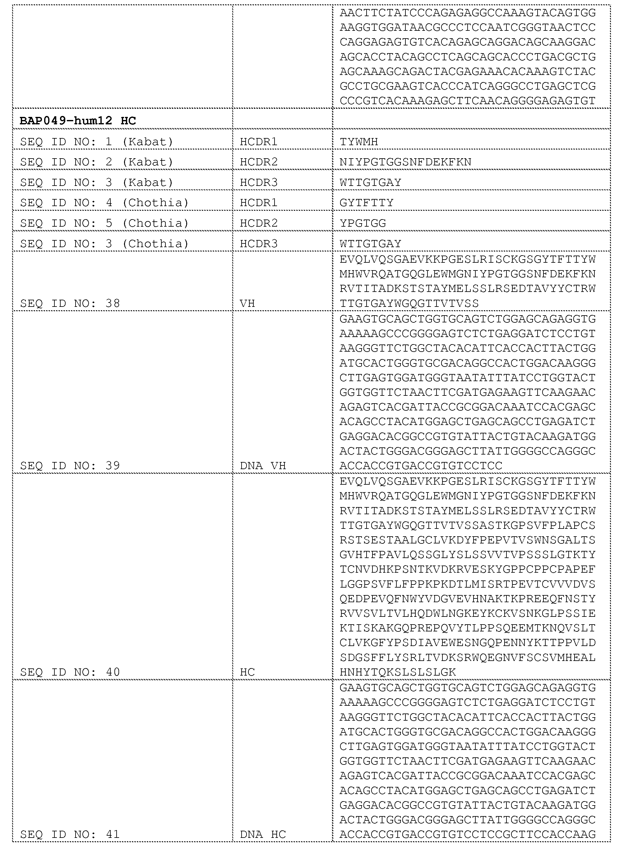


Claims
Priority Applications (29)
| Application Number | Priority Date | Filing Date | Title |
|---|---|---|---|
| SG11201604939SA SG11201604939SA (en) | 2014-01-24 | 2015-01-23 | Antibody molecules to pd-1 and uses thereof |
| SI201530651T SI3097121T1 (en) | 2014-01-24 | 2015-01-23 | Antibody molecules to pd-1 and uses thereof |
| MX2016009616A MX2016009616A (en) | 2014-01-24 | 2015-01-23 | Antibody molecules to pd-1 and uses thereof. |
| MYPI2016702232A MY184757A (en) | 2014-01-24 | 2015-01-23 | Antibody molecules to pd-1 and uses thereof |
| EP15703384.6A EP3097121B1 (en) | 2014-01-24 | 2015-01-23 | Antibody molecules to pd-1 and uses thereof |
| MEP-2019-83A ME03366B (en) | 2014-01-24 | 2015-01-23 | Antibody molecules to pd-1 and uses thereof |
| UAA201606726A UA122479C2 (en) | 2014-01-24 | 2015-01-23 | Antibody molecules to pd-1 and uses thereof |
| AU2015209145A AU2015209145B2 (en) | 2014-01-24 | 2015-01-23 | Antibody molecules to PD-1 and uses thereof |
| RS20190360A RS58535B1 (en) | 2014-01-24 | 2015-01-23 | Antibody molecules to pd-1 and uses thereof |
| MA39208A MA39208B1 (en) | 2014-01-24 | 2015-01-23 | Anti-pd-1 antibody molecules and their uses |
| KR1020167022709A KR102357621B1 (en) | 2014-01-24 | 2015-01-23 | Antibody molecules to pd-1 and uses thereof |
| EP18211373.8A EP3514179B1 (en) | 2014-01-24 | 2015-01-23 | Antibody molecules to pd-1 and uses thereof |
| TN2016000272A TN2016000272A1 (en) | 2014-01-24 | 2015-01-23 | Antibody molecules to pd-1 and uses thereof. |
| DK15703384.6T DK3097121T3 (en) | 2014-01-24 | 2015-01-23 | ANTIBODY MOLECULES AGAINST PD-1 AND APPLICATIONS THEREOF |
| LTEP15703384.6T LT3097121T (en) | 2014-01-24 | 2015-01-23 | Antibody molecules to pd-1 and uses thereof |
| CA2935423A CA2935423C (en) | 2014-01-24 | 2015-01-23 | Antibody molecules to pd-1 and uses thereof |
| BR112016015677A BR112016015677A2 (en) | 2014-01-24 | 2015-01-23 | ISOLATED ANTIBODY MOLECULES CAPABLE OF BINDING TO HUMAN PD-1, AS WELL AS THEIR METHOD OF PRODUCTION AND ITS USE, PHARMACEUTICAL COMPOSITIONS, NUCLEIC ACID, EXPRESSION VECTOR, HOST CELL, AND METHOD FOR DETECTION OF PD-1 IN A BIOLOGICAL SAMPLE |
| EA201691488A EA201691488A1 (en) | 2014-01-24 | 2015-01-23 | ANTIBODY MOLECULES AGAINST PD-1 AND THEIR APPLICATIONS |
| NZ721678A NZ721678A (en) | 2014-01-24 | 2015-01-23 | Antibody molecules to pd-1 and uses thereof |
| ES15703384T ES2716685T3 (en) | 2014-01-24 | 2015-01-23 | Antibody molecules for PD-1 and uses thereof |
| CN201580015562.6A CN106103485B (en) | 2014-01-24 | 2015-01-23 | Antibody molecules to PD-1 and uses thereof |
| PL15703384T PL3097121T3 (en) | 2014-01-24 | 2015-01-23 | Antibody molecules to pd-1 and uses thereof |
| JP2016548014A JP6521986B2 (en) | 2014-01-24 | 2015-01-23 | Antibody molecules against PD-1 and uses thereof |
| IL246382A IL246382A0 (en) | 2014-01-24 | 2016-06-22 | Antibody molecules to pd-1 and uses thereof |
| PH12016501456A PH12016501456A1 (en) | 2014-01-24 | 2016-07-22 | Antibody molecules to pd-1 and uses thereof |
| HRP20190516TT HRP20190516T1 (en) | 2014-01-24 | 2019-03-15 | Antibody molecules to pd-1 and uses thereof |
| CY20191100330T CY1121542T1 (en) | 2014-01-24 | 2019-03-19 | ANTIBODY MOLECULES TO PD-1 AND THEIR USE |
| AU2020204366A AU2020204366A1 (en) | 2014-01-24 | 2020-06-30 | Antibody molecules to PD-1 and uses thereof |
| IL278803A IL278803A (en) | 2014-01-24 | 2020-11-18 | Antibody molecules to pd-1 and uses thereof |
Applications Claiming Priority (6)
| Application Number | Priority Date | Filing Date | Title |
|---|---|---|---|
| US201461931512P | 2014-01-24 | 2014-01-24 | |
| US61/931,512 | 2014-01-24 | ||
| US201462059676P | 2014-10-03 | 2014-10-03 | |
| US62/059,676 | 2014-10-03 | ||
| US201462094834P | 2014-12-19 | 2014-12-19 | |
| US62/094,834 | 2014-12-19 |
Publications (2)
| Publication Number | Publication Date |
|---|---|
| WO2015112900A1 true WO2015112900A1 (en) | 2015-07-30 |
| WO2015112900A8 WO2015112900A8 (en) | 2016-08-18 |
Family
ID=52463182
Family Applications (1)
| Application Number | Title | Priority Date | Filing Date |
|---|---|---|---|
| PCT/US2015/012754 WO2015112900A1 (en) | 2014-01-24 | 2015-01-23 | Antibody molecules to pd-1 and uses thereof |
Country Status (36)
| Country | Link |
|---|---|
| US (4) | US9683048B2 (en) |
| EP (2) | EP3097121B1 (en) |
| JP (3) | JP6521986B2 (en) |
| KR (1) | KR102357621B1 (en) |
| CN (3) | CN106103485B (en) |
| AU (2) | AU2015209145B2 (en) |
| BR (1) | BR112016015677A2 (en) |
| CA (1) | CA2935423C (en) |
| CL (1) | CL2016001723A1 (en) |
| CY (1) | CY1121542T1 (en) |
| DK (1) | DK3097121T3 (en) |
| EA (1) | EA201691488A1 (en) |
| EC (1) | ECSP16068976A (en) |
| ES (1) | ES2716685T3 (en) |
| HK (1) | HK1225747B (en) |
| HR (1) | HRP20190516T1 (en) |
| HU (1) | HUE043363T2 (en) |
| IL (2) | IL246382A0 (en) |
| JO (2) | JOP20200094A1 (en) |
| LT (1) | LT3097121T (en) |
| ME (1) | ME03366B (en) |
| MX (2) | MX2016009616A (en) |
| MY (1) | MY184757A (en) |
| NZ (1) | NZ721678A (en) |
| PE (1) | PE20170255A1 (en) |
| PH (1) | PH12016501456A1 (en) |
| PL (1) | PL3097121T3 (en) |
| PT (1) | PT3097121T (en) |
| RS (1) | RS58535B1 (en) |
| SG (2) | SG10201900002QA (en) |
| SI (1) | SI3097121T1 (en) |
| TN (1) | TN2016000272A1 (en) |
| TW (3) | TWI692484B (en) |
| UA (1) | UA122479C2 (en) |
| UY (1) | UY35967A (en) |
| WO (1) | WO2015112900A1 (en) |
Cited By (321)
| Publication number | Priority date | Publication date | Assignee | Title |
|---|---|---|---|---|
| CN105175544A (en) * | 2015-10-20 | 2015-12-23 | 安徽瀚海博兴生物技术有限公司 | Anti-PD-1 humanized monoclonal antibody and application thereof |
| WO2016057846A1 (en) | 2014-10-08 | 2016-04-14 | Novartis Ag | Compositions and methods of use for augmented immune response and cancer therapy |
| WO2016054555A3 (en) * | 2014-10-03 | 2016-06-30 | Novartis Ag | Combination therapies |
| WO2016110593A1 (en) * | 2015-01-09 | 2016-07-14 | Immutep S.A.S. | Combined preparations for the treatment of cancer or infection |
| WO2016141209A1 (en) * | 2015-03-04 | 2016-09-09 | Merck Sharp & Dohme Corp. | Combination of a pd-1 antagonist and eribulin for treating cancer |
| WO2016161239A1 (en) * | 2015-04-03 | 2016-10-06 | Astex Therapeutics Limited | Fgfr/pd-1 combination therapy for the treatment of cancer |
| WO2017017624A1 (en) * | 2015-07-29 | 2017-02-02 | Novartis Ag | Combination of pd-1 antagonist with an egfr inhibitor |
| WO2017019896A1 (en) * | 2015-07-29 | 2017-02-02 | Novartis Ag | Combination therapies comprising antibody molecules to pd-1 |
| US9605070B2 (en) | 2014-01-31 | 2017-03-28 | Novartis Ag | Antibody molecules to TIM-3 and uses thereof |
| WO2017055404A1 (en) | 2015-10-02 | 2017-04-06 | F. Hoffmann-La Roche Ag | Bispecific antibodies specific for pd1 and tim3 |
| WO2017058115A1 (en) | 2015-09-29 | 2017-04-06 | Asia Biotech Pte. Ltd. | Pd-1 antibodies and uses thereof |
| US9644032B2 (en) | 2015-05-29 | 2017-05-09 | Bristol-Myers Squibb Company | Antibodies against OX40 and uses thereof |
| WO2017077382A1 (en) | 2015-11-06 | 2017-05-11 | Orionis Biosciences Nv | Bi-functional chimeric proteins and uses thereof |
| WO2017079116A2 (en) | 2015-11-03 | 2017-05-11 | Janssen Biotech, Inc. | Antibodies specifically binding pd-1 and tim-3 and their uses |
| US9683048B2 (en) | 2014-01-24 | 2017-06-20 | Novartis Ag | Antibody molecules to PD-1 and uses thereof |
| WO2017106656A1 (en) * | 2015-12-17 | 2017-06-22 | Novartis Ag | Antibody molecules to pd-1 and uses thereof |
| WO2017103895A1 (en) | 2015-12-18 | 2017-06-22 | Novartis Ag | Antibodies targeting cd32b and methods of use thereof |
| WO2017106810A2 (en) | 2015-12-17 | 2017-06-22 | Novartis Ag | Combination of c-met inhibitor with antibody molecule to pd-1 and uses thereof |
| WO2017087589A3 (en) * | 2015-11-18 | 2017-07-13 | Merck Sharp & Dohme Corp. | Pd1 and/or lag3 binders |
| WO2017134302A2 (en) | 2016-02-05 | 2017-08-10 | Orionis Biosciences Nv | Targeted therapeutic agents and uses thereof |
| US9815897B2 (en) | 2013-05-02 | 2017-11-14 | Anaptysbio, Inc. | Antibodies directed against programmed death-1 (PD-1) |
| EP3243832A1 (en) | 2016-05-13 | 2017-11-15 | F. Hoffmann-La Roche AG | Antigen binding molecules comprising a tnf family ligand trimer and pd1 binding moiety |
| WO2017194783A1 (en) | 2016-05-13 | 2017-11-16 | Orionis Biosciences Nv | Targeted mutant interferon-beta and uses thereof |
| WO2017194782A2 (en) | 2016-05-13 | 2017-11-16 | Orionis Biosciences Nv | Therapeutic targeting of non-cellular structures |
| WO2017210631A1 (en) | 2016-06-03 | 2017-12-07 | Bristol-Myers Squibb Company | Anti-pd-1 antibody for use in a method of treatment of recurrent small cell lung cancer |
| WO2017210453A1 (en) | 2016-06-02 | 2017-12-07 | Bristol-Myers Squibb Company | Pd-1 blockade with nivolumab in refractory hodgkin's lymphoma |
| WO2017210473A1 (en) | 2016-06-02 | 2017-12-07 | Bristol-Myers Squibb Company | Use of an anti-pd-1 antibody in combination with an anti-cd30 antibody in lymphoma treatment |
| WO2017210624A1 (en) | 2016-06-03 | 2017-12-07 | Bristol-Myers Squibb Company | Anti-pd-1 antibody for use in a method of treating a tumor |
| WO2017210637A1 (en) | 2016-06-03 | 2017-12-07 | Bristol-Myers Squibb Company | Use of anti-pd-1 antibody in the treatment of patients with colorectal cancer |
| WO2017212442A1 (en) | 2016-06-10 | 2017-12-14 | Novartis Ag | Therapeutic uses of a c-raf inhibitor |
| WO2017220989A1 (en) | 2016-06-20 | 2017-12-28 | Kymab Limited | Anti-pd-l1 and il-2 cytokines |
| US9908936B2 (en) | 2014-03-14 | 2018-03-06 | Novartis Ag | Antibody molecules to LAG-3 and uses thereof |
| WO2018050027A1 (en) * | 2016-09-14 | 2018-03-22 | 北京韩美药品有限公司 | Antibody specifically binding to pd-1 and functional fragment thereof |
| WO2018053709A1 (en) * | 2016-09-21 | 2018-03-29 | Wuxi Biologics (Shanghai) Co., Ltd. | The novel monoclonal antibodies to programmed death 1 (pd-1) |
| WO2018055503A1 (en) | 2016-09-20 | 2018-03-29 | Novartis Ag | Combination comprising a pd-1 antagonist and an fgfr4 inhibitor |
| US9957323B2 (en) | 2016-06-20 | 2018-05-01 | Kymab Limited | Anti-ICOS antibodies |
| WO2018081621A1 (en) | 2016-10-28 | 2018-05-03 | Bristol-Myers Squibb Company | Methods of treating urothelial carcinoma using an anti-pd-1 antibody |
| WO2018077893A1 (en) | 2016-10-24 | 2018-05-03 | Orionis Biosciences Nv | Targeted mutant interferon-gamma and uses thereof |
| WO2018085555A1 (en) | 2016-11-03 | 2018-05-11 | Bristol-Myers Squibb Company | Activatable anti-ctla-4 antibodies and uses thereof |
| US9975957B2 (en) | 2014-03-31 | 2018-05-22 | Genentech, Inc. | Anti-OX40 antibodies and methods of use |
| WO2018091661A1 (en) * | 2016-11-18 | 2018-05-24 | Symphogen A/S | Anti-pd-1 antibodies and compositions |
| US9988452B2 (en) | 2014-10-14 | 2018-06-05 | Novartis Ag | Antibody molecules to PD-L1 and uses thereof |
| US9987500B2 (en) | 2014-01-23 | 2018-06-05 | Regeneron Pharmaceuticals, Inc. | Human antibodies to PD-1 |
| WO2018127710A1 (en) * | 2017-01-06 | 2018-07-12 | Crescendo Biologics Limited | Single domain antibodies to programmed cell death (pd-1) |
| WO2018133837A1 (en) * | 2017-01-20 | 2018-07-26 | Tayu Huaxia Biotech Medical Group Co., Ltd. | Anti-pd-1 antibodies and uses thereof |
| WO2018134681A1 (en) | 2017-01-20 | 2018-07-26 | Sanofi | Anti-tgf-beta antibodies and their use |
| CN108348521A (en) * | 2015-08-11 | 2018-07-31 | 诺华股份有限公司 | Bromo- bis--(1H- pyrazol-1-yls) pyrimidine -4- amine of 2,6- of 5- for treating cancer |
| WO2018137576A1 (en) * | 2017-01-24 | 2018-08-02 | 三生国健药业(上海)股份有限公司 | Anti-pd-1 monoclonal antibody, and preparation method therefor and application thereof |
| CN108368174A (en) * | 2015-11-23 | 2018-08-03 | 戊瑞治疗有限公司 | Independent FGFR2 inhibitor use for cancer treatment or the combination with immunostimulant |
| WO2018141964A1 (en) | 2017-02-06 | 2018-08-09 | Orionis Biosciences Nv | Targeted chimeric proteins and uses thereof |
| WO2018142322A1 (en) | 2017-02-03 | 2018-08-09 | Novartis Ag | Anti-ccr7 antibody drug conjugates |
| WO2018144999A1 (en) | 2017-02-06 | 2018-08-09 | Orionis Biosciences, Inc. | Targeted engineered interferon and uses thereof |
| WO2018146612A1 (en) | 2017-02-10 | 2018-08-16 | Novartis Ag | 1-(4-amino-5-bromo-6-(1 h-pyrazol-1-yl)pyrimidin-2-yl)-1 h-pyrazol-4-ol and use thereof in the treatment of cancer |
| WO2018152396A1 (en) | 2017-02-17 | 2018-08-23 | Innate Tumor Immunity, Inc. | Substituted imidazo-quinolines as nlrp3 modulators |
| JP2018525367A (en) * | 2015-07-29 | 2018-09-06 | ノバルティス アーゲー | Combination of anti-PD-1 antibody and anti-M-CSF antibody in cancer treatment |
| WO2018160540A1 (en) | 2017-02-28 | 2018-09-07 | Sanofi | Therapeutic rna |
| WO2018183928A1 (en) | 2017-03-31 | 2018-10-04 | Bristol-Myers Squibb Company | Methods of treating tumor |
| WO2018178250A1 (en) | 2017-03-31 | 2018-10-04 | Boehringer Ingelheim International Gmbh | Anticancer combination therapy |
| WO2018185135A1 (en) | 2017-04-05 | 2018-10-11 | Boehringer Ingelheim International Gmbh | Anticancer combination therapy |
| WO2018185043A1 (en) | 2017-04-05 | 2018-10-11 | F. Hoffmann-La Roche Ag | Bispecific antibodies specifically binding to pd1 and lag3 |
| WO2018187613A2 (en) | 2017-04-07 | 2018-10-11 | Bristol-Myers Squibb Company | Anti-icos agonist antibodies and uses thereof |
| JP2018533626A (en) * | 2015-11-04 | 2018-11-15 | デューク ユニバーシティー | Combination therapy with immunotoxin and checkpoint inhibitor |
| JP2018533973A (en) * | 2015-09-29 | 2018-11-22 | セルジーン コーポレイション | PD-1-binding protein and method of using the same |
| WO2018222718A1 (en) | 2017-05-30 | 2018-12-06 | Bristol-Myers Squibb Company | Treatment of lag-3 positive tumors |
| WO2018222722A2 (en) | 2017-05-30 | 2018-12-06 | Bristol-Myers Squibb Company | Compositions comprising an anti-lag-3 antibody or an anti-lag-3 antibody and an anti-pd-1 or anti-pd-l1 antibody |
| WO2018220546A1 (en) | 2017-05-31 | 2018-12-06 | Novartis Ag | Crystalline forms of 5-bromo-2,6-di(1 h-pyrazol-1-yl)pyrimidin-4-amine and new salts |
| WO2018220169A1 (en) | 2017-06-02 | 2018-12-06 | Boehringer Ingelheim International Gmbh | Anti-cancer combination therapy |
| WO2018223040A1 (en) | 2017-06-01 | 2018-12-06 | Bristol-Myers Squibb Company | Methods of treating a tumor using an anti-pd-1 antibody |
| WO2018222711A2 (en) | 2017-05-30 | 2018-12-06 | Bristol-Myers Squibb Company | Compositions comprising a combination of an anti-lag-3 antibody, a pd-1 pathway inhibitor, and an immunotherapeutic agent |
| WO2018220446A1 (en) | 2017-06-01 | 2018-12-06 | Compugen Ltd. | Triple combination antibody therapies |
| US10155037B2 (en) | 2014-12-09 | 2018-12-18 | Rinat Neuroscience Corp. | Anti-PD-1 antibodies and methods of use thereof |
| EP3421494A1 (en) | 2017-06-29 | 2019-01-02 | Sanofi | Use of isatuximab in combination with an anti-pd-1 antibody |
| WO2019009726A1 (en) * | 2017-07-06 | 2019-01-10 | Merus N.V. | Binding molecules that modulate a biological activity expressed by a cell |
| WO2019014402A1 (en) | 2017-07-14 | 2019-01-17 | Innate Tumor Immunity, Inc. | Nlrp3 modulators |
| US10188729B2 (en) | 2013-08-20 | 2019-01-29 | Merck Sharp & Dohme Corp. | Modulation of tumor immunity |
| WO2019023459A1 (en) | 2017-07-28 | 2019-01-31 | Bristol-Myers Squibb Company | Cyclic dinucleotides as anticancer agents |
| CN109311989A (en) * | 2016-04-14 | 2019-02-05 | 创新微技术公司 | The application method of PD-L1 expression in the treatment of cancer therapy determines |
| JP2019503387A (en) * | 2016-01-27 | 2019-02-07 | ブリストル−マイヤーズ スクイブ カンパニーBristol−Myers Squibb Company | Method for treating lung cancer using a combination of an anti-PD-1 antibody and another anticancer agent |
| WO2019046500A1 (en) | 2017-08-31 | 2019-03-07 | Bristol-Myers Squibb Company | Cyclic dinucleotides as anticancer agents |
| WO2019046498A1 (en) | 2017-08-31 | 2019-03-07 | Bristol-Myers Squibb Company | Cyclic dinucleotides as anticancer agents |
| WO2019046496A1 (en) | 2017-08-31 | 2019-03-07 | Bristol-Myers Squibb Company | Cyclic dinucleotides as anticancer agents |
| WO2019063802A1 (en) | 2017-09-29 | 2019-04-04 | Boehringer Ingelheim International Gmbh | Anti igf, anti pd-1 anti-cancer combination therapy |
| WO2019074887A1 (en) | 2017-10-10 | 2019-04-18 | Bristol-Myers Squibb Company | Cyclic dinucleotides as anticancer agents |
| WO2019075468A1 (en) | 2017-10-15 | 2019-04-18 | Bristol-Myers Squibb Company | Methods of treating tumor |
| WO2019079261A1 (en) | 2017-10-16 | 2019-04-25 | Bristol-Myers Squibb Company | Cyclic dinucleotides as anticancer agents |
| WO2019090263A1 (en) | 2017-11-06 | 2019-05-09 | Genentech, Inc. | Diagnostic and therapeutic methods for cancer |
| WO2019090330A1 (en) | 2017-11-06 | 2019-05-09 | Bristol-Myers Squibb Company | Methods of treating a tumor |
| US10287353B2 (en) | 2016-05-11 | 2019-05-14 | Huya Bioscience International, Llc | Combination therapies of HDAC inhibitors and PD-1 inhibitors |
| WO2019097369A1 (en) | 2017-11-14 | 2019-05-23 | Pfizer Inc. | Ezh2 inhibitor combination therapies |
| US10344090B2 (en) | 2013-12-12 | 2019-07-09 | Shanghai Hangrui Pharmaceutical Co., Ltd. | PD-1 antibody, antigen-binding fragment thereof, and medical application thereof |
| US10350266B2 (en) * | 2017-01-10 | 2019-07-16 | Nodus Therapeutics, Inc. | Method of treating cancer with a multiple integrin binding Fc fusion protein |
| WO2019144098A1 (en) | 2018-01-22 | 2019-07-25 | Bristol-Myers Squibb Company | Compositions and methods of treating cancer |
| WO2019144126A1 (en) | 2018-01-22 | 2019-07-25 | Pascal Biosciences Inc. | Cannabinoids and derivatives for promoting immunogenicity of tumor and infected cells |
| WO2019143607A1 (en) | 2018-01-16 | 2019-07-25 | Bristol-Myers Squibb Company | Methods of treating cancer with antibodies against tim3 |
| WO2019149716A1 (en) | 2018-01-31 | 2019-08-08 | F. Hoffmann-La Roche Ag | Bispecific antibodies comprising an antigen-binding site binding to lag3 |
| US10385131B2 (en) | 2016-05-11 | 2019-08-20 | Huya Bioscience International, Llc | Combination therapies of HDAC inhibitors and PD-L1 inhibitors |
| WO2019160884A1 (en) | 2018-02-13 | 2019-08-22 | Bristol-Myers Squibb Company | Cyclic dinucleotides as anticancer agents |
| CN110167964A (en) * | 2016-11-02 | 2019-08-23 | 英格玛布有限责任公司 | Combine the bispecific antibody and immune drug for BCMA and CD3 for treating Huppert's disease |
| US10392442B2 (en) | 2015-12-17 | 2019-08-27 | Bristol-Myers Squibb Company | Use of anti-PD-1 antibody in combination with anti-CD27 antibody in cancer treatment |
| WO2019173587A1 (en) | 2018-03-08 | 2019-09-12 | Bristol-Myers Squibb Company | Cyclic dinucleotides as anticancer agents |
| WO2019180576A1 (en) | 2018-03-20 | 2019-09-26 | Novartis Ag | Pharmaceutical combinations |
| WO2019191676A1 (en) | 2018-03-30 | 2019-10-03 | Bristol-Myers Squibb Company | Methods of treating tumor |
| WO2019195452A1 (en) | 2018-04-04 | 2019-10-10 | Bristol-Myers Squibb Company | Anti-cd27 antibodies and uses thereof |
| CN110392692A (en) * | 2017-04-03 | 2019-10-29 | 豪夫迈·罗氏有限公司 | Anti- PD-1 antibody and mutant IL-2 or the immunoconjugates with IL-15 |
| US10457725B2 (en) | 2016-05-13 | 2019-10-29 | Regeneron Pharmaceuticals, Inc. | Methods of treating skin cancer by administering a PD-1 inhibitor |
| WO2019209896A1 (en) | 2018-04-25 | 2019-10-31 | Innate Tumor Immunity, Inc. | Nlrp3 modulators |
| EP3569618A1 (en) | 2018-05-19 | 2019-11-20 | Boehringer Ingelheim International GmbH | Antagonizing cd73 antibody |
| KR20190130562A (en) * | 2017-01-23 | 2019-11-22 | 카르스젠 테라퓨틱스 컴퍼니, 리미티드 | BCMA Target Antibodies and Uses thereof |
| JP2019534268A (en) * | 2016-10-12 | 2019-11-28 | ボード・オブ・リージエンツ,ザ・ユニバーシテイ・オブ・テキサス・システム | Methods and compositions for TUSC2 immunotherapy |
| WO2019232319A1 (en) | 2018-05-31 | 2019-12-05 | Peloton Therapeutics, Inc. | Compositions and methods for inhibiting cd73 |
| WO2019246557A1 (en) | 2018-06-23 | 2019-12-26 | Genentech, Inc. | Methods of treating lung cancer with a pd-1 axis binding antagonist, a platinum agent, and a topoisomerase ii inhibitor |
| US10526413B2 (en) | 2015-10-02 | 2020-01-07 | Hoffmann-La Roche Inc. | Bispecific antibodies specific for OX40 |
| WO2020018789A1 (en) | 2018-07-18 | 2020-01-23 | Genentech, Inc. | Methods of treating lung cancer with a pd-1 axis binding antagonist, an antimetabolite, and a platinum agent |
| WO2020021447A1 (en) | 2018-07-25 | 2020-01-30 | Novartis Ag | Nlrp3 inflammasome inhibitors |
| WO2020023707A1 (en) | 2018-07-26 | 2020-01-30 | Bristol-Myers Squibb Company | Lag-3 combination therapy for the treatment of cancer |
| JP2020505438A (en) * | 2017-01-10 | 2020-02-20 | ノダス・セラピューティクスNodus Therapeutics | Combination cancer therapy using integrin binding polypeptide-Fc fusion protein and immunomodulator |
| WO2020037094A1 (en) | 2018-08-16 | 2020-02-20 | Innate Tumor Immunity, Inc. | Substitued 4-amino-1h-imidazo[4,5-c]quinoline compounds and improved methods for their preparation |
| JP2020505350A (en) * | 2017-01-20 | 2020-02-20 | ノバルティス アーゲー | Combination therapy for the treatment of cancer |
| WO2020037091A1 (en) | 2018-08-16 | 2020-02-20 | Innate Tumor Immunity, Inc. | Imidazo[4,5-c]quinoline derived nlrp3-modulators |
| WO2020037092A1 (en) | 2018-08-16 | 2020-02-20 | Innate Tumor Immunity, Inc. | Imidazo[4,5-c]quinoline derived nlrp3-modulators |
| US10570204B2 (en) | 2013-09-26 | 2020-02-25 | The Medical College Of Wisconsin, Inc. | Methods for treating hematologic cancers |
| WO2020041655A1 (en) | 2018-08-24 | 2020-02-27 | Sanofi | Therapeutic rna for solid tumor cancers |
| WO2020051099A1 (en) | 2018-09-03 | 2020-03-12 | Genentech, Inc. | Carboxamide and sulfonamide derivatives useful as tead modulators |
| WO2020076799A1 (en) | 2018-10-09 | 2020-04-16 | Bristol-Myers Squibb Company | Anti-mertk antibodies for treating cancer |
| WO2020081928A1 (en) | 2018-10-19 | 2020-04-23 | Bristol-Myers Squibb Company | Combination therapy for melanoma |
| WO2020086724A1 (en) | 2018-10-23 | 2020-04-30 | Bristol-Myers Squibb Company | Methods of treating tumor |
| US10654929B2 (en) | 2016-11-02 | 2020-05-19 | Jounce Therapeutics, Inc. | Antibodies to PD-1 and uses thereof |
| WO2020102501A1 (en) | 2018-11-16 | 2020-05-22 | Bristol-Myers Squibb Company | Anti-nkg2a antibodies and uses thereof |
| WO2020106695A1 (en) | 2018-11-19 | 2020-05-28 | Ariagen, Inc. | Methods of treating cancer |
| WO2020117849A1 (en) | 2018-12-04 | 2020-06-11 | Bristol-Myers Squibb Company | Methods of analysis using in-sample calibration curve by multiple isotopologue reaction monitoring |
| EP3666905A1 (en) | 2018-12-11 | 2020-06-17 | Sanofi | E. coli positive for pks island as marker of positive response to anti-pd1 therapy in colorectal cancer |
| EP3613768A4 (en) * | 2017-04-20 | 2020-06-17 | Suzhou Stainwei Biotech Inc. | Monoclonal antibody antagonizing and inhibiting binding between human pd-1 antigen and ligand thereof, preparation method therefor and application thereof |
| EP3666794A1 (en) * | 2016-11-01 | 2020-06-17 | AnaptysBio, Inc. | Antibodies directed against programmed death- 1 (pd-1) |
| WO2020123453A2 (en) | 2018-12-11 | 2020-06-18 | Theravance Biopharma R&D Ip, Llc | Alk5 inhibitors |
| WO2020128612A2 (en) | 2018-12-21 | 2020-06-25 | Novartis Ag | Antibodies to pmel17 and conjugates thereof |
| WO2020136133A1 (en) | 2018-12-23 | 2020-07-02 | F. Hoffmann-La Roche Ag | Tumor classification based on predicted tumor mutational burden |
| CN111423512A (en) * | 2019-01-10 | 2020-07-17 | 北京比洋生物技术有限公司 | Multi-targeting fusion protein blocking vascular endothelial cell growth and activating T cells and pharmaceutical composition comprising the same |
| WO2020150116A1 (en) | 2019-01-14 | 2020-07-23 | Innate Tumor Immunity, Inc. | Nlrp3 modulators |
| WO2020150113A1 (en) | 2019-01-14 | 2020-07-23 | Innate Tumor Immunity, Inc. | Substituted quinazolines as nlrp3 modulators, for use in the treatment of cancer |
| WO2020150114A1 (en) | 2019-01-14 | 2020-07-23 | Innate Tumor Immunity, Inc. | Heterocyclic nlrp3 modulators, for use in the treatment of cancer |
| WO2020150115A1 (en) | 2019-01-14 | 2020-07-23 | Innate Tumor Immunity, Inc. | Nlrp3 modulators |
| WO2020150152A1 (en) | 2019-01-14 | 2020-07-23 | Genentech, Inc. | Methods of treating cancer with a pd-1 axis binding antagonist and an rna vaccine |
| WO2020152306A1 (en) | 2019-01-25 | 2020-07-30 | Boehringer Ingelheim International Gmbh | Recombinant rhabdovirus encoding for ccl21 |
| WO2020154189A1 (en) | 2019-01-21 | 2020-07-30 | Sanofi | Therapeutic rna and anti-pd1 antibodies for advanced stage solid tumor cancers |
| US10736940B2 (en) | 2013-12-19 | 2020-08-11 | Immutep S.A.S. | Combined preparations for the treatment of cancer |
| US10766955B2 (en) | 2017-01-20 | 2020-09-08 | Sanofi | Anti-TGF-β antibodies and their use |
| WO2020198672A1 (en) | 2019-03-28 | 2020-10-01 | Bristol-Myers Squibb Company | Methods of treating tumor |
| WO2020198676A1 (en) | 2019-03-28 | 2020-10-01 | Bristol-Myers Squibb Company | Methods of treating tumor |
| WO2020214995A1 (en) | 2019-04-19 | 2020-10-22 | Genentech, Inc. | Anti-mertk antibodies and their methods of use |
| WO2020231766A1 (en) | 2019-05-13 | 2020-11-19 | Bristol-Myers Squibb Company | AGONISTS OF ROR GAMMAt |
| WO2020231713A1 (en) | 2019-05-13 | 2020-11-19 | Bristol-Myers Squibb Company | AGONISTS OF ROR GAMMAt |
| US10844115B2 (en) | 2017-01-20 | 2020-11-24 | Genzyme Corporation | Bone-targeting antibodies |
| WO2020234715A1 (en) | 2019-05-17 | 2020-11-26 | Novartis Ag | Nlrp3 inflammasome inhibitors |
| WO2020239558A1 (en) | 2019-05-24 | 2020-12-03 | Pfizer Inc. | Combination therapies using cdk inhibitors |
| WO2020243568A1 (en) | 2019-05-30 | 2020-12-03 | Bristol-Myers Squibb Company | Methods of identifying a subject suitable for an immuno-oncology (i-o) therapy |
| WO2020243570A1 (en) | 2019-05-30 | 2020-12-03 | Bristol-Myers Squibb Company | Cell localization signature and combination therapy |
| WO2020243563A1 (en) | 2019-05-30 | 2020-12-03 | Bristol-Myers Squibb Company | Multi-tumor gene signatures for suitability to immuno-oncology therapy |
| EP3322731B1 (en) | 2015-07-14 | 2021-01-13 | Bristol-Myers Squibb Company | Method of treating cancer using immune checkpoint inhibitor; antibody that binds to programmed death-1 receptor (pd-1) or programmed death ligand 1 (pd-l1) |
| WO2021026179A1 (en) | 2019-08-06 | 2021-02-11 | Bristol-Myers Squibb Company | AGONISTS OF ROR GAMMAt |
| WO2021024020A1 (en) | 2019-08-06 | 2021-02-11 | Astellas Pharma Inc. | Combination therapy involving antibodies against claudin 18.2 and immune checkpoint inhibitors for treatment of cancer |
| US10934356B2 (en) | 2014-11-13 | 2021-03-02 | The Johns Hopkins University | Checkpoint blockade and microsatellite instability |
| WO2021055994A1 (en) | 2019-09-22 | 2021-03-25 | Bristol-Myers Squibb Company | Quantitative spatial profiling for lag-3 antagonist therapy |
| WO2021062122A1 (en) | 2019-09-25 | 2021-04-01 | Seagen Inc. | Combination anti-cd30 adc, anti-pd-1 and chemotherapeutic for treatment of hematopoietic cancers |
| WO2021062018A1 (en) | 2019-09-25 | 2021-04-01 | Bristol-Myers Squibb Company | Composite biomarker for cancer therapy |
| WO2021092221A1 (en) | 2019-11-06 | 2021-05-14 | Bristol-Myers Squibb Company | Methods of identifying a subject with a tumor suitable for a checkpoint inhibitor therapy |
| WO2021092380A1 (en) | 2019-11-08 | 2021-05-14 | Bristol-Myers Squibb Company | Lag-3 antagonist therapy for melanoma |
| WO2021092220A1 (en) | 2019-11-06 | 2021-05-14 | Bristol-Myers Squibb Company | Methods of identifying a subject with a tumor suitable for a checkpoint inhibitor therapy |
| WO2021092044A1 (en) | 2019-11-05 | 2021-05-14 | Bristol-Myers Squibb Company | M-protein assays and uses thereof |
| WO2021097110A1 (en) | 2019-11-13 | 2021-05-20 | Genentech, Inc. | Therapeutic compounds and methods of use |
| WO2021097256A1 (en) | 2019-11-14 | 2021-05-20 | Cohbar, Inc. | Cxcr4 antagonist peptides |
| WO2021102468A1 (en) | 2019-11-22 | 2021-05-27 | Theravance Biopharma R&D Ip, Llc | Substituted 1,5-naphthyridines or quinolines as alk5 inhibitors |
| WO2021119105A1 (en) | 2019-12-09 | 2021-06-17 | Seagen Inc. | Combination therapy with liv1-adc and pd-1 antagonist |
| WO2021127554A1 (en) | 2019-12-19 | 2021-06-24 | Bristol-Myers Squibb Company | Combinations of dgk inhibitors and checkpoint antagonists |
| US11046776B2 (en) | 2016-08-05 | 2021-06-29 | Genentech, Inc. | Multivalent and multiepitopic antibodies having agonistic activity and methods of use |
| WO2021142203A1 (en) | 2020-01-10 | 2021-07-15 | Innate Tumor Immunity, Inc. | Nlrp3 modulators |
| WO2021143826A1 (en) * | 2020-01-17 | 2021-07-22 | 信达生物制药(苏州)有限公司 | Recombinant anti-programmed cell death protein 1 and anti-cluster of differentiation antigen 137 bispecific antibody preparation and use thereof |
| WO2021152548A1 (en) | 2020-01-30 | 2021-08-05 | Benitah Salvador Aznar | Combination therapy for treatment of cancer and cancer metastasis |
| WO2021155149A1 (en) | 2020-01-31 | 2021-08-05 | Genentech, Inc. | Methods of inducing neoepitope-specific t cells with a pd-1 axis binding antagonist and an rna vaccine |
| US11083705B2 (en) | 2019-07-26 | 2021-08-10 | Eisai R&D Management Co., Ltd. | Pharmaceutical composition for treating tumor |
| WO2021158938A1 (en) | 2020-02-06 | 2021-08-12 | Bristol-Myers Squibb Company | Il-10 and uses thereof |
| US11091555B2 (en) | 2017-05-16 | 2021-08-17 | Five Prime Therapeutics, Inc. | Method of treating gastric cancer with anti-FGFR2-IIIb antibodies and modified FOLFOX6 chemotherapy |
| WO2021178807A1 (en) | 2020-03-06 | 2021-09-10 | Celgene Quanticel Research, Inc. | Combination of an lsd-1 inhibitor and nivolumab for use in treating sclc or sqnsclc |
| WO2021176424A1 (en) | 2020-03-06 | 2021-09-10 | Ona Therapeutics, S.L. | Anti-cd36 antibodies and their use to treat cancer |
| US11117970B2 (en) | 2014-01-23 | 2021-09-14 | Regeneron Pharmaceuticals, Inc. | Human antibodies to PD-L1 |
| WO2021194942A1 (en) | 2020-03-23 | 2021-09-30 | Bristol-Myers Squibb Company | Anti-ccr8 antibodies for treating cancer |
| WO2021203131A1 (en) | 2020-03-31 | 2021-10-07 | Theravance Biopharma R&D Ip, Llc | Substituted pyrimidines and methods of use |
| US11161905B2 (en) | 2017-03-04 | 2021-11-02 | Xiangtan Tenghua Bioscience | Recombinant antibodies to programmed death 1 (PD-1) and uses thereof |
| WO2021220199A1 (en) | 2020-04-30 | 2021-11-04 | Novartis Ag | Ccr7 antibody drug conjugates for treating cancer |
| WO2021236658A1 (en) | 2020-05-19 | 2021-11-25 | Boehringer Ingelheim International Gmbh | Binding molecules for the treatment of cancer |
| US11186637B2 (en) | 2013-09-13 | 2021-11-30 | Beigene Switzerland Gmbh | Anti-PD1 antibodies and their use as therapeutics and diagnostics |
| WO2021245111A1 (en) | 2020-06-03 | 2021-12-09 | Boehringer Ingelheim International Gmbh | Recombinant rhabdovirus encoding for a cd80 extracellular domain fc-fusion protein |
| WO2021253041A1 (en) | 2020-06-10 | 2021-12-16 | Theravance Biopharma R&D Ip, Llc | Naphthyridine derivatives useful as alk5 inhibitors |
| WO2022003568A1 (en) | 2020-06-30 | 2022-01-06 | Dcprime B.V. | Use of leukemia-derived cells in ovarian cancer vaccines |
| WO2022008519A1 (en) | 2020-07-07 | 2022-01-13 | BioNTech SE | Therapeutic rna for hpv-positive cancer |
| WO2022009157A1 (en) | 2020-07-10 | 2022-01-13 | Novartis Ag | Lhc165 and spartalizumab combinations for treating solid tumors |
| WO2022020716A1 (en) | 2020-07-24 | 2022-01-27 | Genentech, Inc. | Heterocyclic inhibitors of tead for treating cancer |
| US11235059B2 (en) | 2013-08-01 | 2022-02-01 | Five Prime Therapeutics, Inc. | Afucosylated anti-FGFR2IIIB antibodies |
| JP2022023048A (en) * | 2016-06-14 | 2022-02-07 | ゼンコア インコーポレイテッド | Bispecific checkpoint inhibitor antibodies |
| US11242393B2 (en) | 2018-03-23 | 2022-02-08 | Bristol-Myers Squibb Company | Antibodies against MICA and/or MICB and uses thereof |
| WO2022047189A1 (en) | 2020-08-28 | 2022-03-03 | Bristol-Myers Squibb Company | Lag-3 antagonist therapy for hepatocellular carcinoma |
| WO2022047412A1 (en) | 2020-08-31 | 2022-03-03 | Bristol-Myers Squibb Company | Cell localization signature and immunotherapy |
| WO2022049526A1 (en) | 2020-09-02 | 2022-03-10 | Pharmabcine Inc. | Combination therapy of a pd-1 antagonist and an antagonist for vegfr-2 for treating patients with cancer |
| WO2022053703A1 (en) | 2020-09-14 | 2022-03-17 | Boehringer Ingelheim International Gmbh | Heterologous prime boost vaccine |
| WO2022066832A1 (en) | 2020-09-24 | 2022-03-31 | Merck Sharp & Dohme Corp. | Stable formulations of programmed death receptor 1 (pd-1) antibodies and hyaluronidase variants and fragments thereof and methods of use thereof |
| US11292842B2 (en) | 2017-02-21 | 2022-04-05 | Regeneron Pharmaceuticals, Inc. | Anti-PD-1 antibodies for treatment of lung cancer |
| WO2022076596A1 (en) | 2020-10-06 | 2022-04-14 | Codiak Biosciences, Inc. | Extracellular vesicle-aso constructs targeting stat6 |
| WO2022076318A1 (en) | 2020-10-05 | 2022-04-14 | Bristol-Myers Squibb Company | Methods for concentrating proteins |
| WO2022087402A1 (en) | 2020-10-23 | 2022-04-28 | Bristol-Myers Squibb Company | Lag-3 antagonist therapy for lung cancer |
| WO2022086957A1 (en) | 2020-10-20 | 2022-04-28 | Genentech, Inc. | Peg-conjugated anti-mertk antibodies and methods of use |
| WO2022093981A1 (en) | 2020-10-28 | 2022-05-05 | Genentech, Inc. | Combination therapy comprising ptpn22 inhibitors and pd-l1 binding antagonists |
| WO2022094567A1 (en) | 2020-10-28 | 2022-05-05 | Ikena Oncology, Inc. | Combination of an ahr inhibitor with a pdx inhibitor or doxorubicine |
| WO2022098648A2 (en) | 2020-11-04 | 2022-05-12 | Genentech, Inc. | Dosing for treatment with anti-cd20/anti-cd3 bispecific antibodies and anti-cd79b antibody drug conjugates |
| WO2022098628A2 (en) | 2020-11-04 | 2022-05-12 | Genentech, Inc. | Subcutaneous dosing of anti-cd20/anti-cd3 bispecific antibodies |
| WO2022098638A2 (en) | 2020-11-04 | 2022-05-12 | Genentech, Inc. | Dosing for treatment with anti-cd20/anti-cd3 bispecific antibodies |
| US11344620B2 (en) | 2014-09-13 | 2022-05-31 | Novartis Ag | Combination therapies |
| WO2022119830A1 (en) | 2020-12-02 | 2022-06-09 | Genentech, Inc. | Methods and compositions for neoadjuvant and adjuvant urothelial carcinoma therapy |
| WO2022120179A1 (en) | 2020-12-03 | 2022-06-09 | Bristol-Myers Squibb Company | Multi-tumor gene signatures and uses thereof |
| WO2022118197A1 (en) | 2020-12-02 | 2022-06-09 | Pfizer Inc. | Time to resolution of axitinib-related adverse events |
| WO2022136255A1 (en) | 2020-12-21 | 2022-06-30 | BioNTech SE | Treatment schedule for cytokine proteins |
| WO2022136266A1 (en) | 2020-12-21 | 2022-06-30 | BioNTech SE | Therapeutic rna for treating cancer |
| WO2022135667A1 (en) | 2020-12-21 | 2022-06-30 | BioNTech SE | Therapeutic rna for treating cancer |
| WO2022146948A1 (en) | 2020-12-28 | 2022-07-07 | Bristol-Myers Squibb Company | Subcutaneous administration of pd1/pd-l1 antibodies |
| WO2022146947A1 (en) | 2020-12-28 | 2022-07-07 | Bristol-Myers Squibb Company | Antibody compositions and methods of use thereof |
| US11390621B2 (en) | 2019-04-15 | 2022-07-19 | Ariagen, Inc. | Chiral indole compounds and their use |
| WO2022157715A1 (en) | 2021-01-22 | 2022-07-28 | Dcprime B.V. | Methods of tumor vaccination |
| US11407830B2 (en) | 2017-01-09 | 2022-08-09 | Tesaro, Inc. | Methods of treating cancer with anti-PD-1 antibodies |
| WO2022169921A1 (en) | 2021-02-04 | 2022-08-11 | Bristol-Myers Squibb Company | Benzofuran compounds as sting agonists |
| US11427576B2 (en) | 2017-11-20 | 2022-08-30 | Ariagen, Inc. | Indole compounds and their use |
| US11440960B2 (en) | 2017-06-20 | 2022-09-13 | Kymab Limited | TIGIT antibodies, encoding nucleic acids and methods of using said antibodies in vivo |
| WO2022190058A1 (en) | 2021-03-12 | 2022-09-15 | Dcprime B.V. | Methods of vaccination and use of cd47 blockade |
| WO2022203090A1 (en) | 2021-03-25 | 2022-09-29 | Astellas Pharma Inc. | Combination therapy involving antibodies against claudin 18.2 for treatment of cancer |
| WO2022212876A1 (en) | 2021-04-02 | 2022-10-06 | The Regents Of The University Of California | Antibodies against cleaved cdcp1 and uses thereof |
| WO2022212400A1 (en) | 2021-03-29 | 2022-10-06 | Juno Therapeutics, Inc. | Methods for dosing and treatment with a combination of a checkpoint inhibitor therapy and a car t cell therapy |
| WO2022221720A1 (en) | 2021-04-16 | 2022-10-20 | Novartis Ag | Antibody drug conjugates and methods for making thereof |
| WO2022232503A1 (en) | 2021-04-30 | 2022-11-03 | Genentech, Inc. | Therapeutic and diagnostic methods and compositions for cancer |
| WO2022228705A1 (en) | 2021-04-30 | 2022-11-03 | F. Hoffmann-La Roche Ag | Dosing for combination treatment with anti-cd20/anti-cd3 bispecific antibody and anti-cd79b antibody drug conjugate |
| US11512132B2 (en) | 2014-07-03 | 2022-11-29 | Beigene, Ltd. | Anti-PD-L1 antibodies and their use as therapeutics and diagnostics |
| WO2022251359A1 (en) | 2021-05-26 | 2022-12-01 | Theravance Biopharma R&D Ip, Llc | Bicyclic inhibitors of alk5 and methods of use |
| US11518808B2 (en) | 2018-01-12 | 2022-12-06 | Amgen Inc. | Anti-PD-1 antibodies and methods of treatment |
| WO2022256534A1 (en) | 2021-06-03 | 2022-12-08 | Synthorx, Inc. | Head and neck cancer combination therapy comprising an il-2 conjugate and pembrolizumab |
| WO2022254227A1 (en) | 2021-06-04 | 2022-12-08 | Kymab Limited | Treatment of pd-l1 negative or low expressing cancer with anti-icos antibodies |
| WO2022266510A1 (en) | 2021-06-18 | 2022-12-22 | Genzyme Corporation | Anti-tgf-beta antibody formulations and their use |
| US11534431B2 (en) | 2016-07-05 | 2022-12-27 | Beigene Switzerland Gmbh | Combination of a PD-1 antagonist and a RAF inhibitor for treating cancer |
| US11541103B2 (en) | 2017-08-03 | 2023-01-03 | Amgen Inc. | Interleukin-21 mutein/ anti-PD-1 antibody conjugates |
| WO2023279092A2 (en) | 2021-07-02 | 2023-01-05 | Genentech, Inc. | Methods and compositions for treating cancer |
| US11547698B2 (en) | 2016-12-26 | 2023-01-10 | Ariagen, Inc. | Aryl hydrocarbon receptor modulators |
| RU2788092C2 (en) * | 2015-12-17 | 2023-01-16 | Новартис Аг | Molecules of antibodies to pd-1 and their use |
| US11555038B2 (en) | 2017-01-25 | 2023-01-17 | Beigene, Ltd. | Crystalline forms of (S)-7-(1-(but-2-ynoyl)piperidin-4-yl)-2-(4-phenoxyphenyl)-4,5,6,7-tetrahydropyrazolo[1,5-a]pyrimidine-3-carboxamide, preparation, and uses thereof |
| WO2023285552A1 (en) | 2021-07-13 | 2023-01-19 | BioNTech SE | Multispecific binding agents against cd40 and cd137 in combination therapy for cancer |
| WO2023007472A1 (en) | 2021-07-30 | 2023-02-02 | ONA Therapeutics S.L. | Anti-cd36 antibodies and their use to treat cancer |
| WO2023010095A1 (en) | 2021-07-28 | 2023-02-02 | F. Hoffmann-La Roche Ag | Methods and compositions for treating cancer |
| WO2023010094A2 (en) | 2021-07-28 | 2023-02-02 | Genentech, Inc. | Methods and compositions for treating cancer |
| US11597768B2 (en) | 2017-06-26 | 2023-03-07 | Beigene, Ltd. | Immunotherapy for hepatocellular carcinoma |
| US11603407B2 (en) | 2017-04-06 | 2023-03-14 | Regeneron Pharmaceuticals, Inc. | Stable antibody formulation |
| WO2023056403A1 (en) | 2021-09-30 | 2023-04-06 | Genentech, Inc. | Methods for treatment of hematologic cancers using anti-tigit antibodies, anti-cd38 antibodies, and pd-1 axis binding antagonists |
| WO2023052531A1 (en) | 2021-09-30 | 2023-04-06 | BioNTech SE | Treatment involving non-immunogenic rna for antigen vaccination and pd-1 axis binding antagonists |
| US11623959B2 (en) | 2015-07-30 | 2023-04-11 | Macrogenics, Inc. | PD-1-binding molecules and methods of use thereof |
| WO2023057882A1 (en) | 2021-10-05 | 2023-04-13 | Pfizer Inc. | Combinations of azalactam compounds with a pd-1 axis binding antagonist for the treatment of cancer |
| WO2023057534A1 (en) | 2021-10-06 | 2023-04-13 | Genmab A/S | Multispecific binding agents against pd-l1 and cd137 in combination |
| US11629189B2 (en) | 2017-12-19 | 2023-04-18 | Kymab Limited | Bispecific antibody for ICOS and PD-L1 |
| WO2023061930A1 (en) | 2021-10-11 | 2023-04-20 | BioNTech SE | Therapeutic rna for lung cancer |
| WO2023077090A1 (en) | 2021-10-29 | 2023-05-04 | Bristol-Myers Squibb Company | Lag-3 antagonist therapy for hematological cancer |
| WO2023080900A1 (en) | 2021-11-05 | 2023-05-11 | Genentech, Inc. | Methods and compositions for classifying and treating kidney cancer |
| WO2023079428A1 (en) | 2021-11-03 | 2023-05-11 | Pfizer Inc. | Combination therapies using tlr7/8 agonist |
| WO2023083439A1 (en) | 2021-11-09 | 2023-05-19 | BioNTech SE | Tlr7 agonist and combinations for cancer treatment |
| WO2023097195A1 (en) | 2021-11-24 | 2023-06-01 | Genentech, Inc. | Therapeutic indazole compounds and methods of use in the treatment of cancer |
| WO2023097194A2 (en) | 2021-11-24 | 2023-06-01 | Genentech, Inc. | Therapeutic compounds and methods of use |
| US11685787B2 (en) | 2017-05-16 | 2023-06-27 | Bristol-Myers Squibb Company | Treatment of cancer with anti-GITR agonist antibodies |
| WO2023122573A1 (en) | 2021-12-20 | 2023-06-29 | Synthorx, Inc. | Head and neck cancer combination therapy comprising an il-2 conjugate and pembrolizumab |
| TWI806870B (en) * | 2018-05-23 | 2023-07-01 | 中國大陸商大有華夏生物醫藥集團有限公司 | Anti-pd-1 antibodies and uses thereof |
| US11692039B2 (en) | 2020-12-31 | 2023-07-04 | Innate Pharma | Multifunctional natural killer (NK) cell engagers binding to NKp46 and CD123 |
| US11701357B2 (en) | 2016-08-19 | 2023-07-18 | Beigene Switzerland Gmbh | Treatment of B cell cancers using a combination comprising Btk inhibitors |
| WO2023147371A1 (en) | 2022-01-26 | 2023-08-03 | Bristol-Myers Squibb Company | Combination therapy for hepatocellular carcinoma |
| WO2023164638A1 (en) | 2022-02-25 | 2023-08-31 | Bristol-Myers Squibb Company | Combination therapy for colorectal carcinoma |
| US11746161B2 (en) | 2017-06-05 | 2023-09-05 | Janssen Biotech, Inc. | Antibodies that specifically bind PD-1 and methods of use |
| WO2023168404A1 (en) | 2022-03-04 | 2023-09-07 | Bristol-Myers Squibb Company | Methods of treating a tumor |
| US11753479B2 (en) | 2014-03-04 | 2023-09-12 | Kymab Limited | Nucleic acids encoding anti-OX40L antibodies |
| WO2023170606A1 (en) | 2022-03-08 | 2023-09-14 | Alentis Therapeutics Ag | Use of anti-claudin-1 antibodies to increase t cell availability |
| WO2023178329A1 (en) | 2022-03-18 | 2023-09-21 | Bristol-Myers Squibb Company | Methods of isolating polypeptides |
| WO2023191816A1 (en) | 2022-04-01 | 2023-10-05 | Genentech, Inc. | Dosing for treatment with anti-fcrh5/anti-cd3 bispecific antibodies |
| US11779604B2 (en) | 2016-11-03 | 2023-10-10 | Kymab Limited | Antibodies, combinations comprising antibodies, biomarkers, uses and methods |
| US11780921B2 (en) | 2017-09-07 | 2023-10-10 | Augusta University Research Institute, Inc. | Antibodies to programmed cell death protein 1 |
| WO2023196987A1 (en) | 2022-04-07 | 2023-10-12 | Bristol-Myers Squibb Company | Methods of treating tumor |
| WO2023196964A1 (en) | 2022-04-08 | 2023-10-12 | Bristol-Myers Squibb Company | Machine learning identification, classification, and quantification of tertiary lymphoid structures |
| US11786529B2 (en) | 2017-11-29 | 2023-10-17 | Beigene Switzerland Gmbh | Treatment of indolent or aggressive B-cell lymphomas using a combination comprising BTK inhibitors |
| WO2023219613A1 (en) | 2022-05-11 | 2023-11-16 | Genentech, Inc. | Dosing for treatment with anti-fcrh5/anti-cd3 bispecific antibodies |
| WO2023218046A1 (en) | 2022-05-12 | 2023-11-16 | Genmab A/S | Binding agents capable of binding to cd27 in combination therapy |
| WO2023230554A1 (en) | 2022-05-25 | 2023-11-30 | Pfizer Inc. | Combination of a braf inhibitor, an egfr inhibitor, and a pd-1 antagonist for the treatment of braf v600e-mutant, msi-h/dmmr colorectal cancer |
| WO2023235847A1 (en) | 2022-06-02 | 2023-12-07 | Bristol-Myers Squibb Company | Antibody compositions and methods of use thereof |
| WO2023240058A2 (en) | 2022-06-07 | 2023-12-14 | Genentech, Inc. | Prognostic and therapeutic methods for cancer |
| US11858996B2 (en) | 2016-08-09 | 2024-01-02 | Kymab Limited | Anti-ICOS antibodies |
| WO2024015897A1 (en) | 2022-07-13 | 2024-01-18 | Genentech, Inc. | Dosing for treatment with anti-fcrh5/anti-cd3 bispecific antibodies |
| EP4310197A1 (en) | 2022-07-21 | 2024-01-24 | Fundación para la Investigación Biomédica del Hospital Universitario Puerta de Hierro Majadahonda | Method for identifying lung cancer patients for a combination treatment of immuno- and chemotherapy |
| WO2024020432A1 (en) | 2022-07-19 | 2024-01-25 | Genentech, Inc. | Dosing for treatment with anti-fcrh5/anti-cd3 bispecific antibodies |
| US11883432B2 (en) | 2020-12-18 | 2024-01-30 | Century Therapeutics, Inc. | Chimeric antigen receptor system with adaptable receptor specificity |
| WO2024023740A1 (en) | 2022-07-27 | 2024-02-01 | Astrazeneca Ab | Combinations of recombinant virus expressing interleukin-12 with pd-1/pd-l1 inhibitors |
| WO2024028782A1 (en) | 2022-08-03 | 2024-02-08 | Novartis Ag | Nlrp3 inflammasome inhibitors |
| US11896643B2 (en) | 2018-02-05 | 2024-02-13 | Orionis Biosciences, Inc. | Fibroblast binding agents and use thereof |
| WO2024049949A1 (en) | 2022-09-01 | 2024-03-07 | Genentech, Inc. | Therapeutic and diagnostic methods for bladder cancer |
| WO2024069009A1 (en) | 2022-09-30 | 2024-04-04 | Alentis Therapeutics Ag | Treatment of drug-resistant hepatocellular carcinoma |
| US11951172B2 (en) | 2018-02-16 | 2024-04-09 | Crescendo Biologics Limited | Therapeutic molecules that bind to LAG3 and PD1 |
| WO2024077095A1 (en) | 2022-10-05 | 2024-04-11 | Genentech, Inc. | Methods and compositions for classifying and treating bladder cancer |
| WO2024077166A1 (en) | 2022-10-05 | 2024-04-11 | Genentech, Inc. | Methods and compositions for classifying and treating lung cancer |
| WO2024091991A1 (en) | 2022-10-25 | 2024-05-02 | Genentech, Inc. | Therapeutic and diagnostic methods for multiple myeloma |
| WO2024116140A1 (en) | 2022-12-01 | 2024-06-06 | Medimmune Limited | Combination therapy for treatment of cancer comprising anti-pd-l1 and anti-cd73 antibodies |
| WO2024115725A1 (en) | 2022-12-01 | 2024-06-06 | BioNTech SE | Multispecific antibody against cd40 and cd137 in combination therapy with anti-pd1 ab and chemotherapy |
| WO2024126457A1 (en) | 2022-12-14 | 2024-06-20 | Astellas Pharma Europe Bv | Combination therapy involving bispecific binding agents binding to cldn18.2 and cd3 and immune checkpoint inhibitors |
| WO2024137776A1 (en) | 2022-12-21 | 2024-06-27 | Bristol-Myers Squibb Company | Combination therapy for lung cancer |
| WO2024137589A2 (en) | 2022-12-20 | 2024-06-27 | Genentech, Inc. | Methods of treating pancreatic cancer with a pd-1 axis binding antagonist and an rna vaccine |
| US12029724B2 (en) | 2016-04-28 | 2024-07-09 | Eisai R&D Management Co., Ltd. | Method for inhibiting tumor growth |
| US12030942B2 (en) | 2015-10-02 | 2024-07-09 | Les Laboratoires Servier | Anti-PD-1 antibodies and compositions |
| US12036204B2 (en) | 2019-07-26 | 2024-07-16 | Eisai R&D Management Co., Ltd. | Pharmaceutical composition for treating tumor |
| WO2024150177A1 (en) | 2023-01-11 | 2024-07-18 | Advesya | Treatment methods for solid tumors |
| US12042560B2 (en) | 2009-03-30 | 2024-07-23 | Eisai R&D Management Co., Ltd. | Liposome composition |
| WO2024153768A1 (en) | 2023-01-20 | 2024-07-25 | Boehringer Ingelheim International Gmbh | Il-12 fc fusion proteins |
| US12054557B2 (en) | 2015-12-22 | 2024-08-06 | Regeneron Pharmaceuticals, Inc. | Combination of anti-PD-1 antibodies and bispecific anti-CD20/anti-CD3 antibodies to treat cancer |
| WO2024160721A1 (en) | 2023-01-30 | 2024-08-08 | Kymab Limited | Antibodies |
| WO2024163477A1 (en) | 2023-01-31 | 2024-08-08 | University Of Rochester | Immune checkpoint blockade therapy for treating staphylococcus aureus infections |
| WO2024175699A1 (en) | 2023-02-23 | 2024-08-29 | Imcheck Therapeutics | Combination of btn3a activating antibody and immune checkpoint inhibitors |
| US12077595B2 (en) | 2017-11-13 | 2024-09-03 | Crescendo Biologics Limited | Single domain antibodies that bind to CD137 |
| US12091681B2 (en) | 2020-03-27 | 2024-09-17 | Mendus B.V. | Ex vivo use of modified cells of leukemic origin for enhancing the efficacy of adoptive cell therapy |
Families Citing this family (238)
| Publication number | Priority date | Publication date | Assignee | Title |
|---|---|---|---|---|
| US8709412B2 (en) * | 2001-06-29 | 2014-04-29 | The Board Of Trustees Of The Leland Stanford Junior University | Modulation of TIM receptor activity in combination with cytoreductive therapy |
| EP2408818A1 (en) * | 2009-03-17 | 2012-01-25 | Université de la Méditerranée | Btla antibodies and uses thereof |
| MX2020004501A (en) | 2010-06-03 | 2021-11-09 | Pharmacyclics Llc | The use of inhibitors of bruton's tyrosine kinase (btk). |
| CN106928362B (en) | 2011-04-29 | 2021-10-26 | 埃派斯进有限公司 | anti-CD 40 antibodies and methods of use thereof |
| EP3444363B1 (en) | 2011-06-03 | 2020-11-25 | Eisai R&D Management Co., Ltd. | Biomarkers for prediciting and assessing responsiveness of thyroid and kidney cancer subjects to lenvatinib compounds |
| EP3409278B8 (en) | 2011-07-21 | 2020-11-04 | Sumitomo Dainippon Pharma Oncology, Inc. | Heterocyclic protein kinase inhibitors |
| PL2786996T3 (en) | 2011-11-29 | 2017-01-31 | Ono Pharmaceutical Co., Ltd. | Purinone derivative hydrochloride |
| KR20180088926A (en) | 2012-07-24 | 2018-08-07 | 파마싸이클릭스 엘엘씨 | Mutations associated with resistance to inhibitors of bruton's tyrosine kinase (btk) |
| LT2968290T (en) | 2013-03-15 | 2019-12-10 | G1 Therapeutics Inc | Transient protection of normal cells during chemotherapy |
| CN105209036B (en) | 2013-04-09 | 2018-10-26 | 莱克斯特生物技术公司 | The preparation of oxa-bicyclo heptane and oxabicyclo heptene |
| EP3003316B1 (en) * | 2013-05-31 | 2020-07-22 | Merck Sharp & Dohme Corp. | Combination therapies for cancer |
| PT3498734T (en) | 2014-02-04 | 2021-12-06 | Pfizer | Combination of a pd-1 antagonist and a vegfr inhibitor for treating cancer |
| US9394365B1 (en) * | 2014-03-12 | 2016-07-19 | Yeda Research And Development Co., Ltd | Reducing systemic regulatory T cell levels or activity for treatment of alzheimer's disease |
| US10519237B2 (en) | 2014-03-12 | 2019-12-31 | Yeda Research And Development Co. Ltd | Reducing systemic regulatory T cell levels or activity for treatment of disease and injury of the CNS |
| KR20230097209A (en) | 2014-03-12 | 2023-06-30 | 예다 리서치 앤드 디벨럽먼트 캄파니 리미티드 | Reducing systemic regulatory t cell levels or activity for treatment of disease and injury of the cns |
| WO2017042633A2 (en) * | 2015-09-10 | 2017-03-16 | Yeda Research And Development Co. Ltd | Reducing systemic regulatory t cell levels or activity for treatment of disease and injury of the cns |
| US10618963B2 (en) | 2014-03-12 | 2020-04-14 | Yeda Research And Development Co. Ltd | Reducing systemic regulatory T cell levels or activity for treatment of disease and injury of the CNS |
| US9885086B2 (en) | 2014-03-20 | 2018-02-06 | Pharmacyclics Llc | Phospholipase C gamma 2 and resistance associated mutations |
| KR102204937B1 (en) | 2014-06-06 | 2021-01-18 | 브리스톨-마이어스 스큅 컴퍼니 | Antibodies against glucocorticoid-induced tumor necrosis factor receptor (gitr) and uses thereof |
| EP3936145A1 (en) * | 2014-07-31 | 2022-01-12 | The University Of Western Australia | A method for the identification of immunotherapy-drug combinations using a network approach |
| JP6919118B2 (en) | 2014-08-14 | 2021-08-18 | ノバルティス アーゲー | Treatment of cancer with GFRα-4 chimeric antigen receptor |
| SG11201700770PA (en) | 2014-08-19 | 2017-03-30 | Novartis Ag | Anti-cd123 chimeric antigen receptor (car) for use in cancer treatment |
| BR112017002827B1 (en) | 2014-08-28 | 2023-04-18 | Eisai R&D Management Co., Ltd | HIGHLY PURE QUINOLINE DERIVATIVE AND METHOD FOR PRODUCING THE SAME |
| WO2016040858A1 (en) | 2014-09-12 | 2016-03-17 | G1 Therapeutics, Inc. | Combinations and dosing regimes to treat rb-positive tumors |
| PL3194443T3 (en) | 2014-09-17 | 2022-01-31 | Novartis Ag | Targeting cytotoxic cells with chimeric receptors for adoptive immunotherapy |
| KR20170068504A (en) | 2014-10-08 | 2017-06-19 | 노파르티스 아게 | Biomarkers predictive of therapeutic responsiveness to chimeric antigen receptor therapy and uses thereof |
| US20180334490A1 (en) | 2014-12-03 | 2018-11-22 | Qilong H. Wu | Methods for b cell preconditioning in car therapy |
| MA41414A (en) * | 2015-01-28 | 2017-12-05 | Centre Nat Rech Scient | ICOS AGONIST BINDING PROTEINS |
| US11161907B2 (en) | 2015-02-02 | 2021-11-02 | Novartis Ag | Car-expressing cells against multiple tumor antigens and uses thereof |
| EP3256161A1 (en) * | 2015-02-09 | 2017-12-20 | Synta Pharmaceuticals Corp. | Combination therapy of hsp90 inhibitors and pd-1 inhibitors for treating cancer |
| KR20240090732A (en) | 2015-02-19 | 2024-06-21 | 컴퓨젠 엘티디. | Anti-pvrig antibodies and methods of use |
| MX2017010474A (en) | 2015-02-25 | 2017-11-28 | Eisai R&D Man Co Ltd | Method for suppressing bitterness of quinoline derivative. |
| CN112263677A (en) | 2015-02-26 | 2021-01-26 | 默克专利股份公司 | PD-1/PD-L1 inhibitors for the treatment of cancer |
| KR20240064733A (en) * | 2015-03-04 | 2024-05-13 | 머크 샤프 앤드 돔 코포레이션 | Combination of a pd-1 antagonist and a vegfr/fgfr/ret tyrosine kinase inhibitor for treating cancer |
| KR20170129802A (en) | 2015-03-10 | 2017-11-27 | 아두로 바이오테크, 인코포레이티드 | Compositions and methods for activating "stimulating factors" -dependent signaling of interferon genes |
| EP3067062A1 (en) * | 2015-03-13 | 2016-09-14 | Ipsen Pharma S.A.S. | Combination of tasquinimod or a pharmaceutically acceptable salt thereof and a pd1 and/or pdl1 inhibitor, for use as a medicament |
| CA2979974A1 (en) * | 2015-03-17 | 2016-09-22 | Omniox, Inc. | Modulation of tumor immunity by protein-mediated o2 delivery |
| EP3271483B1 (en) * | 2015-03-17 | 2021-07-21 | Mayo Foundation for Medical Education and Research | Methods and materials for assessing and treating cancer |
| TWI703984B (en) * | 2015-04-03 | 2020-09-11 | 英商阿斯特克斯治療有限公司 | Fgfr/pd-1 combination therapy for the treatment of cancer |
| WO2016164580A1 (en) | 2015-04-07 | 2016-10-13 | Novartis Ag | Combination of chimeric antigen receptor therapy and amino pyrimidine derivatives |
| CA2982996A1 (en) | 2015-04-17 | 2016-10-20 | David Maxwell Barrett | Methods for improving the efficacy and expansion of chimeric antigen receptor-expressing cells |
| US10864180B2 (en) * | 2015-04-23 | 2020-12-15 | The Johns Hopkins University | Combination of immunotherapy with local chemotherapy for the treatment of malignancies |
| US20160347836A1 (en) * | 2015-05-28 | 2016-12-01 | Bristol-Myers Squibb Company | Treatment of hodgkin's lymphoma using an anti-pd-1 antibody |
| MX2017015984A (en) | 2015-06-09 | 2018-08-15 | Rexahn Pharmaceuticals Inc | Fluorocyclopentenylcytosine uses and processes of preparation. |
| IL256245B (en) | 2015-06-16 | 2022-09-01 | Merck Patent Gmbh | Pd-l1 antagonist combination treatments |
| SG11201710198YA (en) | 2015-06-16 | 2018-01-30 | Eisai R&D Man Co Ltd | Anticancer agent |
| SG10201913303XA (en) | 2015-07-13 | 2020-03-30 | Cytomx Therapeutics Inc | Anti-pd-1 antibodies, activatable anti-pd-1 antibodies, and methods of use thereof |
| JP7032311B2 (en) | 2015-07-16 | 2022-03-08 | バイオエクセル セラピューティクス,インコーポレイテッド | New Techniques for Treating Cancer Using Immune Regulation |
| JP7146632B2 (en) | 2015-07-21 | 2022-10-04 | ノバルティス アーゲー | Methods of Improving Immune Cell Efficacy and Expansion |
| WO2017019897A1 (en) | 2015-07-29 | 2017-02-02 | Novartis Ag | Combination therapies comprising antibody molecules to tim-3 |
| PT3317301T (en) | 2015-07-29 | 2021-07-09 | Novartis Ag | Combination therapies comprising antibody molecules to lag-3 |
| KR20180035906A (en) * | 2015-08-07 | 2018-04-06 | 피어이스 파마슈티컬즈 게엠베하 | New fusion polypeptides specific for LAG-3 and PD-1 |
| WO2017030956A1 (en) * | 2015-08-14 | 2017-02-23 | Agenus Inc. | Method of inducing a t-cell response to phosphopeptides using nucleic acids encoding phosphopeptide mimetics |
| WO2017040660A1 (en) * | 2015-08-31 | 2017-03-09 | Oncomed Pharmaceuticals, Inc. | Combination therapy for treatment of disease |
| US10323091B2 (en) | 2015-09-01 | 2019-06-18 | Agenus Inc. | Anti-PD-1 antibodies and methods of use thereof |
| EP3344996A2 (en) | 2015-09-03 | 2018-07-11 | The Trustees Of The University Of Pennsylvania | Biomarkers predictive of cytokine release syndrome |
| JP2018527377A (en) * | 2015-09-16 | 2018-09-20 | ボード・オブ・リージエンツ,ザ・ユニバーシテイ・オブ・テキサス・システム | Combination of topoisomerase-I inhibitor and immunotherapy in the treatment of cancer |
| KR20180053752A (en) * | 2015-10-02 | 2018-05-23 | 심포젠 에이/에스 | Anti-PD-1 antibodies and compositions |
| WO2017079303A1 (en) * | 2015-11-02 | 2017-05-11 | The Cleveland Clinic Foundation | Sequentially orchestrated immune checkpoint therapy for the treatment and prevention of cancer |
| CN109069597A (en) | 2015-12-22 | 2018-12-21 | 诺华股份有限公司 | Mesothelin Chimeric antigen receptor (CAR) and the combination of anti-PD-L1 antibody inhibition are in anticancer therapy |
| WO2017114851A1 (en) * | 2015-12-29 | 2017-07-06 | INSERM (Institut National de la Santé et de la Recherche Médicale) | Methods for predicting the survival time of patients suffering from a microsatellite unstable cancer |
| CA3010848A1 (en) | 2016-01-08 | 2017-07-13 | Euclises Pharmaceuticals, Inc. | Combination of a chromene compound and a second active agent |
| KR20180100224A (en) | 2016-01-11 | 2018-09-07 | 노바르티스 아게 | Immune-stimulated humanized monoclonal antibodies against human interleukin-2, and fusion proteins thereof |
| JP6902040B2 (en) * | 2016-01-28 | 2021-07-14 | アンスティチュ ナショナル ドゥ ラ サンテ エ ドゥ ラ ルシェルシュ メディカル | How to Increase the Efficacy of Immune Checkpoint Inhibitors |
| NZ743881A (en) | 2016-02-06 | 2023-11-24 | Epimab Biotherapeutics Inc | Fabs-in-tandem immunoglobulin and uses thereof |
| US20190049453A1 (en) * | 2016-02-09 | 2019-02-14 | The General Hospital Corporation | Classification and Treatment of Gastric Cancer |
| CA3013930A1 (en) | 2016-02-12 | 2017-08-17 | Madison Vaccines Inc. | Cancer therapy |
| CA3012294A1 (en) | 2016-02-17 | 2017-08-24 | Novartis Ag | Tgfbeta 2 antibodies |
| TW202409084A (en) | 2016-03-04 | 2024-03-01 | 美商Jn生物科學有限責任公司 | Antibodies to tigit |
| JP2019513347A (en) | 2016-03-04 | 2019-05-30 | ノバルティス アーゲー | Cells expressing multiple chimeric antigen receptor (CAR) molecules and uses thereof |
| EP3439670A4 (en) * | 2016-04-08 | 2019-11-27 | The Regents of the University of California | Modified hyaluronic acid hydrogels and proteins for the time-controlled release of biologic agents |
| AR108377A1 (en) * | 2016-05-06 | 2018-08-15 | Medimmune Llc | BISPECIFIC UNION PROTEINS AND ITS USES |
| US11414491B2 (en) | 2016-05-18 | 2022-08-16 | Mayo Foundation For Medical Education And Research | Targeting PD-L1 on tumor cells |
| MX2018015796A (en) | 2016-06-30 | 2019-05-16 | Nant Holdings Ip Llc | Nant cancer vaccine. |
| US11098077B2 (en) | 2016-07-05 | 2021-08-24 | Chinook Therapeutics, Inc. | Locked nucleic acid cyclic dinucleotide compounds and uses thereof |
| BR112019001570A2 (en) * | 2016-07-28 | 2019-07-09 | Novartis Ag | chimeric antigen receptor combination therapies and pd-1 inhibitors |
| EP3496707A4 (en) * | 2016-08-09 | 2020-03-25 | Angimmune, LLC | Treatment of cancer using a combination of immunomodulation and check point inhibitors |
| CN109906082A (en) | 2016-09-07 | 2019-06-18 | 塔夫茨大学信托人 | Use the combined therapy of immune DASH inhibitor and PGE2 antagonist |
| EP3512547B1 (en) * | 2016-09-14 | 2020-11-11 | AbbVie Biotherapeutics Inc. | Anti-pd-1 antibodies |
| JP2019534859A (en) * | 2016-09-19 | 2019-12-05 | セルジーン コーポレイション | Method for treating vitiligo using PD-1 binding protein |
| US10751414B2 (en) * | 2016-09-19 | 2020-08-25 | Celgene Corporation | Methods of treating psoriasis using PD-1 binding antibodies |
| CN106350488B (en) * | 2016-09-19 | 2019-09-27 | 大连大学 | PD-1 for treating tumour closes the preparation method of CIK |
| WO2018058125A1 (en) * | 2016-09-26 | 2018-03-29 | Ensemble Group Holdings | Methods of assessing and treating cancer in subjects having dysregulated lymphatic systems |
| EP3504234A4 (en) | 2016-09-29 | 2020-12-02 | Beijing Hanmi Pharmaceutical Co., Ltd. | Heterodimeric immunoglobulin constructs and preparation methods thereof |
| WO2018065938A1 (en) | 2016-10-06 | 2018-04-12 | Pfizer Inc. | Dosing regimen of avelumab for the treatment of cancer |
| AU2017355512A1 (en) | 2016-11-04 | 2019-05-23 | Aximmune, Inc. | Beta-alethine, immune modulators, and uses thereof |
| WO2018094275A1 (en) | 2016-11-18 | 2018-05-24 | Tolero Pharmaceuticals, Inc. | Alvocidib prodrugs and their use as protein kinase inhibitors |
| BR112019010051A2 (en) | 2016-11-18 | 2019-09-03 | Beijing Hanmi Pharmaceutical Co Ltd | heterodimeric bispecific antibody, isolated polynucleotide, recombinant expression vector, host cell, composition, method of producing heterodimeric bispecific antibody, use of heterodimeric bispecific antibody, and method of preventing and / or treating a disease |
| EP3548083A1 (en) | 2016-12-03 | 2019-10-09 | Juno Therapeutics, Inc. | Methods for modulation of car-t cells |
| JP2019536805A (en) | 2016-12-05 | 2019-12-19 | ジー1、セラピューティクス、インコーポレイテッドG1 Therapeutics, Inc. | Protecting the immune response during chemotherapy planning |
| MA50948A (en) | 2016-12-07 | 2020-10-14 | Agenus Inc | ANTIBODIES AND METHODS OF USING THE SAME |
| JOP20190133A1 (en) | 2016-12-08 | 2019-06-02 | Innovent Biologics Suzhou Co Ltd | Anti-tim-3 antibodies for combination with anti-pd-1 antibodies |
| US11512134B2 (en) | 2017-08-01 | 2022-11-29 | Eli Lilly And Company | Anti-CD137 antibodies |
| KR102679324B1 (en) * | 2017-01-05 | 2024-06-28 | 네트리 파르마 | Combination treatment with netrin-1 interfering drugs and immune checkpoint inhibitor drugs |
| US11395821B2 (en) | 2017-01-30 | 2022-07-26 | G1 Therapeutics, Inc. | Treatment of EGFR-driven cancer with fewer side effects |
| SG11201909156RA (en) | 2017-04-01 | 2019-10-30 | Beijing hanmi pharm co ltd | Anti-pd-l1/anti-pd-1 natural antibody structure-like heterodimeric bispecific antibody and preparation thereof |
| WO2018187227A1 (en) * | 2017-04-03 | 2018-10-11 | Concologie, Inc. | Methods for treating cancer using ps-targeting antibodies with immuno-oncology agents |
| MX2019011961A (en) | 2017-04-05 | 2019-12-05 | Symphogen As | Combination therapies targeting pd-1, tim-3, and lag-3. |
| US11559537B2 (en) | 2017-04-07 | 2023-01-24 | Trustees Of Tufts College | Combination therapies using caspase-1 dependent anticancer agents and PGE2 antagonists |
| CA3058966A1 (en) | 2017-04-14 | 2018-10-18 | Tollnine, Inc. | Immunomodulating polynucleotides, antibody conjugates thereof, and methods of their use |
| US11492403B2 (en) | 2017-04-20 | 2022-11-08 | Dana-Farber Cancer Institute, Inc. | Anti-phosphotyrosinylated programmed death 1 (PD-1) monoclonal antibodies, methods of making and methods of using thereof |
| AR111651A1 (en) | 2017-04-28 | 2019-08-07 | Novartis Ag | CONJUGATES OF ANTIBODIES THAT INCLUDE TOLL TYPE RECEIVER AGONISTS AND COMBINATION THERAPIES |
| UY37695A (en) | 2017-04-28 | 2018-11-30 | Novartis Ag | BIS 2’-5’-RR- (3’F-A) (3’F-A) CYCLE DINUCLEOTIDE COMPOUND AND USES OF THE SAME |
| AR111760A1 (en) | 2017-05-19 | 2019-08-14 | Novartis Ag | COMPOUNDS AND COMPOSITIONS FOR THE TREATMENT OF SOLID TUMORS THROUGH INTRATUMORAL ADMINISTRATION |
| KR20200041834A (en) | 2017-06-01 | 2020-04-22 | 젠코어 인코포레이티드 | Bispecific antibodies that bind CD123 and CD3 |
| WO2018223004A1 (en) | 2017-06-01 | 2018-12-06 | Xencor, Inc. | Bispecific antibodies that bind cd20 and cd3 |
| WO2018223101A1 (en) | 2017-06-02 | 2018-12-06 | Juno Therapeutics, Inc. | Articles of manufacture and methods for treatment using adoptive cell therapy |
| CN110719799A (en) * | 2017-06-09 | 2020-01-21 | 俄勒冈州普罗维登斯健康与服务部 | Use of CD39 and CD103 for identifying human tumor-reactive T cells for cancer therapy |
| WO2018229715A1 (en) | 2017-06-16 | 2018-12-20 | Novartis Ag | Compositions comprising anti-cd32b antibodies and methods of use thereof |
| AU2018287519B2 (en) | 2017-06-22 | 2021-07-22 | Novartis Ag | IL-1beta binding antibodies for use in treating cancer |
| JP7433910B2 (en) | 2017-06-22 | 2024-02-20 | ノバルティス アーゲー | Antibody molecules against CD73 and uses thereof |
| WO2018237173A1 (en) | 2017-06-22 | 2018-12-27 | Novartis Ag | Antibody molecules to cd73 and uses thereof |
| WO2018235056A1 (en) | 2017-06-22 | 2018-12-27 | Novartis Ag | Il-1beta binding antibodies for use in treating cancer |
| WO2019005635A2 (en) | 2017-06-25 | 2019-01-03 | Systimmune, Inc. | Anti-pd-1 antibodies and methods of making and using thereof |
| WO2019006007A1 (en) | 2017-06-27 | 2019-01-03 | Novartis Ag | Dosage regimens for anti-tim-3 antibodies and uses thereof |
| CN111050545A (en) | 2017-06-29 | 2020-04-21 | 朱诺治疗学股份有限公司 | Mouse model for evaluating toxicity associated with immunotherapy |
| WO2019008386A1 (en) * | 2017-07-06 | 2019-01-10 | Tusk Therapeutics Ltd | Compounds and methods for tumour-specific cell depletion |
| AR112603A1 (en) * | 2017-07-10 | 2019-11-20 | Lilly Co Eli | BIS SPECIFIC ANTIBODIES CONTROL POINT INHIBITORS |
| EP3655023A1 (en) | 2017-07-20 | 2020-05-27 | Novartis AG | Dosage regimens of anti-lag-3 antibodies and uses thereof |
| WO2019055579A1 (en) | 2017-09-12 | 2019-03-21 | Tolero Pharmaceuticals, Inc. | Treatment regimen for cancers that are insensitive to bcl-2 inhibitors using the mcl-1 inhibitor alvocidib |
| AR112834A1 (en) | 2017-09-26 | 2019-12-18 | Novartis Ag | RAPAMYCIN DERIVATIVES |
| US11725056B2 (en) * | 2017-10-03 | 2023-08-15 | Cedars-Sinai Medical Center | Methods for targeting the immune checkpoint PD1 pathway for treating pulmonary fibrosis |
| US10973882B2 (en) | 2017-10-03 | 2021-04-13 | Cedars-Sinai Medical Center | Methods for reducing severity of pulmonary fibrosis |
| US20210040205A1 (en) | 2017-10-25 | 2021-02-11 | Novartis Ag | Antibodies targeting cd32b and methods of use thereof |
| SG11202003501XA (en) | 2017-11-01 | 2020-05-28 | Juno Therapeutics Inc | Antibodies and chimeric antigen receptors specific for b-cell maturation antigen |
| US12031975B2 (en) | 2017-11-01 | 2024-07-09 | Juno Therapeutics, Inc. | Methods of assessing or monitoring a response to a cell therapy |
| CN111902159A (en) | 2017-11-01 | 2020-11-06 | 朱诺治疗学股份有限公司 | Chimeric antigen receptor specific for B Cell Maturation Antigen (BCMA) |
| KR20200085828A (en) * | 2017-11-08 | 2020-07-15 | 젠코어 인코포레이티드 | Bispecific and monospecific antibodies using novel anti-PD-1 sequences |
| CN109793892B (en) * | 2017-11-16 | 2022-07-26 | 江苏恒瑞医药股份有限公司 | Application of anti-PD-1 antibody in preparation of medicine for treating esophageal cancer |
| CN111065411B (en) * | 2017-11-16 | 2023-03-10 | 江苏恒瑞医药股份有限公司 | Use of PD-1 antibody and VEGFR inhibitor for combined treatment of small cell lung cancer |
| WO2019099838A1 (en) | 2017-11-16 | 2019-05-23 | Novartis Ag | Combination therapies |
| EP3717907A1 (en) | 2017-11-30 | 2020-10-07 | Novartis AG | Bcma-targeting chimeric antigen receptor, and uses thereof |
| JP2021505659A (en) * | 2017-12-01 | 2021-02-18 | ソリシメド バイオファーマ インコーポレイテッド | TRPV6 Inhibitors and Combination Therapies for Cancer Treatment |
| PL3720879T3 (en) * | 2017-12-05 | 2022-09-12 | Progastrine Et Cancers S.À R.L. | Combination therapy between anti-progastrin antibody and immunotherapy to treat cancer |
| US12006356B2 (en) | 2017-12-15 | 2024-06-11 | Juno Therapeutics, Inc. | Anti-CCT5 binding molecules and chimeric antigen receptors comprising the same |
| US20220133788A1 (en) * | 2017-12-19 | 2022-05-05 | The Johns Hopkins University | Baff therapy to promote anti-tumor immunity |
| TW201930591A (en) | 2018-01-08 | 2019-08-01 | 瑞士商諾華公司 | Immune-enhancing RNAs for combination with chimeric antigen receptor therapy |
| KR20200120641A (en) | 2018-01-15 | 2020-10-21 | 난징 레전드 바이오테크 씨오., 엘티디. | Single-domain antibody against PD-1 and variants thereof |
| MA54118A (en) | 2018-01-31 | 2021-09-15 | Celgene Corp | MULTIPLE THERAPY USING ADOPTIVE CELL THERAPY AND A CHECKPOINT INHIBITOR |
| CA3090249A1 (en) | 2018-01-31 | 2019-08-08 | Novartis Ag | Combination therapy using a chimeric antigen receptor |
| CN108314734B (en) * | 2018-01-31 | 2021-11-05 | 中国药科大学 | anti-PD-1 monoclonal antibody and application thereof |
| WO2019153200A1 (en) | 2018-02-08 | 2019-08-15 | 北京韩美药品有限公司 | Anti-pd-1/anti-her2 natural antibody structure-like bispecific antibody in heterodimeric form and preparation thereof |
| US20200399383A1 (en) | 2018-02-13 | 2020-12-24 | Novartis Ag | Chimeric antigen receptor therapy in combination with il-15r and il15 |
| US20200399348A1 (en) * | 2018-02-20 | 2020-12-24 | Emory University | HPV Proteins, Antibodies, and Uses in Managing Abnormal Epithelial Cell Growth |
| AU2019233581A1 (en) | 2018-03-13 | 2020-09-03 | Cancer Research Technology Limited | Anti-CD25 for tumour specific cell depletion |
| BR112020018658A2 (en) | 2018-03-15 | 2020-12-29 | KSQ Therapeutics, Inc. | GENE REGULATION COMPOSITIONS AND METHODS FOR IMPROVED IMU-NOTERAPY |
| CA3094534A1 (en) * | 2018-03-19 | 2019-09-26 | Abeome Corporation | High affinity neutralizing monoclonal antibodies to programmed death ligand 1 (pd-l1) and uses thereof |
| MA52076A (en) | 2018-03-23 | 2021-01-27 | Lilly Co Eli | ANTI-CD137 ANTIBODIES FOR COMBINATION WITH ANTI-PD-1 ANTIBODIES |
| KR102432046B1 (en) | 2018-03-23 | 2022-08-12 | 일라이 릴리 앤드 캄파니 | Anti-CD137 Antibody for Combination with Anti-PD-L1 Antibody |
| US20210147547A1 (en) | 2018-04-13 | 2021-05-20 | Novartis Ag | Dosage Regimens For Anti-Pd-L1 Antibodies And Uses Thereof |
| MA52289A (en) | 2018-04-18 | 2021-02-24 | Xencor Inc | FC HETERODIMERUS IL-15 / IL-15RA FUSION PROTEINS AND THEIR USES |
| US11524991B2 (en) | 2018-04-18 | 2022-12-13 | Xencor, Inc. | PD-1 targeted heterodimeric fusion proteins containing IL-15/IL-15Ra Fc-fusion proteins and PD-1 antigen binding domains and uses thereof |
| WO2019213282A1 (en) | 2018-05-01 | 2019-11-07 | Novartis Ag | Biomarkers for evaluating car-t cells to predict clinical outcome |
| AU2019263850A1 (en) * | 2018-05-03 | 2020-11-19 | Shanghai Epimab Biotherapeutics Co., Ltd. | High affinity antibodies to PD-1 and LAG-3 and bispecific binding proteins made therefrom |
| TW202015726A (en) | 2018-05-30 | 2020-05-01 | 瑞士商諾華公司 | Entpd2 antibodies, combination therapies, and methods of using the antibodies and combination therapies |
| WO2019232244A2 (en) | 2018-05-31 | 2019-12-05 | Novartis Ag | Antibody molecules to cd73 and uses thereof |
| KR20210016426A (en) | 2018-06-01 | 2021-02-15 | 노파르티스 아게 | Dosing of bispecific antibodies that bind CD123 and CD3 |
| EP3802611A2 (en) | 2018-06-01 | 2021-04-14 | Novartis AG | Binding molecules against bcma and uses thereof |
| CN110680919A (en) * | 2018-07-06 | 2020-01-14 | 江苏恒瑞医药股份有限公司 | Application of CDK4/6 inhibitor in preparation of medicine for treating tumors in combination with immunotherapy |
| CR20210001A (en) | 2018-07-10 | 2021-04-19 | Novartis Ag | 3-(5-hydroxy-1-oxoisoindolin-2-yl)piperidine-2,6-dione derivatives and their use in the treatment of ikaros family zinc finger 2 (ikzf2)-dependent diseases |
| AR116109A1 (en) | 2018-07-10 | 2021-03-31 | Novartis Ag | DERIVATIVES OF 3- (5-AMINO-1-OXOISOINDOLIN-2-IL) PIPERIDINE-2,6-DIONA AND USES OF THE SAME |
| WO2020021465A1 (en) | 2018-07-25 | 2020-01-30 | Advanced Accelerator Applications (Italy) S.R.L. | Method of treatment of neuroendocrine tumors |
| WO2020044252A1 (en) | 2018-08-31 | 2020-03-05 | Novartis Ag | Dosage regimes for anti-m-csf antibodies and uses thereof |
| WO2020049534A1 (en) | 2018-09-07 | 2020-03-12 | Novartis Ag | Sting agonist and combination therapy thereof for the treatment of cancer |
| EP3856779A1 (en) | 2018-09-28 | 2021-08-04 | Novartis AG | Cd22 chimeric antigen receptor (car) therapies |
| WO2020069409A1 (en) | 2018-09-28 | 2020-04-02 | Novartis Ag | Cd19 chimeric antigen receptor (car) and cd22 car combination therapies |
| SG11202103192RA (en) | 2018-10-03 | 2021-04-29 | Xencor Inc | Il-12 heterodimeric fc-fusion proteins |
| EP3867409A1 (en) | 2018-10-16 | 2021-08-25 | Novartis AG | Tumor mutation burden alone or in combination with immune markers as biomarkers for predicting response to targeted therapy |
| WO2020089811A1 (en) | 2018-10-31 | 2020-05-07 | Novartis Ag | Dc-sign antibody drug conjugates |
| MX2021005024A (en) | 2018-11-01 | 2021-07-21 | Juno Therapeutics Inc | Methods for treatment using chimeric antigen receptors specific for b-cell maturation antigen. |
| TW202021981A (en) | 2018-11-01 | 2020-06-16 | 美商奇諾治療有限公司 | Chimeric antigen receptors specific for g protein-coupled receptor class c group 5 member d (gprc5d) |
| US20220008465A1 (en) | 2018-11-16 | 2022-01-13 | Juno Therapeutics, Inc. | Methods of dosing engineered t cells for the treatment of b cell malignancies |
| PT3886875T (en) | 2018-11-30 | 2024-06-27 | Juno Therapeutics Inc | Methods for treatment using adoptive cell therapy |
| WO2020117988A1 (en) | 2018-12-04 | 2020-06-11 | Tolero Pharmaceuticals, Inc. | Cdk9 inhibitors and polymorphs thereof for use as agents for treatment of cancer |
| WO2020128972A1 (en) | 2018-12-20 | 2020-06-25 | Novartis Ag | Dosing regimen and pharmaceutical combination comprising 3-(1-oxoisoindolin-2-yl)piperidine-2,6-dione derivatives |
| WO2020132646A1 (en) | 2018-12-20 | 2020-06-25 | Xencor, Inc. | Targeted heterodimeric fc fusion proteins containing il-15/il-15ra and nkg2d antigen binding domains |
| CN111349162A (en) * | 2018-12-21 | 2020-06-30 | 神州细胞工程有限公司 | Humanized anti-PD-1 antibodies and uses thereof |
| EP3897613A1 (en) | 2018-12-21 | 2021-10-27 | Novartis AG | Use of il-1beta binding antibodies |
| AU2019409139A1 (en) | 2018-12-21 | 2021-06-03 | Novartis Ag | Use of IL-1β binding antibodies |
| BR112021011351A2 (en) | 2018-12-21 | 2021-11-16 | Novartis Ag | Use of IL-1 beta antibodies in the treatment or prevention of myelodysplastic syndrome |
| WO2020128637A1 (en) | 2018-12-21 | 2020-06-25 | Novartis Ag | Use of il-1 binding antibodies in the treatment of a msi-h cancer |
| WO2020146196A1 (en) | 2019-01-11 | 2020-07-16 | Eli Lilly And Company | Tim-3 antibodies and combinations with other checkpoint inhibitors for the treatment of cancer |
| US20220096651A1 (en) | 2019-01-29 | 2022-03-31 | Juno Therapeutics, Inc. | Antibodies and chimeric antigen receptors specific for receptor tyrosine kinase like orphan receptor 1 (ror1) |
| EP3923980A4 (en) * | 2019-02-08 | 2023-01-11 | Madison Vaccines Inc. | Cancer therapy |
| CA3127502A1 (en) | 2019-02-12 | 2020-08-20 | Sumitomo Dainippon Pharma Oncology, Inc. | Formulations comprising heterocyclic protein kinase inhibitors |
| WO2020165733A1 (en) | 2019-02-12 | 2020-08-20 | Novartis Ag | Pharmaceutical combination comprising tno155 and a pd-1 inhibitor |
| WO2020165834A1 (en) | 2019-02-15 | 2020-08-20 | Novartis Ag | Substituted 3-(1-oxoisoindolin-2-yl)piperidine-2,6-dione derivatives and uses thereof |
| EP3924054A1 (en) | 2019-02-15 | 2021-12-22 | Novartis AG | 3-(1-oxo-5-(piperidin-4-yl)isoindolin-2-yl)piperidine-2,6-dione derivatives and uses thereof |
| US20220088075A1 (en) | 2019-02-22 | 2022-03-24 | The Trustees Of The University Of Pennsylvania | Combination therapies of egfrviii chimeric antigen receptors and pd-1 inhibitors |
| US11793802B2 (en) | 2019-03-20 | 2023-10-24 | Sumitomo Pharma Oncology, Inc. | Treatment of acute myeloid leukemia (AML) with venetoclax failure |
| EP3941463A1 (en) | 2019-03-22 | 2022-01-26 | Sumitomo Dainippon Pharma Oncology, Inc. | Compositions comprising pkm2 modulators and methods of treatment using the same |
| EP3725370A1 (en) | 2019-04-19 | 2020-10-21 | ImmunoBrain Checkpoint, Inc. | Modified anti-pd-l1 antibodies and methods and uses for treating a neurodegenerative disease |
| KR20220028075A (en) | 2019-07-03 | 2022-03-08 | 스미토모 다이니폰 파마 온콜로지, 인크. | Tyrosine kinase non-receptor 1 (TNK1) inhibitors and uses thereof |
| TW202124446A (en) | 2019-09-18 | 2021-07-01 | 瑞士商諾華公司 | Combination therapies with entpd2 antibodies |
| JP2022548881A (en) | 2019-09-18 | 2022-11-22 | ノバルティス アーゲー | ENTPD2 Antibodies, Combination Therapy and Methods of Using Antibodies and Combination Therapy |
| EP4037700A2 (en) | 2019-10-03 | 2022-08-10 | Xencor, Inc. | Targeted il-12 heterodimeric fc-fusion proteins |
| US20240301053A1 (en) | 2019-10-21 | 2024-09-12 | Novartis Ag | Combination therapies with venetoclax and tim-3 inhibitors |
| EP4048285A1 (en) | 2019-10-21 | 2022-08-31 | Novartis AG | Tim-3 inhibitors and uses thereof |
| WO2021086909A1 (en) | 2019-10-29 | 2021-05-06 | Eisai R&D Managment Co., Ltd. | Combination of a pd-1 antagonist, a vegfr/fgfr/ret tyrosine kinase inhibitor and a cbp/beta-catenin inhibitor for treating cancer |
| US20230000864A1 (en) | 2019-11-22 | 2023-01-05 | Sumitomo Pharma Oncology, Inc. | Solid dose pharmaceutical composition |
| CN115175937A (en) | 2019-12-20 | 2022-10-11 | 诺华股份有限公司 | Combination of anti-TIM-3 antibody MBG453 and anti-TGF-beta antibody NIS793 with or without decitabine or anti-PD-1 antibody, gabapentin, for the treatment of myelofibrosis and myelodysplastic syndrome |
| CN114980902A (en) | 2020-01-17 | 2022-08-30 | 诺华股份有限公司 | Combination comprising a TIM-3 inhibitor and a hypomethylated drug for the treatment of myelodysplastic syndrome or chronic myelomonocytic leukemia |
| WO2021171264A1 (en) | 2020-02-28 | 2021-09-02 | Novartis Ag | Dosing of a bispecific antibody that binds cd123 and cd3 |
| TW202146024A (en) | 2020-02-28 | 2021-12-16 | 瑞士商諾華公司 | A triple pharmaceutical combination comprising dabrafenib, an erk inhibitor and a raf inhibitor or a pd-1 inhibitor. |
| CN115427458A (en) | 2020-02-28 | 2022-12-02 | 塔拉克治疗公司 | Transglutaminase mediated conjugation |
| EP3878446A1 (en) | 2020-03-09 | 2021-09-15 | Universite De Geneve | Hsd11b1 inhibitors for use in immunotherapy and uses thereof |
| CN115916223A (en) | 2020-04-10 | 2023-04-04 | 朱诺治疗学股份有限公司 | Methods and uses related to cell therapy engineered with chimeric antigen receptors targeting B cell maturation antigens |
| WO2021226984A1 (en) * | 2020-05-15 | 2021-11-18 | 三生国健药业(上海)股份有限公司 | Tetravalent bispecific antibody against pd-1 and pd-l1 |
| CN111537726A (en) * | 2020-05-29 | 2020-08-14 | 武汉大学 | Method for efficiently and quantitatively detecting PD-L1 level in extracellular vesicle, ELISA kit and using method |
| PE20231078A1 (en) | 2020-06-02 | 2023-07-17 | Arcus Biosciences Inc | ANTI-TIGIT ANTIBODIES |
| EP4165169A1 (en) | 2020-06-11 | 2023-04-19 | Novartis AG | Zbtb32 inhibitors and uses thereof |
| US10988479B1 (en) | 2020-06-15 | 2021-04-27 | G1 Therapeutics, Inc. | Morphic forms of trilaciclib and methods of manufacture thereof |
| KR20230027056A (en) | 2020-06-23 | 2023-02-27 | 노파르티스 아게 | Dosage regimen comprising 3-(1-oxoisoindolin-2-yl)piperidine-2,6-dione derivatives |
| JP2023536164A (en) | 2020-08-03 | 2023-08-23 | ノバルティス アーゲー | Heteroaryl-substituted 3-(1-oxoisoindolin-2-yl)piperidine-2,6-dione derivatives and uses thereof |
| EP4204020A1 (en) | 2020-08-31 | 2023-07-05 | Advanced Accelerator Applications International S.A. | Method of treating psma-expressing cancers |
| WO2022043557A1 (en) | 2020-08-31 | 2022-03-03 | Advanced Accelerator Applications International Sa | Method of treating psma-expressing cancers |
| CN111920948B (en) * | 2020-09-25 | 2021-02-02 | 安可瑞(山西)生物细胞有限公司 | Pharmaceutical composition comprising immune cells for treating cancer |
| CA3199095A1 (en) | 2020-11-06 | 2022-05-12 | Novartis Ag | Cd19 binding molecules and uses thereof |
| TW202237119A (en) | 2020-12-10 | 2022-10-01 | 美商住友製藥腫瘤公司 | Alk-5 inhibitors and uses thereof |
| WO2022156773A1 (en) * | 2021-01-21 | 2022-07-28 | Biocytogen Pharmaceuticals (Beijing) Co., Ltd | Protein complexes targeting il12 pathway |
| WO2022162569A1 (en) | 2021-01-29 | 2022-08-04 | Novartis Ag | Dosage regimes for anti-cd73 and anti-entpd2 antibodies and uses thereof |
| JP2024511373A (en) | 2021-03-18 | 2024-03-13 | ノバルティス アーゲー | Biomarkers and their use for cancer |
| TW202304979A (en) | 2021-04-07 | 2023-02-01 | 瑞士商諾華公司 | USES OF ANTI-TGFβ ANTIBODIES AND OTHER THERAPEUTIC AGENTS FOR THE TREATMENT OF PROLIFERATIVE DISEASES |
| WO2022221227A1 (en) | 2021-04-13 | 2022-10-20 | Nuvalent, Inc. | Amino-substituted heterocycles for treating cancers with egfr mutations |
| AR125874A1 (en) | 2021-05-18 | 2023-08-23 | Novartis Ag | COMBINATION THERAPIES |
| CN117412767A (en) | 2021-05-25 | 2024-01-16 | 雪绒花免疫公司 | C-X-C motif chemokine receptor 6 (CXCR 6) binding molecules and methods of use thereof |
| CA3219799A1 (en) | 2021-05-26 | 2022-12-01 | Novartis Ag | Triazolo-pyrimidine analogues for treating diseases connected to the inhibiton of werner syndrome recq helicase (wrn) |
| CN115466328A (en) * | 2021-06-11 | 2022-12-13 | 广东菲鹏制药股份有限公司 | anti-PD-1 humanized antibody or antigen binding fragment thereof and application thereof |
| AU2022384793A1 (en) | 2021-11-12 | 2024-04-11 | Advanced Accelerator Applications | Combination therapy for treating lung cancer |
| CN114167256B (en) * | 2021-11-19 | 2024-05-07 | 上海御渡半导体科技有限公司 | Analog measurement device and method based on digital TDR technology |
| CN114573698B (en) * | 2022-03-16 | 2023-01-06 | 沈阳三生制药有限责任公司 | FcRn antigen binding protein and preparation method and application thereof |
| CN116925223A (en) * | 2022-04-02 | 2023-10-24 | 普米斯生物技术(珠海)有限公司 | anti-PD-1 monoclonal antibody, derivative and application thereof |
| WO2023214325A1 (en) | 2022-05-05 | 2023-11-09 | Novartis Ag | Pyrazolopyrimidine derivatives and uses thereof as tet2 inhibitors |
| WO2023250400A1 (en) | 2022-06-22 | 2023-12-28 | Juno Therapeutics, Inc. | Treatment methods for second line therapy of cd19-targeted car t cells |
| US20240041929A1 (en) | 2022-08-05 | 2024-02-08 | Juno Therapeutics, Inc. | Chimeric antigen receptors specific for gprc5d and bcma |
| TW202417450A (en) | 2022-10-12 | 2024-05-01 | 瑞士商諾華公司 | Tricyclic compounds and their uses |
| WO2024105553A1 (en) | 2022-11-16 | 2024-05-23 | Novartis Ag | Bicyclic heterocycles and their use as wrn inhibitors |
| WO2024129778A2 (en) | 2022-12-13 | 2024-06-20 | Juno Therapeutics, Inc. | Chimeric antigen receptors specific for baff-r and cd19 and methods and uses thereof |
Citations (6)
| Publication number | Priority date | Publication date | Assignee | Title |
|---|---|---|---|---|
| WO2004056875A1 (en) * | 2002-12-23 | 2004-07-08 | Wyeth | Antibodies against pd-1 and uses therefor |
| WO2006121168A1 (en) * | 2005-05-09 | 2006-11-16 | Ono Pharmaceutical Co., Ltd. | Human monoclonal antibodies to programmed death 1(pd-1) and methods for treating cancer using anti-pd-1 antibodies alone or in combination with other immunotherapeutics |
| WO2008083174A2 (en) * | 2006-12-27 | 2008-07-10 | Emory University | Compositions and methods for the treatment of infections and tumors |
| WO2009101611A1 (en) * | 2008-02-11 | 2009-08-20 | Curetech Ltd. | Monoclonal antibodies for tumor treatment |
| WO2009114335A2 (en) * | 2008-03-12 | 2009-09-17 | Merck & Co., Inc. | Pd-1 binding proteins |
| WO2010036959A2 (en) * | 2008-09-26 | 2010-04-01 | Dana-Farber Cancer Institute | Human anti-pd-1, pd-l1, and pd-l2 antibodies and uses therefor |
Family Cites Families (523)
| Publication number | Priority date | Publication date | Assignee | Title |
|---|---|---|---|---|
| EP0067069A1 (en) | 1981-06-10 | 1982-12-15 | Harold Swallow | Invalid chairs |
| US4433059A (en) | 1981-09-08 | 1984-02-21 | Ortho Diagnostic Systems Inc. | Double antibody conjugate |
| US4444878A (en) | 1981-12-21 | 1984-04-24 | Boston Biomedical Research Institute, Inc. | Bispecific antibody determinants |
| EP0090505B1 (en) | 1982-03-03 | 1990-08-08 | Genentech, Inc. | Human antithrombin iii, dna sequences therefor, expression vehicles and cloning vectors containing such sequences and cell cultures transformed thereby, a process for expressing human antithrombin iii, and pharmaceutical compositions comprising it |
| US4816567A (en) | 1983-04-08 | 1989-03-28 | Genentech, Inc. | Recombinant immunoglobin preparations |
| JPS6147500A (en) | 1984-08-15 | 1986-03-07 | Res Dev Corp Of Japan | Chimera monoclonal antibody and its preparation |
| EP0173494A3 (en) | 1984-08-27 | 1987-11-25 | The Board Of Trustees Of The Leland Stanford Junior University | Chimeric receptors by dna splicing and expression |
| GB8422238D0 (en) | 1984-09-03 | 1984-10-10 | Neuberger M S | Chimeric proteins |
| JPS61134325A (en) | 1984-12-04 | 1986-06-21 | Teijin Ltd | Expression of hybrid antibody gene |
| US4978672A (en) | 1986-03-07 | 1990-12-18 | Ciba-Geigy Corporation | Alpha-heterocyclc substituted tolunitriles |
| GB8607679D0 (en) | 1986-03-27 | 1986-04-30 | Winter G P | Recombinant dna product |
| US5225539A (en) | 1986-03-27 | 1993-07-06 | Medical Research Council | Recombinant altered antibodies and methods of making altered antibodies |
| US5869620A (en) | 1986-09-02 | 1999-02-09 | Enzon, Inc. | Multivalent antigen-binding proteins |
| DE3883899T3 (en) | 1987-03-18 | 1999-04-22 | Sb2, Inc., Danville, Calif. | CHANGED ANTIBODIES. |
| AU1807188A (en) | 1987-04-10 | 1988-11-04 | Xoma Corporation | Human monoclonal antibodies binding determinants of gram negative bacteria |
| ATE95193T1 (en) | 1987-06-17 | 1993-10-15 | Sandoz Ag | CYCLOSPORINS AND THEIR USE AS MEDICINAL PRODUCTS. |
| JPH021556A (en) | 1988-06-09 | 1990-01-05 | Snow Brand Milk Prod Co Ltd | Hybrid antibody and production thereof |
| US5223409A (en) | 1988-09-02 | 1993-06-29 | Protein Engineering Corp. | Directed evolution of novel binding proteins |
| DE68927933T2 (en) | 1988-09-02 | 1997-08-14 | Dyax Corp | PRODUCTION AND SELECTION OF RECOMBINANT PROTEINS WITH DIFFERENT BINDING POINTS |
| FR2636953B1 (en) | 1988-09-26 | 1990-12-14 | Roussy Inst Gustave | NOVEL POLYPEPTIDES PRODUCED BY HUMAN LYMPHOCYTES, GENES ENCODING THESE POLYPEPTIDES AND PHARMACEUTICAL AND BIOLOGICAL APPLICATIONS |
| US5530101A (en) | 1988-12-28 | 1996-06-25 | Protein Design Labs, Inc. | Humanized immunoglobulins |
| GB8905669D0 (en) | 1989-03-13 | 1989-04-26 | Celltech Ltd | Modified antibodies |
| DE3920358A1 (en) | 1989-06-22 | 1991-01-17 | Behringwerke Ag | BISPECIFIC AND OLIGO-SPECIFIC, MONO- AND OLIGOVALENT ANTI-BODY CONSTRUCTS, THEIR PRODUCTION AND USE |
| WO1991000906A1 (en) | 1989-07-12 | 1991-01-24 | Genetics Institute, Inc. | Chimeric and transgenic animals capable of producing human antibodies |
| AU6290090A (en) | 1989-08-29 | 1991-04-08 | University Of Southampton | Bi-or trispecific (fab)3 or (fab)4 conjugates |
| US5208020A (en) | 1989-10-25 | 1993-05-04 | Immunogen Inc. | Cytotoxic agents comprising maytansinoids and their therapeutic use |
| US5859205A (en) | 1989-12-21 | 1999-01-12 | Celltech Limited | Humanised antibodies |
| FR2656800B1 (en) | 1990-01-08 | 1992-05-15 | Roussy Inst Gustave | NEW PROTEINS PRODUCED BY HUMAN LYMPHOCYTES, DNA SEQUENCE ENCODING THESE PROTEINS AND PHARMACEUTICAL AND BIOLOGICAL APPLICATIONS. |
| US5976877A (en) | 1990-01-08 | 1999-11-02 | Institut National De La Sante Et De La Recherche Medicale (Inserm) | Proteins produced by human lymphocytes DNA sequence encoding these proteins and their pharmaceutical and biological uses |
| DE69133566T2 (en) | 1990-01-12 | 2007-12-06 | Amgen Fremont Inc. | Formation of xenogenic antibodies |
| US5273743A (en) | 1990-03-09 | 1993-12-28 | Hybritech Incorporated | Trifunctional antibody-like compounds as a combined diagnostic and therapeutic agent |
| US5427908A (en) | 1990-05-01 | 1995-06-27 | Affymax Technologies N.V. | Recombinant library screening methods |
| GB9012995D0 (en) | 1990-06-11 | 1990-08-01 | Celltech Ltd | Multivalent antigen-binding proteins |
| JPH06508511A (en) | 1990-07-10 | 1994-09-29 | ケンブリッジ アンティボディー テクノロジー リミティド | Method for producing specific binding pair members |
| GB9015198D0 (en) | 1990-07-10 | 1990-08-29 | Brien Caroline J O | Binding substance |
| EP0814159B1 (en) | 1990-08-29 | 2005-07-27 | GenPharm International, Inc. | Transgenic mice capable of producing heterologous antibodies |
| EP0546091B1 (en) | 1990-08-29 | 2007-01-24 | Pharming Intellectual Property BV | Homologous recombination in mammalian cells |
| US5770429A (en) | 1990-08-29 | 1998-06-23 | Genpharm International, Inc. | Transgenic non-human animals capable of producing heterologous antibodies |
| DE69129154T2 (en) | 1990-12-03 | 1998-08-20 | Genentech, Inc., South San Francisco, Calif. | METHOD FOR ENRICHING PROTEIN VARIANTS WITH CHANGED BINDING PROPERTIES |
| US5582996A (en) | 1990-12-04 | 1996-12-10 | The Wistar Institute Of Anatomy & Biology | Bifunctional antibodies and method of preparing same |
| US6596536B1 (en) | 1991-02-08 | 2003-07-22 | Aventis Pharma S.A. | Nucleotide sequences coding for variable regions of the alpha chains of human T lymphocyte receptors, corresponding peptide segments and the diagnostic and therapeutic uses |
| CA2079879C (en) | 1991-02-08 | 2007-11-06 | Thierry Hercend | Nucleotidic sequences coding for alpha chain variable regions in human lymphocite receptors and applications thereof |
| IE920447A1 (en) | 1991-02-12 | 1992-08-12 | Roussel Uclaf | NUCLEOTIDE SEQUENCES CODING FOR ß-CHAIN VARIABLE REGIONS OF¹HUMAN T-LYMPHOCYTE RECEPTORS, CORRESPONDING PEPTIDE SEGMENTS¹AND DIAGNOSTIC AND THERAPEUTIC APPLICATIONS |
| EP0575485A1 (en) | 1991-03-01 | 1993-12-29 | Dyax Corp. | Process for the development of binding mini-proteins |
| US6582959B2 (en) | 1991-03-29 | 2003-06-24 | Genentech, Inc. | Antibodies to vascular endothelial cell growth factor |
| US20030206899A1 (en) | 1991-03-29 | 2003-11-06 | Genentech, Inc. | Vascular endothelial cell growth factor antagonists |
| JP3672306B2 (en) | 1991-04-10 | 2005-07-20 | ザ スクリップス リサーチ インスティテュート | Heterodimeric receptor library using phagemids |
| DE69233482T2 (en) | 1991-05-17 | 2006-01-12 | Merck & Co., Inc. | Method for reducing the immunogenicity of antibody variable domains |
| DE4118120A1 (en) | 1991-06-03 | 1992-12-10 | Behringwerke Ag | TETRAVALENT BISPECIFIC RECEPTORS, THEIR PRODUCTION AND USE |
| US6511663B1 (en) | 1991-06-11 | 2003-01-28 | Celltech R&D Limited | Tri- and tetra-valent monospecific antigen-binding proteins |
| WO1994004679A1 (en) | 1991-06-14 | 1994-03-03 | Genentech, Inc. | Method for making humanized antibodies |
| US5637481A (en) | 1993-02-01 | 1997-06-10 | Bristol-Myers Squibb Company | Expression vectors encoding bispecific fusion proteins and methods of producing biologically active bispecific fusion proteins in a mammalian cell |
| CA2580812A1 (en) | 1991-06-27 | 1993-01-07 | Bristol-Myers Squibb Company | Ctla4 receptor, fusion proteins containing it and uses thereof |
| DE4122599C2 (en) | 1991-07-08 | 1993-11-11 | Deutsches Krebsforsch | Phagemid for screening antibodies |
| US5932448A (en) | 1991-11-29 | 1999-08-03 | Protein Design Labs., Inc. | Bispecific antibody heterodimers |
| DK0654085T3 (en) | 1992-01-23 | 1997-09-22 | Merck Patent Gmbh | Monomeric and dimeric antibody fragment fusion proteins |
| DE69333807T2 (en) | 1992-02-06 | 2006-02-02 | Chiron Corp., Emeryville | MARKERS FOR CANCER AND BIOSYNTHETIC BINDEPROTEIN THEREFOR |
| DE69231123T2 (en) | 1992-03-25 | 2001-02-15 | Immunogen Inc | Conjugates of cell-binding agents and derivatives of CC-1065 |
| ATE165113T1 (en) | 1992-05-08 | 1998-05-15 | Creative Biomolecules Inc | MULTI-VALUE CHIMERIC PROTEINS ANALOGUE AND METHOD FOR THE APPLICATION THEREOF |
| US5260074A (en) | 1992-06-22 | 1993-11-09 | Digestive Care Inc. | Compositions of digestive enzymes and salts of bile acids and process for preparation thereof |
| EP1621554B2 (en) | 1992-08-21 | 2012-08-29 | Vrije Universiteit Brussel | Immunoglobulins devoid of light chains |
| US6005079A (en) | 1992-08-21 | 1999-12-21 | Vrije Universiteit Brussels | Immunoglobulins devoid of light chains |
| US5844094A (en) | 1992-09-25 | 1998-12-01 | Commonwealth Scientific And Industrial Research Organization | Target binding polypeptide |
| GB9221657D0 (en) | 1992-10-15 | 1992-11-25 | Scotgen Ltd | Recombinant bispecific antibodies |
| ATE348110T1 (en) | 1992-10-28 | 2007-01-15 | Genentech Inc | HVEGF RECEPTOR AS A VEGF ANTAGONIST |
| EP0627932B1 (en) | 1992-11-04 | 2002-05-08 | City Of Hope | Antibody construct |
| GB9323648D0 (en) | 1992-11-23 | 1994-01-05 | Zeneca Ltd | Proteins |
| ATE199392T1 (en) | 1992-12-04 | 2001-03-15 | Medical Res Council | MULTIVALENT AND MULTI-SPECIFIC BINDING PROTEINS, THEIR PRODUCTION AND USE |
| US6476198B1 (en) | 1993-07-13 | 2002-11-05 | The Scripps Research Institute | Multispecific and multivalent antigen-binding polypeptide molecules |
| US5635602A (en) | 1993-08-13 | 1997-06-03 | The Regents Of The University Of California | Design and synthesis of bispecific DNA-antibody conjugates |
| WO1995009917A1 (en) | 1993-10-07 | 1995-04-13 | The Regents Of The University Of California | Genetically engineered bispecific tetravalent antibodies |
| IL108501A (en) | 1994-01-31 | 1998-10-30 | Mor Research Applic Ltd | Antibodies and pharmaceutical compositions containing them |
| JPH07291996A (en) | 1994-03-01 | 1995-11-07 | Yuu Honshiyo | Polypeptide related to programmed cell death in human, dna coding the same, vector consisting of the same dna, host cell transformed with the same vector, antibody of the same polypeptide and pharmaceutical composition containing the same polypeptide or the same antibody |
| USRE38313E1 (en) | 1994-05-06 | 2003-11-11 | Institut Gustave Roussy | Soluble polypeptide fractions of the LAG-3 protein, production method, therapeutic composition, anti-idiotype antibodies |
| JP3659261B2 (en) | 1994-10-20 | 2005-06-15 | モルフォシス・アクチェンゲゼルシャフト | Targeted heterojunction of a recombinant protein to a multifunctional complex |
| US5731168A (en) | 1995-03-01 | 1998-03-24 | Genentech, Inc. | Method for making heteromultimeric polypeptides |
| US6066322A (en) | 1995-03-03 | 2000-05-23 | Millennium Pharmaceuticals, Inc. | Methods for the treatment of immune disorders |
| IL117645A (en) | 1995-03-30 | 2005-08-31 | Genentech Inc | Vascular endothelial cell growth factor antagonists for use as medicaments in the treatment of age-related macular degeneration |
| CA2222055A1 (en) | 1995-05-23 | 1996-11-28 | Morphosys Gesellschaft Fur Proteinoptimierung Mbh | Multimeric proteins |
| PT843557E (en) | 1995-07-21 | 2003-03-31 | Applied Research Systems | METHODS FOR DETECTING ISOLAR INDENTIFYING AND LABELING AND SELECTING TH1 LYMPOCHOCYTES BY INTERMEDIATE LAG-3 PROTEIN |
| US6051227A (en) | 1995-07-25 | 2000-04-18 | The Regents Of The University Of California, Office Of Technology Transfer | Blockade of T lymphocyte down-regulation associated with CTLA-4 signaling |
| US5811097A (en) | 1995-07-25 | 1998-09-22 | The Regents Of The University Of California | Blockade of T lymphocyte down-regulation associated with CTLA-4 signaling |
| US6632976B1 (en) | 1995-08-29 | 2003-10-14 | Kirin Beer Kabushiki Kaisha | Chimeric mice that are produced by microcell mediated chromosome transfer and that retain a human antibody gene |
| EP0843961B1 (en) | 1995-08-29 | 2007-01-24 | Kirin Beer Kabushiki Kaisha | Chimeric animal and method for constructing the same |
| BR9606706A (en) | 1995-10-16 | 1999-04-06 | Unilever Nv | Bispecific or bivalent antibody fragment analog use process to produce the same |
| US5968511A (en) | 1996-03-27 | 1999-10-19 | Genentech, Inc. | ErbB3 antibodies |
| EP0894135B1 (en) | 1996-04-04 | 2004-08-11 | Unilever Plc | Multivalent and multispecific antigen-binding protein |
| MY129541A (en) | 1996-06-25 | 2007-04-30 | Novartis Ag | Substituded 3,5-diphenyl-1,2,4-triazoles and their use as pharmaceutical metal chelators |
| US6111090A (en) | 1996-08-16 | 2000-08-29 | Schering Corporation | Mammalian cell surface antigens; related reagents |
| AU4055697A (en) | 1996-08-16 | 1998-03-06 | Schering Corporation | Mammalian cell surface antigens; related reagents |
| AU728911B2 (en) | 1996-11-28 | 2001-01-18 | Institut Gustave Roussy | Mutants of the LAG-3 proteins, products for the expression of these mutants and use |
| KR100856995B1 (en) | 1997-04-07 | 2008-09-04 | 제넨테크, 인크. | Humanized Antibodies and Methods for Forming Humanized Antibodies |
| DE122007000021I1 (en) | 1997-04-07 | 2007-05-24 | Genentech Inc | Anti-vefg Antibodies |
| US20020032315A1 (en) | 1997-08-06 | 2002-03-14 | Manuel Baca | Anti-vegf antibodies |
| US6884879B1 (en) | 1997-04-07 | 2005-04-26 | Genentech, Inc. | Anti-VEGF antibodies |
| EP0981548A4 (en) | 1997-04-30 | 2005-11-23 | Enzon Inc | Single-chain antigen-binding proteins capable of glycosylation, production and uses thereof |
| US20020062010A1 (en) | 1997-05-02 | 2002-05-23 | Genentech, Inc. | Method for making multispecific antibodies having heteromultimeric and common components |
| US20030207346A1 (en) | 1997-05-02 | 2003-11-06 | William R. Arathoon | Method for making multispecific antibodies having heteromultimeric and common components |
| EP1012280B1 (en) | 1997-06-11 | 2004-11-10 | Borean Pharma A/S | Trimerising module |
| EP0900841A1 (en) | 1997-06-18 | 1999-03-10 | Institut National De La Sante Et De La Recherche Medicale (Inserm) | LAG-3 splice variants |
| CO4940418A1 (en) | 1997-07-18 | 2000-07-24 | Novartis Ag | MODIFICATION OF A CRYSTAL OF A DERIVATIVE OF N-PHENYL-2-PIRIMIDINAMINE, PROCESSES FOR ITS MANUFACTURE AND USE |
| EP0893507A1 (en) | 1997-07-25 | 1999-01-27 | Institut Gustave Roussy | Use of MHC class II ligands (CD4 and LAG-3) as adjuvant for vaccination and of LAG-3 in cancer treatment |
| US6509173B1 (en) | 1997-10-21 | 2003-01-21 | Human Genome Sciences, Inc. | Human tumor necrosis factor receptor-like proteins TR11, TR11SV1, and TR11SV2 |
| EP1027439B1 (en) | 1997-10-27 | 2010-03-17 | Bac Ip B.V. | Multivalent antigen-binding proteins |
| DE69922159T2 (en) | 1998-01-23 | 2005-12-01 | Vlaams Interuniversitair Instituut Voor Biotechnologie | MULTI-PURPOSE ANTIBODY DERIVATIVES |
| AU2591599A (en) | 1998-02-09 | 1999-08-23 | Genentech Inc. | Novel tumor necrosis factor receptor homolog and nucleic acids encoding the same |
| HUP9900956A2 (en) | 1998-04-09 | 2002-04-29 | Aventis Pharma Deutschland Gmbh. | Single-chain multiple antigen-binding molecules, their preparation and use |
| ATE298248T1 (en) | 1998-04-15 | 2005-07-15 | Brigham & Womens Hospital | T-CELL INHIBITING RECEPTOR COMPOSITIONS AND USE THEREOF |
| DE19819846B4 (en) | 1998-05-05 | 2016-11-24 | Deutsches Krebsforschungszentrum Stiftung des öffentlichen Rechts | Multivalent antibody constructs |
| GB9812545D0 (en) | 1998-06-10 | 1998-08-05 | Celltech Therapeutics Ltd | Biological products |
| WO2000006605A2 (en) | 1998-07-28 | 2000-02-10 | Micromet Ag | Heterominibodies |
| US6333396B1 (en) | 1998-10-20 | 2001-12-25 | Enzon, Inc. | Method for targeted delivery of nucleic acids |
| ES2308857T3 (en) | 1998-12-03 | 2008-12-01 | The Regents Of The University Of California | STIMULATION OF T-CELLS AGAINST SELF-ANTIGENS THROUGH THE USE OF CTLA-4 BLOCKING AGENTS. |
| US7041474B2 (en) | 1998-12-30 | 2006-05-09 | Millennium Pharmaceuticals, Inc. | Nucleic acid encoding human tango 509 |
| IL129299A0 (en) | 1999-03-31 | 2000-02-17 | Mor Research Applic Ltd | Monoclonal antibodies antigens and diagnosis of malignant diseases |
| US6703020B1 (en) | 1999-04-28 | 2004-03-09 | Board Of Regents, The University Of Texas System | Antibody conjugate methods for selectively inhibiting VEGF |
| US7527787B2 (en) | 2005-10-19 | 2009-05-05 | Ibc Pharmaceuticals, Inc. | Multivalent immunoglobulin-based bioactive assemblies |
| US7534866B2 (en) | 2005-10-19 | 2009-05-19 | Ibc Pharmaceuticals, Inc. | Methods and compositions for generating bioactive assemblies of increased complexity and uses |
| GB9911569D0 (en) | 1999-05-18 | 1999-07-21 | Oxford Biomedica Ltd | Antibodies |
| FR2794025A1 (en) | 1999-05-25 | 2000-12-01 | Transgene Sa | COMPOSITION FOR IMPLEMENTING ANTI-TUMOR OR ANTIVIRAL TREATMENT IN A MAMMAL |
| WO2000073498A1 (en) | 1999-06-02 | 2000-12-07 | Millennium Pharmaceuticals, Inc. | Compositions and methods for the treatment and diagnosis of immune disorders |
| DE60036945T2 (en) | 1999-07-12 | 2008-08-21 | Genentech, Inc., South San Francisco | STIMULATION OR INHIBITION OF ANGIOGENESIS AND HEART VASCULARIZATION WITH TUMOR NEKROSE FACTOR LIGAND / RECEPTOR HOMOLOGES |
| HUP0202882A2 (en) | 1999-08-23 | 2002-12-28 | Dana Farber Cancer Inst Inc | Novel b7-4 molecules and uses therefor |
| EP1210428B1 (en) | 1999-08-23 | 2015-03-18 | Dana-Farber Cancer Institute, Inc. | Pd-1, a receptor for b7-4, and uses therefor |
| US7605238B2 (en) | 1999-08-24 | 2009-10-20 | Medarex, Inc. | Human CTLA-4 antibodies and their uses |
| KR100942863B1 (en) | 1999-08-24 | 2010-02-17 | 메다렉스, 인코포레이티드 | Human ctla-4 antibodies and their uses |
| EP1234031B2 (en) | 1999-11-30 | 2021-11-24 | Mayo Foundation For Medical Education And Research | B7-h1, a novel immunoregulatory molecule |
| US6803192B1 (en) | 1999-11-30 | 2004-10-12 | Mayo Foundation For Medical Education And Research | B7-H1, a novel immunoregulatory molecule |
| GB0018891D0 (en) | 2000-08-01 | 2000-09-20 | Novartis Ag | Organic compounds |
| KR20020093029A (en) | 2000-04-11 | 2002-12-12 | 제넨테크, 인크. | Multivalent Antibodies And Uses Therefor |
| US7030219B2 (en) | 2000-04-28 | 2006-04-18 | Johns Hopkins University | B7-DC, Dendritic cell co-stimulatory molecules |
| JP2004511430A (en) | 2000-05-24 | 2004-04-15 | イムクローン システムズ インコーポレイティド | Bispecific immunoglobulin-like antigen binding protein and production method |
| MXPA02012106A (en) | 2000-06-06 | 2003-06-06 | Bristol Myers Squibb Co | B7-related nucleic acids and polypeptides useful for immunomodulation. |
| WO2002002641A1 (en) | 2000-06-16 | 2002-01-10 | Human Genome Sciences, Inc. | Antibodies that immunospecifically bind to blys |
| EP1297135B1 (en) | 2000-06-28 | 2013-01-09 | Genetics Institute, LLC | Pd-l2 molecules: novel pd-1 ligands and uses therefor |
| US20040220388A1 (en) | 2000-06-30 | 2004-11-04 | Nico Mertens | Novel heterodimeric fusion proteins |
| US20020076406A1 (en) | 2000-07-25 | 2002-06-20 | Leung Shui-On | Multivalent target binding protein |
| PE20020354A1 (en) | 2000-09-01 | 2002-06-12 | Novartis Ag | HYDROXAMATE COMPOUNDS AS HISTONE-DESACETILASE (HDA) INHIBITORS |
| ES2372522T3 (en) | 2000-09-20 | 2012-01-23 | Amgen Inc. | MOLECULES OF TYPE B7 AND USES OF THE SAME. |
| US7132109B1 (en) | 2000-10-20 | 2006-11-07 | University Of Connecticut Health Center | Using heat shock proteins to increase immune response |
| CA2426366A1 (en) | 2000-10-20 | 2002-04-25 | Tsuneya Ohno | Fusion cells and cytokine compositions for treatment of disease |
| JP4261907B2 (en) | 2000-10-20 | 2009-05-13 | 中外製薬株式会社 | Low molecular weight agonist antibody |
| ATE540579T1 (en) | 2000-11-15 | 2012-01-15 | Ono Pharmaceutical Co | PD-1 DEFICIENCY MOUSE AND ITS USE |
| JP2002194491A (en) | 2000-12-27 | 2002-07-10 | Daido Steel Co Ltd | Steel for spring |
| US6995162B2 (en) | 2001-01-12 | 2006-02-07 | Amgen Inc. | Substituted alkylamine derivatives and methods of use |
| US7829084B2 (en) | 2001-01-17 | 2010-11-09 | Trubion Pharmaceuticals, Inc. | Binding constructs and methods for use thereof |
| US7538206B2 (en) | 2001-02-22 | 2009-05-26 | Institut Pasteur | Comparative mycobacterial geneomics as a tool for identifying targets for the diagnosis, prophylaxis or treatment of mycobacterioses |
| WO2002072635A2 (en) | 2001-03-13 | 2002-09-19 | University College London | Specific binding members |
| AR036993A1 (en) | 2001-04-02 | 2004-10-20 | Wyeth Corp | USE OF AGENTS THAT MODULATE THE INTERACTION BETWEEN PD-1 AND ITS LINKS IN THE SUBMODULATION OF IMMUNOLOGICAL ANSWERS |
| AU2002338272B2 (en) | 2001-04-02 | 2007-10-11 | Dana-Farber Cancer Institute, Inc. | PD-1, a receptor for B7-4, and uses therefor |
| US20020146753A1 (en) | 2001-04-06 | 2002-10-10 | Henrik Ditzel | Autoantibodies to glucose-6-phosphate isomerase and their participation in autoimmune disease |
| US7794710B2 (en) | 2001-04-20 | 2010-09-14 | Mayo Foundation For Medical Education And Research | Methods of enhancing T cell responsiveness |
| US6727072B2 (en) | 2001-05-01 | 2004-04-27 | Dako Corporation | EGF-r detection kit |
| US6592849B2 (en) | 2001-06-21 | 2003-07-15 | Colgate Palmolive Company | Chewing gum to control malodorous breath |
| WO2003002609A2 (en) | 2001-06-28 | 2003-01-09 | Domantis Limited | Dual-specific ligand and its use |
| US7838220B2 (en) | 2001-06-29 | 2010-11-23 | The Board Of Trustees Of The Leland Stanford Junior University | T cell regulatory genes associated with immune disease |
| EP1406653A4 (en) | 2001-06-29 | 2005-05-04 | Univ Leland Stanford Junior | T cell regulatory genes and methods of use thereof |
| US8709412B2 (en) | 2001-06-29 | 2014-04-29 | The Board Of Trustees Of The Leland Stanford Junior University | Modulation of TIM receptor activity in combination with cytoreductive therapy |
| JP2003029846A (en) | 2001-07-11 | 2003-01-31 | Sanyo Electric Co Ltd | Flow rate adjusting unit and beverage supplying device equipped with flow rate adjusting unit |
| US20040241745A1 (en) | 2001-07-31 | 2004-12-02 | Tasuku Honjo | Substance specific to pd-1 |
| US6833441B2 (en) | 2001-08-01 | 2004-12-21 | Abmaxis, Inc. | Compositions and methods for generating chimeric heteromultimers |
| ES2276735T3 (en) | 2001-09-14 | 2007-07-01 | Affimed Therapeutics Ag | SINGLE CHAIN MULTIMERIC FV ANTIBODIES IN TANDEM. |
| ATE519779T1 (en) | 2001-09-19 | 2011-08-15 | Roussy Inst Gustave | PROTEINS AND PEPTIDES BINDING TO THE GLU-PRO MOTIV, THERAPEUTIC COMPOSITIONS CONTAINING THEM AND THEIR APPLICATIONS |
| AU2002341752B2 (en) | 2001-09-19 | 2008-04-03 | Alexion Pharmaceuticals, Inc. | Engineered templates and their use in single primer amplification |
| IL145926A0 (en) | 2001-10-15 | 2002-07-25 | Mor Research Applic Ltd | Peptide epitopes of mimotopes useful in immunomodulation |
| CN1582150B (en) | 2001-10-30 | 2011-09-07 | 诺瓦提斯公司 | Staurosporine derivatives as inhibitors of FLT3 receptor tyrosine kinase activity |
| JP4488740B2 (en) | 2001-11-13 | 2010-06-23 | ダナ−ファーバー キャンサー インスティテュート,インコーポレイテッド | Agents that modulate immune cell activation and methods of use thereof |
| AU2002357072A1 (en) | 2001-12-07 | 2003-06-23 | Centocor, Inc. | Pseudo-antibody constructs |
| CA2474497C (en) | 2002-01-30 | 2013-12-03 | The Brigham And Women's Hospital, Inc. | Compositions and methods related to tim-3, a th1-specific cell surface molecule |
| EA008379B1 (en) | 2002-02-01 | 2007-04-27 | Ариад Джин Терапьютикс, Инк. | Phosphorus-containing compounds & uses thereof |
| EP2388265A1 (en) | 2002-02-22 | 2011-11-23 | Genentech, Inc. | Compositions and methods for the treatment of immune related diseases |
| KR20040088572A (en) | 2002-03-01 | 2004-10-16 | 이뮤노메딕스, 인코오포레이티드 | Bispecific antibody point mutations for enhancing rate of clearance |
| EP3000810B1 (en) | 2002-03-13 | 2017-07-19 | Array Biopharma, Inc. | N3 alkylated benzimidazole derivative as mek inhibitor |
| JP4386741B2 (en) | 2002-04-15 | 2009-12-16 | 中外製薬株式会社 | How to create scDb library |
| AU2003230874A1 (en) | 2002-04-16 | 2003-11-03 | Genentech, Inc. | Compositions and methods for the diagnosis and treatment of tumor |
| IL149820A0 (en) | 2002-05-23 | 2002-11-10 | Curetech Ltd | Humanized immunomodulatory monoclonal antibodies for the treatment of neoplastic disease or immunodeficiency |
| AU2003281200A1 (en) | 2002-07-03 | 2004-01-23 | Tasuku Honjo | Immunopotentiating compositions |
| GB0215676D0 (en) | 2002-07-05 | 2002-08-14 | Novartis Ag | Organic compounds |
| US7052694B2 (en) | 2002-07-16 | 2006-05-30 | Mayo Foundation For Medical Education And Research | Dendritic cell potentiation |
| US20040047858A1 (en) | 2002-09-11 | 2004-03-11 | Blumberg Richard S. | Therapeutic anti-BGP(C-CAM1) antibodies and uses thereof |
| EP2322200A3 (en) | 2002-10-29 | 2011-07-27 | Genentech, Inc. | Compositions and methods for the treatment of immune related diseases |
| EP2287192B1 (en) | 2002-11-15 | 2015-08-26 | Novartis Vaccines and Diagnostics, Inc. | Methods for preventing and treating cancer metastasis and bone loss associated with cancer metastasis |
| US7449300B2 (en) | 2002-11-21 | 2008-11-11 | Mayo Foundation For Medical Education And Research | Detection of antibodies specific for B7-H1 in subjects with diseases or pathological conditions mediated by activated T cells |
| GB0230203D0 (en) | 2002-12-27 | 2003-02-05 | Domantis Ltd | Fc fusion |
| EP2270051B1 (en) | 2003-01-23 | 2019-05-15 | Ono Pharmaceutical Co., Ltd. | Antibody specific for human PD-1 and CD3 |
| PT1611112E (en) | 2003-02-11 | 2012-11-02 | Cancer Rec Tech Ltd | Isoxazole compounds as inhibitors of heat shock proteins |
| WO2005035732A2 (en) | 2003-02-19 | 2005-04-21 | Dyax Corporation | Papp-a ligands |
| PT1897548E (en) | 2003-02-28 | 2013-11-19 | Univ Johns Hopkins | T cell regulation |
| GB0305702D0 (en) | 2003-03-12 | 2003-04-16 | Univ Birmingham | Bispecific antibodies |
| US20050003403A1 (en) | 2003-04-22 | 2005-01-06 | Rossi Edmund A. | Polyvalent protein complex |
| CA2525717A1 (en) | 2003-05-23 | 2004-12-09 | Wyeth | Gitr ligand and gitr ligand-related molecules and antibodies and uses thereof |
| MXPA05012723A (en) | 2003-05-30 | 2006-02-08 | Genentech Inc | Treatment with anti-vegf antibodies. |
| NZ544924A (en) | 2003-06-27 | 2009-03-31 | Biogen Idec Inc | Modified binding molecules comprising connecting peptides |
| KR20060041205A (en) | 2003-07-01 | 2006-05-11 | 이뮤노메딕스, 인코오포레이티드 | Multivalent carriers of bi-specific antibodies |
| EP1660126A1 (en) | 2003-07-11 | 2006-05-31 | Schering Corporation | Agonists or antagonists of the clucocorticoid-induced tumour necrosis factor receptor (gitr) or its ligand for the treatment of immune disorders, infections and cancer |
| US7696322B2 (en) | 2003-07-28 | 2010-04-13 | Catalent Pharma Solutions, Inc. | Fusion antibodies |
| WO2005044853A2 (en) | 2003-11-01 | 2005-05-19 | Genentech, Inc. | Anti-vegf antibodies |
| US20050106667A1 (en) | 2003-08-01 | 2005-05-19 | Genentech, Inc | Binding polypeptides with restricted diversity sequences |
| WO2005033144A2 (en) | 2003-10-03 | 2005-04-14 | Brigham And Women's Hospital | Tim-3 polypeptides |
| US8119340B2 (en) | 2003-10-08 | 2012-02-21 | The Feinstein Institute For Medical Research | Methods and compositions for diagnosis and treatment of B cell chronic lymphocytic leukemia |
| US20080241884A1 (en) | 2003-10-08 | 2008-10-02 | Kenya Shitara | Fused Protein Composition |
| WO2005039549A1 (en) | 2003-10-27 | 2005-05-06 | Novartis Ag | Indolyl-pyrroledione derivatives for the treatment of neurological and vascular disorders related to beta-amyloid generation and/or aggregation |
| EP1692318A4 (en) | 2003-12-02 | 2008-04-02 | Genzyme Corp | Compositions and methods to diagnose and treat lung cancer |
| AU2004308439A1 (en) | 2003-12-22 | 2005-07-14 | Centocor, Inc. | Methods for generating multimeric molecules |
| GB0329825D0 (en) | 2003-12-23 | 2004-01-28 | Celltech R&D Ltd | Biological products |
| US20050266425A1 (en) | 2003-12-31 | 2005-12-01 | Vaccinex, Inc. | Methods for producing and identifying multispecific antibodies |
| WO2005073224A2 (en) | 2004-01-23 | 2005-08-11 | Amgen Inc | Quinoline quinazoline pyridine and pyrimidine counds and their use in the treatment of inflammation angiogenesis and cancer |
| US8383575B2 (en) | 2004-01-30 | 2013-02-26 | Paul Scherrer Institut | (DI)barnase-barstar complexes |
| WO2006107617A2 (en) | 2005-04-06 | 2006-10-12 | Ibc Pharmaceuticals, Inc. | Methods for generating stably linked complexes composed of homodimers, homotetramers or dimers of dimers and uses |
| KR20070037570A (en) | 2004-03-24 | 2007-04-05 | 텔로스 파마슈티칼스 인코포레이티드 | Compositions as adjuvants to improve immune responses to vaccines and methods of use |
| RU2368622C2 (en) | 2004-04-13 | 2009-09-27 | Ф.Хоффманн-Ля Рош Аг | Antibodies to p-selectin |
| GB0409799D0 (en) | 2004-04-30 | 2004-06-09 | Isis Innovation | Method of generating improved immune response |
| LT2612862T (en) | 2004-05-13 | 2017-01-25 | Icos Corporation | Quinazolinones as inhibitors of human phosphatidylinositol 3-kinase delta |
| WO2006083289A2 (en) | 2004-06-04 | 2006-08-10 | Duke University | Methods and compositions for enhancement of immunity by in vivo depletion of immunosuppressive cell activity |
| SI1761528T1 (en) | 2004-06-11 | 2008-06-30 | Japan Tobacco Inc | 5-AMINO-2,4,7-TRIOXO-3,4,7,8-TETRAHYDRO-2H-PYRIDO?á2,3-D?åPYRIMIDINE DERIVATIVES AND RELATED COMPOUNDS FOR THE TREATMENT OF CANCER |
| GB0512324D0 (en) | 2005-06-16 | 2005-07-27 | Novartis Ag | Organic compounds |
| US20060009360A1 (en) | 2004-06-25 | 2006-01-12 | Robert Pifer | New adjuvant composition |
| CA2572098C (en) | 2004-06-30 | 2015-01-27 | Mayo Foundation For Medical Education And Research | B7-dc binding antibody |
| WO2006020258A2 (en) | 2004-07-17 | 2006-02-23 | Imclone Systems Incorporated | Novel tetravalent bispecific antibody |
| EP1967529A1 (en) | 2004-07-20 | 2008-09-10 | Symphogen A/S | Anti-rhesus D recombinant polyclonal antibody and methods of manufacture |
| EP1807451A2 (en) | 2004-08-03 | 2007-07-18 | Dyax Corporation | Hk1-binding proteins |
| US20090123413A1 (en) | 2004-08-23 | 2009-05-14 | Britta Hardy | Use of bat monoclonal antibody for immunotherapy |
| US20060204493A1 (en) | 2004-09-02 | 2006-09-14 | Genentech, Inc. | Heteromultimeric molecules |
| TWI309240B (en) * | 2004-09-17 | 2009-05-01 | Hoffmann La Roche | Anti-ox40l antibodies |
| MX2007003533A (en) | 2004-10-01 | 2007-05-23 | Medarex Inc | Methods of treating cd30 positive lymphomas. |
| US7892540B2 (en) | 2004-10-06 | 2011-02-22 | Mayo Foundation For Medical Education And Research | B7-H1 and methods of diagnosis, prognosis, and treatment of cancer |
| US7423128B2 (en) | 2004-11-03 | 2008-09-09 | Amgen Fremont Inc. | Anti-properdin antibodies, and methods for making and using same |
| US7812135B2 (en) | 2005-03-25 | 2010-10-12 | Tolerrx, Inc. | GITR-binding antibodies |
| WO2006106905A1 (en) | 2005-03-31 | 2006-10-12 | Chugai Seiyaku Kabushiki Kaisha | Process for production of polypeptide by regulation of assembly |
| EP1868650B1 (en) | 2005-04-15 | 2018-10-03 | MacroGenics, Inc. | Covalent diabodies and uses thereof |
| WO2006124269A2 (en) | 2005-05-16 | 2006-11-23 | Amgen Fremont Inc. | Human monoclonal antibodies that bind to very late antigen-1 for the treatment of inflammation and other disorders |
| GB0510390D0 (en) | 2005-05-20 | 2005-06-29 | Novartis Ag | Organic compounds |
| US20060263367A1 (en) | 2005-05-23 | 2006-11-23 | Fey Georg H | Bispecific antibody devoid of Fc region and method of treatment using same |
| US8652465B2 (en) | 2005-06-08 | 2014-02-18 | Emory University | Methods and compositions for the treatment of persistent infections |
| JP5372500B2 (en) * | 2005-06-17 | 2013-12-18 | トレラクス リクイデーティング トラスト | ILT3-binding molecules and uses thereof |
| SI1907424T1 (en) | 2005-07-01 | 2015-12-31 | E. R. Squibb & Sons, L.L.C. | Human monoclonal antibodies to programmed death ligand 1 (pd-l1) |
| WO2007004415A1 (en) | 2005-07-01 | 2007-01-11 | Murata Manufacturing Co., Ltd. | Multilayer ceramic substrate, process for producing the same and composite green sheet for production of multilayer ceramic substrate |
| HUE035853T2 (en) | 2005-07-18 | 2018-05-28 | Seattle Genetics Inc | Beta-glucuronide-linker drug conjugates |
| WO2007024715A2 (en) | 2005-08-19 | 2007-03-01 | Abbott Laboratories | Dual variable domain immunoglobin and uses thereof |
| US20070041905A1 (en) | 2005-08-19 | 2007-02-22 | Hoffman Rebecca S | Method of treating depression using a TNF-alpha antibody |
| US7612181B2 (en) | 2005-08-19 | 2009-11-03 | Abbott Laboratories | Dual variable domain immunoglobulin and uses thereof |
| GT200600381A (en) | 2005-08-25 | 2007-03-28 | ORGANIC COMPOUNDS | |
| DE602005018477D1 (en) | 2005-08-26 | 2010-02-04 | Pls Design Gmbh | Bivalent IgY antibody constructs for diagnostic and therapeutic applications |
| TW200804345A (en) | 2005-08-30 | 2008-01-16 | Novartis Ag | Substituted benzimidazoles and methods of preparation |
| WO2007044887A2 (en) | 2005-10-11 | 2007-04-19 | Transtarget, Inc. | Method for producing a population of homogenous tetravalent bispecific antibodies |
| EP1957531B1 (en) | 2005-11-07 | 2016-04-13 | Genentech, Inc. | Binding polypeptides with diversified and consensus vh/vl hypervariable sequences |
| WO2007062466A1 (en) | 2005-11-29 | 2007-06-07 | The University Of Sydney | Demibodies: dimerisation-activated therapeutic agents |
| EP1966241A2 (en) | 2005-12-05 | 2008-09-10 | Symphogen A/S | Anti-orthopoxvirus recombinant polyclonal antibody |
| MY159449A (en) | 2005-12-13 | 2017-01-13 | Incyte Holdings Corp | Heteroaryl substituted pyrrolo[2,3-b]pyridines and pyrrolo[2,3-b]pyrimidines as janus kinase inhibitors |
| WO2007082154A2 (en) | 2006-01-05 | 2007-07-19 | Mayo Foundation For Medical Education And Research | B7-h1 and b7-h4 in cancer |
| WO2007133822A1 (en) | 2006-01-19 | 2007-11-22 | Genzyme Corporation | Gitr antibodies for the treatment of cancer |
| JO2660B1 (en) | 2006-01-20 | 2012-06-17 | نوفارتيس ايه جي | PI-3 Kinase inhibitors and methods of their use |
| MX2008010561A (en) | 2006-02-15 | 2009-03-02 | Imclone Systems Inc | Functional antibodies. |
| KR101516823B1 (en) | 2006-03-17 | 2015-05-07 | 바이오겐 아이덱 엠에이 인코포레이티드 | Stabilized polypeptide compositions |
| WO2007112362A2 (en) | 2006-03-24 | 2007-10-04 | The Regents Of The University Of California | Construction of a multivalent scfv through alkyne-azide 1,3-dipolar cycloaddition |
| WO2007110205A2 (en) | 2006-03-24 | 2007-10-04 | Merck Patent Gmbh | Engineered heterodimeric protein domains |
| EP4218801A3 (en) | 2006-03-31 | 2023-08-23 | Chugai Seiyaku Kabushiki Kaisha | Antibody modification method for purifying bispecific antibody |
| EP2007423A2 (en) | 2006-04-05 | 2008-12-31 | Pfizer Products Incorporated | Ctla4 antibody combination therapy |
| UA93548C2 (en) | 2006-05-05 | 2011-02-25 | Айерем Елелсі | Compounds and compositions as hedgehog pathway modulators |
| EP2799449A1 (en) | 2006-05-25 | 2014-11-05 | Bayer Intellectual Property GmbH | Dimeric molecular complexes |
| US20070274985A1 (en) | 2006-05-26 | 2007-11-29 | Stefan Dubel | Antibody |
| US8409577B2 (en) | 2006-06-12 | 2013-04-02 | Emergent Product Development Seattle, Llc | Single chain multivalent binding proteins with effector function |
| JP5382692B2 (en) | 2006-07-10 | 2014-01-08 | 学校法人藤田学園 | Antibody classification method, antigen identification method, antibody or antibody set acquisition method, antibody panel preparation method, and antibody or antibody set and use thereof |
| PE20110217A1 (en) | 2006-08-02 | 2011-04-01 | Novartis Ag | DERIVATIVES OF 2-OXO-ETHYL-AMINO-PROPIONAMIDE-PYRROLIDIN-2-IL-SUBSTITUTED AS INHIBITORS OF THE BINDING OF THE PROTEIN Smac TO THE INHIBITOR OF THE PROTEIN OF APOPTOSIS |
| WO2008073160A2 (en) | 2006-08-17 | 2008-06-19 | The Trustees Of Columbia University In The City Of New York | Methods for converting or inducing protective immunity |
| KR101433544B1 (en) | 2006-08-18 | 2014-08-27 | 노바르티스 아게 | Prlr-specific antibody and uses thereof |
| US7741446B2 (en) | 2006-08-18 | 2010-06-22 | Armagen Technologies, Inc. | Fusion antibodies that cross the blood-brain barrier in both directions |
| US20080085886A1 (en) | 2006-08-21 | 2008-04-10 | Genentech, Inc. | Aza-benzofuranyl compounds and methods of use |
| WO2008027236A2 (en) | 2006-08-30 | 2008-03-06 | Genentech, Inc. | Multispecific antibodies |
| WO2008085562A2 (en) | 2006-09-20 | 2008-07-17 | The Johns Hopkins University | Combinatorieal therapy of cancer and infectious diseases with anti-b7-h1 antibodies |
| NZ576445A (en) | 2006-11-02 | 2012-03-30 | Daniel J Capon | Hybrid immunoglobulins with moving parts |
| CA2668693A1 (en) | 2006-11-15 | 2008-05-22 | David E. Anderson | Therapeutic uses of tim-3 modulators |
| JP5572388B2 (en) | 2006-11-22 | 2014-08-13 | インサイト・コーポレイション | Imidazotriazines and imidazopyrimidines as kinase inhibitors |
| WO2008067283A2 (en) | 2006-11-27 | 2008-06-05 | Diadexus, Inc. | Ovr110 antibody compositions and methods of use |
| EA019966B1 (en) | 2006-12-08 | 2014-07-30 | АйАрЭм ЭлЭлСи | Compounds and compositions as protein kinase inhibitors |
| CN103641833A (en) | 2006-12-08 | 2014-03-19 | Irm责任有限公司 | Compounds and compositions as protein kinase inhibitors |
| US8907065B2 (en) | 2006-12-15 | 2014-12-09 | Ablynx N.V. | Polypeptides that modulate the interaction between cells of the immune system |
| CN104497143B (en) | 2007-03-29 | 2020-08-25 | 健玛保 | Bispecific antibody and method for producing same |
| US20080260738A1 (en) | 2007-04-18 | 2008-10-23 | Moore Margaret D | Single chain fc, methods of making and methods of treatment |
| EP1987839A1 (en) | 2007-04-30 | 2008-11-05 | I.N.S.E.R.M. Institut National de la Sante et de la Recherche Medicale | Cytotoxic anti-LAG-3 monoclonal antibody and its use in the treatment or prevention of organ transplant rejection and autoimmune disease |
| US9244059B2 (en) | 2007-04-30 | 2016-01-26 | Immutep Parc Club Orsay | Cytotoxic anti-LAG-3 monoclonal antibody and its use in the treatment or prevention of organ transplant rejection and autoimmune disease |
| ES2437327T3 (en) | 2007-06-18 | 2014-01-10 | Merck Sharp & Dohme B.V. | Antibodies for the human programmed PD-1 receptor of programmed death |
| EP3124046B1 (en) | 2007-07-12 | 2019-12-25 | GITR, Inc. | Combination therapies employing gitr binding molecules |
| WO2009029342A2 (en) | 2007-07-13 | 2009-03-05 | The Johns Hopkins University | B7-dc variants |
| US20090028857A1 (en) | 2007-07-23 | 2009-01-29 | Cell Genesys, Inc. | Pd-1 antibodies in combination with a cytokine-secreting cell and methods of use thereof |
| EP2626371A1 (en) | 2007-07-31 | 2013-08-14 | MedImmune, LLC | Multispecific epitope binding proteins and uses thereof |
| EP2178914A2 (en) | 2007-08-15 | 2010-04-28 | Bayer Schering Pharma Aktiengesellschaft | Monospecific and multispecific antibodies and method of use |
| US9243052B2 (en) | 2007-08-17 | 2016-01-26 | Daniel Olive | Method for treating and diagnosing hematologic malignancies |
| US8425897B2 (en) | 2007-08-30 | 2013-04-23 | Immutep S.A. | Compositions containing LAG-3 and cells that secrete GM-CSF and methods of use |
| EP2195342A1 (en) | 2007-09-07 | 2010-06-16 | Ablynx N.V. | Binding molecules with multiple binding sites, compositions comprising the same and uses thereof |
| EP2044949A1 (en) | 2007-10-05 | 2009-04-08 | Immutep | Use of recombinant lag-3 or the derivatives thereof for eliciting monocyte immune response |
| AU2008328785A1 (en) | 2007-11-27 | 2009-06-04 | Ablynx N.V. | Method for obtaining polypeptide constructs comprising two or more single domain antibodies |
| GB2468232B (en) | 2007-11-30 | 2012-10-24 | Glaxo Group Ltd | Antigen-bindng constructs |
| RU2441004C1 (en) | 2007-12-19 | 2012-01-27 | Дженентек, Инк. | 5-anilinoimidazopyridines and methods for using them |
| US8227577B2 (en) | 2007-12-21 | 2012-07-24 | Hoffman-La Roche Inc. | Bivalent, bispecific antibodies |
| US8242247B2 (en) | 2007-12-21 | 2012-08-14 | Hoffmann-La Roche Inc. | Bivalent, bispecific antibodies |
| US20090162359A1 (en) | 2007-12-21 | 2009-06-25 | Christian Klein | Bivalent, bispecific antibodies |
| US9266967B2 (en) | 2007-12-21 | 2016-02-23 | Hoffmann-La Roche, Inc. | Bivalent, bispecific antibodies |
| EP2235064B1 (en) | 2008-01-07 | 2015-11-25 | Amgen Inc. | Method for making antibody fc-heterodimeric molecules using electrostatic steering effects |
| CA2965198C (en) | 2008-01-15 | 2021-02-16 | The Board Of Trustees Of The Leland Stanford Junior University | Markers of acute myeloid leukemia stem cells |
| WO2009097394A2 (en) | 2008-01-29 | 2009-08-06 | The Brigham And Women's Hospital, Inc. | Methods for modulating a population of myeloid-derived suppressor cells and uses thereof |
| US8637542B2 (en) | 2008-03-14 | 2014-01-28 | Intellikine, Inc. | Kinase inhibitors and methods of use |
| AR070924A1 (en) | 2008-03-19 | 2010-05-12 | Novartis Ag | CRYSTAL FORMS AND TWO SOLVATED FORMS OF LACTIC ACID SALTS OF 4- AMINO -5- FLUORO-3- (5- (4-METHYLIPIPERAZIN-1-IL) -1H- BENCIMIDAZOL-2-IL) QUINOLIN -2- (1H) - ONA |
| EP2271673B1 (en) | 2008-03-26 | 2016-02-24 | Cellerant Therapeutics, Inc. | Cytokine receptors associated with myelogenous haematological proliferative disorders and uses thereof |
| US20100260668A1 (en) | 2008-04-29 | 2010-10-14 | Abbott Laboratories | Dual Variable Domain Immunoglobulins and Uses Thereof |
| KR20110014607A (en) | 2008-04-29 | 2011-02-11 | 아보트 러보러터리즈 | Dual variable domain immunoglobulins and uses thereof |
| CN108586463A (en) | 2008-05-21 | 2018-09-28 | 因西特控股公司 | The salt of the fluoro- N- methyl -4- of 2- [7- (quinoline -6- ylmethyls) imidazo [1,2-b] [1,2,4] triazine -2- bases] benzamide and relative preparation method |
| JP5351254B2 (en) | 2008-05-23 | 2013-11-27 | ノバルティス アーゲー | Quinoxaline- and quinoline-carboxamide derivatives |
| UY31861A (en) | 2008-06-03 | 2010-01-05 | Abbott Lab | IMMUNOGLOBULIN WITH DUAL VARIABLE DOMAIN AND USES OF THE SAME |
| MX2010013239A (en) | 2008-06-03 | 2011-02-24 | Abbott Lab | Dual variable domain immunoglobulins and uses thereof. |
| PE20100087A1 (en) | 2008-06-25 | 2010-02-08 | Irm Llc | COMPOUNDS AND COMPOSITIONS AS KINASE INHIBITORS |
| NZ590667A (en) | 2008-07-02 | 2013-01-25 | Emergent Product Dev Seattle | Tgf-b antagonist multi-target binding proteins |
| JP5945096B2 (en) | 2008-07-04 | 2016-07-05 | 小野薬品工業株式会社 | Use of a determination marker for optimizing the therapeutic effect of anti-human PD-1 antibody on cancer |
| MX2011000216A (en) | 2008-07-08 | 2011-03-29 | Intellikine Inc | Kinase inhibitors and methods of use. |
| MX2010014574A (en) | 2008-07-08 | 2011-04-27 | Abbott Lab | Prostaglandin e2 dual variable domain immunoglobulins and uses thereof. |
| US20100041663A1 (en) | 2008-07-18 | 2010-02-18 | Novartis Ag | Organic Compounds as Smo Inhibitors |
| AR072999A1 (en) | 2008-08-11 | 2010-10-06 | Medarex Inc | HUMAN ANTIBODIES THAT JOIN GEN 3 OF LYMPHOCYTARY ACTIVATION (LAG-3) AND THE USES OF THESE |
| US8575427B2 (en) | 2008-08-12 | 2013-11-05 | The United States Of America As Represented By The Secretary Of Agriculture | Chorismate mutase gene from the potato cyst nematode Globodera rostochiensis |
| GEP20135785B (en) | 2008-08-22 | 2013-03-11 | Novartis Ag | Pyrrolopyrimidine compounds as cdk inhibitors |
| US20110159023A1 (en) | 2008-08-25 | 2011-06-30 | Solomon Langermann | Pd-1 antagonists and methods for treating infectious disease |
| AU2009288730B2 (en) | 2008-08-25 | 2013-06-20 | Amplimmune, Inc. | Compositions of PD-1 antagonists and methods of use |
| JP5412519B2 (en) | 2008-09-02 | 2014-02-12 | ノバルティス アーゲー | Picolinamide derivatives as kinase inhibitors |
| UA104147C2 (en) | 2008-09-10 | 2014-01-10 | Новартис Аг | Pyrrolidine dicarboxylic acid derivative and use thereof in the treatment of proliferative diseases |
| EP2342228B1 (en) | 2008-09-12 | 2017-09-06 | Oxford University Innovation Limited | Pd-1 specific antibodies and uses thereof |
| CN102149820B (en) | 2008-09-12 | 2014-07-23 | 国立大学法人三重大学 | Cell capable of expressing exogenous GITR ligand |
| US8927697B2 (en) | 2008-09-12 | 2015-01-06 | Isis Innovation Limited | PD-1 specific antibodies and uses thereof |
| CA2738429C (en) | 2008-09-26 | 2016-10-25 | Intellikine, Inc. | Heterocyclic kinase inhibitors |
| KR101050829B1 (en) | 2008-10-02 | 2011-07-20 | 서울대학교산학협력단 | Anticancer agents comprising an anti-PD-1 antibody or an anti-PD-L1 antibody |
| EP2362783A2 (en) | 2008-10-31 | 2011-09-07 | Biogen Idec MA Inc. | Light targeting molecules and uses thereof |
| WO2010056735A1 (en) | 2008-11-11 | 2010-05-20 | The Trustees Of The University Of Pennsylvania | Compositions and methods for inhibiting an oncogenic protein to enhance immunogenicity |
| AU2009319701B2 (en) | 2008-11-28 | 2014-10-09 | Dana-Farber Cancer Institute, Inc. | Methods for the treatment of infections and tumors |
| HUE031367T2 (en) | 2008-11-28 | 2017-07-28 | Novartis Ag | Pharmaceutical combination comprising a Hsp 90 inhibitor and a mTOR inhibitor |
| SG171812A1 (en) | 2008-12-04 | 2011-07-28 | Abbott Lab | Dual variable domain immunoglobulins and uses thereof |
| CN114835812A (en) | 2008-12-09 | 2022-08-02 | 霍夫曼-拉罗奇有限公司 | anti-PD-L1 antibodies and their use for enhancing T cell function |
| WO2010084999A1 (en) | 2009-01-26 | 2010-07-29 | Protegene, Inc. | Immunosuppressive agents and prophylactic and therapeutic agents for autoimmune diseases |
| EP2393835B1 (en) | 2009-02-09 | 2017-04-05 | Université d'Aix-Marseille | Pd-1 antibodies and pd-l1 antibodies and uses thereof |
| UA103918C2 (en) | 2009-03-02 | 2013-12-10 | Айерем Элелси | N-(hetero)aryl, 2-(hetero)aryl-substituted acetamides for use as wnt signaling modulators |
| NZ611324A (en) | 2009-03-05 | 2015-02-27 | Abbvie Inc | Il-17 binding proteins |
| WO2010102278A1 (en) | 2009-03-06 | 2010-09-10 | President And Fellows Of Harvard College | Methods and compositions for the generation and maintenance of regulatory t cells |
| WO2010110346A1 (en) | 2009-03-24 | 2010-09-30 | 独立行政法人理化学研究所 | Leukemia stem cell markers |
| ES2571235T3 (en) | 2009-04-10 | 2016-05-24 | Kyowa Hakko Kirin Co Ltd | Procedure for the treatment of a blood tumor that uses the anti-TIM-3 antibody |
| ES2708124T3 (en) | 2009-04-27 | 2019-04-08 | Oncomed Pharm Inc | Procedure for preparing heteromultimeric molecules |
| LT2990421T (en) | 2009-04-30 | 2018-04-25 | Tel Hashomer Medical Research Infrastructure And Services Ltd. | Anti ceacam1 antibodies and methods of using same |
| KR20120044294A (en) | 2009-05-01 | 2012-05-07 | 아보트 러보러터리즈 | Dual variable domain immunoglobulins and uses thereof |
| BRPI1012195A2 (en) | 2009-05-01 | 2018-04-24 | Abbott Lab | double variable domain immunoglobulins and their uses |
| EP2436397B1 (en) | 2009-05-29 | 2017-05-10 | Chugai Seiyaku Kabushiki Kaisha | Pharmaceutical composition containing antagonist of egf family ligand as component |
| EP2445520A4 (en) | 2009-06-22 | 2013-03-06 | Medimmune Llc | ENGINEERED Fc REGIONS FOR SITE-SPECIFIC CONJUGATION |
| ES2475945T3 (en) | 2009-06-26 | 2014-07-11 | Novartis Ag | Imidazolidin-2-1,3-disubstituted derivatives as CYP inhibitors 17 |
| SI2769737T1 (en) | 2009-07-20 | 2017-06-30 | Bristol-Myers Squibb Company | Combination of an anti-CTLA4 antibody with etoposide for the synergistic treatment of proliferative diseases |
| UY32808A (en) | 2009-07-29 | 2011-02-28 | Abbott Lab | IMMUNOGLOBULINS AS A DUAL VARIABLE DOMAIN AND USES OF THE SAME |
| AR077975A1 (en) | 2009-08-28 | 2011-10-05 | Irm Llc | PIRAZOL PYRIMIDINE DERIVATIVES AND COMPOSITIONS AS PROTEIN KINASE INHIBITORS |
| UY32870A (en) | 2009-09-01 | 2011-02-28 | Abbott Lab | IMMUNOGLOBULINS WITH DUAL VARIABLE DOMAIN AND USES OF THE SAME |
| US9493578B2 (en) | 2009-09-02 | 2016-11-15 | Xencor, Inc. | Compositions and methods for simultaneous bivalent and monovalent co-engagement of antigens |
| RU2595409C2 (en) | 2009-09-03 | 2016-08-27 | Мерк Шарп И Доум Корп., | Anti-gitr antibodies |
| IT1395574B1 (en) | 2009-09-14 | 2012-10-16 | Guala Dispensing Spa | DISTRIBUTION DEVICE |
| RU2573915C2 (en) | 2009-09-16 | 2016-01-27 | Дженентек, Инк. | Protein complexes containing superhelix and/or banding, and their use |
| CN104826106B (en) | 2009-09-30 | 2019-01-11 | 斯隆凯特林防癌纪念中心 | Combination immunotherapy for treating cancer |
| RU2012119756A (en) | 2009-10-15 | 2013-11-20 | Эбботт Лэборетриз | IMMUNOGLOBULINS WITH TWO VARIABLE DOMAINS AND THEIR APPLICATION |
| UY32979A (en) | 2009-10-28 | 2011-02-28 | Abbott Lab | IMMUNOGLOBULINS WITH DUAL VARIABLE DOMAIN AND USES OF THE SAME |
| GB0919054D0 (en) | 2009-10-30 | 2009-12-16 | Isis Innovation | Treatment of obesity |
| CA2992770A1 (en) | 2009-11-24 | 2011-06-03 | Medimmune Limited | Targeted binding agents against b7-h1 |
| JP2013512251A (en) | 2009-11-24 | 2013-04-11 | アンプリミューン、インコーポレーテッド | Simultaneous inhibition of PD-L1 / PD-L2 |
| RU2016105962A (en) | 2009-12-04 | 2018-11-23 | Дженентек, Инк. | MULTI-SPECIFIC ANTIBODIES, ANTIBODIES ANALOGUES, COMPOSITIONS AND METHODS |
| US8440693B2 (en) | 2009-12-22 | 2013-05-14 | Novartis Ag | Substituted isoquinolinones and quinazolinones |
| JP5856073B2 (en) | 2009-12-29 | 2016-02-09 | エマージェント プロダクト デベロップメント シアトル, エルエルシー | RON binding construct and method of use thereof |
| WO2011091078A2 (en) | 2010-01-19 | 2011-07-28 | Xencor, Inc. | Antibody fc variants with enhanced complement activity |
| WO2011100841A1 (en) | 2010-02-16 | 2011-08-25 | Valorisation-Recherche, Limited Partnership | Pd-1 modulation and uses thereof for modulating hiv replication |
| UY33227A (en) | 2010-02-19 | 2011-09-30 | Novartis Ag | PIRROLOPIRIMIDINE COMPOUNDS AS INHIBITORS OF THE CDK4 / 6 |
| DK2542590T4 (en) | 2010-03-05 | 2020-07-13 | Univ Johns Hopkins | COMPOSITIONS AND PROCEDURE FOR TARGETED IMMUNOMODULATORY ANTIBODIES AND FUSION PROTEINS |
| TW201134488A (en) | 2010-03-11 | 2011-10-16 | Ucb Pharma Sa | PD-1 antibodies |
| EP2545078A1 (en) | 2010-03-11 | 2013-01-16 | UCB Pharma, S.A. | Pd-1 antibody |
| CN103097417B (en) | 2010-04-20 | 2019-04-09 | 根马布股份公司 | Albumen of the FC containing heterodimeric antibodies and preparation method thereof |
| WO2011131472A1 (en) | 2010-04-22 | 2011-10-27 | Institut Gustave Roussy | Compounds and uses thereof to induce an immunogenic cancer cell death in a subject |
| EP2560683B2 (en) | 2010-04-23 | 2022-07-20 | F. Hoffmann-La Roche AG | Production of heteromultimeric proteins |
| US20110280877A1 (en) | 2010-05-11 | 2011-11-17 | Koji Tamada | Inhibition of B7-H1/CD80 interaction and uses thereof |
| UY33386A (en) | 2010-05-14 | 2011-12-30 | Abbott Laboratoires | IL-1 UNION PROTEINS |
| EP3363499A1 (en) | 2010-06-11 | 2018-08-22 | Kyowa Hakko Kirin Co., Ltd. | Anti-tim-3 antibody |
| CA2802344C (en) | 2010-06-18 | 2023-06-13 | The Brigham And Women's Hospital, Inc. | Bi-specific antibodies against tim-3 and pd-1 for immunotherapy in chronic immune conditions |
| UY33492A (en) | 2010-07-09 | 2012-01-31 | Abbott Lab | IMMUNOGLOBULINS WITH DUAL VARIABLE DOMAIN AND USES OF THE SAME |
| WO2012018538A2 (en) | 2010-07-26 | 2012-02-09 | Schering Corporation | Bioassays for determining pd-1 modulation |
| TW201206473A (en) | 2010-08-03 | 2012-02-16 | Abbott Lab | Dual variable domain immunoglobulins and uses thereof |
| SI2606070T1 (en) | 2010-08-20 | 2017-04-26 | Novartis Ag | Antibodies for epidermal growth factor receptor 3 (her3) |
| JP2013539364A (en) | 2010-08-26 | 2013-10-24 | アッヴィ・インコーポレイテッド | Dual variable domain immunoglobulins and uses thereof |
| WO2012054438A1 (en) | 2010-10-22 | 2012-04-26 | Schering Corporation | Anti-pcsk9 |
| US9155802B2 (en) | 2010-11-01 | 2015-10-13 | Symphogen A/S | Pan-HER antibody composition |
| WO2012061374A2 (en) | 2010-11-02 | 2012-05-10 | Abbott Laboratories | Dual variable domain immunoglobulins and uses thereof |
| US20120263722A1 (en) | 2010-11-04 | 2012-10-18 | Abbott Laboratories | Dual Variable Domain Immunoglobulins and Uses Thereof |
| EP2638066A4 (en) | 2010-11-09 | 2015-06-03 | Medimmune Llc | Antibody scaffold for homogenous conjugation |
| SG190997A1 (en) | 2010-12-09 | 2013-07-31 | Univ Pennsylvania | Use of chimeric antigen receptor-modified t cells to treat cancer |
| WO2012088290A2 (en) | 2010-12-22 | 2012-06-28 | Abbott Laboratories | Tri-variable domain binding proteins and uses thereof |
| EP2654792A4 (en) | 2010-12-22 | 2016-05-11 | Abbvie Inc | Half immunoglobulin binding proteins and uses thereof |
| US8814630B2 (en) | 2011-01-04 | 2014-08-26 | Carl T. Rittberger | PVC beehive |
| MX355255B (en) | 2011-02-04 | 2018-04-11 | Genentech Inc | Fc VARIANTS AND METHODS FOR THEIR PRODUCTION. |
| HUE039209T2 (en) | 2011-03-31 | 2018-12-28 | Merck Sharp & Dohme | Stable formulations of antibodies to human programmed death receptor pd-1 and related treatments |
| RS57324B1 (en) | 2011-04-20 | 2018-08-31 | Medimmune Llc | Antibodies and other molecules that bind b7-h1 and pd-1 |
| CA2837169C (en) | 2011-05-24 | 2021-11-09 | Zyngenia, Inc. | Multispecific complexes comprising angiopoietin-2-binding peptide and their uses |
| US20140234320A1 (en) | 2011-06-20 | 2014-08-21 | La Jolla Institute For Allergy And Immunology | Modulators of 4-1bb and immune responses |
| CN103796680A (en) | 2011-06-21 | 2014-05-14 | 约翰霍普金斯大学 | Focused radiation for augmenting immune-based therapies against neoplasms |
| US8841418B2 (en) | 2011-07-01 | 2014-09-23 | Cellerant Therapeutics, Inc. | Antibodies that specifically bind to TIM3 |
| BR112014002353B1 (en) | 2011-08-01 | 2022-09-27 | Genentech, Inc | USES OF PD-1 AXIS BINDING ANTAGONISTS AND MEK INHIBITORS, PHARMACEUTICAL COMPOSITIONS, AND KIT |
| US20130058947A1 (en) | 2011-09-02 | 2013-03-07 | Stem Centrx, Inc | Novel Modulators and Methods of Use |
| WO2013039954A1 (en) | 2011-09-14 | 2013-03-21 | Sanofi | Anti-gitr antibodies |
| US9308253B2 (en) | 2011-09-19 | 2016-04-12 | The Johns Hopkins University | Cancer immunotherapy |
| WO2013054320A1 (en) | 2011-10-11 | 2013-04-18 | Tel Hashomer Medical Research Infrastructure And Services Ltd. | Antibodies to carcinoembryonic antigen-related cell adhesion molecule (ceacam) |
| HUE044633T2 (en) | 2011-10-27 | 2019-11-28 | Genmab As | Production of heterodimeric proteins |
| EP2773661A4 (en) | 2011-10-31 | 2015-06-17 | Merck Sharp & Dohme | Method for preparing antibodies having improved properties |
| EA036814B9 (en) | 2011-11-28 | 2021-12-27 | Мерк Патент Гмбх | Anti-pd-l1 antibody (embodiments), composition comprising this antibody and use thereof |
| GB201120527D0 (en) | 2011-11-29 | 2012-01-11 | Ucl Business Plc | Method |
| WO2013082366A1 (en) | 2011-12-01 | 2013-06-06 | The Brigham And Women's Hospital, Inc. | Anti-ceacam1 recombinant antibodies for cancer therapy |
| CN102492038B (en) | 2011-12-09 | 2014-05-28 | 中国人民解放军军事医学科学院基础医学研究所 | Anti-human Tim-3 neutralized monoclonal antibody L3D and application thereof |
| UY34591A (en) | 2012-01-26 | 2013-09-02 | Novartis Ag | IMIDAZOPIRROLIDINONA COMPOUNDS |
| UY34632A (en) | 2012-02-24 | 2013-05-31 | Novartis Ag | OXAZOLIDIN- 2- ONA COMPOUNDS AND USES OF THE SAME |
| WO2013169693A1 (en) | 2012-05-09 | 2013-11-14 | Bristol-Myers Squibb Company | Methods of treating cancer using an il-21 polypeptide and an anti-pd-1 antibody |
| PE20210667A1 (en) | 2012-05-15 | 2021-04-05 | Novartis Ag | BENZAMIDE DERIVATIVES FOR INHIBITION OF ABL1, ABL2 AND BCR-ABL1 ACTIVITY |
| AU2013261129B2 (en) | 2012-05-15 | 2016-05-12 | Novartis Ag | Compounds and compositions for inhibiting the activity of ABL1, ABL2 and BCR-ABL1 |
| SG11201407190TA (en) | 2012-05-15 | 2014-12-30 | Bristol Myers Squibb Co | Cancer immunotherapy by disrupting pd-1/pd-l1 signaling |
| AU2013261128B2 (en) | 2012-05-15 | 2015-11-12 | Novartis Ag | Benzamide derivatives for inhibiting the activity of ABL1, ABL2 and BCR-ABL1 |
| CN104302634B (en) | 2012-05-15 | 2017-02-08 | 诺华股份有限公司 | Benzamide derivatives for inhibiting the activity of ABL1, ABL2 and BCR-ABL1 |
| CA2872030A1 (en) | 2012-05-31 | 2013-12-05 | Sorrento Therapeutics, Inc. | Antigen binding proteins that bind pd-l1 |
| JP6312659B2 (en) | 2012-05-31 | 2018-04-18 | ジェネンテック, インコーポレイテッド | Cancer treatment method using PD-1 binding antagonist and VEGF antagonist |
| JO3300B1 (en) | 2012-06-06 | 2018-09-16 | Novartis Ag | Compounds and compositions for modulating egfr activity |
| UY34887A (en) | 2012-07-02 | 2013-12-31 | Bristol Myers Squibb Company Una Corporacion Del Estado De Delaware | OPTIMIZATION OF ANTIBODIES THAT FIX THE LYMPHOCYTE ACTIVATION GEN 3 (LAG-3) AND ITS USES |
| KR20150038241A (en) | 2012-07-27 | 2015-04-08 | 노파르티스 아게 | Prediction of treatment response to jak/stat inhibitor |
| KR20150038066A (en) | 2012-07-30 | 2015-04-08 | 알렉스 와 힌 영 | Cancer vaccine comprises tumor cells, an oncolytic virus vector and/or an immune checkpoint modulator |
| EP3698809A1 (en) | 2012-07-31 | 2020-08-26 | The Brigham & Women's Hospital, Inc. | Modulation of the immune response using agents binding tim-3 and ceacam-1 |
| CN104936982B (en) | 2012-08-03 | 2020-04-24 | 丹娜法伯癌症研究院 | anti-PD-L1 and PD-L2 double-binding antibody single reagents and methods of use thereof |
| WO2014047350A1 (en) | 2012-09-20 | 2014-03-27 | Morningside Technology Ventures Ltd. | Oncolytic virus encoding pd-1 binding agents and uses of the same |
| EA038920B1 (en) | 2012-10-02 | 2021-11-10 | Бристол-Майерс Сквибб Компани | Combination of anti-kir antibodies and anti-pd-1 antibodies to treat cancer |
| MX370848B (en) | 2012-10-04 | 2020-01-08 | Dana Farber Cancer Inst Inc | Human monoclonal anti-pd-l1 antibodies and methods of use. |
| EP2916834A1 (en) | 2012-11-08 | 2015-09-16 | Novartis AG | Pharmaceutical combination comprising a b-raf inhibitor and a histone deacetylase inhibitor and their use in the treatment of proliferative diseases |
| MX2015006809A (en) | 2012-11-28 | 2015-08-06 | Novartis Ag | Combination therapy. |
| AR093984A1 (en) | 2012-12-21 | 2015-07-01 | Merck Sharp & Dohme | ANTIBODIES THAT JOIN LEGEND 1 OF SCHEDULED DEATH (PD-L1) HUMAN |
| WO2014165082A2 (en) | 2013-03-13 | 2014-10-09 | Medimmune, Llc | Antibodies and methods of detection |
| TW201514188A (en) | 2013-03-13 | 2015-04-16 | Lantheus Medical Imaging Inc | Process for manufacture of gadofosveset trisodium monohydrate |
| US9498532B2 (en) | 2013-03-13 | 2016-11-22 | Novartis Ag | Antibody drug conjugates |
| MX355945B (en) | 2013-03-14 | 2018-05-07 | Novartis Ag | 3-pyrimidin-4-yl-oxazolidin-2-ones as inhibitors of mutant idh. |
| US9242969B2 (en) | 2013-03-14 | 2016-01-26 | Novartis Ag | Biaryl amide compounds as kinase inhibitors |
| NZ711355A (en) | 2013-03-15 | 2020-06-26 | Glaxosmithkline Ip Dev Ltd | Anti-lag-3 binding proteins |
| EP2981821B2 (en) | 2013-04-02 | 2021-11-03 | Merck Sharp & Dohme Corp. | Immunohistochemical assay for detecting expression of programmed death ligand 1 (pd-l1) in tumor tissue |
| CN105339389B (en) | 2013-05-02 | 2021-04-27 | 安奈普泰斯生物有限公司 | Antibodies against programmed death-1 (PD-1) |
| PL2996473T3 (en) | 2013-05-18 | 2020-06-01 | Aduro Biotech, Inc. | Compositions and methods for activating "stimulator of interferon gene"-dependent signalling |
| CN103242448B (en) * | 2013-05-27 | 2015-01-14 | 郑州大学 | Full-humanized anti-PD-1 monoclonal antibody and preparation method and application thereof |
| AU2014276440A1 (en) | 2013-06-03 | 2015-11-05 | Novartis Ag | Combinations of an anti-PD-L1 antibody and a MEK inhibitor and/or a BRaf inhibitor |
| KR20220042248A (en) | 2013-07-16 | 2022-04-04 | 제넨테크, 인크. | Methods of treating cancer using pd-1 axis binding antagonists and tigit inhibitors |
| AU2014309199B2 (en) | 2013-08-20 | 2018-04-19 | Merck Sharp & Dohme Llc | Treating cancer with a combination of a PD-1 antagonist and dinaciclib |
| AR097306A1 (en) | 2013-08-20 | 2016-03-02 | Merck Sharp & Dohme | MODULATION OF TUMOR IMMUNITY |
| US10077305B2 (en) | 2013-09-10 | 2018-09-18 | Medimmune Limited | Antibodies against PD-1 and uses thereof |
| EP3656398A1 (en) | 2013-09-11 | 2020-05-27 | MedImmune Limited | Anti-b7-h1 antibodies for treating tumors |
| AR097584A1 (en) | 2013-09-12 | 2016-03-23 | Hoffmann La Roche | ANTIBODY COMBINATION THERAPY AGAINST HUMAN CSF-1R AND ANTIBODIES AGAINST HUMAN PD-L1 |
| BR112016005408B1 (en) | 2013-09-13 | 2023-03-21 | Beigene Switzerland Gmbh | ANTI-PD1, F(AB) OR F(AB)2 ANTIBODIES AND REFERRED USE ANTIBODY FOR TREATMENT OF CANCER OR VIRAL INFECTION |
| ES2728578T3 (en) | 2013-09-20 | 2019-10-25 | Bristol Myers Squibb Co | Combination of anti-LAG-3 antibodies and anti-PD-1 antibodies to treat tumors |
| EP3757130A1 (en) | 2013-09-26 | 2020-12-30 | Costim Pharmaceuticals Inc. | Methods for treating hematologic cancers |
| CA2925310C (en) | 2013-09-27 | 2022-12-06 | Genentech, Inc. | Anti-pdl1 antibody formulations |
| AU2014339900B2 (en) | 2013-10-25 | 2019-10-24 | Dana-Farber Cancer Institute, Inc. | Anti-PD-L1 monoclonal antibodies and fragments thereof |
| US11413332B2 (en) | 2013-11-11 | 2022-08-16 | Armo Biosciences, Inc. | Methods of using interleukin-10 for treating diseases and disorders |
| WO2015081158A1 (en) | 2013-11-26 | 2015-06-04 | Bristol-Myers Squibb Company | Method of treating hiv by disrupting pd-1/pd-l1 signaling |
| WO2015088847A1 (en) | 2013-12-11 | 2015-06-18 | Glaxosmithkline Llc | Treating cancer with a combination of a pd-1 antagonist and a vegfr inhibitor |
| ES2746805T3 (en) | 2013-12-12 | 2020-03-06 | Shanghai hengrui pharmaceutical co ltd | PD-1 antibody, antigen-binding fragment thereof and medical application thereof |
| EP3083687A2 (en) | 2013-12-17 | 2016-10-26 | F. Hoffmann-La Roche AG | Combination therapy comprising ox40 binding agonists and pd-1 axis binding antagonists |
| US9045545B1 (en) | 2014-07-15 | 2015-06-02 | Kymab Limited | Precision medicine by targeting PD-L1 variants for treatment of cancer |
| AU2014368898B2 (en) | 2013-12-20 | 2020-06-11 | Dana-Farber Cancer Institute, Inc. | Combination therapy with neoantigen vaccine |
| US10835595B2 (en) | 2014-01-06 | 2020-11-17 | The Trustees Of The University Of Pennsylvania | PD1 and PDL1 antibodies and vaccine combinations and use of same for immunotherapy |
| JP2017509319A (en) | 2014-01-15 | 2017-04-06 | カドモン コーポレイション,リミティド ライアビリティ カンパニー | Immunomodulator |
| TWI681969B (en) | 2014-01-23 | 2020-01-11 | 美商再生元醫藥公司 | Human antibodies to pd-1 |
| TWI680138B (en) | 2014-01-23 | 2019-12-21 | 美商再生元醫藥公司 | Human antibodies to pd-l1 |
| JOP20200094A1 (en) * | 2014-01-24 | 2017-06-16 | Dana Farber Cancer Inst Inc | Antibody molecules to pd-1 and uses thereof |
| US10441654B2 (en) | 2014-01-24 | 2019-10-15 | Children's Hospital Of Eastern Ontario Research Institute Inc. | SMC combination therapy for the treatment of cancer |
| EP3556775B1 (en) | 2014-01-28 | 2021-11-17 | Bristol-Myers Squibb Company | Anti-lag-3 antibodies to treat hematological malignancies |
| JOP20200096A1 (en) | 2014-01-31 | 2017-06-16 | Children’S Medical Center Corp | Antibody molecules to tim-3 and uses thereof |
| CN106456753B (en) | 2014-02-04 | 2021-05-11 | 因塞特公司 | Combination of a PD-1 antagonist and an IDO1 inhibitor for the treatment of cancer |
| CN105979961B (en) | 2014-02-05 | 2020-12-18 | 西达-赛奈医疗中心 | Methods and compositions for treating cancer and infectious diseases |
| EP3105246B1 (en) | 2014-02-10 | 2021-03-31 | Merck Patent GmbH | Targeted tgf beta inhibition |
| WO2015134605A1 (en) | 2014-03-05 | 2015-09-11 | Bristol-Myers Squibb Company | Treatment of renal cancer using a combination of an anti-pd-1 antibody and another anti-cancer agent |
| WO2015138920A1 (en) | 2014-03-14 | 2015-09-17 | Novartis Ag | Antibody molecules to lag-3 and uses thereof |
| CN106714839A (en) | 2014-05-15 | 2017-05-24 | 百时美施贵宝公司 | Treatment of lung cancer using a combination of an anti-PD-1 antibody and another anti-cancer agent |
| DK3149042T3 (en) | 2014-05-29 | 2019-11-04 | Spring Bioscience Corp | PD-L1 antibodies and uses thereof |
| WO2015195163A1 (en) | 2014-06-20 | 2015-12-23 | R-Pharm Overseas, Inc. | Pd-l1 antagonist fully human antibody |
| EP3160505A4 (en) | 2014-07-03 | 2018-01-24 | BeiGene, Ltd. | Anti-pd-l1 antibodies and their use as therapeutics and diagnostics |
| JO3663B1 (en) | 2014-08-19 | 2020-08-27 | Merck Sharp & Dohme | Anti-lag3 antibodies and antigen-binding fragments |
| SG11201700770PA (en) | 2014-08-19 | 2017-03-30 | Novartis Ag | Anti-cd123 chimeric antigen receptor (car) for use in cancer treatment |
| US20170281624A1 (en) | 2014-09-13 | 2017-10-05 | Novartis Ag | Combination therapies of alk inhibitors |
| EA201790737A1 (en) | 2014-10-03 | 2017-08-31 | Новартис Аг | COMBINED THERAPY |
| EP4245376A3 (en) | 2014-10-14 | 2023-12-13 | Novartis AG | Antibody molecules to pd-l1 and uses thereof |
| AU2015339306B2 (en) | 2014-10-29 | 2021-07-22 | Bristol-Myers Squibb Company | Combination therapy for cancer |
| WO2016079049A1 (en) | 2014-11-17 | 2016-05-26 | Medimmune Limited | Therapeutic combinations and methods for treating neoplasia |
| TWI595006B (en) | 2014-12-09 | 2017-08-11 | 禮納特神經系統科學公司 | Anti-pd-1 antibodies and methods of use thereof |
| EP3233918A1 (en) | 2014-12-19 | 2017-10-25 | Novartis AG | Combination therapies |
| MA41867A (en) | 2015-04-01 | 2018-02-06 | Anaptysbio Inc | T-CELL IMMUNOGLOBULIN AND MUCINE PROTEIN 3 ANTIBODIES (TIM-3) |
| US10478494B2 (en) | 2015-04-03 | 2019-11-19 | Astex Therapeutics Ltd | FGFR/PD-1 combination therapy for the treatment of cancer |
| HUE059218T2 (en) | 2015-04-08 | 2022-11-28 | Novartis Ag | Cd20 therapies, cd22 therapies, and combination therapies with a cd19 chimeric antigen receptor (car) - expressing cell |
| SG10201913303XA (en) * | 2015-07-13 | 2020-03-30 | Cytomx Therapeutics Inc | Anti-pd-1 antibodies, activatable anti-pd-1 antibodies, and methods of use thereof |
| PT3317301T (en) | 2015-07-29 | 2021-07-09 | Novartis Ag | Combination therapies comprising antibody molecules to lag-3 |
| EP3328418A1 (en) | 2015-07-29 | 2018-06-06 | Novartis AG | Combination therapies comprising antibody molecules to pd-1 |
| KR20180034426A (en) | 2015-07-29 | 2018-04-04 | 노파르티스 아게 | Combination of anti-PD-1 and anti-M-CSF antibodies in the treatment of cancer |
| WO2017019897A1 (en) | 2015-07-29 | 2017-02-02 | Novartis Ag | Combination therapies comprising antibody molecules to tim-3 |
| WO2017017624A1 (en) | 2015-07-29 | 2017-02-02 | Novartis Ag | Combination of pd-1 antagonist with an egfr inhibitor |
| SG10202111808WA (en) * | 2015-08-11 | 2021-11-29 | Novartis Ag | 5-bromo-2,6-di-(1h-pyrazol-1-yl)pyrimidin-4-amine for use in the treatment of cancer |
| AR105654A1 (en) | 2015-08-24 | 2017-10-25 | Lilly Co Eli | ANTIBODIES PD-L1 (LINKING 1 OF PROGRAMMED CELL DEATH) |
| CN108430503A (en) | 2015-10-28 | 2018-08-21 | 艾杜罗生物科技公司 | Composition for activating " interferon gene stimulant " dependent signals to conduct and method |
| UA123270C2 (en) | 2015-12-07 | 2021-03-10 | Мерк Патент Гмбх | Aqueous pharmaceutical formulation comprising anti-pd-1 antibody avelumab |
| WO2017106656A1 (en) | 2015-12-17 | 2017-06-22 | Novartis Ag | Antibody molecules to pd-1 and uses thereof |
| CN109069597A (en) | 2015-12-22 | 2018-12-21 | 诺华股份有限公司 | Mesothelin Chimeric antigen receptor (CAR) and the combination of anti-PD-L1 antibody inhibition are in anticancer therapy |
| KR20180100224A (en) | 2016-01-11 | 2018-09-07 | 노바르티스 아게 | Immune-stimulated humanized monoclonal antibodies against human interleukin-2, and fusion proteins thereof |
| EP3448428A4 (en) | 2016-04-25 | 2019-11-27 | Medimmune, LLC | Compositions comprising coformulation of anti-pd-l1 and anti-ctla-4 antibodies |
| US9567399B1 (en) | 2016-06-20 | 2017-02-14 | Kymab Limited | Antibodies and immunocytokines |
| US11098077B2 (en) | 2016-07-05 | 2021-08-24 | Chinook Therapeutics, Inc. | Locked nucleic acid cyclic dinucleotide compounds and uses thereof |
| BR112019001570A2 (en) | 2016-07-28 | 2019-07-09 | Novartis Ag | chimeric antigen receptor combination therapies and pd-1 inhibitors |
| CN113112934A (en) | 2017-04-10 | 2021-07-13 | 韩厚华 | Combined furniture with display device |
| HRP20231457T1 (en) | 2017-05-30 | 2024-05-10 | Bristol-Myers Squibb Company | Treatment of lag-3 positive tumors |
| WO2018225062A1 (en) | 2017-06-04 | 2018-12-13 | Rappaport Family Institute For Research In The Medical Sciences | Method of predicting personalized response to cancer therapy and kit therefor |
| JP7433910B2 (en) | 2017-06-22 | 2024-02-20 | ノバルティス アーゲー | Antibody molecules against CD73 and uses thereof |
| WO2019006007A1 (en) | 2017-06-27 | 2019-01-03 | Novartis Ag | Dosage regimens for anti-tim-3 antibodies and uses thereof |
| EP3655023A1 (en) | 2017-07-20 | 2020-05-27 | Novartis AG | Dosage regimens of anti-lag-3 antibodies and uses thereof |
| WO2019018640A1 (en) | 2017-07-21 | 2019-01-24 | Novartis Ag | Dosage regimens for anti-gitr antibodies and uses thereof |
| WO2019099838A1 (en) | 2017-11-16 | 2019-05-23 | Novartis Ag | Combination therapies |
| US20210147547A1 (en) | 2018-04-13 | 2021-05-20 | Novartis Ag | Dosage Regimens For Anti-Pd-L1 Antibodies And Uses Thereof |
| BR112021011351A2 (en) | 2018-12-21 | 2021-11-16 | Novartis Ag | Use of IL-1 beta antibodies in the treatment or prevention of myelodysplastic syndrome |
| CN114423786A (en) | 2019-09-16 | 2022-04-29 | 诺华股份有限公司 | Use of high affinity, ligand blocking, humanized anti-T cell immunoglobulin domain and mucin domain 3(TIM-3) IgG4 antibodies for the treatment of myelofibrosis |
| US20240301053A1 (en) | 2019-10-21 | 2024-09-12 | Novartis Ag | Combination therapies with venetoclax and tim-3 inhibitors |
| EP4048285A1 (en) | 2019-10-21 | 2022-08-31 | Novartis AG | Tim-3 inhibitors and uses thereof |
| CN115175937A (en) | 2019-12-20 | 2022-10-11 | 诺华股份有限公司 | Combination of anti-TIM-3 antibody MBG453 and anti-TGF-beta antibody NIS793 with or without decitabine or anti-PD-1 antibody, gabapentin, for the treatment of myelofibrosis and myelodysplastic syndrome |
| CN114980902A (en) | 2020-01-17 | 2022-08-30 | 诺华股份有限公司 | Combination comprising a TIM-3 inhibitor and a hypomethylated drug for the treatment of myelodysplastic syndrome or chronic myelomonocytic leukemia |
| JP2024511373A (en) | 2021-03-18 | 2024-03-13 | ノバルティス アーゲー | Biomarkers and their use for cancer |
-
2014
- 2014-01-24 JO JOP/2020/0094A patent/JOP20200094A1/en unknown
-
2015
- 2015-01-23 CN CN201580015562.6A patent/CN106103485B/en active Active
- 2015-01-23 CA CA2935423A patent/CA2935423C/en active Active
- 2015-01-23 DK DK15703384.6T patent/DK3097121T3/en active
- 2015-01-23 JP JP2016548014A patent/JP6521986B2/en active Active
- 2015-01-23 US US14/604,415 patent/US9683048B2/en active Active
- 2015-01-23 BR BR112016015677A patent/BR112016015677A2/en not_active Application Discontinuation
- 2015-01-23 EA EA201691488A patent/EA201691488A1/en unknown
- 2015-01-23 EP EP15703384.6A patent/EP3097121B1/en active Active
- 2015-01-23 PT PT15703384T patent/PT3097121T/en unknown
- 2015-01-23 MY MYPI2016702232A patent/MY184757A/en unknown
- 2015-01-23 TW TW104102472A patent/TWI692484B/en active
- 2015-01-23 SG SG10201900002QA patent/SG10201900002QA/en unknown
- 2015-01-23 KR KR1020167022709A patent/KR102357621B1/en active IP Right Grant
- 2015-01-23 WO PCT/US2015/012754 patent/WO2015112900A1/en active Application Filing
- 2015-01-23 MX MX2016009616A patent/MX2016009616A/en active IP Right Grant
- 2015-01-23 SI SI201530651T patent/SI3097121T1/en unknown
- 2015-01-23 CN CN202010507321.6A patent/CN111909270A/en active Pending
- 2015-01-23 SG SG11201604939SA patent/SG11201604939SA/en unknown
- 2015-01-23 NZ NZ721678A patent/NZ721678A/en unknown
- 2015-01-23 TW TW110146013A patent/TW202237655A/en unknown
- 2015-01-23 AU AU2015209145A patent/AU2015209145B2/en active Active
- 2015-01-23 ES ES15703384T patent/ES2716685T3/en active Active
- 2015-01-23 TW TW109112287A patent/TW202045549A/en unknown
- 2015-01-23 TN TN2016000272A patent/TN2016000272A1/en unknown
- 2015-01-23 LT LTEP15703384.6T patent/LT3097121T/en unknown
- 2015-01-23 UY UY0001035967A patent/UY35967A/en not_active Application Discontinuation
- 2015-01-23 RS RS20190360A patent/RS58535B1/en unknown
- 2015-01-23 PE PE2016001261A patent/PE20170255A1/en unknown
- 2015-01-23 HU HUE15703384A patent/HUE043363T2/en unknown
- 2015-01-23 ME MEP-2019-83A patent/ME03366B/en unknown
- 2015-01-23 CN CN202010507274.5A patent/CN111909269A/en active Pending
- 2015-01-23 EP EP18211373.8A patent/EP3514179B1/en active Active
- 2015-01-23 PL PL15703384T patent/PL3097121T3/en unknown
- 2015-01-23 UA UAA201606726A patent/UA122479C2/en unknown
- 2015-01-25 JO JOP/2015/0011A patent/JO3560B1/en active
-
2016
- 2016-06-22 IL IL246382A patent/IL246382A0/en active IP Right Grant
- 2016-07-06 CL CL2016001723A patent/CL2016001723A1/en unknown
- 2016-07-22 PH PH12016501456A patent/PH12016501456A1/en unknown
- 2016-07-22 MX MX2020001909A patent/MX2020001909A/en unknown
- 2016-08-23 EC ECIEPI201668976A patent/ECSP16068976A/en unknown
- 2016-12-13 HK HK16114181A patent/HK1225747B/en not_active IP Right Cessation
-
2017
- 2017-05-15 US US15/595,618 patent/US9815898B2/en active Active
- 2017-10-06 US US15/727,456 patent/US10752687B2/en active Active
-
2018
- 2018-07-02 JP JP2018126166A patent/JP6518370B2/en active Active
-
2019
- 2019-03-15 HR HRP20190516TT patent/HRP20190516T1/en unknown
- 2019-03-19 CY CY20191100330T patent/CY1121542T1/en unknown
- 2019-04-23 JP JP2019081583A patent/JP2019193628A/en not_active Ceased
-
2020
- 2020-06-30 AU AU2020204366A patent/AU2020204366A1/en not_active Abandoned
- 2020-07-10 US US16/925,980 patent/US11827704B2/en active Active
- 2020-11-18 IL IL278803A patent/IL278803A/en unknown
Patent Citations (6)
| Publication number | Priority date | Publication date | Assignee | Title |
|---|---|---|---|---|
| WO2004056875A1 (en) * | 2002-12-23 | 2004-07-08 | Wyeth | Antibodies against pd-1 and uses therefor |
| WO2006121168A1 (en) * | 2005-05-09 | 2006-11-16 | Ono Pharmaceutical Co., Ltd. | Human monoclonal antibodies to programmed death 1(pd-1) and methods for treating cancer using anti-pd-1 antibodies alone or in combination with other immunotherapeutics |
| WO2008083174A2 (en) * | 2006-12-27 | 2008-07-10 | Emory University | Compositions and methods for the treatment of infections and tumors |
| WO2009101611A1 (en) * | 2008-02-11 | 2009-08-20 | Curetech Ltd. | Monoclonal antibodies for tumor treatment |
| WO2009114335A2 (en) * | 2008-03-12 | 2009-09-17 | Merck & Co., Inc. | Pd-1 binding proteins |
| WO2010036959A2 (en) * | 2008-09-26 | 2010-04-01 | Dana-Farber Cancer Institute | Human anti-pd-1, pd-l1, and pd-l2 antibodies and uses therefor |
Non-Patent Citations (2)
| Title |
|---|
| JOHN M. KIRKWOOD ET AL: "Immunotherapy of cancer in 2012", CA: A CANCER JOURNAL FOR CLINICIANS, vol. 62, no. 5, 1 September 2012 (2012-09-01), pages 309 - 335, XP055059122, ISSN: 0007-9235, DOI: 10.3322/caac.20132 * |
| OMID HAMID ET AL: "Safety and Tumor Responses with Lambrolizumab (Anti-PD-1) in Melanoma", NEW ENGLAND JOURNAL OF MEDICINE, vol. 369, no. 2, 11 July 2013 (2013-07-11), pages 134 - 144, XP055182016, ISSN: 0028-4793, DOI: 10.1056/NEJMoa1305133 * |
Cited By (514)
| Publication number | Priority date | Publication date | Assignee | Title |
|---|---|---|---|---|
| US12042560B2 (en) | 2009-03-30 | 2024-07-23 | Eisai R&D Management Co., Ltd. | Liposome composition |
| US9815897B2 (en) | 2013-05-02 | 2017-11-14 | Anaptysbio, Inc. | Antibodies directed against programmed death-1 (PD-1) |
| US10738117B2 (en) | 2013-05-02 | 2020-08-11 | Anaptysbio, Inc. | Antibodies directed against programmed death-1 (PD-1) |
| US11235059B2 (en) | 2013-08-01 | 2022-02-01 | Five Prime Therapeutics, Inc. | Afucosylated anti-FGFR2IIIB antibodies |
| US10994008B2 (en) | 2013-08-20 | 2021-05-04 | Merck Sharp & Dohme Corp. | Modulation of tumor immunity |
| US10188729B2 (en) | 2013-08-20 | 2019-01-29 | Merck Sharp & Dohme Corp. | Modulation of tumor immunity |
| US11673951B2 (en) | 2013-09-13 | 2023-06-13 | Beigene Switzerland Gmbh | Anti-PD1 antibodies and their use as therapeutics and diagnostics |
| US11186637B2 (en) | 2013-09-13 | 2021-11-30 | Beigene Switzerland Gmbh | Anti-PD1 antibodies and their use as therapeutics and diagnostics |
| US10570204B2 (en) | 2013-09-26 | 2020-02-25 | The Medical College Of Wisconsin, Inc. | Methods for treating hematologic cancers |
| US11708412B2 (en) | 2013-09-26 | 2023-07-25 | Novartis Ag | Methods for treating hematologic cancers |
| US10344090B2 (en) | 2013-12-12 | 2019-07-09 | Shanghai Hangrui Pharmaceutical Co., Ltd. | PD-1 antibody, antigen-binding fragment thereof, and medical application thereof |
| US11365255B2 (en) | 2013-12-12 | 2022-06-21 | Suzhou Suncadia Biopharmaceuticals Co., Ltd. | PD-1 antibody, antigen-binding fragment thereof, and medical application thereof |
| US10736940B2 (en) | 2013-12-19 | 2020-08-11 | Immutep S.A.S. | Combined preparations for the treatment of cancer |
| US11117970B2 (en) | 2014-01-23 | 2021-09-14 | Regeneron Pharmaceuticals, Inc. | Human antibodies to PD-L1 |
| US9987500B2 (en) | 2014-01-23 | 2018-06-05 | Regeneron Pharmaceuticals, Inc. | Human antibodies to PD-1 |
| US10737113B2 (en) | 2014-01-23 | 2020-08-11 | Regeneron Pharmaceuticals, Inc. | Human antibodies to PD-1 |
| US10752687B2 (en) | 2014-01-24 | 2020-08-25 | Novartis Ag | Antibody molecules to PD-1 and uses thereof |
| US9683048B2 (en) | 2014-01-24 | 2017-06-20 | Novartis Ag | Antibody molecules to PD-1 and uses thereof |
| US11827704B2 (en) | 2014-01-24 | 2023-11-28 | Novartis Ag | Antibody molecules to PD-1 and uses thereof |
| US9815898B2 (en) | 2014-01-24 | 2017-11-14 | Novartis Ag | Antibody molecules to PD-1 and uses thereof |
| US10981990B2 (en) | 2014-01-31 | 2021-04-20 | Novartis Ag | Antibody molecules to TIM-3 and uses thereof |
| US10472419B2 (en) | 2014-01-31 | 2019-11-12 | Novartis Ag | Antibody molecules to TIM-3 and uses thereof |
| US9884913B2 (en) | 2014-01-31 | 2018-02-06 | Novartis Ag | Antibody molecules to TIM-3 and uses thereof |
| US9605070B2 (en) | 2014-01-31 | 2017-03-28 | Novartis Ag | Antibody molecules to TIM-3 and uses thereof |
| US11155620B2 (en) | 2014-01-31 | 2021-10-26 | Novartis Ag | Method of detecting TIM-3 using antibody molecules to TIM-3 |
| US11773175B2 (en) | 2014-03-04 | 2023-10-03 | Kymab Limited | Antibodies, uses and methods |
| US11753479B2 (en) | 2014-03-04 | 2023-09-12 | Kymab Limited | Nucleic acids encoding anti-OX40L antibodies |
| US9908936B2 (en) | 2014-03-14 | 2018-03-06 | Novartis Ag | Antibody molecules to LAG-3 and uses thereof |
| US10711060B2 (en) | 2014-03-14 | 2020-07-14 | Novartis Ag | Antibody molecules to LAG-3 and uses thereof |
| US9975957B2 (en) | 2014-03-31 | 2018-05-22 | Genentech, Inc. | Anti-OX40 antibodies and methods of use |
| US10730951B2 (en) | 2014-03-31 | 2020-08-04 | Genentech, Inc. | Anti-OX40 antibodies and methods of use |
| US11512132B2 (en) | 2014-07-03 | 2022-11-29 | Beigene, Ltd. | Anti-PD-L1 antibodies and their use as therapeutics and diagnostics |
| US11344620B2 (en) | 2014-09-13 | 2022-05-31 | Novartis Ag | Combination therapies |
| WO2016054555A3 (en) * | 2014-10-03 | 2016-06-30 | Novartis Ag | Combination therapies |
| CN107106687A (en) * | 2014-10-03 | 2017-08-29 | 诺华股份有限公司 | Combined therapy |
| EP3662903A3 (en) * | 2014-10-03 | 2020-10-14 | Novartis AG | Combination therapies |
| WO2016057846A1 (en) | 2014-10-08 | 2016-04-14 | Novartis Ag | Compositions and methods of use for augmented immune response and cancer therapy |
| WO2016057841A1 (en) | 2014-10-08 | 2016-04-14 | Novartis Ag | Compositions and methods of use for augmented immune response and cancer therapy |
| US10851165B2 (en) | 2014-10-14 | 2020-12-01 | Novartis Ag | Antibody molecules to PD-L1 and methods of treating cancer |
| US9988452B2 (en) | 2014-10-14 | 2018-06-05 | Novartis Ag | Antibody molecules to PD-L1 and uses thereof |
| US11591393B2 (en) | 2014-11-13 | 2023-02-28 | The Johns Hopkins University | Checkpoint blockade and microsatellite instability |
| US11753468B2 (en) | 2014-11-13 | 2023-09-12 | The Johns Hopkins University | Checkpoint blockade and microsatellite instability |
| US11649287B2 (en) | 2014-11-13 | 2023-05-16 | The Johns Hopkins University | Checkpoint blockade and microsatellite instability |
| US11634491B2 (en) | 2014-11-13 | 2023-04-25 | The Johns Hopkins University | Checkpoint blockade and microsatellite instability |
| US11325975B2 (en) | 2014-11-13 | 2022-05-10 | The Johns Hopkins University | Checkpoint blockade and microsatellite instability |
| US10934356B2 (en) | 2014-11-13 | 2021-03-02 | The Johns Hopkins University | Checkpoint blockade and microsatellite instability |
| US11325974B2 (en) | 2014-11-13 | 2022-05-10 | The Johns Hopkins University | Checkpoint blockade and microsatellite instability |
| US11629187B2 (en) | 2014-11-13 | 2023-04-18 | The Johns Hopkins University | Checkpoint blockade and microsatellite instability |
| US11643462B2 (en) | 2014-11-13 | 2023-05-09 | The Johns Hopkins University | Checkpoint blockade and microsatellite instability |
| US11339219B2 (en) | 2014-11-13 | 2022-05-24 | The Johns Hopkins University | Checkpoint blockade and microsatellite instability |
| US11718668B2 (en) | 2014-11-13 | 2023-08-08 | The Johns Hopkins University | Checkpoint blockade and microsatellite instability |
| US11338035B2 (en) | 2014-12-09 | 2022-05-24 | Rinat Neuroscience Corp. | Anti-PD-1 antibodies and methods of use thereof |
| US10660953B2 (en) | 2014-12-09 | 2020-05-26 | Rinat Neuroscience Corp. | Anti-PD-1 antibodies and methods of use thereof |
| US10155037B2 (en) | 2014-12-09 | 2018-12-18 | Rinat Neuroscience Corp. | Anti-PD-1 antibodies and methods of use thereof |
| US10874713B2 (en) | 2015-01-09 | 2020-12-29 | Immutep S.A.S. | Combined preparations for the treatment of cancer or infection |
| US11684654B2 (en) | 2015-01-09 | 2023-06-27 | Immutep S.A.S. | Combined preparations for the treatment of cancer or infection |
| WO2016110593A1 (en) * | 2015-01-09 | 2016-07-14 | Immutep S.A.S. | Combined preparations for the treatment of cancer or infection |
| US10940181B2 (en) | 2015-01-09 | 2021-03-09 | Immutep S.A.S. | Combined preparations for the treatment of cancer or infection |
| US10945990B2 (en) | 2015-03-04 | 2021-03-16 | Eisai R&D Management Co., Ltd. | Combination of a PD-1 antagonist and eribulin for treating cancer |
| WO2016141209A1 (en) * | 2015-03-04 | 2016-09-09 | Merck Sharp & Dohme Corp. | Combination of a pd-1 antagonist and eribulin for treating cancer |
| EP3265122B1 (en) | 2015-03-04 | 2022-05-04 | Merck Sharp & Dohme Corp. | Combination of pembrolizumab and eribulin for treating triple-negative breast cancer |
| US10478494B2 (en) | 2015-04-03 | 2019-11-19 | Astex Therapeutics Ltd | FGFR/PD-1 combination therapy for the treatment of cancer |
| WO2016161239A1 (en) * | 2015-04-03 | 2016-10-06 | Astex Therapeutics Limited | Fgfr/pd-1 combination therapy for the treatment of cancer |
| US10683357B2 (en) | 2015-05-29 | 2020-06-16 | Bristol-Myers Squibb Company | Antibodies against OX40 and uses thereof |
| US11466092B2 (en) | 2015-05-29 | 2022-10-11 | Bristol-Myers Squibb Company | Antibodies against OX-40 and uses thereof |
| US9644032B2 (en) | 2015-05-29 | 2017-05-09 | Bristol-Myers Squibb Company | Antibodies against OX40 and uses thereof |
| EP3322731B1 (en) | 2015-07-14 | 2021-01-13 | Bristol-Myers Squibb Company | Method of treating cancer using immune checkpoint inhibitor; antibody that binds to programmed death-1 receptor (pd-1) or programmed death ligand 1 (pd-l1) |
| CN108025051A (en) * | 2015-07-29 | 2018-05-11 | 诺华股份有限公司 | Include the conjoint therapy of anti-PD-1 antibody molecules |
| US11001628B2 (en) | 2015-07-29 | 2021-05-11 | Novartis Ag | Combined use of anti PD-1 and anti M-CSF antibodies in the treatment of cancer |
| WO2017017624A1 (en) * | 2015-07-29 | 2017-02-02 | Novartis Ag | Combination of pd-1 antagonist with an egfr inhibitor |
| JP2018525367A (en) * | 2015-07-29 | 2018-09-06 | ノバルティス アーゲー | Combination of anti-PD-1 antibody and anti-M-CSF antibody in cancer treatment |
| JP2018522027A (en) * | 2015-07-29 | 2018-08-09 | ノバルティス アーゲー | Combination therapy comprising antibody molecules against PD-1 |
| WO2017019896A1 (en) * | 2015-07-29 | 2017-02-02 | Novartis Ag | Combination therapies comprising antibody molecules to pd-1 |
| EP4378957A3 (en) * | 2015-07-29 | 2024-08-07 | Novartis AG | Combination therapies comprising antibody molecules to pd-1 |
| CN108025051B (en) * | 2015-07-29 | 2021-12-24 | 诺华股份有限公司 | Combination therapy comprising anti-PD-1 antibody molecules |
| US11623959B2 (en) | 2015-07-30 | 2023-04-11 | Macrogenics, Inc. | PD-1-binding molecules and methods of use thereof |
| CN108348521A (en) * | 2015-08-11 | 2018-07-31 | 诺华股份有限公司 | Bromo- bis--(1H- pyrazol-1-yls) pyrimidine -4- amine of 2,6- of 5- for treating cancer |
| US10561653B2 (en) | 2015-08-11 | 2020-02-18 | Novartis Ag | 5-bromo-2,6-di-(1H-pyrazol-1-yl)pyrimidin-4-amine for use in the treatment of cancer |
| JP2018528264A (en) * | 2015-08-11 | 2018-09-27 | ノバルティス アーゲー | 5-Bromo-2,6-di- (1H-pyrazol-1-yl) pyrimidin-4-amine for use in the treatment of cancer |
| CN114129568A (en) * | 2015-08-11 | 2022-03-04 | 诺华股份有限公司 | 5-bromo-2, 6-di- (1H-pyrazol-1-yl) pyrimidin-4-amines for the treatment of cancer |
| JP2018533973A (en) * | 2015-09-29 | 2018-11-22 | セルジーン コーポレイション | PD-1-binding protein and method of using the same |
| WO2017058115A1 (en) | 2015-09-29 | 2017-04-06 | Asia Biotech Pte. Ltd. | Pd-1 antibodies and uses thereof |
| EP3356416A4 (en) * | 2015-09-29 | 2019-04-10 | Asia Biotech Pte. Ltd. | Pd-1 antibodies and uses thereof |
| US12030942B2 (en) | 2015-10-02 | 2024-07-09 | Les Laboratoires Servier | Anti-PD-1 antibodies and compositions |
| EP3954709A1 (en) * | 2015-10-02 | 2022-02-16 | F. Hoffmann-La Roche AG | Bispecific antibodies specific for pd1 and tim3 |
| WO2017055404A1 (en) | 2015-10-02 | 2017-04-06 | F. Hoffmann-La Roche Ag | Bispecific antibodies specific for pd1 and tim3 |
| US10287352B2 (en) | 2015-10-02 | 2019-05-14 | Hoffman-La Roche Inc. | Bispecific antibodies specific for PD1 and TIM3 |
| US11130810B2 (en) | 2015-10-02 | 2021-09-28 | Hoffmann-La Roche Inc. | Bispecific antibodies specific for PD1 and TIM3 |
| US10526413B2 (en) | 2015-10-02 | 2020-01-07 | Hoffmann-La Roche Inc. | Bispecific antibodies specific for OX40 |
| CN105175544A (en) * | 2015-10-20 | 2015-12-23 | 安徽瀚海博兴生物技术有限公司 | Anti-PD-1 humanized monoclonal antibody and application thereof |
| EP4046655A1 (en) | 2015-11-03 | 2022-08-24 | Janssen Biotech, Inc. | Antibodies specifically binding pd-1 and their uses |
| WO2017079116A2 (en) | 2015-11-03 | 2017-05-11 | Janssen Biotech, Inc. | Antibodies specifically binding pd-1 and tim-3 and their uses |
| WO2017079115A1 (en) | 2015-11-03 | 2017-05-11 | Janssen Biotech, Inc. | Antibodies specifically binding tim-3 and their uses |
| WO2017079112A1 (en) | 2015-11-03 | 2017-05-11 | Janssen Biotech, Inc. | Antibodies specifically binding pd-1 and their uses |
| EP3370768A4 (en) * | 2015-11-03 | 2019-07-24 | Janssen Biotech, Inc. | Antibodies specifically binding pd-1 and their uses |
| US10894830B2 (en) | 2015-11-03 | 2021-01-19 | Janssen Biotech, Inc. | Antibodies specifically binding PD-1, TIM-3 or PD-1 and TIM-3 and their uses |
| JP2018533626A (en) * | 2015-11-04 | 2018-11-15 | デューク ユニバーシティー | Combination therapy with immunotoxin and checkpoint inhibitor |
| US11065332B2 (en) | 2015-11-04 | 2021-07-20 | Duke University | Combination therapy of immunotoxin and checkpoint inhibitor |
| WO2017077382A1 (en) | 2015-11-06 | 2017-05-11 | Orionis Biosciences Nv | Bi-functional chimeric proteins and uses thereof |
| KR20190116546A (en) * | 2015-11-18 | 2019-10-14 | 머크 샤프 앤드 돔 코포레이션 | Pd1 and/or lag3 binders |
| TWI754621B (en) * | 2015-11-18 | 2022-02-11 | 美商默沙東藥廠 | Pd1 and/or lag3 binders |
| KR102220275B1 (en) * | 2015-11-18 | 2021-02-26 | 머크 샤프 앤드 돔 코포레이션 | Pd1 and/or lag3 binders |
| US11168135B2 (en) | 2015-11-18 | 2021-11-09 | Merck Sharp & Dohme Corp. | PD1 and/or LAG3 binders |
| JP2019506137A (en) * | 2015-11-18 | 2019-03-07 | メルク・シャープ・アンド・ドーム・コーポレーションMerck Sharp & Dohme Corp. | PD1 and / or LAG3 binding substance |
| US11168136B2 (en) | 2015-11-18 | 2021-11-09 | Merck Sharp & Dohme Corp. | PD1 and/or LAG3 binders |
| US11155619B2 (en) | 2015-11-18 | 2021-10-26 | Merck Sharp & Dohme Corp. | PD1 and/or LAG3 binders |
| US10323090B2 (en) | 2015-11-18 | 2019-06-18 | Merck Sharp & Dohme Corp. | PD1 and/or LAG3 binders |
| WO2017087589A3 (en) * | 2015-11-18 | 2017-07-13 | Merck Sharp & Dohme Corp. | Pd1 and/or lag3 binders |
| JP7349787B2 (en) | 2015-11-23 | 2023-09-25 | ファイヴ プライム セラピューティクス インク | FGFR2 inhibitors alone or in combination with immunostimulants in cancer treatment |
| US11447553B2 (en) | 2015-11-23 | 2022-09-20 | Five Prime Therapeutics, Inc. | FGFR2 inhibitors alone or in combination with immune stimulating agents in cancer treatment |
| JP2022046480A (en) * | 2015-11-23 | 2022-03-23 | ファイヴ プライム セラピューティクス インク | Fgfr2 inhibitor alone or in combination with immune stimulating agents in cancer treatment |
| CN108368174A (en) * | 2015-11-23 | 2018-08-03 | 戊瑞治疗有限公司 | Independent FGFR2 inhibitor use for cancer treatment or the combination with immunostimulant |
| JP2018538274A (en) * | 2015-11-23 | 2018-12-27 | ファイヴ プライム セラピューティクス インク | FGFR2 inhibitors alone or in combination with immunostimulants in cancer therapy |
| JP7450592B2 (en) | 2015-11-23 | 2024-03-15 | ファイヴ プライム セラピューティクス インク | FGFR2 inhibitors alone or in combination with immunostimulants in cancer treatment |
| US10668152B2 (en) | 2015-12-17 | 2020-06-02 | Bristol-Myers Squibb Company | Use of anti-PD-1 antibody in combination with anti-CD27 antibody in cancer treatment |
| US10392442B2 (en) | 2015-12-17 | 2019-08-27 | Bristol-Myers Squibb Company | Use of anti-PD-1 antibody in combination with anti-CD27 antibody in cancer treatment |
| RU2788092C2 (en) * | 2015-12-17 | 2023-01-16 | Новартис Аг | Molecules of antibodies to pd-1 and their use |
| WO2017106656A1 (en) * | 2015-12-17 | 2017-06-22 | Novartis Ag | Antibody molecules to pd-1 and uses thereof |
| WO2017106810A2 (en) | 2015-12-17 | 2017-06-22 | Novartis Ag | Combination of c-met inhibitor with antibody molecule to pd-1 and uses thereof |
| CN108495651A (en) * | 2015-12-17 | 2018-09-04 | 诺华股份有限公司 | The antibody molecule and application thereof of anti-PD-1 |
| JP2020143149A (en) * | 2015-12-17 | 2020-09-10 | ノバルティス アーゲー | Antibody molecules to pd-l1 and uses thereof |
| AU2016369537B2 (en) * | 2015-12-17 | 2024-03-14 | Novartis Ag | Antibody molecules to PD-1 and uses thereof |
| US20210121563A1 (en) * | 2015-12-17 | 2021-04-29 | Novartis Ag | Combination of c-met inhibitor with antibody molecule to pd-1 and uses thereof |
| CN108697794A (en) * | 2015-12-17 | 2018-10-23 | 诺华股份有限公司 | The combination and application thereof of C-MET inhibitor and anti-PD-1 antibody molecules |
| JP2019503349A (en) * | 2015-12-17 | 2019-02-07 | ノバルティス アーゲー | Antibody molecules against PD-1 and uses thereof |
| US20200261573A1 (en) * | 2015-12-17 | 2020-08-20 | Novartis Ag | Combination of c-met inhibitor with antibody molecule to pd-1 and uses thereof |
| WO2017106810A3 (en) * | 2015-12-17 | 2017-10-26 | Novartis Ag | Combination of c-met inhibitor with antibody molecule to pd-1 and uses thereof |
| JP2019502695A (en) * | 2015-12-17 | 2019-01-31 | ノバルティス アーゲー | Combination of antibody molecule against PD-1 and C-Met inhibitor and use thereof |
| US11965031B2 (en) | 2015-12-17 | 2024-04-23 | Bristol-Myers Squibb Company | Use of anti-PD-1 antibody in combination with anti-CD27 antibody in cancer treatment |
| WO2017103895A1 (en) | 2015-12-18 | 2017-06-22 | Novartis Ag | Antibodies targeting cd32b and methods of use thereof |
| US12054557B2 (en) | 2015-12-22 | 2024-08-06 | Regeneron Pharmaceuticals, Inc. | Combination of anti-PD-1 antibodies and bispecific anti-CD20/anti-CD3 antibodies to treat cancer |
| JP2022046649A (en) * | 2016-01-27 | 2022-03-23 | ブリストル-マイヤーズ スクイブ カンパニー | Treatment of lung cancer using combination of anti-pd-1 antibody and another anti-cancer agent |
| JP2019503387A (en) * | 2016-01-27 | 2019-02-07 | ブリストル−マイヤーズ スクイブ カンパニーBristol−Myers Squibb Company | Method for treating lung cancer using a combination of an anti-PD-1 antibody and another anticancer agent |
| EP4059957A1 (en) | 2016-02-05 | 2022-09-21 | Orionis Biosciences BV | Bispecific signaling agents and uses thereof |
| EP4421094A2 (en) | 2016-02-05 | 2024-08-28 | Orionis Biosciences BV | Targeted therapeutic agents and uses thereof |
| EP3998281A1 (en) | 2016-02-05 | 2022-05-18 | Orionis Biosciences BV | Cd8 binding agents |
| WO2017134302A2 (en) | 2016-02-05 | 2017-08-10 | Orionis Biosciences Nv | Targeted therapeutic agents and uses thereof |
| WO2017134305A1 (en) | 2016-02-05 | 2017-08-10 | Orionis Biosciences Nv | Bispecific signaling agents and uses thereof |
| EP3909978A1 (en) | 2016-02-05 | 2021-11-17 | Orionis Biosciences BV | Clec9a binding agents and use thereof |
| CN109311989A (en) * | 2016-04-14 | 2019-02-05 | 创新微技术公司 | The application method of PD-L1 expression in the treatment of cancer therapy determines |
| US12029724B2 (en) | 2016-04-28 | 2024-07-09 | Eisai R&D Management Co., Ltd. | Method for inhibiting tumor growth |
| US10385130B2 (en) | 2016-05-11 | 2019-08-20 | Huya Bioscience International, Llc | Combination therapies of HDAC inhibitors and PD-1 inhibitors |
| US11535670B2 (en) | 2016-05-11 | 2022-12-27 | Huyabio International, Llc | Combination therapies of HDAC inhibitors and PD-L1 inhibitors |
| US10287353B2 (en) | 2016-05-11 | 2019-05-14 | Huya Bioscience International, Llc | Combination therapies of HDAC inhibitors and PD-1 inhibitors |
| US10385131B2 (en) | 2016-05-11 | 2019-08-20 | Huya Bioscience International, Llc | Combination therapies of HDAC inhibitors and PD-L1 inhibitors |
| EP3243832A1 (en) | 2016-05-13 | 2017-11-15 | F. Hoffmann-La Roche AG | Antigen binding molecules comprising a tnf family ligand trimer and pd1 binding moiety |
| US10457725B2 (en) | 2016-05-13 | 2019-10-29 | Regeneron Pharmaceuticals, Inc. | Methods of treating skin cancer by administering a PD-1 inhibitor |
| WO2017194782A2 (en) | 2016-05-13 | 2017-11-16 | Orionis Biosciences Nv | Therapeutic targeting of non-cellular structures |
| WO2017194783A1 (en) | 2016-05-13 | 2017-11-16 | Orionis Biosciences Nv | Targeted mutant interferon-beta and uses thereof |
| US11505600B2 (en) | 2016-05-13 | 2022-11-22 | Regeneron Pharmaceuticals, Inc. | Methods of treating skin cancer by administering a PD-1 inhibitor |
| US11299543B2 (en) | 2016-06-02 | 2022-04-12 | Bristol-Myers Squibb Company | Use of an anti-PD-1 antibody in combination with an anti-CD30 antibody in cancer treatment |
| US11083790B2 (en) | 2016-06-02 | 2021-08-10 | Bristol-Myers Squibb Company | Treatment of Hodgkin lymphoma using an anti-PD-1 antibody |
| EP4248989A2 (en) | 2016-06-02 | 2023-09-27 | Bristol-Myers Squibb Company | Use of an anti-pd-1 antibody in combination with an anti-cd30 antibody in lymphoma treatment |
| EP4248990A2 (en) | 2016-06-02 | 2023-09-27 | Bristol-Myers Squibb Company | Pd-1 blockade with nivolumab in refractory hodgkin's lymphoma |
| WO2017210453A1 (en) | 2016-06-02 | 2017-12-07 | Bristol-Myers Squibb Company | Pd-1 blockade with nivolumab in refractory hodgkin's lymphoma |
| WO2017210473A1 (en) | 2016-06-02 | 2017-12-07 | Bristol-Myers Squibb Company | Use of an anti-pd-1 antibody in combination with an anti-cd30 antibody in lymphoma treatment |
| US11332529B2 (en) | 2016-06-03 | 2022-05-17 | Bristol-Myers Squibb Company | Methods of treating colorectal cancer |
| WO2017210624A1 (en) | 2016-06-03 | 2017-12-07 | Bristol-Myers Squibb Company | Anti-pd-1 antibody for use in a method of treating a tumor |
| EP4386005A2 (en) | 2016-06-03 | 2024-06-19 | Bristol-Myers Squibb Company | Anti-pd-1 antibody for use in a method of treatment of recurrent small cell lung cancer |
| WO2017210637A1 (en) | 2016-06-03 | 2017-12-07 | Bristol-Myers Squibb Company | Use of anti-pd-1 antibody in the treatment of patients with colorectal cancer |
| WO2017210631A1 (en) | 2016-06-03 | 2017-12-07 | Bristol-Myers Squibb Company | Anti-pd-1 antibody for use in a method of treatment of recurrent small cell lung cancer |
| KR102515509B1 (en) | 2016-06-03 | 2023-03-28 | 브리스톨-마이어스 스큅 컴퍼니 | Use of Anti-PD-1 Antibodies in the Treatment of Patients with Colorectal Cancer |
| US11767361B2 (en) | 2016-06-03 | 2023-09-26 | Bristol-Myers Squibb Company | Method of treating lung cancer |
| EP3988570A1 (en) | 2016-06-03 | 2022-04-27 | Bristol-Myers Squibb Company | Use of anti-pd-1 antibody in the treatment of patients with colorectal cancer |
| KR20190015406A (en) * | 2016-06-03 | 2019-02-13 | 브리스톨-마이어스 스큅 컴퍼니 | Use of anti-PD-1 antibodies in the treatment of patients with colorectal cancer |
| WO2017212442A1 (en) | 2016-06-10 | 2017-12-14 | Novartis Ag | Therapeutic uses of a c-raf inhibitor |
| JP2019517549A (en) * | 2016-06-10 | 2019-06-24 | ノバルティス アーゲー | Therapeutic use of C-RAF inhibitors |
| CN109310761A (en) * | 2016-06-10 | 2019-02-05 | 诺华股份有限公司 | The therapeutical uses of C-RAF inhibitor |
| AU2017279046B2 (en) * | 2016-06-10 | 2020-07-02 | Novartis Ag | Therapeutic uses of a c-Raf inhibitor |
| JP2022023048A (en) * | 2016-06-14 | 2022-02-07 | ゼンコア インコーポレイテッド | Bispecific checkpoint inhibitor antibodies |
| WO2017220988A1 (en) | 2016-06-20 | 2017-12-28 | Kymab Limited | Multispecific antibodies for immuno-oncology |
| WO2017220989A1 (en) | 2016-06-20 | 2017-12-28 | Kymab Limited | Anti-pd-l1 and il-2 cytokines |
| US10604576B2 (en) | 2016-06-20 | 2020-03-31 | Kymab Limited | Antibodies and immunocytokines |
| WO2017220990A1 (en) | 2016-06-20 | 2017-12-28 | Kymab Limited | Anti-pd-l1 antibodies |
| US11965026B2 (en) | 2016-06-20 | 2024-04-23 | Kymab Limited | Anti-PD-L1 and IL-2 cytokines |
| US9957323B2 (en) | 2016-06-20 | 2018-05-01 | Kymab Limited | Anti-ICOS antibodies |
| US11534431B2 (en) | 2016-07-05 | 2022-12-27 | Beigene Switzerland Gmbh | Combination of a PD-1 antagonist and a RAF inhibitor for treating cancer |
| US11046776B2 (en) | 2016-08-05 | 2021-06-29 | Genentech, Inc. | Multivalent and multiepitopic antibodies having agonistic activity and methods of use |
| US11858996B2 (en) | 2016-08-09 | 2024-01-02 | Kymab Limited | Anti-ICOS antibodies |
| US11701357B2 (en) | 2016-08-19 | 2023-07-18 | Beigene Switzerland Gmbh | Treatment of B cell cancers using a combination comprising Btk inhibitors |
| AU2017328309B2 (en) * | 2016-09-14 | 2020-10-15 | Beijing Hanmi Pharm. Co., Ltd. | Antibody specifically binding to PD-1 and functional fragment thereof |
| US11117967B2 (en) | 2016-09-14 | 2021-09-14 | Beijing Hanmi Pharm. Co., Ltd. | Antibody specifically binding to PD-1 and functional fragment thereof |
| KR20190047013A (en) * | 2016-09-14 | 2019-05-07 | 베이징 한미 파마슈티컬 컴퍼니 리미티드 | An antibody specifically binding to PD-1 and a functional fragment thereof |
| JP2019532669A (en) * | 2016-09-14 | 2019-11-14 | ベイジン・ハンミ・ファーマシューティカル・カンパニー・リミテッドBeijing Hanmi Pharmaceutical Co., Ltd. | Possible antibodies that specifically bind to PD-1 and functional fragments thereof |
| IL265333B1 (en) * | 2016-09-14 | 2024-07-01 | Beijing hanmi pharm co ltd | Antibody specifically binding to pd-1 and functional fragment thereof |
| JP7164528B2 (en) | 2016-09-14 | 2022-11-01 | ベイジン・ハンミ・ファーマシューティカル・カンパニー・リミテッド | Antibodies and functional fragments thereof that specifically bind to PD-1 |
| WO2018050027A1 (en) * | 2016-09-14 | 2018-03-22 | 北京韩美药品有限公司 | Antibody specifically binding to pd-1 and functional fragment thereof |
| KR102266788B1 (en) | 2016-09-14 | 2021-06-22 | 베이징 한미 파마슈티컬 컴퍼니 리미티드 | Antibodies specifically binding to PD-1 and functional fragments thereof |
| EP3514177A4 (en) * | 2016-09-14 | 2020-05-13 | Beijing Hanmi Pharm. Co., Ltd. | Antibody specifically binding to pd-1 and functional fragment thereof |
| JP2021121216A (en) * | 2016-09-14 | 2021-08-26 | ベイジン・ハンミ・ファーマシューティカル・カンパニー・リミテッドBeijing Hanmi Pharmaceutical Co., Ltd. | Antibody capable of specifically binding to pd-1 and functional fragment thereof |
| WO2018055503A1 (en) | 2016-09-20 | 2018-03-29 | Novartis Ag | Combination comprising a pd-1 antagonist and an fgfr4 inhibitor |
| AU2016423559C1 (en) * | 2016-09-21 | 2020-11-19 | Cstone Pharmaceuticals | The novel monoclonal antibodies to programmed death 1 (PD-1) |
| US11414487B2 (en) | 2016-09-21 | 2022-08-16 | Cstone Pharmaceuticals | Monoclonal antibodies to programmed death 1 (PD-1) |
| EP3515938A4 (en) * | 2016-09-21 | 2020-06-10 | Cstone Pharmaceuticals | The novel monoclonal antibodies to programmed death 1 (pd-1) |
| AU2016423559B2 (en) * | 2016-09-21 | 2020-07-30 | Cstone Pharmaceuticals | The novel monoclonal antibodies to programmed death 1 (PD-1) |
| WO2018053709A1 (en) * | 2016-09-21 | 2018-03-29 | Wuxi Biologics (Shanghai) Co., Ltd. | The novel monoclonal antibodies to programmed death 1 (pd-1) |
| RU2757316C2 (en) * | 2016-09-21 | 2021-10-13 | СиСТОНЕ ФАРМАСЬЮТИКАЛС | New monoclonal antibodies to programmed cell death 1 (pd-1) protein |
| JP2019534268A (en) * | 2016-10-12 | 2019-11-28 | ボード・オブ・リージエンツ,ザ・ユニバーシテイ・オブ・テキサス・システム | Methods and compositions for TUSC2 immunotherapy |
| JP2022081616A (en) * | 2016-10-12 | 2022-05-31 | ボード オブ リージェンツ,ザ ユニバーシティ オブ テキサス システム | Methods and Compositions for TUSC2 Immunotherapy |
| US11278592B2 (en) | 2016-10-12 | 2022-03-22 | Board Of Regents, The University Of Texas System | Methods and compositions for TUSC2 immunotherapy |
| WO2018077893A1 (en) | 2016-10-24 | 2018-05-03 | Orionis Biosciences Nv | Targeted mutant interferon-gamma and uses thereof |
| WO2018081621A1 (en) | 2016-10-28 | 2018-05-03 | Bristol-Myers Squibb Company | Methods of treating urothelial carcinoma using an anti-pd-1 antibody |
| EP3666794A1 (en) * | 2016-11-01 | 2020-06-17 | AnaptysBio, Inc. | Antibodies directed against programmed death- 1 (pd-1) |
| US11155624B2 (en) | 2016-11-01 | 2021-10-26 | Anaptysbio, Inc. | Antibodies directed against programmed death-1 (PD-1) |
| CN110167964B (en) * | 2016-11-02 | 2023-12-01 | 百时美施贵宝公司 | Combination of bispecific antibodies and immunopharmaceuticals against BCMA and CD3 for the treatment of multiple myeloma |
| CN110167964A (en) * | 2016-11-02 | 2019-08-23 | 英格玛布有限责任公司 | Combine the bispecific antibody and immune drug for BCMA and CD3 for treating Huppert's disease |
| US11905329B2 (en) | 2016-11-02 | 2024-02-20 | Jounce Therapeutics, Inc. | Anti-PD-1 antibodies and uses thereof |
| US11384147B2 (en) | 2016-11-02 | 2022-07-12 | Jounce Therapeutics, Inc. | Anti-PD-1 antibodies and uses thereof |
| US10654929B2 (en) | 2016-11-02 | 2020-05-19 | Jounce Therapeutics, Inc. | Antibodies to PD-1 and uses thereof |
| US11779604B2 (en) | 2016-11-03 | 2023-10-10 | Kymab Limited | Antibodies, combinations comprising antibodies, biomarkers, uses and methods |
| WO2018085555A1 (en) | 2016-11-03 | 2018-05-11 | Bristol-Myers Squibb Company | Activatable anti-ctla-4 antibodies and uses thereof |
| US11117968B2 (en) | 2016-11-03 | 2021-09-14 | Bristol-Myers Squibb Company | Activatable anti-CTLA-4 antibodies and uses thereof |
| US11359018B2 (en) | 2016-11-18 | 2022-06-14 | Symphogen A/S | Anti-PD-1 antibodies and compositions |
| WO2018091661A1 (en) * | 2016-11-18 | 2018-05-24 | Symphogen A/S | Anti-pd-1 antibodies and compositions |
| JP7178999B2 (en) | 2016-11-18 | 2022-11-28 | シムフォゲン・アクティーゼルスカブ | Anti-PD-1 Antibodies and Compositions |
| TWI780083B (en) * | 2016-11-18 | 2022-10-11 | 丹麥商賽門弗鎮公司 | Anti-pd-1 antibodies and compositions |
| JP2020503001A (en) * | 2016-11-18 | 2020-01-30 | シムフォゲン・アクティーゼルスカブSymphogen A/S | Anti-PD-1 antibodies and compositions |
| US11547698B2 (en) | 2016-12-26 | 2023-01-10 | Ariagen, Inc. | Aryl hydrocarbon receptor modulators |
| US11312771B2 (en) | 2017-01-06 | 2022-04-26 | Crescendo Biologics Limited | Single-domain antibodies to programmed cell death 1 protein (PD-1), encoding nucleic acids and methods of using same |
| JP2020503870A (en) * | 2017-01-06 | 2020-02-06 | クレシェンド・バイオロジックス・リミテッド | Single domain antibody against programmed cell death (PD-1) |
| US11814429B2 (en) | 2017-01-06 | 2023-11-14 | Crescendo Biologics Limited | Single domain antibodies to programmed cell death (PD-1) |
| JP2023010753A (en) * | 2017-01-06 | 2023-01-20 | クレシェンド・バイオロジックス・リミテッド | Single domain antibodies to programmed cell death (pd-1) |
| JP7267921B2 (en) | 2017-01-06 | 2023-05-02 | クレシェンド・バイオロジックス・リミテッド | Single domain antibody against programmed cell death (PD-1) |
| WO2018127710A1 (en) * | 2017-01-06 | 2018-07-12 | Crescendo Biologics Limited | Single domain antibodies to programmed cell death (pd-1) |
| US11591398B2 (en) | 2017-01-06 | 2023-02-28 | Crescendo Biologics Limited | Single domain antibodies to programmed cell death protein 1 (PD-1) |
| US11407830B2 (en) | 2017-01-09 | 2022-08-09 | Tesaro, Inc. | Methods of treating cancer with anti-PD-1 antibodies |
| US10603358B2 (en) | 2017-01-10 | 2020-03-31 | Nodus Therapeutics | Combination tumor treatment with an integrin-binding-Fc fusion protein and immune stimulator |
| JP2020505438A (en) * | 2017-01-10 | 2020-02-20 | ノダス・セラピューティクスNodus Therapeutics | Combination cancer therapy using integrin binding polypeptide-Fc fusion protein and immunomodulator |
| US10350266B2 (en) * | 2017-01-10 | 2019-07-16 | Nodus Therapeutics, Inc. | Method of treating cancer with a multiple integrin binding Fc fusion protein |
| US10465007B2 (en) | 2017-01-20 | 2019-11-05 | Tayu Huaxia Biotech Medical Group Co., Ltd. | Anti-PD-1 antibodies and uses thereof |
| WO2018133837A1 (en) * | 2017-01-20 | 2018-07-26 | Tayu Huaxia Biotech Medical Group Co., Ltd. | Anti-pd-1 antibodies and uses thereof |
| US12049496B2 (en) | 2017-01-20 | 2024-07-30 | Sanofi | Anti-TGF-beta antibodies and their use |
| US11242384B2 (en) | 2017-01-20 | 2022-02-08 | Sanofi | Anti-TGF-beta antibodies and their use |
| WO2018134681A1 (en) | 2017-01-20 | 2018-07-26 | Sanofi | Anti-tgf-beta antibodies and their use |
| US10766955B2 (en) | 2017-01-20 | 2020-09-08 | Sanofi | Anti-TGF-β antibodies and their use |
| US10844115B2 (en) | 2017-01-20 | 2020-11-24 | Genzyme Corporation | Bone-targeting antibodies |
| TWI771361B (en) * | 2017-01-20 | 2022-07-21 | 大陸商大有華夏生物醫藥集團有限公司 | Monoclonal Antibody and Fragments of Human Programmed Death Receptor PD-1 |
| KR102365972B1 (en) | 2017-01-20 | 2022-02-21 | 타유 후아시아 바이오테크 메디컬 그룹 컴퍼니 리미티드 | Anti-PD-1 antibodies and uses thereof |
| EP3571231A4 (en) * | 2017-01-20 | 2020-12-02 | Tayu Huaxia Biotech Medical Group Co., Ltd. | Anti-pd-1 antibodies and uses thereof |
| JP2020505350A (en) * | 2017-01-20 | 2020-02-20 | ノバルティス アーゲー | Combination therapy for the treatment of cancer |
| JP2020505386A (en) * | 2017-01-20 | 2020-02-20 | タユー ファシャ バイオテック メディカル グループ カンパニー, リミテッド | Anti-PD-1 antibody and use thereof |
| US11560430B2 (en) | 2017-01-20 | 2023-01-24 | Tayu Huaxia Biotech Medical Group Co., LTD | Anti-PD-1 antibodies and uses thereof |
| KR20190085553A (en) * | 2017-01-20 | 2019-07-18 | 타유 후아시아 바이오테크 메디컬 그룹 컴퍼니 리미티드 | Anti-PD-1 antibodies and uses thereof |
| JP7280828B2 (en) | 2017-01-23 | 2023-05-24 | クレージュ メディカル カンパニー,リミテッド | Antibodies targeting BCMA and uses thereof |
| US11525006B2 (en) | 2017-01-23 | 2022-12-13 | Crage Medical Co., Limited | BCMA-targeting antibody and use thereof |
| KR20190130562A (en) * | 2017-01-23 | 2019-11-22 | 카르스젠 테라퓨틱스 컴퍼니, 리미티드 | BCMA Target Antibodies and Uses thereof |
| JP2020506688A (en) * | 2017-01-23 | 2020-03-05 | カースゲン セラピューティクス カンパニー リミテッドCarsgen Therapeutics Co., Ltd. | Antibodies targeting BCMA and uses thereof |
| KR102658660B1 (en) | 2017-01-23 | 2024-04-17 | 크라제 메디컬 씨오 리미티드 | BCMA targeting antibodies and uses thereof |
| US11254746B2 (en) | 2017-01-24 | 2022-02-22 | Sunshine Guojian Pharmaceutical (Shanghai) Co., Lt | Anti-PD-1 monoclonal antibody, and preparation method therefor and application thereof |
| WO2018137576A1 (en) * | 2017-01-24 | 2018-08-02 | 三生国健药业(上海)股份有限公司 | Anti-pd-1 monoclonal antibody, and preparation method therefor and application thereof |
| JP7098629B2 (en) | 2017-01-24 | 2022-07-11 | 三生国健薬業(上海)股▲ふん▼有限公司 | Anti-PD-1 monoclonal antibody, its production method and application |
| JP2020505017A (en) * | 2017-01-24 | 2020-02-20 | 三生国健薬業(上海)股▲ふん▼有限公司Sunshine Guojian Pharmaceut Ical(Shanghai)Co.,Ltd | Anti-PD-1 monoclonal antibody, its production method and use |
| US11555038B2 (en) | 2017-01-25 | 2023-01-17 | Beigene, Ltd. | Crystalline forms of (S)-7-(1-(but-2-ynoyl)piperidin-4-yl)-2-(4-phenoxyphenyl)-4,5,6,7-tetrahydropyrazolo[1,5-a]pyrimidine-3-carboxamide, preparation, and uses thereof |
| WO2018142322A1 (en) | 2017-02-03 | 2018-08-09 | Novartis Ag | Anti-ccr7 antibody drug conjugates |
| WO2018144999A1 (en) | 2017-02-06 | 2018-08-09 | Orionis Biosciences, Inc. | Targeted engineered interferon and uses thereof |
| WO2018141964A1 (en) | 2017-02-06 | 2018-08-09 | Orionis Biosciences Nv | Targeted chimeric proteins and uses thereof |
| WO2018146612A1 (en) | 2017-02-10 | 2018-08-16 | Novartis Ag | 1-(4-amino-5-bromo-6-(1 h-pyrazol-1-yl)pyrimidin-2-yl)-1 h-pyrazol-4-ol and use thereof in the treatment of cancer |
| EP3753938A1 (en) | 2017-02-17 | 2020-12-23 | Innate Tumor Immunity, Inc. | Substituted imidazo-quinolines as nlrp3 modulators |
| WO2018152396A1 (en) | 2017-02-17 | 2018-08-23 | Innate Tumor Immunity, Inc. | Substituted imidazo-quinolines as nlrp3 modulators |
| US11292842B2 (en) | 2017-02-21 | 2022-04-05 | Regeneron Pharmaceuticals, Inc. | Anti-PD-1 antibodies for treatment of lung cancer |
| US11926668B2 (en) | 2017-02-21 | 2024-03-12 | Regeneron Pharmaceuticals Inc. | Anti-PD-1 antibodies for treatment of lung cancer |
| WO2018160540A1 (en) | 2017-02-28 | 2018-09-07 | Sanofi | Therapeutic rna |
| US11865159B2 (en) | 2017-02-28 | 2024-01-09 | Sanofi | Therapeutic RNA |
| US11161905B2 (en) | 2017-03-04 | 2021-11-02 | Xiangtan Tenghua Bioscience | Recombinant antibodies to programmed death 1 (PD-1) and uses thereof |
| WO2018178250A1 (en) | 2017-03-31 | 2018-10-04 | Boehringer Ingelheim International Gmbh | Anticancer combination therapy |
| WO2018183928A1 (en) | 2017-03-31 | 2018-10-04 | Bristol-Myers Squibb Company | Methods of treating tumor |
| CN110392692B (en) * | 2017-04-03 | 2023-07-21 | 豪夫迈·罗氏有限公司 | Immunoconjugates of anti-PD-1 antibodies with mutant IL-2 or with IL-15 |
| US12023368B2 (en) | 2017-04-03 | 2024-07-02 | Hoffmann-La Roche Inc. | Immunoconjugates |
| CN110392692A (en) * | 2017-04-03 | 2019-10-29 | 豪夫迈·罗氏有限公司 | Anti- PD-1 antibody and mutant IL-2 or the immunoconjugates with IL-15 |
| US11413331B2 (en) | 2017-04-03 | 2022-08-16 | Hoffmann-La Roche Inc. | Immunoconjugates |
| EP3606946B1 (en) * | 2017-04-03 | 2022-08-24 | F. Hoffmann-La Roche AG | Immunoconjugates of an anti-pd-1 antibody with a mutant il-2 or with il-15 |
| EP4368200A2 (en) | 2017-04-05 | 2024-05-15 | Boehringer Ingelheim International GmbH | Anticancer combination therapy |
| WO2018185135A1 (en) | 2017-04-05 | 2018-10-11 | Boehringer Ingelheim International Gmbh | Anticancer combination therapy |
| WO2018185043A1 (en) | 2017-04-05 | 2018-10-11 | F. Hoffmann-La Roche Ag | Bispecific antibodies specifically binding to pd1 and lag3 |
| US11285207B2 (en) | 2017-04-05 | 2022-03-29 | Hoffmann-La Roche Inc. | Bispecific antibodies specifically binding to PD1 and LAG3 |
| US11603407B2 (en) | 2017-04-06 | 2023-03-14 | Regeneron Pharmaceuticals, Inc. | Stable antibody formulation |
| WO2018187613A2 (en) | 2017-04-07 | 2018-10-11 | Bristol-Myers Squibb Company | Anti-icos agonist antibodies and uses thereof |
| EP3613768A4 (en) * | 2017-04-20 | 2020-06-17 | Suzhou Stainwei Biotech Inc. | Monoclonal antibody antagonizing and inhibiting binding between human pd-1 antigen and ligand thereof, preparation method therefor and application thereof |
| US11685787B2 (en) | 2017-05-16 | 2023-06-27 | Bristol-Myers Squibb Company | Treatment of cancer with anti-GITR agonist antibodies |
| US11091555B2 (en) | 2017-05-16 | 2021-08-17 | Five Prime Therapeutics, Inc. | Method of treating gastric cancer with anti-FGFR2-IIIb antibodies and modified FOLFOX6 chemotherapy |
| US11723975B2 (en) | 2017-05-30 | 2023-08-15 | Bristol-Myers Squibb Company | Compositions comprising an anti-LAG-3 antibody or an anti-LAG-3 antibody and an anti-PD-1 or anti-PD-L1 antibody |
| WO2018222711A2 (en) | 2017-05-30 | 2018-12-06 | Bristol-Myers Squibb Company | Compositions comprising a combination of an anti-lag-3 antibody, a pd-1 pathway inhibitor, and an immunotherapeutic agent |
| EP4245375A2 (en) | 2017-05-30 | 2023-09-20 | Bristol-Myers Squibb Company | Compositions comprising a combination of an anti-lag-3 antibody, a pd-1 pathway inhibitor, and an immunotherapeutic agent |
| WO2018222722A2 (en) | 2017-05-30 | 2018-12-06 | Bristol-Myers Squibb Company | Compositions comprising an anti-lag-3 antibody or an anti-lag-3 antibody and an anti-pd-1 or anti-pd-l1 antibody |
| WO2018222718A1 (en) | 2017-05-30 | 2018-12-06 | Bristol-Myers Squibb Company | Treatment of lag-3 positive tumors |
| EP4306542A2 (en) | 2017-05-30 | 2024-01-17 | Bristol-Myers Squibb Company | Treatment of lag-3 positive tumors |
| US12049503B2 (en) | 2017-05-30 | 2024-07-30 | Bristol-Myers Squibb Company | Treatment of LAG-3 positive tumors |
| US11807686B2 (en) | 2017-05-30 | 2023-11-07 | Bristol-Myers Squibb Company | Treatment of LAG-3 positive tumors |
| WO2018220546A1 (en) | 2017-05-31 | 2018-12-06 | Novartis Ag | Crystalline forms of 5-bromo-2,6-di(1 h-pyrazol-1-yl)pyrimidin-4-amine and new salts |
| WO2018223040A1 (en) | 2017-06-01 | 2018-12-06 | Bristol-Myers Squibb Company | Methods of treating a tumor using an anti-pd-1 antibody |
| US11566073B2 (en) | 2017-06-01 | 2023-01-31 | Bristol-Myers Squibb Company | Methods of treating a tumor using an anti-PD-1 antibody |
| WO2018220446A1 (en) | 2017-06-01 | 2018-12-06 | Compugen Ltd. | Triple combination antibody therapies |
| WO2018220169A1 (en) | 2017-06-02 | 2018-12-06 | Boehringer Ingelheim International Gmbh | Anti-cancer combination therapy |
| US11746161B2 (en) | 2017-06-05 | 2023-09-05 | Janssen Biotech, Inc. | Antibodies that specifically bind PD-1 and methods of use |
| US11440960B2 (en) | 2017-06-20 | 2022-09-13 | Kymab Limited | TIGIT antibodies, encoding nucleic acids and methods of using said antibodies in vivo |
| US11597768B2 (en) | 2017-06-26 | 2023-03-07 | Beigene, Ltd. | Immunotherapy for hepatocellular carcinoma |
| EP3421494A1 (en) | 2017-06-29 | 2019-01-02 | Sanofi | Use of isatuximab in combination with an anti-pd-1 antibody |
| AU2018296067B2 (en) * | 2017-07-06 | 2021-06-10 | Merus N.V. | Binding molecules that modulate a biological activity expressed by a cell |
| US11667714B2 (en) | 2017-07-06 | 2023-06-06 | Merus N.V. | Binding molecules that modulate a biological activity expressed by a cell |
| WO2019009726A1 (en) * | 2017-07-06 | 2019-01-10 | Merus N.V. | Binding molecules that modulate a biological activity expressed by a cell |
| CN111094347A (en) * | 2017-07-06 | 2020-05-01 | 美勒斯公司 | Binding molecules that modulate biological activity expressed by cells |
| CN111094347B (en) * | 2017-07-06 | 2024-09-20 | 美勒斯公司 | Binding molecules that modulate biological activity expressed by cells |
| WO2019014402A1 (en) | 2017-07-14 | 2019-01-17 | Innate Tumor Immunity, Inc. | Nlrp3 modulators |
| WO2019023459A1 (en) | 2017-07-28 | 2019-01-31 | Bristol-Myers Squibb Company | Cyclic dinucleotides as anticancer agents |
| US11541103B2 (en) | 2017-08-03 | 2023-01-03 | Amgen Inc. | Interleukin-21 mutein/ anti-PD-1 antibody conjugates |
| WO2019046500A1 (en) | 2017-08-31 | 2019-03-07 | Bristol-Myers Squibb Company | Cyclic dinucleotides as anticancer agents |
| WO2019046498A1 (en) | 2017-08-31 | 2019-03-07 | Bristol-Myers Squibb Company | Cyclic dinucleotides as anticancer agents |
| WO2019046496A1 (en) | 2017-08-31 | 2019-03-07 | Bristol-Myers Squibb Company | Cyclic dinucleotides as anticancer agents |
| US11780921B2 (en) | 2017-09-07 | 2023-10-10 | Augusta University Research Institute, Inc. | Antibodies to programmed cell death protein 1 |
| WO2019063802A1 (en) | 2017-09-29 | 2019-04-04 | Boehringer Ingelheim International Gmbh | Anti igf, anti pd-1 anti-cancer combination therapy |
| WO2019074887A1 (en) | 2017-10-10 | 2019-04-18 | Bristol-Myers Squibb Company | Cyclic dinucleotides as anticancer agents |
| US11919957B2 (en) | 2017-10-15 | 2024-03-05 | Bristol-Myers Squibb Company | Methods of treating tumor |
| WO2019075468A1 (en) | 2017-10-15 | 2019-04-18 | Bristol-Myers Squibb Company | Methods of treating tumor |
| WO2019079261A1 (en) | 2017-10-16 | 2019-04-25 | Bristol-Myers Squibb Company | Cyclic dinucleotides as anticancer agents |
| WO2019090330A1 (en) | 2017-11-06 | 2019-05-09 | Bristol-Myers Squibb Company | Methods of treating a tumor |
| WO2019090263A1 (en) | 2017-11-06 | 2019-05-09 | Genentech, Inc. | Diagnostic and therapeutic methods for cancer |
| US12077595B2 (en) | 2017-11-13 | 2024-09-03 | Crescendo Biologics Limited | Single domain antibodies that bind to CD137 |
| WO2019097369A1 (en) | 2017-11-14 | 2019-05-23 | Pfizer Inc. | Ezh2 inhibitor combination therapies |
| US11459322B2 (en) | 2017-11-20 | 2022-10-04 | Ariagen, Inc. | Indole compounds and their use |
| US11891386B2 (en) | 2017-11-20 | 2024-02-06 | Ariagen, Inc. | Indole compounds and their use |
| US11427576B2 (en) | 2017-11-20 | 2022-08-30 | Ariagen, Inc. | Indole compounds and their use |
| US11786529B2 (en) | 2017-11-29 | 2023-10-17 | Beigene Switzerland Gmbh | Treatment of indolent or aggressive B-cell lymphomas using a combination comprising BTK inhibitors |
| US11629189B2 (en) | 2017-12-19 | 2023-04-18 | Kymab Limited | Bispecific antibody for ICOS and PD-L1 |
| US11518808B2 (en) | 2018-01-12 | 2022-12-06 | Amgen Inc. | Anti-PD-1 antibodies and methods of treatment |
| WO2019143607A1 (en) | 2018-01-16 | 2019-07-25 | Bristol-Myers Squibb Company | Methods of treating cancer with antibodies against tim3 |
| WO2019144126A1 (en) | 2018-01-22 | 2019-07-25 | Pascal Biosciences Inc. | Cannabinoids and derivatives for promoting immunogenicity of tumor and infected cells |
| WO2019144098A1 (en) | 2018-01-22 | 2019-07-25 | Bristol-Myers Squibb Company | Compositions and methods of treating cancer |
| WO2019149716A1 (en) | 2018-01-31 | 2019-08-08 | F. Hoffmann-La Roche Ag | Bispecific antibodies comprising an antigen-binding site binding to lag3 |
| US11896643B2 (en) | 2018-02-05 | 2024-02-13 | Orionis Biosciences, Inc. | Fibroblast binding agents and use thereof |
| WO2019160884A1 (en) | 2018-02-13 | 2019-08-22 | Bristol-Myers Squibb Company | Cyclic dinucleotides as anticancer agents |
| US11951172B2 (en) | 2018-02-16 | 2024-04-09 | Crescendo Biologics Limited | Therapeutic molecules that bind to LAG3 and PD1 |
| WO2019173587A1 (en) | 2018-03-08 | 2019-09-12 | Bristol-Myers Squibb Company | Cyclic dinucleotides as anticancer agents |
| WO2019180576A1 (en) | 2018-03-20 | 2019-09-26 | Novartis Ag | Pharmaceutical combinations |
| US11242393B2 (en) | 2018-03-23 | 2022-02-08 | Bristol-Myers Squibb Company | Antibodies against MICA and/or MICB and uses thereof |
| WO2019191676A1 (en) | 2018-03-30 | 2019-10-03 | Bristol-Myers Squibb Company | Methods of treating tumor |
| WO2019195452A1 (en) | 2018-04-04 | 2019-10-10 | Bristol-Myers Squibb Company | Anti-cd27 antibodies and uses thereof |
| WO2019209896A1 (en) | 2018-04-25 | 2019-10-31 | Innate Tumor Immunity, Inc. | Nlrp3 modulators |
| EP4353235A2 (en) | 2018-04-25 | 2024-04-17 | Innate Tumor Immunity, Inc. | Nlrp3 modulators |
| US12037344B2 (en) | 2018-04-25 | 2024-07-16 | Innate Tumor Immunity, Inc | NLRP3 modulators |
| EP3569618A1 (en) | 2018-05-19 | 2019-11-20 | Boehringer Ingelheim International GmbH | Antagonizing cd73 antibody |
| WO2019224025A2 (en) | 2018-05-19 | 2019-11-28 | Boehringer Ingelheim International Gmbh | Antagonizing cd73 antibody |
| US11312785B2 (en) | 2018-05-19 | 2022-04-26 | Boehringer Ingelheim International Gmbh | Antagonizing CD73 antibody |
| TWI806870B (en) * | 2018-05-23 | 2023-07-01 | 中國大陸商大有華夏生物醫藥集團有限公司 | Anti-pd-1 antibodies and uses thereof |
| WO2019232319A1 (en) | 2018-05-31 | 2019-12-05 | Peloton Therapeutics, Inc. | Compositions and methods for inhibiting cd73 |
| WO2019246557A1 (en) | 2018-06-23 | 2019-12-26 | Genentech, Inc. | Methods of treating lung cancer with a pd-1 axis binding antagonist, a platinum agent, and a topoisomerase ii inhibitor |
| WO2020018789A1 (en) | 2018-07-18 | 2020-01-23 | Genentech, Inc. | Methods of treating lung cancer with a pd-1 axis binding antagonist, an antimetabolite, and a platinum agent |
| WO2020021447A1 (en) | 2018-07-25 | 2020-01-30 | Novartis Ag | Nlrp3 inflammasome inhibitors |
| WO2020023707A1 (en) | 2018-07-26 | 2020-01-30 | Bristol-Myers Squibb Company | Lag-3 combination therapy for the treatment of cancer |
| WO2020037094A1 (en) | 2018-08-16 | 2020-02-20 | Innate Tumor Immunity, Inc. | Substitued 4-amino-1h-imidazo[4,5-c]quinoline compounds and improved methods for their preparation |
| WO2020037091A1 (en) | 2018-08-16 | 2020-02-20 | Innate Tumor Immunity, Inc. | Imidazo[4,5-c]quinoline derived nlrp3-modulators |
| WO2020037092A1 (en) | 2018-08-16 | 2020-02-20 | Innate Tumor Immunity, Inc. | Imidazo[4,5-c]quinoline derived nlrp3-modulators |
| WO2020041655A1 (en) | 2018-08-24 | 2020-02-27 | Sanofi | Therapeutic rna for solid tumor cancers |
| WO2020051099A1 (en) | 2018-09-03 | 2020-03-12 | Genentech, Inc. | Carboxamide and sulfonamide derivatives useful as tead modulators |
| WO2020076799A1 (en) | 2018-10-09 | 2020-04-16 | Bristol-Myers Squibb Company | Anti-mertk antibodies for treating cancer |
| WO2020081928A1 (en) | 2018-10-19 | 2020-04-23 | Bristol-Myers Squibb Company | Combination therapy for melanoma |
| WO2020086724A1 (en) | 2018-10-23 | 2020-04-30 | Bristol-Myers Squibb Company | Methods of treating tumor |
| WO2020102501A1 (en) | 2018-11-16 | 2020-05-22 | Bristol-Myers Squibb Company | Anti-nkg2a antibodies and uses thereof |
| WO2020106695A1 (en) | 2018-11-19 | 2020-05-28 | Ariagen, Inc. | Methods of treating cancer |
| WO2020117849A1 (en) | 2018-12-04 | 2020-06-11 | Bristol-Myers Squibb Company | Methods of analysis using in-sample calibration curve by multiple isotopologue reaction monitoring |
| EP3666905A1 (en) | 2018-12-11 | 2020-06-17 | Sanofi | E. coli positive for pks island as marker of positive response to anti-pd1 therapy in colorectal cancer |
| WO2020123453A2 (en) | 2018-12-11 | 2020-06-18 | Theravance Biopharma R&D Ip, Llc | Alk5 inhibitors |
| WO2020120501A1 (en) | 2018-12-11 | 2020-06-18 | Sanofi | Pks-island positive e. coli as marker of negative response to anti-pd1 therapy in colorectal cancer |
| EP4406555A2 (en) | 2018-12-21 | 2024-07-31 | Novartis AG | Antibodies to pmel17 and conjugates thereof |
| WO2020128612A2 (en) | 2018-12-21 | 2020-06-25 | Novartis Ag | Antibodies to pmel17 and conjugates thereof |
| WO2020136133A1 (en) | 2018-12-23 | 2020-07-02 | F. Hoffmann-La Roche Ag | Tumor classification based on predicted tumor mutational burden |
| CN111423512A (en) * | 2019-01-10 | 2020-07-17 | 北京比洋生物技术有限公司 | Multi-targeting fusion protein blocking vascular endothelial cell growth and activating T cells and pharmaceutical composition comprising the same |
| CN111423512B (en) * | 2019-01-10 | 2024-01-05 | 北京比洋生物技术有限公司 | Multi-targeting fusion protein for blocking vascular endothelial cell growth and activating T cells and pharmaceutical composition comprising same |
| WO2020150152A1 (en) | 2019-01-14 | 2020-07-23 | Genentech, Inc. | Methods of treating cancer with a pd-1 axis binding antagonist and an rna vaccine |
| WO2020150116A1 (en) | 2019-01-14 | 2020-07-23 | Innate Tumor Immunity, Inc. | Nlrp3 modulators |
| WO2020150113A1 (en) | 2019-01-14 | 2020-07-23 | Innate Tumor Immunity, Inc. | Substituted quinazolines as nlrp3 modulators, for use in the treatment of cancer |
| WO2020150114A1 (en) | 2019-01-14 | 2020-07-23 | Innate Tumor Immunity, Inc. | Heterocyclic nlrp3 modulators, for use in the treatment of cancer |
| WO2020150115A1 (en) | 2019-01-14 | 2020-07-23 | Innate Tumor Immunity, Inc. | Nlrp3 modulators |
| WO2020154189A1 (en) | 2019-01-21 | 2020-07-30 | Sanofi | Therapeutic rna and anti-pd1 antibodies for advanced stage solid tumor cancers |
| WO2020152306A1 (en) | 2019-01-25 | 2020-07-30 | Boehringer Ingelheim International Gmbh | Recombinant rhabdovirus encoding for ccl21 |
| WO2020198672A1 (en) | 2019-03-28 | 2020-10-01 | Bristol-Myers Squibb Company | Methods of treating tumor |
| WO2020198676A1 (en) | 2019-03-28 | 2020-10-01 | Bristol-Myers Squibb Company | Methods of treating tumor |
| US11390621B2 (en) | 2019-04-15 | 2022-07-19 | Ariagen, Inc. | Chiral indole compounds and their use |
| WO2020214995A1 (en) | 2019-04-19 | 2020-10-22 | Genentech, Inc. | Anti-mertk antibodies and their methods of use |
| WO2020231766A1 (en) | 2019-05-13 | 2020-11-19 | Bristol-Myers Squibb Company | AGONISTS OF ROR GAMMAt |
| WO2020231713A1 (en) | 2019-05-13 | 2020-11-19 | Bristol-Myers Squibb Company | AGONISTS OF ROR GAMMAt |
| WO2020234715A1 (en) | 2019-05-17 | 2020-11-26 | Novartis Ag | Nlrp3 inflammasome inhibitors |
| WO2020239558A1 (en) | 2019-05-24 | 2020-12-03 | Pfizer Inc. | Combination therapies using cdk inhibitors |
| WO2020243568A1 (en) | 2019-05-30 | 2020-12-03 | Bristol-Myers Squibb Company | Methods of identifying a subject suitable for an immuno-oncology (i-o) therapy |
| WO2020243570A1 (en) | 2019-05-30 | 2020-12-03 | Bristol-Myers Squibb Company | Cell localization signature and combination therapy |
| WO2020243563A1 (en) | 2019-05-30 | 2020-12-03 | Bristol-Myers Squibb Company | Multi-tumor gene signatures for suitability to immuno-oncology therapy |
| US12036204B2 (en) | 2019-07-26 | 2024-07-16 | Eisai R&D Management Co., Ltd. | Pharmaceutical composition for treating tumor |
| US11083705B2 (en) | 2019-07-26 | 2021-08-10 | Eisai R&D Management Co., Ltd. | Pharmaceutical composition for treating tumor |
| WO2021026179A1 (en) | 2019-08-06 | 2021-02-11 | Bristol-Myers Squibb Company | AGONISTS OF ROR GAMMAt |
| WO2021024020A1 (en) | 2019-08-06 | 2021-02-11 | Astellas Pharma Inc. | Combination therapy involving antibodies against claudin 18.2 and immune checkpoint inhibitors for treatment of cancer |
| WO2021025177A1 (en) | 2019-08-06 | 2021-02-11 | Astellas Pharma Inc. | Combination therapy involving antibodies against claudin 18.2 and immune checkpoint inhibitors for treatment of cancer |
| WO2021055994A1 (en) | 2019-09-22 | 2021-03-25 | Bristol-Myers Squibb Company | Quantitative spatial profiling for lag-3 antagonist therapy |
| WO2021062122A1 (en) | 2019-09-25 | 2021-04-01 | Seagen Inc. | Combination anti-cd30 adc, anti-pd-1 and chemotherapeutic for treatment of hematopoietic cancers |
| WO2021062018A1 (en) | 2019-09-25 | 2021-04-01 | Bristol-Myers Squibb Company | Composite biomarker for cancer therapy |
| WO2021092044A1 (en) | 2019-11-05 | 2021-05-14 | Bristol-Myers Squibb Company | M-protein assays and uses thereof |
| WO2021092221A1 (en) | 2019-11-06 | 2021-05-14 | Bristol-Myers Squibb Company | Methods of identifying a subject with a tumor suitable for a checkpoint inhibitor therapy |
| WO2021092220A1 (en) | 2019-11-06 | 2021-05-14 | Bristol-Myers Squibb Company | Methods of identifying a subject with a tumor suitable for a checkpoint inhibitor therapy |
| WO2021092380A1 (en) | 2019-11-08 | 2021-05-14 | Bristol-Myers Squibb Company | Lag-3 antagonist therapy for melanoma |
| WO2021097110A1 (en) | 2019-11-13 | 2021-05-20 | Genentech, Inc. | Therapeutic compounds and methods of use |
| WO2021097256A1 (en) | 2019-11-14 | 2021-05-20 | Cohbar, Inc. | Cxcr4 antagonist peptides |
| WO2021102468A1 (en) | 2019-11-22 | 2021-05-27 | Theravance Biopharma R&D Ip, Llc | Substituted 1,5-naphthyridines or quinolines as alk5 inhibitors |
| WO2021119105A1 (en) | 2019-12-09 | 2021-06-17 | Seagen Inc. | Combination therapy with liv1-adc and pd-1 antagonist |
| WO2021127554A1 (en) | 2019-12-19 | 2021-06-24 | Bristol-Myers Squibb Company | Combinations of dgk inhibitors and checkpoint antagonists |
| WO2021142203A1 (en) | 2020-01-10 | 2021-07-15 | Innate Tumor Immunity, Inc. | Nlrp3 modulators |
| WO2021143826A1 (en) * | 2020-01-17 | 2021-07-22 | 信达生物制药(苏州)有限公司 | Recombinant anti-programmed cell death protein 1 and anti-cluster of differentiation antigen 137 bispecific antibody preparation and use thereof |
| WO2021152548A1 (en) | 2020-01-30 | 2021-08-05 | Benitah Salvador Aznar | Combination therapy for treatment of cancer and cancer metastasis |
| WO2021155149A1 (en) | 2020-01-31 | 2021-08-05 | Genentech, Inc. | Methods of inducing neoepitope-specific t cells with a pd-1 axis binding antagonist and an rna vaccine |
| WO2021158938A1 (en) | 2020-02-06 | 2021-08-12 | Bristol-Myers Squibb Company | Il-10 and uses thereof |
| WO2021176424A1 (en) | 2020-03-06 | 2021-09-10 | Ona Therapeutics, S.L. | Anti-cd36 antibodies and their use to treat cancer |
| WO2021178807A1 (en) | 2020-03-06 | 2021-09-10 | Celgene Quanticel Research, Inc. | Combination of an lsd-1 inhibitor and nivolumab for use in treating sclc or sqnsclc |
| WO2021194942A1 (en) | 2020-03-23 | 2021-09-30 | Bristol-Myers Squibb Company | Anti-ccr8 antibodies for treating cancer |
| US12091681B2 (en) | 2020-03-27 | 2024-09-17 | Mendus B.V. | Ex vivo use of modified cells of leukemic origin for enhancing the efficacy of adoptive cell therapy |
| WO2021203131A1 (en) | 2020-03-31 | 2021-10-07 | Theravance Biopharma R&D Ip, Llc | Substituted pyrimidines and methods of use |
| WO2021220199A1 (en) | 2020-04-30 | 2021-11-04 | Novartis Ag | Ccr7 antibody drug conjugates for treating cancer |
| WO2021236658A1 (en) | 2020-05-19 | 2021-11-25 | Boehringer Ingelheim International Gmbh | Binding molecules for the treatment of cancer |
| WO2021245111A1 (en) | 2020-06-03 | 2021-12-09 | Boehringer Ingelheim International Gmbh | Recombinant rhabdovirus encoding for a cd80 extracellular domain fc-fusion protein |
| WO2021253041A1 (en) | 2020-06-10 | 2021-12-16 | Theravance Biopharma R&D Ip, Llc | Naphthyridine derivatives useful as alk5 inhibitors |
| WO2022003568A1 (en) | 2020-06-30 | 2022-01-06 | Dcprime B.V. | Use of leukemia-derived cells in ovarian cancer vaccines |
| WO2022008519A1 (en) | 2020-07-07 | 2022-01-13 | BioNTech SE | Therapeutic rna for hpv-positive cancer |
| WO2022009157A1 (en) | 2020-07-10 | 2022-01-13 | Novartis Ag | Lhc165 and spartalizumab combinations for treating solid tumors |
| WO2022020716A1 (en) | 2020-07-24 | 2022-01-27 | Genentech, Inc. | Heterocyclic inhibitors of tead for treating cancer |
| WO2022047189A1 (en) | 2020-08-28 | 2022-03-03 | Bristol-Myers Squibb Company | Lag-3 antagonist therapy for hepatocellular carcinoma |
| WO2022047412A1 (en) | 2020-08-31 | 2022-03-03 | Bristol-Myers Squibb Company | Cell localization signature and immunotherapy |
| WO2022049526A1 (en) | 2020-09-02 | 2022-03-10 | Pharmabcine Inc. | Combination therapy of a pd-1 antagonist and an antagonist for vegfr-2 for treating patients with cancer |
| WO2022053703A1 (en) | 2020-09-14 | 2022-03-17 | Boehringer Ingelheim International Gmbh | Heterologous prime boost vaccine |
| WO2022066832A1 (en) | 2020-09-24 | 2022-03-31 | Merck Sharp & Dohme Corp. | Stable formulations of programmed death receptor 1 (pd-1) antibodies and hyaluronidase variants and fragments thereof and methods of use thereof |
| WO2022076318A1 (en) | 2020-10-05 | 2022-04-14 | Bristol-Myers Squibb Company | Methods for concentrating proteins |
| WO2022076596A1 (en) | 2020-10-06 | 2022-04-14 | Codiak Biosciences, Inc. | Extracellular vesicle-aso constructs targeting stat6 |
| WO2022086957A1 (en) | 2020-10-20 | 2022-04-28 | Genentech, Inc. | Peg-conjugated anti-mertk antibodies and methods of use |
| WO2022087402A1 (en) | 2020-10-23 | 2022-04-28 | Bristol-Myers Squibb Company | Lag-3 antagonist therapy for lung cancer |
| US12098194B2 (en) | 2020-10-23 | 2024-09-24 | Genzyme Corporation | Bone-targeting antibodies |
| WO2022093981A1 (en) | 2020-10-28 | 2022-05-05 | Genentech, Inc. | Combination therapy comprising ptpn22 inhibitors and pd-l1 binding antagonists |
| WO2022094567A1 (en) | 2020-10-28 | 2022-05-05 | Ikena Oncology, Inc. | Combination of an ahr inhibitor with a pdx inhibitor or doxorubicine |
| WO2022098648A2 (en) | 2020-11-04 | 2022-05-12 | Genentech, Inc. | Dosing for treatment with anti-cd20/anti-cd3 bispecific antibodies and anti-cd79b antibody drug conjugates |
| WO2022098628A2 (en) | 2020-11-04 | 2022-05-12 | Genentech, Inc. | Subcutaneous dosing of anti-cd20/anti-cd3 bispecific antibodies |
| WO2022098638A2 (en) | 2020-11-04 | 2022-05-12 | Genentech, Inc. | Dosing for treatment with anti-cd20/anti-cd3 bispecific antibodies |
| WO2022118197A1 (en) | 2020-12-02 | 2022-06-09 | Pfizer Inc. | Time to resolution of axitinib-related adverse events |
| WO2022119830A1 (en) | 2020-12-02 | 2022-06-09 | Genentech, Inc. | Methods and compositions for neoadjuvant and adjuvant urothelial carcinoma therapy |
| WO2022120179A1 (en) | 2020-12-03 | 2022-06-09 | Bristol-Myers Squibb Company | Multi-tumor gene signatures and uses thereof |
| US11883432B2 (en) | 2020-12-18 | 2024-01-30 | Century Therapeutics, Inc. | Chimeric antigen receptor system with adaptable receptor specificity |
| WO2022136255A1 (en) | 2020-12-21 | 2022-06-30 | BioNTech SE | Treatment schedule for cytokine proteins |
| WO2022135667A1 (en) | 2020-12-21 | 2022-06-30 | BioNTech SE | Therapeutic rna for treating cancer |
| WO2022136257A1 (en) | 2020-12-21 | 2022-06-30 | BioNTech SE | Therapeutic rna for treating cancer |
| WO2022136266A1 (en) | 2020-12-21 | 2022-06-30 | BioNTech SE | Therapeutic rna for treating cancer |
| WO2022135666A1 (en) | 2020-12-21 | 2022-06-30 | BioNTech SE | Treatment schedule for cytokine proteins |
| WO2022146947A1 (en) | 2020-12-28 | 2022-07-07 | Bristol-Myers Squibb Company | Antibody compositions and methods of use thereof |
| WO2022146948A1 (en) | 2020-12-28 | 2022-07-07 | Bristol-Myers Squibb Company | Subcutaneous administration of pd1/pd-l1 antibodies |
| US11692039B2 (en) | 2020-12-31 | 2023-07-04 | Innate Pharma | Multifunctional natural killer (NK) cell engagers binding to NKp46 and CD123 |
| WO2022157715A1 (en) | 2021-01-22 | 2022-07-28 | Dcprime B.V. | Methods of tumor vaccination |
| WO2022169921A1 (en) | 2021-02-04 | 2022-08-11 | Bristol-Myers Squibb Company | Benzofuran compounds as sting agonists |
| WO2022190058A1 (en) | 2021-03-12 | 2022-09-15 | Dcprime B.V. | Methods of vaccination and use of cd47 blockade |
| WO2022203090A1 (en) | 2021-03-25 | 2022-09-29 | Astellas Pharma Inc. | Combination therapy involving antibodies against claudin 18.2 for treatment of cancer |
| WO2022212400A1 (en) | 2021-03-29 | 2022-10-06 | Juno Therapeutics, Inc. | Methods for dosing and treatment with a combination of a checkpoint inhibitor therapy and a car t cell therapy |
| WO2022212876A1 (en) | 2021-04-02 | 2022-10-06 | The Regents Of The University Of California | Antibodies against cleaved cdcp1 and uses thereof |
| EP4427590A2 (en) | 2021-04-16 | 2024-09-11 | Novartis AG | Antibody drug conjugates and methods for making thereof |
| WO2022221720A1 (en) | 2021-04-16 | 2022-10-20 | Novartis Ag | Antibody drug conjugates and methods for making thereof |
| WO2022228705A1 (en) | 2021-04-30 | 2022-11-03 | F. Hoffmann-La Roche Ag | Dosing for combination treatment with anti-cd20/anti-cd3 bispecific antibody and anti-cd79b antibody drug conjugate |
| WO2022232503A1 (en) | 2021-04-30 | 2022-11-03 | Genentech, Inc. | Therapeutic and diagnostic methods and compositions for cancer |
| WO2022251359A1 (en) | 2021-05-26 | 2022-12-01 | Theravance Biopharma R&D Ip, Llc | Bicyclic inhibitors of alk5 and methods of use |
| WO2022256534A1 (en) | 2021-06-03 | 2022-12-08 | Synthorx, Inc. | Head and neck cancer combination therapy comprising an il-2 conjugate and pembrolizumab |
| WO2022254227A1 (en) | 2021-06-04 | 2022-12-08 | Kymab Limited | Treatment of pd-l1 negative or low expressing cancer with anti-icos antibodies |
| WO2022266510A1 (en) | 2021-06-18 | 2022-12-22 | Genzyme Corporation | Anti-tgf-beta antibody formulations and their use |
| WO2023279092A2 (en) | 2021-07-02 | 2023-01-05 | Genentech, Inc. | Methods and compositions for treating cancer |
| WO2023285552A1 (en) | 2021-07-13 | 2023-01-19 | BioNTech SE | Multispecific binding agents against cd40 and cd137 in combination therapy for cancer |
| WO2023010095A1 (en) | 2021-07-28 | 2023-02-02 | F. Hoffmann-La Roche Ag | Methods and compositions for treating cancer |
| WO2023010094A2 (en) | 2021-07-28 | 2023-02-02 | Genentech, Inc. | Methods and compositions for treating cancer |
| WO2023007472A1 (en) | 2021-07-30 | 2023-02-02 | ONA Therapeutics S.L. | Anti-cd36 antibodies and their use to treat cancer |
| WO2023051926A1 (en) | 2021-09-30 | 2023-04-06 | BioNTech SE | Treatment involving non-immunogenic rna for antigen vaccination and pd-1 axis binding antagonists |
| WO2023056403A1 (en) | 2021-09-30 | 2023-04-06 | Genentech, Inc. | Methods for treatment of hematologic cancers using anti-tigit antibodies, anti-cd38 antibodies, and pd-1 axis binding antagonists |
| WO2023052531A1 (en) | 2021-09-30 | 2023-04-06 | BioNTech SE | Treatment involving non-immunogenic rna for antigen vaccination and pd-1 axis binding antagonists |
| WO2023057882A1 (en) | 2021-10-05 | 2023-04-13 | Pfizer Inc. | Combinations of azalactam compounds with a pd-1 axis binding antagonist for the treatment of cancer |
| WO2023057534A1 (en) | 2021-10-06 | 2023-04-13 | Genmab A/S | Multispecific binding agents against pd-l1 and cd137 in combination |
| WO2023061930A1 (en) | 2021-10-11 | 2023-04-20 | BioNTech SE | Therapeutic rna for lung cancer |
| WO2023077090A1 (en) | 2021-10-29 | 2023-05-04 | Bristol-Myers Squibb Company | Lag-3 antagonist therapy for hematological cancer |
| WO2023079428A1 (en) | 2021-11-03 | 2023-05-11 | Pfizer Inc. | Combination therapies using tlr7/8 agonist |
| WO2023080900A1 (en) | 2021-11-05 | 2023-05-11 | Genentech, Inc. | Methods and compositions for classifying and treating kidney cancer |
| WO2023083868A1 (en) | 2021-11-09 | 2023-05-19 | BioNTech SE | Tlr7 agonist and combinations for cancer treatment |
| WO2023083439A1 (en) | 2021-11-09 | 2023-05-19 | BioNTech SE | Tlr7 agonist and combinations for cancer treatment |
| WO2023097195A1 (en) | 2021-11-24 | 2023-06-01 | Genentech, Inc. | Therapeutic indazole compounds and methods of use in the treatment of cancer |
| WO2023097194A2 (en) | 2021-11-24 | 2023-06-01 | Genentech, Inc. | Therapeutic compounds and methods of use |
| WO2023122573A1 (en) | 2021-12-20 | 2023-06-29 | Synthorx, Inc. | Head and neck cancer combination therapy comprising an il-2 conjugate and pembrolizumab |
| WO2023147371A1 (en) | 2022-01-26 | 2023-08-03 | Bristol-Myers Squibb Company | Combination therapy for hepatocellular carcinoma |
| WO2023164638A1 (en) | 2022-02-25 | 2023-08-31 | Bristol-Myers Squibb Company | Combination therapy for colorectal carcinoma |
| WO2023168404A1 (en) | 2022-03-04 | 2023-09-07 | Bristol-Myers Squibb Company | Methods of treating a tumor |
| WO2023170606A1 (en) | 2022-03-08 | 2023-09-14 | Alentis Therapeutics Ag | Use of anti-claudin-1 antibodies to increase t cell availability |
| WO2023178329A1 (en) | 2022-03-18 | 2023-09-21 | Bristol-Myers Squibb Company | Methods of isolating polypeptides |
| WO2023191816A1 (en) | 2022-04-01 | 2023-10-05 | Genentech, Inc. | Dosing for treatment with anti-fcrh5/anti-cd3 bispecific antibodies |
| WO2023196987A1 (en) | 2022-04-07 | 2023-10-12 | Bristol-Myers Squibb Company | Methods of treating tumor |
| WO2023196964A1 (en) | 2022-04-08 | 2023-10-12 | Bristol-Myers Squibb Company | Machine learning identification, classification, and quantification of tertiary lymphoid structures |
| WO2023219613A1 (en) | 2022-05-11 | 2023-11-16 | Genentech, Inc. | Dosing for treatment with anti-fcrh5/anti-cd3 bispecific antibodies |
| WO2023218046A1 (en) | 2022-05-12 | 2023-11-16 | Genmab A/S | Binding agents capable of binding to cd27 in combination therapy |
| WO2023230554A1 (en) | 2022-05-25 | 2023-11-30 | Pfizer Inc. | Combination of a braf inhibitor, an egfr inhibitor, and a pd-1 antagonist for the treatment of braf v600e-mutant, msi-h/dmmr colorectal cancer |
| WO2023235847A1 (en) | 2022-06-02 | 2023-12-07 | Bristol-Myers Squibb Company | Antibody compositions and methods of use thereof |
| WO2023240058A2 (en) | 2022-06-07 | 2023-12-14 | Genentech, Inc. | Prognostic and therapeutic methods for cancer |
| WO2024015897A1 (en) | 2022-07-13 | 2024-01-18 | Genentech, Inc. | Dosing for treatment with anti-fcrh5/anti-cd3 bispecific antibodies |
| WO2024020432A1 (en) | 2022-07-19 | 2024-01-25 | Genentech, Inc. | Dosing for treatment with anti-fcrh5/anti-cd3 bispecific antibodies |
| EP4310197A1 (en) | 2022-07-21 | 2024-01-24 | Fundación para la Investigación Biomédica del Hospital Universitario Puerta de Hierro Majadahonda | Method for identifying lung cancer patients for a combination treatment of immuno- and chemotherapy |
| WO2024017510A1 (en) | 2022-07-21 | 2024-01-25 | Fundación Para La Investigación Biomédica Del Hospital Universitario Puerta De Hierro Majadahonda | Method for identifying lung cancer patients for a combination treatment of immuno- and chemotherapy |
| WO2024023740A1 (en) | 2022-07-27 | 2024-02-01 | Astrazeneca Ab | Combinations of recombinant virus expressing interleukin-12 with pd-1/pd-l1 inhibitors |
| WO2024028782A1 (en) | 2022-08-03 | 2024-02-08 | Novartis Ag | Nlrp3 inflammasome inhibitors |
| WO2024049949A1 (en) | 2022-09-01 | 2024-03-07 | Genentech, Inc. | Therapeutic and diagnostic methods for bladder cancer |
| WO2024069009A1 (en) | 2022-09-30 | 2024-04-04 | Alentis Therapeutics Ag | Treatment of drug-resistant hepatocellular carcinoma |
| WO2024077166A1 (en) | 2022-10-05 | 2024-04-11 | Genentech, Inc. | Methods and compositions for classifying and treating lung cancer |
| WO2024077095A1 (en) | 2022-10-05 | 2024-04-11 | Genentech, Inc. | Methods and compositions for classifying and treating bladder cancer |
| WO2024091991A1 (en) | 2022-10-25 | 2024-05-02 | Genentech, Inc. | Therapeutic and diagnostic methods for multiple myeloma |
| WO2024115725A1 (en) | 2022-12-01 | 2024-06-06 | BioNTech SE | Multispecific antibody against cd40 and cd137 in combination therapy with anti-pd1 ab and chemotherapy |
| WO2024116140A1 (en) | 2022-12-01 | 2024-06-06 | Medimmune Limited | Combination therapy for treatment of cancer comprising anti-pd-l1 and anti-cd73 antibodies |
| WO2024126457A1 (en) | 2022-12-14 | 2024-06-20 | Astellas Pharma Europe Bv | Combination therapy involving bispecific binding agents binding to cldn18.2 and cd3 and immune checkpoint inhibitors |
| WO2024137589A2 (en) | 2022-12-20 | 2024-06-27 | Genentech, Inc. | Methods of treating pancreatic cancer with a pd-1 axis binding antagonist and an rna vaccine |
| WO2024137776A1 (en) | 2022-12-21 | 2024-06-27 | Bristol-Myers Squibb Company | Combination therapy for lung cancer |
| WO2024150177A1 (en) | 2023-01-11 | 2024-07-18 | Advesya | Treatment methods for solid tumors |
| WO2024153768A1 (en) | 2023-01-20 | 2024-07-25 | Boehringer Ingelheim International Gmbh | Il-12 fc fusion proteins |
| WO2024160721A1 (en) | 2023-01-30 | 2024-08-08 | Kymab Limited | Antibodies |
| WO2024163477A1 (en) | 2023-01-31 | 2024-08-08 | University Of Rochester | Immune checkpoint blockade therapy for treating staphylococcus aureus infections |
| WO2024175699A1 (en) | 2023-02-23 | 2024-08-29 | Imcheck Therapeutics | Combination of btn3a activating antibody and immune checkpoint inhibitors |
Also Published As
Similar Documents
| Publication | Publication Date | Title |
|---|---|---|
| US11827704B2 (en) | Antibody molecules to PD-1 and uses thereof | |
| US20210009687A1 (en) | Antibody molecules to lag-3 and uses thereof | |
| EA040295B1 (en) | ANTIBODY MOLECULES AGAINST LAG-3 AND THEIR APPLICATIONS | |
| EA045940B1 (en) | ANTI-PD-1 ANTIBODY MOLECULES AND THEIR APPLICATIONS |
Legal Events
| Date | Code | Title | Description |
|---|---|---|---|
| 121 | Ep: the epo has been informed by wipo that ep was designated in this application |
Ref document number: 15703384 Country of ref document: EP Kind code of ref document: A1 |
|
| WWE | Wipo information: entry into national phase |
Ref document number: 246382 Country of ref document: IL |
|
| ENP | Entry into the national phase |
Ref document number: 2935423 Country of ref document: CA |
|
| ENP | Entry into the national phase |
Ref document number: 2015209145 Country of ref document: AU Date of ref document: 20150123 Kind code of ref document: A |
|
| REG | Reference to national code |
Ref country code: BR Ref legal event code: B01A Ref document number: 112016015677 Country of ref document: BR |
|
| WWE | Wipo information: entry into national phase |
Ref document number: 39208 Country of ref document: MA |
|
| WWE | Wipo information: entry into national phase |
Ref document number: 001261-2016 Country of ref document: PE |
|
| ENP | Entry into the national phase |
Ref document number: 2016548014 Country of ref document: JP Kind code of ref document: A |
|
| WWE | Wipo information: entry into national phase |
Ref document number: IDP00201604882 Country of ref document: ID Ref document number: 12016501456 Country of ref document: PH Ref document number: MX/A/2016/009616 Country of ref document: MX |
|
| NENP | Non-entry into the national phase |
Ref country code: DE |
|
| REEP | Request for entry into the european phase |
Ref document number: 2015703384 Country of ref document: EP |
|
| WWE | Wipo information: entry into national phase |
Ref document number: 2015703384 Country of ref document: EP |
|
| WWE | Wipo information: entry into national phase |
Ref document number: NC2016/0000985 Country of ref document: CO |
|
| ENP | Entry into the national phase |
Ref document number: 20167022709 Country of ref document: KR Kind code of ref document: A |
|
| WWE | Wipo information: entry into national phase |
Ref document number: 201691488 Country of ref document: EA |
|
| WWE | Wipo information: entry into national phase |
Ref document number: A201606726 Country of ref document: UA |
|
| ENP | Entry into the national phase |
Ref document number: 112016015677 Country of ref document: BR Kind code of ref document: A2 Effective date: 20160705 |