WO2014199622A1 - 組織構造体及びその作製方法 - Google Patents
組織構造体及びその作製方法 Download PDFInfo
- Publication number
- WO2014199622A1 WO2014199622A1 PCT/JP2014/003067 JP2014003067W WO2014199622A1 WO 2014199622 A1 WO2014199622 A1 WO 2014199622A1 JP 2014003067 W JP2014003067 W JP 2014003067W WO 2014199622 A1 WO2014199622 A1 WO 2014199622A1
- Authority
- WO
- WIPO (PCT)
- Prior art keywords
- cells
- tissue structure
- cell
- culture
- tissue
- Prior art date
- Legal status (The legal status is an assumption and is not a legal conclusion. Google has not performed a legal analysis and makes no representation as to the accuracy of the status listed.)
- Ceased
Links
Images




Classifications
-
- C—CHEMISTRY; METALLURGY
- C12—BIOCHEMISTRY; BEER; SPIRITS; WINE; VINEGAR; MICROBIOLOGY; ENZYMOLOGY; MUTATION OR GENETIC ENGINEERING
- C12M—APPARATUS FOR ENZYMOLOGY OR MICROBIOLOGY; APPARATUS FOR CULTURING MICROORGANISMS FOR PRODUCING BIOMASS, FOR GROWING CELLS OR FOR OBTAINING FERMENTATION OR METABOLIC PRODUCTS, i.e. BIOREACTORS OR FERMENTERS
- C12M23/00—Constructional details, e.g. recesses, hinges
- C12M23/20—Material Coatings
-
- C—CHEMISTRY; METALLURGY
- C12—BIOCHEMISTRY; BEER; SPIRITS; WINE; VINEGAR; MICROBIOLOGY; ENZYMOLOGY; MUTATION OR GENETIC ENGINEERING
- C12N—MICROORGANISMS OR ENZYMES; COMPOSITIONS THEREOF; PROPAGATING, PRESERVING, OR MAINTAINING MICROORGANISMS; MUTATION OR GENETIC ENGINEERING; CULTURE MEDIA
- C12N5/00—Undifferentiated human, animal or plant cells, e.g. cell lines; Tissues; Cultivation or maintenance thereof; Culture media therefor
- C12N5/06—Animal cells or tissues; Human cells or tissues
- C12N5/0697—Artificial constructs associating cells of different lineages, e.g. tissue equivalents
-
- C—CHEMISTRY; METALLURGY
- C12—BIOCHEMISTRY; BEER; SPIRITS; WINE; VINEGAR; MICROBIOLOGY; ENZYMOLOGY; MUTATION OR GENETIC ENGINEERING
- C12M—APPARATUS FOR ENZYMOLOGY OR MICROBIOLOGY; APPARATUS FOR CULTURING MICROORGANISMS FOR PRODUCING BIOMASS, FOR GROWING CELLS OR FOR OBTAINING FERMENTATION OR METABOLIC PRODUCTS, i.e. BIOREACTORS OR FERMENTERS
- C12M35/00—Means for application of stress for stimulating the growth of microorganisms or the generation of fermentation or metabolic products; Means for electroporation or cell fusion
- C12M35/08—Chemical, biochemical or biological means, e.g. plasma jet, co-culture
-
- C—CHEMISTRY; METALLURGY
- C12—BIOCHEMISTRY; BEER; SPIRITS; WINE; VINEGAR; MICROBIOLOGY; ENZYMOLOGY; MUTATION OR GENETIC ENGINEERING
- C12N—MICROORGANISMS OR ENZYMES; COMPOSITIONS THEREOF; PROPAGATING, PRESERVING, OR MAINTAINING MICROORGANISMS; MUTATION OR GENETIC ENGINEERING; CULTURE MEDIA
- C12N5/00—Undifferentiated human, animal or plant cells, e.g. cell lines; Tissues; Cultivation or maintenance thereof; Culture media therefor
- C12N5/06—Animal cells or tissues; Human cells or tissues
- C12N5/0602—Vertebrate cells
- C12N5/069—Vascular Endothelial cells
-
- C—CHEMISTRY; METALLURGY
- C12—BIOCHEMISTRY; BEER; SPIRITS; WINE; VINEGAR; MICROBIOLOGY; ENZYMOLOGY; MUTATION OR GENETIC ENGINEERING
- C12Q—MEASURING OR TESTING PROCESSES INVOLVING ENZYMES, NUCLEIC ACIDS OR MICROORGANISMS; COMPOSITIONS OR TEST PAPERS THEREFOR; PROCESSES OF PREPARING SUCH COMPOSITIONS; CONDITION-RESPONSIVE CONTROL IN MICROBIOLOGICAL OR ENZYMOLOGICAL PROCESSES
- C12Q1/00—Measuring or testing processes involving enzymes, nucleic acids or microorganisms; Compositions therefor; Processes of preparing such compositions
- C12Q1/68—Measuring or testing processes involving enzymes, nucleic acids or microorganisms; Compositions therefor; Processes of preparing such compositions involving nucleic acids
- C12Q1/6813—Hybridisation assays
- C12Q1/6834—Enzymatic or biochemical coupling of nucleic acids to a solid phase
- C12Q1/6837—Enzymatic or biochemical coupling of nucleic acids to a solid phase using probe arrays or probe chips
-
- C—CHEMISTRY; METALLURGY
- C12—BIOCHEMISTRY; BEER; SPIRITS; WINE; VINEGAR; MICROBIOLOGY; ENZYMOLOGY; MUTATION OR GENETIC ENGINEERING
- C12N—MICROORGANISMS OR ENZYMES; COMPOSITIONS THEREOF; PROPAGATING, PRESERVING, OR MAINTAINING MICROORGANISMS; MUTATION OR GENETIC ENGINEERING; CULTURE MEDIA
- C12N2502/00—Coculture with; Conditioned medium produced by
- C12N2502/13—Coculture with; Conditioned medium produced by connective tissue cells; generic mesenchyme cells, e.g. so-called "embryonic fibroblasts"
-
- C—CHEMISTRY; METALLURGY
- C12—BIOCHEMISTRY; BEER; SPIRITS; WINE; VINEGAR; MICROBIOLOGY; ENZYMOLOGY; MUTATION OR GENETIC ENGINEERING
- C12N—MICROORGANISMS OR ENZYMES; COMPOSITIONS THEREOF; PROPAGATING, PRESERVING, OR MAINTAINING MICROORGANISMS; MUTATION OR GENETIC ENGINEERING; CULTURE MEDIA
- C12N2502/00—Coculture with; Conditioned medium produced by
- C12N2502/13—Coculture with; Conditioned medium produced by connective tissue cells; generic mesenchyme cells, e.g. so-called "embryonic fibroblasts"
- C12N2502/1352—Mesenchymal stem cells
- C12N2502/1388—Mesenchymal stem cells from other natural sources
-
- C—CHEMISTRY; METALLURGY
- C12—BIOCHEMISTRY; BEER; SPIRITS; WINE; VINEGAR; MICROBIOLOGY; ENZYMOLOGY; MUTATION OR GENETIC ENGINEERING
- C12N—MICROORGANISMS OR ENZYMES; COMPOSITIONS THEREOF; PROPAGATING, PRESERVING, OR MAINTAINING MICROORGANISMS; MUTATION OR GENETIC ENGINEERING; CULTURE MEDIA
- C12N2502/00—Coculture with; Conditioned medium produced by
- C12N2502/28—Vascular endothelial cells
Definitions
- the present invention relates to a tissue structure having a function comparable to a mature tissue from an undifferentiated cell such as a stem cell, for example, an induced pluripotent stem cell or an embryonic stem cell, and a method for producing the same.
- a stem cell for example, an induced pluripotent stem cell or an embryonic stem cell
- pluripotent stem cells such as iPS cells, which have the ability to differentiate into various functional cells, are differentiated into cells having functions specific to the target organ or cells and used for drug discovery screening and regenerative medicine.
- Non-Patent Document 1 Only a part of in vivo functions can be reproduced, and the functions are much lower than in vivo functions.
- Drug discovery screening tests are required to exhibit the same drug sensitivity and toxic response as in vivo tests, so-called in vivo tests.
- the above-described prior art is insufficient, and there is a demand for a cell that is more mature, that is, has a function expressed at a level comparable to that of a cell in a living body. Yes.
- Non-patent Document 2 For artificial creation of human tissue, a method of seeding cells (scaffold material) using terminally differentiated cells has been devised, but it has been established for organs with complex higher-order functions such as the liver. Currently, there is no such technique (Non-patent Document 2).
- Non-Patent Documents 3 and 4 show that the gene expression profile changes as the cells mature in the development process.
- an organ bud described in Patent Document 3 a method for inducing islet cells from undifferentiated cells described in Patent Document 4, a method for inducing insulin-secreting cells from undifferentiated cells described in Patent Document 5 are disclosed.
- the presence or absence of differentiation is determined for a limited number of evaluation indices by paying attention to a part of the cell functions. Therefore, such cells and organs cannot be determined as a so-called maturation level expressing many gene patterns as described above. Therefore, when determining the effects and toxicity of drugs using cells and organs determined by conventional evaluation indicators, problems such as inability to accurately predict in vivo reactions and problems that artificial organs do not function sufficiently Occurs.
- the inventors have discovered that in order to avoid the above-described problems, it is important to comprehensively grasp the protein expression and gene pattern of these finally differentiated mature cells or living tissues.
- the tissue structure according to an embodiment of the present invention includes vascular cells, mesenchymal cells, factors secreted from vascular cells, factors secreted from mesenchymal cells, and both vascular cells and mesenchymal cells.
- the plurality of functions are expression levels of 10 or more genes, and the 10 or more genes are endoderm, ectoderm or
- the gene expression level of the tissue structure is preferably a gene that varies twice or more than the gene expression level of mesoderm cells.
- the gene expression level is a value analyzed using a DNA chip on which all gene fragments are fixed, and the 10 or more genes are stored in the stem cell-derived endoderm, ectoderm or mesoderm cell. More preferably, it is all genes whose gene expression level of the tissue structure varies more than 2 times the gene expression level.
- the plurality of functions are protein amounts measured for 10 or more types of proteins, and the 10 or more types of proteins are the endoderm derived from the stem cell, the outside It is preferable that the protein amount of the tissue structure varies by 20% or more with respect to the protein amount of germ layers or mesoderm cells. More preferably, the tissue structure has a spheroid shape, and the diameter of the spheroid is 50 ⁇ m to 2 mm.
- the plurality of functions are preferably functions unique to the liver or pancreas.
- the method for producing a tissue structure includes (1) vascular cells, mesenchymal cells, factors secreted from vascular cells, factors secreted from mesenchymal cells, vascular cells and mesenchyme A culture product obtained by co-culturing stem cell-derived endoderm, ectoderm, or mesoderm cells together with at least one cell and / or factor selected from the group consisting of factors secreted by the presence of both lineage cells (2) Pearson's product-moment correlation coefficient is used to test the plurality of functions of the culture product, and (3) the test value is collected from the fetus. From the cells or living tissue, a culture product close to the value of the cells or living tissue collected from the adult is extracted as a tissue structure.
- the plurality of functions are expression levels of 10 or more genes, and the 10 or more genes are endoderm derived from the stem cells,
- the gene expression level of the tissue structure varies more than twice with respect to the gene expression level of the ectoderm or mesoderm cells, and the extraction of the tissue structure results in the stem cell-derived endoderm,
- the gene expression level of germ layers or mesoderm cells and the culture product is measured, and the gene expression level of the culture product is more than twice that of the stem cell-derived endoderm, ectoderm or mesoderm cell. It is preferable to select a culture product having 10 or more kinds of fluctuating genes.
- the 10 or more types More preferably, the gene is a gene in which the gene expression level of the tissue structure varies more than twice the gene expression level of the stem cell-derived endoderm, ectoderm or mesoderm cell.
- the plurality of functions are protein amounts measured for 10 or more types of proteins, and the 10 or more types of proteins are derived from the stem cells.
- the protein content of the tissue structure varies by 20% or more with respect to the protein content of the germ layer, ectoderm or mesoderm cell, and the extraction of the tissue structure results in the endoderm derived from the stem cell,
- the co-culturing step includes a step of forming an aggregate, a step of forming an organ bud, and a step of further culturing and maturing. Is preferred.
- the cells bind and aggregate, and in the step of agglomeration, organ bud, and maturation, cells bind to form a spheroid shape. More preferably, lumps are formed.
- a spheroid formed by the cells has a diameter of 50 ⁇ m to 2 mm.
- the stem cell-derived endoderm, ectoderm or mesoderm cell is preferably a cell selected from embryonic stem cells or induced pluripotent stem cells, and the stem cell-derived endoderm, ectoderm or mesoderm cell is It is more preferable that the cell is capable of differentiating from a cell derived from an induced pluripotent stem cell into an endoderm lineage cell.
- the plurality of functions are preferably functions unique to the liver or pancreas.
- the tissue structure is preferably cultured in a micro container having an equivalent diameter of 20 ⁇ m to 2.5 mm and a depth of 20 ⁇ m to 1000 ⁇ m.
- the tissue structure is preferably cultured using a culture container whose culture surface is a non-cell-adherent surface.
- the culture surface is selected from the group of phospholipids, phospholipid / polymer complexes, poly (2-hydroxyethyl methacrylate) (PHEMA), polyvinyl alcohol, agarose, chitosan, polyethylene glycol, and albumin. It is preferable that the culture vessel is coated with a culture surface in contact with the cells, which is made of one or a combination thereof.
- the ratio of vascular cells: stem-derived endoderm, ectoderm or mesoderm cells: mesenchymal cells is 10: 7 to 10: 1 to 2.
- the cells are preferably co-cultured and seeded at a density of 20 to 2000 cells per micro container.
- the micro container is composed of a bottom portion and an opening portion, and the opening portion is composed of a wall having a taper angle of 1 degree or more and 20 degrees or less surrounding a boundary from the bottom part to an end part, and the bottom part
- tissue structure that comprehensively captures protein expression and gene patterns of terminally differentiated mature cells (tissue structures differentiated from undifferentiated cells) or living tissue, and a method for producing the same. it can.
- FIG. 5 is a cross-sectional view taken along the line VV of the culture container shown in FIG. It is a figure which shows the further another example of a shape of a culture container. It is a figure which shows the result (scattering chart) of an Example.
- the tissue structure according to one embodiment is a mature cell or biological tissue differentiated in vitro, and is produced using the similarity of gene expression patterns of mature cells or biological tissue derived (collected) from adults as an index. To do. Since a gene expression pattern leads to a function that a mature cell has or a function that a mature cell realizes, the “gene expression pattern” can also be referred to as “a plurality of functions”. In the process of physiological organ development, organ cells have a close cell-cell interaction with vascular cells and undifferentiated mesenchymal cells, so that autonomous tissue structure and organ formation with cell differentiation progress. .
- the early process of organ development is artificially reproduced to induce early differentiation through the interaction of multiple cell lineages, and the tissue formation ability of the cells that have undergone initial differentiation To induce.
- the differentiated cell or biological tissue that expresses a plurality of functions of the mature cell in the culture vessel, for example, the cell or organism in the culture vessel via the organ bud that is the origin of the tissue / organ.
- a tissue is cultured and matured to produce a tissue composed of cells and a vascular system.
- the tissue structure of one embodiment includes vascular cells, mesenchymal cells, factors secreted from vascular cells, factors secreted from mesenchymal cells, both vascular cells and mesenchymal cells.
- the tissue structure has a value obtained by testing using a Pearson product-moment correlation coefficient for a plurality of function values, or a cell collected from a fetus or a living body collected from a fetus.
- the tissue structure has the characteristics described above for a plurality of functions by co-culturing at least two types (preferably three types) of cells to form an organ bud and further mature the organ bud. Form.
- the organ bud is described later, it is a structure that can be differentiated into an organ when matured.
- a “biological tissue” is a unit of a structure in which several types of fixed cells gather together in a certain pattern, and has a single role as a whole.
- each organ (organ) in a living body is configured by collecting several types of tissues in a predetermined pattern.
- a collection (cell group) of cells that are composed of differentiated cells and have an arbitrary function is referred to as a tissue.
- stem cell-derived endoderm ectoderm or mesoderm cell
- stem cell-derived tridermal cell stem cell-derived tridermal cell
- Stem cells include cells selected from embryonic stem cells (ES cells) or induced pluripotent stem cells (iPS cells), have infinite proliferative properties, and all endoderm, mesoderm, and ectoderm A cell that can differentiate into organs.
- Endoderm cells refer to cells that can differentiate into mesodermal organs such as the liver, pancreas, intestinal tract, lungs, thyroid gland, parathyroid gland, and urinary tract.
- the ectoderm cells are cells that can differentiate into ectoderm-type organs such as the brain, spinal cord, adrenal medulla, epidermis, hair, nails, skin glands, sensory organs, peripheral nerves, and lenses.
- Mesodermal cells refer to cells that can differentiate into mesodermal organs such as kidney, ureter, heart, blood, gonad, adrenal cortex, muscle, skeleton, dermis, connective tissue, and mesothelium. That is, the stem cell-derived three germ layer cell refers to a cell having an endoderm, ectoderm, or mesodermal organ property derived from a cell selected from ES cells or iPS cells.
- a cell is a cell that can differentiate into an ectodermal organ, mesodermal organ, or endodermal organ can be confirmed by examining the expression of a marker protein (either one or more of the marker proteins are expressed). If so, it can be determined that the cell can differentiate into an endoderm organ.
- a marker protein either one or more of the marker proteins are expressed.
- HHEX, SOX2, HNF4A, AFP, ALB, etc. can be markers
- PDX1, SOX17, SOX9, etc. can be markers, and cells that can differentiate into the intestinal tract Then, CDX2, SOX9, etc.
- markers are markers, and in cells that can differentiate into kidney, SIX2, SALL1, in cells that can differentiate into heart, NKX2-5 MYH6, ACTN2, MYL7, HPPA, in cells that can differentiate into blood, For C-KIT, SCA1, TER119, HOXB4, and cells that can differentiate into the brain and spinal cord, HNK1, AP2, NESTIN, etc. are markers.
- Mesenchymal cells are connective tissue cells that exist mainly in connective tissue derived from the mesoderm and form a support structure for cells that function in the tissue, but their differentiation fate to mesenchymal cells is determined. However, it is a concept that includes cells that have not yet differentiated into mesenchymal cells.
- the mesenchymal cells used in the present invention may be differentiated or undifferentiated.
- Whether a cell is an undifferentiated mesenchymal cell is confirmed by examining whether a marker protein such as Stro-1, CD29, CD44, CD73, CD90, CD105, CD133, CD271, or Nestin is expressed (If any one or more of the marker proteins are expressed, it can be determined that they are undifferentiated mesenchymal cells). Further, a mesenchymal cell that does not express any of the markers in the preceding paragraph can be determined as a differentiated mesenchymal cell.
- mesenchymal stem cells, mesenchymal progenitor cells, mesenchymal cells R. Peters, et al. PLoS One. 30; 5 (12): e15689.
- mesenchymal cells are mainly derived from humans, undifferentiated mesenchymal cells derived from animals other than humans such as mice, rats, dogs, pigs, monkeys and the like may be used.
- “Vessel cells” refers to cells that constitute the vascular endothelium or cells that can differentiate into such cells. Whether a cell is a vascular cell can be confirmed by examining whether a marker protein, for example, TIE2, VEGFR-1, VEGFR-2, VEGFR-3, or CD41 is expressed (any of the above marker proteins). If one or more are expressed, it can be determined to be a vascular cell).
- the vascular cells used in the present invention may be differentiated or undifferentiated. Whether or not the vascular cell is a differentiated cell can be confirmed by CD31 and CD144.
- endothelial cells are included in the vascular cells in the present invention.
- vascular cells are mainly derived from humans, vascular cells derived from animals other than humans, such as mice, rats, dogs, pigs, monkeys, etc., may be used.
- An “organ bud” is a structure that can differentiate into an organ upon maturation, and is a three-dimensional cell that is a stem cell-derived trioderm cell, a vascular cell, and an undifferentiated mesenchymal cell or a cell differentiated therefrom.
- a structure containing Whether a structure is an organ bud is examined, for example, by examining whether the structure can be transplanted into a living body and differentiated into the target organ (if it has differentiated into the target organ, it is determined to be an organ bud) And / or whether or not the structure contains all the three types of cells described above (if all three types of cells are included, it can be determined that the bud is an organ bud).
- Organ buds may be, for example, organ buds that differentiate into organs such as kidney, heart, lung, spleen, esophagus, stomach, thyroid, parathyroid, thymus, gonad, brain, spinal cord, etc., but differentiate into liver Organ buds that differentiate into endoderm organs such as organ buds (liver buds), organ buds that differentiate into pancreas (pancreatic buds), and organ buds that differentiate into the intestinal tract are preferred.
- Whether or not a certain structure is an organ bud that differentiates into an endoderm organ can be confirmed by examining the expression of a marker protein (if one or more of the marker proteins described below are expressed, the organ bud Can be determined.)
- a marker protein For example, in liver buds, HHEX, SOX2, HNF4A, AFP, ALB, etc. are markers; in pancreatic buds, PDX1, SOX17, SOX9, etc. are markers; in organ buds that differentiate into the intestinal tract, CDX2, SOX9, etc. are markers become.
- liverlivebud liver diverticula, liver organoid, pancreatic (dorsal or ventral) buds, pancreatic diverticula, pancreatic organoid, intestinal bud, intestinal diverticula, intestinal organoid (K. Matsumoto, et al .Science.19; 294 (5542): 559-63. (2001)) and the like are included in the organ buds of the present invention.
- Cells collected from the fetus '' are those that are born after a child born from an egg has some form of contact with the mother's body, grows by receiving nutrition from the mother, and develops sufficiently
- the cells collected from include living tissue of the fetal liver and commercially available fetal liver cells.
- a cell collected from an adult is a cell collected from an organism that has grown sufficiently and is capable of reproduction.
- Cells collected from adults include, for example, commercially available human primary hepatocytes and biopsy biological tissues.
- Cells or biological tissues collected from adults sell cells derived from humans and animals from various companies. Hepatocytes can be purchased from Charles River and KAC. It is also possible to collect from animals. For example, when collecting from rats and mice, there is a method of separating hepatocytes by a two-stage collagenase perfusion method. For example, in the case of a rat, a cannula is inserted from the portal vein, blood is removed with a phosphate buffer (pre-reflux solution) heated to 37 degrees, and then collagen is decomposed with a collagenase solution heated to 37 degrees to only cells. A method for recovering can be used. Some “cells or biological tissues collected from fetuses” are commercially available. Available from cell banks. For example, it can be obtained from Veritas Corporation.
- pre-reflux solution pre-reflux solution
- Correlation coefficient is a statistical index indicating the correlation (degree of similarity) between two random variables. In principle, there is no unit, and a real value between -1 and 1 is taken. When close to 1, it is said that there is a positive correlation between the two random variables, and when close to -1, there is a negative correlation. When close to 0 (zero), the correlation of the original random variable is weak. If it is 1 or -1, the two random variables are in a linearly dependent relationship. In general, simply speaking, the correlation coefficient refers to the Pearson product-moment correlation (coefficient). For this test, a normal distribution of the deviation is assumed (parametric), but Spearman's rank correlation coefficient, Kendall's rank correlation coefficient, etc. are other nonparametric methods that do not make such an assumption. Generally used.
- Pearson's product moment correlation coefficient is used to examine the linear relationship between two variables XY. Pearson's product moment correlation coefficient is expressed in lower case r and takes a range of ⁇ 1 ⁇ r ⁇ 1. + (Plus) means a positive correlation and represents a positive relationship in which one increases and the other increases. In addition,-(minus) means a negative correlation, and when one increases, the other indicates a negative relationship that decreases.
- the multiple functions are functions of the tissue structure, in other words, functions that can be detected from the tissue structure.
- a function that can be measured by gene expression level and protein level is used, and in particular, a function that varies when differentiating from a stem cell-derived trigerm cell into a tissue structure is used.
- a function whose amount of function varies is used among the functions of the tissue structure.
- a structure having an amount of function close to that of a cell collected from an adult is selected (extracted) from a cell collected from a fetus.
- the cell collected from the fetus includes the cell collected from the fetus and its biological tissue
- the cell collected from the adult includes the cell collected from the adult and its biological tissue.
- the expression levels of 10 or more genes can be used for a plurality of functions.
- a gene species in which the gene expression level of the tissue structure varies more than twice as much as the gene expression level of the organ bud is included.
- a protein species in which the amount of protein in the tissue structure varies by 20% or more with respect to the amount of protein in the organ bud is included.
- the 10 or more types of genes include genes in which the gene expression level of the tissue structure varies more than twice as much as the gene expression level of the stem cell-derived three germ layer cells. More specifically, as the gene expression level, a value analyzed using a DNA chip on which all gene fragments are fixed is used.
- the 10 or more types of genes include all genes in which the gene expression level of the tissue structure varies more than twice the gene expression level of the stem cell-derived three germ layer cells.
- the expression level of 10 or more genes is used to set the number for determining that the tissue structure has matured from the stem cell-derived three germ layer cells based on the expression level of the gene.
- the plurality of functions can use protein amounts measured for 10 or more types of proteins. In this case, when the protein amount of the stem cell-derived three germ layer cells and the tissue structure is measured, the protein amount of the tissue structure varies by 20% or more with respect to the protein amount of the stem cell-derived three germ layer cells. Includes all proteins.
- the plurality of functions are not limited to the specific examples described above.
- the plurality of functions may be any function or element that can measure the amount and size of the function with an arbitrary device.
- any value (measurement value, analysis value) measured or analyzed for the amount or size of the function may be used. In particular, it is preferable if it fluctuates twice or more.
- the gene expression level can be analyzed using, for example, a gene microarray analyzer.
- the amount of protein can be measured using, for example, a protein array analyzer.
- An example of protein analysis is introduced at the following URL.
- tissue structure having the properties of liver tissue it can be selected from all of the gene species characteristic of liver tissue or from these using DMET (registered trademark) Plus sold by Affymetrix.
- tissue structure containing pancreatic properties, particularly pancreatic islet ⁇ cells it can be selected from all or all of the gene species or protein species varying in the fetus and adult tissue listed in Non-Patent Documents 4 and 5.
- the gene expression level used in the assay is the expression level of the selected gene expressed as GAPDH (glyceraldehyde-3-phosphate dehydrogenase), ⁇ -actin, ⁇ 2-microglobulin, HPRT 1 (hypoxanthine phosphoribosyltransferase 1)
- GAPDH glycosylase dehydrogenase
- ⁇ -actin ⁇ 2-microglobulin
- HPRT 1 hyperxanthine phosphoribosyltransferase 1
- a value normalized by dividing can be used as a measured value (analyzed value).
- a value normalized using a method such as a Shifttile method may be used as a measurement value (analysis value).
- the gene expression level used in the present invention is a value normalized in this manner unless otherwise specified.
- a value obtained by dividing the protein amount of a plurality of selected functions by the amount of albumin, or a value obtained by dividing the protein amount of a plurality of selected functions by the total protein amount can be used as a measured value.
- the tissue structure contains vascular cells, mesenchymal cells, factors secreted from vascular cells, factors secreted from mesenchymal cells, and both vascular and mesenchymal cells. It is produced by co-culturing stem cell-derived three germ layers with at least one cell and / or factor selected from the group consisting of secreted factors.
- the method for producing a tissue structure according to one embodiment is formed, for example, by co-culturing at least two types (preferably three types) of cells to form differentiated cells, and further continuing the co-culture. A cultured product obtained by further maturing the differentiated cells is obtained to produce multiple functions. And what has a some function among culture formations is extracted as a tissue structure.
- “Co-culture” generally refers to culturing cells by mixing two or more different types of cells cultured together.
- the co-culture of one embodiment includes a step of forming an aggregate, a step of co-culturing the formed aggregate and differentiating, and a step of further culturing and maturing the differentiated cells.
- an aggregate is co-cultured to form an organ bud, and the organ bud is matured to produce a tissue structure.
- the differentiated cells are not limited to organ buds, but needless to say include cells and tissues formed by differentiating aggregates.
- the co-culture described below includes (1) a step of forming an aggregate, (2) a step of forming an organ bud, and (3) a step of culturing and maturing. Hereinafter, each step will be described.
- Step of forming an aggregate The step of forming an aggregate forms an aggregate to be used as stem cell-derived three germ layer cells.
- Aggregate formation methods include the method of forming spheroids in droplets known as the hanging drop method, the method of using a vessel with nano-order pillars and mesh structure irregularities on the bottom of the culture vessel, and stirring the roller bottle.
- the method of forming spheroids in a state where cells are suspended in the medium the method of culturing on a gel such as agarose or matrigel, and the spheroids by allowing them to stand still in a culture vessel that has been treated for cell non-adhesion
- Various methods are known, such as a method of forming a film, and any of these methods may be used. Specific methods are introduced in various documents, for example, Non-Patent Document 5 and Non-Patent Document 6).
- organ bud forming step In the step of forming an organ bud, three types of cells, vascular cells, stem cell-derived three germ layer cells, and mesenchymal cells are co-cultured to form an organ bud.
- vascular cells and mesenchymal cells are secreted by the presence of factors secreted from vascular cells, factors secreted from mesenchymal cells, and both vascular and mesenchymal cells. It can also be replaced with a substance such as a factor.
- substances such as factors secreted from vascular cells, factors secreted from mesenchymal cells, and factors secreted by the presence of both vascular and mesenchymal cells include FGF2, FGF5, BMF4 , BMP6, CTGF and the like can be exemplified, but are not limited thereto.
- 10 to 100 ng / ml per 1 ⁇ 10 ⁇ 6 cells is appropriate for FGF2, and preferably about 20 ng / ml, and 10 to 10 ng / ml for BMF4. 100 ng / ml is appropriate, and about 20 ng / ml is preferable.
- the culture medium used for the culture may be any medium as long as a tissue structure is formed.
- Any medium for vascular cell culture may be used, but hEGF (recombinant human epidermal growth factor), VEGF (vascular endothelial growth factor), hydrocortisone, bFGF, ascorbic acid, IGF1, FBS, It is preferable to use those containing at least one of Antibiotics (for example, gentamicin, amphotericin B, etc.), Heparin, L-Glutamine, Phenolred, BBE.
- Antibiotics for example, gentamicin, amphotericin B, etc.
- EGM-2 BulletKit (Lonza), EGM BulletKit (Lonza), VascuLife EnGS Comp Kit (LCT), Human Endothelial-SFM Basal Growth Medium (Invitrogen), Human microvascular endothelial cell growth medium (manufactured by TOYOBO) or the like can be used.
- any medium may be used for the stem cell-derived three germ layer cell culture, but when the target tissue structure is a liver tissue, it is preferable to use a medium for hepatocyte culture, ascorbic acid, HEGF (recombinant human epithelial growth factor) from HCM BulletKit (Lonza) that contains at least one of BSA- FAF, insulin, hydrocortisone, and GA-1000, or a commercially available medium for hepatocyte culture It is preferable to use a medium prepared by adding 1% B27 Supplements (GIBCO) and 10ng / mL hHGF (Sigma-Aldrich) to RPMI1640 (Sigma-Aldrich).
- GEBCO B27 Supplements
- 10ng / mL hHGF Sigma-Aldrich
- GM BulletKit (Lonza) and HCM BulletKit (Lonza) excluding hEGF (recombinant human epithelial cell growth factor) are mixed in a 1: 1 ratio with Dexamethasone, Oncostatin M, and HGF. Use the added one.
- the temperature during the cultivation is not particularly limited, but is preferably 30 to 40 ° C, more preferably 37 ° C.
- the culture period is not particularly limited, but is preferably 3 to 50 days, and more preferably 15 days.
- tissue structure candidate refers to a culture product cultured in the tissue structure production container below.
- a culture formation is formed, and a plurality of functions are tested to make an appropriate (meeting requirement) culture formation a tissue structure.
- a culture container test container
- the culture of the test container is the same medium composition, seeding density, medium replacement frequency, and ratio of cells to medium as in the case of the tissue structure preparation container.
- the gene expression level is measured using the culture product prepared in the test container.
- Multiple functions are the expression levels of 10 or more genes, and the 10 or more genes are genes whose expression level of the tissue structure fluctuates more than 2 times with respect to the gene expression level of stem cell-derived three germ layer cells.
- Extract For the extracted gene species, the gene expression level of the culture product produced in the test container, the cell or biological tissue collected from the fetus, the cell or biological tissue collected from the adult is used for measurement and determination. That is, the determination result of the culture formed in the test container can be replaced with the determination result of the culture formed in the tissue structure preparation container.
- tissue structure preparation container a cell stack or flask is used as the tissue structure preparation container, and a dish or small flask (24 cm 2) is used as the test container.
- a well plate different wells can be handled as test containers (in other words, test wells).
- the maturation step it is preferable to use a medium for a desired tissue as a medium to be used, and it is preferable to mix a medium for vascular cells and a medium for stem cells.
- informatics software As a method for calculating Pearson's correlation coefficient, it is preferable to use informatics software in order to perform work efficiently (for example, assuming that hundreds of genes are tested).
- the software for example, Gene spring or Subio is preferably used. Alternatively, it can be verified using Microsoft Excel software.
- aggregates are cultured using a culture vessel.
- a culture container may be used also in an aggregate formation process, and a culture container may be used in all the processes which culture
- the cells are bonded and aggregated.
- the aggregate formation process, the organ bud formation process, and the maturation process it is preferable that cells are bonded to each other to form a spheroid-shaped lump.
- the diameter of the spheroid formed by the cells is preferably 50 ⁇ m to 2 mm.
- FIG. 1 is a diagram illustrating an example of a culture container according to an embodiment.
- FIG. 1 shows a part of a culture plate 3 having a plurality of culture vessels 1.
- the culture container 1 is provided with a plurality of recesses 10.
- the plurality of dents 10 are preferably arranged regularly from the viewpoint of manufacturing the culture vessel 1 and the efficiency of cell culture.
- the culture container 1 corresponds to one well of a well plate having a plurality of wells, for example.
- a well plate is an experimental / inspection instrument composed of a flat plate with a number of indentations (holes or wells), and each well is used as a test tube or petri dish.
- the number of wells includes, for example, 6, 24, 96, 384, and others.
- the bottom of the well is flat, round, or a combination of many elongated microtubes (deep well plate).
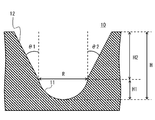
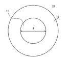
- 2 and 3 are diagrams illustrating examples of the shape of the recess according to the first embodiment.
- FIG. 2 shows a cross-sectional view when one recess 10 is viewed from the side
- FIG. 3 shows a view when one recess 10 is viewed from above.
- Each recess 10 includes a bottom 11 and an opening 12.
- the bottom portion 11 is a portion that becomes the bottom of the culture vessel 1
- the opening portion 12 is a portion that is disposed on the top of the bottom portion 11.
- a portion where the bottom 11 and the opening 12 are in contact is referred to as a boundary.
- the length indicated by the arrow R corresponds to the boundary position.
- the position of the boundary is indicated by a two-dot broken line.
- the bottom part 11 and the opening part 12 are comprised by the continuous surface, and are manufactured as integral.
- the equivalent diameter R is a diameter of an inscribed circle inscribed in the bottom 11 of the recess 10. Here, it refers to the diameter of an inscribed circle inscribed at the boundary between the bottom 11 and the opening 12. More specifically, the equivalent diameter R refers to the diameter of an inscribed circle in the shape of a surface perpendicular to the direction of the height H of the recess 10 at the boundary.
- the depth H is a length from the bottom inside the bottom portion 11 to the upper end of the recess 10. The upper end of the recess 10 is the same as the end (upper end) of the opening 12.
- the depth H is the depth of the space formed by the recess 10.
- FIG. 2 shows the depth H1 of the bottom 11 and the depth H2 of the opening 12 in addition to the depth H of the recess 10.
- the bottom 11 forms a space for culturing cells (first space).
- the bottom 11 has, for example, a hemispherical shape.
- a shape in which a spherical shape having an equivalent diameter R as a diameter is halved can be used.
- the shape of the bottom 11 is not limited to a hemisphere.
- the opening 12 forms a space (second space) that works to assist cell culture and recovery.
- the opening 12 is configured by a wall having a taper angle of 1 degree or more and 20 degrees or less that surrounds the boundary from the bottom part 11 to the end part (tip) of the recess 10.
- the taper angle of the wall constituting the opening 12 is preferably 5 degrees or more and 15 degrees or less, and more preferably 10 degrees.
- the taper angles are indicated by the symbols ⁇ 1 and ⁇ 2.
- the taper angles ⁇ 1 and ⁇ 2 are substantially the same.
- the boundary between the bottom 11 and the opening 12 is formed such that the equivalent diameter R is 50 ⁇ m or more and 1 mm or less.
- the equivalent diameter is preferably 50 ⁇ m or more and 500 ⁇ m or less, more preferably 100 ⁇ m or more and 500 ⁇ m or less.
- the depth H from the bottom to the end of the bottom is formed to be not less than 0.5 times and not more than 3 times the equivalent diameter R.
- the depth H is preferably 0.7 times or more and 1.2 times or less of the equivalent diameter R, and more preferably 0, 8 to 1 times.
- the culture vessel is preferably flat between two adjacent recesses 10.
- the distance between the two recesses 10 is preferably in the range of 5 ⁇ m to 50 ⁇ m. The reason is that the cells can be prevented from climbing on the walls, where the cells adhere, proliferate and inhibit spheroid formation. However, if it is thin, cracks may easily occur due to vibration during cell seeding or medium exchange. Therefore, it is preferable that it is 5 micrometers or more. From such a viewpoint, 5 to 20 ⁇ m is preferable.
- the culture vessel 1 is preferably manufactured as follows.
- the culture vessel 1 is an acrylic resin, polylactic acid, polyglycolic acid, styrene resin, acrylic / styrene copolymer resin, polycarbonate resin, polyester resin, polyvinyl alcohol resin, ethylene / vinyl alcohol copolymer resin, A resin molded article made of one or a combination of a thermoplastic elastomer, a vinyl chloride resin, and a silicon resin is preferable.
- a functional group is formed on each recess 10 of the culture vessel 1 by a surface modification treatment method comprising any one of plasma treatment, glass coating, corona discharge, UV ozone treatment, or a combination thereof, and the water contact angle is 45 degrees. It is preferable to process so that it may become the following.
- a hydrophilic polymer chain that inhibits cell adhesion is preferably immobilized in each recess 10. More preferably, the hydrophilic polymer chain is fixed to each of the recesses 10 treated so that the water contact angle is 45 degrees or less.
- a phospholipid or a phospholipid / polymer complex is immobilized in each recess 10. This immobilization treatment is performed on each of the dents 10 processed so that the water contact angle is 45 degrees or less, each of the dents 10 on which the hydrophilic polymer chain is fixed, or each of the dents 10 in combination thereof. More preferably it is implemented.
- a functional group is formed on each recess 10 by a surface modification treatment method comprising any one of plasma treatment, glass coating, corona discharge, UV ozone treatment, or a combination thereof, so that the water contact angle is 45 degrees or less.
- a hydrophilic polymer chain that inhibits cell adhesion and a cell non-adherent surface on which any one polymer of phospholipid or phospholipid / polymer complex is immobilized Preferably there is.
- This process is more preferably performed together with the processes described above or a combination of the processes.
- the hydrophilic polymer chain described above is preferably polyhydroxyethyl methacrylate, and the average molecular weight of polyhydroxyethyl methacrylate is more preferably 100,000 or more.
- the culture container 1 may be a culture container in which the micropattern shown in FIGS. 4 and 5 is formed in addition to the one shown in FIGS.
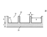
- FIG. 4 shows another example of the shape of the culture vessel used in one embodiment.
- FIG. 5 is a cross-sectional view taken along line VV of the culture vessel shown in FIG.
- the culture container 30 has a culture space 31, a wall 32, and a bottom 33.
- the culture space 31 is an area partitioned by the wall 32 and the bottom 33, and becomes a three-dimensional space area (culture area) in which cells are cultured.
- the culture space 31 is also simply referred to as “space” or “microspace”.
- the wall 32 is a partition wall that partitions the culture space 31, and can be said to be a convex portion that forms an uneven pattern in the culture vessel 30.
- the bottom 33 functions as a substrate of the culture vessel 30 and the surface on the side where the culture space 31 is arranged becomes a part of the culture region (culture surface).
- the bottom 33 is, for example, the same region as the bottom of each well formed on the culture plate of FIG. 1, and the bottom of each well is used.
- the bottom 33 forms the bottom of the culture space 31.
- a part of the surface forming the culture space 31 and the surface of the bottom serving as a culture region is also referred to as a “bottom culture surface 34”.
- the equivalent diameter D, the height (depth) H, the width (thickness) W of the wall 32, and the thickness T of the bottom 33 are shown with respect to the culture space 31 formed in the culture vessel 30. . 4 and 5, the bottom 33 is shown as being integrally formed with the wall 32.
- the equivalent diameter D is the same as the equivalent diameter R in FIG. 2 and refers to the diameter of an inscribed circle inscribed in the culture space 31. More specifically, the equivalent diameter D is the inscribed circle of the shape of the surface parallel to the bottom 33 of the culture space 31 (front shape), in other words, the shape of the surface perpendicular to the direction of the height H of the culture space 31.
- the height H is the length from the bottom of the culture space 31 (bottom culture surface 34) to the top surface of the wall 32, and can also be said to be the depth of the culture space 31. Further, when the bottom culture surface 34 is a flat surface, the height H is the same as the height of the wall 32.
- the width W of the wall 32 is not only the thickness of the wall 32 but also the distance separating the adjacent culture spaces 31.
- the plurality of culture spaces 31 are arranged in an array as shown in FIG.
- the number or size of the culture spaces 31 included in the culture vessel 30 depends on the number of wells (well size) prepared in the culture plate and the sizes of the culture spaces 31 and the walls 32. 4 and 5, nine culture spaces 31 are shown. This is shown for explanation, and does not correspond to the number of culture spaces 31 included in the actual culture container 30 (each well).
- FIG. 6 shows an example of the shape of the recess 20D in which the bottom is linear, in other words, the bottom does not form a space.
- FIG. 6 the front view which looked at the dent 20D in the upper stage from the top, and sectional drawing are shown in the lower stage.
- the recess 20 ⁇ / b> D is composed of the opening 12.
- the recess 10 is also described as a micro container.
- the recess 10 is preferably configured as follows.
- the recess 10 preferably has an equivalent diameter of 20 ⁇ m to 2.5 mm and a depth of 20 ⁇ m to 2.5 mm.
- the tissue structure is preferably cultured using a culture vessel whose culture surface is a non-cell-adherent surface.
- One or a culture surface selected from the group consisting of phospholipid, phospholipid / polymer complex, poly (2-hydroxyethyl methacrylate) (PHEMA), polyvinyl alcohol, agarose, chitosan, polyethylene glycol, and albumin
- PHEMA poly (2-hydroxyethyl methacrylate)
- the culture container 1 is preferably coated with a culture surface in contact with the cells, in which the polymer composed of the combination comes into contact.
- Vascular cells stem cell-derived endoderm, ectoderm or mesoderm cells: mesenchymal cells are co-cultured at a ratio of 10: 7 to 10: 1 to 2 and 20 per recess 10 (microcontainer) Cells are preferably seeded at a density of 1 to 2000 cells.
- the recess 10 is composed of a bottom portion 11 and an opening portion 12, and the opening portion 12 is composed of a wall having a taper angle of 1 to 20 degrees surrounding the boundary from the bottom portion 11 to the end portion. It preferably has a hemispherical shape or a truncated cone shape.
- tissue structure produced as described above can be used for drug discovery screening and regenerative medicine.
- the present invention also provides a method for evaluating a drug using the tissue structure produced by the above method.
- drug evaluation include prediction of drug metabolism profile of drug candidate compounds, drug efficacy evaluation, toxicity evaluation, and drug interaction evaluation.
- liver endoderm cells were divided into vascular endothelial cells (human umbilical cord blood-derived venous endothelial cells) (Lonza, Basel, Switzerland) and undifferentiated mesenchymal cells (between humans).
- Leaf stem cells (Lonza, Basel, Switzerland) were mixed at a ratio of 10: 5-10: 2 to prepare a cell solution.
- Vascular endothelial cells and undifferentiated mesenchymal cells were each fluorescently labeled.
- Example> In FIGS. 1 to 3, a 24-well plate culture vessel having a recess (space) having an equivalent diameter of 500 ⁇ m and a depth of 500 ⁇ m was used. In order to suppress cell adhesion, p-HEMA was coated on the culture surface in contact with the cells. When culturing was performed for 3 to 15 days, aggregates were formed. On the 15th day of culture, aggregates (cultured products) were collected and analyzed for gene expression. At this time, the aggregate was 2 mm or less.
- ⁇ Comparative example> A matrigel-coated ⁇ 3 cm dish was used. The cell suspension was seeded onto a culture dish on which the stock solution or 2-fold diluted Matrigel (BD pharmingen) had been immobilized. When culturing was performed for 3 to 15 days, aggregates were formed. When the size of the aggregate became 2 mm or more and the culture was continued thereafter, nutrients were not supplied to the central part and necrosis occurred. Therefore, the culture was terminated when the diameter of the aggregate reached 2 mm. Gene expression analysis of the culture product thus prepared was performed.
- 2-fold diluted Matrigel BD pharmingen
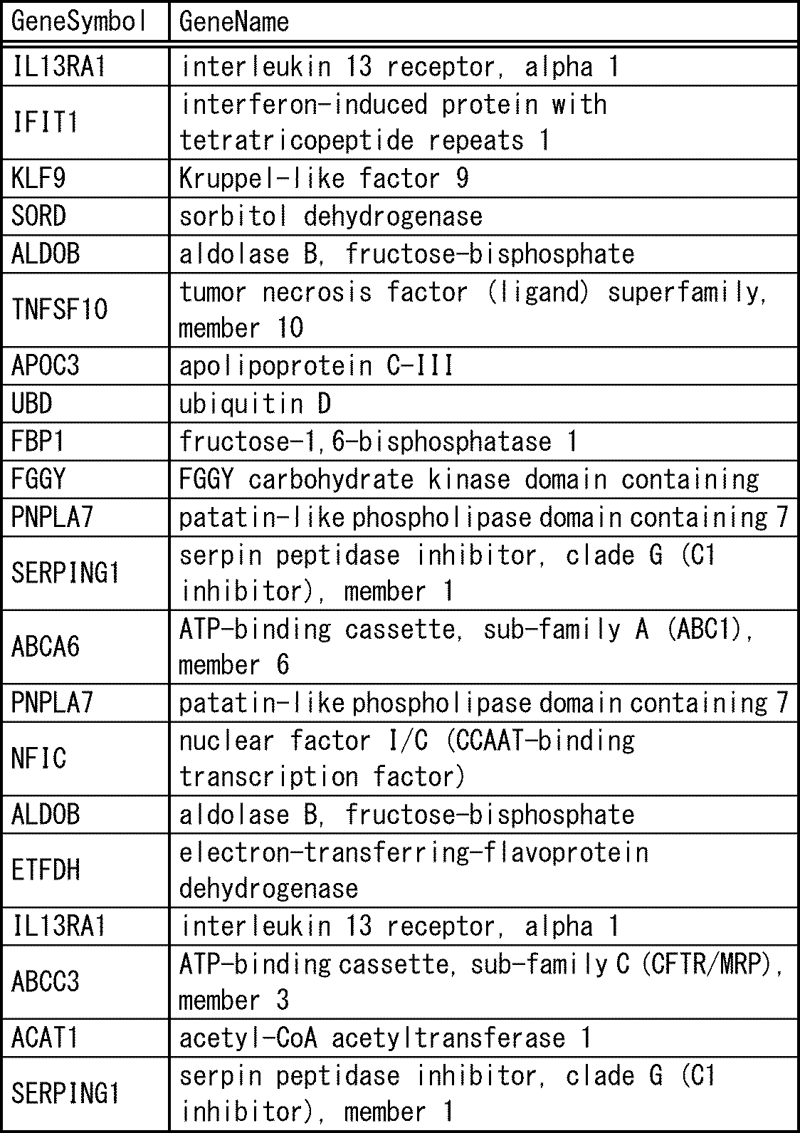
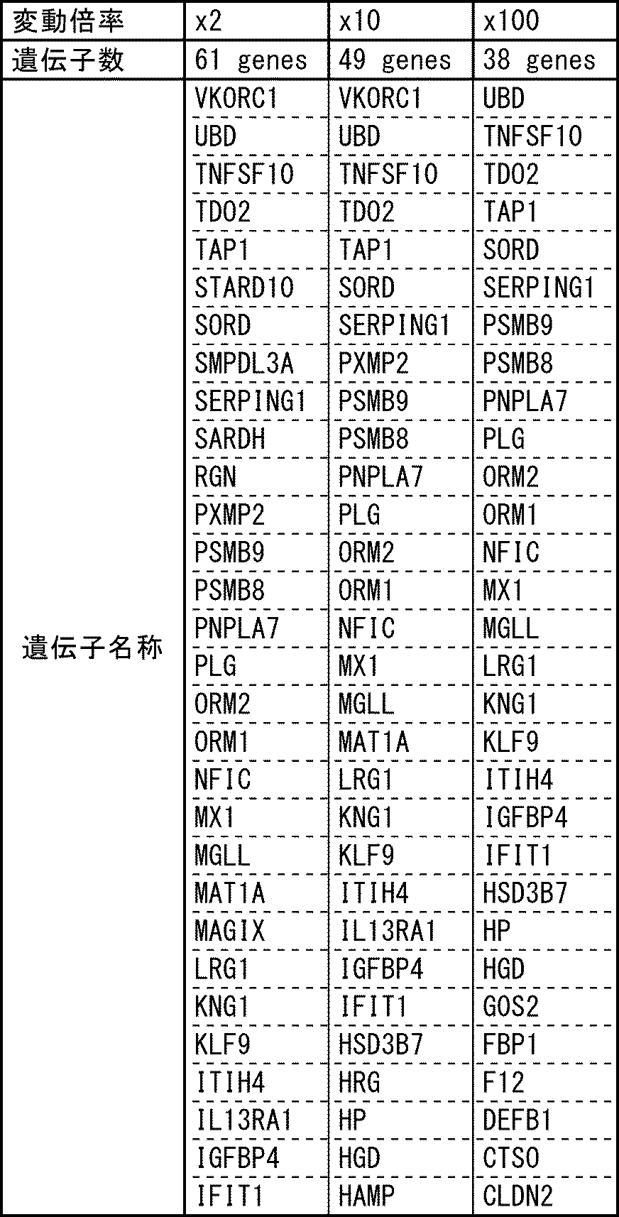
- the gene expression level (value A) of the liver bud prepared according to Patent Document 3 and the gene expression level (value D) of the tissue structures of the examples were calculated. Species whose values D increased by 2 times, 10 times or more and 100 times or more with respect to value A were extracted (Tables 2-1 and 2-2). There were 49 genes that rose more than 10 times. There were 61 gene species that increased more than 2-fold, and 38 gene species increased more than 100-fold.
Landscapes
- Health & Medical Sciences (AREA)
- Engineering & Computer Science (AREA)
- Life Sciences & Earth Sciences (AREA)
- Chemical & Material Sciences (AREA)
- Organic Chemistry (AREA)
- Zoology (AREA)
- Wood Science & Technology (AREA)
- Bioinformatics & Cheminformatics (AREA)
- Biomedical Technology (AREA)
- Genetics & Genomics (AREA)
- Biotechnology (AREA)
- General Engineering & Computer Science (AREA)
- Biochemistry (AREA)
- Microbiology (AREA)
- General Health & Medical Sciences (AREA)
- Cell Biology (AREA)
- Sustainable Development (AREA)
- Molecular Biology (AREA)
- Immunology (AREA)
- Clinical Laboratory Science (AREA)
- Proteomics, Peptides & Aminoacids (AREA)
- General Chemical & Material Sciences (AREA)
- Physiology (AREA)
- Vascular Medicine (AREA)
- Physics & Mathematics (AREA)
- Analytical Chemistry (AREA)
- Biophysics (AREA)
- Micro-Organisms Or Cultivation Processes Thereof (AREA)
- Measuring Or Testing Involving Enzymes Or Micro-Organisms (AREA)
- Apparatus Associated With Microorganisms And Enzymes (AREA)
Abstract
Description
創薬スクリーニング試験においては、生体内での試験、いわゆるin vivo試験と同様の薬剤感受性、毒性反応を示すことが要求される。このような用途で利用するためには、上述した先行技術では不十分であり、より成熟化した、すなわち生体内の細胞がもつ機能に匹敵するレベルの機能が発現している細胞が求められている。
肝細胞の薬物代謝酵素のひとつであるチトクロムP450は57種類の遺伝子があることからも分かるように、細胞は非常に多くの機能を有している。これらが同時または必要に応じて機能することで生命を維持している。
また、肝細胞については80にも及ぶ遺伝子の発現量が胎児期から成体になっていくにつれ増加することが確認されている。さらに膵臓については、非特許文献3,4に、発生過程において、細胞が成熟するに従い遺伝子発現プロファイルが変化することが示されている。
そのため、従来の評価指標で判定された細胞や臓器を用いて薬の作用効果や毒性などを判定する場合、生体内の反応を正確に予測できないという問題や、人工臓器が十分に機能しないという問題が生じる。
さらに、本発明の一実施形態に係る組織構造体において、前記複数の機能が、10種類以上のタンパク質について測定したタンパク質量であり、前記10種類以上のタンパク質が、前記幹細胞由来の内胚葉、外胚葉または中胚葉細胞のタンパク質量に対し前記組織構造体のタンパク量が20%以上変動する全てのタンパク質であることが好ましい。前記組織構造体がスフェロイド形状であり、スフェロイドの直径が50μm~2mmであることがより好ましい。
また、前記複数の機能が、肝臓または膵臓に特有の機能であることが好ましい。
さらに、本発明の一実施形態に係る組織構造体の作製方法において、前記複数の機能が、10種類以上のタンパク質について測定したタンパク質量であり、前記10種類以上のタンパク質が、前記幹細胞由来の内胚葉、外胚葉または中胚葉細胞のタンパク量に対し組織構造体のタンパク量が20%以上変動する全てのタンパク質であること、及び、前記組織構造体の抽出が、前記幹細胞由来の内胚葉、外胚葉または中胚葉細胞及び前記培養形成物のタンパク質量を測定し、前記幹細胞由来の内胚葉、外胚葉または中胚葉細胞のタンパク質量に対し、前記培養形成物のタンパク質量が2倍以上変動するタンパク質を10種類以上有する培養形成物を選択することが好ましい。
また、本発明の一実施形態に係る組織構造体の作製方法において、前記共培養の工程が、凝集体を形成させる工程、器官芽を形成させる工程、さらに培養を行い成熟化させる工程を含むことが好ましい。加えて、凝集体、器官芽、成熟化させる工程において、細胞同士が結合して凝集していることが好ましく、凝集体、器官芽、成熟化させる工程において、細胞同士が結合してスフェロイド形状の塊が形成されていることがより好ましい。
本発明の一実施形態に係る組織構造体の作製方法において、前記細胞同士が形成するスフェロイドの直径が50μm~2mmであることが好ましい。前記幹細胞由来の内胚葉、外胚葉または中胚葉細胞が、胎生幹細胞または人工多能性幹細胞由来から選択される細胞であることが好ましく、前記幹細胞由来の内胚葉、外胚葉または中胚葉細胞が、人工多能性幹細胞由来の細胞から内胚葉系列の細胞に分化可能な細胞であることがより好ましい。
また、前記複数の機能が、肝臓または膵臓に特有の機能であることが好ましい。
本発明の一実施形態に係る組織構造体の作製方法において、前記組織構造体を、相当直径が20μm以上2.5mm以下、深さが20μm以上1000μm以下のマイクロ容器で培養することが好ましい。加えて、前記組織構造体を、培養表面が細胞非接着表面である培養容器を用いて培養することが好ましい。さらに加えて、前記培養表面が、リン脂質、リン脂質・高分子複合体、ポリ(2-ヒドロキシエチルメタクリレート)(PHEMA)、ポリビニルアルコール、アガロース、キトサン、ポリエチレングリコール、及びアルブミン、のグループから選択される一つまたはこれら組合せからなるポリマーが細胞と接触する培養面にコートされている培養容器であることが好ましい。
本発明の一実施形態に係る組織構造体の作製方法において、血管細胞:幹細胞由来の内胚葉、外胚葉または中胚葉細胞:間葉系細胞を、10:7~10:1~2の割合で共培養し、かつ、マイクロ容器1個あたり20個~2000個となる密度で細胞を播種することが好ましい。また、前記マイクロ容器が、底部と開口部とから構成されており、前記開口部が、前記底部との境界から端部までを囲むテーパ角1度以上20度以下の壁で構成され、前記底部が、半球状と円錐台とのいずれかの形状を有することが好ましい。
生理的な器官発生過程において、臓器細胞が血管細胞および未分化な間葉系細胞と密な細胞間相互作用を持つことで、自律的な組織構造の構築と細胞分化を伴う器官形成が進行する。
そこで、一実施形態では、この様な器官発生の早期プロセスを人為的に再現することで、複数の細胞系譜による相互作用を介した初期分化誘導を行い、初期分化を遂げた細胞の組織形成能力を誘導する。そして、培養容器内で成熟細胞が有する複数の機能を発現する、分化させた細胞または生体組織、例えば組織・臓器の元となる器官芽(organ bud)を経由して培養容器内で細胞または生体組織を培養して成熟させ、細胞と血管系とから成る組織の作製を行うものである。
一実施形態の組織構造体は、血管細胞、間葉系細胞、血管細胞から分泌される因子、間葉系細胞から分泌される因子、血管細胞と間葉系細胞の両方が存在することによって分泌される因子からなる群より選択される少なくとも1つの細胞及び/又は因子とともに、幹細胞由来の内胚葉、外胚葉または中胚葉細胞を共培養して得られる組織構造体である。加えて、組織構造体は、複数の機能の値についてピアソンの積率相関係数(Pearson product-moment correlation coefficient)を用いて検定した値が、胎児から採取された細胞または胎児から採取された生体組織より、成体から採取された細胞または成体から採取された生体組織に近い値である。
組織構造体は、少なくとも二種類、(好ましくは三種類)の細胞を共培養することにより、器官芽を形成させ、さらに器官芽を成熟させて、複数の機能について上述した特徴を有する組織構造体を形成する。器官芽については後述するが、成熟することで器官に分化することができる構造体である。
「生体組織(biological tissue)」とは、何種類かの決まった細胞が一定のパターンで集合した構造の単位のことであり、全体としてひとつのまとまった役割をもつ。例えば、生体内の各器官(臓器)は、何種類かの組織が決まったパターンで集まって構成されている。本明細書では、分化した細胞により構成され、任意の機能を有する細胞の集まり(細胞群)を組織という。
幹細胞とは胎生幹細胞(ES細胞)または人工多能性幹細胞由来(iPS細胞)から選択される細胞を含み、無限増殖性を有しており、かつ、内胚葉、中胚葉、外胚葉の全ての器官に分化可能な細胞をいう。
内胚葉細胞とは、肝臓、膵臓、腸管、肺、甲状腺、副甲状腺、尿路などの中胚葉性器官に分化可能な細胞をいう。
外胚葉細胞とは、脳、脊髄、副腎髄質、表皮、毛髪・爪・皮膚腺、感覚器、末梢神経、水晶体などの外胚葉系器官に分化可能な細胞のことをいう。
中胚葉細胞とは、腎臓、尿管、心臓、血液、生殖腺、副腎皮質、筋肉、骨格、真皮、結合組織、中皮などの中胚葉系器官に分化可能な細胞のことをいう。
すなわち、幹細胞由来三胚葉細胞とは、ES細胞またはiPS細胞から選択された細胞に由来する、内胚葉、外胚葉または中胚葉系器官の性質を有する細胞をいう。
ある細胞が外胚葉性器官、中胚葉性器官又は内胚葉性器官に分化可能な細胞であるかどうかは、マーカーとなるタンパク質の発現を調べることにより確認できる(マーカータンパク質のいずれか一つあるいは複数が発現していれば内胚葉性器官に分化可能な細胞であると判断できる。)。例えば、肝臓に分化可能な細胞では、HHEX、SOX2、HNF4A、AFP、 ALBなどがマーカーになり、膵臓に分化可能な細胞では、PDX1、SOX17、SOX9などがマーカーになり、腸管に分化可能な細胞では、CDX2、SOX9などがマーカーになり、腎臓に分化可能な細胞では、SIX2、SALL1、心臓に分化可能な細胞では、NKX2-5 MYH6、ACTN2、MYL7、HPPA、血液に分化可能な細胞では、C-KIT、SCA1、TER119、HOXB4、脳や脊髄に分化可能な細胞では、HNK1、AP2、NESTINなどがマーカーになる。
「成体から採取された細胞」とは、十分に成長して、生殖が可能となった生物体から採取した細胞である。成体から採取された細胞は、例えば、市販されているヒト初代肝細胞やバイオプシーした生体組織を含む。
動物から採取することも可能である。例えば、ラットやマウスから採取する場合、2段階コラゲナーゼ灌流法により肝細胞を分離する方法がある。例えばラットの場合、門脈よりカニューレを入れ、37度に加温したリン酸緩衝液(前還流液)で脱血し、次に37度に加温したコラゲナーゼ溶液でコラーゲンを分解して細胞のみを回収する方法を利用できる。
「胎児から採取された、細胞または生体組織」は、市販されているものがある。セルバンク等から入手可能である。例えば、株式会社ベリタスから入手することができる。
複数の機能は、組織構造体が有する機能、言い換えると、組織構造体から検出可能な機能である。一実施形態では、例えば、遺伝子発現量、タンパク質量によって測定できる機能を用い、特に、幹細胞由来三胚葉細胞から組織構造体に分化するときに、変動する機能を用いる。具体的には、組織構造体が有する機能のうち、胎児から採取された細胞と成体から採取された細胞とが有する機能を比較したときに、機能の量が変動する機能を用いる。そして、組織構造体は、胎児から採取された細胞より成体から採取された細胞に近い機能の量を有する構造体が選択(抽出)される。
ここで、胎児から採取された細胞は、胎児から採取された細胞とその生体組織とを含み、成体から採取された細胞は、成体から採取された細胞とその生体組織とを含む。
遺伝子種を選択する場合は、器官芽の遺伝子発現量に対し組織構造体の遺伝子発現量が2倍以上変動する遺伝子種を含む。タンパク種を選択する場合は、器官芽のタンパク量に対し組織構造体のタンパク量が20%以上変動するタンパク種を含む。
または、10種類以上の遺伝子が、幹細胞由来三胚葉細胞の遺伝子発現量に対し、組織構造体の遺伝子発現量が2倍以上変動する遺伝子を含む。より具体的には、遺伝子発現量は、全ての遺伝子断片が固定されているDNAチップを用いて解析された値を用いる。そして、10種類以上の遺伝子は、幹細胞由来三胚葉細胞の遺伝子発現量に対し、組織構造体の遺伝子発現量が2倍以上変動するすべての遺伝子を含む。
10種類以上の遺伝子の発現量を用いるのは、遺伝子の発現量によって組織構造体が幹細胞由来三胚葉細胞から成熟したことを判定するための数を設定したものである。
別の例として、複数の機能は、10種類以上のタンパク質について測定したタンパク質量を用いることができる。この場合、幹細胞由来三胚葉細胞及び組織構造体のタンパク質量を測定したときに、10種類以上のタンパク質が、幹細胞由来三胚葉細胞のタンパク質量に対し組織構造体のタンパク量が20%以上変動する全てのタンパク質を含む。
なお、複数の機能は、上述した具体例に限られることはない。複数の機能は、任意の装置で機能の量や大きさが測定できるものや要素などであればよい。加えて、幹細胞由来三胚葉細胞または器官芽が組織構造体に分化・成熟するときに、機能の量や大きさを測定または解析した値(測定値、解析値)が変動するものであればよく、特に2倍以上に変動するものであれば好ましい。
複数の機能については、採用する機能を測定する機器を用いて測定する。
遺伝子発現量は、例えば遺伝子マイクロアレイ解析装置を用いて解析することができる。遺伝子解析では、例えば、遺伝子解析法のひとつであるチップでの解析を用いることができ、一例が以下のURLで紹介されている。
http://www.chem-agilent.com/contents.php?id=1002411
http://www.gelifesciences.co.jp/newsletter/biodirect_mail/technical_tips/tips21.html(GEのマイクロアレイに関する原理説明)
タンパク質量は、例えばタンパク質アレイ解析装置を用いて測定することができる。タンパク質解析では、一例が以下のURLで紹介されている。
http://www.filgen.jp/Product/Bioscience2/index.htm
http://www.filgen.jp/Product/BioScience22-MS/index2.htm
加えて、目的臓器で特徴的に発現する遺伝子やタンパク質が分かっている場合には、特定の遺伝子発現量やタンパク質量について分析して、2倍以上変動するか検証することができる。目的臓器で特徴的な機能の種類が10個以上存在することが好ましい。
上記のような方法で網羅的に分析して2倍以上変動するものが10個以上あることがより好ましい。
膵臓の性質、特に膵島β細胞を含む組織構造体の場合、非特許文献4や5に挙げられている胎児と成体組織で変動する遺伝子種またはタンパク種全てまたはこれらから選択することができる。
組織構造体は、血管細胞、間葉系細胞、血管細胞から分泌される因子、間葉系細胞から分泌される因子、血管細胞と間葉系細胞の両方が存在することによって分泌される因子からなる群より選択される少なくとも1つの細胞及び/又は因子とともに、幹細胞由来三胚葉細胞を共培養することによって作製する。
詳細には、一実施形態の組織構造体の作製方法は、例えば、少なくとも二種類(好ましくは三種類)の細胞を共培養して分化した細胞を形成させ、さらに共培養を継続することによって形成した分化した細胞をさらに成熟させた培養形成物を得て、複数の機能を生じさせる。そして、培養形成物のうち、複数の機能を有するものを組織構造体として抽出する。
「共培養」とは、一般には、一緒に培養する2種あるいはそれ以上の異なる種類の細胞を混合し、細胞を培養することをいう。
以下で説明する共培養は、(1)凝集体を形成させる工程、(2)器官芽を形成させる工程、(3)培養を行い成熟化させる工程、を含む。
以下、各工程について説明する。
凝集体を形成させる工程は、幹細胞由来三胚葉細胞として用いる凝集体を形成する。
器官芽を形成させる工程は、三種類の細胞、血管細胞、幹細胞由来三胚葉細胞及び間葉系細胞を共培養して器官芽を形成させる。
成熟化工程では、形成した器官芽を成熟させて培養形成物(組織構造体候補:下記組織構造体作製容器で培養した培養形成物を指す)を形成する。言い換えると、培養形成物を形成し、複数の機能を検定して適切な(要件を満たす)培養形成物を組織構造体とする。
例えば、組織構造体を形成させるために使用する培養容器(組織構造体作製容器)と同じスケール、スケールダウンまたはスケールアップした培養容器(試験容器)を別途用意する。試験容器の培養は、組織構造体作製容器の場合と同じ培地の組成、播種密度、培地交換頻度、細胞と培地量の比率とする。そして試験容器で作製された培養形成物を用いて遺伝子発現量の測定行う。複数の機能が、10種類以上の遺伝子の発現量であり、10種類以上の遺伝子が、幹細胞由来三胚葉細胞の遺伝子発現量に対し、組織構造体の遺伝子発現量が2倍以上変動する遺伝子を抽出する。
抽出した遺伝子種について、試験容器で作製された培養形成物、胎児から採取された細胞または生体組織、成体から採取された細胞または生体組織の遺伝子発現量を測定、判定に用いる。即ち、試験容器で培養した培養形成物の判定結果を、組織構造体作製容器で作製された培養形成物の判定結果に置き換えることができる。
上記はセルスタックやフラスコを組織構造体作製容器に使用、ディッシュや小さいフラスコ(24cm^2)を試験容器として使用した場合を想定している。ウェルプレートの場合は異なるウェルを試験容器(言い換えると、試験ウェル)として取り扱うことができる。
成熟化工程において、使用する培地は所望する組織用の培地を用いることが好ましく、血管細胞用の培地や幹細胞用の培地を混合することが好ましい。
成体との類似性を評価する方法であり、成体に近い機能を有することが前提であることから、成体から採取された細胞または生体組織と、組織構造体の遺伝子発現量は正の相関を示す。負の相関を示した場合は、成熟化が不十分であり成体に近くないと判定する。即ち、成体から採取された細胞または生体組織と組織構造体の遺伝子発現量は正相関を示すことが必要条件である。以下の[a]、[b]との間に、[a]が[b]より小さく、かつ[b]が1より小さい関係(言い換えると、式[a]<[b]<1を満たす関係)が得られれば組織構造体候補を組織構造体と認定する。
[a]「生体から採取された細胞または生体組織の遺伝子発現量」と「培養形成物(試験容器)の遺伝子発現量」の相関係数が正である、かつ、「胎児から採取された細胞または生体組織の遺伝子発現量」と「培養形成物(試験容器)の遺伝子発現量」の相関係数
[b]「生体から採取された細胞または生体組織の遺伝子発現量」と「培養形成物(試験容器)の遺伝子発現量」の相関係数
凝集体形成工程、器官芽形成工程、成熟化工程において、細胞同士が結合して凝集していることが好ましい。また凝集体形成工程、器官芽形成工程、成熟化工程において、細胞同士が結合してスフェロイド形状の塊が形成されていることが好ましい。さらに加えて、細胞同士が形成するスフェロイドの直径が50μm~2mmであることが好ましい。
<培養容器>
図1は、一実施形態の培養容器の一例を示す図である。図1では、複数の培養容器1を有する培養プレート3の一部分を示す。図1の上段には、培養容器1の底に形成される複数の凹み10の一部分を、培養プレート3の上からみた図を示す。培養容器1は、複数の凹み10が配置される。複数の凹み10は、培養容器1の製造や細胞培養の効率の観点から、規則的に配置されることが好ましい。培養容器1は例えば複数のウェルを有するウェルプレートの一つのウェルに相当する。言い換えると、ウェルプレートの各ウェルに複数の凹み10が配置されることになる。
ウェルプレートは、多数のくぼみ(穴またはウェル)のついた平板からなる実験・検査器具であり、各ウェルを試験管あるいはシャーレとして利用するものをいう。ウェルの数には例えば、6、24、96、384などがあり、それ以上の数のものもある。ウェルの底は平らなもの、丸いもののほか、細長いマイクロチューブを多数組み合わせた形式のもの(ディープウェルプレート)もある。
図2、3は、実施形態1の凹みの形状例を示す図である。図2では、一つの凹み10を横から見たときの断面図を示し、図3は、一つの凹み10を上から見たときの図を示す。
各凹み10は、底部11と開口部12とから構成される。底部11は、培養容器1の底になる部分であり、開口部12は、底部11の上部に配置される部分である。底部11と開口部12とが接する部分を境界と記載する。図2では、符号Rの矢印で示す長さの部分が境界の位置に対応する。また、図3では、境界の位置を2点破線で示している。ただし、底部11と開口部12とは連続した面で構成され、一体として製造される。
相当直径Rは、凹み10の底部11に内接する内接円の直径をいう。ここでは、底部11と開口部12との境界において内接する内接円の直径をいう。より詳しくは、相当直径Rは、境界における、凹み10の高さHの方向と垂直になる面の形状の内接円の直径をいう。
深さHは、底部11の内側の底から凹み10の上端までの長さである。凹みの10の上端は、開口部12の端部(上端)と同じである。深さHは、凹み10が形成する空間の深さである。言い換えると、底部11が形成する空間の底から開口部12が形成する空間の上端までの深さである。図2では凹み10の深さHに加え、底部11の深さH1及び開口部12の深さH2を示している。
開口部12は、細胞の培養及び回収を補助するように働く空間(第2空間)を形成する。開口部12は、底部11との境界から凹み10の端部(先端)までを囲むテーパ角が1度以上20度以下の壁で構成される。開口部12を構成する壁のテーパ角が5度以上15度以下度以下であることが好ましく、10度がより好ましい。その理由は、テーパ角が小さすぎると回収する際に細胞が凹みから培地に移行できず、逆に大きすぎると培地交換中に細胞が離脱するからである。
図2ではテーパ角を符号θ1、θ2で示す。図2,3に示す凹み10の形状例では、テーパ角θ1、θ2は略同じ場合を示している。
底部11と開口部12との境界は、相当直径Rが50μm以上1mm以下となるように形成される。スフェロイドの中心部まで栄養を供給したい場合は相当直径50μm以上500μm以下が好ましく、より好ましくは100μm以上500μm以下が好ましい。
加えて、底部の底から端部までの深さHが相当直径Rの0.5倍以上3倍以下となるように形成される。好ましくは、深さHが相当直径Rの0.7倍以上1.2倍以下であり、より好ましくは、0、8~1倍である。
培養容器1が、アクリル系樹脂、ポリ乳酸、ポリグリコール酸、スチレン系樹脂、アクリル・スチレン系共重合樹脂、ポリカーボネート系樹脂、ポリエステル系樹脂、ポリビニルアルコール系樹脂、エチレン・ビニルアルコール系共重合樹脂、熱可塑性エラストマー、塩化ビニル系樹脂、及びシリコン樹脂のうちの1つまたはこれらの組み合わせからなる樹脂成形品であることが好ましい。
加えて、各凹み10へ、細胞接着を阻害する親水性のポリマー鎖が固定化されていることが好ましい。親水性のポリマー鎖は、上述した水接触角が45度以下になるように処理された各凹み10へ固定化されることがより好ましい。
さらに加えて、各凹み10へ、リン脂質、または、リン脂質・高分子複合体が固定化されていることが好ましい。この固定化の処理は、上述した水接触角が45度以下になるように処理された各凹み10、親水性のポリマー鎖が固定化された各凹み10、またはこれらを組合せた各凹み10へ実施されることがより好ましい。
また、上述した親水性のポリマー鎖がポリヒドロキシエチルメタクリレートであることが好ましく、さらに、ポリヒドロキシエチルメタクリレートの平均分子量が10万以上であることがより好ましい。
図4に、一実施形態で用いる培養容器の他の形状例を示す。図5は、図4に示す培養容器のV-V線断面図である。
培養容器30は、培養空間31と、壁32と、底部33とを有する。
培養空間31は、壁32と底部33とで仕切られた領域であり、細胞を培養する三次元の空間領域(培養領域)となる。培養空間31は、単に「空間」、または「マイクロ空間」とも称する。
壁32は、培養空間31を仕切る隔壁であり、培養容器30に凹凸パターンを形成する凸部ともいえる。
底部33は、培養容器30の基板として機能するとともに、培養空間31が配置される側の表面は、培養領域(培養表面)の一部となる。底部33は、例えば、図1の培養プレートに形成された各ウェルの底部と同じ領域であり、各ウェルの底部が用いられる。底部33は、培養空間31の底を形成する。底部33のうち、培養空間31を形成する面の一部分であり、かつ、培養領域となる底部の表面を、「底部培養面34」とも称する。
相当直径Dは、図2の相当直径Rと同様であり、培養空間31に内接する内接円の直径をいう。より詳しくは、相当直径Dは、培養空間31の底部33と平行する面の形状(正面の形状)、言い換えると、培養空間31の高さHの方向と垂直になる面の形状の内接円の直径をいう。培養空間31の正面の形状が、高さHに応じて異なる場合、株化肝細胞を培養する空間領域の最大値を相当直径とする。
高さHは、培養空間31の底(底部培養面34)から壁32の上面までの長さであり、培養空間31の深さでもあるともいえる。また、底部培養面34が平面の場合、高さHは、壁32の高さと同じである。
壁32の幅Wは、壁32の厚さであるとともに、隣接する培養空間31間を隔てる距離であるともいえる。
上述した事項に加え凹み10は、次のように構成されることが好ましい。
凹み10は、相当直径が20μm以上2.5mm以下、深さが20μm以上2.5mm以下であることが好ましい。
組織構造体を、培養表面が細胞非接着表面である培養容器を用いて培養することが好ましい。
培養表面が、リン脂質、リン脂質・高分子複合体、ポリ(2-ヒドロキシエチルメタクリレート)(PHEMA)、ポリビニルアルコール、アガロース、キトサン、ポリエチレングリコール、及びアルブミン、のグループから選択される一つまたはこれら組合せからなるポリマーが細胞と接触する培養面にコートされている培養容器1であることが好ましい。
凹み10が、底部11と開口部12とから構成されており、開口部12が、底部11との境界から端部までを囲むテーパ角1度以上20度以下の壁で構成され、底部11が、半球状と円錐台とのいずれかの形状を有することが好ましい。
[実験方法]
(1)幹細胞由来三胚葉細胞の作製
ヒトiPS細胞(ヒト皮膚由来TkDA3 hiPSCクローン(Koji Eto氏及び Hiromitsu Nakauchi氏より譲渡))を無血清培地にアクチビンを添加して培養することで、CXCR4及びE-cadherin両陽性の内胚葉系細胞を誘導した。得られた内胚葉系細胞をBMP4、FGF2を添加して2日間培養することで、CXCR4陰性 HNF4α陽性の肝臓内胚葉集団を得た。CXCR4及びHNF4αの発現は、Hepatology, 51(1), 297-305,2010の記載に従い、免疫染色及び遺伝子発現解析により確認した。
細胞播種割合:得られた肝臓内胚葉細胞を、血管内皮細胞(ヒト臍帯血由来静脈内皮細胞)(Lonza、Basel、Switzerland)及び未分化間葉系細胞(ヒト間葉系幹細胞)(Lonza、Basel、Switzerland)を各々10:5-10:2の割合で混合し、細胞溶液を作製した。血管内皮細胞および未分化間葉系細胞は、各々蛍光標識を行ったものを用いた。
培地:培養液は、内皮細胞培地キット-2:EGM-2 BulletKit(製品コード CC-3162:Lonza)または、内皮細胞培地キット:EGM BulletKit(製品コード CC-3124:Lonza)を用いた。
培養方法:実施例及び比較例ともに上述した細胞溶液と培地を用いた。
図1~3において、相当直径が500μm、深さが500μmの凹み(空間)を有する24ウェルプレートの培養容器を使用した。細胞接着性を抑制するため細胞が接触する培養表面にp-HEMAをコートした。
3日間~15日間培養を行ったところ凝集体が形成された。培養15日目に凝集体(培養形成物)を回収し遺伝子発現解析を行った。このとき、凝集体は2mm以下であった。
マトリゲルコートしたφ3cmディッシュを用いた。
原液ないし2倍希釈のマトリゲル(BD pharmingen)の固相化を行った培養皿の上へ細胞懸濁液を播種した。
3日間~15日間培養を行ったところ凝集体が形成された。凝集体の大きさが2mm以上となりこれ以降培養すると、中心部に栄養分が供給されず壊死することから、凝集体の直径が2mmになった時点で培養を終了した。このようにして作製した培養形成物の遺伝子発現解析を行った。
1、複数の機能の値の選択
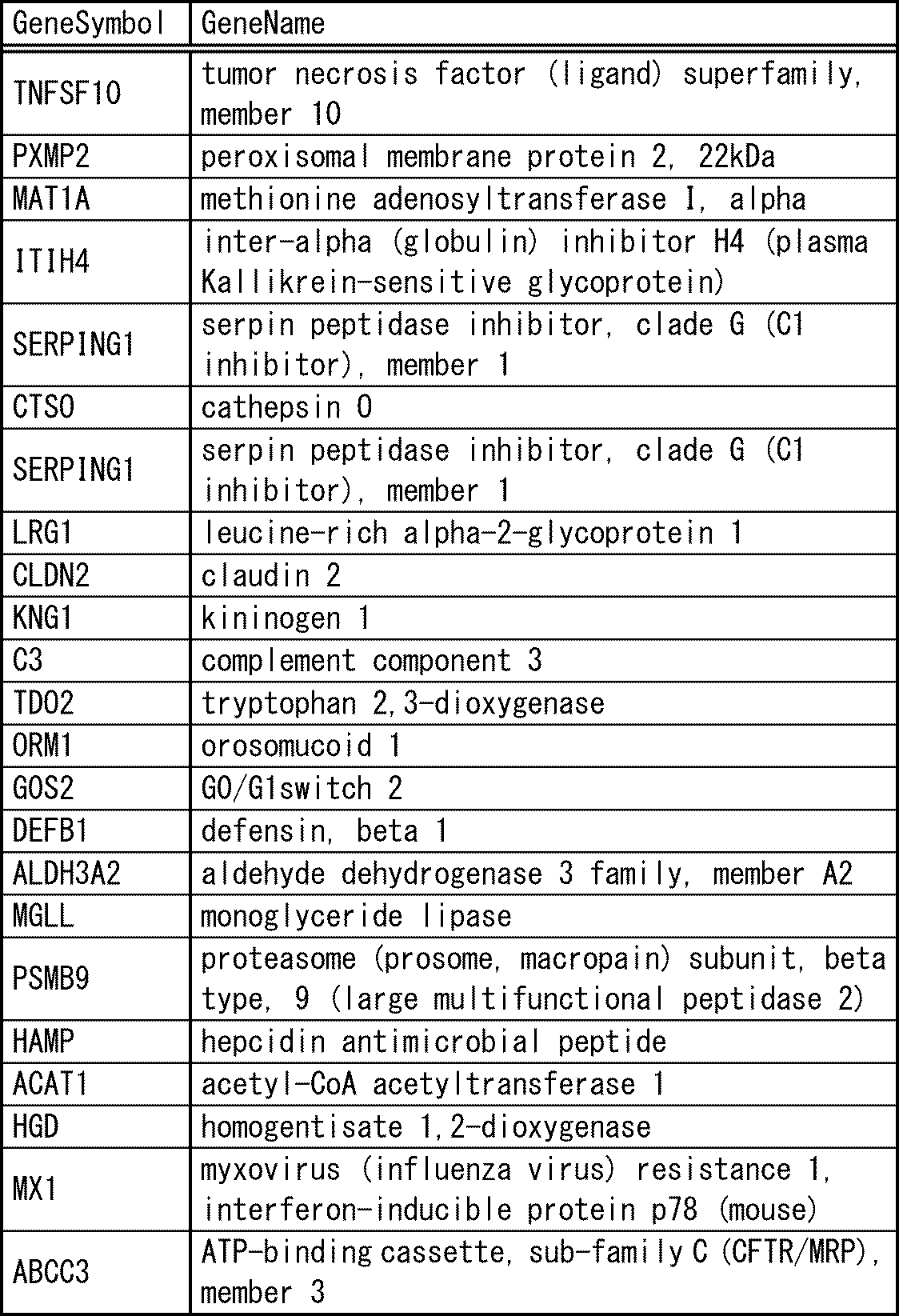
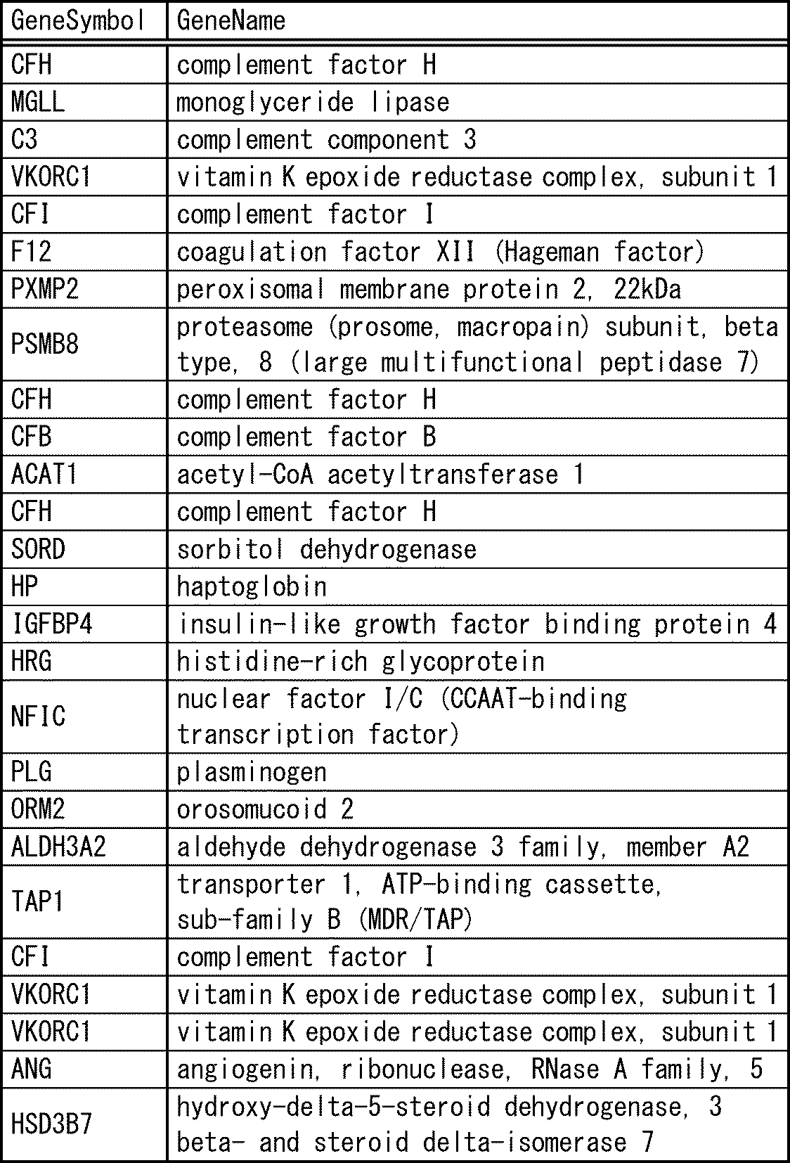
セルバンクから入手したiPS細胞、10週齢胎児由来の肝細胞及び30歳の成人由来の肝細胞を用いて、遺伝子発現量をGeneChip(アジレント・テクノロジー株式会社;Whole Human Genome DNA マイクロアレイ 4x44K v2 G4845A)を用いて網羅的に解析した(以下、成体由来の肝細胞の遺伝子発現量の値を"値B"、胎児由来の肝細胞の遺伝子発現量の値を"値C"とする)。成熟するにつれ(iPS細胞→胎児由来の肝細胞→成体由来の肝細胞)遺伝子発現量が上昇するヒトで発現する遺伝子種70遺伝子を抽出した(表1-1から表1-3)。さらに、特許文献3に従って作製した肝芽の遺伝子発現量(値A)と実施例の組織構造体の遺伝子発現量(値D)を算出した。値A対して値Dが2倍以上、10倍以上及び100倍以上上昇する遺伝種をそれぞれ抽出した(表2-1、表2-2)。10倍以上上昇する遺伝子は49遺伝子あった。2倍以上上昇する遺伝子種は61種、100倍以上上昇する遺伝子種は38遺伝子であった。

値A、B、C及びDは上述した1、複数機能の値の選択の項で用いた値を用いた。比較例の各種遺伝子発現量を測定した(値E)。尚、ここでいう遺伝子発現量は各サンプルから得られたアレイ結果 (全Probeの値) を75%shiftile値 (75パーセンタイル値) で補正、標準化した値のことを言う。
図7に生体由来の肝細胞、胎児由来の肝細胞、実施例及び比較例について、49遺伝子の遺伝子発現量の値の一部を示した。
検定
表1-1から表1-3に示した49遺伝子について、各種遺伝子発現量を求め、Gene Springを用いてピアソンの積率相関係数を算出した。
<実施例>
10倍以上変動する遺伝子 49遺伝子
(値C)と(値D)の相関係数は0.4757であった
(値B)と(値D)の相関係数は0.6314であった
図7に散布図を示した。
よって値Bの成体の肝細胞のほうが1に近いことから組織構造体と認定することができた。
<比較例>
10倍以上変動する遺伝子 49遺伝子
(値C)と(値E)の相関係数は-0.5000であった
(値B)と(値E)の相関係数は-0.3496であった
生体から採取された細胞の遺伝子発現量(値B)と比較例の遺伝子発現量の相関係数は負の相関を示した。このことから、必要条件である"正の相関"の条件を満たさない、組織構造体と認定できない。
3 培養プレート
10、20D 凹み
11 底部
12 開口部
31 培養空間
32 壁
33 底部
34 底部培養面
Claims (22)
- 血管細胞、間葉系細胞、血管細胞から分泌される因子、間葉系細胞から分泌される因子、血管細胞と間葉系細胞の両方が存在することによって分泌される因子からなる群より選択される少なくとも1つの細胞及び/又は因子とともに、幹細胞由来の内胚葉、外胚葉または中胚葉細胞を共培養して得られる組織構造体であって、複数の機能についてピアソンの積率相関係数(Pearson product-moment correlation coefficient)を用いて検定した値が、胎児から採取された細胞または生体組織より、成体から採取された細胞または生体組織の値に近い組織構造体。
- 前記複数の機能が、10種類以上の遺伝子の発現量であり、
前記10種類以上の遺伝子が、前記幹細胞由来の内胚葉、外胚葉または中胚葉細胞の遺伝子発現量に対し、前記組織構造体の遺伝子発現量が2倍以上変動する遺伝子であることを特徴とする請求項1記載の組織構造体。 - 遺伝子発現量が、全ての遺伝子断片が固定されているDNAチップを用いて解析された値であり、
前記10種類以上の遺伝子が、前記幹細胞由来の内胚葉、外胚葉または中胚葉細胞の遺伝子発現量に対し、前記組織構造体の遺伝子発現量が2倍以上変動するすべての遺伝子であることを特徴とする請求項2記載の組織構造体。 - 前記複数の機能が、10種類以上のタンパク質について測定したタンパク質量であり、
前記10種類以上のタンパク質が、前記幹細胞由来の内胚葉、外胚葉または中胚葉細胞のタンパク質量に対し前記組織構造体のタンパク量が20%以上変動する全てのタンパク質であることを特徴とする請求項1記載の組織構造体。 - 前記組織構造体がスフェロイド形状であり、スフェロイドの直径が50μm~2mmであることを特徴とする請求項1乃至4のいずれか一項に記載の組織構造体。
- 前記複数の機能が、肝臓または膵臓に特有の機能であることを特徴とする請求項1乃至5のいずれか一項に記載の組織構造体。
- 血管細胞、間葉系細胞、血管細胞から分泌される因子、間葉系細胞から分泌される因子、血管細胞と間葉系細胞の両方が存在することによって分泌される因子からなる群より選択される少なくとも1つの細胞及び/又は因子とともに、幹細胞由来の内胚葉、外胚葉または中胚葉細胞を共培養して培養形成物を作製し、
前記培養形成物が有する複数の機能に関してピアソンの積率相関係数(Pearson product-moment correlation coefficient)を用いて検定し、
検定した値が、胎児から採取された細胞または生体組織より、成体から採取された細胞または生体組織の値に近い培養形成物を組織構造体として抽出する組織構造体の作製方法。 - 前記複数の機能が、10種類以上の遺伝子の発現量であり、
前記10種類以上の遺伝子が、前記幹細胞由来の内胚葉、外胚葉または中胚葉細胞の遺伝子発現量に対し、前記組織構造体の遺伝子発現量が2倍以上変動する遺伝子であり、
前記組織構造体の抽出が、
前記幹細胞由来の内胚葉、外胚葉または中胚葉細胞及び前記培養形成物の遺伝子発現量を測定し、
前記幹細胞由来の内胚葉、外胚葉または中胚葉細胞の遺伝子発現量に対し、前記培養形成物の遺伝子発現量が2倍以上変動する遺伝子を10種類以上有する培養形成物を選択することを特徴とする請求項7記載の組織構造体の作製方法。 - 前記幹細胞由来の内胚葉、外胚葉または中胚葉細胞と前記組織構造体との遺伝子発現量を、全ての遺伝子断片が固定されているDNAチップを用いて解析したときに、前記10種類以上の遺伝子が、前記幹細胞由来の内胚葉、外胚葉または中胚葉細胞の遺伝子発現量に対し、組織構造体の遺伝子発現量が2倍以上変動するすべての遺伝子であることを特徴とする請求項8記載の組織構造体の作製方法。
- 前記複数の機能が、10種類以上のタンパク質について測定したタンパク質量であり、
前記10種類以上のタンパク質が、前記幹細胞由来の内胚葉、外胚葉または中胚葉細胞のタンパク量に対し組織構造体のタンパク量が20%以上変動する全てのタンパク質であり、
前記組織構造体の抽出が、
前記幹細胞由来の内胚葉、外胚葉または中胚葉細胞及び前記培養形成物のタンパク質量を測定し、
前記幹細胞由来の内胚葉、外胚葉または中胚葉細胞のタンパク質量に対し、前記培養形成物のタンパク質量が2倍以上変動するタンパク質を10種類以上有する培養形成物を選択することを特徴とする請求項7記載の組織構造体の作製方法。 - 前記共培養の工程が、凝集体を形成させる工程、器官芽を形成させる工程、さらに培養を行い成熟化させる工程を含むことを特徴とする請求項7乃至10のいずれか一項に記載の組織構造体の作製方法。
- 凝集体、器官芽、成熟化させる工程において、細胞同士が結合して凝集していることを特徴とする請求項11記載の組織構造体の作製方法。
- 凝集体、器官芽、成熟化させる工程において、細胞同士が結合してスフェロイド形状の塊が形成されていることを特徴とする請求項11または12記載の組織構造体の作製方法。
- 前記細胞同士が形成するスフェロイドの直径が50μm~2mmであることを特徴とする請求項13記載の組織構造体の作製方法。
- 前記幹細胞由来の内胚葉、外胚葉または中胚葉細胞が、胎生幹細胞または人工多能性幹細胞由来から選択される細胞であることを特徴とする請求項7乃至14のいずれか一項に記載の組織構造体の作製方法。
- 前記幹細胞由来の内胚葉、外胚葉または中胚葉細胞が、人工多能性幹細胞由来の細胞から内胚葉系列の細胞に分化可能な細胞であることを特徴とする請求項15記載の組織構造体の作製方法。
- 前記複数の機能が、肝臓または膵臓に特有の機能であることを特徴とする請求項7乃至16のいずれか一項に記載の組織構造体の作製方法。
- 前記組織構造体を、相当直径が20μm以上2.5mm以下、深さが20μm以上1000μm以下のマイクロ容器で培養することを特徴とする請求項7乃至17のいずれか一項に記載の組織構造体の作製方法。
- 前記組織構造体を、培養表面が細胞非接着表面である培養容器を用いて培養することを特徴とする請求項18記載の組織構造体の作製方法。
- 前記培養表面が、リン脂質、リン脂質・高分子複合体、ポリ(2-ヒドロキシエチルメタクリレート)(PHEMA)、ポリビニルアルコール、アガロース、キトサン、ポリエチレングリコール、及びアルブミン、のグループから選択される一つまたはこれら組合せからなるポリマーが細胞と接触する培養面にコートされている培養容器であることを特徴とする請求項19記載の組織構造体の作製方法。
- 血管細胞:幹細胞由来の内胚葉、外胚葉または中胚葉細胞:間葉系細胞を、10:7~10:1~2の割合で共培養し、かつ、マイクロ容器1個あたり20個~2000個となる密度で細胞を播種することを特徴とする請求項18乃至20のいずれか一項に記載の組織構造体の作製方法。
- 前記マイクロ容器が、底部と開口部とから構成されており、
前記開口部が、前記底部との境界から端部までを囲むテーパ角1度以上20度以下の壁で構成され、
前記底部が、半球状と円錐台とのいずれかの形状を有し、
請求項18乃至21のいずれか一項に記載の組織構造体の作製方法。
Priority Applications (10)
| Application Number | Priority Date | Filing Date | Title |
|---|---|---|---|
| CN201480033286.1A CN105283545B (zh) | 2013-06-10 | 2014-06-09 | 组织结构体及其制作方法 |
| US14/897,161 US11060065B2 (en) | 2013-06-10 | 2014-06-09 | Tissue structure and preparation method thereof |
| CA2913559A CA2913559C (en) | 2013-06-10 | 2014-06-09 | Tissue structure and preparation method thereof |
| JP2015522539A JP6400575B2 (ja) | 2013-06-10 | 2014-06-09 | 組織構造体及びその作製方法 |
| EP19212109.3A EP3633030A1 (en) | 2013-06-10 | 2014-06-09 | Tissue structure and manufacturing method therefor |
| AU2014279568A AU2014279568B2 (en) | 2013-06-10 | 2014-06-09 | Tissue structure and manufacturing method therefor |
| EP14811401.0A EP3009506B1 (en) | 2013-06-10 | 2014-06-09 | Tissue structure and manufacturing method therefor |
| KR1020157028227A KR102202160B1 (ko) | 2013-06-10 | 2014-06-09 | 조직 구조체 및 그 제작 방법 |
| PL14811401T PL3009506T3 (pl) | 2013-06-10 | 2014-06-09 | Struktura tkankowa i sposób jej wytwarzania |
| SG11201508747XA SG11201508747XA (en) | 2013-06-10 | 2014-06-09 | Tissue structure and manufacturing method therefor |
Applications Claiming Priority (2)
| Application Number | Priority Date | Filing Date | Title |
|---|---|---|---|
| JP2013-122190 | 2013-06-10 | ||
| JP2013122190 | 2013-06-10 |
Publications (1)
| Publication Number | Publication Date |
|---|---|
| WO2014199622A1 true WO2014199622A1 (ja) | 2014-12-18 |
Family
ID=52021936
Family Applications (1)
| Application Number | Title | Priority Date | Filing Date |
|---|---|---|---|
| PCT/JP2014/003067 Ceased WO2014199622A1 (ja) | 2013-06-10 | 2014-06-09 | 組織構造体及びその作製方法 |
Country Status (10)
| Country | Link |
|---|---|
| US (1) | US11060065B2 (ja) |
| EP (2) | EP3009506B1 (ja) |
| JP (1) | JP6400575B2 (ja) |
| KR (1) | KR102202160B1 (ja) |
| CN (1) | CN105283545B (ja) |
| AU (1) | AU2014279568B2 (ja) |
| CA (1) | CA2913559C (ja) |
| PL (1) | PL3009506T3 (ja) |
| SG (1) | SG11201508747XA (ja) |
| WO (1) | WO2014199622A1 (ja) |
Cited By (20)
| Publication number | Priority date | Publication date | Assignee | Title |
|---|---|---|---|---|
| US10174289B2 (en) | 2014-05-28 | 2019-01-08 | Children's Hospital Medical Center | Methods and systems for converting precursor cells into gastric tissues through directed differentiation |
| US10752879B2 (en) | 2014-05-30 | 2020-08-25 | Corning Incorporated | Culture method and cell cluster |
| WO2020175594A1 (ja) | 2019-02-28 | 2020-09-03 | 公立大学法人横浜市立大学 | 血液凝固および/または補体異常疾患の治療用組成物 |
| WO2020179929A1 (ja) * | 2019-03-06 | 2020-09-10 | 国立大学法人大阪大学 | 細胞組織の作製方法、細胞組織作製セット、および該作製方法により作製された細胞組織を含む培養容器 |
| US10781425B2 (en) | 2010-05-06 | 2020-09-22 | Children's Hospital Medical Center | Methods and systems for converting precursor cells into intestinal tissues through directed differentiation |
| JPWO2019107535A1 (ja) * | 2017-11-30 | 2020-12-10 | 公立大学法人横浜市立大学 | 多能性幹細胞からの立体臓器の構築 |
| US11066650B2 (en) | 2016-05-05 | 2021-07-20 | Children's Hospital Medical Center | Methods for the in vitro manufacture of gastric fundus tissue and compositions related to same |
| JP7162406B1 (ja) * | 2021-07-15 | 2022-10-28 | 富士フイルム株式会社 | 細胞の品質管理方法及び細胞を製造する方法 |
| JPWO2023286819A1 (ja) * | 2021-07-15 | 2023-01-19 | ||
| US11584916B2 (en) | 2014-10-17 | 2023-02-21 | Children's Hospital Medical Center | Method of making in vivo human small intestine organoids from pluripotent stem cells |
| US11767515B2 (en) | 2016-12-05 | 2023-09-26 | Children's Hospital Medical Center | Colonic organoids and methods of making and using same |
| US12281334B2 (en) | 2017-04-14 | 2025-04-22 | Children's Hospital Medical Center | Multi donor stem cell compositions and methods of making same |
| US12297457B2 (en) | 2017-10-10 | 2025-05-13 | Children's Hospital Medical Center | Esophageal tissue and/or organoid compositions and methods of making same |
| US12379372B2 (en) | 2017-12-21 | 2025-08-05 | Children's Hospital Medical Center | Digitalized human organoids and methods of using same |
| US12414967B2 (en) | 2016-11-04 | 2025-09-16 | Children's Hospital Medical Center | Compositions and methods of treating liver disease |
| US12421500B2 (en) | 2018-07-26 | 2025-09-23 | Children's Hospital Medical Center | Hepato-biliary-pancreatic tissues and methods of making same |
| US12428622B2 (en) | 2018-09-12 | 2025-09-30 | Children's Hospital Medical Center | Organoid compositions for the production of hematopoietic stem cells and derivatives thereof |
| US12497597B2 (en) | 2019-05-31 | 2025-12-16 | Children's Hospital Medical Center | Methods of generating and expanding hematopoietic stem cells |
| US12534709B2 (en) | 2019-05-31 | 2026-01-27 | Children's Hospital Medical Center | Shaped organoid compositions and methods of making same |
| US12600943B2 (en) | 2019-02-01 | 2026-04-14 | The University Of Hong Kong | Innervated organoid compositions and methods of making same |
Families Citing this family (3)
| Publication number | Priority date | Publication date | Assignee | Title |
|---|---|---|---|---|
| US10767164B2 (en) | 2017-03-30 | 2020-09-08 | The Research Foundation For The State University Of New York | Microenvironments for self-assembly of islet organoids from stem cells differentiation |
| JP6813923B1 (ja) * | 2020-06-16 | 2021-01-13 | バイオチューブ株式会社 | 体性幹細胞集積組織構造体及びその製造器具 |
| JPWO2022071469A1 (ja) * | 2020-09-30 | 2022-04-07 |
Citations (10)
| Publication number | Priority date | Publication date | Assignee | Title |
|---|---|---|---|---|
| JPH0956814A (ja) | 1995-08-24 | 1997-03-04 | Jms Co Ltd | 中空糸型人工肝臓 |
| JP2000287680A (ja) * | 1999-04-09 | 2000-10-17 | Sentan Kagaku Gijutsu Incubation Center:Kk | 未成熟肝細胞を成熟肝細胞へ分化させる方法 |
| JP2004166717A (ja) | 1999-08-25 | 2004-06-17 | Toyobo Co Ltd | 血管網類似構造体を有する細胞培養用モジュール |
| WO2007058105A1 (ja) | 2005-11-15 | 2007-05-24 | Japan Science And Technology Agency | Es細胞からすい臓を生産する方法 |
| WO2008066199A1 (fr) | 2006-12-01 | 2008-06-05 | Naoya Kobayashi | Procédé d'induction de la différenciation de cellules souches embryonnaires en cellules sécrétrices d'insuline, cellules sécrétrices d'insuline induites par le procédé et leur utilisation |
| WO2008149807A1 (ja) * | 2007-05-30 | 2008-12-11 | Kumamoto University | Es細胞の分化誘導方法 |
| WO2012037456A1 (en) * | 2010-09-17 | 2012-03-22 | President And Fellows Of Harvard College | Functional genomics assay for characterizing pluripotent stem cell utility and safety |
| JP2012529901A (ja) * | 2009-06-18 | 2012-11-29 | セルアーティス アーベー | ヒト多能性幹(hPS)細胞の成長および分化のための3D培養システム |
| WO2013042360A1 (ja) * | 2011-09-20 | 2013-03-28 | 株式会社クラレ | 接着性細胞の培養方法 |
| WO2013047639A1 (ja) | 2011-09-27 | 2013-04-04 | 公立大学法人横浜市立大学 | 組織及び臓器の作製方法 |
Family Cites Families (6)
| Publication number | Priority date | Publication date | Assignee | Title |
|---|---|---|---|---|
| US6027695A (en) | 1998-04-01 | 2000-02-22 | Dupont Pharmaceuticals Company | Apparatus for holding small volumes of liquids |
| JP3553858B2 (ja) | 1999-08-25 | 2004-08-11 | 東洋紡績株式会社 | 血管網類似構造体を有する細胞培養用モジュール |
| CN1490400A (zh) * | 2002-10-17 | 2004-04-21 | 中国科学院力学研究所 | 毛细管微包被法控制细胞空间分布、形状和大小及其应用 |
| CN2817763Y (zh) * | 2005-03-18 | 2006-09-20 | 中国人民解放军第三军医大学第一附属医院 | 人工肝组织化生物反应器 |
| WO2013047974A1 (ko) | 2011-09-27 | 2013-04-04 | 서울대학교 산학협력단 | 3차원 세포 배양 용구 및 이를 이용한 세포의 3차원 배양 방법 |
| JP5621760B2 (ja) | 2011-12-12 | 2014-11-12 | トヨタ自動車株式会社 | 作動ガス循環型エンジン |
-
2014
- 2014-06-09 AU AU2014279568A patent/AU2014279568B2/en active Active
- 2014-06-09 JP JP2015522539A patent/JP6400575B2/ja active Active
- 2014-06-09 CN CN201480033286.1A patent/CN105283545B/zh active Active
- 2014-06-09 US US14/897,161 patent/US11060065B2/en active Active
- 2014-06-09 CA CA2913559A patent/CA2913559C/en active Active
- 2014-06-09 EP EP14811401.0A patent/EP3009506B1/en active Active
- 2014-06-09 KR KR1020157028227A patent/KR102202160B1/ko active Active
- 2014-06-09 WO PCT/JP2014/003067 patent/WO2014199622A1/ja not_active Ceased
- 2014-06-09 PL PL14811401T patent/PL3009506T3/pl unknown
- 2014-06-09 SG SG11201508747XA patent/SG11201508747XA/en unknown
- 2014-06-09 EP EP19212109.3A patent/EP3633030A1/en active Pending
Patent Citations (10)
| Publication number | Priority date | Publication date | Assignee | Title |
|---|---|---|---|---|
| JPH0956814A (ja) | 1995-08-24 | 1997-03-04 | Jms Co Ltd | 中空糸型人工肝臓 |
| JP2000287680A (ja) * | 1999-04-09 | 2000-10-17 | Sentan Kagaku Gijutsu Incubation Center:Kk | 未成熟肝細胞を成熟肝細胞へ分化させる方法 |
| JP2004166717A (ja) | 1999-08-25 | 2004-06-17 | Toyobo Co Ltd | 血管網類似構造体を有する細胞培養用モジュール |
| WO2007058105A1 (ja) | 2005-11-15 | 2007-05-24 | Japan Science And Technology Agency | Es細胞からすい臓を生産する方法 |
| WO2008066199A1 (fr) | 2006-12-01 | 2008-06-05 | Naoya Kobayashi | Procédé d'induction de la différenciation de cellules souches embryonnaires en cellules sécrétrices d'insuline, cellules sécrétrices d'insuline induites par le procédé et leur utilisation |
| WO2008149807A1 (ja) * | 2007-05-30 | 2008-12-11 | Kumamoto University | Es細胞の分化誘導方法 |
| JP2012529901A (ja) * | 2009-06-18 | 2012-11-29 | セルアーティス アーベー | ヒト多能性幹(hPS)細胞の成長および分化のための3D培養システム |
| WO2012037456A1 (en) * | 2010-09-17 | 2012-03-22 | President And Fellows Of Harvard College | Functional genomics assay for characterizing pluripotent stem cell utility and safety |
| WO2013042360A1 (ja) * | 2011-09-20 | 2013-03-28 | 株式会社クラレ | 接着性細胞の培養方法 |
| WO2013047639A1 (ja) | 2011-09-27 | 2013-04-04 | 公立大学法人横浜市立大学 | 組織及び臓器の作製方法 |
Non-Patent Citations (11)
| Title |
|---|
| BASAK E UYGUN ET AL.: "Organ reengineering through development of a transplantable recellularized liver graft using decellularized liver matrix", NAT MED, vol. 16, no. 7, 13 June 2010 (2010-06-13), pages 814 - 820, XP055069907, DOI: doi:10.1038/nm.2170 |
| FRANCESCO PAMPALONI ET AL.: "The third dimension bridges the gap between cell culture and live tissue", NATURE REVIEWS MOLECULAR CELL BIOLOGY, vol. 8, October 2007 (2007-10-01), pages 839 - 845, XP009133896 |
| GUOQIANG GU ET AL.: "Global expression analysis of gene regulatory pathways during endocrine pancreatic development", RESEARCH ARTICLE, 30 September 2003 (2003-09-30), pages 165 - 178 |
| HEPATOLOGY, vol. 51, no. 1, 2010, pages 297 - 305 |
| HJ. JOO ET AL., BLOOD, vol. 118, no. 8, 25 December 2010 (2010-12-25), pages 2094 - 2104 |
| K. MATSUMOTO ET AL., SCIENCE, vol. 294, no. 5542, 19 December 2000 (2000-12-19), pages 559 - 563 |
| MARKUS RIMANN ET AL.: "Synthetic 3D multicellular systems for drug development", CURRENT OPINION IN BIOTECHNOLOGY, vol. 23, 2012, pages 1 - 7 |
| MARTA SZABAT ET AL.: "Kinetics and genomic profiling of adult human and mouse ?-cell maturation", ISLETS, vol. 3, no. 4, July 2011 (2011-07-01), pages 175 - 187 |
| MAYA SCHULDINER ET AL.: "Effects of eight growth factors on the differentiation of cells derived from human embryonic stem cells", PNAS, vol. 97, no. 21, 10 October 2000 (2000-10-10), pages 11307 - 11312, XP002198709, DOI: doi:10.1073/pnas.97.21.11307 |
| R. PETERS ET AL., PLOS ONE, vol. 5, no. 12, 30 December 2009 (2009-12-30), pages E15689 |
| See also references of EP3009506A4 |
Cited By (31)
| Publication number | Priority date | Publication date | Assignee | Title |
|---|---|---|---|---|
| US10781425B2 (en) | 2010-05-06 | 2020-09-22 | Children's Hospital Medical Center | Methods and systems for converting precursor cells into intestinal tissues through directed differentiation |
| US12258584B2 (en) | 2010-05-06 | 2025-03-25 | Children's Hospital Medical Center | Methods and systems for converting precursor cells into intestinal tissues through directed differentiation |
| US10174289B2 (en) | 2014-05-28 | 2019-01-08 | Children's Hospital Medical Center | Methods and systems for converting precursor cells into gastric tissues through directed differentiation |
| US11053477B2 (en) | 2014-05-28 | 2021-07-06 | Children's Hospital Medical Center | Methods and systems for converting precursor cells into gastric tissues through directed differentiation |
| US12241090B2 (en) | 2014-05-28 | 2025-03-04 | Children's Hospital Medical Center | Methods and systems for converting precursor cells into gastric tissues through directed differentiation |
| US10752879B2 (en) | 2014-05-30 | 2020-08-25 | Corning Incorporated | Culture method and cell cluster |
| US11584916B2 (en) | 2014-10-17 | 2023-02-21 | Children's Hospital Medical Center | Method of making in vivo human small intestine organoids from pluripotent stem cells |
| US11066650B2 (en) | 2016-05-05 | 2021-07-20 | Children's Hospital Medical Center | Methods for the in vitro manufacture of gastric fundus tissue and compositions related to same |
| US12414967B2 (en) | 2016-11-04 | 2025-09-16 | Children's Hospital Medical Center | Compositions and methods of treating liver disease |
| US11767515B2 (en) | 2016-12-05 | 2023-09-26 | Children's Hospital Medical Center | Colonic organoids and methods of making and using same |
| US12281334B2 (en) | 2017-04-14 | 2025-04-22 | Children's Hospital Medical Center | Multi donor stem cell compositions and methods of making same |
| US12297457B2 (en) | 2017-10-10 | 2025-05-13 | Children's Hospital Medical Center | Esophageal tissue and/or organoid compositions and methods of making same |
| JPWO2019107535A1 (ja) * | 2017-11-30 | 2020-12-10 | 公立大学法人横浜市立大学 | 多能性幹細胞からの立体臓器の構築 |
| JP7233717B2 (ja) | 2017-11-30 | 2023-03-07 | 公立大学法人横浜市立大学 | 多能性幹細胞からの立体臓器の構築 |
| JP2023055996A (ja) * | 2017-11-30 | 2023-04-18 | 公立大学法人横浜市立大学 | 多能性幹細胞からの立体臓器の構築 |
| JP7829253B2 (ja) | 2017-11-30 | 2026-03-13 | 公立大学法人横浜市立大学 | 多能性幹細胞からの立体臓器の構築 |
| JP7579599B2 (ja) | 2017-11-30 | 2024-11-08 | 公立大学法人横浜市立大学 | 多能性幹細胞からの立体臓器の構築 |
| JP2025003542A (ja) * | 2017-11-30 | 2025-01-09 | 公立大学法人横浜市立大学 | 多能性幹細胞からの立体臓器の構築 |
| US12379372B2 (en) | 2017-12-21 | 2025-08-05 | Children's Hospital Medical Center | Digitalized human organoids and methods of using same |
| US12421500B2 (en) | 2018-07-26 | 2025-09-23 | Children's Hospital Medical Center | Hepato-biliary-pancreatic tissues and methods of making same |
| US12428622B2 (en) | 2018-09-12 | 2025-09-30 | Children's Hospital Medical Center | Organoid compositions for the production of hematopoietic stem cells and derivatives thereof |
| US12600943B2 (en) | 2019-02-01 | 2026-04-14 | The University Of Hong Kong | Innervated organoid compositions and methods of making same |
| WO2020175594A1 (ja) | 2019-02-28 | 2020-09-03 | 公立大学法人横浜市立大学 | 血液凝固および/または補体異常疾患の治療用組成物 |
| WO2020179929A1 (ja) * | 2019-03-06 | 2020-09-10 | 国立大学法人大阪大学 | 細胞組織の作製方法、細胞組織作製セット、および該作製方法により作製された細胞組織を含む培養容器 |
| US12497597B2 (en) | 2019-05-31 | 2025-12-16 | Children's Hospital Medical Center | Methods of generating and expanding hematopoietic stem cells |
| US12534709B2 (en) | 2019-05-31 | 2026-01-27 | Children's Hospital Medical Center | Shaped organoid compositions and methods of making same |
| JP7162406B1 (ja) * | 2021-07-15 | 2022-10-28 | 富士フイルム株式会社 | 細胞の品質管理方法及び細胞を製造する方法 |
| JPWO2023286819A1 (ja) * | 2021-07-15 | 2023-01-19 | ||
| JP2023014070A (ja) * | 2021-07-15 | 2023-01-26 | 富士フイルム株式会社 | 細胞の品質管理方法及び細胞を製造する方法 |
| WO2023286819A1 (ja) * | 2021-07-15 | 2023-01-19 | 富士フイルム株式会社 | 特定細胞の品質管理方法及び特定細胞を製造する方法 |
| JP7794719B2 (ja) | 2021-07-15 | 2026-01-06 | 富士フイルム株式会社 | 細胞の品質管理方法及び細胞を製造する方法 |
Also Published As
| Publication number | Publication date |
|---|---|
| SG11201508747XA (en) | 2015-11-27 |
| CN105283545B (zh) | 2019-12-10 |
| CA2913559C (en) | 2024-05-28 |
| EP3009506A4 (en) | 2016-12-21 |
| JPWO2014199622A1 (ja) | 2017-02-23 |
| US11060065B2 (en) | 2021-07-13 |
| AU2014279568B2 (en) | 2020-03-12 |
| AU2014279568A1 (en) | 2016-01-21 |
| PL3009506T3 (pl) | 2020-06-29 |
| US20160122722A1 (en) | 2016-05-05 |
| CN105283545A (zh) | 2016-01-27 |
| KR20160018460A (ko) | 2016-02-17 |
| EP3009506B1 (en) | 2020-02-19 |
| EP3009506A1 (en) | 2016-04-20 |
| JP6400575B2 (ja) | 2018-10-03 |
| EP3633030A1 (en) | 2020-04-08 |
| CA2913559A1 (en) | 2014-12-18 |
| KR102202160B1 (ko) | 2021-01-12 |
Similar Documents
| Publication | Publication Date | Title |
|---|---|---|
| JP6400575B2 (ja) | 組織構造体及びその作製方法 | |
| JP7125717B2 (ja) | 培養方法 | |
| Lippi et al. | Human cell modeling for cardiovascular diseases | |
| Strobel et al. | Vascularized tissue organoids | |
| Tietze et al. | Spheroid culture of mesenchymal stromal cells results in morphorheological properties appropriate for improved microcirculation | |
| Sahara | Recent advances in generation of in vitro cardiac organoids | |
| EP3719116A1 (en) | Formation of three-dimensional organ from pluripotent stem cells | |
| Correia et al. | Human stem cells for cardiac disease modeling and preclinical and clinical applications—are we on the road to success? | |
| WO2014127170A1 (en) | Bioengineered liver constructs and methods relating thereto | |
| EP3395943B1 (en) | Method for manufacturing tissue/organ by using blood cells | |
| Szepes et al. | Dual function of iPSC-derived pericyte-like cells in vascularization and fibrosis-related cardiac tissue remodeling in vitro | |
| JPWO2019189324A1 (ja) | 細胞塊融合法 | |
| Luttun et al. | Differentiation of multipotent adult progenitor cells into functional endothelial and smooth muscle cells | |
| Ferro et al. | Three-dimensional (3D) cell culture conditions, present and future improvements | |
| CN118480494A (zh) | 一种构建充分血管化心脏类器官的方法及其应用 | |
| Wiegand | Pancreatic Islet Microphysiology System for Disease Modeling of Type 2 Diabetes | |
| WO2025164731A1 (ja) | 大型融合細胞塊、その作製方法、培養器及び支持構造体 | |
| Dong et al. | Research Article Serum-Free Culture System for Spontaneous Human Mesenchymal Stem Cell Spheroid Formation |
Legal Events
| Date | Code | Title | Description |
|---|---|---|---|
| WWE | Wipo information: entry into national phase |
Ref document number: 201480033286.1 Country of ref document: CN |
|
| 121 | Ep: the epo has been informed by wipo that ep was designated in this application |
Ref document number: 14811401 Country of ref document: EP Kind code of ref document: A1 |
|
| ENP | Entry into the national phase |
Ref document number: 2015522539 Country of ref document: JP Kind code of ref document: A |
|
| ENP | Entry into the national phase |
Ref document number: 20157028227 Country of ref document: KR Kind code of ref document: A |
|
| WWE | Wipo information: entry into national phase |
Ref document number: P1511/2015 Country of ref document: AE |
|
| WWE | Wipo information: entry into national phase |
Ref document number: 2014811401 Country of ref document: EP |
|
| ENP | Entry into the national phase |
Ref document number: 2913559 Country of ref document: CA |
|
| REG | Reference to national code |
Ref country code: BR Ref legal event code: B01A Ref document number: 112015027420 Country of ref document: BR |
|
| WWE | Wipo information: entry into national phase |
Ref document number: 14897161 Country of ref document: US |
|
| NENP | Non-entry into the national phase |
Ref country code: DE |
|
| ENP | Entry into the national phase |
Ref document number: 2014279568 Country of ref document: AU Date of ref document: 20140609 Kind code of ref document: A |
|
| ENP | Entry into the national phase |
Ref document number: 112015027420 Country of ref document: BR Kind code of ref document: A2 Effective date: 20151028 |