WO2014192907A1 - 運動神経の障害を呈する疾患の鑑別に用いるマイクロrnaの検出方法 - Google Patents
運動神経の障害を呈する疾患の鑑別に用いるマイクロrnaの検出方法 Download PDFInfo
- Publication number
- WO2014192907A1 WO2014192907A1 PCT/JP2014/064377 JP2014064377W WO2014192907A1 WO 2014192907 A1 WO2014192907 A1 WO 2014192907A1 JP 2014064377 W JP2014064377 W JP 2014064377W WO 2014192907 A1 WO2014192907 A1 WO 2014192907A1
- Authority
- WO
- WIPO (PCT)
- Prior art keywords
- hsa
- mir
- increase
- decrease
- mmn
- Prior art date
Links
Images


Classifications
-
- C—CHEMISTRY; METALLURGY
- C12—BIOCHEMISTRY; BEER; SPIRITS; WINE; VINEGAR; MICROBIOLOGY; ENZYMOLOGY; MUTATION OR GENETIC ENGINEERING
- C12Q—MEASURING OR TESTING PROCESSES INVOLVING ENZYMES, NUCLEIC ACIDS OR MICROORGANISMS; COMPOSITIONS OR TEST PAPERS THEREFOR; PROCESSES OF PREPARING SUCH COMPOSITIONS; CONDITION-RESPONSIVE CONTROL IN MICROBIOLOGICAL OR ENZYMOLOGICAL PROCESSES
- C12Q1/00—Measuring or testing processes involving enzymes, nucleic acids or microorganisms; Compositions therefor; Processes of preparing such compositions
- C12Q1/68—Measuring or testing processes involving enzymes, nucleic acids or microorganisms; Compositions therefor; Processes of preparing such compositions involving nucleic acids
- C12Q1/6876—Nucleic acid products used in the analysis of nucleic acids, e.g. primers or probes
- C12Q1/6883—Nucleic acid products used in the analysis of nucleic acids, e.g. primers or probes for diseases caused by alterations of genetic material
-
- C—CHEMISTRY; METALLURGY
- C12—BIOCHEMISTRY; BEER; SPIRITS; WINE; VINEGAR; MICROBIOLOGY; ENZYMOLOGY; MUTATION OR GENETIC ENGINEERING
- C12Q—MEASURING OR TESTING PROCESSES INVOLVING ENZYMES, NUCLEIC ACIDS OR MICROORGANISMS; COMPOSITIONS OR TEST PAPERS THEREFOR; PROCESSES OF PREPARING SUCH COMPOSITIONS; CONDITION-RESPONSIVE CONTROL IN MICROBIOLOGICAL OR ENZYMOLOGICAL PROCESSES
- C12Q2600/00—Oligonucleotides characterized by their use
- C12Q2600/158—Expression markers
-
- C—CHEMISTRY; METALLURGY
- C12—BIOCHEMISTRY; BEER; SPIRITS; WINE; VINEGAR; MICROBIOLOGY; ENZYMOLOGY; MUTATION OR GENETIC ENGINEERING
- C12Q—MEASURING OR TESTING PROCESSES INVOLVING ENZYMES, NUCLEIC ACIDS OR MICROORGANISMS; COMPOSITIONS OR TEST PAPERS THEREFOR; PROCESSES OF PREPARING SUCH COMPOSITIONS; CONDITION-RESPONSIVE CONTROL IN MICROBIOLOGICAL OR ENZYMOLOGICAL PROCESSES
- C12Q2600/00—Oligonucleotides characterized by their use
- C12Q2600/178—Oligonucleotides characterized by their use miRNA, siRNA or ncRNA
Definitions
- the present invention relates to a detection method used for differentiating a specific disease. More specifically, amyotrophic lateral sclerosis, progressive muscular atrophy, pure exercise-type chronic inflammation, which is a disorder of motor nerve damage using a specific microRNA present in cerebrospinal fluid as an index It is invention regarding the detection method used in the case of differentiation of multiple demyelinating polyneuropathy or multifocal motor neuropathy.
- the present invention provides an invention relating to a standardization method when performing detection using an array for detecting microRNA in cerebrospinal fluid.
- Motor paralysis that can occur with motor nerve disorders is a state in which voluntary movements of the upper and lower limbs are affected, suggesting that there is a disorder somewhere from the motor center to the muscles. Therefore, when examining motor paralysis, central paralysis, anterior horn motoneurons, spinal horn motor neurons, and peripheral nerves to muscles, which are disorders of upper motor neurons that lead from the cerebral cortex to the inclusions, brainstem, spinal cord, and spinal cord horn neurons It is necessary to carry out accurate differentiation / diagnosis in consideration of local diagnosis of disorders of lower motor neurons, neuromuscular junctions, and muscular disorders. Although there are many diseases that cause motor paralysis that can occur with motor nerve disorders, it is essential for medical professionals who deal with neurological diseases to acquire the correct diagnosis process. .
- Non-Patent Documents 1 and 2 disclose techniques for supporting accurate discrimination / diagnosis of diseases that exhibit motor nerve disorders.
- ALS amyotrophic Lateral Sclerosis
- PMA progressive muscular atrophy
- mdCIDP motor dominant chron inflammatory demyelinating polyneuropathy
- MN multifocal motor neuropathy
- microRNA microRNA
- MiRNA is a short-chain RNA consisting of about 21 to 23 bases, and plays an important role in controlling gene expression by acting on the target mRNA in an inhibitory manner. It has been reported that the expression pattern of miRNA also changes in cancer, immune diseases, and neurodegenerative diseases, and reports using the change in miRNA as a disease marker in blood and the like are recognized.
- the present inventor has selected “miRNA detection array” as a group for finding miRNA that can be a target disease marker.
- the “miRNA detection array” is an exhaustive array of probe nucleic acids that can bind to miRNA in a specimen, mainly on the surface of a substrate-like material such as glass or hard polymer. This is a generic name for (arrayed). By contacting the miRNA in the sample, the miRNA is captured and the miRNA labeling signal in the pre-labeled sample is detected, so that the presence or absence of the miRNA in the sample or the signal intensity is detected. The corresponding abundance can be detected.
- the miRNA detection array it is possible to comprehensively identify two samples, for example, miRNA groups having different presence states between a sample of a patient suffering from a disease and a sample of a healthy person, with few experiments. That is, it is possible to describe, classify, and evaluate samples by analyzing a large number of samples with an array for miRNA detection, accumulating data, and comparing the information in parallel.
- MiRNA is a short non-coding RNA that is conserved in the process of evolution, and is known to control gene expression at the translation level.
- the mammalian genome encodes more than 1000 unique miRNAs, and it is estimated that at least 30% of the genes are regulated by these miRNA expression.
- a process called “standardization” is performed in which signal values obtained by bringing a sample into contact with the array are made relative.
- the amount of miRNA in cerebrospinal fluid is small compared to blood, etc., and the amount that can be collected is limited, and a known miRNA that serves as an endogenous control in cerebrospinal fluid is currently known. This is a problem. If we try to standardize miRNA detection arrays for cerebrospinal fluid miRNAs, we have to put miRNAs with poor quantification into the standardization population, which means that elements containing inaccurate information will be included in the standardization population. Means that it is a major obstacle to accurate statistical processing.
- Standardization method of the present invention In order to solve this problem, the present inventor reacts miRNA in a standard amount of cerebrospinal fluid with an array for miRNA detection, and normalizes only miRNAs that are recognized to have steady-state reactivity, preferably further quantitativeness. It was found that this problem can be solved by selecting an appropriate miRNA from those miRNA signals and standardizing signal data based on the miRNA signals.
- the present invention provides a miRNA standardization method using the miRNA detection array (hereinafter also referred to as the standardization method of the present invention), which comprises the following steps (A) to (C): Furthermore, a method for quantifying miRNA using the standardization method is provided.
- a miRNA group in which steady expression is observed is selected, (B) In the miRNA group, select one of the signal values of any microRNA whose signal value is in the upper 25% to 75% range, (C) Replacing the signal value of the selected miRNA with a specific number, obtaining numerical means used in an operation performed for the replacement, and using the numerical means as individual miRNAs constituting the miRNA group By taking these signal values, the miRNA group is normalized by correcting these signal values as relative signal intensities.
- miRNA can be quantified based on the standardized signal intensity.
- the amount of the cerebrospinal fluid specimen used for contacting the miRNA detection array in the standardization method of the present invention is not limited, but is preferably a standard amount.
- the number of miRNAs with steady-state reactivity and quantitativeness that should become the standardization population will increase, but the amount of cerebrospinal fluid that can be collected in the clinical field will be used. It deviates from the miRNA group that can be standardized, and far away from the population of the appropriate standardization in practice.
- the amount of the cerebrospinal fluid sample to be used in the standardization method of the present invention is preferably in the range of about 0.45 to 1.8 mL as the amount of the supernatant excluding the lymphocyte fraction. It is 0.9 mL in the present example and a normal clinical site.
- Cerebrospinal fluid specimens can be collected and secured by a generally known method. This is common in all aspects of the invention. That is, a lumbar puncture is performed in the specimen provider, and a supernatant is obtained by removing the lymphocyte component obtained by performing centrifugation to such an extent that the lymphocyte component can be separated from the collected cerebrospinal fluid. Can be used as a cerebrospinal fluid specimen.
- the cerebrospinal fluid specimen can be stored by freezing, preferably by freezing at ⁇ 80 ° C. or lower. If necessary, the cerebrospinal fluid specimen is thawed, RNA is extracted by a conventional method, and this can be used for detection of miRNA.
- the “signal” described in connection with the standardization method of the present invention is a signal that is manifested by binding of miRNA of a cerebrospinal fluid specimen to a nucleic acid probe on an array for miRNA detection, and in many cases Fluorescent labeling, dye labeling, etc. applied in advance to cerebrospinal fluid sample miRNA.
- the light emission pattern based on these signs is taken by a scanner or the like, and its presence or intensity is detected.
- a signal of the miRNA detection array is observed in a whole amount of cerebrospinal fluid, it is “steady expression”.
- the signal based on miRNA increases depending on the volume of cerebrospinal fluid at that time, it can be defined as “quantitative”.
- the miRNA selected as “miRNA constituting the standardization population” is “miRNA in which stationary expression is observed in the detection array”.
- An “miRNA having a quantitative property” is preferred. This will be described later.
- output information that serves as a detection index in other detection methods is also a “signal” in a broad sense.
- the “signal” related to the standardization method of the present invention and the broad “signal” related to other detection methods include both quantitative signals and qualitative signals.
- the miRNA group constituting the standardization population is selected in the above step (A).
- the miRNA group varies depending on the cerebrospinal fluid sample provider, particularly the disease affected, and the sensitivity of the miRNA detection array Varies depending on the amount of the cerebrospinal fluid sample, and also varies depending on the experimental technique such as miRNA extraction efficiency.
- miRNA array 3D GENE (registered trademark) / TORAY)” (Toray) was used as an array for miRNA detection.
- This miRNA detection array is an array in which sense strand oligonucleotides for detecting about 1700 human miRNAs selected from the database miRBase® Release 17 are immobilized spots on one high-performance DNA chip substrate 3D GENE (registered trademark), Cy (Cyanine) was used as a label applied to the detection target miRNA.
- 3D GENE registered trademark
- Cy Cy (Cyanine) was used as a label applied to the detection target miRNA.
- 337 solid miRNA groups were observed with respect to 0.9 mL as the amount of supernatant from the cerebrospinal fluid sample of a healthy person, excluding the lymphocyte fraction, and the use of a 0.45 mL cerebrospinal fluid sample
- 41 miRNA groups with quantitative expression were observed together with 41 constant expressions. Both of these miRNA groups are appropriate as a standard population. However, these are only examples.
- the “miRNA group constituting the standardization population” selected in the step (a) is ranked in order from the top of the signal value, that is, from the largest, to the individual miRNAs.
- This is a step of selecting a specific signal value as a basis for normalization from among them.
- the specific signal value that is the basis for this standardization one type can be selected from the signal values of any miRNA whose signal value is in the range of the top 25% to 75% of the miRNA group.
- the range of selectable signal values can be preferably the top 35% to 65%, more preferably the top 45% to 55%.
- this specific signal value range it is possible to set the intensity distribution of the signal values of the miRNA group above, that is, within the range of -25% to + 25% centering on the median value. It is preferable to further narrow down the median area. Specifically, it is preferably in the range of ⁇ 10% to + 10% around the median, and more preferably ⁇ 5% to + 5%.
- the miRNA group is strictly composed of “miRNA with quantitativeness”, but it can also be composed of “miRNA with steady expression” and is practical.
- miRNA that does not involve quantitative expression despite steady expression mainly appears due to a defect of a specific miRNA spot in the miRNA detection array or a defect or performance of a detection device. This depends on the yield of the array and detection device products. In reality, even if a certain amount of cerebrospinal fluid specimen is brought into contact with the miRNA detection array and a signal is obtained, and it is assumed that the miRNA of a spot that is recognized as having a constant expression is quantitative, there is a substantial disadvantage. Is not considered acceptable. On the other hand, in the field where miRNA signal intensity is measured to differentiate target diseases, etc., it is excessive and labor intensive to confirm signal quantification using different volumes of cerebrospinal fluid samples. The cost becomes scarce for many reasons.
- the step (C) is a step of standardizing the signal values of individual miRNAs constituting the miRNA group based on the specific signal values selected in the step (B).
- This correction is a process of replacing the specific signal value with a specific number, that is, a correction value.
- the “specific number” is not particularly limited and can be freely selected from real numbers, but is usually selected from any integer of 0 or more.
- the numerical means used in the operation for performing this numerical replacement is obtained and applied to the signal values of the individual miRNAs constituting the miRNA group, so that the signal values of the individual miRNAs are corrected as described above. Relative signal intensity centered on the value and desired standardization can be performed.
- a typical example of the above operation is multiplication, and the numerical means is use of a multiplier used in the multiplication.
- Detection method of identification index of the present invention Using the standardization method of the present invention, miRNA in cerebrospinal fluid can be detected as a differential index of a target disease (hereinafter also referred to as the differential index detection method of the present invention). In the examples described later, miRNA in cerebrospinal fluid was found as a differential index of a disease exhibiting a specific motor nerve disorder using the method for detecting a differential index of the present invention.
- the method for detecting a differential index of the present invention is a cerebrospinal fluid sample group derived from each of a healthy person and a target disease affected person, or a cerebrospinal fluid sample group derived from each of a plurality of target disease affected persons,
- the cerebrospinal fluid sample is contacted with the miRNA detection array to obtain miRNA signal values, and the signal values obtained by the standardization method of the present invention described above for each signal value are used to determine whether the healthy subject and the target disease
- the cerebrospinal fluid sample group derived from each affected person or the cerebrospinal fluid sample group derived from each affected person of a plurality of target diseases is compared, and miRNA serving as a differentiation index between these groups is selected.
- a method for detecting cerebrospinal fluid miRNA that serves as a differential indicator of a target disease is compared, and miRNA serving as a differentiation index between these groups.
- target diseases are not particularly limited, and are all diseases for which miRNA in cerebrospinal fluid may be used as a differentiation index.
- a group of diseases exhibiting motor nerve disorders which will be described later, are exemplified, and further malignant tumors, dementia and the like can be considered, but are not limited thereto.
- Means for detecting miRNA capable of desired differentiation used in the method for detecting a differentiation index of the present invention is not particularly limited, and a Welch method that can be used regardless of whether the population is equally dispersed, The Mann-Whitney method, etc. used when normality cannot be assumed in the comparison between the two groups, is preferably used, and miRNA distribution of cerebrospinal fluid samples between healthy subjects and affected individuals or affected by multiple target diseases
- a miRNA in a specific cerebrospinal fluid sample that is positively or negatively significantly different from the miRNA distribution of the cerebrospinal fluid sample between persons can be used as a disease marker.
- the threshold value (cut-off value) for each disease marker can be obtained specifically and practically according to the technical common sense of those skilled in the art.
- Support method of the present invention By using the standardization method of the present invention, a target disease differentiation support method (hereinafter also referred to as the support method of the present invention) based on the presence of a known target disease differentiation index is provided.
- the support method of the present invention is a method for supporting a detection method using an miRNA detection array for differentiation of a target disease based on miRNA in a cerebrospinal fluid sample as a target disease differentiation index.
- the miRNA amount used as the differentiation index in the test cerebrospinal fluid sample is obtained as a standardized signal intensity, and the signal intensity is used for the detection method, It is.
- the support for the differentiation of the target disease by the support method of the present invention is obtained by, for example, (a) obtaining the signal intensity as a standardized relative value by using the standardization method of the present invention for the cerebrospinal fluid specimen, A comparison between the signal intensity and the cutoff value of a specific miRNA in a cerebrospinal fluid sample that is a differential indicator of a target disease, which is also defined by the signal intensity obtained by the standardization method of the present invention, and (c) the cut Based on the magnitude of the signal intensity with respect to the off-value, the presence or absence of the target disease and the degree of the target cerebrospinal fluid specimen are differentiated.
- the support method of the present invention can also be suitably used in the detection method of the present invention described later.
- the standardization method of the present invention, the detection method of the identification index, and the support method described above can also be performed by executing the steps for performing the method in an apparatus equipped with software in which an algorithm is implemented. Can do.
- This step is the above steps (A) to (C) in the standardization method of the present invention, and the above steps (A) to (C) or, in addition thereto, the step (C) In the step of performing statistical test methods such as the above-mentioned Welch method and Mann-Whitney method on the standardized miRNA group obtained in (1) to detect miRNAs with significant differences as differentiation indicators of target diseases is there.
- the steps (a) to (c) are performed (provided that the step (a) is based on the presence of the steps (A) to (C)).
- These steps are algorithmized by procedures obvious to those skilled in the art and used as software.
- the device on which the software is installed stores an “arithmetic processing unit” for executing the software and data necessary for executing the software, and the data is stored along with the execution of the computer program.
- This is a device having a basic configuration of a computer having at least a “recording unit” that can be stored.
- the algorithm of the software is executed in the arithmetic processing unit in accordance with the processing of newly input data and the addition of data derived from the recording unit upon request, and the desired result in the method involving standardization. Obtainable.
- target diseases that are identified by detecting specific miRNAs in the cerebrospinal fluid based on the above-described method are amyotrophic lateral sclerosis (ALS), progressive muscles. Progressive muscular atrophy (PMA), pure motor chronic inflammatory demyelinating polyneuropathy (mdCIDP), and multifocal motor neuropathy (MMN). Although these four are all diseases that exhibit motor nerve disorders, ALS and PMA are fatal at present, but mdCIDP and MMN are decisive in that there is some therapeutic means. Is different.
- ALS amyotrophic lateral sclerosis
- PMA Progressive muscular atrophy
- mdCIDP pure motor chronic inflammatory demyelinating polyneuropathy
- MNN multifocal motor neuropathy
- ALS Amyotrophic Lateral Sclerosis
- ALS is a neurodegenerative disease in which the nerve cells (motor neurons) in the brain and spinal cord that are in charge of movement are selectively damaged and cannot move their bodies gradually. Both are disabled. Not only the extremities, but also muscle strength related to swallowing, conversation, and breathing is reduced, and daily life is significantly impaired. The prognosis is extremely poor and fatal. If there is no support such as a ventilator, the patient will die mainly due to respiratory failure 2-5 years after onset. Although the life can be extended by using a ventilator, the body is not moving at all. On the other hand, since intelligence and sensory functions are not impaired, it is painful for the patient and can be said to be an intractable disease.
- ALS patients are familial, that is, hereditary, and genetic mutations such as SOD1 and TDP-43 have been found.
- the majority of ALS is arcuate rather than hereditary, and its etiology hypothesis includes the excitatory amino acid excess theory represented by glutamic acid, autoimmunity theory, oxidative stress theory, endoplasmic reticulum (ER) stress theory, virus There are infection theories, trauma theories, etc., but nothing definitive yet.
- Glutamate release inhibitor “Riruzole” is known as the only therapeutic agent, but the life-prolonging effect is 2 to 3 months.
- As symptomatic therapies there are known moderate exercise, countermeasures centered on dietary forms for dysphagia, use of ventilators for respiratory failure, and the like. About 8,500 people in Japan have this disease.
- PMA Progressive muscular atrophy
- TMS magnetic stimulation Potential motor
- CIDP is a demyelinating peripheral neuropathy that causes motor and sensory disorders symmetrically in the upper and lower limbs and progresses over 2 months. CIDP is due to autoimmunity targeting myelin, and cellular immunity and humoral immunity are involved in the developmental mechanism. The prevalence is thought to be 2-3 per 100,000. Symptoms include limb movement and sensory impairment. Autonomic neuropathy is mild if any, and cranial neuropathy is rarely observed. Tendon reflexes decrease or disappear. Peripheral nerve conduction tests show findings suggesting demyelination such as conduction block, reduced conduction velocity, and prolonged distal latency. Sural nerve biopsy shows nodal demyelination and inner sheath edema. Perivascular mononuclear cell infiltration may be observed.
- the motor sensation type is the most common (60.5%), but the pure motor type CIDP (mdCIDP) verified in the examples described later is 8. 6%.
- this mdCIDP may be difficult to distinguish from motor neuropathy diseases such as MMN and ALS, particularly in the early stage of onset, and it is highly necessary to apply the detection method of the present invention.
- CIDP is diagnosed by denying other neuropathy (peripheral neuropathy) based on the clinical course and laboratory findings as described above.
- Multifocal motor neuropathy is an acquired chronic demyelinating peripheral nerve disease characterized by left-right asymmetric upper limb distal dominant muscle weakness and muscle atrophy without sensory impairment. Multifocal inflammatory demyelination is demonstrated by nerve biopsy of the lesion, but unlike CIDP, no abnormalities are found in sensory nerve conduction at the same site. Differentiation from ALS is important because muscle fiber bundle spasms, myxia, and muscle spasms are often observed in muscle atrophy. The fact that muscle weakness may also be observed in muscles without atrophy, and that there is no sign of upper motor neuron is a clinical differentiation point from ALS, but as an actual problem, accurate differentiation is quite difficult.
- MMN without CB motor nerve conduction block
- MMN without CB the effective rate of high-dose immunoglobulin therapy is not different from normal MMN (MMN with CB), and MMN diagnosis that relies only on CB is performed And you could miss a treatable patient.
- An antibody against GM1 ganglioside is known as the only biomarker of MMN.
- antibodies against GM1 ganglioside are found in about half of MMN, they are sometimes detected in ALS at a frequency as low as 5 to 10%, which is not a practical means of discrimination.
- MMN is a subtype of CIDP
- CIDP responds to corticosteroids, plasma purification, and high doses of immunoglobulin
- MMN only has high doses of immunoglobulin.
- it is likely to react with corticosteroids, and it may be worsened by corticosteroids.
- high-dose immunoglobulin therapy is a very expensive treatment.
- Detection method of the present invention has a group consisting of the following miRNAs in a cerebrospinal fluid sample at the time of differentiation of ALS, PMA, mdCIDP, or MMN, which is a disease exhibiting a motor nerve disorder (hereinafter also referred to as a motor neuropathy disease).
- a detection method hereinafter also referred to as the detection method of the present invention
- an increase or decrease in the amount of one or more types hereinafter also referred to as specific miRNAs
- the specific miRNA is as follows.
- Tables 1 to 5 below list the nucleic acid sequences of these miRNAs and the allocation of the sequence numbers shown in the sequence table.
- Tables 1-1 and 1-2 are miRNA groups in which a significant difference was observed between ALS and MMN / mdCIDP in the examples.
- Table 2 shows a significant difference between normal persons and ALS in the examples.
- Tables 3-1 and 3-2 are miRNA groups in which a significant difference was found between normal and MMN / mdCIDP in the examples, and Table 4 shows ALS in the examples.
- MiRNA groups in which significant differences were observed in MMN and MMN and Tables 5-1 and 5-2 are miRNA groups in which significant differences were observed in MMN and PMA in the Examples.
- ALS, PMA, mdCIDP, or MMN which is a disease that impairs motor nerves
- MMN which is a disease that impairs motor nerves
- microRNA detection signals from comprehensive differentiation to more intrusive differentiation using cerebrospinal fluid specimens.
- a standardization means for performing quantification in a cerebrospinal fluid specimen using a microRNA detection array a method for detecting cerebrospinal fluid microRNA that serves as a differentiation index when finding a disease marker using the standardization means, and the standardization means
- Provided is a method for assisting in the differentiation of a target disease using.
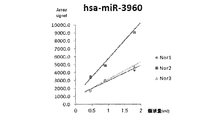
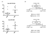
- the cerebrospinal fluid volume-dependent straight line of signal intensity in has-miR-3960 which is a miRNA with high quantification from 0.45 mL of cerebrospinal fluid volume as the supernatant volume excluding the lymphocyte fraction, is high It is drawing shown in 3 healthy persons.
- the signal intensity for each miRNA in 0.9 mL of cerebrospinal fluid was plotted as the amount of supernatant excluding the lymphocyte fraction.
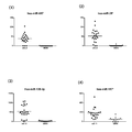
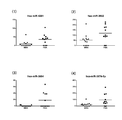
- MdCIDP and (1) hsa-miR-214, (2) hsa-miR-519d, (3) hsa-miR-575, and (4) hsa-miR-30c-2 * in cerebrospinal fluid samples It is drawing which showed distribution of signal intensity of MMN and a healthy person.
- MdCIDP and MMN for (1) hsa-miR-4323, (2) hsa-miR-4265, (3) hsa-miR-4269, and (4) hsa-miR-3605-3p in cerebrospinal fluid samples It is drawing which showed distribution of signal intensity of a healthy person.
- MdCIDP for cerebrospinal fluid samples (1) hsa-miR-3682-3p, (2) hsa-miR-3918, (3) hsa-miR-3937, and (4) hsa-miR-3940-3p 2 is a signal intensity distribution of MMN and a healthy person.
- MdCIDP and MMN for (1) hsa-miR-4446-3p, (2) hsa-miR-4461, (3) hsa-miR-4481 and (4) hsa-miR-4489 in cerebrospinal fluid samples It is drawing which showed distribution of signal intensity of a healthy person.
- MdCIDP and MMN for (1) hsa-miR-2392, (2) hsa-miR-3922-5p, (3) hsa-miR-4634, and (4) hsa-miR-4642 in cerebrospinal fluid samples It is drawing which showed distribution of signal intensity of a healthy person.
- About cerebrospinal fluid samples (1) hsa-miR-4669, (2) hsa-miR-4676-5p, (3) hsa-miR-4691-5p, and (4) hsa-miR-4713-5p , MdCIDP and MMN and a signal intensity distribution of a healthy person.
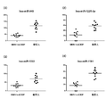
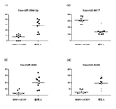
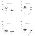
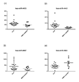
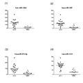
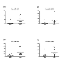
- hsa-miR-24 About (1) hsa-miR-24, (2) hsa-miR-296-5p, (3) hsa-miR-92a-2 *, and (4) hsa-miR-149 * in cerebrospinal fluid samples 5 is a diagram showing signal intensity distributions of LS, ALS, and mdCIDP or MMN.
- ALS (1) hsa-miR-296-3p, (2) hsa-miR-92b *, (3) hsa-miR-920, and (4) hsa-miR-939 in cerebrospinal fluid samples It is also a drawing showing the signal intensity distribution of mdCIDP or MMN.
- FIG. 6 is a diagram showing a signal intensity distribution of mdCIDP or MMN.
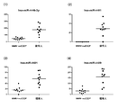
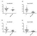
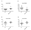
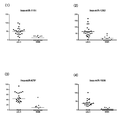
- ALS and mdCIDP for (1) hsa-miR-3917, (2) hsa-miR-3937, (3) hsa-miR-642b, and (4) hsa-miR-4417 in cerebrospinal fluid samples Or it is the figure which showed distribution of the signal strength of MMN.
- ALS and mdCIDP for (1) hsa-miR-4433, (2) hsa-miR-4442, (3) hsa-miR-4447, and (4) hsa-miR-4454 in cerebrospinal fluid samples Or it is the figure which showed distribution of the signal strength of MMN.
- FIG. 6 is a diagram showing a signal intensity distribution of mdCIDP or MMN.
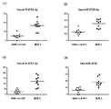
- FIG. 6 is a diagram showing a signal intensity distribution of mdCIDP or MMN. ALS and (1) hsa-miR-4665-5p, (2) hsa-miR-1343, (3) hsa-miR-4688, and (4) hsa-miR-4689 in cerebrospinal fluid samples It is drawing which showed distribution of the signal intensity of mdCIDP or MMN.
- About cerebrospinal fluid samples (1) hsa-miR-4695-5p, (2) hsa-miR-4697-5p, (3) hsa-miR-4706, and (4) hsa-miR-4723-5p 5 is a diagram showing signal intensity distributions of LS, ALS, and mdCIDP or MMN.
- (1) hsa-miR-4725-3p, (2) hsa-miR-4726-5p, (3) hsa-miR-4728-5p, and (4) hsa-miR-4730 in cerebrospinal fluid samples 5 is a diagram showing signal intensity distributions of LS, ALS, and mdCIDP or MMN.
- FIG. 3 is a diagram schematically illustrating a detection process using these together with sensitivity and specificity.
- FIG. 3 is a diagram schematically illustrating a detection process using these together with sensitivity and specificity.
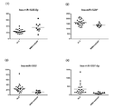
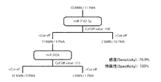
- FIG. (1) Drawing which picked up distribution of signal intensity of hsa-miR-4417 and hsa-miR-4697-5p which are suitable markers for differentiating ALS and mdCIDP or MMN in cerebrospinal fluid samples, and ( 2) A drawing showing the detection process using them together with sensitivity and specificity.
- ALS for cerebrospinal fluid samples (1) hsa-miR-718, (2) hsa-miR-2682 *, (3) hsa-miR-449c *, and (4) hsa-miR-3130-5p It is drawing which showed distribution of the signal intensity of MMN.
- FIG. 3 is a diagram showing signal intensity distributions of ALS and MMN.
- FIG. 4 is a schematic diagram together with (Specificity).
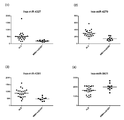
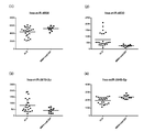
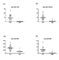
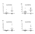
- MMN and PMA for (1) hsa-miR-99a, (2) hsa-miR-23b, (3) hsa-miR-184, and (4) hsa-miR-485-3p in cerebrospinal fluid samples It is drawing which showed distribution of signal intensity.
- hsa-miR-486-5p, (2) hsa-miR-432 *, (3) hsa-miR-675, and (4) hsa-miR-25 * in cerebrospinal fluid samples 2 is a graph showing the signal intensity distribution of PMA and PMA.
- MMN and PMA for (1) hsa-miR-711, (2) hsa-miR-3138, (3) hsa-miR-3162-5p, and (4) hsa-miR-3176 in cerebrospinal fluid samples It is drawing which showed distribution of signal intensity.
- hsa-miR-3187-3p Regarding (1) hsa-miR-3188, (3) hsa-miR-3197, and (4) hsa-miR-514b-5p in cerebrospinal fluid samples 2 is a graph showing the signal intensity distribution of PMA and PMA.
- MMN for cerebrospinal fluid samples (1) hsa-miR-3689a-3p, (2) hsa-miR-3180, (3) hsa-miR-3919, and (4) hsa-miR-3925-5p 2 is a graph showing the signal intensity distribution of PMA and PMA.
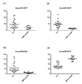
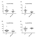
- MMN and PMA signals for (1) hsa-miR-3928, (2) hsa-miR-4425, (3) hsa-miR-4437, and (4) hsa-miR-4450 in cerebrospinal fluid samples It is drawing which showed distribution of intensity.
- MMN and PMA signals for (1) hsa-miR-4461, (2) hsa-miR-4465, (3) hsa-miR-4478, and (4) hsa-miR-2392 in cerebrospinal fluid samples It is drawing which showed distribution of intensity.
- MMN for (1) hsa-miR-4526, (2) hsa-miR-3619-3p, (3) hsa-miR-3940-5p, and (4) hsa-miR-4635 in cerebrospinal fluid samples 2 is a graph showing the signal intensity distribution of PMA and PMA.
- FIG. 3 is a graph showing signal intensity distributions of MMN and PMA.
- About cerebrospinal fluid samples (1) hsa-miR-4779, (2) hsa-miR-2467-5p, (3) hsa-miR-4787-3p, and (4) hsa-miR-4800-5p
- FIG. 3 is a graph showing signal intensity distributions of MMN and PMA.
- FIG. 3 is a diagram schematically illustrating a detection process using these together with sensitivity and specificity.
- a schematic diagram For each of hsa-miR-3162-5p and hsa-miR-3928, which are suitable markers for differentiating MMN and PMA in cerebrospinal fluid specimens, (1) a drawing picking up the signal intensity distribution, and (2) FIG. 3 is a diagram schematically illustrating a detection process using these together with sensitivity and specificity.
- miRNA is quantified in a CSF sample of a donor obtained by collecting and preparing cerebrospinal fluid according to the above-described conventional method, and the quantified value is lower or higher than the comparison target.
- the indicated signal that is, by detecting the increase or decrease in the amount of miRNA, a desired differentiation index can be obtained.
- the method for quantifying miRNA is not particularly limited, and for example, existing means based on the above-mentioned individual miRNA base sequences, and further developed means can be used.
- use of the “microRNA detection array” in which miRNAs are comprehensively used, which is used in the standardization method of the present invention described above, can be mentioned.
- RNA quantification methods based on gene amplification methods such as the RT-PCR method can be mentioned.
- a real-time RT-PCR method using an automated detection device is preferable from the viewpoint of mass processing and rapidity.
- the Northern blot method etc. are mentioned.
- the quantitative value based on the quantitative signal of each miRNA is relatively lower or higher than the comparison target (for example, a healthy person or a different motor neuropathy disease to be compared).
- the quantitative value can be treated as “low positive” or “high positive”.
- Specific threshold values can be set individually and specifically.
- a decrease in the amount of hsa-miR-744 or a decrease in the amount of hsa-miR-4505 in a cerebrospinal fluid sample can be detected as a differentiation index for ALS, mdCIDP, or MMN .
- the detection method of the present invention of this aspect is particularly useful when it is intended to grasp a general tendency of a motor neuropathy disease in a subject.
- the quantitative value of hsa-miR-744 or hsa-miR-4505 in the cerebrospinal fluid specimen decreases. Therefore, when it is observed that the quantitative value of hsa-miR-744 or hsa-miR-4505 in the cerebrospinal fluid sample of the subject is reduced from the standard for healthy subjects, the subject is ALS, mdCIDP, or Therefore, the possibility of suffering from MMN increases, and the direction of subsequent disease discrimination can be narrowed down.
- a detection method of a different aspect described later is provided.
- ALS differentiation indicators Upon differentiation from ALS healthy individuals, one or more selected from the changes in the amount of microRNA in the cerebrospinal fluid samples based on healthy individuals within the following “” are detected as ALS differentiation indicators. can do.
- the above-mentioned “decrease in the amount of hsa-miR-744” or “decrease in the amount of hsa-miR-4505” is also included as a differential indicator of this embodiment, but when this differential indicator is used, it is listed here. It is preferable to use a combination of other indices.
- hsa-miR-10a an increase in the amount of hsa-miR-10a, an increase in the amount of hsa-miR-516b, an increase in the amount of hsa-miR-122 *, and an increase in hsa-miR-4762-3p.
- Changes in the amount of one or more selected microRNAs can be used as a differentiation index of this embodiment.
- the detection method of this preferred embodiment is also advantageous in that a desired differentiation target can be detected with higher sensitivity and specificity.
- the above-mentioned miRNA values in the cerebrospinal fluid specimen show the characteristic increases and decreases listed respectively. Therefore, if the quantitative values of these miRNAs in the cerebrospinal fluid sample of the subject are found to show the listed increases and decreases in comparison with the standard for healthy subjects, the subject may be affected with ALS. Is found to be high.
- mdCIDP or MMN When differentiating mdCIDP or MMN from a healthy person, mdCIDP or an index of one or more selected from changes in the amount of microRNA in the cerebrospinal fluid sample based on the healthy person within the following “” It can be detected as a differentiation index of MMN.
- the above “decrease in the amount of hsa-miR-744” or “decrease in the amount of hsa-miR-4505” is also included as an indicator of this embodiment, but when this indicator is used in this embodiment, it is listed here. It is preferable to use a combination of other indices.
- Hsa-miR-214 decreased, hsa-miR-519d decreased, hsa-miR-575 decreased, hsa-miR-30c-2 * decreased, hsa-miR-135a * decreased, decrease in hsa-miR-185 * amount, decrease in hsa-miR-423-5p amount, decrease in hsa-miR-92b * amount, decrease in hsa-miR-551b * amount, decrease in hsa-miR-550a amount, hsa-miR-885-3p decrease, hsa-miR-936 decrease, hsa-miR-940 decrease, hsa-miR-1225-3p decrease, hsa-miR-1233 decrease, hsa -MiR-1181 decreased, hsa-miR-1268 increased, hsa-miR-1908 decreased, hsa-miR-2276
- an increase in the amount of hsa-miR-4417 can be used as a differentiation index of this embodiment.
- the detection method of this preferred embodiment is also advantageous in that a desired differentiation target can be detected with higher sensitivity and specificity.
- the above miRNA values in the cerebrospinal fluid specimens each show a characteristic increase or decrease listed.
- the subject is afflicted with mdCIDP or MMN It is recognized that the possibility is high. It should be noted that the discrimination between mdCIDP and MMN can be performed according to a method conventionally used by those skilled in the art.
- an increase in the amount of hsa-miR-642b from the group consisting of an increase in the amount of hsa-miR-642b, an increase in the amount of hsa-miR-3197, an increase in the amount of hsa-miR-4697-5p, and a decrease in the amount of hsa-miR-4417.
- One type or two or more types selected (however, the decrease in the amount of hsa-miR-4417 is a combination with one to three types selected from the increase or decrease in the amount of the other three microRNAs) It can be detected as a differentiation index.
- the detection method according to this aspect greatly assists in distinguishing between ALS that is difficult to distinguish between early stages of disease and mdCIDP or MMN.
- the above-mentioned miRNA quantitative values in cerebrospinal fluid samples based on “mdCIDP or MMN” show the characteristic increases and decreases listed respectively.
- the quantified value of these miRNAs in a subject's cerebrospinal fluid sample is observed to show the listed increase or decrease in comparison to the “mdCIDP or MMN” standard, the subject is identified as “mdCIDP or MMN”. Instead, it is recognized that there is a high possibility of suffering from “ALS”.
- the discrimination between mdCIDP and MMN can be performed according to a method conventionally performed by those skilled in the art, as in the above-described discrimination from healthy individuals of “mdCIDP or MMN”.
- the aspect disclosed here is the amount change toward “ALS” based on “mdCIDP or MMN”, this is only relative. That is, conversely, the reference can be set to “ALS” and equivalently replaced with the amount change toward “mdCIDP or MMN”.
- the “increase” of the quantitative value disclosed herein is “decrease”, “decrease” is “increase”, and “MDCIDP or MMN” is a differentiation index based on “ALS”. Is provided.
- More preferred embodiments include increased hsa-miR-139-3p, increased hsa-miR-574-5p, increased hsa-miR-1231, increased hsa-miR-2682 *, and hsa-miR-3130
- One or more changes in the amount of microRNA selected from the group consisting of an increase of ⁇ 5p can be detected as a differential indicator of ALS of this embodiment.
- any one selected from the group consisting of a combination of an increase in hsa-miR-2682 * and an increase in hsa-miR-3130-5p is extremely suitable for detection as an ALS differentiation index. is there.
- the detection methods of these preferred embodiments are advantageous in that a desired differential target can be detected with higher sensitivity and higher specificity.
- the detection method of this embodiment is a great help for differentiating between ALS and MMN, which is particularly difficult to identify each other in the early stages of disease.
- the above-mentioned miRNA quantitative values in the cerebrospinal fluid specimens based on “MMN” each show a characteristic increase listed.
- a quantitative value of these miRNAs in a subject's cerebrospinal fluid sample is shown to show the listed increase in comparison to the “MMN” standard, the subject is not “MMN” but “ It is recognized that there is a high probability of suffering from “ALS”.
- the aspect disclosed here is the amount change toward “ALS” based on “MMN”, this is only relative. That is, conversely, the reference can be set to “ALS” and equivalently replaced with the amount change toward “MMN”.
- the “increase” of the quantitative value disclosed herein is “decrease”, and a discrimination index of “MMN” based on “ALS” is provided.
- More preferred embodiments include an increase in hsa-miR-1275, an increase in hsa-miR-711, an increase in hsa-miR-3138, an increase in hsa-miR-3162-5p, an increase in hsa-miR-3928, and One type or two or more types of changes in the amount of microRNA selected from the group consisting of an increase in hsa-miR-1914 * can be detected as a differential indicator of PMA of this embodiment.
- the detection method of the present embodiment is a great help for differentiating between MMN and PMA, which is particularly difficult to identify each other in the early stages of disease.
- the above-mentioned miRNA quantitative values in the cerebrospinal fluid specimens based on “MMN” each show a characteristic increase listed.
- a quantitative value of these miRNAs in a subject's cerebrospinal fluid sample is shown to show the listed increase in comparison to the “MMN” standard, the subject is not “MMN” but “ It is recognized that the possibility of having “PMA” is high.
- the aspect disclosed here is the amount change toward “PMA” based on “MMN”, this is only relative. That is, conversely, in “PMA”, the reference can be equivalently replaced with a change in quantity toward “MMN”.
- the “increase” of the quantitative value disclosed herein is “decrease”, and a differentiation index of “MMN” based on “PMA” is provided.
- the miRNA in the cerebrospinal fluid which originally has a small amount of miRNA, is corrected by the Global Normalization method (a correction method that sets the median when the signal intensities are arranged in order) to 25, the median is selected. There is a possibility that even if the miRNA is normal, the one whose signal intensity varies considerably will be selected.
- the Global Normalization method a correction method that sets the median when the signal intensities are arranged in order
- the present inventor found that when comparing the signal intensities of miRNA groups of individual samples under the assumption that “the signal values of miRNA groups in the comprehensive analysis of normal cerebrospinal fluid are not significantly different among individuals” as a whole.
- the fluorescence intensity recognized in the top 20 miRNA is replaced with a specific number as a signal value, and the multiplier used in the multiplication for performing this replacement is calculated with respect to the fluorescence intensity recognized in other miRNAs.
- the above 41 miRNA groups were normalized.
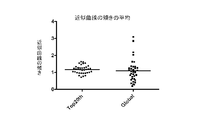
- FIG. 3 shows a signal for each miRNA of 0.9 mL of cerebrospinal fluid as a supernatant amount excluding the lymphocyte fraction between the conventional method (Global normalization method) and the normalization method of the present invention (Top20th normalization method). It is drawing which showed distribution of the inclination between two normal cerebrospinal fluid samples which plotted the intensity
- the signal used this time is “Cy (Cyanine) bound to the test miRNA” as described above.
- the fluorescence intensity of the fluorescent dye is the signal value of this example.
- RNA array 3D GENE (registered trademark) / TORAY)” (Toray) is used as an array for miRNA detection, and all the miRNAs mounted on the array are used as a population.
- miRNA in cerebrospinal fluid that can be used as a marker for three categories of diseases (ALS, MMN, and mdCIDP) was selected.
- sorting method the Welch method based on the null assumption was used, and the significance level was sorted at 5%.
- the breakdown of cerebrospinal fluid samples used for selecting each category is “Healthy person: 10 samples, ALS: 23 samples, mdCIDP: 5 samples, MMN: 5 samples (mdCIDP / MMN: 10 samples)”. . However, in selecting an index for distinguishing ALS from a healthy person, since cerebrospinal fluid in which blood was mixed was excluded, “healthy person: 9 specimens, ALS: 19 specimens”.
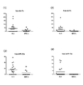
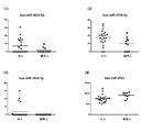
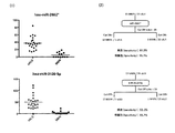
- FIG. 4 shows healthy subjects for (1) hsa-miR-744 and (2) hsa-miR-4505 in cerebrospinal fluid samples.
- 2 is a diagram showing signal intensity distributions of ALS, ALS, and mdCIDP and MMN.
- the vertical axis of the graph is the signal intensity
- the horizontal line in the graph is the average value of each group.
- the signal of the disease group was significantly decreased between the healthy subject and ALS, and between the healthy subject and mdCIDP / MMN.
- hsa-miR-744 and hsa-miR-4505 are particularly useful when it is intended to grasp the general tendency of motor neuropathy disorders in subjects.
- Hsa-miR-744 and hsa-miR-4505 can be used in combination with other markers that can distinguish these diseases, which will be described later. Is preferred.
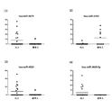
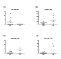
- FIG. 5 shows the signal intensity of a marker that provides a differentiation index between ALS and healthy individuals. Distribution is shown.
- FIG. 5-1 shows (1) hsa-let-7a, (2) hsa-let-7c, (3) hsa-miR-23a, and (4) hsa-miR- About 10a, the distribution of the signal intensity of ALS and a healthy person is shown.
- the signal intensity of hsa-let-7a was significantly increased in ALS in healthy individuals.
- the signal intensity of hsa-let-7c was significantly increased in ALS in healthy individuals.
- the signal intensity of hsa-miR-23a was significantly increased in ALS compared with healthy individuals.
- the signal intensity of hsa-miR-10a was significantly increased in ALS in healthy individuals.
- these miRNAs present in the cerebrospinal fluid are markers that provide a differentiation index between healthy individuals and ALS.
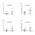
- FIG. 5-2 shows (1) hsa-miR-200a, (2) hsa-miR-524-5p, (3) hsa-miR-516b, and (4) hsa- About miR-552, the distribution of signal intensity between ALS and healthy individuals is shown.
- the signal intensity of hsa-miR-200a was significantly increased in ALS compared with healthy individuals.
- the signal intensity of hsa-miR-524-5p was significantly reduced in ALS in healthy individuals.
- the signal intensity of hsa-miR-516b was significantly increased in ALS compared with healthy individuals.
- the signal intensity of hsa-miR-552 was significantly increased in ALS in healthy individuals.
- these miRNAs present in the cerebrospinal fluid are markers that provide a differentiation index between healthy individuals and ALS.
- Fig. 5-3 shows (1) hsa-miR-573, (2) hsa-miR-644, (3) hsa-miR-24-2 *, and (4) hsa in cerebrospinal fluid samples.
- -MiR-122 * shows the distribution of signal intensity between ALS and healthy individuals.
- the signal intensity of hsa-miR-573 was significantly increased in ALS in healthy individuals.
- the signal intensity of hsa-miR-644 was significantly increased in ALS in healthy individuals.
- the signal intensity of hsa-miR-24-2 * was significantly increased in ALS compared with healthy individuals.
- the signal intensity of hsa-miR-122 * was significantly increased in ALS compared with healthy individuals.
- these miRNAs present in the cerebrospinal fluid are markers that provide a differentiation index between healthy individuals and ALS.
- Fig. 5-4 shows (1) hsa-miR-145 *, (2) hsa-miR-361-3p, (3) hsa-miR-1291, and (4) hsa in cerebrospinal fluid samples.
- -miR-1260 shows the distribution of signal intensity between ALS and healthy individuals. (1) The signal intensity of hsa-miR-145 * was significantly increased in ALS compared with healthy individuals. (2) The signal intensity of hsa-miR-361-3p was significantly increased in ALS compared with healthy individuals. (3) The signal intensity of hsa-miR-1291 was significantly increased in ALS in healthy individuals. (4) The signal intensity of hsa-miR-1260 was significantly reduced in ALS in healthy individuals.
- these miRNAs present in the cerebrospinal fluid are markers that provide a differentiation index between healthy individuals and ALS.
- Fig. 5-5 shows (1) hsa-miR-1273, (2) hsa-miR-3161, (3) hsa-miR-4310, and (4) hsa-miR- About 4313, the distribution of the signal intensity of ALS and a healthy person is shown.
- the signal intensity of hsa-miR-1273 was significantly reduced in ALS in healthy individuals.
- the signal intensity of hsa-miR-3161 was significantly increased in ALS compared with healthy individuals.
- the signal intensity of hsa-miR-4310 was significantly increased in ALS in healthy individuals.
- the signal intensity of hsa-miR-4313 was significantly reduced in ALS in healthy individuals.
- these miRNAs present in the cerebrospinal fluid are markers that provide a differentiation index between healthy individuals and ALS.
- Figure 5-6 shows (1) hsa-miR-4479, (2) hsa-miR-4493, (3) hsa-miR-4524, and (4) hsa-miR- 3688-5p shows the distribution of signal intensity between ALS and healthy individuals.
- the signal intensity of hsa-miR-4479 was significantly increased in ALS in healthy individuals.
- the signal intensity of hsa-miR-4493 was significantly reduced in ALS in healthy individuals.
- the signal intensity of hsa-miR-4524 was significantly increased in ALS in healthy individuals.
- the signal intensity of hsa-miR-3688-5p was significantly increased in ALS compared with healthy individuals.
- these miRNAs present in the cerebrospinal fluid are markers that provide a differentiation index between healthy individuals and ALS.
- FIG. 5-7 shows (1) hsa-miR-4633-5p, (2) hsa-miR-4708-5p, (3) hsa-miR-3545-3p, and ( 4) About hsa-miR-4741, distribution of signal intensity of ALS and healthy persons is shown.
- the signal intensity of hsa-miR-4633-5p was significantly increased in ALS compared with healthy individuals.
- the signal intensity of hsa-miR-4708-5p was significantly increased in ALS compared with healthy individuals.
- the signal intensity of hsa-miR-3545-3p was significantly increased in ALS compared with healthy individuals.
- the signal intensity of hsa-miR-4741 was significantly reduced in ALS compared with healthy individuals.
- these miRNAs present in the cerebrospinal fluid are markers that provide a differentiation index between healthy individuals and ALS.
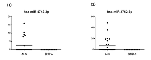
- Fig. 5-8 shows the distribution of signal intensity between ALS and healthy individuals for (1) hsa-miR-4742-3p and (2) hsa-miR-4762-3p in cerebrospinal fluid samples. ing. (1) The signal intensity of hsa-miR-4742-3p was significantly increased in ALS compared with healthy individuals. (2) The signal intensity of hsa-miR-4762-3p was significantly increased in ALS compared with healthy individuals.
- these miRNAs present in the cerebrospinal fluid are markers that provide a differentiation index between healthy individuals and ALS.
- FIG. 6 shows the signal intensity distribution of these four types of markers.
- FIG. 7 is a diagram schematically illustrating a detection process using these four kinds of markers in a cerebrospinal fluid specimen together with its sensitivity (Sensitivity) and specificity (Specificity). As a result, in these markers, when any one of the above four types was detected as a positive signal, it was judged as positive, and thus an excellent discrimination result with a sensitivity of 89.5% and a specificity of 100%. Was recognized
- FIG. 8 shows a marker that provides a differentiation index between mdCIDP or MMN and a healthy person The signal intensity distribution is shown.
- FIG. 8-1 shows (1) hsa-miR-214, (2) hsa-miR-519d, (3) hsa-miR-575, and (4) hsa-miR- For 30c-2 *, the distribution of signal intensity of mdCIDP and MMN and healthy persons is shown.
- the signal intensity of hsa-miR-214 was significantly reduced in mdCIDP / MMN for healthy individuals.
- the signal intensity of hsa-miR-519d was significantly decreased in mdCIDP / MMN for healthy individuals.
- the signal intensity of hsa-miR-575 was significantly reduced in mdCIDP / MMN for healthy individuals.
- the signal intensity of hsa-miR-30c-2 * was significantly decreased in mdCIDP / MMN with respect to healthy individuals.
- these miRNAs present in the cerebrospinal fluid are markers that provide a differentiation index between healthy people and CIDP or MMN.
- FIG. 8-2 shows (1) hsa-miR-135a *, (2) hsa-miR-185 *, (3) hsa-miR-423-5p, and (4) in the cerebrospinal fluid specimen.
- hsa-miR-92b * the signal intensity distribution of mdCIDP and MMN and a healthy person is shown.
- the signal intensity of hsa-miR-135a * was significantly decreased in mdCIDP / MMN in healthy subjects.
- the signal intensity of hsa-miR-185 * was significantly decreased in mdCIDP / MMN for healthy individuals.
- hsa-miR-423-5p showed a significant decrease in signal intensity in mdCIDP / MMN compared to healthy individuals.
- the signal intensity of hsa-miR-92b * was significantly decreased in mdCIDP / MMN for healthy individuals.
- these miRNAs present in the cerebrospinal fluid are markers that provide a differentiation index between healthy people and mdCIDP or MMN.
- FIG. 8-3 shows (1) hsa-miR-551b *, (2) hsa-miR-550a, (3) hsa-miR-885-3p, and (4) hsa in the cerebrospinal fluid sample.
- -miR-936 the signal intensity distributions of mdCIDP and MMN and healthy individuals are shown.
- the signal intensity of hsa-miR-551b * was significantly decreased in mdCIDP / MMN in healthy subjects.
- the signal intensity of hsa-miR-550a was significantly decreased in mdCIDP / MMN for healthy individuals.
- these miRNAs present in the cerebrospinal fluid are markers that provide a differentiation index between healthy people and CIDP or MMN.
- FIG. 8-4 shows (1) hsa-miR-940, (2) hsa-miR-1225-3p, (3) hsa-miR-1233, and (4) hsa- About miR-1181, the distribution of signal intensity of mdCIDP and MMN and healthy persons is shown.
- the signal intensity of hsa-miR-940 was significantly reduced in mdCIDP / MMN for healthy individuals.
- the signal intensity of hsa-miR-1225-3p was significantly reduced in mdCIDP / MMN in healthy subjects.
- the signal intensity of hsa-miR-1233 was significantly reduced in mdCIDP / MMN for healthy individuals.
- the signal intensity of hsa-miR-1181 was significantly decreased in mdCIDP / MMN for healthy individuals.
- these miRNAs present in the cerebrospinal fluid are markers that provide a differentiation index between healthy people and mdCIDP or MMN.
- Fig. 8-5 shows (1) hsa-miR-1268, (2) hsa-miR-1908, (3) hsa-miR-2276, and (4) hsa-miR- About 711, the distribution of the signal intensity of mdCIDP and MMN and a healthy person is shown.
- the signal intensity of hsa-miR-1268 was significantly increased in mdCIDP / MMN for healthy individuals.
- the signal intensity of hsa-miR-1908 was significantly reduced in mdCIDP / MMN for healthy individuals.
- the signal intensity of hsa-miR-2276 was significantly reduced in mdCIDP / MMN for healthy individuals.
- the signal intensity of hsa-miR-711 was significantly decreased in mdCIDP / MMN for healthy individuals.
- these miRNAs present in the cerebrospinal fluid are markers that provide a differentiation index between healthy people and mdCIDP or MMN.
- Figure 8-6 shows (1) hsa-miR-718, (2) hsa-miR-1193, (3) hsa-miR-3184, and (4) hsa-miR- About 3197, the distribution of the signal intensity of mdCIDP and MMN and a healthy person is shown.
- the signal intensity of hsa-miR-718 was significantly reduced in mdCIDP / MMN for healthy individuals.
- the signal intensity of hsa-miR-1193 was significantly reduced in mdCIDP / MMN for healthy individuals.
- the signal intensity of hsa-miR-3184 was significantly decreased in mdCIDP / MMN for healthy individuals.
- the signal intensity of hsa-miR-3197 was significantly reduced in mdCIDP / MMN for healthy individuals.
- these miRNAs present in the cerebrospinal fluid are markers that provide a differentiation index between healthy people and mdCIDP or MMN.
- Fig. 8-7 shows (1) hsa-miR-4323, (2) hsa-miR-4265, (3) hsa-miR-4269, and (4) hsa-miR- About 3605-3p, the distribution of the signal intensity of mdCIDP and MMN and a healthy person is shown.
- the signal intensity of hsa-miR-4323 was significantly reduced in mdCIDP / MMN for healthy individuals.
- the signal intensity of hsa-miR-4265 was significantly reduced in mdCIDP / MMN for healthy individuals.
- these miRNAs present in the cerebrospinal fluid are markers that provide a differentiation index between healthy people and mdCIDP or MMN.
- FIGS. 8-8 show (1) hsa-miR-3648, (2) hsa-miR-3663-5p, (3) hsa-miR-3667-3p, and (4) in the cerebrospinal fluid specimen.
- hsa-miR-3679-5p the signal intensity distribution of mdCIDP and MMN and a healthy person is shown.
- the signal intensity of hsa-miR-3648 was significantly reduced in mdCIDP / MMN for healthy individuals.
- the signal intensity of hsa-miR-3663-5p was significantly reduced in mdCIDP / MMN compared to healthy individuals.
- these miRNAs present in the cerebrospinal fluid are markers that provide a differentiation index between healthy people and mdCIDP or MMN.
- FIGS. 8-9 show (1) hsa-miR-3682-3p, (2) hsa-miR-3918, (3) hsa-miR-3937, and (4) hsa- About miR-3940-3p, the distribution of the signal intensity of mdCIDP and MMN and a healthy person is shown.
- the signal intensity of hsa-miR-3682-3p was significantly decreased in mdCIDP / MMN compared to healthy individuals.
- the signal intensity of hsa-miR-3918 was significantly reduced in mdCIDP / MMN for healthy individuals.
- these miRNAs present in the cerebrospinal fluid are markers that provide a differentiation index between healthy people and mdCIDP or MMN.
- FIGS. 8-10 show (1) hsa-miR-3944-3p, (2) hsa-miR-4417, (3) hsa-miR-4430, and (4) hsa- About miR-4440, the distribution of the signal intensity of mdCIDP and MMN and healthy persons is shown.
- the signal intensity of hsa-miR-3944-3p was significantly reduced in mdCIDP / MMN in healthy subjects.
- the signal intensity of hsa-miR-4417 was significantly increased in mdCIDP / MMN for healthy individuals.
- the signal intensity of hsa-miR-4430 was significantly reduced in mdCIDP / MMN for healthy individuals.
- the signal intensity of hsa-miR-4440 was significantly reduced in mdCIDP / MMN for healthy individuals.
- these miRNAs present in the cerebrospinal fluid are markers that provide a differentiation index between healthy people and mdCIDP or MMN.
- FIGS. 8-11 show (1) hsa-miR-4446-3p, (2) hsa-miR-4461, (3) hsa-miR-4481, and (4) hsa- About miR-4489, the distribution of signal intensity of mdCIDP and MMN and healthy persons is shown.
- the signal intensity of hsa-miR-4446-3p was significantly reduced in mdCIDP / MMN compared to healthy individuals.
- the signal intensity of hsa-miR-4461 was significantly reduced in mdCIDP / MMN for healthy individuals.
- the signal intensity of hsa-miR-4481 was significantly decreased in mdCIDP / MMN for healthy individuals.
- the signal intensity of hsa-miR-4489 was significantly reduced in mdCIDP / MMN for healthy individuals.
- these miRNAs present in the cerebrospinal fluid are markers that provide a differentiation index between healthy people and mdCIDP or MMN.
- FIGS. 8-12 show (1) hsa-miR-2392, (2) hsa-miR-3922-5p, (3) hsa-miR-4634, and (4) hsa- About miR-4642, the distribution of the signal intensity of mdCIDP and MMN and healthy persons is shown.
- the signal intensity of hsa-miR-2392 was significantly reduced in mdCIDP / MMN for healthy individuals.
- the signal intensity of hsa-miR-3922-5p was significantly reduced in mdCIDP / MMN compared to healthy individuals.
- these miRNAs present in the cerebrospinal fluid are markers that provide a differentiation index between healthy people and mdCIDP or MMN.
- FIGS. 8-13 show (1) hsa-miR-4669, (2) hsa-miR-4676-5p, (3) hsa-miR-4691-5p, and (4) in the cerebrospinal fluid specimen.
- hsa-miR-4713-5p the signal intensity distribution of mdCIDP and MMN and a healthy person is shown.
- the signal intensity of hsa-miR-4669 was significantly reduced in mdCIDP / MMN for healthy individuals.
- the signal intensity of hsa-miR-4676-5p was significantly reduced in mdCIDP / MMN compared to healthy individuals.
- these miRNAs present in the cerebrospinal fluid are markers that provide a differentiation index between healthy people and mdCIDP or MMN.
- FIGS. 8-14 show (1) hsa-miR-4722-3p, (2) hsa-miR-4725-3p, (3) hsa-miR-4727-3p, and ( 4) About hsa-miR-4736, the distribution of signal intensity of mdCIDP and MMN and healthy persons is shown.
- the signal intensity of hsa-miR-4722-3p was significantly decreased in mdCIDP / MMN compared to healthy individuals.
- the signal intensity of hsa-miR-4725-3p was significantly reduced in mdCIDP / MMN for healthy subjects.
- these miRNAs present in the cerebrospinal fluid are markers that provide a differentiation index between healthy people and mdCIDP or MMN.
- Figure 8-15 shows the distribution of signal intensity of mdCIDP and MMN and healthy individuals for (1) hsa-miR-371b-3p and (2) hsa-miR-2467-3p in cerebrospinal fluid samples Is shown.
- the signal intensity of hsa-miR-371b-3p was significantly decreased in mdCIDP / MMN with respect to healthy subjects.
- the signal intensity of hsa-miR-2467-3p was significantly reduced in mdCIDP / MMN compared to healthy individuals.
- these miRNAs present in the cerebrospinal fluid are markers that provide a differentiation index between healthy people and mdCIDP or MMN.
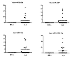
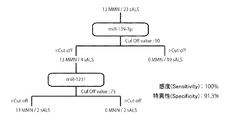
- FIG. 9 is a suitable marker for distinguishing healthy person from mdCIDP and MMN in cerebrospinal fluid specimen.
- -miR-4717 (1) Drawing which picked up signal intensity distribution, (2) ROC curve (receiver operating characteristic curve), and (3) Detection process using the manufacturer
- FIG. 2 is a diagram schematically showing sensitivity and specificity. FIG. As a result, the marker was found to have excellent discrimination results with a sensitivity of 80.0% and a specificity of 90.0%.
- FIG. 10 shows ALS and mdCIDP or MMN alternatives.
- 2 shows the distribution of signal intensity of markers that provide a differential indicator.
- FIG. 10-1 shows (1) hsa-miR-24, (2) hsa-miR-296-5p, and (3) hsa-miR-92a-2 in the cerebrospinal fluid sample.
- * And (4) hsa-miR-149 * show the signal intensity distribution of ALS and mdCIDP or MMN.
- the signal intensity of ALS is expressed by an increase / decrease based on the signal intensity of mdCIDP or MMN (the same applies to the description of the series of FIG. 10 below).
- (1) The signal intensity of hsa-miR-24 was significantly increased in ALS.
- the signal intensity of hsa-miR-296-5p was significantly increased in ALS.
- the signal intensity of hsa-miR-92a-2 * was significantly reduced in ALS.
- the signal intensity of hsa-miR-149 * was significantly increased in ALS.
- these miRNAs present in the cerebrospinal fluid are markers that give an alternative differentiation index of ALS and mdCIDP or MMN.
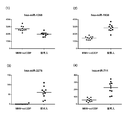
- FIG. 10-2 shows (1) hsa-miR-296-3p, (2) hsa-miR-92b *, (3) hsa-miR-920, and (4) hsa in the cerebrospinal fluid specimen.
- -miR-939 the signal intensity distribution of ALS and mdCIDP or MMN is shown.
- the signal intensity of hsa-miR-296-3p was significantly increased in ALS.
- the signal intensity of hsa-miR-92b * was significantly increased in ALS.
- the signal intensity of hsa-miR-920 was significantly increased in ALS.
- the signal intensity of hsa-miR-939 was significantly reduced in ALS.
- these miRNAs present in the cerebrospinal fluid are markers that give an alternative differentiation index of ALS and mdCIDP or MMN.
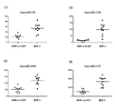
- FIG. 10-3 shows (1) hsa-miR-1225-5p, (2) hsa-miR-1228 *, (3) hsa-miR-1202, and (4) hsa in the cerebrospinal fluid sample.
- -miR-1207-5p the signal intensity distribution of ALS and mdCIDP or MMN is shown.
- the signal intensity of hsa-miR-1225-5p was significantly reduced in ALS.
- the signal intensity of hsa-miR-1228 * was significantly increased in ALS.
- the signal intensity of hsa-miR-1202 was significantly increased in ALS.
- the signal intensity of hsa-miR-1207-5p was significantly increased in ALS.
- these miRNAs present in the cerebrospinal fluid are markers that give an alternative differentiation index of ALS and mdCIDP or MMN.
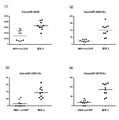
- FIG. 10-4 shows (1) hsa-miR-1908, (2) hsa-miR-1909, (3) hsa-miR-3131, and (4) hsa-miR- 3141 shows the distribution of signal intensity of ALS and mdCIDP or MMN.
- the signal intensity of hsa-miR-1908 was significantly increased in ALS.
- the signal intensity of hsa-miR-1909 was significantly increased in ALS.
- the signal intensity of hsa-miR-3131 was significantly increased in ALS.
- the signal intensity of hsa-miR-3141 was significantly increased in ALS.
- these miRNAs present in the cerebrospinal fluid are markers that give an alternative differentiation index of ALS and mdCIDP or MMN.
- FIG. 10-5 shows (1) hsa-miR-1260b, (2) hsa-miR-3178, (3) hsa-miR-3180-3p, and (4) hsa- For miR-3185, the signal intensity distribution of ALS and mdCIDP or MMN is shown.
- the signal intensity of hsa-miR-1260b was significantly reduced in ALS.
- the signal intensity of hsa-miR-3178 was significantly increased in ALS.
- the signal intensity of hsa-miR-3180-3p was significantly increased in ALS.
- the signal intensity of hsa-miR-3185 was significantly increased in ALS.
- these miRNAs present in the cerebrospinal fluid are markers that give an alternative differentiation index of ALS and mdCIDP or MMN.
- FIG. 10-6 shows (1) hsa-miR-3195, (2) hsa-miR-3197, (3) hsa-miR-4257, and (4) hsa-miR- 4259 shows the distribution of signal intensity of ALS and mdCIDP or MMN.
- the signal intensity of hsa-miR-3195 was significantly increased in ALS.
- the signal intensity of hsa-miR-3197 was significantly increased in ALS.
- the signal intensity of hsa-miR-4257 was significantly increased in ALS.
- the signal intensity of hsa-miR-4259 was significantly increased in ALS.
- these miRNAs present in the cerebrospinal fluid are markers that give an alternative differentiation index of ALS and mdCIDP or MMN.
- Fig. 10-7 shows (1) hsa-miR-4327, (2) hsa-miR-4270, (3) hsa-miR-4281, and (4) hsa-miR- 3621 shows the distribution of the signal intensity of ALS and mdCIDP or MMN.
- the signal intensity of hsa-miR-4327 was significantly increased in ALS.
- the signal intensity of hsa-miR-4270 increased significantly in ALS.
- the signal intensity of hsa-miR-4281 increased significantly in ALS.
- the signal intensity of hsa-miR-3621 was significantly reduced in ALS.
- these miRNAs present in the cerebrospinal fluid are markers that give an alternative differentiation index of ALS and mdCIDP or MMN.
- FIG. 10-8 shows (1) hsa-miR-3622a-5p, (2) hsa-miR-3648, (3) hsa-miR-3656, and (4) hsa- About miR-3663-3p, the signal intensity distribution of ALS and mdCIDP or MMN is shown.
- the signal intensity of hsa-miR-3622a-5p was significantly increased in ALS.
- the signal intensity of hsa-miR-3648 was significantly increased in ALS.
- the signal intensity of hsa-miR-3656 was significantly reduced in ALS.
- the signal intensity of hsa-miR-3663-3p was significantly increased in ALS.
- these miRNAs present in the cerebrospinal fluid are markers that give an alternative differentiation index of ALS and mdCIDP or MMN.
- FIGS. 10-9 show (1) hsa-miR-3917, (2) hsa-miR-3937, (3) hsa-miR-642b, and (4) hsa-miR- For 4417, the distribution of the signal intensity of ALS and mdCIDP or MMN is shown.
- the signal intensity of hsa-miR-3917 was significantly increased in ALS.
- the signal intensity of hsa-miR-3937 was significantly increased in ALS.
- the signal intensity of hsa-miR-642b was significantly increased in ALS.
- the signal intensity of hsa-miR-4417 was significantly reduced in ALS.
- these miRNAs present in the cerebrospinal fluid are markers that give an alternative differentiation index of ALS and mdCIDP or MMN.
- FIGS. 10-10 show (1) hsa-miR-4433, (2) hsa-miR-4442, (3) hsa-miR-4447, and (4) hsa-miR- For 4454, the distribution of the signal intensity of ALS and mdCIDP or MMN is shown.
- the signal intensity of hsa-miR-4433 was significantly increased in ALS.
- the signal intensity of hsa-miR-4442 was significantly increased in ALS.
- the signal intensity of hsa-miR-4447 was significantly increased in ALS.
- the signal intensity of hsa-miR-4454 was significantly reduced in ALS.
- these miRNAs present in the cerebrospinal fluid are markers that give an alternative differentiation index of ALS and mdCIDP or MMN.
- FIGS. 10-11 show (1) hsa-miR-4466, (2) hsa-miR-4484, (3) hsa-miR-4488, and (4) hsa-miR- in the cerebrospinal fluid specimen.
- the signal intensity distribution of ALS and mdCIDP or MMN is shown.
- the signal intensity of hsa-miR-4466 was significantly reduced in ALS.
- the signal intensity of hsa-miR-4484 was significantly increased in ALS.
- the signal intensity of hsa-miR-4488 was significantly reduced in ALS.
- the signal intensity of hsa-miR-4492 was significantly reduced in ALS.
- these miRNAs present in the cerebrospinal fluid are markers that give an alternative differentiation index of ALS and mdCIDP or MMN.
- FIGS. 10-12 show (1) hsa-miR-4508, (2) hsa-miR-4530, (3) hsa-miR-3619-3p, and (4) hsa- About miR-3940-5p, the distribution of the signal intensity of ALS and mdCIDP or MMN is shown.
- the signal intensity of hsa-miR-4508 was significantly reduced in ALS.
- the signal intensity of hsa-miR-4530 was significantly increased in ALS.
- the signal intensity of hsa-miR-3619-3p was significantly increased in ALS.
- the signal intensity of hsa-miR-3940-5p was significantly reduced in ALS.
- these miRNAs present in the cerebrospinal fluid are markers that give an alternative differentiation index of ALS and mdCIDP or MMN.
- FIGS. 10-13 show (1) hsa-miR-3960, (2) hsa-miR-4634, (3) hsa-miR-4640-5p, and (4) hsa- For miR-4655-5p, the signal intensity distribution of ALS and mdCIDP or MMN is shown.
- the signal intensity of hsa-miR-3960 was significantly reduced in ALS.
- the signal intensity of hsa-miR-4634 was significantly reduced in ALS.
- the signal intensity of hsa-miR-4640-5p was significantly increased in ALS.
- the signal intensity of hsa-miR-4655-5p was significantly increased in ALS.
- these miRNAs present in the cerebrospinal fluid are markers that give an alternative differentiation index of ALS and mdCIDP or MMN.
- FIGS. 10-14 show (1) hsa-miR-4665-5p, (2) hsa-miR-1343, (3) hsa-miR-4688, and (4) hsa- For miR-4689, the signal intensity distribution of ALS and mdCIDP or MMN is shown.
- the signal intensity of hsa-miR-4665-5p was significantly increased in ALS.
- the signal intensity of hsa-miR-1343 was significantly increased in ALS.
- the signal intensity of hsa-miR-4688 was significantly increased in ALS.
- the signal intensity of hsa-miR-4689 was significantly increased in ALS.
- these miRNAs present in the cerebrospinal fluid are markers that give an alternative differentiation index of ALS and mdCIDP or MMN.
- FIGS. 10-15 show (1) hsa-miR-4695-5p, (2) hsa-miR-4697-5p, (3) hsa-miR-4706, and (4) in the cerebrospinal fluid specimen.
- About hsa-miR-4723-5p the signal intensity distribution of ALS and mdCIDP or MMN is shown.
- the signal intensity of hsa-miR-4695-5p was significantly increased in ALS.
- the signal intensity of hsa-miR-4697-5p was significantly increased in ALS.
- the signal intensity of hsa-miR-4706 was significantly increased in ALS.
- the signal intensity of hsa-miR-4723-5p was significantly increased in ALS.
- these miRNAs present in the cerebrospinal fluid are markers that give an alternative differentiation index of ALS and mdCIDP or MMN.
- FIGS. 10-16 show (1) hsa-miR-4725-3p, (2) hsa-miR-4726-5p, (3) hsa-miR-4728-5p, and ( 4) About hsa-miR-4730, the distribution of signal intensity of ALS and mdCIDP or MMN is shown.
- the signal intensity of hsa-miR-4725-3p was significantly increased in ALS.
- the signal intensity of hsa-miR-4726-5p was significantly increased in ALS.
- the signal intensity of hsa-miR-4728-5p was significantly increased in ALS.
- the signal intensity of hsa-miR-4730 was significantly increased in ALS.
- these miRNAs present in the cerebrospinal fluid are markers that give an alternative differentiation index of ALS and mdCIDP or MMN.
- FIGS. 10-17 show (1) hsa-miR-4749-5p, (2) hsa-miR-4758-5p, (3) hsa-miR-4763-3p, and ( 4) About hsa-miR-4787-5p, the signal intensity distribution of ALS and mdCIDP or MMN is shown.
- the signal intensity of hsa-miR-4749-5p was significantly increased in ALS.
- the signal intensity of hsa-miR-4758-5p was significantly increased in ALS.
- the signal intensity of hsa-miR-4763-3p was significantly increased in ALS.
- the signal intensity of hsa-miR-4787-5p was significantly reduced in ALS.
- these miRNAs present in the cerebrospinal fluid are markers that give an alternative differentiation index of ALS and mdCIDP or MMN.
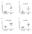
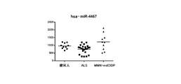
- FIG. 11 shows the distribution of signal intensity of healthy persons, ALS, and mdCIDP and MMN for hsa-miR-4467 in the cerebrospinal fluid specimen.
- the signal intensity of hsa-miR-4467 is significant in ALS. It was confirmed that it had decreased.
- miRNA present in these cerebrospinal fluid is a marker that gives an alternative differential index of ALS and mdCIDP or MMN.
- an AUC area Under the curve: the area under the ROC curve was 0.95 or more.
- the miRNA indices selected by this were an increase in the amount of hsa-miR-642b, an increase in the amount of hsa-miR-3197, an increase in the amount of hsa-miR-4697-5p, and a decrease in the amount of hsa-miR-4417.
- the decrease in the amount of hsa-miR-4417 is used as a combination with 1 to 3 kinds of differential indicators selected from the increase and decrease of the other 3 kinds of microRNA amounts.
- FIG. 12 is a drawing showing the signal intensity distribution of ALS in a cerebrospinal fluid sample and these four markers suitable for distinguishing mdCIDP or MMN
- FIG. 13 shows the ALS in the cerebrospinal fluid sample.
- ROC curves receiveriverreceiveoperating characteristic curve
- FIGS. 14 to 16 show particularly preferable discrimination modes.
- FIG. 14 shows (1) a signal intensity distribution pick-up of hsa-miR-4417 and hsa-miR-642b, which are suitable markers for distinguishing ALS and mdCIDP or MMN in cerebrospinal fluid samples, and (2) It is the figure which schematized the detection process using these with sensitivity (Sensitivity) and specificity (Specificity). In this embodiment, very good discrimination results with a sensitivity of 100% and a specificity of 95.7% were recognized.
- FIG. 15 shows (1) a signal intensity distribution pick-up of hsa-miR-4417 and hsa-miR-3197, which are suitable markers for distinguishing ALS and mdCIDP or MMN in cerebrospinal fluid samples, and (2) It is the figure which schematized the detection process using these with sensitivity (Sensitivity) and specificity (Specificity). In this embodiment, very good discrimination results with a sensitivity of 91.3% and a specificity of 90.0% were recognized.
- FIG. 16 is a drawing in which (1) signal intensity distribution is picked up for hsa-miR-4417 and hsa-miR-4697-5p, which are suitable markers for differentiating ALS and mdCIDP or MMN in cerebrospinal fluid samples. (2) It is the figure which schematized the detection process using these with sensitivity (Sensitivity) and specificity (Specificity). In this embodiment, very good discrimination results with a sensitivity of 91.3% and a specificity of 90.0% were recognized.
- miRNA in cerebrospinal fluid that can be used as a marker related to ALS, MMN, and PMA is used for the detection method of the differential index of the present invention having the same contents as the previous process of establishing a disease marker. And sorted. The Welch method based on the null assumption was used as the selection method. The breakdown of cerebrospinal fluid samples used for selecting each category is “Healthy person: 10 samples, sALS: 23 samples, MMN: 13 samples, PMA11 sample”.
- FIG. 17 (FIGS. 17-1 to 17-8) provides a differential indicator of ALS (arcogenic, sALS) and MMN The distribution of marker signal intensity is shown.
- the sorting method the Welch method based on the null assumption was used, and the significance level was sorted at 0.001%.
- FIG. 5-1 shows (1) hsa-miR-214, (2) hsa-miR-518c *, (3) hsa-miR-574-3p, and (4) hsa in the cerebrospinal fluid specimen.
- -miR-642a the signal intensity distribution of ALS and MMN is shown.
- the signal intensity of hsa-miR-214 was significantly increased in ALS relative to MMN.
- the signal intensity of hsa-miR-518c * was significantly increased in ALS relative to MMN.
- the signal intensity of hsa-miR-574-3p was significantly increased in ALS relative to MMN.
- the signal intensity of hsa-miR-642a was significantly increased in ALS compared to MMN.
- these miRNAs present in the cerebrospinal fluid are markers that provide a differentiation index between ALS and MMN.
- FIG. 17-2 shows (1) hsa-miR-657, (2) hsa-miR-25 *, (3) hsa-miR-139-3p, and (4) hsa in the cerebrospinal fluid specimen.
- -miR-187 * the signal intensity distributions of ALS and MMN are shown.
- the signal intensity of hsa-miR-657 was significantly increased in ALS relative to MMN.
- the signal intensity of hsa-miR-25 * was significantly increased in ALS compared to MMN.
- the signal intensity of hsa-miR-139-3p was significantly increased in ALS relative to MMN.
- the signal intensity of hsa-miR-187 * was significantly increased in ALS compared to MMN.
- these miRNAs present in the cerebrospinal fluid are markers that provide a differentiation index between ALS and MMN.
- FIG. 17-3 shows (1) hsa-miR-135a *, (2) hsa-miR-185 *, (3) hsa-miR-574-5p, and (4) in the cerebrospinal fluid specimen.
- hsa-miR-1231 the signal intensity distribution of ALS and MMN is shown.
- the signal intensity of hsa-miR-135a * was significantly increased in ALS relative to MMN.
- the signal intensity of hsa-miR-185 * was significantly increased in ALS relative to MMN.
- the signal intensity of hsa-miR-574-5p was significantly increased in ALS relative to MMN.
- the signal intensity of hsa-miR-1231 was significantly increased in ALS compared to MMN.
- these miRNAs present in the cerebrospinal fluid are markers that provide a differentiation index between ALS and MMN.
- FIG. 17-4 shows (1) hsa-miR-1181, (2) hsa-miR-1292, (3) hsa-miR-675 *, and (4) hsa-miR in the cerebrospinal fluid specimen.
- the signal intensity distribution of ALS and MMN is shown.
- the signal intensity of hsa-miR-1181 was significantly increased in ALS relative to MMN.
- the signal intensity of hsa-miR-1292 was significantly increased in ALS relative to MMN.
- the signal intensity of hsa-miR-675 * was significantly increased in ALS relative to MMN.
- the signal intensity of hsa-miR-1539 was significantly increased in ALS relative to MMN.
- these miRNAs present in the cerebrospinal fluid are markers that provide a differentiation index between ALS and MMN.
- Fig. 17-5 shows (1) hsa-miR-718, (2) hsa-miR-2682 *, (3) hsa-miR-449c *, and (4) hsa-
- the distribution of signal intensity of ALS and MMN is shown for miR-3130-5p.
- (1) The signal intensity of hsa-miR-718 was significantly increased in ALS compared to MMN.
- the signal intensity of hsa-miR-2682 * was significantly increased in ALS compared to MMN.
- the signal intensity of hsa-miR-449c * was significantly increased in ALS relative to MMN.
- the signal intensity of hsa-miR-3130-5p was significantly increased in ALS relative to MMN.
- these miRNAs present in the cerebrospinal fluid are markers that provide a differentiation index between ALS and MMN.
- FIG. 17-6 shows (1) hsa-miR-3138, (2) hsa-miR-3156-5p, (3) hsa-miR-3197, and (4) hsa-
- the distribution of signal intensity of ALS and MMN is shown for miR-4297.
- the signal intensity of hsa-miR-3138 was significantly increased in ALS compared to MMN.
- the signal intensity of hsa-miR-3156-5p was significantly increased in ALS relative to MMN.
- the signal intensity of hsa-miR-3197 was significantly increased in ALS compared to MMN.
- the signal intensity of hsa-miR-4297 was significantly increased in ALS compared to MMN.
- these miRNAs present in the cerebrospinal fluid are markers that provide a differentiation index between ALS and MMN.
- FIG. 17-7 shows (1) hsa-miR-4276, (2) hsa-miR-3667-3p, (3) hsa-miR-3937, and (4) hsa-
- the distribution of signal intensity of ALS and MMN is shown for miR-4437.
- the signal intensity of hsa-miR-4276 was significantly increased in ALS relative to MMN.
- the signal intensity of hsa-miR-3667-3p was significantly increased in ALS relative to MMN.
- the signal intensity of hsa-miR-3937 was significantly increased in ALS relative to MMN.
- the signal intensity of hsa-miR-4437 was significantly increased in ALS compared to MMN.
- these miRNAs present in the cerebrospinal fluid are markers that provide a differentiation index between ALS and MMN.
- FIGS. 17-8 show (1) hsa-miR-4446-3p, (2) hsa-miR-4481, (3) hsa-miR-4639-3p, and (4) in the cerebrospinal fluid specimen.
- hsa-miR-4701-3p the signal intensity distribution of ALS and MMN is shown.
- the signal intensity of hsa-miR-4446-3p was significantly increased in ALS relative to MMN.
- the signal intensity of hsa-miR-4481 was significantly increased in ALS compared to MMN.
- the signal intensity of hsa-miR-4639-3p was significantly increased in ALS relative to MMN.
- the signal intensity of hsa-miR-4701-3p was significantly increased in ALS relative to MMN.
- these miRNAs present in the cerebrospinal fluid are markers that provide a differentiation index between ALS and MMN.
- FIG. 17-9 shows signal intensity distributions of ALS and MMN for (1) hsa-miR-4726-3p and (2) hsa-miR-4754 in cerebrospinal fluid samples.
- the signal intensity of hsa-miR-4726-3p was significantly increased in ALS relative to MMN.
- the signal intensity of hsa-miR-4754 was significantly increased in ALS compared to MMN.
- these miRNAs present in the cerebrospinal fluid are markers that provide a differentiation index between ALS and MMN.
- [C] -1 (2) Suitable marker for providing an ALS / MMN differentiation index
- a high AUC> 0.95 Sensitivity and high specificity can be expected, including increased hsa-miR-139-3p, increased hsa-miR-574-5p, increased hsa-miR-1231, increased hsa-miR-2682 *, and increase in hsa-miR-3130-5p.
- MMN and ALS can be differentiated with high sensitivity and high specificity.
- ALS and MMN could be distinguished with a sensitivity of 100% and a specificity of 91%.
- the increase in hsa-miR-2682 * and the increase in hsa-miR-3130-5p are illustrated together with the sensitivity (Sensitivity) and specificity (Specificity). .
- ALS and MMN could be distinguished with a sensitivity of 92.3% and a specificity of 100%.
- FIG. 21 shows the distribution of signal intensity of a marker that provides a differential indicator of MMN and PMA. Show.
- the sorting method the Welch method based on the null assumption was used, and the significance level was sorted at 5%.
- FIG. 21-1 shows (1) hsa-miR-99a, (2) hsa-miR-23b, (3) hsa-miR-184, and (4) hsa-miR- For 485-3p, the signal intensity distribution of MMN and PMA is shown.
- the signal intensity of hsa-miR-99a was significantly reduced in PMA relative to MMN.
- the signal intensity of hsa-miR-23b was significantly reduced in PMA compared to MMN.
- the signal intensity of hsa-miR-184 was significantly increased in PMA with respect to MMN.
- the signal intensity of hsa-miR-485-3p was significantly reduced in PMA relative to MMN.
- these miRNAs present in the cerebrospinal fluid are markers that provide a differentiation index between ALS and MMN.
- FIG. 21-2 shows (1) hsa-miR-486-5p, (2) hsa-miR-432 *, (3) hsa-miR-675, and (4) hsa in the cerebrospinal fluid specimen.
- -miR-25 * the signal intensity distribution of MMN and PMA is shown.
- the signal intensity of hsa-miR-486-5p was significantly decreased in PMA with respect to MMN.
- the signal intensity of hsa-miR-432 * was significantly decreased in PMA with respect to MMN.
- the signal intensity of hsa-miR-675 was significantly increased in PMA compared to MMN.
- the signal intensity of hsa-miR-25 * was significantly increased in PMA relative to MMN.
- these miRNAs present in the cerebrospinal fluid are markers that provide a differentiation index between ALS and MMN.
- FIG. 21-3 shows (1) hsa-miR-26b *, (2) hsa-miR-920, (3) hsa-miR-1296, and (4) hsa-miR in cerebrospinal fluid samples. It is drawing which showed distribution of the signal intensity of MMN and PMA about -1182.
- the signal intensity of hsa-miR-26b * was significantly decreased in PMA with respect to MMN.
- the signal intensity of hsa-miR-920 was significantly increased in PMA with respect to MMN.
- the signal intensity of hsa-miR-1296 was significantly increased in PMA with respect to MMN.
- the signal intensity of hsa-miR-1182 was significantly increased in PMA relative to MMN.
- these miRNAs present in the cerebrospinal fluid are markers that provide a differentiation index between ALS and MMN.
- FIG. 21-4 shows (1) hsa-miR-1250, (2) hsa-miR-1275, (3) hsa-miR-1292, and (4) hsa-miR- About 1914, the distribution of the signal intensity of MMN and PMA is shown.
- the signal intensity of hsa-miR-1250 was significantly increased in PMA with respect to MMN.
- the signal intensity of hsa-miR-1275 was significantly increased in PMA with respect to MMN.
- the signal intensity of hsa-miR-1292 was significantly increased in PMA with respect to MMN.
- the signal intensity of hsa-miR-1914 was significantly increased in PMA relative to MMN.
- these miRNAs present in the cerebrospinal fluid are markers that provide a differentiation index between ALS and MMN.
- FIG. 21-5 shows (1) hsa-miR-711, (2) hsa-miR-3138, (3) hsa-miR-3162-5p, and (4) hsa- For miR-3176, the signal intensity distributions of MMN and PMA are shown.
- the signal intensity of hsa-miR-711 was significantly increased in PMA with respect to MMN.
- the signal intensity of hsa-miR-3138 was significantly increased in PMA relative to MMN.
- the signal intensity of hsa-miR-3162-5p was significantly increased in PMA relative to MMN.
- the signal intensity of hsa-miR-3176 was significantly increased in PMA relative to MMN.
- these miRNAs present in the cerebrospinal fluid are markers that provide a differentiation index between ALS and MMN.
- FIG. 21-6 shows (1) hsa-miR-3187-3p, (2) hsa-miR-3188, (3) hsa-miR-3197, and (4) hsa- For miR-514b-5p, the signal intensity distribution of MMN and PMA is shown.
- the signal intensity of hsa-miR-3187-3p was significantly increased in PMA with respect to MMN.
- the signal intensity of hsa-miR-3188 was significantly increased in PMA with respect to MMN.
- the signal intensity of hsa-miR-3197 was significantly increased in PMA with respect to MMN.
- the signal intensity of hsa-miR-514b-5p was significantly increased in PMA with respect to MMN.
- these miRNAs present in the cerebrospinal fluid are markers that provide a differentiation index between ALS and MMN.
- FIG. 21-7 shows (1) hsa-miR-4261, (2) hsa-miR-3652, (3) hsa-miR-3654, and (4) hsa-miR- For 3679-5p, the signal intensity distribution of MMN and PMA is shown.
- the signal intensity of hsa-miR-4261 was significantly increased in PMA relative to MMN.
- the signal intensity of hsa-miR-3652 was significantly increased in PMA with respect to MMN.
- the signal intensity of hsa-miR-3654 was significantly increased in PMA compared to MMN.
- the signal intensity of hsa-miR-3679-5p was significantly increased in PMA with respect to MMN.
- these miRNAs present in the cerebrospinal fluid are markers that provide a differentiation index between ALS and MMN.
- FIG. 21-8 shows (1) hsa-miR-3689a-3p, (2) hsa-miR-3180, (3) hsa-miR-3919, and (4) hsa- For miR-3925-5p, the signal intensity distribution of MMN and PMA is shown.
- the signal intensity of hsa-miR-3689a-3p was significantly increased in PMA with respect to MMN.
- the signal intensity of hsa-miR-3180 was significantly reduced in PMA compared to MMN.
- the signal intensity of hsa-miR-3919 was significantly increased in PMA compared to MMN.
- the signal intensity of hsa-miR-3925-5p was significantly increased in PMA with respect to MMN.
- these miRNAs present in the cerebrospinal fluid are markers that provide a differentiation index between ALS and MMN.
- FIG. 21-9 shows (1) hsa-miR-3928, (2) hsa-miR-4425, (3) hsa-miR-4437, and (4) hsa-miR- About 4450, the distribution of the signal intensity of MMN and PMA is shown.
- the signal intensity of hsa-miR-3928 was significantly increased in PMA with respect to MMN.
- the signal intensity of hsa-miR-4425 was significantly increased in PMA with respect to MMN.
- the signal intensity of hsa-miR-4437 was significantly increased in PMA with respect to MMN.
- the signal intensity of hsa-miR-4450 was significantly increased in PMA with respect to MMN.
- these miRNAs present in the cerebrospinal fluid are markers that provide a differentiation index between ALS and MMN.
- FIG. 21-10 shows (1) hsa-miR-4461, (2) hsa-miR-4465, (3) hsa-miR-4478, and (4) hsa-miR- For 2392, the signal intensity distribution of MMN and PMA is shown.
- the signal intensity of hsa-miR-4461 was significantly increased in PMA with respect to MMN.
- the signal intensity of hsa-miR-4465 was significantly increased in PMA with respect to MMN.
- the signal intensity of hsa-miR-4478 was significantly increased in PMA relative to MMN.
- the signal intensity of hsa-miR-2392 was significantly increased in PMA compared to MMN.
- these miRNAs present in the cerebrospinal fluid are markers that provide a differentiation index between ALS and MMN.
- FIGS. 21-11 show (1) hsa-miR-4526, (2) hsa-miR-3619-3p, (3) hsa-miR-3940-5p, and (4) in the cerebrospinal fluid specimen.
- hsa-miR-4635 the signal intensity distribution of MMN and PMA is shown.
- the signal intensity of hsa-miR-4526 was significantly increased in PMA relative to MMN.
- the signal intensity of hsa-miR-3619-3p was significantly increased in PMA with respect to MMN.
- the signal intensity of hsa-miR-3940-5p was significantly reduced in PMA relative to MMN.
- the signal intensity of hsa-miR-4635 was significantly increased in PMA relative to MMN.
- these miRNAs present in the cerebrospinal fluid are markers that provide a differentiation index between ALS and MMN.
- Fig. 21-12 shows (1) hsa-miR-4646-5p, (2) hsa-miR-4667-5p, (3) hsa-miR-4691-5p, and ( 4) The signal intensity distribution of MMN and PMA is shown for hsa-miR-4709-3p.
- the signal intensity of hsa-miR-4646-5p was significantly increased in PMA with respect to MMN.
- the signal intensity of hsa-miR-4667-5p was significantly increased in PMA with respect to MMN.
- the signal intensity of hsa-miR-4691-5p was significantly increased in PMA with respect to MMN.
- the signal intensity of hsa-miR-4709-3p was significantly increased in PMA relative to MMN.
- these miRNAs present in the cerebrospinal fluid are markers that provide a differentiation index between ALS and MMN.
- FIGS. 21-13 show (1) hsa-miR-4740-5p, (2) hsa-miR-4751, (3) hsa-miR-4753-5p, and (4) in the cerebrospinal fluid specimen.
- hsa-miR-4769-5p the signal intensity distribution of MMN and PMA is shown.
- the signal intensity of hsa-miR-4740-5p was significantly increased in PMA with respect to MMN.
- the signal intensity of hsa-miR-4751 was significantly increased in PMA compared to MMN.
- the signal intensity of hsa-miR-4753-5p was significantly increased in PMA with respect to MMN.
- hsa-miR-4769-5p showed a significant decrease in signal intensity in PMA versus MMN.
- these miRNAs present in the cerebrospinal fluid are markers that provide a differentiation index between ALS and MMN.
- FIGS. 21-14 show (1) hsa-miR-4779, (2) hsa-miR-2467-5p, (3) hsa-miR-4787-3p, and (4) in the cerebrospinal fluid specimen.
- hsa-miR-4800-5p the signal intensity distribution of MMN and PMA is shown.
- the signal intensity of hsa-miR-4779 was significantly increased in PMA relative to MMN.
- the signal intensity of hsa-miR-2467-5p was significantly increased in PMA relative to MMN.
- the signal intensity of hsa-miR-4787-3p was significantly increased in PMA relative to MMN.
- the signal intensity of hsa-miR-4800-5p was significantly increased in PMA with respect to MMN.
- these miRNAs present in the cerebrospinal fluid are markers that provide a differentiation index between ALS and MMN.
- [C] -2 (2) Suitable marker for providing a differentiation index between MMN and PMA
- a marker for providing a differentiation index for MMN and PMA high sensitivity at a significance level of 1% Are expected to increase hsa-miR-1275, increase hsa-miR-711, increase hsa-miR-3138, increase hsa-miR-3162-5p, increase hsa-miR-3928, Is mentioned.
- an increase in hsa-miR-3162-5p and an increase in hsa-miR-3928 can be mentioned.
- an increase in hsa-miR-1914 * can be cited as an indicator of the balance of AUC> 0.85 at a significance level of 1-5%.
- FIG. 22 shows (1) the signal intensity distribution and (2) these for each of these preferred two types, namely hsa-miR-3162-5p and hsa-miR-3928.
- the detection process using is shown in a diagram together with sensitivity and specificity.
- MMN and PMA could be distinguished with a sensitivity of 84.6% and specificity of 90.9%.
- FIG. 23 the increase of hsa-miR-3162-5p and hsa-miR-3928 is illustrated with a detection process using a combination of these together with sensitivity and specificity. In this combination, MMN and PMA could be distinguished with a sensitivity of 76.9% and a specificity of 100%.
Abstract
本発明は、運動神経の障害を呈する疾患において、特に筋萎縮性側索硬化症(ALS)、進行性筋萎縮症(PMA)、純粋慢性炎症性脱髄性多発ニューロパチー(mdCIDP)、又は、多巣性運動性ニューロパチー(MMN)の鑑別を行うための疾病マーカーを見出して、特に初期段階での的確な鑑別が難しいこれらの疾病の鑑別を容易に行う途を提供することを課題とする発明である。そして本発明者は、髄液中のマイクロRNAを母集団とする、特定の218種類マイクロRNAの増減が、これらの4種類の疾患の優れた鑑別指標となることを見出し、本発明を完成した。さらに本発明者は、髄液中のマイクロRNAから特定疾患の鑑別指標を見出し、次いで当該鑑別指標を用いた鑑別を、マイクロRNA検出用基板を用いて行う際の標準化法を確立し、髄液中のマイクロRNAを用いる疾病の鑑別手段を提供するものである。
Description
本発明は、特定の疾患の鑑別に用いる検出方法に関する発明である。より具体的には、髄液中に存在する特定のマイクロRNAを指標として用いる、運動神経の障害を呈する疾患である、筋萎縮性側索硬化症、進行性筋萎縮症、純粋運動型慢性炎症性脱髄性多発ニューロパチー、又は、多巣性運動性ニューロパチー、の鑑別に際して用いる検出方法に関する発明である。
さらに本発明は、髄液中のマイクロRNAの検出用アレイを用いて検出を行う際の標準化方法に関する発明を提供する。
運動神経の障害に伴って起こり得る運動麻痺は、上下肢の随意的な運動が侵された状態であり、運動中枢から筋肉に至るまでのどこかに障害があることを示唆するものである。従って運動麻痺を診察する場合には、大脳皮質から内包、脳幹、脊髄、脊髄前角運動ニューロンへとつながる上位運動ニューロンの障害である中枢性麻痺、脊髄前角運動ニューロン、末梢神経から筋に至るまでの下位運動ニューロンの障害、神経筋接合部の障害、及び、筋肉の障害についての局所診断を念頭に入れて、正確な鑑別・診断を行う必要がある。運動神経の障害に伴って起こり得る運動麻痺を来す疾患は数多く存在するが、その正しい診断の過程を身に付けることは、神経内科系の疾患を扱う医療関係者にとっての必須の事項である。
しかしながら、運動神経の障害を呈する疾患には紛らわしいものも多く、その的確な鑑別・診断をサポートするために、視診、診察手技、鑑別の基礎知識等の基礎的な鑑別・診断手段に加えて、画像診断技術や神経生理学的診断法が開発されている。
具体的には、下記の非特許文献1、2に運動神経の障害を呈する疾患の的確な鑑別・診断をサポートするための技術が開示されている。
Neurology(2009).73(29):1686-1692
Neurology(2006).67(4):592-596
上記した状況のもと、本発明者らは、運動神経の障害を呈する疾患において、特に筋萎縮性側索硬化症(Amyotrophic Lateral Sclerosis:ALS)、進行性筋萎縮症(Progressive muscular atrophy:PMA)、純粋慢性炎症性脱髄性多発ニューロパチー(motor dominant chronic inflammatory demyelinating polyneuropathy:mdCIDP)、又は、多巣性運動性ニューロパチー(multifocal motor neuropathy:MMN)の鑑別を行うための疾病マーカーを見出して、特に初期段階での的確な鑑別が難しいこれらの疾病の鑑別を容易に行う途を提供することを課題として、検討を行った。例えば、後述するようにmdCIDP又はMMNの治療には「免疫グロブリンの大量投与」が有効であることが分かっている。しかしながらこの療法のコストは、1回100万円以上で、しかもこれが年6~12回必要、と非常に高額であるため、この療法が適合しないALSやPMAに類似した症例に対して、元々無効である可能性を見越して治療を行うことは困難である。よって、現時点では免疫グロブリンの大量投与は、未だ実質的な治療項目とはいえない。仮に、ALSやPMAと、mdCIDP又はMMNとの鑑別が容易になれば、現状ではリスク視されている免疫グロブリンの大量投与を的確に行うことが可能となり、mdCIDPやMMNの患者にとっては、早期に適切な治療を受けることによって回復可能性を高めることができると考えられる。
このような具体的な課題のもと、本発明者は髄液検体中のマイクロRNA(microRNA(miRNA))を、上記の運動神経の障害を呈する疾患の指標とする可能性についての検討を行った。
miRNAは21~23塩基程度からなる短鎖RNAで、標的となるmRNAに抑制性に働くことで遺伝子発現の制御に重要な役割を果たしている。このmiRNAの発現パターンが癌や免疫疾患、神経変性疾患でも変化することが報告されており、miRNAの変化を血液等における疾患マーカーとして利用した報告が認められる。
(1)標準化法やデータ解析法の基となる手段について
本発明者は、目的とする疾患マーカーとなり得るmiRNAを見出す基として「miRNA検出用アレイ」を選択した。「miRNA検出用アレイ」は、主にガラスや硬質ポリマー等の基板状素材の表面に、検体中のmiRNAと結合し得るプローブ核酸を網羅的、すなわち、少なくとも500種類以上のプローブ核酸を整列・配置(アレイ化)したものの総称である。検体中のmiRNAと接触することにより、当該miRNAを捕捉して、事前に標識された検体におけるmiRNAの標識シグナルを検出することによって、当該検体中のmiRNAの存在又は非存在、あるいは、シグナル強度に応じた存在量を検出することができる。特にmiRNA検出用アレイでは、2つの検体、例えば、疾病に罹患した患者の検体と健常人の検体の間で存在状態の異なるmiRNA群を、数少ない実験で網羅的に同定できる。すなわち、多数の検体についてmiRNA検出用アレイで解析を行い、データを蓄積し、それらの情報を並列比較することにより、検体の記述、分類、評価を行うことが可能である。
本発明者は、目的とする疾患マーカーとなり得るmiRNAを見出す基として「miRNA検出用アレイ」を選択した。「miRNA検出用アレイ」は、主にガラスや硬質ポリマー等の基板状素材の表面に、検体中のmiRNAと結合し得るプローブ核酸を網羅的、すなわち、少なくとも500種類以上のプローブ核酸を整列・配置(アレイ化)したものの総称である。検体中のmiRNAと接触することにより、当該miRNAを捕捉して、事前に標識された検体におけるmiRNAの標識シグナルを検出することによって、当該検体中のmiRNAの存在又は非存在、あるいは、シグナル強度に応じた存在量を検出することができる。特にmiRNA検出用アレイでは、2つの検体、例えば、疾病に罹患した患者の検体と健常人の検体の間で存在状態の異なるmiRNA群を、数少ない実験で網羅的に同定できる。すなわち、多数の検体についてmiRNA検出用アレイで解析を行い、データを蓄積し、それらの情報を並列比較することにより、検体の記述、分類、評価を行うことが可能である。
miRNAは、進化の過程において保存されている、短い非コードのRNAで、翻訳レベルで遺伝子発現を制御していることが知られている。哺乳動物のゲノムには1000種類を超える特有のmiRNAがコードされており、少なくとも遺伝子の30%がこれらのmiRNA発現に制御されていると推測されている。
最近では髄液中のmiRNAを中枢原発悪性リンパ腫、神経膠腫、アルツハイマー病等の疾病マーカーとして利用した報告も認められる。
通常miRNA検出用アレイの使用に際しては、検体をアレイに接触させて得られるシグナル値を相対化する「標準化」と呼ばれる工程が行われる。しかしながら髄液中のmiRNA量は、血液等に比べると少量であり、かつ、その採取可能な量も限定されており、しかも髄液中の内因性コントロールとなる既知のmiRNAが、現状では知られていないことが問題点として挙げられる。miRNA検出用アレイを髄液のmiRNAに対して標準化しようとすると、定量性の乏しいmiRNAまで標準化の母集団に入れざるを得ず、それは、不正確な情報を含んだ要素を、標準化の母集団が含むことを意味するものであり、正確な統計処理を行う際の大きな妨げになることになる。
<本発明の標準化方法>
本発明者は、この問題を解決するために標準的な採取量の髄液中のmiRNAをmiRNA検出用アレイに反応させて、定常反応性、好適にはさらに定量性の認められるmiRNAのみを標準化の母集団を構成するmiRNAとし、それらのmiRNAのシグナルの中から適切なものを選び、さらにこれを基にシグナルデータの標準化を行うことにより、この問題を解決できることを見出した。
本発明者は、この問題を解決するために標準的な採取量の髄液中のmiRNAをmiRNA検出用アレイに反応させて、定常反応性、好適にはさらに定量性の認められるmiRNAのみを標準化の母集団を構成するmiRNAとし、それらのmiRNAのシグナルの中から適切なものを選び、さらにこれを基にシグナルデータの標準化を行うことにより、この問題を解決できることを見出した。
すなわち本発明は、以下の(A)~(C)の工程を含むことを特徴とする、miRNA検出用アレイを用いたmiRNAの標準化方法(以下、本発明の標準化方法ともいう)を提供し、さらに当該標準化方法を用いたmiRNAの定量方法を提供する。
(A)髄液検体をmiRNA検出用アレイに接触させて得られるmiRNAのシグナル値に基づき、定常発現が認められるmiRNA群を選別し、
(B)当該miRNA群においてシグナル値が上位25%~75%の範囲にある、いずれかのマイクロRNAのシグナル値のうちの一種類を選択し、
(C)当該選択されたmiRNAのシグナル値を特定の数に置き換え、当該置き換えをするために行われる演算において用いられる数的手段を求め、当該数的手段を当該miRNA群を構成する個々のmiRNAのシグナル値に対して講じることで、これらのシグナル値を相対的なシグナル強度とする補正を行うことにより、当該miRNA群の標準化を行う。
(A)髄液検体をmiRNA検出用アレイに接触させて得られるmiRNAのシグナル値に基づき、定常発現が認められるmiRNA群を選別し、
(B)当該miRNA群においてシグナル値が上位25%~75%の範囲にある、いずれかのマイクロRNAのシグナル値のうちの一種類を選択し、
(C)当該選択されたmiRNAのシグナル値を特定の数に置き換え、当該置き換えをするために行われる演算において用いられる数的手段を求め、当該数的手段を当該miRNA群を構成する個々のmiRNAのシグナル値に対して講じることで、これらのシグナル値を相対的なシグナル強度とする補正を行うことにより、当該miRNA群の標準化を行う。
そして、当該標準化がなされたシグナル強度を基にmiRNAの定量を行うことができる。
上記工程(A)に関し、本発明の標準化方法においてmiRNA検出用アレイに接触させるために用いられる髄液検体量は限定されるものではないが、標準的な量であることが好適である。すなわち標準量を超えた大量の髄液を用いれば、標準化の母集団となるべき定常反応性や定量性のあるmiRNAの数は増えるものの、臨床現場で現実に採取可能な髄液量を用いて標準化が可能なmiRNA群からは逸脱してしまい、現実的に適切な標準化の母集団からはかけ離れてしまう。逆に標準量よりも髄液量が少ないと母集団となるべきmiRNAの数は過度に限定され、標準化の母集団としての信頼性が乏しくなる原因となり得る。この意味において本発明の標準化方法において用いられるべき髄液検体量は概ねリンパ球画分を除いた上清量として0.45~1.8mLの範囲であることが好適である。本実施例、及び、通常の臨床の現場では0.9mLである。
髄液検体は、通常公知の方法により採取確保することができる。これは本発明のあらゆる態様において共通する。すなわち検体提供者において腰椎穿刺を行い、これにより採取した髄液に対してリンパ球成分を分離することができる程度の遠心を施して得られたリンパ球成分を除いた上清を確保し、これを髄液検体として用いることができる。当該髄液検体の保存は、冷凍保存、好ましくは-80℃以下の冷凍保存により行うことができる。必要に応じて髄液検体の解凍を行い、常法によりRNAの抽出を行い、これをmiRNAの検出に供することができる。
本発明の標準化方法に関連して記される「シグナル」とは、髄液検体のmiRNAがmiRNA検出用アレイ上の核酸プローブと結合することにより顕在化するシグナルのことであり、多くの場合は髄液検体のmiRNAに予め施した蛍光標識、色素標識等である。これらの標識に基づく発光パターンをスキャナー等に取って、その有無や強度を検出する。その際、一通りの髄液量においてmiRNA検出用アレイのシグナルが認められる場合は「定常発現」である。また、その際にmiRNAに基づくシグナルが髄液の容量に依存して増加する場合を「定量性有り」とすることができる。本発明の標準化方法の工程(A)において、「標準化の母集団を構成するmiRNA」として選別されるmiRNAは、「検出用アレイにおいて定常発現が認められるmiRNA」であるが、厳密には「さらに定量性の認められるmiRNA」が好適である。これについては後述する。
ここに記す本発明の標準化方法における定義とは別に、他の検出方法、例えば後述するRT-PCR法やノーザンブロット法等における検出指標となる出力情報も広義の「シグナル」である。本発明の標準化方法に関わる「シグナル」も、他の検出方法に関わる広義の「シグナル」も、定量シグナルと定性シグナルの双方を含むものである。
上記の工程(A)において標準化の母集団を構成するmiRNA群が選別されるが、当該miRNA群は、髄液検体提供者、特に罹患している疾病によっても異なり、miRNA検出用アレイの鋭敏性によっても異なり、髄液検体量によっても異なり、miRNAの抽出効率等の実験手技等によっても異なる。後述する実施例では、miRNA検出用アレイとして「miRNA array(3D GENE(登録商標)/TORAY)」(東レ)を用いた。このmiRNA検出用アレイは、高性能DNAチップ基板3D GENE(登録商標)1枚にデータベースmiRBase Release17から選定したヒト約1700種のmiRNAを検出するセンス鎖オリゴヌクレオチドを固定化スポットとしたアレイであり、検出対象miRNAに施す標識はCy(Cyanine)を用いた。その結果、健常人の髄液検体をリンパ球画分を除いた上清量として0.9mLに対して337個の定常発現しているmiRNA群が認められ、0.45mLの髄液検体の使用で41個の定常発現と共に定量性の伴うmiRNA群が認められた。これらのmiRNA群は、両者共に標準化の母集団として適切なものである。しかしながら、これらはあくまでも一実施例である。
上記の工程(B)は、工程(a)において選別された、「標準化の母集団を構成するmiRNA群」を、個々のmiRNAをシグナル値の上位、すなわち大きなものから順番にランク付けして、その中から標準化の基となる特定のシグナル値を選択する工程である。この標準化の基となる特定のシグナル値は、上記miRNA群のうちシグナル値が上位25%~75%の範囲にある、いずれかのmiRNAのシグナル値から一種類を選択することが可能である。この選択可能なシグナル値の範囲は、好適には上位35%~65%、より好適には上位45%~55%とすることができる。さらにこの特定のシグナル値の範囲の選択の方法として、上記miRNA群のシグナル値の強度分布の中央値領域、つまり、中央値を中心として、-25%~+25%の範囲内とすることが可能であり、さらに当該中央値領域を絞り込むことが好適である。具体的には同じく中央値を中心として、-10%~+10%の範囲とすることが好適であり、-5%~+5%とすることがさらに好適である。
標準化の基となる特定のシグナル値は、異なる髄液検体間で標準化がなされた個々のmiRNA同士のシグナル強度を二次元プロットして近似される直線がy=x(傾きが1)への収束性が高くなるものを選択することが好ましい。後述する実施例において、本発明の標準化法を用いると、理想的な傾きの当該直線が得られることが明らかになった。
上述したように上記miRNA群は、「定量性を伴うmiRNA」で構成させることが厳密であるが、「定常発現を伴うmiRNA」で構成させることも可能であり、かつ、実用的である。
すなわち、「定常発現を伴うにもかかわらず、定量性を伴わないmiRNA」は、主にmiRNA検出用アレイの特定のmiRNAスポットの不具合や検出装置の不具合や性能により現れるものであり、当該検出用アレイや検出装置の製品としての歩留まりに依るものである。現実的には、ある量の髄液検体をmiRNA検出用アレイに接触させてシグナルが得られて、定常発現を伴うと認められたスポットのmiRNAに定量性を仮定しても、実質的な不都合は認められないと考えられる。その一方で、miRNAのシグナル強度を計測してターゲット疾患の鑑別等を行う現場において、異なる容量の髄液検体を用いてシグナルの定量性を確認することは過度の重労働になり、かつ、実質的な理由に乏しいコスト高となる。仮に、標準化の母集団として選択した「定常発現を伴うmiRNA群」の中に、「定量発現を伴わないmiRNA」が含まれていたとしても、特定のシグナル値は上記のように規定した選択可能範囲内であることが強く見込まれる。よって、標準化の母集団として「定常発現を伴うmiRNA群」を採用することは、現実的な本発明の標準化法の運用として妥当なものといえる。
上記の工程(C)は、工程(B)において選択された特定のシグナル値を基に、上記miRNA群を構成する個々のmiRNAのシグナル値を標準化する工程である。
第一に、上記の特定のシグナル値の補正を行うことが必要である。この補正は、上記特定のシグナル値を、ある特定の数、すなわち補正値に置き換える過程である。この「特定の数」は、特に限定されるものでなく、実数の中から自由に選択することが可能であるが、通常は0以上の整数のいずれかから選ばれる。
この数的置き換えを行うための演算において用いられる数的手段を求めて、これを当該miRNA群を構成する個々のmiRNAのシグナル値に対して講じることで、個々のmiRNAのシグナル値が上記の補正値を中心とした相対的なシグナル強度となり、所望の標準化を行うことができる。
上記の演算の典型例は乗算であり、その数的手段は当該乗算において用いられた乗数の使用である。
このようにmiRNA群を構成する個々のmiRNAのシグナル値が標準化されることにより、上述したような様々な要因が加わり単純に比較することが困難な初発のシグナル値はシグナル強度として相対化がなされて、確度高い比較定量を行うことが可能となる。
<本発明の鑑別指標の検出方法>
本発明の標準化方法を用いて、ターゲット疾患の鑑別指標として髄液中のmiRNAを検出することができる(以下、本発明の鑑別指標の検出方法ともいう)。後述する実施例では、本発明の鑑別指標の検出方法を用いて、特定の運動神経の障害を呈する疾患の鑑別指標となる髄液中のmiRNAを見出した。
本発明の標準化方法を用いて、ターゲット疾患の鑑別指標として髄液中のmiRNAを検出することができる(以下、本発明の鑑別指標の検出方法ともいう)。後述する実施例では、本発明の鑑別指標の検出方法を用いて、特定の運動神経の障害を呈する疾患の鑑別指標となる髄液中のmiRNAを見出した。
本発明の鑑別指標の検出方法は、健常人とターゲット疾患の罹患者のそれぞれに由来する髄液検体群、又は、複数のターゲット疾患の罹患者間のそれぞれに由来する髄液検体群において、それぞれの髄液検体をmiRNA検出用アレイに接触させてmiRNAのシグナル値を得て、それぞれのシグナル値について、上述した本発明の標準化方法により得られるシグナル強度を基に、上記の健常人とターゲット疾患の罹患者のそれぞれに由来する髄液検体群、又は、複数のターゲット疾患の罹患者間のそれぞれに由来する髄液検体群における比較を行い、これらの群間の鑑別指標となるmiRNAを選別する、ターゲット疾患の鑑別指標となる髄液miRNAの検出方法、である。
上記のターゲット疾患は特に限定されず、髄液中のmiRNAを鑑別指標として用いることができる可能性のある疾患の全てである。例えば、後述する運動神経の障害を呈する疾患群が例示され、さらに悪性腫瘍、痴呆症等が考えられるが、これらに限定されるものではない。
本発明の鑑別指標の検出方法において用いられる、所望する鑑別が可能なmiRNAを検出する手段は特に限定されず、母集団が等分散であるか否かに関わらず用いることが可能なWelch法、2群比較において正規性が仮定できない場合に用いられるMann-Whitney法等が好適に用いられて、健常人とターゲット疾患の罹患者の間の髄液検体のmiRNA分布、又は、複数のターゲット疾患罹患者間の髄液検体のmiRNA分布、との間にポジティブ又はネガティブに有意差の認められる特定の髄液検体中のmiRNAを、疾病マーカーとして用いることができる。
各々の疾病マーカーにおける閾値(cut off値)は、当業者の技術常識に従って個別具体的に求めて実用に供することができる。
<本発明の支援方法>
本発明の標準化方法を用いて、既知のターゲット疾患の鑑別指標の存在を前提とするターゲット疾患の鑑別の支援方法(以下、本発明の支援方法ともいう)が提供される。
本発明の標準化方法を用いて、既知のターゲット疾患の鑑別指標の存在を前提とするターゲット疾患の鑑別の支援方法(以下、本発明の支援方法ともいう)が提供される。
本発明の支援方法は、ターゲット疾患の鑑別指標となる髄液検体におけるmiRNAに基づき、当該ターゲット疾患の鑑別のためのmiRNA検出用アレイを用いる検出方法を支援するための方法であって、本発明の標準化方法により、被検髄液検体における上記の鑑別指標として用いられるmiRNA量を、標準化されたシグナル強度として得て、当該シグナル強度を上記検出方法のために用いることを特徴とする支援方法、である。
本発明の支援方法によるターゲット疾患の鑑別に対する支援は、例えば、(a)被検髄液検体に対して本発明の標準化方法により、シグナル強度を標準化された相対値として得て、(b)当該シグナル強度と、同じく本発明の標準化方法により得られるシグナル強度により定義された、ターゲット疾患の鑑別指標となる髄液検体中の特定のmiRNAのカットオフ値とを比較して、(c)当該カットオフ値に対するシグナル強度の大小を基に、被検髄液検体におけるターゲット疾患の有無や程度の鑑別を行うことにより、行うことができる。
本発明の支援方法は、後述する本発明の検出方法においても好適に用いることができる。
<ソフトウエアの使用>
上述した本発明の標準化方法、同鑑別指標の検出方法、及び、同支援方法は、いずれもその方法を行うための工程をアルゴリズム化したソフトウエアを搭載した装置、において実行することによっても行うことができる。当該工程は、本発明の標準化方法では上記の工程(A)~(C)であり、同鑑別指標の検出方法では当該工程(A)~(C)、あるいは、これに加えて当該工程(C)で得られた標準化されたmiRNA群に対して、上記したWelch法、Mann-Whitney法等の統計学的な検定方法を行い、有意差を伴うmiRNAをターゲット疾患の鑑別指標として検出する工程である。さらに、本発明の支援方法では上記の工程(a)~(c)である(ただし、工程(a)は上記の工程(A)~(C)の存在を前提としている)。これらの工程は、当業者において自明の手順でアルゴリズム化され、ソフトウエアとして使用される。当該ソフトウエアが搭載される装置は、当該ソフトウエアを実行するための「演算処理部」、及び、当該ソフトウエアの実行に必要なデータが蓄積され、かつ、コンピュータプログラムの実行に伴ってデータが蓄積され得る「記録部」が最低限備わったコンピュータを基本構成とする装置である。当該ソフトウエアのアルゴリズムは、新たに入力されるデータの処理と、要求により記録部から導出されるデータの付加等に伴い演算処理部において実行され、これらの標準化を伴う方法において所望される結果を得ることができる。
上述した本発明の標準化方法、同鑑別指標の検出方法、及び、同支援方法は、いずれもその方法を行うための工程をアルゴリズム化したソフトウエアを搭載した装置、において実行することによっても行うことができる。当該工程は、本発明の標準化方法では上記の工程(A)~(C)であり、同鑑別指標の検出方法では当該工程(A)~(C)、あるいは、これに加えて当該工程(C)で得られた標準化されたmiRNA群に対して、上記したWelch法、Mann-Whitney法等の統計学的な検定方法を行い、有意差を伴うmiRNAをターゲット疾患の鑑別指標として検出する工程である。さらに、本発明の支援方法では上記の工程(a)~(c)である(ただし、工程(a)は上記の工程(A)~(C)の存在を前提としている)。これらの工程は、当業者において自明の手順でアルゴリズム化され、ソフトウエアとして使用される。当該ソフトウエアが搭載される装置は、当該ソフトウエアを実行するための「演算処理部」、及び、当該ソフトウエアの実行に必要なデータが蓄積され、かつ、コンピュータプログラムの実行に伴ってデータが蓄積され得る「記録部」が最低限備わったコンピュータを基本構成とする装置である。当該ソフトウエアのアルゴリズムは、新たに入力されるデータの処理と、要求により記録部から導出されるデータの付加等に伴い演算処理部において実行され、これらの標準化を伴う方法において所望される結果を得ることができる。
(2)具体的な疾患マーカーについて
本発明においては、上記の手法に基づき髄液中の特定のmiRNAの検出により鑑別を行うターゲット疾患は、筋萎縮性側索硬化症(ALS)、進行性筋萎縮症(Progressive muscular atrophy:PMA)、純粋運動型慢性炎症性脱髄性多発ニューロパチー(mdCIDP)、及び、多巣性運動性ニューロパチー(MMN)である。これらの四者はいずれも運動神経の障害を呈する疾患であるが、ALSとPMAは現在のところは致死的であるものの、mdCIDPとMMNはある程度の治療手段が存在するという点において、決定的に相違する。
本発明においては、上記の手法に基づき髄液中の特定のmiRNAの検出により鑑別を行うターゲット疾患は、筋萎縮性側索硬化症(ALS)、進行性筋萎縮症(Progressive muscular atrophy:PMA)、純粋運動型慢性炎症性脱髄性多発ニューロパチー(mdCIDP)、及び、多巣性運動性ニューロパチー(MMN)である。これらの四者はいずれも運動神経の障害を呈する疾患であるが、ALSとPMAは現在のところは致死的であるものの、mdCIDPとMMNはある程度の治療手段が存在するという点において、決定的に相違する。
以下、これらの運動神経の障害を呈する疾患についての個別の説明を行い、次いで、本発明の疾病マーカーを用いた検出方法についての開示を行う。
<筋萎縮性側索硬化症(Amyotrophic Lateral Sclerosis:ALS)>
ALSは、脳と脊髄にある、運動を担当する神経細胞(運動ニューロン)が選択的に障害を受け、徐々に体を動かすことができなくなる神経変性疾患であり、上位運動ニューロンと下位運動ニューロンの両方が障害を受ける。四肢のみならず、嚥下、会話、呼吸に関する筋力が低下し、日常生活が著しく損なわれる極めて予後が不良で致死的である。人工呼吸器等のサポートが無ければ発症後2~5年で主に呼吸不全で死亡する。人工呼吸器の使用で延命は可能であるものの、体は全く動かない状態である。その反面で知能や感覚機能は障害されないため、患者にとっての苦痛は大きく、難病中の難病といえる。
ALSは、脳と脊髄にある、運動を担当する神経細胞(運動ニューロン)が選択的に障害を受け、徐々に体を動かすことができなくなる神経変性疾患であり、上位運動ニューロンと下位運動ニューロンの両方が障害を受ける。四肢のみならず、嚥下、会話、呼吸に関する筋力が低下し、日常生活が著しく損なわれる極めて予後が不良で致死的である。人工呼吸器等のサポートが無ければ発症後2~5年で主に呼吸不全で死亡する。人工呼吸器の使用で延命は可能であるものの、体は全く動かない状態である。その反面で知能や感覚機能は障害されないため、患者にとっての苦痛は大きく、難病中の難病といえる。
ALS患者の約5%が家族性、すなわち遺伝性で、SOD1やTDP-43等の遺伝子変異が見つかっている。しかしながらALSの大半は遺伝性ではない弧発性であり、その病因仮説としては、グルタミン酸に代表される興奮性アミノ酸の過剰説、自己免疫説、酸化ストレス説、小胞体(ER)ストレス説、ウイルス感染説、外傷説等があるが、決定的なものは未だ無い。唯一の治療薬としてグルタミン酸放出抑制剤「Riruzole」が知られているが、延命効果は2~3ヶ月である。対症療法として、適度の運動、嚥下障害に対する食事形態を中心とする対策、呼吸不全に対する人工呼吸器の使用等が知られている。日本全国で約8500人がこの病気に罹患している。
ALSの診断は、現在臨床症状とその経過、及び、神経生理検査所見を組み合わせた臨床診断基準に従って、上位運動ニューロン徴候と下位運動ニューロン徴候の有無を基に行われているものの、未だ確立しておらず、適切なバイオマーカーも存在しない。
<進行性筋萎縮症(Progressive muscular atrophy:PMA)>
下位運動ニューロン徴候のみを示す進行性の筋萎縮症で、発症初期にPMAに特徴的な下位運動ニューロン徴候を示す症例の22%は、5年以内に上位運動ニューロン徴候を示し、磁気刺激運動誘発電位(TMS)では1/3に上位運動ニューロン機能異常を、剖検で約半数で錐体路変性を認め、TDP-43異常病理所見の報告もあることから、PMAはALSの一部であるとの報告も多くなされている。事実、具体的な治療と予後はALSに近く、決定的な治療法は現状では存在しない。
下位運動ニューロン徴候のみを示す進行性の筋萎縮症で、発症初期にPMAに特徴的な下位運動ニューロン徴候を示す症例の22%は、5年以内に上位運動ニューロン徴候を示し、磁気刺激運動誘発電位(TMS)では1/3に上位運動ニューロン機能異常を、剖検で約半数で錐体路変性を認め、TDP-43異常病理所見の報告もあることから、PMAはALSの一部であるとの報告も多くなされている。事実、具体的な治療と予後はALSに近く、決定的な治療法は現状では存在しない。
しかしながら、一方ではALSよりも症状の進行が緩やかな症例が多い等の、ALSとは異なる点も認められ、さらに少なくとも発症初期には上位運動ニューロン徴候を欠くため、現在のALS診断基準ではALSとは診断することができない。また、下位ニューロン徴候のみであることは、後述するMMNとの鑑別が非常に困難であることを意味する。
<純粋運動型慢性炎症性脱髄性多発ニューロパチー(chronic inflammatory demyelinating polyneuropathy:mdCIDP)>
CIDPは、上下肢に対称性に運動・感覚障害を来す脱髄性の末梢神経障害であり、2ヶ月を超えて進行する。CIDPはミエリンを標的とする自己免疫によるものであり、細胞性免疫や液性免疫が発生機序に関わっている。有病率は10万人あたり2~3人と考えられる。症状としては四肢の運動・感覚障害が認められる。自律神経障害はあっても軽度であり、脳神経障害が認められることも少ない。腱反射は低下ないし消失する。末梢神経伝導検査では伝導ブロックや伝導速度の低下、遠位部潜時の延長等の脱髄を示唆する所見が認められる。腓腹神経生検では節性脱髄や神経内鞘浮腫等が認められる。血管周囲の単核細胞浸潤が認められることもある。
CIDPは、上下肢に対称性に運動・感覚障害を来す脱髄性の末梢神経障害であり、2ヶ月を超えて進行する。CIDPはミエリンを標的とする自己免疫によるものであり、細胞性免疫や液性免疫が発生機序に関わっている。有病率は10万人あたり2~3人と考えられる。症状としては四肢の運動・感覚障害が認められる。自律神経障害はあっても軽度であり、脳神経障害が認められることも少ない。腱反射は低下ないし消失する。末梢神経伝導検査では伝導ブロックや伝導速度の低下、遠位部潜時の延長等の脱髄を示唆する所見が認められる。腓腹神経生検では節性脱髄や神経内鞘浮腫等が認められる。血管周囲の単核細胞浸潤が認められることもある。
CIDPの臨床症状の解析では、運動感覚型が最も多いとされているが(60.5%)、後述する実施例において検証を行った純粋運動型のCIDP(mdCIDP)は、全CIDPの8.6%である。しかしながら、このmdCIDPは特に発症早期においてMMNやALS等の運動神経障害疾患との鑑別が困難になることがあり、本発明の検出方法を適用する必要性が大きい。
CIDPの診断は、上記のような臨床経過や検査所見に基づき、さらに他のニューロパチー(末梢神経障害)を否定することにより行われている。
治療としては、副腎皮質ステロイド薬、血漿浄化、免疫グロブリンの大量投与が有効である。そして、これらの方法が無効であった場合には、特に副作用に留意しつつ免疫抑制剤の投与やインターフェロンの投与等が行われる。
<多巣性運動性ニューロパチー(multifocal motor neuropathy:MMN)>
MMNは、感覚障害を伴わない左右非対称性の上肢遠位優位筋力低下と筋萎縮を主徴とする後天性慢性脱髄性末梢神経疾患である。病変部の神経生検では多巣性炎症性脱髄が証明されるが、CIDPとは異なり、同部位の感覚神経伝導には全く異常性が認められない。筋萎縮にしばしば著明な筋線維束性攣縮やミオキシア、筋痙攣が認められるため、ALSとの鑑別が重要である。萎縮の認められない筋にも筋力低下が認められる場合があること、上位運動ニューロン兆候の無いことがALSとの臨床的な鑑別点であるが、現実問題として的確な鑑別はかなり困難である。
MMNは、感覚障害を伴わない左右非対称性の上肢遠位優位筋力低下と筋萎縮を主徴とする後天性慢性脱髄性末梢神経疾患である。病変部の神経生検では多巣性炎症性脱髄が証明されるが、CIDPとは異なり、同部位の感覚神経伝導には全く異常性が認められない。筋萎縮にしばしば著明な筋線維束性攣縮やミオキシア、筋痙攣が認められるため、ALSとの鑑別が重要である。萎縮の認められない筋にも筋力低下が認められる場合があること、上位運動ニューロン兆候の無いことがALSとの臨床的な鑑別点であるが、現実問題として的確な鑑別はかなり困難である。
さらにMMNの発症には免疫学的機序が推察され、免疫グロブリンの大量投与による治療が可能な疾患である。通常の診断はCIDPと同様に神経伝導検査で運動神経伝導ブロック(CB:Conduction Block)や伝導遅延を確認することで行われるが、伝導ブロックが検出できないケースも存在する(MMN without CBと称される)。MMN without CBは、診断基準上はMMNでないにもかかわらず、免疫グロブリンの大量投与療法の有効率は通常のMMN(MMN with CB)と差が無く、CBだけに頼ったMMN診断を行っていると、治療可能な患者を見逃してしまうことにもなりかねない。
唯一のMMNのバイオマーカーとして、GM1ガングリオシドに対する抗体が知られている。しかし、GM1ガングリオシドに対する抗体がMMNの約半数で認められるものの、ALSにおいても5~10%の低頻度で検出されることもあるため、実用的な鑑別手段とはならない。
下位運動ニューロン徴候のみのPMA症例や初期ALS症例等を、特にMMN without CBと鑑別することは、現在は不可能である。しかしながら両者の治療と予後は全く異なっている。
MMNをCIDPの一亜型とする立場もあるが、上記のように、CIDPが、副腎皮質ステロイド薬、血漿浄化、免疫グロブリンの大量投与に反応するのに対し、MMNは免疫グロブリンの大量投与のみにしか反応しない場合が多く、副腎皮質ステロイド薬で悪化することもある等から、CIDPとは異なる疾患単位とする見方が有力である。免疫グロブリンの大量投与療法は、非常に高額の治療法であることは上述した通りである。
上述した、ALS、PMA、mdCIDP、及び、MMNの相互重複関係を図1にベン図として示す。
<本発明の検出方法>
本発明者は、上記の運動神経の障害を呈する疾患(以下、運動神経障害疾患ともいう)である、ALS、PMA、mdCIDP、又は、MMNの鑑別に際して、髄液検体における下記のmiRNAからなる群から選ばれる1種又は2種以上(以下、特定miRNAともいう)の量の増減を鑑別指標として検出することを特徴とする、検出方法(以下、本発明の検出方法ともいう)を提供するに至った。
本発明者は、上記の運動神経の障害を呈する疾患(以下、運動神経障害疾患ともいう)である、ALS、PMA、mdCIDP、又は、MMNの鑑別に際して、髄液検体における下記のmiRNAからなる群から選ばれる1種又は2種以上(以下、特定miRNAともいう)の量の増減を鑑別指標として検出することを特徴とする、検出方法(以下、本発明の検出方法ともいう)を提供するに至った。
当該特定miRNAは、下記の通りである。
hsa-miR-24、hsa-miR-296-5p、hsa-miR-92a-2*、hsa-miR-149*、hsa-miR-296-3p、hsa-miR-92b*、hsa-miR-744、hsa-miR-920、hsa-miR-939、hsa-miR-1225-5p、hsa-miR-1228*、hsa-miR-1202、hsa-miR-1207-5p、hsa-miR-1908、hsa-miR-1909、hsa-miR-3131、hsa-miR-3141、hsa-miR-1260b、hsa-miR-3178、hsa-miR-3180-3p、hsa-miR-3185、hsa-miR-3195、hsa-miR-3197、hsa-miR-4257、hsa-miR-4259、hsa-miR-4327、hsa-miR-4270、hsa-miR-4281、hsa-miR-3621、hsa-miR-3622a-5p、hsa-miR-3648、hsa-miR-3656、hsa-miR-3663-3p、hsa-miR-3917、hsa-miR-3937、hsa-miR-642b、hsa-miR-4417、hsa-miR-4433、hsa-miR-4442、hsa-miR-4447、hsa-miR-4454、hsa-miR-4466、hsa-miR-4467、hsa-miR-4484、hsa-miR-4488、hsa-miR-4492、hsa-miR-4505、hsa-miR-4508、hsa-miR-4530、hsa-miR-3619-3p、hsa-miR-3940-5p、hsa-miR-3960、hsa-miR-4634、hsa-miR-4640-5p、hsa-miR-4655-5p、hsa-miR-4665-5p、hsa-miR-1343、hsa-miR-4688、hsa-miR-4689、hsa-miR-4695-5p、hsa-miR-4697-5p、hsa-miR-4706、hsa-miR-4723-5p、hsa-miR-4725-3p、hsa-miR-4726-5p、hsa-miR-4728-5p、hsa-miR-4730、hsa-miR-4749-5p、hsa-miR-4758-5p、hsa-miR-4763-3p、hsa-miR-4787-5p、hsa-let-7a、hsa-let-7c、hsa-miR-23a、hsa-miR-10a、hsa-miR-200a、hsa-miR-524-5p、hsa-miR-516b、hsa-miR-552、hsa-miR-573、hsa-miR-644、hsa-miR-24-2*、hsa-miR-122*、hsa-miR-145*、hsa-miR-361-3p、hsa-miR-1291、hsa-miR-1260、hsa-miR-1273、hsa-miR-3161、hsa-miR-4310、hsa-miR-4313、hsa-miR-4479、hsa-miR-4493、hsa-miR-4524、hsa-miR-3688-5p、hsa-miR-4633-5p、hsa-miR-4708-5p、hsa-miR-3545-3p、hsa-miR-4741、hsa-miR-4742-3p、hsa-miR-4762-3p、hsa-miR-214、hsa-miR-519d、hsa-miR-575、hsa-miR-30c-2*、hsa-miR-135a*、hsa-miR-185*、hsa-miR-423-5p、hsa-miR-551b*、hsa-miR-550a、hsa-miR-885-3p、hsa-miR-936、hsa-miR-940、hsa-miR-1225-3p、hsa-miR-1233、hsa-miR-1181、hsa-miR-1268、hsa-miR-2276、hsa-miR-711、hsa-miR-718、hsa-miR-1193、hsa-miR-3184、hsa-miR-4323、hsa-miR-4265、hsa-miR-4269、hsa-miR-3605-3p、hsa-miR-3663-5p、hsa-miR-3667-3p、hsa-miR-3679-5p、hsa-miR-3682-3p、hsa-miR-3918、hsa-miR-3940-3p、hsa-miR-3944-3p、hsa-miR-4430、hsa-miR-4440、hsa-miR-4446-3p、hsa-miR-4461、hsa-miR-4481、hsa-miR-4489、hsa-miR-2392、hsa-miR-3922-5p、hsa-miR-4642、hsa-miR-4669、hsa-miR-4676-5p、hsa-miR-4691-5p、hsa-miR-4713-5p、hsa-miR-4722-3p、hsa-miR-4727-3p、hsa-miR-4736、hsa-miR-371b-3p、hsa-miR-2467-3p、hsa-miR-518c*、hsa-miR-574-3p、hsa-miR-642a、hsa-miR-657、hsa-miR-25*、hsa-miR-139-3p、hsa-miR-187*、hsa-miR-574-5p、hsa-miR-1231、hsa-miR-1292、hsa-miR-675*、hsa-miR-1539、hsa-miR-2682*、hsa-miR-449c*、hsa-miR-3130-5p、hsa-miR-3138、hsa-miR-3156-5p、hsa-miR-4297、hsa-miR-4276、hsa-miR-4437、hsa-miR-4639-3p、hsa-miR-4701-3p、hsa-miR-4726-3p、hsa-miR-4754、hsa-miR-3928、hsa-miR-3162-5p、hsa-miR-1275、hsa-miR-4787-3p、hsa-miR-1914*、hsa-miR-4753-5p、hsa-miR-4667-5p、hsa-miR-3180、hsa-miR-4769-5p、hsa-miR-4526、hsa-miR-485-3p、hsa-miR-4779、hsa-miR-4261、hsa-miR-4646-5p、hsa-miR-486-5p、hsa-miR-3188、hsa-miR-432*、hsa-miR-3652、hsa-miR-4425、hsa-miR-99a、hsa-miR-26b*、hsa-miR-4800-5p、hsa-miR-4635、hsa-miR-4478、hsa-miR-4751、hsa-miR-4709-3p、hsa-miR-3689a-3p、hsa-miR-3187-3p、hsa-miR-3654、hsa-miR-1296、hsa-miR-184、hsa-miR-3176、hsa-miR-675、hsa-miR-4740-5p、hsa-miR-3919、hsa-miR-514b-5p、hsa-miR-1182、hsa-miR-2467-5p、hsa-miR-4450、hsa-miR-3925-5p、hsa-miR-1250、hsa-miR-23b、及び、hsa-miR-4465;
下記表1~5において、これらのmiRNAの核酸配列と、配列表に示される配列番号の割り振りを列挙する。表1-1及び1-2は、共に実施例においてALSと、MMN/mdCIDPで有意差が認められたmiRNA群であり、表2は実施例において健常人(Normal)とALSで有意差が認められたmiRNA群であり、表3-1及び3-2は、共に実施例において健常人(Normal)と、MMN/mdCIDPで有意差が認められたmiRNA群であり、表4は実施例においてALSとMMNで有意差が認められたmiRNA群であり、表5-1及び5-2は実施例においてMMNとPMAで有意差が認められたmiRNA群である。


本発明により、運動神経に障害を呈する疾患である、ALS、PMA、mdCIDP、又は、MMNを、髄液検体を用いて、包括的な鑑別から、より突っ込んだ鑑別まで、マイクロRNA検出シグナルに基づく的確な鑑別指標が提供される。また、マイクロRNA検出用アレイを用いて髄液検体における定量を行う際の標準化手段、当該標準化手段を用いる疾患マーカーを見出す際の鑑別指標となる髄液マイクロRNAの検出方法、及び、当該標準化手段を用いるターゲット疾患の鑑別の支援方法が提供される。
本発明の検出方法においては、数種類の優れた性質を有する運動神経障害疾患のmiRNAマーカーが認められる。いずれも上に挙げた4種類の運動神経障害疾患を鑑別する上において極めて有用性の高いものである。
本発明の検出方法においては、上述した常法に従い髄液を採取し調製した提供者の髄液検体におけるmiRNAを定量して、その定量値が、比較対象よりも低値又は高値であることを示すシグナルを検出すること、すなわち当該miRNA量の増減を検出することにより、所望の鑑別指標を得ることができる。
miRNAの定量方法は、特に限定されず、例えば、上記した個別のmiRNAの塩基配列に基づく既存の手段、さらには将来開発される手段を含めて用いることができる。例えば、上述した本発明の標準化方法において用いた、miRNAが網羅的に搭載された「マイクロRNA検出用アレイ」の使用が挙げられる。また、RT-PCR法等の遺伝子増幅法を基礎とするRNAの定量方法を挙げることができる。この遺伝子増幅法を用いる場合には、大量処理と迅速性という観点から、自動化された検出機器によるリアルタイムRT-PCR法が好適である。その他、ノーザンブロット法等が挙げられる。
このmiRNA個々の定量シグナルに基づく定量値が、相対的に比較対象(例えば、健常人や、比較対象となる異なる運動神経障害疾患)よりも、閾値において低値又は高値である場合には、当該定量値は「低値陽性」又は「高値陽性」として扱うことができる。具体的な閾値は個別具体的に設定することができる。
より詳細は実施例として後述するが、ここで本発明の検出方法の具体的な態様の概略を開示する。
(A)運動神経障害疾患の鑑別に際して、髄液検体におけるhsa-miR-744量の減少又はhsa-miR-4505量の減少を、ALS、mdCIDP、又は、MMNの鑑別指標として検出することができる。
この態様の本発明の検出方法は、特に被験者における運動神経障害疾患の大まかな傾向を把握することを意図する場合に特に有用である。後述する実施例において示すように、上記3種の運動神経障害疾患においては、髄液検体におけるhsa-miR-744又はhsa-miR-4505の定量値は減少する。よって、被験者の髄液検体におけるhsa-miR-744又はhsa-miR-4505の定量値が健常人の標準よりも減少していることが認められる場合には、当該被験者は、ALS、mdCIDP、又は、MMNに罹患している可能性が高くなり、以後の疾病鑑別の方向性を絞り込むことが可能となる。より突っ込んだ鑑別目的に対しては、後述する異なる態様の検出方法が提供されている。
(B)ALSの健常人との鑑別に際して、髄液検体における下記「 」内の健常人を基準としたマイクロRNA量の変化からから選ばれる1種又は2種以上を、ALSの鑑別指標として検出することができる。なお、上記の「hsa-miR-744量の減少」又は「hsa-miR-4505量の減少」も本態様の鑑別指標として含まれるが、この鑑別指標を用いる場合には、ここに列挙された他の指標を組み合わせて用いることが好適である。
「hsa-let-7a量の増加、hsa-let-7c量の増加、hsa-miR-23a量の増加、hsa-miR-10a量の増加、hsa-miR-200a量の増加、hsa-miR-524-5p量の減少、hsa-miR-516b量の増加、hsa-miR-552量の増加、hsa-miR-573量の増加、hsa-miR-644量の増加、hsa-miR-24-2*量の増加、hsa-miR-122*量の増加、hsa-miR-145*量の増加、hsa-miR-361-3p量の増加、hsa-miR-1291量の増加、hsa-miR-1260量の減少、hsa-miR-1273量の減少、hsa-miR-3161量の増加、hsa-miR-4310量の増加、hsa-miR-4313量の減少、hsa-miR-4479量の増加、hsa-miR-4493量の減少、hsa-miR-4524量の増加、hsa-miR-3688-5p量の増加、hsa-miR-4633-5p量の増加、hsa-miR-4708-5p量の増加、hsa-miR-3545-3p量の増加、hsa-miR-4741量の減少、hsa-miR-4742-3p量の増加、hsa-miR-4762-3p量の増加、has-miR-4467量の減少、has-miR-4505量の減少、及び、hsa-miR-744量の減少」
好適には、特に、hsa-miR-10a量の増加、hsa-miR-516b量の増加、hsa-miR-122*量の増加、及び、hsa-miR-4762-3pの増加、からなる群から選ばれる1種又は2種以上のマイクロRNA量の変化を、本態様の鑑別指標として用いることができる。この好適態様の検出方法は、より高感度及び高特異度で所望の鑑別ターゲットの検出が可能であるという点においても有利である。
後述する実施例において示すように、ALSにおいては、髄液検体における上記のmiRNA値が、それぞれ列挙された特徴的な増減を示す。よって、被験者の髄液検体におけるこれらのmiRNAの定量値が健常人の標準との比較において、列挙された増減を示すことが認められる場合には、当該被験者は、ALSに罹患している可能性が高いことが認められる。
(C)mdCIDP又はMMNの健常人との鑑別に際して、髄液検体における下記「 」内の健常人を基準としたマイクロRNA量の変化からから選ばれる1種又は2種以上を指標として、mdCIDP又はMMNの鑑別指標として検出することができる。なお、上記の「hsa-miR-744量の減少」又は「hsa-miR-4505量の減少」も本態様の指標として含まれるが、この指標を本態様において用いる場合には、ここに列挙された他の指標を組み合わせて用いることが好適である。
「hsa-miR-214量の減少、hsa-miR-519d量の減少、hsa-miR-575量の減少、hsa-miR-30c-2*量の減少、hsa-miR-135a*量の減少、hsa-miR-185*量の減少、hsa-miR-423-5p量の減少、hsa-miR-92b*量の減少、hsa-miR-551b*量の減少、hsa-miR-550a量の減少、hsa-miR-885-3p量の減少、hsa-miR-936量の減少、hsa-miR-940量の減少、hsa-miR-1225-3p量の減少、hsa-miR-1233量の減少、hsa-miR-1181量の減少、hsa-miR-1268量の増加、hsa-miR-1908量の減少、hsa-miR-2276量の減少、hsa-miR-711量の減少、hsa-miR-718量の減少、hsa-miR-1193量の減少、hsa-miR-3184量の減少、hsa-miR-3197量の減少、hsa-miR-4323量の減少、hsa-miR-4265量の減少、hsa-miR-4269量の減少、hsa-miR-3605-3p量の減少、hsa-miR-3648量の減少、hsa-miR-3663-5p量の減少、hsa-miR-3667-3p量の減少、hsa-miR-3679-5p量の減少、hsa-miR-3682-3p量の減少、hsa-miR-3918量の減少、hsa-miR-3937量の減少、hsa-miR-3940-3p量の減少、hsa-miR-3944-3p量の減少、hsa-miR-4417量の増加、hsa-miR-4430量の減少、hsa-miR-4440量の減少、hsa-miR-4446-3p量の減少、hsa-miR-4461量の減少、hsa-miR-4481量の減少、hsa-miR-4489量の減少、hsa-miR-2392量の減少、hsa-miR-3922-5p量の減少、hsa-miR-4634量の増加、hsa-miR-4642量の減少、hsa-miR-4669量の減少、hsa-miR-4676-5p量の減少、hsa-miR-4691-5p量の減少、hsa-miR-4713-5p量の減少、hsa-miR-4722-3p量の減少、hsa-miR-4725-3p量の減少、hsa-miR-4727-3p量の減少、hsa-miR-4736量の減少、hsa-miR-371b-3p量の減少、hsa-miR-744量の減少、hsa-miR-4505量の減少、及び、hsa-miR-2467-3p量の減少」
好適には、特に、hsa-miR-4417量の増加を、本態様の鑑別指標として用いることができる。この好適態様の検出方法は、より高感度及び高特異度で所望の鑑別ターゲットの検出が可能であるという点においても有利である。
後述する実施例において示すように、mdCIDP又はMMNにおいては、髄液検体における上記のmiRNA値が、それぞれ列挙された特徴的な増減を示す。よって、被験者の髄液検体におけるこれらのmiRNAの定量値が健常人の標準との比較において、列挙された増減を示すことが認められる場合には、当該被験者は、mdCIDP又はMMNに罹患している可能性が高いことが認められる。なお、mdCIDPとMMNとの間の鑑別は、従来から当業者間において行われている手法に従い行うことができる。
(D)(1)ALS、並びに、(2)mdCIDP又はMMN、の択一的な鑑別に際して、髄液検体における下記「 」内のmdCIDP又はMMNを基準としたマイクロRNA量の変化から選ばれる1種又は2種以上を、ALSの鑑別指標として検出することができる。
「hsa-miR-24量の増加、hsa-miR-296-5p量の増加、hsa-miR-92a-2*量の減少、hsa-miR-149*量の増加、hsa-miR-296-3p量の増加、hsa-miR-92b*量の増加、hsa-miR-744量の増加、hsa-miR-920量の増加、hsa-miR-939量の減少、hsa-miR-1225-5p量の減少、hsa-miR-1228*量の増加、hsa-miR-1202量の増加、hsa-miR-1207-5p量の増加、hsa-miR-1908量の増加、hsa-miR-1909量の増加、hsa-miR-3131量の増加、hsa-miR-3141量の増加、hsa-miR-1260b量の減少、hsa-miR-3178量の増加、hsa-miR-3180-3p量の増加、hsa-miR-3185量の増加、hsa-miR-3195量の増加、hsa-miR-3197量の増加、hsa-miR-4257量の増加、hsa-miR-4259量の増加、hsa-miR-4327量の増加、hsa-miR-4270量の増加、hsa-miR-4281量の増加、hsa-miR-3621量の減少、hsa-miR-3622a-5p量の増加、hsa-miR-3648量の増加、hsa-miR-3656量の減少、hsa-miR-3663-3p量の増加、hsa-miR-3917量の増加、hsa-miR-3937量の増加、hsa-miR-642b量の増加、hsa-miR-4417量の減少、hsa-miR-4433量の増加、hsa-miR-4442量の増加、hsa-miR-4447量の増加、hsa-miR-4454量の減少、hsa-miR-4466量の減少、hsa-miR-4467量の減少、hsa-miR-4484量の増加、hsa-miR-4488量の減少、hsa-miR-4492量の減少、hsa-miR-4505量の増加、hsa-miR-4508量の減少、hsa-miR-4530量の増加、hsa-miR-3619-3p量の増加、hsa-miR-3940-5p量の減少、hsa-miR-3960量の減少、hsa-miR-4634量の減少、hsa-miR-4640-5p量の増加、hsa-miR-4655-5p量の増加、hsa-miR-4665-5p量の増加、hsa-miR-1343量の増加、hsa-miR-4688量の増加、hsa-miR-4689量の増加、hsa-miR-4695-5p量の増加、hsa-miR-4697-5p量の増加、hsa-miR-4706量の増加、hsa-miR-4723-5p量の増加、hsa-miR-4725-3p量の増加、hsa-miR-4726-5p量の増加、hsa-miR-4728-5p量の増加、hsa-miR-4730量の増加、hsa-miR-4749-5p量の増加、hsa-miR-4758-5p量の増加、hsa-miR-4763-3p量の増加、及び、hsa-miR-4787-5p量の減少」
より好適な態様として、hsa-miR-642b量の増加、hsa-miR-3197量の増加、hsa-miR-4697-5p量の増加、及び、hsa-miR-4417量の減少、からなる群から選ばれる1種又は2種以上(ただし、hsa-miR-4417量の減少は他の3種のマイクロRNA量の増減から選ばれる1~3種との組み合わせである)を、本態様のALSの鑑別指標として検出することが可能である。そしてさらに、(a)hsa-miR-4417量の減少、並びに、(b)hsa-miR-642b量の増加、hsa-miR-3197量の増加、及び、hsa-miR-4697-5p量の増加からなる群から選ばれるいずれか1種をALSの鑑別指標として検出することが極めて好適である。これらの好適態様の検出方法は、より高感度及び高特異度で所望の鑑別ターゲットの検出が可能であるという点においても有利である。
上述したように、本態様の検出方法は、特に罹患初期の相互鑑別に困難を伴うALSと、mdCIDP又はMMNの鑑別に大いなる助けとなるものである。本態様を用いることにより、高額の費用負担が問題となっている「免疫グロブリンの大量投与療法」を行うか否かの判断が容易になり、患者と医療提供者双方にとって極めて重要な鑑別を容易に行うことができる。
後述する実施例において示すように、ここに定義された相互鑑別においては「mdCIDP又はMMN」を基準とした髄液検体における上記のmiRNA定量値が、それぞれ列挙された特徴的な増減を示す。よって、被験者の髄液検体におけるこれらのmiRNAの定量値が「mdCIDP又はMMN」の標準との比較において、列挙された増減を示すことが認められる場合には、当該被験者は、「mdCIDP又はMMN」ではなくて「ALS」に罹患している可能性が高いことが認められる。mdCIDPとMMNとの間の鑑別は、従来から当業者間において行われている手法に従い行うことができることは、上述の「mdCIDP又はMMN」の健常人からの鑑別と同様である。
なお、ここに開示した態様は「mdCIDP又はMMN」を基準とした、「ALS」に向けた量変化であるが、これはあくまでも相対的なものである。すなわち、逆に基準を「ALS」に置いて、「mdCIDP又はMMN」に向けた量変化に等価的に置き換えて用いることができる。すなわちこの逆の基準を用いると、ここに開示された定量値の「増加」は「減少」として、「減少」は「増加」として、「ALS」を基準とした「mdCIDP又はMMN」の鑑別指標が提供される。
(E)ALSとMMNとの鑑別に際して、髄液検体における下記「 」内のMMNを基準としたマイクロRNA量の変化からから選ばれる1種又は2種以上を指標として、ALSの鑑別指標として検出することができる。
「hsa-miR-214の増加、hsa-miR-518c*の増加、hsa-miR-574-3pの増加、hsa-miR-642aの増加、hsa-miR-657の増加、hsa-miR-25*の増加、hsa-miR-139-3pの増加、hsa-miR-187*の増加、hsa-miR-135a*の増加、 hsa-miR-185*の増加、hsa-miR-574-5pの増加、hsa-miR-1231の増加、hsa-miR-1181の増加、hsa-miR-1292の増加、hsa-miR-675*の増加、hsa-miR-1539の増加、hsa-miR-718の増加、hsa-miR-2682*の増加、hsa-miR-449c*の増加、hsa-miR-3130-5pの増加、hsa-miR-3138の増加、hsa-miR-3156-5pの増加、hsa-miR-3197の増加、hsa-miR-4297の増加、hsa-miR-4276の増加、hsa-miR-3667-3pの増加、hsa-miR-3937の増加、hsa-miR-4437の増加、hsa-miR-4446-3pの増加、hsa-miR-4481の増加、hsa-miR-4639-3pの増加、hsa-miR-4701-3pの増加、hsa-miR-4726-3pの増加、hsa-miR-4754の増加」
より好適な態様として、hsa-miR-139-3pの増加、hsa-miR-574-5pの増加、hsa-miR-1231の増加、hsa-miR-2682*の増加、及び、hsa-miR-3130-5pの増加、からなる群から選ばれるマイクロRNA量の変化の1種又は2種以上を、本態様のALSの鑑別指標として検出することが可能である。そしてさらに、(a)hsa-miR-2682*の増加、(b)hsa-miR-3130-5pの増加、(c)hsa-miR-139-3pの増加とhsa-miR-1231の増加の組み合わせ、並びに、(d)hsa-miR-2682*の増加とhsa-miR-3130-5pの増加の組み合わせ、からなる群から選ばれるいずれか1種をALSの鑑別指標として検出することが極めて好適である。これらの好適態様の検出方法は、より高感度及び高特異度で所望の鑑別ターゲットの検出が可能であるという点においても有利である。
上述したように、本態様の検出方法は、特に罹患初期の相互鑑別に困難を伴うALSとMMNの鑑別に大いなる助けとなるものである。本態様を用いることにより、高額の費用負担が問題となっている「免疫グロブリンの大量投与療法」を行うか否かの判断が容易になり、患者と医療提供者双方にとって極めて重要な鑑別を容易に行うことができる。
後述する実施例において示すように、ここに定義された相互鑑別においては「MMN」を基準とした髄液検体における上記のmiRNA定量値が、それぞれ列挙された特徴的な増加を示す。よって、被験者の髄液検体におけるこれらのmiRNAの定量値が「MMN」の標準との比較において、列挙された増加を示すことが認められる場合には、当該被験者は、「MMN」ではなくて「ALS」に罹患している可能性が高いことが認められる。
なお、ここに開示した態様は「MMN」を基準とした、「ALS」に向けた量変化であるが、これはあくまでも相対的なものである。すなわち、逆に基準を「ALS」に置いて、「MMN」に向けた量変化に等価的に置き換えて用いることができる。すなわちこの逆の基準を用いると、ここに開示された定量値の「増加」は「減少」として、「ALS」を基準とした「MMN」の鑑別指標が提供される。
(F)MMNとPMAとの鑑別に際して、髄液検体における下記「 」内のMMNを基準としたマイクロRNA量の変化からから選ばれる1種又は2種以上を指標として、PMAの鑑別指標として検出することができる。
「hsa-miR-3928の増加、hsa-miR-711の増加、hsa-miR-3138の増加、hsa-miR-3162-5pの増加、hsa-miR-1275の増加、hsa-miR-4787-3pの増加、hsa-miR-1914*の増加、hsa-miR-25*の増加、hsa-miR-3619-3pの増加、hsa-miR-4753-5pの増加、hsa-miR-4667-5pの増加、hsa-miR-3180の減少、hsa-miR-4769-5pの増加、hsa-miR-4526の増加、hsa-miR-485-3pの減少、hsa-miR-4779の増加、hsa-miR-4261の増加、hsa-miR-4646-5pの増加、hsa-miR-3197の増加、hsa-miR-486-5pの減少、hsa-miR-3188の増加、hsa-miR-432*の減少、hsa-miR-1292の増加、hsa-miR-3652の増加、hsa-miR-4425の増加、hsa-miR-99aの減少、hsa-miR-3679-5pの増加、hsa-miR-4461の増加、hsa-miR-26b*の減少、hsa-miR-3940-5pの減少、hsa-miR-4800-5pの増加、hsa-miR-4635の増加、hsa-miR-4478の増加、hsa-miR-4751の増加、hsa-miR-4691-5pの増加、hsa-miR-4709-3pの増加、hsa-miR-3689a-3pの増加、hsa-miR-4437の増加、hsa-miR-3187-3pの増加、hsa-miR-3654の増加、hsa-miR-1296の増加、hsa-miR-184の増加、hsa-miR-920の増加、hsa-miR-3176の増加、hsa-miR-675の増加、hsa-miR-4740-5pの増加、hsa-miR-3919の増加、hsa-miR-514b-5pの増加、hsa-miR-1182の増加、hsa-miR-2467-5pの増加、hsa-miR-4450の増加、hsa-miR-3925-5pの増加、hsa-miR-1250の増加、hsa-miR-2392の増加、hsa-miR-23bの減少、及び、hsa-miR-4465の増加」
より好適な態様として、hsa-miR-1275の増加、hsa-miR-711の増加、hsa-miR-3138の増加、hsa-miR-3162-5pの増加、hsa-miR-3928の増加、及び、hsa-miR-1914*の増加、からなる群から選ばれるマイクロRNA量の変化の1種又は2種以上を、本態様のPMAの鑑別指標として検出することが可能である。
さらに好適な態様として、hsa-miR-3162-5pの増加、及び/又は、hsa-miR-3928の増加、を、本態様のPMAの鑑別指標として検出することが可能である。
これらの好適態様の検出方法は、より高感度及び高特異度で所望の鑑別ターゲットの検出が可能であるという点においても有利である。
上述したように、本態様の検出方法は、特に罹患初期の相互鑑別に困難を伴うMMNとPMAの鑑別に大いなる助けとなるものである。本態様を用いることにより、高額の費用負担が問題となっている「免疫グロブリンの大量投与療法」を行うか否かの判断が容易になり、患者と医療提供者双方にとって極めて重要な鑑別を容易に行うことができる。
後述する実施例において示すように、ここに定義された相互鑑別においては「MMN」を基準とした髄液検体における上記のmiRNA定量値が、それぞれ列挙された特徴的な増加を示す。よって、被験者の髄液検体におけるこれらのmiRNAの定量値が「MMN」の標準との比較において、列挙された増加を示すことが認められる場合には、当該被験者は、「MMN」ではなくて「PMA」に罹患している可能性が高いことが認められる。
なお、ここに開示した態様は「MMN」を基準とした、「PMA」に向けた量変化であるが、これはあくまでも相対的なものである。すなわち、逆に基準を「PMA」において、「MMN」に向けた量変化に等価的に置き換えて用いることができる。すなわちこの逆の基準を用いると、ここに開示された定量値の「増加」は「減少」として、「PMA」を基準とした「MMN」の鑑別指標が提供される。
以下、本発明の実施例を記載する。
[A] 統計解析法(本発明の標準化方法)の確立
通常の診断目的で、腰椎穿刺により採種したヒト髄液を遠心分離(500g×10分)にかけてリンパ球成分を分離した上清を確保し、前述した「miRNA array(3D GENE(登録商標)/TORAY)」(東レ)に添付されたプロトコールに従い、miRNeasy Mini Kit(Qiagen社)を用いてmiRNA抽出を行った。当該RNA画分を、同じく「miRNA array(3D GENE(登録商標)/TORAY)」(東レ)による解析を行った。臨床診断で「正常」と判明した提供者の髄液(正常対照)を、リンパ球画分を除いた上清量としてそれぞれ0.45mL、0.9mL、1.8mLでの当該マイクロアレイにおける反応性を検証した。その結果、同0.9mLで337個のmiRNAにおいて定常発現が確認され、同0.45mLで41個のmiRNAが定常発現と定量性(容量依存性)が確認された。ここに上記41個のmiRNAを表6(6-1及び6-2)に示す。
通常の診断目的で、腰椎穿刺により採種したヒト髄液を遠心分離(500g×10分)にかけてリンパ球成分を分離した上清を確保し、前述した「miRNA array(3D GENE(登録商標)/TORAY)」(東レ)に添付されたプロトコールに従い、miRNeasy Mini Kit(Qiagen社)を用いてmiRNA抽出を行った。当該RNA画分を、同じく「miRNA array(3D GENE(登録商標)/TORAY)」(東レ)による解析を行った。臨床診断で「正常」と判明した提供者の髄液(正常対照)を、リンパ球画分を除いた上清量としてそれぞれ0.45mL、0.9mL、1.8mLでの当該マイクロアレイにおける反応性を検証した。その結果、同0.9mLで337個のmiRNAにおいて定常発現が確認され、同0.45mLで41個のmiRNAが定常発現と定量性(容量依存性)が確認された。ここに上記41個のmiRNAを表6(6-1及び6-2)に示す。


その結果、有意なシグナルを示した337個のmiRNAのうち、発現量の多い41個のmiRNAのみで定量性が確認された。
ここで、上記41個のmiRNAの一つである「has-miR-3960」を例にとると(図2)、健常人3例において、アレイ上のシグナル値に容量依存性が示され、定量性があることが確認された。
本発明の標準化方法の実施例として、ここでは上記の「41個のmiRNA」を用いた具体例を開示する。ここに示す「41個のmiRNA」による傾向は、上記の「337個のmiRNA」においても認められた。
前述したように、miRNAの発現の比較に際しては、miRNA抽出効率等の実験手技等によるバラつきが生じるため、評価に際しては標準化が必要である。従来まで数多く用いられてきたGlobal Normalization法(シグナル強度を順に並べた際の中央値を25とする補正法)で、元々miRNAの存在量が少ない髄液中のmiRNAを補正すると、中央値として選択したmiRNAが正常の個体間であってもそのシグナル強度がかなり変動するものを選択してしまう可能性がありうる。
本発明者は、「正常髄液の網羅的解析におけるmiRNA群のシグナル値は全体として個体間で大きな差はない」という仮定のもと、個々のサンプルのmiRNA群のシグナル強度を比較した場合に近似直線がy=xにより収束する標準化法がより理想的であると考えた。そこで上記のように定量性を確認し得た、個体間のバラつきの少ない41個のmiRNAの中央領域にある上位20番目のmiRNAでの補正(Top20th Normalization法)を行った。
すなわち、当該上位20番目のmiRNAにおいて認められる蛍光強度をシグナル値として、これを特定数に置き換え、この置き換えを行う乗算において用いられた乗数を、他のmiRNAに認められた蛍光強度に対しての乗算に適用して、上記の41個のmiRNA群の標準化を行った。
従来のGlobal Normalization法と本発明が作出したTop20th Normalization法を比較したところ、後者で補正したマイクロRNAアレイのシグナル値の個体間の近似直線の傾きがよりy=xに近付くことが示され、髄液中miRNAのアレイシグナルの評価に際してはTop20th Normalization(本発明の標準化方法)がより理想的であることが示された(図3)。図3は、従来法(Global normalization法)と、本発明の標準化方法(Top20th normalization法)との間における、リンパ球画分を除いた上清量として0.9mLの髄液のmiRNA毎のシグナル強度をプロットした正常髄液検体2者間の傾きの分布を示した図面である。なお今回用いたシグナルは、前述した通りに「被験miRNAに結合させたCy(Cyanine)」である。当該蛍光色素の蛍光強度が、今回の実施例のシグナル値である。
[B] 各種の疾病マーカーの確立(1)
上記した「miRNA array(3D GENE(登録商標)/TORAY)」(東レ)を、miRNA検出用アレイとして用い、当該アレイに搭載された全てのmiRNAを母集団として、上述した本発明の鑑別指標の検出方法により、3範疇の疾病(ALS、MMN、及び、mdCIDP)のマーカーとして用いることができる髄液中のmiRNAを選別した。選別手法は、帰無仮定を基にしたWelch法を用い、有意水準は5%で選別した。
上記した「miRNA array(3D GENE(登録商標)/TORAY)」(東レ)を、miRNA検出用アレイとして用い、当該アレイに搭載された全てのmiRNAを母集団として、上述した本発明の鑑別指標の検出方法により、3範疇の疾病(ALS、MMN、及び、mdCIDP)のマーカーとして用いることができる髄液中のmiRNAを選別した。選別手法は、帰無仮定を基にしたWelch法を用い、有意水準は5%で選別した。
また、各範疇の選別のために用いた髄液検体の内訳は、「健常人:10検体、ALS:23検体、mdCIDP:5検体、MMN:5検体(mdCIDP/MMN:10検体)」である。ただしALSと健常人を鑑別するための指標の選別においては、血液の混入が認められた髄液は除外したゆえに、「健常人:9検体、ALS:19検体」である。
[B]-1:ALS、mdCIDP、又は、MMNの鑑別指標を提供するマーカー
図4は、髄液検体中の(1)hsa-miR-744と(2)hsa-miR-4505について、健常人、ALS、並びにmdCIDP及びMMNのシグナル強度の分布を示した図面である。図中、グラフの縦軸はシグナル強度であり、グラフの中の横線は各群の平均値である。hsa-miR-744は、健常人とALS、及び、健常人とmdCIDP/MMNの間で、共に疾病群のシグナルが有意に減少していた。
図4は、髄液検体中の(1)hsa-miR-744と(2)hsa-miR-4505について、健常人、ALS、並びにmdCIDP及びMMNのシグナル強度の分布を示した図面である。図中、グラフの縦軸はシグナル強度であり、グラフの中の横線は各群の平均値である。hsa-miR-744は、健常人とALS、及び、健常人とmdCIDP/MMNの間で、共に疾病群のシグナルが有意に減少していた。
このようにhsa-miR-744とhsa-miR-4505は、特に被験者における運動神経障害疾患の大まかな傾向を把握することを意図する場合に特に有用である。しかしながらhsa-miR-744とhsa-miR-4505を用いて、ALS、又は、mdCIDP/MMNをそれぞれ単独で鑑別する場合には、後述するこれらの疾病を鑑別可能な他のマーカーと併用することが好適である。
[B]-2(1):ALSと健常人の鑑別指標を提供するマーカー
図5(図5-1~図5-8)は、ALSと健常人の鑑別指標を提供するマーカーのシグナル強度の分布を示している。
図5(図5-1~図5-8)は、ALSと健常人の鑑別指標を提供するマーカーのシグナル強度の分布を示している。
<1> 図5-1は髄液検体中の、(1)hsa-let-7a、(2)hsa-let-7c、(3)hsa-miR-23a、及び、(4)hsa-miR-10aについて、ALSと健常人のシグナル強度の分布を示している。
(1)hsa-let-7aは、健常人に対してALSにおいてシグナル強度が有意に増加していた。
(2)hsa-let-7cは、健常人に対してALSにおいてシグナル強度が有意に増加していた。
(3)hsa-miR-23aは、健常人に対してALSにおいてシグナル強度が有意に増加していた。
(4)hsa-miR-10aは、健常人に対してALSにおいてシグナル強度が有意に増加していた。
(1)hsa-let-7aは、健常人に対してALSにおいてシグナル強度が有意に増加していた。
(2)hsa-let-7cは、健常人に対してALSにおいてシグナル強度が有意に増加していた。
(3)hsa-miR-23aは、健常人に対してALSにおいてシグナル強度が有意に増加していた。
(4)hsa-miR-10aは、健常人に対してALSにおいてシグナル強度が有意に増加していた。
よって髄液に存在するこれらのmiRNAは、健常人とALSの鑑別指標を与えるマーカーであることが判明した。
<2> 図5-2は髄液検体中の、(1)hsa-miR-200a、(2)hsa-miR-524-5p、(3)hsa-miR-516b、及び、(4)hsa-miR-552について、ALSと健常人のシグナル強度の分布を示している。
(1)hsa-miR-200aは、健常人に対してALSにおいてシグナル強度が有意に増加していた。
(2)hsa-miR-524-5pは、健常人に対してALSにおいてシグナル強度が有意に減少していた。
(3)hsa-miR-516bは、健常人に対してALSにおいてシグナル強度が有意に増加していた。
(4)hsa-miR-552は、健常人に対してALSにおいてシグナル強度が有意に増加していた。
(1)hsa-miR-200aは、健常人に対してALSにおいてシグナル強度が有意に増加していた。
(2)hsa-miR-524-5pは、健常人に対してALSにおいてシグナル強度が有意に減少していた。
(3)hsa-miR-516bは、健常人に対してALSにおいてシグナル強度が有意に増加していた。
(4)hsa-miR-552は、健常人に対してALSにおいてシグナル強度が有意に増加していた。
よって髄液に存在するこれらのmiRNAは、健常人とALSの鑑別指標を与えるマーカーであることが判明した。
<3> 図5-3は髄液検体中の、(1)hsa-miR-573、(2)hsa-miR-644、(3)hsa-miR-24-2*、及び、(4)hsa-miR-122*について、ALSと健常人のシグナル強度の分布を示している。
(1)hsa-miR-573は、健常人に対してALSにおいてシグナル強度が有意に増加していた。
(2)hsa-miR-644は、健常人に対してALSにおいてシグナル強度が有意に増加していた。
(3)hsa-miR-24-2*は、健常人に対してALSにおいてシグナル強度が有意に増加していた。
(4)hsa-miR-122*は、健常人に対してALSにおいてシグナル強度が有意に増加していた。
(1)hsa-miR-573は、健常人に対してALSにおいてシグナル強度が有意に増加していた。
(2)hsa-miR-644は、健常人に対してALSにおいてシグナル強度が有意に増加していた。
(3)hsa-miR-24-2*は、健常人に対してALSにおいてシグナル強度が有意に増加していた。
(4)hsa-miR-122*は、健常人に対してALSにおいてシグナル強度が有意に増加していた。
よって髄液に存在するこれらのmiRNAは、健常人とALSの鑑別指標を与えるマーカーであることが判明した。
<4> 図5-4は髄液検体中の、(1)hsa-miR-145*、(2)hsa-miR-361-3p、(3)hsa-miR-1291、及び、(4)hsa-miR-1260について、ALSと健常人のシグナル強度の分布を示している。
(1)hsa-miR-145*は、健常人に対してALSにおいてシグナル強度が有意に増加していた。
(2)hsa-miR-361-3pは、健常人に対してALSにおいてシグナル強度が有意に増加していた。
(3)hsa-miR-1291は、健常人に対してALSにおいてシグナル強度が有意に増加していた。
(4)hsa-miR-1260は、健常人に対してALSにおいてシグナル強度が有意に減少していた。
(1)hsa-miR-145*は、健常人に対してALSにおいてシグナル強度が有意に増加していた。
(2)hsa-miR-361-3pは、健常人に対してALSにおいてシグナル強度が有意に増加していた。
(3)hsa-miR-1291は、健常人に対してALSにおいてシグナル強度が有意に増加していた。
(4)hsa-miR-1260は、健常人に対してALSにおいてシグナル強度が有意に減少していた。
よって髄液に存在するこれらのmiRNAは、健常人とALSの鑑別指標を与えるマーカーであることが判明した。
<5> 図5-5は髄液検体中の、(1)hsa-miR-1273、(2)hsa-miR-3161、(3)hsa-miR-4310、及び、(4)hsa-miR-4313について、ALSと健常人のシグナル強度の分布を示している。
(1)hsa-miR-1273は、健常人に対してALSにおいてシグナル強度が有意に減少していた。
(2)hsa-miR-3161は、健常人に対してALSにおいてシグナル強度が有意に増加していた。
(3)hsa-miR-4310は、健常人に対してALSにおいてシグナル強度が有意に増加していた。
(4)hsa-miR-4313は、健常人に対してALSにおいてシグナル強度が有意に減少していた。
(1)hsa-miR-1273は、健常人に対してALSにおいてシグナル強度が有意に減少していた。
(2)hsa-miR-3161は、健常人に対してALSにおいてシグナル強度が有意に増加していた。
(3)hsa-miR-4310は、健常人に対してALSにおいてシグナル強度が有意に増加していた。
(4)hsa-miR-4313は、健常人に対してALSにおいてシグナル強度が有意に減少していた。
よって髄液に存在するこれらのmiRNAは、健常人とALSの鑑別指標を与えるマーカーであることが判明した。
<6> 図5-6は髄液検体中の、(1)hsa-miR-4479、(2)hsa-miR-4493、(3)hsa-miR-4524、及び、(4)hsa-miR-3688-5pについて、ALSと健常人のシグナル強度の分布を示している。
(1)hsa-miR-4479は、健常人に対してALSにおいてシグナル強度が有意に増加していた。
(2)hsa-miR-4493は、健常人に対してALSにおいてシグナル強度が有意に減少していた。
(3)hsa-miR-4524は、健常人に対してALSにおいてシグナル強度が有意に増加していた。
(4)hsa-miR-3688-5pは、健常人に対してALSにおいてシグナル強度が有意に増加していた。
(1)hsa-miR-4479は、健常人に対してALSにおいてシグナル強度が有意に増加していた。
(2)hsa-miR-4493は、健常人に対してALSにおいてシグナル強度が有意に減少していた。
(3)hsa-miR-4524は、健常人に対してALSにおいてシグナル強度が有意に増加していた。
(4)hsa-miR-3688-5pは、健常人に対してALSにおいてシグナル強度が有意に増加していた。
よって髄液に存在するこれらのmiRNAは、健常人とALSの鑑別指標を与えるマーカーであることが判明した。
<7> 図5-7は髄液検体中の、(1)hsa-miR-4633-5p、(2)hsa-miR-4708-5p、(3)hsa-miR-3545-3p、及び、(4)hsa-miR-4741について、ALSと健常人のシグナル強度の分布を示している。
(1)hsa-miR-4633-5pは、健常人に対してALSにおいてシグナル強度が有意に増加していた。
(2)hsa-miR-4708-5pは、健常人に対してALSにおいてシグナル強度が有意に増加していた。
(3)hsa-miR-3545-3pは、健常人に対してALSにおいてシグナル強度が有意に増加していた。
(4)hsa-miR-4741は、健常人に対してALSにおいてシグナル強度が有意に減少していた。
(1)hsa-miR-4633-5pは、健常人に対してALSにおいてシグナル強度が有意に増加していた。
(2)hsa-miR-4708-5pは、健常人に対してALSにおいてシグナル強度が有意に増加していた。
(3)hsa-miR-3545-3pは、健常人に対してALSにおいてシグナル強度が有意に増加していた。
(4)hsa-miR-4741は、健常人に対してALSにおいてシグナル強度が有意に減少していた。
よって髄液に存在するこれらのmiRNAは、健常人とALSの鑑別指標を与えるマーカーであることが判明した。
<8> 図5-8は髄液検体中の、(1)hsa-miR-4742-3p、及び、(2)hsa-miR-4762-3pについて、ALSと健常人のシグナル強度の分布を示している。
(1)hsa-miR-4742-3pは、健常人に対してALSにおいてシグナル強度が有意に増加していた。
(2)hsa-miR-4762-3pは、健常人に対してALSにおいてシグナル強度が有意に増加していた。
(1)hsa-miR-4742-3pは、健常人に対してALSにおいてシグナル強度が有意に増加していた。
(2)hsa-miR-4762-3pは、健常人に対してALSにおいてシグナル強度が有意に増加していた。
よって髄液に存在するこれらのmiRNAは、健常人とALSの鑑別指標を与えるマーカーであることが判明した。
<9> 重複する指標について
上述した「hsa-miR-744量の減少」と「hsa-miR-4505量の減少」は、健常人とALSからALSを鑑別し得る指標である(図4)。また図11(後述)に示すように、has-miR-4467量の減少は、健常人とALSからALSを鑑別し得る指標である。
上述した「hsa-miR-744量の減少」と「hsa-miR-4505量の減少」は、健常人とALSからALSを鑑別し得る指標である(図4)。また図11(後述)に示すように、has-miR-4467量の減少は、健常人とALSからALSを鑑別し得る指標である。
[B]-2(2):髄液検体中におけるALSと健常人を鑑別する好適なマーカー
上述した髄液検体中におけるALSと健常人を鑑別するマーカー群の中から、「hsa-miR-10a量の増加」、「hsa-miR-516b量の増加」、「hsa-miR-122*量の増加」、及び、「hsa-miR-4762-3pの増加」をピックアップし、これらについてさらに検証した。図6は、これらの4種のマーカーの、シグナル強度の分布を改めて示した図面である。図7は、髄液検体中におけるこれらの4種のマーカーを用いた検出工程を、その感度(Sensitivity)と特異性(Specificity)と共に図式化した図面である。その結果、これらのマーカーにおいては、前記4種のうちいずれか一つでもシグナルが検出されたものを陽性と判断することにより、感度89.5%、特異性100%という非常に優れた鑑別成績が認められた。
上述した髄液検体中におけるALSと健常人を鑑別するマーカー群の中から、「hsa-miR-10a量の増加」、「hsa-miR-516b量の増加」、「hsa-miR-122*量の増加」、及び、「hsa-miR-4762-3pの増加」をピックアップし、これらについてさらに検証した。図6は、これらの4種のマーカーの、シグナル強度の分布を改めて示した図面である。図7は、髄液検体中におけるこれらの4種のマーカーを用いた検出工程を、その感度(Sensitivity)と特異性(Specificity)と共に図式化した図面である。その結果、これらのマーカーにおいては、前記4種のうちいずれか一つでもシグナルが検出されたものを陽性と判断することにより、感度89.5%、特異性100%という非常に優れた鑑別成績が認められた。
[B]-3(1):mdCIDP/MMNと健常人の鑑別指標を提供するマーカー
図8(図8-1~図8-15)は、mdCIDP又はMMNと健常人の鑑別指標を提供するマーカーのシグナル強度の分布を示している。
図8(図8-1~図8-15)は、mdCIDP又はMMNと健常人の鑑別指標を提供するマーカーのシグナル強度の分布を示している。
<1> 図8-1は髄液検体中の、(1)hsa-miR-214、(2)hsa-miR-519d、(3)hsa-miR-575、及び、(4)hsa-miR-30c-2*について、mdCIDP及びMMNと健常人のシグナル強度の分布を示している。
(1)hsa-miR-214は、健常人に対してmdCIDP/MMNにおいてシグナル強度が有意に減少していた。
(2)hsa-miR-519dは、健常人に対してmdCIDP/MMNにおいてシグナル強度が有意に減少していた。
(3)hsa-miR-575は、健常人に対してmdCIDP/MMNにおいてシグナル強度が有意に減少していた。
(4)hsa-miR-30c-2*は、健常人に対してmdCIDP/MMNにおいてシグナル強度が有意に減少していた。
(1)hsa-miR-214は、健常人に対してmdCIDP/MMNにおいてシグナル強度が有意に減少していた。
(2)hsa-miR-519dは、健常人に対してmdCIDP/MMNにおいてシグナル強度が有意に減少していた。
(3)hsa-miR-575は、健常人に対してmdCIDP/MMNにおいてシグナル強度が有意に減少していた。
(4)hsa-miR-30c-2*は、健常人に対してmdCIDP/MMNにおいてシグナル強度が有意に減少していた。
よって髄液に存在するこれらのmiRNAは、健常人とCIDP又はMMNの鑑別指標を与えるマーカーであることが判明した。
<2> 図8-2は髄液検体中の、(1)hsa-miR-135a*、(2)hsa-miR-185*、(3)hsa-miR-423-5p、及び、(4)hsa-miR-92b*について、mdCIDP及びMMNと健常人のシグナル強度の分布を示している。
(1)hsa-miR-135a*は、健常人に対してmdCIDP/MMNにおいてシグナル強度が有意に減少していた。
(2)hsa-miR-185*は、健常人に対してmdCIDP/MMNにおいてシグナル強度が有意に減少していた。
(3)hsa-miR-423-5pは、健常人に対してmdCIDP/MMNにおいてシグナル強度が有意に減少していた。
(4)hsa-miR-92b*は、健常人に対してmdCIDP/MMNにおいてシグナル強度が有意に減少していた。
(1)hsa-miR-135a*は、健常人に対してmdCIDP/MMNにおいてシグナル強度が有意に減少していた。
(2)hsa-miR-185*は、健常人に対してmdCIDP/MMNにおいてシグナル強度が有意に減少していた。
(3)hsa-miR-423-5pは、健常人に対してmdCIDP/MMNにおいてシグナル強度が有意に減少していた。
(4)hsa-miR-92b*は、健常人に対してmdCIDP/MMNにおいてシグナル強度が有意に減少していた。
よって髄液に存在するこれらのmiRNAは、健常人とmdCIDP又はMMNの鑑別指標を与えるマーカーであることが判明した。
<3> 図8-3は髄液検体中の、(1)hsa-miR-551b*、(2)hsa-miR-550a、(3)hsa-miR-885-3p、及び、(4)hsa-miR-936について、mdCIDP及びMMNと健常人のシグナル強度の分布を示している。
(1)hsa-miR-551b*は、健常人に対してmdCIDP/MMNにおいてシグナル強度が有意に減少していた。
(2)hsa-miR-550aは、健常人に対してmdCIDP/MMNにおいてシグナル強度が有意に減少していた。
(3)hsa-miR-885-3pは、健常人に対してmdCIDP/MMNにおいてシグナル強度が有意に減少していた。
(4)hsa-miR-936は、健常人に対してmdCIDP/MMNにおいてシグナル強度が有意に減少していた。
(1)hsa-miR-551b*は、健常人に対してmdCIDP/MMNにおいてシグナル強度が有意に減少していた。
(2)hsa-miR-550aは、健常人に対してmdCIDP/MMNにおいてシグナル強度が有意に減少していた。
(3)hsa-miR-885-3pは、健常人に対してmdCIDP/MMNにおいてシグナル強度が有意に減少していた。
(4)hsa-miR-936は、健常人に対してmdCIDP/MMNにおいてシグナル強度が有意に減少していた。
よって髄液に存在するこれらのmiRNAは、健常人とCIDP又はMMNの鑑別指標を与えるマーカーであることが判明した。
<4> 図8-4は髄液検体中の、(1)hsa-miR-940、(2)hsa-miR-1225-3p、(3)hsa-miR-1233、及び、(4)hsa-miR-1181について、mdCIDP及びMMNと健常人のシグナル強度の分布を示している。
(1)hsa-miR-940は、健常人に対してmdCIDP/MMNにおいてシグナル強度が有意に減少していた。
(2)hsa-miR-1225-3pは、健常人に対してmdCIDP/MMNにおいてシグナル強度が有意に減少していた。
(3)hsa-miR-1233は、健常人に対してmdCIDP/MMNにおいてシグナル強度が有意に減少していた。
(4)hsa-miR-1181は、健常人に対してmdCIDP/MMNにおいてシグナル強度が有意に減少していた。
(1)hsa-miR-940は、健常人に対してmdCIDP/MMNにおいてシグナル強度が有意に減少していた。
(2)hsa-miR-1225-3pは、健常人に対してmdCIDP/MMNにおいてシグナル強度が有意に減少していた。
(3)hsa-miR-1233は、健常人に対してmdCIDP/MMNにおいてシグナル強度が有意に減少していた。
(4)hsa-miR-1181は、健常人に対してmdCIDP/MMNにおいてシグナル強度が有意に減少していた。
よって髄液に存在するこれらのmiRNAは、健常人とmdCIDP又はMMNの鑑別指標を与えるマーカーであることが判明した。
<5> 図8-5は髄液検体中の、(1)hsa-miR-1268、(2)hsa-miR-1908、(3)hsa-miR-2276、及び、(4)hsa-miR-711について、mdCIDP及びMMNと健常人のシグナル強度の分布を示している。
(1)hsa-miR-1268は、健常人に対してmdCIDP/MMNにおいてシグナル強度が有意に増加していた。
(2)hsa-miR-1908は、健常人に対してmdCIDP/MMNにおいてシグナル強度が有意に減少していた。
(3)hsa-miR-2276は、健常人に対してmdCIDP/MMNにおいてシグナル強度が有意に減少していた。
(4)hsa-miR-711は、健常人に対してmdCIDP/MMNにおいてシグナル強度が有意に減少していた。
(1)hsa-miR-1268は、健常人に対してmdCIDP/MMNにおいてシグナル強度が有意に増加していた。
(2)hsa-miR-1908は、健常人に対してmdCIDP/MMNにおいてシグナル強度が有意に減少していた。
(3)hsa-miR-2276は、健常人に対してmdCIDP/MMNにおいてシグナル強度が有意に減少していた。
(4)hsa-miR-711は、健常人に対してmdCIDP/MMNにおいてシグナル強度が有意に減少していた。
よって髄液に存在するこれらのmiRNAは、健常人とmdCIDP又はMMNの鑑別指標を与えるマーカーであることが判明した。
<6> 図8-6は髄液検体中の、(1)hsa-miR-718、(2)hsa-miR-1193、(3)hsa-miR-3184、及び、(4)hsa-miR-3197について、mdCIDP及びMMNと健常人のシグナル強度の分布を示している。
(1)hsa-miR-718は、健常人に対してmdCIDP/MMNにおいてシグナル強度が有意に減少していた。
(2)hsa-miR-1193は、健常人に対してmdCIDP/MMNにおいてシグナル強度が有意に減少していた。
(3)hsa-miR-3184は、健常人に対してmdCIDP/MMNにおいてシグナル強度が有意に減少していた。
(4)hsa-miR-3197は、健常人に対してmdCIDP/MMNにおいてシグナル強度が有意に減少していた。
(1)hsa-miR-718は、健常人に対してmdCIDP/MMNにおいてシグナル強度が有意に減少していた。
(2)hsa-miR-1193は、健常人に対してmdCIDP/MMNにおいてシグナル強度が有意に減少していた。
(3)hsa-miR-3184は、健常人に対してmdCIDP/MMNにおいてシグナル強度が有意に減少していた。
(4)hsa-miR-3197は、健常人に対してmdCIDP/MMNにおいてシグナル強度が有意に減少していた。
よって髄液に存在するこれらのmiRNAは、健常人とmdCIDP又はMMNの鑑別指標を与えるマーカーであることが判明した。
<7> 図8-7は髄液検体中の、(1)hsa-miR-4323、(2)hsa-miR-4265、(3)hsa-miR-4269、及び、(4)hsa-miR-3605-3pについて、mdCIDP及びMMNと健常人のシグナル強度の分布を示している。
(1)hsa-miR-4323は、健常人に対してmdCIDP/MMNにおいてシグナル強度が有意に減少していた。
(2)hsa-miR-4265は、健常人に対してmdCIDP/MMNにおいてシグナル強度が有意に減少していた。
(3)hsa-miR-4269は、健常人に対してmdCIDP/MMNにおいてシグナル強度が有意に減少していた。
(4)hsa-miR-3605-3pは、健常人に対してmdCIDP/MMNにおいてシグナル強度が有意に減少していた。
(1)hsa-miR-4323は、健常人に対してmdCIDP/MMNにおいてシグナル強度が有意に減少していた。
(2)hsa-miR-4265は、健常人に対してmdCIDP/MMNにおいてシグナル強度が有意に減少していた。
(3)hsa-miR-4269は、健常人に対してmdCIDP/MMNにおいてシグナル強度が有意に減少していた。
(4)hsa-miR-3605-3pは、健常人に対してmdCIDP/MMNにおいてシグナル強度が有意に減少していた。
よって髄液に存在するこれらのmiRNAは、健常人とmdCIDP又はMMNの鑑別指標を与えるマーカーであることが判明した。
<8> 図8-8は髄液検体中の、(1)hsa-miR-3648、(2)hsa-miR-3663-5p、(3)hsa-miR-3667-3p、及び、(4)hsa-miR-3679-5pについて、mdCIDP及びMMNと健常人のシグナル強度の分布を示している。
(1)hsa-miR-3648は、健常人に対してmdCIDP/MMNにおいてシグナル強度が有意に減少していた。
(2)hsa-miR-3663-5pは、健常人に対してmdCIDP/MMNにおいてシグナル強度が有意に減少していた。
(3)hsa-miR-3667-3pは、健常人に対してmdCIDP/MMNにおいてシグナル強度が有意に減少していた。
(4)hsa-miR-3679-5pは、健常人に対してmdCIDP/MMNにおいてシグナル強度が有意に減少していた。
(1)hsa-miR-3648は、健常人に対してmdCIDP/MMNにおいてシグナル強度が有意に減少していた。
(2)hsa-miR-3663-5pは、健常人に対してmdCIDP/MMNにおいてシグナル強度が有意に減少していた。
(3)hsa-miR-3667-3pは、健常人に対してmdCIDP/MMNにおいてシグナル強度が有意に減少していた。
(4)hsa-miR-3679-5pは、健常人に対してmdCIDP/MMNにおいてシグナル強度が有意に減少していた。
よって髄液に存在するこれらのmiRNAは、健常人とmdCIDP又はMMNの鑑別指標を与えるマーカーであることが判明した。
<9> 図8-9は髄液検体中の、(1)hsa-miR-3682-3p、(2)hsa-miR-3918、(3)hsa-miR-3937、及び、(4)hsa-miR-3940-3pについて、mdCIDP及びMMNと健常人のシグナル強度の分布を示している。
(1)hsa-miR-3682-3pは、健常人に対してmdCIDP/MMNにおいてシグナル強度が有意に減少していた。
(2)hsa-miR-3918は、健常人に対してmdCIDP/MMNにおいてシグナル強度が有意に減少していた。
(3)hsa-miR-3937は、健常人に対してmdCIDP/MMNにおいてシグナル強度が有意に減少していた。
(4)hsa-miR-3940-3pは、健常人に対してmdCIDP/MMNにおいてシグナル強度が有意に減少していた。
(1)hsa-miR-3682-3pは、健常人に対してmdCIDP/MMNにおいてシグナル強度が有意に減少していた。
(2)hsa-miR-3918は、健常人に対してmdCIDP/MMNにおいてシグナル強度が有意に減少していた。
(3)hsa-miR-3937は、健常人に対してmdCIDP/MMNにおいてシグナル強度が有意に減少していた。
(4)hsa-miR-3940-3pは、健常人に対してmdCIDP/MMNにおいてシグナル強度が有意に減少していた。
よって髄液に存在するこれらのmiRNAは、健常人とmdCIDP又はMMNの鑑別指標を与えるマーカーであることが判明した。
<10> 図8-10は髄液検体中の、(1)hsa-miR-3944-3p、(2)hsa-miR-4417、(3)hsa-miR-4430、及び、(4)hsa-miR-4440について、mdCIDP及びMMNと健常人のシグナル強度の分布を示している。
(1)hsa-miR-3944-3pは、健常人に対してmdCIDP/MMNにおいてシグナル強度が有意に減少していた。
(2)hsa-miR-4417は、健常人に対してmdCIDP/MMNにおいてシグナル強度が有意に増加していた。
(3)hsa-miR-4430は、健常人に対してmdCIDP/MMNにおいてシグナル強度が有意に減少していた。
(4)hsa-miR-4440は、健常人に対してmdCIDP/MMNにおいてシグナル強度が有意に減少していた。
(1)hsa-miR-3944-3pは、健常人に対してmdCIDP/MMNにおいてシグナル強度が有意に減少していた。
(2)hsa-miR-4417は、健常人に対してmdCIDP/MMNにおいてシグナル強度が有意に増加していた。
(3)hsa-miR-4430は、健常人に対してmdCIDP/MMNにおいてシグナル強度が有意に減少していた。
(4)hsa-miR-4440は、健常人に対してmdCIDP/MMNにおいてシグナル強度が有意に減少していた。
よって髄液に存在するこれらのmiRNAは、健常人とmdCIDP又はMMNの鑑別指標を与えるマーカーであることが判明した。
<11> 図8-11は髄液検体中の、(1)hsa-miR-4446-3p、(2)hsa-miR-4461、(3)hsa-miR-4481、及び、(4)hsa-miR-4489について、mdCIDP及びMMNと健常人のシグナル強度の分布を示している。
(1)hsa-miR-4446-3pは、健常人に対してmdCIDP/MMNにおいてシグナル強度が有意に減少していた。
(2)hsa-miR-4461は、健常人に対してmdCIDP/MMNにおいてシグナル強度が有意に減少していた。
(3)hsa-miR-4481は、健常人に対してmdCIDP/MMNにおいてシグナル強度が有意に減少していた。
(4)hsa-miR-4489は、健常人に対してmdCIDP/MMNにおいてシグナル強度が有意に減少していた。
(1)hsa-miR-4446-3pは、健常人に対してmdCIDP/MMNにおいてシグナル強度が有意に減少していた。
(2)hsa-miR-4461は、健常人に対してmdCIDP/MMNにおいてシグナル強度が有意に減少していた。
(3)hsa-miR-4481は、健常人に対してmdCIDP/MMNにおいてシグナル強度が有意に減少していた。
(4)hsa-miR-4489は、健常人に対してmdCIDP/MMNにおいてシグナル強度が有意に減少していた。
よって髄液に存在するこれらのmiRNAは、健常人とmdCIDP又はMMNの鑑別指標を与えるマーカーであることが判明した。
<12> 図8-12は髄液検体中の、(1)hsa-miR-2392、(2)hsa-miR-3922-5p、(3)hsa-miR-4634、及び、(4)hsa-miR-4642について、mdCIDP及びMMNと健常人のシグナル強度の分布を示している。
(1)hsa-miR-2392は、健常人に対してmdCIDP/MMNにおいてシグナル強度が有意に減少していた。
(2)hsa-miR-3922-5pは、健常人に対してmdCIDP/MMNにおいてシグナル強度が有意に減少していた。
(3)hsa-miR-4634は、健常人に対してmdCIDP/MMNにおいてシグナル強度が有意に増加していた。
(4)hsa-miR-4642は、健常人に対してmdCIDP/MMNにおいてシグナル強度が有意に減少していた。
(1)hsa-miR-2392は、健常人に対してmdCIDP/MMNにおいてシグナル強度が有意に減少していた。
(2)hsa-miR-3922-5pは、健常人に対してmdCIDP/MMNにおいてシグナル強度が有意に減少していた。
(3)hsa-miR-4634は、健常人に対してmdCIDP/MMNにおいてシグナル強度が有意に増加していた。
(4)hsa-miR-4642は、健常人に対してmdCIDP/MMNにおいてシグナル強度が有意に減少していた。
よって髄液に存在するこれらのmiRNAは、健常人とmdCIDP又はMMNの鑑別指標を与えるマーカーであることが判明した。
<13> 図8-13は髄液検体中の、(1)hsa-miR-4669、(2)hsa-miR-4676-5p、(3)hsa-miR-4691-5p、及び、(4)hsa-miR-4713-5pについて、mdCIDP及びMMNと健常人のシグナル強度の分布を示している。
(1)hsa-miR-4669は、健常人に対してmdCIDP/MMNにおいてシグナル強度が有意に減少していた。
(2)hsa-miR-4676-5pは、健常人に対してmdCIDP/MMNにおいてシグナル強度が有意に減少していた。
(3)hsa-miR-4691-5pは、健常人に対してmdCIDP/MMNにおいてシグナル強度が有意に減少していた。
(4)hsa-miR-4713-5pは、健常人に対してmdCIDP/MMNにおいてシグナル強度が有意に減少していた。
(1)hsa-miR-4669は、健常人に対してmdCIDP/MMNにおいてシグナル強度が有意に減少していた。
(2)hsa-miR-4676-5pは、健常人に対してmdCIDP/MMNにおいてシグナル強度が有意に減少していた。
(3)hsa-miR-4691-5pは、健常人に対してmdCIDP/MMNにおいてシグナル強度が有意に減少していた。
(4)hsa-miR-4713-5pは、健常人に対してmdCIDP/MMNにおいてシグナル強度が有意に減少していた。
よって髄液に存在するこれらのmiRNAは、健常人とmdCIDP又はMMNの鑑別指標を与えるマーカーであることが判明した。
<14> 図8-14は髄液検体中の、(1)hsa-miR-4722-3p、(2)hsa-miR-4725-3p、(3)hsa-miR-4727-3p、及び、(4)hsa-miR-4736について、mdCIDP及びMMNと健常人のシグナル強度の分布を示している。
(1)hsa-miR-4722-3pは、健常人に対してmdCIDP/MMNにおいてシグナル強度が有意に減少していた。
(2)hsa-miR-4725-3pは、健常人に対してmdCIDP/MMNにおいてシグナル強度が有意に減少していた。
(3)hsa-miR-4727-3pは、健常人に対してmdCIDP/MMNにおいてシグナル強度が有意に減少していた。
(4)hsa-miR-4736は、健常人に対してmdCIDP/MMNにおいてシグナル強度が有意に減少していた。
(1)hsa-miR-4722-3pは、健常人に対してmdCIDP/MMNにおいてシグナル強度が有意に減少していた。
(2)hsa-miR-4725-3pは、健常人に対してmdCIDP/MMNにおいてシグナル強度が有意に減少していた。
(3)hsa-miR-4727-3pは、健常人に対してmdCIDP/MMNにおいてシグナル強度が有意に減少していた。
(4)hsa-miR-4736は、健常人に対してmdCIDP/MMNにおいてシグナル強度が有意に減少していた。
よって髄液に存在するこれらのmiRNAは、健常人とmdCIDP又はMMNの鑑別指標を与えるマーカーであることが判明した。
<15> 図8-15は髄液検体中の、(1)hsa-miR-371b-3p、及び、(2)hsa-miR-2467-3pについて、mdCIDP及びMMNと健常人のシグナル強度の分布を示している。
(1)hsa-miR-371b-3pは、健常人に対してmdCIDP/MMNにおいてシグナル強度が有意に減少していた。
(2)hsa-miR-2467-3pは、健常人に対してmdCIDP/MMNにおいてシグナル強度が有意に減少していた。
(1)hsa-miR-371b-3pは、健常人に対してmdCIDP/MMNにおいてシグナル強度が有意に減少していた。
(2)hsa-miR-2467-3pは、健常人に対してmdCIDP/MMNにおいてシグナル強度が有意に減少していた。
よって髄液に存在するこれらのmiRNAは、健常人とmdCIDP又はMMNの鑑別指標を与えるマーカーであることが判明した。
<16> 重複する指標について
上述した「hsa-miR-744量の減少」と「hsa-miR-4505量の減少」は、健常人とmdCIDP又はMMNからmdCIDP又はMMNを鑑別し得る指標である(図4)。
上述した「hsa-miR-744量の減少」と「hsa-miR-4505量の減少」は、健常人とmdCIDP又はMMNからmdCIDP又はMMNを鑑別し得る指標である(図4)。
[B]-3(2):髄液検体中におけるmdCIDP又はMMNと健常人を鑑別する好適なマーカー
図9は、髄液検体中におけるmdCIDP及びMMNと健常人を鑑別する好適なマーカーであるhsa-miR-4717の、(1)シグナル強度の分布をピックアップした図面、(2)ROC曲線(receiver operating characteristic curve:受動者動作特性曲線)、及び、(3)当該メーカーを用いた検出工程を、感度(Sensitivity)と特異性(Specificity)と共に図式化した図面、である。これにより当該マーカーは、感度80.0%、特異性90.0%という非常に優れた鑑別成績が認められた。
図9は、髄液検体中におけるmdCIDP及びMMNと健常人を鑑別する好適なマーカーであるhsa-miR-4717の、(1)シグナル強度の分布をピックアップした図面、(2)ROC曲線(receiver operating characteristic curve:受動者動作特性曲線)、及び、(3)当該メーカーを用いた検出工程を、感度(Sensitivity)と特異性(Specificity)と共に図式化した図面、である。これにより当該マーカーは、感度80.0%、特異性90.0%という非常に優れた鑑別成績が認められた。
[B]-4(1):ALS、並びに、mdCIDP又はMMNの択一的な鑑別マーカー
図10(図10-1~図10-17)は、ALS、並びに、mdCIDP又はMMNの択一的な鑑別指標を提供するマーカーのシグナル強度の分布を示している。
図10(図10-1~図10-17)は、ALS、並びに、mdCIDP又はMMNの択一的な鑑別指標を提供するマーカーのシグナル強度の分布を示している。
<1> 図10-1は髄液検体中の、髄液検体中の、(1)hsa-miR-24、(2)hsa-miR-296-5p、(3)hsa-miR-92a-2*、及び、(4)hsa-miR-149*について、ALS、並びに、mdCIDP又はMMNのシグナル強度の分布を示している。ここでは、mdCIDP又はMMNのシグナル強度を基準とした増減で、ALSのシグナル強度を表現する(以下の図10のシリーズの説明において同様である)。
(1)hsa-miR-24は、ALSにおいてシグナル強度が有意に増加していた。
(2)hsa-miR-296-5pは、ALSにおいてシグナル強度が有意に増加していた。
(3)hsa-miR-92a-2*は、ALSにおいてシグナル強度が有意に減少していた。
(4)hsa-miR-149*は、ALSにおいてシグナル強度が有意に増加していた。
(1)hsa-miR-24は、ALSにおいてシグナル強度が有意に増加していた。
(2)hsa-miR-296-5pは、ALSにおいてシグナル強度が有意に増加していた。
(3)hsa-miR-92a-2*は、ALSにおいてシグナル強度が有意に減少していた。
(4)hsa-miR-149*は、ALSにおいてシグナル強度が有意に増加していた。
よって髄液に存在するこれらのmiRNAは、ALS、並びに、mdCIDP又はMMNの択一的な鑑別指標を与えるマーカーであることが判明した。
<2> 図10-2は髄液検体中の、(1)hsa-miR-296-3p、(2)hsa-miR-92b*、(3)hsa-miR-920、及び、(4)hsa-miR-939について、ALS、並びに、mdCIDP又はMMNのシグナル強度の分布を示している。
(1)hsa-miR-296-3pは、ALSにおいてシグナル強度が有意に増加していた。
(2)hsa-miR-92b*は、ALSにおいてシグナル強度が有意に増加していた。
(3)hsa-miR-920は、ALSにおいてシグナル強度が有意に増加していた。
(4)hsa-miR-939は、ALSにおいてシグナル強度が有意に減少していた。
(1)hsa-miR-296-3pは、ALSにおいてシグナル強度が有意に増加していた。
(2)hsa-miR-92b*は、ALSにおいてシグナル強度が有意に増加していた。
(3)hsa-miR-920は、ALSにおいてシグナル強度が有意に増加していた。
(4)hsa-miR-939は、ALSにおいてシグナル強度が有意に減少していた。
よって髄液に存在するこれらのmiRNAは、ALS、並びに、mdCIDP又はMMNの択一的な鑑別指標を与えるマーカーであることが判明した。
<3> 図10-3は髄液検体中の、(1)hsa-miR-1225-5p、(2)hsa-miR-1228*、(3)hsa-miR-1202、及び、(4)hsa-miR-1207-5pについて、ALS、並びに、mdCIDP又はMMNのシグナル強度の分布を示している。
(1)hsa-miR-1225-5pは、ALSにおいてシグナル強度が有意に減少していた。
(2)hsa-miR-1228*は、ALSにおいてシグナル強度が有意に増加していた。
(3)hsa-miR-1202は、ALSにおいてシグナル強度が有意に増加していた。
(4)hsa-miR-1207-5pは、ALSにおいてシグナル強度が有意に増加していた。
(1)hsa-miR-1225-5pは、ALSにおいてシグナル強度が有意に減少していた。
(2)hsa-miR-1228*は、ALSにおいてシグナル強度が有意に増加していた。
(3)hsa-miR-1202は、ALSにおいてシグナル強度が有意に増加していた。
(4)hsa-miR-1207-5pは、ALSにおいてシグナル強度が有意に増加していた。
よって髄液に存在するこれらのmiRNAは、ALS、並びに、mdCIDP又はMMNの択一的な鑑別指標を与えるマーカーであることが判明した。
<4> 図10-4は髄液検体中の、(1)hsa-miR-1908、(2)hsa-miR-1909、(3)hsa-miR-3131、及び、(4)hsa-miR-3141について、ALS、並びに、mdCIDP又はMMNのシグナル強度の分布を示している。
(1)hsa-miR-1908は、ALSにおいてシグナル強度が有意に増加していた。
(2)hsa-miR-1909は、ALSにおいてシグナル強度が有意に増加していた。
(3)hsa-miR-3131は、ALSにおいてシグナル強度が有意に増加していた。
(4)hsa-miR-3141は、ALSにおいてシグナル強度が有意に増加していた。
(1)hsa-miR-1908は、ALSにおいてシグナル強度が有意に増加していた。
(2)hsa-miR-1909は、ALSにおいてシグナル強度が有意に増加していた。
(3)hsa-miR-3131は、ALSにおいてシグナル強度が有意に増加していた。
(4)hsa-miR-3141は、ALSにおいてシグナル強度が有意に増加していた。
よって髄液に存在するこれらのmiRNAは、ALS、並びに、mdCIDP又はMMNの択一的な鑑別指標を与えるマーカーであることが判明した。
<5> 図10-5は髄液検体中の、(1)hsa-miR-1260b、(2)hsa-miR-3178、(3)hsa-miR-3180-3p、及び、(4)hsa-miR-3185について、ALS、並びに、mdCIDP又はMMNのシグナル強度の分布を示している。
(1)hsa-miR-1260bは、ALSにおいてシグナル強度が有意に減少していた。
(2)hsa-miR-3178は、ALSにおいてシグナル強度が有意に増加していた。
(3)hsa-miR-3180-3pは、ALSにおいてシグナル強度が有意に増加していた。
(4)hsa-miR-3185は、ALSにおいてシグナル強度が有意に増加していた。
(1)hsa-miR-1260bは、ALSにおいてシグナル強度が有意に減少していた。
(2)hsa-miR-3178は、ALSにおいてシグナル強度が有意に増加していた。
(3)hsa-miR-3180-3pは、ALSにおいてシグナル強度が有意に増加していた。
(4)hsa-miR-3185は、ALSにおいてシグナル強度が有意に増加していた。
よって髄液に存在するこれらのmiRNAは、ALS、並びに、mdCIDP又はMMNの択一的な鑑別指標を与えるマーカーであることが判明した。
<6> 図10-6は髄液検体中の、(1)hsa-miR-3195、(2)hsa-miR-3197、(3)hsa-miR-4257、及び、(4)hsa-miR-4259について、ALS、並びに、mdCIDP又はMMNのシグナル強度の分布を示している。
(1)hsa-miR-3195は、ALSにおいてシグナル強度が有意に増加していた。
(2)hsa-miR-3197は、ALSにおいてシグナル強度が有意に増加していた。
(3)hsa-miR-4257は、ALSにおいてシグナル強度が有意に増加していた。
(4)hsa-miR-4259は、ALSにおいてシグナル強度が有意に増加していた。
(1)hsa-miR-3195は、ALSにおいてシグナル強度が有意に増加していた。
(2)hsa-miR-3197は、ALSにおいてシグナル強度が有意に増加していた。
(3)hsa-miR-4257は、ALSにおいてシグナル強度が有意に増加していた。
(4)hsa-miR-4259は、ALSにおいてシグナル強度が有意に増加していた。
よって髄液に存在するこれらのmiRNAは、ALS、並びに、mdCIDP又はMMNの択一的な鑑別指標を与えるマーカーであることが判明した。
<7> 図10-7は髄液検体中の、(1)hsa-miR-4327、(2)hsa-miR-4270、(3)hsa-miR-4281、及び、(4)hsa-miR-3621について、ALS、並びに、mdCIDP又はMMNのシグナル強度の分布を示している。
(1)hsa-miR-4327は、ALSにおいてシグナル強度が有意に増加していた。
(2)hsa-miR-4270は、ALSにおいてシグナル強度が有意に増加していた。
(3)hsa-miR-4281は、ALSにおいてシグナル強度が有意に増加していた。
(4)hsa-miR-3621は、ALSにおいてシグナル強度が有意に減少していた。
(1)hsa-miR-4327は、ALSにおいてシグナル強度が有意に増加していた。
(2)hsa-miR-4270は、ALSにおいてシグナル強度が有意に増加していた。
(3)hsa-miR-4281は、ALSにおいてシグナル強度が有意に増加していた。
(4)hsa-miR-3621は、ALSにおいてシグナル強度が有意に減少していた。
よって髄液に存在するこれらのmiRNAは、ALS、並びに、mdCIDP又はMMNの択一的な鑑別指標を与えるマーカーであることが判明した。
<8> 図10-8は髄液検体中の、(1)hsa-miR-3622a-5p、(2)hsa-miR-3648、(3)hsa-miR-3656、及び、(4)hsa-miR-3663-3pについて、ALS、並びに、mdCIDP又はMMNのシグナル強度の分布を示している。
(1)hsa-miR-3622a-5pは、ALSにおいてシグナル強度が有意に増加していた。
(2)hsa-miR-3648は、ALSにおいてシグナル強度が有意に増加していた。
(3)hsa-miR-3656は、ALSにおいてシグナル強度が有意に減少していた。
(4)hsa-miR-3663-3pは、ALSにおいてシグナル強度が有意に増加していた。
(1)hsa-miR-3622a-5pは、ALSにおいてシグナル強度が有意に増加していた。
(2)hsa-miR-3648は、ALSにおいてシグナル強度が有意に増加していた。
(3)hsa-miR-3656は、ALSにおいてシグナル強度が有意に減少していた。
(4)hsa-miR-3663-3pは、ALSにおいてシグナル強度が有意に増加していた。
よって髄液に存在するこれらのmiRNAは、ALS、並びに、mdCIDP又はMMNの択一的な鑑別指標を与えるマーカーであることが判明した。
<9> 図10-9は髄液検体中の、(1)hsa-miR-3917、(2)hsa-miR-3937、(3)hsa-miR-642b、及び、(4)hsa-miR-4417について、ALS、並びに、mdCIDP又はMMNのシグナル強度の分布を示している。
(1)hsa-miR-3917は、ALSにおいてシグナル強度が有意に増加していた。
(2)hsa-miR-3937は、ALSにおいてシグナル強度が有意に増加していた。
(3)hsa-miR-642bは、ALSにおいてシグナル強度が有意に増加していた。
(4)hsa-miR-4417は、ALSにおいてシグナル強度が有意に減少していた。
(1)hsa-miR-3917は、ALSにおいてシグナル強度が有意に増加していた。
(2)hsa-miR-3937は、ALSにおいてシグナル強度が有意に増加していた。
(3)hsa-miR-642bは、ALSにおいてシグナル強度が有意に増加していた。
(4)hsa-miR-4417は、ALSにおいてシグナル強度が有意に減少していた。
よって髄液に存在するこれらのmiRNAは、ALS、並びに、mdCIDP又はMMNの択一的な鑑別指標を与えるマーカーであることが判明した。
<10> 図10-10は髄液検体中の、(1)hsa-miR-4433、(2)hsa-miR-4442、(3)hsa-miR-4447、及び、(4)hsa-miR-4454について、ALS、並びに、mdCIDP又はMMNのシグナル強度の分布を示している。
(1)hsa-miR-4433は、ALSにおいてシグナル強度が有意に増加していた。
(2)hsa-miR-4442は、ALSにおいてシグナル強度が有意に増加していた。
(3)hsa-miR-4447は、ALSにおいてシグナル強度が有意に増加していた。
(4)hsa-miR-4454は、ALSにおいてシグナル強度が有意に減少していた。
(1)hsa-miR-4433は、ALSにおいてシグナル強度が有意に増加していた。
(2)hsa-miR-4442は、ALSにおいてシグナル強度が有意に増加していた。
(3)hsa-miR-4447は、ALSにおいてシグナル強度が有意に増加していた。
(4)hsa-miR-4454は、ALSにおいてシグナル強度が有意に減少していた。
よって髄液に存在するこれらのmiRNAは、ALS、並びに、mdCIDP又はMMNの択一的な鑑別指標を与えるマーカーであることが判明した。
<11> 図10-11は髄液検体中の、(1)hsa-miR-4466、(2)hsa-miR-4484、(3)hsa-miR-4488、及び、(4)hsa-miR-4492について、ALS、並びに、mdCIDP又はMMNのシグナル強度の分布を示している。
(1)hsa-miR-4466は、ALSにおいてシグナル強度が有意に減少していた。
(2)hsa-miR-4484は、ALSにおいてシグナル強度が有意に増加していた。
(3)hsa-miR-4488は、ALSにおいてシグナル強度が有意に減少していた。
(4)hsa-miR-4492は、ALSにおいてシグナル強度が有意に減少していた。
(1)hsa-miR-4466は、ALSにおいてシグナル強度が有意に減少していた。
(2)hsa-miR-4484は、ALSにおいてシグナル強度が有意に増加していた。
(3)hsa-miR-4488は、ALSにおいてシグナル強度が有意に減少していた。
(4)hsa-miR-4492は、ALSにおいてシグナル強度が有意に減少していた。
よって髄液に存在するこれらのmiRNAは、ALS、並びに、mdCIDP又はMMNの択一的な鑑別指標を与えるマーカーであることが判明した。
<12> 図10-12は髄液検体中の、(1)hsa-miR-4508、(2)hsa-miR-4530、(3)hsa-miR-3619-3p、及び、(4)hsa-miR-3940-5pについて、ALS、並びに、mdCIDP又はMMNのシグナル強度の分布を示している。
(1)hsa-miR-4508は、ALSにおいてシグナル強度が有意に減少していた。
(2)hsa-miR-4530は、ALSにおいてシグナル強度が有意に増加していた。
(3)hsa-miR-3619-3pは、ALSにおいてシグナル強度が有意に増加していた。
(4)hsa-miR-3940-5pは、ALSにおいてシグナル強度が有意に減少していた。
(1)hsa-miR-4508は、ALSにおいてシグナル強度が有意に減少していた。
(2)hsa-miR-4530は、ALSにおいてシグナル強度が有意に増加していた。
(3)hsa-miR-3619-3pは、ALSにおいてシグナル強度が有意に増加していた。
(4)hsa-miR-3940-5pは、ALSにおいてシグナル強度が有意に減少していた。
よって髄液に存在するこれらのmiRNAは、ALS、並びに、mdCIDP又はMMNの択一的な鑑別指標を与えるマーカーであることが判明した。
<13> 図10-13は髄液検体中の、(1)hsa-miR-3960、(2)hsa-miR-4634、(3)hsa-miR-4640-5p、及び、(4)hsa-miR-4655-5pについて、ALS、並びに、mdCIDP又はMMNのシグナル強度の分布を示している。
(1)hsa-miR-3960は、ALSにおいてシグナル強度が有意に減少していた。
(2)hsa-miR-4634は、ALSにおいてシグナル強度が有意に減少していた。
(3)hsa-miR-4640-5pは、ALSにおいてシグナル強度が有意に増加していた。
(4)hsa-miR-4655-5pは、ALSにおいてシグナル強度が有意に増加していた。
(1)hsa-miR-3960は、ALSにおいてシグナル強度が有意に減少していた。
(2)hsa-miR-4634は、ALSにおいてシグナル強度が有意に減少していた。
(3)hsa-miR-4640-5pは、ALSにおいてシグナル強度が有意に増加していた。
(4)hsa-miR-4655-5pは、ALSにおいてシグナル強度が有意に増加していた。
よって髄液に存在するこれらのmiRNAは、ALS、並びに、mdCIDP又はMMNの択一的な鑑別指標を与えるマーカーであることが判明した。
<14> 図10-14は髄液検体中の、(1)hsa-miR-4665-5p、(2)hsa-miR-1343、(3)hsa-miR-4688、及び、(4)hsa-miR-4689について、ALS、並びに、mdCIDP又はMMNのシグナル強度の分布を示している。
(1)hsa-miR-4665-5pは、ALSにおいてシグナル強度が有意に増加していた。
(2)hsa-miR-1343は、ALSにおいてシグナル強度が有意に増加していた。
(3)hsa-miR-4688は、ALSにおいてシグナル強度が有意に増加していた。
(4)hsa-miR-4689は、ALSにおいてシグナル強度が有意に増加していた。
(1)hsa-miR-4665-5pは、ALSにおいてシグナル強度が有意に増加していた。
(2)hsa-miR-1343は、ALSにおいてシグナル強度が有意に増加していた。
(3)hsa-miR-4688は、ALSにおいてシグナル強度が有意に増加していた。
(4)hsa-miR-4689は、ALSにおいてシグナル強度が有意に増加していた。
よって髄液に存在するこれらのmiRNAは、ALS、並びに、mdCIDP又はMMNの択一的な鑑別指標を与えるマーカーであることが判明した。
<15> 図10-15は髄液検体中の、(1)hsa-miR-4695-5p、(2)hsa-miR-4697-5p、(3)hsa-miR-4706、及び、(4)hsa-miR-4723-5pについて、ALS、並びに、mdCIDP又はMMNのシグナル強度の分布を示している。
(1)hsa-miR-4695-5pは、ALSにおいてシグナル強度が有意に増加していた。
(2)hsa-miR-4697-5pは、ALSにおいてシグナル強度が有意に増加していた。
(3)hsa-miR-4706は、ALSにおいてシグナル強度が有意に増加していた。
(4)hsa-miR-4723-5pは、ALSにおいてシグナル強度が有意に増加していた。
(1)hsa-miR-4695-5pは、ALSにおいてシグナル強度が有意に増加していた。
(2)hsa-miR-4697-5pは、ALSにおいてシグナル強度が有意に増加していた。
(3)hsa-miR-4706は、ALSにおいてシグナル強度が有意に増加していた。
(4)hsa-miR-4723-5pは、ALSにおいてシグナル強度が有意に増加していた。
よって髄液に存在するこれらのmiRNAは、ALS、並びに、mdCIDP又はMMNの択一的な鑑別指標を与えるマーカーであることが判明した。
<16> 図10-16は髄液検体中の、(1)hsa-miR-4725-3p、(2)hsa-miR-4726-5p、(3)hsa-miR-4728-5p、及び、(4)hsa-miR-4730について、ALS、並びに、mdCIDP又はMMNのシグナル強度の分布を示している。
(1)hsa-miR-4725-3pは、ALSにおいてシグナル強度が有意に増加していた。
(2)hsa-miR-4726-5pは、ALSにおいてシグナル強度が有意に増加していた。
(3)hsa-miR-4728-5pは、ALSにおいてシグナル強度が有意に増加していた。
(4)hsa-miR-4730は、ALSにおいてシグナル強度が有意に増加していた。
(1)hsa-miR-4725-3pは、ALSにおいてシグナル強度が有意に増加していた。
(2)hsa-miR-4726-5pは、ALSにおいてシグナル強度が有意に増加していた。
(3)hsa-miR-4728-5pは、ALSにおいてシグナル強度が有意に増加していた。
(4)hsa-miR-4730は、ALSにおいてシグナル強度が有意に増加していた。
よって髄液に存在するこれらのmiRNAは、ALS、並びに、mdCIDP又はMMNの択一的な鑑別指標を与えるマーカーであることが判明した。
<17> 図10-17は髄液検体中の、(1)hsa-miR-4749-5p、(2)hsa-miR-4758-5p、(3)hsa-miR-4763-3p、及び、(4)hsa-miR-4787-5pについて、ALS、並びに、mdCIDP又はMMNのシグナル強度の分布を示している。
(1)hsa-miR-4749-5pは、ALSにおいてシグナル強度が有意に増加していた。
(2)hsa-miR-4758-5pは、ALSにおいてシグナル強度が有意に増加していた。
(3)hsa-miR-4763-3pは、ALSにおいてシグナル強度が有意に増加していた。
(4)hsa-miR-4787-5pは、ALSにおいてシグナル強度が有意に減少していた。
(1)hsa-miR-4749-5pは、ALSにおいてシグナル強度が有意に増加していた。
(2)hsa-miR-4758-5pは、ALSにおいてシグナル強度が有意に増加していた。
(3)hsa-miR-4763-3pは、ALSにおいてシグナル強度が有意に増加していた。
(4)hsa-miR-4787-5pは、ALSにおいてシグナル強度が有意に減少していた。
よって髄液に存在するこれらのmiRNAは、ALS、並びに、mdCIDP又はMMNの択一的な鑑別指標を与えるマーカーであることが判明した。
<18> 重複する指標について
hsa-miR-744とhsa-miR-4505は、ALSにおいてシグナル強度が有意に増加していた(図4)。
hsa-miR-744とhsa-miR-4505は、ALSにおいてシグナル強度が有意に増加していた(図4)。
また、図11は髄液検体中のhsa-miR-4467について、健常人、ALS、並びにmdCIDP及びMMNのシグナル強度の分布を示しており、hsa-miR-4467については、ALSにおいてシグナル強度が有意に減少していたことが確認された。
これらの髄液に存在するmiRNAは、ALS、並びに、mdCIDP又はMMNの択一的な鑑別指標を与えるマーカーであることが判明した。
[B]-4(2):ALS、並びに、mdCIDP又はMMNの択一的な鑑別マーカーの好適例
この択一的な鑑別マーカーの好適例については、さらにROC曲線を作成した場合のAUC(area under the curve:ROC曲線下面積)が0.95以上のものを選択した。これにより選択されたmiRNA指標は、hsa-miR-642b量の増加、hsa-miR-3197量の増加、hsa-miR-4697-5p量の増加、及び、hsa-miR-4417量の減少である。ただし、hsa-miR-4417量の減少は他の3種のマイクロRNA量の増減から選ばれる1~3種の鑑別指標との組み合わせとして用いられる。
この択一的な鑑別マーカーの好適例については、さらにROC曲線を作成した場合のAUC(area under the curve:ROC曲線下面積)が0.95以上のものを選択した。これにより選択されたmiRNA指標は、hsa-miR-642b量の増加、hsa-miR-3197量の増加、hsa-miR-4697-5p量の増加、及び、hsa-miR-4417量の減少である。ただし、hsa-miR-4417量の減少は他の3種のマイクロRNA量の増減から選ばれる1~3種の鑑別指標との組み合わせとして用いられる。
図12は、髄液検体中におけるALS、並びに、mdCIDP又はMMNを鑑別する好適なこれらの4種のマーカーの、シグナル強度の分布をピックアップした図面であり、図13は、髄液検体中におけるALS、並びに、mdCIDP又はMMNを鑑別するこれらのマーカーのROC曲線(receiver operating characteristic curve:受動者動作特性曲線)を示している。
図14~16は、特に好適な鑑別態様を示している。
図14は、髄液検体中におけるALS、並びに、mdCIDP又はMMNを鑑別する好適なマーカーであるhsa-miR-4417及びhsa-miR-642bの、(1)シグナル強度の分布をピックアップした図面、及び、(2)これらを用いた検出工程を、感度(Sensitivity)と特異性(Specificity)と共に図式化した図面である。この態様では、感度100%、特異性95.7%という、非常に優れた鑑別成績が認められた。
図15は、髄液検体中におけるALS、並びに、mdCIDP又はMMNを鑑別する好適なマーカーであるhsa-miR-4417及びhsa-miR-3197の、(1)シグナル強度の分布をピックアップした図面、及び、(2)これらを用いた検出工程を、感度(Sensitivity)と特異性(Specificity)と共に図式化した図面である。この態様では、感度91.3%、特異性90.0%という、非常に優れた鑑別成績が認められた。
図16は、髄液検体中におけるALS、並びに、mdCIDP又はMMNを鑑別する好適なマーカーであるhsa-miR-4417及びhsa-miR-4697-5pの、(1)シグナル強度の分布をピックアップした図面、及び、(2)これらを用いた検出工程を、感度(Sensitivity)と特異性(Specificity)と共に図式化した図面である。この態様では、感度91.3%、特異性90.0%という、非常に優れた鑑別成績が認められた。
[C] 各種の疾病マーカーの確立(2)
本実施例では、ALS、MMN、及び、PMAに関連するマーカーとして用いることができる髄液中のmiRNAを、前回行った疾病マーカーの確立工程と同内容の本発明の鑑別指標の検出方法を用いて選別した。選別手法は、帰無仮定を基にしたWelch法を用いた。 また、各範疇の選別のために用いた髄液検体の内訳は、「健常人:10検体、sALS:23検体、MMN:13検体、PMA11検体」である。
本実施例では、ALS、MMN、及び、PMAに関連するマーカーとして用いることができる髄液中のmiRNAを、前回行った疾病マーカーの確立工程と同内容の本発明の鑑別指標の検出方法を用いて選別した。選別手法は、帰無仮定を基にしたWelch法を用いた。 また、各範疇の選別のために用いた髄液検体の内訳は、「健常人:10検体、sALS:23検体、MMN:13検体、PMA11検体」である。
[C]-1(1):ALSとMMNの鑑別指標を提供するマーカー
図17(図17-1~図17-8)は、ALS(弧発性、sALS)とMMNの鑑別指標を提供するマーカーのシグナル強度の分布を示している。選別手法は、帰無仮定を基にしたWelch法を用い、有意水準は0.001%で選別した。
図17(図17-1~図17-8)は、ALS(弧発性、sALS)とMMNの鑑別指標を提供するマーカーのシグナル強度の分布を示している。選別手法は、帰無仮定を基にしたWelch法を用い、有意水準は0.001%で選別した。
<1> 図5-1は髄液検体中の、(1)hsa-miR-214、(2)hsa-miR-518c*、(3)hsa-miR-574-3p、及び、(4)hsa-miR-642aについて、ALSとMMNのシグナル強度の分布を示している。
(1)hsa-miR-214は、MMNに対してALSにおいてシグナル強度が有意に増加していた。
(2)hsa-miR-518c*は、MMNに対してALSにおいてシグナル強度が有意に増加していた。
(3)hsa-miR-574-3p は、MMNに対してALSにおいてシグナル強度が有意に増加していた。
(4)hsa-miR-642a は、MMNに対してALSにおいてシグナル強度が有意に増加していた。
(1)hsa-miR-214は、MMNに対してALSにおいてシグナル強度が有意に増加していた。
(2)hsa-miR-518c*は、MMNに対してALSにおいてシグナル強度が有意に増加していた。
(3)hsa-miR-574-3p は、MMNに対してALSにおいてシグナル強度が有意に増加していた。
(4)hsa-miR-642a は、MMNに対してALSにおいてシグナル強度が有意に増加していた。
よって髄液に存在するこれらのmiRNAは、ALSとMMNの鑑別指標を与えるマーカーであることが判明した。
<2> 図17-2は髄液検体中の、(1)hsa-miR-657、(2)hsa-miR-25*、(3)hsa-miR-139-3p、及び、(4)hsa-miR-187*について、ALSとMMNのシグナル強度の分布を示している。
(1)hsa-miR-657は、MMNに対してALSにおいてシグナル強度が有意に増加していた。
(2)hsa-miR-25*は、MMNに対してALSにおいてシグナル強度が有意に増加していた。
(3)hsa-miR-139-3p は、MMNに対してALSにおいてシグナル強度が有意に増加していた。
(4)hsa-miR-187*は、MMNに対してALSにおいてシグナル強度が有意に増加していた。
(1)hsa-miR-657は、MMNに対してALSにおいてシグナル強度が有意に増加していた。
(2)hsa-miR-25*は、MMNに対してALSにおいてシグナル強度が有意に増加していた。
(3)hsa-miR-139-3p は、MMNに対してALSにおいてシグナル強度が有意に増加していた。
(4)hsa-miR-187*は、MMNに対してALSにおいてシグナル強度が有意に増加していた。
よって髄液に存在するこれらのmiRNAは、ALSとMMNの鑑別指標を与えるマーカーであることが判明した。
<3> 図17-3は髄液検体中の、(1)hsa-miR-135a*、(2)hsa-miR-185*、(3)hsa-miR-574-5p、及び、(4)hsa-miR-1231について、ALSとMMNのシグナル強度の分布を示している。
(1)hsa-miR-135a*は、MMNに対してALSにおいてシグナル強度が有意に増加していた。
(2)hsa-miR-185*は、MMNに対してALSにおいてシグナル強度が有意に増加していた。
(3)hsa-miR-574-5pは、MMNに対してALSにおいてシグナル強度が有意に増加していた。
(4)hsa-miR-1231は、MMNに対してALSにおいてシグナル強度が有意に増加していた。
(1)hsa-miR-135a*は、MMNに対してALSにおいてシグナル強度が有意に増加していた。
(2)hsa-miR-185*は、MMNに対してALSにおいてシグナル強度が有意に増加していた。
(3)hsa-miR-574-5pは、MMNに対してALSにおいてシグナル強度が有意に増加していた。
(4)hsa-miR-1231は、MMNに対してALSにおいてシグナル強度が有意に増加していた。
よって髄液に存在するこれらのmiRNAは、ALSとMMNの鑑別指標を与えるマーカーであることが判明した。
<4> 図17-4は髄液検体中の、(1)hsa-miR-1181、(2)hsa-miR-1292、(3)hsa-miR-675*、及び、(4)hsa-miR-1539について、ALSとMMNのシグナル強度の分布を示している。
(1)hsa-miR-1181は、MMNに対してALSにおいてシグナル強度が有意に増加していた。
(2)hsa-miR-1292は、MMNに対してALSにおいてシグナル強度が有意に増加していた。
(3)hsa-miR-675*は、MMNに対してALSにおいてシグナル強度が有意に増加していた。
(4)hsa-miR-1539は、MMNに対してALSにおいてシグナル強度が有意に増加していた。
(1)hsa-miR-1181は、MMNに対してALSにおいてシグナル強度が有意に増加していた。
(2)hsa-miR-1292は、MMNに対してALSにおいてシグナル強度が有意に増加していた。
(3)hsa-miR-675*は、MMNに対してALSにおいてシグナル強度が有意に増加していた。
(4)hsa-miR-1539は、MMNに対してALSにおいてシグナル強度が有意に増加していた。
よって髄液に存在するこれらのmiRNAは、ALSとMMNの鑑別指標を与えるマーカーであることが判明した。
<5> 図17-5は髄液検体中の、(1)hsa-miR-718、(2)hsa-miR-2682*、(3)hsa-miR-449c*、及び、(4)hsa-miR-3130-5pについて、ALSとMMNのシグナル強度の分布を示している。
(1)hsa-miR-718は、MMNに対してALSにおいてシグナル強度が有意に増加していた。
(2)hsa-miR-2682*は、MMNに対してALSにおいてシグナル強度が有意に増加していた。
(3)hsa-miR-449c*は、MMNに対してALSにおいてシグナル強度が有意に増加していた。
(4)hsa-miR-3130-5pは、MMNに対してALSにおいてシグナル強度が有意に増加していた。
(1)hsa-miR-718は、MMNに対してALSにおいてシグナル強度が有意に増加していた。
(2)hsa-miR-2682*は、MMNに対してALSにおいてシグナル強度が有意に増加していた。
(3)hsa-miR-449c*は、MMNに対してALSにおいてシグナル強度が有意に増加していた。
(4)hsa-miR-3130-5pは、MMNに対してALSにおいてシグナル強度が有意に増加していた。
よって髄液に存在するこれらのmiRNAは、ALSとMMNの鑑別指標を与えるマーカーであることが判明した。
<6> 図17-6は髄液検体中の、(1)hsa-miR-3138、(2)hsa-miR-3156-5p、(3)hsa-miR-3197、及び、(4)hsa-miR-4297について、ALSとMMNのシグナル強度の分布を示している。
(1)hsa-miR-3138は、MMNに対してALSにおいてシグナル強度が有意に増加していた。
(2)hsa-miR-3156-5pは、MMNに対してALSにおいてシグナル強度が有意に増加していた。
(3)hsa-miR-3197は、MMNに対してALSにおいてシグナル強度が有意に増加していた。
(4)hsa-miR-4297は、MMNに対してALSにおいてシグナル強度が有意に増加していた。
(1)hsa-miR-3138は、MMNに対してALSにおいてシグナル強度が有意に増加していた。
(2)hsa-miR-3156-5pは、MMNに対してALSにおいてシグナル強度が有意に増加していた。
(3)hsa-miR-3197は、MMNに対してALSにおいてシグナル強度が有意に増加していた。
(4)hsa-miR-4297は、MMNに対してALSにおいてシグナル強度が有意に増加していた。
よって髄液に存在するこれらのmiRNAは、ALSとMMNの鑑別指標を与えるマーカーであることが判明した。
<7> 図17-7は髄液検体中の、(1)hsa-miR-4276、(2)hsa-miR-3667-3p、(3)hsa-miR-3937、及び、(4)hsa-miR-4437について、ALSとMMNのシグナル強度の分布を示している。
(1)hsa-miR-4276は、MMNに対してALSにおいてシグナル強度が有意に増加していた。
(2)hsa-miR-3667-3pは、MMNに対してALSにおいてシグナル強度が有意に増加していた。
(3)hsa-miR-3937は、MMNに対してALSにおいてシグナル強度が有意に増加していた。
(4)hsa-miR-4437は、MMNに対してALSにおいてシグナル強度が有意に増加していた。
(1)hsa-miR-4276は、MMNに対してALSにおいてシグナル強度が有意に増加していた。
(2)hsa-miR-3667-3pは、MMNに対してALSにおいてシグナル強度が有意に増加していた。
(3)hsa-miR-3937は、MMNに対してALSにおいてシグナル強度が有意に増加していた。
(4)hsa-miR-4437は、MMNに対してALSにおいてシグナル強度が有意に増加していた。
よって髄液に存在するこれらのmiRNAは、ALSとMMNの鑑別指標を与えるマーカーであることが判明した。
<8> 図17-8は髄液検体中の、(1)hsa-miR-4446-3p、(2)hsa-miR-4481、(3)hsa-miR-4639-3p、及び、(4)hsa-miR-4701-3pについて、ALSとMMNのシグナル強度の分布を示している。
(1)hsa-miR-4446-3pは、MMNに対してALSにおいてシグナル強度が有意に増加していた。
(2)hsa-miR-4481は、MMNに対してALSにおいてシグナル強度が有意に増加していた。
(3)hsa-miR-4639-3pは、MMNに対してALSにおいてシグナル強度が有意に増加していた。
(4)hsa-miR-4701-3pは、MMNに対してALSにおいてシグナル強度が有意に増加していた。
(1)hsa-miR-4446-3pは、MMNに対してALSにおいてシグナル強度が有意に増加していた。
(2)hsa-miR-4481は、MMNに対してALSにおいてシグナル強度が有意に増加していた。
(3)hsa-miR-4639-3pは、MMNに対してALSにおいてシグナル強度が有意に増加していた。
(4)hsa-miR-4701-3pは、MMNに対してALSにおいてシグナル強度が有意に増加していた。
よって髄液に存在するこれらのmiRNAは、ALSとMMNの鑑別指標を与えるマーカーであることが判明した。
<9> 図17-9は髄液検体中の、(1)hsa-miR-4726-3p、及び、(2)hsa-miR-4754について、ALSとMMNのシグナル強度の分布を示している。
(1)hsa-miR-4726-3pは、MMNに対してALSにおいてシグナル強度が有意に増加していた。
(2)hsa-miR-4754は、MMNに対してALSにおいてシグナル強度が有意に増加していた。
(1)hsa-miR-4726-3pは、MMNに対してALSにおいてシグナル強度が有意に増加していた。
(2)hsa-miR-4754は、MMNに対してALSにおいてシグナル強度が有意に増加していた。
よって髄液に存在するこれらのmiRNAは、ALSとMMNの鑑別指標を与えるマーカーであることが判明した。
[C]-1(2):ALSとMMNの鑑別指標を提供する好適なマーカー
上記の34種類のALSとMMNの鑑別指標を与えるマーカーとして用いられるmiRNAのうち、さらにAUC>0.95の高感度と高特異度が期待できるものとして、hsa-miR-139-3pの増加、hsa-miR-574-5pの増加、hsa-miR-1231の増加、hsa-miR-2682*の増加、及び、hsa-miR-3130-5pの増加、が挙げられる。これらの1種又は2種以上を、MMNに対するALSの鑑別指標として用いることにより、MMNとALSを高感度かつ高特異度で鑑別することができる。図18では、これらの好適なマーカーのうちhsa-miR-2682*の増加及びhsa-miR-3130-5pの増加のそれぞれについて、(1)シグナル強度の分布をピックアップし、及び、(2)これらを用いた検出工程を、感度(Sensitivity)と特異性(Specificity)と共に図式化して示している。これらの2種類の指標を単独で用いると、共に感度92.3%、特異度95.7%で、ALSとMMNを鑑別することができた。また、図19では、hsa-miR-139-3pの増加及びhsa-miR-1231の増加について、これらを組み合わせて用いた検出工程を、感度(Sensitivity)と特異性(Specificity)と共に図式化している。この組み合わせにおいては、感度100%、特異度91%で、ALSとMMNを鑑別することができた。さらに図20では、hsa-miR-2682*の増加及びhsa-miR-3130-5pの増加について、これらを組み合わせて用いた検出工程を、感度(Sensitivity)と特異性(Specificity)と共に図式化している。この組み合わせにおいては、感度92.3%、特異度100%で、ALSとMMNを鑑別することができた。
上記の34種類のALSとMMNの鑑別指標を与えるマーカーとして用いられるmiRNAのうち、さらにAUC>0.95の高感度と高特異度が期待できるものとして、hsa-miR-139-3pの増加、hsa-miR-574-5pの増加、hsa-miR-1231の増加、hsa-miR-2682*の増加、及び、hsa-miR-3130-5pの増加、が挙げられる。これらの1種又は2種以上を、MMNに対するALSの鑑別指標として用いることにより、MMNとALSを高感度かつ高特異度で鑑別することができる。図18では、これらの好適なマーカーのうちhsa-miR-2682*の増加及びhsa-miR-3130-5pの増加のそれぞれについて、(1)シグナル強度の分布をピックアップし、及び、(2)これらを用いた検出工程を、感度(Sensitivity)と特異性(Specificity)と共に図式化して示している。これらの2種類の指標を単独で用いると、共に感度92.3%、特異度95.7%で、ALSとMMNを鑑別することができた。また、図19では、hsa-miR-139-3pの増加及びhsa-miR-1231の増加について、これらを組み合わせて用いた検出工程を、感度(Sensitivity)と特異性(Specificity)と共に図式化している。この組み合わせにおいては、感度100%、特異度91%で、ALSとMMNを鑑別することができた。さらに図20では、hsa-miR-2682*の増加及びhsa-miR-3130-5pの増加について、これらを組み合わせて用いた検出工程を、感度(Sensitivity)と特異性(Specificity)と共に図式化している。この組み合わせにおいては、感度92.3%、特異度100%で、ALSとMMNを鑑別することができた。
[C]-2(1):MMNとPMAの鑑別指標を提供するマーカー
図21(図21-1~図17-14)は、MMNとPMAの鑑別指標を提供するマーカーのシグナル強度の分布を示している。選別手法は、帰無仮定を基にしたWelch法を用い、有意水準は5%で選別した。
図21(図21-1~図17-14)は、MMNとPMAの鑑別指標を提供するマーカーのシグナル強度の分布を示している。選別手法は、帰無仮定を基にしたWelch法を用い、有意水準は5%で選別した。
<1> 図21-1は髄液検体中の、(1)hsa-miR-99a、(2)hsa-miR-23b、(3)hsa-miR-184、及び、(4)hsa-miR-485-3pについて、MMNとPMAのシグナル強度の分布を示している。
(1)hsa-miR-99aは、MMNに対してPMAにおいてシグナル強度が有意に減少していた。
(2)hsa-miR-23bは、MMNに対してPMAにおいてシグナル強度が有意に減少していた。
(3)hsa-miR-184は、MMNに対してPMAにおいてシグナル強度が有意に増加していた。
(4)hsa-miR-485-3pは、MMNに対してPMAにおいてシグナル強度が有意に減少していた。
(1)hsa-miR-99aは、MMNに対してPMAにおいてシグナル強度が有意に減少していた。
(2)hsa-miR-23bは、MMNに対してPMAにおいてシグナル強度が有意に減少していた。
(3)hsa-miR-184は、MMNに対してPMAにおいてシグナル強度が有意に増加していた。
(4)hsa-miR-485-3pは、MMNに対してPMAにおいてシグナル強度が有意に減少していた。
よって髄液に存在するこれらのmiRNAは、ALSとMMNの鑑別指標を与えるマーカーであることが判明した。
<2> 図21-2は髄液検体中の、(1)hsa-miR-486-5p、(2)hsa-miR-432*、(3)hsa-miR-675、及び、(4)hsa-miR-25*について、MMNとPMAのシグナル強度の分布を示している。
(1)hsa-miR-486-5pは、MMNに対してPMAにおいてシグナル強度が有意に減少していた。
(2)hsa-miR-432*は、MMNに対してPMAにおいてシグナル強度が有意に減少していた。
(3)hsa-miR-675は、MMNに対してPMAにおいてシグナル強度が有意に増加していた。
(4)hsa-miR-25*は、MMNに対してPMAにおいてシグナル強度が有意に増加していた。
(1)hsa-miR-486-5pは、MMNに対してPMAにおいてシグナル強度が有意に減少していた。
(2)hsa-miR-432*は、MMNに対してPMAにおいてシグナル強度が有意に減少していた。
(3)hsa-miR-675は、MMNに対してPMAにおいてシグナル強度が有意に増加していた。
(4)hsa-miR-25*は、MMNに対してPMAにおいてシグナル強度が有意に増加していた。
よって髄液に存在するこれらのmiRNAは、ALSとMMNの鑑別指標を与えるマーカーであることが判明した。
<3> 図21-3は髄液検体中の、(1)hsa-miR-26b*、(2)hsa-miR-920、(3)hsa-miR-1296、及び、(4)hsa-miR-1182について、MMNとPMAのシグナル強度の分布を示した図面である。
(1)hsa-miR-26b*は、MMNに対してPMAにおいてシグナル強度が有意に減少していた。
(2)hsa-miR-920は、MMNに対してPMAにおいてシグナル強度が有意に増加していた。
(3)hsa-miR-1296は、MMNに対してPMAにおいてシグナル強度が有意に増加していた。
(4)hsa-miR-1182は、MMNに対してPMAにおいてシグナル強度が有意に増加していた。
(1)hsa-miR-26b*は、MMNに対してPMAにおいてシグナル強度が有意に減少していた。
(2)hsa-miR-920は、MMNに対してPMAにおいてシグナル強度が有意に増加していた。
(3)hsa-miR-1296は、MMNに対してPMAにおいてシグナル強度が有意に増加していた。
(4)hsa-miR-1182は、MMNに対してPMAにおいてシグナル強度が有意に増加していた。
よって髄液に存在するこれらのmiRNAは、ALSとMMNの鑑別指標を与えるマーカーであることが判明した。
<4> 図21-4は髄液検体中の、(1)hsa-miR-1250、(2)hsa-miR-1275、(3)hsa-miR-1292、及び、(4)hsa-miR-1914について、MMNとPMAのシグナル強度の分布を示している。
(1)hsa-miR-1250は、MMNに対してPMAにおいてシグナル強度が有意に増加していた。
(2)hsa-miR-1275は、MMNに対してPMAにおいてシグナル強度が有意に増加していた。
(3)hsa-miR-1292は、MMNに対してPMAにおいてシグナル強度が有意に増加していた。
(4)hsa-miR-1914は、MMNに対してPMAにおいてシグナル強度が有意に増加していた。
(1)hsa-miR-1250は、MMNに対してPMAにおいてシグナル強度が有意に増加していた。
(2)hsa-miR-1275は、MMNに対してPMAにおいてシグナル強度が有意に増加していた。
(3)hsa-miR-1292は、MMNに対してPMAにおいてシグナル強度が有意に増加していた。
(4)hsa-miR-1914は、MMNに対してPMAにおいてシグナル強度が有意に増加していた。
よって髄液に存在するこれらのmiRNAは、ALSとMMNの鑑別指標を与えるマーカーであることが判明した。
<5> 図21-5は髄液検体中の、(1)hsa-miR-711、(2)hsa-miR-3138、(3)hsa-miR-3162-5p、及び、(4)hsa-miR-3176について、MMNとPMAのシグナル強度の分布を示している。
(1)hsa-miR-711は、MMNに対してPMAにおいてシグナル強度が有意に増加していた。
(2)hsa-miR-3138は、MMNに対してPMAにおいてシグナル強度が有意に増加していた。
(3)hsa-miR-3162-5pは、MMNに対してPMAにおいてシグナル強度が有意に増加していた。
(4)hsa-miR-3176は、MMNに対してPMAにおいてシグナル強度が有意に増加していた。
(1)hsa-miR-711は、MMNに対してPMAにおいてシグナル強度が有意に増加していた。
(2)hsa-miR-3138は、MMNに対してPMAにおいてシグナル強度が有意に増加していた。
(3)hsa-miR-3162-5pは、MMNに対してPMAにおいてシグナル強度が有意に増加していた。
(4)hsa-miR-3176は、MMNに対してPMAにおいてシグナル強度が有意に増加していた。
よって髄液に存在するこれらのmiRNAは、ALSとMMNの鑑別指標を与えるマーカーであることが判明した。
<6> 図21-6は髄液検体中の、(1)hsa-miR-3187-3p、(2)hsa-miR-3188、(3)hsa-miR-3197、及び、(4)hsa-miR-514b-5pについて、MMNとPMAのシグナル強度の分布を示している。
(1)hsa-miR-3187-3pは、MMNに対してPMAにおいてシグナル強度が有意に増加していた。
(2)hsa-miR-3188は、MMNに対してPMAにおいてシグナル強度が有意に増加していた。
(3)hsa-miR-3197は、MMNに対してPMAにおいてシグナル強度が有意に増加していた。
(4)hsa-miR-514b-5pは、MMNに対してPMAにおいてシグナル強度が有意に増加していた。
(1)hsa-miR-3187-3pは、MMNに対してPMAにおいてシグナル強度が有意に増加していた。
(2)hsa-miR-3188は、MMNに対してPMAにおいてシグナル強度が有意に増加していた。
(3)hsa-miR-3197は、MMNに対してPMAにおいてシグナル強度が有意に増加していた。
(4)hsa-miR-514b-5pは、MMNに対してPMAにおいてシグナル強度が有意に増加していた。
よって髄液に存在するこれらのmiRNAは、ALSとMMNの鑑別指標を与えるマーカーであることが判明した。
<7> 図21-7は髄液検体中の、(1)hsa-miR-4261、(2)hsa-miR-3652、(3)hsa-miR-3654、及び、(4)hsa-miR-3679-5pについて、MMNとPMAのシグナル強度の分布を示している。
(1)hsa-miR-4261は、MMNに対してPMAにおいてシグナル強度が有意に増加していた。
(2)hsa-miR-3652は、MMNに対してPMAにおいてシグナル強度が有意に増加していた。
(3)hsa-miR-3654は、MMNに対してPMAにおいてシグナル強度が有意に増加していた。
(4)hsa-miR-3679-5pは、MMNに対してPMAにおいてシグナル強度が有意に増加していた。
(1)hsa-miR-4261は、MMNに対してPMAにおいてシグナル強度が有意に増加していた。
(2)hsa-miR-3652は、MMNに対してPMAにおいてシグナル強度が有意に増加していた。
(3)hsa-miR-3654は、MMNに対してPMAにおいてシグナル強度が有意に増加していた。
(4)hsa-miR-3679-5pは、MMNに対してPMAにおいてシグナル強度が有意に増加していた。
よって髄液に存在するこれらのmiRNAは、ALSとMMNの鑑別指標を与えるマーカーであることが判明した。
<8> 図21-8は髄液検体中の、(1)hsa-miR-3689a-3p、(2)hsa-miR-3180、(3)hsa-miR-3919、及び、(4)hsa-miR-3925-5pについて、MMNとPMAのシグナル強度の分布を示している。
(1)hsa-miR-3689a-3pは、MMNに対してPMAにおいてシグナル強度が有意に増加していた。
(2)hsa-miR-3180は、MMNに対してPMAにおいてシグナル強度が有意に減少していた。
(3)hsa-miR-3919は、MMNに対してPMAにおいてシグナル強度が有意に増加していた。
(4)hsa-miR-3925-5pは、MMNに対してPMAにおいてシグナル強度が有意に増加していた。
(1)hsa-miR-3689a-3pは、MMNに対してPMAにおいてシグナル強度が有意に増加していた。
(2)hsa-miR-3180は、MMNに対してPMAにおいてシグナル強度が有意に減少していた。
(3)hsa-miR-3919は、MMNに対してPMAにおいてシグナル強度が有意に増加していた。
(4)hsa-miR-3925-5pは、MMNに対してPMAにおいてシグナル強度が有意に増加していた。
よって髄液に存在するこれらのmiRNAは、ALSとMMNの鑑別指標を与えるマーカーであることが判明した。
<9> 図21-9は髄液検体中の、(1)hsa-miR-3928、(2)hsa-miR-4425、(3)hsa-miR-4437、及び、(4)hsa-miR-4450について、MMNとPMAのシグナル強度の分布を示している。
(1)hsa-miR-3928は、MMNに対してPMAにおいてシグナル強度が有意に増加していた。
(2)hsa-miR-4425は、MMNに対してPMAにおいてシグナル強度が有意に増加していた。
(3)hsa-miR-4437は、MMNに対してPMAにおいてシグナル強度が有意に増加していた。
(4)hsa-miR-4450は、MMNに対してPMAにおいてシグナル強度が有意に増加していた。
(1)hsa-miR-3928は、MMNに対してPMAにおいてシグナル強度が有意に増加していた。
(2)hsa-miR-4425は、MMNに対してPMAにおいてシグナル強度が有意に増加していた。
(3)hsa-miR-4437は、MMNに対してPMAにおいてシグナル強度が有意に増加していた。
(4)hsa-miR-4450は、MMNに対してPMAにおいてシグナル強度が有意に増加していた。
よって髄液に存在するこれらのmiRNAは、ALSとMMNの鑑別指標を与えるマーカーであることが判明した。
<10> 図21-10は髄液検体中の、(1)hsa-miR-4461、(2)hsa-miR-4465、(3)hsa-miR-4478、及び、(4)hsa-miR-2392について、MMNとPMAのシグナル強度の分布を示している。
(1)hsa-miR-4461は、MMNに対してPMAにおいてシグナル強度が有意に増加していた。
(2)hsa-miR-4465は、MMNに対してPMAにおいてシグナル強度が有意に増加していた。
(3)hsa-miR-4478は、MMNに対してPMAにおいてシグナル強度が有意に増加していた。
(4)hsa-miR-2392は、MMNに対してPMAにおいてシグナル強度が有意に増加していた。
(1)hsa-miR-4461は、MMNに対してPMAにおいてシグナル強度が有意に増加していた。
(2)hsa-miR-4465は、MMNに対してPMAにおいてシグナル強度が有意に増加していた。
(3)hsa-miR-4478は、MMNに対してPMAにおいてシグナル強度が有意に増加していた。
(4)hsa-miR-2392は、MMNに対してPMAにおいてシグナル強度が有意に増加していた。
よって髄液に存在するこれらのmiRNAは、ALSとMMNの鑑別指標を与えるマーカーであることが判明した。
<11> 図21-11は髄液検体中の、(1)hsa-miR-4526、(2)hsa-miR-3619-3p、(3)hsa-miR-3940-5p、及び、(4)hsa-miR-4635について、MMNとPMAのシグナル強度の分布を示している。
(1)hsa-miR-4526は、MMNに対してPMAにおいてシグナル強度が有意に増加していた。
(2)hsa-miR-3619-3pは、MMNに対してPMAにおいてシグナル強度が有意に増加していた。
(3)hsa-miR-3940-5pは、MMNに対してPMAにおいてシグナル強度が有意に減少していた。
(4)hsa-miR-4635は、MMNに対してPMAにおいてシグナル強度が有意に増加していた。
(1)hsa-miR-4526は、MMNに対してPMAにおいてシグナル強度が有意に増加していた。
(2)hsa-miR-3619-3pは、MMNに対してPMAにおいてシグナル強度が有意に増加していた。
(3)hsa-miR-3940-5pは、MMNに対してPMAにおいてシグナル強度が有意に減少していた。
(4)hsa-miR-4635は、MMNに対してPMAにおいてシグナル強度が有意に増加していた。
よって髄液に存在するこれらのmiRNAは、ALSとMMNの鑑別指標を与えるマーカーであることが判明した。
<12> 図21-12は髄液検体中の、(1)hsa-miR-4646-5p、(2)hsa-miR-4667-5p、(3)hsa-miR-4691-5p、及び、(4)hsa-miR-4709-3pについて、MMNとPMAのシグナル強度の分布を示している。
(1)hsa-miR-4646-5pは、MMNに対してPMAにおいてシグナル強度が有意に増加していた。
(2)hsa-miR-4667-5pは、MMNに対してPMAにおいてシグナル強度が有意に増加していた。
(3)hsa-miR-4691-5pは、MMNに対してPMAにおいてシグナル強度が有意に増加していた。
(4)hsa-miR-4709-3pは、MMNに対してPMAにおいてシグナル強度が有意に増加していた。
(2)hsa-miR-4667-5pは、MMNに対してPMAにおいてシグナル強度が有意に増加していた。
(3)hsa-miR-4691-5pは、MMNに対してPMAにおいてシグナル強度が有意に増加していた。
(4)hsa-miR-4709-3pは、MMNに対してPMAにおいてシグナル強度が有意に増加していた。
よって髄液に存在するこれらのmiRNAは、ALSとMMNの鑑別指標を与えるマーカーであることが判明した。
<13> 図21-13は髄液検体中の、(1)hsa-miR-4740-5p、(2)hsa-miR-4751、(3)hsa-miR-4753-5p、及び、(4)hsa-miR-4769-5pについて、MMNとPMAのシグナル強度の分布を示している。
(1)hsa-miR-4740-5pは、MMNに対してPMAにおいてシグナル強度が有意に増加していた。
(2)hsa-miR-4751は、MMNに対してPMAにおいてシグナル強度が有意に増加していた。
(3)hsa-miR-4753-5pは、MMNに対してPMAにおいてシグナル強度が有意に増加していた。
(4)hsa-miR-4769-5pは、MMNに対してPMAにおいてシグナル強度が有意に減少していた。
(1)hsa-miR-4740-5pは、MMNに対してPMAにおいてシグナル強度が有意に増加していた。
(2)hsa-miR-4751は、MMNに対してPMAにおいてシグナル強度が有意に増加していた。
(3)hsa-miR-4753-5pは、MMNに対してPMAにおいてシグナル強度が有意に増加していた。
(4)hsa-miR-4769-5pは、MMNに対してPMAにおいてシグナル強度が有意に減少していた。
よって髄液に存在するこれらのmiRNAは、ALSとMMNの鑑別指標を与えるマーカーであることが判明した。
<14> 図21-14は髄液検体中の、(1)hsa-miR-4779、(2)hsa-miR-2467-5p、(3)hsa-miR-4787-3p、及び、(4)hsa-miR-4800-5pについて、MMNとPMAのシグナル強度の分布を示している。
(1)hsa-miR-4779は、MMNに対してPMAにおいてシグナル強度が有意に増加していた。
(2)hsa-miR-2467-5pは、MMNに対してPMAにおいてシグナル強度が有意に増加していた。
(3)hsa-miR-4787-3pは、MMNに対してPMAにおいてシグナル強度が有意に増加していた。
(4)hsa-miR-4800-5pは、MMNに対してPMAにおいてシグナル強度が有意に増加していた。
(1)hsa-miR-4779は、MMNに対してPMAにおいてシグナル強度が有意に増加していた。
(2)hsa-miR-2467-5pは、MMNに対してPMAにおいてシグナル強度が有意に増加していた。
(3)hsa-miR-4787-3pは、MMNに対してPMAにおいてシグナル強度が有意に増加していた。
(4)hsa-miR-4800-5pは、MMNに対してPMAにおいてシグナル強度が有意に増加していた。
よって髄液に存在するこれらのmiRNAは、ALSとMMNの鑑別指標を与えるマーカーであることが判明した。
[C]-2(2):MMNとPMAの鑑別指標を提供する好適なマーカー
上記の56種類のMMNとPMAの鑑別指標を与えるマーカーとして用いられるmiRNAのうち、さらに有意水準1%の高感度が期待できるものとして、hsa-miR-1275の増加、hsa-miR-711の増加、hsa-miR-3138の増加、及び、hsa-miR-3162-5pの増加、hsa-miR-3928の増加、が挙げられる。さらにこれらの5種類の指標のうちAUC>0.85の高特異度の指標として、hsa-miR-3162-5pの増加、及び、hsa-miR-3928の増加が挙げられる。さらに有意水準1~5%でAUC>0.85のバランスの指標としてhsa-miR-1914*の増加が挙げられる。これらの6種類により、MMNとPMAの鑑別を好適に行うことが可能であり、特に好適には、hsa-miR-3162-5pの増加、及び/又は、hsa-miR-3928の増加、が挙げられる。
上記の56種類のMMNとPMAの鑑別指標を与えるマーカーとして用いられるmiRNAのうち、さらに有意水準1%の高感度が期待できるものとして、hsa-miR-1275の増加、hsa-miR-711の増加、hsa-miR-3138の増加、及び、hsa-miR-3162-5pの増加、hsa-miR-3928の増加、が挙げられる。さらにこれらの5種類の指標のうちAUC>0.85の高特異度の指標として、hsa-miR-3162-5pの増加、及び、hsa-miR-3928の増加が挙げられる。さらに有意水準1~5%でAUC>0.85のバランスの指標としてhsa-miR-1914*の増加が挙げられる。これらの6種類により、MMNとPMAの鑑別を好適に行うことが可能であり、特に好適には、hsa-miR-3162-5pの増加、及び/又は、hsa-miR-3928の増加、が挙げられる。
図22は、これらの好適な2種、すなわちhsa-miR-3162-5pの増加及びhsa-miR-3928の増加のそれぞれについて、(1)シグナル強度の分布をピックアップし、及び、(2)これらを用いた検出工程を、感度(Sensitivity)と特異性(Specificity)と共に図式化して示している。これらの2種類の指標を単独で用いると、共に感度84.6%、特異度90.9%で、MMNとPMAを鑑別することができた。図23では、hsa-miR-3162-5p 及びhsa-miR-3928の増加について、これらを組み合わせて用いた検出工程を、感度(Sensitivity)と特異性(Specificity)と共に図式化している。この組み合わせにおいては、感度76.9%、特異度100%で、MMNとPMAを鑑別することができた。
Claims (22)
- 運動神経の障害を呈する疾患における、筋萎縮性側索硬化症、進行性筋萎縮症、純粋運動型慢性炎症性脱髄性多発ニューロパチー、又は、多巣性運動性ニューロパチーの鑑別に際して、髄液検体における下記のマイクロRNA:
hsa-miR-24、hsa-miR-296-5p、hsa-miR-92a-2*、hsa-miR-149*、hsa-miR-296-3p、hsa-miR-92b*、hsa-miR-744、hsa-miR-920、hsa-miR-939、hsa-miR-1225-5p、hsa-miR-1228*、hsa-miR-1202、hsa-miR-1207-5p、hsa-miR-1908、hsa-miR-1909、hsa-miR-3131、hsa-miR-3141、hsa-miR-1260b、hsa-miR-3178、hsa-miR-3180-3p、hsa-miR-3185、hsa-miR-3195、hsa-miR-3197、hsa-miR-4257、hsa-miR-4259、hsa-miR-4327、hsa-miR-4270、hsa-miR-4281、hsa-miR-3621、hsa-miR-3622a-5p、hsa-miR-3648、hsa-miR-3656、hsa-miR-3663-3p、hsa-miR-3917、hsa-miR-3937、hsa-miR-642b、hsa-miR-4417、hsa-miR-4433、hsa-miR-4442、hsa-miR-4447、hsa-miR-4454、hsa-miR-4466、hsa-miR-4467、hsa-miR-4484、hsa-miR-4488、hsa-miR-4492、hsa-miR-4505、hsa-miR-4508、hsa-miR-4530、hsa-miR-3619-3p、hsa-miR-3940-5p、hsa-miR-3960、hsa-miR-4634、hsa-miR-4640-5p、hsa-miR-4655-5p、hsa-miR-4665-5p、hsa-miR-1343、hsa-miR-4688、hsa-miR-4689、hsa-miR-4695-5p、hsa-miR-4697-5p、hsa-miR-4706、hsa-miR-4723-5p、hsa-miR-4725-3p、hsa-miR-4726-5p、hsa-miR-4728-5p、hsa-miR-4730、hsa-miR-4749-5p、hsa-miR-4758-5p、hsa-miR-4763-3p、hsa-miR-4787-5p、hsa-let-7a、hsa-let-7c、hsa-miR-23a、hsa-miR-10a、hsa-miR-200a、hsa-miR-524-5p、hsa-miR-516b、hsa-miR-552、hsa-miR-573、hsa-miR-644、hsa-miR-24-2*、hsa-miR-122*、hsa-miR-145*、hsa-miR-361-3p、hsa-miR-1291、hsa-miR-1260、hsa-miR-1273、hsa-miR-3161、hsa-miR-4310、hsa-miR-4313、hsa-miR-4479、hsa-miR-4493、hsa-miR-4524、hsa-miR-3688-5p、hsa-miR-4633-5p、hsa-miR-4708-5p、hsa-miR-3545-3p、hsa-miR-4741、hsa-miR-4742-3p、hsa-miR-4762-3p、hsa-miR-214、hsa-miR-519d、hsa-miR-575、hsa-miR-30c-2*、hsa-miR-135a*、hsa-miR-185*、hsa-miR-423-5p、hsa-miR-551b*、hsa-miR-550a、hsa-miR-885-3p、hsa-miR-936、hsa-miR-940、hsa-miR-1225-3p、hsa-miR-1233、hsa-miR-1181、hsa-miR-1268、hsa-miR-2276、hsa-miR-711、hsa-miR-718、hsa-miR-1193、hsa-miR-3184、hsa-miR-4323、hsa-miR-4265、hsa-miR-4269、hsa-miR-3605-3p、hsa-miR-3663-5p、hsa-miR-3667-3p、hsa-miR-3679-5p、hsa-miR-3682-3p、hsa-miR-3918、hsa-miR-3940-3p、hsa-miR-3944-3p、hsa-miR-4430、hsa-miR-4440、hsa-miR-4446-3p、hsa-miR-4461、hsa-miR-4481、hsa-miR-4489、hsa-miR-2392、hsa-miR-3922-5p、hsa-miR-4642、hsa-miR-4669、hsa-miR-4676-5p、hsa-miR-4691-5p、hsa-miR-4713-5p、hsa-miR-4722-3p、hsa-miR-4727-3p、hsa-miR-4736、hsa-miR-371b-3p、hsa-miR-2467-3p、hsa-miR-518c*、hsa-miR-574-3p、hsa-miR-642a、hsa-miR-657、hsa-miR-25*、hsa-miR-139-3p、hsa-miR-187*、hsa-miR-574-5p、hsa-miR-1231、hsa-miR-1292、hsa-miR-675*、hsa-miR-1539、hsa-miR-2682*、hsa-miR-449c*、hsa-miR-3130-5p、hsa-miR-3138、hsa-miR-3156-5p、hsa-miR-4297、hsa-miR-4276、hsa-miR-4437、hsa-miR-4639-3p、hsa-miR-4701-3p、hsa-miR-4726-3p、hsa-miR-4754、hsa-miR-3928、hsa-miR-3162-5p、hsa-miR-1275、hsa-miR-4787-3p、hsa-miR-1914*、hsa-miR-4753-5p、hsa-miR-4667-5p、hsa-miR-3180、hsa-miR-4769-5p、hsa-miR-4526、hsa-miR-485-3p、hsa-miR-4779、hsa-miR-4261、hsa-miR-4646-5p、hsa-miR-486-5p、hsa-miR-3188、hsa-miR-432*、hsa-miR-3652、hsa-miR-4425、hsa-miR-99a、hsa-miR-26b*、hsa-miR-4800-5p、hsa-miR-4635、hsa-miR-4478、hsa-miR-4751、hsa-miR-4709-3p、hsa-miR-3689a-3p、hsa-miR-3187-3p、hsa-miR-3654、hsa-miR-1296、hsa-miR-184、hsa-miR-3176、hsa-miR-675、hsa-miR-4740-5p、hsa-miR-3919、hsa-miR-514b-5p、hsa-miR-1182、hsa-miR-2467-5p、hsa-miR-4450、hsa-miR-3925-5p、hsa-miR-1250、hsa-miR-23b、及び、hsa-miR-4465、からなる群から選ばれる1種又は2種以上;
の量の増減を鑑別指標として検出することを特徴とする、検出方法。 - 運動神経の障害を呈する疾患の鑑別に際して、髄液検体におけるhsa-miR-744量の減少又は、hsa-miR-4505量の減少を、筋萎縮性側索硬化症、純粋運動型慢性炎症性脱髄性多発ニューロパチー、又は、多巣性運動性ニューロパチーの鑑別指標として検出することを特徴とする、請求項1に記載の検出方法。
- 筋萎縮性側索硬化症の健常人との鑑別に際して、髄液検体における下記の健常人を基準としたマイクロRNA量の変化:
hsa-let-7a量の増加、hsa-let-7c量の増加、hsa-miR-23a量の増加、hsa-miR-10a量の増加、hsa-miR-200a量の増加、hsa-miR-524-5p量の減少、hsa-miR-516b量の増加、hsa-miR-552量の増加、hsa-miR-573量の増加、hsa-miR-644量の増加、hsa-miR-24-2*量の増加、hsa-miR-122*量の増加、hsa-miR-145*量の増加、hsa-miR-361-3p量の増加、hsa-miR-1291量の増加、hsa-miR-1260量の減少、hsa-miR-1273量の減少、hsa-miR-3161量の増加、hsa-miR-4310量の増加、hsa-miR-4313量の減少、hsa-miR-4479量の増加、hsa-miR-4493量の減少、hsa-miR-4524量の増加、hsa-miR-3688-5p量の増加、hsa-miR-4633-5p量の増加、hsa-miR-4708-5p量の増加、hsa-miR-3545-3p量の増加、hsa-miR-4741量の減少、hsa-miR-4742-3p量の増加、hsa-miR-4762-3p量の増加、has-miR-4467量の減少、has-miR-4505量の減少、及び、hsa-miR-744量の減少、からなる群から選ばれる1種又は2種以上;
を、筋萎縮性側索硬化症の鑑別指標として検出することを特徴とする、請求項1に記載の検出方法。 - 筋萎縮性側索硬化症の健常人との鑑別に際して、髄液検体における下記の健常人を基準とした、hsa-miR-10a量の増加、hsa-miR-516b量の増加、hsa-miR-122*量の増加、及び、hsa-miR-4762-3pの増加、からなる群から選ばれる1種又は2種以上のマイクロRNA量の変化を鑑別指標として検出することを特徴とする、請求項1に記載の検出方法。
- 純粋運動型慢性炎症性脱髄性多発ニューロパチー、又は、多巣性運動性ニューロパチーの健常人との鑑別に際して、髄液検体における下記の健常人を基準としたマイクロRNA量の変化:
hsa-miR-214量の減少、hsa-miR-519d量の減少、hsa-miR-575量の減少、hsa-miR-30c-2*量の減少、hsa-miR-135a*量の減少、hsa-miR-185*量の減少、hsa-miR-423-5p量の減少、hsa-miR-92b*量の減少、hsa-miR-551b*量の減少、hsa-miR-550a量の減少、hsa-miR-885-3p量の減少、hsa-miR-936量の減少、hsa-miR-940量の減少、hsa-miR-1225-3p量の減少、hsa-miR-1233量の減少、hsa-miR-1181量の減少、hsa-miR-1268量の増加、hsa-miR-1908量の減少、hsa-miR-2276量の減少、hsa-miR-711量の減少、hsa-miR-718量の減少、hsa-miR-1193量の減少、hsa-miR-3184量の減少、hsa-miR-3197量の減少、hsa-miR-4323量の減少、hsa-miR-4265量の減少、hsa-miR-4269量の減少、hsa-miR-3605-3p量の減少、hsa-miR-3648量の減少、hsa-miR-3663-5p量の減少、hsa-miR-3667-3p量の減少、hsa-miR-3679-5p量の減少、hsa-miR-3682-3p量の減少、hsa-miR-3918量の減少、hsa-miR-3937量の減少、hsa-miR-3940-3p量の減少、hsa-miR-3944-3p量の減少、hsa-miR-4417量の増加、hsa-miR-4430量の減少、hsa-miR-4440量の減少、hsa-miR-4446-3p量の減少、hsa-miR-4461量の減少、hsa-miR-4481量の減少、hsa-miR-4489量の減少、hsa-miR-2392量の減少、hsa-miR-3922-5p量の減少、hsa-miR-4634量の増加、hsa-miR-4642量の減少、hsa-miR-4669量の減少、hsa-miR-4676-5p量の減少、hsa-miR-4691-5p量の減少、hsa-miR-4713-5p量の減少、hsa-miR-4722-3p量の減少、hsa-miR-4725-3p量の減少、hsa-miR-4727-3p量の減少、hsa-miR-4736量の減少、hsa-miR-371b-3p量の減少、hsa-miR-744量の減少、hsa-miR-4505量の減少、及び、hsa-miR-2467-3p量の減少、からなる群から選ばれる1種又は2種以上;
を、純粋運動型慢性炎症性脱髄性多発ニューロパチー、又は、多巣性運動性ニューロパチーの鑑別指標として検出することを特徴とする、請求項1に記載の検出方法。 - 純粋運動型慢性炎症性脱髄性多発ニューロパチー、又は、多巣性運動性ニューロパチーの健常人との鑑別に際して、髄液検体におけるhsa-miR-4417の量の増加を純粋運動型慢性炎症性脱髄性多発ニューロパチー、又は、多巣性運動性ニューロパチーの鑑別指標として検出することを特徴とする、請求項1に記載の検出方法。
- (1)筋萎縮性側索硬化症、並びに、(2)純粋運動型慢性炎症性脱髄性多発ニューロパチー又は多巣性運動性ニューロパチー、からなる運動神経の障害を呈する疾患の択一的な鑑別に際して、髄液検体における下記の純粋運動型慢性炎症性脱髄性多発ニューロパチー又は多巣性運動性ニューロパチーを基準としたマイクロRNAの量の変化;
hsa-miR-24量の増加、hsa-miR-296-5p量の増加、hsa-miR-92a-2*量の減少、hsa-miR-149*量の増加、hsa-miR-296-3p量の増加、hsa-miR-92b*量の増加、hsa-miR-744量の増加、hsa-miR-920量の増加、hsa-miR-939量の減少、hsa-miR-1225-5p量の減少、hsa-miR-1228*量の増加、hsa-miR-1202量の増加、hsa-miR-1207-5p量の増加、hsa-miR-1908量の増加、hsa-miR-1909量の増加、hsa-miR-3131量の増加、hsa-miR-3141量の増加、hsa-miR-1260b量の減少、hsa-miR-3178量の増加、hsa-miR-3180-3p量の増加、hsa-miR-3185量の増加、hsa-miR-3195量の増加、hsa-miR-3197量の増加、hsa-miR-4257量の増加、hsa-miR-4259量の増加、hsa-miR-4327量の増加、hsa-miR-4270量の増加、hsa-miR-4281量の増加、hsa-miR-3621量の減少、hsa-miR-3622a-5p量の増加、hsa-miR-3648量の増加、hsa-miR-3656量の減少、hsa-miR-3663-3p量の増加、hsa-miR-3917量の増加、hsa-miR-3937量の増加、hsa-miR-642b量の増加、hsa-miR-4417量の減少、hsa-miR-4433量の増加、hsa-miR-4442量の増加、hsa-miR-4447量の増加、hsa-miR-4454量の減少、hsa-miR-4466量の減少、hsa-miR-4467量の減少、hsa-miR-4484量の増加、hsa-miR-4488量の減少、hsa-miR-4492量の減少、hsa-miR-4505量の増加、hsa-miR-4508量の減少、hsa-miR-4530量の増加、hsa-miR-3619-3p量の増加、hsa-miR-3940-5p量の減少、hsa-miR-3960量の減少、hsa-miR-4634量の減少、hsa-miR-4640-5p量の増加、hsa-miR-4655-5p量の増加、hsa-miR-4665-5p量の増加、hsa-miR-1343量の増加、hsa-miR-4688量の増加、hsa-miR-4689量の増加、hsa-miR-4695-5p量の増加、hsa-miR-4697-5p量の増加、hsa-miR-4706量の増加、hsa-miR-4723-5p量の増加、hsa-miR-4725-3p量の増加、hsa-miR-4726-5p量の増加、hsa-miR-4728-5p量の増加、hsa-miR-4730量の増加、hsa-miR-4749-5p量の増加、hsa-miR-4758-5p量の増加、hsa-miR-4763-3p量の増加、及び、hsa-miR-4787-5p量の減少、からなる群から選ばれる1種又は2種以上;
を、筋萎縮性側索硬化症の鑑別指標として検出することを特徴とする、請求項1に記載の検出方法。 - (1)筋萎縮性側索硬化症、並びに、(2)純粋運動型慢性炎症性脱髄性多発ニューロパチー又は多巣性運動性ニューロパチー、からなる運動神経の障害を呈する疾患の択一的な鑑別に際して、髄液検体における下記の純粋運動型慢性炎症性脱髄性多発ニューロパチー又は多巣性運動性ニューロパチーを基準としたマイクロRNAの量の変化:
hsa-miR-642b量の増加、hsa-miR-3197量の増加、hsa-miR-4697-5p量の増加、及び、hsa-miR-4417量の減少、からなる群のマイクロRNAから選ばれる1種又は2種以上(ただし、hsa-miR-4417量の減少は他の3種のマイクロRNA量の増減から選ばれる1~3種との組み合わせである);
を筋萎縮性側索硬化症の鑑別指標として検出することを特徴とする、請求項1に記載の検出方法。 - (1)筋萎縮性側索硬化症、並びに、(2)純粋運動型慢性炎症性脱髄性多発ニューロパチー又は多巣性運動性ニューロパチーからなる運動神経の障害を呈する疾患の択一的な鑑別に際して、
髄液検体における下記の筋萎縮性側索硬化症を基準としたマイクロRNA量の変化:
(a)hsa-miR-4417量の増加、並びに、(b)hsa-miR-642b量の減少、hsa-miR-3197量の減少、及び、hsa-miR-4697-5p量の減少からなる群から選ばれるいずれか1種;
を純粋運動型慢性炎症性脱髄性多発ニューロパチー又は多巣性運動性ニューロパチーの鑑別指標として検出することを特徴とする、請求項1に記載の検出方法。 - 筋萎縮性側索硬化症と多巣性運動性ニューロパチーとの鑑別に際して、髄液検体における下記の多巣性運動性ニューロパチーを基準としたマイクロRNA量の変化:
hsa-miR-214の増加、hsa-miR-518c*の増加、hsa-miR-574-3pの増加、hsa-miR-642aの増加、hsa-miR-657の増加、hsa-miR-25*の増加、hsa-miR-139-3pの増加、hsa-miR-187*の増加、hsa-miR-135a*の増加、 hsa-miR-185*の増加、hsa-miR-574-5pの増加、hsa-miR-1231の増加、hsa-miR-1181の増加、hsa-miR-1292の増加、hsa-miR-675*の増加、hsa-miR-1539の増加、hsa-miR-718の増加、hsa-miR-2682*の増加、hsa-miR-449c*の増加、hsa-miR-3130-5pの増加、hsa-miR-3138の増加、hsa-miR-3156-5pの増加、hsa-miR-3197の増加、hsa-miR-4297の増加、hsa-miR-4276の増加、hsa-miR-3667-3pの増加、hsa-miR-3937の増加、hsa-miR-4437の増加、hsa-miR-4446-3pの増加、hsa-miR-4481の増加、hsa-miR-4639-3pの増加、hsa-miR-4701-3pの増加、hsa-miR-4726-3pの増加、及び、hsa-miR-4754の増加、からなる群から選ばれる1種又は2種以上;
を、筋萎縮性側索硬化症の鑑別指標として検出することを特徴とする、請求項1に記載の検出方法。 - 筋萎縮性側索硬化症と多巣性運動性ニューロパチーとの鑑別に際して、髄液検体における多巣性運動性ニューロパチーを基準とした、hsa-miR-139-3pの増加、hsa-miR-574-5pの増加、hsa-miR-1231の増加、hsa-miR-2682*の増加、及び、hsa-miR-3130-5pの増加、からなる群から選ばれるマイクロRNA量の変化の1種又は2種以上を筋萎縮性側索硬化症の鑑別指標として検出することを特徴とする、請求項1に記載の検出方法。
- 筋萎縮性側索硬化症と多巣性運動性ニューロパチーとの鑑別に際して、髄液検体における多巣性運動性ニューロパチーを基準とした、(a)hsa-miR-2682*の増加、(b)hsa-miR-3130-5pの増加、(c)hsa-miR-139-3pの増加とhsa-miR-1231の増加の組み合わせ、並びに、(d)hsa-miR-2682*の増加とhsa-miR-3130-5pの増加の組み合わせ、からなる群から選ばれる1種又は2種以上、を筋萎縮性側索硬化症の鑑別指標として検出することを特徴とする、請求項1に記載の検出方法。
- 多巣性運動性ニューロパチーと進行性筋萎縮症との鑑別に際して、髄液検体における多巣性運動性ニューロパチーを基準とした、hsa-miR-3928の増加、hsa-miR-711の増加、hsa-miR-3138の増加、hsa-miR-3162-5pの増加、hsa-miR-1275の増加、hsa-miR-4787-3pの増加、hsa-miR-1914*の増加、hsa-miR-25*の増加、hsa-miR-3619-3pの増加、hsa-miR-4753-5pの増加、hsa-miR-4667-5pの増加、hsa-miR-3180の減少、hsa-miR-4769-5pの増加、hsa-miR-4526の増加、hsa-miR-485-3pの減少、hsa-miR-4779の増加、hsa-miR-4261の増加、hsa-miR-4646-5pの増加、hsa-miR-3197の増加、hsa-miR-486-5pの減少、hsa-miR-3188の増加、hsa-miR-432*の減少、hsa-miR-1292の増加、hsa-miR-3652の増加、hsa-miR-4425の増加、hsa-miR-99aの減少、hsa-miR-3679-5pの増加、hsa-miR-4461の増加、hsa-miR-26b*の減少、hsa-miR-3940-5pの減少、hsa-miR-4800-5pの増加、hsa-miR-4635の増加、hsa-miR-4478の増加、hsa-miR-4751の増加、hsa-miR-4691-5pの増加、hsa-miR-4709-3pの増加、hsa-miR-3689a-3pの増加、hsa-miR-4437の増加、hsa-miR-3187-3pの増加、hsa-miR-3654の増加、hsa-miR-1296の増加、hsa-miR-184の増加、hsa-miR-920の増加、hsa-miR-3176の増加、hsa-miR-675の増加、hsa-miR-4740-5pの増加、hsa-miR-3919の増加、hsa-miR-514b-5pの増加、hsa-miR-1182の増加、hsa-miR-2467-5pの増加、hsa-miR-4450の増加、hsa-miR-3925-5pの増加、hsa-miR-1250の増加、hsa-miR-2392の増加、hsa-miR-23bの減少、及び、hsa-miR-4465の増加、からなる群から選ばれるマイクロRNA量の変化の1種又は2種以上を、進行性筋萎縮症の鑑別指標として検出することを特徴とする、請求項1に記載の検出方法。
- 多巣性運動性ニューロパチーと進行性筋萎縮症との鑑別に際して、髄液検体における多巣性運動性ニューロパチーを基準とした、hsa-miR-1275の増加、hsa-miR-711の増加、hsa-miR-3138の増加、hsa-miR-3162-5pの増加、hsa-miR-3928の増加、及び、hsa-miR-1914*の増加、からなる群から選ばれるマイクロRNA量の変化の1種又は2種以上を、進行性筋萎縮症の鑑別指標として検出することを特徴とする、請求項1に記載の検出方法。
- 多巣性運動性ニューロパチーと進行性筋萎縮症との鑑別に際して、髄液検体における多巣性運動性ニューロパチーを基準とした、hsa-miR-3162-5pの増加、及び/又は、hsa-miR-3928の増加、を、進行性筋萎縮症の鑑別指標として検出することを特徴とする、請求項1に記載の検出方法。
- 以下の(1)~(3)の工程を含むことを特徴とする、マイクロRNA検出用アレイを用いたマイクロRNAの定量方法;
(1)髄液検体をマイクロRNA検出用アレイに接触させて得られるマイクロRNAのシグナル値に基づき、定常発現が認められるマイクロRNA群を選別し、
(2)当該マイクロRNA群においてシグナル値が上位25%~75%の範囲にある、いずれかのマイクロRNAのシグナル値のうちの一種類を選択し、
(3)当該選択されたmiRNAのシグナル値を特定の数に置き換え、当該置き換えをするために行われる演算において用いられる数的手段を求め、当該数的手段を当該miRNA群を構成する個々のmiRNAのシグナル値に対して講じることで、これらのシグナル値を相対的なシグナル強度とする補正を行うことにより、当該miRNA群の標準化を行い、当該標準化がなされたシグナル強度を基にmiRNAの定量を行う。 - 上記工程(3)の演算は乗算であり、数的手段は当該乗算において用いられた乗数の使用であることを特徴とする、請求項16に記載のマイクロRNAの定量方法。
- 工程(2)の上位25%~75%の範囲は、マイクロRNA群のシグナル値の中央値領域であることを特徴とする、請求項16又は17に記載のマイクロRNAの定量方法。
- 定常発現が認められるマイクロRNA群は、さらに定量性が認められるマイクロRNA群であることを特徴とする、請求項16~18のいずれかに記載のマイクロRNAの定量方法。
- 健常人とターゲット疾患の罹患者のそれぞれに由来する髄液検体群、又は、複数のターゲット疾患の罹患者間のそれぞれに由来する髄液検体群において、それぞれの髄液検体をマイクロRNA検出用アレイに接触させてマイクロRNAのシグナル値を得て、それぞれのシグナル値について請求項16~19のいずれかに記載のマイクロRNAの定量方法における標準化により得られるシグナル強度を基に、上記の健常人とターゲット疾患の罹患者のそれぞれに由来する髄液検体群、又は、複数のターゲット疾患の罹患者間のそれぞれに由来する髄液検体群における比較を行い、これらの群間の鑑別指標となるマイクロRNAを選別することを特徴とする、ターゲット疾患の鑑別指標となる髄液マイクロRNAの検出方法。
- ターゲット疾患の鑑別指標となる髄液検体におけるマイクロRNAに基づき、当該ターゲット疾患の鑑別のためのマイクロRNA検出用アレイを用いる検出方法を支援するための方法であって、請求項16~19のいずれかに記載のマイクロRNAの定量方法を用いて、被検髄液検体における上記の鑑別指標として用いられるマイクロRNA量を、標準化されたシグナル強度として得て、当該シグナル強度を上記検出方法のために用いることを特徴とする、支援方法。
- 請求項1~15のいずれかに記載の検出方法を支援することを特徴とする、請求項21に記載の支援方法。
Applications Claiming Priority (2)
| Application Number | Priority Date | Filing Date | Title |
|---|---|---|---|
| JP2013113912 | 2013-05-30 | ||
| JP2013-113912 | 2013-05-30 |
Publications (1)
| Publication Number | Publication Date |
|---|---|
| WO2014192907A1 true WO2014192907A1 (ja) | 2014-12-04 |
Family
ID=51988929
Family Applications (1)
| Application Number | Title | Priority Date | Filing Date |
|---|---|---|---|
| PCT/JP2014/064377 WO2014192907A1 (ja) | 2013-05-30 | 2014-05-30 | 運動神経の障害を呈する疾患の鑑別に用いるマイクロrnaの検出方法 |
Country Status (1)
| Country | Link |
|---|---|
| WO (1) | WO2014192907A1 (ja) |
Cited By (12)
| Publication number | Priority date | Publication date | Assignee | Title |
|---|---|---|---|---|
| CN105349643A (zh) * | 2015-11-05 | 2016-02-24 | 中国航天员科研训练中心 | 预测睡眠剥夺后5-羟色胺变化的方法与microRNAs标志物 |
| JP2016158525A (ja) * | 2015-02-27 | 2016-09-05 | 国立大学法人北海道大学 | がんの悪性度、予後及び/又は抗がん剤治療の有効性を判定するためのバイオマーカー、抗がん剤を選択するためのコンパニオン診断薬並びに抗がん剤 |
| CN106939354A (zh) * | 2017-05-11 | 2017-07-11 | 中国人民解放军军事医学科学院放射与辐射医学研究所 | miRNA‑4530作为肺癌诊断标志物的应用 |
| CN109266751A (zh) * | 2018-12-03 | 2019-01-25 | 中国科学院苏州纳米技术与纳米仿生研究所 | 用于鼻咽癌诊断的生物标志物组合以及应用 |
| WO2019116412A1 (en) * | 2017-12-15 | 2019-06-20 | Istituto Superiore Di Sanita' | Method for in vitro diagnosis of amyotrophic lateral sclerosis |
| US10487324B2 (en) | 2015-02-25 | 2019-11-26 | Washington University | Methods to detect motor neuron disease comprising micro-RNAs |
| CN110511994A (zh) * | 2019-09-09 | 2019-11-29 | 中南大学湘雅二医院 | miRNA-4769-3p及其同源物的应用 |
| WO2020073933A1 (zh) * | 2018-10-11 | 2020-04-16 | 伽蓝(集团)股份有限公司 | 一种以皮肤光老化靶标筛选改善皮肤光老化的活性物的方法及改善皮肤光老化的活性物 |
| JPWO2019004436A1 (ja) * | 2017-06-29 | 2020-05-07 | 東レ株式会社 | 肺がんの検出のためのキット、デバイス及び方法 |
| JP2020141681A (ja) * | 2014-06-13 | 2020-09-10 | 東レ株式会社 | 乳がんの検出キット又はデバイス及び検出方法 |
| KR20220155145A (ko) * | 2021-05-14 | 2022-11-22 | 한양대학교 산학협력단 | miRNA를 포함하는 신경퇴행성 질환의 예후 예측용 바이오마커 및 이의 용도 |
| US11519927B2 (en) | 2014-06-18 | 2022-12-06 | Toray Industries, Inc. | Lung cancer detection kit or device, and detection method |
-
2014
- 2014-05-30 WO PCT/JP2014/064377 patent/WO2014192907A1/ja active Application Filing
Non-Patent Citations (3)
| Title |
|---|
| AKIRA MACHIDA ET AL.: "Kin Ishukusei Sokusaku Kokasho ni Okeru Zuieki microRNA Profiling Kaiseki", SOCIETAS NEUROLOGICA JAPONICA GAKUJUTSU TAIKAI PROGRAM SHOROKUSHU, vol. 54 TH, 22 May 2013 (2013-05-22), pages 422 * |
| BOLSTAD B.M. ET AL.: "A comparison of normalization methods for high density oligonucleotide array data based on variance and bias.", BIOINFORMATICS, vol. 19, no. 2, 2003, pages 185 - 193, XP008041261, DOI: doi:10.1093/bioinformatics/19.2.185 * |
| SCHMIDT M.T. ET AL.: "Impact of DNA microarray data transformation on gene expression analysis - comparison of two normalization methods.", ACTA BIOCHIM. POL., vol. 58, no. 4, 2011, pages 573 - 580 * |
Cited By (22)
| Publication number | Priority date | Publication date | Assignee | Title |
|---|---|---|---|---|
| JP2020141681A (ja) * | 2014-06-13 | 2020-09-10 | 東レ株式会社 | 乳がんの検出キット又はデバイス及び検出方法 |
| JP7058399B2 (ja) | 2014-06-13 | 2022-04-22 | 東レ株式会社 | 乳がんの検出キット又はデバイス及び検出方法 |
| US11906536B2 (en) | 2014-06-18 | 2024-02-20 | Toray Industries, Inc. | Lung cancer detection kit or device, and detection method |
| US11519927B2 (en) | 2014-06-18 | 2022-12-06 | Toray Industries, Inc. | Lung cancer detection kit or device, and detection method |
| US11225663B2 (en) | 2015-02-25 | 2022-01-18 | Washington University | Methods to detect motor neuron disease comprising micro-RNAs |
| US10487324B2 (en) | 2015-02-25 | 2019-11-26 | Washington University | Methods to detect motor neuron disease comprising micro-RNAs |
| JP2016158525A (ja) * | 2015-02-27 | 2016-09-05 | 国立大学法人北海道大学 | がんの悪性度、予後及び/又は抗がん剤治療の有効性を判定するためのバイオマーカー、抗がん剤を選択するためのコンパニオン診断薬並びに抗がん剤 |
| CN105349643B (zh) * | 2015-11-05 | 2018-09-07 | 中国航天员科研训练中心 | 预测睡眠剥夺后5-羟色胺变化的方法与microRNAs标志物 |
| CN105349643A (zh) * | 2015-11-05 | 2016-02-24 | 中国航天员科研训练中心 | 预测睡眠剥夺后5-羟色胺变化的方法与microRNAs标志物 |
| CN106939354B (zh) * | 2017-05-11 | 2020-05-15 | 中国人民解放军军事医学科学院放射与辐射医学研究所 | miRNA-4530作为肺癌诊断标志物的应用 |
| CN106939354A (zh) * | 2017-05-11 | 2017-07-11 | 中国人民解放军军事医学科学院放射与辐射医学研究所 | miRNA‑4530作为肺癌诊断标志物的应用 |
| US11634778B2 (en) | 2017-06-29 | 2023-04-25 | Toray Industries, Inc. | Kit, device, and method for detecting lung cancer |
| JPWO2019004436A1 (ja) * | 2017-06-29 | 2020-05-07 | 東レ株式会社 | 肺がんの検出のためのキット、デバイス及び方法 |
| JP7306633B2 (ja) | 2017-06-29 | 2023-07-11 | 東レ株式会社 | 肺がんの検出のためのキット、デバイス及び方法 |
| WO2019116412A1 (en) * | 2017-12-15 | 2019-06-20 | Istituto Superiore Di Sanita' | Method for in vitro diagnosis of amyotrophic lateral sclerosis |
| WO2020073933A1 (zh) * | 2018-10-11 | 2020-04-16 | 伽蓝(集团)股份有限公司 | 一种以皮肤光老化靶标筛选改善皮肤光老化的活性物的方法及改善皮肤光老化的活性物 |
| CN109266751B (zh) * | 2018-12-03 | 2021-11-23 | 中国科学院苏州纳米技术与纳米仿生研究所 | 用于鼻咽癌诊断的生物标志物组合以及应用 |
| CN109266751A (zh) * | 2018-12-03 | 2019-01-25 | 中国科学院苏州纳米技术与纳米仿生研究所 | 用于鼻咽癌诊断的生物标志物组合以及应用 |
| CN110511994B (zh) * | 2019-09-09 | 2023-05-26 | 中南大学湘雅二医院 | miRNA-4769-3p及其同源物的应用 |
| CN110511994A (zh) * | 2019-09-09 | 2019-11-29 | 中南大学湘雅二医院 | miRNA-4769-3p及其同源物的应用 |
| KR20220155145A (ko) * | 2021-05-14 | 2022-11-22 | 한양대학교 산학협력단 | miRNA를 포함하는 신경퇴행성 질환의 예후 예측용 바이오마커 및 이의 용도 |
| KR102563723B1 (ko) | 2021-05-14 | 2023-08-03 | 한양대학교 산학협력단 | miRNA를 포함하는 신경퇴행성 질환의 예후 예측용 바이오마커 및 이의 용도 |
Similar Documents
| Publication | Publication Date | Title |
|---|---|---|
| WO2014192907A1 (ja) | 運動神経の障害を呈する疾患の鑑別に用いるマイクロrnaの検出方法 | |
| CN111440884B (zh) | 源于肠道的诊断肌少症的菌群及其用途 | |
| US10017821B2 (en) | Biomarkers for diagnosing ischemia | |
| CN110291210A (zh) | 创伤性脑损伤的生物标志物 | |
| EP3294903B1 (en) | Method for in vitro diagnosis of dementia with lewy bodies using alphasynuclein gene transcripts | |
| WO2020113271A1 (en) | Methods of diagnosing a disease state | |
| KR20160080165A (ko) | 퇴행성 뇌질환 진단용 조성물 및 이를 이용한 퇴행성 뇌질환의 진단 방법 | |
| WO2021113394A2 (en) | Methods of detection and analysis of nucleic acid in neural-derived exosomes | |
| JP5348595B2 (ja) | 線維筋痛症の検査法 | |
| EP2492353A1 (en) | Biomarkers for predicting therapeutic effect in hyposensitization therapy | |
| WO2015171510A2 (en) | Circulatory micrornas (mirnas) as biomarkers for diabetic retinopathy (dr) and age-related macular degeneration (amd) | |
| JP6281831B2 (ja) | 神経変性疾患の制御因子 | |
| WO2023288130A1 (en) | Methods of detecting sjögren's syndrome using salivary exosomes | |
| CN114438195A (zh) | 阿尔茨海默病检测试剂盒、存储介质及电子设备 | |
| JP7394441B2 (ja) | 脳腫瘍を検査する方法 | |
| CN113151450A (zh) | 一种用于散发性肌萎缩性侧索硬化症和帕金森病早期诊断的生物标志物及其应用 | |
| CN108753959B (zh) | 一种位于disc1fp1基因的与放疗引起的放射性脑损伤相关的snp标志物及其应用 | |
| EP3449009B1 (en) | Microrna biomarkers in blood for diagnosis of alzheimer's disease | |
| CN112779331B (zh) | 一种血管痴呆血清外泌体microRNAs标志物及其应用 | |
| US20190086428A1 (en) | Alzheimer's disease early diagnosis and/or prognosis in circulating immune cells based on heparan sulfates and/or of heparan sulfate sulfotransferases | |
| KR102571131B1 (ko) | 치주염 감별용 바이오마커 조성물, 치주염 중증도 감별용 바이오마커 조성물, 및 이를 이용한 진단 키트 | |
| Kerrebijn | Elucidating cell types associated with case-control differences in cortical thickness in neuropsychiatric disorders | |
| JP2013138637A (ja) | 疾患関連マイクロrnaとその利用 | |
| JP2011045334A (ja) | 細胞老化関連因子 | |
| CN114736961A (zh) | 基于转录因子识别老年期抑郁症的诊断试剂、应用及系统 |
Legal Events
| Date | Code | Title | Description |
|---|---|---|---|
| 121 | Ep: the epo has been informed by wipo that ep was designated in this application |
Ref document number: 14803888 Country of ref document: EP Kind code of ref document: A1 |
|
| NENP | Non-entry into the national phase |
Ref country code: DE |
|
| 122 | Ep: pct application non-entry in european phase |
Ref document number: 14803888 Country of ref document: EP Kind code of ref document: A1 |
|
| NENP | Non-entry into the national phase |
Ref country code: JP |