WO2014104366A1 - ヒト角膜内皮細胞シート - Google Patents
ヒト角膜内皮細胞シート Download PDFInfo
- Publication number
- WO2014104366A1 WO2014104366A1 PCT/JP2013/085262 JP2013085262W WO2014104366A1 WO 2014104366 A1 WO2014104366 A1 WO 2014104366A1 JP 2013085262 W JP2013085262 W JP 2013085262W WO 2014104366 A1 WO2014104366 A1 WO 2014104366A1
- Authority
- WO
- WIPO (PCT)
- Prior art keywords
- sheet
- corneal endothelial
- human corneal
- gelatin
- endothelial cell
- Prior art date
Links
Images


Classifications
-
- A—HUMAN NECESSITIES
- A61—MEDICAL OR VETERINARY SCIENCE; HYGIENE
- A61L—METHODS OR APPARATUS FOR STERILISING MATERIALS OR OBJECTS IN GENERAL; DISINFECTION, STERILISATION OR DEODORISATION OF AIR; CHEMICAL ASPECTS OF BANDAGES, DRESSINGS, ABSORBENT PADS OR SURGICAL ARTICLES; MATERIALS FOR BANDAGES, DRESSINGS, ABSORBENT PADS OR SURGICAL ARTICLES
- A61L27/00—Materials for grafts or prostheses or for coating grafts or prostheses
- A61L27/36—Materials for grafts or prostheses or for coating grafts or prostheses containing ingredients of undetermined constitution or reaction products thereof, e.g. transplant tissue, natural bone, extracellular matrix
- A61L27/38—Materials for grafts or prostheses or for coating grafts or prostheses containing ingredients of undetermined constitution or reaction products thereof, e.g. transplant tissue, natural bone, extracellular matrix containing added animal cells
- A61L27/3804—Materials for grafts or prostheses or for coating grafts or prostheses containing ingredients of undetermined constitution or reaction products thereof, e.g. transplant tissue, natural bone, extracellular matrix containing added animal cells characterised by specific cells or progenitors thereof, e.g. fibroblasts, connective tissue cells, kidney cells
- A61L27/3808—Endothelial cells
-
- A—HUMAN NECESSITIES
- A61—MEDICAL OR VETERINARY SCIENCE; HYGIENE
- A61L—METHODS OR APPARATUS FOR STERILISING MATERIALS OR OBJECTS IN GENERAL; DISINFECTION, STERILISATION OR DEODORISATION OF AIR; CHEMICAL ASPECTS OF BANDAGES, DRESSINGS, ABSORBENT PADS OR SURGICAL ARTICLES; MATERIALS FOR BANDAGES, DRESSINGS, ABSORBENT PADS OR SURGICAL ARTICLES
- A61L27/00—Materials for grafts or prostheses or for coating grafts or prostheses
- A61L27/14—Macromolecular materials
- A61L27/22—Polypeptides or derivatives thereof, e.g. degradation products
- A61L27/222—Gelatin
-
- A—HUMAN NECESSITIES
- A61—MEDICAL OR VETERINARY SCIENCE; HYGIENE
- A61L—METHODS OR APPARATUS FOR STERILISING MATERIALS OR OBJECTS IN GENERAL; DISINFECTION, STERILISATION OR DEODORISATION OF AIR; CHEMICAL ASPECTS OF BANDAGES, DRESSINGS, ABSORBENT PADS OR SURGICAL ARTICLES; MATERIALS FOR BANDAGES, DRESSINGS, ABSORBENT PADS OR SURGICAL ARTICLES
- A61L27/00—Materials for grafts or prostheses or for coating grafts or prostheses
- A61L27/14—Macromolecular materials
- A61L27/22—Polypeptides or derivatives thereof, e.g. degradation products
- A61L27/24—Collagen
-
- A—HUMAN NECESSITIES
- A61—MEDICAL OR VETERINARY SCIENCE; HYGIENE
- A61L—METHODS OR APPARATUS FOR STERILISING MATERIALS OR OBJECTS IN GENERAL; DISINFECTION, STERILISATION OR DEODORISATION OF AIR; CHEMICAL ASPECTS OF BANDAGES, DRESSINGS, ABSORBENT PADS OR SURGICAL ARTICLES; MATERIALS FOR BANDAGES, DRESSINGS, ABSORBENT PADS OR SURGICAL ARTICLES
- A61L27/00—Materials for grafts or prostheses or for coating grafts or prostheses
- A61L27/28—Materials for coating prostheses
- A61L27/34—Macromolecular materials
-
- A—HUMAN NECESSITIES
- A61—MEDICAL OR VETERINARY SCIENCE; HYGIENE
- A61L—METHODS OR APPARATUS FOR STERILISING MATERIALS OR OBJECTS IN GENERAL; DISINFECTION, STERILISATION OR DEODORISATION OF AIR; CHEMICAL ASPECTS OF BANDAGES, DRESSINGS, ABSORBENT PADS OR SURGICAL ARTICLES; MATERIALS FOR BANDAGES, DRESSINGS, ABSORBENT PADS OR SURGICAL ARTICLES
- A61L27/00—Materials for grafts or prostheses or for coating grafts or prostheses
- A61L27/36—Materials for grafts or prostheses or for coating grafts or prostheses containing ingredients of undetermined constitution or reaction products thereof, e.g. transplant tissue, natural bone, extracellular matrix
- A61L27/3641—Materials for grafts or prostheses or for coating grafts or prostheses containing ingredients of undetermined constitution or reaction products thereof, e.g. transplant tissue, natural bone, extracellular matrix characterised by the site of application in the body
-
- A—HUMAN NECESSITIES
- A61—MEDICAL OR VETERINARY SCIENCE; HYGIENE
- A61L—METHODS OR APPARATUS FOR STERILISING MATERIALS OR OBJECTS IN GENERAL; DISINFECTION, STERILISATION OR DEODORISATION OF AIR; CHEMICAL ASPECTS OF BANDAGES, DRESSINGS, ABSORBENT PADS OR SURGICAL ARTICLES; MATERIALS FOR BANDAGES, DRESSINGS, ABSORBENT PADS OR SURGICAL ARTICLES
- A61L27/00—Materials for grafts or prostheses or for coating grafts or prostheses
- A61L27/36—Materials for grafts or prostheses or for coating grafts or prostheses containing ingredients of undetermined constitution or reaction products thereof, e.g. transplant tissue, natural bone, extracellular matrix
- A61L27/3683—Materials for grafts or prostheses or for coating grafts or prostheses containing ingredients of undetermined constitution or reaction products thereof, e.g. transplant tissue, natural bone, extracellular matrix subjected to a specific treatment prior to implantation, e.g. decellularising, demineralising, grinding, cellular disruption/non-collagenous protein removal, anti-calcification, crosslinking, supercritical fluid extraction, enzyme treatment
- A61L27/3691—Materials for grafts or prostheses or for coating grafts or prostheses containing ingredients of undetermined constitution or reaction products thereof, e.g. transplant tissue, natural bone, extracellular matrix subjected to a specific treatment prior to implantation, e.g. decellularising, demineralising, grinding, cellular disruption/non-collagenous protein removal, anti-calcification, crosslinking, supercritical fluid extraction, enzyme treatment characterised by physical conditions of the treatment, e.g. applying a compressive force to the composition, pressure cycles, ultrasonic/sonication or microwave treatment, lyophilisation
-
- A—HUMAN NECESSITIES
- A61—MEDICAL OR VETERINARY SCIENCE; HYGIENE
- A61L—METHODS OR APPARATUS FOR STERILISING MATERIALS OR OBJECTS IN GENERAL; DISINFECTION, STERILISATION OR DEODORISATION OF AIR; CHEMICAL ASPECTS OF BANDAGES, DRESSINGS, ABSORBENT PADS OR SURGICAL ARTICLES; MATERIALS FOR BANDAGES, DRESSINGS, ABSORBENT PADS OR SURGICAL ARTICLES
- A61L27/00—Materials for grafts or prostheses or for coating grafts or prostheses
- A61L27/50—Materials characterised by their function or physical properties, e.g. injectable or lubricating compositions, shape-memory materials, surface modified materials
- A61L27/54—Biologically active materials, e.g. therapeutic substances
-
- A—HUMAN NECESSITIES
- A61—MEDICAL OR VETERINARY SCIENCE; HYGIENE
- A61P—SPECIFIC THERAPEUTIC ACTIVITY OF CHEMICAL COMPOUNDS OR MEDICINAL PREPARATIONS
- A61P27/00—Drugs for disorders of the senses
- A61P27/02—Ophthalmic agents
-
- A—HUMAN NECESSITIES
- A61—MEDICAL OR VETERINARY SCIENCE; HYGIENE
- A61P—SPECIFIC THERAPEUTIC ACTIVITY OF CHEMICAL COMPOUNDS OR MEDICINAL PREPARATIONS
- A61P43/00—Drugs for specific purposes, not provided for in groups A61P1/00-A61P41/00
-
- C—CHEMISTRY; METALLURGY
- C12—BIOCHEMISTRY; BEER; SPIRITS; WINE; VINEGAR; MICROBIOLOGY; ENZYMOLOGY; MUTATION OR GENETIC ENGINEERING
- C12N—MICROORGANISMS OR ENZYMES; COMPOSITIONS THEREOF; PROPAGATING, PRESERVING, OR MAINTAINING MICROORGANISMS; MUTATION OR GENETIC ENGINEERING; CULTURE MEDIA
- C12N5/00—Undifferentiated human, animal or plant cells, e.g. cell lines; Tissues; Cultivation or maintenance thereof; Culture media therefor
- C12N5/06—Animal cells or tissues; Human cells or tissues
- C12N5/0602—Vertebrate cells
- C12N5/0618—Cells of the nervous system
- C12N5/0621—Eye cells, e.g. cornea, iris pigmented cells
-
- A—HUMAN NECESSITIES
- A61—MEDICAL OR VETERINARY SCIENCE; HYGIENE
- A61L—METHODS OR APPARATUS FOR STERILISING MATERIALS OR OBJECTS IN GENERAL; DISINFECTION, STERILISATION OR DEODORISATION OF AIR; CHEMICAL ASPECTS OF BANDAGES, DRESSINGS, ABSORBENT PADS OR SURGICAL ARTICLES; MATERIALS FOR BANDAGES, DRESSINGS, ABSORBENT PADS OR SURGICAL ARTICLES
- A61L2300/00—Biologically active materials used in bandages, wound dressings, absorbent pads or medical devices
- A61L2300/60—Biologically active materials used in bandages, wound dressings, absorbent pads or medical devices characterised by a special physical form
- A61L2300/64—Animal cells
-
- A—HUMAN NECESSITIES
- A61—MEDICAL OR VETERINARY SCIENCE; HYGIENE
- A61L—METHODS OR APPARATUS FOR STERILISING MATERIALS OR OBJECTS IN GENERAL; DISINFECTION, STERILISATION OR DEODORISATION OF AIR; CHEMICAL ASPECTS OF BANDAGES, DRESSINGS, ABSORBENT PADS OR SURGICAL ARTICLES; MATERIALS FOR BANDAGES, DRESSINGS, ABSORBENT PADS OR SURGICAL ARTICLES
- A61L2430/00—Materials or treatment for tissue regeneration
- A61L2430/16—Materials or treatment for tissue regeneration for reconstruction of eye parts, e.g. intraocular lens, cornea
-
- C—CHEMISTRY; METALLURGY
- C12—BIOCHEMISTRY; BEER; SPIRITS; WINE; VINEGAR; MICROBIOLOGY; ENZYMOLOGY; MUTATION OR GENETIC ENGINEERING
- C12N—MICROORGANISMS OR ENZYMES; COMPOSITIONS THEREOF; PROPAGATING, PRESERVING, OR MAINTAINING MICROORGANISMS; MUTATION OR GENETIC ENGINEERING; CULTURE MEDIA
- C12N2533/00—Supports or coatings for cell culture, characterised by material
- C12N2533/50—Proteins
- C12N2533/52—Fibronectin; Laminin
-
- C—CHEMISTRY; METALLURGY
- C12—BIOCHEMISTRY; BEER; SPIRITS; WINE; VINEGAR; MICROBIOLOGY; ENZYMOLOGY; MUTATION OR GENETIC ENGINEERING
- C12N—MICROORGANISMS OR ENZYMES; COMPOSITIONS THEREOF; PROPAGATING, PRESERVING, OR MAINTAINING MICROORGANISMS; MUTATION OR GENETIC ENGINEERING; CULTURE MEDIA
- C12N2533/00—Supports or coatings for cell culture, characterised by material
- C12N2533/50—Proteins
- C12N2533/54—Collagen; Gelatin
-
- C—CHEMISTRY; METALLURGY
- C12—BIOCHEMISTRY; BEER; SPIRITS; WINE; VINEGAR; MICROBIOLOGY; ENZYMOLOGY; MUTATION OR GENETIC ENGINEERING
- C12N—MICROORGANISMS OR ENZYMES; COMPOSITIONS THEREOF; PROPAGATING, PRESERVING, OR MAINTAINING MICROORGANISMS; MUTATION OR GENETIC ENGINEERING; CULTURE MEDIA
- C12N2535/00—Supports or coatings for cell culture characterised by topography
-
- C—CHEMISTRY; METALLURGY
- C12—BIOCHEMISTRY; BEER; SPIRITS; WINE; VINEGAR; MICROBIOLOGY; ENZYMOLOGY; MUTATION OR GENETIC ENGINEERING
- C12N—MICROORGANISMS OR ENZYMES; COMPOSITIONS THEREOF; PROPAGATING, PRESERVING, OR MAINTAINING MICROORGANISMS; MUTATION OR GENETIC ENGINEERING; CULTURE MEDIA
- C12N2537/00—Supports and/or coatings for cell culture characterised by physical or chemical treatment
- C12N2537/10—Cross-linking
Definitions
- the present invention relates to the development of a human corneal endothelial cell sheet and an effective corneal endothelial cell transplantation therapy using the sheet.
- the corneal endothelium is composed of a single layer of cells constituting the innermost layer of the cornea, and plays the most important role in maintaining the transparency of the cornea, that is, maintaining visual acuity by its barrier function and pump function.
- Corneal endothelial cells are not proliferated and repaired in vivo, and the number of cells decreases as the cells are damaged due to surgical trauma or with aging.
- Bullous keratopathy is a disease in which the corneal stroma becomes edematous and cloudy due to a decrease in the density of corneal endothelial cells, and the visual acuity is extremely reduced, and the only effective treatment is corneal transplantation.
- the conventional full-thickness keratoplasty for bullous keratopathy certainly improves visual acuity compared to preoperatively, but the patient's satisfaction is due to severe irregular astigmatism, myopia and hyperopia that occurs after transplantation. Is not expensive. Furthermore, in addition to a long hospitalization period, it often takes one year or more until visual stabilization is actually achieved, and it is rare to obtain visual acuity 1.0.
- corneal endothelium transplantation in which a part of the corneal endothelium of the donor cornea is used as a graft, has been attempted as a new corneal transplantation technique, but the thickness of the graft is 150 ⁇ m. , There is an optical loss at the interface, and sufficient corrected visual acuity cannot be obtained (corrected visual acuity of about 0.5). For this reason, expectations for the development of new treatments are extremely high.
- the present inventors previously used human corneal endothelial cells obtained by culturing isolated and cultured human corneal endothelial cells on a transparent type I collagen sheet.
- a method for reconstructing the cornea by proposing a laminate including a culture layer and transplanting the laminate to the cornea from which the corneal endothelium and the Descemet's membrane have been removed was proposed (Patent Document 1). Further, it has been found that corneal endothelial cells can be cultured in large quantities when corneal endothelial cells are cultured in a culture solution containing an ascorbic acid derivative (Patent Document 2).
- a single corneal eye will supply a large amount of transplanted corneal endothelial cell sheet that is as effective as or better than corneal transplantation.
- a method for producing a corneal endothelial cell sheet using gelatin hydrogel as a support (Patent Document 3), and a corneal button including a biodegradable polymer support matrix and an endothelial cell layer absorbed by intraocular implantation (Patent Document 4) Is already known.
- the cultured cell sheet was considered to be used without problems due to the elasticity of the cells and cell carriers even when transplanted in a place where there are irregularities, in fact, even without creating a curved surface, There was no problem so far. For this reason, the same applies to cultured corneal endothelial cell sheets.
- the transplant area may be a diameter of several mm to 10 mm, a cultured corneal endothelial cell sheet having a curvature matched to the corneal endothelial surface is transplanted. The need to do was not considered at all.
- the idea of seeding cells on a curved sheet cannot be reached with common sense.
- corneal endothelium transplantation practiced in clinical practice is a treatment method in which a donor cornea is sliced and the graft is adhered to a curved corneal endothelium surface. Has been implemented without.
- An object of the present invention is to develop a more excellent corneal endothelial cell sheet that realizes recovery of visual function similar to that before visual loss, a short hospitalization period, and early visual recovery after surgery, and effective corneal endothelial cells using the same Is to provide transplant therapy.
- the present inventors have unexpectedly supported when seeding and culturing human corneal endothelial cells on a cell support having a curvature suitable for the corneal endothelial surface.
- a uniform corneal endothelial cell layer is formed on the body and the obtained corneal endothelial cell sheet having a curvature suitable for the corneal endothelial surface is transplanted to the cornea from which the corneal endothelium has been removed, a planar corneal endothelial cell sheet is transplanted.
- the present invention was completed by finding out that the effect is remarkably excellent as compared with the case.
- the present invention is as follows.
- [1] A human corneal endothelial cell sheet having a curvature suitable for the human corneal endothelial surface.
- [2] The human corneal endothelial cell sheet according to [1], which is supported by a cell support having a curvature suitable for the human corneal endothelial surface.
- [3] The human corneal endothelial cell sheet according to the above [1] or [2], wherein the cell support is a sheet made from collagen.
- [4] The human corneal endothelial cell sheet according to [3] above, wherein the sheet made of collagen is a gelatin sheet.
- the gelatin sheet has the following steps: (1) a step of gelling an aqueous gelatin solution poured into a mold having a curvature suitable for the human corneal endothelium surface; (2) A step of drying the gelatinized gelatin obtained in (1) to form sheet gelatin; (3) A step of thermally cross-linking the sheet-like gelatin obtained in (2) to obtain a gelatin sheet;
- a corneal endothelial tissue regenerative medical material superior to conventional ones is provided.
- the human corneal endothelial cell sheet of the present invention is characterized by having a curvature suitable for the human corneal endothelial surface.
- the curvature suitable for the human corneal endothelial surface means that when the human corneal endothelial cell sheet is transplanted on the inner surface of the human cornea from which the corneal endothelium (or Desme membrane) has been removed, It means a curvature that can be in close contact with the inner surface (transplanted surface) of the human cornea as it is.
- a specific radius of curvature suitable for the human corneal endothelium is about 6 to about 10 mm.
- human corneal endothelial cell in the present invention refers to a cobblestone cell located in the innermost layer of the cornea of a human eyeball, and refers to a cell obtained by separating and culturing these cobblestone cells.
- Human corneal endothelial cells may be collected from human corneal endothelium, human corneal endothelial cells produced by inducing differentiation of undifferentiated cells (iPS cells, ES cells, etc.), or established as a stock Although it may be human corneal endothelial cells, in consideration of using a corneal endothelial cell sheet obtained by applying the method of the present invention for corneal transplantation, primary cells collected from human corneal endothelium or its passage cells are used.
- the cornea As a donor for human corneal endothelial cells, the cornea is considered a sequestering antigen that is immune to recognition by the immune system, and even if the HLA type does not match, it often does not cause rejection, so the recipient and HLA type are not necessarily the same. It is not necessary, but preferably one or more of HLA-A, HLA-B, HLA-DR and HLA-C, more preferably two or more, more preferably three or more Collected from matching human individuals.
- the method for collecting human corneal endothelial cells from human corneal endothelium is not particularly limited, and those skilled in the art can appropriately select the method.
- a Descemet's membrane is collected from a human corneal piece with human corneal endothelial cells attached thereto, then chopped and cultured in a medium containing collagenase at 5% CO 2 and 37 ° C. for 1 to 3 hours. Thereafter, fibroblasts are removed by centrifugal washing, and trypsin digestion is performed, whereby corneal endothelial cells are obtained as pellets.
- collagenase A can be used, such as collagenase A from Roche, collagenase type IA from Sigma, collagenase type I from Worthington, etc., and those prepared in a medium so as to be 0.2% each are used.
- a DME medium containing 15% fetal calf serum (FCS) and 2 ng / mL basic fibroblast growth factor (bFGF) can be used.
- the human corneal endothelial cell sheet of the present invention refers to a sheet-like structure obtained by culturing the above human corneal endothelial cells, and the cell sheet is adhered to the posterior surface of the cornea and has a visual function after transplantation surgery.
- Any cell sheet that recovers may be used. That is, it may be a sheet-like cell aggregate composed only of corneal endothelial cells, or a corneal endothelial cell and a support (biopolymer film) together to form a sheet-like structure. Also good.
- the support biopolymer film
- the human corneal endothelial cell sheet preferably has a configuration in which the human corneal endothelial cell is supported by a cell support having a curvature suitable for the human corneal endothelial surface.
- a cell support having a curvature suitable for the human corneal endothelium has a contact lens-like thin sheet structure.
- the diameter of the circle (substantially circle) formed by the outer edge of the support sheet is appropriately selected according to the transplant area, but is usually about 5 to about 12 mm considering the size of the human cornea.
- the radius of curvature of the support sheet is usually about 6 to about 10 mm.
- the thickness of the support sheet is suitably about 5 to 100 ⁇ m, but it is more preferable to approximate the thickness of the human cornea desmembrane, and the thickness at the time of implantation (swelling) is about 5 to about 40 ⁇ m. And more preferably from about 5 to about 25 ⁇ m.
- the biopolymer constituting the cell support in the present invention is a biocompatible polymer and is not particularly limited as long as it can be molded to have a curvature suitable for the human corneal endothelium surface.
- One type selected from extracellular matrix molecules such as laminin, elastin, fibronectin, vitronectin, fibrinogen, thrombospondin, heparan sulfate, chondroitin sulfate, RGDS, bFGF combined with polycarbophil, EGF combined with polycarbophil, etc.
- a biopolymer using a polymer complex composed of more molecules as a raw material is exemplified.
- the cell support of the present invention may be appropriately combined with one or more of the above biopolymers.
- the origin of these biopolymers is not particularly limited, and biopolymers derived not only from humans but also from pigs, cows, fish and the like can be used.
- collagen When collagen is used as the biopolymer, it can be purified from a collagen raw material before being modified with acid or alkali in the course of gelatin production.
- collagen raw materials include type I collagen, type II collagen, type III collagen, type IV collagen, and the like, and these may be used in combination.
- Commercially available collagen such as those commercially available as a coating substrate for cell culture can also be used.
- Collagen can be prepared under various conditions such as heat denaturation, vitrification technology, or peptide modification. Examples of such collagen include gelatin that can unfold a triple helix structure by heat denaturation, and the like.
- Vitrigel produced through a vitrification step, atelocollagen obtained by removing telopeptide, and the like can be mentioned, and these can also be used as biopolymers using collagen as a raw material.
- the cell support in the present invention is preferably a biopolymer using collagen as a raw material.
- biopolymers using collagen as a raw material include gelatin, vitrigel, and atelocollagen.
- gelatin is particularly preferable.
- Gelatin is mainly produced from cow bone, cow skin, and pig skin, but it may be made from fish skin and scales such as salmon, and its origin is not particularly limited. Methods for extracting and purifying gelatin from these raw materials are well known. Commercially available gelatin can also be used.
- Atelocollagen without immune activity can also be particularly preferably used from the viewpoint of transplantation.
- Atelocollagen is obtained by separating from connective tissues such as animal skin, bones, blood vessels, and tendons. Short-fiber insoluble collagen that is cross-linked between collagen molecules is converted into protein-separating enzymes such as pepsin. It is obtained by digestion and digestion of telopeptides that are present at both ends of the collagen molecule and are involved in cross-linking by treatment with or alkali.
- bovine-derived atelocollagen it is preferable to use atelocollagen derived from skin without the risk of BSE infection.
- telocollagen in addition, as for atelocollagen, commercially available products include atelocollagen powder and atelocollagen solution manufactured by Koken Co., Ltd. Also, atelocollagen solutions manufactured by Koken Co., Ltd. include IAC-30, IAC-50 (bovine dermis-derived collagen acidic solution), MEN-02, HAN-02, DME-02 (bovine dermis-derived collagen neutrality) Solution). Atelocollagen can be used as a single component of the support, or other biopolymer supports can be coated with atelocollagen.
- the biopolymer support can be coated with an extracellular matrix component other than atelocollagen.
- an extracellular matrix component is preferably an extracellular matrix molecule having affinity for collagen (gelatin), and examples thereof include extracellular matrix molecules such as laminin, elastin, fibronectin, vitronectin, and fibrinogen.
- laminin is preferably used from the viewpoint of satisfactorily producing a corneal endothelial cell sheet having a curvature, and laminin-5 (laminin 332) is more preferably used.
- a more suitable human corneal cell sheet can be produced by coating a biopolymer support having a curvature with laminin.
- the method of coating is not particularly limited.
- an extracellular matrix molecule such as laminin is dissolved in a solvent (eg, PBS, acetic acid, etc.) at an appropriate concentration, and the cell support is immersed in the solution and incubated.
- a method of applying and spraying the solution on a cell support using an appropriate instrument e.g, PBS, acetic acid, etc.
- a biopolymer solution such as gelatin has a curvature suitable for the human corneal endothelium surface.
- concentration of the biopolymer solution in the case of using a biopolymer made of collagen such as gelatin as the biopolymer is not particularly limited, but is preferably selected within the range of about 1 to about 5% by weight. Can do.
- an acrylic mold, a Teflon (registered trademark) mold, a silicon mold, or the like can be used, but the mold is not limited thereto.
- gelation is performed, and a sheet is produced through a drying process. Since the dried sheet is very thin, a Teflon (registered trademark) mold that can be easily removed from the mold without tearing the sheet can be preferably used.
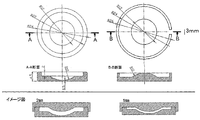
- a template having a curvature suitable for the human corneal endothelium for example, as shown in FIG.
- a desired cell support gel can be formed by covering with a mold and solidifying.
- the thickness of the support gel at the stage of being molded with a mold can be appropriately set, for example, between about 0.5 and about 2 mm.
- Gelatinized gelatin, collagen, and the like can then be dried to form a sheet. Drying can be performed by air drying under aseptic conditions such as in a clean bench.
- the biopolymer is in a solution state or a molded sheet subjected to crosslinking.
- Crosslinking can be performed by heating, physical crosslinking by irradiation with ultraviolet rays or ⁇ rays, or chemical crosslinking using a condensing agent such as water-soluble carbodiimide.
- a cell support sheet having a desired thickness can be obtained by changing the crosslinking method, the concentration of the condensing agent, the treatment time, the reaction temperature, and the like.
- a gelatin sheet is used as a cell support.
- collagen especially atelocollagen having no immune activity from the viewpoint of transplantation
- the gelatin sheet is coated.
- atelocollagen those described above can be used similarly.
- the gelatin sheet is coated with atelocollagen, for example, by immersing the gelatin sheet prepared as described above in about 1 to about 5% by weight of atelocollagen solution, at about 20 to 50 ° C., preferably about 30 to 40 ° C. It can be performed by incubating for about 3 hours.
- the human corneal endothelial cell sheet obtained by laminating human corneal endothelial cells on the cell support sheet having a curvature suitable for the human corneal endothelial surface thus obtained can be produced, for example, by the following method. it can.
- Pre-culture of human corneal endothelial cells Human corneal endothelial cells collected from human corneal endothelium as described above are cultured in a basic medium such as D-MEM or MEM that is generally used for culturing animal cells. (Primary culture). For example, it is preferable to contain 5-15% fetal bovine serum (FBS), a growth factor, etc. in a low glucose concentration medium (D-MEM, etc.).
- FBS fetal bovine serum
- D-MEM low glucose concentration medium
- the glucose concentration contained in the medium is lower than the normal glucose concentration, and is 2.0 g / L or less, for example, 0.1 to 2.0 g / L, preferably 0.1 to 1.0 g / L.
- growth factors examples include hepatocyte growth factor (HGF), epidermal growth factor (EGF), recombinant EGF (rEGF), fibroblast growth factor (FGF), etc., and one or more factors combined as appropriate Can be contained in the medium.
- the concentration of these growth factors is usually 1 to 100 ng / mL, preferably 2 to 5 ng / mL.
- antiseptics such as doxycycline and fungicides such as fungizone can be added as necessary.
- about 1.0 to 3.0%, preferably 1.0 to 2.0% of hyaluronic acid (sodium hyaluronate) can be added.
- an ascorbic acid derivative may be added to the medium in addition to the above medium components.
- the ascorbic acid derivative is not particularly limited as long as it increases the proliferation ability of corneal endothelial cells.
- ascorbic acid 2-phosphate ascorbic acid 2-diphosphate, ascorbic acid 2-triphosphate
- ascorbic acid 2-polyphosphate Ascorbic acid phosphates such as: Ascorbic acid 2-phosphate diester, Ascorbic acid 2-phosphate 6-palmitic acid, Ascorbic acid 2-phosphate 6-myristic acid, Ascorbic acid 2-phosphate 6-stearic acid, Ascorbic acid 2-phosphate 6-oleic acid, ascorbic acid 2-glucoside, ascorbic acid 2-glucoside 6-palmitic acid, ascorbic acid 2-glucoside 6-myristic acid, ascorbic acid 2-glucoside 6-stearic acid, ascorbic acid 2-glucoside Ascorbic acid esters such as 6-oleic acid and ascorbic acid 2-sulfuric acid, L-ascorbic acid alkyl ester L- ascorbic acid phosphoric acid ester, L- ascorbic acid sulfuric ester, and the like.
- ascorbic acid derivatives described above such as salts with alkali metals such as sodium and potassium, and salts with alkaline earth metals such as calcium and magnesium are also included in the ascorbic acid derivatives.
- ascorbic acid 2-phosphate is particularly preferable as a substance that enhances the proliferation ability of corneal endothelial cells.
- the content of the ascorbic acid derivative in the culture solution is not particularly limited as long as the proliferation ability of corneal endothelial cells is increased or a corneal endothelial cell sheet applicable to corneal transplantation is obtained, and is appropriately determined by those skilled in the art. It is possible. However, as a general guideline, it is usually 5 to 1,000 ⁇ g / mL, more preferably 20 to 100 ⁇ g / mL, from the viewpoint of mass culture of corneal endothelial cells.
- Culture is performed in a culture vessel coated with a matrix such as Matrigel or collagen (eg, dish, petri dish, tissue culture dish, multi-dish, microplate, microwell plate, multiplate, multiwell plate, chamber slide, petri dish, tube , Trays, culture bags, etc.).
- the culture temperature is 35 to 38 ° C, preferably 37 ° C.
- the cells are cultured in an incubator of 90 to 100% wet (preferably 100% wet) and 5 to 15% CO 2 (preferably 10% CO 2 ). Culturing can be performed until the cells are confluent (steady state is about 1 to 5 days).
- Primary cultures of human corneal endothelial cells are passaged when the cells reach confluence.
- the cells are washed with PBS, dispersed in trypsin / EDTA, centrifuged, and seeded in a culture vessel containing the same medium as described above to a cell density of 500-60,000 cells / cm 2 , Culture under culture conditions. The same passage is repeated when the cells reach confluence.
- the number of passages of the cells used for preparing the human corneal endothelial cell sheet is not particularly limited, but those that have been passaged about 1 to 5 times are preferably used.
- the human corneal endothelial cells prepared as described above are seeded on a cell support sheet having a curvature suitable for the human corneal endothelial surface, and cultured, whereby the present invention is obtained.
- the human corneal endothelial cell sheet is obtained.
- the support sheet is allowed to stand on the plate with the convex surface of the cell support sheet facing down.
- a membrane of a Corning Transwell plate preferably having Snapwell inserts having a 12 mm diameter membrane replaced with a cell support sheet can be used. At this time, the cell support is fixed so that the bottom surface of the cell support contacts the culture dish.
- a corneal cell endothelial sheet for transplantation When seeding cells on a cell support sheet, it is possible to produce a corneal cell endothelial sheet for transplantation with higher quality by seeding the cells at a higher density rather than growing the cells after seeding (seeding density: 2,000 to 8,000). Pieces / mm 2 , preferably 4,000 to 6,000 pieces / mm 2 ). Therefore, ascorbic acid derivatives are not necessarily required at the time of seeding on the cell support sheet, and commonly used DME medium, MEM, etc. can be used. For example, a low glucose concentration medium (DME medium, etc.) ) Containing fetal calf serum (FCS), the above-mentioned growth factors, etc. can be used.
- DME medium low glucose concentration medium
- FCS fetal calf serum
- culturing for 1 to 2 weeks or more using DME medium containing 15% fetal bovine serum and 2 ng / mL bFGF can function equivalent to uncultured corneal endothelial cells (barrier function, pump function, cell adhesion) From the viewpoint of obtaining performance).
- the cell support sheet Before the cells are seeded on the cell support sheet, the cell support sheet may be coated with an extracellular matrix component such as atelocollagen or laminin.
- an extracellular matrix component such as atelocollagen or laminin.
- coating with atelocollagen or laminin surprisingly prevents cells from accumulating at specific locations, forms a more uniform monolayer structure than gelatin alone, and prevents cells from detaching.
- a human corneal endothelial cell sheet in which cells are formed at a higher density can be produced.
- a suitable human corneal endothelial cell sheet thus produced has functions (barrier function, pump function, cell adhesion ability, etc.) equivalent to or higher than those of uncultured corneal endothelial cells.
- the human corneal endothelial cell sheet thus obtained has a uniform human corneal endothelial cell layer (preferably a monolayer) on the cell support, and functions as a corneal endothelial cell (barrier function, pump function, cell adhesion ability). It has the same cell density as in vivo cells and a cell density (3,000 cells / mm 2 ) or more that is equivalent to that of normal human corneal endothelium.
- the human corneal endothelial cell sheet thus produced can be obtained by a method known per se, for example, a method in which the entire cornea is removed and the corneal endothelial cell sheet is attached and then restored (Hitani, K. et al., Mol. Vis. 2008 14: 1-9) or a method of carrying a keratotomy and carrying the cultured corneal endothelial cell sheet with a silicon sheet into the anterior chamber with a settle from the incision site (Mimura, T. et. al., Invest. Ophthalmol. Vis. Sci. 2004 45: 2992-7) and the like.
- transplantation methods require a high level of operator skill, are accompanied by high invasiveness, and are disadvantageous in that they damage corneal endothelial cells during transplantation. Therefore, at the time of transplantation, it is possible to maintain the survival of the corneal endothelial cells in a sterile environment until the time of use in order to perform the operation easily, and at the time of transplantation, it is not influenced as much as possible by the operator's skill. It is desirable to store the sheet in an implantable device that can guide the sheet non-invasively to the implantation site in the eyeball. Although it does not specifically limit as such a transplant device, For example, the transplant device described in the international publication 2011/096593 pamphlet can be used preferably.
- Pasting the gelatin sheet 5 mL of water for injection was poured into a 35 mm dish, and the curved gelatin sheet was swollen for one day in advance.
- the insert with the membrane was taken out from Corning Transwell (# 3801), and the membrane was removed from the insert using tweezers.
- 5 mL of water for injection was poured into a 35 mm dish and floated with the convex surface of the curvature gelatin sheet swollen with water facing upward.
- the ring from which the membrane was removed was placed under the curvature gelatin sheet, and the water for injection was extracted after the circumference of the ring and the sheet was matched.
- the wrinkles on the sheet were removed with tweezers and air-dried in a clean bench (safety cabinet). After fixing the ring and the curvature sheet with an O-ring, it was set in Transwell and swollen with growth medium.
- Laminin-Coated Gelatin Sheet Water for injection was removed from Transwell (curved with water for injection) to which a curved gelatin sheet had been applied, and 160 ⁇ L of PBS was added to the concave portion of the curved sheet. Furthermore, a laminin solution (rLAMININ-5: Oriental Yeast Co., Ltd.) was added to PBS so as to have a final concentration of 5 ⁇ g / mL, and the mixture was incubated at 37 ° C. for 2 hours. After washing twice with 5 mL of PBS, growth medium was added.
- rLAMININ-5 Oriental Yeast Co., Ltd.
- the Descemet's membrane piece with corneal endothelial cells attached on a Petri dish is further cut into small pieces of about 2 mm square, then collected in a low-adsorption centrifuge tube (manufactured by Sumitomo Bakelite Co., Ltd.), and 0.2% collagenase (trade name: Incubation was carried out at 37 ° C. and 5% CO 2 for 1 to 3 hours in a basal medium containing collagenase A and collagenase activity:> 0.15 U / mg, manufactured by Roche. Collagenase-treated cells were diluted with basal medium and centrifuged at 20 ⁇ g for 2 minutes three times to remove cells floating in the supernatant.
- phosphate buffered saline PBS
- EDTA ethylenediaminetetraacetic acid
- a cell pellet was obtained by adding a basal medium and centrifuging at 500 ⁇ g for 5 minutes.
- the obtained cell pellet was resuspended in a basal medium containing 100 ⁇ g / mL ascorbic acid 2-phosphate (manufactured by Wako Pure Chemical Industries, Ltd.) and then seeded on each dish prepared by the following method.
- the cells were cultured in an incubator at 37 ° C. and 5% CO 2 for 2 to 4 weeks while changing the medium every 2-3 days.
- a 2% atelocollagen implant (manufactured by Koken Co., Ltd.) was diluted 400 times with 10 mM acetic acid to prepare a 50 ⁇ g / mL atelocollagen implant solution. 1 mL was added to a 35 mm dish, allowed to stand at 37 ° C. for 1 hour, and then washed twice with 2 mL of PBS to prepare a dish coated with atelocollagen.
- Subculture of human corneal endothelial cells The donor primary culture cells obtained in Example 5 were subcultured as follows. Primary cultured cells were washed with PBS and then dispersed with 0.5% trypsin / 0.2% EDTA. A basal medium was added thereto, centrifuged at 500 ⁇ g for 5 minutes, suspended in a basal medium containing 100 ⁇ g / mL ascorbic acid 2-phosphate, and a dish coated with atelocollagen prepared in the same manner as in Example 5 above. above, were seeded respectively at a cell density of 1,000 / cm 2, 37 ° C., it was cultured in 5% CO 2. The same passage operation was repeated when the cells became confluent.
- atelocollagen implant solution was prepared in the same manner as in Example 6 above, 5 mL per well was added, and the mixture was allowed to stand at 37 ° C. for 1 hour. After removing the atelocollagen implant solution, it was washed twice with 5 mL of PBS per well, and 5 mL of medium was added. The subcultured cells were prepared to 1 ⁇ 10 6 cells / mL, the medium was removed from the concave surface of the curvature sheet, and 416 ⁇ L of the cell suspension was added.
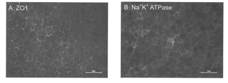
- the cells were cultured in an environment of 37 ° C. and 5% CO 2 for 7 days. As a result, a monolayer structure similar to that of endothelial cells in vivo was observed. Expression of the functional marker protein (FIG. 2A: ZO-1, FIG. 2B: Na + / K + ATPase) was also equivalent to that of endothelial cells in vivo by immunostaining. The density was calculated to be 3,000 cells / mm 2 or more.
- the sheet was attached to the back of the cornea by injecting air, the incision was sutured with 10-0 nylon suture, and the eyelid was sutured to finish.
- wrinkles occurred on the outer peripheral portion of the sheet (the wrinkle portion is indicated by an arrow).
- the curved sheet according to the present invention was transplanted, no wrinkles were generated on the sheet, and the sheet was in close contact with the back of the cornea.
- FIG. 5 shows changes in corneal thickness up to 4 weeks after the operation. Compared with the control (non-sheet transplanted, sheet only), the human cell sheet transplanted group had a thinner corneal thickness, demonstrating its effectiveness.
- the human corneal endothelial cell sheet of the present invention has a curvature adapted to the corneal endothelial surface, it exhibits a significantly superior effect compared to the case of transplanting a conventional flat corneal endothelial cell sheet. It is useful as a material for regenerating endothelial tissue.
Landscapes
- Health & Medical Sciences (AREA)
- Life Sciences & Earth Sciences (AREA)
- Chemical & Material Sciences (AREA)
- Engineering & Computer Science (AREA)
- Medicinal Chemistry (AREA)
- General Health & Medical Sciences (AREA)
- Biomedical Technology (AREA)
- Animal Behavior & Ethology (AREA)
- Public Health (AREA)
- Veterinary Medicine (AREA)
- Transplantation (AREA)
- Epidemiology (AREA)
- Dermatology (AREA)
- Oral & Maxillofacial Surgery (AREA)
- Chemical Kinetics & Catalysis (AREA)
- Cell Biology (AREA)
- Zoology (AREA)
- Bioinformatics & Cheminformatics (AREA)
- Organic Chemistry (AREA)
- Botany (AREA)
- Biotechnology (AREA)
- Genetics & Genomics (AREA)
- Wood Science & Technology (AREA)
- Molecular Biology (AREA)
- Ophthalmology & Optometry (AREA)
- Urology & Nephrology (AREA)
- Pharmacology & Pharmacy (AREA)
- Neurosurgery (AREA)
- Neurology (AREA)
- Microbiology (AREA)
- Nuclear Medicine, Radiotherapy & Molecular Imaging (AREA)
- Biochemistry (AREA)
- General Engineering & Computer Science (AREA)
- Biophysics (AREA)
- General Chemical & Material Sciences (AREA)
- Vascular Medicine (AREA)
- Materials For Medical Uses (AREA)
- Micro-Organisms Or Cultivation Processes Thereof (AREA)
Abstract
Description
またゼラチンハイドロゲルを支持体として用いた角膜内皮細胞シートの製造方法(特許文献3)や、眼内移植で吸収される生物分解性ポリマー支持マトリックスと内皮細胞層を含む角膜ボタン(特許文献4)が既に知られているところである。
このため、培養角膜内皮細胞シートにおいても同様に考えられており、特に移植面積が数mmから10 mm程度の直径でよいことから、角膜内皮面に合わせた曲率をもつ培養角膜内皮細胞シートを移植する必要は全く考慮されなかった。そればかりか、支持体上に細胞が均等に積層されることの重要性を鑑みれば、常識的に考えて、曲面のシート上に細胞を播種しようなどという発想には到底至らない。
また、臨床で行われている角膜内皮移植は、ドナー角膜を薄切して曲面である角膜内皮面に移植片を接着する治療法であるが、意図的に曲面を作製するような薄切をすることなく実施されている。
[1]ヒト角膜内皮面に適した曲率を有することを特徴とする、ヒト角膜内皮細胞シート。
[2]ヒト角膜内皮面に適した曲率を有する細胞支持体により支持される、上記[1]に記載のヒト角膜内皮細胞シート。
[3]細胞支持体が、コラーゲンを原料とするシートである、上記[1]または[2]に記載のヒト角膜内皮細胞シート。
[4]コラーゲンを原料とするシートが、ゼラチンシートである、上記[3]に記載のヒト角膜内皮細胞シート。
[5]ゼラチンシートの厚みが5~40 μmである、上記[4]に記載のヒト角膜内皮細胞シート。
[6]ゼラチンシートが、以下の工程:
(1)ヒト角膜内皮面に適した曲率を有する鋳型に流し込んだゼラチン水溶液をゲル化する工程;
(2)(1)で得られたゲル化ゼラチンを乾燥して、シート状ゼラチンを形成する工程;
(3)(2)で得られたシート状ゼラチンを熱架橋して、ゼラチンシートを得る工程;
により製造されることを特徴とする、上記[4]または[5]に記載のヒト角膜内皮細胞シート。
[7]ゼラチンシートが、ラミニンまたはアテロコラーゲンでコートされている、上記[4]~[6]のいずれかに記載のヒト角膜内皮細胞シート。
[8]以下の工程:
(1)ヒト角膜内皮面に適した曲率を有する鋳型に流し込んだゼラチン水溶液をゲル化する工程;
(2)(1)で得られたゲル化ゼラチンを乾燥して、シート状ゼラチンを形成する工程;
(3)(2)で得られたシート状ゼラチンを熱架橋して、ゼラチンシートを得る工程;
(4)(3)で得られたゼラチンシートにヒト角膜内皮細胞を播種し、ヒト角膜内皮細胞層を形成する工程;
を含んでなる、ヒト角膜内皮面に適した曲率を有するヒト角膜内皮細胞シートの製造方法。
[9]ゼラチン水溶液の濃度が1~5重量%である、上記[8]に記載のヒト角膜内皮細胞シートの製造方法。
[10]ゲル化ゼラチンの厚みが0.5~2 mmである、上記[8]または[9]に記載のヒト角膜内皮細胞シートの製造方法。
[11]ゼラチンシートが、ラミニンまたはアテロコラーゲンでコートされている、上記[8]~[10]のいずれかに記載のヒト角膜内皮細胞シートの製造方法。
[12]ゼラチンシートのシート厚みが5~40 μmである、上記[8]~[11]のいずれかに記載のヒト角膜内皮細胞シートの製造方法。
[13]鋳型がテフロン(登録商標)製の鋳型である、上記[8]~[12]のいずれかに記載のヒト角膜内皮細胞シートの製造方法。
ヒト角膜内皮細胞のドナーとしては、角膜は免疫系による認識を免れた隔絶抗原であるとされ、HLAタイプの一致しない場合でも拒絶反応を起こさない場合も多いので、必ずしもレシピエントとHLAタイプが一致している必要はないが、好ましくはHLA-A、HLA-B、HLA-DR及びHLA-Cのうちの1座以上、より好ましくは2座以上、さらに好ましくは3座以上が、レシピエントと一致するヒト個体から採取されたものである。
上記方法では、コラゲナーゼは、ロッシュ社のコラゲナーゼA、シグマ社のコラゲナーゼタイプIA、ワーシントン社のコラゲナーゼタイプIなどを用いることが可能であり、それぞれ0.2%となるように培地で調製したものを使用する。また培地としては15%牛胎児血清(FCS)および2 ng/mLの塩基性線維芽細胞増殖因子(bFGF)を含むDME培地を用いることができる。
ヒト角膜内皮面に適した曲率を有するためには、ヒト角膜内皮細胞シートは、ヒト角膜内皮細胞が、ヒト角膜内皮面に適した曲率を有する細胞支持体により支持される構成をとることが望ましい。ヒト角膜内皮面に適した曲率を有する細胞支持体は、コンタクトレンズ状の薄層シート構造を有する。該支持体シートの外縁により形成される円(略円)の直径は、移植面積に応じて適宜選択されるが、ヒト角膜のサイズを考慮すれば、通常約5~約12 mm程度である。また支持体シートの曲率半径は通常約6~約10 mm程度である。支持体シートの厚さは5~100 μm程度が適当であるが、ヒト角膜のデスメ膜の厚さに近似させることがより好ましく、移植時(膨潤時)の厚さとして約5~約40 μmであることが好ましく、約5~約25 μmであることがより好ましい。
鋳型としては、例えば、アクリル製、テフロン(登録商標)製、シリコン製等の鋳型を用いることができるが、これらに限定されない。バイオポリマーをこれらの鋳型に流し込んだ後にゲル化を行い、乾燥工程を経てシートを作製する。乾燥後のシートは極めて薄いため、シートが破れることなく容易に鋳型から外すことができるテフロン(登録商標)製の鋳型が好ましく用いられ得る。ヒト角膜内皮面に適した曲率を有する鋳型は、例えば、図1に示すように、同じ曲率半径の凹凸を有する上下2つの鋳型を作製し、下側の鋳型にバイオポリマー溶液を流し込み、上側の鋳型で蓋をして固化することにより、所望の細胞支持体ゲルに成型することができる。コラーゲンを原料とするバイオポリマーの細胞支持体シートを作製する場合、鋳型により成型された段階での支持体ゲルの厚さを、例えば、約0.5~約2mmの間で適宜設定することができる。ゲル化されたゼラチンやコラーゲン等を、次いで乾燥することにより、シート状とすることができる。乾燥は、例えば、クリーンベンチ内などの無菌条件下で風乾により行うことができる。
上記のようにしてヒト角膜内皮より採取したヒト角膜内皮細胞は、動物細胞の培養に一般的に使用されるD-MEM、MEM等の基礎培地中で培養(初代培養)することができる。例えば、低グルコース濃度の培地(D-MEM等)にウシ胎児血清 (FBS) 5~15%、成長因子等を含有することが好ましい。ここで、培地に含有させるグルコース濃度は、通常のグルコース濃度よりも低濃度であり、2.0 g/L 以下、例えば0.1~2.0 g/L、好ましくは0.1~1.0 g/Lである。また、成長因子としては肝細胞増殖因子(HGF)、上皮成長因子(EGF)、組換えEGF(rEGF)、線維芽細胞増殖因子(FGF)等が挙げられ、1つ又は複数の因子を適宜組み合わせて培地に含有させることができる。これらの成長因子の含有濃度は、通常1~100ng/mL、好ましくは2~5 ng/mLである。さらに、上記培地組成に加えて、必要に応じてドキシサイクリン(doxycycline)等の防腐剤、ファンギゾン(Fungizone)等の防カビ剤を添加することもできる。さらに、1.0~3.0%程度、好ましくは1.0~2.0%のヒアルロン酸(ヒアルロン酸ナトリウム)を添加することもできる。
上記したアスコルビン酸誘導体の塩である、ナトリウム、カリウム等のアルカリ金属との塩、カルシウム、マグネシウム等のアルカリ土類金属との塩も、アスコルビン酸誘導体に包含される。中でも、特に角膜内皮細胞の増殖能を高めるものとして、アスコルビン酸2-リン酸が好ましい。
培養温度は35~38℃、好ましくは37℃である。そして、90~100%湿潤(好ましくは100%湿潤)、5~15% CO2(好ましくは10% CO2)のインキュベータ内で培養する。培養は、細胞が集密(コンフルエント)になった段階(定常状態1~5日程度)まで行うことができる。
上記のようにして調製されたヒト角膜内皮細胞を、前記ヒト角膜内皮面に適した曲率を有する細胞支持体シート上に播種し、培養することにより、本発明のヒト角膜内皮細胞シートが得られる。細胞播種に際しては、細胞支持体シートの凸面を下にしてプレート上に支持体シートを静置する。例えば、コーニング社のトランズウェルプレート(12 mm径のメンブレンを有するSnapwell insertsを有するものが好ましい)のメンブレンを細胞支持体シートで置換したものを用いることができる。このときに細胞支持体の底面が培養ディッシュに接触するようにして固定する。このようにして固定した細胞支持体は、平面からなる培養ディッシュに接触している部分において微細な凹凸が発生している。このように調整した細胞支持体シート上にピペット等を用いて細胞懸濁液を上から滴下することにより細胞を播種する。播種直後は細胞支持体の凹凸により細胞が不均一に接着するが、驚くべきことに培養を続けるうちに均一なヒト角膜内皮細胞層(好ましくは単層)が生じる。
したがって移植の際には、その操作を簡便に行うべく、使用時まで無菌環境下で角膜内皮細胞の生存を維持させることが可能であって、移植時には術者の技量にできるだけ左右されることなく眼球内の移植部位へ該シートを非侵襲的に誘導することが可能な移植用器具に格納することが望ましい。そのような移植用器具としては特に限定されないが、例えば、国際公開第2011/096593号パンフレットに記載される移植用器具を好ましく用いることができる。
操作は清潔なクリーンベンチ(安全キャビネット)内で行うのが望ましく、使用する容器や器具もすべて滅菌されたものもしくは滅菌された使い捨て(ディスポーザブル)タイプのものを使用した。新田ゼラチン社のビーマトリックスゼラチンLS-H粉末を2.0 g秤量し、ボトルに採取した。注射用水を100 mL添加し室温で1時間振盪させた後、50℃の恒温槽で30分間振盪しながら溶解させた。室温で冷ました後、4℃で保存した。
4℃で保存していた2%ゼラチンを37℃の恒温槽で溶解させた。図1のように設計して作製した曲面を有するテフロン(登録商標)鋳型の蓋をはずし、凹面に2%ゼラチン溶液を600 μL流し入れて面全体にいきわたらせ、蓋をして4℃で1日冷やしゲル化した。ピンセットを用いて鋳型の蓋をゆっくりと開け、ゲル化したゼラチンをクリーンベンチ(安全キャビネット)内で2日間乾燥させた。乾燥後シート状になったゼラチンを鋳型よりゆっくり剥がし、真空乾燥装置内で140℃、72時間架橋処理を行った。比較対象物として平面の鋳型を用いて同様の方法にて平面ゼラチンシートを作製した。架橋処理後のゼラチンシートは-30℃で保存した。
35 mmディッシュへ注射用水を5 mLそそぎ、曲率ゼラチンシートをあらかじめ1日膨潤させておいた。コーニングトランズウェル(#3801)よりメンブレンのついたインサートを取り出し、ピンセットを用いてインサートよりメンブレンを取り除いた。35 mmディッシュへ注射用水を5 mLそそぎ、水で膨潤させておいた曲率ゼラチンシートの凸面を上にして浮かべた。メンブレンを除去したリングを曲率ゼラチンシートの下にいれ、リングとシートの円周を合わせたうえで注射用水を抜き取った。ピンセットでシートのシワをのぞき、クリーンベンチ(安全キャビネット)内で風乾させた。リングと曲率シートをOリングで固定した後、トランズウェルにセットし、増殖培地で膨潤させた。
曲率ゼラチンシート貼り付け済みのトランズウェル(注射用水にて膨潤済)より注射用水を除去し、曲率シートの凹部にPBSを160 μL添加した。さらにPBS中にラミニン溶液(rLAMININ-5: オリエンタル酵母工業株式会社)を最終濃度が5 μg/mL となるよう添加して混和し、37℃で2時間インキュベートした。PBS 5 mLで2回洗浄した後、増殖培地を添加した。
35 mmペトリディッシュにヒト強角膜片を移し、内皮面を15%牛胎児血清(FCS)及び2 ng/mL塩基性繊維芽細胞増殖因子(bFGF)を含むDME培地(以下基礎培地と記載)で洗浄した。
微細なセッシを用いて、角膜内皮をデスメ膜ごと角膜の内面の周辺部から中心へ向かってシート状に剥ぎ取り、35mmペトリディッシュに移した。ペトリディッシュ上で角膜内皮細胞が付着したデスメ膜片をさらに2 mm角程の小片に細切後、低吸着遠心チューブ(住友ベークライト(株)製)に回収し、0.2%のコラゲナーゼ(商品名:コラゲナーゼA、コラゲナーゼ活性:>0.15 U/mg、ロシュ(Roche)(株)製)を含む基礎培地中で、37℃、5% CO2で1~3時間インキュベートした。
コラゲナーゼ処理した細胞を基礎培地で希釈し、20xg、2分間の遠心洗浄を3回繰り返し、上清に浮遊する細胞を除去した。次にリン酸緩衝生理食塩水(PBS)で希釈し、同様に20xg、2分間の遠心洗浄を1回行った後、沈殿した細胞塊に0.5%トリプシン/0.2%エチレンジアミン四酢酸(EDTA)を加え、37℃、5% CO2で5分間インキュベートした。基礎培地を加え、500xg、5分間の遠心をすることにより細胞ペレットを得た。得られた細胞ペレットを、100 μg/mLアスコルビン酸2-リン酸(和光純薬(株)製)を含む基礎培地に再懸濁後、下記の方法で作製したディッシュ上に、それぞれ播種し、37℃、5% CO2のインキュベータ内で2~4週間、2~3日毎に培地を交換しながら培養した。
2%アテロコラーゲンインプラント(高研(株)製)を10 mM酢酸で400倍希釈し、50 μg/mLのアテロコラーゲンインプラント溶液を調製した。35 mmディッシュに1 mL加え、37℃で1時間放置後、2 mLのPBSで2回洗浄することによりアテロコラーゲンでコートされたディッシュを作製した。
上記実施例5で得たドナーの初代培養細胞を、それぞれ次の通りに継代培養した。
初代培養細胞をPBSで洗浄後、0.5%トリプシン/0.2% EDTAで分散させた。これに基礎培地を加え、500xgで5分間遠心した後、100 μg/mLアスコルビン酸2-リン酸を含む基礎培地に懸濁し、上記実施例5と同様の方法で作製したアテロコラーゲンでコートされたディッシュ上に、1,000個/cm2の細胞密度でそれぞれ播種し、37℃、5% CO2で培養した。細胞がコンフレントになった時点で同様の継代操作を繰り返した。
曲率ゼラチンシート貼り付け済みのトランズウェル(培地膨潤済)より培地を除去した。上記実施例6と同様に50 μg/mLアテロコラーゲンインプラント溶液を調製し、1ウェルあたり5 mL加え、37℃で1時間放置した。アテロコラーゲンインプラント溶液を除去した後、1ウェルあたり5 mLのPBSで2回洗浄し、培地5 mLを添加した。
継代培養された細胞を、1×10⁶細胞/mLに調製し、曲率シートの凹面より培地を取り除いた後、細胞懸濁液を416μL添加した。37℃、5% CO2の環境下で7日間培養した。
その結果、生体内の内皮細胞と同様の単層構造が認められた。免疫染色により機能マーカータンパク質(図2A: ZO-1、図2B: Na+/K+ATPase)の発現も生体内の内皮細胞と同等であった。密度計算をしたところ3,000細胞/mm2以上であった。
実施例4で得られたラミニンコート済の曲率ゼラチンシートより増殖培地を除去し、新しい増殖培地を4 mL添加した。継代培養された細胞を1×10⁶ cells/mLとなるように基礎培地に懸濁し、曲率シートの凹面より培地を取り除いた後、該細胞懸濁液を416 μL添加した。37℃、5% CO2の環境下で7日間培養した。
その結果、生体内の内皮細胞と同様の単層構造が認められた。免疫染色による機能マーカータンパク質の発現も、生体内の内皮細胞や上記(1)の場合と同等であった。
実施例2で作製した曲面及び平面のゼラチンシートを35 mmペトリディッシュ上に置き注射用蒸留水に浸して一晩膨潤した。膨潤シートをトリパンブルーで染色後、6 mmトレパンで切り出し、シート移植用インジェクター内にシートを充填した。カニクイザルにメスで角膜トンネルを作製し、前房内にインジェクターを挿入、インジェクター挿入孔の反対側に作製したサイドポートよりDSAEK鑷子を挿入し、シートをインジェクターより引き出した。空気を注入することによりシートを角膜裏面に貼り付け、切開創を10-0ナイロン縫合糸で縫合し、眼瞼縫合して終了とした。図3に示すように、従来法である平面のゼラチンシートを移植した時にはシートの外周部分に皺が発生した(皺部分を矢印で示す)。一方、本発明品である曲面シートを移植した時にはシートに皺は発生せず角膜裏面に密着した。
(2)ラミニンコートした曲率シートの観察
上記実施例4および9に記載の方法で、曲率ゼラチンシートをラミニンでコーティングし、該シートに細胞を播種することでヒト角膜内皮細胞シートを作製した。比較のために、何もコーティングしていない曲率ゼラチンシートにも細胞を播種した。それぞれのヒト角膜内皮細胞シートを倒立顕微鏡下で観察したところ、何もコーティングしていない単なるゼラチンシートに播種した場合は、ロットによって部分的に細胞の剥離が認められたが(図4左)、ラミニン-5コートでは、細胞が剥離することは全くなかった(図4右)。こうして得られた曲率シートは、均一なヒト角膜内皮細胞層であり、正常なヒト眼の角膜内皮と同等(3,000細胞/mm2)以上の細胞密度を有していた。また生体内の細胞と同等またはそれ以上の角膜内皮細胞としての機能(バリア機能、ポンプ機能、細胞接着能)を保持していると考えられた。
この結果は、単なるゼラチンシートよりも、ラミニンでコーティングしたゼラチンシートを用いた方が、移植により適したヒト角膜内皮細胞シートが得られることを示している。
カニクイザルにメスで角膜トンネルを作製し、20Gソフトテーパードニードルで中央部分の内皮(直径8 mm程度)を掻破した。前房内に粘弾性物質を注入し、I/A洗浄した。眼内灌流液で5倍希釈したトリパンブルー溶液を前房内に注入後、I/A洗浄し、内皮細胞が十分に除去されていることを確認した。実施例7のトランズウェルに固定された細胞シートをトランズウェルごと取り出し、35 mmペトリディッシュ上に置いた。トリパンブルーで染色後、6 mmトレパンで切り出した。シートを無血清培地で洗浄し、その後培地成分は除去した。シート上に粘弾性物質を塗布した。再び35 mmペトリディシュに無血清培地を満たし、シート移植用インジェクター内にシートを充填した。前房内にインジェクターを挿入、インジェクター挿入孔の反対側に作製したサイドポートよりDSAEK鑷子を挿入し、シートをインジェクターより引き出した。空気を注入することによりシートを角膜裏面に貼り付け、切開創を10-0ナイロン縫合糸で縫合し、眼瞼縫合して終了とした。その後1週間毎に角膜厚を測定し、浮腫の状態を観察した。比較対照としてシート無移植群とシートのみ移植群を用いた。
図5に術後4週までの角膜厚の推移を示す。コントロール(シート無移植、シートのみ)と比べ、ヒト細胞シート移植群では角膜厚が薄くなっておりその有効性が明らかとなった。
Claims (13)
- ヒト角膜内皮面に適した曲率を有することを特徴とする、ヒト角膜内皮細胞シート。
- ヒト角膜内皮面に適した曲率を有する細胞支持体により支持される、請求項1に記載のヒト角膜内皮細胞シート。
- 細胞支持体が、コラーゲンを原料とするシートである、請求項1または2に記載のヒト角膜内皮細胞シート。
- コラーゲンを原料とするシートが、ゼラチンシートである、請求項3に記載のヒト角膜内皮細胞シート。
- ゼラチンシートの厚みが5~40 μmである、請求項4に記載のヒト角膜内皮細胞シート。
- ゼラチンシートが、以下の工程:
(1)ヒト角膜内皮面に適した曲率を有する鋳型に流し込んだゼラチン水溶液をゲル化する工程;
(2)(1)で得られたゲル化ゼラチンを乾燥して、シート状ゼラチンを形成する工程;
(3)(2)で得られたシート状ゼラチンを熱架橋して、ゼラチンシートを得る工程;
により製造されることを特徴とする、請求項4または5に記載のヒト角膜内皮細胞シート。 - ゼラチンシートが、ラミニンまたはアテロコラーゲンでコートされている、請求項4~6のいずれか一項に記載のヒト角膜内皮細胞シート。
- 以下の工程:
(1)ヒト角膜内皮面に適した曲率を有する鋳型に流し込んだゼラチン水溶液をゲル化する工程;
(2)(1)で得られたゲル化ゼラチンを乾燥して、シート状ゼラチンを形成する工程;
(3)(2)で得られたシート状ゼラチンを熱架橋して、ゼラチンシートを得る工程;
(4)(3)で得られたゼラチンシートにヒト角膜内皮細胞を播種し、ヒト角膜内皮細胞層を形成する工程;
を含んでなる、ヒト角膜内皮面に適した曲率を有するヒト角膜内皮細胞シートの製造方法。 - ゼラチン水溶液の濃度が1~5重量%である、請求項8に記載のヒト角膜内皮細胞シートの製造方法。
- ゲル化ゼラチンの厚みが0.5~2 mmである、請求項8または9に記載のヒト角膜内皮細胞シートの製造方法。
- ゼラチンシートが、ラミニンまたはアテロコラーゲンでコートされている、請求項8~10のいずれか一項に記載のヒト角膜内皮細胞シートの製造方法。
- ゼラチンシートのシート厚みが5~40 μmである、請求項8~11のいずれか一項に記載のヒト角膜内皮細胞シートの製造方法。
- 鋳型がテフロン(登録商標)製の鋳型である、請求項8~12のいずれか一項に記載のヒト角膜内皮細胞シートの製造方法。
Priority Applications (3)
| Application Number | Priority Date | Filing Date | Title |
|---|---|---|---|
| EP13869392.4A EP2940126A4 (en) | 2012-12-27 | 2013-12-27 | SHEET OF HUMAN CORNEAL ENDOTHELIAL CELLS |
| US14/758,058 US20150374881A1 (en) | 2012-12-27 | 2013-12-27 | Human corneal endothelial cell sheet |
| JP2014554623A JP5946046B2 (ja) | 2012-12-27 | 2013-12-27 | ヒト角膜内皮細胞シート |
Applications Claiming Priority (2)
| Application Number | Priority Date | Filing Date | Title |
|---|---|---|---|
| JP2012-286050 | 2012-12-27 | ||
| JP2012286050 | 2012-12-27 |
Publications (1)
| Publication Number | Publication Date |
|---|---|
| WO2014104366A1 true WO2014104366A1 (ja) | 2014-07-03 |
Family
ID=51021427
Family Applications (1)
| Application Number | Title | Priority Date | Filing Date |
|---|---|---|---|
| PCT/JP2013/085262 WO2014104366A1 (ja) | 2012-12-27 | 2013-12-27 | ヒト角膜内皮細胞シート |
Country Status (4)
| Country | Link |
|---|---|
| US (1) | US20150374881A1 (ja) |
| EP (1) | EP2940126A4 (ja) |
| JP (1) | JP5946046B2 (ja) |
| WO (1) | WO2014104366A1 (ja) |
Cited By (9)
| Publication number | Priority date | Publication date | Assignee | Title |
|---|---|---|---|---|
| WO2016093359A1 (ja) * | 2014-12-11 | 2016-06-16 | 学校法人慶應義塾 | 治療用角膜内皮代替細胞スフェアの製造方法 |
| JP2017078045A (ja) * | 2015-10-21 | 2017-04-27 | 地方独立行政法人東京都立産業技術研究センター | ゼラチンまたはその化学修飾体、それを含有する水性組成物および医療用積層体、ならびに医療用積層体の製造方法および細胞シートの単離方法 |
| WO2017170343A1 (ja) * | 2016-03-29 | 2017-10-05 | 富士フイルム株式会社 | 細胞シートを含有する積層体、心疾患治療剤および細胞シート積層用フィルム |
| JP2018512930A (ja) * | 2015-03-26 | 2018-05-24 | フラウンホファー ゲセルシャフト ツール フェールデルンク ダー アンゲヴァンテン フォルシュンク エー.ファオ. | 人工デスメ膜 |
| WO2018235786A1 (ja) | 2017-06-19 | 2018-12-27 | 国立大学法人大阪大学 | 角膜内皮細胞マーカー及びその利用 |
| JP2020525232A (ja) * | 2017-07-03 | 2020-08-27 | ビスコファン,エセ.アー | 生物組織を再生するためのパッチおよびそれを製造する方法 |
| WO2020247420A1 (en) * | 2019-06-03 | 2020-12-10 | Advanced Solutions Life Sciences, Llc | System and method for fabricating a cornea |
| WO2022172930A1 (ja) * | 2021-02-09 | 2022-08-18 | 信越化学工業株式会社 | 移植デバイス |
| WO2024116998A1 (ja) * | 2022-11-28 | 2024-06-06 | ニッタ株式会社 | 細胞シート作製装置及び細胞シート作製装置用無菌閉鎖系回路 |
Families Citing this family (5)
| Publication number | Priority date | Publication date | Assignee | Title |
|---|---|---|---|---|
| CN112494722A (zh) * | 2020-12-15 | 2021-03-16 | 中新国际联合研究院 | 一种快速上皮化的胶原基角膜再生修复材料及其制备方法 |
| EP4056206A1 (en) * | 2021-03-11 | 2022-09-14 | Precise Bio Inc. | Artificial endothelial keratoplasty graft and methods of preparation thereof |
| CN113101411A (zh) * | 2021-04-14 | 2021-07-13 | 广州宏达医疗设备有限公司 | 一种角膜基质内人工角膜内皮及其制备方法 |
| CN116322812B (zh) * | 2022-08-25 | 2024-03-22 | 山东第一医科大学附属眼科研究所(山东省眼科研究所、山东第一医科大学附属青岛眼科医院) | 一种大直径人工角膜内皮片及其应用 |
| CN116492505B (zh) * | 2023-05-11 | 2023-12-08 | 山东第一医科大学附属眼科医院(山东省眼科医院) | 一种人工角膜内皮移植片及其应用 |
Citations (8)
| Publication number | Priority date | Publication date | Assignee | Title |
|---|---|---|---|---|
| JP2004024852A (ja) * | 2002-04-30 | 2004-01-29 | Amniotec:Kk | 角膜内皮様シート、及びその作製方法 |
| JP2005229869A (ja) | 2004-02-18 | 2005-09-02 | Satoshi Yamagami | ヒト角膜内皮細胞の培養物層積層体及びその作製方法 |
| WO2007043255A1 (ja) * | 2005-09-13 | 2007-04-19 | Arblast Co., Ltd. | 培養角膜内皮シート及びその作製方法 |
| WO2007047425A1 (en) | 2005-10-12 | 2007-04-26 | Cellular Bioengineering, Inc. | Resorbable cornea button |
| US20080050423A1 (en) * | 2006-08-23 | 2008-02-28 | National Tsing Hua University | Biopolymer-bioengineered cell sheet construct |
| WO2008143149A1 (ja) * | 2007-05-11 | 2008-11-27 | Dai Nippon Printing Co., Ltd. | 寸法が保持された細胞シート、その製造方法、及びそのための細胞培養担体 |
| WO2011021706A1 (ja) | 2009-08-19 | 2011-02-24 | 国立大学法人東北大学 | 角膜移植用シート |
| WO2011096593A1 (ja) | 2010-02-05 | 2011-08-11 | 財団法人先端医療振興財団 | 角膜内皮細胞の培養方法、移植用角膜内皮細胞シートの製造方法および角膜内皮細胞培養キット |
Family Cites Families (5)
| Publication number | Priority date | Publication date | Assignee | Title |
|---|---|---|---|---|
| EP1600177B1 (en) * | 2003-02-20 | 2016-05-25 | Cellseed Inc. | Endothelial cell sheet for cornea regeneration, method of producing the same and method of using the same |
| KR101319227B1 (ko) * | 2003-10-10 | 2013-10-16 | 게 밍 루이 | 각막 내피세포 및 관련세포를 생체고분자 위에서성장시키고, 인공의 이식용 각막을 제조하는 방법 및조성물 |
| WO2011074208A1 (ja) * | 2009-12-18 | 2011-06-23 | 国立大学法人東北大学 | 皮膚真皮又は羊膜透明化による角膜移植材料調製法 |
| JP2013116045A (ja) * | 2010-03-08 | 2013-06-13 | Osaka Univ | 三次元細胞集合体の作製方法およびそれに用いる細胞培養用三次元ゲル担体並びに三次元細胞集合体 |
| CA2804592C (en) * | 2010-07-08 | 2019-10-29 | Lifecell Corporation | Method for shaping tissue matrices |
-
2013
- 2013-12-27 JP JP2014554623A patent/JP5946046B2/ja active Active
- 2013-12-27 EP EP13869392.4A patent/EP2940126A4/en not_active Withdrawn
- 2013-12-27 US US14/758,058 patent/US20150374881A1/en not_active Abandoned
- 2013-12-27 WO PCT/JP2013/085262 patent/WO2014104366A1/ja active Application Filing
Patent Citations (8)
| Publication number | Priority date | Publication date | Assignee | Title |
|---|---|---|---|---|
| JP2004024852A (ja) * | 2002-04-30 | 2004-01-29 | Amniotec:Kk | 角膜内皮様シート、及びその作製方法 |
| JP2005229869A (ja) | 2004-02-18 | 2005-09-02 | Satoshi Yamagami | ヒト角膜内皮細胞の培養物層積層体及びその作製方法 |
| WO2007043255A1 (ja) * | 2005-09-13 | 2007-04-19 | Arblast Co., Ltd. | 培養角膜内皮シート及びその作製方法 |
| WO2007047425A1 (en) | 2005-10-12 | 2007-04-26 | Cellular Bioengineering, Inc. | Resorbable cornea button |
| US20080050423A1 (en) * | 2006-08-23 | 2008-02-28 | National Tsing Hua University | Biopolymer-bioengineered cell sheet construct |
| WO2008143149A1 (ja) * | 2007-05-11 | 2008-11-27 | Dai Nippon Printing Co., Ltd. | 寸法が保持された細胞シート、その製造方法、及びそのための細胞培養担体 |
| WO2011021706A1 (ja) | 2009-08-19 | 2011-02-24 | 国立大学法人東北大学 | 角膜移植用シート |
| WO2011096593A1 (ja) | 2010-02-05 | 2011-08-11 | 財団法人先端医療振興財団 | 角膜内皮細胞の培養方法、移植用角膜内皮細胞シートの製造方法および角膜内皮細胞培養キット |
Non-Patent Citations (7)
| Title |
|---|
| HITANI, K. ET AL., MOL. VIS., vol. 14, 2008, pages 1 - 9 |
| LAI,J-Y. ET AL.: "Functional assessment of cross-linked porous gelatin hydrogels for bioengineered cell sheet carriers", BIOMACROMOLECULES, vol. 11, 2010, pages 1387 - 1397, XP055146372 * |
| MASAHIRO YAMAGUCHI ET AL.: "Baiyo Kakumaku Naihi Ishoku Update", JAPANESE JOURNAL OF CLINICAL OPHTHALMOLOGY, vol. 66, no. 11, 30 October 2012 (2012-10-30), pages 318 - 322, XP008179580 * |
| MASAHIRO YAMAGUCHI ET AL.: "Rinsho Oyo ni Muketa Baiyo Hito Kakumaku Naihi Saibo Sheet no Ishoku Jutsushiki no Kento", DAI 115 KAI ANNUAL MEETING OF THE JAPANESE OPHTHALMOLOGICAL SOCIETY KOEN SHOROKU, vol. 245, 15 April 2011 (2011-04-15), pages 02 - 219, XP008179577 * |
| MIMURA, T. ET AL., INVEST. OPHTHALMOL. VIS. SCI., vol. 45, 2004, pages 2992 - 7 |
| RYUTARO FUJIMOTO ET AL.: "Development of Hybrid Keratoprosthesis (5) : Incorporation of Mechanical Compatibility, Differential Cell Adhesivity and Curved Shape", FOLIA OPHTHALMOGICA JAPONICA, 28 July 1995 (1995-07-28), pages 717 - 724, XP008179566 * |
| SHIMMURA,S. ET AL.: "Transplantation of corneal endothelium with Descemet's membrane using a hyroxyethyl methacrylate polymer as a carrier", BR.J.OPHTHALMOL., vol. 89, 2005, pages 134 - 137, XP055259951 * |
Cited By (18)
| Publication number | Priority date | Publication date | Assignee | Title |
|---|---|---|---|---|
| US10501725B2 (en) | 2014-12-11 | 2019-12-10 | Keio University | Method for producing therapeutic corneal endothelial substitute cell sphere |
| JPWO2016093359A1 (ja) * | 2014-12-11 | 2017-10-05 | 学校法人慶應義塾 | 治療用角膜内皮代替細胞スフェアの製造方法 |
| WO2016093359A1 (ja) * | 2014-12-11 | 2016-06-16 | 学校法人慶應義塾 | 治療用角膜内皮代替細胞スフェアの製造方法 |
| JP2018512930A (ja) * | 2015-03-26 | 2018-05-24 | フラウンホファー ゲセルシャフト ツール フェールデルンク ダー アンゲヴァンテン フォルシュンク エー.ファオ. | 人工デスメ膜 |
| US11504225B2 (en) | 2015-03-26 | 2022-11-22 | Fraunhofer-Gesellschaft zur Förderung der angewandten Forschung e.V. | Artificial Descemet construct |
| JP2017078045A (ja) * | 2015-10-21 | 2017-04-27 | 地方独立行政法人東京都立産業技術研究センター | ゼラチンまたはその化学修飾体、それを含有する水性組成物および医療用積層体、ならびに医療用積層体の製造方法および細胞シートの単離方法 |
| WO2017069116A1 (ja) * | 2015-10-21 | 2017-04-27 | 地方独立行政法人東京都立産業技術研究センター | ゼラチンまたはその化学修飾体、それを含有する水性組成物および医療用積層体、ならびに医療用積層体の製造方法および細胞シートの単離方法 |
| US10815393B2 (en) | 2015-10-21 | 2020-10-27 | Tokyo Metropolitan Industrial Technology Research Institute | Gelatin, chemically modified product thereof, aqueous composition and medical laminate containing same, production method for medical laminate, and cell sheet isolation method |
| WO2017170343A1 (ja) * | 2016-03-29 | 2017-10-05 | 富士フイルム株式会社 | 細胞シートを含有する積層体、心疾患治療剤および細胞シート積層用フィルム |
| JP2020099355A (ja) * | 2016-03-29 | 2020-07-02 | 富士フイルム株式会社 | 細胞シートを含有する積層体、心疾患治療剤および細胞シート積層用フィルム |
| JPWO2017170343A1 (ja) * | 2016-03-29 | 2019-01-17 | 富士フイルム株式会社 | 細胞シートを含有する積層体、心疾患治療剤および細胞シート積層用フィルム |
| WO2018235786A1 (ja) | 2017-06-19 | 2018-12-27 | 国立大学法人大阪大学 | 角膜内皮細胞マーカー及びその利用 |
| JP2020525232A (ja) * | 2017-07-03 | 2020-08-27 | ビスコファン,エセ.アー | 生物組織を再生するためのパッチおよびそれを製造する方法 |
| JP7216029B2 (ja) | 2017-07-03 | 2023-01-31 | ビスコファン,エセ.アー | 生物組織を再生するためのパッチおよびそれを製造する方法 |
| WO2020247420A1 (en) * | 2019-06-03 | 2020-12-10 | Advanced Solutions Life Sciences, Llc | System and method for fabricating a cornea |
| US11571496B2 (en) | 2019-06-03 | 2023-02-07 | Advanced Solutions Life Sciences, Llc | System and method for fabricating a cornea |
| WO2022172930A1 (ja) * | 2021-02-09 | 2022-08-18 | 信越化学工業株式会社 | 移植デバイス |
| WO2024116998A1 (ja) * | 2022-11-28 | 2024-06-06 | ニッタ株式会社 | 細胞シート作製装置及び細胞シート作製装置用無菌閉鎖系回路 |
Also Published As
| Publication number | Publication date |
|---|---|
| JPWO2014104366A1 (ja) | 2017-01-19 |
| JP5946046B2 (ja) | 2016-07-05 |
| EP2940126A4 (en) | 2016-07-20 |
| US20150374881A1 (en) | 2015-12-31 |
| EP2940126A1 (en) | 2015-11-04 |
Similar Documents
| Publication | Publication Date | Title |
|---|---|---|
| JP5946046B2 (ja) | ヒト角膜内皮細胞シート | |
| US10052350B2 (en) | Fabrication of gelatin hydrogel sheet for the transplantation of corneal endothelium | |
| San Choi et al. | Bioengineering endothelialized neo-corneas using donor-derived corneal endothelial cells and decellularized corneal stroma | |
| Lai et al. | Tissue-engineered human corneal endothelial cell sheet transplantation in a rabbit model using functional biomaterials | |
| CA2542041C (en) | Methods and compositions for growing corneal endothelial and related cells on biopolymers and creation of artifical corneal transplants | |
| JPWO2005087286A1 (ja) | 生体組織シート及びその作製方法、並びに同シートを用いる移植方法 | |
| WO2005038015A1 (en) | Human corneal endothelial cells and methods of obtaining and culturing cells for corneal cell transplantation | |
| WO2005075002A1 (ja) | 医療用材料及びその製造方法 | |
| WO2007043255A1 (ja) | 培養角膜内皮シート及びその作製方法 | |
| Faye et al. | Focus on cell therapy to treat corneal endothelial diseases | |
| Hartmann et al. | Human and porcine anterior lens capsule as support for growing and grafting retinal pigment epithelium and iris pigment epithelium | |
| JPWO2007083685A1 (ja) | 生体内で細胞増殖可能な角膜内皮製剤 | |
| Insler et al. | Heterologous transplantation versus enhancement of human corneal endothelium | |
| US20070280993A1 (en) | Corneal Epithelial Sheet, Method Of constructing The Same, And Transplantation Method Using The Sheet | |
| WO2007032224A1 (ja) | 培養細胞シート及びその作製方法 | |
| Hsu et al. | Transplantation of human corneal endothelial cells using functional biomaterials: poly (N-isopropylacrylamide) and gelatin | |
| US20230069065A1 (en) | Novel corneal tissues and methods of making the same | |
| McCulley et al. | In vitro transfer of rabbit corneal epithelium from carriers to denuded corneas or cryolathed lenticules | |
| CN116426477A (zh) | 一种诱导人羊膜上皮干细胞向人角膜基质细胞分化的方法及其应用 | |
| CN113230463A (zh) | 一种模仿角膜内皮载体的水凝胶支架材料及其制备方法 | |
| CN117881437A (zh) | 胶原iv生物墨水 | |
| Suresh et al. | Standardization of human corneal endothelial cell isolation and the use of denuded human amniotic membrane as a scaffold for human corneal endothelial cells | |
| Suresh et al. | Standardization of Human Corneal Endothelial Cell Isolation and the Use of |
Legal Events
| Date | Code | Title | Description |
|---|---|---|---|
| 121 | Ep: the epo has been informed by wipo that ep was designated in this application |
Ref document number: 13869392 Country of ref document: EP Kind code of ref document: A1 |
|
| ENP | Entry into the national phase |
Ref document number: 2014554623 Country of ref document: JP Kind code of ref document: A |
|
| WWE | Wipo information: entry into national phase |
Ref document number: 14758058 Country of ref document: US |
|
| NENP | Non-entry into the national phase |
Ref country code: DE |
|
| REEP | Request for entry into the european phase |
Ref document number: 2013869392 Country of ref document: EP |
|
| WWE | Wipo information: entry into national phase |
Ref document number: 2013869392 Country of ref document: EP |