WO2014041847A1 - 内視鏡システム - Google Patents
内視鏡システム Download PDFInfo
- Publication number
- WO2014041847A1 WO2014041847A1 PCT/JP2013/063302 JP2013063302W WO2014041847A1 WO 2014041847 A1 WO2014041847 A1 WO 2014041847A1 JP 2013063302 W JP2013063302 W JP 2013063302W WO 2014041847 A1 WO2014041847 A1 WO 2014041847A1
- Authority
- WO
- WIPO (PCT)
- Prior art keywords
- light
- illumination
- predetermined period
- predetermined
- illumination light
- Prior art date
Links
Images


Classifications
-
- A—HUMAN NECESSITIES
- A61—MEDICAL OR VETERINARY SCIENCE; HYGIENE
- A61B—DIAGNOSIS; SURGERY; IDENTIFICATION
- A61B1/00—Instruments for performing medical examinations of the interior of cavities or tubes of the body by visual or photographical inspection, e.g. endoscopes; Illuminating arrangements therefor
- A61B1/00163—Optical arrangements
- A61B1/00172—Optical arrangements with means for scanning
-
- A—HUMAN NECESSITIES
- A61—MEDICAL OR VETERINARY SCIENCE; HYGIENE
- A61B—DIAGNOSIS; SURGERY; IDENTIFICATION
- A61B1/00—Instruments for performing medical examinations of the interior of cavities or tubes of the body by visual or photographical inspection, e.g. endoscopes; Illuminating arrangements therefor
- A61B1/00002—Operational features of endoscopes
- A61B1/00004—Operational features of endoscopes characterised by electronic signal processing
- A61B1/00006—Operational features of endoscopes characterised by electronic signal processing of control signals
-
- A—HUMAN NECESSITIES
- A61—MEDICAL OR VETERINARY SCIENCE; HYGIENE
- A61B—DIAGNOSIS; SURGERY; IDENTIFICATION
- A61B1/00—Instruments for performing medical examinations of the interior of cavities or tubes of the body by visual or photographical inspection, e.g. endoscopes; Illuminating arrangements therefor
- A61B1/00064—Constructional details of the endoscope body
- A61B1/00071—Insertion part of the endoscope body
- A61B1/0008—Insertion part of the endoscope body characterised by distal tip features
- A61B1/00096—Optical elements
-
- A—HUMAN NECESSITIES
- A61—MEDICAL OR VETERINARY SCIENCE; HYGIENE
- A61B—DIAGNOSIS; SURGERY; IDENTIFICATION
- A61B1/00—Instruments for performing medical examinations of the interior of cavities or tubes of the body by visual or photographical inspection, e.g. endoscopes; Illuminating arrangements therefor
- A61B1/00163—Optical arrangements
- A61B1/00165—Optical arrangements with light-conductive means, e.g. fibre optics
-
- A—HUMAN NECESSITIES
- A61—MEDICAL OR VETERINARY SCIENCE; HYGIENE
- A61B—DIAGNOSIS; SURGERY; IDENTIFICATION
- A61B1/00—Instruments for performing medical examinations of the interior of cavities or tubes of the body by visual or photographical inspection, e.g. endoscopes; Illuminating arrangements therefor
- A61B1/06—Instruments for performing medical examinations of the interior of cavities or tubes of the body by visual or photographical inspection, e.g. endoscopes; Illuminating arrangements therefor with illuminating arrangements
- A61B1/0655—Control therefor
-
- A—HUMAN NECESSITIES
- A61—MEDICAL OR VETERINARY SCIENCE; HYGIENE
- A61B—DIAGNOSIS; SURGERY; IDENTIFICATION
- A61B1/00—Instruments for performing medical examinations of the interior of cavities or tubes of the body by visual or photographical inspection, e.g. endoscopes; Illuminating arrangements therefor
- A61B1/06—Instruments for performing medical examinations of the interior of cavities or tubes of the body by visual or photographical inspection, e.g. endoscopes; Illuminating arrangements therefor with illuminating arrangements
- A61B1/0661—Endoscope light sources
-
- A—HUMAN NECESSITIES
- A61—MEDICAL OR VETERINARY SCIENCE; HYGIENE
- A61B—DIAGNOSIS; SURGERY; IDENTIFICATION
- A61B1/00—Instruments for performing medical examinations of the interior of cavities or tubes of the body by visual or photographical inspection, e.g. endoscopes; Illuminating arrangements therefor
- A61B1/06—Instruments for performing medical examinations of the interior of cavities or tubes of the body by visual or photographical inspection, e.g. endoscopes; Illuminating arrangements therefor with illuminating arrangements
- A61B1/07—Instruments for performing medical examinations of the interior of cavities or tubes of the body by visual or photographical inspection, e.g. endoscopes; Illuminating arrangements therefor with illuminating arrangements using light-conductive means, e.g. optical fibres
-
- G—PHYSICS
- G02—OPTICS
- G02B—OPTICAL ELEMENTS, SYSTEMS OR APPARATUS
- G02B23/00—Telescopes, e.g. binoculars; Periscopes; Instruments for viewing the inside of hollow bodies; Viewfinders; Optical aiming or sighting devices
- G02B23/24—Instruments or systems for viewing the inside of hollow bodies, e.g. fibrescopes
- G02B23/2407—Optical details
- G02B23/2461—Illumination
- G02B23/2469—Illumination using optical fibres
-
- G—PHYSICS
- G02—OPTICS
- G02B—OPTICAL ELEMENTS, SYSTEMS OR APPARATUS
- G02B23/00—Telescopes, e.g. binoculars; Periscopes; Instruments for viewing the inside of hollow bodies; Viewfinders; Optical aiming or sighting devices
- G02B23/24—Instruments or systems for viewing the inside of hollow bodies, e.g. fibrescopes
- G02B23/26—Instruments or systems for viewing the inside of hollow bodies, e.g. fibrescopes using light guides
-
- G—PHYSICS
- G02—OPTICS
- G02B—OPTICAL ELEMENTS, SYSTEMS OR APPARATUS
- G02B26/00—Optical devices or arrangements for the control of light using movable or deformable optical elements
- G02B26/08—Optical devices or arrangements for the control of light using movable or deformable optical elements for controlling the direction of light
- G02B26/10—Scanning systems
- G02B26/103—Scanning systems having movable or deformable optical fibres, light guides or waveguides as scanning elements
Definitions
- the present invention relates to an endoscope system, and more particularly to an endoscope system that acquires an image by scanning a subject.
- a subject is set in advance by swinging the tip of an illumination fiber that guides illumination light emitted from a light source unit. Obtained by separating the return light received by the light receiving fiber for each color component. An image of the subject is generated using the signal.
- the present invention has been made in view of the above-described circumstances, and provides an endoscope system capable of reducing the risk that illumination light used for scanning a subject adversely affects a human body.
- the purpose is that.
- An endoscope system draws a trajectory according to a predetermined scanning pattern, and an optical transmission unit configured to transmit illumination light emitted from a light source and emit the light from a light emission surface.
- a drive unit capable of swinging an end including the light emitting surface of the optical transmission unit, and a predetermined unit in which the end of the optical transmission unit is rocked to a predetermined part of the predetermined scanning pattern.
- a light receiving unit configured to receive the illumination light emitted from the light transmission unit within the period, and a signal corresponding to the intensity of the detected illumination light detected by the illumination light received by the light receiving unit And detecting the fluctuation of the signal level in the signal output from the light detection unit within the predetermined period and outside the predetermined period, and the detected predetermined period
- Signal level fluctuation pattern A determination unit configured to determine whether or not the variation pattern corresponds to the predetermined variation pattern, and a determination result that the variation pattern of the signal level within the predetermined period does not correspond to the predetermined variation pattern
- a control unit configured to perform control for reducing the amount of illumination light supplied from the light source to the light guide unit to 0 or a predetermined value.
- FIG. 4 is a sectional view taken along line IV-IV in FIG. 2.
- the flowchart which shows an example of the process etc. which are performed by the endoscope system which concerns on the Example of this invention.
- the figure which shows an example of the fluctuation pattern of the signal level detected when the emission state of illumination light is abnormal.
- the schematic diagram which shows the example different from FIG. 2 of the internal structure of the front-end
- FIG.2 and FIG.12 The schematic diagram which shows the example different from FIG.2 and FIG.12 of the internal structure of the front-end
- FIG.5 and FIG.6 The figure which shows the example different from FIG.5, FIG6 and FIG.15 of the signal waveform of the drive signal supplied to the actuator provided in the endoscope.
- FIG. 4 is a diagram showing a Lissajous locus drawn when illumination light is irradiated in time series on a virtual XY plane as shown in FIG. 3.
- the schematic diagram which shows the example different from FIG.2, FIG.12, FIG.13 and FIG. 14 of the internal structure of the front-end
- the figure which shows an example of the structure for detecting the return light radiate
- FIG. 1 is a diagram illustrating a configuration of a main part of an endoscope system according to an embodiment of the present invention.
- the endoscope system 1 includes a scanning endoscope 2 that can be inserted into a body cavity of a subject, a main body device 3 connected to the endoscope 2, and a main body device 3. And a monitor 4 connected to the.
- the endoscope 2 includes an insertion portion 11 formed with an elongated cylindrical shape and flexibility.
- a connector (not shown) or the like for detachably connecting the endoscope 2 to the main body device 3 is provided at the proximal end portion of the insertion portion 11.
- FIG. 2 is a schematic diagram showing an example of the internal configuration of the distal end portion of the endoscope.
- the distal end portion 11 ⁇ / b> A of the insertion portion 11 is provided on the light emission side of the illumination fiber 12 having a function as a light transmission portion that transmits illumination light supplied from the main body device 3.
- the objective optical system 14 configured to collect and emit the illumination light to be emitted and the light emission side end of the illumination fiber 12 to swing based on the drive signal output from the main body device 3
- an actuator 15 capable of Each part of the illumination fiber 12, the objective optical system 14, and the actuator 15 is accommodated in a flexible sheath 51. Further, a plurality of light receiving fibers 13 are embedded in an annular shape inside the sheath 51.
- the objective optical system 14 includes a lens 14a into which illumination light from the illumination fiber 12 is incident, and a lens 14b that emits illumination light that has passed through the lens 14a. Moreover, the lens 14a and the lens 14b are each provided with positive refractive power.
- FIG. 3 is a diagram illustrating an example of a virtual XY plane set on the surface of the subject.
- the point SA on the XY plane in FIG. 3 is the insertion axis when the insertion axis of the insertion unit 11 is virtually set in a direction corresponding to the back side from the front side of the paper. It shows the intersection with the page.
- the X-axis direction on the XY plane in FIG. 3 is set as a direction from the left side to the right side of the drawing.
- the Y-axis direction in the XY plane in FIG. 3 is set as a direction from the lower side to the upper side of the drawing.
- the X axis and the Y axis constituting the XY plane of FIG. 3 intersect at the point SA.
- FIG. 4 is a cross-sectional view taken along line IV-IV in FIG.
- a ferrule 41 as a joining member is disposed between the illumination fiber 12 and the actuator 15.
- the ferrule 41 is made of, for example, zirconia (ceramic) or nickel.
- the ferrule 41 is formed as a quadrangular prism, and has side surfaces 42a and 42c perpendicular to the X-axis direction and side surfaces 42b and 42d perpendicular to the Y-axis direction.
- the illumination fiber 12 is fixedly disposed substantially at the center of the ferrule 41.
- the ferrule 41 may be formed in a shape other than the quadrangular column as long as it is a rectangular column.
- the actuator 15 includes an actuator 15a arranged along the side surface 42a, an actuator 15b arranged along the side surface 42b, an actuator 15c arranged along the side surface 42c, and a side surface 42d. And an actuator 15d arranged along the axis.
- the actuators 15a and 15c are formed by, for example, piezoelectric elements (piezo elements), and are configured to be driven according to a first drive signal output from the D / A converter 34a of the driver unit 22. .
- the actuators 15b and 15d are formed by piezoelectric elements (piezo elements), for example, and are configured to be driven according to the second drive signal output from the D / A converter 34b of the driver unit 22. .
- the distal end surface of the distal end portion 11A of the insertion portion 11 is covered with a light guide plate 16 that is a transparent member formed to have a circular shape when viewed from the direction indicated by the arrow AR1. Yes.
- the light guide plate 16 is formed to have a predetermined refractive index distribution based on at least one of the refractive index of the objective optical system 14 (of the lens 14b) and the refractive index of air. Specifically, for example, the light guide plate 16 causes the illumination light that has entered the interior through the objective optical system 14 within a predetermined period to be incident on the light receiving fiber 13 by total reflection at least once (or an odd number of times). The illumination light emitted from the illumination fiber 12 through the objective optical system 14 outside the predetermined period is transmitted and emitted to the subject, and the return light of the illumination light emitted to the subject outside the predetermined period Is formed so as to have a predetermined refractive index distribution capable of being transmitted through and incident on the light receiving fiber 13.
- the main unit 3 includes a light source unit 21, a driver unit 22, a detection unit 23, a light guide 24, an optical attenuator 25, a memory 26, and a controller 27.
- the light source unit 21 includes a light source 31a, a light source 31b, a light source 31c, and a multiplexer 32.
- the light source 31a includes a light source that emits a laser such as a laser or an SLD (Super Luminescent Diode), for example, and when turned on by the control of the controller 27, light in a red wavelength band (hereinafter, R (Also referred to as light) is emitted to the multiplexer 32.
- a laser such as a laser or an SLD (Super Luminescent Diode)
- SLD Super Luminescent Diode
- the light source 31b includes a light source that emits laser light, such as a laser or an SLD (Super Luminescent Diode), for example.
- a light source that emits laser light
- SLD Super Luminescent Diode
- the light source 31b is turned on under the control of the controller 27, light in the green wavelength band (hereinafter referred to as G (Also referred to as light) is emitted to the multiplexer 32.
- the light source 31c includes a light source that emits a laser, such as a laser or an SLD (Super Luminescent Diode), for example.
- a laser such as a laser or an SLD (Super Luminescent Diode)
- SLD Super Luminescent Diode
- the multiplexer 32 is configured so that the R light emitted from the light source 31 a, the G light emitted from the light source 31 b, and the B light emitted from the light source 31 c can be combined and supplied to the light guide 24. Has been.
- the optical attenuator 25 is disposed on the optical path of the illumination light emitted from the light guide 24 to the light incident side end of the illumination fiber 12 and increases or decreases the attenuation amount according to the control of the controller 27. Thus, the amount of illumination light supplied to the illumination fiber 12 can be adjusted.
- the driver unit 22 includes a signal generator 33, digital / analog (hereinafter referred to as D / A) converters 34a and 34b, and an amplifier 35.
- D / A digital / analog
- the signal generator 33 is a predetermined drive signal as shown in FIG. 5, for example, as a first drive signal for swinging the end including the light emitting surface of the illumination fiber 12 in the X-axis direction.
- a waveform signal is generated and output to the D / A converter 34a.
- FIG. 5 is a diagram illustrating an example of a signal waveform of the first drive signal supplied to the actuator.
- the signal generator 33 is based on the control of the controller 27 as a second drive signal for swinging the end including the light emitting surface of the illumination fiber 12 in the Y-axis direction, for example, as shown in FIG.
- a signal having a waveform in which the phase of the first drive signal is shifted by 90 ° is generated and output to the D / A converter 34b.
- FIG. 6 is a diagram illustrating an example of a signal waveform of the second drive signal supplied to the actuator.
- the D / A converter 34 a is configured to convert the digital first drive signal output from the signal generator 33 into an analog first drive signal and output the analog first drive signal to the amplifier 35.
- the D / A converter 34 b is configured to convert the digital second drive signal output from the signal generator 33 into an analog second drive signal and output the analog second drive signal to the amplifier 35.
- the amplifier 35 is configured to amplify the first drive signal output from the D / A converter 34a and output it to the actuators 15a and 15c.
- the amplifier 35 is configured to amplify the second drive signal output from the D / A converter 34b and output it to the actuators 15b and 15d.
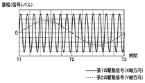
- the amplitude value (signal level) of the first drive signal illustrated in FIG. 5 gradually increases starting from the time T1 at which the minimum value is reached, and gradually decreases after reaching the maximum value at time T2. At time T3, it becomes the minimum value again.
- the amplitude value (signal level) of the second drive signal illustrated in FIG. 6 gradually increases starting from the time T1 at which the minimum value is reached, and gradually decreases after reaching the maximum value near the time T2. Then, it becomes the minimum value again at time T3.
- FIG. 7A is a diagram illustrating a first spiral trajectory drawn when illumination light is irradiated in time series on a virtual XY plane as shown in FIG. 3.
- FIG. 7B is a diagram illustrating a second spiral locus drawn when illumination light is irradiated in time series on the virtual XY plane as illustrated in FIG. 3.
- illumination light is applied to a position corresponding to the point SA on the surface of the subject.
- the irradiation position of the illumination light on the surface of the subject moves from point SA to point YMAX as shown in FIG. 7A.
- the displacement is performed so as to draw a first spiral locus.
- the amplitude values of the first and second drive signals decrease from time T2 to time T3, the irradiation position of the illumination light on the surface of the subject goes from point YMAX to point SA as shown in FIG. 7B. It is displaced so as to draw a second spiral locus.
- illumination light is applied to the point SA on the surface of the subject.
- the light guide plate 16 having the predetermined refractive index distribution as described above is disposed so as to cover the light exit surface of the objective optical system 14 (lens 14b).
- the portion corresponding to the outermost circumferences of the first and second spiral trajectories on the surface of the subject is not irradiated with illumination light, but other than the outermost circumferences of the first and second spiral trajectories on the surface of the subject.
- the corresponding portion is irradiated with illumination light.
- the actuator 15 is based on the first and second drive signals supplied from the driver unit 22 with a spiral scanning pattern corresponding to the locus of the irradiation position of the illumination light as illustrated in FIGS. 7A and 7B.
- the end portion including the light exit surface of the illumination fiber 12 can be swung.
- the illumination fiber 12 swings on the outermost periphery of the spiral scanning pattern (according to the locus of the illumination light irradiation position as exemplified in FIGS. 7A and 7B).
- Illumination light that has entered the interior through the objective optical system 14 within a predetermined period NPA that is moved is incident on the light receiving fiber 13 by total reflection at least once (or an odd number of times), and is illuminated outside the NPA for a predetermined period.
- the illumination light emitted from the optical fiber 12 through the objective optical system 14 is transmitted and emitted to the subject, and the return light of the illumination light emitted to the subject is transmitted outside the NPA for a predetermined period to transmit the light receiving fiber 13. It is formed so as to have a predetermined refractive index distribution that can be incident on the light source.
- the detection unit 23 includes a duplexer 36, detectors 37a, 37b, and 37c, and analog-digital (hereinafter referred to as A / D) converters 38a, 38b, and 38c.
- a / D analog-digital
- the duplexer 36 includes a dichroic mirror or the like, and separates light emitted from the light exit surface of the light receiving fiber 13 into light for each color component of R (red), G (green), and B (blue).
- the detectors 37a, 37b and 37c are configured to emit light.
- the detector 37a detects the intensity of the R light output from the duplexer 36, generates an analog R signal corresponding to the detected intensity of the R light, and outputs the analog R signal to the A / D converter 38a. It is configured.
- the detector 37b detects the intensity of the G light output from the duplexer 36, generates an analog G signal corresponding to the detected intensity of the G light, and outputs the analog G signal to the A / D converter 38b. It is configured.
- the detector 37c detects the intensity of the B light output from the duplexer 36, generates an analog B signal according to the detected intensity of the B light, and outputs the analog B signal to the A / D converter 38c. It is configured.
- the A / D converter 38 a is configured to convert the analog R signal output from the detector 37 a into a digital R signal and output the digital R signal to the controller 27.
- the A / D converter 38b is configured to convert the analog G signal output from the detector 37b into a digital G signal and output it to the controller 27.
- the A / D converter 38c is configured to convert the analog B signal output from the detector 37c into a digital B signal and output it to the controller 27.
- the memory 26 stores in advance a control program for controlling each part of the main unit 3 and can be used to determine whether the illumination light emitted from the illumination fiber 12 is good or bad. Information is also stored.
- the controller 27 is configured to read a control program stored in the memory 26 and control the light source unit 21 and the driver unit 22 based on the read control program.
- the controller 27 generates an image for one frame based on the R signal, the G signal, and the B signal output from the detection unit 23 within a period that does not overlap with the predetermined period NPA from the time T1 to the time T2.
- the generated image can be displayed on the monitor 4.
- the controller 27 generates an image for one frame based on the R signal, the G signal, and the B signal output from the detection unit 23 within a period not overlapping with the predetermined period NPA from the time T2 to the time T3.
- the generated image can be displayed on the monitor 4.
- the controller 27 illuminates light emitted from the illumination fiber 12 based on the information stored in the memory 26 and the R, G, and B signals output from the detection unit 23 within a predetermined period NPA. It is configured to determine whether the light emission state is good or not, and to control the light source unit 21 and / or the optical attenuator 25 according to the determined result. Details of such determination processing and control will be described later.
- the R signal, the G signal, and the B signal output from the detection unit 23 within the predetermined period NPA are only used to determine whether the illumination state of the illumination light emitted from the illumination fiber 12 is good or bad. Used. Therefore, in this embodiment, light is received from each position corresponding to between the point SA and the point YB on the surface of the subject as depicted by solid lines in the spiral trajectories shown in FIGS. 7A and 7B. An image corresponding to the return light incident on the optical fiber 13 is displayed on the monitor 4.
- the controller 27 controls the light source unit 21 to emit a predetermined amount of illumination light from the light sources 31a, 31b and the light source 31c. Control for outputting the second drive signal to the actuator 15 is performed on the driver unit 22, and control for reducing the attenuation in the optical attenuator 25 to 0 is performed. Alternatively, when the power of each part of the endoscope system 1 is turned on, the controller 27 controls the light source unit 21 to emit the maximum amount of illumination light from the light sources 31a and 31b and the light source 31c.
- Control for outputting the first and second drive signals to the actuator 15 is performed on the driver unit 22, and control for setting the attenuation amount in the optical attenuator 25 to a predetermined attenuation amount DB other than 0 is performed. Then, under such control of the controller 27, the end including the light emission surface of the illumination fiber 12 is swung, and the mixed light of R light, G light, and B light is emitted from the illumination fiber 12 as illumination light. Emitted.
- the controller 27 causes the monitor 4 to display an image generated based on the R signal, the G signal, and the B signal output from the detection unit 23 outside the NPA for a predetermined period, while the information stored in the memory 26 is displayed.
- the emission state of the illumination light emitted from the illumination fiber 12 by performing the following processing based on the R signal, G signal, and B signal output from the detection unit 23 within the predetermined period NPA Judgment of good or bad.
- FIG. 8 is a flowchart illustrating an example of processing performed by the endoscope system according to the embodiment of the present invention.
- the controller 27 sequentially detects the signal level of at least one of the R, G, and B signals output from the detection unit 23 over a period from time T1 to time T3 (FIG. 8 step S1).
- the controller 27 having a function as a determination unit changes the signal level within a predetermined period NPA based on the detection result of the signal level in step S1 of FIG. 8 and the information stored in the memory 26. Is determined whether or not corresponds to a predetermined pattern (step S2 in FIG. 8).
- the controller 27 reads, for example, information related to a signal level variation pattern as shown in FIG. 9 from the memory 26 and then includes a predetermined period included in the signal level detection result in step S1 of FIG. It is determined whether or not the signal level fluctuation in the NPA corresponds to the signal level fluctuation pattern included in the information read from the memory 26.
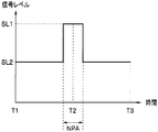
- FIG. 9 is a diagram illustrating an example of a signal level variation pattern detected when illumination light is normally emitted.
- the signal level variation pattern illustrated in FIG. 9 is emitted from the illumination fiber 12 while being swung along a locus along a spiral scanning pattern (as illustrated in FIGS. 7A and 7B). Obtained in this case, and is shown as a variation pattern substantially corresponding to a rectangular wave such that the signal level SL1 within the predetermined period NPA is always higher than the signal level SL2 outside the predetermined period NPA.
- the memory 26 stores in advance information related to a signal level variation pattern acquired when illumination light emitted according to a predetermined scanning pattern is normal.
- the signal level SL1 described above indicates a signal level detected within a predetermined period NPA when illumination light is normally emitted from the illumination fiber 12.
- the signal level SL2 described above indicates a signal level detected outside the NPA for a predetermined period when illumination light is normally emitted from the illumination fiber 12.
- step S2 of FIG. 8 the controller 27 changes the signal level within the predetermined period NPA included in the signal level detection result of step S1 of FIG. Is obtained, the illumination light is normally emitted from the illumination fiber 12 and the control for supplying the illumination light to the illumination fiber 12 is continued. However, the processing from step S1 in FIG. 8 is performed again.
- step S2 of FIG. 8 the controller 27 changes the signal level fluctuation within the predetermined period NPA included in the signal level detection result in step S1 of FIG. Is obtained, the illumination light emitted from the illumination fiber 12 is assumed to be abnormal, and the illumination light supplied from the light source unit 21 to the illumination fiber 12 is estimated. Control is performed to reduce the amount of light to 0 or a predetermined value (step S3 in FIG. 8).
- FIG. 10 is a diagram illustrating an example of a signal level variation pattern detected when the illumination light emission state is abnormal.
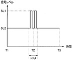
- FIG. 11 is a diagram illustrating an example different from FIG. 9 of the variation pattern of the signal level detected when the illumination light emission state is abnormal.
- the signal level variation pattern illustrated in FIG. 10 is acquired, for example, when the illumination fiber 12 is broken, and the signal level within the predetermined period NPA and the signal outside the predetermined period NPA. It is shown as a pattern whose level approaches zero uniformly.
- the signal level variation pattern illustrated in FIG. 11 is caused by, for example, an abnormality occurring in the operation of at least one of the actuators 15a to 15d (as illustrated in FIGS. 7A and 7B). ) This is obtained when the illumination light is emitted from the illumination fiber 12 while swinging along a locus deviating from the spiral scanning pattern.
- the detection timing of the signal level SL1 and the detection timing of the signal level SL2 are shown as patterns that are mixed in the NPA for a predetermined period.
- the controller 27 reduces the light amount of the illumination light supplied to the illumination fiber 12 to 0, for example, by controlling the light sources 31a, 31b and the light source 31c from on to off.
- the controller 27 controls the amount of illumination light supplied to the illumination fiber 12 by, for example, performing control to increase the attenuation amount of the illumination light in the optical attenuator 25 from 0 or a predetermined attenuation amount DB. Reduce to a predetermined value.
- the predetermined value is set as a light quantity that ensures safety to the human body even if illumination light continues to be emitted while the oscillation of the illumination fiber 12 by the actuator 15 is stopped. .
- the above-mentioned predetermined value is set as a light quantity such that the signal level in the signal output from the detection unit 23 is always 1 mW or less, for example.
- a series of processing as shown in FIG. 8 is performed during the operation of the endoscope system 1, so that the illumination light emitted from the illumination fiber 12 is abnormal. Can occur, the amount of illumination light supplied to the illumination fiber 12 can be quickly reduced to such a level that the safety to the human body is ensured. As a result, it is used when scanning the subject. It is possible to reduce the risk that the illumination light to be adversely affected on the human body.
- the light used for generating the image to be displayed on the monitor 4 (return light from the subject) and the light used for determining the quality of the emission state of the illumination light emitted from the illumination fiber 12. Both of these can be received by the light receiving fiber 13. Therefore, according to the present embodiment, the configuration for determining the quality of the emission state of the illumination light emitted from the illumination fiber 12 while maintaining the diameter of the insertion portion 11 at substantially the same level as the conventional one. Can be realized.
- FIG. 12 is a schematic diagram illustrating an example of the internal configuration of the distal end portion of the endoscope, which is different from FIG.
- FIG. 13 is a schematic diagram illustrating an example of the internal configuration of the distal end portion of the endoscope, which is different from FIGS. 2 and 12.
- the distal end surface of the distal end portion 11B of the insertion portion 11 has a refractive index distribution set under the same conditions as the light guide plate 16, and an arrow AR2
- the light guide plate 16A which is a transparent member formed so as to have an annular shape when viewed from the direction shown, may be covered.
- the distal end surface of the distal end portion 11C of the insertion portion 11 has a refractive index distribution set under the same conditions as the light guide plate 16, and from the direction indicated by the arrow AR3. You may cover with the light-guide plate 16B which is a transparent member formed so that it may become fan shape seeing.
- FIG. 14 relates to a second embodiment of the present invention.
- the insertion portion 11 of this embodiment has a tip portion 11D as shown in FIG. 14 instead of the tip portions 11A to 11C described in the first embodiment.
- FIG. 14 is a schematic diagram showing an example of the internal configuration of the distal end portion of the endoscope, which is different from those shown in FIGS.
- the distal end portion 11D of the insertion portion 11 includes an end portion on the light emitting side of the illumination fiber 12, an end portion on the light incident side of the light receiving fiber 13, and
- the objective optical system 14, the actuator 15, the reflection member 17 accommodated in the sheath 51, and a plurality of monitoring fibers 18 are configured.
- the reflection member 17 is formed of a reflection mirror, a reflection coat, metal, or the like, and has the above-described spiral scanning pattern (according to the locus of the irradiation position of illumination light as exemplified in FIGS. 7A and 7B).
- the illumination light is emitted from the illumination fiber 12 at a position where the illumination fiber 12 can enter the monitoring fiber 18 within a predetermined period NPA in which the illumination fiber 12 is swung on the outer periphery.
- the reflecting member 17 covers an area corresponding to the outermost part of the light incident surface of the lens 14a in an annular shape when viewed from the direction indicated by the arrow AR4. It is provided as follows.
- the plurality of monitoring fibers 18 are fixed in a state of being arranged in an annular shape so that a light incident surface is disposed at each position facing the reflecting member 17.
- the monitoring fiber 18 is configured to merge with the light receiving fiber 13 in the vicinity of the proximal end portion of the insertion portion 11, for example. Therefore, the illumination light received by the monitoring fiber 18 is incident on the branching filter 36 of the detection unit 23 through substantially the same path as the light receiving fiber 13.
- the series of processes of FIG. 8 can be applied substantially similarly. Therefore, according to the present embodiment, when an abnormality occurs in the illumination light emitted from the illumination fiber 12, the light quantity of the illumination light supplied to the illumination fiber 12 is ensured to be safe for the human body. As a result, it is possible to reduce the risk that the illumination light used for scanning the subject will adversely affect the human body.
- the light (return light from the subject) used for generating the image to be displayed on the monitor 4 can be received by the light receiving fiber 13 and the light emitted from the illumination fiber 12 can be received.
- the monitoring fiber 18 can receive light used for determining whether the light emission state is good or bad. Therefore, according to the present embodiment, the configuration for determining the quality of the emission state of the illumination light emitted from the illumination fiber 12 while maintaining the image quality of the image displayed on the monitor 4 at substantially the same level as the conventional one. Can be realized.
- the reflecting member 17 and the monitoring fiber 18 are not limited to being provided in an annular shape, for example, four locations in the X-axis direction and the Y-axis direction according to the arrangement positions of the actuators 15a to 15d
- the monitoring fiber 18 may be provided, and only the portion of the light incident surface of the lens 14a facing the light incident surface of the monitoring fiber 18 may be covered with the reflecting member 17.
- (Third embodiment) 15 and 16 relate to a third embodiment of the present invention.
- the illumination fiber 12 of the present embodiment is configured to be swung in a raster-like scan pattern instead of the spiral scan pattern as described in the first embodiment according to the operation of the actuator 15. Yes.
- FIG. 15 is a diagram illustrating an example of the signal waveform of the drive signal supplied to the actuator provided in the endoscope, which is different from those in FIGS. 5 and 6.
- FIG. 16 is a diagram showing a raster-like trajectory drawn when illumination light is irradiated in time series on a virtual XY plane as shown in FIG.
- the driver unit 22 of the present embodiment can generate a first drive signal and a second drive signal each having a waveform as shown in FIG. It is configured as follows.
- the light guide plate 16 of this embodiment has the objective optical system 14 within a predetermined period NPB in which the illumination fiber 12 is swung to the outermost part of the raster-like scanning pattern (dotted line portion of the locus shown in FIG. 16).
- the illumination light that has entered the interior is incident on the light receiving fiber 13 by being totally reflected one or more times (or an odd number of times), and is emitted from the illumination fiber 12 through the objective optical system 14 outside the NPB for a predetermined period.
- It has a predetermined refractive index distribution that allows light to pass through and be emitted to the subject, and further allows the return light of the illumination light emitted to the subject to be transmitted outside the NPB for a predetermined period and to enter the light receiving fiber 13. It is formed to do.
- the controller 27 of the present embodiment performs a predetermined period NPB based on the signal level detection result obtained by performing substantially the same processing as step S1 in FIG. 8 and the information stored in the memory 26. Is determined whether or not the signal level variation corresponds to a predetermined pattern, and is supplied to the illumination fiber 12 when the signal level variation within the predetermined period NPB does not correspond to the predetermined pattern. It is comprised so that control for reducing the light quantity of the illumination light to be performed can be performed.
- the present embodiment when an abnormality occurs in the illumination light emitted from the illumination fiber 12, the light quantity of the illumination light supplied to the illumination fiber 12 is secured to the human body. As a result, it is possible to reduce the risk that the illumination light used for scanning the subject will adversely affect the human body.
- the illumination fiber 12 has a spiral scan pattern as described in the first embodiment and a raster scan pattern as described in the third embodiment according to the operation of the actuator 15. Instead, it is configured to be swung with a Lissajous scanning pattern.
- FIG. 17 is a diagram illustrating an example of the signal waveform of the drive signal supplied to the actuator provided in the endoscope, which is different from those in FIGS. 5, 6, and 15.
- FIG. 18 is a diagram showing a Lissajous locus drawn when illumination light is irradiated in time series on the virtual XY plane as shown in FIG.
- the driver unit 22 of the present embodiment can generate a first drive signal and a second drive signal each having a waveform as shown in FIG. It is configured as follows.
- the light guide plate 16 of this embodiment has the objective optical system 14 within the predetermined period NPC in which the illumination fiber 12 is swung to the outermost part of the Lissajous scanning pattern (the dotted line portion of the locus shown in FIG. 18).
- the illumination light that has entered the interior is incident on the light receiving fiber 13 by being totally reflected one or more times (or an odd number of times), and is emitted from the illumination fiber 12 through the objective optical system 14 outside the NPC for a predetermined period.
- It has a predetermined refractive index distribution capable of transmitting light to be emitted to the subject, and further allowing the return light of the illumination light emitted to the subject to be transmitted outside the NPC for a predetermined period and to be incident on the light receiving fiber 13. It is formed to do.
- the controller 27 within the predetermined period NPC, based on the detection result of the signal level obtained by performing substantially the same processing as step S1 of FIG. 8 and the information stored in the memory 26. Is determined whether or not the signal level variation corresponds to a predetermined pattern, and is supplied to the illumination fiber 12 when the signal level variation within the predetermined period NPC does not correspond to the predetermined pattern. It is comprised so that control for reducing the light quantity of the illumination light to be performed can be performed.
- the present embodiment when an abnormality occurs in the illumination light emitted from the illumination fiber 12, the light quantity of the illumination light supplied to the illumination fiber 12 is secured to the human body. As a result, it is possible to reduce the risk that the illumination light used for scanning the subject will adversely affect the human body.
- (Fifth embodiment) 19 to 21 relate to a fifth embodiment of the present invention.
- the insertion portion 11 of this embodiment has a tip portion 11E as shown in FIG. 19 instead of the tip portions 11A to 11D described in the first and second embodiments.
- FIG. 19 is a schematic diagram illustrating an example of the internal configuration of the distal end portion of the endoscope, which is different from FIGS. 2, 12, 13 and 14.
- the distal end portion 11 ⁇ / b> E of the insertion portion 11 has a circular shape inside the sheath 51, the illumination fiber 12, the objective optical system 14, and the actuator 15 housed in the sheath 51. And a plurality of light receiving fibers 13 embedded in an annular shape. That is, the distal end portion 11E of the insertion portion 11 is configured by removing the light guide plate 16 from the distal end portion 11A of the first embodiment.
- the main body device 3 of the present embodiment can detect return light generated when the illumination light transmitted by the illumination fiber 12 is reflected on the light emission surface of the illumination fiber 12 and the light emission surface of the lens 14b. It is configured.
- FIG. 20 is a diagram illustrating an example of a configuration for detecting return light emitted from the illumination fiber.
- the main body device 3 of the present embodiment includes an optical member 61 provided on the optical path from the optical attenuator 25 to the light incident surface of the illumination fiber 12, and an optical member. And a light detection unit 62 into which the return light having passed through 61 is incident.
- the optical member 61 is constituted by, for example, a glass plate disposed so as to be inclined with respect to the optical axis of the illumination light emitted from the light attenuator 25.
- the illumination light emitted to the light incident surface can be transmitted, and the return light emitted from the light incident surface of the illumination fiber 12 can be reflected to the light detection unit 62 side.
- the optical member 61 has a function capable of separating the illumination light emitted from the light attenuator 25 to the light incident surface of the illumination fiber 12 and the return light emitted from the light incident surface of the illumination fiber 12. It has.
- the light detection unit 62 is configured by, for example, a photodiode, and has a function of generating an electrical signal corresponding to the intensity of the return light incident through the optical member 61 and outputting the electrical signal to the controller 27. is doing.
- the controller 27 of the present embodiment generates an image for one frame based on the R signal, the G signal, and the B signal output from the detection unit 23 within a period from time T1 to time T2, and the generated image is displayed.
- the monitor 4 can be displayed.
- controller 27 generates an image for one frame based on the R signal, the G signal, and the B signal output from the detection unit 23 within the period from time T2 to time T3. An image can be displayed on the monitor 4.
- the controller 27 of the present embodiment sequentially detects the signal level of the electrical signal output from the light detection unit 62 over a period from time T1 to time T3, and then the fluctuation of the detected signal level has a predetermined pattern. It is comprised so that determination regarding whether it corresponds to can be performed.
- the controller 27 reads information related to a signal level variation pattern as shown in FIG. 21 from the memory 26, and then outputs the information from the light detection unit 62 during a period from time T1 to time T3. It is determined whether or not the fluctuation of the signal level of the electric signal corresponds to the fluctuation pattern of the signal level included in the information read from the memory 26.
- FIG. 21 is a diagram illustrating an example different from FIG. 9 of the variation pattern of the signal level detected when the illumination light is normally emitted.
- the signal level variation pattern illustrated in FIG. 21 is that the illumination light transmitted by the illumination fiber 12 is transmitted when the illumination fiber 12 is swung along a locus along a spiral scanning pattern. Is obtained according to the intensity of the return light generated by reflection on the light exit surface and the light exit surface of the lens 14b.
- the signal level variation pattern illustrated in FIG. 21 is the return light incident on the light detection unit 62 when the illumination fiber 12 is swung along a locus along the spiral scanning pattern.
- the signal level SL3 becomes the maximum at time T1 when the intensity of the light reaches the maximum, and the signal level decreases nonlinearly between times T1 and T2 when the intensity of the return light incident on the light detection unit 62 gradually decreases.
- the signal level SL4 becomes the minimum signal level SL4 at the time T2 when the intensity of the return light incident on the detection unit 62 becomes the minimum, and the signal is output between the times T2 and T3 when the intensity of the return light incident on the light detection unit 62 gradually increases.
- the pattern is shown as a pattern in which the level increases nonlinearly and becomes the maximum signal level SL3 at time T3 when the intensity of the return light incident on the light detection unit 62 becomes maximum again.
- the controller 27 determines the determination result that the signal level fluctuation of the electrical signal output from the light detection unit 62 during the period from time T1 to time T3 corresponds to the fluctuation pattern included in the information read from the memory 26. If it is obtained, it is presumed that the illumination light is normally emitted from the illumination fiber 12, the control for supplying the illumination light to the illumination fiber 12, and the output from the light detection unit 62 The monitoring of the signal level of the electric signal is continued.
- the present embodiment when an abnormality occurs in the illumination light emitted from the illumination fiber 12, the light quantity of the illumination light supplied to the illumination fiber 12 is secured to the human body. As a result, it is possible to reduce the risk that the illumination light used for scanning the subject will adversely affect the human body.
Abstract
内視鏡システムは、光源から発せられた照明光を伝送して出射する光伝送部と、所定の走査パターンに応じた軌跡を描くように光伝送部を揺動させる駆動部と、所定の走査パターンの所定の部分に光伝送部が揺動される所定の期間内に出射された照明光を受光可能な受光部と、受光部により受光された照明光の強度に応じた信号を出力する光検出部と、光検出部から出力される信号における信号レベルの変動を検出し、当該検出した信号レベルの変動パターンが所定の変動パターンに該当するか否かを判定する判定部と、所定の期間内の信号レベルの変動パターンが所定の変動パターンに該当しない場合に、光源から導光部へ供給される照明光の光量を減少させる制御部と、を有する。
Description
本発明は、内視鏡システムに関し、特に、被写体を走査して画像を取得する内視鏡システムに関するものである。
医療分野の内視鏡においては、被検者の負担を軽減するために、当該被検者の体腔内に挿入される挿入部を細径化するための種々の技術が提案されている。そして、このような技術の一例として、前述の挿入部に相当する部分に固体撮像素子を有しない光走査型内視鏡、及び、当該光走査型内視鏡を具備して構成されたシステムが知られている。
具体的には、前述の光走査型内視鏡を具備するシステムは、例えば、光源部から発せられた照明光を導光する照明用ファイバの先端部を揺動させることにより被写体を予め設定された走査パターンで走査し、当該被写体からの戻り光を照明用ファイバの周囲に配置された受光用ファイバで受光し、当該受光用ファイバで受光された戻り光を各色成分毎に分離して得た信号を用いて当該被写体の画像を生成するように構成されている。
そして、前述のような構成を具備するシステムとしては、例えば、日本国特開2011-19706号公報の医療用観察システムが従来知られている。
具体的には、日本国特開2011-19706号公報によれば、前述の光走査型内視鏡に略相当する走査型医療用プローブが患者の体外にある場合に、レーザー光源から前記走査型医療用プローブへ出射されるレーザー光の光量を制限することができるように構成された医療用観察システムが開示されている。
しかし、日本国特開2011-19706号公報に開示された構成によれば、走査型医療用プローブから出射されるレーザー光の出射状態の良否を判別することができない。その結果、日本国特開2011-19706号公報に開示された構成によれば、例えば、実際には走査型医療用プローブが体外に配置されているにも係らず、当該走査型医療用プローブが体腔内に配置されていると検出してしまうような誤検出が生じた場合において、人体に対して悪影響を及ぼす光量を具備するレーザー光が出射されてしまうおそれがある、という課題が生じている。
本発明は、前述した事情に鑑みてなされたものであり、被写体の走査の際に用いられる照明光が人体に対して悪影響を及ぼす危険性を低減することが可能な内視鏡システムを提供することを目的としている。
本発明の一態様の内視鏡システムは、光源から発せられた照明光を伝送して光出射面から出射するように構成された光伝送部と、所定の走査パターンに応じた軌跡を描くように前記光伝送部の前記光出射面を含む端部を揺動させることが可能な駆動部と、前記所定の走査パターンの所定の部分に前記光伝送部の前記端部が揺動される所定の期間内に前記光伝送部から出射された照明光を受光できるように構成された受光部と、前記受光部により受光された照明光を検出し、当該検出した照明光の強度に応じた信号を出力するように構成された光検出部と、前記光検出部から出力される信号における信号レベルの変動を前記所定の期間内及び前記所定の期間外において検出し、当該検出した前記所定の期間内の信号レベルの変動パターンが所定の変動パターンに該当するか否かを判定するように構成された判定部と、前記所定の期間内の信号レベルの変動パターンが前記所定の変動パターンに該当しないとの判定結果が得られた場合において、前記光源から前記導光部へ供給される照明光の光量を0または所定値まで減少させるための制御を行うように構成された制御部と、を有する。
以下、本発明の実施の形態について、図面を参照しつつ説明を行う。
(第1の実施例)
図1から図13は、本発明の第1の実施例に係るものである。図1は、本発明の実施例に係る内視鏡システムの要部の構成を示す図である。
図1から図13は、本発明の第1の実施例に係るものである。図1は、本発明の実施例に係る内視鏡システムの要部の構成を示す図である。
内視鏡システム1は、例えば図1に示すように、被検者の体腔内に挿入可能な走査型の内視鏡2と、内視鏡2に接続される本体装置3と、本体装置3に接続されるモニタ4と、を有して構成されている。
内視鏡2は、細長の円筒形状及び可撓性を備えて形成された挿入部11を有して構成されている。なお、挿入部11の基端部には、内視鏡2を本体装置3に着脱自在に接続するための図示しないコネクタ等が設けられている。
図2は、内視鏡の先端部の内部構成の一例を示す模式図である。図2に模式的に示すように、挿入部11の先端部11Aには、本体装置3から供給される照明光を伝送する光伝送部としての機能を具備する照明用ファイバ12の光出射側の端部と、被写体からの戻り光及び後述の導光板16を経て入射される照明光を受光して本体装置3へ導く受光用ファイバ13の光入射側の端部と、照明用ファイバ12から出射される照明光を集光して出射するように構成された対物光学系14と、本体装置3から出力される駆動信号に基づいて照明用ファイバ12の光出射側の端部を揺動させることが可能なアクチュエータ15と、が設けられている。また、照明用ファイバ12、対物光学系14及びアクチュエータ15の各部は、可撓性を有するシース51に収容されている。さらに、シース51の内部には、複数の受光用ファイバ13が円環状に埋設されている。
対物光学系14は、照明用ファイバ12からの照明光が入射されるレンズ14aと、レンズ14aを経た照明光を出射するレンズ14bと、を有して構成されている。また、レンズ14a及びレンズ14bは、正の屈折力をそれぞれ具備して形成されている。
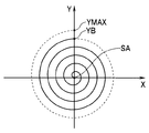
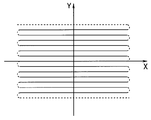
ここで、以降においては、挿入部11の長手方向の軸に相当する挿入軸(または対物光学系14の光軸)に対して垂直な仮想の平面として、図3に示すようなXY平面を被写体の表面に設定する場合を例に挙げつつ説明を進める。図3は、被写体の表面に設定される仮想的なXY平面の一例を示す図である。
具体的には、図3のXY平面上の点SAは、紙面手前側から奥側に相当する方向に挿入部11の挿入軸が存在するものとして仮想的に設定した場合における、当該挿入軸と紙面との交点を示している。また、図3のXY平面におけるX軸方向は、紙面左側から右側に向かう方向として設定されている。また、図3のXY平面におけるY軸方向は、紙面下側から上側に向かう方向として設定されている。また、図3のXY平面を構成するX軸及びY軸は、点SAにおいて交差している。
図4は、図2のIV-IV線断面図である。図4に示すように、照明用ファイバ12とアクチュエータ15との間には、接合部材としてのフェルール41が配置されている。具体的には、フェルール41は、例えば、ジルコニア(セラミック)またはニッケル等により形成されている。
フェルール41は、図4に示すように、四角柱として形成されており、X軸方向に対して垂直な側面42a及び42cと、Y軸方向に対して垂直な側面42b及び42dとを有する。また、フェルール41の略中心には、照明用ファイバ12が固定配置されている。なお、フェルール41は、角柱である限りにおいては、四角柱以外の他の形状として形成されていてもよい。
アクチュエータ15は、図4に示すように、側面42aに沿って配置されたアクチュエータ15aと、側面42bに沿って配置されたアクチュエータ15bと、側面42cに沿って配置されたアクチュエータ15cと、側面42dに沿って配置されたアクチュエータ15dと、を有している。
アクチュエータ15a及び15cは、例えば、圧電素子(ピエゾ素子)により形成されており、ドライバユニット22のD/A変換器34aから出力される第1の駆動信号に応じて駆動するように構成されている。
アクチュエータ15b及び15dは、例えば、圧電素子(ピエゾ素子)により形成されており、ドライバユニット22のD/A変換器34bから出力される第2の駆動信号に応じて駆動するように構成されている。
挿入部11の先端部11Aの先端面は、図2に模式的に示すように、矢印AR1に示す方向から見て円形状となるように形成された透明部材である導光板16により覆われている。
導光板16は、対物光学系14の(レンズ14bの)屈折率及び空気の屈折率のうちの少なくとも一方に基づく所定の屈折率分布を具備するように形成されている。具体的には、導光板16は、例えば、所定の期間内に対物光学系14を経て内部に侵入した照明光を1回以上(または奇数回)全反射することにより受光用ファイバ13へ入射させ、前記所定の期間外に照明用ファイバ12から対物光学系14を経て出射された照明光を透過させて被写体へ出射し、さらに、前記所定の期間外に被写体に出射された照明光の戻り光を透過させて受光用ファイバ13に入射させることが可能な所定の屈折率分布を具備するように形成されている。
一方、本体装置3は、光源ユニット21と、ドライバユニット22と、検出ユニット23と、ライトガイド24と、光減衰器25と、メモリ26と、コントローラ27と、を有して構成されている。
光源ユニット21は、光源31aと、光源31bと、光源31cと、合波器32と、を有して構成されている。
光源31aは、例えば、レーザーまたはSLD(スーパールミネッセントダイオード)等のようなレーザー放射をする光源を具備し、コントローラ27の制御によりオンされた際に、赤色の波長帯域の光(以降、R光とも称する)を合波器32へ出射するように構成されている。
光源31bは、例えば、レーザーまたはSLD(スーパールミネッセントダイオード)等のようなレーザー放射をする光源を具備し、コントローラ27の制御によりオンされた際に、緑色の波長帯域の光(以降、G光とも称する)を合波器32へ出射するように構成されている。
光源31cは、例えば、レーザーまたはSLD(スーパールミネッセントダイオード)等のようなレーザー放射をする光源を具備し、コントローラ27の制御によりオンされた際に、青色の波長帯域の光(以降、B光とも称する)を合波器32へ出射するように構成されている。
合波器32は、光源31aから発せられたR光と、光源31bから発せられたG光と、光源31cから発せられたB光と、を合波してライトガイド24へ供給できるように構成されている。
光減衰器25は、ライトガイド24から照明用ファイバ12の光入射側の端部へ出射される照明光の光路上に配置されているとともに、コントローラ27の制御に応じて減衰量を増減することにより、照明用ファイバ12に供給される照明光の光量を調整することができるように構成されている。
ドライバユニット22は、信号発生器33と、デジタルアナログ(以下、D/Aという)変換器34a及び34bと、アンプ35と、を有して構成されている。
信号発生器33は、コントローラ27の制御に基づき、照明用ファイバ12の光出射面を含む端部をX軸方向に揺動させる第1の駆動信号として、例えば図5に示すような、所定の波形の信号を生成してD/A変換器34aに出力するように構成されている。図5は、アクチュエータに供給される第1の駆動信号の信号波形の一例を示す図である。
また、信号発生器33は、コントローラ27の制御に基づき、照明用ファイバ12の光出射面を含む端部をY軸方向に揺動させる第2の駆動信号として、例えば図6に示すような、前述の第1の駆動信号の位相を90°ずらした波形の信号を生成してD/A変換器34bに出力するように構成されている。図6は、アクチュエータに供給される第2の駆動信号の信号波形の一例を示す図である。
D/A変換器34aは、信号発生器33から出力されたデジタルの第1の駆動信号をアナログの第1の駆動信号に変換してアンプ35へ出力するように構成されている。
D/A変換器34bは、信号発生器33から出力されたデジタルの第2の駆動信号をアナログの第2の駆動信号に変換してアンプ35へ出力するように構成されている。
アンプ35は、D/A変換器34aから出力された第1の駆動信号を増幅してアクチュエータ15a及び15cへ出力するように構成されている。また、アンプ35は、D/A変換器34bから出力された第2の駆動信号を増幅してアクチュエータ15b及び15dへ出力するように構成されている。
ここで、図5において例示した第1の駆動信号の振幅値(信号レベル)は、最小値となる時刻T1を起点として徐々に増加し、時刻T2において最大値になった後で徐々に減少し、時刻T3で再び最小値となる。
また、図6において例示した第2の駆動信号の振幅値(信号レベル)は、最小値となる時刻T1を起点として徐々に増加し、時刻T2の近辺において最大値になった後で徐々に減少し、時刻T3で再び最小値となる。
そして、図5に示すような第1の駆動信号がアクチュエータ15a及び15cに供給されるとともに、図6に示すような第2の駆動信号がアクチュエータ15b及び15dに供給されると、照明用ファイバ12の光出射面を含む端部が点SAを中心とした渦巻状に揺動され、このような揺動に応じて被写体の表面が図7A及び図7Bに示すような渦巻状に走査される。図7Aは、図3のような仮想的なXY平面に時系列で照明光が照射される際に描かれる、第1の渦巻状の軌跡を表す図である。図7Bは、図3のような仮想的なXY平面に時系列で照明光が照射される際に描かれる、第2の渦巻状の軌跡を表す図である。
具体的には、時刻T1においては、被写体の表面の点SAに相当する位置に照明光が照射される。その後、第1及び第2の駆動信号の振幅値が時刻T1から時刻T2にかけて増加するに伴い、被写体の表面における照明光の照射位置が、図7Aに示すような、点SAから点YMAXへ向かう第1の渦巻状の軌跡を描くように変位する。また、第1及び第2の駆動信号の振幅値が時刻T2から時刻T3にかけて減少するに伴い、被写体の表面における照明光の照射位置が、図7Bに示すような、点YMAXから点SAへ向かう第2の渦巻状の軌跡を描くように変位する。そして、時刻T3においては、被写体の表面における点SAに照明光が照射される。
但し、本実施例によれば、前述のような所定の屈折率分布を具備する導光板16が対物光学系14(レンズ14b)の光出射面を覆うように配置されていることに起因し、被写体の表面における第1及び第2の渦巻状の軌跡の最外周に相当する部分には照明光が照射されない一方で、被写体の表面における第1及び第2の渦巻状の軌跡の最外周以外に相当する部分には照明光が照射されるようになっている。
具体的には、例えば、図7A及び図7Bに示した渦巻状の軌跡においてそれぞれ点線で描かれているような、被写体の表面の点YBの直後から点YMAXまでの間に相当する各位置には照明光が照射されない一方で、図7A及び図7Bに示した渦巻状の軌跡においてそれぞれ実線で描かれているような、被写体の表面の点SAから点YBまでの間に相当する各位置には照明光が照射される。
すなわち、アクチュエータ15は、ドライバユニット22から供給される第1及び第2の駆動信号に基づき、図7A及び図7Bに例示したような照明光の照射位置の軌跡に応じた渦巻状の走査パターンにより、照明用ファイバ12の光出射面を含む端部を揺動させることが可能な構成を具備している。
また、本実施例の導光板16は、前述の(図7A及び図7Bに例示したような照明光の照射位置の軌跡に応じた)渦巻状の走査パターンの最外周に照明用ファイバ12が揺動される所定の期間NPA内に対物光学系14を経て内部に侵入した照明光を1回以上(または奇数回)全反射することにより受光用ファイバ13へ入射させ、所定の期間NPA外に照明用ファイバ12から対物光学系14を経て出射された照明光を透過させて被写体へ出射し、さらに、所定の期間NPA外に被写体に出射された照明光の戻り光を透過させて受光用ファイバ13に入射させることが可能な所定の屈折率分布を具備するように形成されている。
一方、検出ユニット23は、分波器36と、検出器37a、37b及び37cと、アナログデジタル(以下、A/Dという)変換器38a、38b及び38cと、を有して構成されている。
分波器36は、ダイクロイックミラー等を具備し、受光用ファイバ13の光出射面から出射された光をR(赤)、G(緑)及びB(青)の各色成分毎の光に分離して検出器37a、37b及び37cへ出射するように構成されている。
検出器37aは、分波器36から出力されるR光の強度を検出し、当該検出したR光の強度に応じたアナログのR信号を生成してA/D変換器38aへ出力するように構成されている。
検出器37bは、分波器36から出力されるG光の強度を検出し、当該検出したG光の強度に応じたアナログのG信号を生成してA/D変換器38bへ出力するように構成されている。
検出器37cは、分波器36から出力されるB光の強度を検出し、当該検出したB光の強度に応じたアナログのB信号を生成してA/D変換器38cへ出力するように構成されている。
A/D変換器38aは、検出器37aから出力されたアナログのR信号をデジタルのR信号に変換してコントローラ27へ出力するように構成されている。
A/D変換器38bは、検出器37bから出力されたアナログのG信号をデジタルのG信号に変換してコントローラ27へ出力するように構成されている。
A/D変換器38cは、検出器37cから出力されたアナログのB信号をデジタルのB信号に変換してコントローラ27へ出力するように構成されている。
メモリ26には、本体装置3の各部の制御を行うための制御プログラム等が予め格納されているとともに、照明用ファイバ12から出射される照明光の出射状態の良否を判別する際に利用可能な情報が併せて格納されている。
コントローラ27は、メモリ26に格納された制御プログラムを読み出し、当該読み出した制御プログラムに基づいて光源ユニット21及びドライバユニット22の制御を行うように構成されている。
コントローラ27は、時刻T1から時刻T2までのうち、所定の期間NPAと重複しない期間内に検出ユニット23から出力されるR信号、G信号及びB信号に基づいて1フレーム分の画像を生成し、当該生成した画像をモニタ4に表示させることができるように構成されている。
また、コントローラ27は、時刻T2から時刻T3までのうち、所定の期間NPAと重複しない期間内に検出ユニット23から出力されるR信号、G信号及びB信号に基づいて1フレーム分の画像を生成し、当該生成した画像をモニタ4に表示させることができるように構成されている。
さらに、コントローラ27は、メモリ26に格納された情報と、所定の期間NPA内に検出ユニット23から出力されるR信号、G信号及びB信号と、に基づき、照明用ファイバ12から出射される照明光の出射状態の良否を判別し、さらに、当該判別した結果に応じた制御を光源ユニット21及び(または)光減衰器25に対して行うことができるように構成されている。なお、このような判別処理及び制御の詳細については、後程説明する。
すなわち、本実施例においては、所定の期間NPA内に検出ユニット23から出力されるR信号、G信号及びB信号が、照明用ファイバ12から出射される照明光の出射状態の良否の判定のみに用いられる。そのため、本実施例においては、図7A及び図7Bに示した渦巻状の軌跡においてそれぞれ実線で描かれているような、被写体の表面の点SAから点YBまでの間に相当する各位置から受光用ファイバ13に入射される戻り光に応じた画像がモニタ4に表示される。
続いて、以上に述べたような構成を具備する内視鏡システム1の動作等について説明する。
内視鏡システム1の各部の電源が投入されると、コントローラ27は、光源31a、31b及び光源31cから所定の光量の照明光をそれぞれ出射させる制御を光源ユニット21に対して行い、第1及び第2の駆動信号をアクチュエータ15へ出力させる制御をドライバユニット22に対して行うとともに、光減衰器25における減衰量を0にするための制御を行う。または、内視鏡システム1の各部の電源が投入されると、コントローラ27は、光源31a、31b及び光源31cから最大の光量の照明光をそれぞれ出射させる制御を光源ユニット21に対して行い、第1及び第2の駆動信号をアクチュエータ15へ出力させる制御をドライバユニット22に対して行うとともに、光減衰器25における減衰量を0以外の所定の減衰量DBにするための制御を行う。そして、このようなコントローラ27の制御により、照明用ファイバ12の光出射面を含む端部が揺動されるとともに、R光、G光及びB光の混合光が照明光として照明用ファイバ12から出射される。
その後、コントローラ27は、所定の期間NPA外に検出ユニット23から出力されるR信号、G信号及びB信号に基づいて生成した画像をモニタ4に表示させる一方で、メモリ26に格納された情報と、所定の期間NPA内に検出ユニット23から出力されるR信号、G信号及びB信号と、に基づいて以下のような処理を行うことにより、照明用ファイバ12から出射される照明光の出射状態の良否を判別する。
ここで、照明用ファイバ12から出射される照明光の出射状態の良否を判別する際に行われる処理の具体例について説明する。図8は、本発明の実施例に係る内視鏡システムにより行われる処理等の一例を示すフローチャートである。
まず、コントローラ27は、検出ユニット23から出力されるR信号、G信号及びB信号のうちの少なくともいずれか1つの色信号の信号レベルを、時刻T1から時刻T3までの期間にかけて順次検出する(図8のステップS1)。
次に、判定部としての機能を具備するコントローラ27は、図8のステップS1による信号レベルの検出結果と、メモリ26に格納された情報と、に基づき、所定の期間NPA内の信号レベルの変動が所定のパターンに該当しているか否かに係る判定を行う(図8のステップS2)。
具体的には、コントローラ27は、例えば、図9に示すような信号レベルの変動パターンに係る情報をメモリ26から読み込んだ後、図8のステップS1による信号レベルの検出結果に含まれる所定の期間NPA内の信号レベルの変動が、メモリ26から読み込んだ情報に含まれる信号レベルの変動パターンに該当するか否かを判定する。図9は、正常に照明光が出射されている場合に検出される信号レベルの変動パターンの一例を示す図である。
図9に例示した信号レベルの変動パターンは、(図7A及び図7Bに例示したような)渦巻状の走査パターンに沿った軌跡で揺動されつつ照明用ファイバ12から照明光が出射されている場合に取得されるものであり、所定の期間NPA内の信号レベルSL1が所定の期間NPA外の信号レベルSL2に比べて常時大きくなるような、矩形波に略相当する変動パターンとして示される。
すなわち、本実施例のメモリ26には、所定の走査パターンに応じて出射される照明光が正常である場合に取得される信号レベルの変動パターンに係る情報が予め格納されている。
なお、前述の信号レベルSL1は、照明用ファイバ12から正常に照明光が出射されている場合において、所定の期間NPA内に検出される信号レベルを示している。また、前述の信号レベルSL2は、照明用ファイバ12から正常に照明光が出射されている場合において、所定の期間NPA外に検出される信号レベルを示している。
一方、コントローラ27は、図8のステップS2において、図8のステップS1による信号レベルの検出結果に含まれる所定の期間NPA内の信号レベルの変動が、メモリ26から読み込んだ情報に含まれる変動パターンに該当するとの判定結果を得た場合には、照明用ファイバ12から正常に照明光が出射されているものと推定し、照明用ファイバ12への照明光の供給を実施するための制御を継続しつつ、図8のステップS1からの処理を再度行う。
また、コントローラ27は、図8のステップS2において、図8のステップS1による信号レベルの検出結果に含まれる所定の期間NPA内の信号レベルの変動が、メモリ26から読み込んだ情報に含まれる変動パターンに該当しないとの判定結果を得た場合には、照明用ファイバ12から出射される照明光に異常が発生しているものと推定し、光源ユニット21から照明用ファイバ12へ供給される照明光の光量を0または所定値まで低下させるための制御を行う(図8のステップS3)。
具体的には、コントローラ27は、図8のステップS1による信号レベルの検出結果に含まれる所定の期間NPA内の信号レベルの変動が、例えば、図10または図11に示すようなパターンである場合において、照明用ファイバ12から出射される照明光に異常が発生しているものと推定する。図10は、照明光の出射状態が異常である場合に検出される信号レベルの変動パターンの一例を示す図である。図11は、照明光の出射状態が異常である場合に検出される信号レベルの変動パターンの、図9とは異なる例を示す図である。
図10に例示した信号レベルの変動パターンは、例えば、照明用ファイバ12に折れが発生している場合に取得されるものであり、所定の期間NPA内の信号レベル及び所定の期間NPA外の信号レベルが一様に0に近づくようなパターンとして示される。
図11に例示した信号レベルの変動パターンは、例えば、アクチュエータ15a~15dのうちの少なくともいずれか1つの動作に異常が発生していることに起因し、(図7A及び図7Bに例示したような)渦巻状の走査パターンから外れた軌跡で揺動されつつ照明用ファイバ12から照明光が出射されている場合に取得されるものであり、信号レベルSL1の検出タイミングと信号レベルSL2の検出タイミングとが所定の期間NPA内において混在しているようなパターンとして示される。
一方、コントローラ27は、例えば、光源31a、31b及び光源31cをオンからオフへ切り替えるような制御を行うことにより、照明用ファイバ12へ供給される照明光の光量を0まで低下させる。または、コントローラ27は、例えば、光減衰器25における照明光の減衰量を0または所定の減衰量DBから増加するような制御を行うことにより、照明用ファイバ12へ供給される照明光の光量を所定値まで低下させる。
なお、前述の所定値は、アクチュエータ15による照明用ファイバ12の揺動が停止している状態で照明光が出射され続けたとしても、人体に対する安全性が確保されるような光量として設定される。具体的には、前述の所定値は、例えば、検出ユニット23から出力される信号における信号レベルが常時1mW以下になるような光量として設定される。
以上に述べたような本実施例によれば、図8に示したような一連の処理が内視鏡システム1の動作中に行われることにより、照明用ファイバ12から出射される照明光に異常が発生した場合において、照明用ファイバ12へ供給される照明光の光量を、人体に対する安全性が確保されるような光量まで速やかに低下させることができ、その結果、被写体の走査の際に用いられる照明光が人体に対して悪影響を及ぼす危険性を低減することができる。
また、本実施例によれば、モニタ4に表示させる画像の生成に用いられる光(被写体からの戻り光)と、照明用ファイバ12から出射される照明光の出射状態の良否判定に用いられる光と、の両方を受光用ファイバ13で受光することができるように構成されている。そのため、本実施例によれば、挿入部11の径の太さを従来と略同程度に維持しつつ、照明用ファイバ12から出射される照明光の出射状態の良否判定を行うための構成を実現することができる。
なお、本実施例によれば、導光板16と同様の条件で(渦巻状の走査パターンの最外周に照明用ファイバ12が揺動される所定の期間NPA内に対物光学系14を経て内部に侵入した照明光を1回以上全反射することにより受光用ファイバ13へ入射させることができるという条件を具備して)設定された屈折率分布を具備する限りにおいては、円形状以外の他の形状の導光板を挿入部11の先端部の先端面に設けて構成してもよい。図12は、内視鏡の先端部の内部構成の、図2とは異なる例を示す模式図である。図13は、内視鏡の先端部の内部構成の、図2及び図12とは異なる例を示す模式図である。
具体的には、例えば図12に模式的に示すように、挿入部11の先端部11Bの先端面を、導光板16と同様の条件で設定された屈折率分布を具備するとともに、矢印AR2に示す方向から見て円環形状となるように形成された透明部材である導光板16Aにより覆ってもよい。または、例えば図13に模式的に示すように、挿入部11の先端部11Cの先端面を、導光板16と同様の条件で設定された屈折率分布を具備するとともに、矢印AR3に示す方向から見て扇形状となるように形成された透明部材である導光板16Bにより覆ってもよい。
(第2の実施例)
図14は、本発明の第2の実施例に係るものである。
図14は、本発明の第2の実施例に係るものである。
なお、本実施例においては、第1の実施例と同様の構成等を有する部分に関する詳細な説明を省略するとともに、第1の実施例と異なる構成等を有する部分に関して主に説明を行う。
本実施例の挿入部11は、第1の実施例において説明した先端部11A~11Cの代わりに、図14に示すような先端部11Dを有して構成されている。図14は、内視鏡の先端部の内部構成の、図2、図12及び図13とは異なる例を示す模式図である。
具体的には、挿入部11の先端部11Dは、図14に模式的に示すように、照明用ファイバ12の光出射側の端部と、受光用ファイバ13の光入射側の端部と、対物光学系14と、アクチュエータ15と、シース51に収容された反射部材17及び複数のモニタリング用ファイバ18と、を有して構成されている。
反射部材17は、反射ミラー、反射コートまたは金属等により形成されており、前述の(図7A及び図7Bに例示したような照明光の照射位置の軌跡に応じた)渦巻状の走査パターンの最外周に照明用ファイバ12が揺動される所定の期間NPA内に照明用ファイバ12から出射される照明光をモニタリング用ファイバ18に入射させることが可能な位置に配置されている。
具体的には、反射部材17は、例えば図14に模式的に示すように、矢印AR4に示す方向から見た際に、レンズ14aの光入射面の最外部に相当する領域を円環状に覆うように設けられている。
複数のモニタリング用ファイバ18は、反射部材17に対向する各位置に光入射面が配置されるように、円環状に並べられた状態で固定されている。
なお、モニタリング用ファイバ18は、図示しないが、例えば、挿入部11の基端部近辺において、受光用ファイバ13と合流するように構成されている。そのため、モニタリング用ファイバ18により受光された照明光は、受光用ファイバ13と略同一の経路を経て検出ユニット23の分波器36に入射される。
そして、以上に述べたような先端部11Dを有して挿入部11が構成されている場合においても、図8の一連の処理を略同様に適用することができる。そのため、本実施例によれば、照明用ファイバ12から出射される照明光に異常が発生した場合において、照明用ファイバ12へ供給される照明光の光量を、人体に対する安全性が確保されるような光量まで速やかに低下させることができ、その結果、被写体の走査の際に用いられる照明光が人体に対して悪影響を及ぼす危険性を低減することができる。
また、本実施例によれば、モニタ4に表示させる画像の生成に用いられる光(被写体からの戻り光)を受光用ファイバ13で受光することができるとともに、照明用ファイバ12から出射される照明光の出射状態の良否判定に用いられる光をモニタリング用ファイバ18で受光することができるように構成されている。そのため、本実施例によれば、モニタ4に表示させる画像の画質を従来と略同程度に維持しつつ、照明用ファイバ12から出射される照明光の出射状態の良否判定を行うための構成を実現することができる。
なお、本実施例によれば、反射部材17及びモニタリング用ファイバ18をそれぞれ円環状に設けるものに限らず、例えば、アクチュエータ15a~15dの配置位置に応じたX軸方向及びY軸方向の4箇所にモニタリング用ファイバ18を設けるとともに、レンズ14aの光入射面におけるモニタリング用ファイバ18の光入射面に対向する部分のみを反射部材17で覆うようにしてもよい。
(第3の実施例)
図15及び図16は、本発明の第3の実施例に係るものである。
図15及び図16は、本発明の第3の実施例に係るものである。
なお、本実施例においては、第1及び第2の実施例と同様の構成等を有する部分に関する詳細な説明を省略するとともに、第1及び第2の実施例と異なる構成等を有する部分に関して主に説明を行う。
本実施例の照明用ファイバ12は、アクチュエータ15の動作に応じ、第1の実施例において説明したような渦巻状の走査パターンではなく、ラスタ状の走査パターンで揺動されるように構成されている。図15は、内視鏡に設けられたアクチュエータに供給される駆動信号の信号波形の、図5及び図6とは異なる例を示す図である。図16は、図3のような仮想的なXY平面に時系列で照明光が照射される際に描かれる、ラスタ状の軌跡を表す図である。
具体的には、本実施例のドライバユニット22は、例えば、図15に示すような波形をそれぞれ具備する第1の駆動信号及び第2の駆動信号を生成してアクチュエータ15に供給することができるように構成されている。
また、本実施例の導光板16は、ラスタ状の走査パターンの最外部(図16に示す軌跡の点線部分)に照明用ファイバ12が揺動される所定の期間NPB内に対物光学系14を経て内部に侵入した照明光を1回以上(または奇数回)全反射することにより受光用ファイバ13へ入射させ、所定の期間NPB外に照明用ファイバ12から対物光学系14を経て出射された照明光を透過させて被写体へ出射し、さらに、所定の期間NPB外に被写体に出射された照明光の戻り光を透過させて受光用ファイバ13に入射させることが可能な所定の屈折率分布を具備するように形成されている。
さらに、本実施例のコントローラ27は、図8のステップS1と略同様の処理を行うことにより得られる信号レベルの検出結果と、メモリ26に格納された情報と、に基づき、所定の期間NPB内の信号レベルの変動が所定のパターンに該当しているか否かに係る判定を行うとともに、所定の期間NPB内の信号レベルの変動が所定のパターンに該当していない場合に照明用ファイバ12へ供給される照明光の光量を低下させるための制御を行うことができるように構成されている。
従って、本実施例によれば、照明用ファイバ12から出射される照明光に異常が発生した場合において、照明用ファイバ12へ供給される照明光の光量を、人体に対する安全性が確保されるような光量まで速やかに低下させることができ、その結果、被写体の走査の際に用いられる照明光が人体に対して悪影響を及ぼす危険性を低減することができる。
(第4の実施例)
図17及び図18は、本発明の第4の実施例に係るものである。
図17及び図18は、本発明の第4の実施例に係るものである。
なお、本実施例においては、第1~第3の実施例と同様の構成等を有する部分に関する詳細な説明を省略するとともに、第1~第3の実施例と異なる構成等を有する部分に関して主に説明を行う。
本実施例の照明用ファイバ12は、アクチュエータ15の動作に応じ、第1の実施例において説明したような渦巻状の走査パターン、及び、第3の実施例において説明したようなラスタ状の走査パターンではなく、リサージュ状の走査パターンで揺動されるように構成されている。図17は、内視鏡に設けられたアクチュエータに供給される駆動信号の信号波形の、図5、図6及び図15とは異なる例を示す図である。図18は、図3のような仮想的なXY平面に時系列で照明光が照射される際に描かれる、リサージュ状の軌跡を表す図である。
具体的には、本実施例のドライバユニット22は、例えば、図17に示すような波形をそれぞれ具備する第1の駆動信号及び第2の駆動信号を生成してアクチュエータ15に供給することができるように構成されている。
また、本実施例の導光板16は、リサージュ状の走査パターンの最外部(図18に示す軌跡の点線部分)に照明用ファイバ12が揺動される所定の期間NPC内に対物光学系14を経て内部に侵入した照明光を1回以上(または奇数回)全反射することにより受光用ファイバ13へ入射させ、所定の期間NPC外に照明用ファイバ12から対物光学系14を経て出射された照明光を透過させて被写体へ出射し、さらに、所定の期間NPC外に被写体に出射された照明光の戻り光を透過させて受光用ファイバ13に入射させることが可能な所定の屈折率分布を具備するように形成されている。
さらに、本実施例のコントローラ27は、図8のステップS1と略同様の処理を行うことにより得られる信号レベルの検出結果と、メモリ26に格納された情報と、に基づき、所定の期間NPC内の信号レベルの変動が所定のパターンに該当しているか否かに係る判定を行うとともに、所定の期間NPC内の信号レベルの変動が所定のパターンに該当していない場合に照明用ファイバ12へ供給される照明光の光量を低下させるための制御を行うことができるように構成されている。
従って、本実施例によれば、照明用ファイバ12から出射される照明光に異常が発生した場合において、照明用ファイバ12へ供給される照明光の光量を、人体に対する安全性が確保されるような光量まで速やかに低下させることができ、その結果、被写体の走査の際に用いられる照明光が人体に対して悪影響を及ぼす危険性を低減することができる。
(第5の実施例)
図19から図21は、本発明の第5の実施例に係るものである。
図19から図21は、本発明の第5の実施例に係るものである。
なお、本実施例においては、第1~第4の実施例と同様の構成等を有する部分に関する詳細な説明を省略するとともに、第1~第4の実施例と異なる構成等を有する部分に関して主に説明を行う。
本実施例の挿入部11は、第1及び第2の実施例において説明した先端部11A~11Dの代わりに、図19に示すような先端部11Eを有して構成されている。図19は、内視鏡の先端部の内部構成の、図2、図12、図13及び図14とは異なる例を示す模式図である。
具体的には、挿入部11の先端部11Eは、図19に模式的に示すように、シース51に収容された照明用ファイバ12、対物光学系14及びアクチュエータ15と、シース51の内部に円環状に埋設された複数の受光用ファイバ13と、を有して構成されている。すなわち、挿入部11の先端部11Eは、第1の実施例の先端部11Aから導光板16を取り除いたものとして構成されている。
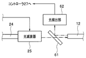
一方、本実施例の本体装置3は、照明用ファイバ12により伝送された照明光が照明用ファイバ12の光出射面及びレンズ14bの光出射面において反射することにより生じる戻り光を検出できるように構成されている。図20は、照明用ファイバから出射される戻り光を検出するための構成の一例を示す図である。
具体的には、本実施例の本体装置3は、例えば図20に示すように、光減衰器25から照明用ファイバ12の光入射面までの光路上に設けられた光学部材61と、光学部材61を経た戻り光が入射される光検出部62と、を有して構成されている。
光学部材61は、例えば、光減衰器25から出射される照明光の光軸に対して斜めになるように配置されたガラス板等により構成されており、光減衰器25から照明用ファイバ12の光入射面へ出射される照明光を透過させることが可能であるともに、照明用ファイバ12の光入射面から出射される戻り光を光検出部62側へ反射することが可能な機能を具備している。換言すると、光学部材61は、光減衰器25から照明用ファイバ12の光入射面へ出射される照明光と、照明用ファイバ12の光入射面から出射される戻り光と、を分離可能な機能を具備している。
光検出部62は、例えば、フォトダイオード等により構成されており、光学部材61を経て入射される戻り光の強度に応じた電気信号を生成してコントローラ27へ出力することが可能な機能を具備している。
本実施例のコントローラ27は、時刻T1から時刻T2までの期間内に検出ユニット23から出力されるR信号、G信号及びB信号に基づいて1フレーム分の画像を生成し、当該生成した画像をモニタ4に表示させることができるように構成されている。
また、本実施例のコントローラ27は、時刻T2から時刻T3までの期間内に検出ユニット23から出力されるR信号、G信号及びB信号に基づいて1フレーム分の画像を生成し、当該生成した画像をモニタ4に表示させることができるように構成されている。
一方、本実施例のコントローラ27は、光検出部62から出力される電気信号の信号レベルを、時刻T1から時刻T3までの期間にかけて順次検出した後、当該検出した信号レベルの変動が所定のパターンに該当しているか否かに係る判定を行うことができるように構成されている。
具体的には、コントローラ27は、例えば、図21に示すような信号レベルの変動パターンに係る情報をメモリ26から読み込んだ後、時刻T1から時刻T3までの期間に光検出部62から出力された電気信号の信号レベルの変動が、メモリ26から読み込んだ情報に含まれる信号レベルの変動パターンに該当するか否かを判定する。図21は、正常に照明光が出射されている場合に検出される信号レベルの変動パターンの、図9とは異なる例を示す図である。
図21に例示した信号レベルの変動パターンは、渦巻状の走査パターンに沿った軌跡で照明用ファイバ12が揺動されている場合に、照明用ファイバ12により伝送された照明光が照明用ファイバ12の光出射面及びレンズ14bの光出射面において反射することにより生じる戻り光の強度に応じて取得されるものである。
具体的には、図21に例示した信号レベルの変動パターンは、渦巻状の走査パターンに沿った軌跡で照明用ファイバ12が揺動されている場合において、光検出部62に入射される戻り光の強度が最大となる時刻T1において最大の信号レベルSL3となり、光検出部62に入射される戻り光の強度が徐々に減少する時刻T1からT2の間に信号レベルが非線形的に減少し、光検出部62に入射される戻り光の強度が最小となる時刻T2において最小の信号レベルSL4となり、光検出部62に入射される戻り光の強度が徐々に増加する時刻T2からT3の間に信号レベルが非線形的に増加し、光検出部62に入射される戻り光の強度が再び最大となる時刻T3において最大の信号レベルSL3となるようなパターンとして示される。
そして、コントローラ27は、時刻T1から時刻T3までの期間に光検出部62から出力された電気信号の信号レベルの変動が、メモリ26から読み込んだ情報に含まれる変動パターンに該当するとの判定結果を得た場合には、照明用ファイバ12から正常に照明光が出射されているものと推定し、照明用ファイバ12への照明光の供給を実施するための制御、及び、光検出部62から出力される電気信号の信号レベルのモニタリングをそれぞれ継続する。
また、時刻T1から時刻T3までの期間に光検出部62から出力された電気信号の信号レベルの変動が、メモリ26から読み込んだ情報に含まれる変動パターンに該当しないとの判定結果を得た場合には、照明用ファイバ12から出射される照明光に異常が発生しているものと推定し、照明用ファイバ12へ供給される照明光の光量を低下させるための制御を行う。
従って、本実施例によれば、照明用ファイバ12から出射される照明光に異常が発生した場合において、照明用ファイバ12へ供給される照明光の光量を、人体に対する安全性が確保されるような光量まで速やかに低下させることができ、その結果、被写体の走査の際に用いられる照明光が人体に対して悪影響を及ぼす危険性を低減することができる。
なお、本発明は、上述した各実施例に限定されるものではなく、発明の趣旨を逸脱しない範囲内において種々の変更や応用が可能であることは勿論である。
本出願は、2012年9月13日に日本国に出願された特願2012-201963号を優先権主張の基礎として出願するものであり、上記の開示内容は、本願明細書、請求の範囲、図面に引用されたものとする。
Claims (8)
- 光源から発せられた照明光を伝送して光出射面から出射するように構成された光伝送部と、
所定の走査パターンに応じた軌跡を描くように前記光伝送部の前記光出射面を含む端部を揺動させることが可能な駆動部と、
前記所定の走査パターンの所定の部分に前記光伝送部の前記端部が揺動される所定の期間内に前記光伝送部から出射された照明光を受光できるように構成された受光部と、
前記受光部により受光された照明光を検出し、当該検出した照明光の強度に応じた信号を出力するように構成された光検出部と、
前記光検出部から出力される信号における信号レベルの変動を前記所定の期間内及び前記所定の期間外において検出し、当該検出した前記所定の期間内の信号レベルの変動パターンが所定の変動パターンに該当するか否かを判定するように構成された判定部と、
前記所定の期間内の信号レベルの変動パターンが前記所定の変動パターンに該当しないとの判定結果が得られた場合において、前記光源から前記導光部へ供給される照明光の光量を0または所定値まで減少させるための制御を行うように構成された制御部と、
を有することを特徴とする内視鏡システム。 - 前記所定の走査パターンは、渦巻状の走査パターンであり、かつ、前記所定の期間は、前記渦巻状の走査パターンの最外周に前記光伝送部の前記端部が揺動される期間であることを特徴とする請求項1に記載の内視鏡システム。
- 前記受光部は、前記所定の期間内に前記光伝送部から出射された照明光と、前記所定の期間外に前記光伝送部から被写体へ出射された照明光の戻り光と、をそれぞれ受光するための受光用ファイバを有して構成されていることを特徴とする請求項2に記載の内視鏡システム。
- 前記受光部は、少なくとも前記受光用ファイバの光入射面を覆うように設けられているとともに、前記所定の期間内に前記光伝送部から出射された照明光を1回以上全反射することにより前記受光用ファイバに入射させることが可能な所定の屈折率分布を具備するように形成された導光板をさらに有することを特徴とする請求項3に記載の内視鏡システム。
- 前記受光部は、前記所定の期間内に前記光伝送部から出射された照明光を受光するためのモニタリング用ファイバと、前記所定の期間外に前記光伝送部から被写体へ出射された照明光の戻り光を受光するための受光用ファイバと、を有して構成されていることを特徴とする請求項2に記載の内視鏡システム。
- 前記受光部は、前記所定の期間内に前記光伝送部から出射された照明光を反射することにより前記モニタリング用ファイバに入射させることが可能な位置に配置された反射部材をさらに有することを特徴とする請求項5に記載の内視鏡システム。
- 前記所定の走査パターンは、ラスタ状の走査パターンであり、かつ、前記所定の期間は、前記ラスタ状の走査パターンの最外部に前記光伝送部の前記端部が揺動される期間であることを特徴とする請求項1に記載の内視鏡システム。
- 前記所定の走査パターンは、リサージュ状の走査パターンであり、かつ、前記所定の期間は、前記リサージュ状の走査パターンの最外部に前記光伝送部の前記端部が揺動される期間であることを特徴とする請求項1に記載の内視鏡システム。
Priority Applications (4)
| Application Number | Priority Date | Filing Date | Title |
|---|---|---|---|
| EP13837201.6A EP2759252A4 (en) | 2012-09-13 | 2013-05-13 | ENDOSCOPIC SYSTEM |
| JP2013558832A JP5490340B1 (ja) | 2012-09-13 | 2013-05-13 | 内視鏡システム |
| CN201380003500.4A CN103889309A (zh) | 2012-09-13 | 2013-05-13 | 内窥镜系统 |
| US14/161,823 US9113775B2 (en) | 2012-09-13 | 2014-01-23 | Endoscope system |
Applications Claiming Priority (2)
| Application Number | Priority Date | Filing Date | Title |
|---|---|---|---|
| JP2012201963 | 2012-09-13 | ||
| JP2012-201963 | 2012-09-13 |
Related Child Applications (1)
| Application Number | Title | Priority Date | Filing Date |
|---|---|---|---|
| US14/161,823 Continuation US9113775B2 (en) | 2012-09-13 | 2014-01-23 | Endoscope system |
Publications (1)
| Publication Number | Publication Date |
|---|---|
| WO2014041847A1 true WO2014041847A1 (ja) | 2014-03-20 |
Family
ID=50277983
Family Applications (1)
| Application Number | Title | Priority Date | Filing Date |
|---|---|---|---|
| PCT/JP2013/063302 WO2014041847A1 (ja) | 2012-09-13 | 2013-05-13 | 内視鏡システム |
Country Status (5)
| Country | Link |
|---|---|
| US (1) | US9113775B2 (ja) |
| EP (1) | EP2759252A4 (ja) |
| JP (1) | JP5490340B1 (ja) |
| CN (1) | CN103889309A (ja) |
| WO (1) | WO2014041847A1 (ja) |
Cited By (5)
| Publication number | Priority date | Publication date | Assignee | Title |
|---|---|---|---|---|
| WO2015194188A1 (ja) * | 2014-06-19 | 2015-12-23 | オリンパス株式会社 | 光走査型内視鏡装置 |
| JPWO2014188719A1 (ja) * | 2013-05-21 | 2017-02-23 | オリンパス株式会社 | 光走査ユニット、光走査型観察装置、および光ファイバ走査装置 |
| CN107072464A (zh) * | 2014-10-28 | 2017-08-18 | 奥林巴斯株式会社 | 光扫描型内窥镜装置 |
| JPWO2018225117A1 (ja) * | 2017-06-05 | 2020-04-02 | ギガフォトン株式会社 | レーザ装置、及びeuv光生成システム |
| WO2022085439A1 (ja) * | 2020-10-20 | 2022-04-28 | パナソニックIpマネジメント株式会社 | 内視鏡システム |
Families Citing this family (7)
| Publication number | Priority date | Publication date | Assignee | Title |
|---|---|---|---|---|
| JP5945638B2 (ja) * | 2014-05-28 | 2016-07-05 | オリンパス株式会社 | 光走査型観察装置及び光走査型観察装置の作動方法 |
| JP2018015110A (ja) * | 2016-07-26 | 2018-02-01 | オリンパス株式会社 | 内視鏡プロセッサ |
| CN109031656A (zh) * | 2018-07-27 | 2018-12-18 | 成都理想境界科技有限公司 | 一种光纤扫描投影装置、设备和检测方法 |
| GB2579801B (en) | 2018-12-13 | 2021-04-14 | Exalos Ag | Superluminescent diode module |
| GB2580956B (en) | 2019-01-31 | 2023-01-25 | Exalos Ag | Amplified Spontaneous Emission Semiconductor Source |
| CN110151108A (zh) * | 2019-05-10 | 2019-08-23 | 南京航空航天大学 | 内窥式激光散斑血流血氧成像系统 |
| CN112617721B (zh) * | 2020-12-31 | 2022-12-30 | 青岛海泰新光科技股份有限公司 | 一种内窥镜、内窥镜冷光源的连接反馈装置 |
Citations (5)
| Publication number | Priority date | Publication date | Assignee | Title |
|---|---|---|---|---|
| JP2001174744A (ja) * | 1999-10-06 | 2001-06-29 | Olympus Optical Co Ltd | 光走査プローブ装置 |
| JP2010131112A (ja) * | 2008-12-03 | 2010-06-17 | Hoya Corp | 内視鏡装置 |
| JP2010534862A (ja) * | 2007-07-25 | 2010-11-11 | ユニヴァーシティ オブ ワシントン | 圧電アクチュエータによる光ファイバの作動、及び圧電アクチュエータにより生成される電圧の検出 |
| JP2010268961A (ja) * | 2009-05-21 | 2010-12-02 | Hoya Corp | 医療用観察システム |
| JP2011019706A (ja) | 2009-07-15 | 2011-02-03 | Hoya Corp | 医療用観察システムおよびプロセッサ |
Family Cites Families (10)
| Publication number | Priority date | Publication date | Assignee | Title |
|---|---|---|---|---|
| US6845190B1 (en) * | 2000-11-27 | 2005-01-18 | University Of Washington | Control of an optical fiber scanner |
| WO2007084915A2 (en) * | 2006-01-17 | 2007-07-26 | University Of Washington | Scanning fiber-optic nonlinear optical imaging and spectroscopy endoscope |
| WO2008024101A1 (en) * | 2006-08-21 | 2008-02-28 | University Of Washington | Optical fiber scope with both non-resonant illumination and resonant collection/imaging for multiple modes of operation |
| US20080058629A1 (en) * | 2006-08-21 | 2008-03-06 | University Of Washington | Optical fiber scope with both non-resonant illumination and resonant collection/imaging for multiple modes of operation |
| US8305432B2 (en) * | 2007-01-10 | 2012-11-06 | University Of Washington | Scanning beam device calibration |
| US7583872B2 (en) * | 2007-04-05 | 2009-09-01 | University Of Washington | Compact scanning fiber device |
| US20080281159A1 (en) * | 2007-05-08 | 2008-11-13 | University Of Washington | Coordinating image acquisition among multiple endoscopes |
| US7791009B2 (en) * | 2007-11-27 | 2010-09-07 | University Of Washington | Eliminating illumination crosstalk while using multiple imaging devices with plural scanning devices, each coupled to an optical fiber |
| US20090137893A1 (en) * | 2007-11-27 | 2009-05-28 | University Of Washington | Adding imaging capability to distal tips of medical tools, catheters, and conduits |
| US20100137684A1 (en) | 2008-12-03 | 2010-06-03 | Hoya Corporation | Endoscope system with scanning function |
-
2013
- 2013-05-13 JP JP2013558832A patent/JP5490340B1/ja not_active Expired - Fee Related
- 2013-05-13 WO PCT/JP2013/063302 patent/WO2014041847A1/ja active Application Filing
- 2013-05-13 CN CN201380003500.4A patent/CN103889309A/zh active Pending
- 2013-05-13 EP EP13837201.6A patent/EP2759252A4/en not_active Withdrawn
-
2014
- 2014-01-23 US US14/161,823 patent/US9113775B2/en active Active
Patent Citations (5)
| Publication number | Priority date | Publication date | Assignee | Title |
|---|---|---|---|---|
| JP2001174744A (ja) * | 1999-10-06 | 2001-06-29 | Olympus Optical Co Ltd | 光走査プローブ装置 |
| JP2010534862A (ja) * | 2007-07-25 | 2010-11-11 | ユニヴァーシティ オブ ワシントン | 圧電アクチュエータによる光ファイバの作動、及び圧電アクチュエータにより生成される電圧の検出 |
| JP2010131112A (ja) * | 2008-12-03 | 2010-06-17 | Hoya Corp | 内視鏡装置 |
| JP2010268961A (ja) * | 2009-05-21 | 2010-12-02 | Hoya Corp | 医療用観察システム |
| JP2011019706A (ja) | 2009-07-15 | 2011-02-03 | Hoya Corp | 医療用観察システムおよびプロセッサ |
Non-Patent Citations (1)
| Title |
|---|
| See also references of EP2759252A4 |
Cited By (11)
| Publication number | Priority date | Publication date | Assignee | Title |
|---|---|---|---|---|
| JPWO2014188719A1 (ja) * | 2013-05-21 | 2017-02-23 | オリンパス株式会社 | 光走査ユニット、光走査型観察装置、および光ファイバ走査装置 |
| WO2015194188A1 (ja) * | 2014-06-19 | 2015-12-23 | オリンパス株式会社 | 光走査型内視鏡装置 |
| JP2016002406A (ja) * | 2014-06-19 | 2016-01-12 | オリンパス株式会社 | 光走査型内視鏡装置 |
| CN106455961A (zh) * | 2014-06-19 | 2017-02-22 | 奥林巴斯株式会社 | 光扫描型内窥镜装置 |
| US10609297B2 (en) | 2014-06-19 | 2020-03-31 | Olympus Corporation | Optical scanning endoscope apparatus with light amount detector |
| CN107072464A (zh) * | 2014-10-28 | 2017-08-18 | 奥林巴斯株式会社 | 光扫描型内窥镜装置 |
| CN107072464B (zh) * | 2014-10-28 | 2018-10-23 | 奥林巴斯株式会社 | 光扫描型内窥镜装置 |
| JPWO2018225117A1 (ja) * | 2017-06-05 | 2020-04-02 | ギガフォトン株式会社 | レーザ装置、及びeuv光生成システム |
| JP6990701B2 (ja) | 2017-06-05 | 2022-01-12 | ギガフォトン株式会社 | レーザ装置、及びeuv光生成システム |
| WO2022085439A1 (ja) * | 2020-10-20 | 2022-04-28 | パナソニックIpマネジメント株式会社 | 内視鏡システム |
| TWI829022B (zh) * | 2020-10-20 | 2024-01-11 | 日商松下知識產權經營股份有限公司 | 內視鏡系統 |
Also Published As
| Publication number | Publication date |
|---|---|
| US9113775B2 (en) | 2015-08-25 |
| JPWO2014041847A1 (ja) | 2016-08-18 |
| EP2759252A1 (en) | 2014-07-30 |
| JP5490340B1 (ja) | 2014-05-14 |
| US20140180012A1 (en) | 2014-06-26 |
| EP2759252A4 (en) | 2015-08-19 |
| CN103889309A (zh) | 2014-06-25 |
Similar Documents
| Publication | Publication Date | Title |
|---|---|---|
| JP5490340B1 (ja) | 内視鏡システム | |
| US9661998B2 (en) | Scanning endoscope with vibration absorbing member | |
| JP2010117442A (ja) | 光走査型内視鏡、光走査型内視鏡プロセッサ、および光走査型内視鏡装置 | |
| JP2011255015A (ja) | 内視鏡装置 | |
| JP2011217836A (ja) | 電子内視鏡装置 | |
| JP2010520778A (ja) | 側視型スコープ及びその画像化方法 | |
| JP2010268961A (ja) | 医療用観察システム | |
| EP2789289B1 (en) | Endoscope device and treatment device | |
| JP5617057B2 (ja) | 内視鏡装置 | |
| CN109068970B (zh) | 内窥镜装置 | |
| JP2011217835A (ja) | 内視鏡の形状検出装置 | |
| JP5784857B1 (ja) | 内視鏡の本体装置及び内視鏡システム | |
| JP5767411B1 (ja) | 内視鏡及び内視鏡システム | |
| JP6143953B2 (ja) | 走査型内視鏡システム | |
| JP2010042128A (ja) | 医療用プローブ、および医療用観察システム | |
| JP2016214459A (ja) | 走査型内視鏡 | |
| JP5830453B2 (ja) | 走査型内視鏡 | |
| JP2011101665A (ja) | 電子内視鏡システム | |
| JP5953452B1 (ja) | 走査型内視鏡 | |
| WO2016017199A1 (ja) | 光走査型観察システム | |
| JP2011041754A (ja) | 光走査型内視鏡 | |
| JP2015089440A (ja) | 走査型内視鏡システム | |
| JP2017077284A (ja) | 走査型内視鏡 | |
| JP2010113309A (ja) | 光走査型内視鏡装置、光走査型内視鏡、および光走査型内視鏡プロセッサ |
Legal Events
| Date | Code | Title | Description |
|---|---|---|---|
| ENP | Entry into the national phase |
Ref document number: 2013558832 Country of ref document: JP Kind code of ref document: A |
|
| REEP | Request for entry into the european phase |
Ref document number: 2013837201 Country of ref document: EP |
|
| 121 | Ep: the epo has been informed by wipo that ep was designated in this application |
Ref document number: 13837201 Country of ref document: EP Kind code of ref document: A1 |
|
| NENP | Non-entry into the national phase |
Ref country code: DE |