WO2013141346A1 - パクリタキセルモノグリコシド及び/又はドタキセルモノグリコシドを内包するリポソームの製造方法 - Google Patents
パクリタキセルモノグリコシド及び/又はドタキセルモノグリコシドを内包するリポソームの製造方法 Download PDFInfo
- Publication number
- WO2013141346A1 WO2013141346A1 PCT/JP2013/058242 JP2013058242W WO2013141346A1 WO 2013141346 A1 WO2013141346 A1 WO 2013141346A1 JP 2013058242 W JP2013058242 W JP 2013058242W WO 2013141346 A1 WO2013141346 A1 WO 2013141346A1
- Authority
- WO
- WIPO (PCT)
- Prior art keywords
- cells
- monoglycoside
- liposome
- antibody
- paclitaxel
- Prior art date
Links
- 0 CCC[C@]([C@](C[C@@](*)(C1)C2)[C@@](C*)C(*)[C@@]3(C)[C@@]2[C@](C*)(CC2)[C@]2C[C@@]3OC(C*)=*)[C@@]1O*(ICCC)=O Chemical compound CCC[C@]([C@](C[C@@](*)(C1)C2)[C@@](C*)C(*)[C@@]3(C)[C@@]2[C@](C*)(CC2)[C@]2C[C@@]3OC(C*)=*)[C@@]1O*(ICCC)=O 0.000 description 2
Images


Classifications
-
- A—HUMAN NECESSITIES
- A61—MEDICAL OR VETERINARY SCIENCE; HYGIENE
- A61K—PREPARATIONS FOR MEDICAL, DENTAL OR TOILETRY PURPOSES
- A61K31/00—Medicinal preparations containing organic active ingredients
- A61K31/33—Heterocyclic compounds
- A61K31/335—Heterocyclic compounds having oxygen as the only ring hetero atom, e.g. fungichromin
- A61K31/337—Heterocyclic compounds having oxygen as the only ring hetero atom, e.g. fungichromin having four-membered rings, e.g. taxol
-
- A—HUMAN NECESSITIES
- A61—MEDICAL OR VETERINARY SCIENCE; HYGIENE
- A61K—PREPARATIONS FOR MEDICAL, DENTAL OR TOILETRY PURPOSES
- A61K31/00—Medicinal preparations containing organic active ingredients
- A61K31/70—Carbohydrates; Sugars; Derivatives thereof
- A61K31/7042—Compounds having saccharide radicals and heterocyclic rings
- A61K31/7048—Compounds having saccharide radicals and heterocyclic rings having oxygen as a ring hetero atom, e.g. leucoglucosan, hesperidin, erythromycin, nystatin, digitoxin or digoxin
-
- A—HUMAN NECESSITIES
- A61—MEDICAL OR VETERINARY SCIENCE; HYGIENE
- A61K—PREPARATIONS FOR MEDICAL, DENTAL OR TOILETRY PURPOSES
- A61K47/00—Medicinal preparations characterised by the non-active ingredients used, e.g. carriers or inert additives; Targeting or modifying agents chemically bound to the active ingredient
- A61K47/50—Medicinal preparations characterised by the non-active ingredients used, e.g. carriers or inert additives; Targeting or modifying agents chemically bound to the active ingredient the non-active ingredient being chemically bound to the active ingredient, e.g. polymer-drug conjugates
- A61K47/69—Medicinal preparations characterised by the non-active ingredients used, e.g. carriers or inert additives; Targeting or modifying agents chemically bound to the active ingredient the non-active ingredient being chemically bound to the active ingredient, e.g. polymer-drug conjugates the conjugate being characterised by physical or galenical forms, e.g. emulsion, particle, inclusion complex, stent or kit
- A61K47/6905—Medicinal preparations characterised by the non-active ingredients used, e.g. carriers or inert additives; Targeting or modifying agents chemically bound to the active ingredient the non-active ingredient being chemically bound to the active ingredient, e.g. polymer-drug conjugates the conjugate being characterised by physical or galenical forms, e.g. emulsion, particle, inclusion complex, stent or kit the form being a colloid or an emulsion
- A61K47/6911—Medicinal preparations characterised by the non-active ingredients used, e.g. carriers or inert additives; Targeting or modifying agents chemically bound to the active ingredient the non-active ingredient being chemically bound to the active ingredient, e.g. polymer-drug conjugates the conjugate being characterised by physical or galenical forms, e.g. emulsion, particle, inclusion complex, stent or kit the form being a colloid or an emulsion the form being a liposome
- A61K47/6913—Medicinal preparations characterised by the non-active ingredients used, e.g. carriers or inert additives; Targeting or modifying agents chemically bound to the active ingredient the non-active ingredient being chemically bound to the active ingredient, e.g. polymer-drug conjugates the conjugate being characterised by physical or galenical forms, e.g. emulsion, particle, inclusion complex, stent or kit the form being a colloid or an emulsion the form being a liposome the liposome being modified on its surface by an antibody
-
- A—HUMAN NECESSITIES
- A61—MEDICAL OR VETERINARY SCIENCE; HYGIENE
- A61K—PREPARATIONS FOR MEDICAL, DENTAL OR TOILETRY PURPOSES
- A61K9/00—Medicinal preparations characterised by special physical form
- A61K9/10—Dispersions; Emulsions
- A61K9/127—Liposomes
-
- A—HUMAN NECESSITIES
- A61—MEDICAL OR VETERINARY SCIENCE; HYGIENE
- A61P—SPECIFIC THERAPEUTIC ACTIVITY OF CHEMICAL COMPOUNDS OR MEDICINAL PREPARATIONS
- A61P35/00—Antineoplastic agents
-
- C—CHEMISTRY; METALLURGY
- C07—ORGANIC CHEMISTRY
- C07K—PEPTIDES
- C07K16/00—Immunoglobulins [IGs], e.g. monoclonal or polyclonal antibodies
- C07K16/18—Immunoglobulins [IGs], e.g. monoclonal or polyclonal antibodies against material from animals or humans
- C07K16/32—Immunoglobulins [IGs], e.g. monoclonal or polyclonal antibodies against material from animals or humans against translation products of oncogenes
-
- A—HUMAN NECESSITIES
- A61—MEDICAL OR VETERINARY SCIENCE; HYGIENE
- A61K—PREPARATIONS FOR MEDICAL, DENTAL OR TOILETRY PURPOSES
- A61K39/00—Medicinal preparations containing antigens or antibodies
- A61K2039/505—Medicinal preparations containing antigens or antibodies comprising antibodies
-
- C—CHEMISTRY; METALLURGY
- C07—ORGANIC CHEMISTRY
- C07K—PEPTIDES
- C07K2317/00—Immunoglobulins specific features
- C07K2317/20—Immunoglobulins specific features characterized by taxonomic origin
- C07K2317/24—Immunoglobulins specific features characterized by taxonomic origin containing regions, domains or residues from different species, e.g. chimeric, humanized or veneered
-
- C—CHEMISTRY; METALLURGY
- C07—ORGANIC CHEMISTRY
- C07K—PEPTIDES
- C07K2317/00—Immunoglobulins specific features
- C07K2317/70—Immunoglobulins specific features characterized by effect upon binding to a cell or to an antigen
- C07K2317/73—Inducing cell death, e.g. apoptosis, necrosis or inhibition of cell proliferation
Definitions
- the present invention relates to a method for producing a liposome encapsulating paclitaxel monoglycoside and / or dotaxel monoglycoside.
- anticancer agents have a mechanism that suppresses the division of cancer cells whose cell growth control is broken. Such anti-cancer agents are useful because they exhibit a very effective anti-cancer effect, but they exhibit cytostatic effects even on normal cells, so there are many side effects when used. Often hinders.
- anti-cancer agents have been required to have DDS technology that delivers drugs specifically to cancer cells without acting on normal cells.
- various anticancer agents have evolved in combination with DDS technology.
- a technique using a liposome composed of a lipid bilayer membrane is suitably used.
- Patent Document 1 For example, a technique using such a liposome and a platinum complex anticancer agent such as oxaliplatin, cisplatin or carboplatin has been developed (Patent Document 1). Moreover, when introducing such an anticancer agent using the remote loading method, it is limited to an anticancer agent having weak basicity, and a solubility gradient was used as a method for solving this. Although a remote loading method has been developed, it can be applied only to a drug having high water solubility, and its introduction efficiency is very low (Patent Document 2).
- paclitaxel, dotaxel, etc. that are applicable to breast cancer, cervical cancer, etc., are hardly soluble in water and are troublesome and dangerous in that they are administered after dissolving in a solvent such as alcohol. There was a problem in that.
- paclitaxel derivatives have been prepared for the purpose of improving the solubility of paclitaxel, dotaxel and the like in water.
- monosaccharides such as glucose, galactose, mannose, and xylose are added to paclitaxel. Additional technology has been developed.
- paclitaxel derivatives such as paclitaxel monoglycoside and dotaxel monoglycoside cannot provide sufficient water solubility, and no knowledge has been obtained about DDS preparations combining these with liposomes.
- the present inventor has conducted extensive research. As a result, in order to efficiently introduce paclitaxel derivatives such as paclitaxel monoglycoside and dotaxel monoglycoside into the liposome, the paclitaxel derivative and the lipid constituting the liposome are used. It has been found that mixing may be performed in the presence of a specific proportion of solvent components.
- paclitaxel monoglycoside, dotaxel monoglycoside and the like can be encapsulated in the liposome with high efficiency by previously containing a mixed solvent for dissolving the paclitaxel derivative in the liposome.
- liposomes encapsulating paclitaxel, dotaxel monoglycoside and the like obtained by such a method exhibit excellent effects as DDS.
- the present invention has been completed on the basis of such knowledge, and widely includes the following aspects.
- Item 1 A method for producing a liposome encapsulating paclitaxel monoglycoside and / or dotaxel monoglycoside, wherein paclitaxel monoglycoside and / or dotaxel monoglycoside, polyoxyethylene ester derivative, lower alcohol, lipid constituting the liposome, and Including a step of subjecting a mixture containing a buffer or water to a liposome-forming treatment, The paclitaxel monoglycoside and / or dotaxel monoglycoside is not less than 1500 and less than 3000% by weight based on the total volume of the mixture, Per 1 part by volume of the buffer or water, Containing 0.1 to 0.2 parts by volume of the polyoxyethylene ester derivative, Containing 0.1 to 0.2 parts by volume of the lower alcohol, A production method comprising encapsulating 0.1 to 2.5 parts by weight of the paclitaxel monoglycoside and / or dotaxel monoglycoside per 1 part by
- Liposome formation treatment can be performed by thin film static hydration method, freeze drying method, droplet method, AC method, ultrasonic method, reverse phase evaporation method, lipid dissolution method, spray drying method, CO 2 / H 2 O emulsion Item 2.
- the production method according to Item 1 which is either a production method or a production method using a microchannel.
- Glycoside is from glucoside, galactoside, mannoside, xyloside, fructoside, rhamnoside, arabinoside, alloside, altroside, idid, N-acetylglucosaminide, N-acetylgalactosaminide, taloside, glucuronoside, glucosaminide, galactosaminide, and fucoside Item 3.
- the method according to Item 1 or 2 wherein the method is one glycoside selected from the group consisting of:
- Item 4 The method according to any one of Items 1 to 3, wherein the paclitaxel monoglycoside is 7- ⁇ -glucosyloxyacetyl paclitaxel.
- Item 5 The method according to any one of Items 1 to 4, wherein the polyoxyethylene ester derivative is a polyoxyethylene castor oil ester.
- Item 6 The method according to any one of Items 1 to 5, wherein the polyoxyethylene castor oil ester is Cremophor (registered trademark) EL.
- Item 7 A liposome preparation containing an antibody that specifically encapsulates a solution of paclitaxel monoglycoside and / or dotaxel monoglycoside and that specifically recognizes cancer cells.
- Item 8 A solution in which paclitaxel and / or dotaxel monoglycoside is dissolved in a mixed solvent containing a polyoxyethylene ester derivative, a lower alcohol, and a buffer solution or water.
- Glycoside is from glucoside, galactoside, mannoside, xyloside, fructoside, rhamnoside, arabinoside, alloside, altroside, idid, N-acetylglucosaminide, N-acetylgalactosaminide, taloside, glucuronoside, glucosaminide, galactosaminide, and fucoside Item 9.
- the liposome preparation according to Item 7 or 8 which is one type of glycoside selected from the group consisting of:
- Item 10 The liposome preparation according to any one of Items 7 to 9, wherein the paclitaxel monoglycoside is 7- ⁇ -glucosyloxyacetyl paclitaxel.
- Item 11 The liposome preparation according to any one of Items 7 to 10, wherein the polyoxyethylene ester derivative is a polyoxyethylene castor oil ester.
- Item 12 The liposome preparation according to any one of Items 7 to 11, wherein the polyoxyethylene castor oil ester is Cremophor (registered trademark) EL.
- Item 13 The liposome preparation according to any one of Items 7 to 12, wherein the liposome contains DPPC and cholesterol in a substance amount ratio of 3: 0.5 to 3, respectively.
- Item 14 The liposome preparation according to any one of Items 7 to 15, wherein the molar amount of paclitaxel monoglycoside relative to 1 molar amount of the total lipid of the liposome is 1.0 to 15.5 ⁇ 10 -2 .
- Item 17 The liposome preparation according to any one of Items 7 to 16, wherein the cancer cell is a breast cancer cell.
- Item 18 The liposome preparation according to any one of Items 7 to 17, wherein the antibody is an antibody that specifically binds to the HER2 protein.
- the present invention includes inventions of the following modes.
- Item I A method for producing a liposome having an antibody encapsulating paclitaxel monoglycoside and / or dotaxel monoglycoside and specifically recognizing a cancer cell, comprising a polyoxyethylene ester derivative, a lower alcohol, and a buffer or water And a step of contacting a solution in which paclitaxel monoglycoside and / or dotaxel monoglycoside is dissolved in a buffer solution or water containing alkylene glycol.
- Item II The production method according to Item I, wherein the solution in which paclitaxel monoglycoside and / or dotaxel monoglycoside is dissolved is a buffer solution containing alkylene glycol or a solution in water.
- Item III glycoside is from glucoside, galactoside, mannoside, xyloside, fructoside, rhamnoside, arabinoside, alloside, altroside, idid, N-acetylglucosaminide, N-acetylgalactosaminide, taloside, glucuronoside, glucosaminide, galactosaminide, and fucoside Item 3.
- Item IV The method according to any one of Items I to III, wherein the paclitaxel monoglycoside is 7- ⁇ -glucosyloxyacetyl paclitaxel.
- Item V The production method according to any one of Items I to IV, wherein the polyoxyethylene ester derivative is a polyoxyethylene castor oil ester.
- Item VI The method according to any one of Items I to V, wherein the polyoxyethylene castor oil ester is Cremophor (registered trademark) EL.
- Item VII The production method according to any one of Items I to VI, wherein the liposome contains DPPC and cholesterol in a substance amount ratio of 3: 0.5 to 3, respectively.
- Item VIII The production method according to any one of Items I to VII, wherein the contact time is 5 to 60 minutes.
- Item IX The production method according to any one of Items I to VIII, wherein the cancer cells are breast cancer cells.
- Item X The production method according to any one of Items I to IX, wherein the antibody is an antibody that specifically binds to the HER2 protein.
- paclitaxel monoglycoside and / or dotaxel monoglycoside can be efficiently encapsulated in liposomes.
- the liposome encapsulating paclitaxel monoglycoside and / or dotaxel monoglycoside obtained by the method of the present invention can bind an antibody that specifically recognizes cancer cells, and the present invention to which such an antibody is bound.
- the liposome obtained by this production method is very useful as DDS.
- the acute toxicity test result by Test Example 4. The vertical axis of the graph indicates the survival rate, and the horizontal axis indicates the number of administrations. 6 is a fluorescent photographic image showing accumulation of liposomes encapsulating paclitaxel monoglycoside obtained by the production method of the present invention according to Test Example 5 and carrying antibodies that recognize breast cancer cells in breast cancer cells. The bar in the figure indicates 10 ⁇ m.
- the vertical axis of the graph indicates the survival rate, and the horizontal axis indicates the number of days after administration.
- the vertical axis of the graph represents body weight, and the horizontal axis represents the number of days after administration. Results of in vivo delivery experiment according to Test Example 10.
- FIG. 6 is a graph of the imaging results shown in FIG. (A) Results of tumor tissue, (B) is a result of liver, and (C) is a result of quantifying the degree of accumulation in tumor tissue with respect to the liver as a ratio. For the digitization, tumors and liver sites were selected according to threshold values, and the total fluorescence intensity was measured.
- the in-vivo anticancer activity experiment result by the test example 11.
- FIG. The vertical axis of the graph indicates tumor volume, and the horizontal axis indicates the number of days after administration.
- the in-vivo anticancer activity experiment result by the test example 11.
- FIG. The vertical axis of the graph represents body weight, and the horizontal axis represents the number of days after administration.
- the in-vivo anticancer activity experiment result by the test example 11.
- FIG. The vertical axis of the graph indicates the survival rate, and the horizontal axis indicates the number of days after administration.
- the arrow in the figure indicates a tumor.
- a method for producing a liposome encapsulating paclitaxel monoglycoside and / or dotaxel monoglycoside which is one embodiment of the method for producing liposome of the present invention (hereinafter sometimes referred to as “the production method of the first embodiment”), And a step of subjecting the mixture containing paclitaxel monoglycoside and / or dotaxel monoglycoside, polyoxyethylene derivative, lower alcohol, lipid constituting the liposome, and buffer solution or water to a liposome forming treatment.
- a solution containing paclitaxel monoglycoside and / or dotaxel monoglycoside and a polyoxyethylene derivative, a lower alcohol, and a buffer or water is prepared in advance, and this is mixed with the lipid constituting the liposome, and then It may be a method used for liposome formation treatment.
- a solution containing paclitaxel monoglycoside and / or dotaxel monoglycoside, a polyoxyethylene derivative, and a lower alcohol is prepared and mixed with a mixture comprising a lipid constituting a liposome and a buffer or water. Then, you may use for a liposome formation process.
- the order of mixing paclitaxel monoglycoside and / or dotaxel monoglycoside and lipid is not limited, but the total amount of paclitaxel monoglycoside and / or dotaxel monoglycoside, polyoxyethylene derivative, lower alcohol, and buffer or water is not limited.
- the amount of paclitaxel monoglycoside and / or dotaxel monoglycoside to be used is about 1500 to less than 3000% by weight. Preferably it is about 1700 or more and less than 2500 weight% or less, More preferably, it is about 1800 or more and 2200 weight% or less.
- the amount of the polyoxyethylene derivative used is about 0.1 to 0.2 parts by volume, preferably about 0.12 to 0.19 parts by volume, more preferably 0.13 to 0.1 parts by volume with respect to 1 part by volume of the buffer solution or water. About 0.18 capacity part.
- the amount of lower ethanol used is about 0.1 to 0.2 parts by volume, preferably about 0.12 to 0.19 parts by volume, more preferably 0.13 to 0. About 18 capacity parts.
- Capacity part used in this specification is a numerical value obtained by measurement under normal pressure and room temperature.
- the polyoxyethylene ester derivative is not particularly limited.
- examples thereof include siloxane copolymers, polyoxyethylene octylphenyl ether, polyoxyethylene stearyl ether, polyoxyethylene stearamide, polyoxyethylene cetyl ether, polyoxyethylene polyoxypropylene glycol, polyoxyethylene castor oil ester and the like.
- polyoxyethylene castor oil ester is preferable, and polyoxyalkylene (C24) castor oil fatty acid ester (Cremophor (registered trademark) EL) is more preferable.
- the lower alcohol is not particularly limited, but it may be usually an alcohol having 1 to 4 carbon atoms.
- the buffer solution is not particularly limited, and examples thereof include PBS, MES, ADA, PIPES, ACES, collamine hydrochloride, BES, TES, HEPES, citric acid, boric acid, and tartaric acid.
- Examples of the components of the liposome include phospholipids, cholesterols, fatty acids and the like.
- Specific examples of phospholipids include phosphatidylcholine, phosphatidylserine, phosphatidylglycerol, phosphatidylinositol, phosphatidylethanolamine, phosphatidic acid, cardiolipin, sphingomyelin, egg yolk lecithin, soybean lecithin, lysolecithin, and hydrogenated ones in a conventional manner.
- Natural phospholipids distearoylphosphatidylcholine, dipalmitoylphosphatidylcholine (DPPC), dipalmitoylphosphatidylethanolamine (DPPE), dithiopyridine-dipalmitoylphosphatidylethanolamine (DTP-DPPE), dipalmitoylphosphatidylglycerol (DPPG), dipalmitoylphosphatidylserine (DPPS), eleostearoylphosphatidyl Phosphorus, Jer osteoprotegerin aroyl phosphatidyl ethanolamine, synthetic phospholipids such as Jer osteoprotegerin aroyl phosphatidylserine and the like.
- DPPC dipalmitoylphosphatidylcholine
- DPPE dipalmitoylphosphatidylethanolamine
- DTP-DPPE dipalmitoylphosphatidylglycerol
- DPPS dipalmitoylphosphat
- phospholipids may be appropriately modified. Specific modifications are not particularly limited, but examples include modifications with polyalkylene glycols such as polyethylene glycol and polypropylene glycol. These phospholipids may be used in appropriate combination.
- cholesterols examples include cholesterol and phytosterol.
- fatty acids examples include oleic acid, palmitooleic acid, linoleic acid, and fatty acid mixtures containing these unsaturated fatty acids.
- liposomes containing unsaturated fatty acids with small side chains are effective in preparing liposomes with a small particle size because of the curvature.
- the liposome obtained by the production method of the first aspect of the present invention encapsulates paclitaxel monoglycoside and / or dotaxel monoglycoside in the liposome.
- the term “encapsulation” means that paclitaxel monoglycoside and / or dotaxel monoglycoside may be completely contained inside the liposome, but the lipid bilayer constituting the liposome contains paclitaxel monoglycoside. And / or a state in which a dotaxel monoglycoside molecule penetrates also means “encapsulation”.
- the term “enclosure” may be used in the same meaning as “enclosure”.
- the liposome obtained by the production method of the first aspect of the present invention contains about 0.1 to 2.5 parts by weight of paclitaxel monoglycoside and / or dotaxel monoglycoside per 1 part by weight of total lipid.
- the amount is more preferably 1.3 to 2.4 parts by weight, and more preferably about 1.6 to 2.3 parts by weight.
- Paclitaxel monoglycoside and dotaxel monoglycoside are obtained by adding a monosaccharide to paclitaxel and dotaxel, respectively.
- the position at which a monosaccharide is added to paclitaxel is not particularly limited.
- the monosaccharide is added to the 10th or 7th position of the taxane ring of paclitaxel. It is known that a sugar derivative at the 7-position of paclitaxel exists in nature, and is preferably added to the 7-position from the viewpoint of bond stability.
- the position at which a monosaccharide is added to dotaxel is not particularly limited.
- a monosaccharide is added to the 10th or 7th position of the taxane ring of dotaxel.
- Specific monosaccharides are not particularly limited.
- glucose, galactose, mannose, xylose, fructose, rhamnose, arabinose, allose, altrose, idose, N-acetylglucosamine, N-acetylgalactosamine, talose, glucuron Examples include acid, glucosamine, galactosamine, and fucose.
- glucose, galactose, mannose, xylose and the like are preferably used from the viewpoint of not excessively impairing the anticancer effect of paclitaxel and dotaxel.
- glycoside in paclitaxel monoglycoside and dotaxel monoglycoside glucoside, galactoside, mannoside, xyloside, fructoside, rhamnoside, arabinoside, alloside, altroside, idside, N-acetylglucosaminide, N-acetylgalactoside
- glucoside, galactoside, mannoside, xyloside, fructoside, rhamnoside, arabinoside, alloside, altroside, idside, N-acetylglucosaminide, N-acetylgalactoside examples include saminide, taloside, glucuronoside, glucosaminide, galactosaminide, and fucoside.
- the above-mentioned paclitaxel monoglycoside and dotaxel monoglycoside may have a further group between paclitaxel and dotaxel and a monosaccharide, and examples thereof include an alkanoyl group having 1 to 8 carbon atoms.
- the paclitaxel monoglycoside and dotaxel monoglycoside described above are alkanoyl having 1 to 8 carbon atoms in which the hydrogen atom of the hydroxyl group at the 7-position or 10-position of the taxane ring of paclitaxel and dotaxel is substituted with the above-mentioned monosaccharide. It may be substituted with an oxy group.
- the paclitaxel before addition of the monosaccharide may be a known paclitaxel derivative. Further, paclitaxel derivatives described in JP-A-8-73449, JP-A-7-233159 and the like can also be mentioned.
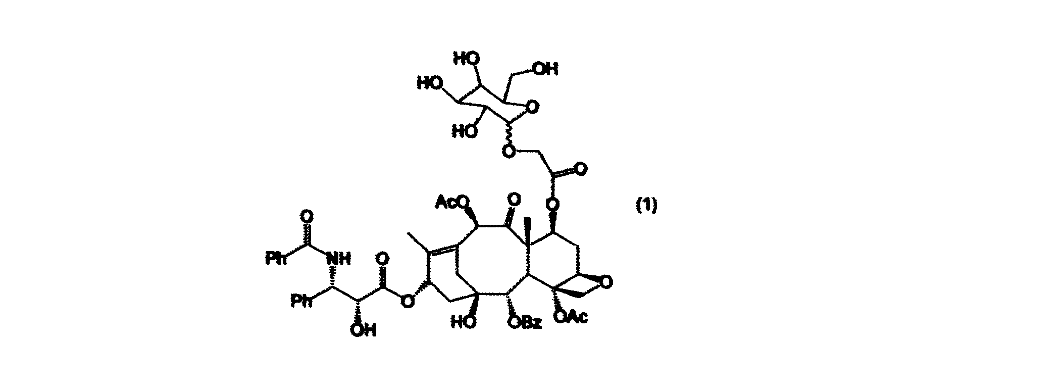
- the most preferable paclitaxel monoglycoside is 7- ⁇ -glucosyloxyacetyl paclitaxel represented by the following formula (1).
- paclitaxel monoglycoside can be produced using a known method, for example, with reference to the method described in Non-Patent Document 1.
- a desired monosaccharide to be added to paclitaxel is acetylated in advance, and as described above, a sugar chain is imparted by esterification at the 7-position or 10-position of the taxane ring of paclitaxel.
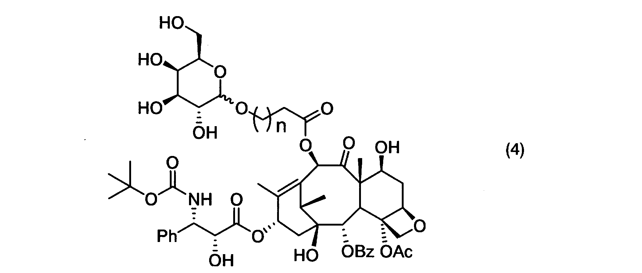
- dotaxel monoglycoside represented by the following formulas (2) to (4).
- the ⁇ -glucosyl group in the formula may be a ⁇ -glucosyl group, an ⁇ -galactosyl group, a ⁇ -galactosyl group, or an ⁇ -mannosyl group.
- n 2, 3, 4, 6, or 8.
- dotaxel monoglycoside can be produced using a known method, for example, HETEROCYCLES, 2001, Vol 54, N0.2, 561-566. Biol. Pharm. Bull. 2008, 31 (6) 1155-1158 (2008), etc.
- the type of liposome produced by the production method of the first aspect of the present invention is determined by the constituent lipids of the above-mentioned liposome, and any of anionic liposomes, cationic liposomes, and amphoteric liposomes can be used. Good.
- cationic liposomes are preferable to other types of liposomes from the viewpoint of efficient substance introduction into cells, but from the viewpoint of causing non-specific adsorption to cells, paclitaxel monolith obtained by the method of the present invention.
- Liposomes containing glycosides and / or dotaxel monoglycosides are not preferred because they are inferior in exhibiting the DDS effect on specific cells.
- the liposome formation treatment method in the production method of the first aspect of the present invention can be produced using a known method, and is not particularly limited.
- the above-described phospholipid, cholesterol and the like are dissolved in a suitable organic solvent, and placed in a suitable container under reduced pressure.
- the solvent is distilled off to form a phospholipid film on the inner surface of the container, and a solution containing the above-mentioned paclitaxel monoglycoside and / or dotaxel monoglycoside, preferably a buffer solution thereof, is added and stirred to form paclitaxel monoglycoside.
- a liposome encapsulating dotaxel monoglycoside can be obtained.
- Such liposomes may be lyophilized and stored.
- the particle diameter of the liposome obtained by the production method of the first aspect of the present invention is not particularly limited, but the liposome obtained by the production method of the present invention is paclitaxel monoglycoside and / or dotaxel having anticancer activity. From the viewpoint of being suitably used as a liposome preparation encapsulating monoglycoside, it is usually about 50 to 300 nm. A liposome preparation satisfying such a numerical range is preferable because it can pass through capillaries, particularly capillaries based on angiogenesis induced by cancer cells, when administered in vivo. Liposomes having a particle diameter of 50 nm or more are preferable because leakage to cells hardly occurs. Liposomes having a particle size of 300 nm or less are preferable because they are less likely to be eaten by blood leukocytes (macrophages) after being administered in vivo.
- macrophages blood leukocytes
- the liposome obtained by the production method of the first aspect of the present invention can be adjusted to a predetermined particle size. Specifically, this can be achieved by changing various conditions in advance during the liposome formation treatment, but can also be achieved by passing through a filter with a controlled pore size.
- a method for adjusting the particle size of the liposome preparation by passing through a filter a method using an extruder or the like can be mentioned.
- the liposome obtained by the production method of the first aspect of the present invention may have an antibody that recognizes cancer cells.
- an antibody may be a monoclonal antibody or a polyclonal antibody, and the animal species from which the antibody is derived is not particularly limited.
- antigen recognition such as single chain antibody, domino antibody, short chain antibody, multivalent antibody, bispecific antibody, Fab antibody, F (ab ′) 2 antibody, chimeric antibody, humanized antibody, etc.
- it may be an antibody having any molecular structure, and is not limited to an antibody having an immunoglobulin structure typified by IgG.
- Such an antibody is bound to the lipid bilayer of the liposome obtained by the above-described production method of the present invention, and the binding mode is not particularly limited, and is appropriately selected according to the structure of the antibody to be used. Thus, a liposome having a desired antibody can be obtained.
- the antibody may be bound to the liposome using SPDP (N-succinimidyl-3- (2-pyridyldithio) propionate).
- SPDP N-succinimidyl-3- (2-pyridyldithio) propionate
- the antibody may be supported by binding the antibody to the liposome encapsulating paclitaxel monoglycoside and / or dotaxel monoglycoside by the production method of the first aspect of the present invention described above.
- the antibody may be bound in advance to the lipid. In order to allow more antibodies to be bound to be presented on the surface of the liposome, it is preferable to bind the antibody to the liposome in which paclitaxel monoglycoside and / or dotaxel monoglycoside is encapsulated by the production method of the present invention. .
- the antibody of the liposome of the present invention recognizes cancer cells.
- cancer cells are not particularly limited, but lung cancer cells, non-small cell lung cancers, breast cancer cells, esophageal cancers, gastric cancer cells, liver cancer cells, pancreatic cancer cells, colon cancer cells, ovarian cancers Cervical cancer cells, endometrial cancer cells, prostate cancer cells, head and neck cancer cells (including oral cancer cells, pharyngeal cancer cells, laryngeal cancer cells, nasal cavity or sinus cancer cells, salivary gland cancer cells, thyroid cancer cells, etc. ) And the like.
- non-small cell lung cancer cells breast cancer cells, esophageal cancer cells, gastric cancer cells, endometrial cancer cells, ovarian cancer cells, prostate cancer cells and the like are preferable based on clinical application findings of paclitaxel.
- the above-mentioned antibody recognizing cancer cells is a protein existing on the surface of cancer cells (for example, CD protein forming CD protein group such as CD44, CD133, etc .; growth factor or receptor for hormone; transmembrane or membrane A protein having a binding domain, etc.), an antibody that specifically recognizes biomolecules such as peptides and sugar chains.
- CD protein forming CD protein group such as CD44, CD133, etc .
- growth factor or receptor for hormone transmembrane or membrane A protein having a binding domain, etc.
- Such an antibody is not particularly limited as long as it is known to be expressed on the surface layer of each cancer cell.
- antibodies that recognize breast cancer cells include breast cancer cells collected from patients suffering from breast cancer and Hs274.
- T cells Hs606 cells, BT-20 cells, UACC-812 cells, HCC1954 cells, Hs574.
- antibodies that recognize lung cancer cells include lung cancer cells collected from patients suffering from lung cancer, and Hs229. T cells, NCI-H2066 cells, NCI-H2286 cells, NCI-H1703 cells, Hs573. T cell, A549 cell, A427 cell, N417 cell, NCI-H596 cell, SW1573 cell, NCI-H835U cell, MC11 cell, NCI-H727 cell, NCI-H720 cell, NCI-H810 cell, NCI-H292 cell, NCI- H2126 cell, H69 cell, NCI-H1688 cell, NCI-H1417 cell, NCI-H1672 cell, NCI-H1836 cell, DMS79 cell, DMS53 cell, DMS114 cell, SW1271 cell, NCI-H2227 cell, NCI-H1963 cell, SHP- Antibodies that recognize biomolecules such as proteins, peptides, sugar chains, etc.
- H69 cells present on the surface layer of 77 cells, H69 cells, H69AR cells, NCI-H2170 cells, NCI-H520 cells, SW900 cells and the like may be used.
- anti-HER2 antibody, anti-EGFR antibody, anti-CEA antibody and the like can be mentioned.
- antibodies that recognize non-small cell lung cancer cells include non-small cell lung cancer cells collected from patients suffering from non-small cell lung cancer, NCI-H23 cells that are non-small cell lung cancer tissue-derived cells, NCI- H522 cells, NCI-H1435 cells, NCI-H1563 cells, NCI-H1651 cells, NCI-H1734 cells, NCI-H1793 cells, NCI-H1838 cells, NCI-H1975 cells, NCI-H2073 cells, NCI-H2085 cells, NCI- Using antibodies that recognize biomolecules such as proteins, peptides, sugar chains, etc.
- H2228 cells present on the surface of H2228 cells, NCI-H2342 cells, NCI-H2347 cells, NCI-H2135 cells, NCI-H2172 cells, NCI-H2444 cells, etc. Good.
- Specific examples include anti-HER2 antibody and anti-EGFR antibody.
- antibodies that recognize esophageal cancer cells include esophageal cancer cells collected from patients suffering from esophageal cancer, esophageal cancer tissue-derived cells, SGF-3 cells, EC-YO cells, TE-1 cells, TE-2 cells. , TE-3 cells, TE-4 cells, TE-5 cells, TE-6 cells, TE-7 cells, TE-8 cells, TE-9 cells, TE-10 cells, TE-11 cells, TE-12 cells.
- Antibodies that recognize biomolecules such as proteins, peptides, sugar chains, etc. present on the surface of TE-13 cells, TE-14 cells, TE-15 cells and the like may be used. Specific examples include anti-HER2 antibody and anti-EGFR antibody.
- antibodies that recognize gastric cancer cells include gastric cancer cells collected from patients suffering from gastric cancer, gastric cancer tissue-derived cells AZ521 cells, AGS cells, SNU-1 cells, SNU-5 cells, SNU-16.
- Antibodies that recognize biomolecules such as proteins, peptides, and sugar chains present on the surface of cells, NCI-N87 cells, Hs746T cells, KATO III cells and the like may be used.
- Specific examples include anti-HER2 antibody, anti-EGFR antibody, anti-CEA antibody, anti-SLX antibody and the like.
- liver cancer cells examples include liver cancer cells collected from patients suffering from liver cancer, HepG2 cells, Huh-7 cells, C3A cells, SNU-398 cells, and SNU-449 cells, which are liver cancer tissue-derived cells. , SNU-182 cells, SNU-475 cells, Hep3B2.1-7 cells, PLHC-1 cells, SNU-387 cells, SNU-423 cells, SK-HEP-1 surface proteins, peptides, sugar chains
- An antibody that recognizes biomolecules such as these may be used. Specific examples include anti-HER2 antibody.
- pancreatic cancer cells examples include pancreatic cancer cells collected from patients suffering from pancreatic cancer, MIAPaCa-2 cells, BxPC-3 cells, HPAF-II cells, HPAC cells, Panc03. 27 cells, Panc08.13 cells, Panc02.03 cells, Panc02.13 cells, Panc04.03 cells, Panc05.04 cells, Capan-2 cells, CFPAC-1 cells, PL45 cells, Panc10.05 cells, PANC-1 cells AsPC-1 cells, Capan-1 cells, SW1990 cells, Hs766T cells, SU.
- An antibody that recognizes biomolecules such asPC-1 cells, Capan-1 cells, SW1990 cells, Hs766T cells, SU.
- An antibody that recognizes biomolecules such asPC-1 cells, Capan-1 cells, SW1990 cells, Hs766T cells, SU.
- An antibody that recognizes biomolecules such asPC-1 cells, Capan-1 cells, SW1990 cells, Hs766T cells, SU.
- An antibody that recognizes biomolecules such
- Examples of antibodies that recognize colon cancer cells include colon cancer cells collected from patients suffering from colon cancer, WiDr cells, Caco-2 cells, NCI-H548 cells, Hs255.
- T cells Hs587.
- Int cells HT-29 cells, HCT-8 cells, Hs675.
- Antibody may be used. Specifically, anti-HER2 antibody, anti-EGFR antibody, anti-CEA antibody and the like can be mentioned.
- Examples of antibodies that recognize ovarian cancer include ovarian cancer cells collected from patients suffering from ovarian cancer, PA-1 cells derived from ovarian cancer tissue, Caov-3 cells, TOV-21G cells, TOV-112D cells, Hs38. . T cells, Hs571. T cells, ES-2 cells, TE84.
- Antibodies that recognize biomolecules such as proteins, peptides, and sugar chains present on the surface of T cells, NIH: OVCAR-3 cells, SK-OV-3 cells, Caov-4 cells, OV-90 cells, etc. may be used. . Specific examples include anti-HER2 antibodies.
- Examples of antibodies that recognize cervical cancer cells include cervical cancer cells collected from patients suffering from cervical cancer, HeLa cells, HeLa229 cells, HeLaS3 cells, H1HeLa cells, Hs588. T cell, GH329 cell, GH354 cell, HeLaNR1 cell, C-4I cell, C-4II cell, DoTc2 4510 cell, C-33A cell, SW756 cell, SiHa cell, HT-3 cell, MS751 cell, CaSki cell, ME-
- An antibody that recognizes biomolecules such as proteins, peptides, and sugar chains present on the surface of 180 cells or the like may be used. Specific examples include anti-HER2 antibody.
- Examples of antibodies that recognize endometrial cancer cells include endometrial cancer cells collected from patients suffering from endometrial cancer, HHUA cells, KLE cells, HEC-1-A cells, and HEC-1 cells derived from endometrial cancer tissues.
- -B cells HEC-6 cells, HEC-50 cells, HEC-59 cells, HEC-108 cells, HEC-116 cells, RL95-2 cells, SK-UT-1 cells, SK-UT-1B cells, MES- Antibodies that recognize biomolecules such as proteins, peptides, sugar chains, etc. present on the surface of SA cells, MES-SA / Dx5 cells, MES-SA / MX2 cells, AN3CA cells, SNG-P cells, SNG-M cells, etc. Use it. Specifically, anti-HER 2 antibodies, anti-CEA antibodies and the like.
- Examples of antibodies that recognize prostate cancer cells include prostate cancer cells collected from patients suffering from prostate cancer, LNCaP cells, 22Rv1 cells, PC-3 cells, MDA PCa 2b cells, TRAMP-C3 cells, which are prostate cancer tissue-derived cells. And antibodies that recognize biomolecules such as proteins, peptides, sugar chains, etc. present on the surface of DU145 cells, NCI-H660 cells, TSU-PR1PC-82 cells, PPC-1 cells, VCRU-Pr-2 cells, etc. Good. Specific examples include anti-HER2 antibody and anti-EGFR antibody.
- antibodies that recognize oral cancer cells include oral cancer cells collected from patients suffering from oral cancer, and Hs53.
- An antibody that recognizes biomolecules such as proteins, peptides, and sugar chains present on the surface of T cells and the like may be used.
- antibodies that recognize pharyngeal cancer cells include pharyngeal cancer cells collected from patients suffering from pharyngeal cancer, C666-1 cells, NPC-TY861 cells, MPC-Y851 cells derived from pharyngeal cancer tissues.
- Antibodies that recognize biomolecules such as proteins, peptides, and sugar chains present on the surface of cells, MPC-K852 cells, KKK-YT cells, MPC-ST cells and the like may be used.
- antibodies that recognize laryngeal cancer cells include laryngeal cancer cells collected from patients suffering from laryngeal cancer, FaDu cells that are cells derived from laryngeal cancer tissue, Hs840.
- Antibodies that recognize biomolecules such as proteins, peptides, sugar chains, etc. present on the surface of T cells, Detroit 562 cells, etc. may be used.
- antibodies that recognize nasal cavity or sinus cancer cells include nasal cavity or nasal cavity collected from patients suffering from sinus cancer, or sinus cancer cells, nasal cavity, or sinus cancer tissue-derived cells.
- An antibody that recognizes biomolecules such as proteins, peptides, and sugar chains present on the surface of RPMI2650 cells or the like may be used.
- antibodies that recognize salivary gland cancer cells include proteins and peptides present on the surface of salivary gland cancer cells collected from patients suffering from salivary gland cancer, SGT-1 cells that are derived from salivary gland cancer tissue, etc.
- An antibody that recognizes a biomolecule such as a sugar chain may be used.
- antibodies that recognize thyroid cancer cells include thyroid cancer cells collected from patients suffering from thyroid cancer, thyroid cancer tissue-derived cells such as HTC / C3 cells, SW579 cells, and TT cells.
- Antibodies that recognize biomolecules such as proteins, peptides, sugar chains, etc. present in
- antibodies that recognize these head and neck cancer cells include anti-HER2 antibodies and anti-EGFR antibodies.
- the most preferable one is an anti-HER2 antibody from the viewpoint that the paclitaxel monoglycoside and / or dotaxel monoglycoside of the liposome preparation of the present invention is significantly expressed on the surface layer of cancer cells. is there.
- Such an antibody can be produced by a known method, but an antibody drug generally used as a molecular target drug may be obtained and its active ingredient may be used.
- an antibody drug generally used as a molecular target drug
- its active ingredient may be used.
- the above-mentioned anti-HER2 antibody against HER2 that specifically recognizes breast cancer cells and the like is an active ingredient of an antibody drug sold as Herceptin (registered trademark) by Chugai Pharmaceutical.
- Another embodiment of the method for producing the liposome of the present invention includes paclitaxel monoglycoside and / or dotaxel monoglycoside, and contains cancer cells.
- a method for producing a liposome having an antibody that specifically recognizes wherein the paclitaxel monoglycoside is incorporated into a liposome containing a polyoxyethylene ester derivative, a lower alcohol, and a buffer or water, and a buffer or water containing an alkylene glycol. And / or a process comprising contacting a solution in which dotaxel monoglycoside is dissolved.
- a polyoxyethylene ester derivative, a lower alcohol, and a liposome containing a buffer solution or water, and paclitaxel monoglycoside and / or dotaxel monoglycoside are dissolved in a buffer solution or water containing alkylene glycol.
- a method (method 1) comprising a step of contacting the prepared solution and then binding an antibody specifically recognizing a cancer cell to the obtained liposome, and a polyoxyethylene ester derivative, One obtained by binding an antibody that specifically recognizes cancer cells to a liposome containing a lower alcohol and a buffer solution or water, and paclitaxel monoglycoside and / or daughter in a buffer solution or water containing alkylene glycol.
- the step of contacting a solution in which xelmonoglycoside is dissolved Or a method (method 2) include.
- paclitaxel monoglycoside “dotaxel monoglycoside” “cancer cell”, “antibody specifically recognizing cancer cell”, “liposome” lipid composition, “liposome” in the production method of the second aspect Method (formation treatment method), “liposome” particle size, “liposome” binding method of antibody specifically recognizing cancer cells to “liposome”, “polyoxyethylene ester derivative”, “lower alcohol”, etc.
- the liposome may be either before or after the binding of an antibody that specifically recognizes cancer cells, and either before or after encapsulating paclitaxel monoglycoside and / or dotaxel monoglycoside. It may be.
- DPPC and cholesterol are contained, and the specific substance amount ratio thereof is not particularly limited, but is usually from 3: 0.5 to It may be about 3, preferably 3: 1 to 3, more preferably 3: 1 to 2, and most preferably 3: 1.
- the polyoxyethylene ester derivative encapsulated in the liposome, the lower alcohol, and the buffer solution or water these volume ratios are the same as those of the production method of the first aspect described above.
- the polyoxyethylene ester derivative is usually used in an amount of usually about 0.1 to 0.2 parts by volume, preferably 0.12 to 0.19, relative to 1 part by volume of buffer solution or water.
- the lower ethanol is usually about 0.1 to 0.2 parts by volume, preferably about 0.12 to 0.19 parts by volume, more preferably 0 to 1 part by volume of buffer or water.
- the capacity may be about 13 to 0.18 capacity part.
- Such a method of encapsulating a polyoxyethylene ester derivative, a lower alcohol, and a buffer solution or water in the liposome may refer to the production method of the first aspect as appropriate.
- the alkylene glycol in the production method of the second aspect is not particularly limited, and examples thereof include ethylene glycol, propylene glycol, butylene glycol and the like. These alkylene glycols may be used in appropriate combination.
- the amount of alkylene glycol used is usually about 0.1 to 1.5 parts by volume, preferably 0.2 to 1.0 parts per 1 part by volume of the above-mentioned buffer or water. It is about a capacity part, more preferably about 0.3 to 0.8 capacity part, and most preferably 0.4 to 0.7 capacity part.
- the solubility of paclitaxel monoglycoside and / or dotaxel monoglycoside in a buffer solution or water containing alkylene glycol is usually about 0.1 to 2 mg / mL.
- the paclitaxel monoglycoside and / or dotaxel monoglycoside is added to a liposome containing a polyoxyethylene ester derivative, a lower alcohol, and a buffer solution or water, and a buffer solution or water containing an alkylene glycol.
- the time for contacting with the solution in which is dissolved is not particularly limited, it is usually about 5 to 60 minutes. More preferably, it is about 10 to 30 minutes, and further preferably about 10 to 20 minutes.
- the introduction efficiency of paclitaxel monoglycoside and / or dotaxel monoglycoside into the liposome can be usually about 50 to 90%, more preferably 70 to 90%. .
- Such introduction efficiency can be obtained by the packaging efficiency (EE:%) described in the examples described later.
- the process of refine purifying the obtained liposome even if it is the above-mentioned 1st aspect or 2nd aspect.
- the specific purification method is not particularly limited as long as a known method is adopted, and examples thereof include a chromatographic method using a gel filtration resin, an ultrafiltration method, a dialysis method, and the like.
- the liposome obtained by the above-mentioned method can be appropriately stored by a known method and is not particularly limited. For example, it may be freeze-dried, and if necessary, a paraoxybenzoate containing a known preservative. You may preserve
- the liposome obtained by the production method of the present invention described above can give an efficient anticancer activity effect by the paclitaxel monoglycoside and / or dotaxel monoglycoside contained in the liposome to the above-mentioned cancer cells. Specifically, it is possible to suppress the growth of cancer cells and thus reduce the cancer tissue.
- the liposome containing paclitaxel monoglycoside and / or dotaxel monoglycoside obtained by the production method of the present invention, or containing a solution of paclitaxel monoglycoside and / or dotaxel monoglycoside contains the antibody contained therein. Since it is delivered specifically to cancer cells by working, there is also an advantage that side effects are reduced.
- the liposome obtained by the production method of the present invention having an antibody specifically recognizing cancer cells as described above is useful as a therapeutic agent for cancer as a liposome preparation.
- the liposome preparation of the present invention contains an antibody that encloses a solution of paclitaxel monoglycoside and / or dotaxel monoglycoside and specifically recognizes cancer cells.
- the liposome preparation of the present invention may be produced by using the liposome obtained by the production method of the present invention as it is or by adopting a known formulation technique.
- the liposome preparation of the present invention contains an antibody that encloses a solution of paclitaxel monoglycoside and / or dotaxel monoglycoside and specifically recognizes cancer cells.
- the solution of paclitaxel monoglycoside and / or dotaxel monoglycoside in the liposome preparation of the present invention is prepared by mixing paclitaxel monoglycoside and / or dotaxel mono in a mixed solvent containing a polyoxyethylene ester derivative, a lower alcohol, and a buffer or water. It is preferable that the glycoside is soluble.
- paclitaxel monoglycoside “paclitaxel monoglycoside”, “dotaxel monoglycoside”, “cancer cell”, “antibody specifically recognizing cancer cell”, lipid composition of “liposome”, and method for producing “liposome” (Formation treatment method), “liposome” particle size, antibody binding method to “liposome”, “polyoxyethylene ester derivative”, “lower alcohol”, etc. May be used as they are or after being appropriately modified.
- the lipid composition of the liposome is the same as that of the above-described method for producing the liposome of the present invention, but in particular it is sufficient that DPPC and cholesterol are usually contained in a substance amount ratio of about 3: 0.5 to 3, preferably 3: 1 to 3, more preferably 3: 1 to 2, and most preferably 3: 1.
- the molar amount of paclitaxel monoglycoside and / or dotaxel monoglycoside in the liposome preparation of the present invention is usually 1.0 to 15.0 ⁇ 10 ⁇ 2 times the molar amount of the total lipid in the liposome.
- the molar amount is preferably about 7.0 to 15.0 ⁇ 10 ⁇ 2 times.
- the molar amount of such paclitaxel monoglycoside and / or dotaxel monoglycoside can be determined by the supporting rate (LE:%) described in Examples described later.
- the liposome preparation of the present invention is efficiently and specifically delivered to cancer cells when administered to a living body. It also has the effect of entering the cell after delivery.
- the method for administering such a liposome preparation is not particularly limited, but for example, it is preferably administered directly into blood. Specific administration includes intravenous administration, transarterial administration, intramuscular administration, intracardiac administration, subcutaneous administration, intraosseous administration, intradermal administration, subarachnoid (cavity) administration, intraperitoneal administration, intravesical administration, and the like. It is done.
- the administration means is not particularly limited, and a known method such as injection, infusion, infusion pump or the like may be adopted.
- the dosage of the liposome preparation of the present invention is determined by the age, sex, degree of cancer, etc. of the patient who desires cancer treatment, and is not specifically determined. In terms of the amount of paclitaxel monoglycoside and / or dotaxel monoglycoside contained in the solution, it may be about 10 to 100 mg / kg per time.
- the administration interval, the number of administrations, and the like may be appropriately determined depending on the age, sex, and degree of cancer of the patient who desires cancer treatment as described above.
- the cancer to be administered is not particularly limited as long as it is a cancer detailed in the above-described method for producing liposomes of the present invention, and may be appropriately selected.
- the antibody is supported on the liposome preparation of the present invention and administered as a DDS in vivo, it may be modified with, for example, polyethylene glycol if there is a problem that it accumulates not only in the target site but also in the liver to some extent. It is also effective to apply. As such a method, a known method may be adopted as appropriate.
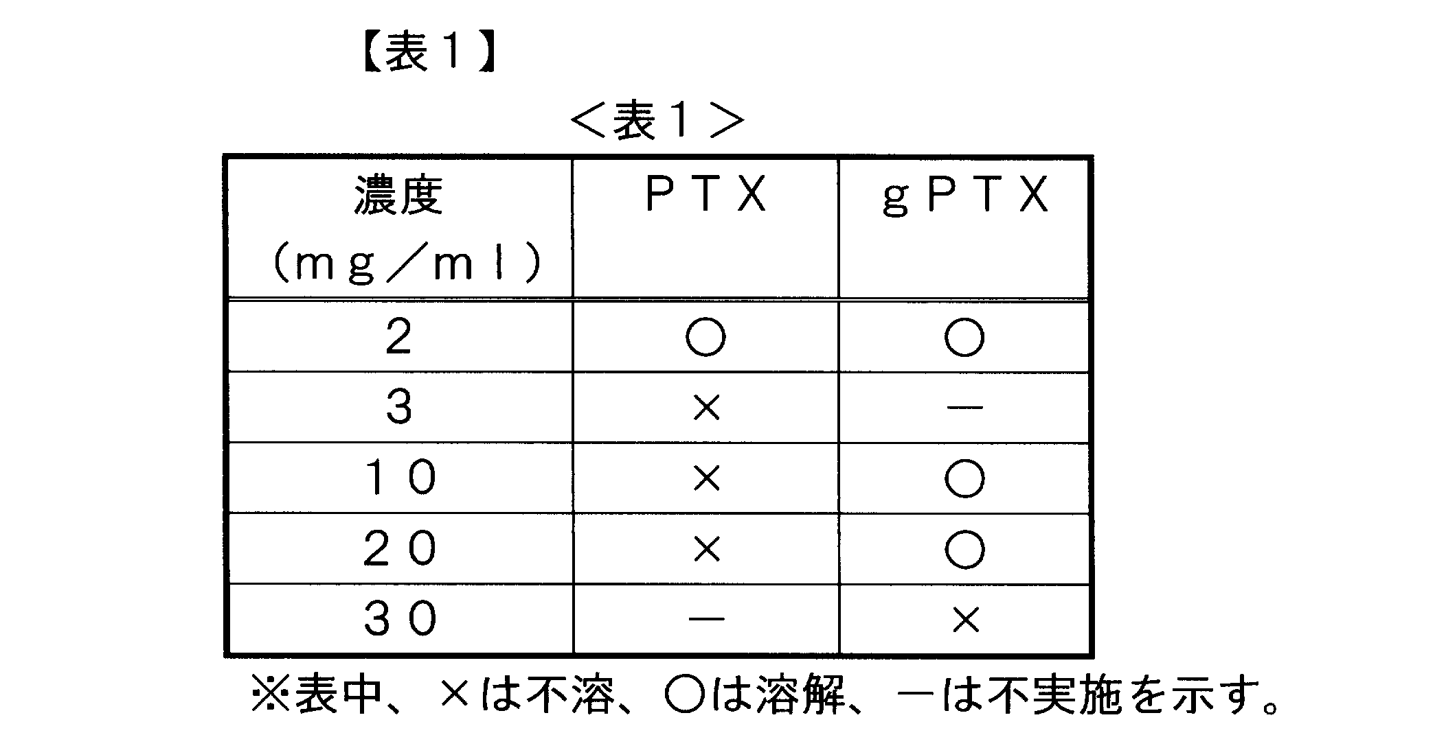
- Test Example 1 Solubility of 7- ⁇ -glucosyloxyacetyl paclitaxel The following chemical formula (1)
- gPTX 7- ⁇ -glucosyloxyacetyl paclitaxel
- PTX in this example powdery gPTX and paclitaxel (hereinafter referred to as PTX in this example) prepared by the method described in Non-Patent Document 1 above were weighed, 120 ⁇ L of ethanol was added, and vortexing was performed. . Furthermore, 120 ⁇ L of Cremophor (registered trademark) EL (available from Wako Pure Chemical Industries, Ltd.) was mixed with the solution, and then 760 ⁇ L of PBS (pH 7.4) was further mixed to confirm the solubility of gPTX and PTX. The results are shown in Table 1.
- PTX which is a non-glycoside
- gPTX was dissolved only at a concentration of about 2 mg / ml
- gPTX was dissolved at a concentration as high as 20 mg / ml. That is, both PTX and gPTX are normally non-soluble in water, and Cremophor: ethanol: PBS (pH 7.4) is a non-aqueous mixed solvent having a volume ratio of 12:12:76.
- Cremophor: ethanol: PBS pH 7.4
- gPTX did not show sufficient solubility, it was revealed that gPTX can be dissolved in the above-mentioned non-aqueous mixed solvent at a very high concentration.
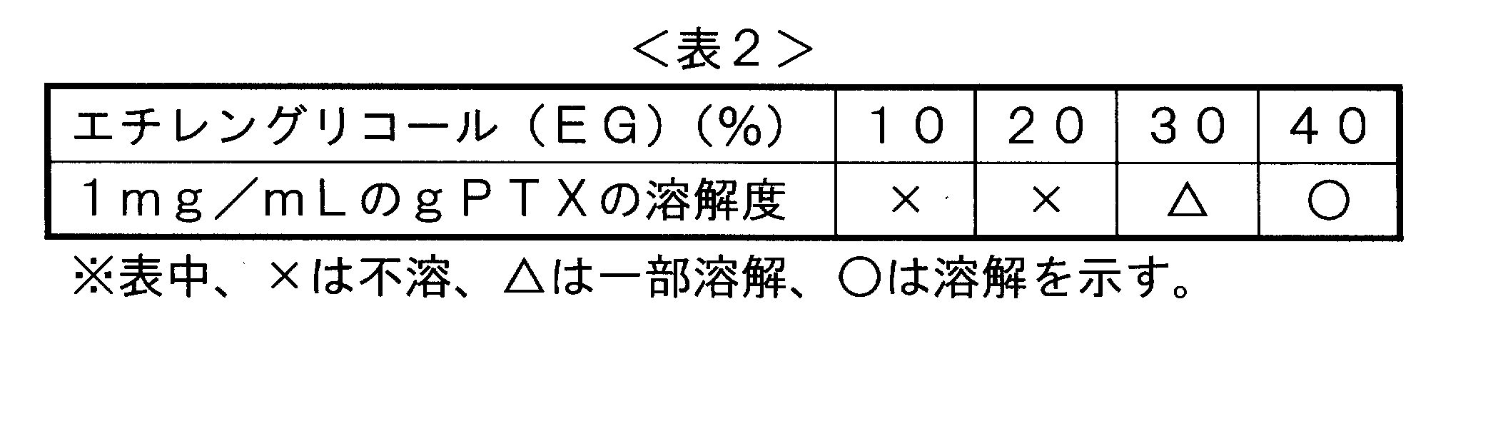
- Table 2 shows the results of a similar experiment on the solubility of gPTX in a 10 mM phosphate buffer containing 10 to 40% (volume) ethylene glycol (EG). This is a result of mixing gPTX so that the concentration is 1 mg / mL.
- gPTX does not dissolve in PBS containing 10-20% (volume) ethylene glycol, but starts to dissolve in PBS containing 30% (volume) ethylene glucose, and contains 40% (volume) ethylene glucose. It was revealed that it was dissolved in PBS. The solubility at this time was about 1 mg (gPTX) / mL. Accordingly, it was revealed that gPTX itself can hardly be dissolved by an aqueous solvent alone, but can be dissolved as a mixed solvent containing about 30 to 40% (volume) of ethylene glycol.
- a small lamella vesicle (SLV) was prepared by performing 5 minutes of sonication at intervals of 1 minute. Ultrafiltration (Amicon Ultra 100K membrane (manufactured by Millipore)) was performed to remove unencapsulated drug solution and free lipid.
- the liposome encapsulating gPTX is hereinafter referred to as gPTX-L, and the liposome encapsulating PTX is hereinafter referred to as PTX-L.
- gPTX was encapsulated in liposomes using the above 1 mg / ml gPTX solution (40% (volume) ethylene glycol 10 mM phosphate buffer) instead of gPTX.
- SLV small lamella vesicle
- Ultrafiltration Amicon Ultra 100K membrane (manufactured by Millipore) was performed to remove unencapsulated drug solution and free lipid.
- an antibody to be bound to the liposome was prepared.
- 1 mg of N-Succinimidyl3- (2-pyridyldithio) propionate (SPDP) was dissolved in 500 ⁇ l of dehydrated methanol to obtain a 2 mg / ml SPDP solution.
- 5 ⁇ L of the SPDP solution was placed in 1 ml of Trastuzumab (humanized anti-HER2 monoclonal antibody) solution (1 mg / ml) and stirred for 30 minutes at room temperature.
- the SPDP-modified Trastuzumab solution was placed in a dialysis tube (fraction molecular weight: 14,000), and dialyzed against 100 mM acetate buffer (pH 4.5). Dialysis was performed in a cool dark place at 4 ° C., and external liquid exchange was performed 3 hours 4 times and 12 hours 2 times.
- the SPDP-modified Trastuzumab solution was mixed with 500 ⁇ l of a 50 mM dithiothritol (DTT) solution and stirred at room temperature for 30 minutes.
- Ultrafiltration ((Amicon Ultra 10K membrane (manufactured by Millipore)) was performed to remove unreacted DTT and by-product pyridine-2-thione.
- the mixture was mixed with the liposome containing PTX or gPTX described above, and trastuzumab was bound to the liposome.
- the liposome in which the gPTX is encapsulated in the antibody-bound liposome is hereinafter referred to as gPTX-IL, and the liposome in which the PTX is encapsulated is referred to as PTX-IL.
- the liposome in which the above gPTX is encapsulated in a liposome to which no antibody is bound is hereinafter referred to as gPTX-L, and the liposome in which PTX is encapsulated is referred to as PTX-L.
- the efficiency of paclitaxel monoglycoside encapsulated in the obtained liposomes or immunoliposomes was calculated according to the following procedure.
- calibration curves of gPTX and PTX were obtained.
- a calibration curve is obtained by detecting a drug solution (Cremophor: ethanol: PBS (pH 7.4) 12:12:76; volume ratio) of 0.01 to 0.5 mg / ml by HPLC, and determining from the peak area at each drug concentration. Created.
- Encapsulation efficiency drug content / initial use drug amount ⁇ 100
- Loading rate (LE:%) ⁇ (drug content / drug molar weight) / number of initial lipid moles ⁇ ⁇ 100 (2)
- the particle size of the obtained liposome was also measured as the Z average particle size based on the dynamic light scattering method. These results are shown in Table 3 below.
- PTX dissolved in the cremophor-containing solvent shows only a numerical value of about 1.2%
- gPTX dissolved in the cremophor-containing solvent is 13
- a numerical value as high as 7% was shown, and a numerical value as high as 11.4% was shown from the encapsulation experiment in the liposome bound with the antibody.
- the amount of gPTX supported per lipid constituting the liposome increased by about 9.5 to 11.4 times compared to PTX. That is, gPTX is more advantageous for inclusion in liposomes.
- gPTX dissolved in a 40% (volume) ethylene glycol solution contains only 0.41% in the liposome and about 0.51% in the liposome to which the antibody is bound. It was also clarified that the efficiency of encapsulation was increased by about 30 to 40 times, with gPTX dissolved in 17.6% in the liposome and 14.8% in the antibody-bound liposome.
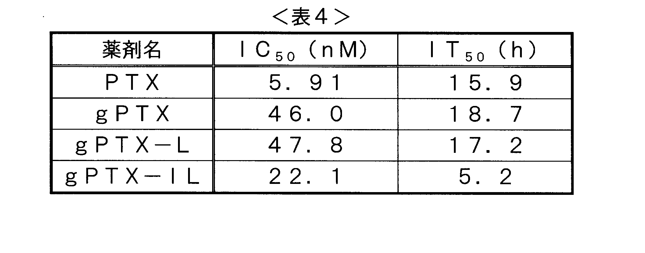
- Test Example 3 Evaluation of anti-cancer activity MTT assay of IC 50 of gPTX-L and gPTX-IL prepared using PTX dissolved in dimethyl sulfoxide, gPTX, and gPTX dissolved in 40% (volume) ethylene glycol solution Evaluated by.
- test cells human-derived breast cancer cells SK-BR-3 were used.
- IT 50 was calculated. IT 50 represents the time to reach a concentration that is half of the drug concentration at which cells are killed 100%, and can be easily calculated based on the survival curve obtained by plotting the cell viability against the time axis. Can be calculated.
- gPTX-IL shows a value about one-third that of these three. That is, it is shown that the time taken for gPTX-IL to bring the cells to death is much shorter than the other three. This is thought to be because gPTX-IL was actively delivered to breast cancer cells and taken into breast cancer cells, and exhibited high anticancer effects due to PTX itself. Therefore, it is clear that gPTX-IL exhibits an excellent DDS effect on breast cancer cells.
- Test Example 5 Cell observation Liposomes in which FITC is encapsulated in SK-BR-3 cells, which are human-derived breast cancer cells (the lipid composition is the same as described above: hereinafter referred to as FITC-L), and the same method as described above.
- the liposomes bound with trastuzumab prepared in this way (the lipid composition is the same as described above: hereinafter referred to as FITC-IL) are allowed to act, and after incubation for 2 hours, the cells are observed according to a conventional method using a fluorescence microscope did. The results are shown in FIG.
- FITC-L showed no accumulation in cells, whereas FITC-IL observed not only accumulation in cells but also uptake into cells. From the above, it has been clarified that the liposome bound with trastuzumab binds to HER2 expressed on the SK-BR-3 cell surface and is further taken up into the cell.
- Test Example 7 Preparation modification method of gPTX-encapsulated liposome (examination of heating time)
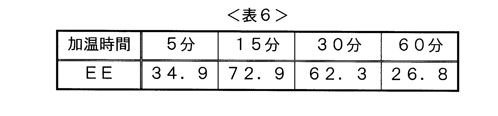
- the encapsulation efficiency was measured when the heating time was 5 minutes, 10 minutes, 30 minutes, and 60 minutes when the DPPC: Chol substance amount ratio was 3: 1.
- the measurement method was measured and calculated by the same method as in Test Example 2 described above. These results are shown in Table 6 below.
- Test Example 8 Acute toxicity test using gPTX-encapsulated liposomes obtained by the modified method An acute toxicity test was conducted using the gPTX-encapsulated liposomes (gPTX-L) obtained in Test Example 7 described above.
- One administration was performed at 200 ⁇ l / 20 g.
- the total dose of gPTX is 150 and 100 mg / kg in total in terms of gPTX, respectively.
- a follow-up was conducted during and after administration. The results are shown in FIG.
- Test Example 9 Cell delivery experiment using gPTX-encapsulated liposome obtained by the modified method
- the above-mentioned liposome (gPTX-L) encapsulating gPTX was subjected to 3 mg / ml Mal-PEG-DSPE dissolved in PBS. 470 ⁇ l was added and immersed in a 50 ° C. hot water bath for 10 minutes.
- an antibody to be bound to the liposome was prepared.
- HEPES buffer solution 25 mM HEPES, 140 mM NaCl, pH 8.0
- 2-iminothiolane solution was added, and reacted for 1 hour at room temperature under light shielding. Introduced. Unreacted substances were removed from the reacted solution using a Sephadex G-25 column, and the buffer solution was replaced with PBS.
- the trastuzumab solution into which the SH group was introduced and the liposome solution were mixed and shaken at 4 ° C. overnight to bind the trastuzumab to the liposome.
- ultrafiltration VIVASPIN 2, 300K membrane (manufactured by Sartorius)
- gPTX-IL liposome encapsulating gPTX to which an antibody was bound was obtained.
- 470 ⁇ l of 3 mg / ml mPEG-DSPE was added to the liposome solution and allowed to penetrate into a 50 ° C. hot water bath for 10 minutes. Thereafter, ultrafiltration (Amicon Ultra 100K membrane (manufactured by Millipore)) was performed.
- the anticancer activity was measured in the same manner as in Test Example 3 described above.
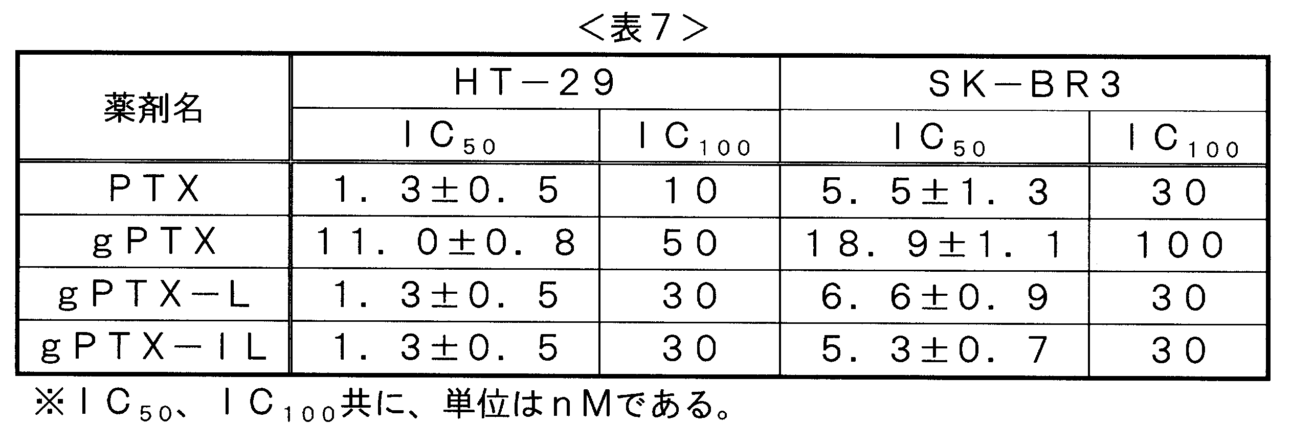
- the cells used were human-derived breast cancer cells SK-BR-3 and colon adenocarcinoma HT-29. Specifically, cells were seeded in 96 well-plate at 5000 cells / well and incubated for 24 hours. Thereafter, PTX, gPTX, gPTX-L and gPTX-IL were added at various concentrations and incubated.
- PTX, gPTX, gPTX-L, and gPTX-IL all exhibit extremely excellent anticancer activity against breast cancer cells and colon adenocarcinoma cells. It became. Further, from comparison between gPTX and PTX, the anticancer activity was attenuated by the sugar chain binding to PTX, but by encapsulating it in liposomes (see gPTX-L and gPTX-IL), anticancer activity. It has also been clarified that is exhibited in the same manner as PTX. However, there was no particular difference in anticancer activity between gPTX-L and gPTX-IL. That is, it was suggested that the anticancer activity is not particularly changed by the presence or absence of the antibody.
- gPTX-IL has a lower IT 50 value than gPTX-L. This indicates that the time to kill various cancer cells is short, suggesting that PTX can be efficiently delivered to the cancer cells in a shorter time, thereby killing the cancer cells faster. ing.
- Test Example 10 In Vivo Delivery Experiment of Liposomes Having Antibodies
- liposomes bound with antibodies encapsulating a fluorescent dye were prepared. Specifically, 10 mg of human serum albumin (HSA) was dissolved in 1 ml of 0.1 M sodium carbonate buffer (pH 9.3) to obtain a 10 mg / ml HSA solution. 1 ml of 10 mg / ml HSA solution was dissolved in 1 vial of Cy5.5 Monofunctional dye. This was shaken at room temperature for 30 minutes, and then unreacted substances were removed using a Sephadex G-25 column to recover Cy5.5-bound HSA (HSA-Cy5.5).
- HSA human serum albumin
- a lipid film was prepared according to the above test example and immersed in a 60 ° C. bath for 5 minutes, and then the HSA-Cy5.5 solution was added to dissolve the lipid film. While this solution was immersed in a 60 ° C. warm bath, sonication was performed for 3 minutes. Ultrafiltration (Amicon Ultra 100K membrane (manufactured by Millipore)) was performed to remove unencapsulated HSA-Cy5.5. Thereafter, an antibody (Trastumab) was bound to the obtained liposome using the method shown in Test Example 8.
- HT-29 cells were administered with 3.0 ⁇ 10 6 cells / mouse by subcutaneous injection and liposomes encapsulating HSA-Cy5.5 to which the antibody was bound.
- liposomes encapsulating HSA-Cy5.5 to which no antibody was bound were also administered.
- HSA-Cy5.5 encapsulated liposomes (L) or liposomes with antibodies (IL) were administered by tail vein injection.
- liposomes with antibodies always accumulate in tumor tissue more than liposomes without antibodies, while liposomes without antibodies always have liver tissue. It has become clear that there is much accumulation. In addition, the liposome without the antibody did not accumulate in the tumor tissue 48 hours after administration, unlike the liposome with the antibody.
- Test Example 11 In Vivo Anticancer Activity Experiment of Liposomes Encapsulating gPTX and Antibody-Injecting HT-29 Cells Subcutaneously at 3.0 ⁇ 10 6 cells / mouse into ICR-nu / nu mice (5 weeks old) Administered. When the tumor volume reaches about 50 to 200 mm 3 , the above-described gPTX, gPTX-L prepared in Test Example 7, gPTX-IL prepared in Test Example 9, Trastuzumab, and gPTX in Test Example 9 are included.
- One administration was performed at 200 ⁇ l / 20 g.
- the total dose of gPTX is 150 mg / kg and Trastuzumab is 200 mg / kg.
- the tumor volume transition after administration was observed. The results are shown in the figure.
- liposomes encapsulating gPTX and bound with Trastuzumab a monoclonal antibody that specifically recognizes Her2, markedly suppresses growth in tumor tissues of HT-29 tumor-bearing mice that are colon adenocarcinoma. It became clear that it was effective. In addition, it has also been clarified that such liposomes exhibit effects such as extremely low side effects because they do not show weight loss upon administration and have a low lethality. From the above, it has been clarified that such liposomes are useful as liposome preparations that exhibit extremely excellent effects on cancer cells.
Landscapes
- Health & Medical Sciences (AREA)
- Chemical & Material Sciences (AREA)
- Life Sciences & Earth Sciences (AREA)
- General Health & Medical Sciences (AREA)
- Medicinal Chemistry (AREA)
- Veterinary Medicine (AREA)
- Public Health (AREA)
- Animal Behavior & Ethology (AREA)
- Pharmacology & Pharmacy (AREA)
- Epidemiology (AREA)
- Organic Chemistry (AREA)
- Dispersion Chemistry (AREA)
- Immunology (AREA)
- Molecular Biology (AREA)
- Oncology (AREA)
- Biophysics (AREA)
- Biochemistry (AREA)
- Genetics & Genomics (AREA)
- Proteomics, Peptides & Aminoacids (AREA)
- Engineering & Computer Science (AREA)
- Bioinformatics & Cheminformatics (AREA)
- Chemical Kinetics & Catalysis (AREA)
- General Chemical & Material Sciences (AREA)
- Nuclear Medicine, Radiotherapy & Molecular Imaging (AREA)
- Pharmaceuticals Containing Other Organic And Inorganic Compounds (AREA)
- Medicinal Preparation (AREA)
- Medicines Containing Antibodies Or Antigens For Use As Internal Diagnostic Agents (AREA)
Abstract
Description
該パクリタキセルモノグリコシド及び/又はドタキセルモノグリコシドを混合物の総容量に対して1500以上~3000重量%未満含み、
該緩衝液若しくは水1容量部当たり、
該ポリオキシエチレンエステル誘導体を0.1~0.2容量部含み、
該低級アルコールを0.1~0.2容量部含むことを特徴とし、
該リポソームを構成する総脂質1重量部当たり、0.1~2.5重量部の前記パクリタキセルモノグリコシド及び/又はドタキセルモノグリコシドを内包することを特徴とする製造方法。
本発明のリポソームの製造方法の1つの態様(以後、「第1の態様の製造方法」と呼ぶことがある)である、パクリタキセルモノグリコシド及び/又はドタキセルモノグリコシドを内包するリポソームの製造方法は、パクリタキセルモノグリコシド及び/又はドタキセルモノグリコシド、ポリオキシエチレン誘導体、低級アルコール、リポソームを構成する脂質、及び緩衝液若しくは水を含む混合物に対してリポソーム形成処理を施す工程を含むものである。


2抗体、抗CEA抗体等が挙げられる。
<リポソーム製剤>
本発明のリポソーム製剤は、パクリタキセルモノグリコシド及び/又はドタキセルモノグリコシドの溶解液を内包し、且つガン細胞を特異的に認識する抗体を有する。
下記化学式(1)
1,2-Dipalmitoyl-rac-glycero-3-phosphocholine(DPPC)、1,2-Dipalmitoyl-sn-glycerol-3-phospho-rac-(1-glycerol)sodium salt(DPPG)、及びコレステロールを、それぞれ13.5、1.5、及び1.5mg秤量し、50mlのナスフラスコ内で混合した。混合した脂質を3mlの有機溶媒(クロロホルム:メタノール=9:1)で溶解した後、ロータリーエバポレーターで乾燥後、2時間の真空乾燥をすることにより溶媒を完全に除去し脂質フィルムを調製した。
担持率(LE:%)={(薬剤含有量/薬剤モル重量)/初期脂質モル数}×100 (2)

ジメチルスルホキシドに溶解したPTX、並びにgPTX、及び40%(容量)のエチレングリコール溶液に溶解したgPTXを用いて作製したgPTX-L並びにgPTX-ILのIC50をMTTアッセイによって評価した。試験対象細胞としては、ヒト由来の乳ガン細胞であるSK-BR-3を用いた。また、IT50を算出した。IT50とは、細胞が100%死滅する薬剤濃度の半分の濃度に到達するまでの時間を表すものであり、時間軸に対して細胞生存率をプロットすることにより得られる生存曲線を基に容易に算出できる。これらの結果を、表4に示す。
BALB/c マウス(♀, 5週齢)にgPTX、gPTX-L、溶媒コントロール(クレモホール:エタノール:超純水=12:12:76;容量比)を尾静脈注射により1時間のインターバルをおき、5回の投与を行った(N=3)。1回の投与は200μl/20gで行った。gPTXの合計投与量は200mg/kgである。投与途中および投与後経過観察を行った。結果を図1に示す。
ヒト由来乳ガン細胞であるSK-BR-3細胞に、FITCを内包したリポソーム(脂質組成は上述のものと同じ:以後、FITC-Lとする。)、及び上記方法と同様にして作成したTrastuzumabが結合したリポソーム(脂質組成は上述のものと同じ:以後、FITC-ILとする。)を作用させ、2時間のインキュベートの後、蛍光顕微鏡を用いた常法に従って細胞を観察した。結果を、図2に示す。
DPPCを13.2、10.6もしくは8.8mg、コレステロールを2.1、3.7もしくは4.6mgおよびmPEG-DSPEを2.1mg秤量し、50mlのナスフラスコ内で混合した。混合した脂質を3mlの有機溶媒(容量比:クロロホルム:メタノール=9:1;容量比)で溶解し、湯浴型ソニケータにより5分間ソニケーションを行った後、ロータリーエバポレーターで乾燥後、一晩、真空乾燥することにより溶媒を完全に除去し脂質フィルムを調製した。

試験例6に示す実験において、DPPC:Cholの物質量比が3:1とした場合の加温時間を、5分、10分、30分、及び60分とした場合の封入効率を測定した。測定方法は、上述の試験例2と同様の方法で測定し、算出した。これらの結果を下記の表6に示す。
上述の試験例7にて得られたgPTX内包リポソーム(gPTX-L)を用いて、急性毒性試験を行った。
上述のgPTXを内包するリポソーム(gPTX-L)に対して、PBSに溶解させた3mg/mlのMal-PEG-DSPEを470μl加え、50℃の湯浴中に10分間浸漬させた。

次いで、抗体が結合したリポソームの腫瘍組織への送達をインビボでの実験で検証するために、蛍光色素を内包する抗体が結合したリポソームを作製した。具体的には、10mgのヒト血清アルブミン(HSA)を0.1Mの炭酸ナトリウム緩衝液(pH9.3)1mlに溶解させ、10mg/mlのHSA溶液を得た。10mg/mlのHSA溶液1mlを1バイアルのCy5.5Monofunctional dyeへ溶解させた。これを室温で30分間振盪した後、Sephadex G-25カラムを用いて、未反応物を取り除き、Cy5.5結合HSA(HSA-Cy5.5)を回収した。
ICRーnu/nuマウス(♀、5週齢)へHTー29細胞を3.0×106cells /mouseで皮下注射により投与した。腫瘍体積が50~200mm3程度になった時点で、上述のgPTX、試験例7にて作製したgPTXーL、試験例9にて作成したgPTXーIL、Trastuzumab、試験例9にてgPTXを内包させず、且つ抗体(Trastuzumab)を結合させずに作製したリポソーム(empty L)、試験例9にてgPTXを内包させず、且つ抗体(Trastuzumab)を結合させて作製したリポソーム(empty IL)、CEP緩衝液、及びPBSを尾静脈注射により3時間のインターバルをおき、2回投与を行った(N=4)。1回の投与は200μl/20gで行った。gPTXの合計投与量は150mg/kgであり、Trastuzumabは200mg/kgである。投与後の腫瘍体積推移の観察を行った。結果を図に示す。腫瘍体積は下記式(3)により算出した。
腫瘍体積=(腫瘍短辺2×腫瘍長辺)/2 (3)
また、各投与群の体重推移、及び生存率も測定した。結果を図6~9に示す。
Claims (15)
- パクリタキセルモノグリコシド及び/又はドタキセルモノグリコシドを内包し、且つガン細胞を特異的に認識する抗体を有するリポソームの製造方法であって、ポリオキシエチレンエステル誘導体、低級アルコール、及び緩衝液若しくは水を包含するリポソームと、アルキレングリコールを含む緩衝液又は水に、パクリタキセルモノグリコシド及び/又はドタキセルモノグリコシドが溶解した溶液に接触させる工程を含む、製造方法。
- グリコシドが、グルコシド、ガラクトシド、マンノシド、キシロシド、フルクトシド、ラムノシド、アラビノシド、アロシド、アルトロシド、イドシド、N-アセチルグルコサミニド、N-アセチルガラクトサミニド、タロシド、グルクロノシド、グルコサミニド、ガラクトサミニド、及びフコシドからなる群より選択される1種のグリコシドである、請求項1に記載の製造方法。
- ポリオキシエチレンエステル誘導体が、ポリオキシエチレンヒマシ油エステルである請求項1又は2に記載の製造方法。
- リポソームが、DPPC及びコレステロールをそれぞれ3:0.5~3の物質量比で含有する、請求項1~3の何れか1項に記載の製造方法。
- 接触時間が、10~40分である請求項1~4の何れか1項に記載の製造方法。
- ガン細胞が、乳ガン細胞である請求項1~5の何れか1項に記載の製造方法。
- 抗体が、HER2タンパク質と特異的に結合する抗体である請求項1~6の何れか1項に記載の製造方法。
- パクリタキセルモノグリコシド及び/又はドタキセルモノグリコシドの溶解液を内包し、且つガン細胞を特異的に認識する抗体を有するリポソーム製剤。
- パクリタキセルモノグリコシド及び/又はドタキセルモノグリコシドの溶解液が、ポリオキシエチレンエステル誘導体、低級アルコール、及び緩衝液若しくは水を含む混合溶媒にパクリタキセルモノグリコシド及び/又はドタキセルモノグリコシドが溶解する溶解液である、請求項8に記載のリポソーム製剤。
- グリコシドが、グルコシド、ガラクトシド、マンノシド、キシロシド、フルクトシド、ラムノシド、アラビノシド、アロシド、アルトロシド、イドシド、N-アセチルグルコサミニド、N-アセチルガラクトサミニド、タロシド、グルクロノシド、グルコサミニド、ガラクトサミニド、及びフコシドからなる群より選択される1種のグリコシドである請求項8又は9に記載のリポソーム製剤。
- ポリオキシエチレンエステル誘導体が、ポリオキシエチレンヒマシ油エステルである、請求項8~10の何れか1項に記載のリポソーム製剤。
- リポソームが、DPPC及びコレステロールをそれぞれ3:0.5~3の重量比で含有する請求項8~11の何れか1項に記載のリポソーム製剤。
- リポソームの全脂質の1モル量に対するパクリタキセルモノグリコシドのモル量が、1.0~15.0×10⁻²である、請求項8~12の何れか1項に記載のリポソーム製剤。
- ガン細胞が、乳ガン細胞である請求項8~13の何れか1項に記載のリポソーム製剤。
- 抗体が、HER2タンパク質と特異的に結合する抗体である請求項8~14のいずれか1項に記載のリポソーム製剤。
Priority Applications (4)
| Application Number | Priority Date | Filing Date | Title |
|---|---|---|---|
| EP13764209.6A EP2829273B1 (en) | 2012-03-22 | 2013-03-22 | Method for producing liposome encapsulating paclitaxel monoglycoside and/or docetaxel monoglycoside |
| JP2013537718A JP5490326B2 (ja) | 2012-03-22 | 2013-03-22 | パクリタキセルモノグリコシド及び/又はドセタキセルモノグリコシドを内包するリポソームの製造方法 |
| CN201380015012.5A CN104203251B (zh) | 2012-03-22 | 2013-03-22 | 内包紫杉醇单糖苷和/或紫杉萜单糖苷的脂质体的制造方法 |
| US14/386,855 US10682330B2 (en) | 2012-03-22 | 2013-03-22 | Method for producing liposome encapsulating paclitaxel monoglycoside and/or docetaxel monoglycoside |
Applications Claiming Priority (2)
| Application Number | Priority Date | Filing Date | Title |
|---|---|---|---|
| JP2012-065743 | 2012-03-22 | ||
| JP2012065743 | 2012-03-22 |
Publications (1)
| Publication Number | Publication Date |
|---|---|
| WO2013141346A1 true WO2013141346A1 (ja) | 2013-09-26 |
Family
ID=49222795
Family Applications (1)
| Application Number | Title | Priority Date | Filing Date |
|---|---|---|---|
| PCT/JP2013/058242 WO2013141346A1 (ja) | 2012-03-22 | 2013-03-22 | パクリタキセルモノグリコシド及び/又はドタキセルモノグリコシドを内包するリポソームの製造方法 |
Country Status (5)
| Country | Link |
|---|---|
| US (1) | US10682330B2 (ja) |
| EP (1) | EP2829273B1 (ja) |
| JP (1) | JP5490326B2 (ja) |
| CN (1) | CN104203251B (ja) |
| WO (1) | WO2013141346A1 (ja) |
Cited By (1)
| Publication number | Priority date | Publication date | Assignee | Title |
|---|---|---|---|---|
| WO2017061562A1 (ja) * | 2015-10-07 | 2017-04-13 | 塩水港精糖株式会社 | タキサン化合物を内包するリポソーム |
Families Citing this family (4)
| Publication number | Priority date | Publication date | Assignee | Title |
|---|---|---|---|---|
| US11013690B2 (en) * | 2016-01-04 | 2021-05-25 | Academia Sinica | Esterification/saponification-based method for liposomal encapsulation of hydrophilic glucuronides |
| US10864161B2 (en) | 2017-10-13 | 2020-12-15 | American University Of Sharjah | Systems and methods for targeted breast cancer therapies |
| CN110981837A (zh) * | 2019-12-03 | 2020-04-10 | 沈阳药科大学 | 紫杉醇弱酸性衍生物主动载药脂质体及其制备与应用 |
| CN114588103B (zh) * | 2022-03-31 | 2023-04-11 | 山东大学 | 负载紫杉醇脂质体的泊洛沙姆温敏凝胶及制备方法与应用 |
Citations (6)
| Publication number | Priority date | Publication date | Assignee | Title |
|---|---|---|---|---|
| JPH09286794A (ja) * | 1996-02-20 | 1997-11-04 | Ensuiko Sugar Refining Co Ltd | タキソイド誘導体およびその製造法 |
| WO1999018113A1 (fr) * | 1997-10-08 | 1999-04-15 | Bio Research Corporation Of Yokohama | Derives taxoides et leur procede de production |
| WO1999022759A1 (en) * | 1997-10-31 | 1999-05-14 | Walter Reed Army Institute Of Research | Method of inhibiting side effects of pharmaceutical compositions containing amphiphilic vehicles or drug carrier molecules |
| JP2006265152A (ja) * | 2005-03-23 | 2006-10-05 | Beacle Inc | 癌治療用薬剤及びその作製方法 |
| WO2008072584A1 (ja) | 2006-12-08 | 2008-06-19 | Katayama Chemical Industries Co., Ltd. | アンミン白金錯体を高濃度で内包するリポソームおよびその製造方法 |
| JP2009132629A (ja) | 2007-11-28 | 2009-06-18 | Terumo Corp | リポソーム製剤の製造方法 |
Family Cites Families (6)
| Publication number | Priority date | Publication date | Assignee | Title |
|---|---|---|---|---|
| CA2240595A1 (en) * | 1995-12-21 | 1997-07-03 | Bijan Almassian | Taxane composition and method |
| US5767297A (en) | 1997-02-05 | 1998-06-16 | Ensuiko Sugar Refining Co., Ltd. | Taxoid derivative and method of producing thereof |
| US7361683B2 (en) * | 2004-11-24 | 2008-04-22 | Yung Shin Pharm. Ind., Co., Ltd | Paclitaxel aqueous injection solution and methods for preparing the same |
| US20080206139A1 (en) * | 2006-11-03 | 2008-08-28 | The Penn State Research Foundation | Delivery system for diagnostic and therapeutic agents |
| CN102552123A (zh) * | 2011-12-31 | 2012-07-11 | 江苏奥赛康药业股份有限公司 | 一种供注射用紫杉醇组合物及其制备方法 |
| JP6171227B2 (ja) * | 2012-02-23 | 2017-08-02 | 株式会社リュージュサイエンス | 陽イオン性多糖類共重合体の抗癌剤デリバリーシステム |
-
2013
- 2013-03-22 EP EP13764209.6A patent/EP2829273B1/en not_active Not-in-force
- 2013-03-22 WO PCT/JP2013/058242 patent/WO2013141346A1/ja active Application Filing
- 2013-03-22 CN CN201380015012.5A patent/CN104203251B/zh not_active Expired - Fee Related
- 2013-03-22 US US14/386,855 patent/US10682330B2/en active Active
- 2013-03-22 JP JP2013537718A patent/JP5490326B2/ja not_active Expired - Fee Related
Patent Citations (6)
| Publication number | Priority date | Publication date | Assignee | Title |
|---|---|---|---|---|
| JPH09286794A (ja) * | 1996-02-20 | 1997-11-04 | Ensuiko Sugar Refining Co Ltd | タキソイド誘導体およびその製造法 |
| WO1999018113A1 (fr) * | 1997-10-08 | 1999-04-15 | Bio Research Corporation Of Yokohama | Derives taxoides et leur procede de production |
| WO1999022759A1 (en) * | 1997-10-31 | 1999-05-14 | Walter Reed Army Institute Of Research | Method of inhibiting side effects of pharmaceutical compositions containing amphiphilic vehicles or drug carrier molecules |
| JP2006265152A (ja) * | 2005-03-23 | 2006-10-05 | Beacle Inc | 癌治療用薬剤及びその作製方法 |
| WO2008072584A1 (ja) | 2006-12-08 | 2008-06-19 | Katayama Chemical Industries Co., Ltd. | アンミン白金錯体を高濃度で内包するリポソームおよびその製造方法 |
| JP2009132629A (ja) | 2007-11-28 | 2009-06-18 | Terumo Corp | リポソーム製剤の製造方法 |
Non-Patent Citations (6)
| Title |
|---|
| BIOL. PHARM. BULL., vol. 31, no. 6, 2008, pages 1155 - 1158 |
| BIOL. PHARM. BULL., vol. 31, no. 6, June 2008 (2008-06-01), pages 1155 - 8 |
| HETEROCYCLES, vol. 54, no. 2, 2001, pages 561 - 566 |
| KEI SHIMODA ET AL.: "Chemo-enzymatic synthesis of ester-linked taxol-oligosaccharide conjugates as potential prodrugs", TETRAHEDRON LETTERS, vol. 49, 2008, pages 601 - 604, XP055169196 * |
| MASAHARU MURAKAMI ET AL.: "Encapsulation of glycosylated paclitaxel into liposome targeting Her2", JOURNAL OF JAPANESE BIOCHEMICAL SOCIETY, December 2010 (2010-12-01), XP008174937 * |
| YANG T. ET AL.: "Antitumor effect of paclitaxel- loaded PEGylated immunoliposomes against human breast cancer cells", PHARMACEUTICAL RESEARCH, vol. 24, no. 12, 2007, pages 2402 - 2411, XP019555920 * |
Cited By (4)
| Publication number | Priority date | Publication date | Assignee | Title |
|---|---|---|---|---|
| WO2017061562A1 (ja) * | 2015-10-07 | 2017-04-13 | 塩水港精糖株式会社 | タキサン化合物を内包するリポソーム |
| KR20180052123A (ko) | 2015-10-07 | 2018-05-17 | 엔스이코 세이토 가부시키가이샤 | 탁산 화합물을 내포하는 리포좀 |
| JPWO2017061562A1 (ja) * | 2015-10-07 | 2018-07-05 | 塩水港精糖株式会社 | タキサン化合物を内包するリポソーム |
| US20180280300A1 (en) * | 2015-10-07 | 2018-10-04 | Ensuiko Sugar Refining Co., Ltd. | Liposome including taxane compound |
Also Published As
| Publication number | Publication date |
|---|---|
| EP2829273A4 (en) | 2015-11-04 |
| JP5490326B2 (ja) | 2014-05-14 |
| US10682330B2 (en) | 2020-06-16 |
| CN104203251B (zh) | 2017-05-31 |
| JPWO2013141346A1 (ja) | 2015-08-03 |
| US20150056270A1 (en) | 2015-02-26 |
| EP2829273B1 (en) | 2018-08-29 |
| CN104203251A (zh) | 2014-12-10 |
| US20160256566A2 (en) | 2016-09-08 |
| EP2829273A1 (en) | 2015-01-28 |
Similar Documents
| Publication | Publication Date | Title |
|---|---|---|
| Torchilin | Antibody-modified liposomes for cancer chemotherapy | |
| ElBayoumi et al. | Tumor-targeted nanomedicines: enhanced antitumor efficacy in vivo of doxorubicin-loaded, long-circulating liposomes modified with cancer-specific monoclonal antibody | |
| Bariwal et al. | Nanodiscs: a versatile nanocarrier platform for cancer diagnosis and treatment | |
| Yang et al. | Antitumor effect of paclitaxel-loaded PEGylated immunoliposomes against human breast cancer cells | |
| Allahou et al. | Investigating the application of liposomes as drug delivery systems for the diagnosis and treatment of cancer | |
| Fathi et al. | Liposomal drug delivery systems for targeted cancer therapy: is active targeting the best choice? | |
| Drummond et al. | Liposome targeting to tumors using vitamin and growth factor receptors | |
| Brown et al. | The treatment of breast cancer using liposome technology | |
| Waelti et al. | Targeting her-2/neu with antirat Neu virosomes for cancer therapy | |
| JP5490326B2 (ja) | パクリタキセルモノグリコシド及び/又はドセタキセルモノグリコシドを内包するリポソームの製造方法 | |
| Lasic | Phannacokinetics and Antitumor Activity of Anthracyclines Precipitated in Sterically Stabilized (Stealth®) Liposomes | |
| Fidan et al. | Recent advances in liposome-based targeted cancer therapy | |
| Saraf et al. | Targeting approaches for the diagnosis and treatment of cancer | |
| JP6495466B2 (ja) | タキサン化合物を内包するリポソーム | |
| US20220378936A1 (en) | Delivery system complexes comprising a precipitate of an active agent and methods of use | |
| WO2009148169A1 (ja) | アンミン白金錯体を高濃度に内包するリポソームを用いた腫瘍治療技術 | |
| Yang et al. | EGFR-targeted pemetrexed therapy of malignant pleural mesothelioma | |
| WO2020232701A1 (zh) | 单醣标记的纳米脂质体药物递送系统,其制法及其作为药物靶定递送载体的应用 | |
| WO2018214944A1 (zh) | 用c(RGD-ACP-K)修饰的长循环脂质体 | |
| Varshochian et al. | Utilizing liposomes and lipid nanoparticles to overcome challenges in breast cancer treatment | |
| US9757462B2 (en) | Combined pharmaceutical preparation | |
| JP2011211996A (ja) | 腫瘍特異性を有するリポソーム | |
| Brown | Anti-GD2 Etoposide-Loaded Immunoliposomes for the Treatment of GD2 Positive Tumors | |
| Kannan | Development and Evaluation of Paclitaxel-Loaded Liposomal Formulations for Targeted Drug Delivery to Breast Cancer | |
| Zalba | Design and in-vitro/in-vivo evaluation in colon cancer cells of targeted oxaliplatin liposomes to epidermal growth factor receptor by conjugation of different ligands |
Legal Events
| Date | Code | Title | Description |
|---|---|---|---|
| ENP | Entry into the national phase |
Ref document number: 2013537718 Country of ref document: JP Kind code of ref document: A |
|
| 121 | Ep: the epo has been informed by wipo that ep was designated in this application |
Ref document number: 13764209 Country of ref document: EP Kind code of ref document: A1 |
|
| WWE | Wipo information: entry into national phase |
Ref document number: 14386855 Country of ref document: US |
|
| WWE | Wipo information: entry into national phase |
Ref document number: 2013764209 Country of ref document: EP |
|
| NENP | Non-entry into the national phase |
Ref country code: DE |