WO2013094392A1 - 測定装置、測定方法、プログラムおよび記録媒体 - Google Patents
測定装置、測定方法、プログラムおよび記録媒体 Download PDFInfo
- Publication number
- WO2013094392A1 WO2013094392A1 PCT/JP2012/081255 JP2012081255W WO2013094392A1 WO 2013094392 A1 WO2013094392 A1 WO 2013094392A1 JP 2012081255 W JP2012081255 W JP 2012081255W WO 2013094392 A1 WO2013094392 A1 WO 2013094392A1
- Authority
- WO
- WIPO (PCT)
- Prior art keywords
- vein
- living body
- measurement
- light
- depth
- Prior art date
- Legal status (The legal status is an assumption and is not a legal conclusion. Google has not performed a legal analysis and makes no representation as to the accuracy of the status listed.)
- Ceased
Links
Images


Classifications
-
- A—HUMAN NECESSITIES
- A61—MEDICAL OR VETERINARY SCIENCE; HYGIENE
- A61B—DIAGNOSIS; SURGERY; IDENTIFICATION
- A61B5/00—Measuring for diagnostic purposes; Identification of persons
- A61B5/145—Measuring characteristics of blood in vivo, e.g. gas concentration or pH-value ; Measuring characteristics of body fluids or tissues, e.g. interstitial fluid or cerebral tissue
- A61B5/1455—Measuring characteristics of blood in vivo, e.g. gas concentration or pH-value ; Measuring characteristics of body fluids or tissues, e.g. interstitial fluid or cerebral tissue using optical sensors, e.g. spectral photometrical oximeters
- A61B5/14551—Measuring characteristics of blood in vivo, e.g. gas concentration or pH-value ; Measuring characteristics of body fluids or tissues, e.g. interstitial fluid or cerebral tissue using optical sensors, e.g. spectral photometrical oximeters for measuring blood gases
- A61B5/14552—Details of sensors specially adapted therefor
-
- A—HUMAN NECESSITIES
- A61—MEDICAL OR VETERINARY SCIENCE; HYGIENE
- A61B—DIAGNOSIS; SURGERY; IDENTIFICATION
- A61B5/00—Measuring for diagnostic purposes; Identification of persons
- A61B5/0059—Measuring for diagnostic purposes; Identification of persons using light, e.g. diagnosis by transillumination, diascopy, fluorescence
- A61B5/0082—Measuring for diagnostic purposes; Identification of persons using light, e.g. diagnosis by transillumination, diascopy, fluorescence adapted for particular medical purposes
-
- A—HUMAN NECESSITIES
- A61—MEDICAL OR VETERINARY SCIENCE; HYGIENE
- A61B—DIAGNOSIS; SURGERY; IDENTIFICATION
- A61B5/00—Measuring for diagnostic purposes; Identification of persons
- A61B5/48—Other medical applications
- A61B5/4887—Locating particular structures in or on the body
- A61B5/489—Blood vessels
Definitions
- the present disclosure relates to a measuring device, a measuring method, a program, and a recording medium.
- the present disclosure proposes a new and improved measuring apparatus, measuring method, program, and recording medium capable of accurately measuring the blood component of the vein by optical measurement.
- a living body is irradiated with measurement light having a predetermined wavelength, and the measurement light scattered in the living body and emitted from the surface of the living body is detected and emitted.
- a measurement unit that collects measurement light using a lens array having a plurality of light receiving lenses arranged in an array to obtain a captured image of the living body, and is present inside the living body based on the captured image Using the vein position specifying unit for specifying the position of the vein, the vein depth specifying unit for specifying the depth of the vein based on the captured image, and the detected measurement using the position of the vein and the depth of the vein.
- a measuring apparatus including a blood component estimation unit that estimates the blood component of the vein based on information obtained from light.
- At least a part of a living body is irradiated with measurement light having a predetermined wavelength, and the measurement light scattered in the living body and emitted from the surface of the living body is detected, and the emission is performed.
- a part of a living body is irradiated with measurement light having a predetermined wavelength, and the measurement light scattered in the living body and emitted from the surface of the living body is detected, and the emission is performed.
- a computer communicable with a measuring unit that collects the measured light using a lens array having a plurality of light receiving lenses arranged in an array and obtains a captured image of the living body.
- a vein position specifying function for specifying the position of a vein existing inside the living body, a vein depth specifying function for specifying the depth of the vein based on the captured image, and the position of the vein and the depth of the vein are used.
- At least a part of the living body is irradiated with measuring light having a predetermined wavelength, and the measuring light scattered in the living body and emitted from the surface of the living body is detected, and the emitted measuring light is arrayed
- a vein that exists inside the living body based on the captured image in a computer that can communicate with a measuring unit that collects light using a lens array having a plurality of light receiving lenses and acquires the captured image of the living body.
- the detected measurement light using the vein position specifying function for specifying the position of the vein, the vein depth specifying function for specifying the depth of the vein based on the captured image, and the position of the vein and the depth of the vein A computer-readable recording medium on which a program for realizing the blood component estimation function for estimating the blood component of the vein based on the information obtained from the above is recorded is provided.
- At least a part of a living body is irradiated with measurement light having a predetermined wavelength, and the measurement light scattered in the living body and emitted from the surface of the living body is detected, and the emission is performed.
- a measuring unit that collects the measured light using a lens array having a plurality of light receiving lenses arranged in an array to obtain a captured image of the living body, and inside the living body based on the captured image
- a vein position specifying unit that specifies the position of an existing vein
- a vein depth specifying unit that specifies the depth of the vein based on the captured image
- a surface between the surface of the living body and the vein based on the depth of the vein is irradiated with measurement light having a predetermined wavelength, and the measurement light scattered in the living body and emitted from the surface of the living body is detected, and the emission is performed.
- estimating the thickness of the body tissue present in the vein excluding the influence of the body tissue for the estimated thickness from the information obtained from the measurement light detected at the position of the vein, and at the position of the vein in the adjacent adjacent area Extracts time-varying components derived from pulsation of arteries existing inside the living body from information obtained from the emitted measuring light, and extracts the time-varying components from information obtained from measuring light detected at the position of the vein And a blood component estimator for estimating the blood component of the vein.
- At least a part of a living body is irradiated with measurement light having a predetermined wavelength, and the measurement light scattered in the living body and emitted from the surface of the living body is detected, and the emission is performed.
- the time-varying component derived from the pulsation of the artery present inside the living body is extracted from the detected information, and the time-varying component is further excluded from the information obtained from the measurement light detected at the position of the vein
- a method of measurement includes estimating a blood component of the blood.
- a part of a living body is irradiated with measurement light having a predetermined wavelength, and the measurement light scattered in the living body and emitted from the surface of the living body is detected, and the emission is performed.
- a computer communicable with a measuring unit that collects the measured light using a lens array having a plurality of light receiving lenses arranged in an array and obtains a captured image of the living body.
- a vein position specifying function for specifying the position of a vein existing inside the living body;
- a vein depth specifying function for specifying the depth of the vein based on the captured image; and a thickness of a body tissue existing between the surface of the living body and the vein based on the depth of the vein;
- the information obtained from the measurement light detected in the adjacent region adjacent to the position of the vein while excluding the influence of the body tissue for the estimated thickness from the information obtained from the measurement light detected at the position The time-varying component derived from the pulsation of the artery present inside the living body is extracted from the information, and the venous blood is further excluded from the information obtained from the measurement light detected at the position of the vein
- a program for realizing a blood component estimation function for estimating a middle component is provided.
- blood components in veins can be accurately measured by optical measurement.
- FIG. 6 is a diagram for describing information obtained from measurement light in the first embodiment of the present disclosure. It is a figure for demonstrating the process of the vein position specific
- FIG. 6 is a flowchart illustrating processing in the first embodiment of the present disclosure. It is a figure which shows the structure of the measurement part of the measuring apparatus which concerns on 2nd Embodiment of this indication. It is a figure which shows the structure of the measurement part of the measuring apparatus which concerns on 3rd Embodiment of this indication. It is a figure which shows the structure of the measurement part of the measuring apparatus which concerns on 4th Embodiment of this indication. It is a figure which shows the structure of the measurement part of the measuring apparatus which concerns on 5th Embodiment of this indication. It is a figure which shows the structure of the measurement part of the measuring apparatus which concerns on 6th Embodiment of this indication.
- FIG. 3 is a block diagram for describing a hardware configuration of an information processing apparatus according to an embodiment of the present disclosure.
- FIG. 1 is a block diagram illustrating an overall configuration of a measurement apparatus according to the first embodiment of the present disclosure.
- the measurement apparatus 100 includes a measurement unit 110, a measurement control unit 120, a measurement data acquisition unit 130, a vein position specification unit 140, a vein depth specification unit 150, and a blood component estimation unit 160.
- the measurement result output unit 170 and the storage unit 180 are included.
- the measurement unit 110 functions as a measurement probe that measures at least a part of the living body B.
- the measurement unit 110 irradiates at least a part of the living body B with the measuring light L having a predetermined wavelength, and detects and emits the measuring light L scattered from the living body B and emitted from the surface of the living body B.
- the measurement light L is condensed using a lens array having a plurality of light receiving lenses arranged in an array to obtain a captured image of the living body B.
- the measurement unit 110 irradiates a part (measurement site) of the living body B with red light or near infrared light having a predetermined wavelength as the measurement light L, and captures a skin image of the measurement site. Further, the measurement unit 110 detects the measurement light L scattered inside the living body B. At this time, the measurement light L is partially absorbed by arteries, veins, and other body tissues, so that the amount of light is reduced compared to the time of irradiation. For example, the measurement unit 110 measures the distribution of the light amount of the measurement light L emitted from the surface of the living body B and sets it as measurement data regarding the measurement site. This measurement data is an example of information obtained from the measurement light detected from the measurement unit 110.
- the blood component of the vein existing inside the living body B is estimated by optical measurement.
- the optical measurement method may use a light absorption characteristic of a substance in the body, or may use a scattering characteristic or an optical rotation characteristic.
- the measurement unit 110 irradiates the measurement light L with light having an appropriate wavelength according to the technique used in the measurement apparatus 100. For example, when measuring the amount of glucose contained in venous blood using the light absorption characteristic as described above, the measurement unit 110 irradiates near infrared light of 1400 nm to 2200 nm as measurement light L.
- the measurement light L emitted by the measurement unit 110 is not limited to light having a single wavelength.
- the measurement unit 110 may irradiate the measurement light L with a plurality of lights having different wavelengths in a time division manner. In this case, the measurement unit 110 may use different lights for acquiring the captured image and measuring the light amount distribution.
- the measurement control unit 120 is realized by, for example, a CPU (Central Processing Unit), a ROM (Read Only Memory), a RAM (Random Access Memory), a communication device, and the like.
- the measurement control unit 120 controls driving of a light source unit, an image sensor, and the like included in the measurement unit 110, and controls overall measurement processing of the living body B in the measurement unit 110.
- the measurement control unit 120 controls scanning timing of the image sensor, selection of pixels, and the like based on a predetermined synchronization signal. Further, the measurement control unit 120 controls the irradiation timing, the light amount, and the like of the measurement light L from the light source unit.
- the measurement control unit 120 may refer to various programs, parameters, or databases recorded in the storage unit 180 or the like for the above control.
- the measurement data acquisition unit 130 is realized by, for example, a CPU, a ROM, a RAM, a communication device, and the like.
- the measurement data acquisition unit 130 acquires the captured image and measurement data acquired by the measurement unit 110 and outputs them to the vein position specifying unit 140, the vein depth specifying unit 150, and the blood component estimation unit 160.
- the measurement data acquisition unit 130 may acquire measurement data in time series by sequentially acquiring measurement data output from the measurement unit 110 at predetermined timings.
- the measurement data acquisition unit 130 may store the acquired measurement data in the storage unit 180 as history information in association with time information related to the date and time when the data was acquired.
- the vein position specifying unit 140 is realized by, for example, a CPU, a ROM, a RAM, and the like.
- the vein position specifying unit 140 specifies the position of a vein existing inside the living body B based on the captured image acquired from the measurement unit 110 via the measurement data acquisition unit 130. Details of the processing of the vein position specifying unit 140 will be described later.
- the vein depth specifying unit 150 is realized by, for example, a CPU, a ROM, a RAM, and the like.
- the vein depth specifying unit 150 specifies the depth of the vein existing inside the living body B based on the captured image acquired from the measuring unit 110 via the measurement data acquiring unit 130. Details of the processing of the vein depth specifying unit 150 will be described later.
- the blood component estimation unit 160 is realized by, for example, a CPU, a ROM, a RAM, and the like.
- the blood component estimation unit 160 estimates blood components of veins existing inside the living body B based on the measurement data acquired from the measurement unit 110 via the measurement data acquisition unit 130.
- the blood component estimating unit 160 uses the vein position specified by the vein position specifying unit 140 and the vein depth specified by the vein depth specifying unit 160 in estimating the blood component.
- the blood component estimation unit 160 may further calibrate the estimation result based on information on the blood component of the vein measured in advance by blood collection.
- the blood component estimation unit 160 may store the estimated blood component data in the storage unit 180 as history information in association with time information related to the date and time when the data was acquired. Details of the processing of blood component estimation unit 160 will be described later.
- the measurement result output unit 170 is realized by, for example, a CPU, a ROM, a RAM, an output device, a communication device, and the like.
- the measurement result output unit 170 outputs information related to the blood component of the vein existing inside the living body B, which is estimated by the blood component estimation unit 160.
- the measurement result output unit 170 may output this information to an output device such as a display included in the measurement device 100, or may output the information as a paper medium using a printer or the like.
- the measurement result output unit 170 may output information on the blood component of the vein to a display provided on the outside of the measurement apparatus 100 or various information processing apparatuses. As described above, the measurement result output unit 170 outputs information on the blood component of the vein, so that the user of the measurement apparatus 100 can grasp the measurement result.
- the storage unit 180 is realized by, for example, a RAM or a storage device included in the measurement apparatus 100.
- the storage unit 180 may store measurement data and captured images measured by the measurement unit 110, and various programs, parameters, data, and the like used for processing executed by the measurement apparatus 100.
- the storage unit 180 may record intermediate data such as variables generated when the measurement apparatus 100 executes processing.
- Each processing unit such as the measurement control unit 120, the measurement data acquisition unit 130, the vein position specifying unit 140, the vein depth specifying unit 150, the blood component estimation unit 160, and the measurement result output unit 170 freely accesses the storage unit 180. However, it may be possible to write or read data.
- the measurement control unit 120, the measurement data acquisition unit 130, the vein position specification unit 140, the vein depth specification unit 150, the blood component estimation unit 160, and the measurement result output unit 170 are realized as a part of the measurement apparatus 100. Alternatively, it may be realized by an external device such as a computer connected to the measurement apparatus 100. Further, the measurement data generated by the measurement unit 110 is stored in a removable storage medium or the like, and this storage medium is removed from the measurement apparatus 100, and the measurement control unit 120, the measurement data acquisition unit 130, the vein position specifying unit 140, the vein The measurement data may be analyzed by connecting to another device having the depth specifying unit 150, the blood component estimation unit 160, and the measurement result output unit 170.
- a computer program for realizing each function of the measurement apparatus according to the present embodiment as described above may be produced and installed in a personal computer or the like.
- a computer-readable recording medium storing such a computer program may be provided.
- the recording medium can be, for example, a magnetic disk, an optical disk, a magneto-optical disk, a flash memory, or the like.
- the above computer program may be distributed via a network, for example, without using a recording medium.
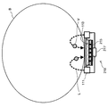
- FIG. 2 is a diagram illustrating a configuration of a measurement unit of the measurement apparatus according to the first embodiment of the present disclosure.
- the measurement unit 110 includes a light source unit 111, a microlens array 113, and an image sensor 115.
- the light source unit 111 irradiates the living body B with the measurement light L having a predetermined wavelength.
- the light source unit 111 is provided adjacent to the microlens array 113 such that the emission surface of the measurement light L faces the living body B.
- the light source unit 111 may be provided at the end of the microlens array 113.
- a light emitting diode LED: Light Emitting Diode
- a small laser is used as described above, the light source unit 111 may irradiate light having a single wavelength as the measurement light L, or may irradiate a plurality of lights having different wavelengths in a time-sharing manner.
- a microlens array (MLA) 113 guides the measurement light L reflected and diffused inside the living body B and emitted from the surface of the living body B to the image sensor 115.
- the microlens array 112 includes, for example, a plurality of light receiving lenses arranged in a grid, and each light receiving lens emits measurement light L emitted from the surface of the living body B in a predetermined region. guided to. Since the microlens array 113 is a lens array with little curvature of field and no distortion in the depth direction, good measurement data can be obtained by guiding the measurement light L to the image sensor 115 using the microlens array 113. Obtainable.
- the image sensor 115 converts the intensity of the measurement light L received by a photodetector (PD) or the like into an electrical signal and outputs the electrical signal to the measurement data acquisition unit 130.
- Examples of the image sensor 115 include a CCD (Charge Coupled Devices) type image sensor, a CMOS (Complementary Metal Oxide Semiconductor) type image sensor, a sensor using an organic EL as a light receiving element, and a TFT (Thin Film Transistor) type two-dimensional image sensor. area sensor is used.
- the image sensor 115 acquires both a captured image for specifying the position and depth of a vein, which will be described later, and a light amount distribution for estimating blood components.
- the image sensor 115 supports light in a wide wavelength band. Note that when light having different wavelengths is emitted from the light source unit 111 in a time-sharing manner, the imaging element 115 may acquire the captured image and the light amount distribution using light having different wavelengths.
- the measurement light L that is irradiated by the light source unit 111 and reflected / diffused inside the living body B passes through the vein V in which at least a part thereof exists inside the living body B.
- some change occurs in the measurement light L depending on the characteristics of the blood component of the vein V.
- the change in the measurement light L is measured using the microlens array 113 and the image sensor 115, and the blood component of the vein V is estimated.
- the characteristic of the blood component used here may be an absorption characteristic, a scattering characteristic, an optical rotation characteristic, or the like.
- the measurement unit 110 appropriately includes other components according to the characteristics used here. For example, when utilizing the optical rotation characteristics of blood components, a polarizing filter may be provided between the microlens array 113 and the living body B.
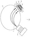
- FIG. 3 is a diagram for describing information obtained from the measurement light in the first embodiment of the present disclosure. Since the living body B is a medium that scatters light very well, the measurement light L emitted from the light source unit 110 travels while diffusing inside the living body B, is emitted from the surface of the living body B at a certain position, and is a microlens. It enters the array 113, as determined by the imaging element 115.
- the blood component estimation unit 160 of the measuring apparatus 100 models such characteristics as light scattering and attenuation at the position of each light receiving lens of the microlens array 113 along the optical path of the measurement light L. It is used for estimation of blood components.
- the measurement light L emitted from the light source unit 111 and incident on the microlens array 113 is not only the vein V on the optical path, but also the artery A, the skin, Also affected by body tissues such as subcutaneous tissue. Therefore, in order to accurately estimate the blood component of the vein V, it is desirable to exclude the influence of other components such as the artery A and body tissue from the measurement result.
- the position of the vein V is specified by the vein position specifying unit 140 and the depth of the vein V is specified by the vein depth specifying unit 150.
- position corresponds to the position in the direction parallel to the measurement unit 110, that is, the coordinates of the x-axis and y-axis in the drawing
- depth is perpendicular to the measurement unit 110. This corresponds to the depth in a specific direction, that is, the coordinate of the z axis in the figure.
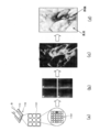
- FIG. 4 is a diagram for describing processing of the vein position specifying unit according to the first embodiment of the present disclosure.
- the vein position specifying unit 140 performs image processing on a captured image acquired by the measurement unit 110 as illustrated in (a), and obtains information indicating the position of the vein V as illustrated in (b). Generate.
- Measurement light L emitted from the light source unit 111 of the measuring unit 110 and entering the living body B is scattered inside the living body B and enters the microlens array 113.
- the position corresponding to the vein V is an area having lower luminance than the surroundings.
- the shape of the vein V represented in the captured image is also referred to as a vein pattern.
- the vein position specifying unit 140 extracts a vein pattern by applying a difference filter to the captured image, for example.
- the difference filter is a filter that outputs a large value as an output value at a portion where the difference between the pixel of interest and the surrounding pixels is large between the pixel of interest and the surrounding pixels.
- the difference filter is a filter that emphasizes a line or an edge in an image by a calculation using a difference in gradation value between a pixel of interest and a neighboring pixel.
- a differential filter such as a primary spatial differential filter or a secondary spatial differential filter may be used as the differential filter.
- the primary spatial differential filter is a filter that calculates a difference between gradation values of adjacent pixels in the horizontal direction and the vertical direction for the pixel of interest
- the secondary spatial differential filter is the pixel of interest. Is a filter that extracts a portion where the amount of change in the difference in gradation value is large.
- the following Log (Laplacian of Gaussian) filter can be used.
- the Log filter (Formula 103) is represented by the second derivative of a Gaussian filter (Formula 102) which is a smoothing filter using a Gaussian function.
- ⁇ represents a standard deviation of the Gaussian function and is a variable representing the degree of smoothing of the Gaussian filter.
- ⁇ in the following equation 103 is a parameter representing the standard deviation of the Gaussian function as in the equation 102, and changing the value of ⁇ can change the output value when the log filter processing is performed. it can.
- the vein position specifying unit 140 may perform post-processing such as threshold processing, binarization processing, and thinning processing on the captured image after applying the difference filter as described above. Through such post-processing, a skeleton having a vein pattern as shown in FIG.
- the vein position specifying unit 140 provides, for example, the vein pattern extracted as described above to the blood component estimating unit 160 as information indicating the position of the vein V existing inside the living body.
- FIG. 5 is a diagram for describing processing of the vein depth specifying unit according to the first embodiment of the present disclosure.
- the vein depth specifying unit 150 performs image processing on a captured image as shown in (b) acquired by the measuring unit 110 as shown in (a), and as shown in (c). Obtain parallax information.
- the vein depth specifying unit 150 acquires the depth information of the vein V as shown in (d) based on the parallax information.
- the vein depth specifying unit 150 may use the method of Light Field Photography.
- the microlens array 113 includes a plurality of light receiving lenses 1131 arranged in a lattice pattern or the like.
- the light guided by each of the light receiving lenses 1131 forms an image on the light receiving element 1151 on the imaging element 115, and a captured image is generated by the light receiving element 1151.
- a plurality of light receiving elements 1151 are assigned to one light receiving lens 1131. Although almost the same image is formed on the light receiving elements 1151 assigned to the same light receiving lens 1131, the direction of the imaging target with respect to each light receiving element 1151 is different depending on the arrangement position of the light receiving elements 1151. Therefore, as shown in (b), parallax is included in the vertical direction and the horizontal direction between the images captured by the respective light receiving elements 1151 assigned to the same light receiving lens 1131.
- the vein depth specifying unit 150 detects edges by differentiating the captured images obtained from the respective light receiving elements 1151 corresponding to the same light receiving lens 1131.
- the vein depth specifying unit 150 detects the above-described parallax by comparing where the same point of the imaging target indicated by the edge is located in each captured image.
- the parallax detected here is shown as a map in FIG.
- the light receiving lens 1131 is set so as to include the vein V inside the living body B in the depth of field, the detected edge is derived from the image of the vein V.
- the vein depth specifying unit 150 uses the information on the parallax between the captured images detected as described above and the positional relationship between the light receiving elements 1151 that acquired the captured images, and the light receiving element 1151.
- a separation distance from the imaging target, that is, the vein V is calculated. From here, if the distance from the light receiving element 1151 to the living body B is reduced, the depth from the surface of the living body B to the vein V is calculated.
- a map of the depth thus calculated is shown in (d). In the illustrated example, the portion where the depth is detected is the region of the vein V, and the portion where the depth is not detected is the region of the epidermis other than the vein V.
- the difference in the direction of the imaging target between the light receiving elements 1151 corresponding to the same light receiving lens 1131 is also the difference in the direction of light incident on the light receiving lens 1131. That is, each light receiving element 1151 receives light in different directions incident on the light receiving lens 1131.
- the light rays represented by the image acquired by the light receiving element 1151 corresponding to each light receiving lens 1131 can be synthesized by software. Thus, an image focused on an arbitrary position can be generated.
- the vein depth specifying unit 150 may acquire a captured image in which a focal position is arbitrarily set using this technique, and calculate the depth of the vein V in more detail.
- Processing of blood component estimation unit 6 and 7 are diagrams for describing processing of the blood component estimation unit according to the first embodiment of the present disclosure.
- the blood component estimation unit 160 analyzes the optical spectrum of the measurement light L emitted from the region corresponding to the vein V of the living body B.
- the region corresponding to the vein V is grasped based on, for example, information on the position of the vein V specified by the vein position specifying unit 140.
- the optical spectrum of the measurement light L in the region of the vein V includes the influence of the blood component of the artery A on the change due to the blood component of the vein V, and the vein V And the influence of the components of the skin and subcutaneous tissue existing between the body B and the surface of the living body B are superimposed. Therefore, the blood component estimation unit 160 separates the influence of these components from the measurement result.
- the blood component estimation unit 160 separates the influence of the skin and the subcutaneous tissue based on the information indicating the depth of the vein V acquired by the vein depth specifying unit 150. As shown in the drawing, as a result of the processing by the vein depth specifying unit 150, the depth (for example, D1 to D3) of the vein V from the surface of the living body B is calculated.
- the blood component estimation unit 160 mixes the depth D1 to D3, that is, the thickness of the skin and subcutaneous tissue, with the light absorption spectrum of the skin and subcutaneous tissue measured in advance, and mixes it with the optical spectrum of the measurement light L. Calculate the effects of skin and subcutaneous tissue components and remove them from the measurement results.
- the blood component estimation unit 160 includes information indicating the position of the vein V acquired by the vein position specifying unit 140 and the time-series change of the optical spectrum of the measurement light L. to separate them on the basis of. For example, the blood component estimation unit 160 extracts a time-varying component synchronized with the pulse included in the optical spectrum of the measurement light L detected in the region adjacent to the position of the vein V, and uses this vector component as the pulse of the artery. remove from the measurement results as a component derived from dynamic.
- the use of the optical spectrum of the measurement light L detected in the region adjacent to the position of the vein V is considered that the measurement light L detected in this adjacent region does not pass through the vein V and indicates a noise component. This is because it is considered appropriate as an optical spectrum.
- the blood component estimation unit 160 separates the influence of the artery A, the skin, and the skin tissue from the optical spectrum of the measurement light L emitted from the vein V region by the processing as described above.
- the blood component estimation unit 160 can estimate the blood component of the vein V more accurately by analyzing the optical spectrum from which the influence of the artery A, skin, and skin tissue is separated.
- FIG. 8 is a flowchart illustrating a process according to the first embodiment of the present disclosure.
- the measurement unit 110 measures a living body (step S101).
- the measurement unit 110 irradiates the measurement light L from the light source unit 111 to enter the living body B, and scatters the measurement light L emitted from the surface of the living body B to the microlens array 113.
- the living body B is measured by being incident and receiving light by the image sensor 115.
- the vein position specifying unit 140 specifies the position of the vein V inside the living body B (step S103).
- the vein position specifying unit 140 specifies the position of the vein V, for example, by applying image processing using a difference filter to the captured image of the living body B acquired by the imaging element 115.
- the vein depth specifying unit 150 specifies the depth of the vein V (step S105).
- the vein depth specifying unit 150 determines the depth of the vein V based on the parallax extracted from the captured images respectively acquired by the plurality of light receiving elements 1151 of the imaging element 115 corresponding to the light receiving lenses 1131 of the microlens array 113. Is identified.
- the blood component estimation unit 160 determines the body tissue such as skin or subcutaneous tissue existing between the vein V and the surface of the living body B.
- the thickness is estimated, and the influence of the body tissue corresponding to this thickness is separated from the optical spectrum of the measurement light L acquired by the image sensor 115 in the region of the vein V (step S107).
- the blood component estimation unit 160 images the time-varying component synchronized with the pulse of the optical spectrum of the measurement light L in the region adjacent to the position of the vein V at the position of the vein V as an effect of the arterial component. Separated from the optical spectrum of the measurement light L acquired by the element 115 (step S109).
- the blood component estimation unit 160 estimates the blood component of the vein V based on the optical spectrum from which the effects of the artery A, skin, and skin tissue are separated in steps S107 and S109 (step S111).
- step S103 and step S105 that receive the captured image acquired in step S101 may be executed in parallel or in reverse order after execution of step S101.
- step S109 using the information on the position of the vein V specified in step S103 may be executed in parallel with step S105 or step S107 or before these steps.
- the influence of body components and arteries from the optical spectrum of the measurement light L detected in the vein V region can be separated and the blood component of the vein V can be accurately measured.
- the measurement unit 110 can be thinned by using an optical system including the microlens array 113 and the imaging element 115 for the measurement unit 110.
- a plurality of light receiving elements 1151 of the imaging element 115 correspond to each light receiving lens 1131 of the microlens array 113.
- the depth of the vein V based on the parallax information can be specified and the resolution of the captured image can be compatible.
- the measuring unit 110 can be reduced in size.
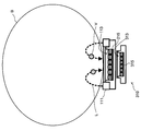
- the measurement unit 210 includes a light source unit 111, a microlens array 113, an image sensor 215, and a photodetector 217.
- the light source unit 111 and the microlens array 113 are the same components as those in the first embodiment.
- the image sensor 215 acquires a captured image exclusively and does not acquire the distribution of the light amount of the measurement light L. Therefore, the image sensor 215 does not necessarily correspond to light in a wide wavelength band.
- the light amount distribution of the measurement light L is acquired by the photodetector 217 provided on the opposite side across the imaging element 215 when viewed from the living body B. For this reason, a material such as silicon that transmits light having a wavelength to be detected by the photodetector 217 is used for the imaging element 215.
- the photodetector 217 is provided on the back side of the image sensor 215 when viewed from the living body B, and acquires the distribution of the light amount of the measurement light L. It is desirable that the photodetector 217 corresponds to light in a wide wavelength band using a material such as indium gallium arsenide (InGaAs).
- a material such as indium gallium arsenide (InGaAs).
- a photodetector 217 that detects the measurement light L is provided separately from the imaging element 215 that acquires the captured image of the living body B. Therefore, in the present embodiment, the image sensor 215 may not be compatible with a wide band.
- the configuration of the measurement unit 310 is different from that of the measurement unit 110 according to the first embodiment, but the other components are substantially the same. Detailed description is omitted.
- the measurement unit 310 includes a light source unit 111, microlens arrays 113 and 313, and image sensors 215 and 315.
- the light source unit 111 and the microlens array 113 are the same components as those in the first embodiment.
- the image sensor 215 is a component similar to that of the second embodiment.
- the microlens array 313 and the image sensor 315 are used in place of the photodetector 217 in the second embodiment. That is, the microlens array 313 and the image sensor 315 are provided on the back side of the image sensor 215 when viewed from the living body B. That is, the microlens array 113 and the image sensor 215, and the microlens array 313 and the image sensor 315 are superimposed in the direction in which the measurement light L is emitted.
- the image sensor 315 corresponds to light in a wide wavelength band using a material such as indium gallium arsenide (InGaAs) in order to acquire the distribution of the light amount of the measurement light L. It is desirable to be a thing. Note that the areas of the microlens 313 and the image sensor 315 may be smaller than the areas of the microlens array 113 and the image sensor 215.
- a material such as indium gallium arsenide (InGaAs) in order to acquire the distribution of the light amount of the measurement light L. It is desirable to be a thing.
- the areas of the microlens 313 and the image sensor 315 may be smaller than the areas of the microlens array 113 and the image sensor 215.
- another microlens array 313 and another imaging device 315 that detect the measurement light L are provided in addition to the imaging device 215 that acquires a captured image of the living body B. .
- the measuring unit 110 can be thinned.
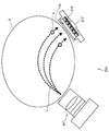
- the configuration of the measurement unit 410 is different from that of the measurement unit 110 according to the first embodiment, but the other components are substantially the same. Detailed description is omitted.
- the measurement unit 410 includes a light source unit 411, a microlens array 113, and an image sensor 115.
- the microlens array 113 and the image sensor 115 are the same components as those in the first embodiment.
- the light source unit 411 irradiates the living body B with measurement light L having a predetermined wavelength.
- the light source unit 411 includes, for example, an LED array, a polarizing filter, an objective lens, and the like.
- the light source unit 411 is the same as the light source unit 111 according to the first embodiment in that it is arranged so that the emission surface of the measurement light L faces the living body B, but is provided separately from the microlens array 113. that is different from that of the light source unit 111.
- the measurement light L irradiated from the light source unit 411 and incident on the microlens array 113 passes through a wider range inside the living body B. That is, the measurement unit 410 can acquire measurement data for a wider range inside the living body B. Therefore, for example, the blood component of the vein V can be measured regardless of the position and orientation of the living body B placed on the measurement unit 410 by the measurement subject.
- the configuration of the measurement unit 510 is different from that of the measurement unit 110 according to the first embodiment, but the other components are substantially the same. Detailed description is omitted.
- the measurement unit 510 includes a light source unit 411, a microlens array 113, an image sensor 215, and a photodetector 217.
- the light source unit 411 is a component similar to that of the fourth embodiment.
- the microlens array 113, the image sensor 215, and the photodetector 217 are the same components as those in the second embodiment.
- the present embodiment is a combination of the second embodiment and the fourth embodiment. Therefore, in addition to being able to acquire measurement data for a wider range inside the living body B, the image sensor 215 may not be compatible with a wide band.
- the configuration of the measurement unit 610 is different from that of the measurement unit 110 according to the first embodiment, but the other components are substantially the same. Detailed description is omitted.
- the measurement unit 610 includes a light source unit 411, microlens arrays 113 and 313, and image sensors 215 and 315.
- the light source unit 411 is a component similar to that of the fourth embodiment.
- the microlens arrays 113 and 313 and the image sensors 215 and 315 are the same components as those in the third embodiment.
- the present embodiment is a combination of the third embodiment and the fourth embodiment. Therefore, in addition to being able to acquire measurement data for a wider range inside the living body B, the imaging element 215 need not be compatible with a wide band, and the measurement unit 610 can be made thin.
- the blood component of the vein can be accurately measured by optical measurement.
- measurement similar to blood component analysis of venous blood measured in a hospital can be performed non-invasively.
- blood component analysis can be performed with less frequent calibration than conventional body fluid analysis.
- the influence on the measurement result by an individual difference can be reduced by canceling the influence by skin tissue etc.
- changes in blood components due to the influence of meals and the like are more gradual than in arterial blood, so that stable measurement is possible.
- FIG. 14 is a block diagram for explaining a hardware configuration of the information processing apparatus 900 according to the embodiment of the present disclosure.
- the information processing apparatus 900 mainly includes a CPU 901, a ROM 903, and a RAM 905.
- the information processing apparatus 900 further includes a host bus 907, a bridge 909, an external bus 911, an interface 913, a sensor 914, an input device 915, an output device 917, a storage device 919, a drive 921, a connection port 923, and a communication device 925. provided.
- the CPU 901 functions as an arithmetic processing device and a control device, and controls all or a part of the operation in the information processing device 900 according to various programs recorded in the ROM 903, the RAM 905, the storage device 919, or the removable recording medium 927.
- the ROM 903 stores programs used by the CPU 901, calculation parameters, and the like.
- the RAM 905 primarily stores programs used by the CPU 901, parameters that change as appropriate during execution of the programs, and the like. These are connected to each other by a host bus 907 constituted by an internal bus such as a CPU bus.
- the host bus 907 is connected to an external bus 911 such as a PCI (Peripheral Component Interconnect / Interface) bus via a bridge 909.
- PCI Peripheral Component Interconnect / Interface
- the sensor 914 is, for example, detection means for detecting biological information unique to the user or various information used for acquiring such biological information.
- Examples of the sensor 914 include various image pickup devices such as a CCD (Charge Coupled Device) and a CMOS (Complementary Metal Oxide Semiconductor).
- the sensor 914 may further include an optical system such as a lens and a light source used for imaging a living body part.
- the sensor 914 may be a microphone or the like for acquiring sound or the like.
- the sensor 914 may include various measuring devices such as a thermometer, an illuminometer, a hygrometer, a speedometer, and an accelerometer in addition to the above-described ones.
- the input device 915 is an operation means operated by the user such as a mouse, a keyboard, a touch panel, a button, a switch, and a lever. Further, the input device 915 may be, for example, remote control means using infrared rays or other radio waves, or may be an external connection device 929 such as a mobile phone or a PDA that supports the operation of the information processing device 900. good. Furthermore, the input device 915 includes an input control circuit that generates an input signal based on information input by a user using the above-described operation means and outputs the input signal to the CPU 901, for example. A user of the information processing apparatus 900 can input various data and instruct a processing operation to the information processing apparatus 900 by operating the input device 915.
- the output device 917 is a device that can notify the user of the acquired information visually or audibly. Examples of such devices include CRT display devices, liquid crystal display devices, plasma display devices, EL display devices and display devices such as lamps, audio output devices such as speakers and headphones, printer devices, mobile phones, and facsimiles.
- the output device 917 outputs results obtained by various processes performed by the information processing apparatus 900. Specifically, the display device displays the results obtained by various processes performed by the information processing device 900 as text or images.
- the audio output device converts an audio signal composed of reproduced audio data, acoustic data, and the like into an analog signal and outputs the analog signal.
- the storage device 919 is a data storage device configured as an example of a storage unit of the information processing device 900.
- the storage device 919 includes, for example, a magnetic storage device such as an HDD (Hard Disk Drive), a semiconductor storage device, an optical storage device, or a magneto-optical storage device.
- the storage device 919 stores programs executed by the CPU 901, various data, various data acquired from the outside, and the like.
- the drive 921 is a reader / writer for a recording medium, and is built in or externally attached to the information processing apparatus 900.
- the drive 921 reads information recorded on a removable recording medium 927 such as a mounted magnetic disk, optical disk, magneto-optical disk, or semiconductor memory, and outputs the information to the RAM 905.
- the drive 921 can write a record on a removable recording medium 927 such as a magnetic disk, an optical disk, a magneto-optical disk, or a semiconductor memory.
- the removable recording medium 927 is, for example, a DVD medium, an HD-DVD medium, a Blu-ray medium, or the like.
- the removable recording medium 927 may be a compact flash (registered trademark) (CompactFlash: CF), a flash memory, or an SD memory card (Secure Digital memory card). Further, the removable recording medium 927 may be, for example, an IC card (Integrated Circuit card) on which a non-contact IC chip is mounted, an electronic device, or the like.
- CompactFlash CompactFlash: CF
- flash memory a flash memory
- SD memory card Secure Digital memory card
- the removable recording medium 927 may be, for example, an IC card (Integrated Circuit card) on which a non-contact IC chip is mounted, an electronic device, or the like.
- the connection port 923 is a port for directly connecting a device to the information processing apparatus 900.
- Examples of the connection port 923 include a USB (Universal Serial Bus) port, an IEEE 1394 port, a SCSI (Small Computer System Interface) port, and the like.
- As another example of the connection port 923 there are an RS-232C port, an optical audio terminal, an HDMI (High-Definition Multimedia Interface) port, and the like.
- the communication device 925 is a communication interface configured with, for example, a communication device for connecting to the communication network 931.
- the communication device 925 is, for example, a communication card for a wired or wireless LAN (Local Area Network), Bluetooth (registered trademark), or WUSB (Wireless USB).
- the communication device 925 may be a router for optical communication, a router for ADSL (Asymmetric Digital Subscriber Line), or a modem for various communication.
- the communication device 925 can transmit and receive signals and the like according to a predetermined protocol such as TCP / IP, for example, with the Internet or other communication devices.
- the communication network 931 connected to the communication device 925 is configured by a wired or wireless network, and may be, for example, the Internet, a home LAN, infrared communication, radio wave communication, satellite communication, or the like. .
- each component described above may be configured using a general-purpose member, or may be configured by hardware specialized for the function of each component. Therefore, it is possible to change the hardware configuration to be used as appropriate according to the technical level at the time of carrying out this embodiment.
- At least a part of a living body is irradiated with measurement light having a predetermined wavelength, and the measurement light scattered in the living body and emitted from the surface of the living body is detected, and the emitted measurement light is
- a measurement unit that collects light using a lens array having a plurality of light receiving lenses arranged in an array to obtain a captured image of the living body;
- a vein position specifying unit for specifying the position of a vein existing inside the living body based on the captured image;
- a vein depth specifying unit that specifies the depth of the vein based on the captured image;
- a blood component estimation unit that estimates a blood component of the vein based on information obtained from the detected measurement light by using the position of the vein and the depth of the vein.
- the blood component estimation unit estimates the thickness of the body tissue existing between the surface of the living body and the vein based on the depth of the vein, and the measurement light detected at the position of the vein.
- the measurement apparatus according to (1), wherein the blood component of the vein is estimated by excluding the influence of the body tissue for the estimated thickness from the information obtained from the information.
- the blood component estimation unit compares information obtained from measurement light detected in an adjacent region adjacent to the vein position and information obtained from measurement light detected at the vein position.
- the blood component estimation unit corrects information obtained from the measurement light detected at the position of the vein using information obtained from measurement light detected in an adjacent region adjacent to the vein position.
- the blood component estimation unit extracts a time-varying component derived from pulsation of an artery present in the living body from information obtained from measurement light detected in the adjacent region, and the position of the vein
- the measurement unit includes an image sensor that acquires the captured image, In the imaging device, a plurality of light receiving elements are assigned to one light receiving lens, The vein depth specifying unit specifies the depth of the vein based on parallax information extracted from the captured images respectively acquired by the plurality of light receiving elements corresponding to the same light receiving lens.
- the measuring device according to any one of 5).
- the measurement apparatus (7) The measurement apparatus according to (6), wherein the vein depth specifying unit extracts information on the parallax from the captured image using a method of Light Field Photography.
- the blood component estimation unit calibrates the estimation result of the blood component based on a blood component measured in advance by collecting blood according to any one of (1) to (7).
- the measuring device described. (9) The measurement apparatus according to any one of (1) to (8), wherein the measurement unit includes an image sensor that detects the emitted measurement light and acquires the captured image. (10) The measurement according to any one of (1) to (8), wherein the measurement unit includes a detector that detects the emitted measurement light and an image sensor that acquires the captured image. apparatus.
- the measurement apparatus according to (10), wherein the measurement unit includes a lens array different from the lens array and an image sensor different from the image sensor as the detector. (12) The imaging device and the detector are overlapped in a direction in which the measurement light is emitted, and the imaging device transmits at least part of the measurement light, according to (10) or (11) measuring device. (13) The measurement unit includes a light source unit that irradiates the measurement light, The measurement apparatus according to any one of (1) to (12), wherein the light source unit is provided at an end of the lens array. (14) The measurement unit includes a light source unit that irradiates the measurement light, The measuring apparatus according to any one of (1) to (12), wherein the light source unit is provided separately from the lens array.
- At least a part of the living body is irradiated with measurement light having a predetermined wavelength, and the measurement light scattered in the living body and emitted from the surface of the living body is detected, and the emitted measurement light is To a computer that can communicate with a measurement unit that collects light by using a lens array having a plurality of light receiving lenses arranged in an array and obtains a captured image of the living body, A vein position specifying function for specifying the position of a vein existing inside the living body based on the captured image; A vein depth specifying function for specifying the depth of the vein based on the captured image; A blood component estimation function for estimating a blood component of the vein based on information obtained from the detected measurement light using the position of the vein and the depth of the vein.
- At least a part of the living body is irradiated with measurement light having a predetermined wavelength, and the measurement light scattered in the living body and emitted from the surface of the living body is detected, and the emitted measurement light is To a computer that can communicate with a measurement unit that collects light by using a lens array having a plurality of light receiving lenses arranged in an array and obtains a captured image of the living body, A vein position specifying function for specifying the position of a vein existing inside the living body based on the captured image; A vein depth specifying function for specifying the depth of the vein based on the captured image; A blood component estimation function for estimating the blood component of the vein based on information obtained from the detected measurement light using the position of the vein and the depth of the vein is recorded.
- At least a part of the living body is irradiated with measuring light having a predetermined wavelength, and the measuring light scattered in the living body and emitted from the surface of the living body is detected, and the emitted measuring light is A measurement unit that collects light using a lens array having a plurality of light receiving lenses arranged in an array to obtain a captured image of the living body; A vein position specifying unit for specifying the position of a vein existing inside the living body based on the captured image; A vein depth specifying unit that specifies the depth of the vein based on the captured image; The thickness of the body tissue existing between the surface of the living body and the vein is estimated based on the depth of the vein, and the estimated thickness is obtained from information obtained from the measurement light detected at the position of the vein A time-varying component derived from the pulsation of an artery present inside the living body from information obtained from measurement light detected in an adjacent region adjacent to the position of the vein.
- a blood component estimation unit that extracts and estimates the blood component of the vein by further excluding the time-varying component from the information obtained from the measurement light detected at the position of the vein.
- At least a part of a living body is irradiated with measurement light having a predetermined wavelength, and the measurement light scattered in the living body and emitted from the surface of the living body is detected, and the emitted measurement light is Condensing using a lens array having a plurality of light receiving lenses arranged in an array to obtain a captured image of the living body; Identifying the position of a vein present inside the living body based on the captured image; Identifying the depth of the vein based on the captured image; The thickness of the body tissue existing between the surface of the living body and the vein is estimated based on the depth of the vein, and the estimated thickness is obtained from information obtained from the measurement light detected at the position of the vein
- a time-varying component derived from the pulsation of an artery present inside the living body from information obtained from measurement light detected in an adjacent region adjacent
- At least a part of a living body is irradiated with measurement light having a predetermined wavelength, and the measurement light scattered in the living body and emitted from the surface of the living body is detected, and the emitted measurement light is
- a computer that can communicate with a measurement unit that collects light by using a lens array having a plurality of light receiving lenses arranged in an array and obtains a captured image of the living body, A vein position specifying function for specifying the position of a vein existing inside the living body based on the captured image; A vein depth specifying function for specifying the depth of the vein based on the captured image;
- the thickness of the body tissue existing between the surface of the living body and the vein is estimated based on the depth of the vein, and the estimated thickness is obtained from information obtained from the measurement light detected at the position of the vein
Landscapes
- Health & Medical Sciences (AREA)
- Life Sciences & Earth Sciences (AREA)
- Physics & Mathematics (AREA)
- Biomedical Technology (AREA)
- Medical Informatics (AREA)
- Biophysics (AREA)
- Pathology (AREA)
- Engineering & Computer Science (AREA)
- Veterinary Medicine (AREA)
- Heart & Thoracic Surgery (AREA)
- Public Health (AREA)
- Molecular Biology (AREA)
- Surgery (AREA)
- Animal Behavior & Ethology (AREA)
- General Health & Medical Sciences (AREA)
- Optics & Photonics (AREA)
- Spectroscopy & Molecular Physics (AREA)
- Vascular Medicine (AREA)
- Measurement Of The Respiration, Hearing Ability, Form, And Blood Characteristics Of Living Organisms (AREA)
Priority Applications (2)
| Application Number | Priority Date | Filing Date | Title |
|---|---|---|---|
| US14/364,542 US20140323831A1 (en) | 2011-12-19 | 2012-12-03 | Measurement device, measurement method, program, and recording medium |
| CN201280061381.3A CN103987317B (zh) | 2011-12-19 | 2012-12-03 | 测量设备和方法 |
Applications Claiming Priority (2)
| Application Number | Priority Date | Filing Date | Title |
|---|---|---|---|
| JP2011-277614 | 2011-12-19 | ||
| JP2011277614A JP5990906B2 (ja) | 2011-12-19 | 2011-12-19 | 測定装置、測定方法、プログラムおよび記録媒体 |
Publications (1)
| Publication Number | Publication Date |
|---|---|
| WO2013094392A1 true WO2013094392A1 (ja) | 2013-06-27 |
Family
ID=48668291
Family Applications (1)
| Application Number | Title | Priority Date | Filing Date |
|---|---|---|---|
| PCT/JP2012/081255 Ceased WO2013094392A1 (ja) | 2011-12-19 | 2012-12-03 | 測定装置、測定方法、プログラムおよび記録媒体 |
Country Status (4)
| Country | Link |
|---|---|
| US (1) | US20140323831A1 (https=) |
| JP (1) | JP5990906B2 (https=) |
| CN (1) | CN103987317B (https=) |
| WO (1) | WO2013094392A1 (https=) |
Cited By (1)
| Publication number | Priority date | Publication date | Assignee | Title |
|---|---|---|---|---|
| CN106132299A (zh) * | 2014-03-31 | 2016-11-16 | 索尼公司 | 测量装置、测量方法、程序和记录媒介 |
Families Citing this family (7)
| Publication number | Priority date | Publication date | Assignee | Title |
|---|---|---|---|---|
| WO2015151587A1 (ja) * | 2014-03-31 | 2015-10-08 | ソニー株式会社 | 測定装置、測定方法、プログラム及び記録媒体 |
| WO2017159574A1 (ja) * | 2016-03-18 | 2017-09-21 | テルモ株式会社 | 心機能測定装置、心機能測定方法および心機能測定プログラム |
| KR102531994B1 (ko) | 2017-12-29 | 2023-05-15 | 삼성전자주식회사 | 생체 성분 측정 장치 및 방법 |
| JP7093963B2 (ja) * | 2018-05-22 | 2022-07-01 | メディカルフォトニクス株式会社 | 血管検知装置 |
| CN110279406B (zh) * | 2019-05-06 | 2022-07-15 | 苏宁金融服务(上海)有限公司 | 一种基于摄像头的无接触式的脉率测量方法及装置 |
| JP7570064B2 (ja) * | 2019-12-10 | 2024-10-21 | パナソニックIpマネジメント株式会社 | 空間温度推定システム |
| JP7574850B2 (ja) * | 2020-07-01 | 2024-10-29 | 日本電気株式会社 | 受光装置 |
Citations (3)
| Publication number | Priority date | Publication date | Assignee | Title |
|---|---|---|---|---|
| JP2007044203A (ja) * | 2005-08-09 | 2007-02-22 | Toshiba Corp | 生体情報計測装置及びその方法 |
| JP2009006037A (ja) * | 2007-06-29 | 2009-01-15 | Sysmex Corp | 非侵襲血液成分測定方法及び非侵襲血液成分測定装置 |
| JP2010267087A (ja) * | 2009-05-14 | 2010-11-25 | Sony Corp | 静脈撮像装置、静脈画像補間方法およびプログラム |
Family Cites Families (10)
| Publication number | Priority date | Publication date | Assignee | Title |
|---|---|---|---|---|
| DE69634858T2 (de) * | 1995-12-27 | 2006-05-11 | Sysmex Corp. | Nichtinvasive blutuntersuchungsvorrichtung |
| US20080107309A1 (en) * | 2006-11-03 | 2008-05-08 | Cerni Consulting, Llc | Method and apparatus for biometric identification |
| JP4832273B2 (ja) * | 2006-12-13 | 2011-12-07 | 日立マクセル株式会社 | 生体認証用撮像モジュール、生体認証装置及びプリズム |
| US20100145175A1 (en) * | 2008-08-22 | 2010-06-10 | Soldo Monnett H | Systems and methods for verification of sample integrity |
| JP2009032227A (ja) * | 2007-06-22 | 2009-02-12 | Hitachi Ltd | 指静脈認証装置および情報処理装置 |
| US8559756B2 (en) * | 2007-08-06 | 2013-10-15 | Adobe Systems Incorporated | Radiance processing by demultiplexing in the frequency domain |
| JP4905326B2 (ja) * | 2007-11-12 | 2012-03-28 | ソニー株式会社 | 撮像装置 |
| JP5130885B2 (ja) * | 2007-12-03 | 2013-01-30 | ソニー株式会社 | 情報処理装置、情報処理方法およびプログラム |
| JP4748199B2 (ja) * | 2008-09-30 | 2011-08-17 | ソニー株式会社 | 静脈撮像装置および静脈撮像方法 |
| JP5326792B2 (ja) * | 2009-05-14 | 2013-10-30 | ソニー株式会社 | 静脈撮像装置、位置ズレ補間方法およびプログラム |
-
2011
- 2011-12-19 JP JP2011277614A patent/JP5990906B2/ja not_active Expired - Fee Related
-
2012
- 2012-12-03 CN CN201280061381.3A patent/CN103987317B/zh not_active Expired - Fee Related
- 2012-12-03 US US14/364,542 patent/US20140323831A1/en not_active Abandoned
- 2012-12-03 WO PCT/JP2012/081255 patent/WO2013094392A1/ja not_active Ceased
Patent Citations (3)
| Publication number | Priority date | Publication date | Assignee | Title |
|---|---|---|---|---|
| JP2007044203A (ja) * | 2005-08-09 | 2007-02-22 | Toshiba Corp | 生体情報計測装置及びその方法 |
| JP2009006037A (ja) * | 2007-06-29 | 2009-01-15 | Sysmex Corp | 非侵襲血液成分測定方法及び非侵襲血液成分測定装置 |
| JP2010267087A (ja) * | 2009-05-14 | 2010-11-25 | Sony Corp | 静脈撮像装置、静脈画像補間方法およびプログラム |
Cited By (2)
| Publication number | Priority date | Publication date | Assignee | Title |
|---|---|---|---|---|
| CN106132299A (zh) * | 2014-03-31 | 2016-11-16 | 索尼公司 | 测量装置、测量方法、程序和记录媒介 |
| CN106132299B (zh) * | 2014-03-31 | 2020-04-14 | 索尼公司 | 测量装置、测量方法、程序和记录媒介 |
Also Published As
| Publication number | Publication date |
|---|---|
| CN103987317A (zh) | 2014-08-13 |
| CN103987317B (zh) | 2016-08-24 |
| US20140323831A1 (en) | 2014-10-30 |
| JP2013126510A (ja) | 2013-06-27 |
| JP5990906B2 (ja) | 2016-09-14 |
Similar Documents
| Publication | Publication Date | Title |
|---|---|---|
| JP5990906B2 (ja) | 測定装置、測定方法、プログラムおよび記録媒体 | |
| JP5948831B2 (ja) | 測定装置、測定方法、プログラム及び記録媒体 | |
| JP6539877B2 (ja) | 測定装置、測定方法、プログラム及び記録媒体 | |
| JP6539876B2 (ja) | 測定装置、測定方法、プログラム及び記録媒体 | |
| JP5948836B2 (ja) | 測定装置、測定方法、プログラム及び記録媒体 | |
| US10271746B2 (en) | Method and system for carrying out photoplethysmography | |
| JP4772036B2 (ja) | 血流を検出する装置及び方法 | |
| JP4748199B2 (ja) | 静脈撮像装置および静脈撮像方法 | |
| JP5970785B2 (ja) | 生体計測装置、生体計測方法、プログラムおよび記録媒体 | |
| US20190125221A1 (en) | Imaging apparatus, authentication processing apparatus, imaging method, authentication processing method, and program | |
| JP2014138691A (ja) | 画像処理装置、電子機器、内視鏡装置、プログラム及び画像処理方法 | |
| JP2009165630A (ja) | 静脈認証装置および静脈認証方法 | |
| JP5326793B2 (ja) | 静脈撮像装置、静脈画像補間方法およびプログラム | |
| JP2013103094A (ja) | 測定装置、測定方法、プログラム及び記録媒体 | |
| JP5935498B2 (ja) | 情報処理装置、情報処理方法、プログラム及び情報処理システム | |
| JP7480708B2 (ja) | 情報処理装置、情報処理方法、及びプログラム | |
| JP2013126510A5 (https=) | ||
| JP5056662B2 (ja) | 皮下パターン取得装置、皮下パターン取得方法および構造物テンプレート | |
| JP6010898B2 (ja) | 生体計測装置、生体計測方法、プログラムおよび記録媒体 | |
| KR20190001496A (ko) | 정보 처리 장치, 프로그램, 방법 및 시스템 |
Legal Events
| Date | Code | Title | Description |
|---|---|---|---|
| 121 | Ep: the epo has been informed by wipo that ep was designated in this application |
Ref document number: 12859051 Country of ref document: EP Kind code of ref document: A1 |
|
| WWE | Wipo information: entry into national phase |
Ref document number: 14364542 Country of ref document: US |
|
| NENP | Non-entry into the national phase |
Ref country code: DE |
|
| 122 | Ep: pct application non-entry in european phase |
Ref document number: 12859051 Country of ref document: EP Kind code of ref document: A1 |