WO2013065646A1 - Méthode de mesure de l'abondance de gènes - Google Patents
Méthode de mesure de l'abondance de gènes Download PDFInfo
- Publication number
- WO2013065646A1 WO2013065646A1 PCT/JP2012/077916 JP2012077916W WO2013065646A1 WO 2013065646 A1 WO2013065646 A1 WO 2013065646A1 JP 2012077916 W JP2012077916 W JP 2012077916W WO 2013065646 A1 WO2013065646 A1 WO 2013065646A1
- Authority
- WO
- WIPO (PCT)
- Prior art keywords
- primer
- gene
- base sequence
- nucleic acid
- genes
- Prior art date
Links
Images


Classifications
-
- C—CHEMISTRY; METALLURGY
- C12—BIOCHEMISTRY; BEER; SPIRITS; WINE; VINEGAR; MICROBIOLOGY; ENZYMOLOGY; MUTATION OR GENETIC ENGINEERING
- C12Q—MEASURING OR TESTING PROCESSES INVOLVING ENZYMES, NUCLEIC ACIDS OR MICROORGANISMS; COMPOSITIONS OR TEST PAPERS THEREFOR; PROCESSES OF PREPARING SUCH COMPOSITIONS; CONDITION-RESPONSIVE CONTROL IN MICROBIOLOGICAL OR ENZYMOLOGICAL PROCESSES
- C12Q1/00—Measuring or testing processes involving enzymes, nucleic acids or microorganisms; Compositions therefor; Processes of preparing such compositions
- C12Q1/68—Measuring or testing processes involving enzymes, nucleic acids or microorganisms; Compositions therefor; Processes of preparing such compositions involving nucleic acids
- C12Q1/6844—Nucleic acid amplification reactions
- C12Q1/6851—Quantitative amplification
-
- C—CHEMISTRY; METALLURGY
- C12—BIOCHEMISTRY; BEER; SPIRITS; WINE; VINEGAR; MICROBIOLOGY; ENZYMOLOGY; MUTATION OR GENETIC ENGINEERING
- C12Q—MEASURING OR TESTING PROCESSES INVOLVING ENZYMES, NUCLEIC ACIDS OR MICROORGANISMS; COMPOSITIONS OR TEST PAPERS THEREFOR; PROCESSES OF PREPARING SUCH COMPOSITIONS; CONDITION-RESPONSIVE CONTROL IN MICROBIOLOGICAL OR ENZYMOLOGICAL PROCESSES
- C12Q1/00—Measuring or testing processes involving enzymes, nucleic acids or microorganisms; Compositions therefor; Processes of preparing such compositions
- C12Q1/68—Measuring or testing processes involving enzymes, nucleic acids or microorganisms; Compositions therefor; Processes of preparing such compositions involving nucleic acids
- C12Q1/6844—Nucleic acid amplification reactions
- C12Q1/686—Polymerase chain reaction [PCR]
Definitions
- the present invention relates to a method of measuring gene abundance.
- genes there are multiple genes in one genome.
- the abundance of such a gene in the genome may affect the utility of the gene diagnosis or the effect of the drug, and the amount of the gene in the nucleic acid (for example, genome etc.) in the target sample may be grasped or measured. It may be required to Examples of such detection include measurement of copy number of gene, measurement of increase and decrease of gene amount, diagnosis of copy number variation (CNV), and the like.
- FISH method see, for example, Proc. Natl. Acad. Sci. USA, Vol. 89, pp. 5321-5325, June 1992
- competitive DNA binding There is a method using a fluorescent labeling method such as CGH method (see, for example, JP-A-7-505053) using In these methods, it is possible to confirm the abundance of the gene by reacting the fluorescent labeling compound directly to the chromosome and then observing the image processing or fluorescence microscope.
- CGH method see, for example, JP-A-7-505053
- 2010-538614 is a method for determining the relative copy number of a target polynucleotide sequence in a target genome, comprising: targeting gene sequence and reference in a sample containing the target genomic DNA
- the gene sequences are nucleic acid amplified, the sequence of each gene amplified by digital PCR is assayed, and the ratio between the number of amplified polynucleotide molecules containing the target gene sequence and the number of amplified polynucleotide molecules containing the reference gene sequence, The change in copy number is determined.
- JP-A-2011-516069 discloses a two-step nucleic acid amplification process and in between: And C. recovering the nucleic acid amplicon of interest.
- nucleic acids are amplified using target specific primers, thereby producing at least one nucleic acid amplicon comprising at least one consensus primer binding site, and obtaining the resulting nucleic acid amplifier
- the recon is separated (rescued) from the target specific primers used and then amplified using the common primers.
- double-stranded nucleic acids are respectively amplified by using a pair of primer pairs whose directions are different from each other to generate an amplification product (sometimes called an amplicon) of a target gene sequence.
- an amplification product sometimes called an amplicon
- the double-stranded amplicon accumulates exponentially in one reaction system in the process of nucleic acid amplification using a primer pair, and then, along with the increase in the accumulation amount of the generated complementary amplicon strand, It is known that the speed of the amplification reaction decreases and eventually ceases (plateau phase). The amount of amplicon does not increase because nucleic acid amplification no longer occurs when this plateau period is reached.
- multiplex PCR is often used in which primers specific to each mutant are set (see, for example, JP-A-2012-100628).
- multiplex PCR it is necessary to prepare a large number of primers and perform optimization in consideration of the possibility that each primer will form a dimer. Through this process of optimization, it is necessary to recreate the primers and to examine whether they can be measured or not through experiments.
- the degree of difficulty of this optimization process increases, and it takes a great deal of time, effort and cost to find out the optimum primer set.
- an object of the present invention is to provide a method for measuring gene abundance, which measures the abundance of genes in nucleic acid in a target sample more accurately and conveniently than in the prior art.
- a nucleic acid encoding at least two genes which may differ in the amount of gene present in the nucleic acid in the target sample can be introduced into the amplification product with a single additional base sequence, and A nucleic acid using a first primer set comprising at least two first primers corresponding to each of two genes, and a second primer for amplifying a nucleic acid comprising said single additional base sequence
- a first primer set comprising at least two first primers corresponding to each of two genes
- a second primer for amplifying a nucleic acid comprising said single additional base sequence
- the first primer set is used to amplify the base sequence of the first primer and the complementary strand side of the base sequence to which the first primer hybridizes, and the single additional base
- the measurement method according to any one of [1] to [4], which comprises a third primer containing no sequence.
- the amount of the third primer in the reaction solution is 0.25 to 4 times the amount of the first primer in the reaction solution, based on the amount of the substance [5] ]
- the amount of the second primer in the reaction solution is 1 to 400 times the amount by mass of the substance compared to the amount of each primer contained in the first primer set in the reaction solution
- the measuring method according to any one of [1] to [6], which is a quantity.
- At least one of the at least two genes is a reference gene of which the abundance in the nucleic acid in the target sample is known in advance, and at least one is the target for measuring the abundance in the nucleic acid in the target sample.
- the measurement method according to any one of [1] to [11], which is a target gene.
- the method includes determining the abundance of the target gene in a target sample by comparing the detection signal of the amplification product derived from the reference gene with the detection signal of the amplification product derived from the target gene. 12].
- At least one of the at least two genes is a 5 'gene region upstream of the fusion point of the fusion gene, and at least one is 3' of the downstream side of the fusion point of the fusion gene
- the detection signal of the amplification product from the 5 'gene region is compared with the detection signal of the amplification product from the 3' gene region to detect the presence of the fusion gene in the sample [14] the measurement method according to [14].
- a first set of primers capable of introducing a single additional base sequence into an amplification product and containing at least two first primers corresponding to each of the at least two genes, and the single addition
- a first primer set capable of introducing a single additional nucleotide sequence into an amplification product and containing at least two first primers corresponding to each of the at least two genes, and the single addition
- a second primer for amplifying a nucleic acid containing a nucleotide sequence and a primer for amplifying the nucleotide sequence on the complementary strand side of the nucleotide sequence to which the first primer hybridizes, and the single additional nucleotide sequence
- a third primer which is not contained, in any one of [5] to [18].
- the label is a fluorescent label.
- At least two signals based on the abundance of amplification products corresponding to the at least two genes containing the single additional base sequence introduced by the first primer and nucleic acid amplified by the second primer A detection unit that detects the presence of the at least two genes detected by the detection unit, and an operation unit that calculates the abundance of the at least two genes, [1] to [18]
- the gene measuring apparatus to which the measuring method of the abundance of the gene as described in any one of is applicable.
- At least two signals based on the abundance of amplification products corresponding to the at least two genes containing a single additional nucleotide sequence introduced by the first primer and nucleic acid amplified by the second primer Any of [1] to [18] including a detection device for detecting, and an arithmetic device for calculating the abundance of the at least two genes by comparing the at least two detection signals detected by the detection unit.
- the gene measuring system which enforces the measuring method of the abundance of the gene as described in or one.
- a gene diagnosis method performed using the measurement method according to any one of [1] to [18].
- the present invention it is possible to provide a method of measuring the gene abundance, which measures the abundance of the gene in the nucleic acid in the target sample more accurately and conveniently than in the prior art.
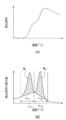
- (A) is an example of a melting curve of the nucleic acid mixture
- (B) is an example of a differential melting curve. It is a graph which shows the result of Tm analysis concerning Example 1 of the present invention. It is a graph which shows the result of Tm analysis concerning comparative example 1 of the present invention. It is a graph which shows the result of Tm analysis concerning comparative example 2 of the present invention. It is a graph which shows the result of Tm analysis concerning comparative example 3 of the present invention. It is a graph which shows the result of Tm analysis concerning Example 2 of this invention. It is a graph which shows the result of Tm analysis concerning Example 3 of this invention. It is a graph which shows the result of Tm analysis concerning Example 4 of this invention.
- the method for measuring the abundance of the gene of the present invention comprises amplifying nucleic acid encoding at least two genes which may differ in the abundance of the gene in the nucleic acid in the target sample in one reaction solution and amplifying a single additional base sequence.
- a first primer set comprising at least two first primers capable of being introduced into a product and corresponding to each of the at least two genes, and a first primer set for amplifying a nucleic acid comprising the single additional base sequence; Nucleic acid amplification using two primers to obtain at least two amplification products comprising the single additional nucleotide sequence and corresponding to each of the at least two genes, the at least two amplification products Detecting each signal based on the abundance of and measuring the abundance of the at least two genes from the signal; It is a method for measuring the standing amount.
- gene also encompasses “gene region” that indicates a partial region of a gene. Further, the “gene” may be one that is encoded by a nucleotide sequence, and includes not only one that expresses a specific function but also one that does not express a specific function.
- a first primer set including a predetermined first primer and a second primer are present in one reaction solution and the at least two genes (hereinafter referred to as "measurement target genes") Since nucleic acid amplification is performed on each of the nucleic acids, single addition base sequences are introduced into a plurality of amplification products corresponding to individual measurement target genes by nucleic acid amplification using a predetermined first primer. . For this reason, even if there are a plurality of measurement target genes in the reaction solution, a single, ie, common addition base sequence is introduced into the corresponding amplification product.
- nucleic acid amplification products Since a plurality of types of nucleic acids (or amplification products) into which this single additional base sequence has been introduced and corresponding to the gene to be measured all contain one common additional base sequence, it is possible to use one second primer. Nucleic acid amplification becomes possible uniformly. As a result, even if the abundances of multiple genes present in the reaction solution differ, the abundances of amplification products derived from individual genes obtained by nucleic acid amplification with the same second primer differ from each other. Unlike nucleic acid amplification using, the amount of each gene is reflected. As a result, the abundances of at least two genes in the nucleic acid in the target sample can be measured more accurately and easily than in the prior art.
- the “Tm value” is a temperature at which double-stranded nucleic acid dissociates (dissociation temperature: Tm), and generally, the temperature at which the absorbance at 260 nm reaches 50% of the total increase in absorbance and It is defined. That is, as the solution containing double-stranded nucleic acid, eg, double-stranded DNA, is heated, the absorbance at 260 nm increases. This is because the hydrogen bond between both strands in double-stranded DNA is released by heating and dissociated into single-stranded DNA (DNA melting).
- Tm dissociation temperature
- the Tm value is set based on this phenomenon.
- the Tm value in the present invention refers to the temperature at which 50% of the bases form a duplex and the remaining 50% melts into a single strand, unless otherwise specified.
- template nucleic acid or “template” refers to a base sequence to which a primer anneals as a template when performing nucleic acid amplification.
- the term "step” is included in the term if the intended purpose of this step is achieved, even if it can not be clearly distinguished from other steps, as well as independent steps.
- a numerical range indicated by using “to” indicates a range including numerical values described before and after “to” as the minimum value and the maximum value, respectively.
- the amount of each component in the composition is the total amount of the plurality of substances present in the composition unless a plurality of substances corresponding to each component are present in the composition. Means The outline of the present invention will be described below.
- the method for measuring the abundance of a gene according to the present invention comprises, in one reaction solution, a nucleic acid encoding at least two genes which may differ in the abundance of the gene in the nucleic acid in the target sample, a single additional base sequence.
- a first primer set comprising at least two first primers capable of being introduced into an amplification product and corresponding to each of the at least two genes, and for amplifying a nucleic acid comprising the single additional base sequence
- nucleic acid amplification using a second primer to obtain at least two amplification products containing the single additional base sequence and corresponding to each of the at least two genes
- nucleic acid amplification Detecting a signal based on the abundance of the at least two amplification products, and based on the signal, the abundance of the at least two genes Measuring hereinafter, referred to as "abundance measurement step", including.
- the nucleic acid amplification step is capable of introducing a nucleic acid encoding at least two genes that may differ in the amount of gene present in the nucleic acid in the target sample in one reaction solution into a single additional base sequence and Using a first primer set comprising at least two first primers corresponding to each of the at least two genes, and a second primer for amplifying a nucleic acid comprising the single additional base sequence Carrying out nucleic acid amplification to obtain at least two amplification products comprising the single additional nucleotide sequence and corresponding to each of the at least two genes.
- the reaction solution contains at least two types of genes, ie, genes to be measured.
- the genes to be measured are those which may differ from one another in the abundance of the gene in the nucleic acid in the subject sample, and generally include those in which the abundance of the gene in the nucleic acid in the subject sample is different It may be included.
- the nucleotide sequence encoding the measurement target gene in the reaction solution corresponds to a nucleic acid serving as a template for nucleic acid amplification described later.
- the sample that can be a source of the nucleic acid in the reaction solution is not particularly limited.
- any biological fluid such as blood, oral mucosal suspension, somatic cells such as nails and hair, germ cells, milk, ascites fluid, paraffin-embedded tissue, gastric fluid, gastric lavage fluid, peritoneal fluid, amniotic fluid, cell culture, etc. It includes those which are derived from or can be derived from the source.
- the template nucleic acid can be used directly as obtained from the source or after pretreatment to modify the properties of the sample.
- genomic DNA from whole blood can be isolated to prepare a nucleic acid of a gene to be measured. Isolation of genomic DNA from whole blood can be performed by a conventionally known method.
- a commercially available genomic DNA isolation kit (trade name GFX Genomic Blood DNA Purification kit; manufactured by GE Healthcare Biosciences) can be used.
- the nucleic acid in the reaction solution may be single stranded or double stranded.
- the nucleic acid sequence in the reaction solution may be, for example, DNA, or total RNA, RNA such as mRNA, or the like.
- amplification products generated by nucleic acid amplification are also present in the reaction solution after the nucleic acid amplification step. Therefore, nucleic acids in the reaction solution in the present invention also include these amplification products.
- the gene to be measured in the reaction solution may be the subject of measurement of the abundance in the present invention if it is at least two types of genes.
- at least one of the genes to be measured is a reference gene of which the abundance in nucleic acids in a sample of interest is known in advance, and at least one is a target whose abundance in nucleic acids in a sample of interest is to be measured. It is a gene.
- the abundance of the reference gene is used as a reference (index), The abundance of nucleic acids in a target sample can be easily measured.
- the amount of nucleic acid in the target sample refers to the copy number of the gene of a predetermined size, the size of the entire copy of the gene in the nucleic acid in the target sample, and the increase or decrease of genes involved in disease.
- the gene usable as the reference gene may be a gene which has been previously known to be in the same amount among individuals, or a gene which is a relatively stable amount with no change in the amount over time.
- RNaseP, sod2, COL8A1, gamma-actin and the like can be mentioned.
- the combination of the reference gene and the target gene is not particularly limited, and may be on the same chromosome on the genome map or at a distant place on the genome map on another chromosome.
- the selection of the target gene and the reference gene is not particularly limited.
- the genes that can be used as the target gene differ depending on the intended use of the measured abundance.
- a gene showing polymorphism a gene whose abundance is increased or decreased due to a disease, a gene whose base in the base sequence is deleted due to a disease, a gene whose expression amount changes depending on a sample, etc. It can be mentioned.
- the combination of the reference gene and the target gene can be appropriately selected depending on the nature of the target gene.
- a gene which is previously known to be equivalent in abundance among individuals may be used as a reference gene and a gene showing polymorphism may be listed as a target gene.
- the abundance of the gene does not change with time and the relatively stable abundance gene is used as a reference gene, and the abundance due to the disease
- the target gene can be a gene whose increase or decrease or deletion of bases is a target gene.
- a housekeeping gene is used as a reference gene and a gene whose expression level changes depending on the sample is targeted It can mention to a gene.
- At least one of the genes to be measured is a gene region at the 5 'side upstream of the fusion point of the fusion gene (hereinafter, also referred to as "5' gene region”), It is also preferable that at least one gene region at the 3 'side downstream of the fusion point of the fusion gene (hereinafter, also referred to as "3' gene region").
- 5' gene region a gene region at the 5 'side upstream of the fusion point of the fusion gene
- 3' gene region at the 3 'side downstream of the fusion point of the fusion gene
- the fusion gene is not particularly limited as long as a part of a certain gene is fused to a part of another gene and exists as one gene.
- ALK fusion gene BCR-ABL fusion gene, AML1-MTG8 fusion gene, RET fusion gene, ROS1 fusion gene and the like can be mentioned.
- the ALK gene (NCBI Accession No. NM — 004304.4) encodes an ALK (anaplastic lymphoma kinase) receptor tyrosine kinase.
- ALK fusion gene for example, J. Clon. Oncol.
- attention is focused on only one of the genes constituting the fusion gene, and the abundance of the 5 'gene region upstream of the fusion point of the gene and the downstream 3' side of the gene region The abundances of the gene regions of can be measured and compared.
- the fusion point means a boundary where two different genes are fused.
- the 4125th base in the ALK gene (corresponding to the 1760th base in SEQ ID NO: 23) is a fusion point.
- the gene region at the 5 'side and the gene region at the 3' side for measuring the abundance of genes are not particularly limited as long as they are regions upstream and downstream of the fusion point.
- the gene region on the 5 'side and the gene region on the 3' side, which measure the abundance of genes, can be measured if it is one or more bases away from the fusion point, but in a region far enough apart For example, they are more preferably separated by about 10 bases or more, and more preferably separated by about 50 bases or more.
- the type of fusion gene mutation that can be detected can be freely set by appropriately setting the distance between the gene region at the 5 'side and the gene region at the 3' side from the fusion point according to the type of fusion gene mutation to be measured. It is possible to change it and to detect more variants (Variant).
- the distance (number of bases) from the fusion point to the gene region is the distance closer to the fusion point among the primers that hybridize to the 5 'gene region or 3' gene region to be measured. Calculate based on the 5 'end of the hybridized primer.
- the fusion gene is not limited to the ALK fusion gene.
- the method according to the present invention can be applied to any gene as long as it has a base sequence registered in a database such as GenBank, as with the ALK fusion gene.
- the first primer set is a primer set that includes a first primer and that amplifies double-stranded nucleic acids in the region to be amplified in the measurement target gene.
- the first primer is capable of introducing a single additional nucleotide sequence into an amplification product and corresponds to each of the at least two genes.
- the region to be amplified is a region corresponding to a part of the base sequence encoding the gene to be measured, preferably 40 to 5000 bases in length, more preferably 50 to 1000 bases in length, and more preferably Preferably, the length is 60 to 200 bases.
- corresponding to each of the at least two genes means that the base sequence characteristic of the measurement target gene can be amplified as the amplification target region. Therefore, in the nucleic acid amplification step, the number of first primers corresponding to the type of the measurement target gene is used.
- the first primer needs to be capable of introducing a single additional base sequence into an amplification product.
- "in which a single additional base sequence can be introduced into the amplification product” means not only generation of an amplification product containing the single additional base sequence itself composed of a predetermined base sequence, but also It means both that an amplification product containing a base sequence complementary to the base sequence of is generated.
- the first primer comprises a single additional base sequence and a part of the base sequence of the nucleic acid encoding the target gene as a template.
- a nucleic acid-amplifiable base sequence may be included, and a linker sequence may be included between them.
- the base sequence capable of nucleic acid amplification using a part of the base sequence of the nucleic acid encoding the measurement target gene as a template can be, for example, a base sequence capable of hybridizing to the base sequence of the amplification target region.
- the first primer is, for example, a single additional base sequence and a base sequence capable of hybridizing to the base sequence of the amplification target region of the measurement target gene from the viewpoints of ease of operation, measurement sensitivity of measurement method, etc. It is preferable that it is a combination of
- a single addition base sequence is one kind of (single) addition which can be commonly introduced to all amplification products which may be present in one reaction system used for one measurement.
- a base sequence is meant, including its complementary sequence.
- the “single additional base sequence” of the first primer may be any of the single additional base sequence and its complementary base sequence.
- the “single additional base sequence” introduced to the amplification product and the “single additional base sequence” of the first primer are collectively referred to simply as “single additional base sequence” unless otherwise specified. Do.
- the structure (base sequence and length) of such a single additional base sequence is not particularly limited, and any sequence can be selected.
- the base sequences in the respective amplification target regions of at least two genes be sequences having non-homologous base sequences
- the base sequence of the region including the region of interest and the region adjacent to the region to be amplified is a sequence consisting of a nonhomologous base sequence. More preferably, it is a sequence consisting of a nucleotide sequence not present in the nucleotide sequence of the gene present.
- non-homologous means, for example, having 50% or less, preferably 25% or less of homology of the region to be amplified.
- a base sequence capable of hybridizing to the base sequence of the amplification target region means annealing to a single-stranded nucleic acid (template nucleic acid) containing the base sequence to be amplified under normal nucleic acid amplification conditions. It means that it has a sequence capable of forming a double stranded nucleic acid with the template nucleic acid.
- the base sequence capable of hybridizing to the nucleic acid sequence of the amplification target region in the first primer is not particularly limited as long as it can amplify the base sequence of the template nucleic acid in the amplification target region, and the target gene Those skilled in the art can appropriately design based on the nucleotide sequence of
- the “hybridizable base sequence” relating to the first primer includes a base sequence completely complementary to the base sequence of the template nucleic acid, and is completely complementary under the nucleic acid amplification conditions described later.
- One or more bases may also contain a base sequence in which one or several bases are deleted, substituted, or added, to such an extent that the affinity to the single-stranded nucleic acid is not largely impaired.
- the number of inserted, deleted or substituted bases includes one or two or more bases, and varies depending on the entire length of the first primer, and for example, one to ten bases, preferably one to five bases are mentioned.
- regulating additional base sequences may be those which will be described later alone or in combination of two or more appropriately selected from those which will be described later.
- such an adjustment additional base sequence contains at least one selected from the group consisting of a mismatched base and a degenerate base relative to the base sequence of the amplification target region.
- the type and number of bases of the mismatched base and the degenerated base are not particularly limited, but may be set from adjustment of expected Tm value, influence on amplification efficiency, and adjustment of the number of identical primer molecules.
- a degradation-inducing base selected from the group consisting of uracil base, AP site, and RNA base may be introduced. When these degradation-inducing bases are introduced, the Tm value of the first primer for the amplification product can be lowered by degrading the degradation-inducing bases after obtaining an amplification product by the first primer.
- advantages such as being able to preferentially advance nucleic acid amplification by the second primer can be obtained.
- the length and Tm value of the first primer are usually, but not limited to, 12 mer to 60 mer and 40 ° C. to 85 ° C., preferably 16 mer to 50 mer and 50 ° C. to 80 ° C.
- the single additional base sequence in the first primer and the base sequence capable of hybridizing to the base sequence of the amplification target region of the gene may have the same length or different lengths. Good.
- the position of the single additional base sequence in the first primer may be any position.
- the single additional base sequence is disposed at the 5 'end of the first primer. By arranging the single additional base sequence on the 5 'end side of the base sequence that hybridizes to the amplification target region of the gene, for example, the first primer interferes with the reaction when the measurement target gene is first amplified. Absent.
- the second primer is used to amplify a nucleic acid containing the single additional base sequence amplified by the first primer set.
- the amplification product containing the single additional base sequence obtained using the first primer set containing the first primer can be further nucleic acid amplified and accumulated in the reaction solution.
- the second primer can have a base sequence capable of hybridizing to the single additional base sequence or a complementary base sequence thereof.
- the complementarity of the single additional nucleotide sequence contained in a part of the first primer is preferable.
- a base sequence that is homologous to the single additional base sequence contained in a part of the first primer is preferable.
- the amplification product containing the single additional base sequence can be efficiently and effectively accumulated in the reaction solution.
- the second primer can form a double-stranded nucleic acid between the single additional base sequence or its complementary base sequence under nucleic acid amplification conditions
- one or more bases are additionally present. It may be a deletion, substitution or addition.
- the position of the insertion, deletion or substitution is not particularly limited.
- the number of inserted, deleted or substituted bases includes one or two or more bases, and varies depending on the entire length of the second primer, and for example, one to ten bases, preferably one to five bases are mentioned.
- the second primer contains a sequence homologous to the single additional base sequence contained in a part of the first primer.
- “homologous” to the single additional nucleotide sequence means having 80% or more homology to the single additional nucleotide sequence.
- the sequence homologous to the single additional base sequence in the second primer preferably has a homology of at least 90%, most preferably 100%, to the single additional base sequence.
- the Tm value of the second primer (the Tm value of the amplification product is set to the second primer so that the nucleic acid amplification with the second primer proceeds in preference to the nucleic acid amplification with the first primer. It is preferable that the Tm value of the first primer is higher than the Tm value of the first primer (meaning the respective amplification target regions of the nucleic acid encoding the at least two genes or the Tm value for the amplification product). Therefore, the second primer is an additional sequence different from the single additional nucleotide sequence and its complementary nucleotide sequence, and can be adjusted to have a Tm value higher than that of the first primer. Additional sequences (hereinafter referred to as adjustment additional nucleotide sequences).
- the Tm value of the second primer can be increased relative to the amplification product.
- the additional sequence for this purpose is not particularly limited, but may be a base sequence having a high GC content, and the like.
- an artificial nucleic acid may be introduced into the second primer.
- an artificial nucleic acid LNA, BNA, PNA etc. can be mentioned.
- an RNA primer may be introduced to make the Tm value of the second primer higher than the Tm value of the first primer.
- MGB minor groove binder
- the length and Tm value of the second primer are generally, but not limited to, 12 mer to 60 mer and 40 ° C. to 85 ° C., preferably 16 mer to 50 mer and 50 ° C. to 80 ° C.
- the Tm value of the second primer is at least compared to the Tm value of the first primer.
- the temperature is higher than 0.1 ° C., 1 ° C., 3 ° C., 5 ° C.
- the difference between the Tm value of the second primer and the Tm value of the first primer is lower than 30 ° C., 25 ° C., and 20 ° C. Based on this, it is preferable that the difference between the Tm value of the second primer and the Tm value of the first primer is 0.1 ° C. to 30 ° C. higher, 1 ° C. to 30 ° C., 3 ° C.
- the temperature be raised by 5 ° C to 25 ° C.
- the Tm value of the second primer is set higher than the Tm value of the first primer, the Tm value of the second primer is 50 ° C. to 85 ° C., and the first primer The Tm value of is preferably 40.degree. C. to 75.degree. Further, it is more preferable that the Tm value of the second primer is 55 ° C to 80 ° C, and the Tm value of the first primer is 50 ° C to 75 ° C.
- the first primer set includes, in addition to the first primer, another primer for amplifying the other single-stranded nucleic acid of the double-stranded nucleic acid of the gene to be measured.
- the other primer may be a similar first primer similarly designed to include the single additional nucleotide sequence (a set consisting of a pair of first primers), and
- the first primer may be used for nucleic acid amplification on the complementary strand side of the hybridizing single strand, and may be a third primer which does not contain the single additional base sequence.
- the third primer does not contain the single additional base sequence, and has a nucleic acid sequence capable of hybridizing to the nucleic acid sequence of the complementary strand of the single strand of the measurement subject gene that the first primer recognizes What is necessary.
- the description described for the first primer can be applied as it is to the “hybridizable base sequence” for the third primer.
- the second primer can form a second primer set with the other primer for amplifying the other nucleic acid of the double stranded nucleic acid having the single additional base sequence.
- the other primer included in the second primer set may be a set comprising the pair of the second primers, and the third primer together with the third primer included in the first primer set.
- a second set of primers can also be constructed. That is, when the first primer set includes the third primer, each of the first primer and the second primer is paired with each other (the first primer or the second primer and the third primer.
- a primer set used in the nucleic acid amplification step for example, a combination of a pair of first primer set and a pair of second primer set, a combination of the first primer and the second primer and a third primer It can be done.
- the first primer and the second primer may contain the adjustment additional base sequence.
- combinations include, but are not limited to, for example: A first primer having a base sequence complementary to the single additional base sequence and a base sequence of the amplification target region of the measurement target gene, a second primer not containing a tuning additional base sequence, Combination with three primers; A combination of a first primer with a lower Tm value than the second primer, a second primer with a higher Tm value than the first primer, and a third primer; A combination of a first primer with a higher Tm value than the second primer, a second primer with a lower Tm value than the first primer, and a third primer; A combination of a first primer comprising a tuning additional sequence which results in a lower Tm value than the second primer, a second primer free of mismatches or degenerate bases, and a third primer; A first primer containing an adjustment additional base sequence such as a mismatch or degenerate base giving a Tm value lower than that of the second primer, and a second primer containing no adjustment additional base sequence such as a mismatch
- Combination with a third primer A first primer not including substitution and the like for both the single additional base sequence and the base sequence of the amplification target region of the measurement target gene, and a control additional base sequence having a Tm value higher than that of the first primer
- a combination of a second primer including: and a third primer
- a first primer containing an adjustment additional base sequence such as a mismatch or degenerate base, and a second primer containing a adjustment additional base sequence such as a mismatch or a degenerate base giving a higher Tm value than the first primer In combination with a third primer;
- the configuration of the first primer corresponding to the gene to be measured in the reaction solution is the same (for example, both are composed of the first primer containing neither a mismatch nor a degenerate base). And may be heterologous (eg, one applies the first primer without mismatch or degenerate base, the other applies the first primer with mismatch or degenerate base) However, from the viewpoint of advancing nucleic acid amplification with the same amplification efficiency for a plurality of measurement target genes, it is preferable that they are the same.
- the reaction solution contains 0.25 times the amount of the third primer relative to the amount of the first primer paired with the third primer. It can be contained in an amount of 4-fold. If it is 0.25 times or more, for example, amplification tends to be possible without greatly impairing the amplification balance of the first primer and the third primer, and if it is 4 times or less, for example, the third primer.
- the second primer easily reacts with the amplification product can be obtained.
- the amount of third primer can be equal to the amount of first primer. Also, in order to adjust the primer sequences actually used, the reagent formulation, and the numerical values of the detection results obtained, it is possible to finely adjust and optimize whether to increase or decrease the amount of the third primer. In addition to this advantage, this can, for example, make it possible to control the strength of the detection signal of the reference gene or target gene, or the gene region of the 5 'side or the gene region of the 3' side. The advantage can be obtained that the amplification efficiency of the gene and target gene can be regulated.
- the amount of the third primer is preferably about 1 to 4 times the amount of the first primer based on the amount of the substance, in terms of facilitating transfer to the reaction with the second primer, etc.
- the amount of the third primer can be about 0.25 times to 1 times the amount of the first primer based on the amount of the substance, more preferably 0.5 times to 1 time It can be done.
- the reaction solution may include the second primer in an equivalent amount or a large amount based on the amount of each primer contained in the first primer set.
- This has an advantage of being able to reliably amplify an amplification product amplified by nucleic acid amplification by the first primer set, for example.
- a large amount of a nucleic acid amplification product by the second primer can be obtained, which is more easily transferred to the nucleic acid amplification of the second primer set than the nucleic acid amplification by the first primer set, and the depletion of the second primer Can also provide one or more of the advantages of preventing the nucleic acid amplification product from reaching a plateau.
- the second primer is 1-fold to 400-fold (molar ratio), preferably 1-fold, of the larger amount of the respective primers constituting the first primer set in the reaction solution.
- the amount may be increased to 40 times (molar ratio), more preferably 1 to 20 times (molar ratio).
- reaction solution may include the third primer so as to satisfy both the quantitative ratio with the first primer and the quantitative ratio with the second primer. This can have the advantages described above.
- the length of each primer contained in each primer set may be the same or different.
- a method using a polymerase As a method of nucleic acid amplification, a method using a polymerase is preferable, and examples thereof include PCR method, ICAN method, LAMP method, NASBA method and the like.
- amplification is preferably performed in the presence of a probe described later.
- Those skilled in the art can easily adjust the reaction conditions for amplification and the like according to the probe and polymerase used.
- DNA polymerase used for the PCR method a commonly used DNA polymerase can be used without particular limitation.
- GeneTaq manufactured by Nippon Gene
- PrimeSTAR Max DNA Polymerase manufactured by Takara Bio
- Taq polymerase and the like can be mentioned.
- the amount of polymerase used is not particularly limited as long as it is a commonly used concentration.
- a concentration of 0.01 U to 100 U can be preferably applied to a reaction solution volume of 50 ⁇ l.
- the heating temperature in the dissociation (dissociation step) of the PCR amplification product is not particularly limited as long as the amplification product can be dissociated. For example, 85 ° C. to 95 ° C.
- the heating time is also not particularly limited. Usually, it is 1 second to 10 minutes, preferably 1 second to 5 minutes.
- hybridization between the dissociated single-stranded nucleic acid and each of the primers can be performed, for example, by lowering the heating temperature in the dissociation step after the dissociation step.
- the temperature condition is, for example, 50 ° C. to 80 ° C.
- a plurality of conditions may be set in the reaction process of nucleic acid amplification. For example, in a nucleic acid amplification reaction in which 50 cycles of PCR reaction are performed, reaction conditions may be adjusted such that the first 10 cycles are performed at 55 ° C. and the second 40 cycles are performed at 65 ° C.
- the volume and concentration of each composition in the reaction solution of the hybridization step are not particularly limited.
- the concentration of the primer in the reaction solution is, for example, 0.001 ⁇ M to 1 ⁇ M for the first primer and the third primer, preferably 0.01 ⁇ M to 0.5 ⁇ M
- the concentration of the second primer is, for example, 0.01 ⁇ M to 10 ⁇ M, preferably 0.05 ⁇ M to 5 ⁇ M.
- the reaction solution may contain at least two detection probes that respectively recognize the at least two amplification products in order to effectively detect a signal in a signal detection step described later.
- the detection probe is not particularly limited as long as the target amplification product can be detected.
- the length of the detection probe is not particularly limited, but is preferably 5 mer to 50 mer, and more preferably 10 mer to 30 mer. If the length of the probe is within this range, for example, detection sensitivity can be enhanced.
- the detection probe is preferably 70% to 100% identical in sequence to the nucleic acid sequence of each of the genes to be measured, and particularly preferably 80% or more identical.
- a fluorescent label to be described later is provided at the 3 'end in order to prevent the extension of the probe itself as a reaction target of DNA polymerase.
- it is added, or a phosphate group is further added to the 3 'end of the probe.
- the detection probe is a labeled probe to which a label is attached.
- the labeling substance in the labeled probe include, for example, fluorescent dyes and fluorophores.
- a probe that is labeled with a fluorescent dye shows fluorescence alone, and decreases fluorescence (for example, quenching) by hybridization is preferable.
- a probe utilizing such a fluorescence quenching phenomenon is generally called a fluorescence quenching probe.
- the probe it is preferable that the base of the 3 'region (for example, the 3' end) or the 5 'region (for example, the 5' end) of the oligonucleotide is labeled with a fluorescent dye, and be labeled
- the base is preferably cytosine (C).
- C cytosine
- G guanine
- Such probes are generally referred to as guanine quenching probes and are known as so-called Q Probes.
- Q Probes When such a guanine quenching probe hybridizes to the detection target sequence, the C of the end labeled with the fluorescent dye approaches G in the detection target sequence to weaken the light emission of the fluorescent dye (the fluorescence intensity is Decrease)).
- the labeling substance can usually be bound to a phosphate group of a nucleotide.
- the fluorescent dye is not particularly limited, and examples thereof include fluorescein, phosphor, rhodamine, polymethine dye derivatives and the like, and commercially available fluorescent dyes include, for example, BODIPY FL, FluorePrime, Fluoredite, FAM, Cy3 and Cy5 And TAMRA.
- a plurality of detection probes are applied because a plurality of genes to be measured are present in the sample solution.
- combinations of fluorescent dyes that can be used for a plurality of probes preferably used are not particularly limited as long as they can be detected under different conditions, for example, Pacific Blue (detection wavelength 445 nm to 480 nm), TAMRA (detection wavelength 585 nm) And combinations of BODIPY FL (detection wavelength 520 nm to 555 nm).
- Pacific Blue detection wavelength 445 nm to 480 nm
- TAMRA detection wavelength 585 nm
- BODIPY FL detection wavelength 520 nm to 555 nm.
- the detection probe is a labeled probe labeled with a labeling substance such as a fluorescent dye
- a labeling substance such as a fluorescent dye
- the unlabeled probe may be used in combination.
- the unlabeled probe may have, for example, a phosphate added to its 3 'end.
- the addition ratio (molar ratio) of the detection probe to the nucleic acid in the reaction solution is not particularly limited. It is preferably 1 time or less, more preferably 0.1 time or less with respect to the nucleic acid in the sample. Thereby, for example, a sufficient detection signal can be secured.
- the addition ratio of the detection probe to the nucleic acid may be, for example, a molar ratio to double-stranded nucleic acid or a molar ratio to single-stranded nucleic acid.
- the abundance measurement step includes detecting a signal based on the abundance of the at least two amplification products, and measuring the abundance of the at least two genes from the signal. Since the abundances of at least two amplification products reflect the abundances of at least two genes, by measuring the signal based on the abundance of the amplification products obtained in the nucleic acid amplification step, it is possible to detect in the target sample.
- the measurement of the amount of the gene is obtained by obtaining a detection signal of the amplification product (hereinafter referred to as a signal detection step), and measuring the amount by comparing the obtained detection signal (hereinafter referred to as a signal comparison step). It can be carried out.
- the detection of the signal may be a method of measuring the amount of the amplification product accumulated in the reaction solution, and the type of the signal is not particularly limited.
- As the signal detection step preferably, a detection method using a detection probe capable of detecting a target amplification product can be used.
- the signal comparison step includes measuring the abundance of each gene by comparing each detection signal corresponding to each gene obtained in the signal detection step. It does not specifically limit about the method of measurement of the contrast of the said detection signal, and the abundance of each gene from a contrast result.
- the abundance of each gene may be calculated by comparing the detection signal corresponding to the gene.
- the amount of amplification products of a plurality of measurement target genes in the reaction solution can be measured sequentially as the number of PCR cycles elapses, and the abundance of the measurement target gene can be measured from the obtained measurement results.
- the amount of amplification product generally increases exponentially with the number of PCR cycles, the initial cycle period, which can not be detected by general detection methods, exponentially increases with detectable amplification period, reaction rate It is divided into declining plateau periods.
- the abundance of the measurement target gene is reached even when reaching the plateau stage Accumulate in the reaction solution in the ratio according to the Therefore, by applying type identification means for identifying the type of the amplification product, the amplification products derived from the measurement target gene accumulated in the reaction solution are identified, and the respective quantitative ratios are measured. The abundance of the target gene of interest can be measured.
- the type identification means include the Tm value of the detection probe, a labeled species, and a DNA microarray on which the probe is immobilized.
- the gene to be measured includes a reference gene and a target gene
- the abundance of the target gene may be measured from the ratio to the abundance of the reference gene.
- the gene to be measured is the gene region at the 5 'side and the gene region at the 3' side in the fusion gene
- the abundance of the gene region at the 3 'side from the ratio to the abundance of the gene region at the 5' side
- the abundance of the 5 'gene region may be measured from the ratio to the abundance of the 3' gene region.
- the abundance measurement step may be one in which the abundance of the at least two genes is measured by Tm analysis of at least two detection signals corresponding to each of the genes.
- the signal detection step and the signal comparison step more preferably include the following steps (I) to (IV).
- the following step (I) may be performed simultaneously with the above-described nucleic acid amplification step.
- (I) Contacting the detection probe with a single-stranded nucleic acid (amplification product) in a sample to obtain a hybridization product (hybridization step).
- hybridization step Dissociating the hybridization product by changing the temperature of the sample containing the hybridization product, and measuring the fluctuation of the detection signal based on the dissociation of the hybridization product (measurement step).
- Tm value detection step detecting a Tm value which is a dissociation temperature of the hybridization product based on the fluctuation of the detection signal.
- IV detecting the presence or abundance ratio of a target amplification product in single-stranded nucleic acid in the sample based on the Tm value (abundance ratio detection step).
- the detection probe is brought into contact with the single-stranded nucleic acid in the sample, and both are hybridized.
- Single-stranded nucleic acid in a sample can be prepared, for example, by dissociating the amplification product obtained as described above.
- the heating temperature in the dissociation of the amplification product is not particularly limited as long as the amplification product can be dissociated. For example, 85 ° C. to 95 ° C.
- the heating time is also not particularly limited. Usually, it is 1 second to 10 minutes, preferably 1 second to 5 minutes.
- Hybridization between the dissociated single-stranded nucleic acid and the detection probe can be performed, for example, by decreasing the heating temperature in the dissociation step after the dissociation step.
- the temperature condition is, for example, 25 ° C. to 50 ° C.
- the volume and concentration of each composition in the reaction solution of the hybridization step are not particularly limited, and conditions that are usually applied can be applied.
- the hybridization product of the obtained single-stranded nucleic acid and the detection probe is gradually heated, and the fluctuation of the fluorescence signal with the temperature rise is measured.
- Q Probe fluorescence intensity decreases (or is quenched) in a hybridized state with single-stranded nucleic acid as compared to a dissociated state. Therefore, for example, the hybridization product in which the fluorescence is decreasing (or quenching) may be gradually heated, and the increase in fluorescence intensity with increasing temperature may be measured.
- the temperature range for measuring the fluctuation of the fluorescence intensity is not particularly limited.
- the start temperature is from room temperature to 85 ° C., preferably 25 ° C.
- the temperature increase rate is not particularly limited, and is, for example, 0.1 ° C./second to 20 ° C./second, preferably 0.3 ° C./second to 5 ° C./second.
- the signal used to measure the abundance of the at least two types of genes is obtained by the absorbance or fluorescence value obtained from the same wavelength.
- the abundance of the said measurement object gene can be measured by the simplified measurement and detection.
- the labels of the detection probes may be the same.
- the Tm values of the respective primers used are different.
- the fluctuation of the signal is analyzed and determined as the Tm value.
- the derivative value (-d fluorescence intensity / dt) at each temperature can be calculated from the obtained fluorescence intensity, and the temperature showing the lowest value can be determined as the Tm value.
- the point at which the amount of increase in fluorescence intensity (the amount of increase in fluorescence intensity / t) per unit time is the highest may be determined as the Tm value.
- the amount of decrease in fluorescence intensity may be measured.
- the signal fluctuation during hybridization may be measured. That is, the temperature of the sample to which the probe is added may be lowered to measure the fluorescence signal fluctuation associated with the temperature drop when forming a hybridization product.
- the fluorescence intensity when hybridizing to the complementary sequence is reduced (quenched) compared to the fluorescence intensity when not hybridizing to the complementary sequence
- the fluorescence intensity is large because the probe is in a dissociated state, but when a hybridization product is formed due to a drop in temperature, the fluorescence decreases (or is quenched). Therefore, for example, the temperature of the heated sample may be gradually decreased to measure the decrease in fluorescence intensity as the temperature decreases.
- the probe when a labeled probe whose signal is increased by hybridization is used, when the probe is added to the sample, the probe is in a dissociated state, so the fluorescence intensity is small (or quenching) but due to the temperature drop. When a hybrid is formed, the fluorescence intensity will be increased. Therefore, for example, the temperature of the sample may be gradually decreased to measure the increase in fluorescence intensity as the temperature decreases.
- the abundance of the target amplification product in the single-stranded nucleic acid in the sample is detected based on the Tm value, and the abundance of the gene to be measured is detected from the abundance. .
- the detection signal of the amplification product obtained in the signal detection step the signals of a plurality of amplification products derived from a plurality of genes to be measured are mixed. Since these signals reflect the abundance of the amplification product, it is necessary to measure the abundance of the gene to be measured, ie, the abundance of the nucleic acid in the sample by comparing and comparing the respective detection signals. Can.
- the presence of the target gene can be conveniently determined by using the detection signal of the reference gene as a reference.
- the quantity can be measured.
- the gene to be measured is the gene region at the 5 'side and the gene region at the 3' side in the fusion gene
- the detection signal of the gene region at the 5 'side and the detection signal of the gene region at the 3' side The presence of the fusion gene in the sample can be conveniently detected by comparing with.
- the method of measuring the gene abundance based on the detection signal and any analysis method that can be used for this purpose can be applied.
- Tm analysis methods include those described in WO2009 / 081965 and WO2010 / 001969.
- Tm analysis method a method of determining the abundance of each of the genes in the target sample by analyzing the area of the target gene as the reference gene and the target gene and analyzing the area of the result of Tm analysis will be described below.
- FIG. 1 (A) shows a melting curve represented by the relationship between the temperature of a nucleic acid mixture of an arbitrary target gene and a reference gene and the detection signal such as absorbance or fluorescence intensity
- FIG. 1 (B) shows the temperature and detection signal
- the melting curve (it is also called a differential melting curve) represented by the relationship with the derivative value of is shown.
- the temperature range ⁇ T R including Tm R is, for example, a temperature with the lower limit of the temperature at which the differential value of the detection signal is minimum between Tm R and Tm T and the upper limit of the temperature corresponding to the base of the peak of the detection signal. Range can be set.
- the temperature range [Delta] T T containing Tm T for example, the lower limit temperature corresponding to the temperature at which the differential value of the detection signal becomes minimum between Tm R and Tm T limit, the foot of the peak of the detection signal Temperature range can be set.
- the temperature range ⁇ T R and the temperature range ⁇ T T are set to have the same width (for example, 10 ° C.) or different widths (for example, the temperature range ⁇ T R is 10 ° C.
- the temperature range ⁇ T R and the temperature range ⁇ T T may be set so as to be plus X ° C. and minus X ° C. from the respective melting temperatures Tm (X ° C. is, for example, 15 ° C. or less, preferably 10 ° C. or less) Good.
- T s is a lower limit value in each temperature range
- T e is an upper limit value
- the base value B (T) at each temperature T is a value obtained by the following equation (2), and represents a background level included in the detection signal.
- the area S R in the temperature range ⁇ T R and the area S T in the temperature range ⁇ T T are determined according to the equations (1) and (2), and the relationship between the area ratio and the abundance ratio of each nucleic acid is calculated.
- the abscissa may represent the abundance ratio (the ratio of the target gene T to the total amount of nucleic acids), and the ordinate may represent the area ratio (S T / S R ).
- the area ratio may be determined by S R / S T.
- the abundance of the target gene can be determined by comparing the area of the reference gene with the area of the target gene.
- the target gene may be quantified by preparing a standard curve for the measurement target gene whose specific abundance is known in advance as described above.
- the abundances of a plurality of genes to be measured are measured using the calibration curve prepared as described above.
- the measurement of the abundance may obtain the relative abundance of the other gene in comparison with the abundance of one gene, based on the specific abundance of the one gene.
- the specific abundance of other genes may be obtained.
- the abundance of the target gene can be obtained as a relative amount based on the abundance of the reference gene, or may be obtained as a specific amount.
- the copy number of the target gene can be determined by comparing the detection signal of the amplification product derived from the reference gene with the detection signal of the amplification product derived from the target gene. .
- the gene to be measured is the gene region on the 5 'side and the gene region on the 3' side, a detection signal of an amplification product derived from the gene region on the 5 'side by the same method as described above, The presence of the fusion gene in the sample can be detected by comparison with the detection signal of the amplification product from the 3 'gene region.
- a gene measurement kit is a kit for the gene measurement method described above, which includes the first primer set including the first primer described above and the second primer. According to the present measurement kit, it is possible to simply measure the abundance of measurement target genes having different abundances.
- the measurement kit may include the third primer described above. The matters described above are applied as they are to the first primer, the second primer and the third primer in the present gene measurement kit.
- the measurement kit may include at least two detection probes each having a nucleic acid sequence capable of hybridizing to the nucleic acid amplification region of the at least two genes and provided with a label. This makes it possible to more simply measure measurement target genes having different abundances using the detection probe.
- the detection probe included in the measurement kit one in which the label is a fluorescent label is preferable. The matters described above apply to the label and the fluorescent label as they are.
- the measurement kit may further include, in addition to the probe, reagents required to perform nucleic acid amplification in the measurement method according to the present invention.
- each of the probes, each of the primers, and the other reagents may be separately stored, or part of them may be a mixture.
- “individual storage” may be any type as long as each reagent can be maintained in a non-contact state, and may not necessarily be stored in a separate container which can be handled independently.
- the measurement kit of the present invention uses the first primer set containing the first primer and the second primer to create a differential melting curve for a sample containing the gene to be measured, and analyze its Tm value Conducted to include instruction manual described to measure the amount of gene to be measured, or instruction manual describing various reagents included or additionally included in the measurement kit preferable.
- the measurement kit of the present invention can be used to detect fusion gene mutations.
- the first primer set for example, SEQ ID NOs: 19 and 21
- a second primer for example, a measurement kit comprising SEQ ID NO: 22
- the ALK fusion gene measurement kit comprises the first primer set (for example, SEQ ID NOs: 19 and 21) including a first primer capable of amplifying a specific gene region in the ALK gene, and a second primer (for example, For example, it may be a measurement kit including SEQ ID NO: 22) and a third primer (eg, SEQ ID NOs: 18 and 20).
- the gene measuring apparatus is an apparatus to which the method for measuring the amount of the gene described above is applicable, which comprises the single additional base sequence introduced by the first primer and a second primer A detection unit for detecting at least two signals based on the abundance of amplification products corresponding to the at least two genes amplified by nucleic acid, and the at least two detection signals detected by the detection unit, And a calculation unit that calculates the abundances of at least two genes. If it is this gene measuring device, the method of measuring the abundance of the gene according to the present invention described above can be implemented more simply.
- the detection unit is not particularly limited as long as it can detect a detection signal, and can be appropriately selected according to, for example, the type of detection probe to be used. For example, if the detection probe is a fluorescently labeled probe, a fluorescent detector can be included.
- the calculation unit calculates the abundance of the measurement target gene in the reaction solution based on each detection signal obtained by the detection unit.
- the detection signal of the melting curve is differentiated with respect to temperature to calculate a differential melting curve representing the relationship between the temperature and the differential value of the detection signal, and a calibration curve is prepared in advance for the differential melting curve Setting the temperature range set at the time of setting, the area surrounded by the straight line passing through the point corresponding to the lower limit of the temperature range of the differential melting curve and the point corresponding to the upper limit, and the differential melting curve Determining the area corresponding to each of the genes, calculating the ratio of the obtained areas, calculating the abundance ratio of the measurement target gene corresponding to the area ratio based on the calibration curve prepared in advance .
- any of known devices may be used as the device including the detection unit and the calculation unit.
- the devices described in WO2009 / 081965 and WO2010 / 001969 may be applied as they are.
- a gene measurement system is an apparatus to which the method for measuring the amount of the gene described above is applicable, comprising a single additional base sequence introduced by the first primer and a second primer
- a detection device for detecting at least two signals based on the abundance of amplification products corresponding to the at least two genes amplified by nucleic acid, and the at least two detection signals detected by the detection unit in comparison
- a computing device for computing the abundance of at least two genes. If it is this gene measuring device, the method of measuring the abundance of the gene according to the present invention described above can be implemented more simply.
- the detection device and the calculation device used in the present system those described as the detection unit and the calculation unit in the gene measurement device can be applied as they are. Further, as in the case of the measurement device, the devices described in, for example, WO2009 / 081965 and WO2010 / 001969 may be applied as they are to the measurement device and the arithmetic device.
- the gene diagnosis method of the present invention is a diagnosis method performed using the gene measurement method described above. According to this method, based on the abundance of at least two genes having different abundances in the nucleic acid in the target sample, diagnosis of diseases associated with such abundance, drug metabolism ability, drug sensitivity etc. it can.
- the present invention is applied to the diagnosis of copy number variation in which the abundance of the gene to be measured is the copy number and one of the indicators is that the copy number of the nucleic acid in the sample is increased or decreased.
- diseases having copy number variation as an index include neuroblastoma and the like due to copy number variation of chromosome 1q21.1. According to the present invention, it is possible to diagnose with high accuracy the detection of the abundance of genes involved in these diseases, drug metabolic ability and drug sensitivity.
- Example 1 The abundance of the specific region sequence (SEQ ID NO: 1) of the human CYP2D6 gene was measured using the CYP2D6 gene as a target gene.
- the specific region sequence of human sod2 gene (SEQ ID NO: 2) was used as a reference gene.
- the genomic nucleotide sequence of the CYP2D6 gene is available as GenBank NG008376, and the genomic nucleotide sequence of the sod2 gene is available as GenBank NG008729.
- An artificial nucleic acid was prepared by inserting the nucleic acid sequence of SEQ ID NO: 1 or SEQ ID NO: 2 into pUC57, and samples a to d with various plasmid solutions were prepared according to Table 1 in order to change the copy number of CYP2D6. Using these plasmid solutions, various samples (various templates) containing target genes and reference genes at predetermined copy numbers were prepared.
- the excitation wavelength was set to 520 nm to 555 nm, and the measurement wavelength was set to 585 to 700 nm, and changes in fluorescence intensity derived from the fluorescence labeled probe were respectively measured. It is known that peaks are observed around 58 ° C. in the case of the target gene (CYP2D6) and around 70 ° C. in the case of the reference gene (sod2).
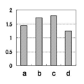
- Tm values were analyzed based on area analysis. Specifically, for samples a to d, differential melting curves are prepared from the results of Tm analysis to obtain Tm values and respective areas of the target gene and the reference gene, and the target gene relative to the area of the reference gene is obtained. The ratio of the areas was determined. The results are shown in Table 8 and FIG. 2A.
- a first primer (SEQ ID NO: 3, Tm value 70.4 ° C.) consisting of a single additional nucleotide sequence and a nucleotide sequence complementary to the nucleotide sequence characteristic of CYP2D6 is used as a CYP2D6 nucleic acid, It amplified using the 3rd primer (sequence number 4, Tm value 55.7 degreeC) comprised by the base sequence complementary to a characteristic base sequence.
- a nucleic acid of sod2 and a first primer (SEQ ID NO: 6, Tm value of 69.7 ° C.) composed of the single additional nucleotide sequence and a nucleotide sequence complementary to the nucleotide sequence characteristic of sod2
- a third primer (SEQ ID NO: 5, Tm value 57.3 ° C.) composed of a nucleotide sequence complementary to the nucleotide sequence characteristic of sod2.
- Each amplification product was amplified using the second primer (SEQ ID NO: 7, Tm value 59.6 ° C.) composed only of the single additional nucleotide sequence and the respective third primer.
- the probe for detection of CYP2D6 (SEQ ID NO: 8) and the probe for detection of sod2 (SEQ ID NO: 9) are as described in Table 2.
- the Tm value of each primer is a value calculated using Meltcalc.
- the base sequence written in lower case indicates the single additional base sequence.
- Comparative Examples 1 to 3 In Comparative Example 1, as shown in Table 4 below, a primer (2D6-Int6r-F1: SEQ ID NO: 10, sod2-R1: SEQ ID NO: 11) not containing the single additional base sequence in both CYP2D6 and sod2 PCR and Tm analysis were performed as in Example 1 except that the primer set was used and the formulation of Table 5 was used without using the second primer.
- Comparative Example 2 As shown in Table 4 below, a primer (2D6-Int6r-F1: SEQ ID NO: 10, sod2-R1: SEQ ID NO: 11) not containing the single additional base sequence in both CYP2D6 and sod2 PCR and Tm analysis were performed in the same manner as in Example 1 except that the formulations in Table 6 were used using the primer set.
- PCR and Tm analysis were performed in the same manner as in Example 1 except that the second primer (PEN3 F4: SEQ ID NO: 7, see Table 2) was not used as the formulation of Table 7.
- the sequences of each primer used in Comparative Examples 1 to 3 and the results of Tm analysis are shown in Table 8 and FIGS. 2B to 2D.
- Example 1 As shown in Table 8 and FIGS. 2A to 2D, in Example 1, the area ratio increased according to the copy number of the target gene in the samples a to b, and the result reflected in the increase of the copy number was obtained. On the other hand, in all of Comparative Examples 1 to 3, the area ratio did not increase according to the increase of the copy number in the sample, and the result reflected in the increase of the copy number was not obtained. The same tendency was observed even when Example 1 and Comparative Example 1 were repeated three times each (data not shown).
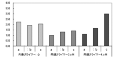
- Example 2 the second primer was changed to PEN3 F4 + CTACG having the additional nucleotide sequence for adjustment (CTACGCTACGCTACGcgctgtagtcgaagacgatgtttacg: SEQ ID NO: 12, 41 mer, Tm value 69.4 ° C.) and the formulation shown in Table 9 was followed.
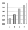
- Tm analysis in the same manner as in Example 1 except that each reagent was used and PCR was treated at 95 ° C. for 60 seconds and then repeated 50 cycles with 1 cycle at 95 ° C. and 15 seconds at 60 ° C. Did. The results are shown in Table 11 and FIG.
- the first primer for CYP2D6 is a 2D6-P3F4 in which 4 bases of the single additional base sequence at the 5 'end are deleted and the base sequence corresponding to the amplification target region of CYP2D6 is degenerate.
- Example 2 and Example 3 As shown in Table 11 and FIGS. 3 and 4, in Example 2 and Example 3, the area ratio increases in accordance with the copy number of the target gene in the samples a to b, and the result reflecting the increase in copy number is It was obtained. Also, by changing the conditions of the primer and the reaction temperature conditions, the waveform of the detection graph changes, and results of different area ratios can be obtained. Therefore, the reagent prescription is optimized by appropriately changing these conditions. It can be performed.
- Example 4 the first primer was prepared from 2D6-P3F4-Int6-F2 (gtagtcgaagacgatgtttacgTGAGCC CATCTGGG (B) AACA: SEQ ID NO: 13, 41 mer, Tm value 67.6 ° C. to 69.
- PCR and Tm analysis were performed in the same manner as in Example 1 except that 45 cycles at 45 ° C. were repeated as one cycle.
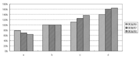
- PCR and Tm analysis were performed in the same manner as in Example 4 except that the amount of each primer was changed as shown in Table 13. The results are shown in Table 14, FIGS. 5 and 6, respectively.
- Example 4 uses a second primer having a higher Tm value than the first primer and uses a smaller amount of the first primer than the third primer.
- Example 5 uses the same combination of primers as Example 4 and uses a larger amount of the first primer than the third primer. In any case, as shown in Table 14, FIG. 5 and FIG. 6, it is possible to obtain the result appropriately reflecting the increase in the copy number of the target gene.
- Example 6 to 8 the probe for CYP2D6 was changed to 3T-2D6-Int6-R2 (gtacccttcctccc- (TAMRA): SEQ ID NO: 15), and the amount of the second primer (PEN3 F4) as shown in Table 15 PCR and Tm analysis were carried out in the same manner as in Example 1 except that each reagent according to the formulation changed to 100 nM (Example 6), 400 nM (Example 7) or 1200 nM (Example 8) was used, respectively. The results are shown in Table 16 and FIG. 7, respectively. In FIG. 7, the area ratio in each sample of Examples 6 to 8 is shown as a ratio when the area ratio when the amount of the target gene (CYP2D6) is 250 copies / ⁇ L is 100%.
- Example 6 to Example 8 even if the amount of the second primer present in the reaction solution is changed, the area ratio according to the copy number of the target gene in the samples a to d The result was obtained reflecting the increase in copy number. In addition, it was found that when the amount of the second primer was larger than the amount of the other primers in the reaction solution, the rate of increase of the area ratio became larger, and when it became smaller, the rate of increase of the area ratio became smaller. In any case, as shown in Table 16 and FIG. 7, it is possible to obtain a result that appropriately reflects the increase in the copy number of the target gene.
- the amount of gene present in the nucleic acid in the target sample can be measured more accurately and conveniently than in the prior art.
- Example 9 As a gene region on the 5 'side of the ALK gene, NCBI accession no. The 2460 to 2801st base sequence of NM_004304.4 (SEQ ID NO: 24) and the 4690 to 5031th base sequence (SEQ ID NO: 25) were inserted into pcDNA3.1 (+) as the gene region on the 3 ′ side. RNA was synthesized from artificial nucleic acid (plasmid) to prepare a template containing the copy number described in Table 17.
- a fully automatic SNPs tester (trade name i-densy (trade name), manufactured by ARKRAY, Inc.), the probes shown in Table 18 below, and various primers, and using the test reagents of the prescription described in Table 19, RT- PCR and Tm analysis were performed.
- the polylase used is Taq polymerase.
- PEN 3 R 2 common primer
- PEN 3 R 2 common primer
- the PCR was repeated 50 cycles of treating the sample after reverse transcription reaction at 95 ° C. for 60 seconds and then making one cycle at 95 ° C. and 15 seconds at 58 ° C.
- Tm analysis is performed after PCR for 1 second at 95 ° C and 60 seconds at 40 ° C, and then the temperature is raised from 40 ° C to 75 ° C at a temperature rise rate of 1 ° C / 3 seconds for a while. Change of the typical fluorescence intensity was measured.
- the excitation wavelength was set to 520 nm to 555 nm, and the measurement wavelength was set to 585 to 700 nm, and changes in fluorescence intensity derived from the fluorescence labeled probe were respectively measured. It is known that an amplification product derived from the 5 'gene region has a peak at around 51 ° C, and an amplification product derived from the 3' gene region has a peak at 58 ° C.
- the sequences of the primers and probes used are as described in Table 18.
- the 5 'gene region is a first primer for the 5' gene region; P3R2-ALK5'-R5 (SEQ ID NO: 19, Tm value 70.5 ° C) and a third primer; ALK5'-F2 ( It amplified using SEQ ID NO: 18, Tm value 59.9 ° C.).
- the gene region on the 3 'side is a first primer for the gene region on the 3'side;P3R2-ALK3'-R3 (SEQ ID NO: 21, Tm value 70.1 ° C), and a third primer; ALK3'-F3 ( It amplified using SEQ ID NO: 20, Tm value 60.8 ° C.).
- Each amplification product is a second primer which is a common primer composed only of the single additional base sequence; PEN3 R2 (SEQ ID NO: 22, Tm value 58.6 ° C.) and each third primer It amplified using.
- the detection probe (SEQ ID NO: 16) for the 5 'gene region and the detection probe (SEQ ID NO: 17) for the 3' gene region are as described in Table 18.
- the Tm value of each primer is a value calculated using Meltcalc. In Table 18, the base sequences indicated by lower case letters indicate single additional base sequences.
- the presence or absence of a fusion gene mutation can be measured simply and inexpensively and accurately.
Landscapes
- Chemical & Material Sciences (AREA)
- Organic Chemistry (AREA)
- Life Sciences & Earth Sciences (AREA)
- Zoology (AREA)
- Wood Science & Technology (AREA)
- Proteomics, Peptides & Aminoacids (AREA)
- Health & Medical Sciences (AREA)
- Engineering & Computer Science (AREA)
- Chemical Kinetics & Catalysis (AREA)
- Biophysics (AREA)
- Biochemistry (AREA)
- Microbiology (AREA)
- Molecular Biology (AREA)
- Biotechnology (AREA)
- Analytical Chemistry (AREA)
- Physics & Mathematics (AREA)
- Immunology (AREA)
- Bioinformatics & Cheminformatics (AREA)
- General Engineering & Computer Science (AREA)
- General Health & Medical Sciences (AREA)
- Genetics & Genomics (AREA)
- Measuring Or Testing Involving Enzymes Or Micro-Organisms (AREA)
- Apparatus Associated With Microorganisms And Enzymes (AREA)
Abstract
Priority Applications (5)
| Application Number | Priority Date | Filing Date | Title |
|---|---|---|---|
| CN201280053730.7A CN104024433B (zh) | 2011-10-31 | 2012-10-29 | 基因丰度的测定方法 |
| KR1020147010864A KR101483576B1 (ko) | 2011-10-31 | 2012-10-29 | 유전자 존재량의 측정 방법 |
| JP2013509391A JP5570657B2 (ja) | 2011-10-31 | 2012-10-29 | 遺伝子存在量の測定方法 |
| EP12844982.4A EP2774995A4 (fr) | 2011-10-31 | 2012-10-29 | Méthode de mesure de l'abondance de gènes |
| US13/664,674 US9121051B2 (en) | 2011-10-31 | 2012-10-31 | Method of determining the abundance of a target nucleotide sequence of a gene of interest |
Applications Claiming Priority (2)
| Application Number | Priority Date | Filing Date | Title |
|---|---|---|---|
| JP2011238953 | 2011-10-31 | ||
| JP2011-238953 | 2011-10-31 |
Related Child Applications (1)
| Application Number | Title | Priority Date | Filing Date |
|---|---|---|---|
| US13/664,674 Continuation US9121051B2 (en) | 2011-10-31 | 2012-10-31 | Method of determining the abundance of a target nucleotide sequence of a gene of interest |
Publications (1)
| Publication Number | Publication Date |
|---|---|
| WO2013065646A1 true WO2013065646A1 (fr) | 2013-05-10 |
Family
ID=48191993
Family Applications (1)
| Application Number | Title | Priority Date | Filing Date |
|---|---|---|---|
| PCT/JP2012/077916 WO2013065646A1 (fr) | 2011-10-31 | 2012-10-29 | Méthode de mesure de l'abondance de gènes |
Country Status (5)
| Country | Link |
|---|---|
| EP (1) | EP2774995A4 (fr) |
| JP (1) | JP5570657B2 (fr) |
| KR (1) | KR101483576B1 (fr) |
| CN (1) | CN104024433B (fr) |
| WO (1) | WO2013065646A1 (fr) |
Cited By (1)
| Publication number | Priority date | Publication date | Assignee | Title |
|---|---|---|---|---|
| JP2019010099A (ja) * | 2017-06-30 | 2019-01-24 | エシコン・インコーポレイテッドEthicon, Incorporated | 生物学的インジケータの起動を確認するためのシステム及び方法 |
Citations (9)
| Publication number | Priority date | Publication date | Assignee | Title |
|---|---|---|---|---|
| JP2005512577A (ja) | 2001-12-19 | 2005-05-12 | ブランデイズ ユニバーシティー | Late−pcr |
| JP2007289149A (ja) * | 2006-01-25 | 2007-11-08 | Kyoto Sangyo Univ | 5’領域ディファレンシャルディスプレー法 |
| WO2009081965A1 (fr) | 2007-12-26 | 2009-07-02 | Arkray, Inc. | Procédé et dispositif d'analyse de courbe de fusion |
| WO2010001969A1 (fr) | 2008-07-02 | 2010-01-07 | アークレイ株式会社 | Procédé d'amplification d'une séquence d'acides nucléiques cible, procédé de détection de la mutation à l'aide du procédé, et réactifs en vue d'une utilisation dans les procédés |
| WO2010071147A1 (fr) * | 2008-12-16 | 2010-06-24 | アークレイ株式会社 | Procédé de détection de régulations de l'amplification d'acide nucléique et utilisation associée |
| JP2010538614A (ja) | 2007-09-07 | 2010-12-16 | フルイダイム コーポレイション | コピー数変動の決定、方法およびシステム |
| WO2011043220A1 (fr) | 2009-10-06 | 2011-04-14 | 富士レビオ株式会社 | Procédé de détection d'un gène de fusion |
| JP2011516069A (ja) | 2008-04-03 | 2011-05-26 | ハドソン アルファ インスティテュート フォー バイオテクノロジー | 多数の標的の増幅のためのアンプリコンレスキューマルチプレックスポリメラーゼ連鎖反応 |
| JP2012100628A (ja) | 2010-11-12 | 2012-05-31 | Mitsubishi Chemical Medience Corp | Eml4−alk融合遺伝子の高感度検出方法 |
Family Cites Families (11)
| Publication number | Priority date | Publication date | Assignee | Title |
|---|---|---|---|---|
| US20070059700A1 (en) * | 2003-05-09 | 2007-03-15 | Shengce Tao | Methods and compositions for optimizing multiplex pcr primers |
| JP4322554B2 (ja) * | 2003-05-19 | 2009-09-02 | 株式会社ニチレイフーズ | Dna増幅反応の効率向上方法 |
| CN1280422C (zh) * | 2004-08-26 | 2006-10-18 | 北京博奥生物芯片有限责任公司 | 一种不对称pcr扩增方法及其应用 |
| AU2008255569A1 (en) * | 2007-06-01 | 2008-12-04 | Monoquant Pty Ltd | A method for DNA breakpoint analysis |
| JP2011500063A (ja) * | 2007-10-22 | 2011-01-06 | モノクアント・ピーティーワイ・リミテッド | Dna増幅方法 |
| EP2336358A1 (fr) * | 2008-05-06 | 2011-06-22 | QIAGEN GmbH | Détection simultanée de plusieurs séquences d'acide nucléique dans une réaction |
| WO2010088288A2 (fr) * | 2009-01-28 | 2010-08-05 | Fluidigm Corporation | Détermination de différences du nombre de copies par amplification |
| US8039215B2 (en) * | 2009-03-10 | 2011-10-18 | Roche Molecular Systems, Inc. | Multiplex quantitative nucleic acid amplification and melting assay |
| JP5267293B2 (ja) * | 2009-04-13 | 2013-08-21 | 島根県 | リアルタイムpcr法による食品媒介病原菌の網羅的迅速検出方法 |
| EP2248915B1 (fr) * | 2009-05-05 | 2013-09-18 | Qiagen GmbH | Détection de plusieurs séquences d'acide nucléique dans une cartouche de réaction |
| EP3546596B1 (fr) * | 2009-05-26 | 2022-05-11 | Quest Diagnostics Investments Incorporated | Utilisation d'une trousse pour la détection de dysfonctionnements géniques |
-
2012
- 2012-10-29 JP JP2013509391A patent/JP5570657B2/ja active Active
- 2012-10-29 EP EP12844982.4A patent/EP2774995A4/fr not_active Withdrawn
- 2012-10-29 KR KR1020147010864A patent/KR101483576B1/ko active IP Right Grant
- 2012-10-29 CN CN201280053730.7A patent/CN104024433B/zh active Active
- 2012-10-29 WO PCT/JP2012/077916 patent/WO2013065646A1/fr active Application Filing
Patent Citations (9)
| Publication number | Priority date | Publication date | Assignee | Title |
|---|---|---|---|---|
| JP2005512577A (ja) | 2001-12-19 | 2005-05-12 | ブランデイズ ユニバーシティー | Late−pcr |
| JP2007289149A (ja) * | 2006-01-25 | 2007-11-08 | Kyoto Sangyo Univ | 5’領域ディファレンシャルディスプレー法 |
| JP2010538614A (ja) | 2007-09-07 | 2010-12-16 | フルイダイム コーポレイション | コピー数変動の決定、方法およびシステム |
| WO2009081965A1 (fr) | 2007-12-26 | 2009-07-02 | Arkray, Inc. | Procédé et dispositif d'analyse de courbe de fusion |
| JP2011516069A (ja) | 2008-04-03 | 2011-05-26 | ハドソン アルファ インスティテュート フォー バイオテクノロジー | 多数の標的の増幅のためのアンプリコンレスキューマルチプレックスポリメラーゼ連鎖反応 |
| WO2010001969A1 (fr) | 2008-07-02 | 2010-01-07 | アークレイ株式会社 | Procédé d'amplification d'une séquence d'acides nucléiques cible, procédé de détection de la mutation à l'aide du procédé, et réactifs en vue d'une utilisation dans les procédés |
| WO2010071147A1 (fr) * | 2008-12-16 | 2010-06-24 | アークレイ株式会社 | Procédé de détection de régulations de l'amplification d'acide nucléique et utilisation associée |
| WO2011043220A1 (fr) | 2009-10-06 | 2011-04-14 | 富士レビオ株式会社 | Procédé de détection d'un gène de fusion |
| JP2012100628A (ja) | 2010-11-12 | 2012-05-31 | Mitsubishi Chemical Medience Corp | Eml4−alk融合遺伝子の高感度検出方法 |
Non-Patent Citations (6)
| Title |
|---|
| CLINICAL CHEMISTRY, 2009, pages 1546 - 1554 |
| J. CLIN. ONCOL., vol. 27, no. 26, 10 September 2009 (2009-09-10), pages 4232 - 5 |
| KAKIHARA Y. ET AL.: "Cost-saving design of PCR primers containing additional sequences", ITE LETTERS ON BATTERIES, NEW TECHNOLOGIES & MEDICINE, vol. 6, no. 2, 2005, pages 135 - 139, XP001505011 * |
| PROC. NATL. ACAD. SCI. USA, vol. 89, June 1992 (1992-06-01), pages 5321 - 5325 |
| SCHOUTEN J.P. ET AL.: "Relative quantification of 40 nucleic acid sequences by multiplex ligation-dependent probe amplification, art e57", NUC. ACID. RES., vol. 30, no. 12, 2002, pages 1 - 13, XP002362868 * |
| See also references of EP2774995A4 * |
Cited By (2)
| Publication number | Priority date | Publication date | Assignee | Title |
|---|---|---|---|---|
| JP2019010099A (ja) * | 2017-06-30 | 2019-01-24 | エシコン・インコーポレイテッドEthicon, Incorporated | 生物学的インジケータの起動を確認するためのシステム及び方法 |
| JP7258479B2 (ja) | 2017-06-30 | 2023-04-17 | エシコン・インコーポレイテッド | 生物学的インジケータの起動を確認するためのシステム及び方法 |
Also Published As
| Publication number | Publication date |
|---|---|
| KR20140088105A (ko) | 2014-07-09 |
| EP2774995A4 (fr) | 2015-04-29 |
| EP2774995A1 (fr) | 2014-09-10 |
| CN104024433B (zh) | 2016-11-16 |
| JPWO2013065646A1 (ja) | 2015-04-02 |
| JP5570657B2 (ja) | 2014-08-13 |
| CN104024433A (zh) | 2014-09-03 |
| KR101483576B1 (ko) | 2015-01-21 |
Similar Documents
| Publication | Publication Date | Title |
|---|---|---|
| JP2012235778A (ja) | アセトアルデヒド脱水素酵素2及びアルコール脱水素酵素2の遺伝子変異を同時に検出する方法 | |
| US20130078631A1 (en) | Probe, and polymorphism detection method using the same | |
| JP2013090622A (ja) | 多型検出用プローブ、多型検出方法、薬効判定方法及び多型検出用キット | |
| CN104450869B (zh) | 一种双脱氧核苷修饰的引物方法、反应体系及其在突变检测中的应用 | |
| JP4942508B2 (ja) | abl遺伝子変異の検出用プローブおよびその用途 | |
| JP6153758B2 (ja) | 多型検出用プローブ、多型検出方法、薬効判定方法及び多型検出用キット | |
| EP2520663A1 (fr) | Sonde de détection de mutation génétique | |
| US9121051B2 (en) | Method of determining the abundance of a target nucleotide sequence of a gene of interest | |
| JP5757909B2 (ja) | 多型検出用プローブ、多型検出方法、薬効判定方法及び多型検出用試薬キット | |
| WO2013065646A1 (fr) | Méthode de mesure de l'abondance de gènes | |
| JP6205216B2 (ja) | 変異検出用プローブ、変異検出方法、薬効判定方法及び変異検出用キット | |
| Zhang et al. | Single nucleotide polymorphism genotyping of ALDH2 gene based on asymmetric PCR and fluorescent probe-mediated melting curves | |
| US20120107815A1 (en) | Polymorphism Detection Probe, Polymorphism Detection Method, Evaluation of Drug Efficacy, and Polymorphism Detection Kit | |
| WO2016158898A1 (fr) | Procédé de détection d'une mutation de gène et oligonucléotide marqué par fluorescence l'utilisant | |
| JP5930825B2 (ja) | Egfrエクソン19多型検出試験用試薬キット及びその用途 | |
| WO2018079579A1 (fr) | Procédé de détection d'une séquence de base cible, procédé de conception et de production de sonde, et kit | |
| JP6059453B2 (ja) | 同一又は近傍の検出波長を有する蛍光色素で修飾された複数のオリゴヌクレオチドを用いて、1種類の波長で複数の遺伝子多型を検出する方法 | |
| JP5860667B2 (ja) | Egfrエクソン21l858r遺伝子多型検出用プライマーセット及びその用途 | |
| JP5940771B2 (ja) | abl遺伝子多型の検出用プローブおよびその用途 | |
| WO2011052754A1 (fr) | Sonde de détection de polymorphisme dans les gènes egfr et utilisation associée | |
| KR20120124033A (ko) | 유전자 변이 검출용 프로브, 유전자 변이 검출 방법 및 유전자 변이 검출용 시약 키트 | |
| JP2017163889A (ja) | K−ras遺伝子増幅用フォワードプライマーセット、K−ras遺伝子増幅用キット、K−ras遺伝子増幅方法、多型解析方法、及び薬効判定方法 | |
| JP2013094116A (ja) | Slc28a1/a2/a3遺伝子の多型検出用プローブおよびその用途 |
Legal Events
| Date | Code | Title | Description |
|---|---|---|---|
| ENP | Entry into the national phase |
Ref document number: 2013509391 Country of ref document: JP Kind code of ref document: A |
|
| 121 | Ep: the epo has been informed by wipo that ep was designated in this application |
Ref document number: 12844982 Country of ref document: EP Kind code of ref document: A1 |
|
| ENP | Entry into the national phase |
Ref document number: 20147010864 Country of ref document: KR Kind code of ref document: A |
|
| NENP | Non-entry into the national phase |
Ref country code: DE |
|
| REEP | Request for entry into the european phase |
Ref document number: 2012844982 Country of ref document: EP |
|
| WWE | Wipo information: entry into national phase |
Ref document number: 2012844982 Country of ref document: EP |