WO2012128363A1 - Pedfマイクロニードルアレイ及びその製造方法 - Google Patents
Pedfマイクロニードルアレイ及びその製造方法 Download PDFInfo
- Publication number
- WO2012128363A1 WO2012128363A1 PCT/JP2012/057598 JP2012057598W WO2012128363A1 WO 2012128363 A1 WO2012128363 A1 WO 2012128363A1 JP 2012057598 W JP2012057598 W JP 2012057598W WO 2012128363 A1 WO2012128363 A1 WO 2012128363A1
- Authority
- WO
- WIPO (PCT)
- Prior art keywords
- pedf
- microneedle
- microneedle array
- peptide
- skin
- Prior art date
Links
Images
Classifications
-
- A—HUMAN NECESSITIES
- A61—MEDICAL OR VETERINARY SCIENCE; HYGIENE
- A61M—DEVICES FOR INTRODUCING MEDIA INTO, OR ONTO, THE BODY; DEVICES FOR TRANSDUCING BODY MEDIA OR FOR TAKING MEDIA FROM THE BODY; DEVICES FOR PRODUCING OR ENDING SLEEP OR STUPOR
- A61M37/00—Other apparatus for introducing media into the body; Percutany, i.e. introducing medicines into the body by diffusion through the skin
- A61M37/0015—Other apparatus for introducing media into the body; Percutany, i.e. introducing medicines into the body by diffusion through the skin by using microneedles
- A61M2037/0046—Solid microneedles
Definitions
- the present invention relates to a microneedle array for administering PEDF.
- the internal use method As a method for injecting pharmaceuticals into the body, the internal use method, the injection method, the transdermal method, etc. are known, and each has its own characteristics.
- the internal use method is simple, depending on the drug, absorption efficiency is poor, or the digestive tract and liver are burdened.
- the injection method is efficient, it is painful and requires the hands of specialists such as doctors.
- the transdermal method can control the dose of the drug temporally to reduce side effects such as pain and inflammation due to a temporary increase in the local concentration of the drug, thereby relieving the patient's anxiety and burden.
- a microneedle has been proposed as a method for solving these problems and reliably supplying a medicinal component to a specific place on the skin surface layer and / or the skin stratum corneum (Patent Document 1). Since the microneedle is very thin, there is no pain or bleeding when it is inserted into the skin surface layer and / or skin stratum corneum, and the puncture wound is quickly closed, which is suitable as a method for reliably supplying the drug under the skin. .
- a substrate provided with a plurality of microneedles on a substrate is called a microneedle array.
- the one provided with an adhesive tape or the like for fixing the microneedle array on the skin is called a microneedle patch.
- Patent Document 2 A substance that dissolves and disappears in the living body has been proposed as a material of the microneedle.
- Patent Document 2 A substance that dissolves and disappears in the living body has been proposed as a material of the microneedle.
- the microneedle dissolves and disappears in the skin surface layer and / or the skin stratum corneum. Can be supplied to.
- microneedle materials For example, maltose (patent document 2), hyaluronic acid (patent documents 4, 5, 6), dextran (patent document 4), polyvinylpyrrolidone (patent document 7), gelatin (patent documents 3, 5, 6), collagen (patent Documents 5 and 6), chitosan (Patent Document 5), proteins (Patent Documents 3 and 4), and biodegradable resins (Patent Document 1) are disclosed.
- a microneedle containing a medical drug is called a medical microneedle.
- Many drugs are expensive, and it is necessary to contain the drug only at the tip of the microneedle in order to avoid an increase in cost.
- An application method for applying a drug to the tip of a microneedle has already been proposed (Patent Document 8).
- PEDF Pigment epithelium-derived factor protein
- the problem to be solved by the present invention is to provide a transdermal administration method using microneedles, which is superior to the internal use / injection method and the conventional application method when administering PEDF or its active partial peptide to the human body. There is.
- the PEDF microneedle array of the present invention is characterized by comprising microneedles containing a pharmaceutically effective amount of one or more drugs selected from the group of PEDF and its active partial peptide.
- PEDF is expensive.
- it is preferable to manufacture the microneedle array so that PEDF is present only in the microneedle portion of the microneedle array and not included in the substrate portion. This is because only the tip portion of the microneedle is inserted and administered into the body, and the substrate portion is a portion that supports the microneedle and is not directly related to drug administration.
- a welding method as a method of containing a drug only in the microneedle portion.
- the microneedle material is made water-soluble, PEDF is dissolved in an aqueous solution made of that material or other water-soluble materials, and the tip of the microneedle array that has already been molded is immersed in the solution to make the drug microscopic.
- This is a method of containing only at the tip of the needle array.
- the tip of the microneedle array When the tip of the microneedle array is immersed in the drug solution, the tip of the microneedle array partially dissolves, and when the drug solution adheres to the tip, it is integrated, and when the microneedle is inserted into the skin, the drug-containing portion is peeled off. It wo n’t fall.
- the microneedle array material and the drug solution are preferably compatible.
- a method of manufacturing a microneedle array step by step as another method for containing a drug only in the microneedle portion.
- An aqueous solution in which PEDF is added to a microneedle material is cast into a mold in which the shape of the microneedle is perforated, and the moisture is evaporated and dried at room temperature or by heating to produce only the microneedle portion.
- An aqueous solution of only the microneedle material is cast on the substrate, the substrate is laminated and then peeled off, and the microneedle is transferred onto the substrate. Also by this method, a microneedle array containing PEDF only in the microneedle portion can be obtained.
- the material is desirably a water-soluble polymer.
- water-soluble polymers include synthetic polymers such as polyvinylpyrrolidone and polyvinyl alcohol, semi-synthetic polymers such as carboxymethylcellulose and ethylcellulose, polysaccharides such as hyaluronic acid, chondroitin sulfate, dextran, dextrin, and proteoglycan. Can be given.
- a microneedle comprising a mixture obtained by adding a monosaccharide or a disaccharide to these polymer materials is more desirable because it has a feature that the dissolution rate of the microneedle in the skin is shortened.
- a microneedle containing a polysaccharide as a main material and containing a pharmaceutically effective amount of PEDF can be particularly preferably used.
- a microneedle containing PEDF in a material obtained by adding about 20 to 100 parts by weight of glucose to 100 parts by weight of polysaccharide is more preferable because the needle solubility in the skin is increased and PEDF is immediately dissolved.
- the molecular weight of hyaluronic acid constituting the main component of the microneedle is 50,000 to 4,000,000. If the molecular weight of hyaluronic acid is less than 50,000, it is inconvenient from the viewpoint of mechanical strength. Moreover, when it exceeds 4 million, the solution viscosity at the time of preparing aqueous solution is too high, and it is inconvenient to handle.
- the microneedle array of the present invention is a microneedle array in which a plurality of fine microneedles are formed on the surface of a substrate, and the shape of the microneedles is easy to be inserted into the skin and does not cause pain when inserting.
- a conical shape, a truncated cone shape, or a coneide shape was used.
- the Conide type is a so-called volcano type, and the side surface of the truncated cone is curved inward.
- the root diameter of the microneedle is reduced, the amount of material to be inserted into the skin is reduced, and it is easy to break when it is inserted into the skin. If it is thick, it is painful when it is inserted into the skin. 0.0 mm is appropriate. When the tip diameter is thin (pointed), it tends to break when piercing the skin, and when the tip diameter is thick, it is difficult to pierce the skin and is painful, so 0.01 to 0.08 mm is appropriate.
- the height of the microneedle is low, it becomes difficult to supply the drug to a predetermined position on the skin surface layer and / or the skin stratum corneum, and if it is high, it is easy to break when inserted into the skin. It is. Also, when the pitch between the microneedles becomes shorter, it becomes difficult to pierce the skin, and when the pitch becomes longer, the number of microneedles per area decreases, and a large amount of medicine cannot be supplied to a predetermined narrow site. 0.4 to 1.0 mm is appropriate.
- PEDF can be produced by expressing the DNA encoding the protein by the method described in Non-Patent Document 1, but can also be purchased. A specific production method is described in Patent Literature 9 [0018] and the like.
- PEDF includes all PEDF variants having functionally equivalent characteristics to human PEDF.
- PEDF variants are described in, for example, Patent Documents 10 to 12.
- a PEDF variant comprising an amino acid sequence having one or more amino acid residue substitutions, deletions, and / or additions to the amino acid sequence of human PEDF, and having a function equivalent to that of human PEDF
- This variant is a preferred variant.
- Functionally equivalent to human PEDF has a hair loss inhibiting activity or hair growth promoting activity equivalent to human PEDF, or a function capable of preventing apoptosis of hair root cells including hair matrix cells, equivalent to human PEDF.
- an active partial peptide having an amino acid sequence having homology with the amino acid sequence of human PEDF can be contained in the microneedle of the present invention as long as it is functionally equivalent to human PEDF.
- the active partial peptide refers to a peptide having excellent hair loss inhibitory activity and hair growth promoting activity similar to or higher than PEDF among many fragments obtained by chemically or enzymatically degrading PEDF.
- the amino acid sequence is equivalent to a part of the amino acid sequence of PEDF.
- Peptide 1 His Leu Thr Phe Pro Leu Asp Tyr His Leu Asn Gln Pro Phe Ile Phe Val Leu Arg Asp: 20-residue peptide corresponding to residues 381-400 of PEDF 2: His Leu Thr Phe Pro Leu Asp: 7-residue peptide corresponding to residues 381-387 of PEDF 3: Tyr His Leu Asn Gln Pro: 6 residue peptide corresponding to 388-393 residues of PEDF 4: Phe Ile Phe Val Leu Arg Asp: 7 residues corresponding to 394-400 residues of PEDF
- the dose of PEDF or active partial peptide can be appropriately determined depending on the age, weight, sex, symptoms, etc. of the administration subject.
- the usual dose is 0.5 ⁇ g to 100 mg, preferably 1 ⁇ g to 10 mg per dose.
- Administration is daily or every few days for several months. If the amount is too small, it is ineffective, and if the amount is too large, an unexpected side effect may occur.
- the filling amount of PEDF or active partial peptide into the microneedles it is desirable to fill the whole administration amount into one microneedle in consideration of administration convenience.
- the manufacturing method of the microneedle array of the present invention is not particularly limited, and may be manufactured by any conventionally known method.
- a microneedle material aqueous solution is cast into a mold having a microneedle shape, and the substrate is peeled off after drying by evaporating moisture at room temperature or by heating.
- the microneedle array In order to apply the microneedle array produced by the method as described above to the skin for several hours, it is preferable to make a microneedle patch by lining the back surface of the substrate of the microneedle array with an adhesive film.
- the microneedle array includes a substrate and a plurality of needles or the like provided integrally with the substrate on one side of the substrate.
- An adhesive film having a larger area than the substrate is laminated on the surface of the microneedle array on the opposite side of the surface on which the plurality of needles are provided. Thereby, the microneedle array can be lined with an adhesive film to form a microneedle patch.
- an adhesive film a base material and an appropriate adhesive film in which an adhesive layer is provided on one side of the base material can be used.
- a substrate examples include polyester film, polyethylene film, polyurethane film, urethane foam, and paper.
- the adhesive which consists of acrylic ester, a styrene-isoprene thermoplastic rubber adhesive, etc. can be used conveniently.
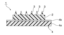
- FIG. 2 is a cross-sectional view schematically showing an embodiment of the microneedle patch.
- the microneedle patch 1 has a substrate 2, and a plurality of needles 3 are integrally formed on one surface of the substrate 2.
- An adhesive film 4 is laminated on the opposite surface of the substrate 2.
- the adhesive film 4 has the base material 4a and the adhesive 4b provided in the single side
- PEDF or an active partial peptide thereof is selectively supplied to a part that requires hair production, for example, the head, using a microneedle, hair loss is suppressed and hair growth promoting activity appears.
- Microneedles have moderate hardness and resistance to breakage, and easily dissolve in the skin surface layer and / or skin stratum corneum. For this reason, there are few failures at the time of insertion, and it is easy to use even for beginners.
- PEDF or an active partial peptide thereof can be gradually supplied only to a portion requiring hair production.
- PEDF is distributed and supplied throughout the body, and the local concentration of the hair producing part becomes insufficient, and the effect is limited.
- a sufficient amount of PEDF was locally administered or administered by injection or injection, side effects were caused and there was a risk of inducing serious illness.
- PEDF subcutaneous injection is accompanied by severe pain because the amount of PEDF injection rises instantaneously and locally It is known that it is not practical to repeatedly apply such painful injections to many places.
- the necessary amount of PEDF can be gradually supplied to the necessary local area by appropriately selecting the microneedle material and adjusting the dissolution time. Therefore, it is easy to administer intermittently over several days or weeks.
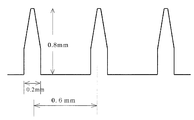
- FIG. 1 is an example of a cross-sectional view of a microneedle array.
- FIG. 2 is a schematic cross-sectional view showing a microneedle patch as a microneedle array according to an embodiment of the present invention.
- PEDF and each peptide used in the present invention were prepared by Upwell Co., Ltd.
- Table 1 summarizes the material of the microneedle used in this example and the PEDF contained in the microneedle.
- the material composition of the microneedle is indicated by the weight ratio of each component constituting the microneedle.
- FIG. 1 shows a part of a cross-sectional view of the microneedle array of this embodiment.
- the hyaluronic acid used in this example is manufactured by Kikkoman Biochemifa Co., Ltd. (formerly Kibun Food Chemifa Co., Ltd.), its molecular weight is 800,000 (trade name: FCH-80), and dextran is manufactured by Nippon Bulk Chemical Co., Ltd. Name: Dextran 70) and polyvinylpyrrolidone are manufactured by BASF Japan (trade name: Kollidon 12PF).
- molecular weight is a weight average molecular weight and means the quantity measured by gel permeation chromatography (GPC).
- Each microneedle array is cast in a mold in which the shape of the microneedle is perforated, and an aqueous solution in which PEDF is added to the microneedle material is cast, and the microneedle portion is dried by evaporating moisture at room temperature or by heating. create.
- the weight% of PEDF in Table 1 is the weight% of PEDF in this PEDF-added aqueous solution.
- An aqueous solution of only the microneedle material is cast on the substrate, the substrate is laminated and then peeled off, and the microneedle is transferred onto the substrate. According to this method, a microneedle array in which PEDF is contained only in the microneedles is obtained, and the use efficiency of PEDF is good.
- microneedle uniform microneedle in this specification.
- a welding method as described in Example 2 is more appropriate.
- Microneedles root diameter 0.2 mm, tip diameter 0.04 mm, a frustoconical length 0.8 mm, are arranged in a lattice pattern on 0.6mm interval, a circular patch type array of diameter 1 cm 2 There are 244 microneedles.
- the hardness of the base portion of the produced microneedle array was measured by a JIS pencil hardness measurement method.
- mice Male rats (12 weeks old) were anesthetized with Nembutal (30 mb / kg) and the abdominal skin was shaved, and the microneedles of each composition described above were lined with one protective bandage and administered. The administration time was 2 hours. The present composition 3, which was expected to dissolve immediately, was taken out after 30 minutes. The extracted microneedle was observed with a microscope, and the dissolution state of the tip was observed. Table 2 summarizes the pencil hardness of each microneedle and the observation results after administration.
- the composition of the microneedle can be obtained by using a polysaccharide as a main material, but if the proportion of vinylpyrrolidone is large, it is too hard and the needle becomes brittle. Moreover, short-time needle dissolution of polysaccharide microneedles containing glucose can also be confirmed. According to another test result, it was confirmed that the microneedle composed only of polysaccharides has insufficient solubility when applied to the skin for 30 minutes.
- a microneedle containing no drug of the material of the composition 1 was prepared.
- the microneedle array was a 1 cm diameter circular patch.
- dissolved 100 micrograms of microneedle raw materials of this composition 1, PEDF 5 mg, and trehalose (Hayashibara Laboratory, Okayama City) in 500 microliters of water was created.
- the tip portion of the microneedle needle 100 ⁇ m was immersed in the aqueous solution for 1 second, and then dried to obtain a tip PEDF welded microneedle array. Thereby, about 10 ⁇ g of PEDF is contained in the microneedle needle portion.
- Microneedles prepared by such a manufacturing method are referred to as welding microneedles in this specification, and the adhesive needle plaster (Micropore, 3M Company) is used on the back of the microneedle array to stabilize the application to the skin for several hours. ) was lined with a 2.5 cm diameter circular film to obtain a microneedle patch.
- microneedle array using peptide 1 A microneedle not containing the drug of the composition 2 was prepared. Further, a solution was prepared by dissolving 5 mg of the microneedle material of the present composition 2, peptide 1 and 100 ⁇ g of trehalose (Hayashibara Laboratory, Okayama City) in 500 ⁇ l of water.
- microneedle array The back surface of the microneedle array was lined with an adhesive film in which a bandage (Micropore, 3M Company) was cut into a 2.5 cm diameter to stabilize the application to the skin for several hours to obtain a microneedle patch.
- a bandage Micropore, 3M Company
- Example 4 Hair loss inhibitory activity of PEDF
- Microneedle A Intraneedle uniform microneedle containing 10 ⁇ g of PEDF: This composition 1 was used.
- Microneedle B In-needle uniform microneedle containing 0.5 ⁇ g of PEDF: The composition of composition 1 with a reduced PEDF concentration was used.
- Microneedle C Welding method microneedle containing 10 ⁇ g of PEDF: This composition 3 was used.
- Microneedle D Microneedle not containing PEDF: This composition 1 was used.
- the microneedles are frustoconical with a root diameter of 0.2 mm, a tip diameter of 0.04 mm, and a length of 0.8 mm, arranged in a grid at intervals of 0.6 mm, and are 1 cm 2 circular patch type arrays. There were 244 microneedles. The back of the array was lined with an adhesive sheet in which an acrylic adhesive was applied to one side of a circular PET film having a diameter of 2 cm.
- a hair loss mouse was created as a model animal as follows. C57BL6 / J mice (purchased from Claire), 6 to 8 weeks old and weighing 18 to 20 g, were bred for 10 weeks under conditions free of specific pathogens. The breeding method is “the thermae animal ethics committee or the US Department of Agriculture (USDA)“ the ”. It was carried out according to the protocol permitted in accordance with the guidelines of Guide for the Care and Use of Laboratory Animals.
- mice were allowed to eat a low-protein, high-fat choline-deficient diet from the start of the study.
- the composition of this choline-deficient diet (unit:% by weight) is: vitamin-free casein 8.000; lard 37.950; granulated sugar 48.375; L-cystine 0.625; vitamin Mix 1.000; mineral Mix 4.000; Oily vitamin mixture 0.050.
- vitamin Mix has the following composition (unit: mg / g of feed): Vitamin K3 0.3125, Vitamin B1 0.3125, Vitamin B2 0.5000, Vitamin B6 0.3125, DL-pantothenic acid Ca 1.2500, Niacin 1.2500, the rest has granulated sugar
- Mineral Mix is Composition (unit: mg / 100 g of feed): Ca (H 2 PO 4 ) H 2 O 585.000, lactic acid Ca 1,328.000, Fe citrate 118.800, MgSO 4 ⁇ 7H 2 O 548.000, K 2 HPO 4 959.200, NaH 2 PO 4 ⁇ 2H 2 O
- the oily vitamin mixture has the following composition (unit: mg / 100 g of feed): Vitamin A (500,000 IU / g) 2.5000, Vitamin D3 (500,000 IU) / G) having 0.6250, vitamin E 25.0000, and peanut oil 21.8750.
- mice Each of the mice was divided into the following 4 groups of 5 mice, and the following administration test was conducted.
- One group Administered microneedle A and fed a choline-deficient diet;
- Two groups Administered microneedle C and fed a choline-deficient diet;
- 3 groups Administered Macroclone B and fed a choline-deficient diet;
- 4 groups Administered microneedle D and fed a choline-deficient diet;
- mice On the test start date (day 0), each group of mice was given one microneedle to the epilation skin. Thereafter, the administration was continued twice a week while eating a choline-deficient diet, and after 6 weeks, the hair amount test of the administration part was evaluated by measuring the weight of the collected hair. The evaluation results are shown in Table 3.
- microneedle arrays the peptide is uniformly dissolved in all of the needle portion and the substrate portion.
- the obtained microneedle array was lined with a circular protective bandage having a diameter of 2 cm.
- the microneedle array was administered to the shaved skin of a male rat together with a protective adhesive bandage, it was confirmed by microscopic observation that the needle part was completely dissolved in the skin with three types of microneedles after 1 hour.
- the microneedle-forming mold used in this test is a Conede type with a recess root diameter of 0.6 mm, a tip diameter of 0.02 mm, and a depth of 0.2 mm, and is arranged in a grid at intervals of 0.6 mm. About 244 pieces are formed per 1 cm 2 .
Abstract
皮膚表層及び/又は皮膚角質層に折れることなく容易且つ均一に刺入でき、かつ容易に溶解するPEDFマイクロニードルアレイを提供する。 PEDFマイクロニードルアレイは、PEDF及びその活性部分ペプチドの群の中から選択された1若しくは複数の薬物を医薬的に有効量含有させたマイクロニードルから構成される。活性部分ペプチドとして4種類のペプチドが有効であることを示している。PEDF及びその活性部分ペプチドは高価であるためマイクロニードルの先端部分にのみ保持させることが好ましい。マイクロニードルの素材は水溶性高分子、特に多糖類であることが好ましい。このマイクロニードルアレイにより、毛髪の脱毛抑制または発毛促進を誘導することができる。
Description
本発明はPEDF投与用マイクロニードルアレイに関する。
医薬品を体内に投入する方法としては内服法、注射法、経皮法などが知られており、それぞれ特徴を有している。内服法は簡単であるが、薬剤によっては吸収効率が悪く、あるいは消化管や肝臓に負担をかける。注射法は効率がよいが、苦痛を伴い、医師等の専門家の手を患わさなければならない。これに対し経皮法は薬物の投与量を時間的にコントロールして薬物局所濃度の一時的増加による苦痛や炎症などの副作用を軽減でき、患者の不安・負担を和らげることができる。
従来の経皮法は、薬効成分を含む製剤を皮膚表面に塗布又は貼付し、薬物を皮膚や粘膜を透過させて投与していた。しかし、この方法は発汗や外的圧力等により薬物が脱落するほか、体内への異物侵入を抑止する皮膚角質層のバリアー機能のため透過効率が低い欠点があった。
これらの問題を解決し、皮膚表層及び/又は皮膚角質層の特定の場所に薬効成分を確実に供給する方法として、マイクロニードルが提案された(特許文献1)。マイクロニードルは非常に細いので皮膚表層及び/又は皮膚角質層に刺入した際痛みも出血もなく且つ穿刺創は速やかに閉鎖されるので、皮膚下に薬物を確実に供給する方法として好適である。基板上に複数のマイクロニードルを備えたものをマイクロニードルアレイという。さらにマイクロニードルアレイを皮膚上に固定するための粘着テープ等を備えたものをマイクロニードルパッチという。
マイクロニードルの材質として、生体内で溶解消失する物質が提案されている(特許文献2)。このようなマイクロニードルに薬物を含有させて皮膚に刺入すると、マイクロニードルは皮膚表層及び/又は皮膚角質層において溶解消失するので、皮膚表層及び/又は皮膚角質層の特定の場所に薬物を確実に供給することができる。
しかしながらマイクロニードルの機械的強度が小さい場合は皮膚に刺入する際に折れて刺入できず、機械的強度が大きい場合はマイクロニードルが皮膚内で容易に折れず残留させることが困難な欠点があった。これまでいろいろな材質がマイクロニードル素材として提案されている。例えば、マルトース(特許文献2)、ヒアルロン酸(特許文献4、5、6)、デキストラン(特許文献4)、ポリビニルピロリドン(特許文献7)、ゼラチン(特許文献3、5、6)、コラーゲン(特許文献5、6)、キトサン(特許文献5)、蛋白質(特許文献3、4)、生分解性樹脂(特許文献1)を用いたものが公表されている。
医療用の薬物を含むマイクロニードルは医療用マイクロニードルと呼ばれている。医薬品の中には高価なものが多く、高コスト化を避けるためにはマイクロニードル先端部分にのみ薬剤を含有させる必要がある。薬物をマイクロニードル先端に塗布する塗布法がすでに提案されている(特許文献8)。
ストレス社会のため近年薄毛人口が増加し、育毛・発毛に対する関心が高まっている。ヘアケア市場にはすでに多くの育毛・発毛剤が上市されている。
色素上皮由来因子蛋白質(以下「PEDF」と略記する)は優れた脱毛抑制活性及び発毛促進活性を有する(特許文献9)。しかしながら、これを注射法で投与すると薬液の皮内での注入量の一時的急増のため激しい痛みが生じる。
PEDFの変異体(特許文献10~12)やPEDFの製造方法(非特許文献1)はすでに知られている。
色素上皮由来因子蛋白質(以下「PEDF」と略記する)は優れた脱毛抑制活性及び発毛促進活性を有する(特許文献9)。しかしながら、これを注射法で投与すると薬液の皮内での注入量の一時的急増のため激しい痛みが生じる。
PEDFの変異体(特許文献10~12)やPEDFの製造方法(非特許文献1)はすでに知られている。
コールドスプリングハーバー研究所(Cold Spring HarborLaboratory)編「分子クロニング実験指針(Molecular Cloning: A LaboratoryManual)」第2版、コールドスプリングハーバー研究所出版(Cold Spring Harbor Laboratory Press),1989年。
本発明の解決しようとする課題は、PEDFないしはその活性部分ペプチドを人体に投与するに際し、内服法・注射法や在来の塗布法より優れた、マイクロニードルを利用する経皮投与法を提供することにある。
本発明のPEDFマイクロニードルアレイは、PEDF及びその活性部分ペプチドの群の中から選択された1若しくは複数の薬物を医薬的に有効量含有させたマイクロニードルから構成されていることを特徴とする。
PEDFは高価である。またPEDFの利用率を高めるためにPEDFはマイクロニードルアレイのマイクロニードル部分にのみ存在させ、基板部分には含ませないようにマイクロニードルアレイを製造することが好ましい。体内に刺入され、投与されるのはマイクロニードルの先端部分のみで、基板部分はマイクロニードルを支持する部分であって、薬剤投与に直接関係しないからである。
薬物をマイクロニードルアレイの先端に付着させる方法として、従来薬物をマイクロニードルアレイ先端に塗布する塗布法が知られているが、この方法では薬物がマイクロニードルの皮膚刺入の際剥がれ落ちることが多く、不満足であった。
マイクロニードル部分にのみ薬剤を含有させる方法として溶着法がある。これはマイクロニードル素材を水溶性とし、その素材もしくは他の水溶性素材からなる水溶液中にPEDFを溶解させておき、その溶液内にすでに成形されたマイクロニードルアレイの先端を浸すことにより薬物をマイクロニードルアレイ先端部にのみ含有させる方法である。マイクロニードルアレイの先端を薬物溶液に浸したとき、マイクロニードルアレイの先端が部分的に溶解し、薬物溶液が先端に付着したとき一体化し、マイクロニードルの皮膚への刺入に際し薬物含有部分がはがれ落ちることがない。
溶着法によりマイクロニードルアレイ先端に薬物を効率よく付着させるためには、マイクロニードルアレイ素材と薬物溶液は相溶性があることが好ましい。
マイクロニードル部分にのみ薬剤を含有させる別の方法として、マイクロニードルアレイを段階的に製造する方法がある。マイクロニードルの形状が穿設された型に、マイクロニードル素材にPEDFを添加した水溶液を流延し、室温下又は加熱して水分を蒸発して乾燥し、マイクロニードル部分のみを製造する。その上にマイクロニードル素材のみの水溶液を流延して基板を積層した後剥離し、基板上にマイクロニードルを転写する。この方法によってもマイクロニードル部分にのみPEDFが含有されたマイクロニードルアレイが得られる。
本発明のPEDFマイクロニードルアレイは、PEDFが水溶性であるため、素材が水溶性の高分子であることが望ましい。水溶性高分子の例としては、ポリビニルピロリドン、ポリビニルアルコールのような合成高分子、カルボキシメチルセルロース、エチルセルロースのような半合成高分子、ヒアルロン酸、コンドロイチン硫酸、デキストラン、デキストリン、プロテオグリカンのような多糖類などがあげられる。これらの高分子素材に単糖類、2糖類を添加した混合物からなるマイクロニードルは、マイクロニードルの皮膚内溶解速度が短縮されるという特徴を有するのでより望ましい。
多糖類を主素材とするマイクロニードルに医薬的に有効量のPEDFを含有させたものは特に好適に使用できる。
また、多糖類100重量部にグルコースを約20~100重量部添加した素材にPEDFを含有させたマイクロニードルは、皮膚内での針溶解性が高まりPEDFの即溶解性を与えるのでより好ましい。
マイクロニードルの主成分を構成するヒアルロン酸の分子量は5万以上400万以下であることが望ましい。ヒアルロン酸の分子量が5万未満であると機械的強度の観点から不都合である。また400万を超えると水溶液とした際の溶液粘度が高すぎて取扱に不便である。
本発明のマイクロニードルアレイは、複数の微細なマイクロニードルが基板の表面に形成されてなるマイクロニードルアレイであって、該マイクロニードルの形状は皮膚に刺入しやすく且つ刺入に際し苦痛を伴わないように円錐型、円錐台型又はコニーデ型とした。なお、コニーデ型とは、いわゆる火山型と呼ばれる形状であり、円錐台型の側面が内側方向に湾曲した形状である。
マイクロニードルの根元直径は細くなると皮膚内に刺入する素材の量が減少すると共に皮膚に刺入する際に折れやすくなり、太くなると皮膚に刺入する際に苦痛を伴うので0.15~1.0mmが適当である。先端直径は、細くなると(尖っていると)皮膚に刺入する際に折れやすくなり、太くなると皮膚に刺入しにくくなり苦痛を伴うので0.01~0.08mmが適当である。
マイクロニードルの高さは、低くなると皮膚表層及び/又は皮膚角質層の所定位置に薬剤を供給しにくくなり、高くなると皮膚に刺入する際に折れやすくなるので0.1~1.0mmが適当である。又、マイクロニードルとマイクロニードルの間のピッチは、短くなると皮膚に刺入しにくくなり、長くなると面積あたりのマイクロニードルの数が少なくなり、所定の狭い部位に多量の薬剤を供給できなくなるので、0.4~1.0mmが適当である。
PEDFは非特許文献1に記載の方法で、該蛋白質をコードするDNAを発現させることにより産生できるが、購入することもできる。具体的な産生手法は、特許文献9の[0018]等に記載されている。
本明細書では、PEDFには、ヒトPEDFと機能的に同等な特性を有するすべてのPEDF変異体を含むものとする。PEDF変異体は、例えば特許文献10~12に記載されている。
ヒトPEDFのアミノ酸配列に対して、1つ又は複数のアミノ酸残基の置換、欠失、及び/又は付加を有するアミノ酸配列を含むPEDF変異体であって、かつヒトPEDFと同等な機能を有するPEDFの変異体は、好ましい変異体である。ヒトPEDFと機能的に同等とは、ヒトPEDFと同等の脱毛抑制活性又は発毛促進活性を有すること、又はヒトPEDFと同等に毛母細胞を含む毛根部の細胞のアポトーシスを防止し得る機能を有することを意味する(特許文献9[0020]~[0022])。
また、ヒトPEDFのアミノ酸配列と相同性を有するアミノ酸配列を有する活性部分ペプチドも、ヒトPEDFと機能的に同等であれば、本発明のマイクロニードルに含有させることができる。本明細書において活性部分ペプチドとは、PEDFを化学的ないしは酵素的に分解した多くのフラグメントの中でPEDFと同様あるいはそれ以上の優れた脱毛抑制性及び発毛促進活性を有するペプチドをいうものとし、そのアミノ酸配列がPEDFのアミノ酸配列の一部と同等なものをいう。
脱毛抑制性及び発毛促進活性を確認できた活性部分ぺプチドの例として以下の4種がある。本明細書において、それぞれペプチド1、ペプチド2、ペプチド3、ペプチド4と記載する。
ペプチド1:His Leu
Thr Phe Pro Leu Asp Tyr His Leu Asn Gln Pro Phe Ile Phe Val Leu Arg Asp:PEDFの381―400残基に相当する20残基
ペプチド2:His Leu
Thr Phe Pro Leu Asp:PEDFの381-387残基に相当する7残基
ペプチド3:Tyr His
Leu Asn Gln Pro : PEDFの388-393残基に相当する6残基
ペプチド4:Phe Ile Phe Val Leu Arg Asp:PEDFの394-400残基に相当する7残基
ペプチド1:His Leu
Thr Phe Pro Leu Asp Tyr His Leu Asn Gln Pro Phe Ile Phe Val Leu Arg Asp:PEDFの381―400残基に相当する20残基
ペプチド2:His Leu
Thr Phe Pro Leu Asp:PEDFの381-387残基に相当する7残基
ペプチド3:Tyr His
Leu Asn Gln Pro : PEDFの388-393残基に相当する6残基
ペプチド4:Phe Ile Phe Val Leu Arg Asp:PEDFの394-400残基に相当する7残基
PEDFないしは活性部分ペプチドの投与量は、投与対象の年齢、体重、性別、症状、などで適宜決定されうる。通常の投与量は1回あたり0.5μgから100mg、好適には1μgから10mgである。投与は毎日あるいは数日おきに数ヶ月間投与する。少なければ効果がなく、多すぎれば予期せぬ副作用が起こることが懸念される。マイクロニードルへのPEDFないしは活性部分ペプチドの充填量は、投与の便を考慮すると投与全量を1枚のマイクロニードルに充填することが望ましい。
本発明のマイクロニードルアレイの製造方法は、特に限定されず、従来公知の任意の方法で製造されればよい。
例えば、マイクロニードルの形状が穿設された型に、マイクロニードル素材水溶液を流延し、室温下又は加熱して水分を蒸発して乾燥し、乾燥後基板を剥離する。
例えば、マイクロニードルの形状が穿設された型に、マイクロニードル素材水溶液を流延し、室温下又は加熱して水分を蒸発して乾燥し、乾燥後基板を剥離する。
以上のような方法で作製したマイクロニードルアレイを皮膚に数時間適用するためにはマイクロニードルアレイの基板の裏面に粘着性フィルムで裏打ちしてマイクロニードルパッチとすることが好ましい。より具体的には、マイクロニードルアレイは、基板と、基板の片面において該基板と一体に設けられた複数のニードル等を有する。このマイクロニードルアレイの基板の複数のニードルが設けられている面と反対側の面に、基板よりも大きな面積の粘着性フィルムを積層する。それによって、粘着性フィルムによりマイクロニードルアレイを裏打ちして、マイクロニードルパッチとすることができる。このような粘着性フィルムとしては、基材と、基材の片面に粘着剤層が設けられた適宜の粘着性フィルムを用いることができる。このような基材としては、ポリエステルフィルム、ポリエチレンフィルム、ポリウレタンフィルム、ウレタンフォーム、紙などが挙げられる。また、上記粘着剤としては、アクリルエステル、スチレン‐イソプレン系熱可塑性ゴム粘着剤等からなる粘着剤を好適に使用出来る。
図2は、上記マイクロニードルパッチの一実施形態を略図的に示す断面図である。図2に示すように、マイクロニードルパッチ1は、基板2を有し、基板2の片面に複数のニードル3が一体的に構成されている。また、基板2の反対側の面に、粘着性フィルム4が積層されている。粘着性フィルム4は、基材4aと、基材4aの片面に設けられた粘着剤4bとを有する。
PEDF若しくはその活性部分ペプチドを毛髪産生を必要とする部分、例えば頭部に、マイクロニードルにより選択的に供給すれば、脱毛が抑制され、発毛促進活性が現れる。
マイクロニードルは適度の硬さと折れにくさを有し、しかも皮膚表層及び/又は皮膚角質層において容易に溶解する。このため刺入に際し失敗が少なく、初心者にも使用しやすい。
本発明のPEDFマイクロニードルアレイの構成によれば、PEDF若しくはその活性部分ペプチドを毛髪の産生を必要とする部分のみに徐々に供給することができる。
従来の内服法や注射法では、体内全体にPEDFが分配供給され、毛髪産生部分の局所濃度が不十分となり、効果が限定的であった。局所的に十分な効果が得られるほどの量のPEDFを内服あるいは注射により投与すると副作用を生じ、重篤な疾病を誘発する危険があった。
これに対し毛髪産生を必要とする部分、例えば頭部の多数箇所に微量ずつ分けて注射する方法が考えられるが、PEDF皮下注射はPEDF注入量が瞬間的かつ局所的に急上昇するため激痛を伴うことが知られており、このような激痛を伴う注射を多数箇所に、それも反復して適用することは現実的でない。
一方マイクロニードルによれば、マイクロニードル素材を適切に選択して溶解時間を調節することにより、必要局所に必要量のPEDFを徐々に供給することができる。従って数日あるいは数週間にわたって断続的に投与することが容易である。
次に、本発明を詳細に説明するが、本発明は実施例に限定されるものではない。本発明で用いたPEDF及び各ペプチドは株式会社アップウエルにより作成された。
本実施例で用いたマイクロニードルの材質とマイクロニードルに含有させたPEDFをまとめて表1に示す。マイクロニードルの材質組成は、マイクロニードルを構成する各成分の重量比で示す。図1に本実施例のマイクロニードルアレイの断面図の一部分を示す。

本実施例で用いられているヒアルロン酸はキッコーマンバイオケミファ(株)(旧名(株)紀文フードケミファ)製でその分子量は80万(商品名:FCH-80)、デキストランは日本バルク薬品製(商品名:デキストラン70)、ポリビニルピロリドンはBASFジャパン製(商品名:コリドン12PF)である。また、本発明において分子量とは重量平均分子量であり、ゲルパーミェーションクロマトグラフィー(GPC)により測定された量をいう。
各マイクロニードルアレイは、マイクロニードルの形状が穿設された型に、マイクロニードル素材にPEDFを添加した水溶液を流延し、室温下又は加熱して水分を蒸発して乾燥してマイクロニードル部分を作成する。表1のPEDFの重量%は、このPEDF添加水溶液中のPEDFの重量%である。その上にマイクロニードル素材のみの水溶液を流延して基板を積層した後剥離し、基板上にマイクロニードルを転写する。この方法によればマイクロニードルにのみPEDFが含有されたマイクロニードルアレイが得られ、PEDFの利用効率がよい。しかし、PEDFの含有を針部のみに限定することは困難で一部は基板にも含浸される。このようにして製造したマイクロニードルを、本明細書では針内均一マイクロニードルと記載する。PEDFをより効率的に針先にのみ装填するためには実施例2で述べるような溶着法がさらに適切である。
マイクロニードルは、根元直径0.2mm、先端直径0.04mm、長さ0.8mmの円錐台状であり、0.6mm間隔に格子状に配列されており、直径1cm2の円形パッチ型アレイでありマイクロニードルは244個形成されている。
作製したマイクロニードルアレイのベース部の硬度をJIS鉛筆硬度測定法により測定した。
雄性ラット(12週齢)をネンブタール(30mb/kg)で麻酔後腹部皮膚を剃毛し、上記の各組成のマイクロニードルを1枚の保護絆創膏で裏打ちして投与した。投与時間は2時間であった。また即溶解が期待された本組成3については30分後に取り出した。取出したマイクロニードルを顕微鏡観察し、先端部の溶解状況を観察した。各マイクロニードルの鉛筆硬度と投与後の観察結果を表2にまとめた。
この表より、マイクロニードルの組成は多糖類を主素材とすることで良好な結果を得られるが、ビニルピロリドンの割合が多いと硬すぎて針が脆くなることが分かる。また、グルコースを含有する多糖類マイクロニードルの短時間針溶解も確認できる。別の試験結果から多糖類のみからなるマイクロニードルでは30分の皮膚適用では針の溶解性は不十分であることを確認している。

(溶着法によるPEDFマイクロニードルアレイの作成と評価)
前記本組成1の素材の薬物を含まないマイクロニードルを作成した。マイクロニードルアレイは直径1cmの円形パッチであった。また、本組成1のマイクロニードル素材とPEDF5mg及びトレハロース(林原研究所、岡山市)100μgを500μlの水に溶解した溶液を作成した。溶着法により、すなわち上記マイクロニードルの針の先端部100μmを上記水溶液に1秒間浸漬して引き上げ乾燥させ、先端PEDF溶着マイクロニードルアレイを得た。これによりマイクロニードル針部にPEDFが約10μg含有される。このような製造法により作成したマイクロニードルを、本明細書では溶着法マイクロニードルと記載する マイクロニードルアレイの裏面には皮膚への数時間の適用を安定化させるために絆創膏(マイクロポア、3M社)を直径2.5cmの円形に切り出した粘着フィルムを裏打ちしマイクロニードルパッチを得た。
前記本組成1の素材の薬物を含まないマイクロニードルを作成した。マイクロニードルアレイは直径1cmの円形パッチであった。また、本組成1のマイクロニードル素材とPEDF5mg及びトレハロース(林原研究所、岡山市)100μgを500μlの水に溶解した溶液を作成した。溶着法により、すなわち上記マイクロニードルの針の先端部100μmを上記水溶液に1秒間浸漬して引き上げ乾燥させ、先端PEDF溶着マイクロニードルアレイを得た。これによりマイクロニードル針部にPEDFが約10μg含有される。このような製造法により作成したマイクロニードルを、本明細書では溶着法マイクロニードルと記載する マイクロニードルアレイの裏面には皮膚への数時間の適用を安定化させるために絆創膏(マイクロポア、3M社)を直径2.5cmの円形に切り出した粘着フィルムを裏打ちしマイクロニードルパッチを得た。
このマイクロニードルパッチを雄性ラットに投与したところ、上記と同様マイクロニードルは皮膚内で完全に溶解することを認めた。
(ペプチド1を用いるマイクロニードルアレイの作成と評価)
前記本組成2の素材の薬物を含まないマイクロニードルを作成した。また本組成2のマイクロニードル素材とペプチド1の5mg及びトレハロース(林原研究所、岡山市)100μgを500μlの水に溶解した溶液を作成した。
前記本組成2の素材の薬物を含まないマイクロニードルを作成した。また本組成2のマイクロニードル素材とペプチド1の5mg及びトレハロース(林原研究所、岡山市)100μgを500μlの水に溶解した溶液を作成した。
溶着法により、すなわち上記マイクロニードルの針の先端部100μmを上記水溶液に1秒間浸漬して引き上げ乾燥させ、先端PEDF溶着マイクロニードルアレイを得た。これによりマイクロニードル針部にPEDFが約10μg含有される。
マイクロニードルアレイの裏面には皮膚への数時間の適用を安定化させるために絆創膏(マイクロポア、3M社)を直径2.5cmの円形に切り出した粘着フィルムを裏打ちしマイクロニードルパッチを得た。
このマイクロニードルパッチを雄性ラット剃毛背部皮膚に投与したところ、マイクロニードルは皮膚内で完全に溶解することを認めた。
(実施例4:PEDFの脱毛抑制活性)
4種類のマイクロニードルを以下のように作成した。
マイクロニードルA: PEDF10μgを含有する針内均一マイクロニードル:本組成1を用いた。
マイクロニードルB: PEDF0.5μgを含有する針内均一マイクロニードル:本組成1のPEDF濃度を減少させた組成を用いた。
マイクロニードルC: PEDF10μgを含有する溶着法マイクロニードル:本組成3を用いた。
マイクロニードルD: PEDFを含まないマイクロニードル:本組成1を用いた。
4種類のマイクロニードルを以下のように作成した。
マイクロニードルA: PEDF10μgを含有する針内均一マイクロニードル:本組成1を用いた。
マイクロニードルB: PEDF0.5μgを含有する針内均一マイクロニードル:本組成1のPEDF濃度を減少させた組成を用いた。
マイクロニードルC: PEDF10μgを含有する溶着法マイクロニードル:本組成3を用いた。
マイクロニードルD: PEDFを含まないマイクロニードル:本組成1を用いた。
マイクロニードルは、根元直径0.2mm、先端直径0.04mm、長さ0.8mmの円錐台状であり、0.6mm間隔に格子状に配列されており、1cm2の円形パッチ型アレイでありマイクロニードルは244個形成されていた。アレイの後ろは直径2cmの円形PETフィルムの片面にアクリル粘着剤を塗布した粘着シートで裏打ちした。
モデル動物として脱毛マウスを以下のように作成した。
6~8週齢の体重18~20gのC57BL6/Jマウス(クレアより購入)を特定病原体未感染条件下において10週間飼育した。飼育方法は、久留米大学動物実験倫理委員会もしくは米国農務省(USDA)の「the
Guide for the Care and Use of Laboratory Animals」のガイドラインに沿って許可されたプロトコルに従って行った。
6~8週齢の体重18~20gのC57BL6/Jマウス(クレアより購入)を特定病原体未感染条件下において10週間飼育した。飼育方法は、久留米大学動物実験倫理委員会もしくは米国農務省(USDA)の「the
Guide for the Care and Use of Laboratory Animals」のガイドラインに沿って許可されたプロトコルに従って行った。
コリン欠乏食給餌群では、試験開始日から、マウスに低蛋白高脂肪のコリン欠乏食を自由に摂食させた。このコリン欠乏食の組成(単位は質量%)は、ビタミンフリーカゼイン8.000;ラード37.950;グラニュー糖48.375;L-シスチン0.625;ビタミンMix1.000;ミネラルMix4.000;および油性ビタミン混合物0.050である。ここで、ビタミンMixは、以下の組成(単位はmg/飼料g):ビタミンK3
0.3125、ビタミンB1 0.3125、ビタミンB2 0.5000、ビタミンB6 0.3125、DL-パントテン酸Ca 1.2500、ナイアシン 1.2500、残りはグラニュー糖を有し、ミネラルMixは、以下の組成(単位はmg/飼料100g中):Ca(H2PO4)H2O 585.000、乳酸Ca
1,328.000、クエン酸Fe 118.800、MgSO4・7H2O 548.000、K2HPO4 959.200、NaH2PO4・2H2O
453.600、およびNaCl 174.000を有し、油性ビタミン混合物は、以下の組成(単位はmg/飼料100g中):ビタミンA(50万IU/g)2.5000、ビタミンD3(50万IU/g)0.6250、ビタミンE 25.0000、およびピーナッツ油21.8750を有する。
それぞれマウス5ひきずつの下記の4群に分け、次のような投与試験を行った。
1群.マイクロニードルAを投与し、コリン欠乏食を給餌した;
2群.マイクロニードルCを投与し、コリン欠乏食を給餌した;
3群.マククロニードルBを投与し、コリン欠乏食を給餌した;
4群.マイクロニードルDを投与し、コリン欠乏食を給餌した;
1群.マイクロニードルAを投与し、コリン欠乏食を給餌した;
2群.マイクロニードルCを投与し、コリン欠乏食を給餌した;
3群.マククロニードルBを投与し、コリン欠乏食を給餌した;
4群.マイクロニードルDを投与し、コリン欠乏食を給餌した;
試験開始日(0日目)に、各群のマウスにそれぞれのマイクロニードル1枚を脱毛皮膚に投与した。その後もコリン欠乏食を摂食させつつ2回/週に投与を継続し6週間後に投与部の毛髪量試験を毛髪採取重量測定により評価した。評価結果を表3に示す。

表3から明らかなように、1群、2群、3群ともに育毛効果が確認された。1群2群はPEDF含量が多いので育毛効果も大きかった。
室温で水1gにヒアルロン酸ナトリウム(キッコーマンバイオケミファ(株)製、商品名FCH-SU、培養由来、重量平均分子量10万)100mgおよびペプチド2の1mgを溶解しヒアルロン酸水溶液を得た。本水溶液を下記のディメンジョンを有するマイクロニードル形成鋳型に厚さ2mmに流延して乾燥させ、針高さ200μmのコニーデ型マイクロニードルを有する直径1cmのマイクロニードルアレイを得た。同様にしてペプチド3、ペプチド4に関してもマイクロニードルアレイを得た。これらのマイクロニードルアレイは針部および基板部すべてに均一にペプチドが溶解している。得られたマイクロニードルアレイを直径2cmの円形の保護絆創膏で裏打ちした。マイクロニードルアレイを保護絆創膏と共に雄性ラットの剃毛皮膚に投与したところ1時間後には針部は3種マイクロニードルとも完全に皮膚内溶解していることを顕微鏡観察により確認した。
本試験に使用したマイクロニードル形成鋳型は凹部根元の直径が0.6mm、先端直径が0.02mm、深さ0.2mmのコニーデ型であり、0.6mm間隔に格子状に配列されており、1cm2あたり約244個形成されている。
Claims (12)
- PEDF及びその活性部分ペプチドの群の中から選択された1若しくは複数の薬物を医薬的に有効量含有させたマイクロニードルから構成されるPEDFマイクロニードルアレイ。
- 前記活性部分ペプチドがペプチド1、ペプチド2、ペプチド3及びペプチド4の群の中から選択された1若しくは複数のペプチドであることを特徴とする請求項1のPEDFマイクロニードルアレイ。
- 前記PEDFまたはその活性部分ペプチドは前記マイクロニードルの先端部分にのみ存在していることを特徴とする請求項1又は請求項2に記載のPEDFマイクロニードルアレイ。
- 前記PEDFまたはその活性部分ペプチドを含有するマイクロニードルは、溶着法により作成したことを特徴とする請求項3に記載のPEDFマイクロニードルアレイ。
- マイクロニードルの素材が水溶性高分子であることを特徴とする請求項1ないし請求項4のいずれか1項に記載のPEDFマイクロニードルアレイ。
- 前記水溶性高分子が水溶性の多糖類であることを特徴とする請求項5に記載のPEDFマイクロニードルアレイ。
- 前記水溶性の多糖類が、ヒアルロン酸であることを特徴とする請求項6に記載のPEDFマイクロニードルアレイ。
- 前記ヒアルロン酸の分子量が5万以上、400万以下であることを特徴とする請求項7に記載のPEDFマイクロニードルアレイ。
- マイクロニードルが水溶性高分子100重量部と単糖類ないしは2糖類の20~100重量部との混合物からなる素材とすることを特徴とする請求項1ないし請求項4のいずれか1項に記載のPEDFマイクロニードルアレイ。
- 前記マイクロニードルの形状が円錐型、円錐台型又はコニーデ型であり、その根元直径は0.15~1.0mm、先端直径は0.01~0.08mm、高さは0.1~1.2mmであり、マイクロニードルとマイクロニードルのピッチが0.4~1.0mmであることを特徴とする請求項1ないし請求項9のいずれか1項に記載のPEDFマイクロニードルアレイ。
- マイクロニードルアレイの後ろを片面に粘着剤を塗布した粘着性支持体シートで裏打ちしたことを特徴とする請求項1ないし請求項10のいずれか1項に記載のPEDFマイクロニードル投与システム
- 請求項1ないし請求項10のいずれか1項に記載のPEDFマイクロニードルアレイを無傷の皮膚へ適用し、毛髪の脱毛抑制または発毛促進を誘導することを特徴とする毛髪再生方法。
Priority Applications (1)
| Application Number | Priority Date | Filing Date | Title |
|---|---|---|---|
| JP2013506042A JPWO2012128363A1 (ja) | 2011-03-24 | 2012-03-23 | Pedfマイクロニードルアレイ及びその製造方法 |
Applications Claiming Priority (2)
| Application Number | Priority Date | Filing Date | Title |
|---|---|---|---|
| JP2011-087736 | 2011-03-24 | ||
| JP2011087736 | 2011-03-24 |
Publications (1)
| Publication Number | Publication Date |
|---|---|
| WO2012128363A1 true WO2012128363A1 (ja) | 2012-09-27 |
Family
ID=46879501
Family Applications (1)
| Application Number | Title | Priority Date | Filing Date |
|---|---|---|---|
| PCT/JP2012/057598 WO2012128363A1 (ja) | 2011-03-24 | 2012-03-23 | Pedfマイクロニードルアレイ及びその製造方法 |
Country Status (2)
| Country | Link |
|---|---|
| JP (1) | JPWO2012128363A1 (ja) |
| WO (1) | WO2012128363A1 (ja) |
Cited By (10)
| Publication number | Priority date | Publication date | Assignee | Title |
|---|---|---|---|---|
| JP2014004077A (ja) * | 2012-06-22 | 2014-01-16 | Toppan Printing Co Ltd | 針状体の製造方法および針状体 |
| WO2014142135A1 (ja) * | 2013-03-12 | 2014-09-18 | 武田薬品工業株式会社 | マイクロニードルパッチ |
| WO2017159767A1 (ja) * | 2016-03-16 | 2017-09-21 | コスメディ製薬株式会社 | フコイダン育毛剤 |
| KR20190017880A (ko) * | 2016-09-01 | 2019-02-20 | 히사미쓰 세이야꾸 가부시키가이샤 | 마이크로 니들·시트 |
| WO2019088315A1 (ko) * | 2017-11-01 | 2019-05-09 | 주식회사 엘지생활건강 | 탈모 방지제 및 발모 치료제 전달을 위한 용해성 미세바늘 패치 |
| WO2019176900A1 (ja) | 2018-03-13 | 2019-09-19 | 株式会社メドレックス | 粘着部材、及びマイクロニードル貼付剤 |
| US10603477B2 (en) | 2014-03-28 | 2020-03-31 | Allergan, Inc. | Dissolvable microneedles for skin treatment |
| US11065428B2 (en) | 2017-02-17 | 2021-07-20 | Allergan, Inc. | Microneedle array with active ingredient |
| US11351348B2 (en) | 2016-12-12 | 2022-06-07 | Medrx Co., Ltd. | Microneedle patch |
| US11801372B2 (en) | 2017-12-26 | 2023-10-31 | Mishima Kosan Co., Ltd. | Microneedle array |
Citations (4)
| Publication number | Priority date | Publication date | Assignee | Title |
|---|---|---|---|---|
| US20050148508A1 (en) * | 2002-01-03 | 2005-07-07 | Shmuel Shaltiel | Pigment epithelium derived factor from human plasma and methods of use thereof |
| JP2007197330A (ja) * | 2006-01-23 | 2007-08-09 | Seitai Shigen Laboratory Inc | 脱毛抑制または発毛促進用組成物 |
| WO2008139648A1 (ja) * | 2007-05-15 | 2008-11-20 | Hisamitsu Pharmaceutical Co., Inc. | マイクロニードルのコーティング方法 |
| WO2011002034A1 (ja) * | 2009-07-01 | 2011-01-06 | 凸版印刷株式会社 | 針状体 |
Family Cites Families (4)
| Publication number | Priority date | Publication date | Assignee | Title |
|---|---|---|---|---|
| WO2007078922A2 (en) * | 2005-12-30 | 2007-07-12 | Neurotech Usa Inc. | Micronized device for the delivery of biologically active molecules and methods of use thereof |
| JP2007291009A (ja) * | 2006-04-25 | 2007-11-08 | Seitai Shigen Laboratory Inc | 血管内膜肥厚抑制剤 |
| CN101437543A (zh) * | 2006-05-04 | 2009-05-20 | 佛维雅制药股份有限公司 | 用于治疗新生血管病的包含vegf抑制剂和丝氨酸蛋白酶的组合 |
| GB0719577D0 (en) * | 2007-10-08 | 2007-11-14 | Kirby Andrew J | Microimplant devices and methods of making and use thereof |
-
2012
- 2012-03-23 JP JP2013506042A patent/JPWO2012128363A1/ja active Pending
- 2012-03-23 WO PCT/JP2012/057598 patent/WO2012128363A1/ja active Application Filing
Patent Citations (4)
| Publication number | Priority date | Publication date | Assignee | Title |
|---|---|---|---|---|
| US20050148508A1 (en) * | 2002-01-03 | 2005-07-07 | Shmuel Shaltiel | Pigment epithelium derived factor from human plasma and methods of use thereof |
| JP2007197330A (ja) * | 2006-01-23 | 2007-08-09 | Seitai Shigen Laboratory Inc | 脱毛抑制または発毛促進用組成物 |
| WO2008139648A1 (ja) * | 2007-05-15 | 2008-11-20 | Hisamitsu Pharmaceutical Co., Inc. | マイクロニードルのコーティング方法 |
| WO2011002034A1 (ja) * | 2009-07-01 | 2011-01-06 | 凸版印刷株式会社 | 針状体 |
Cited By (17)
| Publication number | Priority date | Publication date | Assignee | Title |
|---|---|---|---|---|
| JP2014004077A (ja) * | 2012-06-22 | 2014-01-16 | Toppan Printing Co Ltd | 針状体の製造方法および針状体 |
| WO2014142135A1 (ja) * | 2013-03-12 | 2014-09-18 | 武田薬品工業株式会社 | マイクロニードルパッチ |
| JPWO2014142135A1 (ja) * | 2013-03-12 | 2017-02-16 | 武田薬品工業株式会社 | マイクロニードルパッチ |
| US10668260B2 (en) | 2013-03-12 | 2020-06-02 | Takeda Pharmaceutical Company Limited | Microneedle patch |
| US10603477B2 (en) | 2014-03-28 | 2020-03-31 | Allergan, Inc. | Dissolvable microneedles for skin treatment |
| US10987503B2 (en) | 2014-03-28 | 2021-04-27 | Allergan, Inc. | Dissolvable microneedles for skin treatment |
| JP2017171653A (ja) * | 2016-03-16 | 2017-09-28 | コスメディ製薬株式会社 | フコイダン育毛剤 |
| WO2017159767A1 (ja) * | 2016-03-16 | 2017-09-21 | コスメディ製薬株式会社 | フコイダン育毛剤 |
| US11890298B2 (en) | 2016-03-16 | 2024-02-06 | Cosmed Pharmaceutical Co., Ltd. | Fucoidan hair growth agent |
| KR20190017880A (ko) * | 2016-09-01 | 2019-02-20 | 히사미쓰 세이야꾸 가부시키가이샤 | 마이크로 니들·시트 |
| KR102249243B1 (ko) | 2016-09-01 | 2021-05-06 | 히사미쓰 세이야꾸 가부시키가이샤 | 마이크로 니들·시트 |
| US11007358B2 (en) | 2016-09-01 | 2021-05-18 | Hisamitsu Pharmaceutical Co., Inc. | Microneedle sheet |
| US11351348B2 (en) | 2016-12-12 | 2022-06-07 | Medrx Co., Ltd. | Microneedle patch |
| US11065428B2 (en) | 2017-02-17 | 2021-07-20 | Allergan, Inc. | Microneedle array with active ingredient |
| WO2019088315A1 (ko) * | 2017-11-01 | 2019-05-09 | 주식회사 엘지생활건강 | 탈모 방지제 및 발모 치료제 전달을 위한 용해성 미세바늘 패치 |
| US11801372B2 (en) | 2017-12-26 | 2023-10-31 | Mishima Kosan Co., Ltd. | Microneedle array |
| WO2019176900A1 (ja) | 2018-03-13 | 2019-09-19 | 株式会社メドレックス | 粘着部材、及びマイクロニードル貼付剤 |
Also Published As
| Publication number | Publication date |
|---|---|
| JPWO2012128363A1 (ja) | 2014-07-24 |
Similar Documents
| Publication | Publication Date | Title |
|---|---|---|
| WO2012128363A1 (ja) | Pedfマイクロニードルアレイ及びその製造方法 | |
| US11419816B2 (en) | Method and device for transdermal delivery of parathyroid hormone using a microprojection array | |
| JP6865524B2 (ja) | 治療剤を送達するためのマイクロアレイおよび使用方法 | |
| JP4427691B2 (ja) | マイクロニードルアレイ | |
| AU2013219321B2 (en) | Microneedle of short-time dissolution type | |
| JP4875457B2 (ja) | 経皮的薬剤配達装置 | |
| KR101813735B1 (ko) | 프로테오글리칸을 함유하는 마이크로니들 어레이 | |
| JP5472673B2 (ja) | マイクロニードルアレイ | |
| US20040138610A1 (en) | Active agent delivery device having composite members | |
| JP2016511014A5 (ja) | ||
| TW200539907A (en) | Delivery of polymer conjugates of therapeutic peptides and proteins via coated microprojections | |
| ES2954426T3 (es) | Aguja para disolverse en la piel y dispositivo de agujas | |
| AU2018360546B2 (en) | Dental local anesthetic microneedle array | |
| WO2018155431A1 (ja) | マイクロニードルデバイス | |
| US20210154456A1 (en) | Microneedle patch for immunostimulatory drug delivery | |
| ITUA20164671A1 (it) | Cerotto transdermico per la somministrazione di sostanze attive | |
| Queiroz et al. | Microneedles as an alternative technology for transdermal drug delivery systems: a patent |
Legal Events
| Date | Code | Title | Description |
|---|---|---|---|
| 121 | Ep: the epo has been informed by wipo that ep was designated in this application |
Ref document number: 12760428 Country of ref document: EP Kind code of ref document: A1 |
|
| ENP | Entry into the national phase |
Ref document number: 2013506042 Country of ref document: JP Kind code of ref document: A |
|
| NENP | Non-entry into the national phase |
Ref country code: DE |
|
| 122 | Ep: pct application non-entry in european phase |
Ref document number: 12760428 Country of ref document: EP Kind code of ref document: A1 |