US5739281A - Interative method of at least three cycles for the refolding of proteins - Google Patents
Interative method of at least three cycles for the refolding of proteins Download PDFInfo
- Publication number
- US5739281A US5739281A US08/469,486 US46948695A US5739281A US 5739281 A US5739281 A US 5739281A US 46948695 A US46948695 A US 46948695A US 5739281 A US5739281 A US 5739281A
- Authority
- US
- United States
- Prior art keywords
- seq
- polypeptide
- denaturing
- protein
- disulphide
- Prior art date
- Legal status (The legal status is an assumption and is not a legal conclusion. Google has not performed a legal analysis and makes no representation as to the accuracy of the status listed.)
- Expired - Lifetime
Links
- 108090000623 proteins and genes Proteins 0.000 title claims abstract description 263
- 102000004169 proteins and genes Human genes 0.000 title claims abstract description 253
- 238000000034 method Methods 0.000 title claims abstract description 215
- 235000018102 proteins Nutrition 0.000 claims abstract description 242
- 238000003776 cleavage reaction Methods 0.000 claims abstract description 69
- 230000007017 scission Effects 0.000 claims abstract description 69
- 241000283690 Bos taurus Species 0.000 claims abstract description 43
- 239000000203 mixture Substances 0.000 claims abstract description 34
- 108010039209 Blood Coagulation Factors Proteins 0.000 claims abstract description 24
- 102000015081 Blood Coagulation Factors Human genes 0.000 claims abstract description 24
- 239000003114 blood coagulation factor Substances 0.000 claims abstract description 24
- 125000000151 cysteine group Chemical group N[C@@H](CS)C(=O)* 0.000 claims abstract description 17
- 239000000758 substrate Substances 0.000 claims abstract description 14
- 230000003647 oxidation Effects 0.000 claims abstract description 4
- 238000007254 oxidation reaction Methods 0.000 claims abstract description 4
- 108090000765 processed proteins & peptides Proteins 0.000 claims description 301
- 229920001184 polypeptide Polymers 0.000 claims description 287
- 102000004196 processed proteins & peptides Human genes 0.000 claims description 287
- 230000002829 reductive effect Effects 0.000 claims description 120
- XSQUKJJJFZCRTK-UHFFFAOYSA-N Urea Chemical compound NC(N)=O XSQUKJJJFZCRTK-UHFFFAOYSA-N 0.000 claims description 105
- 125000003275 alpha amino acid group Chemical group 0.000 claims description 81
- DGVVWUTYPXICAM-UHFFFAOYSA-N β‐Mercaptoethanol Chemical compound OCCS DGVVWUTYPXICAM-UHFFFAOYSA-N 0.000 claims description 74
- 125000000539 amino acid group Chemical group 0.000 claims description 69
- RWSXRVCMGQZWBV-WDSKDSINSA-N glutathione Chemical compound OC(=O)[C@@H](N)CCC(=O)N[C@@H](CS)C(=O)NCC(O)=O RWSXRVCMGQZWBV-WDSKDSINSA-N 0.000 claims description 67
- 108010024636 Glutathione Proteins 0.000 claims description 53
- 239000004202 carbamide Substances 0.000 claims description 53
- 239000007791 liquid phase Substances 0.000 claims description 40
- 150000001875 compounds Chemical class 0.000 claims description 34
- 239000012071 phase Substances 0.000 claims description 34
- PJJJBBJSCAKJQF-UHFFFAOYSA-N guanidinium chloride Chemical compound [Cl-].NC(N)=[NH2+] PJJJBBJSCAKJQF-UHFFFAOYSA-N 0.000 claims description 29
- 239000003795 chemical substances by application Substances 0.000 claims description 28
- FFEARJCKVFRZRR-BYPYZUCNSA-N L-methionine Chemical compound CSCC[C@H](N)C(O)=O FFEARJCKVFRZRR-BYPYZUCNSA-N 0.000 claims description 21
- 229930182817 methionine Natural products 0.000 claims description 21
- 239000011159 matrix material Substances 0.000 claims description 20
- 238000006243 chemical reaction Methods 0.000 claims description 17
- 239000000126 substance Substances 0.000 claims description 17
- 239000000499 gel Substances 0.000 claims description 13
- MGFYIUFZLHCRTH-UHFFFAOYSA-N nitrilotriacetic acid Chemical class OC(=O)CN(CC(O)=O)CC(O)=O MGFYIUFZLHCRTH-UHFFFAOYSA-N 0.000 claims description 13
- 230000015572 biosynthetic process Effects 0.000 claims description 12
- 229960003180 glutathione Drugs 0.000 claims description 12
- 102000004190 Enzymes Human genes 0.000 claims description 10
- 108090000790 Enzymes Proteins 0.000 claims description 10
- 239000003153 chemical reaction reagent Substances 0.000 claims description 10
- 230000009467 reduction Effects 0.000 claims description 8
- -1 thiocholine Chemical compound 0.000 claims description 8
- 230000007704 transition Effects 0.000 claims description 7
- ZMXDDKWLCZADIW-UHFFFAOYSA-N N,N-Dimethylformamide Chemical group CN(C)C=O ZMXDDKWLCZADIW-UHFFFAOYSA-N 0.000 claims description 6
- 239000008346 aqueous phase Substances 0.000 claims description 6
- 238000000502 dialysis Methods 0.000 claims description 6
- 230000001965 increasing effect Effects 0.000 claims description 6
- 239000000693 micelle Substances 0.000 claims description 6
- XUJNEKJLAYXESH-UHFFFAOYSA-N cysteine Natural products SCC(N)C(O)=O XUJNEKJLAYXESH-UHFFFAOYSA-N 0.000 claims description 5
- 235000018417 cysteine Nutrition 0.000 claims description 5
- 229920000642 polymer Polymers 0.000 claims description 5
- 239000007787 solid Substances 0.000 claims description 5
- YBJHBAHKTGYVGT-ZKWXMUAHSA-N (+)-Biotin Chemical group N1C(=O)N[C@@H]2[C@H](CCCCC(=O)O)SC[C@@H]21 YBJHBAHKTGYVGT-ZKWXMUAHSA-N 0.000 claims description 4
- PCLIMKBDDGJMGD-UHFFFAOYSA-N N-bromosuccinimide Chemical compound BrN1C(=O)CCC1=O PCLIMKBDDGJMGD-UHFFFAOYSA-N 0.000 claims description 4
- 230000008859 change Effects 0.000 claims description 4
- 238000004128 high performance liquid chromatography Methods 0.000 claims description 4
- 239000007788 liquid Substances 0.000 claims description 4
- 230000000717 retained effect Effects 0.000 claims description 4
- CWERGRDVMFNCDR-UHFFFAOYSA-N thioglycolic acid Chemical compound OC(=O)CS CWERGRDVMFNCDR-UHFFFAOYSA-N 0.000 claims description 4
- 239000012501 chromatography medium Substances 0.000 claims description 3
- 239000000835 fiber Substances 0.000 claims description 3
- 229960004198 guanidine Drugs 0.000 claims description 3
- 150000003839 salts Chemical class 0.000 claims description 3
- 125000004105 2-pyridyl group Chemical group N1=C([*])C([H])=C([H])C([H])=C1[H] 0.000 claims description 2
- 108090001008 Avidin Proteins 0.000 claims description 2
- 101001012262 Bos taurus Enteropeptidase Proteins 0.000 claims description 2
- AVXURJPOCDRRFD-UHFFFAOYSA-N Hydroxylamine Chemical compound ON AVXURJPOCDRRFD-UHFFFAOYSA-N 0.000 claims description 2
- 108010090804 Streptavidin Proteins 0.000 claims description 2
- RHQDFWAXVIIEBN-UHFFFAOYSA-N Trifluoroethanol Chemical compound OCC(F)(F)F RHQDFWAXVIIEBN-UHFFFAOYSA-N 0.000 claims description 2
- 125000001931 aliphatic group Chemical group 0.000 claims description 2
- 125000003118 aryl group Chemical group 0.000 claims description 2
- 229960002685 biotin Drugs 0.000 claims description 2
- 235000020958 biotin Nutrition 0.000 claims description 2
- 239000011616 biotin Substances 0.000 claims description 2
- 229920002678 cellulose Polymers 0.000 claims description 2
- 239000001913 cellulose Substances 0.000 claims description 2
- ATDGTVJJHBUTRL-UHFFFAOYSA-N cyanogen bromide Chemical compound BrC#N ATDGTVJJHBUTRL-UHFFFAOYSA-N 0.000 claims description 2
- 230000003247 decreasing effect Effects 0.000 claims description 2
- DNJIEGIFACGWOD-UHFFFAOYSA-N ethyl mercaptane Natural products CCS DNJIEGIFACGWOD-UHFFFAOYSA-N 0.000 claims description 2
- 239000001257 hydrogen Substances 0.000 claims description 2
- 229910052739 hydrogen Inorganic materials 0.000 claims description 2
- 150000002500 ions Chemical class 0.000 claims description 2
- IFPHDUVGLXEIOQ-UHFFFAOYSA-N ortho-iodosylbenzoic acid Chemical compound OC(=O)C1=CC=CC=C1I=O IFPHDUVGLXEIOQ-UHFFFAOYSA-N 0.000 claims description 2
- 229920002401 polyacrylamide Polymers 0.000 claims description 2
- 125000001174 sulfone group Chemical group 0.000 claims description 2
- 150000003568 thioethers Chemical group 0.000 claims description 2
- 229920003169 water-soluble polymer Polymers 0.000 claims description 2
- 239000012074 organic phase Substances 0.000 claims 3
- UFHFLCQGNIYNRP-UHFFFAOYSA-N Hydrogen Chemical compound [H][H] UFHFLCQGNIYNRP-UHFFFAOYSA-N 0.000 claims 1
- ATTZFSUZZUNHBP-UHFFFAOYSA-N Piperonyl sulfoxide Chemical compound CCCCCCCCS(=O)C(C)CC1=CC=C2OCOC2=C1 ATTZFSUZZUNHBP-UHFFFAOYSA-N 0.000 claims 1
- 239000011543 agarose gel Substances 0.000 claims 1
- ZHNUHDYFZUAESO-UHFFFAOYSA-N formamide Substances NC=O ZHNUHDYFZUAESO-UHFFFAOYSA-N 0.000 claims 1
- 210000001236 prokaryotic cell Anatomy 0.000 claims 1
- 108020001507 fusion proteins Proteins 0.000 abstract description 178
- 102000037865 fusion proteins Human genes 0.000 abstract description 174
- 238000004519 manufacturing process Methods 0.000 abstract description 37
- 108010022999 Serine Proteases Proteins 0.000 abstract description 33
- 102000012479 Serine Proteases Human genes 0.000 abstract description 33
- 102000007056 Recombinant Fusion Proteins Human genes 0.000 abstract description 21
- 108010008281 Recombinant Fusion Proteins Proteins 0.000 abstract description 21
- 238000000338 in vitro Methods 0.000 abstract description 17
- 238000004153 renaturation Methods 0.000 abstract description 15
- 238000009825 accumulation Methods 0.000 abstract description 3
- 238000001212 derivatisation Methods 0.000 abstract description 3
- 210000003000 inclusion body Anatomy 0.000 abstract description 3
- 230000001747 exhibiting effect Effects 0.000 abstract description 2
- 230000002441 reversible effect Effects 0.000 abstract description 2
- 230000001580 bacterial effect Effects 0.000 abstract 1
- FAPWRFPIFSIZLT-UHFFFAOYSA-M Sodium chloride Chemical compound [Na+].[Cl-] FAPWRFPIFSIZLT-UHFFFAOYSA-M 0.000 description 206
- QKNYBSVHEMOAJP-UHFFFAOYSA-N 2-amino-2-(hydroxymethyl)propane-1,3-diol;hydron;chloride Chemical compound Cl.OCC(N)(CO)CO QKNYBSVHEMOAJP-UHFFFAOYSA-N 0.000 description 128
- 239000000872 buffer Substances 0.000 description 118
- 239000011780 sodium chloride Substances 0.000 description 102
- 229920000936 Agarose Polymers 0.000 description 101
- 239000000523 sample Substances 0.000 description 89
- 108020004414 DNA Proteins 0.000 description 79
- 235000001014 amino acid Nutrition 0.000 description 76
- 150000001413 amino acids Chemical class 0.000 description 76
- 239000012634 fragment Substances 0.000 description 59
- 210000004027 cell Anatomy 0.000 description 58
- 238000012360 testing method Methods 0.000 description 58
- 125000004122 cyclic group Chemical group 0.000 description 55
- 150000007523 nucleic acids Chemical group 0.000 description 48
- 125000003729 nucleotide group Chemical group 0.000 description 48
- 239000002773 nucleotide Substances 0.000 description 46
- 241000588724 Escherichia coli Species 0.000 description 41
- 108020004707 nucleic acids Proteins 0.000 description 37
- 102000039446 nucleic acids Human genes 0.000 description 37
- ISWSIDIOOBJBQZ-UHFFFAOYSA-N Phenol Chemical compound OC1=CC=CC=C1 ISWSIDIOOBJBQZ-UHFFFAOYSA-N 0.000 description 36
- 239000000047 product Substances 0.000 description 35
- 239000000243 solution Substances 0.000 description 34
- 238000003752 polymerase chain reaction Methods 0.000 description 33
- 238000002415 sodium dodecyl sulfate polyacrylamide gel electrophoresis Methods 0.000 description 32
- 239000013612 plasmid Substances 0.000 description 29
- 235000019750 Crude protein Nutrition 0.000 description 27
- LFQSCWFLJHTTHZ-UHFFFAOYSA-N Ethanol Chemical compound CCO LFQSCWFLJHTTHZ-UHFFFAOYSA-N 0.000 description 27
- 238000004458 analytical method Methods 0.000 description 26
- 239000000463 material Substances 0.000 description 26
- 238000000746 purification Methods 0.000 description 26
- 102000002265 Human Growth Hormone Human genes 0.000 description 25
- 108010000521 Human Growth Hormone Proteins 0.000 description 25
- 108091028043 Nucleic acid sequence Proteins 0.000 description 25
- 239000003638 chemical reducing agent Substances 0.000 description 25
- 108010053070 Glutathione Disulfide Proteins 0.000 description 24
- 239000000854 Human Growth Hormone Substances 0.000 description 24
- 230000001351 cycling effect Effects 0.000 description 24
- YPZRWBKMTBYPTK-BJDJZHNGSA-N glutathione disulfide Chemical compound OC(=O)[C@@H](N)CCC(=O)N[C@H](C(=O)NCC(O)=O)CSSC[C@@H](C(=O)NCC(O)=O)NC(=O)CC[C@H](N)C(O)=O YPZRWBKMTBYPTK-BJDJZHNGSA-N 0.000 description 24
- YPZRWBKMTBYPTK-UHFFFAOYSA-N oxidized gamma-L-glutamyl-L-cysteinylglycine Natural products OC(=O)C(N)CCC(=O)NC(C(=O)NCC(O)=O)CSSCC(C(=O)NCC(O)=O)NC(=O)CCC(N)C(O)=O YPZRWBKMTBYPTK-UHFFFAOYSA-N 0.000 description 23
- 108010013645 tetranectin Proteins 0.000 description 23
- 229920005654 Sephadex Polymers 0.000 description 22
- 239000012507 Sephadex™ Substances 0.000 description 22
- 238000010804 cDNA synthesis Methods 0.000 description 21
- 238000010276 construction Methods 0.000 description 21
- 230000014509 gene expression Effects 0.000 description 21
- 230000008569 process Effects 0.000 description 21
- 108020004635 Complementary DNA Proteins 0.000 description 20
- 230000027455 binding Effects 0.000 description 20
- 238000005119 centrifugation Methods 0.000 description 20
- KCXVZYZYPLLWCC-UHFFFAOYSA-N EDTA Chemical compound OC(=O)CN(CC(O)=O)CCN(CC(O)=O)CC(O)=O KCXVZYZYPLLWCC-UHFFFAOYSA-N 0.000 description 19
- 239000002299 complementary DNA Substances 0.000 description 19
- 238000002360 preparation method Methods 0.000 description 19
- FWMNVWWHGCHHJJ-SKKKGAJSSA-N 4-amino-1-[(2r)-6-amino-2-[[(2r)-2-[[(2r)-2-[[(2r)-2-amino-3-phenylpropanoyl]amino]-3-phenylpropanoyl]amino]-4-methylpentanoyl]amino]hexanoyl]piperidine-4-carboxylic acid Chemical compound C([C@H](C(=O)N[C@H](CC(C)C)C(=O)N[C@H](CCCCN)C(=O)N1CCC(N)(CC1)C(O)=O)NC(=O)[C@H](N)CC=1C=CC=CC=1)C1=CC=CC=C1 FWMNVWWHGCHHJJ-SKKKGAJSSA-N 0.000 description 18
- UXVMQQNJUSDDNG-UHFFFAOYSA-L Calcium chloride Chemical compound [Cl-].[Cl-].[Ca+2] UXVMQQNJUSDDNG-UHFFFAOYSA-L 0.000 description 18
- 102100024554 Tetranectin Human genes 0.000 description 18
- 239000001110 calcium chloride Substances 0.000 description 18
- 229910001628 calcium chloride Inorganic materials 0.000 description 18
- 238000002523 gelfiltration Methods 0.000 description 18
- 239000012460 protein solution Substances 0.000 description 18
- 108010014173 Factor X Proteins 0.000 description 17
- 102000035195 Peptidases Human genes 0.000 description 17
- 108091005804 Peptidases Proteins 0.000 description 17
- 239000004365 Protease Substances 0.000 description 17
- 241001529936 Murinae Species 0.000 description 16
- 239000000284 extract Substances 0.000 description 16
- 108020003175 receptors Proteins 0.000 description 16
- 102000005962 receptors Human genes 0.000 description 16
- 229920002684 Sepharose Polymers 0.000 description 15
- 238000004925 denaturation Methods 0.000 description 15
- 235000019419 proteases Nutrition 0.000 description 15
- 230000036425 denaturation Effects 0.000 description 14
- 239000013578 denaturing buffer Substances 0.000 description 14
- 238000010561 standard procedure Methods 0.000 description 14
- 101000963759 Homo sapiens Melanocortin-2 receptor accessory protein Proteins 0.000 description 13
- 102100040147 Melanocortin-2 receptor accessory protein Human genes 0.000 description 13
- 108010006519 Molecular Chaperones Proteins 0.000 description 13
- 239000003398 denaturant Substances 0.000 description 13
- 239000012149 elution buffer Substances 0.000 description 13
- 239000013604 expression vector Substances 0.000 description 13
- 230000001413 cellular effect Effects 0.000 description 12
- 238000003780 insertion Methods 0.000 description 12
- 230000037431 insertion Effects 0.000 description 12
- 102000012410 DNA Ligases Human genes 0.000 description 11
- 108010061982 DNA Ligases Proteins 0.000 description 11
- 239000013613 expression plasmid Substances 0.000 description 11
- 238000004255 ion exchange chromatography Methods 0.000 description 11
- 238000004811 liquid chromatography Methods 0.000 description 11
- 108091008146 restriction endonucleases Proteins 0.000 description 11
- 230000035939 shock Effects 0.000 description 11
- LHOLVWOLWSNGKW-DMTCNVIQSA-N (2r,3r)-3,4-bis(sulfanyl)butane-1,2-diol Chemical compound OC[C@@H](O)[C@@H](S)CS LHOLVWOLWSNGKW-DMTCNVIQSA-N 0.000 description 10
- KDXKERNSBIXSRK-UHFFFAOYSA-N Lysine Natural products NCCCCC(N)C(O)=O KDXKERNSBIXSRK-UHFFFAOYSA-N 0.000 description 10
- 102000005431 Molecular Chaperones Human genes 0.000 description 10
- 210000004899 c-terminal region Anatomy 0.000 description 10
- 241000672609 Escherichia coli BL21 Species 0.000 description 9
- 239000012614 Q-Sepharose Substances 0.000 description 9
- 230000000694 effects Effects 0.000 description 9
- 238000002474 experimental method Methods 0.000 description 9
- 230000004927 fusion Effects 0.000 description 9
- 239000012160 loading buffer Substances 0.000 description 9
- 239000002609 medium Substances 0.000 description 9
- 230000003287 optical effect Effects 0.000 description 9
- 239000008188 pellet Substances 0.000 description 9
- 229940030793 psoriasin Drugs 0.000 description 9
- 239000011550 stock solution Substances 0.000 description 9
- 108020005087 unfolded proteins Proteins 0.000 description 9
- XLYOFNOQVPJJNP-UHFFFAOYSA-N water Substances O XLYOFNOQVPJJNP-UHFFFAOYSA-N 0.000 description 9
- 102000005871 S100 Calcium Binding Protein A7 Human genes 0.000 description 8
- 108010005256 S100 Calcium Binding Protein A7 Proteins 0.000 description 8
- 238000013461 design Methods 0.000 description 8
- 229940088598 enzyme Drugs 0.000 description 8
- 108700010833 lambda phage proteins Proteins 0.000 description 8
- 239000000178 monomer Substances 0.000 description 8
- 238000003786 synthesis reaction Methods 0.000 description 8
- 239000013598 vector Substances 0.000 description 8
- 238000005406 washing Methods 0.000 description 8
- 102000013566 Plasminogen Human genes 0.000 description 7
- 108010051456 Plasminogen Proteins 0.000 description 7
- 108020004511 Recombinant DNA Proteins 0.000 description 7
- QKSKPIVNLNLAAV-UHFFFAOYSA-N bis(2-chloroethyl) sulfide Chemical compound ClCCSCCCl QKSKPIVNLNLAAV-UHFFFAOYSA-N 0.000 description 7
- 238000005516 engineering process Methods 0.000 description 7
- 239000003446 ligand Substances 0.000 description 7
- 230000003204 osmotic effect Effects 0.000 description 7
- 150000003573 thiols Chemical class 0.000 description 7
- CSCPPACGZOOCGX-UHFFFAOYSA-N Acetone Chemical compound CC(C)=O CSCPPACGZOOCGX-UHFFFAOYSA-N 0.000 description 6
- 101000605403 Homo sapiens Plasminogen Proteins 0.000 description 6
- 101000869689 Homo sapiens Protein S100-A7 Proteins 0.000 description 6
- 239000000539 dimer Substances 0.000 description 6
- 102000051448 human S100A7 Human genes 0.000 description 6
- 239000000543 intermediate Substances 0.000 description 6
- 238000012545 processing Methods 0.000 description 6
- 230000014616 translation Effects 0.000 description 6
- 241001515965 unidentified phage Species 0.000 description 6
- 241000894006 Bacteria Species 0.000 description 5
- 108091034117 Oligonucleotide Proteins 0.000 description 5
- 230000001419 dependent effect Effects 0.000 description 5
- 238000011534 incubation Methods 0.000 description 5
- 230000004048 modification Effects 0.000 description 5
- 238000012986 modification Methods 0.000 description 5
- 230000030788 protein refolding Effects 0.000 description 5
- 239000002904 solvent Substances 0.000 description 5
- 229920002307 Dextran Polymers 0.000 description 4
- XUJNEKJLAYXESH-REOHCLBHSA-N L-Cysteine Chemical compound SC[C@H](N)C(O)=O XUJNEKJLAYXESH-REOHCLBHSA-N 0.000 description 4
- 239000004472 Lysine Substances 0.000 description 4
- CSNNHWWHGAXBCP-UHFFFAOYSA-L Magnesium sulfate Chemical compound [Mg+2].[O-][S+2]([O-])([O-])[O-] CSNNHWWHGAXBCP-UHFFFAOYSA-L 0.000 description 4
- 102000016349 Myosin Light Chains Human genes 0.000 description 4
- 108010067385 Myosin Light Chains Proteins 0.000 description 4
- 238000013459 approach Methods 0.000 description 4
- 238000004132 cross linking Methods 0.000 description 4
- 238000012217 deletion Methods 0.000 description 4
- 230000037430 deletion Effects 0.000 description 4
- 238000011161 development Methods 0.000 description 4
- 230000018109 developmental process Effects 0.000 description 4
- YVKSGVDJQXLXDV-BYPYZUCNSA-N ethyl (2r)-2-amino-3-sulfanylpropanoate Chemical compound CCOC(=O)[C@@H](N)CS YVKSGVDJQXLXDV-BYPYZUCNSA-N 0.000 description 4
- 244000005700 microbiome Species 0.000 description 4
- 238000005457 optimization Methods 0.000 description 4
- 230000004481 post-translational protein modification Effects 0.000 description 4
- 230000000750 progressive effect Effects 0.000 description 4
- NJRXVEJTAYWCQJ-UHFFFAOYSA-N thiomalic acid Chemical compound OC(=O)CC(S)C(O)=O NJRXVEJTAYWCQJ-UHFFFAOYSA-N 0.000 description 4
- 238000013519 translation Methods 0.000 description 4
- MZOFCQQQCNRIBI-VMXHOPILSA-N (3s)-4-[[(2s)-1-[[(2s)-1-[[(1s)-1-carboxy-2-hydroxyethyl]amino]-4-methyl-1-oxopentan-2-yl]amino]-5-(diaminomethylideneamino)-1-oxopentan-2-yl]amino]-3-[[2-[[(2s)-2,6-diaminohexanoyl]amino]acetyl]amino]-4-oxobutanoic acid Chemical compound OC[C@@H](C(O)=O)NC(=O)[C@H](CC(C)C)NC(=O)[C@H](CCCN=C(N)N)NC(=O)[C@H](CC(O)=O)NC(=O)CNC(=O)[C@@H](N)CCCCN MZOFCQQQCNRIBI-VMXHOPILSA-N 0.000 description 3
- 102000005367 Carboxypeptidases Human genes 0.000 description 3
- 108010006303 Carboxypeptidases Proteins 0.000 description 3
- 102000000496 Carboxypeptidases A Human genes 0.000 description 3
- 108010080937 Carboxypeptidases A Proteins 0.000 description 3
- 102000005572 Cathepsin A Human genes 0.000 description 3
- 108010059081 Cathepsin A Proteins 0.000 description 3
- 108020004705 Codon Proteins 0.000 description 3
- 102000018997 Growth Hormone Human genes 0.000 description 3
- 108010051696 Growth Hormone Proteins 0.000 description 3
- PWKSKIMOESPYIA-BYPYZUCNSA-N L-N-acetyl-Cysteine Chemical compound CC(=O)N[C@@H](CS)C(O)=O PWKSKIMOESPYIA-BYPYZUCNSA-N 0.000 description 3
- OKKJLVBELUTLKV-UHFFFAOYSA-N Methanol Chemical compound OC OKKJLVBELUTLKV-UHFFFAOYSA-N 0.000 description 3
- 125000000729 N-terminal amino-acid group Chemical group 0.000 description 3
- 229920003171 Poly (ethylene oxide) Polymers 0.000 description 3
- 239000002202 Polyethylene glycol Substances 0.000 description 3
- HEMHJVSKTPXQMS-UHFFFAOYSA-M Sodium hydroxide Chemical compound [OH-].[Na+] HEMHJVSKTPXQMS-UHFFFAOYSA-M 0.000 description 3
- 239000007983 Tris buffer Substances 0.000 description 3
- 108060008682 Tumor Necrosis Factor Proteins 0.000 description 3
- 102000000852 Tumor Necrosis Factor-alpha Human genes 0.000 description 3
- 241000700605 Viruses Species 0.000 description 3
- 238000012512 characterization method Methods 0.000 description 3
- 230000008878 coupling Effects 0.000 description 3
- 238000010168 coupling process Methods 0.000 description 3
- 238000005859 coupling reaction Methods 0.000 description 3
- 230000002255 enzymatic effect Effects 0.000 description 3
- 230000006870 function Effects 0.000 description 3
- 230000002068 genetic effect Effects 0.000 description 3
- 238000009396 hybridization Methods 0.000 description 3
- NBZBKCUXIYYUSX-UHFFFAOYSA-N iminodiacetic acid Chemical compound OC(=O)CNCC(O)=O NBZBKCUXIYYUSX-UHFFFAOYSA-N 0.000 description 3
- 238000005342 ion exchange Methods 0.000 description 3
- 238000005259 measurement Methods 0.000 description 3
- 239000012528 membrane Substances 0.000 description 3
- 108020004999 messenger RNA Proteins 0.000 description 3
- 229920001223 polyethylene glycol Polymers 0.000 description 3
- 108020001580 protein domains Proteins 0.000 description 3
- 230000012846 protein folding Effects 0.000 description 3
- 230000001105 regulatory effect Effects 0.000 description 3
- 238000012163 sequencing technique Methods 0.000 description 3
- 238000011282 treatment Methods 0.000 description 3
- LENZDBCJOHFCAS-UHFFFAOYSA-N tris Chemical compound OCC(N)(CO)CO LENZDBCJOHFCAS-UHFFFAOYSA-N 0.000 description 3
- 238000000108 ultra-filtration Methods 0.000 description 3
- PGOHTUIFYSHAQG-LJSDBVFPSA-N (2S)-6-amino-2-[[(2S)-5-amino-2-[[(2S)-2-[[(2S)-2-[[(2S)-2-[[(2S)-4-amino-2-[[(2S)-2-[[(2S)-2-[[(2S)-2-[[(2S)-2-[[(2S)-5-amino-2-[[(2S)-5-amino-2-[[(2S)-2-[[(2S)-2-[[(2S)-2-[[(2S,3R)-2-[[(2S)-5-amino-2-[[(2S)-2-[[(2S)-2-[[(2S,3R)-2-[[(2S)-2-[[(2S)-2-[[(2S)-2-[[(2S)-2-[[(2S)-5-amino-2-[[(2S)-1-[(2S,3R)-2-[[(2S)-2-[[(2S)-2-[[(2R)-2-[[(2S)-2-[[(2S)-2-[[2-[[(2S)-2-[[(2S)-2-[[(2S)-2-[[(2S)-1-[(2S)-2-[[(2S)-2-[[(2S)-2-[[(2S)-2-amino-4-methylsulfanylbutanoyl]amino]-3-(1H-indol-3-yl)propanoyl]amino]-5-carbamimidamidopentanoyl]amino]propanoyl]pyrrolidine-2-carbonyl]amino]-3-methylbutanoyl]amino]-4-methylpentanoyl]amino]-4-methylpentanoyl]amino]acetyl]amino]-3-hydroxypropanoyl]amino]-4-methylpentanoyl]amino]-3-sulfanylpropanoyl]amino]-4-methylsulfanylbutanoyl]amino]-5-carbamimidamidopentanoyl]amino]-3-hydroxybutanoyl]pyrrolidine-2-carbonyl]amino]-5-oxopentanoyl]amino]-3-hydroxypropanoyl]amino]-3-hydroxypropanoyl]amino]-3-(1H-imidazol-5-yl)propanoyl]amino]-4-methylpentanoyl]amino]-3-hydroxybutanoyl]amino]-3-(1H-indol-3-yl)propanoyl]amino]-5-carbamimidamidopentanoyl]amino]-5-oxopentanoyl]amino]-3-hydroxybutanoyl]amino]-3-hydroxypropanoyl]amino]-3-carboxypropanoyl]amino]-3-hydroxypropanoyl]amino]-5-oxopentanoyl]amino]-5-oxopentanoyl]amino]-3-phenylpropanoyl]amino]-5-carbamimidamidopentanoyl]amino]-3-methylbutanoyl]amino]-4-methylpentanoyl]amino]-4-oxobutanoyl]amino]-5-carbamimidamidopentanoyl]amino]-3-(1H-indol-3-yl)propanoyl]amino]-4-carboxybutanoyl]amino]-5-oxopentanoyl]amino]hexanoic acid Chemical compound CSCC[C@H](N)C(=O)N[C@@H](Cc1c[nH]c2ccccc12)C(=O)N[C@@H](CCCNC(N)=N)C(=O)N[C@@H](C)C(=O)N1CCC[C@H]1C(=O)N[C@@H](C(C)C)C(=O)N[C@@H](CC(C)C)C(=O)N[C@@H](CC(C)C)C(=O)NCC(=O)N[C@@H](CO)C(=O)N[C@@H](CC(C)C)C(=O)N[C@@H](CS)C(=O)N[C@@H](CCSC)C(=O)N[C@@H](CCCNC(N)=N)C(=O)N[C@@H]([C@@H](C)O)C(=O)N1CCC[C@H]1C(=O)N[C@@H](CCC(N)=O)C(=O)N[C@@H](CO)C(=O)N[C@@H](CO)C(=O)N[C@@H](Cc1cnc[nH]1)C(=O)N[C@@H](CC(C)C)C(=O)N[C@@H]([C@@H](C)O)C(=O)N[C@@H](Cc1c[nH]c2ccccc12)C(=O)N[C@@H](CCCNC(N)=N)C(=O)N[C@@H](CCC(N)=O)C(=O)N[C@@H]([C@@H](C)O)C(=O)N[C@@H](CO)C(=O)N[C@@H](CC(O)=O)C(=O)N[C@@H](CO)C(=O)N[C@@H](CCC(N)=O)C(=O)N[C@@H](CCC(N)=O)C(=O)N[C@@H](Cc1ccccc1)C(=O)N[C@@H](CCCNC(N)=N)C(=O)N[C@@H](C(C)C)C(=O)N[C@@H](CC(C)C)C(=O)N[C@@H](CC(N)=O)C(=O)N[C@@H](CCCNC(N)=N)C(=O)N[C@@H](Cc1c[nH]c2ccccc12)C(=O)N[C@@H](CCC(O)=O)C(=O)N[C@@H](CCC(N)=O)C(=O)N[C@@H](CCCCN)C(O)=O PGOHTUIFYSHAQG-LJSDBVFPSA-N 0.000 description 2
- 108091032973 (ribonucleotides)n+m Proteins 0.000 description 2
- SYFQYGMJENQVQT-UHFFFAOYSA-N 6-amino-2-[bis(carboxymethyl)amino]hexanoic acid Chemical compound NCCCCC(C(O)=O)N(CC(O)=O)CC(O)=O SYFQYGMJENQVQT-UHFFFAOYSA-N 0.000 description 2
- SLXKOJJOQWFEFD-UHFFFAOYSA-N 6-aminohexanoic acid Chemical compound NCCCCCC(O)=O SLXKOJJOQWFEFD-UHFFFAOYSA-N 0.000 description 2
- BVKZGUZCCUSVTD-UHFFFAOYSA-M Bicarbonate Chemical compound OC([O-])=O BVKZGUZCCUSVTD-UHFFFAOYSA-M 0.000 description 2
- 101000918472 Bos taurus Coagulation factor X Proteins 0.000 description 2
- 125000001433 C-terminal amino-acid group Chemical group 0.000 description 2
- 102000003670 Carboxypeptidase B Human genes 0.000 description 2
- 108090000087 Carboxypeptidase B Proteins 0.000 description 2
- 108091026890 Coding region Proteins 0.000 description 2
- 108020003215 DNA Probes Proteins 0.000 description 2
- 239000003298 DNA probe Substances 0.000 description 2
- 101710126503 Envelope glycoprotein G Proteins 0.000 description 2
- 102000010911 Enzyme Precursors Human genes 0.000 description 2
- 108010062466 Enzyme Precursors Proteins 0.000 description 2
- 108010058683 Immobilized Proteins Proteins 0.000 description 2
- LSDPWZHWYPCBBB-UHFFFAOYSA-N Methanethiol Chemical compound SC LSDPWZHWYPCBBB-UHFFFAOYSA-N 0.000 description 2
- 241001494479 Pecora Species 0.000 description 2
- 238000002123 RNA extraction Methods 0.000 description 2
- 241000277331 Salmonidae Species 0.000 description 2
- 230000004913 activation Effects 0.000 description 2
- 238000001042 affinity chromatography Methods 0.000 description 2
- AVKUERGKIZMTKX-NJBDSQKTSA-N ampicillin Chemical compound C1([C@@H](N)C(=O)N[C@H]2[C@H]3SC([C@@H](N3C2=O)C(O)=O)(C)C)=CC=CC=C1 AVKUERGKIZMTKX-NJBDSQKTSA-N 0.000 description 2
- 229960000723 ampicillin Drugs 0.000 description 2
- 230000003321 amplification Effects 0.000 description 2
- 238000003556 assay Methods 0.000 description 2
- 230000004071 biological effect Effects 0.000 description 2
- 230000015556 catabolic process Effects 0.000 description 2
- 230000003197 catalytic effect Effects 0.000 description 2
- 239000007795 chemical reaction product Substances 0.000 description 2
- 238000012258 culturing Methods 0.000 description 2
- 238000010217 densitometric analysis Methods 0.000 description 2
- 238000010790 dilution Methods 0.000 description 2
- 239000012895 dilution Substances 0.000 description 2
- 230000002708 enhancing effect Effects 0.000 description 2
- 239000012467 final product Substances 0.000 description 2
- 238000007429 general method Methods 0.000 description 2
- 230000003834 intracellular effect Effects 0.000 description 2
- 230000000670 limiting effect Effects 0.000 description 2
- 229910052943 magnesium sulfate Inorganic materials 0.000 description 2
- 210000004962 mammalian cell Anatomy 0.000 description 2
- 230000000873 masking effect Effects 0.000 description 2
- 229910052751 metal Inorganic materials 0.000 description 2
- 239000002184 metal Substances 0.000 description 2
- 125000001360 methionine group Chemical group N[C@@H](CCSC)C(=O)* 0.000 description 2
- LGQLOGILCSXPEA-UHFFFAOYSA-L nickel sulfate Chemical compound [Ni+2].[O-]S([O-])(=O)=O LGQLOGILCSXPEA-UHFFFAOYSA-L 0.000 description 2
- 229910000363 nickel(II) sulfate Inorganic materials 0.000 description 2
- 238000003199 nucleic acid amplification method Methods 0.000 description 2
- 239000002245 particle Substances 0.000 description 2
- 230000037361 pathway Effects 0.000 description 2
- 238000010647 peptide synthesis reaction Methods 0.000 description 2
- 239000002243 precursor Substances 0.000 description 2
- 238000001243 protein synthesis Methods 0.000 description 2
- 230000017854 proteolysis Effects 0.000 description 2
- 230000003362 replicative effect Effects 0.000 description 2
- 238000011160 research Methods 0.000 description 2
- 239000011347 resin Substances 0.000 description 2
- 229920005989 resin Polymers 0.000 description 2
- 230000028327 secretion Effects 0.000 description 2
- 238000000926 separation method Methods 0.000 description 2
- 125000003607 serino group Chemical group [H]N([H])[C@]([H])(C(=O)[*])C(O[H])([H])[H] 0.000 description 2
- 238000001179 sorption measurement Methods 0.000 description 2
- 239000007858 starting material Substances 0.000 description 2
- 238000003860 storage Methods 0.000 description 2
- 238000006467 substitution reaction Methods 0.000 description 2
- 239000000725 suspension Substances 0.000 description 2
- 125000003396 thiol group Chemical group [H]S* 0.000 description 2
- 230000000472 traumatic effect Effects 0.000 description 2
- 239000007160 ty medium Substances 0.000 description 2
- 238000005084 2D-nuclear magnetic resonance Methods 0.000 description 1
- TYMLOMAKGOJONV-UHFFFAOYSA-N 4-nitroaniline Chemical compound NC1=CC=C([N+]([O-])=O)C=C1 TYMLOMAKGOJONV-UHFFFAOYSA-N 0.000 description 1
- 108020005029 5' Flanking Region Proteins 0.000 description 1
- QTBSBXVTEAMEQO-UHFFFAOYSA-M Acetate Chemical compound CC([O-])=O QTBSBXVTEAMEQO-UHFFFAOYSA-M 0.000 description 1
- 241000193830 Bacillus <bacterium> Species 0.000 description 1
- 102000004506 Blood Proteins Human genes 0.000 description 1
- 108010017384 Blood Proteins Proteins 0.000 description 1
- 102000014914 Carrier Proteins Human genes 0.000 description 1
- 108700010070 Codon Usage Proteins 0.000 description 1
- 108091035707 Consensus sequence Proteins 0.000 description 1
- 241000196324 Embryophyta Species 0.000 description 1
- 241000588722 Escherichia Species 0.000 description 1
- 241000206602 Eukaryota Species 0.000 description 1
- 102000010834 Extracellular Matrix Proteins Human genes 0.000 description 1
- 108010037362 Extracellular Matrix Proteins Proteins 0.000 description 1
- 241000233866 Fungi Species 0.000 description 1
- 101000986792 Gallus gallus Ovostatin Proteins 0.000 description 1
- 102000002812 Heat-Shock Proteins Human genes 0.000 description 1
- 108010004889 Heat-Shock Proteins Proteins 0.000 description 1
- 241000238631 Hexapoda Species 0.000 description 1
- 108010093488 His-His-His-His-His-His Proteins 0.000 description 1
- 101000611183 Homo sapiens Tumor necrosis factor Proteins 0.000 description 1
- 102000008394 Immunoglobulin Fragments Human genes 0.000 description 1
- 108010021625 Immunoglobulin Fragments Proteins 0.000 description 1
- 102100034349 Integrase Human genes 0.000 description 1
- 108091092195 Intron Proteins 0.000 description 1
- 239000004201 L-cysteine Substances 0.000 description 1
- 101710172064 Low-density lipoprotein receptor-related protein Proteins 0.000 description 1
- 241000124008 Mammalia Species 0.000 description 1
- 241001465754 Metazoa Species 0.000 description 1
- 102000007474 Multiprotein Complexes Human genes 0.000 description 1
- 108010085220 Multiprotein Complexes Proteins 0.000 description 1
- 108091008109 Pseudogenes Proteins 0.000 description 1
- 102000057361 Pseudogenes Human genes 0.000 description 1
- 108010092799 RNA-directed DNA polymerase Proteins 0.000 description 1
- 102000013674 S-100 Human genes 0.000 description 1
- 108700021018 S100 Proteins 0.000 description 1
- 240000004808 Saccharomyces cerevisiae Species 0.000 description 1
- 101100386054 Saccharomyces cerevisiae (strain ATCC 204508 / S288c) CYS3 gene Proteins 0.000 description 1
- 241000607142 Salmonella Species 0.000 description 1
- MTCFGRXMJLQNBG-UHFFFAOYSA-N Serine Natural products OCC(N)C(O)=O MTCFGRXMJLQNBG-UHFFFAOYSA-N 0.000 description 1
- 108091081024 Start codon Proteins 0.000 description 1
- QAOWNCQODCNURD-UHFFFAOYSA-L Sulfate Chemical compound [O-]S([O-])(=O)=O QAOWNCQODCNURD-UHFFFAOYSA-L 0.000 description 1
- 108090000631 Trypsin Proteins 0.000 description 1
- 102000004142 Trypsin Human genes 0.000 description 1
- 101710162629 Trypsin inhibitor Proteins 0.000 description 1
- 229940122618 Trypsin inhibitor Drugs 0.000 description 1
- 102000018594 Tumour necrosis factor Human genes 0.000 description 1
- 108050007852 Tumour necrosis factor Proteins 0.000 description 1
- 102000009270 Tumour necrosis factor alpha Human genes 0.000 description 1
- 108050000101 Tumour necrosis factor alpha Proteins 0.000 description 1
- 102000003990 Urokinase-type plasminogen activator Human genes 0.000 description 1
- 108090000435 Urokinase-type plasminogen activator Proteins 0.000 description 1
- 241000251539 Vertebrata <Metazoa> Species 0.000 description 1
- 238000002441 X-ray diffraction Methods 0.000 description 1
- JLCPHMBAVCMARE-UHFFFAOYSA-N [3-[[3-[[3-[[3-[[3-[[3-[[3-[[3-[[3-[[3-[[3-[[5-(2-amino-6-oxo-1H-purin-9-yl)-3-[[3-[[3-[[3-[[3-[[3-[[5-(2-amino-6-oxo-1H-purin-9-yl)-3-[[5-(2-amino-6-oxo-1H-purin-9-yl)-3-hydroxyoxolan-2-yl]methoxy-hydroxyphosphoryl]oxyoxolan-2-yl]methoxy-hydroxyphosphoryl]oxy-5-(5-methyl-2,4-dioxopyrimidin-1-yl)oxolan-2-yl]methoxy-hydroxyphosphoryl]oxy-5-(6-aminopurin-9-yl)oxolan-2-yl]methoxy-hydroxyphosphoryl]oxy-5-(6-aminopurin-9-yl)oxolan-2-yl]methoxy-hydroxyphosphoryl]oxy-5-(6-aminopurin-9-yl)oxolan-2-yl]methoxy-hydroxyphosphoryl]oxy-5-(6-aminopurin-9-yl)oxolan-2-yl]methoxy-hydroxyphosphoryl]oxyoxolan-2-yl]methoxy-hydroxyphosphoryl]oxy-5-(5-methyl-2,4-dioxopyrimidin-1-yl)oxolan-2-yl]methoxy-hydroxyphosphoryl]oxy-5-(4-amino-2-oxopyrimidin-1-yl)oxolan-2-yl]methoxy-hydroxyphosphoryl]oxy-5-(5-methyl-2,4-dioxopyrimidin-1-yl)oxolan-2-yl]methoxy-hydroxyphosphoryl]oxy-5-(5-methyl-2,4-dioxopyrimidin-1-yl)oxolan-2-yl]methoxy-hydroxyphosphoryl]oxy-5-(6-aminopurin-9-yl)oxolan-2-yl]methoxy-hydroxyphosphoryl]oxy-5-(6-aminopurin-9-yl)oxolan-2-yl]methoxy-hydroxyphosphoryl]oxy-5-(4-amino-2-oxopyrimidin-1-yl)oxolan-2-yl]methoxy-hydroxyphosphoryl]oxy-5-(4-amino-2-oxopyrimidin-1-yl)oxolan-2-yl]methoxy-hydroxyphosphoryl]oxy-5-(4-amino-2-oxopyrimidin-1-yl)oxolan-2-yl]methoxy-hydroxyphosphoryl]oxy-5-(6-aminopurin-9-yl)oxolan-2-yl]methoxy-hydroxyphosphoryl]oxy-5-(4-amino-2-oxopyrimidin-1-yl)oxolan-2-yl]methyl [5-(6-aminopurin-9-yl)-2-(hydroxymethyl)oxolan-3-yl] hydrogen phosphate Polymers Cc1cn(C2CC(OP(O)(=O)OCC3OC(CC3OP(O)(=O)OCC3OC(CC3O)n3cnc4c3nc(N)[nH]c4=O)n3cnc4c3nc(N)[nH]c4=O)C(COP(O)(=O)OC3CC(OC3COP(O)(=O)OC3CC(OC3COP(O)(=O)OC3CC(OC3COP(O)(=O)OC3CC(OC3COP(O)(=O)OC3CC(OC3COP(O)(=O)OC3CC(OC3COP(O)(=O)OC3CC(OC3COP(O)(=O)OC3CC(OC3COP(O)(=O)OC3CC(OC3COP(O)(=O)OC3CC(OC3COP(O)(=O)OC3CC(OC3COP(O)(=O)OC3CC(OC3COP(O)(=O)OC3CC(OC3COP(O)(=O)OC3CC(OC3COP(O)(=O)OC3CC(OC3COP(O)(=O)OC3CC(OC3COP(O)(=O)OC3CC(OC3CO)n3cnc4c(N)ncnc34)n3ccc(N)nc3=O)n3cnc4c(N)ncnc34)n3ccc(N)nc3=O)n3ccc(N)nc3=O)n3ccc(N)nc3=O)n3cnc4c(N)ncnc34)n3cnc4c(N)ncnc34)n3cc(C)c(=O)[nH]c3=O)n3cc(C)c(=O)[nH]c3=O)n3ccc(N)nc3=O)n3cc(C)c(=O)[nH]c3=O)n3cnc4c3nc(N)[nH]c4=O)n3cnc4c(N)ncnc34)n3cnc4c(N)ncnc34)n3cnc4c(N)ncnc34)n3cnc4c(N)ncnc34)O2)c(=O)[nH]c1=O JLCPHMBAVCMARE-UHFFFAOYSA-N 0.000 description 1
- 230000002159 abnormal effect Effects 0.000 description 1
- 238000010521 absorption reaction Methods 0.000 description 1
- 238000001261 affinity purification Methods 0.000 description 1
- 229960002684 aminocaproic acid Drugs 0.000 description 1
- 230000000692 anti-sense effect Effects 0.000 description 1
- 230000000890 antigenic effect Effects 0.000 description 1
- 125000000637 arginyl group Chemical group N[C@@H](CCCNC(N)=N)C(=O)* 0.000 description 1
- CKLJMWTZIZZHCS-REOHCLBHSA-N aspartic acid group Chemical group N[C@@H](CC(=O)O)C(=O)O CKLJMWTZIZZHCS-REOHCLBHSA-N 0.000 description 1
- 230000004888 barrier function Effects 0.000 description 1
- 238000007630 basic procedure Methods 0.000 description 1
- 239000011324 bead Substances 0.000 description 1
- 230000009286 beneficial effect Effects 0.000 description 1
- PXXJHWLDUBFPOL-UHFFFAOYSA-N benzamidine Chemical compound NC(=N)C1=CC=CC=C1 PXXJHWLDUBFPOL-UHFFFAOYSA-N 0.000 description 1
- LZCZIHQBSCVGRD-UHFFFAOYSA-N benzenecarboximidamide;hydron;chloride Chemical compound [Cl-].NC(=[NH2+])C1=CC=CC=C1 LZCZIHQBSCVGRD-UHFFFAOYSA-N 0.000 description 1
- 108091008324 binding proteins Proteins 0.000 description 1
- 125000004057 biotinyl group Chemical group [H]N1C(=O)N([H])[C@]2([H])[C@@]([H])(SC([H])([H])[C@]12[H])C([H])([H])C([H])([H])C([H])([H])C([H])([H])C(*)=O 0.000 description 1
- 238000009395 breeding Methods 0.000 description 1
- 230000001488 breeding effect Effects 0.000 description 1
- 235000014121 butter Nutrition 0.000 description 1
- 150000001718 carbodiimides Chemical class 0.000 description 1
- PFKFTWBEEFSNDU-UHFFFAOYSA-N carbonyldiimidazole Chemical compound C1=CN=CN1C(=O)N1C=CN=C1 PFKFTWBEEFSNDU-UHFFFAOYSA-N 0.000 description 1
- 238000004113 cell culture Methods 0.000 description 1
- 238000004587 chromatography analysis Methods 0.000 description 1
- 239000003593 chromogenic compound Substances 0.000 description 1
- 239000013599 cloning vector Substances 0.000 description 1
- 239000013065 commercial product Substances 0.000 description 1
- 238000010835 comparative analysis Methods 0.000 description 1
- 230000001276 controlling effect Effects 0.000 description 1
- 239000012043 crude product Substances 0.000 description 1
- 239000012531 culture fluid Substances 0.000 description 1
- 230000004069 differentiation Effects 0.000 description 1
- 238000007865 diluting Methods 0.000 description 1
- 238000009826 distribution Methods 0.000 description 1
- 238000010828 elution Methods 0.000 description 1
- 230000001159 endocytotic effect Effects 0.000 description 1
- 125000004494 ethyl ester group Chemical group 0.000 description 1
- 210000003527 eukaryotic cell Anatomy 0.000 description 1
- 238000011156 evaluation Methods 0.000 description 1
- 210000002744 extracellular matrix Anatomy 0.000 description 1
- 238000001914 filtration Methods 0.000 description 1
- 238000001502 gel electrophoresis Methods 0.000 description 1
- 238000010353 genetic engineering Methods 0.000 description 1
- 108020002326 glutamine synthetase Proteins 0.000 description 1
- 230000013595 glycosylation Effects 0.000 description 1
- 238000006206 glycosylation reaction Methods 0.000 description 1
- 125000003630 glycyl group Chemical group [H]N([H])C([H])([H])C(*)=O 0.000 description 1
- 229960000789 guanidine hydrochloride Drugs 0.000 description 1
- 238000003306 harvesting Methods 0.000 description 1
- 125000000487 histidyl group Chemical group [H]N([H])C(C(=O)O*)C([H])([H])C1=C([H])N([H])C([H])=N1 0.000 description 1
- 102000057041 human TNF Human genes 0.000 description 1
- 125000004435 hydrogen atom Chemical group [H]* 0.000 description 1
- 230000003053 immunization Effects 0.000 description 1
- 230000006872 improvement Effects 0.000 description 1
- 239000012535 impurity Substances 0.000 description 1
- 238000001727 in vivo Methods 0.000 description 1
- 238000010348 incorporation Methods 0.000 description 1
- 230000001939 inductive effect Effects 0.000 description 1
- 208000015181 infectious disease Diseases 0.000 description 1
- 230000005764 inhibitory process Effects 0.000 description 1
- 208000014674 injury Diseases 0.000 description 1
- 230000003993 interaction Effects 0.000 description 1
- PGLTVOMIXTUURA-UHFFFAOYSA-N iodoacetamide Chemical compound NC(=O)CI PGLTVOMIXTUURA-UHFFFAOYSA-N 0.000 description 1
- JDNTWHVOXJZDSN-UHFFFAOYSA-N iodoacetic acid Chemical compound OC(=O)CI JDNTWHVOXJZDSN-UHFFFAOYSA-N 0.000 description 1
- 239000003456 ion exchange resin Substances 0.000 description 1
- 229920003303 ion-exchange polymer Polymers 0.000 description 1
- 230000002427 irreversible effect Effects 0.000 description 1
- 238000002955 isolation Methods 0.000 description 1
- 210000002510 keratinocyte Anatomy 0.000 description 1
- 238000011031 large-scale manufacturing process Methods 0.000 description 1
- 230000031700 light absorption Effects 0.000 description 1
- 210000004185 liver Anatomy 0.000 description 1
- 230000007246 mechanism Effects 0.000 description 1
- 230000001404 mediated effect Effects 0.000 description 1
- 102000006240 membrane receptors Human genes 0.000 description 1
- 108020004084 membrane receptors Proteins 0.000 description 1
- 229910021645 metal ion Inorganic materials 0.000 description 1
- 238000013508 migration Methods 0.000 description 1
- 230000005012 migration Effects 0.000 description 1
- 239000003607 modifier Substances 0.000 description 1
- 238000012544 monitoring process Methods 0.000 description 1
- 229940126619 mouse monoclonal antibody Drugs 0.000 description 1
- 238000002703 mutagenesis Methods 0.000 description 1
- 231100000350 mutagenesis Toxicity 0.000 description 1
- 230000035772 mutation Effects 0.000 description 1
- 239000005445 natural material Substances 0.000 description 1
- 229930014626 natural product Natural products 0.000 description 1
- 239000003960 organic solvent Substances 0.000 description 1
- 230000036961 partial effect Effects 0.000 description 1
- 230000026731 phosphorylation Effects 0.000 description 1
- 238000006366 phosphorylation reaction Methods 0.000 description 1
- 230000003169 placental effect Effects 0.000 description 1
- 239000002797 plasminogen activator inhibitor Substances 0.000 description 1
- 238000002264 polyacrylamide gel electrophoresis Methods 0.000 description 1
- 210000004896 polypeptide structure Anatomy 0.000 description 1
- 229920002689 polyvinyl acetate Polymers 0.000 description 1
- 239000011118 polyvinyl acetate Substances 0.000 description 1
- 239000011148 porous material Substances 0.000 description 1
- 238000012987 post-synthetic modification Methods 0.000 description 1
- 239000002244 precipitate Substances 0.000 description 1
- 125000002924 primary amino group Chemical group [H]N([H])* 0.000 description 1
- 230000002250 progressing effect Effects 0.000 description 1
- 125000001500 prolyl group Chemical group [H]N1C([H])(C(=O)[*])C([H])([H])C([H])([H])C1([H])[H] 0.000 description 1
- 239000013636 protein dimer Substances 0.000 description 1
- 238000000751 protein extraction Methods 0.000 description 1
- 238000001742 protein purification Methods 0.000 description 1
- 230000002797 proteolythic effect Effects 0.000 description 1
- 230000001185 psoriatic effect Effects 0.000 description 1
- 238000012113 quantitative test Methods 0.000 description 1
- 239000000376 reactant Substances 0.000 description 1
- 230000008707 rearrangement Effects 0.000 description 1
- 238000004064 recycling Methods 0.000 description 1
- 238000010405 reoxidation reaction Methods 0.000 description 1
- 230000008439 repair process Effects 0.000 description 1
- 230000004044 response Effects 0.000 description 1
- 238000012552 review Methods 0.000 description 1
- JQXXHWHPUNPDRT-WLSIYKJHSA-N rifampicin Chemical compound O([C@](C1=O)(C)O/C=C/[C@@H]([C@H]([C@@H](OC(C)=O)[C@H](C)[C@H](O)[C@H](C)[C@@H](O)[C@@H](C)\C=C\C=C(C)/C(=O)NC=2C(O)=C3C([O-])=C4C)C)OC)C4=C1C3=C(O)C=2\C=N\N1CC[NH+](C)CC1 JQXXHWHPUNPDRT-WLSIYKJHSA-N 0.000 description 1
- 229960001225 rifampicin Drugs 0.000 description 1
- 239000012898 sample dilution Substances 0.000 description 1
- 238000013341 scale-up Methods 0.000 description 1
- 238000012216 screening Methods 0.000 description 1
- 238000005204 segregation Methods 0.000 description 1
- 210000003491 skin Anatomy 0.000 description 1
- 239000007790 solid phase Substances 0.000 description 1
- 239000011877 solvent mixture Substances 0.000 description 1
- 230000010473 stable expression Effects 0.000 description 1
- 238000003756 stirring Methods 0.000 description 1
- 101150035983 str1 gene Proteins 0.000 description 1
- 239000003774 sulfhydryl reagent Substances 0.000 description 1
- 229910021653 sulphate ion Inorganic materials 0.000 description 1
- 239000006228 supernatant Substances 0.000 description 1
- 238000012956 testing procedure Methods 0.000 description 1
- 230000036962 time dependent Effects 0.000 description 1
- 238000004448 titration Methods 0.000 description 1
- 231100000331 toxic Toxicity 0.000 description 1
- 230000002588 toxic effect Effects 0.000 description 1
- 238000013518 transcription Methods 0.000 description 1
- 230000035897 transcription Effects 0.000 description 1
- 230000001131 transforming effect Effects 0.000 description 1
- 230000014621 translational initiation Effects 0.000 description 1
- 230000008733 trauma Effects 0.000 description 1
- 239000012588 trypsin Substances 0.000 description 1
- 239000002753 trypsin inhibitor Substances 0.000 description 1
- 229960005356 urokinase Drugs 0.000 description 1
- 238000010626 work up procedure Methods 0.000 description 1
Images


Classifications
-
- C—CHEMISTRY; METALLURGY
- C12—BIOCHEMISTRY; BEER; SPIRITS; WINE; VINEGAR; MICROBIOLOGY; ENZYMOLOGY; MUTATION OR GENETIC ENGINEERING
- C12N—MICROORGANISMS OR ENZYMES; COMPOSITIONS THEREOF; PROPAGATING, PRESERVING, OR MAINTAINING MICROORGANISMS; MUTATION OR GENETIC ENGINEERING; CULTURE MEDIA
- C12N9/00—Enzymes; Proenzymes; Compositions thereof; Processes for preparing, activating, inhibiting, separating or purifying enzymes
- C12N9/14—Hydrolases (3)
- C12N9/48—Hydrolases (3) acting on peptide bonds (3.4)
- C12N9/50—Proteinases, e.g. Endopeptidases (3.4.21-3.4.25)
- C12N9/64—Proteinases, e.g. Endopeptidases (3.4.21-3.4.25) derived from animal tissue
- C12N9/6421—Proteinases, e.g. Endopeptidases (3.4.21-3.4.25) derived from animal tissue from mammals
- C12N9/6424—Serine endopeptidases (3.4.21)
- C12N9/6435—Plasmin (3.4.21.7), i.e. fibrinolysin
-
- C—CHEMISTRY; METALLURGY
- C07—ORGANIC CHEMISTRY
- C07K—PEPTIDES
- C07K1/00—General methods for the preparation of peptides, i.e. processes for the organic chemical preparation of peptides or proteins of any length
- C07K1/04—General methods for the preparation of peptides, i.e. processes for the organic chemical preparation of peptides or proteins of any length on carriers
-
- C—CHEMISTRY; METALLURGY
- C07—ORGANIC CHEMISTRY
- C07K—PEPTIDES
- C07K1/00—General methods for the preparation of peptides, i.e. processes for the organic chemical preparation of peptides or proteins of any length
- C07K1/107—General methods for the preparation of peptides, i.e. processes for the organic chemical preparation of peptides or proteins of any length by chemical modification of precursor peptides
- C07K1/113—General methods for the preparation of peptides, i.e. processes for the organic chemical preparation of peptides or proteins of any length by chemical modification of precursor peptides without change of the primary structure
- C07K1/1136—General methods for the preparation of peptides, i.e. processes for the organic chemical preparation of peptides or proteins of any length by chemical modification of precursor peptides without change of the primary structure by reversible modification of the secondary, tertiary or quarternary structure, e.g. using denaturating or stabilising agents
-
- C—CHEMISTRY; METALLURGY
- C07—ORGANIC CHEMISTRY
- C07K—PEPTIDES
- C07K1/00—General methods for the preparation of peptides, i.e. processes for the organic chemical preparation of peptides or proteins of any length
- C07K1/14—Extraction; Separation; Purification
-
- C—CHEMISTRY; METALLURGY
- C07—ORGANIC CHEMISTRY
- C07K—PEPTIDES
- C07K14/00—Peptides having more than 20 amino acids; Gastrins; Somatostatins; Melanotropins; Derivatives thereof
- C07K14/435—Peptides having more than 20 amino acids; Gastrins; Somatostatins; Melanotropins; Derivatives thereof from animals; from humans
- C07K14/46—Peptides having more than 20 amino acids; Gastrins; Somatostatins; Melanotropins; Derivatives thereof from animals; from humans from vertebrates
- C07K14/47—Peptides having more than 20 amino acids; Gastrins; Somatostatins; Melanotropins; Derivatives thereof from animals; from humans from vertebrates from mammals
-
- C—CHEMISTRY; METALLURGY
- C07—ORGANIC CHEMISTRY
- C07K—PEPTIDES
- C07K14/00—Peptides having more than 20 amino acids; Gastrins; Somatostatins; Melanotropins; Derivatives thereof
- C07K14/435—Peptides having more than 20 amino acids; Gastrins; Somatostatins; Melanotropins; Derivatives thereof from animals; from humans
- C07K14/575—Hormones
- C07K14/61—Growth hormone [GH], i.e. somatotropin
-
- C—CHEMISTRY; METALLURGY
- C07—ORGANIC CHEMISTRY
- C07K—PEPTIDES
- C07K14/00—Peptides having more than 20 amino acids; Gastrins; Somatostatins; Melanotropins; Derivatives thereof
- C07K14/435—Peptides having more than 20 amino acids; Gastrins; Somatostatins; Melanotropins; Derivatives thereof from animals; from humans
- C07K14/705—Receptors; Cell surface antigens; Cell surface determinants
- C07K14/70503—Immunoglobulin superfamily
- C07K14/70539—MHC-molecules, e.g. HLA-molecules
-
- C—CHEMISTRY; METALLURGY
- C07—ORGANIC CHEMISTRY
- C07K—PEPTIDES
- C07K14/00—Peptides having more than 20 amino acids; Gastrins; Somatostatins; Melanotropins; Derivatives thereof
- C07K14/435—Peptides having more than 20 amino acids; Gastrins; Somatostatins; Melanotropins; Derivatives thereof from animals; from humans
- C07K14/705—Receptors; Cell surface antigens; Cell surface determinants
- C07K14/70596—Molecules with a "CD"-designation not provided for elsewhere
-
- C—CHEMISTRY; METALLURGY
- C07—ORGANIC CHEMISTRY
- C07K—PEPTIDES
- C07K14/00—Peptides having more than 20 amino acids; Gastrins; Somatostatins; Melanotropins; Derivatives thereof
- C07K14/81—Protease inhibitors
- C07K14/8107—Endopeptidase (E.C. 3.4.21-99) inhibitors
-
- C—CHEMISTRY; METALLURGY
- C07—ORGANIC CHEMISTRY
- C07K—PEPTIDES
- C07K16/00—Immunoglobulins [IGs], e.g. monoclonal or polyclonal antibodies
- C07K16/18—Immunoglobulins [IGs], e.g. monoclonal or polyclonal antibodies against material from animals or humans
- C07K16/24—Immunoglobulins [IGs], e.g. monoclonal or polyclonal antibodies against material from animals or humans against cytokines, lymphokines or interferons
- C07K16/241—Tumor Necrosis Factors
-
- C—CHEMISTRY; METALLURGY
- C12—BIOCHEMISTRY; BEER; SPIRITS; WINE; VINEGAR; MICROBIOLOGY; ENZYMOLOGY; MUTATION OR GENETIC ENGINEERING
- C12N—MICROORGANISMS OR ENZYMES; COMPOSITIONS THEREOF; PROPAGATING, PRESERVING, OR MAINTAINING MICROORGANISMS; MUTATION OR GENETIC ENGINEERING; CULTURE MEDIA
- C12N15/00—Mutation or genetic engineering; DNA or RNA concerning genetic engineering, vectors, e.g. plasmids, or their isolation, preparation or purification; Use of hosts therefor
- C12N15/09—Recombinant DNA-technology
- C12N15/11—DNA or RNA fragments; Modified forms thereof; Non-coding nucleic acids having a biological activity
- C12N15/62—DNA sequences coding for fusion proteins
-
- C—CHEMISTRY; METALLURGY
- C12—BIOCHEMISTRY; BEER; SPIRITS; WINE; VINEGAR; MICROBIOLOGY; ENZYMOLOGY; MUTATION OR GENETIC ENGINEERING
- C12N—MICROORGANISMS OR ENZYMES; COMPOSITIONS THEREOF; PROPAGATING, PRESERVING, OR MAINTAINING MICROORGANISMS; MUTATION OR GENETIC ENGINEERING; CULTURE MEDIA
- C12N15/00—Mutation or genetic engineering; DNA or RNA concerning genetic engineering, vectors, e.g. plasmids, or their isolation, preparation or purification; Use of hosts therefor
- C12N15/09—Recombinant DNA-technology
- C12N15/63—Introduction of foreign genetic material using vectors; Vectors; Use of hosts therefor; Regulation of expression
- C12N15/70—Vectors or expression systems specially adapted for E. coli
-
- C—CHEMISTRY; METALLURGY
- C12—BIOCHEMISTRY; BEER; SPIRITS; WINE; VINEGAR; MICROBIOLOGY; ENZYMOLOGY; MUTATION OR GENETIC ENGINEERING
- C12N—MICROORGANISMS OR ENZYMES; COMPOSITIONS THEREOF; PROPAGATING, PRESERVING, OR MAINTAINING MICROORGANISMS; MUTATION OR GENETIC ENGINEERING; CULTURE MEDIA
- C12N9/00—Enzymes; Proenzymes; Compositions thereof; Processes for preparing, activating, inhibiting, separating or purifying enzymes
- C12N9/14—Hydrolases (3)
- C12N9/48—Hydrolases (3) acting on peptide bonds (3.4)
- C12N9/50—Proteinases, e.g. Endopeptidases (3.4.21-3.4.25)
- C12N9/64—Proteinases, e.g. Endopeptidases (3.4.21-3.4.25) derived from animal tissue
- C12N9/6421—Proteinases, e.g. Endopeptidases (3.4.21-3.4.25) derived from animal tissue from mammals
- C12N9/6424—Serine endopeptidases (3.4.21)
- C12N9/6432—Coagulation factor Xa (3.4.21.6)
-
- C—CHEMISTRY; METALLURGY
- C12—BIOCHEMISTRY; BEER; SPIRITS; WINE; VINEGAR; MICROBIOLOGY; ENZYMOLOGY; MUTATION OR GENETIC ENGINEERING
- C12Y—ENZYMES
- C12Y304/00—Hydrolases acting on peptide bonds, i.e. peptidases (3.4)
- C12Y304/21—Serine endopeptidases (3.4.21)
- C12Y304/21006—Coagulation factor Xa (3.4.21.6)
-
- C—CHEMISTRY; METALLURGY
- C12—BIOCHEMISTRY; BEER; SPIRITS; WINE; VINEGAR; MICROBIOLOGY; ENZYMOLOGY; MUTATION OR GENETIC ENGINEERING
- C12Y—ENZYMES
- C12Y304/00—Hydrolases acting on peptide bonds, i.e. peptidases (3.4)
- C12Y304/21—Serine endopeptidases (3.4.21)
- C12Y304/21007—Plasmin (3.4.21.7), i.e. fibrinolysin
-
- C—CHEMISTRY; METALLURGY
- C07—ORGANIC CHEMISTRY
- C07K—PEPTIDES
- C07K2319/00—Fusion polypeptide
- C07K2319/50—Fusion polypeptide containing protease site
-
- C—CHEMISTRY; METALLURGY
- C07—ORGANIC CHEMISTRY
- C07K—PEPTIDES
- C07K2319/00—Fusion polypeptide
- C07K2319/70—Fusion polypeptide containing domain for protein-protein interaction
- C07K2319/74—Fusion polypeptide containing domain for protein-protein interaction containing a fusion for binding to a cell surface receptor
- C07K2319/75—Fusion polypeptide containing domain for protein-protein interaction containing a fusion for binding to a cell surface receptor containing a fusion for activation of a cell surface receptor, e.g. thrombopoeitin, NPY and other peptide hormones
-
- C—CHEMISTRY; METALLURGY
- C12—BIOCHEMISTRY; BEER; SPIRITS; WINE; VINEGAR; MICROBIOLOGY; ENZYMOLOGY; MUTATION OR GENETIC ENGINEERING
- C12N—MICROORGANISMS OR ENZYMES; COMPOSITIONS THEREOF; PROPAGATING, PRESERVING, OR MAINTAINING MICROORGANISMS; MUTATION OR GENETIC ENGINEERING; CULTURE MEDIA
- C12N2310/00—Structure or type of the nucleic acid
- C12N2310/10—Type of nucleic acid
- C12N2310/12—Type of nucleic acid catalytic nucleic acids, e.g. ribozymes
- C12N2310/124—Type of nucleic acid catalytic nucleic acids, e.g. ribozymes based on group I or II introns
- C12N2310/1241—Tetrahymena
-
- C—CHEMISTRY; METALLURGY
- C12—BIOCHEMISTRY; BEER; SPIRITS; WINE; VINEGAR; MICROBIOLOGY; ENZYMOLOGY; MUTATION OR GENETIC ENGINEERING
- C12N—MICROORGANISMS OR ENZYMES; COMPOSITIONS THEREOF; PROPAGATING, PRESERVING, OR MAINTAINING MICROORGANISMS; MUTATION OR GENETIC ENGINEERING; CULTURE MEDIA
- C12N2310/00—Structure or type of the nucleic acid
- C12N2310/10—Type of nucleic acid
- C12N2310/12—Type of nucleic acid catalytic nucleic acids, e.g. ribozymes
- C12N2310/127—DNAzymes
Definitions
- This invention relates to recombinant DNA technology and, in particular to protein engineering technologies for the production of correctly folded proteins by expression of genes or gene fragments in a host organism, heterologous or homologous, as recombinant protein products, by describing novel general principles and methodology for efficient in vitro refolding of misfolded and/or insoluble proteins, including proteins containing disulphide bonds.
- This invention further relates to the refolding of unfolded or misfolded polypeptides of any other origin.
- the invention also relates to novel designs of encrypted recognition sites for factor X a cleavage of chimeric proteins, sites that only become recognized after in vitro derivatization.
- bovine coagulation factor X a Two analogues of bovine coagulation factor X a , suitable for small-, medium-, or large-scale technological applications involving specific cleavage of chimeric proteins at sites designed for cleavage by factor X a are provided, too.
- the invention relates to designs of reversible disulphide-blocking reagents, useful as auxiliary compounds for refolding of cysteine-containing proteins, including a general assay procedure by which such disulphide exchange reagents can be evaluated for suitability for this specific purpose.
- the three-dimensional structure of a protein is, in turn, specified by the sequence information represented by the specific sequential arrangement of amino acid residues in the linear polypeptide chain(s).
- the structure information embedded in the amino acid sequence of a polypeptide is by itself sufficient, under proper conditions, to direct the folding process, of which the end product is the completely and correctly folded protein.
- the linear sequence of amino acid residues in the polypeptide chain is specified by the nucleotide sequence in the coding region of the genetic material directing the assembly of the polypeptide chain by the cellular machinery.
- the translation table governing translation of nucleic acid sequence information into amino acid sequence is known and is almost universal among known organisms and hence allows nucleic acid segments coding for any polypeptide segment to direct assembly of polypeptide product across virtually any cross-species barrier.
- Each type of organism relies on its own characteristic array of genetic elements present within its own genes to interact with the molecular machinery of the cell, which in response to specific intracellular and extracellular factors regulates the expression of a given gene in terms of transcription and translation.
- the immediate fate of a polypeptide expressed in a host is influenced by the nature of the polypeptide, the nature of the host, and possible host organism stress states invoked during production of a given polypeptide.
- a gene product expressed in a moderate level and similar or identical to a protein normally present in the host cell will often undergo normal processing and accumulation in the appropriate cellular compartment or secretion, whichever is the natural fate of this endogenous gene product.
- a recombinant gene product which is foreign to the cell or is produced at high levels often activates cellular defence mechanisms similar to those activated by heat shock or exposure to toxic amino acid analogues, pathways that have been designed by nature to help the cell to get rid of "wrong" polypeptide material by controlled intracellular proteolysis or by segregation of unwanted polypeptide material into storage particles ("inclusion bodies").
- the recombinant protein in these storage particles is often deposited in a misfolded and aggregated state, in which case it becomes necessary to dissolve the product under denaturing and reducing conditions and then fold the recombinant polypeptide by in vitro methods to obtain a useful protein product.
- eukaryotic genes in eukaryotic cells often allows the direct isolation of the correctly folded and processed gene product from cell culture fluids or from cellular material. This approach is often used to obtain relatively small amounts of a protein for biochemical studies and is presently also exploited industrially for production of a number of biomedical products.
- eukaryotic expression technology is expensive in terms of technological complexity, labour- and material costs.
- the time scale of the development phase required to establish an expression system is at least several months, even for laboratory scale production.
- the nature and extent of post-translational modification of the recombinant product often differs from that of the natural product because such modifications are under indirect genetic control in the host cell. Sequence signals invoking a post-synthetic modification are often mutually recognized among eukaryotes, but availability of the appropriate suite of modification enzymes is given by the nature and state of the host cell.
- Prokaryotic host cells do not possess the enzymatic machinery required to carry out post-translational modification, and an eukaryotic gene product will therefore necessarily be produced in its unmodified form. Moreover, the product must be synthesized with an N-terminal extension, at least one additional methionine residue arising from the required translation initiation codon, more often also including an N-terminal segment corresponding to that of a highly expressed host protein.
- refolding yields can be greatly increased by taking into account that the protein folding process is a kinetically controlled process and that interconversion between folded, unfolded and misfolded conformers of the protein are subject to hysteresis and time-dependent phenomena that can be exploited to design a cyclic denaturation-renaturation process, in which refolded protein product accumulates incrementally in each cycle at the expense of unfolded and misfolded conformers, to generate a new refolding process of much greater potential than the basic traditional approach.
- folded protein is meant a polypeptide in (a) conformational state(s) corresponding to that or those occurring in the protein in its biologically active form or unique stable intermediates that in subsequent steps may be converted to generate the biologically active species.
- the covalent structure of the folded protein in terms of crosslinking between pairs of cysteine residues in the polypeptide is identical to that of the protein in its biologically active form.
- the term "unfolded protein” refers to a polypeptide in conformational states less compact and well-defined than that or those corresponding to the protein in its biologically active, hence folded, form.
- the covalent structure of the unfolded protein in terms of crosslinking between pairs of cysteine residues in the polypeptide may or may not be identical to that of the protein in its biologically active form.
- Closely related to an unfolded protein is a "misfolded protein” which is a polypeptide in a conformational state which is virtually thermodynamically stable, sometimes even more so than that or those states corresponding to the protein in its folded form, but which does not exhibit the same degree, if any, of the biological activity of the folded protein.
- the covalent structure in terms of crosslinking between pairs of cysteine residues in the polypeptide may or may not be the same as that of the folded protein.
- refolded protein a polypeptide which has been converted from an unfolded state to attain its biologically active conformation and covalent structure in terms of crosslinking between correct pairs of cysteine residues in the polypeptide.
- the new generally applicable protein refolding strategy has been designed on the basis of the following general properties of protein structure.
- a newly formed dead-end aggregate is more easily “denatured” i.e. converted into an unfolded form than the correctly refolded protein because the structure of the dead-end aggregate is more disordered. Probably misfolding is also in general a kinetically controlled process.
- the body of evidence available to support (b) includes detailed studies of folding and unfolding pathways and intermediates for several model proteins. Also illustrative is the observation made for many disulphide bonded proteins that the stability of disulphide bonds against reduction at limiting concentrations of reducing and denaturing agents is often significantly different for each disulphide bridge of a given protein, and that the disulphide bridges in the folded protein are in general much less prone to reduction or disulphide exchange than "non-native" disulphide bonds in a denatured protein or protein aggregate.
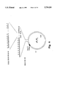
- a solvent composition step from 100% B to 0% B converts unfolded protein to dead-end aggregate (75% yield) and refolded protein (25% yield).
- Cycles 7 through 12 Dead-end aggregates will be converted to unfolded protein in each step whereas protein recoverable as refolded product will accumulate in the following amounts, cycle by cycle: 25%, 44%, 58%, 68%, 76% and 82%.
- the protein in solution could e.g. be held in an ultrafiltration device, held in a dialysis device or be confined to one of the phases of a suitable aqueous two-phase system, all of which might allow the concentration of low-molecular weight chemical solutes in the protein solution to be controlled by suitable devices.
- the protein could be adsorbed to a suitable surface in contact with a liquid phase, the chemical composition of which could be controlled as required.
- a suitable surface could e.g. be a filtration device, a hollow-fibre device or a beaded chromatographic medium. Adsorption of the protein to the surface could be mediated by non-specific interactions, e.g. as described in WO 86/05809 (Thomas Edwin Creighton), by folding-compatible covalent bonds between surface and protein or via specific designs of affinity handles in a recombinant derivative of the protein exhibiting a specific and denaturation-resistant affinity for a suitably derivatized surface.
- Recombinant proteins of this general design adsorbed on Nickel-chelating agarose beads could then be subjected to the present cyclic refolding process in a chromatographic column "refolding reactor" perfused with a mixture of suitable denaturing and non-denaturing buffers, delivered by an array of calibrated pumps, the flow rates of which was time-programmed through computer control.
- a general scheme of solid-state refolding entails cycling the immobilized protein as outlined above or by any other means and implementations between denaturing and non-denaturing conditions in a progressive manner, in which the concentration of the denaturing agent is gradually reduced from high starting values towards zero over a train of many renaturation-denaturation cycles.
- progressive denaturation-renaturation cycling may be enhanced by using equipment similar to advanced chromatography equipment with on-line facilities to monitor buffer compositions of folding reactor effluent.
- Information on effluent composition with regard to reductant and disulphide reshuffling reagent concentration profile would reveal productive cycling, and could therefore be used as input to an intelligent processor unit, in turn regulating the progression of denaturant concentration in a feed-back loop to ensure that most of the cycling effort is spent within the productive phase of the denaturation-renaturation cycle train.
- An intelligent monitoring and control system could furthermore use the available information to direct usable portions of reactor effluent to salvage/recycling subsystems thereby minimizing expenses for large scale operations.
- the final product may be eluted from the affinity matrix in a concentrated form, processed to liberate the mature authentic protein by cleavage at the designed protease cleavage site and then subjected to final work-up using standard protein purification and handling techniques, well-known within the field of protein chemistry.
- the present invention relates to a method for generating a processed ensemble of polypeptide molecules, in which processed ensemble the conformational states represented contain a substantial fraction of polypeptide molecules in one particular uniform conformation, from an initial ensemble of polypeptide molecules which have the same amino acid sequence as the processed ensemble of polypeptide molecules, comprising subjecting the initial ensemble of polypeptide molecules to a series of at least two successive cycles each of which comprises a sequence of
- the term "ensemble” is used in the meaning it has acquired in the art, that is, it designates a collection of molecules having essential common features. Initially (“an initial ensemble”), they have at least their amino acid sequence in common (and of course retain this common feature).
- an initial ensemble they have at least their amino acid sequence in common (and of course retain this common feature).
- a processed ensemble the conformational states represented in the ensemble will contain a substantial fraction of polypeptide molecules with one particular conformation.
- the substantial fraction of polypeptide molecules with one particular conformation in the processed ensemble may vary dependent on the parameters of the treatment by the method of the invention, the size of the protein in the particular conformation, the length and identity of the amino acid sequence of the molecules, etc.
- Denaturing step refers to exposure of an ensemble of polypeptide molecules during a time interval to physical and/or chemical circumstances which subject the ensemble of polypeptide molecules to conditions characterized by more severe denaturing power than those characterizing conditions immediately prior to the denaturing step.
- the term "renaturing step” refers to exposure of an ensemble of polypeptide molecules during a time interval to physical and/or chemical circumstances which subject the ensemble of polypeptide molecules to conditions characterized by less severe denaturing power than those characterizing conditions immediately prior to the denaturing step.
- the "substantial fraction” mentioned above will depend in magnitude on the ensemble of polypeptide molecules which are subjected to the method of the invention. If the processed ensemble of polypeptides consists of monomeric proteins of relatively short lengths and without intramolecular disulphide bridges the method will in general result in very high yields, whereas complicated molecules (such as polymeric proteins with a complicated disulphide bridging topology) may result in lower yields, even if the conditions of the method of the invention are fully optimized.
- An interesting aspect of the invention relates to a method described above wherein the processed ensemble comprises a substantial fraction of polypeptide molecules in one conformational state the substantial fraction constituting at least 1% (w/w) of the initial ensemble of polypeptide molecules.
- Higher yields are preferred, such as at least 5%, at least 10%, at least 20%, and at least 25% of the initial ensemble of polypeptide molecules.
- More preferred are yields of at least 30%, such as at least 40%, 50%, 60%, 70%, and at least 80%.
- Especially preferred are yields of at least 85%, such as 90%, 95%, 97%, and even at 99%. Sometimes yields close to 100% are observed.
- the processed ensemble will comprise a substantial fraction of polypeptide molecules in one particular uniform conformation which in addition have substantially identical disulphide bridging topology.
- polypeptide molecules subjected to the method of the invention will be molecules which have an amino acid sequence identical to that of an authentic polypeptide, or molecules which comprise an amino acid sequence corresponding to that of an authentic polypeptide joined to one or two additional polypeptide segments.
- authentic protein or polypeptide is meant a polypeptide with primary structure, including N- and C-terminal structures, identical to that of the corresponding natural protein.
- the term also denotes a polypeptide which has a known primary structure which is not necessarily identical to that of a natural protein, which polypeptide is the intentional end-product of a protein synthesis.
- natural protein is meant a protein as isolated in biologically active form from an organism, in which it is present not as a consequence of genetic manipulation.
- artificial protein or polypeptide as used in the present specification and claims is intended to relate to a protein/polypeptide which is not available from any natural sources, i.e. it cannot be isolated and purified from any natural source.
- An artificial protein/polypeptide is thus the result of human intervention, and may for instance be a product of recombinant DNA manipulation or a form of in vitro peptide synthesis. According to the above definitions such an artificial protein may be an authentic protein, but not a natural protein.
- the invention also relates to a method wherein natural proteins as well as artificial proteins are subjected to the refolding processes described herein.
- the authentic polypeptide is joined to polypeptide segments having auxiliary functions during the cycling and other previous or subsequent processing, e.g. as "handles" for binding the polypeptide to a carrier, as solubility modifiers, as expression boosters which have exerted their beneficial function during translation of messenger RNA, etc.
- auxiliary polypeptide segment will preferably be linked to the authentic polypeptide via a cleavable junction, and where two such auxiliary polypeptide segments are linked to the authentic polypeptide, this may be via similar cleavable junctions which will normally be cleaved simultaneously, or through dissimilar cleavable junctions which may be cleaved in any time sequence.
- the cycling (which, as explained above, comprises at least two successive cycles) will give rise to at least one event where a renaturing step is succeeded by a denaturing step where at least a substantial fraction of the refolded polypeptides will be denatured again.
- the processing will comprise at least 3 cycles, often at least 5 cycles and more often at least 8 cycles, such as at least 10 cycles and, in some cases at least 25 cycles.
- the series of cycles will normally not exceed 2000 cycles and will often comprise at most 1000 cycles and more often at most 500 cycles. The number of cycles used will depend partly on the possibilities made available by the equipment in which the cycling is performed.
- the rate with which the liquid phase in contact with the column can be exchanged will constitute one limit to what can realistically be achieved.
- high performance liquid chromatography (HPLC) equipment will permit very fast exchange of the liquid environment and thus make cycle numbers in the range of hundreds or thousands realistic.
- the cycling series will often comprise at most 200 cycles, more often at most 100 cycles and yet more often at most 50 cycles.
- each denaturing step may be a duration which, under the particular conditions in question, is at least one millisecond and at most one hour
- the duration of each renaturing step may be a duration which, under the particular conditions in question, is at least 1 second and at most 12 hours.
- the denaturing conditions of each individual denaturing step are kept substantially constant for a period of time, and the renaturing conditions of each individual renaturing step are kept substantially constant for a period of time, the periods of time during which conditions are kept substantially constant being separated by transition periods during which the conditions are changed.
- the transition period between steps for which conditions are kept substantially constant may have a duration varying over a broad range, such as between 0.1 second and 12 hours and will normally be closely adapted to the durations of the denaturing and renaturing steps proper.
- the period of time for which the denaturing conditions of a denaturing step are kept substantially constant may, e.g. have a duration of at least one millisecond and at most one hour, often at most 30 minutes, and the period of time for which the renaturing conditions of a renaturing step are kept substantially constant has a duration of at least 1 second and at most 12 hours, and often at most 2 hours.
- the period of time for which the denaturing conditions of a denaturing step are kept substantially constant will often have a duration of between 1 and 10 minutes, and the period of time for which the renaturing conditions of a renaturing step are kept substantially constant will often have a duration of between 1 and 45 minutes.
- the pressure may be very high (up to 5000 Bar) when using an HPLC system when performing the method of the invention, and under such circumstances very rapid steps may be accomplished and/or necessary.
- the temperature parameter is of importance, as some proteins only will refold properly at temperatures far from the physiological range. Both temperature and pressure will of course have an effect on the kinetics of the refolding procedure of the invention, and therefore the above-indicated time intervals of renaturing and denaturing steps are realistic boundaries for the many possible embodiments of the invention.
- the polypeptide molecules are normally in contact with a liquid phase during the denaturing and renaturing steps, the liquid phase normally being an aqueous phase.
- the liquid phase normally is an aqueous phase.
- any reagents or auxiliary substances used in the method will normally be dissolved in the liquid phase, normally in an aqueous phase.
- the liquid phase may also be constituted by one or more organic solvents.
- Chaperones are a group of recently described proteins that show a common feature in their capability of enhancing refolding of unfolded or partly unfolded proteins. Often, the chaperones are multimolecular complexes. Many of these chaperones are heat-shock proteins, which means that in vivo, they are serving as factors doing post traumatic "repair" on proteins that have been destabilized by the trauma. To be able to fulfil this function, chaperones tend to be more stable to traumatic events than many other proteins and protein complexes.
- the method of the invention does not depend on the use of a molecular chaperone or a molecular chaperone complex, it is, of course, possible to have a suitable molecular chaperone or molecular chaperone complex present during at least one renaturing step, and it may be preferred to have a molecular chaperone or a molecular chaperone complex present during substantially all cycles.
- the polypeptide molecules are preferably substantially confined to an environment which allows changing or exchanging the liquid phase substantially without entraining the polypeptide molecules.
- the polypeptide molecules may be contained in a dialysis device, or they may be confined to one of the phases of a suitable liquid two-phase system.
- a suitable aqueous two phase system may, e.g., contain a polymer selected from the group consisting of polyethylene oxide (polyethylene glycol), polyvinyl acetate, dextran and dextran sulphate.
- one phase contains polyethylene oxide (polyethylene glycol) and the other phase contains dextran, whereby the polypeptide molecules will be confined to the dextran-containing phase.
- the carrier may be a substance having molecules of such a size that the molecules with the polypeptide molecules bound thereto, when dissolved or dispersed in a liquid phase, can be retained by means of a filter, or the carrier may be a substance capable of forming micelles or participating in the formation of micelles allowing the liquid phase to be changed or exchanged substantially without entraining the micelles.
- the carrier may also be a water-soluble polymer having molecules of a size which will substantially not be able to pass through the pores of a filter or other means used in confining the system.
- the polypeptide molecules are suitably non-covalently adsorbed to the carrier through a moiety having affinity to a component of the carrier.
- a moiety may, e.g., be a biotin group or an analogue thereof bound to an amino acid moiety of the polypeptide, the carrier having avidin, streptavidin or analogues thereof attached thereto so as to establish a system with a strong affinity between the thus modified polypeptide molecules and the thus modified carrier.
- the affinity between the modified polypeptide and the modified carrier should be sufficiently stable so that the adsorption will be substantially unaffected by the denaturing conditions; the removal of the polypeptide molecules from the carrier after the cycling should be performed using specific cleaving, such as is explained in the following.
- An example of a suitable amino acid residue to which a biotinyl group may be bound is lysine.
- CPY carboxy peptidase Y
- the moiety having affinity to the carrier is the polypeptide segment SEQ ID NO: 47, in which case the carrier suitably comprises a Nitrilotriacetic Acid derivative (NTA) charged with Ni ++ ions, for instance an NTA-agarose matrix which has been bathed in a solution comprising Ni ++ .
- NTA Nitrilotriacetic Acid derivative
- An important aspect of the invention relates to the presence of suitable means in the polypeptide molecule preparing the molecule for later cleavage into two or more segments, wherein one segment is an authentic polypeptide as defined above.
- Such combined polypeptide molecules may for this purpose comprise a polypeptide segment which is capable of direction preferential cleavage by a cleaving agent at a specific peptide bond.
- the polypeptide segment in question may be one which directs the cleavage as a result of the conformation of the segment which serves as a recognition site for the cleaving agent.
- the cleavage directing polypeptide segment may for instance be capable of directing preferential cleavage at a specific peptide bond by a cleaving agent selected from the group consisting of cyanogen bromide, hydroxylamine, iodosobenzoic acid and N-bromosuccinimide.
- the cleavage-directing polypeptide segment may be one which is capable of directing preferential cleavage at a specific peptide bond by a cleaving agent which is an enzyme and one such possible enzyme is bovine enterokinase or an analogue and/or homologue thereof.
- the cleaving agent is the enzyme bovine coagulation factor X a or an analogue and/or homologue thereof (such analogues will be discussed in greater detail further below), and the polypeptide segment which directs preferential cleavage is a sequence which is substantially selectively recognized by the bovine coagulation factor X a or an analogue and/or homologue thereof.
- Important such segments are polypeptide segments that have a sequence selected from the group consisting of SEQ ID NO: 38, SEQ ID NO: 40, SEQ ID NO: 41 and SEQ ID NO: 42.
- An interesting feature of the invention is the possibility of masking and unmasking polypeptide segments with respect to their ability to direct cleavage at a specific peptide bond, whereby it is obtained that different segments of the polypeptide can be cleaved at different stages in the cycles.
- the polypeptide molecules comprise a polypeptide segment which is in vitro-convertible into a derivatized polypeptide segment capable of directing preferential cleavage by a cleaving agent at a specific peptide bond
- a masking/unmasking effect as mentioned becomes available.
- An especially interesting version of this strategy is where the in vitro-convertible polypeptide segment is convertible into a derivatized polypeptide segment which is substantially selectively recognized by the bovine coagulation factor X a of an analogue and/or homologue thereof.
- cysteine and methionine residues can be converted into modified residues, which modified residues make the segments having amino acid sequences selected from the group consisting of SEQ ID NO: 43, SEQ ID NO: 44, SEQ ID NO: 45 and SEQ ID NO: 46 in vitro convertible into segments recognized by bovine coagulation factor X a or an analogue and/or homologue thereof.
- one possible solution involving the cysteine residue is that a polypeptide segment with the amino acid sequence SEQ ID NO: 43 or SEQ ID NO: 44, is converted into a derivatized polypeptide which is substantially selectively recognized by bovine coagulation factor X a , by reacting the cysteine residue with N (2 mercaptoethyl)morpholyl-2-thiopyridyl disulphide or mercaptothioacetate-2-thiopyridyl disulphide.
- a possible strategy according to the invention involving methionine is that a polypeptide segment with the amino acid sequence SEQ ID NO: 45 or SEQ ID NO: 46, is converted into a derivatized polypeptide, which is substantially selectively recognized by bovine coagulation factor X a , by oxidation of the thioether moiety in the methionine side group to a sulphozide or sulphone derivative.
- Preferred embodiments of the method according to the invention are those wherein the cleavage-directing segments with the amino acid sequences SEQ ID NO: 38, SEQ ID NO: 40, SEQ ID NO: 41 or SEQ ID NO: 42, or the masked cleavage-directing segments with the amino acid sequences SEQ ID NO: 43, SEQ ID NO: 44, SEQ ID NO: 45 and SEQ ID NO: 46 are linked N-terminally to the authentic polypeptide, because then no further processing other than the selective cleaving is necessary in order to obtain the authentic polypeptide in solution.
- one possible reason for linking the cleavage directing sequences at the C-terminal and of the authentic polypeptide would be that the correct folding of the polypeptide molecules is dependent on a free N-terminal of the polypeptide molecules.
- the part of the cleaving directing sequence remaining after cleaving can be removed by suitable use of carboxypeptidases A and B.
- the change of conditions during the transition period between the steps may according to the invention be accomplished by changing the chemical composition of the liquid phase with which the polypeptide molecules are in contact.
- denaturing of the polypeptide molecules may be accomplished by contacting the polypeptide molecules with a liquid phase in which at least one denaturing compound is dissolved
- renaturing of the polypeptide molecules is accomplished by contacting the polypeptide molecules with a liquid phase which either contains at least one dissolved denaturing compound in such a concentration that the contact with the liquid phase will tend to renature rather than denature the ensemble of polypeptide molecules in their respective conformation states resulting from the preceding step, or contains substantially no denaturing compound.
- denaturing compound refers to a compound which when present as one of the solutes in a liquid phase comprising polypeptide molecules may destabilize folded states of the polypeptide molecules leading to partial or complete unfolding of the polypeptide chains.
- the denaturing effect exerted by a denaturing compound increases with increasing concentration of the denaturing compound in the solution, but may furthermore be enhanced or moderated due to the presence of other solutes in the solution, or by changes in physical parameters, e.g. temperature or pressure.
- urea examples of suitable denaturing compounds to be used in the method according to the invention may be mentioned urea, guanidine-HCl, di-C 1-6 alkylformamides such as dimethylformamide and di-C 1-6 -alkylsulphones.
- the liquid phase used in at least one of the denaturing steps and/or in at least one of the renaturing steps may according to the invention contain a least one disulphide-reshuffling system.
- Disulphide reshuffling systems are redox systems which contain mixtures of reducing and oxidating agents, the presence of which facilitate the breaking and making of disulphide bonds in a polypeptide or between polypeptides. Accordingly, “disulphide reshuffling agents” or “disulphide reshuffling compounds” are such reducing and oxidating agents which facilitate the breaking and making of disulphide bonds in a polypeptide or between polypeptides.
- the disulphide-reshuffling system contained in the aqueous phase which is in contact with the proteins comprises as a disulphide reshuffling system a mixture of a mercaptan and its corresponding disulphide compound.
- cysteine residues in the polypeptide molecules may have been converted to mixed disulphide products of either glutathione, thiocholine, mercaptoethanol or mercaptoacetic acid, during at least one of the denaturing/renaturing cycles.
- a converted polypeptide is termed a "fully disulphide-blocked polypeptide or protein" and this term thus refers to a polypeptide or a protein in which cysteine residues have been converted to a mixed-disulphide in which each cysteine residue is disulphide-linked to a mercaptan, e.g. glutathione.
- the conversion of the cysteine residues to mixed disulphide products may be accomplished by reacting a fully denatured and fully reduced ensemble of polypeptide molecules with an excess of a reagent which is a high-energy mixed disulphide compound, such as aliphatic-aromatic disulphide compound, e.g. 2 thiopyridyl glutathionyl disulphide, or by any other suitable method.
- a reagent which is a high-energy mixed disulphide compound, such as aliphatic-aromatic disulphide compound, e.g. 2 thiopyridyl glutathionyl disulphide, or by any other suitable method.
- mixed disulphides having a relatively unstable S--S bond
- Examples of such mixed disulphides are glutathionyl-2-thiopyridyl disulphide, 2 thiocholyl 2-thiopyridyl disulphide, 2-mercaptoethanol-2-thiopyridyl disulphide and mercaptoacetate-2-thiopyridyl disulphide.
- the disulphide-reshuffling system contains glutathione, 2-mercaptoethanol or thiocholine, each of which is in admixture with its corresponding symmetrical disulphide.
- the suitability of a given mixture of thiols for use as selective reducing and/or disulphide-reshuffling system in a cyclic refolding/reoxidation procedure for a specific protein product can be directly assayed by incubating ensembles of samples of a mixture of folded and misfolded protein with an array of thiol mixtures at several different concentrations of denaturant exerting weakly, intermediate or strongly denaturing effects on the protein. Following incubation, the disulphide topology in each sample is then locked by reaction with an excess of thiol-blocking reagent (e.g. Iodoacetamide) before subjecting each set of samples to SDS-PAGE under non-reducing conditions.
- thiol-blocking reagent e.g. Iodoacetamide
- This reagent testing procedure may be used as a general procedure for selecting advantageous reducing and/or thiol/disulphide reshuffling reagents.
- Example 12 demonstrates application of this analytical procedure to assess the suitability for selective reduction of misfolded forms of a model protein for 5 thiol reagents and thereby demonstrates the operability of the above procedure.
- a very important aspect of the invention is a method for protein refolding as described herein, wherein at least one disulphide-reshuffling system contained in liquid phase in at least one renaturing and/or denaturing step is one which is capable of reducing and/or reshuffling incorrectly formed disulphide bridges under conditions with respect to concentration of the denaturing agent at which unfolded and/or misfolded proteins are denatured and at which there is substantially no reduction and/or reshuffling of correctly formed disulphide bridges.
- An interesting embodiment of the invention is a method as described above, wherein a disulphide reshuffling system is used in at least one denaturing/renaturing step and resulting in a ratio between the relative amount of reduced/reshuffled initially incorrectly formed disulphide bridges and the relative amount of reduced/reshuffled initially correctly formed disulphide bridges of at least 1.05.
- the ratio will preferably be higher, such as 1.1, 1.5, 2.0, 3.0, 5.0, 10, 100, 1000, but even higher ratios are realistic and are thus especially preferred according to the invention.
- the ratio has to be greater than 1 in order to allow the net formation of correctly formed disulphide bridges in a protein sample. Normally the ratio should be as high as possible, but even ratios which are marginally above 1 will allow the net formation of correctly formed disulphide bridges in the method of the invention, the important parameter in ensuring a high yield being the number of denaturing/renaturing cycles. Ratios just above one require that many cycles are completed before a substantive yield of correctly formed disulphide bridges is achieved, whereas high ratios only require a limited number of cycles.
- the disulphide-reshuffling system X selecting as the disulphide reshuffling system X, the disulphide-reshuffling system which exhibit the capability of reducing initially incorrectly formed disulphide bridges without substantially reducing and/or reshuffling initially correctly formed disulphide bridges in the widest range of concentrations of the chosen denaturing agent.
- disulphide-reshuffling system may be employed, for instance in different cycles in the cyclic refolding method of the invention, but also simultaneously in the same cycles. This will e.g. be the case when it is likely or has been established by e.g. the method outlined above that the overall yield of correctly folded protein with correct disulphide bridging topology will be higher if using different disulphide-reshuffling systems in the method of the invention.
- step 1) to the initial mixture of reactants in step 1) is added a known amount of radioactively-labelled correctly folded protein.
- step 2) for instance by non-reducing SDS-PAGE
- step 2) the content of radioactivity in the correctly folded protein fraction is determined as well.
- denaturing may also be achieved or enhanced by decreasing pH of the liquid phase, or by increasing pH of the liquid phase.
- the polarity of the liquid phase used in the renaturing may according to the invention have been modified by the addition of a salt, a polymer and/or a hydrofluoro compound such as trifluoroethanol.
- the denaturing and renaturing of the polypeptide molecules may also be accomplished by direct changes in physical parameters to which the polypeptide molecules are exposed, such as temperature or pressure, or these measures may be utilized to enhance or moderate the denaturing or renaturing resulting from the other measures mentioned above.
- a most important practical embodiment of the method is performed by accomplishing chemical changes in the liquid phase by changing between a denaturing solution B and a renaturing solution A.
- concentration of one or more denaturing compounds in B will often be adjusted after each cycle, and as one important example, the concentration of one or more denaturing compounds in B will be decremented after each cycle, but in another important embodiment, the concentration of one or more denaturing compounds in medium B is kept constant in each cycle.
- concentration of denaturing compound(s) medium B is kept constant, is especially interesting when the most productive phase of the cycling process (with respect to correctly folded protein) has been identified, and large scale production of correctly folded protein is desired.
- concentration(s) of denaturing compound(s) of medium B in this embodiment is the concentration(s) which has been established to ensure maximum productivity in the cyclic process according to the invention.
- polypeptide molecules of the ensemble which is subjected to the method of the invention normally have a length of at least 25 amino acid residues, such as at least 30 amino acid residues or at least 50 amino acid residues.
- polypeptide molecules of the ensemble normally have a length of at most 5000 amino acid residues, such as at most 2000 amino acid residues or at most 1000 or 800 amino acid residues.
- the method of the invention has made possible the production of correctly folded diabody molecules (diabodies are described in Holliger et al., 1993).
- An important aspect of the invention therefore relates to a method for producing correctly folded diabody molecules, wherein an initial ensemble of polypeptide molecules comprising unfolded and/or misfolded polypeptides having amino acid sequences identical to the amino acid sequences of monomer fragments of diabody molecules is subjected to a series of at least two successive cycles, each of which comprises a sequence of
- Such a method for the correct folding of diabodies can be envisaged in any of the above-mentioned scenarios and aspects of the refolding method of the invention, that is, with respect to the choice of physical/chemical conditions as well as cycling schedules.
- an important aspect of the method for correct folding of diabodies is a method as the above-identified, wherein the polypeptide molecules are in contact with a liquid phase containing at least one disulphide reshuffling system in at least one denaturing or renaturing step.
- the preferred denaturing agent to be used in such a liquid phase is urea
- the preferred disulphide reshuffling system comprises glutathione as the main reducing agent.
- a particular aspect of the invention relates to a polypeptide which is a proenzyme of a serine protease, but is different from any naturally occurring serine protease and, in particular, has an amino acid sequence different from that of bovine coagulation factor X (Protein Identification Resource (PIR), National Biomedical Research Foundation, Georgetown University, Medical Center, U.S.A., entry: P1;EXBO) and which can be proteolytically activated to generate the active serine protease by incubation of a solution of the polypeptide in a non-denaturing buffer with a substance that cleaves the polypeptide to liberate a new N-terminal residue,
- PIR Protein Identification Resource
- the substrate specificity of the serine protease being identical to or better than that of bovine blood coagulation factor X a , as assessed by each of the ratios (k(I)/k(V) and k(III)/k(V) between cleavage rate against each of the substrates I and III:
- serine proteases are enzymes which are believed to have a catalytic system consisting of an active site serine which is aligned with a histidine residue, and it is believed that the activation of the enzymes from the corresponding proenzymes is based on the liberation of a new N-terminal residue, the ⁇ -amino group of which is capable of repositioning within the polypeptide structure to form a salt bridge to an aspartic acid residue preceding an active-site serine residue, thereby forming the catalytic site characteristic of serine proteases.
- the "artificial" serine proteases defined above are extremely valuable polypeptide cleaving tools for use in the method of the invention and in other methods where it is decisive to have a cleaving tool which will selectively cleave proteins, even large folded proteins.
- the above-defined artificial serine proteases in activated form are capable of selectively recognizing the cleaving-directing polypeptide segment SEQ ID NO: 38, but in contrast to bovine coagulation factor X a , they can be established with such amino acid sequences that they can be readily produced using recombinant DNA techniques.
- the preferred artificial serine proteases of the invention are ones which have amino acid sequences allowing their synthesis by recombinant DNA techniques, in particular in prokaryote cells such as E. coli.
- the artificial serine proteases of the invention when produced in a prokaryote, may be given an enzymatically active conformation, in which the catalytically active domains are suitably exposed, by cycling according to the method of the present invention.
- the quantitative test for selectivity of the artificial serine proteases involves determination of the cleavage rate, k, determined as the initial slope of a curve of absorption of light at 405 nm (absorption maximum of free paranitroaniline) versus time at 20° C.
- the selectivity of the artificial serine proteases should be characterized by the value of (k(I)/k(V) being at most 0.06, and the value k(III)/k(V) being at most 0.5. It is preferred that (k(I)/k(V) is at most 0.05 and k(III)/k(V) is at most 0.4, and more preferred that (k(I)/k(V) is at most 0.04 and k(III)/k(V) is at most 0.15.
- a more comprehensive specificity characterization involves further model substrates: thus, the substrate specificity could be assessed to be identical to or better than that of bovine blood coagulation factor X a by each of the ratios (k(I)/k(V), k(II)/k(V), k(III)/k(V) and k(IV)/k(V)) between cleavage rate against each of the substrates I-IV:
- (k(I)/k(V) should be at most 0.06, k(II)/k(V) should be at most 0.03, k(III)/k(V) should be at most 0.5, and k(IV)/k(V)) should be at most 0.01, and it is preferred that (k(I)/k(V) is at most 0.05, k(II)/k(V) is at most 0.025, k(III)/k(V) is at most 0.4, and k(IV)/k(V) is at most 0.008, and more preferred that (k(I)/k(V) is at most 0.04, k(II)/k(V) is at most 0.015, k(III)/k(V) is at most 0.15, and k(IV)/k(V)) is at most 0.005.
- the serine protease type polypeptide is defined above will normally have a molecular weight, M r , of at most 70,000 and at least 15,000.
- One such novel polypeptide according to the invention has the amino acid sequence SEQ ID NO: 2 or is an analogue and/or homologue thereof.
- Other important embodiments of the polypeptide of the invention have an amino acid sequence which is a subsequence of SEQ ID NO: 2 or an analogue and/or homologue of such a subsequence.
- an analogue of a polypeptide encoded by the DNA sequence or "an analogue of a polypeptide having the amino acid sequence” is meant any polypeptide which is capable of performing as bovine coagulation factor X a in the tests mentioned above.
- polypeptides from different sources such as different mammals or vertebrates, which vary e.g. to a certain extent in the amino acid composition, or the post-translational modifications e.g. glycosylation or phosphorylation, as compared to the artificial serine protease described in the examples.
- analogue is thus used in the present context to indicate a protein or polypeptide of a similar amino acid composition or sequence as the characteristic amino acid sequence SEQ ID NO: 2 derived from an artificial serine protease as described in Example 5, allowing for minor variations that alter the amino acid sequence e.g. deletions, site directed mutations, insertions of extra amino acids, or combinations thereof, to generate artificial serine protease analogues.
- an analogue designates a variation of the polypeptide in which one or several amino acids may have been deleted or exchanged, and/or amino acids may have been introduced, provided the enzymatic activity with the above-defined specificity is retained, as can be assessed as described above.
- an analogue of a polypeptide according to the invention may have a sequence homology at the polypeptide level of at least 60% identity compared to the sequence of a fragment of SEQ ID NO: 2, allowing for deletions and/or insertions of at most 50 amino acid residues.
- polypeptide sequences or analogues thereof which has a homology of at least 60% with the polypeptide shown in SEQ ID NO: 2 encoded for by the DNA sequence of the invention SEQ ID NO: 1 or analogues and/or homologues thereof, constitute an important embodiment of this invention.
- sequence homology is meant the identity in sequence of either the amino acids in segments of two or more amino acids in a amino acid sequence, or the nucleotides in segments of two or more nucleotides in a nucleotide sequence.
- sequence homology is meant the identity in sequence of either the amino acids in segments of two or more amino acids in a amino acid sequence, or the nucleotides in segments of two or more nucleotides in a nucleotide sequence.
- the term "homologous” is thus used here to illustrate the degree of identity between the amino acid sequence of a given polypeptide and the amino sequence shown in SEQ ID NO: 2.
- the amino acid sequence to be compared with the amino acid sequence shown in SEQ ID NO: 2 may be deduced from a nucleotide sequence such as a DNA or RNA sequence, e.g. obtained by hybridization as defined in the following, or may be obtained by conventional amino acid sequencing methods.
- Another embodiment relates to a polypeptide having an amino acid sequence from which a consecutive string of 20 amino acids is homologous to a degree of at least 40% with a string of amino acids of the same length selected from the amino acid sequence shown in SEQ ID NO: 2.
- One serine protease polypeptide according to the invention has the amino acid sequence of SEQ ID NO: 2, residues 166-484, or is an analogue and/or homologue thereof.
- any of the artificial serine proteases defined above replacement of the cleaving sequence corresponding to residues 230-233 in SEQ ID NO: 2 with one of the cleavage-directing sequences defined above will give rise to extremely useful cleaving enzymes for use in the method according to the invention, in that these can be selectively and very efficiently cleaved by enzymes having the specific enzymatic activity of bovine coagulation factor X a , and thus by artificial serine proteases as defined above, including by molecules identical to themselves.
- the artificial serine proteases can be produced by recombinant DNA techniques, and hence, another important embodiment of the invention relates to a nucleic acid fragment capable of encoding an polypeptide according as defined above, in particular a DNA fragment which is capable of encoding an artificial serine protease polypeptide as defined above.
- the invention relates to a nucleotide sequence encoding a polypeptide of the invention as defined above.
- the invention relates to a nucleotide sequence having the nucleotide sequence shown in the DNA sequence SEQ ID NO: 1 or an analogue thereof which has a homology with any of the DNA sequences shown in SEQ ID NO: 1 of at least 60%, and/or encodes a polypeptide, the amino acid sequence of which is at least 60% homologous with the amino acid sequences shown in SEQ ID NO: 2.
- analogue with regard to the DNA fragments of the invention is intended to indicate a nucleotide sequence which encodes a polypeptide identical or substantially identical to the polypeptide encoded by a DNA fragment of the invention. It is well known that the same amino acid may be encoded by various codons, the codon usage being related, inter alia, to the preference of the organisms in question expressing the nucleotide sequence. Thus, one of more nucleotides or codons of the DNA fragment of the invention may be exchanged by others which, when expressed, result in a polypeptide identical or substantially identical to the polypeptide encoded by the DNA fragment in question.
- analogue is intended to allow for variations in the sequence such as substitution, insertion (including introns), addition and rearrangaement of one or more nucleotides, which variations do not have any substantial effect on the polypeptide encoded by the DNA fragment.
- nucleotide sequence which differs from the DNA sequence shown in SEQ ID NO: 1 in that at least one nucleotide has been substituted, added, inserted, deleted and/or rearranged.
- substitution is intended to mean the replacement of one or more nucleotides in the full nucleotide sequence with one or more different nucleotides
- addition is understood to mean the addition of one or more nucleotides at either end of the full nucleotide sequence
- insertion is intended to mean the introduction of one or more nucleotides within the full nucleotide sequence
- deletion is intended to indicate that one or more nucleotides have been deleted from the full nucleotide sequence whether at either end of the sequence or at any suitable point within it
- “rearrangement” is intended to mean that two or more nucleotide residues have been exchanged within the DNA or polypeptide sequence, respectively.
- the DNA fragment may, however, also be modified by mutagenesis either before or after inserting it in the organism.
- the DNA or protein sequence of the invention may be modified in such a way that it does not lose any of its biophysical, biochemical or biological properties, or part of such properties (one and/or all) or all of such properties (one and/or all).
- An example of a specific analogue of the DNA sequence of the invention is a DNA sequence which comprises the DNA sequence shown in SEQ ID NO: 1 and particularly adapted for expression in E. coli.
- This DNA sequence is one which, when inserted in E. coli together with suitable regulatory sequences, results in the expression of a polypeptide having substantially the amino acid sequence shown in SEQ ID NO: 2.
- this DNA sequence comprises specific codons recognized by E. coli.
- fragment As used in the present specification and claims with respect to fragments, sequences, homologues and analogues according to the invention should of course be understood as not comprising these phenomena in their natural environment, but rather, e.g., in isolated, purified, in vitro or recombinant form.
- nucleic acid fragment according to the invention is a nucleic acid fragment as define above in which at least 60% of the coding triplets encode the same amino acids as a nucleic acid fragment of the nucleic acid which encodes bovine coagulation factor x, allowing for insertions and/or deletions of a most 150 nucleotides.
- An example of such a nucleic acid fragment is SEQ ID NO: 1, nucleotides 76-1527, and analogues and/or homologues there of.
- SEQ ID NO: 1 nucleotides 319-1527, and analogues and/or homologues thereof.
- Still another example is SEQ ID NO: 1, nucleotides 571-1527, and analogues and/or homologues thereof.
- the DNA fragment described above and constituting and important aspect of the invention may be obtained directly from the genomic DNA or by isolating mRNA and converting it into the corresponding DNA sequence by using reverse transcriptase, thereby producing a cDNA.
- obtaining the DNA fragment from genomic DNA it is derived directly by screening for genomic sequences as is well known for the person skilled in the art. It can be accomplished by hybridization to a DNA probe designed on the basis of knowledge of the sequences of the invention, or the sequence information obtained by amino acid sequencing of a purified serine protease.
- the DNA is of complementary DNA (cDNA) origin, it may be obtained by preparing a cDNA library with mRNA from cells containing an artificial serine protease. Hybridization can be accomplished by a DNA probe designed on the basis of knowledge of the cDNA sequence, or the sequence information obtained by amino acid sequencing of a purified artificial serine protease.
- the DNA fragment of the invention or an analogue an/or homologue thereof of the invention can be replicated by fusing it with a vector and inserting the complex into a suitable microorganism or a mammalian cell line.
- the DNA fragment can be manufactured using chemical synthesis.
- polymerase chain reaction (PCR) primers can be synthesized based on the nucleotide sequence shown in SEQ ID NO: 1. These primers can then be used to amplify the whole or a part of a sequence encoding an artificial serine protease polypeptide.
- Suitable polypeptides of the invention can be produced using recombinant DNA technology. More specifically, the polypeptides may by produced by a method which comprises culturing or breeding an organism carrying the DNA sequence shown in SEQ ID NO: 1 or an analogue and/or homologue thereof of the invention under conditions leading to expression of said DNA fragment, and subsequently recovering the expressed polypeptide from the said organism.
- the organism which is used for the production of the polypeptide may be a higher organism, e.g. and animal, or a lower organism, e.g. a microorganism.
- the DNA fragment of the invention (described above) should be introduced in the organism either directly or with the help of a suitable vector.
- the polypeptides may be produced in the mammalian call lines by introducing the DNA fragment or an analogue and/or homologue thereof of the invention either directly or with the help of an expression vector.
- the DNA fragment of the invention can also be cloned in a suitable stable expression vector and then put into a suitable cell line.
- the cells expressing the desired polypeptides are then selected using the conditions suitable for the vector and the cell line used.
- the selected cells are then grown further and form a very important and continuous source of the desired polypeptides.
- an expression system comprising a nucleic acid fragment as defined above and encoding an artificial serine protease polypeptide as defined above, the system comprising a 5' flanking sequence capable of mediating expression of said nucleic acid fragment.
- the expression system may be a replicable expression vector carrying the nucleic acid fragment, which vector is capable of replicating in a host organism or a cell line; the vector may, e.g., be a plasmid, phage, cosmid, mini-chromosome or virus; the vector may be one which, when introduced in a host cell, is integrated in the host cell genome.
- the organism may be a microorganism such as a bacterium, a yeast, a protozoan, or a cell derived from a multicellular organism such as a fungus, an insect cell, a plant cell, a mammalian cell or a cell line.
- a microorganism such as a bacterium of the genus Escherichia, Bacillus or Salmonella.
- a further aspect of the invention relates to a method of producing an artificial serine protease polypeptide as defined above, comprising the following steps of:
- step b 3. culturing the host organism produced in step b to express the polypeptide
- polypeptides may for instance by accomplished by subjecting the polypeptide molecules to carboxypeptidase A or B, whereby selected amino acid residues may be removed from the C-terminus of the polypeptide molecules.
- carboxypeptidase A or B whereby selected amino acid residues may be removed from the C-terminus of the polypeptide molecules.
- the polypeptide produced may be isolated by a method comprising one of more steps life affinity chromatography using immobilized polypeptide or antibodies reactive with said polypeptide an/or other chromatographic and electrophoretic procedures.
- a polypeptide of the invention may be prepared by the well known methods of liquid or solid phase peptide synthesis utilizing the successive coupling of the individual amino acids of the polypeptide sequence.
- the polypeptide can be synthesized by the coupling of individual amino acids forming fragments of the polypeptide sequence which are later coupled so as to result in the desired polypeptide.
- the invention also relates to the use of an artificial serine protease polypeptide as defined above for cleaving polypeptides at the cleavage site for bovine coagulation factor X a , the cleavage site having the amino acid sequence selected from the group consisting of SEQ ID NO: 38, SEQ ID NO: 40, SEQ ID NO: 41 and SEQ ID NO: 42, and to the use of a an artificial serine protease polypeptide as defined above for cleaving polypeptides at the cleavage site for bovine coagulation factor X a , the cleavage site having a modified version of the amino acid sequence selected from the group of SEQ ID NO: 43, SEQ ID NO: 44, SEQ ID NO: 45 and SEQ ID NO: 46, which has been converted to a cleavable form as described further above.
- FIG. 1 Schematic representation of segment of a cyclic denaturation/renaturation time programme.
- Solvent composition is expressed in terms of a binary mixture of a non-denaturing ⁇ buffer A ⁇ and a denaturing ⁇ buffer B ⁇ in terms of relative content of buffer B.
- Three consecutive cycles are represented, each consisting of a renaturation phase ⁇ F ⁇ and a denaturation phase ⁇ D ⁇ . Changes in level of denaturing power of the solvent mixture during denaturation phases in consecutive cycles are denoted ⁇ k ⁇ .
- FIG. 2 Construction of the expression plasmids pT 7 H 6 FX-h ⁇ 2m and pT 7 H 6 FX-m ⁇ 2m.
- Bam HI and Hind III purchased from Boehringer, Germany
- T 4 DNA ligase purchased from Boehringer, Germany
- FIG. 3 Amino acid sequences of human- and murine ⁇ 2 -microglobulin.
- A Predicted amino acid sequence of the full length reading frame encoding human ⁇ 2 -microglobulin (SEQ ID NO: 49). Amino acid residue one (Ile) in the processed mature protein is indicated.
- B Predicted amino acid sequence of the full length reading frame encoding murine ⁇ 2 -microglobulin (SEQ ID NO: 50). Amino acid residue one (Ile) in the processed mature protein is indicated.
- FIG. 4 Construction of the expression plasmid pT 7 H 6 FX-hGH.
- FIG. 5 Amino acid sequence of human Growth Hormone (Somatotropin).
- the predicted amino acid sequence of the full length reading frame encoding human Growth Hormone (SEQ ID NO: 51).
- the first Amino acid residue in the processed mature protein (Phe 1 ) is indicated.
- FIG. 6 Construction of the plasmids pT 7 H 6 FX-#1, #2, and #3 expressing amino acid residue no. 20 (Ala) to 109 (Arg), amino acid residue no 20 (Ala) to 190 (Ala), and amino acid residue no. 20 (Ala) to 521 (Lys) of the human ⁇ 2 -Macroglobulin Receptor Protein ( ⁇ 2 MR) (SEQ ID NO: 52).
- Bam HI and Hind III purchased from Boehringer, Germany
- T 4 DNA ligase purchased from Boehringer, Germany
- FIG. 7 Construction of the plasmids pLcIIMLCH 6 FX-#4, #5, and #6 expressing amino acid residue no. 803 (Gly) to 1265 (Asp), amino acid residue no. 849 (Val) to 1184 (Gln), and amino acid residue no. 1184 (Gln) to 1582 (Lys) of the human ⁇ 2 -Macroglobulin Receptor Protein ( ⁇ 2 MR) (SEQ ID NO: 52).
- FIG. 8 Construction of the plasmids pLcIIMLCH 6 FX-#7, #8, and #9 expressing amino acid residue no. 803 (Gly) to 1582 (Lys), amino acid residue no. 2519 (Ala) to 2941 (Ile), and amino acid residue no. 3331 (Val) to 3778 (Ile) of the human ⁇ 2 -Macroglobulin Receptor Protein ( ⁇ 2 MR) (SEQ ID NO: 52).
- FIGS. 9a and 9b Amino acid sequence of human ⁇ 2 -Macroglobulin Receptor Protein ( ⁇ 2 MR) (SEQ ID NO: 52).
- FIG. 10 Construction of the expression plasmid pLcIIMLCH 6 FX-FX.increment. ⁇ .
- FIG. 11 Amino acid sequence of bovine blood coagulation Factor X (FX).
- the predicted amino acid sequence of the full length reading frame encoding bovine FX (SEQ ID NO: 53).
- the N-terminal amino acid residue Ser 82 and the C terminal Trp 484 residue in the FX.increment. ⁇ construct are identified.
- FIG. 12 Construction of the expression plasmid pLcIIMLCH 6 FX-K1.
- FIG. 13 Construction of the expression plasmid pLcIIH 6 FX-K4.
- FIG. 14 Amino acid sequence of human "Glu"-Plasminogen (SEQ ID NO: 54). The N- and C-terminal amino acid residues in the K1 and K4 constructs are identified by their numbers in the sequence.
- FIG. 15 SDS-PAGE analysis of production and in vitro folding of recombinant human ⁇ 2 -microglobulin.
- Lane 1 Crude protein extract before application to the Ni 2+ NTA-agarose column (reduced sample).
- Lane 2 Column flow-through during application of the crude protein extract onto the Ni 2+ NTA-agarose column (reduced sample)
- Lane 3 Human ⁇ 2 -microglobulin eluted from the Ni 2+ NTA-agarose column after the cyclic folding procedure by the non-denaturing elution buffer (reduced sample).
- Lane 4 Protein markers (Pharmacia, Sweden): From top of gel; 94 kDa, 67 kDa, 43 kDa, 30 kDa, 20.1 kDa, and 14.4 kDa (reduced sample)
- Lane 6 Recombinant human ⁇ 2 -microglobulin after FX a cleavage and final purification (non-reduced sample).
- FIG. 16 SDS-PAGE analysis of in vitro folding of recombinant human Growth Hormone; hGH (Somatotropin).
- Lane 1 Protein markers (Pharmacia, Sweden): From top of gel; 94 kDa, 67 kDa, 43 kDa, 30 kDa, 20.1 kDa, and 14.4 kDa (reduced sample)
- Lane 2 Human hGH eluted from the Ni 2+ NTA-agarose column after the cyclic folding procedure by the non-denaturing elution buffer (non-reduced sample).
- Lane 3 Human hGH eluted from the Ni 2+ NTA-agarose column after the cyclic folding procedure by the denaturing elution buffer B from the folding procedure (non-reduced sample).
- Lane 4-18 Fractions collected during the separation of monomeric hGH-fusion protein from dimer and multimer fusion proteins after the cyclic folding procedure by ion exchange chromatography on Q-Sepharose (Pharmacia, Sweden). The monomeric protein was eluted in a peak well separated from the peak containing the dimer and multimer proteins (non-reduced samples).
- FIG. 17 SDS-PAGE analysis of in vitro folding of recombinant kringle 1 and 4 from human plasminogen and recombinant fusion protein #4 derived from human ⁇ 2 -Macroglobulin Receptor Protein ( ⁇ 2 MR).
- Lane 1 Protein markers (Pharmacia, Sweden): From top of gel; 94 kDa, 67 kDa, 43 kDa, 30 kDa, 20.1 kDa, and 14.4 kDa (reduced sample).
- Lane 2 Crude K1-fusion protein extract before application to the Ni 2+ NTA-agarose column (reduced sample).
- Lane 3 K1-fusion protein eluted from the Ni 2+ NTA-agarose column after the cyclic folding procedure by the non-denaturing elution buffer (reduced sample).
- Lane 4 Same as lane 3 (non-reduced sample).
- Lane 5 Flow-through from the lysine-agarose column during application of the K1-fusion protein (non-reduced sample).
- Lane 6 K1-fusion protein eluted from the lysine-agarose column (non-reduced sample).
- Lane 7 K4-fusion protein eluted from the Ni 2+ NTA-agarose column after the cyclic folding procedure by the non-denaturing elution buffer (reduced sample).
- Lane 8 Same as lane 7 (non-reduced sample).
- Lane 9 ⁇ 2 MR#4 fusion protein eluted from the Ni 2+ NTA-agarose column after the cyclic folding procedure by the non-denaturing elution buffer (reduced sample).
- Lane 10 Same as lane 9 (non-reduced sample).
- FIG. 18 Construction of the expression plasmid pT 7 H 6 FX ⁇ 2 MRBDv.
- FIG. 19 Amino acid sequence of the receptor-binding domain of human ⁇ 2 -Macroglobulin (from residue Val 1299 to Ala 1451 ) (SEQ ID NO: 55).
- FIG. 20 Construction of the expression plasmid pT 7 H 6 FX-TETN.
- FIG. 21 Amino acid sequence of human monomeric Tetranectin.
- the predicted amino acid sequence of the full length reading frame encoding human Tetranectin (SEQ ID NO: 56).
- the first Amino acid residue in the processed mature protein (Glu 1 ) is indicated.
- FIG. 22 Construction of the expression plasmid pT 7 H 6 FX-DB32.
- Bam HI and Hind III purchased from Boehringer, Germany
- T 4 DNA ligase purchased from Boehringer, Germany
- FIG. 23 Amino acid sequence of the artificial diabody DB32 (SEQ ID NO: 57).
- FIG. 24 The expression plasmid pT 7 H 6 FX-PS.4.
- FIG. 25 Amino acid sequence of human psoriasin.
- FIGS. 26a and 26b SDS-PAGE analysis of purification and FX a cleavage of recombinant Mab 32 diabody.
- FIG. 26a Different stages of the purification
- Lanes 1 and 2 Crude product from folding.
- Lanes 3 Final purified Mab 32 diabody fusion protein product
- Lane 4 Supernatant of crude folding product after 50-fold concentration and centrifugation.
- Lane 5 Pellet from crude folding product after 50-fold concentration and centrifugation.
- FIG. 26b FX a cleavage of Mab 32 diabody fusion protein.
- Lanes 1 and 5 purified Mab 32 diabody fusion protein
- Lane 2 Molar ration 1:5 Fx a :Mab 32 diabody fusion protein at 37° C. for 20 hours.
- Lane 3 Molar ratio 1:2 FX a : Mab 32 diabody fusion protein at 37° C. for 20 hours p1
- Lane 4 Molar ratio 1:1 FX a :Mab 32 diabody fusion protein at 37° C. for 20 hours.
- FIG. 27 Suitability of glutathione as reducing agent in cyclic refolding of human ⁇ 2 -microglobulin fusion protein.
- Lane 1 Reduced sample of test no. 1.
- Lane 2 Non-reduced sample of test no.1.
- Lane 3 Non-reduced sample of test no.2.
- Lane 5 Non-reduced sample of test no.4.
- Lane 7 Non-reduced sample of test no.6.
- Lane 8 Non-reduced sample of test no.7.
- Lane 9 Non-reduced sample of test no.8.
- Lane 12 Non-reduced sample of test no.11.
- FIG. 28 Suitability of L-cystein ethyl ester as reducing agent in cyclic refolding of human ⁇ 2 -microglobulin fusion protein.
- Lane 1 Reduced sample of test no. 1.
- Lane 2 Non-reduced sample of test no.1.
- Lane 3 Non-reduced sample of test no.2.
- Lane 5 Non-reduced sample of test no.4.
- Lane 7 Non-reduced sample of test no.6.
- Lane 8 Non-reduced sample of test no.7.
- Lane 9 Non-reduced sample of test no.8.
- FIG. 29 Suitability of 2-Mercaptoethanol as reducing agent in cyclic refolding of human ⁇ 2 -microglobulin fusion protein.
- Lane 1 Reduced sample of test no. 1.
- Lane 2 Non-reduced sample of test no.1.
- Lane 3 Non-reduced sample of test no.2.
- Lane 5 Non-reduced sample of test no.4.
- Lane 7 Non-reduced sample of test no.6.
- Lane 8 Non-reduced sample of test no.7.
- Lane 9 Non-reduced sample of test no.8.
- FIG. 30 Suitability of Mercaptosuccinic acid as reducing agent in cyclic refolding of human ⁇ a microglobulin fusion protein.
- Lane 1 Non-reduced sample of test no.1.
- Lane 2 Non-reduced sample of test no.2.
- Lane 3 Non-reduced sample of test no.3.
- Lane 5 Non-reduced sample of test no.5.
- Lane 6 Non-reduced sample of test no.6.
- Lane 7 Non-reduced sample of test no.7.
- Lane 8 Non-reduced sample of test no.8.
- FIG. 31 Suitability of N-Acetyl-L-cysteine as reducing agent in cyclic refolding of human ⁇ 2 -microglobulin fusion protein.
- Lane 1 Reduced sample of test no.1.
- Lane 2 Non-reduced sample of test no.1.
- Lane 3 Non-reduced sample of test no.2.
- Lane 5 Non-reduced sample of test no.4.
- Lane 7 Non-reduced sample of test no.6.
- Lane 8 Non-reduced sample of test no.7.
- Lane 9 Non-reduced sample of test no.8.
- FIG. 32 SDS-PAGE analysis of cyclic refolding of human ⁇ 2 -microglobulin fusion protein.
- Lane 1 Crude protein extract before application to the Ni 2+ NTA-agarose column (reduced sample).
- Lane 2 8 ⁇ l sample of soluble fraction of refolded h ⁇ 2 m as described in EXAMPLE 1.
- Lane 3 4 ⁇ l sample of soluble fraction of refolded h ⁇ 2 m as described in EXAMPLE 1.
- Lane 4 2 ⁇ l sample of soluble fraction of refolded h ⁇ 2 m as described in EXAMPLE 1.
- Lane 5 8 ⁇ l sample of insoluble fraction of refolded h ⁇ 2 m as described in EXAMPLE 1.
- Lane 6 and 7 h ⁇ 2 m final product after purification by ion exchange chromatography.
- Lane 8 and 9 Refolded h ⁇ 2 m after optimized refolding protocol as described in EXAMPLE 13.
- FIG. 33 SDS-PAGE analysis of refolding of human ⁇ 2 -microglobulin fusion protein by buffer step and linear gradient.
- Lane 1 Sample from soluble fraction of refolded h ⁇ 2 m, folded by the buffer step protocol as described in EXAMPLE 13.
- Lane 2 and 3 Sample of insoluble fraction of refolded h ⁇ 2 m, folded by the buffer step protocol as described in EXAMPLE 13.
- Lane 4 Protein molecular weight markers (Pharmacia, Sweden): From top of gel; 94 kDa, 67 kDa, 43 kDa, 30 kDa, 20.1 kDa, and 14.4 kDa (reduced sample).
- Lane 5 Sample of soluble fraction of refolded h ⁇ 2 m, folded by the linear gradient protocol as described in EXAMPLE 13
- Lane 6 and 7 Sample of insoluble fraction of refolded h ⁇ 2 m, folded by the linear gradient protocol as described in EXAMPLE 13.
- FIG. 34 The general scheme of the design of the fusion proteins described in the examples.
- a "booster segment” enhancing the level of expression of the fusion protein in the cell expressing the DNA encoding the fusion protein.
- the "6H” indicates the 6 histidinyl residues which constitute an ion chelating site used as a "affinity handle” during purification and refolding of the fusion proteins.
- the "FX” at the C-terminal of the 6-histidinyl site is the FX a cleavage site.
- protein represents the protein which is going to be refolded according to the method of the invention.
- Example 1 to 11 given in this section which are used to exemplify the "cyclic folding procedure", all describe the process of folding a recombinant cleavable hybrid protein (fusion protein) produced in E. coli, purified from a crude protein extract and subjected to folding without further purification by one general procedure.
- fusion protein recombinant cleavable hybrid protein
- the nucleotide sequence encoding the recombinant protein, which is to be produced, is a the 5'-end used to a nucleotide sequence encoding an amino acid sequence specifying a FX a cleavage site (FX), in turn linked N-terminally to a segment containing six histidinyl residues (SEQ ID NO: 47).
- FX FX a cleavage site
- SEQ ID NO: 47 six histidinyl residues
- the six histidinyl residues constitute a metal ion chelating site, which is utilized as affinity handle during purification of the fusion protein and subsequently as the point of contact to the solid matrix during the cyclic folding process.
- ⁇ booster segments e.g. a segment derived from the N-terminus of the ⁇ cII protein in some cases followed by a segment derived from myosin light chain
- ⁇ booster segments are inserted N-terminal to the affinity handle in order to improve the level of expression of the fusion protein in E. coli.
- the fusion proteins are all designed according to the same general scheme (cf. FIG. 34).
- the presence of booster segments, affinity handle and FX a cleavage site might complicate refolding of the recombinant protein of interest.
- the cyclic folding process is initiated immediately after the affinity purification of the fusion protein. This means that fusion protein material, which has been partially degraded by the E. coli host, is retained on the affinity matrix in addition to the full length fusion protein column. This degraded fusion protein may well interfere severely with refolding of the full-length fusion protein, thereby reducing the apparent efficiency of the process.
- the folding efficiency results reported in Examples 1 to 11 therefore cannot directly be compared to the efficiency of the process of refolding a purified fusion protein.
- Examples 1 to 11 describe the refolding procedure for 21 different proteins, protein domains or domain-clusters, ranging from a size of 82 amino acids (K1, Example 6) to 780 amino acids ( ⁇ 2 MR#7, Example 4), and the number of disulphide bridges in the proteins ranges from zero ( ⁇ 2 MRAP, Example 3) to 33 ( ⁇ 2 MR#4, Example 4) and 36 ( ⁇ 2 MR#7, Example 4).
- the efficiency of the refolding of the proteins ranges from 15 to 95%, and the yield of active protein lies in the order on 10-100 mg for refolding on a 40 ml Ni+NTA-agarose column (NTA denotes a substituted nitrilotriacetic acid).
- This example describes the production in E. coli of both human ⁇ 2 -microglobulin and murine ⁇ 2 -microglobulin as FX a cleavable fusion proteins, and the purification of the recombinant human and murine ⁇ 2 -microglobulin after FX a cleavage.
- Plasmid clones containing the full length cDNAs encoding the human and the murine ⁇ 2 -microglobulin proteins were used as templates in a Polymerase Chain Reaction (PCR) (Saiki et al., 1988) designed to produce cDNA fragments corresponding to the mature human (corresponding to amino acid residue Ile 1 Met 99 ) and the mature murine (corresponding to amino acid residue Ile 1 Met 99 ) ⁇ 2 -microglobulin proteins, by use of the primers SEQ ID NO: 3 and SEQ ID NO: 4 (for the human ⁇ 2 -microglobulin) and SEQ ID NO: 5 and SEQ ID NO: 6 (for the murine ⁇ 2 -microglobulin).
- PCR Polymerase Chain Reaction
- the amplified coding reading frames were at their 5 ⁇ -ends, via the PCR-reaction, linked to nucleotide sequences, included in SEQ ID NO: 3 and 5, encoding the amino acid sequence SEQ ID NO: 37, which constitute a cleavage site for the bovine restriction protease FX a (Nagai and Thogersen, 1987).
- the amplified DNA fragments were subcloned into the E. coli expression vector pT 7 H 6 (Christensen et al., 1991).
- FIG. 2 The construction of the resulting plasmids pT 7 H 6 FX-h ⁇ 2 m (expressing human ⁇ 2 -microglobulin) and pT 7 H 6 FX-h ⁇ 2 m (expressing murine ⁇ 2 -microglobulin) is outlined in FIG. 2 and in FIG. 3 is shown the amino acid sequences of the expressed proteins (SEQ ID NO: 49 (human) and SEQ ID NO: 50 (murine)).
- Human and murine ⁇ 2 microglobulin were produced by growing and expressing the plasmids pT 7 H 6 FX-h ⁇ 2 m and -m ⁇ 2 m in E. coli BL21 cells in a medium scale (2 ⁇ 1 liter) as described by Studler and Moffat, J. Mol. Biol., 189: 113-130, 1986. Exponentially growing cultures at 37° C. were at OD 600 0.8 infected with bacteriophage ⁇ CE6 at a multiplicity of approximately 5. Cultures were grown at 37° C. for another three hours before cells were harvested by centrifugation.
- Cells were lysed by osmotic shock and sonification and total cellular protein extracted into phenol (adjusted to pH 8 with Trisma base). Protein was precipitated from the phenol phase by addition of 2.5 volumes of ethanol and centrifugation. The protein pellet was dissolved in a buffer containing 6M guanidinium chloride, 50 mM Tris-HCl pH 8 and 0.1M dithioerythriol.
- Ni 2+ activated NTA-agarose matrix (Ni 2+ NTA-agarose) is commercially available from Diagen GmbH, Germany. During the course of this work it was found, however, that this commercial product did not perform as well as expected. Our observations were, that the commercial Ni 2+ NTS-agarose matrix was easily blocked when applying the denatured and reduced total protein extract, that the capacity for fusion protein was lower than expected, and that the matrix could only be regenerated successfully a few times over.
- the Sepharose CL-6B (initially 100 ml. suspension) was activated after removal of water by acetone with 7 g. of 1,1'-carbonyldiimidazol under stirring for 15 to 30 min. Upon activation the Sepharose CL-6B was washed with acetone followed by water and 1M Na 2 CO 3 .
- the NTA-agarose matrix was loaded into a column and "charged" with Ni 2+ by slowly passing through 5 column volumes of a 10% NiSO 4 solution. The amount of Ni 2+ on the NTA-agarose matrix, prepared by this procedure, has been determined to 14 ⁇ moles per ml matrix.
- Ni 2+ NTA-agarose matrix was packed in a standard class column for liquid chromatography (internal diameter: 2.6 cm) to a volume of 40 ml. After charging the Ni 2+ NTA-agarose column was washed with two column volumes of water, one column volume of 1M Tris-HCl pH 8 and two column volumes of loading buffer before application of the crude protein extract.
- the fusion proteins MGSHHHHHHGSIEGR-h ⁇ 2 m and MGSHHHHHHGSIEGR-m ⁇ 2 m (wherein MGSHHHHHHGSIEGR is SEQ ID NO: 48) respectively, were purified from the majority of coil and ⁇ phage proteins by washing with one column volume of the loading buffer followed by 6M guanidinium chloride, 50 mM Tris-HCl, 10 mM 2-mercaptoethanol, and 3 mM methionine until the optical density (OD) at 280 nm of the column eluates were stable.
- the fusion proteins were refolded on the Ni 2+ NTA-agarose column using a gradient manager profile as described in table 1 and 0.5M NaCl. 50 mN Tris-HCl pH 8, and 1.2 mM/0.4 mM reduced/oxidized glutathione as buffer A and 8M urea, 0.5M NaCl, 50 mM Tris-HCl pH 8, 3 mM methionine, and 6 mM reduced glutathione as buffer B.
- the reduced/oxidized glutathione solution was freshly prepared as a 200 times stock solution by addition of 9.9M H 2 O 2 to a stirred solution of 0.2M reduced glutathione before addition to buffer A.
- the h ⁇ 2 m and m ⁇ 2 m fusion proteins were eluted from the Ni 2+ NTA-agarose columns with a buffer containing 0.5M NaCl, 50 mM Tris-HCl, 20 mM EDTA pH 8.
- Fusion protein that were aggregated and precipitated on the Ni 2+ NTA-agarose columns were eluted in buffer B. Approximately 75% of the fusion protein material was eluted by non-denaturing elution buffer (see FIG. 16, lanes 2 and 3).
- Monomeric h ⁇ 2 m and m ⁇ 2 m fusion proteins were purified from dimer and higher order multimers by ion exchange chromatography on S-Sepharose (Pharmacia, Sweden):
- the fusion proteins eluted by the non denaturing elution buffer (approximately 70% of the fusion protein material) was gelfiltrated into a buffer containing 5 mM NaCl and 5 mM Tris-HCl pH 8 on Sephadex G-25 and diluted 1:1 with water before applied onto the S-Sepharose ion exchange columns.
- Fusion proteins were eluted over 5 column volumes with a liner gradient from 2.5 mM NaCl, 2.5 mM Tris-HCl pH 8 to 100 mM NaCl, 25 mM Tris-HCl pH 8.
- the monomeric h ⁇ 2 m as well as m ⁇ 2 m fusion proteins eluted in the very beginning of the gradient, whereas dimers and higher order multimers eluted later.
- Fractions containing the monomeric fusion proteins were diluted with water and reloaded onto the S-Sepharose columns and one-step eluted in 1M NaCl, 50 mM Tris-HCl pH 8.
- the monomeric fusion proteins were cleaved with the restriction protease FX a overnight at room temperature in a weight to weight ration of approximately 200 to one.
- the recombinant b ⁇ 2 m and m ⁇ 2 m proteins were purified from the N terminal fusion tail, liberated from the cleaved fusion protein and FX a by ion exchange chromatography on Q-Sepharose columns (Pharmacia, Sweden): Upon gelfiltration on Sephadex G-25 into 5 mM NaCl, 5 mM Tris-HCl pH 8 and 1:1 dilution with water, recombinant h ⁇ 2 m and m ⁇ 2 m were eluted in a linear gradient (over 5 column volumes) from 2.5 mM NaCl, 2.5 mM Tris-HCl pH 8 to 100 mM NaCl, 25 mM Tris-HCl pH 8.
- Fractions containing the cleaved recombinant proteins were diluted with water and reloaded to the Q-Sepharose columns and one-step eluted in 1M NaCl, 50 mM Tris-HCl pH 8. Recombinant h ⁇ 2 m and m ⁇ 2 m proteins were gelfiltrated into freshly prepared 20 mM NH 4 HCO 3 and lyophilized twice.
- the yield of fully processed recombinant human ⁇ 2 -microglobulin produced by this procedure was 30 mg.
- the yield of fully processed recombinant murine ⁇ 2 -microglobulin produced by this procedure was 10 mg.
- This example describes the production in E. coli of human growth hormone (hGH) as a FX a cleavable fusion protein, and the purification of the recombinant hGH after FX a cleavage.
- hGH human growth hormone
- a plasmid clone containing the cDNA encoding the hGH (generously provided by Dr. Henrik Dalboge (Dalboge et al., 1987) were used as template in a Polymerase Chain Reaction (PCR) (Saiki et al., 1988), using the primers SEQ ID NO: 7 and SEQ ID NO: 8, designed to produce a cDNA fragment corresponding to the mature hGH (corresponding to amino acid residue Phe 1 to Phe 191 ) protein.
- PCR Polymerase Chain Reaction
- the amplified coding reading frame was at the 5'-end, via the PCR-reaction, linked to a nucleotide sequence, included in SEQ ID NO: 7, encoding the amino acid sequence SEQ ID NO: 37 which constitute a cleavage site for the bovine restriction protease FX a (Nagai and Thogersen, 1987).
- the amplified DNA fragment was subcloned into the E. coli expression vector pT 7 H 6 (Christensen et al., 1991).
- the construction of the resulting plasmid pT 7 H 6 FX-hGH (expressing human Growth Hormone) is outlined in FIG. 4 and in FIG. 5 is shown the amino acid sequence of the expressed protein (SEQ ID NO: 51).
- Recombinant human Growth Hormone was produced by growing and expressing the plasmid pT 7 H 6 FX-hGH in E. coli BL21 cells in a medium scale (2 ⁇ 1 liter) as described by Studier and Moffat, J. Mol. Biol., 189: 113-130, 1986. Exponentially growing cultures at 37° C. were at OD 600 0.8 infected with bacteriophage ⁇ CE6 at a multiplicity of approximately 5. Cultures were grown at 37° C. for another three hours before cells were harvested by centrifugation. Cells were lysed by osmotic shock and sonification and total cellular protein extracted into phenol (adjusted to pH 8 with Trisma base).
- Protein was precipitated from the phenol phase by addition of 22.5 volumes of ethanol and centrifugation. The protein pellet was dissolved in a buffer containing 6M guanidinium chloride, 50 mM Tris-HCl pH 8 and 50 mM dithioerythriol.
- the fusion protein, MGSHHHHHHGSIEGR-hGH (wherein MGSHHHHHHGSIEGR is SEQ ID NO: 48 ) was purified from the majority of E. coli and ⁇ phage proteins by washing with one column volume of the loading buffer followed by 6M guanidinium chloride, 50 mM Tris-HCl, 5 mM 2 mercaptoethanol, and 1 mM methionine until the optical density (OD) at 280 nm of the eluate was stable.
- the fusion protein was refolded on the Ni 2+ NTA-agarose columns using a gradient manager profile as described in table 2 and 0.5M NaCl 50 mM Tris-HCl pH 8, and 1.0 mM/0.1 mM reduced/oxidized glutathione as buffer A and 8M urea, 0.5M NaCl, 50 mM Tris-HCl pH 8, 1 mM methionine, and 5 mM reduced glutathione as buffer B.
- the reduced/oxidized glutathione solution was freshly prepared as a 200 times stock solution by addition of 9.9M H 2 ) 2 to a stirred solution of 0.2M reduced glutathione before addition to buffer A.
- the hGH fusion protein was eluted form the Ni 2+ NTA-agarose column with a buffer containing 0.5M NaCl, 50 mM Tris-HCl, 20 mM EDTA pH 8. Fusion protein that was aggregated and precipitated on the Ni 2+ NTA-agarose column was eluted in buffer B.
- Monomeric hGH fusion protein was purified from dimer and higher order multimers by ion exchange chromatography on Q-Sepharose (Pharmacia, Sweden): After gelfiltration into a buffer containing 25 mM NaCl and 25 mM Tris-HCl pH 8 on Sephadex G-25 the fusion protein material, eluted by the non-denaturing buffer, was applied onto a Q-Sepharose ion exchange column. Fusion protein was eluted over 5 column volumes with a linear gradient from 25 mM NaCl, 25 mM Tris-HCl pH 8 to 200 mM NaCl, 50 mM Tris-HCl pH 8.
- the recombinant hGH protein was isolated from uncleaved fusion protein and the liberated fusion tail, upon gelfiltration on Sephadex G-25 into 8M Urea, 50 mM Tris-HCl pH 8, to remove Ni 2+ IDA and Benzamidine, by passage through a small Ni 2+ NTA-agarose column followed inline by a small Nd 3 NTA agarose column and subsequently a non Ni 2+ activated NTA-agarose column to ensure complete removal of FX a and of Ni 2+ and Nd 3+ , respectively.
- Recombinant hGH was purified from a minor fraction of recombinant breakdown product by ion exchange chromatography on Q-Sepharose: hGH was eluted in a linear gradient (over 5 column volumes) from 8M Urea, 50 mM Tris HCl pH 8 to 8M Urea, 250 mM NaCl, 25 mM Tris-HCl pH 8. Fractions containing the cleaved purified recombinant protein was gelfiltrated into freshly prepared 20 mM NH 4 HCO 3 and lyophilized twice.
- the yield of fully processed recombinant human growth hormone produced by this procedure was 10 mg.
- the recombinant human growth hormone produced by this procedure co-migrated both in reducing and non-reducing SDS-PAGE and in non-denaturing PAGE analysis with biologically active recombinant human growth hormone generously provided by Novo-Nordisk A/S.
- the plasmid used for expression in E. coli BL21 cells of the human ⁇ 2 -Macroglobulin Receptor Associated Protein ( ⁇ 2 -MRAP), pT7H6FX- ⁇ 2 MRAP and the conditions used for production of the fusion protein has previously been described by us in Nykjar et al., J. Biol. Chem. 267: 14543-14546, 1992.
- the primers SEQ ID NO: 9 and SEQ ID NO: 10 were used in the PCR employed for multiplying the ⁇ 2 MRAP encoding DNA.
- the fusion protein, MGSHHHHHHGSIEGR ⁇ 2 MRAP (wherein MGSHHHHHHGSIEGR is SEQ ID NO: 48) was purified from the majority of E. coli and ⁇ phage proteins by washing with one column volume of the loading buffer followed by 6M guanidinium chloride, 50 mM Tris-HCl, and 1 mM methionine until the optical density (OD) at 280 nm of the eluate was stable.
- the fusion protein was refolded on the Ni 2+ NTA-agarose column using a gradient manager profile as described in table 3 and 0.5M NaCl, 50 mM Tris-HCl pH 8, 2 mM CaCl 3 and 1 mM 2-mercaptoethanol as buffer A and 6M guanidinium chloride, 50 mM Tris-HCl pH 8, 2 mM CaCl 2 and 1 mM 2-mercaptoethanol as buffer B.
- the ⁇ 2 MRAP fusion protein was eluted from the Ni 2+ NTA agarose column with a buffer containing 0.5M NaCl, 50 mM Tris-HCl, 20 mM EDTA pH 8.
- the fusion protein MGSHHHHHHGSIEGR- ⁇ 2 MRAP (wherein MGSHHHHHHGSIEGR is SEQ ID NO: 48) was cleaved with the bovine restriction protease FX a overnight at room temperature in a weight to weight ration of 200:1 in the elution buffer.
- MGSHHHHHHGSIEGR is SEQ ID NO: 48
- the bovine restriction protease FX a overnight at room temperature in a weight to weight ration of 200:1 in the elution buffer.
- the protein solution was passed through a Ni 2+ NTA agarose column thereby removing uncleaved fusion protein and the liberated fusion N-terminal tail originating from cleaved fusion proteins.
- the protein solution was diluted 1:4 with water and the ⁇ 2 MRAP protein purified from FX a by ion exchange chromatography on Q Sepharose (Pharmacia, Sweden).
- the Q-Sepharose column was eluted with a linear gradient over 6 column volumes from 25 mM NaCl, 25 mM Tris-HCl pH 8 to 250 mM NaCl, 25 MM Tris-HCl pH 8.
- the ⁇ 2 MRAP protein eluted in the very beginning of the linear gradient whereas FX a eluted later.
- the yield of ⁇ 2 MRAP protein produced and refolded by this procedure was 40 mg.
- the ligand binding characteristics i.e. binding to the ⁇ 2 -Macroglobulin Receptor and interference with the binding of human Urokinase Plasminogen Activator--Plasminogen Activator inhibitor type-T complex to the ⁇ 2 -M Receptor
- Dr. Nykjar The ligand binding characteristics has, according to Dr. Nykjar, been found identical to the ligand binding characteristics of the purified natural protein.
- the human ⁇ 2 -Macroglobulin Receptor/Low Density Lipoprotein Receptor-Related Protein ( ⁇ 2 MR) is a 600 kDa endocytotic membrane receptor.
- ⁇ 2 -MR is synthesized as a 4524 amino acid single chain precursor protein. The precursor is processed into a 85 kDa transmembrane ⁇ -chain and a 500 kDa ⁇ -chain, non-covalently bound to the extracellular domain of the ⁇ -chain.
- the ⁇ 2 -MR is known to bind Ca 2+ in a structure dependent manner (i.e. the reduced protein does not bind Ca 2+ ) and is believed to be multifunctional in the sense that ⁇ 2 -MR binds ligands of different classes.
- the entire amino acid sequence of the ⁇ -chain can be represented by clusters of three types of repeats also found in other membrane bound receptors and in various plasma proteins:
- this type of repeat spans approximately 40 amino acid residues and is characterised by the sequential appearance of the six cysteinyl residues contained in the repeat. Some authors have named this repeat complement-type domain.
- This example describes the production in E. coli of a number of domains and domain-clusters derived from the ⁇ 2 -MR protein as FX a cleavable fusion proteins and the purification, in vitro folding, and the FX a cleavage and processing of these recombinant proteins.
- a plasmid clone containing the full length cDNA encoding the human ⁇ 2 -MR protein was used as template in a series of Polymerase Chain Reactions (PCR) designed to produce cDNA fragments corresponding to a number of polypeptides representing domains and domain-clusters derived from the ⁇ 2 -MR protein:
- #5 Contains only the 8 type-A domains also present in #4, corresponding to amino acid residues 849 to 1184 in the ⁇ 2 -MR protein.
- the primers SEQ ID NO: 17 and SEQ ID NO: 18 were used in the PCR.
- #6 Contains the two C terminal type-B domains from #4, followed by 8 YWTD repeats and one type-B domain, corresponding to amino acid residues 1184 to 1502 in the ⁇ 2 -MR protein.
- the primers SEQ ID NO: 19 and SEQ ID NO: 20 were used in the PCR.
- #7 Contains the whole region included in constructs #4 to #6, corresponding to amino acid residues 803 t 1582 in the ⁇ 2 -MR protein.
- the primers SEQ ID NO: 15 and SEQ ID NO: 20 were used in the PCR.
- #8 Contains 10 type-A domains, corresponding to amino acid residues 2520 to 2941 in the ⁇ 2 -MR protein.
- the primers SEQ ID NO: 21 and SEQ ID NO: 22 were used in the PCR.
- #9 Contains 11 type-A domains, corresponding to amino acid residues 3331 to 3778 in the ⁇ 2 -MR protein.
- the primers SEQ ID NO: 23 and SEQ ID NO: 24 were used in the PCR.
- the amplified nucleotide sequences encoding the domains and domain-clusters were at their 5'-end, via the PCR-reaction, linked to nucleotide sequences (included in SEQ ID NO: 11, 15, 17, 19, 21 and 23) encoding the amino acid sequence SEQ ID NO: 37 which constitutes a cleavage site for the bovine restriction protease FX a (Nagai and Th.o slashed.gersen, Methods in Enzymology, 152:461-481, 1987).
- the amplified DNA fragments were either subcloned into the E.
- coli expression vector pT 7 H 6 (Christensen et al., FEBS Letters, 281:181-184, 1991) or the expression plasmid pLcIIMLCH 6 , which is modified from pLcIIMLC (Nagai et al., Nature, 332:284-286, 1988) by the insertion of an oligonucleotide encoding six histidinyl residues C-terminal of the myosin light chain fragment.
- the construction of the resulting plasmids pT 7 H 6 FX-#1 to #3 and pLcIIMLCH 6 FX-#4 to #9 is outlined in FIGS. 6-8 and in FIG. 9 is shown the amino acid sequence of the expressed protein (SEQ ID NO: 52).
- the domain clusters subcloned in the pLcIIMLCH 6 series were grown and expressed in E. coli QY13 cells as described in Nagai and Th.o slashed.gersen. Methods in Enzymology, 152:461-481, 1987. Exponentially growing cultures (4 liter) at 30° C. were at OD 600 1.0 transferred to 42° C. for 15 min. This heat shock induces synthesis of the fusion proteins. The cultures were further incubated at 37° C. for three to four hours before cells were harvested by centrifugation. Cells were lysed by osmotic shock and sonification and total cellular protein extracted into phenol (adjusted to pH 8 with Trisma base).
- Crude protein was precipitated from the phenol phase by addition of 2.5 volumes of ethanol and centrifugation.
- the protein pellet was dissolved in a butter containing 6M guanidinium chloride, 50 mM Tris-HCl pH 8 and 0.1M dithioerythriol.
- a butter containing 6M guanidinium chloride 50 mM Tris-HCl pH 8 and 0.1M dithioerythriol.
- the crude protein preparations were applied to a Ni 2+ activated NTA-agarose columns for purification (Hochuli et al., 1988) of the fusion proteins and subsequently to undergo the cyclic folding procedure.
- the fusion proteins were purified from the majority of E. coli and ⁇ phage proteins by washing with one column volume of the loading buffer followed by 6M guanidinium chloride, 50 mM Tris-HCl, 10 mM 2 mercaptoethanol, and 2 mM methionine until the optical density (OD) at 280 nm of the eluate was stable.
- Each of the fusion proteins were refolded on the Ni 2+ NTA-agarose column using a gradient manager profile as described in table 4 and 0.5 M NaCl, 50 mM Tris-HCl pH 8, 2 mM CaCl 2 , 0.33 mM methionine, and 2.0 mM/0.2 mM reduced/oxidized glutathione as buffer A and 4M urea, 0.5M NaCl, 50 mM Tris-HCl pH 8, 2 mM CaCl 2 , 2 mM methionine, and 3 mM reduced glutathione as buffer B.
- the reduced/oxidized glutathione solution was freshly prepared as a 100 times stock solution by addition of 9.9M H 2 O 2 to a stirred solution of 0.2M reduced glutathione before addition to buffer A.
- the fusion proteins representing domains and domain-clusters derived from the ⁇ 2 -MR protein were eluted from the Ni 2+ NTA-agarose column with a buffer containing 0.5M NaCl, 50 mM Tris-HCl, 5 mM EDTA pH 8. Fusion proteins that were aggregated and precipitated on the Ni 2+ NTA-agarose column were eluted in buffer B.
- fusion protein material expressed from the plasmids pT 7 H 6 FX-#1 and #2 representing the N-terminal two and four cysteine-rich domains of the ⁇ 2 -MR protein were eluted from the Ni 2+ NTA-agarose column by the non-denaturing buffer. The majority of this fusion protein material appeared as nonomers as judged by non-reducing SDS-PAGE analysis. The yields of monomeric fusion protein #1 and #2 were estimated to be approximately 50 mg.
- fusion protein material expressed from all other expression plasmids representing domain-clusters derived from the ⁇ 2 -MR protein was eluted from the Ni 2+ NTA-agarose column by the non denaturing buffer. Between 30% (fusion proteins #5 and #7) and 65% (fusion protein #1) of these fusion proteins appeared as nonomers as judged by non-reducing SDS-PAGE analysis (see FIG. 17, lanes 9 and 10).
- Each fusion protein eluted by the non-denaturing elution buffer was cleaved with the restriction protease FX a overnight at room temperature in an estimated weight to weight ratio of 100 to one.
- Each of the recombinant proteins was found to bind Ca 2+ in a structure dependent manner.
- This example describes the production in E. coli of one fragment derived from bovine FX a as a FX a cleavable fusion protein and the purification, in vitro folding, and the processing of the recombinant protein.
- the cDNA encoding bovine FX was cloned by specific amplification in a Polymerase Chain Reaction (PCR) of the nucleotide sequences encoding bovine FX from amino acid residues Ser 82 to Trp 404 (SEQ ID NO: 2, residues 82-484) (FX ⁇ , amino acid numbering relates to the full coding reading frame) using 1st strand oligo-dT primed cDNA synthesized from total bovine liver RNA as template.
- Primers used in the PCR were SEQ ID NO: 25 and SEQ ID NO: 26.
- RNA extraction and cDNA synthesis were performed using standard procedures.
- the amplified reading frame encoding FX ⁇ was at the 5'-end, via the PCR-reaction, linked to nucleotide sequences encoding the amino acid sequence SEQ ID NO: 37 which constitute a cleavage site for the bovine restriction protease FX a (Nagal, and Th.o slashed.gersen. Methods in Enzymology, 152:461-481, 1987).
- the amplified DNA fragments was cloned into the E.
- coli expression vector pLcIIMLCH 6 which is modified from pLcIIMLC (Nagai et al., Nature, 332:284-286, 1988) by the insertion of an oligonucleotide encoding six histidinyl residues C-terminal of the myosin light chain fragment.
- the construction of the resulting plasmid pLcIIMLCH 6 FX-FX ⁇ is outlined in FIG. 10 and in FIG. 11 is shown the amino acid sequence of the expressed protein (SEQ ID NO: 53).
- the pLcIIMLCH 6 -FX ⁇ plasmid was grown and expressed in E. coli QY13 cells as described in Nagai and Th.o slashed.gersen (Methods in Enzymology, 152:461-481, 1987). Exponentially growing cultures at 30° C. were at OD 600 1.0 incubated at 42° C. for 15 min. This heat shock induces synthesis of the fusion proteins. The cultures are further incubated at 37° C. for three to four hours before cells are harvested by centrifugation. Cells were lysed by osmotic shock and sonification and total cellular protein extracted into phenol (adjusted to pH 8 with Trisma base).
- Crude protein was precipitated from the phenol phase by addition of 2.5 volumes of ethanol and centrifugation.
- the protein pellet was dissolved in a buffer containing 6M guanidinium chloride, 50 mM Tris-Hcl pH 8 and 0.1M dithioerythriol.
- a buffer containing 6M guanidinium chloride 50 mM Tris-Hcl pH 8 and 0.1M dithioerythriol.
- 8M Urea 1M NaCl
- 50 mM Tris-Hcl pH 8 10 mM 2-mercaptoethanol
- the crude protein preparation was applied to a Ni 2+ activated NTA-agarose matrix for purification (Hochuli et al., 1988.) of the FX ⁇ fusion protein and subsequently to undergo the cyclic folding procedure.
- the fusion proteins were purified from the majority of E. coli and ⁇ phage proteins by washing with one column volume of the loading buffer followed by 6M guanidinium chloride, 50 mM Tris-HCl, and 10 mM 2-mercaptoethanol until the optical density (OD) at 280 nm of the eluate was stable.
- the fusion protein was refolded on the Ni 2+ NTA-agarose column using a gradient manager profile as described in table 5 and 0.5M NaCl, 50 mM Tris HCl pH 8, 2 mM CaCl 2 , and 2.0 mM/0.2 mM reduced/oxidized glutathione as buffer A and 8M urea, 0.5M NaCl, 50 mN Tris-HCl pH 8, 2 mM CaCl 2 , and 3 mM reduced glutathione as buffer B.
- the reduced/oxidized glutathione solution was freshly prepared as a 100 times stock solution by addition of 9.9M H 2 O 2 to a stirred solution of 0.2M reduced glutothione before addition to buffer A.
- the FX ⁇ fusion protein was eluted from the Ni 2+ NTA-agarose column with a buffer containing 0.5M NaCl, 50 mM Tris-HC1, 5 mM EDTA pH 8. Fusion protein that was aggregated and precipitated on the Ni 2+ NTA-agarose column was eluted in buffer B.
- FX ⁇ fusion protein material Approximately 33% of the FX ⁇ fusion protein material was eluted from the Ni 2+ NTA-agarose column by the non-denaturing buffer. The amount of FX ⁇ fusion protein was estimated to 15 mg. Only about one third of this fusion protein material appeared as nonomers as judged by non-reducing SDS-PAGE analysis corresponding to an overall efficiency of the folding procedure of approximately 10%.
- FX ⁇ fusion protein in non-denaturing buffer was activated by passing the recombinant protein solution through a small column of trypsin-agarose (trypsin immobilized on Sepharose CL-6B (Pharmacia, Sweden)).
- the activated recombinant FX ⁇ fusion protein was assayed for protocolytic activity and substrate specificity profile using standard procedures with chromogenic substrates.
- the activity and substrate specificity profile was indistinguishable from that obtained for natural bovine FX a .
- This example describes the production in E. coli of the lysine binding kringle domains 1 and 4 from human plasminogen (K1 and K4, respectively) as KX a cleavable fusion proteins and the purification and in vitro folding of the K1- and K4-fusion proteins.
- a plasmid clone containing the full length cDNA encoding human plasminogen cloned into the general cloning vector pUC18 was used as template in a Polymerase Chain Reaction (PCR) designed to produce cDNA fragments corresponding to K1 (corresponding to amino acid residues Ser 81 to Glu 162 in so-called Glu-plasminogen) and K4 (corresponding to amino acid residues Val 354 to Ala 439 in so-called Glu-plasminogen).
- the primers SEQ ID NO: 27 and SEQ ID NO: 28 were used in the PCR producing K1 and the primers SEQ ID NO: 29 and SEQ ID NO: 30 were used in the PCR producing K4.
- the amplified reading frames encoding K1 and K4 were at their 5'-ends, via the PCR-reaction, linked to nucleotide sequences, included in SEQ ID NO: 27 and SEQ ID NO: 29, encoding the amino acid sequence SEQ ID NO: 37 which constitutes a cleavage site for the bovine restriction protease FX a (Nagai and Th.o slashed.gersen. Methods in Enzymology, 152:161-181, 1987).
- the amplified K1 DNA fragment was cloned into the E.
- coli expression vector pLcIIMLCH 6 which is modified from pLcIIMLC (Nagai et al., Nature, 332:284-286, 1988) by the insertion of an oligonucleotide encoding six histidinyl residures C-terminal of the myosin light chain fragment.
- the construction of the resulting plasmid pLcIIMLCH 6 FX-K1 is outlined in FIG. 12.
- the amplified K4 DNA fragment was cloned into the E. coli expression vector pLcIIH 6 , which is modified from pLcII (Nagai and Th.o slashed.gersen.
- Both the pLcIIMLCH 6 -K1 plasmid and the pLcIIH 6 FX-K4 plasmid were grown and expressed in E. coli QY13 cells as described in Nagai and Th.o slashed.gersen. Methods in Enzymology, 152:461-481, 1987. Exponentially growing cultures at 30° C. were at OD 600 1.0 transferred to 42° C. for 15 min. This heat shock induced synthesis of the fusion proteins. The cultures were further incubated at 37° C. for three to four hours before cells were harvested by centrifugation. Cells were lysed by osmotic shock and sonification and total cellular protein extracted into phenol (adjusted to pH 8 with Trisma base).
- Crude protein was precipitated from the phenol phase by addition of 2.5 volumes of ethanol and centrifugation.
- the protein pellet was dissolved in a buffer containing 6M guanidinium chloride, 50 mM Tris-HCl pH 8 and 0.1M dithioerythriol.
- a buffer containing 6M guanidinium chloride 50 mM Tris-HCl pH 8 and 0.1M dithioerythriol.
- the crude protein preparation was applied to a Ni 2+ activated NTA--agarose matrix for purification (Hochuli et al., 1988.) of the K1- and K-4-fusion proteins and subsequently to undergo the cyclic folding procedure.
- the fusion proteins were purified from the majority of E. coli and ⁇ phage proteins by washing with one column volume of the loading buffer followed by 6M guanidinium chloride, 50 mM Tris-HCl, 10 mM 2-mercaptoethanol, and 2 mM methionine until the optical density (OD) at 280 nm of the column eluate was stable.
- the fusion protein was refolded on the Ni 2+ NTA-agarose column using a gradient manager profile as described in table 4 with 0.5M NaCl, 50 mM Tris-HCl pH 8, 10 mM 6 aminohexanoic acid ( ⁇ -aminocapronic acid, ⁇ -ACA), 0.33 mM methionine, and 2.0 mM/0.2 reduced/oxidized glutathione as buffer A and 4M Urea, 0.5M NaCl, 50 mM Tris-HCl pH 8, 10 mM ⁇ -ACA, 2 mM methionine, and 3 mM reduced glutathione as buffer B.
- the reduced/oxidized glutathione solution was freshly prepared as a 100 times stock solution by addition of 9.9M H 2 O 2 to a stirred solution of 0.2M reduced glutathione before addition to buffer A.
- each of the K1 and K4 fusion proteins were eluted from the Ni 2+ NTA-agarose column with a buffer containing 0.5M NaCl, 50 mM Tris-HCl, 5 mM EDTA pH 8. Fusion proteins that were aggregated and precipitated on the Ni 2+ NTA-agarose column were eluted in buffer B.
- K1- and K4-fusion protein material was eluted from the Ni 2+ NTA-agarose columns by the non-denaturing buffer.
- the estimated yields of K1-fusion protein and K4-fusion protein were approximately 60 mg.
- Virtually all of the K1-fusion protein as well as the K4-fusion protein appeared as nonomers as judged by non-reducing SDS-PAGE analysis corresponding to an efficiency of the folding procedure above 90%.
- the K1-fusion protein and the K4-fusion protein were further purified by affinity chromatography on lysine-Sepharose CL-6B (Pharmacia, Sweden).
- the fusion proteins were eluted from the affinity columns by a buffer containing 0.5M NaCl, 50 mM Tris-HCl pH 8, 10 mM ⁇ -ACA.
- Binding to lysine-Sepharose is normally accepted as an indication of correct folding of lysine binding kringle domains.
- the general yield of fully processed recombinant K1 and K4 protein domains by this procedure is 5 mg/liter culture.
- This example describes the production in E. coli of the receptor-binding domain of human ⁇ 2 -Macroglobulin ( ⁇ 2 -MRBDv) as a FX a cleavable fusion protein, and the purification of the recombinant ⁇ 2 -MRBDv after FX a cleavage.
- the 462 bp DNA fragment encoding the ⁇ 2 -Macroglobulin reading frame from amino acid residues Val 1299 to Ala 1451 was amplified in a Polymerase Chain Reaction (PCR), essentially following the protocol of Salki et al., (1988).
- PCR Polymerase Chain Reaction
- pA2M gene containing the full length cDNA of human ⁇ 2 -Macroglobulin was used as template, and the oligonucleotides SEQ ID NO: 31 and SEQ ID NO: 32 as primers.
- the amplified coding reading frame was at the 5'-end, via the PCR-reaction, linked to a nucleotide sequence, included in SEQ ID NO: 7, encoding the amino acid sequence SEQ ID NO: 37 which constitute a cleavage site for the bovine restriction protease FX a (Nagai and Th.o slashed.gersen, 1987).
- the amplified DNA fragment was subcloned into the E. coli expression vector pT 7 H 6 (Christensen et al., 1991).
- the construction of the resulting plasmid pT 7 H 6 FX- ⁇ 2 MRDv (expressing human ⁇ 2 -MRDv) is outlined in FIG. 18 and the amino acid sequence of the expressed protein is shown in FIG. 19 (SEQ ID NO: 55).
- Recombinant human ⁇ 2 -MRDv was produced by growing and expressing the plasmid pT 7 H 6 FX- ⁇ 2 MRDv in E. coli BL21 cells in a medium scale (2 ⁇ 1 liter) as described by Studier and Moffat, J. Mol. Biol., 189:113-130, 1986. Exponentially growing cultures at 37° C. were at OD 600 0.8 infected with bacteriophage ⁇ CE6 at a multiplicity of approximately 5. Cultures were grown at 37° C. for another three hours before cells were harvested by centrifugation. Cells were lysed by osmotic shock and sonification and total cellular protein extracted into phenol (adjusted to pH 8 with Trisma base).
- Protein was precipitated from the phenol phase by addition of 2.5 volumes of ethanol and centrifugation. The protein pellet was dissolved in a buffer containing 6M guanidinium chloride, 50 mM Tris-HCl pH 8 and 50 mM dithioerythriol.
- the fusion protein, MGSHHHHHHGSIEGR- ⁇ 2 MRDv (wherein MGSHHHHHHGSIEGR is SEQ ID NO: 48) was purified from the majority of E. coli and ⁇ phage proteins by washing with one column volume of the loading buffer followed by 6M guanidinium chloride, 50 mM Tris-HCl, and 10 mM 2-mercaptochthanol, until the optical density (OD) at 280 nm of the eluate was stable.
- the fusion protein was refolded on the Ni 2+ NTA-agarose column using a gradient manager profile as described in table 4 and 0.5M NaCl, 50 mM Tris-HCl pH 8, and 2.0 mM/0.2 mM reduced/oxidized glutathione as buffer A and 8M urea, 0.5M NaCl, 50 mM Tris-HCl pH 8, and 5 mM reduced glutathione as buffer B.
- the reduced/oxidized glutathione solution was freshly prepared as a 200 times stock solution by addition of 9.9M H 2 O 2 to a stirred solution of 0.2M reduced glutathione before addition to buffer A.
- the ⁇ 2 MRDv fusion protein was eluted from the Ni 2+ NTA-agarose column with a buffer containing 0.5M NaCl, 50 mM Tris-HCl, 20 mM EDTA pH 8. Fusion protein that was aggregated and precipitated on the Ni 2+ NTA-agarose column was eluted in buffer B.
- fusion protein material Approximately 50% of the fusion protein material was eluted in the aqueous elution buffer. Half of this fusion protein material appeared monomeric and folded as judged by non-reducing SDS-PAGE analysis.
- Recombinant ⁇ 2 MRDv protein was liberated from the N-terminal fusion tail by cleavage with the restriction protease FX a at room temperature in a weight to weight ratio of approximately 50 to one for four hours. After cleavage the ⁇ 2 MRDv protein was isolated from uncleaved fusion protein, the liberated fusion tail, and FX a , by gelfiltration on Sephadex G-25 into 10 nM NaCl, 50 mM Tris-HCl pH 8, followed by ion exchange chromatography on Q-Sepharose: ⁇ 2 MRDv was eluted in a linear gradient (over 10 column volumes) from 10 mM NaCl, 10 mM Tris-HCl pH 8 to 500 mM NaCl, 10 mM Tris-HCl pH 8. The ⁇ 2 MRDv protein eluted at 150 mM NaCl.
- the recombinant ⁇ 2 -MRDv domain binds to the ⁇ 2 -M receptor with a similar affinity for the receptor as exhibited by the complete ⁇ 2 -Macroglobulin molecule (referring to the estimated K D in one ligand-one receptor binding (Moestrup and Gliemann 1991). Binding analysis was performed by Dr. S.o slashed.ren K. Moestrup and stud. scient. Kare Lehamnn).
- Tetranectin is a tetrameric protein consisting of four identical and non-covalently linked single chain subunits of 181 amino acid residues (17 kDa). Each subunit contains three disulphide bridges and binds Ca 2+ . Tetranectin is found in plasma and associated with extracellular matrix. Tetranectin binds specifically to plasminogen kringle 4. This binding can specifically be titrated by lysine or ⁇ -amino acids.
- cDNA encoding the reading frame corresponding to the mature tetranectin single chain subunit was cloned by specific amplification in a Polymerase Chain Reaction (PCR) (Saiki et al., 1988) of the nucleotide sequences from amino acid residue Glu 1 to Val 181 using 1st strand oligo-dT primed cDNA synthesized from total human placental RNA as template.
- Primers used in the PCR were SEQ ID NO: 33 and SEQ ID NO: 34.
- RNA extraction and cDNA synthesis were performed using standard procedures.
- the amplified reading frame encoding the monomer subunit of tetranectin was at the 5'-end, via the PCR-reaction, linked to nucleotide sequences encoding the amino acid sequence SEQ ID NO: 37 which constitute a cleavage site for the bovine restriction protease FX a (Nagai, and Th.o slashed.gersen, 1987).
- a glycine residue was, due to the specific design of the 5'-PCR primer (SEQ ID NO: 33), inserted between the C-terminal arginine residue of the FX a cleavage site (SEQ ID NO: 37) and the tetranectin Glu 1 -residue.
- the amplified DNA fragment was subcloned into the E. coli expression vector pT 7 H 6 (Christensen et al., 1991).
- the construction of the resulting plasmid pT 7 H 6 FX-TETN (expressing the tetranectin monomer) is outlined in FIG. 20 and the amino acid sequence of the expressed protein is shown in FIG. 21 (SEQ ID NO: 56).
- the plasmid pT 7 H 6 FX-TETN was grown in medium scale (4 ⁇ 1 liter; 2 ⁇ TY medium, 5 mM MgSO 4 and 100 ⁇ g ampicillin) in E. coli BL21 cells, as described by Studier and Moffat, J. Mol. Biol., 189:113-130, 1986. Exponentially growing cultures at 37° C. were at OD 600 0.8 infected with bacteriophage ⁇ CE6 at a multiplicity of approximately 5. Cultures were grown at 37° C. for another three hours and the cells harvested by centrifugation.
- Cells were resuspended in 150 ml of 0.5M NaCl, 10 mM Tris HCl pH 8, and 1 mM EDTA pH 8. Phenol (100 ml adjusted to pH 8) was added and the mixture sonicated to extract the total protein. Protein was precipitated from the phenol phase by 2.5 volumes of ethanol and centrifugation.
- the protein pellet was dissolved in a buffer containing 6M guanidinium chloride, 50 mM Tris-HCl pH 0 and 0.1M dithioerythriol. Following gel filtration on Sephadex G-25 (Pharmacia, LKB, Sweden) into 8M Urea, 1M NaCl, 50 mM Tris-HCl pH 8 and 10 mM 2-mercaptoethanol, the crude protein preparation was applied to a Ni 2+ activated NTA-agarose column (Ni 2+ NTA-agarose, 75 ml pre-washed with 8M urea, 1M NaCl, 50 mM Tris-HCl, pH 8, and 10 mM 2-mercaptoethanol) for purification (Hochuli et al., 1988) of the fusion protein, MGSHHHHHHGSIEGR-TETN (wherein MGSHHHHHHGSIEGR is SEQ ID NO: 48).
- the column was washed with 200 ml of 8M urea, 1M NaCl, 50 mM Tris-HCl pH 8, and 10 mM 2-mercaptoethanol (Buffer I) and 100 ml 6M guanidinium chloride, 50 mM Tris-HCl pH 8 and 10 mM 2-mercaptoethanol (Buffer II).
- the MGSHHHHHHGSIEGR-TETN fusion protein was eluted with Buffer II containing 10 mM EDTA pH 8 and the elute was gel filtered on Sephadex G25 using Buffer I as eluant.
- the eluted protein was then refolded.
- the fusion protein MGSHHHHHHGSIEGR-TETN (wherein MGSHHHHHHGSIEGR is SEQ ID NO: 48) was mixed with 100 ml Ni 2+ NTA-agarose.
- the resin containing bound protein was packed into a 5 cm diameter column and washed with Buffer I supplemented with CaCl 2 to 2 mM.
- the fusion protein was refolded on the Ni 2+ NTA-agarose column at 11°-12° C.
- the tetranectin fusion protein was eluted from the Ni 2+ NTA-agarose column with a buffer containing 0.5M NaCl, 50 mM Tris-HCl, 25 mM EDTA pH 8.
- the tetranectin fusion protein was cleaved with FX a at 4° C. overnight in a molar ratio of 1:300. After FX a cleavage the protein sample was concentrated 10 fold by ultrafiltration on a YM10 membrane (Amicon).
- Recombinant tetranectin was, after ten times dilution of the protein sample with 2 mM CaCl 2 , isolated by ion-exchange chromatography on Q-Sepharose (Pharmacia, Sweden) in a linear gradient over 10 column volumes from 10 mM Tris-HCl pH 8, 2 mM CaCl 2 to 10 mM Tris-HCl pH 8, 2 mM CaCl 2 , and 0.5M NaCl.
- Diabodies (described in Holliger et al., 1993) are artificial bivalent and bispecific antibody fragments.
- TNF- ⁇ tumour necrosis factor alpha
- pCANTAB5-myc-Mab32-5 containing Mab32 encoded in the diabody format (PCT/GB93/02492) was generously provided by Dr. G. Winter, Cambridge Antibody Technology (CAT) Ltd., Cambridge, UK.
- pCANTAB5-myc-Mab32-5 DNA was used as template in a Polymerase Chain Reaction (PCR) (Saiki et al., 1988), using the primers SEQ ID NO: 35 and SEQ ID NO: 36, designed to produce a cDNA fragment corresponding to the complete artificial diabody.
- PCR Polymerase Chain Reaction
- the amplified coding reading frame was at the 5'-end, via the PCR-reaction, linked to a nucleotide sequence, included in SEQ ID NO: 35, encoding the amino acid sequence SEQ ID NO: 37 which constitutes a cleavage site for the bovine restriction protease FX.sub. ⁇ (Nagai and Th.o slashed.gersen, 1987).
- the amplified DNA fragment was subcloned into the E. coli expression vector pT 7 H 6 (Christensen et al., 1991).
- the construction of the resulting plasmid pT 7 H 6 FX-DB32 (expressing the Mab32 diabody) is outlined in FIG. 22 and the amino acid sequence of the expressed protein is shown in FIG. 23 (SEQ ID NO: 57).
- the plasmid pT 7 H 6 FX-DB32 was grown in medium scale (4 ⁇ 1 liter; 2 ⁇ TY medium, 5 mM MgSO 4 and 100 ⁇ g ampicillin) in E. coli BL21 cells, as described by Studier and Moffat, J. Mol. Biol., 189:113 130, 1986. Exponentially growing cultures at 37° C. were at OD 600 0.8 infected with bacteriophage ⁇ CE6 at a multiplicity of approximately 5. Forty minutes after infection, rifampicin was added (0.2 g in 2 ml methanol per liter media). Cultures were grown at 37° C. for another three hours and the cells harvested by centrifugation.
- Cells were resuspended in 150 ml of 0.5M NaCl, 10 mM Tris-HCl, pH 8, and 1 mM EDTA pH 8. Phenol (100 ml adjusted to pH 8) was added and the mixture sonicated to extract the total protein. Protein was precipitated from the phenol phase by 2.5 volumes of ethanol and centrifugation.
- the protein pellet was dissolved in a buffer containing 6M guanidinium chloride, 50 mM Tris-HCl pH 8 and 0.1M dithioerythriol. Following gel filtration on Sephadex G-25 (Pharmacia, LKB, Sweden) into 8M Urea, 1M NaCl, 50 mM Tris-HCl pH 8 and 10 mM 2-mercaptoethanol, the crude protein preparation was applied to a Ni 2+ activated NTA-agarose column (Ni 2+ NTA-agarose, 75 ml pre-washed with 8M urea, 1M NaCl, 50 mM Tris-HCl pH 8, and 10 mM 2-mercaptoethanol) for purification (Hochuli et al., 1988) of the fusion protein, MGSHHHHHHGSIEGR-DB32 (wherein MGSHHHHHHGSIEGR is SEQ ID NO: 48).
- the column was washed with 200 ml of 0M urea, 1M NaCl, 50 mM Tris-HCl pH 8, and 10 mM 2-mercaptoethanol (Buffer I) and 100 ml 6M guanidinium chloride, 50 mM Tris HCl pH 8 and 10 mM 2-mercaptoethanol (Buffer II).
- Buffer II 100 ml 6M guanidinium chloride, 50 mM Tris HCl pH 8 and 10 mM 2-mercaptoethanol
- Buffer II The MGSHHHHHHGSIEGR-DB32 fusion protein was eluted with Buffer II containing 10 mM EDTA pH 8 and the elute was gel filtered on Sephadex G25 using Buffer I as eluant.
- the protein eluted was then refolded.
- the fusion protein MGSHHHHHHGSIEGR-DB32 (wherein MGSHHHHHHGSIEGR is SEQ ID NO: 48) was mixed with 100 ml Ni 2+ NTA-agarose.
- the resin containing bound protein was packed into a 5 cm diameter column and washed with Buffer I.
- the fusion protein was refolded on the Ni 2+ NTA-agarose column at 11°-12° C.
- the DB32 fusion protein was eluted from the Ni 2+ NTA-agarose column with a buffer containing 0.5M NaCl, 50 mM Tris-HCl, 25 mM EDTA pH 8 and adjusted to 5 mM GSH, 0.5 mM GSSG and incubated for 12 to 15 hours at 20° C.
- the fusion protein was then concentrated 50 fold by ultrafiltration using YM10 membranes and clarified by centrifugation.
- the DB32 fusion protein dimer was purified by gel filtration using a Superose 12 column (Pharmacia, Sweden) with PBS as eluant.
- the overall yield of correctly folded DB32 fusion protein from this procedure was 4 mg per liter.
- FIG. 26 An analysis by non-reducing SDS-PAGE from different stages of the purification is shown in FIG. 26.
- the MGSHHHHHHGSIEGR (SEQ ID NO: 48) N-terminal fusion peptide was cleaved off the DB32 protein by cleavage with the restriction protease FX a (molar ratio 1:5 FX a :DB32 fusion protein) at 37° C. for 20 hours. This is shown as the appearance of a lower molecular weight band just below the uncleaved fusion protein in FIG. 26.
- DB32 was found to bind specifically to TNF- ⁇ and to compete with the Mab32 whole antibody for binding to TNF- ⁇ . Furthermore both DB32 and Mab32 were competed in binding to TNA- ⁇ by sheep anti-301 antiserum, which had been raised by immunizing sheep with a peptide encoding the first 18 amino acids of human TNF- ⁇ and comprised at least part of the epitope recognised by the murine Mab32.
- Psoriasin is a single domain Ca 2+ -binding protein of 100 amino acid residues (11.5 kDa). Psoriasin contains a single disulphide bridge. The protein which is believed to be a member of the S100 Protein family is highly up-regulated in psoriatic skin and in primary human keratinocytes undergoing abnormal differentiation.
- the plasmid pT 7 H 6 FX-PS.4 (kindly provided by Dr. P. Madsen, Institute of Medical Biochemistry, University of Aarhus, Denmark) has previously been described by Hoffmann et al., (1994).
- the nucleotide sequence encoding the psoriasin protein from Ser 2 to Gln 101 is in the 5'-end linked to the nucleotide sequence encoding the amino acid sequence MGSHHHHHHGSIEGR (SEQ ID NO: 48).
- a map of pT 7 H 6 FX-PS.4 is given in FIG. 24 and the amino acid sequence of human psoriasin is listed in FIG. 25 (SEQ ID NO: 58).
- Recombinant human psoriasin was grown and expressed from the plasmid pT 7 H 6 FX-PS.4 in E. coli BL21 cells and total cellular protein extracted as described (Hoffmann et al., 1994). Ethanol precipitated total protein was dissolved in a buffer containing 6M guanidinium chloride, 50 mM Tris-HCl pH 8 and 50 mM dithioerythriol.
- the fusion protein, MGSHHHHHHGSIEGR-psoriasin (wherein MGSHHHHHHGSIEGR is SEQ ID NO: 48) was purified from the majority of E. coli and ⁇ phage proteins by washing with one column volume of the loading buffer followed by 6M guanidinium chloride, 50 mM Tris-HCl, and 5 mM 2-mercaptoethanol until the optical density (OD) at 280 nm of the eluate was stable.
- the fusion protein was refolded on the Ni 2+ NTA-agarose column using a gradient manager profile as described in table 4 and 0.5M NaCl, 50 mM Tris HCl pH 8, 2 mM CaCl 2 and 1.0 mM/0.1 mM reduced/oxidized glutathione as buffer A and 8M urea, 0.5M NaCl, 50 mM Tris-HCl pH 8, 2 mM CaCl 2 and 5 mM reduced glutathione as buffer B.
- the reduced/oxidized glutathione solution was freshly prepared as a 200 times stock solution by addition of 9.9M H 2 O 2 to a stirred solution of 0.2M reduced glutathione before addition to buffer A.
- the psoriasin fusion protein was eluted from the Ni 2+ NTA-agarose column with a buffer containing 0.5M NaCl, 50 mM Tris-HCl, 10 mM EDTA pH 8. Fusion protein that was aggregated and precipitated on the Ni 2+ NTA-agarose column was eluted in buffer B.
- the psoriasin fusion protein was cleaved with FX a in a molar ratio of 100:1 for 48 hrs at room temperature. After gelfiltration into a buffer containing 20 mM Na-acetate pH 5 and 20 mM NaCl on Sephadex G-25 the protein sample was applied onto an S-Sepharose ion exchange column (Pharmacia). Monomeric recombinant psoriasin was eluted over 5 column volumes with a linear gradient from 20 mM Na acetate pH 5, 20 mM NaCl to 0.5M NaCl. Monomeric psoriasin eluted at 150 mM NaCl.
- a number of disulphide bridge containing proteins like ⁇ 2 -microglobulin, are known to refold with high efficiency (>95%) when subjected to high levels of denaturing agents as long as their disulphide bridges remain intact.
- This example describes how to evaluate suitability of a thiol compound for use in cyclic refolding on the basis of its ability to discriminate correct from incorrect disulphide bridges and how to optimize levels of denaturing agent and/or reducing agent to be used in the denaturation steps in order to maximize the number of productive cycles.
- model systems we chose a mixture of mono-, di- and multimeric forms of purified recombinant human ⁇ 2 -microglobulin.
- Our specific aim was to analyze the stability of different topological forms of human ⁇ 2 -microglobulin against reduction by five different reducing agents at various concentrations of denaturing agent.
- the protein solution was aliquoted into a number of tubes and varying amounts of urea added while keeping the concentration of protein and salt at a constant level.
- Reducing agent either glutathione, cysteine ethyl ester, N-acetyl-L-cysteine, mercaptosuccinic acid or 2-mercaptoethanol was added to the ensemble of protein samples with varying urea concentrations. Each reducing agents was added to a final concentration of 4 mM. The protein samples were incubated at room temperature for 10 min and then free thiol groups were blocked by addition of iodoacetic acid to a final concentration of 12 mM. Finally, the protein samples were analyzed by non-reducing SDS-PAGE (FIGS. 17-32).
- compositions of the test-samples used in the non-reducing SDS-PAGE as well as the results are given below in the following tables; in the rows indicating the ability of the chosen reducing agent to reduce disulphide bridges the marking "+++” indicates good ability, “"” indicates intermediate ability, “+” indicates weak ability, whereas no marking indicates that no measurable effect could be observed.
- ⁇ 1 -m may be separated by non-reducing SDS-PAGE gel electrophoresis.
- the fastest migrating band represents the oxidized monomeric form. This band is immediately followed by the reduced ⁇ 2 -m with a slightly slower migration rate, whereas the multimeric forms of the protein are migrating much slower in the gel.
- Glutothione has clearly the capacity of selectively reducing incorrect disulphide bridges at higher concentrations of urea compared to cysteine ethyl ester and 2-mercaptoethanol and therefore glutathione among the selection of thiols tested would be the reducing agent of choice for cyclic refolding of human ⁇ 2 -microglobulin.
- concentration of urea in the reducing buffer B for the refolding procedure used in Example 13 was lowered from 8M (Example 1) to 6M, which led to an improvement of overall refolding yield of human ⁇ 2 -microglobulin from 53% to 87%.
- Purified refolded recombinant human ⁇ 2 -microglobulin fusion protein obtained as described in EXAMPLE 1, was reduced and denatured to obtain starting materials devoid of impurities, such as proteolytic breakdown products or minor fractions of fusion protein damaged by irreversible oxidation or other chemical derivatization.
- the optimization procedure described in EXAMPLE 12 was used to modify the conditions for cyclic refolding described in EXAMPLE 1 to increase the number of productive cycles.
- the optimized refolding protocol was identical to that described in EXAMPLE 1, as were buffers and other experimental parameters, except that the Buffer B in the present experiments was 6M urea, 50 mM Tris-HCl pH 8, 0.5M NaCl, 4 mM glutathione.
- the yields of correctly folded fusion protein were then measured by quantitative densitometric analysis (Optical scanner HW and CS 370 Densitometric Analysis SW package from Hoeffer Scientific, CA, U.S.A.) of Coomassie stained SDS-PAGE gels on which suitably diluted measured aliquots of soluble and insoluble fractions had been separated under reducing or non-reducing conditions, as required to allow separation of correctly disulphide-bridged monomer from soluble polymers in soluble fractions.
- Optical scanner HW and CS 370 Densitometric Analysis SW package from Hoeffer Scientific, CA, U.S.A. Coomassie stained SDS-PAGE gels on which suitably diluted measured aliquots of soluble and insoluble fractions had been separated under reducing or non-reducing conditions, as required to allow separation of correctly disulphide-bridged monomer from soluble polymers in soluble fractions.
- Monomeric fusion protein was purified from multimers by ion exchange chromatography on S-Sepharose (Pharmacia, Sweden): The soluble fraction obtained after refolding was gel filtered on Sephadex G-25 (Pharmacia, Sweden) into a buffer containing 5 mM NaCl and 5 mM Tris-HCl pH 8, diluted to double volume with water and then applied to the S-Sepharose column, which was then eluted using a gradient (5 column volumes from 2.5 mM Tris-HCl pH 8, 2.5 mM NaCl to 25 mM Tris-HCl pH 8, 100 mM NaCl). The monomeric correctly folded fusion protein purified to >95% purity (FIG.
- the fusion protein was then subjected to buffer cycling according to the scheme shown in Table 1 using Buffer A: 50 mM Tris-HCl pH 8, 0.5M NaCl and 3.2 mM/0.4 mM reduced/oxidized glutathione and Buffer B: 50 mM Tris-HCl pH 8, 0.5M NaCl, 6M urea and 4 mM reduced glutathione.
- Buffer A 50 mM Tris-HCl pH 8, 0.5M NaCl and 3.2 mM/0.4 mM reduced/oxidized glutathione
- Buffer B 50 mM Tris-HCl pH 8, 0.5M NaCl, 6M urea and 4 mM reduced glutathione.
- a 24 hour linear gradient from 100% B to 100% A was then applied at 2 ml/min, using Buffer A: 50 mM Tris-HCl pH 8, 0.5M NaCl and 3.2 mM/0.4 mM reduced/oxidized glutathione and Buffer B: 50 mM Tris-HCl pH 8, 0.5M NaCl, 6M urea and 4 mM reduced glutathione.
- Buffer A 50 mM Tris-HCl pH 8, 0.5M NaCl, 6M urea and 4 mM reduced glutathione
- Buffer B 50 mM Tris-HCl pH 8, 0.5M NaCl, 6M urea and 4 mM reduced glutathione.
- the soluble fraction of fusion protein was eluted in a buffer containing 50 mM Tris-HCl pH 8, 0.5M NaCl and 20 mM EDTA.
- the remaining insoluble fraction was extracted from the column in a buffer containing 50 mM Tris-HCl pH
- Buffer containing 50 mM Tris-HCl pH 8, 0.5M NaCl and 3.2 mM/0.4 mM reduced/oxidized glutathione was then applied to the column at 2 ml/min for 24 hours before recovering the soluble fraction of fusion protein in a buffer containing 50 mM Tris-HCl pH 8, 0.5M NaCl and 20 mM EDTA.
- the remaining insoluble fraction was extracted from column in a buffer containing 50 mM Tris-HCl pH 8, 1M NaCl, 8M urea, 10 mM 2-mercaptoethanol and 20 mM EDTA.
Landscapes
- Chemical & Material Sciences (AREA)
- Health & Medical Sciences (AREA)
- Life Sciences & Earth Sciences (AREA)
- Organic Chemistry (AREA)
- Genetics & Genomics (AREA)
- Engineering & Computer Science (AREA)
- Biochemistry (AREA)
- Zoology (AREA)
- General Health & Medical Sciences (AREA)
- Molecular Biology (AREA)
- Medicinal Chemistry (AREA)
- Biophysics (AREA)
- Wood Science & Technology (AREA)
- Bioinformatics & Cheminformatics (AREA)
- Biomedical Technology (AREA)
- Proteomics, Peptides & Aminoacids (AREA)
- General Engineering & Computer Science (AREA)
- Biotechnology (AREA)
- Gastroenterology & Hepatology (AREA)
- Toxicology (AREA)
- Immunology (AREA)
- Microbiology (AREA)
- Cell Biology (AREA)
- Analytical Chemistry (AREA)
- Plant Pathology (AREA)
- Physics & Mathematics (AREA)
- Endocrinology (AREA)
- Crystallography & Structural Chemistry (AREA)
- Chemical Kinetics & Catalysis (AREA)
- General Chemical & Material Sciences (AREA)
- Preparation Of Compounds By Using Micro-Organisms (AREA)
- Peptides Or Proteins (AREA)
Priority Applications (1)
| Application Number | Priority Date | Filing Date | Title |
|---|---|---|---|
| US08/469,486 US5739281A (en) | 1993-02-04 | 1995-06-06 | Interative method of at least three cycles for the refolding of proteins |
Applications Claiming Priority (6)
| Application Number | Priority Date | Filing Date | Title |
|---|---|---|---|
| DK130/93 | 1993-02-04 | ||
| DK13093A DK13093D0 (da) | 1993-02-04 | 1993-02-04 | Processing of proteins |
| DK13993A DK13993D0 (da) | 1993-02-05 | 1993-02-05 | Processing of proteins |
| DK139/93 | 1993-02-05 | ||
| US19206094A | 1994-02-04 | 1994-02-04 | |
| US08/469,486 US5739281A (en) | 1993-02-04 | 1995-06-06 | Interative method of at least three cycles for the refolding of proteins |
Related Parent Applications (1)
| Application Number | Title | Priority Date | Filing Date |
|---|---|---|---|
| US19206094A Continuation | 1993-02-04 | 1994-02-04 |
Publications (1)
| Publication Number | Publication Date |
|---|---|
| US5739281A true US5739281A (en) | 1998-04-14 |
Family
ID=26063372
Family Applications (2)
| Application Number | Title | Priority Date | Filing Date |
|---|---|---|---|
| US08/469,486 Expired - Lifetime US5739281A (en) | 1993-02-04 | 1995-06-06 | Interative method of at least three cycles for the refolding of proteins |
| US08/469,658 Expired - Lifetime US5917018A (en) | 1993-02-04 | 1995-09-18 | Iterative method of at least five cycles for the refolding of proteins |
Family Applications After (1)
| Application Number | Title | Priority Date | Filing Date |
|---|---|---|---|
| US08/469,658 Expired - Lifetime US5917018A (en) | 1993-02-04 | 1995-09-18 | Iterative method of at least five cycles for the refolding of proteins |
Country Status (14)
| Country | Link |
|---|---|
| US (2) | US5739281A (fi) |
| EP (1) | EP0686162B1 (fi) |
| JP (1) | JP3695467B2 (fi) |
| KR (1) | KR100310739B1 (fi) |
| AT (1) | ATE241642T1 (fi) |
| AU (1) | AU674568B2 (fi) |
| CA (1) | CA2155335C (fi) |
| DE (1) | DE69432744T2 (fi) |
| DK (1) | DK0686162T3 (fi) |
| ES (1) | ES2199959T3 (fi) |
| FI (1) | FI113272B (fi) |
| NO (1) | NO316274B1 (fi) |
| NZ (1) | NZ261571A (fi) |
| WO (1) | WO1994018227A2 (fi) |
Cited By (26)
| Publication number | Priority date | Publication date | Assignee | Title |
|---|---|---|---|---|
| WO2002004481A2 (en) * | 2000-07-12 | 2002-01-17 | Eli Lilly And Company | Process to increase protein stability |
| WO2002024929A2 (en) * | 2000-09-22 | 2002-03-28 | Ramot At Tel Aviv University Ltd | A SOLUBLE BETA 2 MICROGLOBULIN (β2M)/HFE MONOCHAIN FOR BIOTECHNOLOGICAL AND THERAPEUTIC APPLICATIONS |
| US20030207402A1 (en) * | 1997-08-22 | 2003-11-06 | Erhard Kopetzki | Autocatalytically activatable zymogenic precursors of proteases and their use |
| WO2006060083A1 (en) * | 2004-10-22 | 2006-06-08 | Amgen Inc. | Methods for refolding polypeptides |
| US20060134690A1 (en) * | 2002-07-15 | 2006-06-22 | Jorg Peters | Process for the purification of interleukin-4 and its muteins |
| US20060166252A1 (en) * | 2000-04-17 | 2006-07-27 | Ladner Robert C | Novel methods of constructing libraries of genetic packages that collectively display the members of a diverse family of peptides, polypeptides or proteins |
| US20060246515A1 (en) * | 2002-08-12 | 2006-11-02 | Li Zhu | High throughput generation and affinity maturation of humanized antibody |
| US20060252917A1 (en) * | 2001-02-08 | 2006-11-09 | University Of Utah | Methods for refolding conformationally constrained peptides |
| US20060257937A1 (en) * | 2000-12-18 | 2006-11-16 | Dyax Corp., A Delaware Corporation | Focused libraries of genetic packages |
| US20070009886A1 (en) * | 2002-11-12 | 2007-01-11 | Incyte Corporation | Carbohydrate-associated proteins |
| US20070099283A1 (en) * | 2001-02-09 | 2007-05-03 | Rainer Mueller | Recombinant proteinase k |
| US20090143300A1 (en) * | 2007-11-20 | 2009-06-04 | Ping Wang | Treatment of sepsis and septic shock using ghrelin and growth hormone |
| US20090181855A1 (en) * | 2007-09-14 | 2009-07-16 | Adimab, Inc. | Rationally Designed, Synthetic Antibody Libraries and Uses Therefor |
| US20090311181A1 (en) * | 2006-03-20 | 2009-12-17 | The Regents Of The University Of California | Engineered Anti-Prostate Stem Cell Antigen (PSCA) Antibodies for Cancer Targeting |
| US20100028995A1 (en) * | 2004-02-23 | 2010-02-04 | Anaphore, Inc. | Tetranectin Trimerizing Polypeptides |
| US20100056386A1 (en) * | 2007-09-14 | 2010-03-04 | Adimab, Inc. | Rationally Designed, Synthetic Antibody Libraries and Uses Therefor |
| US20110082054A1 (en) * | 2009-09-14 | 2011-04-07 | Dyax Corp. | Libraries of genetic packages comprising novel hc cdr3 designs |
| US20110112990A1 (en) * | 2009-11-09 | 2011-05-12 | The Regents Of The University Of Colorado, A Body Corporate | Efficient Production of Peptides |
| US20110118147A1 (en) * | 2008-03-13 | 2011-05-19 | Ladner Robert C | Libraries of genetic packages comprising novel hc cdr3 designs |
| US20110172125A1 (en) * | 2008-04-24 | 2011-07-14 | Dyax Corp. | Libraries of genetic packages comprising novel hc cdr1, cdr2, and cdr3 and novel lc cdr1, cdr2, and cdr3 designs |
| US20110311535A1 (en) * | 2008-12-18 | 2011-12-22 | Dana-Farber Cancer Institute, Inc. | Nkg2d-fc for immunotherapy |
| US8288322B2 (en) | 2000-04-17 | 2012-10-16 | Dyax Corp. | Methods of constructing libraries comprising displayed and/or expressed members of a diverse family of peptides, polypeptides or proteins and the novel libraries |
| US8940298B2 (en) | 2007-09-04 | 2015-01-27 | The Regents Of The University Of California | High affinity anti-prostate stem cell antigen (PSCA) antibodies for cancer targeting and detection |
| US9354228B2 (en) | 2010-07-16 | 2016-05-31 | Adimab, Llc | Antibody libraries |
| US9884921B2 (en) | 2014-07-01 | 2018-02-06 | Pfizer Inc. | Bispecific heterodimeric diabodies and uses thereof |
| US10865232B2 (en) | 2015-11-13 | 2020-12-15 | Dana-Farber Cancer Institute, Inc. | NKG2D-IG fusion protein for cancer immunotherapy |
Families Citing this family (21)
| Publication number | Priority date | Publication date | Assignee | Title |
|---|---|---|---|---|
| AU674568B2 (en) * | 1993-02-04 | 1997-01-02 | Anaphore, Inc. | Improved method for the refolding of proteins |
| WO1997000886A1 (en) * | 1995-06-22 | 1997-01-09 | Eli Lilly And Company | Obesity protein intermediates and their preparation and use |
| US7368111B2 (en) | 1995-10-06 | 2008-05-06 | Cambridge Antibody Technology Limited | Human antibodies specific for TGFβ2 |
| AU703145B2 (en) * | 1995-11-07 | 1999-03-18 | Toshio Tanaka | Method for expression cloning of gene encoding ligand-bound protein |
| DE59711957D1 (de) * | 1996-06-11 | 2004-10-28 | Roche Diagnostics Gmbh | Rekombinante blutgerinnungsproteasen |
| EP1012280B1 (en) | 1997-06-11 | 2004-11-10 | Borean Pharma A/S | Trimerising module |
| US6492497B1 (en) | 1999-04-30 | 2002-12-10 | Cambridge Antibody Technology Limited | Specific binding members for TGFbeta1 |
| CN1268641C (zh) | 2000-11-10 | 2006-08-09 | 普罗蒂奥制药公司 | 载脂蛋白类似物 |
| US20040132094A1 (en) | 2000-12-13 | 2004-07-08 | Michael Etzerodt | Combinatorial libraries of proteins having the scaffold structure of c-type lectinlike domains |
| NZ526312A (en) | 2000-12-13 | 2004-11-26 | Borean Pharma As | Combinatorial libraries of proteins having the scaffold structure of C-type lectin-like domains |
| AU2003249014B2 (en) * | 2002-07-09 | 2009-07-02 | Genentech, Inc. | Compositions and methods for the diagnosis and treatment of tumor |
| US20040072262A1 (en) * | 2002-10-11 | 2004-04-15 | Montero-Julian Felix A. | Methods and systems for detecting MHC class I binding peptides |
| CN101899106A (zh) | 2002-10-29 | 2010-12-01 | 阿纳福公司 | 三聚细胞因子的三聚结合蛋白 |
| JP4833056B2 (ja) | 2003-04-23 | 2011-12-07 | アナフォア インコーポレイテッド | グランザイムbプロテアーゼを使用した融合タンパク質の開裂 |
| WO2005094185A2 (en) * | 2004-03-30 | 2005-10-13 | Sudershan Biotech Limited | Recombinant calf-chymosin and a process for producing the same |
| EP1845103B1 (en) * | 2006-04-10 | 2015-02-25 | Boehringer Ingelheim RCV GmbH & Co KG | Method for refolding a protein |
| EP2420253A1 (en) | 2010-08-20 | 2012-02-22 | Leadartis, S.L. | Engineering multifunctional and multivalent molecules with collagen XV trimerization domain |
| US20150139991A1 (en) | 2010-10-15 | 2015-05-21 | Leadartis, S.L. | Generation of multifunctional and multivalent polypeptide complexes with collagen xviii trimerization domain |
| EP2441776A1 (en) | 2010-10-15 | 2012-04-18 | Leadartis, S.L. | Generation of multifunctional and multivalent polypeptide complexes with collagen XVIII trimerization domain |
| US20210246217A1 (en) | 2018-06-06 | 2021-08-12 | Leadartis, S.L. | Trimeric polypeptide complexes and uses thereof |
| US20240092942A1 (en) | 2021-03-05 | 2024-03-21 | Leadartis, S.L. | Trimeric polypeptides and uses thereof in the treatment of cancer |
Citations (12)
| Publication number | Priority date | Publication date | Assignee | Title |
|---|---|---|---|---|
| EP0035384A2 (en) * | 1980-02-29 | 1981-09-09 | The Regents Of The University Of California | Deoxynucleotide linkers to be attached to a cloned DNA coding sequence |
| US4444878A (en) * | 1981-12-21 | 1984-04-24 | Boston Biomedical Research Institute, Inc. | Bispecific antibody determinants |
| EP0161937A2 (en) * | 1984-05-16 | 1985-11-21 | Celltech Limited | Recombinant fusion proteins |
| WO1986005809A1 (en) * | 1985-03-29 | 1986-10-09 | Thomas Edwin Creighton | A process for the production of a protein |
| US4656255A (en) * | 1985-09-09 | 1987-04-07 | International Minerals & Chemical Corp. | Protein recovery |
| US4999422A (en) * | 1988-04-15 | 1991-03-12 | Biogen, N.V. | Continuous method of refolding proteins |
| EP0432419A1 (en) * | 1989-12-05 | 1991-06-19 | American Cyanamid Company | Method for solubilization and naturation of somatotropins utilizing low urea concentration |
| US5074977A (en) * | 1987-05-05 | 1991-12-24 | The Washington Technology Center | Digital biosensors and method of using same |
| US5077392A (en) * | 1988-10-17 | 1991-12-31 | Boehringer Mannheim Gmbh | Process for activation of recombinant protein produced by prokaryotes |
| US5231168A (en) * | 1988-09-16 | 1993-07-27 | Statens Seruminstitut | Malaria antigen |
| US5284933A (en) * | 1987-03-10 | 1994-02-08 | Hoffmann-La Roche Inc. | Affinity peptides |
| US5324436A (en) * | 1991-12-13 | 1994-06-28 | The Administrators Of The Tulane Educational Fund | Use of hydrate formation to control membrane mimetic systems |
Family Cites Families (2)
| Publication number | Priority date | Publication date | Assignee | Title |
|---|---|---|---|---|
| GB2138004B (en) * | 1983-03-25 | 1987-05-13 | Celltech Ltd | A process for the production of a protein |
| AU674568B2 (en) * | 1993-02-04 | 1997-01-02 | Anaphore, Inc. | Improved method for the refolding of proteins |
-
1994
- 1994-02-04 AU AU60380/94A patent/AU674568B2/en not_active Ceased
- 1994-02-04 CA CA002155335A patent/CA2155335C/en not_active Expired - Fee Related
- 1994-02-04 EP EP94906891A patent/EP0686162B1/en not_active Expired - Lifetime
- 1994-02-04 ES ES94906891T patent/ES2199959T3/es not_active Expired - Lifetime
- 1994-02-04 JP JP51754494A patent/JP3695467B2/ja not_active Expired - Fee Related
- 1994-02-04 KR KR1019950703244A patent/KR100310739B1/ko not_active IP Right Cessation
- 1994-02-04 AT AT94906891T patent/ATE241642T1/de active
- 1994-02-04 NZ NZ261571A patent/NZ261571A/en not_active IP Right Cessation
- 1994-02-04 WO PCT/DK1994/000054 patent/WO1994018227A2/en active IP Right Grant
- 1994-02-04 DK DK94906891T patent/DK0686162T3/da active
- 1994-02-04 DE DE69432744T patent/DE69432744T2/de not_active Expired - Lifetime
-
1995
- 1995-06-06 US US08/469,486 patent/US5739281A/en not_active Expired - Lifetime
- 1995-07-28 NO NO19952989A patent/NO316274B1/no not_active IP Right Cessation
- 1995-08-03 FI FI953705A patent/FI113272B/fi not_active IP Right Cessation
- 1995-09-18 US US08/469,658 patent/US5917018A/en not_active Expired - Lifetime
Patent Citations (12)
| Publication number | Priority date | Publication date | Assignee | Title |
|---|---|---|---|---|
| EP0035384A2 (en) * | 1980-02-29 | 1981-09-09 | The Regents Of The University Of California | Deoxynucleotide linkers to be attached to a cloned DNA coding sequence |
| US4444878A (en) * | 1981-12-21 | 1984-04-24 | Boston Biomedical Research Institute, Inc. | Bispecific antibody determinants |
| EP0161937A2 (en) * | 1984-05-16 | 1985-11-21 | Celltech Limited | Recombinant fusion proteins |
| WO1986005809A1 (en) * | 1985-03-29 | 1986-10-09 | Thomas Edwin Creighton | A process for the production of a protein |
| US4656255A (en) * | 1985-09-09 | 1987-04-07 | International Minerals & Chemical Corp. | Protein recovery |
| US5284933A (en) * | 1987-03-10 | 1994-02-08 | Hoffmann-La Roche Inc. | Affinity peptides |
| US5074977A (en) * | 1987-05-05 | 1991-12-24 | The Washington Technology Center | Digital biosensors and method of using same |
| US4999422A (en) * | 1988-04-15 | 1991-03-12 | Biogen, N.V. | Continuous method of refolding proteins |
| US5231168A (en) * | 1988-09-16 | 1993-07-27 | Statens Seruminstitut | Malaria antigen |
| US5077392A (en) * | 1988-10-17 | 1991-12-31 | Boehringer Mannheim Gmbh | Process for activation of recombinant protein produced by prokaryotes |
| EP0432419A1 (en) * | 1989-12-05 | 1991-06-19 | American Cyanamid Company | Method for solubilization and naturation of somatotropins utilizing low urea concentration |
| US5324436A (en) * | 1991-12-13 | 1994-06-28 | The Administrators Of The Tulane Educational Fund | Use of hydrate formation to control membrane mimetic systems |
Non-Patent Citations (35)
| Title |
|---|
| Brey et al, "Dielectric Measurements of Water Sorbed on Ovalbumin and Lysozyme", J. Cell. Interface Sci. 30(1): 13-20 (May 1969). |
| Brey et al, Dielectric Measurements of Water Sorbed on Ovalbumin and Lysozyme , J. Cell. Interface Sci. 30(1): 13 20 (May 1969). * |
| Buchner, J., et al., "Renaturation, Purification and Characterization of Recombinant Fab -Fragments Produced in Escherichia Coli", 1991, Biotechnology, vol. 9, pp. 157-162. |
| Buchner, J., et al., Renaturation, Purification and Characterization of Recombinant F ab Fragments Produced in Escherichia Coli , 1991, Biotechnology, vol. 9, pp. 157 162. * |
| Dalb ge, H., et al., A Novel Enzymatic Method for Production of Authentic hGH From an Escherichia Coli Produced hGH Precursor , 1987, Bio/Technology, vol. 5, pp. 161 164. * |
| Dalb.o slashed.ge, H., et al., "A Novel Enzymatic Method for Production of Authentic hGH From an Escherichia Coli Produced hGH-Precursor", 1987, Bio/Technology, vol. 5, pp. 161-164. |
| Fung, M., et al., "Blood Coagulation Factor X mRNA Encodes a Single Polypeptide Chain Containing a Prepro Leader Sequence", 1984, Nucleic Acids Research, vol. 12, pp. 4481-4492. |
| Fung, M., et al., Blood Coagulation Factor X mRNA Encodes a Single Polypeptide Chain Containing a Prepro Leader Sequence , 1984, Nucleic Acids Research, vol. 12, pp. 4481 4492. * |
| Hoffmann et al, "Purification of uis-tagged proteins . . ." Nuc. Acids Res. 19(22): 6337-6338 (1991). |
| Hoffmann et al, Purification of uis tagged proteins . . . Nuc. Acids Res. 19(22): 6337 6338 (1991). * |
| Holliger et al "`Diabodies`: Small bivalent and bispecific antibody fragments" PNAS 90: 6444-6448 (Jul. 1993). |
| Holliger et al Diabodies : Small bivalent and bispecific antibody fragments PNAS 90: 6444 6448 (Jul. 1993). * |
| Holtet, T.H. et al., Febs Letters, vol. 344, No. 2,3, May 16, 1994, pp. 242 246. * |
| Holtet, T.H. et al., Febs Letters, vol. 344, No. 2,3, May 16, 1994, pp. 242-246. |
| Jaenicke, R., et al., MRC Lab. of Molec. Biol., Cambridge, UK, "Folding Proteins", 1989, Protein Structure, A Practical Approach, IRL Press, pp. 191-223. |
| Jaenicke, R., et al., MRC Lab. of Molec. Biol., Cambridge, UK, Folding Proteins , 1989, Protein Structure, A Practical Approach, IRL Press, pp. 191 223. * |
| Katagiri et al, "Bovine endo Thelial cell plasminogen activator inhibitor" Eur. J. Biochem. 176 : 81-87 (1988). |
| Katagiri et al, Bovine endo Thelial cell plasminogen activator inhibitor Eur. J. Biochem. 176 : 81 87 (1988). * |
| Kaul et al "Isolation and characterization of human factor X cDNA" Gene 41 : 311-314 (1986). |
| Kaul et al Isolation and characterization of human factor X cDNA Gene 41 : 311 314 (1986). * |
| Messier et al, Gene 99 : 291 294 (1991) Cloning and expression in COS 1 cells of a full length cDNA encoding human coagulation factor X . * |
| Messier et al, Gene 99 : 291-294 (1991) "Cloning and expression in COS-1 cells of a full-length cDNA encoding human coagulation factor X". |
| Morita et al, "Localization of the Structural Difference between Bovine Blood Coagulation Factors X1 and X2 . . .", J. Biol. Chem. 261(9):4008-4014 (Mar. 1986). |
| Morita et al, Localization of the Structural Difference between Bovine Blood Coagulation Factors X 1 and X 2 . . . , J. Biol. Chem. 261(9):4008 4014 (Mar. 1986). * |
| Mozhaev et al, Chem Abst. 91:248, abst 119,502; (1979). * |
| Mozhaev et al, Chem Abst. 91:248, abst#119,502; (1979). |
| Nagai, K., et al., "Synthesis and Sequence-Specific Proteolysis of Hybrid Proteins Produced in Escherichia Coli", 1987, Methods in Enzymology, vol. 153, pp. 461-481. |
| Nagai, K., et al., Synthesis and Sequence Specific Proteolysis of Hybrid Proteins Produced in Escherichia Coli , 1987, Methods in Enzymology, vol. 153, pp. 461 481. * |
| Reeck et al., 1987, Homology in Proteins and nucleic acids: a terminology muddle and a way out of it, Cell, 50: 667. * |
| Schein, C., "Solubility as a Function of Protein Structure and Solvent Components", 1990, Bio/Technology, vol. 8, pp. 308-317. |
| Schein, C., Solubility as a Function of Protein Structure and Solvent Components , 1990, Bio/Technology, vol. 8, pp. 308 317. * |
| Sleigh, 1993, Antibody based therapeutics the third generation, Australasian Biotech., 3: 328 331. * |
| Sleigh, 1993, Antibody-based therapeutics -the third generation, Australasian Biotech., 3: 328-331. |
| Zhang et al., "Reconstitution of the Diiron Sites in Hemerythrin and Myo-hemerythrin", Biochem. 30: 583-589 (1991). |
| Zhang et al., Reconstitution of the Diiron Sites in Hemerythrin and Myo hemerythrin , Biochem. 30: 583 589 (1991). * |
Cited By (65)
| Publication number | Priority date | Publication date | Assignee | Title |
|---|---|---|---|---|
| US20030207402A1 (en) * | 1997-08-22 | 2003-11-06 | Erhard Kopetzki | Autocatalytically activatable zymogenic precursors of proteases and their use |
| US10829541B2 (en) | 2000-04-17 | 2020-11-10 | Dyax Corp. | Methods of constructing libraries comprising displayed and/or expressed members of a diverse family of peptides, polypeptides or proteins and the novel libraries |
| US8901045B2 (en) | 2000-04-17 | 2014-12-02 | Dyax Corp. | Methods of constructing libraries comprising displayed and/or expressed members of a diverse family of peptides, polypeptides or proteins and the novel libraries |
| US8288322B2 (en) | 2000-04-17 | 2012-10-16 | Dyax Corp. | Methods of constructing libraries comprising displayed and/or expressed members of a diverse family of peptides, polypeptides or proteins and the novel libraries |
| US20060166252A1 (en) * | 2000-04-17 | 2006-07-27 | Ladner Robert C | Novel methods of constructing libraries of genetic packages that collectively display the members of a diverse family of peptides, polypeptides or proteins |
| US9382535B2 (en) | 2000-04-17 | 2016-07-05 | Dyax Corp. | Methods of constructing libraries of genetic packages that collectively display the members of a diverse family of peptides, polypeptides or proteins |
| US9683028B2 (en) | 2000-04-17 | 2017-06-20 | Dyax Corp. | Methods of constructing libraries comprising displayed and/or expressed members of a diverse family of peptides, polypeptides or proteins and the novel libraries |
| WO2002004481A3 (en) * | 2000-07-12 | 2002-09-26 | Lilly Co Eli | Process to increase protein stability |
| WO2002004481A2 (en) * | 2000-07-12 | 2002-01-17 | Eli Lilly And Company | Process to increase protein stability |
| WO2002024929A2 (en) * | 2000-09-22 | 2002-03-28 | Ramot At Tel Aviv University Ltd | A SOLUBLE BETA 2 MICROGLOBULIN (β2M)/HFE MONOCHAIN FOR BIOTECHNOLOGICAL AND THERAPEUTIC APPLICATIONS |
| WO2002024929A3 (en) * | 2000-09-22 | 2003-05-08 | Univ Ramot | A SOLUBLE BETA 2 MICROGLOBULIN (β2M)/HFE MONOCHAIN FOR BIOTECHNOLOGICAL AND THERAPEUTIC APPLICATIONS |
| US10604753B2 (en) | 2000-12-18 | 2020-03-31 | Dyax Corp. | Focused libraries of genetic packages |
| US8466091B2 (en) | 2000-12-18 | 2013-06-18 | Dyax Corp. | Focused libraries of genetic packages |
| US8895475B2 (en) | 2000-12-18 | 2014-11-25 | Robert Charles Ladner | Focused libraries of genetic packages |
| US20060257937A1 (en) * | 2000-12-18 | 2006-11-16 | Dyax Corp., A Delaware Corporation | Focused libraries of genetic packages |
| US8399384B2 (en) | 2000-12-18 | 2013-03-19 | Dyax Corp. | Focused libraries of genetic packages |
| US9617536B2 (en) | 2000-12-18 | 2017-04-11 | Dyax Corp. | Focused libraries of genetic packages |
| US9803190B2 (en) | 2000-12-18 | 2017-10-31 | Dyax Corp. | Focused libraries of genetic packages |
| US20060252917A1 (en) * | 2001-02-08 | 2006-11-09 | University Of Utah | Methods for refolding conformationally constrained peptides |
| US20070099283A1 (en) * | 2001-02-09 | 2007-05-03 | Rainer Mueller | Recombinant proteinase k |
| US20060134690A1 (en) * | 2002-07-15 | 2006-06-22 | Jorg Peters | Process for the purification of interleukin-4 and its muteins |
| US20110060128A1 (en) * | 2002-07-15 | 2011-03-10 | Bayer Schering Pharma Aktiengesellschaft | Process for the purification of interleukin-4 and its muteins |
| US9464286B2 (en) | 2002-08-12 | 2016-10-11 | Adimab, Llc | High throughput generation and affinity maturation of humanized antibody |
| US20060246515A1 (en) * | 2002-08-12 | 2006-11-02 | Li Zhu | High throughput generation and affinity maturation of humanized antibody |
| US10329555B2 (en) | 2002-08-12 | 2019-06-25 | Adimab, Llc | High throughput generation and affinity maturation of humanized antibody |
| US20070009886A1 (en) * | 2002-11-12 | 2007-01-11 | Incyte Corporation | Carbohydrate-associated proteins |
| US20100028995A1 (en) * | 2004-02-23 | 2010-02-04 | Anaphore, Inc. | Tetranectin Trimerizing Polypeptides |
| WO2006060083A1 (en) * | 2004-10-22 | 2006-06-08 | Amgen Inc. | Methods for refolding polypeptides |
| US20090311181A1 (en) * | 2006-03-20 | 2009-12-17 | The Regents Of The University Of California | Engineered Anti-Prostate Stem Cell Antigen (PSCA) Antibodies for Cancer Targeting |
| US8940871B2 (en) | 2006-03-20 | 2015-01-27 | The Regents Of The University Of California | Engineered anti-prostate stem cell antigen (PSCA) antibodies for cancer targeting |
| US8940298B2 (en) | 2007-09-04 | 2015-01-27 | The Regents Of The University Of California | High affinity anti-prostate stem cell antigen (PSCA) antibodies for cancer targeting and detection |
| US9527919B2 (en) | 2007-09-04 | 2016-12-27 | The Regents Of The University Of California | High affinity anti-prostate stem cell antigen (PSCA) antibodies for cancer targeting and detection |
| US20090181855A1 (en) * | 2007-09-14 | 2009-07-16 | Adimab, Inc. | Rationally Designed, Synthetic Antibody Libraries and Uses Therefor |
| US8691730B2 (en) | 2007-09-14 | 2014-04-08 | Adimab, Llc | Rationally designed, synthetic antibody libraries and uses therefor |
| US11008568B2 (en) | 2007-09-14 | 2021-05-18 | Adimab, Llc | Rationally designed, synthetic antibody libraries and uses therefor |
| US11008383B2 (en) | 2007-09-14 | 2021-05-18 | Adimab, Llc | Rationally designed, synthetic antibody libraries and uses therefor |
| US10189894B2 (en) | 2007-09-14 | 2019-01-29 | Adimab, Llc | Rationally designed, synthetic antibody libraries and uses therefor |
| US20100056386A1 (en) * | 2007-09-14 | 2010-03-04 | Adimab, Inc. | Rationally Designed, Synthetic Antibody Libraries and Uses Therefor |
| US10196635B2 (en) | 2007-09-14 | 2019-02-05 | Adimab, Llc | Rationally designed, synthetic antibody libraries and uses therefor |
| US8877688B2 (en) | 2007-09-14 | 2014-11-04 | Adimab, Llc | Rationally designed, synthetic antibody libraries and uses therefor |
| US8324151B2 (en) * | 2007-11-20 | 2012-12-04 | The Feinstein Institute For Medical Research | Treatment of sepsis and septic shock using ghrelin and growth hormone |
| US20090143300A1 (en) * | 2007-11-20 | 2009-06-04 | Ping Wang | Treatment of sepsis and septic shock using ghrelin and growth hormone |
| US11926926B2 (en) | 2008-03-13 | 2024-03-12 | Takeda Pharmaceutical Company Limited | Libraries of genetic packages comprising novel HC CDR3 designs |
| US20110118147A1 (en) * | 2008-03-13 | 2011-05-19 | Ladner Robert C | Libraries of genetic packages comprising novel hc cdr3 designs |
| US10718066B2 (en) | 2008-03-13 | 2020-07-21 | Dyax Corp. | Libraries of genetic packages comprising novel HC CDR3 designs |
| US9873957B2 (en) | 2008-03-13 | 2018-01-23 | Dyax Corp. | Libraries of genetic packages comprising novel HC CDR3 designs |
| US20110172125A1 (en) * | 2008-04-24 | 2011-07-14 | Dyax Corp. | Libraries of genetic packages comprising novel hc cdr1, cdr2, and cdr3 and novel lc cdr1, cdr2, and cdr3 designs |
| US11598024B2 (en) | 2008-04-24 | 2023-03-07 | Takeda Pharmaceutical Company Limited | Libraries of genetic packages comprising novel HC CDR1, CDR2, and CDR3 and novel LC CDR1, CDR2, and CDR3 designs |
| US9388510B2 (en) | 2008-04-24 | 2016-07-12 | Dyax Corp. | Libraries of genetic packages comprising novel HC CDR1, CDR2, and CDR3 and novel LC CDR1, CDR2, and CDR3 designs |
| US10683342B2 (en) | 2008-04-24 | 2020-06-16 | Dyax Corp. | Libraries of genetic packages comprising novel HC CDR1, CDR2, and CDR3 and novel LC CDR1, CDR2, and CDR3 designs |
| US20110311535A1 (en) * | 2008-12-18 | 2011-12-22 | Dana-Farber Cancer Institute, Inc. | Nkg2d-fc for immunotherapy |
| US10865233B2 (en) * | 2008-12-18 | 2020-12-15 | Dana-Farber Cancer Institute, Inc. | NKG2D-fc for immunotherapy |
| US20220002382A1 (en) * | 2008-12-18 | 2022-01-06 | Dana-Farber Cancer Institute, Inc. | Nkg2d-fc for immunotherapy |
| US20110082054A1 (en) * | 2009-09-14 | 2011-04-07 | Dyax Corp. | Libraries of genetic packages comprising novel hc cdr3 designs |
| US8796431B2 (en) * | 2009-11-09 | 2014-08-05 | The Regents Of The University Of Colorado, A Body Corporate | Efficient production of peptides |
| US20110112990A1 (en) * | 2009-11-09 | 2011-05-12 | The Regents Of The University Of Colorado, A Body Corporate | Efficient Production of Peptides |
| US20160208304A1 (en) * | 2009-11-09 | 2016-07-21 | The Regents Of The University Of Colorado, A Body Corporate | Efficient production of peptides |
| US9266934B2 (en) | 2009-11-09 | 2016-02-23 | The Regents Of The University Of Colorado, A Body Corporate | Efficient production of peptides |
| US10138478B2 (en) | 2010-07-16 | 2018-11-27 | Adimab, Llc | Antibody libraries |
| US10889811B2 (en) | 2010-07-16 | 2021-01-12 | Adimab, Llc | Antibody libraries |
| US9354228B2 (en) | 2010-07-16 | 2016-05-31 | Adimab, Llc | Antibody libraries |
| US12018403B2 (en) | 2010-07-16 | 2024-06-25 | Adimab, Llc | Antibody libraries |
| US9884921B2 (en) | 2014-07-01 | 2018-02-06 | Pfizer Inc. | Bispecific heterodimeric diabodies and uses thereof |
| US10865232B2 (en) | 2015-11-13 | 2020-12-15 | Dana-Farber Cancer Institute, Inc. | NKG2D-IG fusion protein for cancer immunotherapy |
| US11851472B2 (en) | 2015-11-13 | 2023-12-26 | Dana-Farber Cancer Institute, Inc. | NKG2D-IG fusion protein for cancer immunotherapy |
Also Published As
| Publication number | Publication date |
|---|---|
| AU6038094A (en) | 1994-08-29 |
| WO1994018227A3 (en) | 1994-10-13 |
| DE69432744D1 (de) | 2003-07-03 |
| KR960701083A (ko) | 1996-02-24 |
| NO952989L (no) | 1995-10-03 |
| NZ261571A (en) | 1997-03-24 |
| WO1994018227A2 (en) | 1994-08-18 |
| AU674568B2 (en) | 1997-01-02 |
| ATE241642T1 (de) | 2003-06-15 |
| ES2199959T3 (es) | 2004-03-01 |
| KR100310739B1 (ko) | 2002-05-30 |
| DK0686162T3 (da) | 2003-09-22 |
| NO952989D0 (no) | 1995-07-28 |
| CA2155335C (en) | 2001-06-05 |
| DE69432744T2 (de) | 2004-03-25 |
| JP3695467B2 (ja) | 2005-09-14 |
| NO316274B1 (no) | 2004-01-05 |
| EP0686162A1 (en) | 1995-12-13 |
| FI953705A0 (fi) | 1995-08-03 |
| US5917018A (en) | 1999-06-29 |
| FI953705A (fi) | 1995-08-03 |
| EP0686162B1 (en) | 2003-05-28 |
| JPH08506243A (ja) | 1996-07-09 |
| FI113272B (fi) | 2004-03-31 |
| CA2155335A1 (en) | 1994-08-18 |
Similar Documents
| Publication | Publication Date | Title |
|---|---|---|
| US5739281A (en) | Interative method of at least three cycles for the refolding of proteins | |
| JP2566933B2 (ja) | 真核細胞融合タンパク質、その生産及び用途、並びに方法 | |
| CA1339208C (en) | Fusion proteins containing a hinge region for enhanced cleavage | |
| US6010883A (en) | Recombinant fusion proteins | |
| Kuhelj et al. | The preparation of catalytically active human cathepsin B from its precursor expressed in Escherichia coli in the form of inclusion bodies | |
| US6037143A (en) | Enzymatic method for modification of recombinant polypeptides | |
| Louis et al. | Autoprocessing of the HIV‐1 protease using purified wild‐type and mutated fusion proteins expressed at high levels in Escherichia coli | |
| JPH07147984A (ja) | ポリクリングルプラスミノーゲン活性化因子をコードする遺伝子およびそれを含有するベクター | |
| JP3169131B2 (ja) | 外在的なペプチド配列を用いた処理による生物学的に活性なポリペプチドの製造方法およびそのポリペプチド | |
| EP0123928A2 (en) | Recombinant DNA coding for a polypeptide displaying milk clotting activity | |
| WO1999007735A2 (en) | Recombinant expression of insulin c-peptide | |
| EP0511978B1 (en) | Method of purifying recombinant polypeptides | |
| Zhao et al. | Cloning, expression, and renaturation studies of reteplase | |
| US6077694A (en) | Method for over-expression and rapid purification of biosynthetic proteins | |
| EP0527778B1 (en) | Improved process of purifying recombinant proteins and compounds useful in such process | |
| Mehta et al. | Optimized gene synthesis, high level expression, isotopic enrichment, and refolding of human interleukin-5 | |
| HU219544B (hu) | Eljárás aminoterminális dipeptid vagy dipeptidek polipeptid előanyagról történő eltávolítására | |
| JP2845558B2 (ja) | メチオニンアミノペプチダーゼのdna配列 | |
| CN102321602B (zh) | β-内酰胺酶促进功能蛋白折叠的用途及融合蛋白的制备 | |
| Miller | Cloning, expression and mutational analysis of recombinant human haptoglobin | |
| JPS633790A (ja) | プラスミド組換え体 | |
| MXPA00001204A (en) | Recombinant expression of insulin c-peptide | |
| JPS62143684A (ja) | チオレドキシンシヤツフルア−ゼとその使用 |
Legal Events
| Date | Code | Title | Description |
|---|---|---|---|
| FEPP | Fee payment procedure |
Free format text: PAYOR NUMBER ASSIGNED (ORIGINAL EVENT CODE: ASPN); ENTITY STATUS OF PATENT OWNER: LARGE ENTITY |
|
| STCF | Information on status: patent grant |
Free format text: PATENTED CASE |
|
| FPAY | Fee payment |
Year of fee payment: 4 |
|
| FEPP | Fee payment procedure |
Free format text: PAYOR NUMBER ASSIGNED (ORIGINAL EVENT CODE: ASPN); ENTITY STATUS OF PATENT OWNER: LARGE ENTITY Free format text: PAYER NUMBER DE-ASSIGNED (ORIGINAL EVENT CODE: RMPN); ENTITY STATUS OF PATENT OWNER: LARGE ENTITY |
|
| FEPP | Fee payment procedure |
Free format text: PAYOR NUMBER ASSIGNED (ORIGINAL EVENT CODE: ASPN); ENTITY STATUS OF PATENT OWNER: LARGE ENTITY Free format text: PAYER NUMBER DE-ASSIGNED (ORIGINAL EVENT CODE: RMPN); ENTITY STATUS OF PATENT OWNER: LARGE ENTITY |
|
| AS | Assignment |
Owner name: BOREAN PHARMA A/S, DENMARK Free format text: MERGER;ASSIGNOR:DENZYME APS;REEL/FRAME:014128/0265 Effective date: 20030425 |
|
| FPAY | Fee payment |
Year of fee payment: 8 |
|
| AS | Assignment |
Owner name: HOFFMAN-LA ROCHE INC., NEW JERSEY Free format text: ASSIGNMENT OF ASSIGNORS INTEREST;ASSIGNOR:ROCHE BIO DENMARK A/S;REEL/FRAME:019224/0883 Effective date: 20060612 Owner name: BOREAN PHARMA APS, DENMARK Free format text: ASSIGNMENT OF ASSIGNORS INTEREST;ASSIGNOR:ROCHE BIO DENMARK A/S;REEL/FRAME:019224/0883 Effective date: 20060612 |
|
| AS | Assignment |
Owner name: IMMUNONEWCO DK APS C/O LETT LAW FIRM, DENMARK Free format text: ASSIGNMENT OF ASSIGNORS INTEREST;ASSIGNOR:BOREAN PHARMA APS;REEL/FRAME:020638/0931 Effective date: 20071130 |
|
| AS | Assignment |
Owner name: DENZYME APS, DENMARK Free format text: ASSIGNMENT OF ASSIGNORS INTEREST;ASSIGNORS:THOGERSEN, HANS CHRISTIAN;HOLTET, THOR LAS;ETZERODT, MICHAEL;REEL/FRAME:020817/0267 Effective date: 19940325 |
|
| AS | Assignment |
Owner name: ROCHE BIO DENMARK A/S, DENMARK Free format text: CHANGE OF NAME;ASSIGNOR:BOREAN PHARMA A/S;REEL/FRAME:020817/0423 Effective date: 20060920 |
|
| AS | Assignment |
Owner name: ANAPHORE, INC., CALIFORNIA Free format text: ASSIGNMENT OF ASSIGNORS INTEREST;ASSIGNOR:IMMUNONEWCO DK APS;REEL/FRAME:020886/0932 Effective date: 20080428 |
|
| FPAY | Fee payment |
Year of fee payment: 12 |
|
| SULP | Surcharge for late payment |
Year of fee payment: 11 |