US11022600B2 - Method for assessing state of differentiation of cells - Google Patents
Method for assessing state of differentiation of cells Download PDFInfo
- Publication number
- US11022600B2 US11022600B2 US15/307,948 US201515307948A US11022600B2 US 11022600 B2 US11022600 B2 US 11022600B2 US 201515307948 A US201515307948 A US 201515307948A US 11022600 B2 US11022600 B2 US 11022600B2
- Authority
- US
- United States
- Prior art keywords
- cells
- test cells
- state
- test
- differentiation
- Prior art date
- Legal status (The legal status is an assumption and is not a legal conclusion. Google has not performed a legal analysis and makes no representation as to the accuracy of the status listed.)
- Active, expires
Links
- 238000000034 method Methods 0.000 title claims description 42
- 230000024245 cell differentiation Effects 0.000 title claims description 33
- 210000004027 cell Anatomy 0.000 claims abstract description 206
- 238000012360 testing method Methods 0.000 claims abstract description 104
- 239000012228 culture supernatant Substances 0.000 claims abstract description 68
- YGPSJZOEDVAXAB-UHFFFAOYSA-N kynurenine Chemical compound OC(=O)C(N)CC(=O)C1=CC=CC=C1N YGPSJZOEDVAXAB-UHFFFAOYSA-N 0.000 claims abstract description 48
- 230000004069 differentiation Effects 0.000 claims abstract description 39
- 238000002290 gas chromatography-mass spectrometry Methods 0.000 claims abstract description 12
- 238000004895 liquid chromatography mass spectrometry Methods 0.000 claims abstract 6
- 239000001963 growth medium Substances 0.000 claims description 26
- 210000001778 pluripotent stem cell Anatomy 0.000 claims description 18
- 239000006228 supernatant Substances 0.000 claims description 11
- 150000001875 compounds Chemical class 0.000 abstract description 38
- KRKNYBCHXYNGOX-UHFFFAOYSA-N citric acid Chemical compound OC(=O)CC(O)(C(O)=O)CC(O)=O KRKNYBCHXYNGOX-UHFFFAOYSA-N 0.000 abstract description 18
- CIWBSHSKHKDKBQ-JLAZNSOCSA-N Ascorbic acid Chemical compound OC[C@H](O)[C@H]1OC(=O)C(O)=C1O CIWBSHSKHKDKBQ-JLAZNSOCSA-N 0.000 abstract description 14
- KIDHWZJUCRJVML-UHFFFAOYSA-N putrescine Chemical compound NCCCCN KIDHWZJUCRJVML-UHFFFAOYSA-N 0.000 abstract description 14
- LCTONWCANYUPML-UHFFFAOYSA-N Pyruvic acid Chemical compound CC(=O)C(O)=O LCTONWCANYUPML-UHFFFAOYSA-N 0.000 abstract description 12
- AUNGANRZJHBGPY-SCRDCRAPSA-N Riboflavin Chemical compound OC[C@@H](O)[C@@H](O)[C@@H](O)CN1C=2C=C(C)C(C)=CC=2N=C2C1=NC(=O)NC2=O AUNGANRZJHBGPY-SCRDCRAPSA-N 0.000 abstract description 12
- PXQPEWDEAKTCGB-UHFFFAOYSA-N orotic acid Chemical compound OC(=O)C1=CC(=O)NC(=O)N1 PXQPEWDEAKTCGB-UHFFFAOYSA-N 0.000 abstract description 12
- 210000000130 stem cell Anatomy 0.000 abstract description 12
- 238000004458 analytical method Methods 0.000 abstract description 11
- YPWSLBHSMIKTPR-UHFFFAOYSA-N Cystathionine Natural products OC(=O)C(N)CCSSCC(N)C(O)=O YPWSLBHSMIKTPR-UHFFFAOYSA-N 0.000 abstract description 7
- ILRYLPWNYFXEMH-UHFFFAOYSA-N D-cystathionine Natural products OC(=O)C(N)CCSCC(N)C(O)=O ILRYLPWNYFXEMH-UHFFFAOYSA-N 0.000 abstract description 7
- ILRYLPWNYFXEMH-WHFBIAKZSA-N L-cystathionine Chemical compound [O-]C(=O)[C@@H]([NH3+])CCSC[C@H]([NH3+])C([O-])=O ILRYLPWNYFXEMH-WHFBIAKZSA-N 0.000 abstract description 7
- 239000005700 Putrescine Substances 0.000 abstract description 7
- 229960005070 ascorbic acid Drugs 0.000 abstract description 7
- 235000010323 ascorbic acid Nutrition 0.000 abstract description 7
- 239000011668 ascorbic acid Substances 0.000 abstract description 7
- AUNGANRZJHBGPY-UHFFFAOYSA-N D-Lyxoflavin Natural products OCC(O)C(O)C(O)CN1C=2C=C(C)C(C)=CC=2N=C2C1=NC(=O)NC2=O AUNGANRZJHBGPY-UHFFFAOYSA-N 0.000 abstract description 6
- JPIJQSOTBSSVTP-GBXIJSLDSA-N D-threonic acid Chemical compound OC[C@@H](O)[C@H](O)C(O)=O JPIJQSOTBSSVTP-GBXIJSLDSA-N 0.000 abstract description 6
- MTCFGRXMJLQNBG-UHFFFAOYSA-N Serine Natural products OCC(N)C(O)=O MTCFGRXMJLQNBG-UHFFFAOYSA-N 0.000 abstract description 6
- XUJNEKJLAYXESH-UHFFFAOYSA-N cysteine Natural products SCC(N)C(O)=O XUJNEKJLAYXESH-UHFFFAOYSA-N 0.000 abstract description 6
- 235000018417 cysteine Nutrition 0.000 abstract description 6
- 230000006698 induction Effects 0.000 abstract description 6
- 229960005010 orotic acid Drugs 0.000 abstract description 6
- 229940107700 pyruvic acid Drugs 0.000 abstract description 6
- 235000019192 riboflavin Nutrition 0.000 abstract description 6
- 229960002477 riboflavin Drugs 0.000 abstract description 6
- 239000002151 riboflavin Substances 0.000 abstract description 6
- MTCFGRXMJLQNBG-REOHCLBHSA-N (2S)-2-Amino-3-hydroxypropansäure Chemical compound OC[C@H](N)C(O)=O MTCFGRXMJLQNBG-REOHCLBHSA-N 0.000 abstract description 4
- XUJNEKJLAYXESH-REOHCLBHSA-N L-Cysteine Chemical compound SC[C@H](N)C(O)=O XUJNEKJLAYXESH-REOHCLBHSA-N 0.000 abstract description 4
- 229960002433 cysteine Drugs 0.000 abstract description 4
- 229960001153 serine Drugs 0.000 abstract description 4
- 239000000090 biomarker Substances 0.000 description 14
- 238000012258 culturing Methods 0.000 description 14
- 238000004949 mass spectrometry Methods 0.000 description 7
- 239000000126 substance Substances 0.000 description 7
- 108090000623 proteins and genes Proteins 0.000 description 6
- 239000003550 marker Substances 0.000 description 5
- 239000006144 Dulbecco’s modified Eagle's medium Substances 0.000 description 4
- 230000003203 everyday effect Effects 0.000 description 4
- 150000002500 ions Chemical class 0.000 description 4
- 230000014759 maintenance of location Effects 0.000 description 4
- 239000000047 product Substances 0.000 description 4
- OKKJLVBELUTLKV-UHFFFAOYSA-N Methanol Chemical compound OC OKKJLVBELUTLKV-UHFFFAOYSA-N 0.000 description 3
- 239000003795 chemical substances by application Substances 0.000 description 3
- 238000003752 polymerase chain reaction Methods 0.000 description 3
- 230000002123 temporal effect Effects 0.000 description 3
- 238000011282 treatment Methods 0.000 description 3
- HEDRZPFGACZZDS-UHFFFAOYSA-N Chloroform Chemical compound ClC(Cl)Cl HEDRZPFGACZZDS-UHFFFAOYSA-N 0.000 description 2
- 101001094700 Homo sapiens POU domain, class 5, transcription factor 1 Proteins 0.000 description 2
- MSPCIZMDDUQPGJ-UHFFFAOYSA-N N-methyl-N-(trimethylsilyl)trifluoroacetamide Chemical compound C[Si](C)(C)N(C)C(=O)C(F)(F)F MSPCIZMDDUQPGJ-UHFFFAOYSA-N 0.000 description 2
- 102100035423 POU domain, class 5, transcription factor 1 Human genes 0.000 description 2
- JUJWROOIHBZHMG-UHFFFAOYSA-N Pyridine Chemical compound C1=CC=NC=C1 JUJWROOIHBZHMG-UHFFFAOYSA-N 0.000 description 2
- 238000001212 derivatisation Methods 0.000 description 2
- 238000010586 diagram Methods 0.000 description 2
- 239000003814 drug Substances 0.000 description 2
- 238000000605 extraction Methods 0.000 description 2
- 238000012744 immunostaining Methods 0.000 description 2
- 239000010422 internal standard material Substances 0.000 description 2
- 108010082117 matrigel Proteins 0.000 description 2
- 238000004445 quantitative analysis Methods 0.000 description 2
- 230000001172 regenerating effect Effects 0.000 description 2
- XLYOFNOQVPJJNP-UHFFFAOYSA-N water Substances O XLYOFNOQVPJJNP-UHFFFAOYSA-N 0.000 description 2
- BITYXLXUCSKTJS-UHFFFAOYSA-N 2-isopropylmalic acid Chemical compound CC(C)C(O)(C(O)=O)CC(O)=O BITYXLXUCSKTJS-UHFFFAOYSA-N 0.000 description 1
- 102000004163 DNA-directed RNA polymerases Human genes 0.000 description 1
- 108090000626 DNA-directed RNA polymerases Proteins 0.000 description 1
- 208000033962 Fontaine progeroid syndrome Diseases 0.000 description 1
- 101000713275 Homo sapiens Solute carrier family 22 member 3 Proteins 0.000 description 1
- 229930040373 Paraformaldehyde Natural products 0.000 description 1
- 238000000692 Student's t-test Methods 0.000 description 1
- SHGAZHPCJJPHSC-YCNIQYBTSA-N all-trans-retinoic acid Chemical compound OC(=O)\C=C(/C)\C=C\C=C(/C)\C=C\C1=C(C)CCCC1(C)C SHGAZHPCJJPHSC-YCNIQYBTSA-N 0.000 description 1
- 239000000427 antigen Substances 0.000 description 1
- 102000036639 antigens Human genes 0.000 description 1
- 108091007433 antigens Proteins 0.000 description 1
- 238000004040 coloring Methods 0.000 description 1
- 239000002299 complementary DNA Substances 0.000 description 1
- 238000012790 confirmation Methods 0.000 description 1
- 238000007796 conventional method Methods 0.000 description 1
- 238000001514 detection method Methods 0.000 description 1
- 230000000694 effects Effects 0.000 description 1
- 238000001962 electrophoresis Methods 0.000 description 1
- 238000010828 elution Methods 0.000 description 1
- 210000001671 embryonic stem cell Anatomy 0.000 description 1
- 239000007850 fluorescent dye Substances 0.000 description 1
- -1 for example Proteins 0.000 description 1
- XNXVOSBNFZWHBV-UHFFFAOYSA-N hydron;o-methylhydroxylamine;chloride Chemical compound Cl.CON XNXVOSBNFZWHBV-UHFFFAOYSA-N 0.000 description 1
- 210000004263 induced pluripotent stem cell Anatomy 0.000 description 1
- 239000007788 liquid Substances 0.000 description 1
- 239000002609 medium Substances 0.000 description 1
- 108020004999 messenger RNA Proteins 0.000 description 1
- 230000002503 metabolic effect Effects 0.000 description 1
- 239000002207 metabolite Substances 0.000 description 1
- 238000002552 multiple reaction monitoring Methods 0.000 description 1
- 229920002866 paraformaldehyde Polymers 0.000 description 1
- 238000005191 phase separation Methods 0.000 description 1
- 235000018102 proteins Nutrition 0.000 description 1
- 102000004169 proteins and genes Human genes 0.000 description 1
- UMJSCPRVCHMLSP-UHFFFAOYSA-N pyridine Natural products COC1=CC=CN=C1 UMJSCPRVCHMLSP-UHFFFAOYSA-N 0.000 description 1
- 238000003753 real-time PCR Methods 0.000 description 1
- 229930002330 retinoic acid Natural products 0.000 description 1
- 230000028327 secretion Effects 0.000 description 1
- 210000002966 serum Anatomy 0.000 description 1
- 229960001727 tretinoin Drugs 0.000 description 1
- 229910021642 ultra pure water Inorganic materials 0.000 description 1
- 239000012498 ultrapure water Substances 0.000 description 1
Images


Classifications
-
- G—PHYSICS
- G01—MEASURING; TESTING
- G01N—INVESTIGATING OR ANALYSING MATERIALS BY DETERMINING THEIR CHEMICAL OR PHYSICAL PROPERTIES
- G01N33/00—Investigating or analysing materials by specific methods not covered by groups G01N1/00 - G01N31/00
- G01N33/48—Biological material, e.g. blood, urine; Haemocytometers
- G01N33/50—Chemical analysis of biological material, e.g. blood, urine; Testing involving biospecific ligand binding methods; Immunological testing
- G01N33/5005—Chemical analysis of biological material, e.g. blood, urine; Testing involving biospecific ligand binding methods; Immunological testing involving human or animal cells
- G01N33/5008—Chemical analysis of biological material, e.g. blood, urine; Testing involving biospecific ligand binding methods; Immunological testing involving human or animal cells for testing or evaluating the effect of chemical or biological compounds, e.g. drugs, cosmetics
- G01N33/5044—Chemical analysis of biological material, e.g. blood, urine; Testing involving biospecific ligand binding methods; Immunological testing involving human or animal cells for testing or evaluating the effect of chemical or biological compounds, e.g. drugs, cosmetics involving specific cell types
- G01N33/5073—Stem cells
-
- G—PHYSICS
- G01—MEASURING; TESTING
- G01N—INVESTIGATING OR ANALYSING MATERIALS BY DETERMINING THEIR CHEMICAL OR PHYSICAL PROPERTIES
- G01N33/00—Investigating or analysing materials by specific methods not covered by groups G01N1/00 - G01N31/00
- G01N33/48—Biological material, e.g. blood, urine; Haemocytometers
- G01N33/50—Chemical analysis of biological material, e.g. blood, urine; Testing involving biospecific ligand binding methods; Immunological testing
- G01N33/5005—Chemical analysis of biological material, e.g. blood, urine; Testing involving biospecific ligand binding methods; Immunological testing involving human or animal cells
-
- C—CHEMISTRY; METALLURGY
- C12—BIOCHEMISTRY; BEER; SPIRITS; WINE; VINEGAR; MICROBIOLOGY; ENZYMOLOGY; MUTATION OR GENETIC ENGINEERING
- C12Q—MEASURING OR TESTING PROCESSES INVOLVING ENZYMES, NUCLEIC ACIDS OR MICROORGANISMS; COMPOSITIONS OR TEST PAPERS THEREFOR; PROCESSES OF PREPARING SUCH COMPOSITIONS; CONDITION-RESPONSIVE CONTROL IN MICROBIOLOGICAL OR ENZYMOLOGICAL PROCESSES
- C12Q1/00—Measuring or testing processes involving enzymes, nucleic acids or microorganisms; Compositions therefor; Processes of preparing such compositions
- C12Q1/02—Measuring or testing processes involving enzymes, nucleic acids or microorganisms; Compositions therefor; Processes of preparing such compositions involving viable microorganisms
- C12Q1/04—Determining presence or kind of microorganism; Use of selective media for testing antibiotics or bacteriocides; Compositions containing a chemical indicator therefor
-
- G—PHYSICS
- G01—MEASURING; TESTING
- G01N—INVESTIGATING OR ANALYSING MATERIALS BY DETERMINING THEIR CHEMICAL OR PHYSICAL PROPERTIES
- G01N2560/00—Chemical aspects of mass spectrometric analysis of biological material
Definitions
- the present invention relates to a method for assessing the state of differentiation of cells.
- Patent Literature 1 a method which uses the technique of immunostaining (for example, see Patent Literature 1) and a method which quantitatively determines the level of expression of a marker gene (for example, see Patent Literature 2) have been widely used in order to assess the state of differentiation of cells.
- the cells to be subjected to the assessment e.g. pluripotent stem cells
- the cells to be subjected to the assessment are fixed with paraformaldehyde (or other agents) and subjected to an antigen-antibody reaction.
- SSEA-4 and TRA1-60 have been commonly used as the antibody for determining whether or not the pluripotent stem cells are in the undifferentiated state (for example, see Patent Literature 1).
- a secondary antibody which can be bound to the aforementioned antibody is added to the cells.
- a fluorescent label (or similar agent) which has been previously given to the secondary antibody is detected. Based on the detection result, it is possible to make an assessment on whether or not an antigen for the aforementioned antibody is present on the cells, i.e. whether or not the cells in question are in the undifferentiated state.
- mRNA is extracted from the pluripotent stem cells and converted into cDNA using transcriptase. After that, the marker gene is amplified by PCR (polymerase chain reaction). In this reaction, NANOG are POU5F1 (OCT3/4) are widely used as the marker gene for determining whether or not pluripotent stem cells are in the undifferentiated state (for example, see Non Patent Literature 1).
- the obtained PCR product is detected by electrophoresis, or with a real-time PCR device, to determine the amount of expression of the marker gene in the cells. Based on the determination result, an assessment is made on whether or not the cells are in the undifferentiated state.
- Patent Literature 1 JP 2004-313184 A
- Patent Literature 2 JP 2006-042663 A
- Non Patent Literature 1 Nature Biotechnology, 2007, Vol. 25, pp. 803-816
- the present invention has been developed in view of the previously described points. Its objective is to provide a method for assessing the state of differentiation of cells in a non-invasive manner.
- the present inventors have discovered that the amount of putrescine, kynurenine, cystathionine, ascorbic acid, riboflavin, pyruvic acid, serine, cysteine, threonic acid, citric acid, and orotic acid present in a culture supernatant change depending on the state of differentiation of the cells.
- the present invention has been conceived.
- the cell differentiation state assessment method developed for solving the previously described problem is a method for assessing the state of differentiation of test cells based on the amount of a specified substance in a culture supernatant of the test cells, the test cells being either stem cells whose state of differentiation is unknown or cells obtained from stem cells by differentiation induction, where:
- the specified substance is at least one compound selected from the group of putrescine, kynurenine, cystathionine, ascorbic acid, riboflavin, pyruvic acid, serine, cysteine, threonic acid, citric acid, and orotic acid.
- the state of differentiation of the test cells may be assessed by comparing the amount of the specified substance in a culture supernatant of the test cells and the amount of the specified substance in a culture supernatant of control cells whose state of differentiation is known.
- the stem cells may be pluripotent stem cells, such as ES cells (embryonic stem cells) or iPS cells (induced pluripotent stem cells).
- ES cells embryonic stem cells
- iPS cells induced pluripotent stem cells
- the amount of the specified substance in the culture supernatant may be quantitatively determined by mass spectrometry.
- the cell differentiation state assessment method it is possible to assess the state of differentiation of cells in a non-invasive manner without breaking the cells as in the conventional methods. Therefore, after the assessment on the state of differentiation is completed, the test cells can be used for other purposes, e.g. as the cell source for regenerative medicine. In the case of assessing the change in the state of differentiation over time, it is unnecessary to perform any complex task as in the conventional case, such as the concurrent culturing of the cells using a plurality of culture dishes. The change in the state of differentiation over time can be easily assessed for cells in the same culture dish.
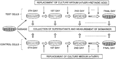
- FIG. 1 is a model diagram illustrating the cell differentiation state assessment method in one example of the present invention.
- FIG. 2 is a graph showing a temporal change in the amount of various substances determined by a GC-MS analysis of a culture supernatant in the aforementioned example.
- FIG. 3 is a graph showing a temporal change in the amount of various substances determined by an LC-MS analysis of a culture supernatant in the aforementioned example.
- FIG. 4 is a table showing the components of DMEM/F12.
- FIG. 5 is the first part of a table showing the components of mTeSR1.
- FIG. 6 is the second part of the table showing the components of mTeSR1.
- the state of differentiation of test cells is assessed based on the amount of a biomarker in a culture supernatant of the test cells, where at least one compound selected from the group of putrescine, kynurenine, cystathionine, ascorbic acid, riboflavin, pyruvic acid, serine, cysteine, threonic acid, citric acid, and orotic acid is used as the biomarker.
- stem cells may be used, typical examples of which are pluripotent stem cells, such as ES cells and iPS cells. Cells obtained from the stem cells by differentiation induction may also be used as the test cells.
- the culture medium for culturing these kinds of test cells any culture medium generally used for the culturing of stem cells can be used, such as DMEM/F12 or culture media containing DMEM/F12 as the main component (e.g. mTeSR1).
- FIG. 4 shows the components of DMEM/F12.
- a quantitative analysis by mass spectrometry and particularly, a quantitative analysis using a liquid chromatogram mass spectrometer (LC-MS) or gas chromatograph mass spectrometer (GC-MS) can suitably, but not exclusively, be used.
- LC-MS liquid chromatogram mass spectrometer
- GC-MS gas chromatograph mass spectrometer
- an agent or the like which makes each biomarker develop a specific color or emit specific light may be added to the culture supernatant, in which case the amount of the biomarker can be determined based on the intensity of the coloring or emission of light.
- FIG. 1 is a model diagram showing the process steps of the cell differentiation state assessment method the present example.
- the aforementioned KhES-1 line was subcultured in four culture dishes (60 mm in diameter) coated with BioCoat Matrigel® (Corning International K.K.). (For simplification, only one culture dish is shown in FIG. 1 ).
- As the culture medium mTeSR1 (modified Tenneille Serum Replacer 1) was used. The culture medium was replaced every day. The components of mTeSR1 are as shown in FIGS. 5 and 6 .
- the KhES-3 line was also similarly subcultured in four culture dishes. With the first day of the subculturing (passage) of the cells counted as the zeroth day, the culturing was continued until the cells reached confluence. Every day, when the culture medium was replaced, the culture supernatant was collected from each culture dish as the sample for mass spectrometry. For the zeroth day of the culturing, mTeSR1 was directly used as the sample for mass spectrometry.
- the aforementioned KhES-1 line was subcultured in four culture dishes (60 mm in diameter) coated with Matrigel. (For simplification, only one culture dish is shown in FIG. 1 ).
- As the culture medium mTeSR1 was used. With the culture medium replaced every day, the culturing was continued until the cells reached confluence.
- the KhES-3 line was also similarly subcultured in four culture dishes. With the first day of the passage counted as the zeroth day, when the culture medium was replaced on the second and subsequent days, the old medium was replaced by mTeSR1 with retinoic acid added to the final concentration of 0.1 ⁇ M in order to give a differentiation induction stimulus. Every day, when the culture medium was replaced, the culture supernatant was collected from each culture dish as the sample for mass spectrometry. For the zeroth day of the culturing, mTeSR1 was directly used as the sample for mass spectrometry.
- isopropylmalic acid was added to each of the samples, which were subsequently treated with an extraction solution (methanol, chloroform, and water mixed at a ratio of 2.5:1:1) to remove proteins. After the extraction, the supernatant was collected and dried.
- an extraction solution methanol, chloroform, and water mixed at a ratio of 2.5:1:1
- the criteria used for the identification of the compounds were whether or not the difference between the retention index (a numerical value showing a relative retention time) specified in the database and the retention index of a derivatized compound in the sample was within a range of ⁇ 5, as well as whether or not both the target ion and confirmation ion designated in the database were detected for the derivatized compound in the sample.
- the quantitative determination of the compounds was performed by calculating the area of a mass chromatogram created for an ion characteristic of each derivatized compound in the sample according to the conditions specified in the database.
- the quantity value (area value) of each compound determined by the previously described GC-MS and LC-MS analyses was divided by the quantity value (area value) of the internal standard material, and the obtained value was adopted as the index value of the amount of each compound in the culture supernatant.
- an average of the index values obtained from the results of the GC-MS and LC-MS analyses performed on the culture supernatants collected from the four culture dishes was calculated. Using the averages of the index values, the control cells and test cells were compared with each other in terms of the amount of each compound.
- Tables 1-4 show compounds which were judged to have a significant difference in amount between the culture supernatant of the control cells and that of the test cells.
- E denotes exponential in decimal; for example, “1.326E-02” means “1.326 ⁇ 10 ⁇ 2 ”.
- Tables 1 and 2 show compounds whose amount in the culture supernatant of the control cells was determined to be higher than in the culture supernatant of the test cells. Specifically, Table 1 is the result obtained for the KhES-1 line, while Table 2 is the result obtained for the KhES-3 line.
- the “variation” in these tables means the aforementioned A/B value, i.e. the ratio of the “average of the index values calculated for the control cells” to the “average of the index values calculated for the test cells”.
- Tables 3 and 4 show compounds whose amount in the culture supernatant of the test cells was determined to be higher than in the culture supernatant of the control cells. Specifically, Table 3 is the result obtained for the KhES-1 line, while Table 4 is the result obtained for the KhES-3 line.
- the “variation” in these tables means the aforementioned B/A value, i.e. the ratio of the “average of the index values calculated for the test cells” to the “average of the index values calculated for the control cells”.
- each of the compounds listed in Tables 1-4 shows a change in the amount of metabolic expenditure inside the cells and/or the amount of secretion to the outside of the cells as a result of the differentiation induction, and therefore, can be used as a biomarker for assessing the state of differentiation of the cells.
- the test cells are stem cells and whether or not these cells are maintained in the undifferentiated state is unknown, while the control cells are stem cells which are unmistakably in the undifferentiated state.
- control cells may also be cells which are unmistakably differentiated.
- the control cells may also be cells which are unmistakably differentiated.
- the ratio of the “amount in the culture supernatant of the test cells” to the “amount in the culture supernatant of the control cells” is equal to or higher than a predetermined threshold, it is possible to conclude that the test cells are in the undifferentiated state.
- the ratio of the “amount in the culture supernatant of the control cells” to the “amount in the culture supernatant of the test cells” is equal to or higher than a predetermined threshold, it is possible to conclude that the test cells are in the undifferentiated state.
- test cells are differentiation-induced cells derived from stem cells and whether or not undifferentiated cells remain is unknown, while the control cells are cells which are unmistakably undifferentiated.
- control cells are cells which are unmistakably undifferentiated.
- test cells are differentiation-induced cells derived from stem cells and whether or not undifferentiated cells remain is unknown, while the cells used as the control cells are unmistakably differentiated as opposed to the previous example.
- the ratio of the “amount in the culture supernatant of the test cells” to the “amount in the culture supernatant of the control cells” is equal to or higher than a predetermined threshold, it is possible to conclude that undifferentiated cells are mixed in the test cells.
- the ratio of the “amount in the culture supernatant of the control cells” to the “amount in the culture supernatant of the test cells” is equal to or higher than a predetermined threshold, it is possible to conclude that undifferentiated cells are mixed in the test cells. Regardless of which method is used for the determination, the levels of amount of the compounds in the culture supernatant of the control cells do not need to be simultaneously measured with those of the test cells; instead, previously measured data may be used.
- FIGS. 2 and 3 show the change in the amount of each of the biomarkers in the culture supernatant over the period from the zeroth to sixth days of the culturing of the KhES-1 line. It should be noted that the results shown in FIG. 2 were obtained by GC-MS analysis, while those shown in FIG. 3 were obtained by LC-MS analysis. As is evident from these figures, it was confirmed that, although there is no difference in the amount of the biomarker compounds between the test cells and the control cells immediately after the beginning of the culturing, the difference in the amount of each biomarker compound between the test cells and the control cells increases with time. Although FIGS. 2 and 3 only show the results obtained for the KhES-1 line, it was also confirmed that the KhES-3 line shows a similar change.
Landscapes
- Health & Medical Sciences (AREA)
- Life Sciences & Earth Sciences (AREA)
- Chemical & Material Sciences (AREA)
- Engineering & Computer Science (AREA)
- Immunology (AREA)
- Molecular Biology (AREA)
- Organic Chemistry (AREA)
- Biomedical Technology (AREA)
- Hematology (AREA)
- Urology & Nephrology (AREA)
- Biotechnology (AREA)
- Microbiology (AREA)
- Physics & Mathematics (AREA)
- Analytical Chemistry (AREA)
- Biochemistry (AREA)
- General Health & Medical Sciences (AREA)
- Proteomics, Peptides & Aminoacids (AREA)
- Wood Science & Technology (AREA)
- Zoology (AREA)
- Cell Biology (AREA)
- Toxicology (AREA)
- Bioinformatics & Cheminformatics (AREA)
- Food Science & Technology (AREA)
- Pathology (AREA)
- General Physics & Mathematics (AREA)
- Medicinal Chemistry (AREA)
- Tropical Medicine & Parasitology (AREA)
- Biophysics (AREA)
- General Engineering & Computer Science (AREA)
- Genetics & Genomics (AREA)
- Developmental Biology & Embryology (AREA)
- Measuring Or Testing Involving Enzymes Or Micro-Organisms (AREA)
- Micro-Organisms Or Cultivation Processes Thereof (AREA)
- Investigating Or Analysing Biological Materials (AREA)
Applications Claiming Priority (4)
| Application Number | Priority Date | Filing Date | Title |
|---|---|---|---|
| JPJP2014-094507 | 2014-05-01 | ||
| JP2014-094507 | 2014-05-01 | ||
| JP2014094507 | 2014-05-01 | ||
| PCT/JP2015/062128 WO2015166845A1 (ja) | 2014-05-01 | 2015-04-21 | 細胞の分化状態の評価方法 |
Publications (2)
| Publication Number | Publication Date |
|---|---|
| US20170052171A1 US20170052171A1 (en) | 2017-02-23 |
| US11022600B2 true US11022600B2 (en) | 2021-06-01 |
Family
ID=54358576
Family Applications (1)
| Application Number | Title | Priority Date | Filing Date |
|---|---|---|---|
| US15/307,948 Active 2036-09-14 US11022600B2 (en) | 2014-05-01 | 2015-04-21 | Method for assessing state of differentiation of cells |
Country Status (8)
| Country | Link |
|---|---|
| US (1) | US11022600B2 (ja) |
| EP (1) | EP3138922B1 (ja) |
| JP (2) | JP6332440B2 (ja) |
| KR (1) | KR101931420B1 (ja) |
| CN (1) | CN106460028B (ja) |
| AU (1) | AU2015254267B2 (ja) |
| SG (1) | SG11201609091YA (ja) |
| WO (1) | WO2015166845A1 (ja) |
Families Citing this family (14)
| Publication number | Priority date | Publication date | Assignee | Title |
|---|---|---|---|---|
| US10704073B2 (en) | 2014-09-29 | 2020-07-07 | Tokyo Electron Limited | Method for determining undifferentiated state of pluripotent stem cells by culture medium analysis |
| WO2017068727A1 (ja) * | 2015-10-23 | 2017-04-27 | 株式会社島津製作所 | 細胞の分化状態の評価方法 |
| WO2018008149A1 (ja) * | 2016-07-08 | 2018-01-11 | 株式会社島津製作所 | クロマトグラフ質量分析用データ処理装置 |
| CN110392732B (zh) * | 2017-03-02 | 2023-07-28 | 株式会社岛津制作所 | 细胞分析方法和细胞分析装置 |
| WO2019031545A1 (ja) * | 2017-08-08 | 2019-02-14 | 東京エレクトロン株式会社 | 多能性幹細胞の未分化状態を判定する方法、多能性幹細胞の継代培養方法およびそれら方法に使用される装置 |
| US11209405B2 (en) | 2017-12-01 | 2021-12-28 | Shimadzu Corporation | Liquid chromatograph analysis system |
| JP6879380B2 (ja) * | 2017-12-05 | 2021-06-02 | 株式会社島津製作所 | 生体試料自動分析システム |
| US11209404B2 (en) | 2017-12-05 | 2021-12-28 | Shimadzu Corporation | Biological sample analyzing system |
| CN109917025A (zh) * | 2017-12-12 | 2019-06-21 | 中国科学院大连化学物理研究所 | 评估人体外受精胚胎质量及差异代谢物的筛选方法 |
| CN112673110A (zh) * | 2018-06-18 | 2021-04-16 | 株式会社岛津制作所 | 细胞的基因组异常评价方法 |
| WO2021014515A1 (ja) * | 2019-07-19 | 2021-01-28 | 東京エレクトロン株式会社 | 細胞の分化状態の評価方法 |
| WO2021144956A1 (ja) * | 2020-01-17 | 2021-07-22 | 株式会社日立ハイテク | 細胞の分化状態の評価方法及び細胞培養システム |
| JP7462452B2 (ja) * | 2020-03-27 | 2024-04-05 | 株式会社日立製作所 | 培養細胞の分化レベルを評価するための評価装置および評価方法、並びに自動細胞培養システム |
| JP7585957B2 (ja) | 2021-05-11 | 2024-11-19 | 株式会社島津製作所 | 神経細胞への分化状態の評価方法 |
Citations (6)
| Publication number | Priority date | Publication date | Assignee | Title |
|---|---|---|---|---|
| JP2004313184A (ja) | 2003-03-28 | 2004-11-11 | National Institute Of Agrobiological Sciences | 未分化細胞を選別する方法およびその利用 |
| JP2006042663A (ja) | 2004-08-03 | 2006-02-16 | Reprocell Inc | Es細胞の識別マーカー |
| EP2275531A1 (en) | 2008-03-31 | 2011-01-19 | Oriental Yeast Co., Ltd. | Method for proliferation of pluripotent stem cell |
| WO2011139907A2 (en) | 2010-04-29 | 2011-11-10 | Atyr Pharma, Inc. | Innovative discovery of therapeutic, diagnostic, and antibody compositions related to protein fragments of valyl trna synthetases |
| WO2012123401A1 (en) * | 2011-03-11 | 2012-09-20 | Tigenix, S.A. | Cell populations having immunoregulatory activity, method for isolation and uses |
| WO2013065302A1 (ja) | 2011-11-01 | 2013-05-10 | 独立行政法人産業技術総合研究所 | 未分化細胞検出方法及び複合糖質検出方法 |
Family Cites Families (13)
| Publication number | Priority date | Publication date | Assignee | Title |
|---|---|---|---|---|
| JPH05199863A (ja) * | 1991-03-08 | 1993-08-10 | Asahi Breweries Ltd | システインおよびその誘導体の高生産微生物 |
| WO1995002407A1 (fr) * | 1993-07-14 | 1995-01-26 | Yamanouchi Pharmaceutical Co., Ltd. | Neuroprotecteur et nouvel ester orotique |
| JP3002144B2 (ja) * | 1996-10-01 | 2000-01-24 | 張 銘 烈 | 癌細胞分化を促進する薬学組成物及びその製造方法 |
| CN1196480C (zh) * | 2001-07-03 | 2005-04-13 | 北京巨能亚太生命科学研究中心 | L-苏糖酸钙在制备预防或治疗骨折的药物中的用途 |
| DE602004030254D1 (de) * | 2003-08-01 | 2011-01-05 | Two Cells Co Ltd | Scaffoldfreies, selbstorganisiertes, 3 dimensionales, synthetisches gewebe |
| WO2007000884A1 (ja) * | 2005-06-29 | 2007-01-04 | National University Corporation Kanazawa University | 骨・関節疾患の予防・治療剤およびそのスクリーニング方法 |
| JP2013520163A (ja) * | 2010-02-23 | 2013-06-06 | アンスティチュ ナショナル ドゥ ラ サンテ エ ドゥ ラ ルシェルシュ メディカル | ヒト多能性幹細胞からヒトメラノサイトを調製するための方法 |
| WO2012056997A1 (ja) * | 2010-10-28 | 2012-05-03 | 国立大学法人熊本大学 | 多能性幹細胞の分化誘導効率を改善するための方法及び培地 |
| WO2012126013A2 (en) * | 2011-03-17 | 2012-09-20 | Minerva Biotechnologies Corporation | Method for making pluripotent stem cells |
| JP2013155122A (ja) * | 2012-01-27 | 2013-08-15 | Shiseido Co Ltd | 幹細胞賦活化剤 |
| JP2013240308A (ja) * | 2012-05-23 | 2013-12-05 | Kagoshima Univ | ヒトES/iPS細胞における遺伝子発現方法 |
| WO2013187194A1 (ja) * | 2012-06-12 | 2013-12-19 | 医療法人社団 土合会 | 骨疾患の治療に有効な医薬組成物 |
| EP3184643B1 (en) * | 2014-08-20 | 2020-02-19 | FUJIFILM Wako Pure Chemical Corporation | Method for determining state of differentiation of stem cells, and novel differentiation marker used therefor |
-
2015
- 2015-04-21 US US15/307,948 patent/US11022600B2/en active Active
- 2015-04-21 SG SG11201609091YA patent/SG11201609091YA/en unknown
- 2015-04-21 KR KR1020167031400A patent/KR101931420B1/ko active Active
- 2015-04-21 EP EP15785591.7A patent/EP3138922B1/en active Active
- 2015-04-21 AU AU2015254267A patent/AU2015254267B2/en active Active
- 2015-04-21 WO PCT/JP2015/062128 patent/WO2015166845A1/ja active Application Filing
- 2015-04-21 CN CN201580021824.XA patent/CN106460028B/zh active Active
- 2015-04-21 JP JP2016516327A patent/JP6332440B2/ja active Active
-
2018
- 2018-05-02 JP JP2018088721A patent/JP6536711B2/ja active Active
Patent Citations (10)
| Publication number | Priority date | Publication date | Assignee | Title |
|---|---|---|---|---|
| JP2004313184A (ja) | 2003-03-28 | 2004-11-11 | National Institute Of Agrobiological Sciences | 未分化細胞を選別する方法およびその利用 |
| JP2006042663A (ja) | 2004-08-03 | 2006-02-16 | Reprocell Inc | Es細胞の識別マーカー |
| EP2275531A1 (en) | 2008-03-31 | 2011-01-19 | Oriental Yeast Co., Ltd. | Method for proliferation of pluripotent stem cell |
| US20110117645A1 (en) | 2008-03-31 | 2011-05-19 | Oriental Yeast Co., Ltd. | Method for proliferation of pluripotent stem cells |
| WO2011139907A2 (en) | 2010-04-29 | 2011-11-10 | Atyr Pharma, Inc. | Innovative discovery of therapeutic, diagnostic, and antibody compositions related to protein fragments of valyl trna synthetases |
| US20130202574A1 (en) | 2010-04-29 | 2013-08-08 | Pangu Biopharma Limited | Innovative discovery of therapeutic, diagnostic, and antibody compositions related to protein fragments of valyl-trna synthetases |
| US20150361411A1 (en) | 2010-04-29 | 2015-12-17 | Atyr Pharma, Inc. | Innovative discovery of therapeutic, diagnostic, and antibody compositions related to protein fragments of valyl-trna synthetases |
| WO2012123401A1 (en) * | 2011-03-11 | 2012-09-20 | Tigenix, S.A. | Cell populations having immunoregulatory activity, method for isolation and uses |
| WO2013065302A1 (ja) | 2011-11-01 | 2013-05-10 | 独立行政法人産業技術総合研究所 | 未分化細胞検出方法及び複合糖質検出方法 |
| US9500650B2 (en) * | 2011-11-01 | 2016-11-22 | National Institute Of Advanced Industrial Science And Technology | Undifferentiated cell detection method and complex carbohydrate detection method |
Non-Patent Citations (7)
| Title |
|---|
| Australian Office Action dated Jun. 27, 2017 in Patent Application No. 2015254267. |
| Cezar, G.G. et al. Identification of small molecules from human embryonic stem cells using metabolomics. Stem Cells and Development 16: 869-882. specif, pp. 869, 870, 876. * |
| EngMT. Tateno, H. et al. Undifferentiated cell detection method and complex carbohydrate detection method. International Patent Application Publication No. WO 2013/065302 A1. Published May 10, 2013. pp. 1-56. * |
| Extended European Search Report dated Mar. 23, 2017 in Patent Application No. 15785591.7. |
| International Preliminary Report on Patentability and Written Opinion dated Nov. 1, 2016 in PCT/JP2015/062128 filed Apr. 21, 2015 (with English translation). |
| International Search Report dated Jun. 30, 2015 in PCT/JP2015/062128 filed Apr. 21, 2015. |
| Jones, S.P. et al. 2013. Published online Sep. 15, 2013. The kynurenine pathway in stem cell biology. International Journal of Tryptophan Research 6: 57-66. specif, pp. 59, 62, 64. * |
Also Published As
| Publication number | Publication date |
|---|---|
| EP3138922B1 (en) | 2020-12-09 |
| JP6332440B2 (ja) | 2018-05-30 |
| CN106460028A (zh) | 2017-02-22 |
| JPWO2015166845A1 (ja) | 2017-04-20 |
| AU2015254267B2 (en) | 2018-03-08 |
| EP3138922A1 (en) | 2017-03-08 |
| EP3138922A4 (en) | 2017-04-26 |
| JP2018121659A (ja) | 2018-08-09 |
| WO2015166845A1 (ja) | 2015-11-05 |
| KR101931420B1 (ko) | 2018-12-20 |
| CN106460028B (zh) | 2020-03-10 |
| SG11201609091YA (en) | 2016-12-29 |
| JP6536711B2 (ja) | 2019-07-03 |
| KR20160143804A (ko) | 2016-12-14 |
| AU2015254267A1 (en) | 2016-11-24 |
| US20170052171A1 (en) | 2017-02-23 |
Similar Documents
| Publication | Publication Date | Title |
|---|---|---|
| US11022600B2 (en) | Method for assessing state of differentiation of cells | |
| Müller et al. | Automated sample preparation with SP 3 for low‐input clinical proteomics | |
| JP6748654B2 (ja) | 細胞の分化状態の評価方法 | |
| Moussaieff et al. | Glycolysis-mediated changes in acetyl-CoA and histone acetylation control the early differentiation of embryonic stem cells | |
| Estes et al. | Identification of endothelial cells and progenitor cell subsets in human peripheral blood | |
| EP3230439B1 (en) | Human blood brain barrier model | |
| Lau et al. | Unique properties of a subset of human pluripotent stem cells with high capacity for self-renewal | |
| NZ749272A (en) | Phospholipid ether analogs for the identification and isolation of circulating tumor cells | |
| US20190079094A1 (en) | Subcellular localization of target analytes | |
| Galkowski et al. | Of cytometry, stem cells and fountain of youth | |
| Sebastião et al. | Stem cells characterization: OMICS reinforcing analytics | |
| US9632096B2 (en) | Methods of assessing the immunomodulatory potential of a multipotent stromal cell (MSC) population, and systems and kits for practicing the same | |
| He et al. | Label‐Free Impedance Analysis of Induced Pluripotent Stem Cell‐Derived Spinal Cord Progenitor Cells for Rapid Safety and Efficacy Profiling | |
| Mancia et al. | Quantitative methods to characterize morphological properties of cell lines | |
| CN110836972A (zh) | 一种基于γ-H2AX的生物标志物遗传毒性检测方法 | |
| KR20150036838A (ko) | 중간엽줄기세포의 지방세포 분화용 마커 및 그 용도 | |
| Soriano et al. | CONSERVED NUCLEAR MORPHOLOGY IDENTIFIES FUNCTIONAL RADIAL GLIA NEURAL PROGENITORS | |
| CN107064087A (zh) | 一种基于高内涵技术定量分析苯或苯的代谢物致细胞dna损伤的方法 | |
| CN107064086A (zh) | 一种基于高内涵技术定量分析苯并[a]芘致细胞DNA损伤的方法 | |
| WO2024039744A1 (en) | Methods of forming patient-derived 3d cell cultures for tracking live immune-tumor interactions | |
| JPWO2019244853A1 (ja) | 細胞のゲノム異常の評価方法 | |
| CN104160017A (zh) | 测定致畸风险的方法 | |
| Kelly | Proteomic characterisation of clonal populations from a humanlung carcinoma cell line |
Legal Events
| Date | Code | Title | Description |
|---|---|---|---|
| AS | Assignment |
Owner name: SHIMADZU CORPORATION, JAPAN Free format text: ASSIGNMENT OF ASSIGNORS INTEREST;ASSIGNORS:SUZUKI, TAKASHI;NAKATSUJI, NORIO;SUEMORI, HIROFUMI;AND OTHERS;SIGNING DATES FROM 20161013 TO 20161021;REEL/FRAME:040175/0954 |
|
| STPP | Information on status: patent application and granting procedure in general |
Free format text: FINAL REJECTION MAILED |
|
| STPP | Information on status: patent application and granting procedure in general |
Free format text: RESPONSE AFTER FINAL ACTION FORWARDED TO EXAMINER |
|
| STCV | Information on status: appeal procedure |
Free format text: NOTICE OF APPEAL FILED |
|
| STCV | Information on status: appeal procedure |
Free format text: APPEAL BRIEF (OR SUPPLEMENTAL BRIEF) ENTERED AND FORWARDED TO EXAMINER |
|
| STCV | Information on status: appeal procedure |
Free format text: EXAMINER'S ANSWER TO APPEAL BRIEF MAILED |
|
| STCV | Information on status: appeal procedure |
Free format text: ON APPEAL -- AWAITING DECISION BY THE BOARD OF APPEALS |
|
| STCV | Information on status: appeal procedure |
Free format text: BOARD OF APPEALS DECISION RENDERED |
|
| STPP | Information on status: patent application and granting procedure in general |
Free format text: AMENDMENT / ARGUMENT AFTER BOARD OF APPEALS DECISION |
|
| STPP | Information on status: patent application and granting procedure in general |
Free format text: NOTICE OF ALLOWANCE MAILED -- APPLICATION RECEIVED IN OFFICE OF PUBLICATIONS |
|
| STPP | Information on status: patent application and granting procedure in general |
Free format text: PUBLICATIONS -- ISSUE FEE PAYMENT RECEIVED |
|
| STPP | Information on status: patent application and granting procedure in general |
Free format text: PUBLICATIONS -- ISSUE FEE PAYMENT VERIFIED |
|
| STCF | Information on status: patent grant |
Free format text: PATENTED CASE |
|
| MAFP | Maintenance fee payment |
Free format text: PAYMENT OF MAINTENANCE FEE, 4TH YEAR, LARGE ENTITY (ORIGINAL EVENT CODE: M1551); ENTITY STATUS OF PATENT OWNER: LARGE ENTITY Year of fee payment: 4 |