US10370435B2 - Binding molecules directed against influenza hemagglutinin and uses thereof - Google Patents
Binding molecules directed against influenza hemagglutinin and uses thereof Download PDFInfo
- Publication number
- US10370435B2 US10370435B2 US15/548,843 US201615548843A US10370435B2 US 10370435 B2 US10370435 B2 US 10370435B2 US 201615548843 A US201615548843 A US 201615548843A US 10370435 B2 US10370435 B2 US 10370435B2
- Authority
- US
- United States
- Prior art keywords
- seq
- influenza
- binding
- virus
- amino acid
- Prior art date
- Legal status (The legal status is an assumption and is not a legal conclusion. Google has not performed a legal analysis and makes no representation as to the accuracy of the status listed.)
- Active
Links
- 230000027455 binding Effects 0.000 title abstract description 159
- 101710154606 Hemagglutinin Proteins 0.000 title abstract description 152
- 101710093908 Outer capsid protein VP4 Proteins 0.000 title abstract description 151
- 101710135467 Outer capsid protein sigma-1 Proteins 0.000 title abstract description 151
- 101710176177 Protein A56 Proteins 0.000 title abstract description 151
- 239000000185 hemagglutinin Substances 0.000 title abstract description 145
- 206010022000 influenza Diseases 0.000 title description 90
- 108010003723 Single-Domain Antibodies Proteins 0.000 claims description 118
- 125000003275 alpha amino acid group Chemical group 0.000 claims description 89
- 150000007523 nucleic acids Chemical class 0.000 claims description 39
- 241000282414 Homo sapiens Species 0.000 claims description 26
- 108020004707 nucleic acids Proteins 0.000 claims description 24
- 102000039446 nucleic acids Human genes 0.000 claims description 24
- 108090000765 processed proteins & peptides Proteins 0.000 claims description 9
- 229920001184 polypeptide Polymers 0.000 claims description 7
- 102000004196 processed proteins & peptides Human genes 0.000 claims description 7
- 239000008194 pharmaceutical composition Substances 0.000 claims description 5
- 230000003472 neutralizing effect Effects 0.000 abstract description 58
- 241000712431 Influenza A virus Species 0.000 abstract description 43
- 241000713196 Influenza B virus Species 0.000 abstract description 26
- 241000699670 Mus sp. Species 0.000 description 89
- 241000712461 unidentified influenza virus Species 0.000 description 74
- 210000004027 cell Anatomy 0.000 description 72
- 238000006386 neutralization reaction Methods 0.000 description 68
- 238000001727 in vivo Methods 0.000 description 58
- 208000037797 influenza A Diseases 0.000 description 56
- 241000700605 Viruses Species 0.000 description 54
- 230000004083 survival effect Effects 0.000 description 45
- 230000004927 fusion Effects 0.000 description 32
- 239000013598 vector Substances 0.000 description 29
- 208000015181 infectious disease Diseases 0.000 description 28
- 230000035772 mutation Effects 0.000 description 27
- 235000001014 amino acid Nutrition 0.000 description 26
- 229940024606 amino acid Drugs 0.000 description 26
- 150000001413 amino acids Chemical class 0.000 description 26
- 108090000623 proteins and genes Proteins 0.000 description 26
- 208000037798 influenza B Diseases 0.000 description 25
- 235000006576 Althaea officinalis Nutrition 0.000 description 24
- 230000037396 body weight Effects 0.000 description 24
- 231100000518 lethal Toxicity 0.000 description 24
- 230000001665 lethal effect Effects 0.000 description 24
- 238000003556 assay Methods 0.000 description 19
- 102000004169 proteins and genes Human genes 0.000 description 19
- 241000272527 Mareca penelope Species 0.000 description 17
- 239000007853 buffer solution Substances 0.000 description 17
- 238000000034 method Methods 0.000 description 17
- 235000018102 proteins Nutrition 0.000 description 17
- 230000004580 weight loss Effects 0.000 description 17
- 108020004705 Codon Proteins 0.000 description 16
- 102000018071 Immunoglobulin Fc Fragments Human genes 0.000 description 15
- 108010091135 Immunoglobulin Fc Fragments Proteins 0.000 description 15
- 230000008859 change Effects 0.000 description 15
- 230000000694 effects Effects 0.000 description 15
- 239000000203 mixture Substances 0.000 description 15
- 239000000523 sample Substances 0.000 description 14
- 230000003612 virological effect Effects 0.000 description 14
- 238000011282 treatment Methods 0.000 description 13
- 230000006872 improvement Effects 0.000 description 12
- 241001136800 Anas acuta Species 0.000 description 11
- 238000011534 incubation Methods 0.000 description 11
- 230000036515 potency Effects 0.000 description 11
- 230000010530 Virus Neutralization Effects 0.000 description 10
- 230000010056 antibody-dependent cellular cytotoxicity Effects 0.000 description 10
- 230000005764 inhibitory process Effects 0.000 description 10
- 238000004519 manufacturing process Methods 0.000 description 10
- 239000002609 medium Substances 0.000 description 10
- 230000002265 prevention Effects 0.000 description 10
- 239000000427 antigen Substances 0.000 description 9
- 102000036639 antigens Human genes 0.000 description 9
- 108091007433 antigens Proteins 0.000 description 9
- 239000000539 dimer Substances 0.000 description 9
- 210000002966 serum Anatomy 0.000 description 9
- 241001293113 Anas clypeata Species 0.000 description 8
- 241001293118 Anas crecca Species 0.000 description 8
- 235000002198 Annona diversifolia Nutrition 0.000 description 8
- 108091028043 Nucleic acid sequence Proteins 0.000 description 8
- 241000287462 Phalacrocorax carbo Species 0.000 description 8
- 238000012512 characterization method Methods 0.000 description 8
- 239000003814 drug Substances 0.000 description 8
- 239000012636 effector Substances 0.000 description 8
- 230000000903 blocking effect Effects 0.000 description 7
- 230000006870 function Effects 0.000 description 7
- 210000004602 germ cell Anatomy 0.000 description 7
- 238000000338 in vitro Methods 0.000 description 7
- 230000003993 interaction Effects 0.000 description 7
- 238000006467 substitution reaction Methods 0.000 description 7
- 241000588724 Escherichia coli Species 0.000 description 6
- 241001465754 Metazoa Species 0.000 description 6
- 239000011324 bead Substances 0.000 description 6
- 239000013604 expression vector Substances 0.000 description 6
- 239000000284 extract Substances 0.000 description 6
- 230000006698 induction Effects 0.000 description 6
- 238000001990 intravenous administration Methods 0.000 description 6
- 210000004962 mammalian cell Anatomy 0.000 description 6
- 230000001932 seasonal effect Effects 0.000 description 6
- 239000013638 trimer Substances 0.000 description 6
- 108020004414 DNA Proteins 0.000 description 5
- 238000002965 ELISA Methods 0.000 description 5
- 241000282842 Lama glama Species 0.000 description 5
- 101710099301 Low affinity immunoglobulin gamma Fc region receptor III-A Proteins 0.000 description 5
- 102100029193 Low affinity immunoglobulin gamma Fc region receptor III-A Human genes 0.000 description 5
- 241000701076 Macacine alphaherpesvirus 1 Species 0.000 description 5
- 238000007792 addition Methods 0.000 description 5
- 206010064097 avian influenza Diseases 0.000 description 5
- 239000011230 binding agent Substances 0.000 description 5
- 230000015572 biosynthetic process Effects 0.000 description 5
- 239000000872 buffer Substances 0.000 description 5
- 229940079593 drug Drugs 0.000 description 5
- 239000012634 fragment Substances 0.000 description 5
- 230000003053 immunization Effects 0.000 description 5
- 238000002649 immunization Methods 0.000 description 5
- 230000002401 inhibitory effect Effects 0.000 description 5
- PGZUMBJQJWIWGJ-ONAKXNSWSA-N oseltamivir phosphate Chemical compound OP(O)(O)=O.CCOC(=O)C1=C[C@@H](OC(CC)CC)[C@H](NC(C)=O)[C@@H](N)C1 PGZUMBJQJWIWGJ-ONAKXNSWSA-N 0.000 description 5
- 238000002823 phage display Methods 0.000 description 5
- 239000000546 pharmaceutical excipient Substances 0.000 description 5
- 102000005962 receptors Human genes 0.000 description 5
- 108020003175 receptors Proteins 0.000 description 5
- 230000004044 response Effects 0.000 description 5
- 238000012216 screening Methods 0.000 description 5
- 238000013207 serial dilution Methods 0.000 description 5
- 229940061367 tamiflu Drugs 0.000 description 5
- 230000008685 targeting Effects 0.000 description 5
- 229960005486 vaccine Drugs 0.000 description 5
- 108091006020 Fc-tagged proteins Proteins 0.000 description 4
- DHMQDGOQFOQNFH-UHFFFAOYSA-N Glycine Chemical compound NCC(O)=O DHMQDGOQFOQNFH-UHFFFAOYSA-N 0.000 description 4
- 241000282838 Lama Species 0.000 description 4
- 241000845082 Panama Species 0.000 description 4
- 101001083556 Pseudomonas alcaligenes Maleylpyruvate hydrolase Proteins 0.000 description 4
- 241000831652 Salinivibrio sharmensis Species 0.000 description 4
- FAPWRFPIFSIZLT-UHFFFAOYSA-M Sodium chloride Chemical compound [Na+].[Cl-] FAPWRFPIFSIZLT-UHFFFAOYSA-M 0.000 description 4
- 238000012575 bio-layer interferometry Methods 0.000 description 4
- 239000003795 chemical substances by application Substances 0.000 description 4
- 238000003745 diagnosis Methods 0.000 description 4
- 238000005516 engineering process Methods 0.000 description 4
- 210000003743 erythrocyte Anatomy 0.000 description 4
- 238000009472 formulation Methods 0.000 description 4
- 230000035931 haemagglutination Effects 0.000 description 4
- 230000028993 immune response Effects 0.000 description 4
- 239000003112 inhibitor Substances 0.000 description 4
- 230000000813 microbial effect Effects 0.000 description 4
- 239000002773 nucleotide Substances 0.000 description 4
- 125000003729 nucleotide group Chemical group 0.000 description 4
- 229960003752 oseltamivir Drugs 0.000 description 4
- VSZGPKBBMSAYNT-RRFJBIMHSA-N oseltamivir Chemical compound CCOC(=O)C1=C[C@@H](OC(CC)CC)[C@H](NC(C)=O)[C@@H](N)C1 VSZGPKBBMSAYNT-RRFJBIMHSA-N 0.000 description 4
- 230000036961 partial effect Effects 0.000 description 4
- 239000013641 positive control Substances 0.000 description 4
- 230000003389 potentiating effect Effects 0.000 description 4
- 230000000069 prophylactic effect Effects 0.000 description 4
- 238000000746 purification Methods 0.000 description 4
- 230000002829 reductive effect Effects 0.000 description 4
- 230000001225 therapeutic effect Effects 0.000 description 4
- 230000009385 viral infection Effects 0.000 description 4
- 238000005406 washing Methods 0.000 description 4
- 108091032973 (ribonucleotides)n+m Proteins 0.000 description 3
- 238000012815 AlphaLISA Methods 0.000 description 3
- 241000894006 Bacteria Species 0.000 description 3
- PEDCQBHIVMGVHV-UHFFFAOYSA-N Glycerine Chemical compound OCC(O)CO PEDCQBHIVMGVHV-UHFFFAOYSA-N 0.000 description 3
- 241000282412 Homo Species 0.000 description 3
- 101000917858 Homo sapiens Low affinity immunoglobulin gamma Fc region receptor III-A Proteins 0.000 description 3
- 108060003951 Immunoglobulin Proteins 0.000 description 3
- QNAYBMKLOCPYGJ-REOHCLBHSA-N L-alanine Chemical compound C[C@H](N)C(O)=O QNAYBMKLOCPYGJ-REOHCLBHSA-N 0.000 description 3
- ZMANZCXQSJIPKH-UHFFFAOYSA-N Triethylamine Chemical compound CCN(CC)CC ZMANZCXQSJIPKH-UHFFFAOYSA-N 0.000 description 3
- 239000002671 adjuvant Substances 0.000 description 3
- 235000004279 alanine Nutrition 0.000 description 3
- 238000004458 analytical method Methods 0.000 description 3
- 230000000840 anti-viral effect Effects 0.000 description 3
- SQVRNKJHWKZAKO-UHFFFAOYSA-N beta-N-Acetyl-D-neuraminic acid Natural products CC(=O)NC1C(O)CC(O)(C(O)=O)OC1C(O)C(O)CO SQVRNKJHWKZAKO-UHFFFAOYSA-N 0.000 description 3
- 239000012228 culture supernatant Substances 0.000 description 3
- 229940042406 direct acting antivirals neuraminidase inhibitors Drugs 0.000 description 3
- 238000010494 dissociation reaction Methods 0.000 description 3
- 230000005593 dissociations Effects 0.000 description 3
- RAXXELZNTBOGNW-UHFFFAOYSA-N imidazole Natural products C1=CNC=N1 RAXXELZNTBOGNW-UHFFFAOYSA-N 0.000 description 3
- 102000018358 immunoglobulin Human genes 0.000 description 3
- 238000004020 luminiscence type Methods 0.000 description 3
- 230000007246 mechanism Effects 0.000 description 3
- 230000008018 melting Effects 0.000 description 3
- 238000002844 melting Methods 0.000 description 3
- 230000003647 oxidation Effects 0.000 description 3
- 238000007254 oxidation reaction Methods 0.000 description 3
- 210000003819 peripheral blood mononuclear cell Anatomy 0.000 description 3
- 230000017854 proteolysis Effects 0.000 description 3
- 230000010076 replication Effects 0.000 description 3
- SQVRNKJHWKZAKO-OQPLDHBCSA-N sialic acid Chemical compound CC(=O)N[C@@H]1[C@@H](O)C[C@@](O)(C(O)=O)OC1[C@H](O)[C@H](O)CO SQVRNKJHWKZAKO-OQPLDHBCSA-N 0.000 description 3
- 108010061514 sialic acid receptor Proteins 0.000 description 3
- 239000002911 sialidase inhibitor Substances 0.000 description 3
- 239000006228 supernatant Substances 0.000 description 3
- 239000000725 suspension Substances 0.000 description 3
- 238000012360 testing method Methods 0.000 description 3
- 229960001028 zanamivir Drugs 0.000 description 3
- ARAIBEBZBOPLMB-UFGQHTETSA-N zanamivir Chemical compound CC(=O)N[C@@H]1[C@@H](N=C(N)N)C=C(C(O)=O)O[C@H]1[C@H](O)[C@H](O)CO ARAIBEBZBOPLMB-UFGQHTETSA-N 0.000 description 3
- UBCHPRBFMUDMNC-UHFFFAOYSA-N 1-(1-adamantyl)ethanamine Chemical compound C1C(C2)CC3CC2CC1(C(N)C)C3 UBCHPRBFMUDMNC-UHFFFAOYSA-N 0.000 description 2
- ORILYTVJVMAKLC-UHFFFAOYSA-N Adamantane Natural products C1C(C2)CC3CC1CC2C3 ORILYTVJVMAKLC-UHFFFAOYSA-N 0.000 description 2
- 108091003079 Bovine Serum Albumin Proteins 0.000 description 2
- 241000282836 Camelus dromedarius Species 0.000 description 2
- 108090000565 Capsid Proteins Proteins 0.000 description 2
- 108700010070 Codon Usage Proteins 0.000 description 2
- 230000006820 DNA synthesis Effects 0.000 description 2
- 108010021472 Fc gamma receptor IIB Proteins 0.000 description 2
- 108010087819 Fc receptors Proteins 0.000 description 2
- 102000009109 Fc receptors Human genes 0.000 description 2
- WQZGKKKJIJFFOK-GASJEMHNSA-N Glucose Natural products OC[C@H]1OC(O)[C@H](O)[C@@H](O)[C@@H]1O WQZGKKKJIJFFOK-GASJEMHNSA-N 0.000 description 2
- 239000004471 Glycine Substances 0.000 description 2
- 206010069767 H1N1 influenza Diseases 0.000 description 2
- 102000008394 Immunoglobulin Fragments Human genes 0.000 description 2
- 108010021625 Immunoglobulin Fragments Proteins 0.000 description 2
- CKLJMWTZIZZHCS-REOHCLBHSA-N L-aspartic acid Chemical compound OC(=O)[C@@H](N)CC(O)=O CKLJMWTZIZZHCS-REOHCLBHSA-N 0.000 description 2
- FFEARJCKVFRZRR-BYPYZUCNSA-N L-methionine Chemical compound CSCC[C@H](N)C(O)=O FFEARJCKVFRZRR-BYPYZUCNSA-N 0.000 description 2
- 102100029205 Low affinity immunoglobulin gamma Fc region receptor II-b Human genes 0.000 description 2
- 241000699666 Mus <mouse, genus> Species 0.000 description 2
- 108010008281 Recombinant Fusion Proteins Proteins 0.000 description 2
- 102000007056 Recombinant Fusion Proteins Human genes 0.000 description 2
- MTCFGRXMJLQNBG-UHFFFAOYSA-N Serine Natural products OCC(N)C(O)=O MTCFGRXMJLQNBG-UHFFFAOYSA-N 0.000 description 2
- 108010090804 Streptavidin Proteins 0.000 description 2
- 229930006000 Sucrose Natural products 0.000 description 2
- CZMRCDWAGMRECN-UGDNZRGBSA-N Sucrose Chemical compound O[C@H]1[C@H](O)[C@@H](CO)O[C@@]1(CO)O[C@@H]1[C@H](O)[C@@H](O)[C@H](O)[C@@H](CO)O1 CZMRCDWAGMRECN-UGDNZRGBSA-N 0.000 description 2
- 239000012505 Superdex™ Substances 0.000 description 2
- 102000004142 Trypsin Human genes 0.000 description 2
- 108090000631 Trypsin Proteins 0.000 description 2
- 101150117115 V gene Proteins 0.000 description 2
- 230000004520 agglutination Effects 0.000 description 2
- RGCKGOZRHPZPFP-UHFFFAOYSA-N alizarin Chemical compound C1=CC=C2C(=O)C3=C(O)C(O)=CC=C3C(=O)C2=C1 RGCKGOZRHPZPFP-UHFFFAOYSA-N 0.000 description 2
- DKNWSYNQZKUICI-UHFFFAOYSA-N amantadine Chemical compound C1C(C2)CC3CC2CC1(N)C3 DKNWSYNQZKUICI-UHFFFAOYSA-N 0.000 description 2
- 229960003805 amantadine Drugs 0.000 description 2
- 230000000890 antigenic effect Effects 0.000 description 2
- 239000003443 antiviral agent Substances 0.000 description 2
- 230000008901 benefit Effects 0.000 description 2
- 229960000074 biopharmaceutical Drugs 0.000 description 2
- 210000004369 blood Anatomy 0.000 description 2
- 239000008280 blood Substances 0.000 description 2
- 210000004899 c-terminal region Anatomy 0.000 description 2
- 238000004113 cell culture Methods 0.000 description 2
- 230000001413 cellular effect Effects 0.000 description 2
- 230000002596 correlated effect Effects 0.000 description 2
- 230000000875 corresponding effect Effects 0.000 description 2
- 230000006240 deamidation Effects 0.000 description 2
- 238000011033 desalting Methods 0.000 description 2
- 238000001514 detection method Methods 0.000 description 2
- 238000011161 development Methods 0.000 description 2
- 238000010790 dilution Methods 0.000 description 2
- 239000012895 dilution Substances 0.000 description 2
- 201000010099 disease Diseases 0.000 description 2
- 208000037265 diseases, disorders, signs and symptoms Diseases 0.000 description 2
- 230000002349 favourable effect Effects 0.000 description 2
- 239000012091 fetal bovine serum Substances 0.000 description 2
- 230000002068 genetic effect Effects 0.000 description 2
- 239000008103 glucose Substances 0.000 description 2
- 230000013595 glycosylation Effects 0.000 description 2
- 238000006206 glycosylation reaction Methods 0.000 description 2
- 230000036541 health Effects 0.000 description 2
- 238000010211 hemagglutination inhibition (HI) assay Methods 0.000 description 2
- 238000005734 heterodimerization reaction Methods 0.000 description 2
- 230000001900 immune effect Effects 0.000 description 2
- 238000004255 ion exchange chromatography Methods 0.000 description 2
- 238000002955 isolation Methods 0.000 description 2
- 238000006317 isomerization reaction Methods 0.000 description 2
- 238000011898 label-free detection Methods 0.000 description 2
- 230000001404 mediated effect Effects 0.000 description 2
- 239000012528 membrane Substances 0.000 description 2
- 108020004999 messenger RNA Proteins 0.000 description 2
- 229930182817 methionine Natural products 0.000 description 2
- 230000004048 modification Effects 0.000 description 2
- 238000012986 modification Methods 0.000 description 2
- 239000013642 negative control Substances 0.000 description 2
- 230000001717 pathogenic effect Effects 0.000 description 2
- 239000013612 plasmid Substances 0.000 description 2
- 230000004481 post-translational protein modification Effects 0.000 description 2
- 230000001737 promoting effect Effects 0.000 description 2
- 238000011321 prophylaxis Methods 0.000 description 2
- 238000003127 radioimmunoassay Methods 0.000 description 2
- 230000009467 reduction Effects 0.000 description 2
- 239000011347 resin Substances 0.000 description 2
- 229920005989 resin Polymers 0.000 description 2
- 230000000241 respiratory effect Effects 0.000 description 2
- 229960000888 rimantadine Drugs 0.000 description 2
- 239000011780 sodium chloride Substances 0.000 description 2
- 238000002415 sodium dodecyl sulfate polyacrylamide gel electrophoresis Methods 0.000 description 2
- 239000000126 substance Substances 0.000 description 2
- 239000005720 sucrose Substances 0.000 description 2
- 201000010740 swine influenza Diseases 0.000 description 2
- 208000024891 symptom Diseases 0.000 description 2
- 238000001890 transfection Methods 0.000 description 2
- 238000012546 transfer Methods 0.000 description 2
- 239000012588 trypsin Substances 0.000 description 2
- XLYOFNOQVPJJNP-UHFFFAOYSA-N water Chemical compound O XLYOFNOQVPJJNP-UHFFFAOYSA-N 0.000 description 2
- 210000005253 yeast cell Anatomy 0.000 description 2
- QKNYBSVHEMOAJP-UHFFFAOYSA-N 2-amino-2-(hydroxymethyl)propane-1,3-diol;hydron;chloride Chemical compound Cl.OCC(N)(CO)CO QKNYBSVHEMOAJP-UHFFFAOYSA-N 0.000 description 1
- 102100033400 4F2 cell-surface antigen heavy chain Human genes 0.000 description 1
- ODHCTXKNWHHXJC-VKHMYHEASA-N 5-oxo-L-proline Chemical compound OC(=O)[C@@H]1CCC(=O)N1 ODHCTXKNWHHXJC-VKHMYHEASA-N 0.000 description 1
- 241000710929 Alphavirus Species 0.000 description 1
- 241000272525 Anas platyrhynchos Species 0.000 description 1
- 241000272517 Anseriformes Species 0.000 description 1
- KLKHFFMNGWULBN-VKHMYHEASA-N Asn-Gly Chemical compound NC(=O)C[C@H](N)C(=O)NCC(O)=O KLKHFFMNGWULBN-VKHMYHEASA-N 0.000 description 1
- JHFNSBBHKSZXKB-VKHMYHEASA-N Asp-Gly Chemical compound OC(=O)C[C@H](N)C(=O)NCC(O)=O JHFNSBBHKSZXKB-VKHMYHEASA-N 0.000 description 1
- UKGGPJNBONZZCM-WDSKDSINSA-N Asp-Pro Chemical compound OC(=O)C[C@H](N)C(=O)N1CCC[C@H]1C(O)=O UKGGPJNBONZZCM-WDSKDSINSA-N 0.000 description 1
- DCXYFEDJOCDNAF-UHFFFAOYSA-N Asparagine Natural products OC(=O)C(N)CC(N)=O DCXYFEDJOCDNAF-UHFFFAOYSA-N 0.000 description 1
- 241000271566 Aves Species 0.000 description 1
- 241000282832 Camelidae Species 0.000 description 1
- 241000283707 Capra Species 0.000 description 1
- 208000034628 Celiac artery compression syndrome Diseases 0.000 description 1
- 108010001857 Cell Surface Receptors Proteins 0.000 description 1
- 102000000844 Cell Surface Receptors Human genes 0.000 description 1
- KRKNYBCHXYNGOX-UHFFFAOYSA-K Citrate Chemical compound [O-]C(=O)CC(O)(CC([O-])=O)C([O-])=O KRKNYBCHXYNGOX-UHFFFAOYSA-K 0.000 description 1
- 108091026890 Coding region Proteins 0.000 description 1
- 208000035473 Communicable disease Diseases 0.000 description 1
- 108010047041 Complementarity Determining Regions Proteins 0.000 description 1
- 108091035707 Consensus sequence Proteins 0.000 description 1
- 241000699802 Cricetulus griseus Species 0.000 description 1
- MYMOFIZGZYHOMD-UHFFFAOYSA-N Dioxygen Chemical compound O=O MYMOFIZGZYHOMD-UHFFFAOYSA-N 0.000 description 1
- 206010059866 Drug resistance Diseases 0.000 description 1
- KCXVZYZYPLLWCC-UHFFFAOYSA-N EDTA Chemical compound OC(=O)CN(CC(O)=O)CCN(CC(O)=O)CC(O)=O KCXVZYZYPLLWCC-UHFFFAOYSA-N 0.000 description 1
- 241000991587 Enterovirus C Species 0.000 description 1
- 241001524679 Escherichia virus M13 Species 0.000 description 1
- 108010021468 Fc gamma receptor IIA Proteins 0.000 description 1
- 241000710198 Foot-and-mouth disease virus Species 0.000 description 1
- 241000287828 Gallus gallus Species 0.000 description 1
- 108700028146 Genetic Enhancer Elements Proteins 0.000 description 1
- JEFZIKRIDLHOIF-BYPYZUCNSA-N Gln-Gly Chemical compound NC(=O)CC[C@H](N)C(=O)NCC(O)=O JEFZIKRIDLHOIF-BYPYZUCNSA-N 0.000 description 1
- WHUUTDBJXJRKMK-UHFFFAOYSA-N Glutamic acid Natural products OC(=O)C(N)CCC(O)=O WHUUTDBJXJRKMK-UHFFFAOYSA-N 0.000 description 1
- 102000003886 Glycoproteins Human genes 0.000 description 1
- 108090000288 Glycoproteins Proteins 0.000 description 1
- 241000700721 Hepatitis B virus Species 0.000 description 1
- 241000238631 Hexapoda Species 0.000 description 1
- 238000011993 High Performance Size Exclusion Chromatography Methods 0.000 description 1
- 101000800023 Homo sapiens 4F2 cell-surface antigen heavy chain Proteins 0.000 description 1
- 101000935587 Homo sapiens Flavin reductase (NADPH) Proteins 0.000 description 1
- 108091006905 Human Serum Albumin Proteins 0.000 description 1
- 102000008100 Human Serum Albumin Human genes 0.000 description 1
- 108010073807 IgG Receptors Proteins 0.000 description 1
- 102000009490 IgG Receptors Human genes 0.000 description 1
- 102100026120 IgG receptor FcRn large subunit p51 Human genes 0.000 description 1
- 208000002979 Influenza in Birds Diseases 0.000 description 1
- 241000235058 Komagataella pastoris Species 0.000 description 1
- DCXYFEDJOCDNAF-REOHCLBHSA-N L-asparagine Chemical compound OC(=O)[C@@H](N)CC(N)=O DCXYFEDJOCDNAF-REOHCLBHSA-N 0.000 description 1
- ZDXPYRJPNDTMRX-VKHMYHEASA-N L-glutamine Chemical group OC(=O)[C@@H](N)CCC(N)=O ZDXPYRJPNDTMRX-VKHMYHEASA-N 0.000 description 1
- QIVBCDIJIAJPQS-VIFPVBQESA-N L-tryptophane Chemical compound C1=CC=C2C(C[C@H](N)C(O)=O)=CNC2=C1 QIVBCDIJIAJPQS-VIFPVBQESA-N 0.000 description 1
- 241000713666 Lentivirus Species 0.000 description 1
- 239000012097 Lipofectamine 2000 Substances 0.000 description 1
- 102100029204 Low affinity immunoglobulin gamma Fc region receptor II-a Human genes 0.000 description 1
- 108060001084 Luciferase Proteins 0.000 description 1
- 239000005089 Luciferase Substances 0.000 description 1
- 239000012515 MabSelect SuRe Substances 0.000 description 1
- 108010052285 Membrane Proteins Proteins 0.000 description 1
- 102000018697 Membrane Proteins Human genes 0.000 description 1
- 230000004988 N-glycosylation Effects 0.000 description 1
- JOCBASBOOFNAJA-UHFFFAOYSA-N N-tris(hydroxymethyl)methyl-2-aminoethanesulfonic acid Chemical compound OCC(CO)(CO)NCCS(O)(=O)=O JOCBASBOOFNAJA-UHFFFAOYSA-N 0.000 description 1
- 206010028813 Nausea Diseases 0.000 description 1
- 102000005348 Neuraminidase Human genes 0.000 description 1
- 108010006232 Neuraminidase Proteins 0.000 description 1
- VEQPNABPJHWNSG-UHFFFAOYSA-N Nickel(2+) Chemical compound [Ni+2] VEQPNABPJHWNSG-UHFFFAOYSA-N 0.000 description 1
- 108700026244 Open Reading Frames Proteins 0.000 description 1
- 229930040373 Paraformaldehyde Natural products 0.000 description 1
- 206010057249 Phagocytosis Diseases 0.000 description 1
- 229920001213 Polysorbate 20 Polymers 0.000 description 1
- DZZCICYRSZASNF-FXQIFTODSA-N Pro-Ala-Ala Chemical compound OC(=O)[C@H](C)NC(=O)[C@H](C)NC(=O)[C@@H]1CCCN1 DZZCICYRSZASNF-FXQIFTODSA-N 0.000 description 1
- 108010076504 Protein Sorting Signals Proteins 0.000 description 1
- 241000711798 Rabies lyssavirus Species 0.000 description 1
- 241000725643 Respiratory syncytial virus Species 0.000 description 1
- 108091028664 Ribonucleotide Proteins 0.000 description 1
- 241000283984 Rodentia Species 0.000 description 1
- 241000702670 Rotavirus Species 0.000 description 1
- 240000004808 Saccharomyces cerevisiae Species 0.000 description 1
- 108010071390 Serum Albumin Proteins 0.000 description 1
- 102000007562 Serum Albumin Human genes 0.000 description 1
- 241000580858 Simian-Human immunodeficiency virus Species 0.000 description 1
- VMHLLURERBWHNL-UHFFFAOYSA-M Sodium acetate Chemical compound [Na+].CC([O-])=O VMHLLURERBWHNL-UHFFFAOYSA-M 0.000 description 1
- 239000007994 TES buffer Substances 0.000 description 1
- 108020005038 Terminator Codon Proteins 0.000 description 1
- 239000004098 Tetracycline Substances 0.000 description 1
- 239000007983 Tris buffer Substances 0.000 description 1
- QIVBCDIJIAJPQS-UHFFFAOYSA-N Tryptophan Natural products C1=CC=C2C(CC(N)C(O)=O)=CNC2=C1 QIVBCDIJIAJPQS-UHFFFAOYSA-N 0.000 description 1
- 241000700618 Vaccinia virus Species 0.000 description 1
- 241001416177 Vicugna pacos Species 0.000 description 1
- 206010058874 Viraemia Diseases 0.000 description 1
- 108020000999 Viral RNA Proteins 0.000 description 1
- 206010047700 Vomiting Diseases 0.000 description 1
- 230000004913 activation Effects 0.000 description 1
- 239000012190 activator Substances 0.000 description 1
- 230000002411 adverse Effects 0.000 description 1
- 239000000443 aerosol Substances 0.000 description 1
- 125000000539 amino acid group Chemical group 0.000 description 1
- 230000010100 anticoagulation Effects 0.000 description 1
- 230000027645 antigenic variation Effects 0.000 description 1
- 229940121357 antivirals Drugs 0.000 description 1
- 238000013459 approach Methods 0.000 description 1
- 229960001230 asparagine Drugs 0.000 description 1
- 235000009582 asparagine Nutrition 0.000 description 1
- 229940009098 aspartate Drugs 0.000 description 1
- 108010093581 aspartyl-proline Proteins 0.000 description 1
- 108010047857 aspartylglycine Proteins 0.000 description 1
- 238000002820 assay format Methods 0.000 description 1
- 230000001580 bacterial effect Effects 0.000 description 1
- 238000004166 bioassay Methods 0.000 description 1
- 239000012620 biological material Substances 0.000 description 1
- 239000012472 biological sample Substances 0.000 description 1
- 230000005540 biological transmission Effects 0.000 description 1
- 239000013622 capto Q Substances 0.000 description 1
- 150000001720 carbohydrates Chemical class 0.000 description 1
- 235000014633 carbohydrates Nutrition 0.000 description 1
- 239000000969 carrier Substances 0.000 description 1
- 239000006143 cell culture medium Substances 0.000 description 1
- 210000004978 chinese hamster ovary cell Anatomy 0.000 description 1
- 238000010367 cloning Methods 0.000 description 1
- 239000013599 cloning vector Substances 0.000 description 1
- 238000012761 co-transfection Methods 0.000 description 1
- 230000000295 complement effect Effects 0.000 description 1
- 239000002299 complementary DNA Substances 0.000 description 1
- 150000001875 compounds Chemical class 0.000 description 1
- 238000010276 construction Methods 0.000 description 1
- 230000009260 cross reactivity Effects 0.000 description 1
- 238000012258 culturing Methods 0.000 description 1
- 230000001186 cumulative effect Effects 0.000 description 1
- 210000000172 cytosol Anatomy 0.000 description 1
- 230000001086 cytosolic effect Effects 0.000 description 1
- 230000034994 death Effects 0.000 description 1
- 231100000517 death Toxicity 0.000 description 1
- 230000005860 defense response to virus Effects 0.000 description 1
- 238000012217 deletion Methods 0.000 description 1
- 230000037430 deletion Effects 0.000 description 1
- 230000001419 dependent effect Effects 0.000 description 1
- 239000000032 diagnostic agent Substances 0.000 description 1
- 229940039227 diagnostic agent Drugs 0.000 description 1
- 238000006471 dimerization reaction Methods 0.000 description 1
- 238000009826 distribution Methods 0.000 description 1
- 231100000673 dose–response relationship Toxicity 0.000 description 1
- 229940112141 dry powder inhaler Drugs 0.000 description 1
- 230000009977 dual effect Effects 0.000 description 1
- 210000003162 effector t lymphocyte Anatomy 0.000 description 1
- 230000029117 egress of virus within host cell Effects 0.000 description 1
- 238000004520 electroporation Methods 0.000 description 1
- 238000010828 elution Methods 0.000 description 1
- 210000001163 endosome Anatomy 0.000 description 1
- 239000002158 endotoxin Substances 0.000 description 1
- 238000000605 extraction Methods 0.000 description 1
- 235000013861 fat-free Nutrition 0.000 description 1
- 238000011832 ferret model Methods 0.000 description 1
- 239000012997 ficoll-paque Substances 0.000 description 1
- 239000013020 final formulation Substances 0.000 description 1
- 238000001943 fluorescence-activated cell sorting Methods 0.000 description 1
- 238000007499 fusion processing Methods 0.000 description 1
- 108020001507 fusion proteins Proteins 0.000 description 1
- 102000037865 fusion proteins Human genes 0.000 description 1
- 238000002523 gelfiltration Methods 0.000 description 1
- 238000001415 gene therapy Methods 0.000 description 1
- 235000013922 glutamic acid Nutrition 0.000 description 1
- 239000004220 glutamic acid Substances 0.000 description 1
- 125000000291 glutamic acid group Chemical group N[C@@H](CCC(O)=O)C(=O)* 0.000 description 1
- ZDXPYRJPNDTMRX-UHFFFAOYSA-N glutamine Chemical group OC(=O)C(N)CCC(N)=O ZDXPYRJPNDTMRX-UHFFFAOYSA-N 0.000 description 1
- 125000000404 glutamine group Chemical group N[C@@H](CCC(N)=O)C(=O)* 0.000 description 1
- 108010078144 glutaminyl-glycine Proteins 0.000 description 1
- 239000001963 growth medium Substances 0.000 description 1
- 210000005260 human cell Anatomy 0.000 description 1
- 230000036039 immunity Effects 0.000 description 1
- 238000003018 immunoassay Methods 0.000 description 1
- 238000010166 immunofluorescence Methods 0.000 description 1
- 230000005847 immunogenicity Effects 0.000 description 1
- 229940072221 immunoglobulins Drugs 0.000 description 1
- 238000003364 immunohistochemistry Methods 0.000 description 1
- 239000012535 impurity Substances 0.000 description 1
- 239000004615 ingredient Substances 0.000 description 1
- 230000009545 invasion Effects 0.000 description 1
- 238000011835 investigation Methods 0.000 description 1
- 238000005342 ion exchange Methods 0.000 description 1
- BPHPUYQFMNQIOC-NXRLNHOXSA-N isopropyl beta-D-thiogalactopyranoside Chemical compound CC(C)S[C@@H]1O[C@H](CO)[C@H](O)[C@H](O)[C@H]1O BPHPUYQFMNQIOC-NXRLNHOXSA-N 0.000 description 1
- 230000002147 killing effect Effects 0.000 description 1
- 230000000670 limiting effect Effects 0.000 description 1
- 238000011068 loading method Methods 0.000 description 1
- 238000003670 luciferase enzyme activity assay Methods 0.000 description 1
- 238000013507 mapping Methods 0.000 description 1
- 230000034217 membrane fusion Effects 0.000 description 1
- 230000002906 microbiologic effect Effects 0.000 description 1
- 235000013336 milk Nutrition 0.000 description 1
- 239000008267 milk Substances 0.000 description 1
- 210000004080 milk Anatomy 0.000 description 1
- 238000001823 molecular biology technique Methods 0.000 description 1
- 238000010369 molecular cloning Methods 0.000 description 1
- 239000000178 monomer Substances 0.000 description 1
- 238000010172 mouse model Methods 0.000 description 1
- 238000000569 multi-angle light scattering Methods 0.000 description 1
- 238000002703 mutagenesis Methods 0.000 description 1
- 231100000350 mutagenesis Toxicity 0.000 description 1
- 229940100662 nasal drops Drugs 0.000 description 1
- 239000007922 nasal spray Substances 0.000 description 1
- 229940097496 nasal spray Drugs 0.000 description 1
- 210000000822 natural killer cell Anatomy 0.000 description 1
- 230000008693 nausea Effects 0.000 description 1
- 239000006199 nebulizer Substances 0.000 description 1
- 108010068617 neonatal Fc receptor Proteins 0.000 description 1
- 229910001453 nickel ion Inorganic materials 0.000 description 1
- 231100000252 nontoxic Toxicity 0.000 description 1
- 230000003000 nontoxic effect Effects 0.000 description 1
- 238000005457 optimization Methods 0.000 description 1
- 230000003204 osmotic effect Effects 0.000 description 1
- 238000004091 panning Methods 0.000 description 1
- 229920002866 paraformaldehyde Polymers 0.000 description 1
- 238000007911 parenteral administration Methods 0.000 description 1
- 230000001575 pathological effect Effects 0.000 description 1
- 239000008188 pellet Substances 0.000 description 1
- 210000005259 peripheral blood Anatomy 0.000 description 1
- 239000011886 peripheral blood Substances 0.000 description 1
- 230000008782 phagocytosis Effects 0.000 description 1
- 238000013081 phylogenetic analysis Methods 0.000 description 1
- 230000036470 plasma concentration Effects 0.000 description 1
- 230000008488 polyadenylation Effects 0.000 description 1
- 229920000642 polymer Polymers 0.000 description 1
- 108091033319 polynucleotide Proteins 0.000 description 1
- 102000040430 polynucleotide Human genes 0.000 description 1
- 239000002157 polynucleotide Substances 0.000 description 1
- 239000000256 polyoxyethylene sorbitan monolaurate Substances 0.000 description 1
- 235000010486 polyoxyethylene sorbitan monolaurate Nutrition 0.000 description 1
- 244000144977 poultry Species 0.000 description 1
- 238000004321 preservation Methods 0.000 description 1
- 230000008569 process Effects 0.000 description 1
- 238000012545 processing Methods 0.000 description 1
- 239000000047 product Substances 0.000 description 1
- 230000002035 prolonged effect Effects 0.000 description 1
- 239000003223 protective agent Substances 0.000 description 1
- 230000001681 protective effect Effects 0.000 description 1
- 238000000159 protein binding assay Methods 0.000 description 1
- 230000004850 protein–protein interaction Effects 0.000 description 1
- 230000006337 proteolytic cleavage Effects 0.000 description 1
- 230000005180 public health Effects 0.000 description 1
- 230000002685 pulmonary effect Effects 0.000 description 1
- 229940043131 pyroglutamate Drugs 0.000 description 1
- 238000010405 reoxidation reaction Methods 0.000 description 1
- 238000011160 research Methods 0.000 description 1
- 230000000717 retained effect Effects 0.000 description 1
- 230000001177 retroviral effect Effects 0.000 description 1
- 238000003757 reverse transcription PCR Methods 0.000 description 1
- 239000002336 ribonucleotide Substances 0.000 description 1
- 125000002652 ribonucleotide group Chemical group 0.000 description 1
- 230000028327 secretion Effects 0.000 description 1
- 238000012163 sequencing technique Methods 0.000 description 1
- 230000035939 shock Effects 0.000 description 1
- 238000002741 site-directed mutagenesis Methods 0.000 description 1
- 229940126586 small molecule drug Drugs 0.000 description 1
- 239000001632 sodium acetate Substances 0.000 description 1
- 235000017281 sodium acetate Nutrition 0.000 description 1
- 239000012064 sodium phosphate buffer Substances 0.000 description 1
- 238000005063 solubilization Methods 0.000 description 1
- 230000007928 solubilization Effects 0.000 description 1
- 239000000243 solution Substances 0.000 description 1
- 239000002904 solvent Substances 0.000 description 1
- 241000894007 species Species 0.000 description 1
- 238000001228 spectrum Methods 0.000 description 1
- 239000007858 starting material Substances 0.000 description 1
- 239000008223 sterile water Substances 0.000 description 1
- 238000007920 subcutaneous administration Methods 0.000 description 1
- 239000000758 substrate Substances 0.000 description 1
- 238000002198 surface plasmon resonance spectroscopy Methods 0.000 description 1
- 229960002180 tetracycline Drugs 0.000 description 1
- 229930101283 tetracycline Natural products 0.000 description 1
- 235000019364 tetracycline Nutrition 0.000 description 1
- 150000003522 tetracyclines Chemical class 0.000 description 1
- 230000004797 therapeutic response Effects 0.000 description 1
- 210000001519 tissue Anatomy 0.000 description 1
- 238000004448 titration Methods 0.000 description 1
- 230000001988 toxicity Effects 0.000 description 1
- 231100000419 toxicity Toxicity 0.000 description 1
- 230000005026 transcription initiation Effects 0.000 description 1
- 238000003146 transient transfection Methods 0.000 description 1
- 238000013519 translation Methods 0.000 description 1
- LENZDBCJOHFCAS-UHFFFAOYSA-N tris Chemical compound OCC(N)(CO)CO LENZDBCJOHFCAS-UHFFFAOYSA-N 0.000 description 1
- AURFVNDXGLQSNN-UHFFFAOYSA-K trisodium 2-hydroxypropane-1,2,3-tricarboxylic acid phosphate Chemical compound [Na+].[Na+].[Na+].[O-]P([O-])([O-])=O.OC(=O)CC(O)(C(O)=O)CC(O)=O AURFVNDXGLQSNN-UHFFFAOYSA-K 0.000 description 1
- 125000000430 tryptophan group Chemical group [H]N([H])C(C(=O)O*)C([H])([H])C1=C([H])N([H])C2=C([H])C([H])=C([H])C([H])=C12 0.000 description 1
- 241001529453 unidentified herpesvirus Species 0.000 description 1
- 230000008673 vomiting Effects 0.000 description 1
- 238000001262 western blot Methods 0.000 description 1
Images


Classifications
-
- C—CHEMISTRY; METALLURGY
- C07—ORGANIC CHEMISTRY
- C07K—PEPTIDES
- C07K16/00—Immunoglobulins [IGs], e.g. monoclonal or polyclonal antibodies
- C07K16/08—Immunoglobulins [IGs], e.g. monoclonal or polyclonal antibodies against material from viruses
- C07K16/10—Immunoglobulins [IGs], e.g. monoclonal or polyclonal antibodies against material from viruses from RNA viruses
- C07K16/1018—Orthomyxoviridae, e.g. influenza virus
-
- A—HUMAN NECESSITIES
- A61—MEDICAL OR VETERINARY SCIENCE; HYGIENE
- A61P—SPECIFIC THERAPEUTIC ACTIVITY OF CHEMICAL COMPOUNDS OR MEDICINAL PREPARATIONS
- A61P31/00—Antiinfectives, i.e. antibiotics, antiseptics, chemotherapeutics
- A61P31/12—Antivirals
- A61P31/14—Antivirals for RNA viruses
- A61P31/16—Antivirals for RNA viruses for influenza or rhinoviruses
-
- C—CHEMISTRY; METALLURGY
- C07—ORGANIC CHEMISTRY
- C07K—PEPTIDES
- C07K16/00—Immunoglobulins [IGs], e.g. monoclonal or polyclonal antibodies
- C07K16/08—Immunoglobulins [IGs], e.g. monoclonal or polyclonal antibodies against material from viruses
-
- G—PHYSICS
- G01—MEASURING; TESTING
- G01N—INVESTIGATING OR ANALYSING MATERIALS BY DETERMINING THEIR CHEMICAL OR PHYSICAL PROPERTIES
- G01N33/00—Investigating or analysing materials by specific methods not covered by groups G01N1/00 - G01N31/00
- G01N33/48—Biological material, e.g. blood, urine; Haemocytometers
- G01N33/50—Chemical analysis of biological material, e.g. blood, urine; Testing involving biospecific ligand binding methods; Immunological testing
- G01N33/53—Immunoassay; Biospecific binding assay; Materials therefor
- G01N33/569—Immunoassay; Biospecific binding assay; Materials therefor for microorganisms, e.g. protozoa, bacteria, viruses
- G01N33/56983—Viruses
-
- A—HUMAN NECESSITIES
- A61—MEDICAL OR VETERINARY SCIENCE; HYGIENE
- A61K—PREPARATIONS FOR MEDICAL, DENTAL OR TOILETRY PURPOSES
- A61K39/00—Medicinal preparations containing antigens or antibodies
- A61K2039/505—Medicinal preparations containing antigens or antibodies comprising antibodies
-
- A—HUMAN NECESSITIES
- A61—MEDICAL OR VETERINARY SCIENCE; HYGIENE
- A61K—PREPARATIONS FOR MEDICAL, DENTAL OR TOILETRY PURPOSES
- A61K39/00—Medicinal preparations containing antigens or antibodies
- A61K2039/54—Medicinal preparations containing antigens or antibodies characterised by the route of administration
- A61K2039/541—Mucosal route
- A61K2039/543—Mucosal route intranasal
-
- A—HUMAN NECESSITIES
- A61—MEDICAL OR VETERINARY SCIENCE; HYGIENE
- A61K—PREPARATIONS FOR MEDICAL, DENTAL OR TOILETRY PURPOSES
- A61K39/00—Medicinal preparations containing antigens or antibodies
- A61K2039/545—Medicinal preparations containing antigens or antibodies characterised by the dose, timing or administration schedule
-
- A—HUMAN NECESSITIES
- A61—MEDICAL OR VETERINARY SCIENCE; HYGIENE
- A61K—PREPARATIONS FOR MEDICAL, DENTAL OR TOILETRY PURPOSES
- A61K39/00—Medicinal preparations containing antigens or antibodies
- A61K39/12—Viral antigens
- A61K39/145—Orthomyxoviridae, e.g. influenza virus
-
- C—CHEMISTRY; METALLURGY
- C07—ORGANIC CHEMISTRY
- C07K—PEPTIDES
- C07K2317/00—Immunoglobulins specific features
- C07K2317/20—Immunoglobulins specific features characterized by taxonomic origin
- C07K2317/22—Immunoglobulins specific features characterized by taxonomic origin from camelids, e.g. camel, llama or dromedary
-
- C—CHEMISTRY; METALLURGY
- C07—ORGANIC CHEMISTRY
- C07K—PEPTIDES
- C07K2317/00—Immunoglobulins specific features
- C07K2317/20—Immunoglobulins specific features characterized by taxonomic origin
- C07K2317/24—Immunoglobulins specific features characterized by taxonomic origin containing regions, domains or residues from different species, e.g. chimeric, humanized or veneered
-
- C—CHEMISTRY; METALLURGY
- C07—ORGANIC CHEMISTRY
- C07K—PEPTIDES
- C07K2317/00—Immunoglobulins specific features
- C07K2317/30—Immunoglobulins specific features characterized by aspects of specificity or valency
- C07K2317/31—Immunoglobulins specific features characterized by aspects of specificity or valency multispecific
-
- C—CHEMISTRY; METALLURGY
- C07—ORGANIC CHEMISTRY
- C07K—PEPTIDES
- C07K2317/00—Immunoglobulins specific features
- C07K2317/30—Immunoglobulins specific features characterized by aspects of specificity or valency
- C07K2317/33—Crossreactivity, e.g. for species or epitope, or lack of said crossreactivity
-
- C—CHEMISTRY; METALLURGY
- C07—ORGANIC CHEMISTRY
- C07K—PEPTIDES
- C07K2317/00—Immunoglobulins specific features
- C07K2317/50—Immunoglobulins specific features characterized by immunoglobulin fragments
- C07K2317/52—Constant or Fc region; Isotype
- C07K2317/526—CH3 domain
-
- C—CHEMISTRY; METALLURGY
- C07—ORGANIC CHEMISTRY
- C07K—PEPTIDES
- C07K2317/00—Immunoglobulins specific features
- C07K2317/50—Immunoglobulins specific features characterized by immunoglobulin fragments
- C07K2317/56—Immunoglobulins specific features characterized by immunoglobulin fragments variable (Fv) region, i.e. VH and/or VL
- C07K2317/569—Single domain, e.g. dAb, sdAb, VHH, VNAR or nanobody®
-
- C—CHEMISTRY; METALLURGY
- C07—ORGANIC CHEMISTRY
- C07K—PEPTIDES
- C07K2317/00—Immunoglobulins specific features
- C07K2317/60—Immunoglobulins specific features characterized by non-natural combinations of immunoglobulin fragments
- C07K2317/62—Immunoglobulins specific features characterized by non-natural combinations of immunoglobulin fragments comprising only variable region components
-
- C—CHEMISTRY; METALLURGY
- C07—ORGANIC CHEMISTRY
- C07K—PEPTIDES
- C07K2317/00—Immunoglobulins specific features
- C07K2317/60—Immunoglobulins specific features characterized by non-natural combinations of immunoglobulin fragments
- C07K2317/64—Immunoglobulins specific features characterized by non-natural combinations of immunoglobulin fragments comprising a combination of variable region and constant region components
-
- C—CHEMISTRY; METALLURGY
- C07—ORGANIC CHEMISTRY
- C07K—PEPTIDES
- C07K2317/00—Immunoglobulins specific features
- C07K2317/70—Immunoglobulins specific features characterized by effect upon binding to a cell or to an antigen
- C07K2317/73—Inducing cell death, e.g. apoptosis, necrosis or inhibition of cell proliferation
- C07K2317/732—Antibody-dependent cellular cytotoxicity [ADCC]
-
- C—CHEMISTRY; METALLURGY
- C07—ORGANIC CHEMISTRY
- C07K—PEPTIDES
- C07K2317/00—Immunoglobulins specific features
- C07K2317/70—Immunoglobulins specific features characterized by effect upon binding to a cell or to an antigen
- C07K2317/76—Antagonist effect on antigen, e.g. neutralization or inhibition of binding
-
- G—PHYSICS
- G01—MEASURING; TESTING
- G01N—INVESTIGATING OR ANALYSING MATERIALS BY DETERMINING THEIR CHEMICAL OR PHYSICAL PROPERTIES
- G01N2333/00—Assays involving biological materials from specific organisms or of a specific nature
- G01N2333/005—Assays involving biological materials from specific organisms or of a specific nature from viruses
- G01N2333/08—RNA viruses
- G01N2333/11—Orthomyxoviridae, e.g. influenza virus
-
- G—PHYSICS
- G01—MEASURING; TESTING
- G01N—INVESTIGATING OR ANALYSING MATERIALS BY DETERMINING THEIR CHEMICAL OR PHYSICAL PROPERTIES
- G01N2800/00—Detection or diagnosis of diseases
- G01N2800/26—Infectious diseases, e.g. generalised sepsis
Definitions
- This application contains a sequence listing, which is submitted electronically via EFS-Web as an ASCII formatted sequence listing with a file name “Sequence Listing 688097_348US”, creation date of Aug. 2, 2017, and having a size of 740.0 KB.
- the sequence listing submitted via EFS-Web is part of the specification and is herein incorporated by reference in its entirety.
- the present invention relates to the field of medicine.
- the invention provides binding molecules, in particular single domain antibodies and multi-domain antibodies binding to influenza hemagglutinin of influenza A and/or B viruses.
- the binding molecules are also capable of neutralizing influenza A and/or B viruses.
- the invention further provides nucleic acid molecules encoding the single domain antibodies and multi-domain antibodies, as well as compositions comprising the same.
- the invention further relates to the diagnosis, prophylaxis and/or treatment of an infection caused by influenza A and/or influenza B viruses.
- Seasonal influenza A is a major public health problem, killing more than 250,000 worldwide each year, while creating an economic burden for millions.
- Pandemic influenza which occurs when a new virus emerges and infects people globally that have little or no immunity, represents even a greater threat to human health; for example, the 1918 “Spanish Flu” pandemic caused an estimated 50 million deaths.
- HPAI highly pathogenic avian influenza
- H5 as well as H7 influenza viruses are endemic in poultry in certain parts of the world. These viruses currently do not appear to be able to transmit readily from person to person, but recent data for avian H5 indicate that only a few amino acid changes are sufficient to enable this virus to spread through aerosol transmission in a mammalian in vivo model system.
- influenza B viruses lack the large animal reservoirs that are key to the emergence of pandemic influenza A strains.
- the cumulative impact of annual epidemics exceeds that of pandemics and although the morbidity and mortality rates attributable to influenza B are lower than those of e.g. H3N2 viruses, they are generally higher than those of H1N1 viruses.
- vaccines are the mainstay of influenza virus infection control, their timely implementation presents several technical challenges. These include (i) prediction of which viral strains will emerge and infect the human population, (ii) the lag period between the appearance of a new viral strain and the availability of a clinically approved vaccine, (iii) poor immunogenicity in certain patient groups, for example the elderly, very young or immune-compromised, and (iv) limited worldwide production capacity.
- Anti-viral drugs such as the neuraminidase inhibitors oseltamivir and zanamivir and the M2 inhibitors amantadine and rimantadine are an important addition to the arsenal of treatment options against both seasonal and pandemic influenza.
- these drugs have limited efficacy if administered late in infection and widespread use is likely to result in the emergence of resistant viral strains.
- oseltamivir in adults is associated with adverse effects, such as nausea, vomiting, psychiatric effects and renal events.
- Antibodies represent one of the earliest classes of protective agents and the passive transfer of serum from convalescent patients was used successfully during previous influenza pandemics. However, this approach has limited potential for implementation on a global scale due to (i) restricted supply of appropriate sera, (ii) high risk of toxicity, (iii) high lot-to-lot variation, (iv) uncertain dosing and (v) difficulties in administration.
- Hemagglutinin or HA is a trimeric glycoprotein that is anchored to the influenza viral coat and has a dual function: it is responsible for binding to the host cell surface receptor sialic acid and, after uptake, it mediates the fusion of the viral and endosomal membrane leading to release of the viral RNA in the cytosol of the cell.
- HA comprises a large and variable head domain and a smaller more-conserved stem domain. Most neutralizing antibodies against HA recognize epitopes in the hypervariable regions in the head region and thus interfere with binding to host cells.
- these broadly neutralizing antibodies have shown an unprecedented breadth of cross-reactivity, enabling them to neutralize many different strains within a subtype, phylogenetic group or even between different groups and subtypes of influenza virus.
- the therapeutic and prophylactic potential of these antibodies has been demonstrated in both mouse and ferret models, and several are now being evaluated in human clinical trials.
- these monoclonal antibodies may also have some inherent limitations which present a major challenge to their broad application in influenza prevention and/or treatment. These limitations may include (i) requirement of parenteral administration; (ii) high cost of goods; (iii) incomplete coverage of circulating influenza strains; (iv) low bioavailability at the site of infection; and (v) risk of emerging drug resistance.
- Single domain antibodies are antibody fragments consisting of a single antigen-binding variable domain. These fragments have several advantages over conventional monoclonal antibodies including; (i) small size (15 kDa), (ii) low cost microbiological production, (iii) simple engineering into multi-specific formats, (iv) high stability with the potential to support non-injectable routes of administration, and/or (iv) potential to access buried or hidden epitopes. These favorable properties make sdAbs an attractive alternative to monoclonal antibodies, especially in the area of infectious disease.
- Neutralizing sdAbs against several different viruses have been described in literature including HIV, Hepatitis B virus, Respiratory Syncytial virus, Rabies virus, FMDV, Poliovirus and Rotavirus (Vanlandschoot et al., 2011).
- HA binding sdAbs that are capable of neutralizing influenza have also been described in literature.
- Hultberg et al. (2011) identified an sdAb (Infl-C8) with neutralizing activity against multiple H5N1 viruses.
- Infl-C8 dimers and trimers showed improved and broadened activity against H5N1 viruses.
- no cross-neutralization of PR8 (H1N1) or X47 (H3N2) influenza viruses was observed.
- WO2009/147248 discloses several sdAbs showing heterosubtypic binding activity in ELISA. However, none of these sdAbs, except one, was active in a virus neutralization assay. This one neutralizing sdAb, called IV146, showed phylogenetic group 1 restricted binding in ELISA and was capable of neutralizing 2 different H5 viruses.
- Tillib et al. (2013) describe multiple sdAbs with in vitro and in vivo neutralizing activity against the H5N2 strain A/Mallard duck/Pennsylvania/10218/84.
- the present invention provides novel single domain antibodies (sdAbs) capable of specifically binding to hemagglutinin (HA) of at least two influenza A virus strains, said at least two influenza virus strains comprising HA of two different subtypes from phylogenetic group 2; or capable of specifically binding to at least one influenza A strain from phylogenetic group 1 and at least one influenza A virus strain from phylogenetic group 2; or capable of specifically binding to hemagglutinin (HA) of at least one influenza B virus strain.
- sdAbs novel single domain antibodies
- the sdAbs are also capable of neutralizing at least two different influenza A virus strains comprising two HA different subtypes from phylogenetic group 2; or at least one influenza A virus strain from phylogenetic group 1 and at least one influenza A virus strain from phylogenetic group; or at least one influenza B virus strain.
- the present invention further provides so-called multi-domain antibodies, i.e. binding molecules comprising at least two, preferably at least three, more preferably at least four, or even more preferably at least five, single domain antibodies as described herein.
- the multi-domain antibodies are capable of neutralizing at least one influenza A virus strain from phylogenetic group 1 and at least one influenza A virus strain from phylogenetic group 2.
- the multi-domain antibodies are capable of neutralizing at least one influenza A virus strain from phylogenetic group 1 and at least one influenza A virus strain from phylogenetic group 2 and at least one influenza B virus strain, preferably at least one influenza B virus strain from the B/Yamagata lineage and at least one influenza virus strain from the B/Victoria lineage.
- the multi-domain antibodies are capable of neutralizing influenza viruses comprising HA of the H1 subtype (such as H1N1 influenza virus strains), influenza viruses comprising HA of the H3 subtype (such as H3N2 influenza virus strains), influenza viruses comprising HA of the H5 subtype (such as H5N1 influenza virus strains), and influenza viruses comprising HA of the H7 subtype (such as H7N9 influenza virus strains), and at least one influenza B virus, preferably at least one influenza B virus strain from the B/Yamagata lineage and at least one influenza virus strain from the B/Victoria lineage.
- influenza viruses comprising HA of the H1 subtype (such as H1N1 influenza virus strains)
- influenza viruses comprising HA of the H3 subtype such as H3N2 influenza virus strains
- influenza viruses comprising HA of the H5 subtype such as H5N1 influenza virus strains
- influenza viruses comprising HA of the H7 subtype such as H7N9 influenza virus strains
- the invention furthermore provides nucleic acid molecules encoding the sdAbs or multi-domain antibodies, as well as vectors and host cells comprising said nucleic acid molecules.
- the invention also provides (pharmaceutical) compositions comprising one or more sdAbs, multi-domain antibodies, nucleic acid molecules and/or vectors as described herein.
- novel influenza hemagglutinin-binding molecules are provided.
- the binding molecules may be single domain antibodies or multi-domain antibodies. At least some of binding molecules of the present invention are unique in that they are cross-neutralizing between phylogenetic groups, i.e. able to bind to and neutralize at least one influenza A virus strain from phylogenetic group 1 and at least one influenza A virus strain from phylogenetic group 2.
- the binding molecules are capable of specifically binding to and neutralizing at least one influenza B virus strain, preferably at least one influenza B virus strain from the B/Yamagata lineage and at least one influenza virus strain from the B/Victoria lineage.
- the binding molecules and nucleic acid sequences of the present invention are suitable for use as a diagnostic, prophylactic, and/or treatment agents for influenza infections, even irrespective of the causative influenza subtype.
- FIG. 1 shows the analysis of the immune response in llama #3 and #4 by ELISA.
- FIG. 2 shows the in vivo efficacy of SD1016, SD1038 and SD1045 against a lethal challenge with A/Puerto Rico/8/1934-MA (H1N1) virus. Survival curves (left) and weight loss (right) of mice treated with 0.5 mg/kg sdAb one day before challenge (at day 0) are shown.
- FIG. 3 shows the in vivo efficacy of SD1036, SD1046 and SD1048 against a lethal challenge with A/Hong Kong/1/1968-MA (H3N2) virus. Survival curves (left) and weight loss (right) of mice treated with 5 or 0.5 mg/kg sdAb one day before challenge (at day 0) are shown.
- FIG. 4 shows the in vivo efficacy of SD1083 and SD1084 against a lethal challenge with B/Florida/4/2006 virus. Survival curves (left) and weight loss (right) of mice treated with 5 or 0.5 mg/kg sdAb one day before challenge (at day 0) are shown.
- FIG. 5 shows the in vivo efficacy of SD1038, MD1211, MD1212 or a 1:1 mixture of SD1038 and SD1036 against a lethal challenge with A/Puerto Rico/8/1934-MA (H1N1) virus. Survival curves (left) and weight loss (right) of mice treated with single or multi-domain antibody one day before challenge (at day 0) are shown.
- FIG. 6 shows the in vivo efficacy of SD1036, MD1211, MD1212 or a 1:1 mixture of SD1036 and SD1038 against a lethal challenge with A/Hong Kong/1/1968-MA (H3N2) virus. Survival curves (left) and weight loss (right) of mice treated with single or multi-domain antibody one day before challenge (at day 0) are shown.
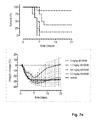
- FIG. 7 shows the in vivo efficacy of SD1038 and MD1212 against a lethal challenge with A/Hong Kong/1/1968-MA (H3N2) virus. Survival curves (left) and weight loss (right) of mice treated with 5, 1.7, 0.6 or 0.2 mg/kg single or multi-domain antibody one day before challenge (at day 0) are shown.
- FIG. 8 shows the in vivo efficacy of MD1221, MD1222 and MD1224 against a lethal challenge with B/Florida/4/2006 virus. Survival curves (left) and weight loss (right) of mice treated with 5 or 0.5 mg/kg multi-domain antibody one day before challenge (at day 0) are shown.
- FIG. 9 shows the in vivo efficacy of MD1301, MD2601 and CR9114 against a lethal challenge with A/Puerto Rico/8/1934-MA (H1N1) virus. Survival curves (left) and weight loss (right) of mice treated with 3 mg/kg (multi-domain) antibody one day before challenge (at day 0) are shown.
- FIG. 10 shows the in vivo efficacy of MD1301, MD2601 and CR9114 against a lethal challenge with A/Puerto Rico/8/1934-MA (H1N1) virus. Survival curves (left) and weight loss (right) of mice treated with 0.2, 0.05 or 0.01 mg/kg (multi-domain) antibody one day before challenge (at day 0) are shown.
- FIG. 11 shows the in vivo efficacy of MD2617 against a lethal challenge with A/Puerto Rico/8/1934-MA (H1N1) virus. Survival curves (left) and weight loss (right) of mice treated intranasally (TOP) or intravenously (BOTTOM) with MD2617 one day before challenge (at day 0) are shown.
- TOP intranasally
- BOTTOM intravenously
- FIG. 12 shows the in vivo efficacy of MD2617 and CR9114 against a lethal challenge with B/Florida/4/2006 (TOP) or A/Hong Kong/1/1968-MA (BOTTOM) virus. Survival curves (left) and weight loss (right) of mice treated intranasally or intravenously with (multi-domain) antibody one day before challenge (at day 0) are shown.
- FIG. 13 shows the in vivo efficacy of MD2407, MD3606 and CR9114 against a lethal challenge with B/Florida/4/2006 virus. Survival curves (left) and weight loss (right) of mice treated with 0.02, 0.1 or 0.5 mg/kg (multi-domain) antibody one day before challenge (at day 0) are shown.
- FIG. 14 shows the in vivo efficacy of MD3606 and CR9114 against a lethal challenge with B/Florida/4/2006 virus. Survival curves (left) and weight loss (right) of mice treated with 0.2, 1 or 5 mg/kg (multi-domain) antibody one day before challenge (at day 0) are shown.
- FIG. 15 shows the in vivo efficacy of MD2407, MD3606 and CR9114 against a lethal challenge with B/Florida/4/2006 virus. Survival curves (left) and weight loss (right) of mice treated with 0.02, 0.1 or 0.5 mg/kg (multi-domain) antibody one day before challenge (at day 0) are shown.
- FIG. 16 shows the in vivo efficacy of MD3606 and CR9114 against a lethal challenge with A/Hong Kong/1/1968-MA (H3N2) virus. Survival curves (left) and weight loss (right) of mice treated with 0.6, 1.7 or 5 mg/kg (multi-domain) antibody one day before challenge (at day 0) are shown.
- FIG. 17 shows the in vivo efficacy of MD2407, MD3606 and CR9114 against a lethal challenge with A/Puerto Rico/8/1934-MA (H1N1) virus. Survival curves (left) and weight loss (right) of mice treated with 0.01, 0.05 or 0.25 mg/kg (multi-domain) antibody one day before challenge (at day 0) are shown.
- FIG. 18 shows the in vivo efficacy of MD3606 and CR9114 against a lethal challenge with A/Puerto Rico/8/1934-MA (H1N1) virus. Survival curves (left) and weight loss (right) of mice treated with 0.6, 1.7 or 5 mg/kg (single domain) antibody one day before challenge (at day 0) are shown.
- binding molecule refers to both single domain antibodies (monomeric binding molecules) and multi-domain antibodies (multimeric binding molecules) according to the invention.
- a single-domain antibody is a binding molecule consisting of a single monomeric variable antibody domain that specifically binds an antigen or epitope independently of other V regions or domains.
- Single domain antibodies are known in the art and are usually derived from naturally occurring “heavy chain only” antibodies, i.e. heavy chain antibodies devoid of light chains. Such heavy chain only antibodies can be obtained from Camelidae species, for example in camel, llama, dromedary, or alpaca (also referred to as camelid antibodies).
- the variable region derived from said heavy chain only antibody is generally known as a VHH domain or single domain antibody (sdAb).
- a single-domain antibody as used herein also refers to an isolated single variable domain (VL or VH) from a conventional immunoglobulin comprising two heavy chains and two light chains.
- This immunoglobulin is preferably human, but may also comprise immunoglobulins from other mammalian species including rodents.
- multi-domain antibody refers to a binding molecule comprising at least two single domain antibodies, linked to each other either directly or by a linking sequence.
- influenza virus subtype in relation to influenza A viruses refers to influenza A virus strains that are characterized by various combinations of the hemagglutinin (H) and neuraminidase (N) viral surface proteins.
- Influenza A virus subtypes may be referred to by their H number, such as for example “influenza virus comprising HA of the H1 or H3 subtype”, or “H1 influenza virus” “H3 influenza virus”, or by a combination of an H number and an N number, such as for example “influenza virus subtype “H3N2” or “H5N1”.
- influenza virus “subtype” specifically includes all individual influenza virus “strains” within such subtype, which usually result from mutations and show different pathogenic profiles, and include natural isolates as well as man-made mutants or reassortants and the like. Such strains may also be referred to as various “isolates” of a viral subtype. Accordingly, as used herein, the terms “strains” and “isolates” may be used interchangeably.
- influenza A virus subtypes can further be classified by reference to their phylogenetic group.
- Phylogenetic analysis has demonstrated a subdivision of influenza hemagglutinins into two main groups: inter alia the H1, H2, H5 and H9 subtypes in phylogenetic group 1 (“group 1” influenza viruses) and inter alia the H3, H4, H7 and H10 subtypes in phylogenetic group 2 (“group 2” influenza viruses).
- influenza B virus strains Two genetically and antigenically distinct subtypes, or “lineages”, of influenza B virus are circulating in humans, as represented by the B/Yamagata/16/88 (also referred to as B/Yamagata) and B/Victoria/2/87 (B/Victoria) lineages.
- B/Yamagata/16/88 also referred to as B/Yamagata
- B/Victoria/2/87 B/Victoria lineages.
- influenza B virus strains are referred to as influenza virus strains derived from the “the B/Yamagata lineage” or the “B/Victoria lineage”.
- neutralizing refers to binding molecules that inhibit an influenza virus from replication, in vitro and/or in vivo within a subject, regardless of the mechanism by which neutralization is achieved.
- neutralization can e.g. be achieved by inhibiting the attachment or adhesion of the virus to the cell surface, or by inhibition of the fusion of viral and cellular membranes following attachment of the virus to the target cell, or by inhibiting viral egress from infected cells, and the like.
- cross-neutralizing” or “cross-neutralization” as used herein in relation to the binding molecules of the invention refers to the ability of the binding molecules of the invention to neutralize different subtypes of influenza A and/or B viruses.
- the term “(immuno)specifically binding” refers to binding molecules that bind to an epitopes of a protein of interest, but which do not substantially recognize and bind other molecules in a sample containing a mixture of antigenic biological molecules.
- the binding may be mediated by covalent or non-covalent interactions or a combination of both.
- influenza refers to the pathological condition resulting from an infection of a cell or a subject by an influenza A or B virus. In specific embodiments, the term refers to a respiratory illness caused by an influenza A or B virus.
- influenza virus infection means the invasion by, multiplication and/or presence of an influenza virus in a cell or a subject.
- novel single domain antibodies capable of specifically binding to hemagglutinin (HA) of at least two influenza A virus strains comprising HA of two different subtypes from phylogenetic group 2 are provided, i.e. sdAbs capable of specifically binding to hemagglutinin (HA) of at least two different influenza A virus strains, said strains comprising HA from two different HA subtypes from phylogenetic group 2.
- sdAbs that are capable of binding to HA of at least one influenza A virus strain from phylogenetic group 1 and to HA of at least one influenza A virus strain from phylogenetic group 2 are provided.
- sdAbs capable of specifically binding to HA of at least one influenza B virus strain are provided.
- Single domain antibodies that are capable of specifically binding to HA of two different subtypes of influenza A virus strains from phylogenetic group 2, or capable from binding to HA of influenza A virus strains from both phylogenetic group 1 (such as influenza viruses comprising HA of the H1, H2, and/or H5 subtype) and phylogenetic group 2 (such as influenza viruses comprising HA of the H3, H7 and/or H10 subtype) have not been described before.
- sdAbs that are capable of specifically binding to HA of influenza B viruses have also not yet been described.
- the sdAbs of the invention bind to conserved neutralizing epitopes in HA.
- the sdAbs bind to an epitope in the stem region of the HA protein of an influenza A or B virus.
- the sdAbs bind to an epitope in the head region of the HA protein.
- the sdAb binds to an epitope in the head region of the HA protein of an influenza B viruses.
- the sdAbs are also capable of neutralizing at least two influenza A virus strains comprising HA of two different subtypes from phylogenetic group 2.
- the sdAbs are capable of neutralizing preferably at least one influenza A virus strain from phylogenetic group 1 (such as e.g. an influenza virus comprising HA of the H1 or H5 subtype) and at least one influenza A virus strain from phylogenetic group 2 (such as e.g. an influenza virus comprising HA of the H3 or H7 subtype); or at least one influenza B virus strain, preferably at least one influenza B virus strain from the B/Yamagata lineage and at least one influenza virus strain from the B/Victoria lineage.
- the single domain antibody according to the invention is a Camelid VHH domain, i.e. a variable domain of a so-called Camelid (heavy chain only) antibody.
- the single domain antibody is a humanized Camelid VHH domain. Humanization of Camelid single domain antibodies requires the introduction and mutagenesis of a limited amount of amino acids in a single polypeptide chain. This is in contrast to humanization of scFv, Fab, (Fab) 2 and IgG, which requires the introduction of amino acid changes in two chains, the light and the heavy chain, and the preservation of the assembly of both chains.
- a single domain antibody of the invention comprises:
- CDR complementarity determining regions
- the single domain antibody is selected from the group consisting of:
- the single domain antibody is selected from the group consisting of:
- a single domain antibody according to the invention comprises an amino acid sequence selected from the group consisting of SEQ ID NO: 1-29, or a homologous amino acid sequence.
- a homologous amino acid sequence of the present invention may comprise additions, deletions or substitutions of one or more amino acids, which do not substantially alter the functional characteristics of the binding molecules of the invention.
- homologous sequence indicates sequence identity, it means a sequence which presents a high sequence identity (more than 70%, 75%, 80%, 85%, 90%, 95% or 98% sequence identity) with the parent sequence.
- one or more amino acids in amino acid sequences described herein may be mutated, i.e. substituted by another amino acid. Such mutations may be introduced to prevent the occurrence of post-translational modifications.
- the most prevalent modifications include proteolysis, glycosylation, oxidation of methionine, and deamidation of asparagine and glutamine residues.
- Other modifications include pyroglutamate formation, aspartate isomerization and tryptophan oxidation.
- amino acid residues and sequence motifs are susceptible to post-translational modification and may therefore be altered by site directed mutagenesis: N-terminal glutamic acid or glutamine, N-glycosylation motif Asn-Xxx-Ser/Thr, solvent exposed methionine or tryptophan residues, proteolytic cleavage site Asp-Pro, deamidation motifs Asn-Gly and Gln-Gly and/or Asp isomerization motif Asp-Gly.
- a sdAb of the invention is humanized.
- the sdAbs comprise an amino acid sequence selected from the group consisting of SEQ ID NO: 146-226 and 340.
- the single domain antibody according to the invention comprises an amino acid sequence selected from the group consisting of SEQ ID NO: 13, or a humanized variant thereof selected from the group consisting of SEQ ID NO: 177-187 and SEQ ID NO: 340; SEQ ID NO: 17 or a humanized variant thereof selected from the group consisting of SEQ ID NO: 146-156; SEQ ID NO: 20 or a humanized variant thereof selected from the group consisting of SEQ ID NO: 157-176; SEQ ID NO: 24 or a humanized variant thereof selected from the group consisting of SEQ ID NO: 188-197; SEQ ID NO: 25 or a humanized variant thereof selected from the group consisting of SEQ ID NO: 198-203; and SEQ ID NO: 27 or a humanized variant thereof selected from the group consisting of SEQ ID NO: 204-226.
- the single domain antibody comprises an amino acid sequence selected from the group consisting of: SEQ ID NO: 187, SEQ ID NO: 340, SEQ ID NO: 155, SEQ ID NO: 176, SEQ ID NO: 197, SEQ ID NO: 203 and SEQ ID NO: 221.
- multi-domain antibodies i.e. binding molecules comprising at least two single domain antibodies as described above.
- the C-terminal end of a first single domain antibody may be linked to the N-terminal end of a next single domain antibody to form a dimeric binding molecule.
- the multi-domain antibodies comprise at least three, at least four, or at least five single domain antibodies as described above to form a multimer, such as a trimer, tetramer, pentamer, etc.
- the linked sdAbs can be the same or can be different sdAbs, i.e. sdAbs having different amino acid sequences and epitope specificities.
- the multi-domain antibodies are single chain molecules. In certain embodiments, the multi-domain antibodies are two-chain molecules, i.e. comprise at least two chains each comprising at least one single-domain antibody. The two chains may be identical or may be different.
- the single domain antibodies may be linked to form any of the multi-domain antibodies disclosed herein using any methods known in the art.
- the single domain antibodies may be linked by chemical linkage, or may be linked together either directly or by short polypeptide linkers.
- Such linker sequence may be a naturally occurring sequence or a non-naturally occurring sequence.
- the linker sequence preferably provides sufficient flexibility to the multi-domain antibody and at the same time is resistant to proteolytic degradation.
- the at least two single domain antibodies are genetically fused via peptide linkers.
- the single domain antibodies are fused genetically at the DNA level, by forming a polynucleotide construct (or nucleic acid sequence) encoding the complete polypeptide construct, i.e. the binding molecule comprising the two or more single domain antibodies.
- the at least two single domain antibodies are linked by a linking sequence comprising from 1 to 100 amino acids, preferably from 1 to 80 amino acids, or from 1 to 60 amino acids, or from 10 to 60 amino acids.
- linkers include, but are not limited to, the linking sequences in Table 15.
- the linking sequence comprises an amino acid sequence selected from SEQ ID NO: 142-145.
- the multi-domain antibodies comprise at least two sdAbs according to the present invention.
- the at least two sdAbs may be selected from Table 14 and/or Table 40.
- the at least two sdAbs are selected from the group consisting of SEQ ID NO: 1-29 and SEQ ID NO: 146-226.
- the at least two sdAbs may be the same (homo-multimer) or may be different (hetero-multimer).
- the multi-domain antibodies comprise at least two, preferably at least three, more preferably at least four sdAbs selected from the group consisting of
- the multi-domain antibodies according to the invention comprise at least two, preferably at least three, more preferably at least four sdAbs selected from the group consisting of: SEQ ID NO: 13, or a humanized variant thereof selected from the group consisting of SEQ ID NO: 177-187 and SEQ ID NO: 340; SEQ ID NO: 17 or a humanized variant thereof selected from the group consisting of SEQ ID NO: 146-156; SEQ ID NO: 20 or a humanized variant thereof selected from the group consisting of SEQ ID NO: 157-176; SEQ ID NO: 24 or a humanized variant thereof selected from the group consisting of SEQ ID NO: 188-177; SEQ ID NO: 25 or a humanized variant thereof selected from the group consisting of SEQ ID NO: 198-203; and SEQ ID NO: 27 or a humanized variant thereof selected from the group consisting of SEQ ID NO: 204-226.
- the multi-domain antibodies according to the invention comprise an amino acid sequence selected from the group consisting of: SEQ ID NO: 30-73.
- the multi-domain antibodies of the invention are capable of neutralizing at least one influenza A virus strain from phylogenetic group 1 (such as e.g. an influenza virus comprising HA of the H1 and/or H5 subtype) and at least one influenza A virus strain from phylogenetic group 2 (such as e.g. an influenza virus comprising HA of the H3 and/or H7 subtype).
- the multi-domain antibodies of the invention are capable of neutralizing at least one influenza A virus strain from phylogenetic group 1 (such as e.g. an influenza virus comprising HA of the H1 and/or H5 subtype) and at least one influenza A virus strain from phylogenetic group 2 (such as e.g.
- an influenza virus comprising HA of the H3 and/or H7 subtype) and at least one influenza B virus strain, preferably at least one influenza B virus strain of the B/Yamagata lineage and at least one influenza virus strain of the B/Victoria lineage.
- the multi-domain antibodies are capable of neutralizing influenza viruses comprising HA of the H1 subtype (such as H1N1 influenza virus strains), influenza viruses comprising HA of the H3 subtype (such as H3N2 influenza virus strains), influenza viruses comprising HA of the H5 subtype (such as H5N1 influenza virus strains), influenza viruses comprising HA of the H7 subtype (such as H7N9 influenza virus strains), and at least one influenza B virus, preferably at least one influenza B virus strain from the B/Yamagata lineage and at least one influenza virus strain from the B/Victoria lineage.
- the multi-domain antibodies of the present invention thus can suitably be used in the prevention and/or treatment of influenza infections, even irrespective of the causative influenza subtype.
- the cross-neutralizing multi-domain antibodies of the invention offer several advantages relative to other small and large anti-influenza molecules.
- the affinity and potency of the multi-domain antibodies, as well as the breadth of neutralization are superior to the affinity, potency and breadth of neutralization of the published broadly neutralizing Abs (bnAbs) targeting influenza HA, like e.g. CR9114 (WO2013/007770) and FI6v3 (Corti et al., 2011).
- the multi-domain antibodies by targeting multiple independent neutralizing epitopes are less prone to the development of drug resistant influenza strains than CR9114 and FI6v3.
- the binding molecules may be monomeric, i.e. be single domain antibodies, or multimeric, i.e. multi-domain antibodies.
- the binding molecules of the invention bind to their target with high affinity and specificity. This is in contrast with small molecule drugs like antivirals which frequently show off-target binding resulting in unwanted side effects.
- the binding molecules of the invention bind to a variety of HA epitopes, some of which are inaccessible to conventional antibodies.
- Influenza HA contains multiple glycosylation sites both in the head and stem region. Carbohydrates attached to these sites render some parts on the HA molecule inaccessible to conventional antibodies.
- binding molecules of the invention are still able to target these potentially functionally important epitopes.
- the binding molecules of the invention are stable under a wide range of extreme conditions. They are typically resistant to elevated temperatures (up to 100° C.), extremes in pH, denaturing agents and proteolytic degradation. The favorable stability of the binding molecules may yield products that can be kept outside of the cold chain and that have longer shelf-lives than other protein drugs like monoclonal antibodies.
- the binding molecules of the invention are all single proteins which can be produced and purified following one single process.
- the binding molecules according to the invention bind to HA of an influenza A virus of group 1 (such as H1N1) and/or an influenza A virus of group 2 (such as H3N2), and/or an influenza B virus, and/or fragments thereof, with an affinity constant (Kd-value) that is lower than 1.0 ⁇ 10 ⁇ 6 M, 1.0 ⁇ 10 31 7 M, preferably lower than 1.0 ⁇ 10 ⁇ 8 M, more preferably lower than 1.0 ⁇ 10 ⁇ 9 M.
- Kd-value affinity constant
- Affinity constants can for instance be measured using surface plasmon resonance, for example using the BIACORE system (Pharmacia Biosensor AB, Uppsala, Sweden), or as described in Example 8.
- the binding molecules exhibit neutralization activity against influenza A and/or B viruses.
- the binding molecules of the invention prevent an influenza A or B virus from infecting host cells by at least 99%, at least 95%, at least 90%, at least 85%, at least 80%, at least 75%, at least 70%, at least 60%, at least 50%, at least 45%, at least 40%, at least 45%, at least 35%, at least 30%, at least 25%, at least 20%, or at least 10% relative to infection of host cells by said influenza virus in the absence of said binding molecules.
- Neutralizing activity can for instance be measured as described herein.
- the binding molecules according to the invention have a neutralizing activity of 1000 nM or less, preferably 100 nM or less, more preferably a neutralizing activity of 10 nM or less, even more preferably 1nM or less, as determined in an in vitro virus neutralization assay (VNA), e.g. as described in the Examples.
- VNA virus neutralization assay
- the binding molecules i.e. the single domain antibodies or multi-domain antibodies
- the binding molecules further comprise an Fc tail.
- the binding molecules, as described above are linked to an Fc fragment of an antibody, preferably a human antibody, such as the Fc fragment of a human IgG antibody, e.g an IgG1, IgG2, IgG3, IgG4, or IgG4.
- the monomeric or multimeric binding molecules, as described herein may be genetically fused to an Fc fragment, either directly or using a linker.
- the binding molecules are linked to the Fc fragment by a linking sequence comprising from 1 to 100 amino acids, preferably from 1 to 80 amino acids, or from 1 to 60 amino acids, or from 10 to 60 amino acids.
- linkers include, but are not limited to, the linking sequences in Table 15.
- the linking sequence comprises an amino acid sequence selected from SEQ ID NO: 142-145.
- a sdAb or multi-domain antibody is genetically fused to the C-terminus of an Fc fragment.
- a single domain antibody or multi-domain antibody is fused to both the N- and the C-terminus of an Fc fragment.
- the Fc fragment is engineered to have minimal effector functions.
- Fc fragments with minimal effector function and conserved half-life have been described in the art and include e.g. IgG2, aglycosylated IgG1 (IgG1 agly), IgG4 with S228P/L234A/L235A substitutions (IgG4 ProAlaAla), IgG2 with H268Q/309L/A330S/P331S changes (IgG2m4) and an Fc variant of IgG2, designated as IgG2 ⁇ , containing V234A/G237A/P238S/H268A/V309L/A3305/P331S substitutions.
- IgG2 ⁇ aglycosylated IgG1
- IgG4 ProAlaAla IgG2 with H268Q/309L/A330S/P331S changes
- IgG2m4 an Fc variant of IgG2, designated
- the Fc fragment is engineered to have enhanced effector functions.
- the binding molecules of the invention thus can be engineered to enhance Fc-mediated effector functions, which in preclinical models of influenza infection have been shown to contribute to drug efficacy.
- Several mutations in the CH2 domain of human IgG1 associated with enhanced effector function have been described in the art.
- mutations include, but are not limited to, alanine mutant at position 333 which increases both ADCC and CDC, a triple mutant (S239D/I332E/A330L) with higher affinity for Fc ⁇ RIIIa and lower affinity for Fc ⁇ RIIb resulting in enhanced ADCC, and another triple mutant (S239D/I332E/G236A) with improved Fc ⁇ RIIIa affinity and Fc ⁇ RIIa/Fc ⁇ RIIb ratio that mediates enhanced phagocytosis.
- Other Fc mutations affecting effector functionality have been described in literature e.g. Strohl, 2009.
- the Fc fragment is engineered to have an extended serum half-life.
- Fc backbones with increased serum half-life are known in the art. These Fc variants include, but are not limited to, hIgG1 Fc with M252Y/S254T/T256E (YTE) mutations, hIgG1 or hIgG2 Fc carrying T250Q/M428L mutations (QL), hIgG1 Fc with N434A mutation, hIgG1 Fc with T307A/E380A/N434A mutations (AAA) or hIgG1 Fc with M428L/N434S (LS) substitutions (Kuo et al., 2011).
- the binding molecules i.e.
- the single domain antibodies or the multi-domain antibodies according to the invention are genetically fused to human serum albumin or a single domain antibody binding to serum albumin.
- the single domain antibodies or the multi-domain antibodies according to the invention are chemically conjugated to PEG.
- the binding molecules of the invention thus can be engineered to have serum half-lives ranging from e.g. just a few hours to several weeks or even months. This opens the possibility for single dose treatment of influenza infection instead of a 2 ⁇ daily regimen as currently used form the neuraminidase inhibitors, such as oseltamivir and zanamivir.
- the single domain antibody or multi-domain antibody as described above may be fused to an Fc tail, preferably an Fc tail which is engineered to promote the formation of a hetero-dimeric Fc molecule.
- Fc tail preferably an Fc tail which is engineered to promote the formation of a hetero-dimeric Fc molecule.
- Mutations promoting Fc-heterodimerization have been described in the art (Klein et al., 2012).
- the mutations promoting the FC-heterodimerization are the knobs-into-holes mutations as described in EP0812357B1 and EP0979281B1.
- the multi-domain antibodies according to the present invention comprise at least one chain comprising an amino acid sequence selected from the group consisting of SEQ ID NO: 74-107, SEQ ID NO: 110-121 and SEQ ID NO: 293-339.
- the multi-domain antibodies comprise two chains, wherein the amino acid sequence of the two chains is identical.
- the two chains comprise an amino acid sequence selected from the group consisting of SEQ ID NO: 74-105 and SEQ ID NO: 293-298.
- the two amino acid chains comprise an amino acid sequence of SEQ ID NO: 293-298.
- the multi-domain antibodies comprise two different amino acid chains.
- the two different amino acid chains are selected from the group consisting of:
- the two different amino acid chains are selected from the group consisting of:
- the present invention further provides nucleic acid molecules (also referred to as nucleic acid sequences) encoding the single domain antibodies or multi-domain antibodies as described above.
- nucleic acid sequences encode binding molecules comprising one or more of the CDR regions as described above.
- the nucleic acid sequences encode a single domain antibody comprising an amino acid sequence selected from the group consisting of SEQ ID NO: 1-29, or a homologous amino acid sequence.
- the nucleic acid sequences encode a single domain antibody comprising an amino acid sequence selected from the group consisting of SEQ ID NO: 146-226 and SEQ ID NO: 340, or a homologous amino acid sequence.
- the nucleic acid sequences encode a multi-domain antibody comprising an amino acid sequence selected from the group consisting of SEQ ID NO: 30-107, SEQ ID NO: 110-121 and SEQ ID NO: 293-339, or a homologous amino acid sequence.
- a nucleic acid sequence according to the invention refers to a polymeric form of nucleotides and includes RNA, mRNA, cDNA, genomic DNA, and synthetic forms and mixed polymers of the above.
- a nucleotide refers to a ribonucleotide, deoxynucleotide or a modified form of either type of nucleotide. The term also includes single- and double-stranded forms of DNA.
- functional variants of these nucleic acid molecules are also intended to be a part of the present invention. Functional variants are nucleic acid sequences that can be directly translated, using the standard genetic code, to provide an amino acid sequence identical to that translated from the parental nucleic acid molecules.
- the nucleic acid molecules encoding the binding molecules according to the invention are codon-optimized for expression in yeast cells or mammalian cells, such as human cells. Methods of codon-optimization are known and have been described previously (e.g. WO 96/09378).
- a sequence is considered codon-optimized if at least one non-preferred codon as compared to a wild type sequence is replaced by a codon that is more preferred.
- a non-preferred codon is a codon that is used less frequently in an organism than another codon coding for the same amino acid, and a codon that is more preferred is a codon that is used more frequently in an organism than a non-preferred codon.
- the frequency of codon usage for a specific organism can be found in codon frequency tables, such as in http://www.kazusa.or.jp/codon.
- nucleic acid sequence encoding an amino acid sequence includes all nucleotide sequences that are degenerate versions of each other and that encode the same amino acid sequence.
- Nucleic acid sequences can be cloned using routine molecular biology techniques, or generated de novo by DNA synthesis, which can be performed using routine procedures by service companies having business in the field of DNA synthesis and/or molecular cloning (e.g. GeneArt, GenScript, Life Technologies, Eurofins).
- the invention also provides vectors comprising at least one nucleic acid sequence as described above.
- vector refers to a nucleic acid molecule into which a second nucleic acid molecule can be inserted for introduction into a host cell where it will be replicated, and in some cases expressed. In other words, a vector is capable of transporting a nucleic acid molecule to which it has been linked.
- Cloning vectors as well as expression vectors are contemplated by the term “vector”, as used herein.
- Certain vectors are capable of autonomous replication in a host into which they are introduced (e.g., vectors having a bacterial origin of replication can replicate in bacteria). Other vectors can be integrated into the genome of a host upon introduction into the host cell, and thereby are replicated along with the host genome. Vectors according to the invention can easily be made by methods well known to the person skilled in the art.
- vectors comprising one or more nucleic acid molecules according to the invention operably linked to one or more expression-regulating nucleic acid sequences are provided.
- expression-regulating nucleic acid sequence refers to nucleic acid sequences necessary for and/or affecting the expression of an operably linked coding sequence in a particular host organism.
- the expression-regulating nucleic acid sequences such as inter alia appropriate transcription initiation, termination, promoter, enhancer sequences; repressor or activator sequences; efficient RNA processing signals such as splicing and polyadenylation signals; sequences that stabilize cytoplasmic mRNA; sequences that enhance translation efficiency (e.g., ribosome binding sites); sequences that enhance protein stability; and when desired, sequences that enhance protein secretion, can be any nucleic acid sequence showing activity in the host organism of choice and can be derived from genes encoding proteins, which are either homologous or heterologous to the host organism.
- Suitable vectors according to the invention are e.g.
- adenovectors such as e.g. Ad26 or Ad35, adenoassociated vectors (AAV), lentivirus, alphavirus, paramyxovirus, vaccinia virus, herpes virus, retroviral vectors etc.
- the vectors are used for gene therapy purposes, as for example described by Adam et al. (2014), Johnson et al. (2009) and Suscovich and Alter (2015).
- the invention further provides host cells comprising a nucleic acid sequence encoding a single domain antibody or a multi-domain antibody as described herein.
- “Host cells”, as used herein, refers to cells into which a vector such as a cloning vector or an expression vector has been introduced.
- the host cells can be prokaryotic or eukaryotic.
- the present invention further provides pharmaceutical compositions comprising one or more single domain antibodies, multi-domain antibodies, nucleic acid molecules and/or vectors as described above.
- the pharmaceutical compositions of the invention may further comprise at least one pharmaceutically acceptable excipient.
- pharmaceutically acceptable excipient is meant any inert substance that is combined with an active molecule such as a binding molecule according to the invention for preparing a suitable composition.
- the pharmaceutically acceptable excipient is an excipient that is non-toxic to recipients at the used dosages and concentrations, and is compatible with other ingredients of the formulation. Pharmaceutically acceptable excipients are widely applied and known in the art.
- the pharmaceutical composition according to the invention may further comprise at least one other therapeutic, prophylactic and/or diagnostic agent.
- Said further therapeutic and/or prophylactic agents may for example be agents that are also capable of preventing and/or treating an influenza virus infection, such as for example M2 inhibitors (e.g., amantidine, rimantadine) and/or neuraminidase inhibitors (e.g., zanamivir, oseltamivir). These can be used in combination with the binding molecules of the invention. “In combination” herein means simultaneously, as separate formulations, or as one single combined formulation, or according to a sequential administration regimen as separate formulations, in any order.
- M2 inhibitors e.g., amantidine, rimantadine
- neuraminidase inhibitors e.g., zanamivir, oseltamivir
- the present invention provides single domain antibodies, multi-domain antibodies, nucleic acid molecules, and/or vectors as described herein for use in the diagnosis, prevention and/or treatment of an influenza infection.
- the invention furthermore provides the use of the single domain antibodies, multi-domain antibodies, nucleic acid molecules, and/or vectors as described herein in the manufacture of a medicament for the diagnosis, prevention and/or treatment of an influenza infection.
- Such infections can occur in small populations, but can also spread around the world in seasonal epidemics or, worse, in global pandemics where millions of individuals are at risk.
- the invention provides binding molecules that can neutralize the infection of influenza strains that cause such seasonal epidemics, as well as potential pandemics.
- the binding molecules of the present invention are capable of cross-neutralizing various influenza subtypes of both phylogenetic group 1, encompassing e.g. H1, H2, H5, H6, H8, H9 and H11 subtypes, and phylogenetic group 2, encompassing e.g. H3, H4, H7 and H10 subtypes, as well as influenza B subtypes.
- the invention further provides methods for preventing and/or treating influenza in a subject, comprising administering a therapeutically effective amount of a single domain antibody, multi-domain antibody, nucleic acid molecule, and/or vector as described herein to a subject in need thereof.
- therapeutically effective amount refers to an amount of the binding molecule or nucleic acid molecule as defined herein that is effective for preventing, ameliorating and/or treating a condition resulting from infection with an influenza virus.
- Ameloriation as used in herein may refer to the reduction of visible or perceptible disease symptoms, viremia, or any other measurable manifestation of influenza infection.
- Prevention encompasses inhibiting or reducing the spread of influenza virus or inhibiting or reducing the onset, development or progression of one or more of the symptoms.
- Prevention and/or treatment may be targeted at patient groups that are susceptible to influenza infection.
- patient groups include, but are not limited to e.g., the elderly (e.g. ⁇ 50 years old, ⁇ 60 years old, and preferably ⁇ 65 years old), the young (e.g. ⁇ 5 years old, ⁇ 1 year old), hospitalized patients and already infected patients who have been treated with an antiviral compound but have shown an inadequate antiviral response.
- Dosage regimens can be adjusted to provide the optimum desired response (e.g., a therapeutic response).
- a suitable dosage range may for instance be 0.01-100 mg/kg body weight, preferably 0.1-50 mg/kg body weight, preferably 0.01-15 mg/kg body weight.
- a single bolus may be administered, several doses may be administered over time, or the dose may be proportionally reduced or increased as deemed necessary.
- binding molecules, nucleic acid molecules and/or vectors according to the invention may be administered to a subject for example intravenously, intranasally, via oral inhalation, pulmonary, subcutaneously, intradermally, intravitreally, orally, intramuscularly etc.
- the optimal route of administration will be influenced by several factors including the physicochemical properties of the active molecules, the urgency of the clinical situation and the relationship of the plasma concentrations of the active molecules to the desired therapeutic effect.
- binding molecules of the invention opens up the possibility for alternative, needle-free delivery, such as by intranasal administration using nose drops or nasal spray or via inhalation using a nebulizer or dry-powder inhaler.
- a nucleic acid molecule or vector encoding at least one single or multi-domain antibody according to the invention thus is administered intranasally, as described for example by Limberis et al. (2013).
- the binding molecules of the invention can be produced very efficiently in microbial systems.
- microbial host cells used in large-scale manufacturing are for example yeast ( P. pastoris ) and E. coli . These microbial systems are considered as the most cost-effective option for biopharmaceutical manufacturing.
- Low COGs is a prerequisite for broad use of anti-flu drugs in influenza prophylaxis and treatment.
- the present invention thus provides methods for producing the binding molecules (i.e. the single domain antibodies or multi-domain antibodies) according to the invention, comprising culturing a host cell as described herein under conditions conducive to the expression of the binding molecule, and optionally, recovering the expressed binding molecule. Methods to recover the binding molecules from culture media are well known to the man skilled in the art.
- the host cells are microbial cells, such as, but not limited to, yeast cells or E.coli.
- the host cells are mammalian cells, such as, but not limited to, CHO cells, HEK cells or PER.C6 cells.
- the present invention further pertains to a method of detecting an influenza virus in a sample, wherein the method comprises the steps of a) contacting said sample with a diagnostically effective amount of a binding molecule according to the invention, and b) determining whether the binding molecule specifically binds to a molecule in the sample.
- the sample may be a biological sample including, but not limited to blood, serum, tissue or other biological material from (potentially) infected subjects.
- the (potentially) infected subjects may be human subjects, but also animals that are suspected as carriers of influenza virus might be tested for the presence of influenza virus using the binding molecules of the invention.
- the binding molecules of the invention are contacted with the sample under conditions which allow the formation of an immunological complex between the binding molecules and the influenza virus or antigenic components thereof that may be present in the sample.
- the formation of an immunological complex if any, indicating the presence of influenza virus in the sample, can then detected and measured by suitable means.
- suitable means include, inter alia, homogeneous and heterogeneous binding immunoassays, such as radioimmunoassays (RIA), ELISA, immunofluorescence, immunohistochemistry, FACS, BIACORE and Western blot analyses.
- Lama glama With the aim to induce a heavy-chain antibody dependent immune response, four llamas ( Lama glama ) were immunized with influenza virus antigens (commercial vaccine Inflexal® and recombinant protein) in the presence of Freund's adjuvant according to the scheme described in Table 1.
- influenza virus antigens commercial vaccine Inflexal® and recombinant protein
- Peripheral blood was collected from the Llamas by venipuncture in citrate anti-coagulation sample tubes at the indicated time points after the 2 nd , 3 th , 4 th , and 5 th immunization (Table 1).
- the homologues and heterologous immune response in each animal was analyzed by comparing the antigen specific serum titers of a sample collected prior to immunization (day 0) and a serum sample collected after antigen administrations (day 28 and day 112) in an HA ELISA.
- recombinant HA protein was captured in a Maxisorp 96-well microtiter plates. After blocking, serial dilutions of serum samples were added, and bound llama IgG was detected by addition of goat anti-llama IgG-HRP. Results are shown in FIG. 1 . These data show that all immunized animals generated a good homologous and heterologous immune response against HA.
- PBMC Peripheral Blood Mononuclear Cells
- PBMC Peripheral Blood Mononuclear Cells
- GE Healthcare Ficoll-Paque plus
- Total RNA extracted from PBMC served as starting material for RT-PCR to amplify the VHH encoding gene fragments.
- These fragments were cloned via SfiI and NotI restriction sites into M13 phagemid vector pDV-LucStuffer (pDV-C06 derived; as described in WO 02/103012) to create a fusion of VHH domain with the pIII protein of the M13 phage (including an AMBER stop codon between the two proteins).
- Ligated vectors were transformed into TG-1 bacteria (Agilent) and 100-150 single colonies where analysed via PCR to determine the quality of each library. Insert frequency and completeness were typically more than 95%.
- Table 2 Phage libraries from individual animals were prepared by using CT helper phages essentially as described (WO 02/103012), sterile filtered, and used for selections. As shown herein, complex phage libraries could be generated from all immunized llamas.
- VHH phage libraries % # # intact intact of which CDR3 length Complexity clones ORF ORF % unique amino acids Cfu L01 104 100 96 98 5-21 AA 6.8E+06 L02 117 114 97 95 5-27 AA 1.4E+07 L03 111 109 98 98 6-28 AA 1.7E+07 L04 111 109 98 96 5-23 AA 1.6E+07
- Antibody fragments were selected using the VHH phage display libraries described above and general phage display technology and MABSTRACT® technology essentially as described in U.S. Pat. No. 6,265,150, in WO 98/15833, and in “Phage display, A Laboratory Manual” by T. Kuhlman, 2001 (which are incorporated by reference herein). Furthermore, the methods and helper phages as described in WO 02/103012 (which is incorporated by reference herein) were used in the present invention.
- binders were performed with hemagglutinin (HA) of influenza A (H1 A/California/07/2009, H1 A/New Caledonia/20/1999, H2 A/Japan/305/1957, H3 A/Brisbane/10/2007, H7 A/Netherlands/219/2003) and/or influenza B (Victoria clade B/Brisbane/60/2008, Yamagata clade B/Florida/04/2006) as target protein.
- the target protein source was either insect cell produced recombinant protein (Protein Sciences, Connecticut, USA) or HA expressed on the surface of influenza infected and fixed (3% paraformaldehyde) MDCK cells.
- SD refers to single domain antibody. If not mentioned otherwise, selections were performed at pH 7.4 and with 5 ⁇ g/ml HA protein.
- CR8033 and CR8071 are monoclonal antibodies (IgG) binding to the head and neck of influenza B HA (Dreyfus et al. 2012).
- immunotubes were coated overnight with HA (5.0 ⁇ g/mL diluted in PBS) and washed with block buffer (2% non-fat dry milk (ELK) in PBS). Aliquots (5-10 ⁇ L) of the phage display libraries were blocked in 2 mL blocking buffer (5% non-heat inactivated fetal bovine serum (FBS), 1% mouse serum, and 2% ELK in PBS) and added to the immunotubes. After 2 h incubation at room temperature (RT) tubes were washed (5 to 15 times with 0.05% Tween-20 in PBS and 3 to 5 times with PBS).
- FBS non-heat inactivated fetal bovine serum
- RT room temperature
- Bound phages were eluted for 10 min with triethylamine (100 mM) and the pH adjusted to 7.5.
- E. coli XL1-Blue were infected with eluted phages, plated, and incubation over night at 37° C. Colonies were counted (between 1E+04 and 1E+06 CFU) and scraped from the plates to prepare an enriched phage library (as described in WO 02/103012).
- Second round selections were carried out using the phages rescued from the first round and followed essentially the same protocol with the exception of altered antigens, pH and or the addition of epitope blocking monoclonal antibodies. Variant panning strategies were applied with the aim to select strong binders specifically targeting the conserved stem region of HA. To select for cross-reactive single domain clones, also lower amounts of different HA antigens, compared to the first round selection were used. A low pH wash step was introduced in the protocol to increase the chance for selecting phages that can bind to the stem and block the conformational change of HA occurring at pH 5.0 (Brandenburg et al. 2013).
- HA coated immunotubes were incubated for 10 min with 5 times diluted Tryple Select (recombinant trypsin; Invitrogen) to cleave the coated HA 0 (into HA 1 -HA 2 ) followed by washing and blocking steps. After incubation with the phages, the tubes were washed as described earlier, followed by 20 minutes incubation in citric acid sodium phosphate buffer at pH 5.1. After three PBS wash steps phage elution continued as described above.
- IgG1 antibodies (10 ⁇ g/mL) during selections blocks immune dominant epitopes at the head or neck of HA and can also increases the chance for selecting single domain phages binding to stem of HA.
- Antibodies CR8033 and CR8071 were added during blocking of HA coated immunotubes and during incubation with the phage libraries.
- Periplasmic extracts were prepared by dissolving bacteria pellets in 150 ⁇ L TES-buffer (100 mM Tris-HCl, 1 mM EDTA, 500 mM sucrose, pH 8.0) for 30 min on ice. The osmotic shock releases the periplasmic fraction and the cleared and sterile filtered sdAb containing supernatant was used for functional screening (see Example 4).
- TES-buffer 100 mM Tris-HCl, 1 mM EDTA, 500 mM sucrose, pH 8.0
- SdAbs from small scale productions (1 mL in 96 deep well plates) were purified and concentrated by using Ni-NTA 96-well spin plates according to manufacturer's instructions.
- Periplasmic extractions of small scale E. coli productions resulted in reproducible protein levels suitable for functional screening.
- VNA virus neutralization assay
- the amount of newly produced virus in the cell culture supernatant was assessed by hemagglutination of 1% turkey red blood cells (TRBC) in V-bottom plates (neutralizing sdAbs reduce viral load in the supernatant resulting in prevention of hemagglutination of TRBC). Since the sdAb input concentration is unknown for the periplasmic extracts, samples were only scored positive or negative for viral neutralization. Multiple influenza strains were tested in parallel to select preferably broad neutralizing sdAbs (see Table 3a).
- sdAb sequences were cloned into a standard eukaryotic expression vector suitable for use in Expi293 suspension cells. Production runs were performed for 5-6 days. Expressed sdAbs are secreted in cell culture media. Before complete depletion of glucose from the medium, the culture supernatant was harvested, centrifuged, and sterile filtered. SdAbs were purified using an anti-His resin containing nickel ions (cOmplete HIS-Tag column; Roche, cat#06781543001) and eluted using a high concentration of Imidazole (300 mM).
- the eluate was buffer exchanged to its final formulation buffer (20 mM NaAc, 75 mM NaCl, 5% Sucrose pH5.5) using a desalting column (HiPrep 26/10 desalting column, GEHC cat#17-5087) and concentrated using Amicon Ultra 3K spin filter (Millipore, cat#UFC900324). After concentration determination, pure sdAbs aliquots were further characterization by SDS-PAGE, HPSEC, SEC-MALS and endotoxin determination. A minimum yield of 5 mg of purified sdAb out of a transfection volume of 600 mL was obtained for all constructs. Only sdAb batches with more than 95% monomeric content and correct molecular mass where used for further characterization.
- Non-tagged sdAbs were purified via multi-step Ion Exchange Chromatography (IEX). The cleared and filtered supernatant was diluted two-fold in dH 2 O to lower the conductivity, the pH set to 8.0, and the sample was loaded onto a positively charged Capto Q Impress resin (GEHC, cat#17-5470-02). Non-charged sdAbs remained in the flow through which was then adjusted to pH 3.5. Now positively charged sdAbs were captured on a negatively charged HiTrap Capto SP ImpRes column (GEHC, cat#17-5468-55) and eluted using a high concentration of sodium chloride.
- IEX Ion Exchange Chromatography
- the pI of a sdAb can vary greatly and this method was used for molecules with a negative to +1.0 charge at pH 8 and at least a charge of more than +10 in the range of pH 3 to 5. Eluted sdAbs were further treated as described above.
- Expi293 cell expression and purification strategies based on HIS-tag and on ion-exchange for tag-less constructs yielded high quantity and quality of monomeric sdAb constructs.
- sdAbs neutralizing titers of purified sdAbs were assessed by testing a range of concentrations on a large panel of influenza virus strains using the virus neutralization assay as described in Example 4. Titers are reported in Table 4-7. Based on their activity, sdAbs can be divided in influenza A group 1 (encompassing of H1, H5, H2, H6, H11, H9, H8, and H12 viruses) neutralizing molecules, influenza A group 2 (encompassing of H3, H4, H14, H7, and H10 viruses) neutralizing molecules, and influenza B (encompassing Yamagata, Victoria, and Predecessor/Old viruses) neutralizing molecules. Interestingly, some of the sdAbs were capable of neutralizing influenza A viruses from both group 1 and group 2 (Table 6).
- the biolayer interferometry platform Octet Red384 (Forté Bio, Pall) was used for label free real time binding analysis of protein—protein interactions in a fast dip and read method on the surface of specific sensors.
- the shift in detected mass at the tip of a functionalized sensor allowed for studying the binding of sdAbs to recombinant HA. When done over a range of concentrations, not only a general binding but also the K D of sdAbs was determined.
- sdAbs with a C-terminal HIS tag were capture on an anti-His sensor (loading phase, Anti-Penta-His sensors, Forté Bio, cat# 18-0020). Subsequently the sdAb loaded sensors was incubated with different HA subtypes (20 ⁇ g/mL) to test for binding (association phase). The last step of the assay is incubating the sensors in kinetic buffer to determine the dissociation rate of the HA-sdAb complex. Binding capabilities of tested sdAbs are listed in Table 8. Often, sdAbs bind HAs of more strains than they are able to neutralize. The broader binding spectrum is related to their individual affinity for HA (see also Table 9 with K D values).
- K D values equilibrium dissociation constants
- Binding competition studies were designed to screen sdAbs for competition amongst themselves and against other HA binding proteins, including well characterized monoclonal antibodies (IgG) with known epitopes on HA. If competition was observed it is assumed that both molecules bind to a similar or at least overlapping epitope at the surface of HA.
- IgG monoclonal antibodies
- an energy transfer can be measured as a luminescence signal of the acceptor bead.
- the signal intensity in this homogeneous assay format is directly proportional to the binding strength (affinity/avidity) between both binding partners.
- a competitor depending on its affinity and concentration (usually tested in a range from100 nM to 0.5 pM) can disrupt the AlphaLISA signal leading to a sigmoidal inhibitor curve which is fitted with a standard four parameter logistic nonlinear regression model in SPSS. Averages of calculated pIC50 values are shown in Table 10 and 11.
- H1_Cal_HA refers to H1N1 A/California/07/2009
- H1_NCa_HA refers to HA of H1N1 A/New Caledonia/20/1999
- H5_Vie_HA refers to HA of H5N1 A/Vietnam/1203/2004
- H3_Bri_HA refers to HA of H3N2 A/Brisbane/10/2007
- H3_Wis_HA refers to HA of A/Wisconsin/67/2005
- H7_Net_HA refers to the HA of H7N7 A/Netherlands/219/2003
- B_Bri_HA’ refers to HA of B/Brisbane/60/2008
- B_Flo_HA’ refers to HA of B/Florida/04
- ‘2D1’ refers to an IgG binding to the receptor binding site of HA characterized by Xu et al. (2010).
- ‘CR8020’ is an IgG binding to the stem of HA characterized by Ekiert et al. (2011).
- ‘39.29’ is an IgG binding to the stem of HA characterized in WO2014078268. All SDxxxx possess a His tag used for detection except for indicated ‘tagless’ versions. Empty cells mean ‘not tested’.
- the hemagglutination inhibition assay was used to test sdAbs for the ability to bind near the top of the HA head-region and physically block the interaction with sialic acid receptors on target cells, here red blood cells. If an sdAb at sufficient concentration blocks the interaction with sialic acid then “agglutination” (red blood cells clumping together) is inhibited.
- a serial dilution of sdAbs was prepared in PBS (25 ⁇ L/well) and 25 ⁇ L of 8 HAU/50 ⁇ L virus dilution was added and mixed. After incubation for 1 h at 37° C., 50 ⁇ L of 1% turkey red blood cells (TRBC) were added and mixed.
- the agglutination pattern is scored visually (tear formation). Besides quadruplicate samples and positive control antibodies, a back titration of the input virus is taken along. The hemagglutination inhibition titer, the minimal concentration at which all interaction of virus with sialic acid receptors on TRBC is blocked was calculated using the Spearman-Kärber formula.
- SD1084 was shown to be a potent HA head binder and prevents the binding to sialic acid receptors.
- stem binding sdAbs similarly to the antibodies they compete with, prevent the conformational change of HA and thereby block viral fusion and subsequent infection
- an assay has been developed which measures the presents of the HA head (HA1) after low pH treatment and reduction of the connecting disulfide bridge between HA1 and HA2 (Brandenburg et al.2013).
- the conformational change assay is based on label-free detection and performed using the biolayer interferometry platform Octet Red384 (Forté Bio, Pall).
- the HA (2 ⁇ g/mL) is then captured on streptavidin sensors (Forté Bio, cat#18-5020) in the Octet subsequently incubated with the binding partner (sdAbs, positive and negative control antibodies at up to 50nM).
- the sensors will be exposed to a pH range (pH6.5 to 5.0 in 0.2 pH steps). If the binding partner does not stabilize and arrest HA it will undergo conformational change; the HA1 head domain moves away while the HA2 domain, encompassing the fusion machinery, refolds and protrudes the fusion peptide outwards. HA1 will now only be connected to HA2 via a disulfide bridge that can be reduced by DTT exposure in the final assay step (50 mM DTT in PBS). HA1 will then dissociate from the biotinylated HA2 domain, resulting in the detection of a significant loss of mass on the detector. Results are summarized in Table 13.
- HA stem binding sdAbs are capable of preventing the conformational change of HA according to their neutralization profile. This ability requires the sdAb to stay bound at low pH similar to the conditions in late endosomes. The level of block of the conformational change is positively correlated with their neutralization titer on the respective influenza strains.
- CDR1 SEQ ID NO
- CDR2 SEQ ID NO
- CDR3 SEQ ID NO
- SD1018 QTYHMG (227) AVTWSGAV (228) AATRSMAPIIQLSPGSYDY (229) SD1071 FTYSTYWMY (230) STNAAGTV (231) ASKDGLIVAATLDDYDY (232) SD1035 SAVSISRMA (233) D1FSGGG (234) SARSAVAAIHWDQYDY (235) SD1016 MFFGIAAMG (236) NITSDFS (237) AADSLGTGWRHYYY (238) SD1072 RTFSMYATG (239) AINSSGDK (240) AAARTLAVVTIPGGYEY (241) SD1074 NFDAIGAMG (242) EITSDGS (243) KADISIYGLTNFPY (244) SD1076 FAFSIATMG (245) DITSGGS (246) NADSLATGWR
- virus neutralization assays performed with purified, monomeric sdAb constructs confirmed the four different classes of sdAbs: influenza A group 1, A group 2, A group 1 and group 2, or influenza B neutralizing sdAbs. Nevertheless, binding studies indicate that many A group 1 or A group 2 neutralizing sdAb can also bind HA belonging to the group they fail to neutralize. This results in a significantly larger number of A group 1 and A group 2 binding sdAbs.
- SdAb which can neutralize or at least bind influenza A and B where not found.
- SdAbs capable of broadly binding and neutralizing where selected for further characterization (including SD1038, SD1036, SD1083, and SD1084).
- Determination of the affinity towards HA shows a positive correlation between binding strength and neutralization titer of selected sdAbs.
- Epitope mapping via competition assays with known HA binding molecules revealed that all but SD1084 bind to the conserved stem of HA. The concentration at which the competition occurred is positively correlated with the neutralization titer meaning that stronger binding and competition results in lower neutralization titers.
- SD1084 on the other hand binds near or at the sialic acid binding site of the HA head, as demonstrated in the hemagglutination inhibition assay, and can prevent the cellular entry of influenza virus by blocking the receptor binding. All other selected sdAbs can bind HA1 and HA2 in the stem of HA and prevent the conformational change of HA during the fusion process as demonstrated in the conformational change assay.
- sdAb dimer constructs the C-terminus of the first sdAb (front) was linked to the N-terminus of the second sdAb (back).
- the linker sequences of different length (10, 15, 35, and 57 amino acids) consist of amino acids glycine (G) and serine (S).
- sdAb homo- and heterodimers were tested in influenza virus neutralization assays as described in Example 6 and showed improved potency and breadth when compared to sdAb building blocks. Results are shown for influenza A neutralizing dimers in Table 18 and for influenza B neutralizing dimers in Table 19.
- sdAb homo- and heterodimers were tested in binding assays as described in Example 6 and showed improved binding strength (avidity) when compared to sdAb building blocks. Results are shown for influenza A neutralizing dimers in Table 20.
- sdAb multimers trimers, tetramers and pentamers
- the sdAb coding sequences were either cloned together or the full-length gene was directly synthesized (Genscript) and ligated into the eukaryotic expression vector.
- the linker sequences of different length (10 or 35 amino acids) consist of amino acids glycine (G) and serine (S).
- a restriction site (NotI) directly following the first sdAb results in three additional alanine (A) amino acids and 2 consecutive restriction sites (PacI and XhoI) directly following the second sdAb results in five additional amino acids (LINLE).
- Linker sequences are shown in Table 15 and complete amino acid sequences of sdAb trimers are shown in Table 23 and of sdAb tetramers and pentamers in Table 24. The position of sdAbs within constructs was varied to allow for the most optimal combination. Expression and purification were performed as described in Example 5.
- K D values equilibrium dissociation constants
- Linking 3 or more sdAbs together can significantly improve potency and breadth of neutralization compared to the individual building blocks.
- influenza strains which could not be neutralized by any of the sdAbs individually can reliably be neutralized by multimeric constructs of the same sdAbs.
- the combination of sdAbs neutralizing influenza A group 1, A group 2, or B resulted in multi-domains capable of neutralizing virtually all tested strains.
- the increase in breadth of neutralization is related to the underlying breadth of binding of used sdAbs.
- the increase in avidity for HA in addition to possible other neutralization mechanisms related to the bivalent nature of the constructs are thought to be responsible for the described improvements. Blocking of viral fusion as the mechanism of viral neutralization was confirmed for tested dimers and multi-domains.
- SdAbs and sdAb multimers can be fused to the Fc region of antibodies.
- the Fc region is defined as part of an antibody, e.g. a human IgG1 molecule, containing the hinge region followed by the CH2 and CH3 domain.
- Different Fc fusion constructs have been generated and compared with sdAb multimers and monoclonal antibodies with regard to HA binding, in vitro neutralization and in vivo efficacy.
- SdAbs or sdAb multimers were fused with or without additional linkers as shown in Table 15 to the C- and/or N-terminus of Fc fragments.
- the Fc-fusion constructs were expressed in mammalian cells and secreted into the medium as dimeric Fc molecules. Complete amino acid sequences of the Fc fusion constructs are shown in Table 30. The position of the sdAbs or sdAb multimers within constructs was varied to allow for the most optimal combination. Homodimeric as well as heterodimeric Fc-fusion molecules were generated. Heterodimeric Fc fusions were generated by introducing single point mutations in the CH3 domain as described by Labrijn et al. (2013). These mutations are K409R and F405L and the Fc chains containing these mutations are, respectively, referred to as FcGa and FcGb.
- Fc-fusion constructs were expressed in suspension Expi293 cells.
- DNA constructs containing the sequences for heterodimeric Fc constructs with the K409R or F405L mutations were transfected as single vector containing the two sequential open reading frames. Transient transfection and expression were performed according to the supplier's instructions and were similar to previously reported conditions for the production of human IgG constructs (Dreyfus et al., 2012). Possible aggregates and impurities were removed by preparative gel filtration (Superdex 75 pg or Superdex 200 pg column, GE Healthcare). Samples were analyzed on SDS-PAGE and fractions corresponding to the expected molecular weight were pooled and concentrated using Amicon Ultra 30K centrifugal filters.
- Target A549 cells were infected with B/Brisbane/60/2008 or B/Florida/04/2006, or transfected with a plasmid encoding H3N2 A/Wisconsin/67/2005 HA using Lipofectamine 2000 (Invitrogen) in OPTI-MEM I (Gibco). After 24 hours, HA expressing target cells were seeded into white 96-wells plates and incubated for 30 min with serial dilutions of Fc fusion constructs or IgG control antibodies.
- ADCC antibody dependent cellular cytotoxicity Reporter Bioassay
- LALA point mutations in the Fc-fragment were used.
- the LALA point mutations (L234A, L235A as described by Hessel et al. 2007) show significantly reduced binding to human Fc ⁇ receptors and induction of ADCC.
- Jurkat effector T-cells stably expressing Fc ⁇ RIIIa V158 and NFAT-RE Luciferase
- Bio-Glo Luciferase Assay Substrate solution Promega
- luminescence in RLUs
- RLU data were fitted using a standard four parameter logistic nonlinear regression model in SPSS.
- Fusing sdAbs or sdAb multimers to the Fc fragment of human IgG1 results in HA binding molecules which preserve the potency and breadth of neutralization of the used individual sdAb or multi-domain building blocks.
- the sdAb building blocks can be fused to the N- as well as the C-terminus of the Fc fragment.
- two Fc chains form a bivalent antibody-like molecule. It is therefore also possible to express in the same cell two different Fc chain constructs varying in their sdAb or multi-domain part and to create a bispecific antibody-like molecule.
- Homodimeric and heterodimeric Fc fusion constructs with sdAb and/or multi-domains attached to the N- and/or C-terminus of the Fc fragment have been successfully created and their breadth in neutralization spanning influenza A and B demonstrated.
- the Fc part of the fusion construct when bound to HA at the surface of infected or transfected cells, can promote productive interaction with Fc ⁇ RIIIa (CD16a) receptors at the surface of effector cells. This can in vivo lead to activation of NK cells and subsequent to induction of ADCC. Next to the induction of effector functions the Fc portion can also interact with neonatal Fc receptor resulting in prolonged in vivo half-life.
- SD1045 at a single dose of 0.5 mg/kg.
- the minimum effective dose (defined as the lowest dose providing 100% protection) is 0.05 mg/kg for the Fc-containing antibodies MD2601 and CR9114 and 0.2 mg/kg for Fc-less MD1301. Mice receiving 0.05 mg/kg CR9114 showed a larger drop in body weight than mice receiving the same dose of MD2601 ( FIG. 10 ).
- mice treated with 5 or 1.7 mg/kg MD3606 showed a smaller drop in body weight than mice treated with the same doses of CR9114 ( FIG. 16 ).
- Protein sequences of sdAbs SD1036, SD1038, SD1046, SD1083, SD1084 and SD1087 were blasted against the IMGT human V genes database (http://www.imgt.org). Each sdAb was subsequently aligned with the most homologous human V gene sequence. The FR4 sequence of each sdAb was aligned with the human J consensus sequence WGQGTLVTVSS. Amino acid differences in the sdAb framework regions (FRs) relative to the aligned human V and J sequences are indicated in table 39.
- Temperature stability was assessed for selected sdAbs by measuring their melting temperatures using DSC. In vitro neutralizing activity was determined in a standard 3-day VNA using MDCK cells and ⁇ 100 TCID 50 of influenza virus. IC 50 values, melting temperatures and expression levels are listed in Tables 41-43. The number of amino acid differences in the sdAb framework regions (FRs) relative to the aligned human V and J sequences as well as the calculated % FR identity are also listed.
- SD1036 11 humanized variants were made. Several of these variants showed equal or in some cases even better neutralizing activity than the parent sdAb. No major differences in expression levels were observed, whereas for the majority of SD1036 variants the onset temperature of melting was increased. Variant SD3097 was selected as final humanized variant because it has the lowest number of FR mutations vs human germline, a high T m onset value and in addition shows potent neutralization of all group 2 influenza strains tested.
- SD1038 21 humanized variants were made. All variants, except SD3031-33, showed similar neutralizing activity against 4 group 1 strains as the parent sdAb. IC 50 values for H3 strains A/Brisbane/10/07 and A/Hong Kong/1/68 were slightly higher for most of the SD1038 variants.
- Variant SD3119 was selected as final humanized variant because it has the lowest number of FR mutations vs human germline, a high T m onset value and shows potent neutralization of all influenza strains tested. In this variant Met in CDR3 is replaced by Ile.
- SD1046 11 humanized variants were made. A first series of variants showed strongly reduced neutralizing activity compared to the parent sdAb. A second series of variants was made which showed very similar activity as SD1046 in VNA. Of these variants, SD3099 was selected as final humanized variant because it has the lowest number of FR mutations vs human germline and shows high temperature stability.
- SD1083 10 humanized variants were made. Several of these variants showed low expression levels in HEK293 cells and 2 did not express at all. Of the 3 variants that expressed well, SD3087 was selected as final humanized variant. This sdAb is more potent than the parent SD1083 in VNA and has a substantially higher T m onset value.
- SD1084 6 humanized variants were made. Four of these variants showed similar neutralizing activity in VNA as the parent sdAb. Of these variants, SD3085 was selected as final humanized variant because it has the highest T m onset value and only 2 FR mutations vs human germline.
- SD1087 23 humanized variants were made. A first series of 8 variants showed no measurable activity in VNA against 2 influenza B strains. A second series of SDAbs was made which included a number of variants showing similar IC 50 values as SD1087. The temperature stability of these variants was lower than that of the parent molecule. None of the variants containing a substitution of Met in CDR2 showed activity in VNA. SD3093 was selected as final humanized variant because it showed only a modest decrease in T m onset value and its activity in VNA was equal to that of SD1087.
- the humanized sdAb variants described in Example 11 were used to generate multimeric Fc fusion constructs.
- the humanized multimeric binding molecules i.e. multimeric binding molecules comprising at least two humanized sdAbs, were fused directly to the N-terminus of the Fc region.
- the Fc-fusion constructs were expressed in mammalian cells and secreted into the medium as dimeric Fc molecules. Complete amino acid sequences of the Fc fusion constructs are shown in Table 44. Homodimeric as well as heterodimeric Fc-fusion molecules were generated.
- Heterodimeric Fc fusions were generated by introducing single point mutations (K409R and F405L) in the CH3 domain of the 2 Fc chains as described by Labrijn et al. (2013) or by introducing the knobs-into-holes mutations as described in EP0812357B1 and EP0979281B1.
- Gene constructs encoding humanized sdAb multimer Fc fusion proteins were codon optimized for mammalian cell expression and incorporated into Lonza pEE12.4 vectors.
- the expression vectors (that utilize the CD4 HC signal peptide) were amplified, purified, and concentrated to a final concentration of >5mg/mL in sterile water for transfection of CHO cell lines using electroporation.
- Heterodimeric Fc fusion proteins were produced by co-transfection of equal amounts of vectors encoding the 2 individual chains. Homodimeric Fc fusions were produced using a single vector construct as noted above. The cell cultures were grown using standard suspension phase shake flask procedures.
- the filtered culture supernatants were applied to HiTrap MabSelect SuRe columns, washed with PBS, eluted with 0.1M sodium acetate pH 3.5, neutralized using 2.5M Tris pH 7.2, and dialyzed into dPBS.
Landscapes
- Health & Medical Sciences (AREA)
- Life Sciences & Earth Sciences (AREA)
- Chemical & Material Sciences (AREA)
- Virology (AREA)
- Organic Chemistry (AREA)
- Immunology (AREA)
- Molecular Biology (AREA)
- Medicinal Chemistry (AREA)
- General Health & Medical Sciences (AREA)
- Biochemistry (AREA)
- Proteomics, Peptides & Aminoacids (AREA)
- Genetics & Genomics (AREA)
- Biophysics (AREA)
- Pulmonology (AREA)
- Communicable Diseases (AREA)
- Engineering & Computer Science (AREA)
- Hematology (AREA)
- Biomedical Technology (AREA)
- Urology & Nephrology (AREA)
- Microbiology (AREA)
- Animal Behavior & Ethology (AREA)
- Veterinary Medicine (AREA)
- Public Health (AREA)
- Pharmacology & Pharmacy (AREA)
- Biotechnology (AREA)
- Pathology (AREA)
- Tropical Medicine & Parasitology (AREA)
- Food Science & Technology (AREA)
- Physics & Mathematics (AREA)
- Analytical Chemistry (AREA)
- Cell Biology (AREA)
- General Physics & Mathematics (AREA)
- Oncology (AREA)
- General Chemical & Material Sciences (AREA)
- Chemical Kinetics & Catalysis (AREA)
- Nuclear Medicine, Radiotherapy & Molecular Imaging (AREA)
- Mycology (AREA)
- Epidemiology (AREA)
- Peptides Or Proteins (AREA)
- Medicines Containing Antibodies Or Antigens For Use As Internal Diagnostic Agents (AREA)
Applications Claiming Priority (4)
| Application Number | Priority Date | Filing Date | Title |
|---|---|---|---|
| EP15153957 | 2015-02-05 | ||
| EP15153957 | 2015-02-05 | ||
| EP15153957.4 | 2015-02-05 | ||
| PCT/EP2016/052556 WO2016124768A1 (en) | 2015-02-05 | 2016-02-05 | Binding molecules directed against influenza hemagglutinin and uses thereof |
Related Parent Applications (1)
| Application Number | Title | Priority Date | Filing Date |
|---|---|---|---|
| PCT/EP2016/052556 A-371-Of-International WO2016124768A1 (en) | 2015-02-05 | 2016-02-05 | Binding molecules directed against influenza hemagglutinin and uses thereof |
Related Child Applications (1)
| Application Number | Title | Priority Date | Filing Date |
|---|---|---|---|
| US16/455,151 Continuation US10703804B2 (en) | 2015-02-05 | 2019-06-27 | Binding molecules directed against influenza hemagglutinin and uses thereof |
Publications (2)
| Publication Number | Publication Date |
|---|---|
| US20180016323A1 US20180016323A1 (en) | 2018-01-18 |
| US10370435B2 true US10370435B2 (en) | 2019-08-06 |
Family
ID=52484335
Family Applications (3)
| Application Number | Title | Priority Date | Filing Date |
|---|---|---|---|
| US15/548,843 Active US10370435B2 (en) | 2015-02-05 | 2016-02-05 | Binding molecules directed against influenza hemagglutinin and uses thereof |
| US16/455,151 Active US10703804B2 (en) | 2015-02-05 | 2019-06-27 | Binding molecules directed against influenza hemagglutinin and uses thereof |
| US16/946,092 Active US11136379B2 (en) | 2015-02-05 | 2020-06-05 | Binding molecules directed against influenza hemagglutinin and uses thereof |
Family Applications After (2)
| Application Number | Title | Priority Date | Filing Date |
|---|---|---|---|
| US16/455,151 Active US10703804B2 (en) | 2015-02-05 | 2019-06-27 | Binding molecules directed against influenza hemagglutinin and uses thereof |
| US16/946,092 Active US11136379B2 (en) | 2015-02-05 | 2020-06-05 | Binding molecules directed against influenza hemagglutinin and uses thereof |
Country Status (17)
| Country | Link |
|---|---|
| US (3) | US10370435B2 (es) |
| EP (1) | EP3253411A1 (es) |
| JP (2) | JP6772157B2 (es) |
| KR (1) | KR20170107562A (es) |
| CN (1) | CN107207611B (es) |
| AU (1) | AU2016214304B2 (es) |
| BR (1) | BR112017016417A2 (es) |
| CA (1) | CA2975655C (es) |
| EA (1) | EA038407B1 (es) |
| HK (1) | HK1243925A1 (es) |
| IL (1) | IL253653B (es) |
| MX (1) | MX2017010009A (es) |
| MY (1) | MY186585A (es) |
| PH (1) | PH12017501216A1 (es) |
| SG (2) | SG11201705614QA (es) |
| WO (1) | WO2016124768A1 (es) |
| ZA (1) | ZA201705300B (es) |
Cited By (4)
| Publication number | Priority date | Publication date | Assignee | Title |
|---|---|---|---|---|
| US20190359693A1 (en) * | 2015-02-05 | 2019-11-28 | Janssen Vaccines & Prevention B.V. | Binding molecules directed against influenza hemagglutinin and uses thereof |
| US10786568B2 (en) | 2017-02-28 | 2020-09-29 | The Trustees Of The University Of Pennsylvania | AAV mediated influenza vaccines |
| US11578341B2 (en) | 2017-02-28 | 2023-02-14 | The Trustees Of The University Of Pennsylvania | Compositions useful in treatment of spinal muscular atrophy |
| US11827906B2 (en) | 2017-02-28 | 2023-11-28 | The Trustees Of The University Of Pennsylvania | Adeno-associated virus (AAV) clade f vector and uses therefor |
Families Citing this family (20)
| Publication number | Priority date | Publication date | Assignee | Title |
|---|---|---|---|---|
| US10053513B2 (en) | 2009-11-30 | 2018-08-21 | Janssen Biotech, Inc. | Antibody Fc mutants with ablated effector functions |
| CA2985081A1 (en) | 2015-05-13 | 2016-12-15 | The Trustees Of The University Of Pennsylvania | Aav-mediated expression of anti-influenza antibodies and methods of use thereof |
| AU2016263808B2 (en) | 2015-05-21 | 2019-01-03 | Harpoon Therapeutics, Inc. | Trispecific binding proteins and methods of use |
| US11623958B2 (en) | 2016-05-20 | 2023-04-11 | Harpoon Therapeutics, Inc. | Single chain variable fragment CD3 binding proteins |
| KR102530742B1 (ko) | 2016-05-20 | 2023-05-09 | 하푼 테라퓨틱스, 인크. | 단일 도메인 혈청 알부민 결합 단백질 |
| CN110234337A (zh) * | 2016-10-27 | 2019-09-13 | 扬森疫苗与预防公司 | 中和流感病毒的化合物 |
| AU2017363302A1 (en) | 2016-11-23 | 2019-06-27 | Harpoon Therapeutics, Inc. | PSMA targeting trispecific proteins and methods of use |
| KR20210087108A (ko) | 2016-11-23 | 2021-07-09 | 하푼 테라퓨틱스, 인크. | 전립선 특이 막 항원 결합 단백질 |
| CN106822887A (zh) * | 2017-01-26 | 2017-06-13 | 中国科学院微生物研究所 | 一种流感病毒四价亚单位疫苗及其应用 |
| EP3589662A4 (en) | 2017-02-28 | 2020-12-30 | Harpoon Therapeutics, Inc. | INDUCTIBLE MONOVALENT ANTIGBINDING PROTEIN |
| EP3621994A4 (en) | 2017-05-12 | 2020-12-30 | Harpoon Therapeutics, Inc. | MESOTHELINE BINDING PROTEINS |
| BR112019023856A2 (pt) | 2017-05-12 | 2020-06-09 | Harpoon Therapeutics Inc | proteínas triespecíficas que visam msln e métodos de utilização |
| BR112020007249B1 (pt) | 2017-10-13 | 2022-11-22 | Harpoon Therapeutics, Inc | Roteína de ligação a antígeno de maturação de célula b, proteína de ligação multiespecífica e uso das referidas proteínas |
| BR112020007196A2 (pt) | 2017-10-13 | 2020-12-01 | Harpoon Therapeutics, Inc. | proteínas triespecíficas e métodos de uso |
| JP7162190B2 (ja) * | 2018-07-17 | 2022-10-28 | パナソニックIpマネジメント株式会社 | インフルエンザウィルス核内蛋白質に結合する抗体、複合体、それを用いた検出装置及び検出方法 |
| US20220017626A1 (en) * | 2018-09-21 | 2022-01-20 | Harpoon Therapeutics, Inc. | Egfr binding proteins and methods of use |
| AU2019346466A1 (en) | 2018-09-25 | 2021-05-20 | Harpoon Therapeutics, Inc. | DLL3 binding proteins and methods of use |
| CA3170833A1 (en) | 2020-02-21 | 2021-08-26 | Harpoon Therapeutics, Inc. | Flt3 binding proteins and methods of use |
| CA3197537A1 (en) * | 2020-11-23 | 2022-05-27 | Davide Corti | Antibodies against influenza a viruses |
| WO2022204793A1 (en) * | 2021-03-31 | 2022-10-06 | Kisoji Biotechnology, Inc. | Binding agents targeting tumors and/or immune cells |
Citations (2)
| Publication number | Priority date | Publication date | Assignee | Title |
|---|---|---|---|---|
| WO2009121004A2 (en) | 2008-03-28 | 2009-10-01 | Sea Lane Biotechnologies, Llc | Neutralizing molecules to viral antigens |
| WO2013007770A1 (en) | 2011-07-14 | 2013-01-17 | Crucell Holland B.V. | Human binding molecules capable of neutralizing influenza a viruses of phylogenetic group 1 and phylogenetic group 2 and influenza b viruses |
Family Cites Families (18)
| Publication number | Priority date | Publication date | Assignee | Title |
|---|---|---|---|---|
| NZ207394A (en) | 1983-03-08 | 1987-03-06 | Commw Serum Lab Commission | Detecting or determining sequence of amino acids |
| NL9101953A (nl) | 1991-11-21 | 1993-06-16 | Seed Capital Investments | Testinrichting omvattende een plaat met een veelvoud van putjes met een bijbehorende doseerinrichting, alsmede een kit die deze inrichtingen omvat en toepassing van de inrichtingen. |
| ATE447617T1 (de) | 1999-04-15 | 2009-11-15 | Crucell Holland Bv | Verwendung von rekombinanten proteinen in menschlichen zellen |
| GB0522460D0 (en) | 2005-11-03 | 2005-12-14 | Prendergast Patrick T | Composition and method for the treatment of avian influenza |
| PL2059532T3 (pl) | 2006-09-07 | 2013-05-31 | Crucell Holland Bv | Ludzkie cząsteczki wiążące zdolne do neutralizowania wirusa grypy H5N1 i ich zastosowania |
| LT2285408T (lt) * | 2008-06-05 | 2019-01-25 | Ablynx N.V. | Aminorūgščių sekos, nukreiptos prieš viruso apvalkalo baltymus, ir tokias sekas turintys polipeptidai, skirti virusinių ligų gydymui |
| US8871207B2 (en) | 2008-07-25 | 2014-10-28 | Humabs, LLC | Neutralizing anti-influenza A virus antibodies and uses thereof |
| WO2010037046A1 (en) * | 2008-09-28 | 2010-04-01 | Fraunhofer Usa, Inc. | Humanized neuraminidase antibody and methods of use thereof |
| US8563305B2 (en) | 2009-04-30 | 2013-10-22 | Oklahoma Medical Research Foundation | Rapid generation of antibodies |
| CN102276719B (zh) * | 2010-06-09 | 2014-12-24 | 中国科学院生物物理研究所 | 禽流感病毒单域抗体、五聚抗体及其制备和应用 |
| CN102023212B (zh) | 2010-09-28 | 2014-04-02 | 汕头大学医学院 | 一种流感病毒神经氨酸酶抗体的快速检测方法 |
| US9587010B2 (en) * | 2011-07-18 | 2017-03-07 | The Institute For Research In Biomedicine | Neutralizing anti-influenza A virus antibodies and uses thereof |
| GB201115214D0 (en) | 2011-09-02 | 2011-10-19 | Health Prot Agency | Influenza virus antibody compositions |
| ES2664625T3 (es) | 2012-03-08 | 2018-04-20 | Janssen Vaccines & Prevention B.V. | Moléculas de unión humanas que pueden unirse a los virus de la influenza b y neutralizarlos, y sus usos |
| CN104507500A (zh) | 2012-05-10 | 2015-04-08 | 麻省理工学院 | 流感中和药剂 |
| US9969794B2 (en) | 2012-05-10 | 2018-05-15 | Visterra, Inc. | HA binding agents |
| US9650434B2 (en) * | 2013-04-02 | 2017-05-16 | Xiamen University | Broad-spectrum monoclonal antibody recognizing HA1 domain of hemagglutinin of influenza virus |
| BR112017016417A2 (pt) * | 2015-02-05 | 2018-03-27 | Janssen Vaccines & Prevention B.V. | moléculas de ligação dirigidas contra hemaglutinina da gripe e seus usos |
-
2016
- 2016-02-05 BR BR112017016417-5A patent/BR112017016417A2/pt active Search and Examination
- 2016-02-05 CA CA2975655A patent/CA2975655C/en active Active
- 2016-02-05 US US15/548,843 patent/US10370435B2/en active Active
- 2016-02-05 EA EA201791735A patent/EA038407B1/ru unknown
- 2016-02-05 KR KR1020177024174A patent/KR20170107562A/ko not_active Application Discontinuation
- 2016-02-05 AU AU2016214304A patent/AU2016214304B2/en active Active
- 2016-02-05 EP EP16703151.7A patent/EP3253411A1/en not_active Ceased
- 2016-02-05 SG SG11201705614QA patent/SG11201705614QA/en unknown
- 2016-02-05 MY MYPI2017702861A patent/MY186585A/en unknown
- 2016-02-05 MX MX2017010009A patent/MX2017010009A/es unknown
- 2016-02-05 SG SG10202103618XA patent/SG10202103618XA/en unknown
- 2016-02-05 CN CN201680008842.9A patent/CN107207611B/zh active Active
- 2016-02-05 WO PCT/EP2016/052556 patent/WO2016124768A1/en active Application Filing
- 2016-02-05 JP JP2017541117A patent/JP6772157B2/ja active Active
-
2017
- 2017-06-29 PH PH12017501216A patent/PH12017501216A1/en unknown
- 2017-07-25 IL IL253653A patent/IL253653B/en unknown
- 2017-08-04 ZA ZA2017/05300A patent/ZA201705300B/en unknown
-
2018
- 2018-03-12 HK HK18103393.7A patent/HK1243925A1/zh unknown
-
2019
- 2019-06-27 US US16/455,151 patent/US10703804B2/en active Active
-
2020
- 2020-06-05 US US16/946,092 patent/US11136379B2/en active Active
- 2020-06-05 JP JP2020098605A patent/JP2020171288A/ja active Pending
Patent Citations (3)
| Publication number | Priority date | Publication date | Assignee | Title |
|---|---|---|---|---|
| WO2009121004A2 (en) | 2008-03-28 | 2009-10-01 | Sea Lane Biotechnologies, Llc | Neutralizing molecules to viral antigens |
| WO2013007770A1 (en) | 2011-07-14 | 2013-01-17 | Crucell Holland B.V. | Human binding molecules capable of neutralizing influenza a viruses of phylogenetic group 1 and phylogenetic group 2 and influenza b viruses |
| US8961978B2 (en) * | 2011-07-14 | 2015-02-24 | Crucell Holland B.V. | Human binding molecules capable of neutralizing influenza A viruses of phylogenetic group 1 and phylogenetic group 2 and influenza B viruses |
Non-Patent Citations (32)
Cited By (6)
| Publication number | Priority date | Publication date | Assignee | Title |
|---|---|---|---|---|
| US20190359693A1 (en) * | 2015-02-05 | 2019-11-28 | Janssen Vaccines & Prevention B.V. | Binding molecules directed against influenza hemagglutinin and uses thereof |
| US10703804B2 (en) * | 2015-02-05 | 2020-07-07 | Janssen Vaccines & Prevention B.V. | Binding molecules directed against influenza hemagglutinin and uses thereof |
| US11136379B2 (en) | 2015-02-05 | 2021-10-05 | Janssen Vaccines & Prevention B.V. | Binding molecules directed against influenza hemagglutinin and uses thereof |
| US10786568B2 (en) | 2017-02-28 | 2020-09-29 | The Trustees Of The University Of Pennsylvania | AAV mediated influenza vaccines |
| US11578341B2 (en) | 2017-02-28 | 2023-02-14 | The Trustees Of The University Of Pennsylvania | Compositions useful in treatment of spinal muscular atrophy |
| US11827906B2 (en) | 2017-02-28 | 2023-11-28 | The Trustees Of The University Of Pennsylvania | Adeno-associated virus (AAV) clade f vector and uses therefor |
Also Published As
| Publication number | Publication date |
|---|---|
| WO2016124768A1 (en) | 2016-08-11 |
| JP2018505677A (ja) | 2018-03-01 |
| JP2020171288A (ja) | 2020-10-22 |
| EA038407B1 (ru) | 2021-08-24 |
| PH12017501216A1 (en) | 2018-01-15 |
| CN107207611B (zh) | 2021-11-09 |
| IL253653A0 (en) | 2017-09-28 |
| US20180016323A1 (en) | 2018-01-18 |
| MX2017010009A (es) | 2017-10-24 |
| US20190359693A1 (en) | 2019-11-28 |
| AU2016214304A1 (en) | 2017-07-20 |
| AU2016214304B2 (en) | 2021-11-11 |
| CA2975655C (en) | 2023-09-19 |
| EP3253411A1 (en) | 2017-12-13 |
| US10703804B2 (en) | 2020-07-07 |
| BR112017016417A2 (pt) | 2018-03-27 |
| CN107207611A (zh) | 2017-09-26 |
| US11136379B2 (en) | 2021-10-05 |
| KR20170107562A (ko) | 2017-09-25 |
| CA2975655A1 (en) | 2016-08-11 |
| EA201791735A1 (ru) | 2017-11-30 |
| HK1243925A1 (zh) | 2018-07-27 |
| SG11201705614QA (en) | 2017-08-30 |
| JP6772157B2 (ja) | 2020-10-21 |
| SG10202103618XA (en) | 2021-05-28 |
| MY186585A (en) | 2021-07-28 |
| US20200299364A1 (en) | 2020-09-24 |
| IL253653B (en) | 2021-10-31 |
| ZA201705300B (en) | 2021-05-26 |
Similar Documents
| Publication | Publication Date | Title |
|---|---|---|
| US11136379B2 (en) | Binding molecules directed against influenza hemagglutinin and uses thereof | |
| JP5683752B2 (ja) | 系統グループ1及び系統グループ2のインフルエンザa型ウイルス、並びにインフルエンザb型ウイルスを中和することが可能なヒト結合分子 | |
| AU2012346669B2 (en) | Influenza A virus specific antibodies | |
| US11926657B2 (en) | Neutralizing anti-influenza binding molecules and uses thereof | |
| De Vlieger et al. | Selective engagement of FcγRIV by a m2e-specific single domain antibody construct protects against influenza A virus infection | |
| US20130109845A1 (en) | Fully human influenza m2 specific antibodies | |
| JP2023506156A (ja) | インフルエンザを処置または予防するための組成物および方法 | |
| Matthys et al. | Promises and challenges of single-domain antibodies to control influenza |
Legal Events
| Date | Code | Title | Description |
|---|---|---|---|
| AS | Assignment |
Owner name: CRUCELL HOLLAND B.V., NETHERLANDS Free format text: ASSIGNMENT OF ASSIGNORS INTEREST;ASSIGNORS:BRANDENBURG, BOERRIES;VOGELS, RONALD;KOLKMAN, JOOST A.;AND OTHERS;REEL/FRAME:043201/0495 Effective date: 20160205 |
|
| AS | Assignment |
Owner name: JANSSEN VACCINES & PREVENTION B.V., NETHERLANDS Free format text: CHANGE OF NAME;ASSIGNOR:CRUCELL HOLLAND B.V.;REEL/FRAME:043610/0237 Effective date: 20160601 |
|
| STPP | Information on status: patent application and granting procedure in general |
Free format text: NOTICE OF ALLOWANCE MAILED -- APPLICATION RECEIVED IN OFFICE OF PUBLICATIONS |
|
| STPP | Information on status: patent application and granting procedure in general |
Free format text: PUBLICATIONS -- ISSUE FEE PAYMENT VERIFIED |
|
| STCF | Information on status: patent grant |
Free format text: PATENTED CASE |
|
| MAFP | Maintenance fee payment |
Free format text: PAYMENT OF MAINTENANCE FEE, 4TH YEAR, LARGE ENTITY (ORIGINAL EVENT CODE: M1551); ENTITY STATUS OF PATENT OWNER: LARGE ENTITY Year of fee payment: 4 |