US10240214B2 - Methods for discriminating between HIV-1 and lentiviral vectors - Google Patents
Methods for discriminating between HIV-1 and lentiviral vectors Download PDFInfo
- Publication number
- US10240214B2 US10240214B2 US15/810,254 US201715810254A US10240214B2 US 10240214 B2 US10240214 B2 US 10240214B2 US 201715810254 A US201715810254 A US 201715810254A US 10240214 B2 US10240214 B2 US 10240214B2
- Authority
- US
- United States
- Prior art keywords
- hiv
- nucleic acid
- cal
- probe
- ltr
- Prior art date
- Legal status (The legal status is an assumption and is not a legal conclusion. Google has not performed a legal analysis and makes no representation as to the accuracy of the status listed.)
- Expired - Fee Related
Links
Images


Classifications
-
- C—CHEMISTRY; METALLURGY
- C12—BIOCHEMISTRY; BEER; SPIRITS; WINE; VINEGAR; MICROBIOLOGY; ENZYMOLOGY; MUTATION OR GENETIC ENGINEERING
- C12Q—MEASURING OR TESTING PROCESSES INVOLVING ENZYMES, NUCLEIC ACIDS OR MICROORGANISMS; COMPOSITIONS OR TEST PAPERS THEREFOR; PROCESSES OF PREPARING SUCH COMPOSITIONS; CONDITION-RESPONSIVE CONTROL IN MICROBIOLOGICAL OR ENZYMOLOGICAL PROCESSES
- C12Q1/00—Measuring or testing processes involving enzymes, nucleic acids or microorganisms; Compositions therefor; Processes of preparing such compositions
- C12Q1/70—Measuring or testing processes involving enzymes, nucleic acids or microorganisms; Compositions therefor; Processes of preparing such compositions involving virus or bacteriophage
- C12Q1/701—Specific hybridization probes
- C12Q1/702—Specific hybridization probes for retroviruses
- C12Q1/703—Viruses associated with AIDS
-
- A—HUMAN NECESSITIES
- A61—MEDICAL OR VETERINARY SCIENCE; HYGIENE
- A61P—SPECIFIC THERAPEUTIC ACTIVITY OF CHEMICAL COMPOUNDS OR MEDICINAL PREPARATIONS
- A61P31/00—Antiinfectives, i.e. antibiotics, antiseptics, chemotherapeutics
- A61P31/12—Antivirals
- A61P31/14—Antivirals for RNA viruses
- A61P31/18—Antivirals for RNA viruses for HIV
-
- C—CHEMISTRY; METALLURGY
- C12—BIOCHEMISTRY; BEER; SPIRITS; WINE; VINEGAR; MICROBIOLOGY; ENZYMOLOGY; MUTATION OR GENETIC ENGINEERING
- C12N—MICROORGANISMS OR ENZYMES; COMPOSITIONS THEREOF; PROPAGATING, PRESERVING, OR MAINTAINING MICROORGANISMS; MUTATION OR GENETIC ENGINEERING; CULTURE MEDIA
- C12N7/00—Viruses; Bacteriophages; Compositions thereof; Preparation or purification thereof
-
- C—CHEMISTRY; METALLURGY
- C12—BIOCHEMISTRY; BEER; SPIRITS; WINE; VINEGAR; MICROBIOLOGY; ENZYMOLOGY; MUTATION OR GENETIC ENGINEERING
- C12N—MICROORGANISMS OR ENZYMES; COMPOSITIONS THEREOF; PROPAGATING, PRESERVING, OR MAINTAINING MICROORGANISMS; MUTATION OR GENETIC ENGINEERING; CULTURE MEDIA
- C12N2740/00—Reverse transcribing RNA viruses
- C12N2740/00011—Details
- C12N2740/10011—Retroviridae
- C12N2740/16011—Human Immunodeficiency Virus, HIV
- C12N2740/16021—Viruses as such, e.g. new isolates, mutants or their genomic sequences
-
- C—CHEMISTRY; METALLURGY
- C12—BIOCHEMISTRY; BEER; SPIRITS; WINE; VINEGAR; MICROBIOLOGY; ENZYMOLOGY; MUTATION OR GENETIC ENGINEERING
- C12N—MICROORGANISMS OR ENZYMES; COMPOSITIONS THEREOF; PROPAGATING, PRESERVING, OR MAINTAINING MICROORGANISMS; MUTATION OR GENETIC ENGINEERING; CULTURE MEDIA
- C12N2740/00—Reverse transcribing RNA viruses
- C12N2740/00011—Details
- C12N2740/10011—Retroviridae
- C12N2740/16011—Human Immunodeficiency Virus, HIV
- C12N2740/16041—Use of virus, viral particle or viral elements as a vector
- C12N2740/16043—Use of virus, viral particle or viral elements as a vector viral genome or elements thereof as genetic vector
Definitions
- This disclosure generally relates to the fields of molecular biology and virology.
- the disclosure relates to methods for discriminating between HIV-1 and lentiviral vectors.
- the present disclosure has industrial applicability in the field of gene therapeutics and medical diagnostics.
- nucleic and amino acid sequences provided herein are shown using standard letter abbreviations for nucleotide bases, and three letter code for amino acids, as defined in 37 C.F.R. 1.822.
- sequence listing is submitted as an ASCII text file, named “2016-05-15_Cal-004WO_ST25.txt” created on May 16, 2016, 5 KB, which is incorporated by reference herein.
- HIV-1 is the causative agent of Acquired Immunodeficiency Syndrome (AIDS) with of the order of 30 million individuals infected world-wide. HIV causes the immune system to fail and increases the probability of death due to opportunistic infections. HIV infection is a major global health problem as evidenced by its designation as a pandemic by the World Health Organization. Most people who are infected with HIV, particularly in the developing world, eventually develop AIDS, which claims the lives of more than one million people every year.
- AIDS Acquired Immunodeficiency Syndrome
- HIV-1 belongs to the retroviridae family of viruses, and is an enveloped virus whose genome consists of two single stranded RNA molecules (ssRNA).
- the primary target of HIV-1 is CD4+ expressing cells, such as CD4+ T cells.
- Glycoprotein of the HIV-1 virus interacts with the CD4 molecule of target cells and with chemokine co-receptors, CCRS or CXCR4 on the surface of target cells.
- the nucleocapsid containing the viral genome dissociates, releasing the contents of the virus, including the ssRNA, into the cytoplasm.
- a reverse transcriptase (RT) enzyme of HIV-1 synthesizes viral double stranded DNA (dsDNA) from the ssRNA genome. Following synthesis of the double stranded HIV-1 DNA molecule, the HIV-1 DNA is integrated into the host genome.
- dsDNA viral double stranded DNA
- the integrated HIV-1 DNA is flanked by identical 5′ and 3′ long terminal repeat sequences (LTR) from which HIV-1 can initiate transcription of the integrated HIV-1 genome.
- Transcription of the viral DNA requires transcription factors, such as NF-kB, which are upregulated in activated T cells.
- NF-kB transcription factors
- viral transcription is most active in the T cell following activation of the T cell, such as during infection.
- Viral RNA resulting from transcription of the integrated HIV-1 genome is subsequently translated and packaged into virus particles which then exit the cell to become infectious virus.
- cART combination antiretroviral therapy
- cART which includes combinations of nucleoside analogue reverse transcriptase inhibitors, protease inhibitors, non-nucleoside reverse transcriptase inhibitors, integrase and fusion inhibitors, slows HIV progression. This, in turn, dramatically decreases the morbidity and mortality rate from HIV/AIDS in regions of the world where the therapy is available.
- cART does not cure or completely eliminate all the symptoms of HIV/AIDS.
- cART therapy can be compromised by drug resistant mutations, and has a range of side effects which can be serious and which appear to be cumulative.
- HIV-based lentiviral vectors are rapidly becoming the retrovirus vector system of choice for research and clinical gene transfer applications.
- the enhanced ability of lentiviral vectors to transduce both quiescent stem cells and non-dividing terminally differentiated cells has led to the development of a wide range of therapeutic gene delivery vectors, as well as promising research tools, such as short hairpin RNA (shRNA) gene knockdown libraries and vectors for induction of pluripotency in terminally differentiated cells.
- shRNA short hairpin RNA
- Early gamma-retroviral clinical gene therapy vectors restored immune function in patients with X-linked severe combined immunodeficiency (SCID-X1), but they were subsequently found to cause proliferative disorders via transactivation of proto-oncogenes.
- SCID-X1 X-linked severe combined immunodeficiency
- Newer lentiviral vector designs may significantly reduce that risk, and they await clinical testing for final validation of their predicted safety. The field remains in flux and the outcomes of the clinical testing are unpredictable.
- anti-HIV-1 lentivirus based gene therapy e.g. a dual-combination anti-HIV-1 lentiviral vector (Cal-1, LVsh5/C46)
- a dual-combination anti-HIV-1 lentiviral vector Cal-1, LVsh5/C46
- PCR based assays are unable to distinguish between HIV-1 integrated DNA and the Cal-1 transgene integrated DNA.
- composition comprising (a) a probe comprising a nucleotide sequence having at least 80% identity to that of SEQ ID NO: 14, the probe conjugated to a reporter moiety; (b) a forward primer comprising a nucleotide sequence having at least 90% identity to that of SEQ ID NO: 2; and (c) a reverse primer comprising a nucleotide sequence having at least 80% identity to that of SEQ ID NO: 6; wherein each of the forward and reverse primers are capable of annealing to a target sequence to amplify the target sequence.
- composition comprising (a) a probe comprising a nucleotide sequence having at least 80% identity to that of SEQ ID NO: 10; the probe conjugated to a reporter moiety; (b) a forward primer comprising a nucleotide sequence having at least 90% identity to that of SEQ ID NO: 2; and (c) a reverse primer comprising a nucleotide sequence having at least 80% identity to that of SEQ ID NO: 6; wherein each of the forward and reverse primers are capable of annealing to a target sequence to amplify the target sequence.
- composition comprising (a) a probe comprising a nucleotide sequence having at least 80% identity to that of SEQ ID NO: 8, the probe conjugated to a reporter moiety; (b) a forward primer comprising a nucleotide sequence having at least 90% identity to that of SEQ ID NO: 4; and (c) a reverse primer comprising a nucleotide sequence having at least 80% identity to that of SEQ ID NO: 6; wherein each of the forward and reverse primers are capable of annealing to a target sequence to amplify the target sequence.
- kits comprising a first composition and a second composition
- the first composition comprises (a) a probe comprising a nucleotide sequence having at least 80% identity to that of SEQ ID NO: 14, the probe conjugated to a reporter moiety; (b) a forward primer comprising a nucleotide sequence having at least 90% identity to that of SEQ ID NO: 2; and (c) a reverse primer comprising a nucleotide sequence having at least 80% identity to that of SEQ ID NO: 6; wherein each of the forward and reverse primers are capable of annealing to a target sequence to amplify the target sequence; and the second composition comprises one of (i) a composition comprising (a) a probe comprising a nucleotide sequence having at least 80% identity to that of SEQ ID NO: 10; the probe conjugated to a reporter moiety; (b) a forward primer comprising a nucleotide sequence having at least 90% identity to that of SEQ ID NO
- a method of quantifying a first target sequence comprising contacting a first sample comprising the first target sequence with a composition comprising (a) a probe comprising a nucleotide sequence having at least 80% identity to that of SEQ ID NO: 14, the probe conjugated to a reporter moiety; (b) a forward primer comprising a nucleotide sequence having at least 90% identity to that of SEQ ID NO: 2; and (c) a reverse primer comprising a nucleotide sequence having at least 80% identity to that of SEQ ID NO: 6; performing a real-time polymerase chain reaction using the first target sequence as the template; and quantifying an amount of a generated first amplicon.
- the method further comprises quantifying a second target sequence within a second sample.
- the second target sequence is detected using a composition comprising (a) a probe comprising a nucleotide sequence having at least 80% identity to that of SEQ ID NO: 10; the probe conjugated to a reporter moiety; (b) a forward primer comprising a nucleotide sequence having at least 90% identity to that of SEQ ID NO: 2; and (c) a reverse primer comprising a nucleotide sequence having at least 80% identity to that of SEQ ID NO: 6; wherein each of the forward and reverse primers are capable of annealing to a target sequence to amplify the target sequence.
- the second target sequence is quantified using a composition
- a composition comprising (a) a probe comprising a nucleotide sequence having at least 80% identity to that of SEQ ID NO: 8, the probe conjugated to a reporter moiety; (b) a forward primer comprising a nucleotide sequence having at least 90% identity to that of SEQ ID NO: 4; and (c) a reverse primer comprising a nucleotide sequence having at least 80% identity to that of SEQ ID NO: 6; wherein each of the forward and reverse primers are capable of annealing to a target sequence to amplify the target sequence.
- the first and second samples are derived from the same source and wherein the quantification of the first and second target sequences takes place in a single reaction chamber. In some embodiments, the first and second samples are derived from the same source and wherein the quantification of the first and second target sequences takes place in separate reaction chambers.
- the first target sequence is a lentiviral nucleic acid sequence and wherein the second target sequence is a HIV nucleic acid sequence.
- the step of quantifying the amount of the generated first amplicon comprises detecting signals from a first reporter moiety; and wherein the step of quantifying the amount of the generated second amplicon comprises detecting signals from a second reporter moiety, wherein the first and second reporter moieties are different.
- the method further comprises assessing an efficacy of gene transfer from a lentiviral vector by comparing (i) a first ratio of the quantified amount of the generated first amplicon to the quantified amount of the generated second amplicon at a first time point; (ii) to a second ratio of the quantified amount of the generated first amplicon to the quantified amount of the generated second amplicon at a second time point.
- an increasing ratio of lentiviral nucleic acid to HIV nucleic acid is indicative of therapeutic efficacy.
- a method of detecting a lentiviral nucleic acid and/or a HIV nucleic acid in a sample comprising: (a) performing multiplex real-time PCR with a lentiviral nucleic acid template and a HIV nucleic acid template in the sample using: (i) a first forward primer having a nucleotide sequence having at least 90% identity to that of SEQ ID NO: 2, a first reverse primer having a nucleotide sequence having at least 90% identity to that of SEQ ID NO: 6, and a first probe having a nucleotide sequence having at least 90% identity to that of SEQ ID NO:14, the first probe having a first reporter moiety; (ii) a second forward primer having a nucleotide sequence having at least 90% identity to that of SEQ ID NO: 2, a second reverse primer having a nucleotide sequence having at least 90% identity to that of SEQ ID NO: 6, and a second probe having a nucleotide sequence having
- a method of detecting a lentiviral nucleic acid and/or a HIV nucleic acid in a sample comprising: (a) performing multiplex real-time PCR with a lentiviral nucleic acid template and a HIV nucleic acid template in the sample using: (i) a first forward primer having a nucleotide sequence having at least 90% identity to that of SEQ ID NO: 2, a first reverse primer having a nucleotide sequence having at least 90% identity to that of SEQ ID NO: 6, and a first probe having a nucleotide sequence having at least 90% identity to that of SEQ ID NO: 14, the first probe having a first reporter moiety; (ii) a second forward primer having a nucleotide sequence having at least 90% identity to that of SEQ ID NO: 4, a second reverse primer having a nucleotide sequence having at least 90% identity to that of SEQ ID NO: 6, and a second probe having a nucleotide sequence having at
- a method of detecting an amount of a lentiviral nucleic acid in a sample comprising: (a) contacting the sample with a first forward primer and a first reverse primer; (b) contacting the sample with a junction probe specific for a junction site within the 3′ LTR of the lentiviral nucleic acid, wherein the junction probe comprises a first portion which is capable of hybridizing to at least a portion of a sequence within the U3 region of the lentiviral nucleic acid 3′LTR and a second portion which is capable of hybridizing to at least a portion of a sequence within the R region of the lentiviral nucleic acid 3′LTR, and wherein the junction probe comprises a first detectable moiety; and detecting signals from the first detectable moiety.
- the first portion of the junction probe hybridizes to a nucleotide sequence having at least 80% identity to that of SEQ ID NO: 12.
- the second portion of the junction probe hybridizes to a nucleotide sequence of SEQ ID NO: 13.
- the junction probe comprises a nucleotide sequence having at least 80% identity to that of SEQ ID NO: 14.
- the junction probe comprises a nucleotide sequence having at least 90% identity to that of SEQ ID NO: 14.
- the method further comprises detecting an amount of an HIV nucleic acid in the sample.
- the detection of the amount of the lentiviral nucleic acid and the amount of HIV nucleic acid in the sample takes place in the same reaction tube.
- the detection of the amount of the HIV nucleic acid comprises contacting the sample with a second probe specific for a TATA-box sequence within a 3′LTR of an HIV nucleic acid sequence, the second probe conjugated to a second detectable moiety; and detecting signals from the second detectable moiety.
- the second probe has a nucleotide sequence having at least 90% identity to that of SEQ ID NO: 10.
- the first forward primer is a NuAf primer and the first reverse primer is a LTR-rev primer.
- the NuAf primer has the sequence of SEQ ID NO: 2.
- the LTR-rev primer has the sequence of SEQ ID NO: 6.
- the detection of the amount of the lentiviral nucleic acid and the amount of HIV nucleic acid in the sample takes place in different reaction tubes.
- the detection of the amount of the HIV nucleic acid comprises contacting the sample with a second forward primer, a second reverse primer, and a second probe having a nucleotide sequence having at least 90% identity to that of SEQ ID NO: 8, wherein the second probe comprises a second detectable moiety; and detecting signals from the second detectable moiety.
- second forward primer hybridizes to a nucleotide sequence of SEQ ID NO: 3.
- the second forward primer comprises a nucleotide sequence having at least 80% identity to that of SEQ ID NO: 4.
- the second reverse primer comprises the sequence of SEQ ID NO: 6.
- a method of detecting a lentiviral nucleic acid in a sample comprising: (a) contacting the sample with a first forward primer and a first reverse primer; (b) contacting the sample with a junction probe specific for a junction site within a 3′LTR of the lentiviral nucleic acid, wherein the 3′LTR of the lentiviral nucleic acid does not comprise a TATA-box sequence, and wherein the junction site spans a portion of the U3 region of the lentiviral nucleic acid 3′LTR and a portion of the R region of the lentiviral nucleic acid 3′LTR, and wherein at least a portion of the junction probe hybridizes to a nucleotide sequence of SEQ ID NO: 13.
- the method further comprises contacting the sample with a second probe specific for a TATA-box sequence within a 3′LTR of an HIV nucleic acid sequence, the second probe having a second detectable moiety, wherein the first and second detectable moieties are different, and detecting signals from the second detectable moiety.
- the method further comprises contacting the sample with a second forward primer, a second reverse primer, and a second probe having a nucleotide sequence having at least 80% identity to that of SEQ ID NO: 8, wherein the second probe comprises a second detectable moiety, wherein the first and second detectable moieties are different; and detecting signals from the second detectable moiety.
- a method of quantifying an amount of a lentiviral nucleic acid and an amount of an HIV nucleic acid in a sample, the lentiviral nucleic acid and the HIV nucleic acid comprising different 3′LTRs comprising amplifying both the lentiviral nucleic acid and the HIV nucleic acid with a forward primer which hybridizes to a sequence within both the 3′LTR of the lentiviral nucleic acid and the 3′LTR of the HIV nucleic acid, and a reverse primer which hybridizes to a sequence within both the 3′LTR of the lentiviral nucleic acid and the 3′LTR of the HIV nucleic acid, and wherein the amplification of both the lentiviral nucleic acid and the HIV nucleic acid occur in a single reaction tube.
- the 3′LTR of the lentiviral nucleic acid comprises at least 50 nucleotides less than the 3′LTR of the HIV nucleic acid. In some embodiments, the 3′LTR of the lentiviral nucleic acid does not comprise a TATA-box sequence. In some embodiments, amplification produces a lentiviral nucleic acid amplicon having a first size and a HIV nucleic acid amplicon having a second size, wherein the amplicon of the lentiviral nucleic acid is smaller than the amplicon of the HIV nucleic acid. In some embodiments, an electrophoretic separation is used to separate the lentiviral nucleic acid amplicon and the HIV nucleic acid amplicon.
- the lentiviral nucleic acid 3′LTR comprises a U3 region having a nucleotide sequence of SEQ ID NO: 15. In some embodiments, the HIV nucleic acid 3′LTR comprises a U3 region having a nucleotide sequence of SEQ ID NO: 16.
- an amplicon obtainable by amplification from a lentiviral nucleic acid-containing sample with a pair of primers, the primers having SEQ ID NO: 2 and SEQ ID NO: 6, the amplicon comprising a 3′LTR that does not comprise a TATA-box sequence.
- nucleic acid sequence comprising a nucleotide sequence having at least 90% identity to that of SEQ ID NO: 14.
- nucleic acid sequence comprising a nucleotide sequence having at least 90% identity to that of SEQ ID NO: 15.
- nucleic acid sequence comprising a nucleotide sequence having at least 70% identity to that of SEQ ID NO: 14 and capable of hybridizing to a fragment of a nucleotide sequence of SEQ ID NO: 15.
- nucleic acid sequence having a first portion capable of hybridizing to a nucleotide sequence having at least 70% identity to that of SEQ ID NO:12, and a second portion capable of hybridizing to a nucleotide sequence of SEQ ID NO: 13.
- a method of quantifying an amount of a lentiviral nucleic acid in a sample, the lentiviral nucleic acid having deletions in a 3′LTR as compared with a wild-type 3′LTR comprising amplifying the lentiviral nucleic acid using a probe specific for the deletions in the the 3′LTR of the lentiviral nucleic acid.
- the probe specific for the deletions in the 3′LTR of the lentiviral nucleic acid comprises a first portion which hybridizes to a sequence within a U3 region of the 3′LTR of the lentiviral nucleic acid and a second portion which hybridizes to a sequence within a R region of the 3′LTR of the lentiviral nucleic acid.
- the sequence within the U3 region of the 3′LTR of the lentiviral nucleic acid comprises a sequence selected from the group consisting of (i) a sequence having at least 90% identity to that of SEQ ID NO: 12; and (ii) SEQ ID NO: 12.
- the sequence within the R region of the 3′LTR of the lentiviral vector comprises that of SEQ ID NO: 13.
- the probe specific for the deletions in the 3′LTR of the lentiviral nucleic acid comprises a sequence selected from the group consisting of (i) a sequence having at least 90% identity to that of SEQ ID NO: 14; and (ii) SEQ ID NO: 14.
- the amplifying of the lentiviral vector nucleic acid further comprises introducing forward and reverse primers specific to sequences within the 3′LTR of the lentiviral vector nucleic acid.
- the forward primer comprises the sequence of SEQ ID NO: 2.
- the reverse primer comprises the sequence of SEQ ID NO: 4.
- the method further comprises quantifying an amount of a wild-type HIV nucleic acid present in the sample.
- the quantifying of the amount of the HIV nucleic acid present in the sample comprises amplifying the wild-type HIV nucleic acid, and wherein amplification of the wild-type HIV nucleic acid takes place in the same reaction tube as the amplification of the lentiviral nucleic acid.
- the amplifying of the HIV nucleic acid utilizes a probe specific for a TATA-box sequence within a U3 region of the wild-type HIV nucleic acid 3′LTR.
- the amplifying of the HIV nucleic acid comprises the same forward and reverse primers used in the amplification of the lentiviral vector nucleic acid.
- the probe specific for the 3′LTR of the lentiviral vector nucleic acid and the probe specific for the TATA-box sequence within the U3 region of the wild-type HIV nucleic acid are each conjugated to a different detectable moiety.
- the amplifying of the wild-type HIV nucleic acid present in the sample takes place in a different reaction tube as the amplification of the lentiviral vector nucleic acid.
- the amplifying of the wild-type HIV nucleic acid utilizes a probe specific for a sequence within a R region of the 3′LTR of the wild-type HIV nucleic acid.
- the amplifying of the HIV nucleic acid comprises the same reverse primer as used in the amplification of the lentiviral vector nucleic acid.
- the amplifying of the wild-type HIV nucleic acid comprises a forward primer specific for a TATA-box sequence within a U3 region of the 3′LTR of the wild-type HIV nucleic acid.
- the probe specific for the 3′LTR of the lentiviral vector nucleic acid and the probe specific for a sequence within the R region of the wild-type HIV nucleic acid are each conjugated to a different detectable moiety.
- a method of discriminating between a lentiviral nucleic acid and an HIV nucleic acid present in a sample the lentiviral nucleic acid comprising a 3′LTR having a U3 region that does not contain a TATA-box sequence, comprising amplifying the lentiviral nucleic acid with a first probe specific to a sequence within the 3′LTR of the lentiviral nucleic acid and amplifying the HIV nucleic acid with a second probe specific to a sequence within a 3′LTR of the HIV nucleic acid.
- FIG. 1A is a schematic representation of the LVsh5/C46 lentiviral vector, where CCR5 shRNA (sh5) is under the human H1 RNA polymerase III promoter; and C46 is under the Ubiquitin C promoter (UbC).
- CCR5 shRNA shRNA
- UbC Ubiquitin C promoter
- Other components of the LVsh5/C46 vector include 5′ and 3′ modified HIV-1 long terminal repeats (LTRs), a central polypurine tract (cPPT), and a woodchuck hepatitis virus posttranscriptional regulatory element (WPRE).
- LTRs 5′ and 3′ modified HIV-1 long terminal repeats
- cPPT central polypurine tract
- WPRE woodchuck hepatitis virus posttranscriptional regulatory element
- the TATA-box of U3 region was deleted in LVsh5/C46, as described further herein, which distinguishes it from a wild-type U3 region.
- FIGS. 1B through 1I provide alignments between the U3 region of the 3′LTR of one particular lentiviral vector, namely Cal-1, and a U3 region of a wild-type 3′LTR, such as found in HIV-1 (HXB2).
- FIG. 1B illustrates that there is no TATA-box in the Cal-1 U3 region, which indicates a mismatched sequence between the U3 region of Cal-1 and HXB2. The mismatched sequences are denoted by dots.
- FIG. 1B includes portions of the sequences set forth in SEQ ID NOS: 15 and 16.
- FIG. 1C also illustrates the R region of the 3′LTR of Cal-1, where the R region of Cal-1 and HIV have the same sequence.
- FIG. 1C illustrates a “junction site” of Cal-1, the junction site bridging a sequence which would be present in a wild-type U3 region.
- FIG. 1C again illustrates the differences between the U3 region of Cal-1 and a wild-type U3 region, such as in HIV.
- FIG. 1C includes a portion of the sequence of SEQ ID NO: 16.
- FIGS. 1D through 1I further illustrates the difference between the U3 regions of Cal-1 and HIV.
- FIGS. 1D, 1F, and 1I each illustrate portions of the U3 sequence of CAL-1, namely portions of SEQ ID NO: 15; and also illustrate portions of the HIV-1 U3 region, namely portions of SEQ ID NO: 16.
- FIG. 1E illustrates a portion of the U3 sequence of CAL-1, namely a portion of SEQ ID NO: 17.
- FIG. 1G illustrates a portion of the HIV-1 U3 region, namely a portion of SEQ ID NO: 16.
- FIG. 1H illustrates portions of all three of SEQ ID NOS: 15, 16, and 17.
- FIG. 2 illustrates an alignment of the Cal-1 U3 region and HIV-1(HXB2) and the location of primers and probes, according to a multiplexed “Method-1” as disclosed herein.
- the Cy5 labelled probe is designed to the unique junction sites of the U3 and R regions of the Cal-1 construct, which is not present in HIV-1; the FAM labelled probe if designed to the TATA-box region in HIV-1, which is not present in Cal-1.
- FIG. 3 illustrates an alignment of the Cal-1 U3 region and HIV-1(HXB2) and the location of primers and probes, according to a dual-tubed “Method-2” as disclosed herein.
- the forward primer (NUAf) and reverse primer (LTR-rev) is able to amplify Cal-1; and the Cy5 labelled probe is designed to a unique junction site of the U3 and R regions of the Cal-1 construct, which is not present in HIV-1.
- the forward primer (TATA) and LTR-rev primer is able to amply only HIV-1; the FAM labelled probe is able to amplify HIV sequences.
- FIG. 4 illustrates an alignment of the Cal-1 U3 region and HIV-1(HXB2) and the location of primers and probes, according to a Multiplexed “Method-3” method as disclosed herein.
- the forward primer (NUAf) and reverse primer (LTR-rev) are able to amplify both Cal-1 and HIV-1.
- the PCR amplified band from Cal-1 is shorter than the PCR amplified band from HIV-1.
- the quantification of the differently sized amplicons enables generation of data relating to the copy number of the Cal-1 and HIV-1 within the reaction.
- FIGS. 5A, 5B, and 5C provides graphs showing the results of initial in-vitro experiments based on MOLT-4 Cells transduced with lenti-Cal-1 with MOI 2.5.
- FIG. 5A showed reverse transcriptase (RT) assay data.
- FIGS. 5B and 5C show quantification results of HIV-1 RNA and Cal-1 RNA copy number using a single tube assay in accordance with a Multiplexed Method-1 described herein.
- FIGS. 6A, 6B, and 6C provide graphs showing the results of experiments enabling the identification of HIV-1 DNA and Cal-1 DNA copy number using a single tube assay in accordance with a Method 1, as described herein.
- FIG. 7 illustrates the comparative sizes of amplicons from Cal-1 and HIV-1 using a single tube assay in accordance with Method 3, as described herein.
- FIGS. 8A and 8B provide graphs showing the results of experiments enabling identifications HIV-1 DNA and Cal-1 DNA copy number using two separate tube assays in accordance with Method 2, as described herein.
- FIG. 9 provide graphs showing the results of experiments enabling identifications HIV-1 DNA copy number using two separate tube assays in accordance Method-2, as described herein
- FIG. 10A provides a flow analysis of the second in-vitro experiments based on MOLT-4 Cells transduced with lenti-ccr5 and Cal-1 (ccr5 and C46).
- MOLT-4 Cell were transduced with either with lenti-sh5 and lenti-Cal-1 with MOI 2.5. Transduction was determined after a 48-hour incubation period, utilizing 2F5 staining followed by flowcytometry analysis.
- Lenti-sh5 transduced MOLT-4 cells indicated a 75% reduction in CCR5 expression.
- Lenti-Cal-1 indicated a 60% knockdown of CCR5. 72 hours post transduction of lenti-Cal-1, approximately 89% of cells expressed C46 with a 60% reduction in CCR5 expression of transduced MOLT-4 cells.
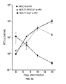
- FIG. 10B provides a reverse transcriptase assay of MOLT4 with ccr5 and MOLT4 with Cal-1, after HIV-1 infection. 48 hours post transduction MOLT4 cells were infected with BaL at MOI 0.2. 7-days post transduction, reverse transcriptase activity in the cultured supernatant was tested. The combination of CCR5 and the C46 fusion inhibitor (Cal-1) showed significantly suppressed in reverse transcriptase activity compared with those of lenti-sh5 transduced MOLT4 cells and untransduced MOLT4 cells.
- FIG. 10C provides time course data for a reverse transcriptase assay of three conditions: (a) MOLT4 transduced with Cal-1; (b) 80% untransduced MOLT4 mixed with 20% Cal-1 transduced; and (c) MOCK control.
- the figure illustrates an over 2 log reduction in reverse transcriptase activity was observed in Cal-1-transduced with MOLT4 cells at day 14, compared with a mixed culture of 20% Cal-1-transduced and 80% untransduced MOLT4 cells and untransduced MOLT4 cells (MOCK control) at day-14.
- FIG. 10D provides time course data for reverse transcriptase real time PCR analysis of these three in-vitro experimental sets. The previous RT data were confirmed by this TaqMan based reverse transcriptase real-time PCR assay.
- the figure illustrates an over 2 log reduction in HIV-1 RNA extracted from the cultured supernatant of Cal-1-transduced MOLT4 cells at day 14 was observed, compared with data from a mixed culture of 20% Cal-1-transduced and 80% untransduced MOLT4 cells and untransduced MOLT4 cells (MOCK control).
- FIG. 11A illustrates the results of Cal-1 detection according to embodiments of the present disclosure, using two separate tube assays in accordance with Method 2, herein.
- Cal-1 integrated DNA was amplified by a TaqMan based DNA PCR method based on assay methods disclosed herein.
- Real-time DNA PCR was conducted with the extracted DNA from three sets of MOLT4 based infectious experiments. The data show that consistent presence of integrated Cal-1 DNA levels (normalized by Actin) were detected in Cal-1 transduced MOLT4 cells throughout experiment from day-4 to day-14. Cal-1 DNA was not detected in MOLT4 cells (MOCK control). Significant reduction of integrated level of Cal-1 DNA in a mixed culture of 20% Cal-1-transduced and 80% untransduced MOLT4 cells was evident at day 4 and 7. By day 10 and 14 these levels had further dropped to down to an undetectable level. Cal-1 copy numbers were normalized with 1000 copies of Action DNA.
- FIG. 11B illustrates the results of HIV-1 detection according to embodiments of the present disclosure using two separate tube assays in accordance with Method 2 herein.
- Over a three log reduction in the integrated level of HIV-1 DNA was observed in the Cal-1 transduced MOLT4 cells compared with that of untransduced MOLT4 cells throughout day-4 to day-14 after HIV-1 infection. This data confirms the protection in MOLT4 cells from HIV-1 infection after transduction of Cal-1 lentiviral vector. Elevated level of HIV-1 DNA in a mixed culture of 20% Cal-1-transduced and 80% untransduced MOLT4 cells on day 10 and day 14, compared with those of day 4 and day 7.
- FIG. 12A sets forth an intracellular analysis of HIV-1 RNA in MOLT-4 cells, based on a 3′LTR assay with Method 2, herein. Over a 3 log reduction of integrated level of HIV-1 viral RNA in the Cal-1 transduced MOLT4 cells was observed compared to untransduced MOLT4 cells throughout days 4 to 14 after HIV-1 infection.
- FIG. 12B sets forth an intracellular analysis of HIV-1 RNA in MOLT-4 cells, based on spliced-Tat assay.
- the similar level of massive reduction was observed RNA in the Cal-1 transduced MOLT4 cells based on spliced-Tat assay.
- TAT protein generated by the spliced-Tat mRNA is able to drive massive amount of un-spliced HIV-1 mRNA.
- Detection of spliced-Tat mRNA is critical marker of an initial HIV-1 transcript. HIV-1 RNA copy numbers were normalized with 1,000,000 copy of GAPDH mRNA.
- FIGS. 12A and 12B data suggested that a massive reduction of HIV-1 intracellular mRNA levels was observed, which is supported by the observed large reduction of integrated DNA levels in the Cal-1 transduced MOLT4 cells (see FIGS. 11A and 11B ).
- FIG. 13A illustrates a reverse-transcriptase assay data based on PBMCs from a healthy donor.
- HIV-1 infection experiments were conducted based on PBMCs from a healthy donor.
- the lenti-Cal-1 transduction was conducted in the same manner as in MOLT4 based experiment above. Over 5 times reduction level of reverse-transcriptase activity was observed in Cal-1 transduced PBMCs after HIV-1 infection on day 4 and day 7, compared with that of untransduced PBMCs.
- FIG. 13B illustrates a reverse-transcriptase real-time PCR analysis on RNA in cultured supernatants with Method 2.
- the reverse-transcriptase data in FIG. 13A were confirmed by a TaqMan based reverse-transcriptase real time PCR assay using the extracted RNA from the cultured supernatant.
- FIG. 14A illustrates the results of Cal-1 DNA detection in PBMCs using two separate tube assays in accordance with a Method 2.
- Cal-1 integrated DNA was amplified by a TaqMan based DNA PCR method described herein. DNA was extracted from two sets of PBMCs infectious experiments. The data indicates a consistent presence of integrated Cal-1 DNA level (normalized by Actin) in Cal-1 transduced PBMCs from day-4 to 10. Cal-1 DNA was not detected in untransduced PBMCs (MOCK control). Cal-1 copy numbers were normalized with 1000 copies of Action DNA.
- FIG. 14B illustrates the HIV-1 DNA detection in PBMCs.
- HIV-1 integrated DNA was amplified by a TaqMan based DNA PCR method using two separate tube assays in accordance with Method 2.
- a five-fold reduction in the integrated levels of HIV-1 DNA was observed in Cal-1 transduced PBMCs compared against untransduced PBMCs on Day-4.
- the data confirms the protection in PBMCs from HIV-1 infection after transduction of Cal-1 lentiviral vector. HIV-1 copy numbers were normalized with 1000 copies of Action DNA. While PBMCs are difficult to transduce with Cal-1, the data positively supports Cal-1 protection from HIV-1.
- FIG. 14C illustrates the results of an intracellular analysis of HIV-1 RNA in PBMCs, based on 3′LTR assay with Method 2.
- HIV-1 intracellular mRNA was amplified by a TaqMan based one-step reverse transcriptase real-time PCR method described herein. A greater than 4 ⁇ reduction of integrated level of HIV-1 viral RNA in the Cal-1 transduced PBMCs was observed in compared with that of untransduced PBMCs on day-4. HIV-1 RNA copy numbers were normalized with 1,000,000 copies of GAPDH mRNA.
- FIG. 14D illustrates the results of an intracellular analysis of HIV-1 RNA in PBMCs, based on spliced-Tat assay.
- the similar level of massive reduction was observed in the Cal-1 transduced PBMCs based on the spliced-Tat assay.
- the data in FIGS. 14A and 14B confirm that impact of reduction of integrated HIV-1 DNA levels in the Cal-1 transduced PBMCs. Therefore, a substantial reduction of HIV-1 intracellular mRNA levels was observed in the Cal-1 transduced PBMCs ( FIG. 14C and Figure D). HIV-1 RNA copy numbers were normalized with 1,000,000 copies of GAPDH mRNA.
- FIGS. 15A through 15E set forth examples of different reagents provided in different wells of 96-well plates.
- FIG. 16A summarizes HIV DNA copies per 1,000 human spleen cells, the data obtained through a BLT infectious model.
- FIG. 16B summarizes HIV DNA copies per 10 ⁇ 3 (actin copy) human spleen cells, the data obtained through a BLT infectious model.
- FIG. 16C summarizes vector copy number per 1,000 human spleen cells, the data obtained through a BLT infectious model.
- FIG. 16D summarizes vector copy number per 10 ⁇ 3 (actin copy) human spleen cells, the data obtained through a BLT infectious model.
- FIG. 17 provides data used to generate the summary tables of FIGS. 16A through 16D , including standard curves.
- FIGS. 18A and 18B provide HIV-1 DNA copy number or Cal-1 DNA copy number, both normalized to actin.
- compositions i.e., amplification primers and probes
- methods, and kits that are particularly useful for detecting and/or quantifying nucleic acids present in a sample, such as those derived from HIV or a lentiviral vector.
- an “amplicon” is defined as any nucleic acid molecule produced by a nucleic acid amplification technique.
- an amplicon comprises a sequence that hybridizes with a primer when contacted therewith, and that can be either an entire molecule or a portion thereof.
- Amplification of a target nucleic acid sequence shall mean an in vitro target amplification technique whereby target sequences are copied, producing amplicons which serve as templates for further cycles of amplification.
- Cal-1 refers to a lentiviral vector comprising a short hairpin RNA CCR5 and a C46 fusion inhibitor. Further details regarding Cal-1 are described in co-pending the the co-pending application published as US Publication No. US2012/0201794, the disclosure of which is incorporated by reference herein in its entirety.
- a method involving steps a, b, and c means that the method includes at least steps a, b, and c.
- steps and processes may be outlined herein in a particular order, the skilled artisan will recognize that the ordering steps and processes may vary.
- HIV human immunodeficiency virus
- HIV refers to any HIV including laboratory strains, wild type strains, mutant strains and any biological sample comprising at least one HIV virus, such as, for example, an HIV clinical isolate.
- HIV strains compatible with the present methods are any such strains that are capable of infecting mammals, particularly humans. Examples are HIV-1, HIV-2, and SIV.
- lentiviral vector is used to denote any form of a nucleic acid derived from a lentivirus and used to transfer genetic material into a cell via transduction.
- the term encompasses lentiviral vector nucleic acids, such as DNA and RNA, encapsulated forms of these nucleic acids, and viral particles in which the viral vector nucleic acids have been packaged.
- LTR long terminal repeat
- U3 enhancer and promoter regions for gene expression
- R/U5 untranslated RNA sequences
- primer refers a short segment of DNA or DNA-containing nucleic acid molecule, which (i) anneals under amplification conditions to a suitable portion of a DNA or RNA sequence to be amplified, and (ii) initiates, and is itself physically extended, via polymerase-mediated synthesis.
- probe refers to an oligonucleotide (i.e. a sequence of nucleotides), whether occurring naturally as in a purified restriction digest or produced synthetically, which is capable of hybridizing to another oligonucleotide of interest. Probes are useful in the detection, identification and isolation of particular gene sequences. It is contemplated that any probe used in the present disclosure will be labelled with a “reporter molecule” or “detectable moiety” such that is detectable in any detection system, including, but not limited to enzyme (e.g., ELISA, as well as enzyme-based histochemical assays), fluorescent, radioactive, calorimetric, and luminescent systems.
- enzyme e.g., ELISA, as well as enzyme-based histochemical assays
- fluorescent e.g., radioactive, calorimetric, and luminescent systems.
- TAR refers to the “trans-activation response” genetic element located in the R region of the LTR. This element mediates the action of tat, by physically binding to the viral trans-activator tat.
- target sequence or “target nucleic acid” each refer to a region of a nucleic acid which is to be amplified, detected, or otherwise analyzed.
- Tat refers to the virally encoded trans-activating protein which functions as an elongation factor. Tat is essential for viral replication as the key viral element for increasing HIV gene expression.
- TATA box is used in reference to a segment of DNA, located approximately 19-27 base pairs upstream from the start point of eukaryotic structural genes, to which RNA polymerase binds.
- the TATA box is approximately 7 base pairs in length, often comprising the sequence “TATAAAA.”
- the TATA box is also sometimes referred to as the “Hogness box.”
- transduce refers to the delivery of a gene(s) using a viral or retroviral vector by means of infection rather than by transfection.
- an anti-HIV gene carried by a retroviral vector a modified retrovirus used as a vector for introduction of nucleic acid into cells
- a “transduced gene” is a gene that has been introduced into the cell via lentiviral or vector infection and provirus integration.
- Viral vectors e.g., “transducing vectors” transduce genes into “target cells” or host cells.
- transgene is a nucleic acid sequence within a lentiviral vector that is not normally present in a cell to be transduced with the lentiviral vector.
- the lentiviral vector serves to introduce this sequence into the transduced cell.
- vector is used in reference to nucleic acid molecules that transfer nucleic acid (e.g., DNA) segment(s) from one cell to another.
- nucleic acid e.g., DNA
- wild-type refers to a gene or nucleic acid sequence which is most frequently observed in a population and is thus arbitrarily designed the “normal” or “wild-type” form of the gene or nucleic acid sequence.
- the lentiviral vector comprises an inactivated or self-inactivating 3′ LTR.
- a “self-inactivating 3′ LTR” is a 3′ LTR that contains a mutation, substitution or deletion that prevents the LTR sequences from driving expression of a downstream gene. It is believed that a copy of the U3 region from the 3′ LTR acts as a template for the generation of LTRs in the integrated provirus. Thus, when the 3′ LTR with an inactivating deletion or mutation integrates as the 5′ LTR of the provirus, no transcription from the 5′ LTR is possible. This eliminates competition between the viral enhancer/promoter and any internal enhancer/promoter.
- Self-inactivating 3′ LTRs are described, for example, in Zufferey et al., J. Virol., Vol. 72:9873-9880, 1998; Miyoshi et at, J. Virol., Vol. 72:8150-8157, 1998; and Iwakuma et al., Virology, Vol. 261:120-132, 1999.
- the 3′ LTR may be made self-inactivating by any method known in the art.
- the U3 element or region of the 3′ LTR contains a deletion of its enhancer sequence, preferably the TATA box, Spl and NF-kappa B sites.
- the provirus that is integrated into the host cell genome will comprise an inactivated 5′ LTR.
- the viral expression vectors of the disclosure preferably do not inhibit vector production in producer cells.
- self-inactivating recombinant lentiviral vectors of the present disclosure comprise a 3′ LTR which has been rendered substantially transcriptionally inactive by virtue of deletions of sequences within the U3 region.
- the lentiviral vectors comprise deletions in the U3 region of the 3′LTR, including removal of a TATA-box sequence (e.g. the sequence of SEQ ID NO: 11 is missing from the U3 region of the 3′LTR of a lentiviral vector).
- TATA-box sequence e.g. the sequence of SEQ ID NO: 11 is missing from the U3 region of the 3′LTR of a lentiviral vector.
- HIV-based lentiviral vectors such vectors tolerate significant U3 deletions, including the removal of the LTR TATA box, without significant reductions in vector titers.
- the lentiviral vectors comprise the removal of between about 100 and about 160 nucleotides from the U3 region of the 3′ LTR as compared with a wild-type U3 3′LTR region. In other embodiments, the lentiviral vectors comprise the removal of between about 120 and about 140 nucleotides from the U3 region of the 3′ LTR as compared with a wild-type U3 3′LTR region. In some embodiments, the lentiviral vectors comprise the removal of about 132 nucleotides from the U3 region of the 3′ LTR as compared with a wild-type U3 3′LTR region.
- SEQ ID NO. 16 provides a wild-type U3 3′LTR region (also referred to herein as “wild-type U3 region” or “wild-type HIV”); while SEQ ID NO. 15 provides a modified U3 region, such as found in a lentiviral vector.
- wild-type U3 region also referred to herein as “wild-type U3 region” or “wild-type HIV”
- SEQ ID NO. 15 provides a modified U3 region, such as found in a lentiviral vector.
- SEQ ID NO: 15 and SEQ ID NO: 16 are compared, the skilled artisan will appreciate that about 132 nucleotides were deleted in the U3 region of the lentiviral vector 3′LTR, including the removal of the TATA-box.
- the R region of the 3′LTR is unaltered, i.e. a wild-type R region (see, for example, SEQ ID NO: 17).
- Cal-1 comprises deletions within the U3 region of the 3′ LTR spanning from nucleotide 423 to 556, and such deletions extend through the TATA-box (namely the sequence of SEQ ID NO: 11).
- the wild-type U3 region of the 3′LTR of HIV comprises an intact TATA-box (see, for example, FIGS. 1B and 1D through 1I ).
- the U3 region of wild-type HIV contains the enhancer and promoter elements that modulate basal and induced expression of the HIV genome in infected cells and in response to cell activation.
- Cal-1 merely illustrates a single lentiviral vector and that other vectors may have a different 3′LTR region, e.g. including U3 regions having different sequences or comprising different nucleotide deletions as compared with a wild-type U3 region, and these different lentiviral vectors may be detected according to the methods disclosed herein.
- the assay methods disclosed herein exploit the differences in the U3 regions of the 3′LTRs of lentiviral vectors and wild-type HIV, and thus allow for specifically designed primers and/or probes to hybridize to lentiviral vectors but not to wild-type HIV, and vice-versa, as described herein.
- primer sequences with suitable hybridization characteristics can be designed based on the the structures of the 3′LTR of lentiviral vectors and HIV.
- primers may be designed with suitable hybridization characteristics based on the sequences of the 3′LTR of lentiviral vectors.
- the primers disclosed herein are particularly contemplated as components of multiplex amplification reactions wherein several amplicon species can be produced from the target-specific primers described herein.
- a forward primer is selected such that it hybridizes to a sequence within a U3 region of the 3′LTR of the lentiviral vector and/or HIV.
- the same forward primer is used in the amplification of both the lentiviral vector and HIV (see FIGS. 2 and 4 ).
- different forward primers are used to amplify the lentiviral vector and HIV (see FIG. 3 ).
- the forward primer is selected such that it hybridizes to a sequence within a U3 region of a 3′LTR having at least 85% identity to that of SEQ ID NO: 1. In other embodiments, the forward primer is selected such that it hybridizes to a sequence within a U3 region of a 3′LTR having at least 90% identity to that of SEQ ID NO: 1. In yet other embodiments, the forward primer is selected such that it hybridizes to a sequence within a U3 region of a 3′LTR having at least 95% identity to that of SEQ ID NO: 1.
- the forward primer is selected such that it hybridizes to a sequence within a U3 region of a 3′LTR having at least 85% identity to that of SEQ ID NO: 3. In other embodiments, the forward primer is selected such that it hybridizes to a sequence within a U3 region of a 3′LTR having at least 90% identity to that of SEQ ID NO: 3. In yet other embodiments, the forward primer is selected such that it hybridizes to a sequence within a U3 region of a 3′LTR having at least 95% identity to that of SEQ ID NO: 3.
- NuAf primer One forward primer suitable for use with the assays described herein is a NuAf primer.
- the NuAf primer comprises the sequence of SEQ ID NO: 2 or a sequence having at least 90% identity to that of the sequence of SEQ ID NO: 2 (see FIGS. 2 and 4 ).
- TATA primer is capable of hybridizing to HIV, but not to lentiviral vectors missing or devoid of a TATA-box sequence in the U3 region of the 3′LTR.
- the TATA primer comprises the sequence of SEQ ID NO:4 or a sequence having at least 90% identity to that of the sequence of SEQ ID NO: 4 (see FIG. 3 ).
- a reverse primer is selected such that it hybridizes to a sequence within a R region of a 3′LTR. In other embodiments, a reverse primer is selected such that it hybridizes to a sequence at the 5′ end of the R region of the 3′LTR. As will be described further herein, in some embodiments, the same reverse primer is used in the amplification of both the lentiviral vector and wild-type HIV, i.e. the reverse primer is designed to a sequence common within the R region of both the lentiviral vector and wild-type HIV.
- the reverse primer is selected such that it hybridizes to a sequence within a R region of a 3′LTR having at least 85% identity to that of SEQ ID NO: 5. In other embodiments, the reverse primer is selected such that it hybridizes to a sequence within a R region of a 3′LTR having at least 90% identity to that of SEQ ID NO: 5. In yet other embodiments, the reverse primer is selected such that it hybridizes to a sequence within a R region of a 3′LTR having at least 95% identity to that of SEQ ID NO: 5.
- LTR-rev primer comprises the sequence of SEQ ID NO: 6.
- the LTR-rev primer is capable of hybridizing to both lentiviral vectors and HIV.
- the probes utilized in the methods disclosed herein belong to a class of probes called “FRET probes” (Förster or fluorescence resonance energy transfer), i.e. those containing a fluorescent reporter and quencher pair.
- FRET probes Förster or fluorescence resonance energy transfer
- the probes utilized in the methods described herein are TAQMAN® probes.
- the TAQMAN® probes (Heid et al., 1996) use the fluorogenic 5′ exonuclease activity of Taq polymerase to measure the amount of target sequences in nucleic acid samples.
- TAQMAN® probes are oligonucleotides that contain a fluorescent dye usually at or near the 5′ base, and a quenching moiety typically at or near the 3′ base.
- the quencher moiety may be a dye such as TAMRA or may be a non-fluorescent molecule such as 4-(4-dimethylaminophenylazo)benzoic acid (DABCYL).
- DBCYL 4-(4-dimethylaminophenylazo)benzoic acid
- FRET fluorescing
- TAQMAN® probes are designed to anneal to an internal region of a PCR product. When the polymerase replicates a template on which a TAQMAN® probe is bound, its 5′ exonuclease activity cleaves the probe.
- the TAQMAN® assay uses universal thermal cycling parameters and PCR reaction conditions. Because the cleavage occurs only if the probe hybridizes to the target, the fluorescence detected originates from specific amplification. The process of hybridization and cleavage does not interfere with the exponential accumulation of the product.
- the probes utilized in the disclosed methods are molecular beacons.
- Molecular beacons are probes for the identification of specific nucleotide sequences present within cells (Tyagi et al., (1998) Nature Biotechnology 16:49-53).
- the molecular beacon can be composed of nucleic acid only such as DNA or RNA, or it can be composed of a peptide nucleic acid (PNA) conjugate. Binding of the molecular beacon to specific nucleotide sequences allows for the identification of the presence of those sequences either in vitro or in vivo.
- PNA peptide nucleic acid
- a molecular beacon includes a conjugate (e.g., a structure such as a quantum dot-tagged bead), a probe, a fluorophore, and a quenching moiety.
- the probe is a single-stranded oligonucleotide comprising a stem and loop structure wherein a hydrophilic attachment group is attached to one end of the single-stranded oligonucleotide and the quenching moiety is attached to the other end of the single-stranded oligonucleotide.
- the fluorophore can be any fluorescent organic dye or a single quantum dot such that its emission does not overlap with that of the quantum dot-tagged bead.
- the quenching moiety desirably quenches the luminescence of the fluorophore. Any suitable quenching moiety that quenches the luminescence of the fluorophore can be used in the conjugate described above.
- the probes utilized in the disclosed methods are dual hybridization probes.
- dual hybridization probes use two sequence-specific oligonucleotide probes in addition to two sequence-specific DNA primers. The two probes are designed to bind to adjacent sequences in the target.
- the dual hybridization probes are labeled with a pair of dyes that exhibit FRET. The donor dye is attached to the 3′ end of the first probe, while the acceptor dye is attached to the 5′ end of the second probe.
- excitation is performed at a wavelength specific to the donor dye, and the reaction is monitored at the emission wavelength of the acceptor dye.
- the probes hybridize to their target sequences in a head-to-tail arrangement. This annealing brings the donor and acceptor dyes into proximity, allowing FRET to occur, resulting in fluorescent emission by the acceptor.
- the increasing amount of acceptor fluorescence is proportional to the amount of PCR product present.
- probes with suitable hybridization characteristics can be designed based on the structures and sequences of the lentiviral vectors and wild-type HIV.
- probes may be designed based on the sequences of the U3 and R regions of the 3′LTR that are unique to the lentiviral vector or HIV.
- a probe may be designed that hybridizes to a lentiviral vector U3 region to the exclusion of a wild-type U3 3′LTR region and vice-versa.
- certain lentiviral vectors comprise a U3 region that, as compared with a wild-type U3 region, comprise certain deletions within the U3 sequence, and the skilled artisan will be able to design a probe to hybridize to these lentiviral vector U3 regions, but not to wild-type U3 regions.
- the methods disclosed herein utilize a junction probe that anneals to sequences spanning the U3 region and the R region of the 3′LTR of the lentiviral vector (i.e. a “junction site”).
- FIG. 1C illustrates a junction site of a lentiviral vector, the junction site comprising a U3 region that comprises certain deletions compared to a wild-type U3 region.
- An appropriately designed junction probe may comprise a portion which hybridizes to this junction site or to a portion or fragment of this junction site.
- the junction probe comprises a first portion and a second portion, wherein the first portion is designed to hybridize to a portion or a fragment of a nucleotide sequence within the U3 region of the 3′LTR of the lentiviral vector; and wherein the second portion is designed to hybridize to a portion or a fragment of a nucleotide sequence within the R region of the 3′LTR.
- the U3 region of the 3′LTR in which the junction probe hybridizes comprises a sequence having at least 80% identify to that of SEQ ID NO: 12. In other embodiments, the U3 region of the 3′LTR of the lentiviral vector in which the junction probe hybridizes comprises a sequence having at least 90% identify to that of SEQ ID NO: 12. In further embodiments, the U3 region of the 3′LTR of the lentiviral vector in which the junction probe hybridizes comprises a sequence having at least 95% identify to that of SEQ ID NO: 12. In yet further embodiments, the U3 region of the 3′LTR of the lentiviral vector in which the junction probe hybridizes comprises the sequence of SEQ ID NO: 12.
- the R region of the 3′LTR of the lentiviral vector in which the junction probe hybridizes comprises a sequence having at least 90% identify to that of SEQ ID NO: 13. In other embodiments, the R region of the 3′LTR of the lentiviral vector in which the junction probe hybridizes comprises the sequence of SEQ ID NO: 13.
- junction probe may be designed to accommodate the different U3 regions and R regions of any lentiviral vector.
- the junction probe comprises the sequence of SEQ ID NO: 14, or a sequence having at least 90% identity to that of SEQ ID NO: 14.
- the junction probe is conjugated to a detectable moiety, such as a fluorescent reporter.
- the fluorescent reporter is selected from the group consisting of Tex-615, Tye-563, Tye-665, Joe, Cy3, Max, Rox, Tet, Texas Red-X, Tamara, & Yakima Yellow.
- the fluorescent reporter comprises a cyanine dye, such as indodicarbocynanine (Cy5TM).
- a TAR-probe i.e. a probe specific to the TAR element in the 3′LTR of HIV.
- the TAR-probe is a TaqMan® probe, such as described herein.
- the probe is selected such that it hybridizes to a sequence within the 3′LTR having at least 85% identity to that of SEQ ID NO: 7.
- the TAR-probe is selected such that it hybridizes to a sequence within the 3′LTR having at least 90% identity to that of SEQ ID NO: 7.
- the TAR-probe is selected such that it hybridizes to a sequence within the 3′LTR having at least 95% identity to that of SEQ ID NO: 7.
- the TAR-probe comprises the sequence of SEQ ID NO: 8.
- the TAR-probe is conjugated to a detectable moiety, such as a fluorescent reporter.
- the fluorescent reporter is selected from the group consisting of Tex-615, Tye-563, Tye-665, Joe, Cy3, Max, Rox, Tet, Texas Red-X, Tamara, & Yakima Yellow.
- the TAR-probe contains a fluorescent reporter and a quencher.
- the fluorescent reporter is fluorescein (CAS 2321-07-5).
- TATA-probe i.e. a probe specific to the TATA-box in the U3 region of a wild-type 3′LTR.
- the TATA-probe is a TaqMan® probe.
- the TATA-probe hybridizes (anti-sense strand targeted) to U3 sequences of the 3′LTR.
- the TATA-probe probe is selected such that it hybridizes to a sequence within the 3′LTR having at least 85% identity to that of SEQ ID NO: 9.
- the TATA-probe is selected such that it hybridizes to a sequence within the U3 region of the 3′LTR having at least 90% identity to that of SEQ ID NO: 9. In yet other embodiments, the TATA-probe is selected such that it hybridizes to a sequence within the U3 region of the 3′LTR having at least 95% identity to that of SEQ ID NO: 9. In some embodiments, the TATA-probe comprises the sequence of SEQ ID NO: 10. In some embodiments, the TATA-probe contains a fluorescent reporter and a quencher.
- the fluorescent reporter is selected from the group consisting of Tex-615, Tye-563, Tye-665, Joe, Cy3, Max, Rox, Tet, Texas Red-X, Tamara, and Yakima Yellow.
- the TAR-probe contains a fluorescent reporter and a quencher.
- the fluorescent reporter is fluorescein (CAS 2321-07-5).
- Samples may be collected using standard DNA extraction kits available on the market and known to those of ordinary skill in the art.
- the DNA extraction kit is a BioLine DNA extraction kit (available from BioLine, Taunton, Mass.).
- non-limiting sources for samples include blood samples, plasma samples, tissue samples, biopsy samples, or samples created after sorting by flow cytometry.
- DNA e.g. proviral DNA or transgene DNA
- DNA may be extracted from the sample using methods known by the skilled in the art such as the procedure described by Maniatis et al., Molecular cloning: A laboratory manual, (Cold Spring Harbor Laboratory, Cold Spring Harbor, N.Y.), 1982, the disclosure of which is hereby incorporated by reference herein in its entirety.
- the procedure involves the preparation of a cell lysate followed by digestion with proteinase K, obtaining DNA purification by a multi-step phenol extraction, ethanol precipitation and ribonuclease digestion.
- RNA may be used in the assay methods described herein.
- RNA reverse transcription into complementary DNA (cDNA) by a suitable reverse transcriptase is needed.
- cDNA complementary DNA
- viral RNA may be isolated using known methods such as that described in Boom, R. et al. (J. Clin. Microbiol.
- cDNA may be synthesized with SENSISCRIPT® RT (Qiagen).
- a one-step reverse transcriptase and Taq-polymerase kit is utilized.
- cDNA is synthesized, followed by a real-time DNA PCR method.
- PCR polymerase chain reaction
- the mixture is denatured and the primers are then annealed to their complementary sequences within the target molecule.
- the primers are extended with a polymerase (e.g. DNA polymerase) so as to form a new pair of complementary strands.
- a polymerase e.g. DNA polymerase
- the steps of denaturation, primer annealing and polymerase extension can be repeated many times (i.e., denaturation, annealing and extension constitute one “cycle”; there can be numerous “cycles”) to obtain a high concentration of an amplified segment (the amplicon) of the desired target sequence.
- the length of the amplified segment of the desired target sequence is determined by the relative positions of the primers with respect to each other, and therefore, this length is a controllable parameter.
- PCR Polymerase chain reaction
- PCR may be used qualitatively or quantitatively.
- One known quantitative amplification technique is “real time PCR.”
- the term “real time PCR” as used herein means that a signal emitted from the PCR assay is monitored during the reaction as an indicator of amplicon production during each PCR amplification cycle, i.e. in “real time,” as opposed to conventional PCR methods, in which an assay signal is detected at the endpoint of the PCR reaction.
- Real time PCR is generally based on the detection and quantitation of a fluorescent reporter, such as those described herein. The signal of the reporter increases in direct proportion to the amount of PCR product in a reaction.
- Reverse transcriptase-polymerase chain reaction refers to generating complementary DNA from an RNA template, and further using the complementary DNA as a template for performing PCR to duplicate, described above, and amplify DNA.
- the reaction mixtures are incubated at a temperature sufficient to synthesize a DNA molecule complementary to all or portion of the RNA template.
- the reaction is incubated at a temperature sufficient to amplify the synthesized DNA molecule.
- amplification of the lentiviral nucleic acid utilizes a forward primer which hybridizes to a portion of the U3 region of the 3′LTR and a reverse primer which hybridizes to a portion of the R region of the 3′LTR.
- amplification of the lentiviral nucleic acid utilizes a NuAf forward primer and a LTR-rev reverse primer.
- the amplicon produced from amplification comprise between about 100 and about 600 nucleic acids. In some embodiments, an amount of the produced amplicon is quantified.
- the amplification of the lentiviral nucleic acid comprises the employment of a probe specific to a sequence within the 3′LTR of the lentiviral vector, i.e. the probe will only hybridize to the 3′LTR of the lentiviral vector and will not hybridize to a wild-type 3′LTR, such as the 3′LTR of HIV.
- the probe is optimized for the deletions in the U3 region of the 3′LTR of the lentiviral vector, as compared with a wild-type 3′LTR that does not comprise such deletions.
- the deletions include the deletion of a TATA-box sequences (again as compared with a wild-type U3 region).
- the probe optimized for the deletions in the U3 region of the 3′LTR is designed to span a portion of the U3 region of the 3′LTR of the lentiviral vector and a portion of the R region of the 3′LTR of the lentiviral vector.
- the probe optimized for the deletions in the U3 region of the 3′LTR is a junction probe.
- the junction probe comprises a sequence having at least 90% identify to that of SEQ ID NO: 14.
- the amplification of the lentiviral nucleic acid or lentiviral transgene comprises quantifying an amount of an amplified nucleic acid, such as by detecting an amount of a fluorescent reporter conjugated the probe.
- the amplification of the lentiviral nucleic acid comprises introducing to a sample a forward primer, a reverse primer, and a junction probe and performing PCR according to procedures known to those of ordinary skill in the art.
- the forward primer is NuAf
- the reverse primer is LTR-rev
- the junction probe comprises the sequence of SEQ ID NO: 14.
- 45-50 cycles of PCR are conducted.
- the PCR method is real-time PCR or quantitative real-time PCR.
- the nucleic acid is RNA and the RNA is first converted to cDNA.
- the nucleic acid is RNA and either reverse-transcriptase PCR or quantitative reverse-transcriptase PCR is utilized.
- the multiplex method takes place in a single reaction system or chamber (hereinafter “reaction tube”). In other embodiments, the multiplex method takes place in separate reaction tubes.
- the lentiviral nucleic acid is a lentiviral transgene.
- the HIV nucleic acid is proviral DNA.
- the lentiviral nucleic acid and/or the HIV nucleic acid are RNA, and the RNA is first converted (e.g. via a reverse transcription process) to cDNA prior to amplification.
- the multiplex method utilizes forward primers capable of hybridizing to sequences within the U3 regions of the 3′LTR of the lentiviral vector and/or HIV. In some embodiments, the multiplex method utilizes reverse primers capable of hybridizing to sequences within the R regions of the 3′LTR of the lentiviral vector and/or HIV. In some embodiments, the multiplex method utilizes the same forward and reverse primers for both the amplification of the lentiviral nucleic acid and for the amplification of the HIV nucleic acid. In these cases, the skilled artisan will be able to select a forward primer capable of hybridizing to the same sequence within the U3 region of both wild-type HIV nucleic acid and the lentiviral vector nucleic acid.
- the multiplex method utilizes different forward primers, but the same reverse primers.
- the forward primers are selected from a NuAf primer and a TATA primer.
- the reverse primer is a LTR-rev primer.
- a first probe specific for the lentiviral nucleic acid is employed in a PCR process (e.g. a real-time PCR process) to amplify and/or quantify an amount of lentiviral nucleic acid
- a second probe specific for HIV is employed in a PCR process to amplify and/or quantify an amount of an HIV nucleic acid, wherein the first and second probes comprise different detectable moieties.
- the first probe is one capable of hybridizing to a sequence within the 3′LTR of the lentiviral vector but not to a sequence within the 3′LTR of wild-type HIV.
- the first probe is optimized for sequence deletions in the 3′LTR of a lentiviral vector nucleic acid as compared with the wild-type 3′LTR sequence.
- the first probe is a junction probe specific to a 3′LTR of the lentiviral vector, e.g. a junction site within the 3′LTR.
- the junction probe is one which is capable of hybridizing to at least a portion of a sequence within the U3 region of the lentiviral vector and capable of hybridizing to at least a portion of a sequence within the R region of the lentiviral vector.
- the junction probe comprises a first portion capable of hybridizing to a first sequence having at least 90% identity to that of SEQ ID NO: 12; and a second portion capable of hybridizing to the sequence of SEQ ID NO: 13.
- the second probe is selected from a TATA probe or a TAR-probe.
- the amplification of the lentiviral nucleic acid and HIV nucleic acid comprises quantifying an amount of the amplified nucleic acids, such as by detecting signals corresponding to different fluorescent reporters conjugated to the probes employed.
- an amount of a lentiviral nucleic acid amplicon and an amount of an HIV nucleic acid amplicon is quantified following electrophoretic separation of the lentiviral nucleic acid amplicons and HIV nucleic acid amplicons.
- the multiplex methods comprise determining a ratio of an amount of a lentiviral nucleic acid to an amount of an HIV nucleic acid present in a sample. In some embodiments, the determined ratio is used to assess the efficacy of treatment or gene therapy, such as with stem cells transduced with a lentiviral vector. In some embodiments, the quantities of lentiviral and HIV nucleic acids may be determined over a course of treatment, i.e.
- a first assessment time point and a second assessment time point may be compared, where an increased amount of lentiviral nucleic acid compared to HIV nucleic acid from the first assessment time point to the second assessment time point is indicative of therapeutic efficiency.
- additional treatment is administered depending on the assessment provided.
- the method first comprises preparing nucleic acids present in a sample, i.e. DNA or RNA, for amplification. If RNA is the starting material within the sample, then the RNA is converted to cNDA, according to means know to those of ordinary skill in the art.
- the forward primer is a NuAf primer.
- the forward primer has the sequence of SEQ ID NO: 2.
- the reverse primer hybridizes to a sequence within the R region of the 3′LTR of both the lentiviral nucleic acid and the HIV nucleic acid.
- the reverse primer is a LTR-rev primer.
- the reverse primer has the sequence of SEQ ID NO: 6.
- the first probe specific to the lentiviral nucleic acid is optimized for the deletions within the U3 region of the 3′LTR of the lentiviral vector as compared with a wild-type HIV nucleic acid (such that the first probe may hybridize to the lentiviral nucleic acid but not to the HIV nucleic acid).
- the probe is a junction probe specific to a junction site within the 3′LTR of the lentiviral nucleic acid, e.g. a junction site that spans a U3 and R region of a 3′LTR.
- the junction probe is capable of hybridizing to first region within the 3′LTR and to a second region within the 3′LTR.
- the junction probe is capable of hybridizing to a portion or a fragment of a nucleotide sequence within an U3 region of the 3′LTR; and to a portion or a fragment of a nucleotide sequence within a R region of the 3′LTR.
- the junction probe comprises a first portion capable of hybridizing to a first sequence having at least 90% identity to that of SEQ ID NO: 12; and a second portion capable of hybridizing to the sequence of SEQ ID NO: 13.
- the junction probe comprises a sequence having at least 80% identify to that of SEQ ID NO: 14.
- the junction probe comprises a sequence having at least 90% identify to that of SEQ ID NO: 14.
- the junction probe comprises a sequence having at least 95% identify to that of SEQ ID NO: 14.
- the second probe specific to the HIV nucleic acid is a TATA-probe.
- the second probe has the sequence of SEQ ID NO: 10 or a sequence having at least 90% identity to that of SEQ ID NO: 10.
- the first probe is labeled with Cy5.
- the second probe is labeled with FAM.
- RNA is a starting material, and as an alternative to first converting the RNA to cDNA, reverse-transcriptase PCR or real-time reverse transcriptase may be utilized.
- the amounts of lentiviral nucleic acid and HIV nucleic acid are quantified, such as by detecting signals from the different detectable moieties conjugated to the different probes.
- a ratio of an amount of a lentiviral nucleic acid to an amount of an HIV nucleic acid is used to assess the efficacy of treatment or gene therapy, such as with stem cells transduced with a lentiviral vector.
- the quantities of lentiviral and HIV nucleic acids may be determined over a course of treatment, i.e.
- a first assessment time point and a second assessment time point may be compared, where an increased amount of lentiviral nucleic acid compared to HIV nucleic acid from the first assessment time point to the second assessment time point is indicative of therapeutic efficiency.
- additional treatment is administered depending on the assessment provided.
- a multiplex method of detecting and/or quantifying a lentiviral nucleic acid and a HIV nucleic acid in a sample where the multiplex method takes place in two separate reaction tubes (“Method 2”).
- a first reaction tube comprises all of the components necessary for amplification of a lentiviral nucleic acid.
- a second reaction tube comprises all of the components necessary for amplification of a HIV nucleic acid.
- this assay again exploits the difference between the 3′LTR lentiviral nucleic acid structure and the wild-type 3′LTR HIV nucleic acid structure and, in particular, utilizes the TATA-box as an amplification start site for the HIV nucleic acid.
- the method first comprises preparing nucleic acids present in a sample, i.e. DNA or RNA, for amplification. If RNA is the starting material within the sample, then the RNA is converted to cNDA. Once the nucleic acids are prepared, forward and reverse primers are introduced. In this particular method, different forward primers are used in the amplification of both the lentiviral nucleic acid and the HIV nucleic acid (see FIG. 3 ).
- the forward primer is a NuAf primer.
- the forward primer has the sequence of SEQ ID NO: 2.
- the forward primer is a TATA primer.
- the forward primer has the sequence of SEQ ID NO: 4.
- the same reverse primer is used in the amplification of both the lentiviral nucleic acid and the HIV nucleic acid.
- the reverse primer hybridizes to a sequence within the R region of the 3′LTR of both the lentiviral nucleic acid and the HIV nucleic acid.
- the reverse primer is a LTR-rev primer.
- the reverse primer has the sequence of SEQ ID NO: 6.
- the specificity of the reaction in the first reaction tube is based upon the specific probe being utilized.
- the specificity of the reaction in the second reaction tube is based upon the forward primer utilizes.
- amplification efficiency is relatively identical such that the assay may provide reliable, comparative data in both LV nucleic acid and HIV nucleic acid detection and/or quantification.
- the first probe specific to the lentiviral nucleic acid is optimized for the deletions within the U3 region of the 3′LTR of the lentiviral vector as compared with a wild-type HIV nucleic acid (such that the first probe may hybridize to the lentiviral nucleic acid but not to the HIV nucleic acid).
- the probe is a junction probe specific to a junction site within the 3′LTR of the lentiviral nucleic acid, e.g. a junction site that spans a U3 and R region of a 3′LTR.
- the junction probe is capable of hybridizing to first region within the 3′LTR and to a second region within the 3′LTR.
- the junction probe is capable of hybridizing to a portion or a fragment of a nucleotide sequence within an U3 region of the 3′LTR; and to a portion or a fragment of a nucleotide sequence within a R region of the 3′LTR.
- the junction probe comprises a first portion capable of hybridizing to a first sequence having at least 90% identity to that of SEQ ID NO: 12; and a second portion capable of hybridizing to the sequence of SEQ ID NO: 13.
- the junction probe comprises a sequence having at least 80% identify to that of SEQ ID NO: 14.
- the junction probe comprises a sequence having at least 90% identify to that of SEQ ID NO: 14.
- the junction probe comprises a sequence having at least 95% identify to that of SEQ ID NO: 14.
- the second probe specific to the HIV nucleic acid is a TAR-probe.
- the second probe has the sequence of SEQ ID NO: 8, or at least 90% identity to a sequence of SEQ ID NO: 8.
- the first probe is labeled with Cy5.
- the second probe is labeled with FAM.
- RNA is a starting material, and as an alternative to first converting the RNA to cDNA, reverse-transcriptase PCR or real-time reverse transcriptase may be utilized.
- the amounts of lentiviral nucleic acid and HIV nucleic acid are quantified, such as by detecting signals from the different detectable moieties conjugated to the different probes.
- a ratio of an amount of a lentiviral nucleic acid to an amount of an HIV nucleic acid is used to assess the efficacy of treatment or gene therapy, such as with stem cells transduced with a lentiviral vector.
- the quantities of lentiviral and HIV nucleic acids may be determined over a course of treatment, i.e.
- a first assessment time point and a second assessment time point may be compared, where an increased amount of lentiviral nucleic acid compared to HIV nucleic acid from the first assessment time point to the second assessment time point is indicative of therapeutic efficiency.
- additional treatment is administered depending on the assessment provided.
- the method first comprises preparing nucleic acids present in a sample, i.e. DNA or RNA, for amplification. If RNA is the starting material within the sample, then the RNA is converted to cNDA.
- the forward primer is a NuAf primer.
- the forward primer has the sequence of SEQ ID NO: 2.
- the reverse primer hybridizes to a sequence within the R region of the 3′LTR of both the lentiviral nucleic acid and the HIV nucleic acid.
- the reverse primer is a LTR-rev primer.
- the reverse primer has the sequence of SEQ ID NO: 6.
- amplification produces a lentiviral nucleic acid amplicon and a HIV nucleic acid amplicon having different sizes (see FIG. 7 , which shows the relative sizes of the amplicons from extracted DNA from Example 1, herein).
- the size difference between the two amplicons ranges from between about 100 and about 560 base pairs, with the lentiviral vector amplicon being shorter (i.e. having comparatively less bases or a lower molecular weight) than the HIV amplicon.
- the size difference between the two amplicons ranges from between about 100 and about 550 base pairs, with the lentiviral vector amplicon being shorter (i.e. having comparatively less bases or a lower molecular weight) than the HIV amplicon.
- the different sized amplicons can be distinguished and/or separated from one another by using any standard electrophoretic separation method (e.g. electrophoresis separation with an Agarose gel in conjunction with a DNA staining procedure, such as Sybr-green, EvaGreen).
- electrophoretic separation method e.g. electrophoresis separation with an Agarose gel in conjunction with a DNA staining procedure, such as Sybr-green, EvaGreen.
- the separated amplicons may then be quantitated such that amounts of lentiviral vector nucleic acid and HIV nucleic in the sample may be derived.
- Droplet Digital PCR may be used to separate the size of the band with EvaGreen dye instantly with accurate quantitative detection and allow for the generation of absolute quantitation of copy number of LV nucleic acid and HIV nucleic acid simultaneously with a simple PCR primer set.
- Droplet Digital PCR technology is a digital PCR method utilizing a water-oil emulsion droplet system. Droplets are formed in a water-oil emulsion to form the partitions that separate the template DNA molecules. The droplets serve essentially the same function as individual test tubes or wells in a plate in which the PCR reaction takes place, albeit in a much smaller format. Without wishing to be bound by any particular theory, it is believed that the Droplet Digital PCR System partitions nucleic acid samples into thousands of nanoliter-sized droplets, and PCR amplification is carried out within each droplet.
- the droplets support PCR amplification of the template molecules they contain and use reagents and workflows similar to those used in a conventional real time PCR method based on EvaGreen staining od double-stranded DNA.
- each droplet is analyzed or read in a similar idea as in flow cytometer to determine the fraction of PCR-positive droplets in the original sample.
- Two amplicon sides are different in LV amplicon and HIV-1 amplicon, when we used in the method described in FIG. 4 .
- the intercalating dyes, EvaGreen bind to double-stranded DNA. Longer double-stranded DNA amplicon, in case of HIV-1, produce much higher intensity and that generated from shorter double-stranded DNA amplicon, in case of Cal-1.
- These data are then analyzed using Poisson statistics to determine the target DNA template concentration in the original sample. By this method, we could identify how many copies of HIV-1 and Cal-1 amplicon in the original sample.
- kits for carrying out the claimed methods provide at least one forward primer, at least one reverse primer, and at least one probe.
- the kits of the present disclosure include a NuAf primer, a LTR-rev primer, and a probe specific to a lentiviral vector.
- the kits of the present disclosure include a NuAf primer, a LTR-rev primer, and a probe having the sequence of SEQ ID NO: 14.
- the kits of the present disclosure include a NuAf primer, a LTR-rev primer, a probe specific to a lentiviral vector and a TATA-probe.
- the kits of the present disclosure include a NuAf primer, a LTR-rev primer, a probe having the sequence of SEQ ID NO: 14, and a TATA-probe.
- kits of the present disclosure comprise a NuAf primer, a TATA-primer, a LTR-rev primer, a probe specific to a lentiviral vector, and a TAR-probe.
- the kits of the present disclosure comprise a NuAf primer, a TATA-primer, a LTR-rev primer, a probe having SEQ ID NO: 14, and a TAR-probe.
- kits of the present disclosure may further comprise other components including reaction tubes, instructions, buffers, reagents, and oligonucleotides.
- MOLT4 Cal-1 (100%). 100% of Cells are transduced with Cal-1 as determined by C46 expression on Flow cytometry.
- 0.6 mL of the cultured cell suspension for DNA analysis and 0.4 mL of the cultured cell suspension for RNA analysis were transferred 2.0 mL of standard Eppendorf tubes. These tubes were centrifuged at 900 g for 3 min. The supernatant was then removed. One mL of PBS was added to each tube, followed by centrifuging at 900 g for 3 min. The supernatant was again removed. The cell pellets were used for RNA and DNA analysis.
- RT assay Reverse Transcriptase assay ( FIG. 5A ) was used to measure the amount of lentivirus in 10 ⁇ L of culture supernatant released from infected MOLT-4.
- the RT assay was able to detect HIV-1 and lentivirus vector in the cultured supernatants (see Suzuki K, et al: Poly A-linked non-isotopic microtiter plate reverse transcriptase assay for sensitive detection of clinical human immunodeficiency virus isolates. J Virol Methods 1995, 55:347-356, the disclosure of which is hereby incorporated by reference herein in its entirety).
- MOLT-4 ( ⁇ ) showed highly increased RT level on Day 10, Day 14, indicating high production of HIV-1 released from infected MOLT-4 along culture day. This data indicated the infection experiment of this set went very well;
- MOLT4 Cal20% ( ⁇ ) showed a good reduction (10 hold) of HIV-1 released from the infected MOLT-4 on day-14;
- MOLT4 Cal-1 ( ⁇ ) showed a great reduction of HIV-1 released from the infected cells
- the RT assay was able to detect RT activity of HIV-1 and the RT assay was also able to detect Cal-1 lentivirus, which was believed to be a carry-over from the process of MOLT-4 transfection.
- RT activity of MOLT4 Cal-1 was higher than that of MOLT4 ( ⁇ ).
- RT activity of MOLT4 Cal20% was also just slightly higher than that of MOLT4 ( ⁇ ).
- HIV-1 specific RNA detection ( FIG. 5B ) was performed based on a single tube assay in accordance with the assay method using a single tube assay in accordance with a Multiplexed Method-1 described herein.
- RNA was extracted from 500 ⁇ L of cultured supernatant using an automated extraction system (EasyMag, bioMerieux) with 60 ⁇ L of elution volume setting.
- LightCycler 480 (Roche) and the white 96-well plate for LightCycler-480 were used in one step reverse transcriptase real-time (RT)-PCR analysis.
- HIV-1 standards 0, 4, 4 ⁇ 10, 4 ⁇ 102, 4 ⁇ 103, 4 ⁇ 104, 4 ⁇ 105, 4 ⁇ 106 copies/ ⁇ L
- HIV-1 specific RNA detection data revealed that at day-4, all three experiments showed the same level of HIV-1 in the RNA extracted from supernatant, using a single tube assay in accordance with Method-1.
- the amount of HIV-1 in MOLT4 Cal-1 ( ⁇ ) showed a greater than 1000 fold reduction of HIV-1 released from the infected MOLT-4 ( ⁇ ) on day10 and day14.
- MOLT4 Cal20% ( ⁇ ) showed about a 10 fold reduction of HIV-1 released from the infected MOLT-4 on day-14, which was identical to RT assay data.
- Cal-1 specific RNA detection ( FIG. 5C ), using a single tube assay in accordance with Method-1, indicated that reduction of Cal-1 RNA level over time in culture was observed in Cal-1 transduced MOLT4 cells ( ⁇ ). The amount of Cal-1 was likely the result of carried-over lentivirus when MOLT-4 cells were transduced with Cal-1 on day-0. There was no detection of Cal-1 RNA in untransduced MOLT-4 cells. Only a small amount of Cal-1 RNA was detected on day 4 from MOLT4 Cal20% ( ⁇ ) group at day-4.
- RT data showed slightly elevated RT levels of MOLT4 Cal-1 ( ⁇ ) as compared to MOLT4 cell alone ( ⁇ ) on day 4. This could have been attributed to lentivirus carry over. A very small level of elevated RT was also observed in MOLT4 Cal20% ( ⁇ ), compared with MOLT-4 infection. This slightly elevated RT could have been indicative of low level of contribution from lentivirus carry-over.
- the levels of Cal-1 RNA in the supernatant at day-14 samples was about 1/1000 of HIV-1 RNA level in the cultured supernatant, indicating extremely minor level of Cal-1 RNA carry-over.
- FIG. 6A HIV-1 integrated DNA
- FIG. 6B Cal-1 integrated DNA
- DNA was extracted from the cell pellets prepared at day 4, 7, 10, 14 using PurLink Genomic DNA kits (ThermoFisher) with 604, of elution volume.
- LightCycler 480 (Roche) and a white 96-well plate for LightCycler-480 were used in this analysis one step real-time PCR analysis.
- Master mix 1 is for dual detection detecting HIV-1 DNA and Cal-1 DNA
- PCR was started with the following condition: 95° C.—2 min, 45 cycles of (94° C.—15 sec, 60° C.—30 sec)
- HIV-1 standards 0, 4, 4 ⁇ 10, 4 ⁇ 102, 4 ⁇ 103, 4 ⁇ 104, 4 ⁇ 105, 4 ⁇ 106 copies/ ⁇ L
- HIV-1 integrated DNA data was normalized with 106 copies of GAPDH ( FIG. 6A ). Significant increase in HIV-1 DNA levels was observed in MOLT4 cells during the culture period. This is indicative of a high level of HIV-1 integration in MOLT4 cells.
- MOLT4 Cal-1-20% showed that HIV-1 DNA was detectable on day 10 and day 14. However, the level of HIV-1 DNA was observed to be about 10-50 fold lower as compared to MOLT4 cells alone. HIV-1 DNA was not detected in HIV-challenged Cal-1 100% transduced MOLT4 cells. It is believed that transduced Cal-1 vector protected MOLT-4 cells from HIV-1 infection.
- Cal-1 integrated DNA data was normalized with 106 copies of GAPDH ( FIG. 6B ). A significant increase in Cal-1 DNA levels was observed in Cal-1 100% transduced MOLT4 cells during the culture period. This was indicative of a high level of HIV-1 integration in MOLT4 cells.
- MOLT4 Cal-1-20% showed that HIV-1 DNA was detected on day 4 and day 7. However, the level of Cal-1 DNA was observed to be 20-60 fold lower as compared to Cal-1 100% transduced MOLT4 cells. A decreased level of Cal-1 DNA was observed on day10 and day14 in MOLT4 Cal-1-20%. This was correlated with HIV-1 DNA data. Because of the loss of Cal-1 DNA with this experimental setting, HIV-1 DNA levels were elevated on day-10 and day14 ( FIG. 6A ). Cal-1 DNA was not detected in MOLT4 cells alone. Cal-1
- FIG. 6C provide graphs showing the results of experiments enabling the identification of HIV-1 RNA copy number using Method-1.
- HIV-1 specific LTR mRNA was analyzed ( FIG. 6C ).
- RNA was extracted from the cell pellets prepared at day 4, 7, 10, 14 using ReliaPrep RNA Miniprep system (Promega) with 60 ul of elution volume.
- LightCycler 480 (Roche) and the white 96-well plate for LightCycler-480 were used in this analysis one step reverse transcriptase real-time (RT)-PCR analysis.
- PCR Master 1 mix Master mix is for HIV-1 mRNA detection.
- PCR Master 2 mix Master mix is for GAPDH mRNA detection.
- PCR Master 1 mix is for HIV-1 mRNA detection.
- DNase RNase free water 11.20 2 x B (from kit) 20.00 Tata forward primer (20 ⁇ M) 0.50 Imai-LTR-Rev (20 ⁇ M) 0.50 HIV-1 Tata Probe (5 ⁇ M) with FAM label 0.60 RT enzyme (from Kit) 0.40 RNase Inhibitor (from kit 0.80
- RT-PCR was started with the following condition: 45° C.—20 min, 95° C.—2 min, 45 cycles of (94° C.—7 sec, 60° C.—30 sec)
- HIV-1 standards 0, 4, 4 ⁇ 10, 4 ⁇ 102, 4 ⁇ 103, 4 ⁇ 104, 4 ⁇ 105, 4 ⁇ 106 copies/ ⁇ L
- GAPDH standards 0, 2, 2 ⁇ 10, 2 ⁇ 102, 2 ⁇ 103, 2 ⁇ 104, 2 ⁇ 105, 2 ⁇ 106 copies/ ⁇ L
- HIV-1 mRNA analysis data was normalized with 106 copies of mRNA expression of GAPDH ( FIG. 6C ). High levels of HIV-1 specific cell associated HIV-1 LTR mRNA were detected in untransduced MOLT4 cells ( ⁇ ) during the culture period.
- HIV-1 cell associated LTR-mRNA was still detectable ( ⁇ ), as illustrated in FIG. 6 C. This was an indication that a very low level of RNA transcription was taking place from presumably a very small population of HIV-1 integrated DNA in MOLT-4 cells.
- the amount of DNA added to each PCR reaction had an estimated cell equivalence of 1-2 ⁇ 10e5 cells. Therefore, the HIV-1 detection sensitivity in this assay method was 2 copies per 1-2 ⁇ 10e5 cells. If the frequency HIV-1 integration was less than 2 copies per 1-2 ⁇ 10e5 cells, the assay could not detect HIV-1 DNA using this assay method.
- Example 3 illustrates the results of two different assay methods illustrated in FIG. 2 (Method-1) and FIG. 3 (Method-2), where the methods were compared using DNA, obtained from the Example 1, herein.
- the result in Table 6 utilizes a multiplex assay method, taking place in a single reaction tube (Method-1); whereas the result in Tables 7 and 8 utilizes a multiplex assay method taking place in two separate reaction tubes (Method-2).
- both assays perform similarly, i.e. the quantitative data obtained from both methods was similar. Accordingly, both methods, as disclosed herein, are able to amplify a lentiviral nucleic acid and an HIV nucleic acid and quantify levels of amplified lentiviral nucleic acid and amplified HIV nucleic acid.
- HIV-1 clinical samples were tested to assess HIV-1 detection sensitivity for the newly developed LTR based assay.
- the study was conducted to assess the detection sensitivity of the developed HIV-1 assay and to confirm any cross-reactivity to the HIV-1 DNA, clinical HIV-1 patient samples were used.
- Fourteen HIV-1 clinical samples were tested to assess HIV-1 detection sensitivity.
- HIV-1 and Cal-1 copy numbers were calculated by standards and normalized by 1,000,000 copies of ACTIN DNA. Both standards generated linear lines, see FIG. 9 . Both HIV-1 and Cal-1 assays were valid. HIV-1 DNA assay was able to detect 14 out of 14 samples (A-N samples), indicated in FIG. 9 . Cal-1 DNA assay was not detected in any of the 14 samples. Cal-1 assay did not have any cross-reactivity to the DNA extracted from the HIV-1 positive patients. Levels of HIV-1 DNA assay in the clinical samples were in the similar levels obtained with 2 and 20 U1 cells spiked data, indicating the experimental setting in Example 4 herein was at a similar level of to the actual clinical setting.
- the setting-a) is negative control of the experiment.
- the setting-b) is Cal-1 transduced CD4 + T cells with MOI-2.
- the setting-c) is Cal-1 transduced CD4 + T cells with MOI-10.
- U1 cells were counted. Aliquots of 2, 20, 200, 2000 U1 cells were spiked into the CD4 cells containing 1 ⁇ 106 cells. Another aliquot of 1 ⁇ 10 6 cells CD4 cells was used without spiked control (Please note that 5 ⁇ 106 cells of the transduced CD4 were divided into equal amount of five aliquots, containing 1 ⁇ 106 cells per aliquot). DNA was extracted with PurLink Genomic DNA kits (ThermoFisher) with 60 ⁇ L of elution volume. Basically we created mimic clinical samples by spiking U1 cells into the above three groups of CD4 + T cells. U1 cell was selected because it is carries two copies of the integrated HIV-1 genome in a single cell. Experimental setting is listed as following:
- FIG. 8A HIV-1 integrated DNA
- FIG. 8B Cal-1 integrated DNA
- PCR was performed using the following condition: 95° C.—2 min 45 cycles of (94° C.—7 sec, 60° C.—30 sec)
- HIV-1 standards 0, 4, 4 ⁇ 10, 4 ⁇ 102, 4 ⁇ 103, 4 ⁇ 104, 4 ⁇ 105, 4 ⁇ 106 copies/ ⁇ L
- HIV-1 integrated DNA data was normalized with 106 copies of Actin ( FIG. 8A ).
- Untransduced CD4 cells showed that two U1 cells spiked into 1 ⁇ 10e6 CD4 cells was detected by this assay, as well as 20, 200, and 2000 U1 cells are detected. Linearly increased HIV-1 DNA copies number were detected to the proportion of spiked amount of U1 cells.
- the HIV-1 detected value in the analysis of 2, 20, 200, 2000 U1 cells showed equivalent data across the three groups, indicating that integrated Cal-1 DNA did not interfere with HIV-1 detection ( FIG. 8A ).
- the Cal-1 integrated DNA data was normalized with 106 copies of Actin ( FIG. 8B ).
- the Cal-1 DNA assay did not detect Cal-1 DNA in the Untransduced CD4 cells, despite spiking U1 cells into CD4 cells.
- the HIV-1 assay is sensitive enough to detect HIV-1 DNA obtained from 1 mL of blood from the patients (14 out of 14 HIV-1 positive samples)
- Sample number 1 is negative control of the experiment
- Sample number 2 is Cal-1 transduced CD4+ cells with MOI2
- Sample number 3 is Cal-1 transduced CD4+ cells with MOI10. These samples were obtained at day 14 after transduction of the CD4 cells with Cal-1-lenti.
- HIV-1 LTR assay should not detect any HIV level in those clinical samples, which have only CAL-1 integrated DNA, since these samples has no HIV-1 infection.
- HIV standard showed good linear detection of HIV-1 DNA from 4-4,000,000 copies/ ⁇ L
- control No. 1 ⁇ MOLT4 w Cal-1> showed no HIV
- control No. 2 ⁇ 80% MOLT4 20% MOLT4-Cal-1> showed some level of HIV detection
- control No. 3 ⁇ MOLT4 without Cal-1> showed much higher levels of HIV detection.
- the new assay based on LTR regions is able to detect CAL-1 integrated DNA using actual clinical samples with the same donor.
- the new assay is able to detect increased copy number in CAL-1 integrated DNA level with higher MOI transduced cells by comparing data with MOI2 and with MOI10.
- HIV-1 assay As to HIV-1 assay: (a) HIV-1 copy numbers in the samples are calculated by HIV-1 standards; (b) HIV-1 copy number was normalized by ACTIN DNA copy number in the same extracted DNA, The Normalized HIV copy numbers are calculated by 1000 copies of Actin, which is highlighted with blue letter in the last column.
- Cal-1 assay (a) Cal-1 copy numbers in the samples were calculated by Cal-1 standards; (b) Cal-1 copy number was normalized by ACTIN DNA copy number in the same extracted DNA, The Normalized CAl-1 copy numbers were calculated by 1000 copies of Actin, which is highlighted with blue letter in the last column.
- Actin assay (a) actin copy numbers in the samples were calculated by Actin standards; (b) these copy numbers were used for normalization of HIV-1 and Cal-1 copy number in the samples.
- Table 12 provides: HIV-1 copy numbers in the samples calculated by HIV-1 standards; the HIV-1 copy number was normalized by ACTIN DNA copy number in the same extracted DNA.
- the Normalized HIV copy numbers were calculated by 1000 copies of Actin, which is listed in the last column.
- the cp (crossing point-PCR-cycle) value is the cycle at which fluorescence achieved a defined threshold.
- the HIV standard showed good linear detection of HIV-1 DNA from 4-4,000,000 copies/ ⁇ L. Three samples with the same donor (untransduced, transduced with MOI2, transduced with MOI10) showed no HIV-1 DNA detection.
- In house control No. 1 ⁇ MOLT4 with Cal-1> with HIV infected samples showed no HIV.
- In house control No. 2 ⁇ 80% MOLT4 20% MOLT4-Cal1> with HIV infected samples showed some level of HIV detection.
- In house control No. 3 ⁇ MOLT4 without Cal1> with HIV infected samples showed much higher levels of HIV detection.
- Table 14 provides Actin analysis data. Actin copy numbers in the samples were calculated by Actin standards. These copy numbers were used for normalization of HIV-1 and Cal-1 copy number in the samples.
- FIG. 10 provides graphs showing transduction efficiency in MOLT-4 cells ( FIG. 10A ) and cultured supernatants analyses to detect amount of HIV-1 virus released from the HIV-1 infected MOLT-4 cells by RT assay ( FIG. 10B , C) and by one step reverse transcriptase real-time (RT)-PCR analysis ( FIG. 10D ).
- MOLT-4 Cell were transduced with either with lenti-sh5 and lenti-Cal-1 with MOI 2.5. Transduction was determined after 48 hours incubation, utilizing 2F5 and ccr5 staining on MOLT-4 cells with flow cytometry analysis.
- FIG. 10A provides a flow analysis of MOLT-4 cells with lenti-ccr5 and Cal-1 (ccr5 and C46).
- Lenti-ccr5 alone transduced MOLT-4 cells indicated about a 75% reduction in CCR5 expression.
- the data of Lenti-Cal-1 transduced MOLT-4 cells revealed that about 89% of the cells expressed C46 and about a 68% reduction in CCR5 expression was also observed.
- FIG. 10B provides a reverse transcriptase assay of MOLT4 with lenti-ccr5 and MOLT4 with Cal-1, after HIV-1 infection using two separate tube assays in accordance with Method-2. 48 hours post transduction MOLT4 cells were infected with BaL at MOI 0.2. 7-days post transduction, reverse transcriptase activity in the cultured supernatant was tested. The combination of CCR5 and the C46 fusion inhibitor (Cal-1) showed significantly suppressed in reverse transcriptase activity compared with those of lenti-ccr5 transduced MOLT4 cells and untransduced MOLT4 cells.
- MOLT-4 cells only without any transduction of Cal-1 lentivirus
- MOLT4 Cal-1 (100%). 100% of Cells were transduced with Cal-1 as determined by C46 expression on Flow cytometry.
- Example 1 The experimental procedure was the same as in Example 1 herein. Briefly, about 0.5 million of these cells were infected with HIV-1 BaL. These cells were cultured in a 25-cm 2 culture Flask using 10 mL of a standard RPMI-1640 based medium containing 10% FBS with of 1 ⁇ glutamax supplement in CO2 incubator. Cultured supernatant samples (1 mL) were taken at day 4, 7, 10, 14 for analysis of both RT assay ( FIG. 10C ) and reverse transcriptase real-time (RT)-PCR assay ( FIG. 10D ). Cultured cell samples were also prepared at day 4, 7, 10, 14 for analysis of both Intracellular analyses of DNA ( FIG. 11AB ) and RNA ( FIG. 12AB ).
- 0.6 mL of the cultured cell suspension for DNA analysis and 0.4 mL of the cultured cell suspension for RNA analysis were transferred 2.0 mL of standard Eppendorf tubes. These tubes were centrifuged at 900 g for 3 min. The supernatant was removed. One mL of PBS was added to each tube, followed by centrifuge at 900 g for 3 min. The supernatant was removed. The cell pellets were used for RNA and DNA analysis.
- FIG. 10C provides time course data for a reverse transcriptase assay of three conditions: (a) MOLT4 transduced with Cal-1; (b) 80% untransduced MOLT4 mixed with 20% Cal-1 transduced; and (c) MOCK control.
- the data reveals that an over 2 log reduction in reverse transcriptase activity was observed in Cal-1-transduced with MOLT4 cells at day 14, compared with a mixed culture of 20% Cal-1-transduced and 80% untransduced MOLT4 cells and untransduced MOLT4 cells (MOCK control) at day-14.
- RT assay Reverse Transcriptase assay
- FIG. 10D provides a graph showing the results of the experiment enabling the identification of HIV-1 RNA copy number with Method 2 using 500 ⁇ L of cultured supernatant.
- RNA was extracted from cultured supernatant using an automated extraction system (EasyMag, bioMerieux) with 60 ⁇ L of elution volume setting.
- the LightCycler-480 (Roche) and the white 96-well plate for LightCycler-480 were used in one step reverse transcriptase real-time PCR analysis.
- RT-PCR was started with the following condition: 45° C.—20 min, 95° C.—2 min 45 cycles of (94° C.—7 sec, 60° C.—30 sec)
- HIV-1 standards 0, 4, 4 ⁇ 10, 4 ⁇ 102, 4 ⁇ 103, 4 ⁇ 104, 4 ⁇ 105, 4 ⁇ 106 copies/ ⁇ L
- FIG. 10D provides time course data for one step reverse transcriptase real-time (RT)-PCR analysis.
- the previous RT data were confirmed by this TaqMan based real time PCR assay.
- the figure reveals that an over 2 log reduction in HIV-1 RNA extracted from the cultured supernatant of Cal-1-transduced MOLT4 cells at day 14 was observed, compared with data from a mixed culture of 20% Cal-1-transduced and 80% untransduced MOLT4 cells and untransduced MOLT4 cells (MOCK control).
- FIGS. 11A and 11B provide graphs showing the results of experiments enabling the identification of HIV-1 DNA and Cal-1 DNA copy number using two separate tube assays in accordance with Method-2.
- DNA was extracted from the cell pellets prepared at day 4, 7, 10, 14 using PurLink Genomic DNA kits (ThermoFisher) with 60 ul of elution volume.
- LightCycler 480 (Roche) and the white 96-well plate for LightCycler-480 were used in this analysis one step real-time PCR analysis.
- Master mix 3 is for detection of Actin detection
- PCR was started with the following condition: 95° C.—2 min 45 cycles of (94° C.—7 sec, 60° C.—30 sec)
- HIV-1 standards 0, 4, 4 ⁇ 10, 4 ⁇ 102, 4 ⁇ 103, 4 ⁇ 104, 4 ⁇ 105, 4 ⁇ 106 copies/ ⁇ L
- Cal-1 integrated DNA and HIV-1 integrated DNA data were normalized with 1000 copies of Actin ( FIG. 11AB ).
- FIG. 11A illustrates the results of Cal-1 detection according to embodiments of the present disclosure.
- Cal-1 integrated DNA was amplified by a TaqMan based DNA PCR method based on assay methods, illustrated in FIG. 3 , disclosed herein.
- Real-time DNA PCR was conducted with the extracted DNA from three sets of MOLT4 based infectious experiments. The data show that consistent presence of integrated Cal-1 DNA levels (normalized by Actin) were detected in Cal-1 transduced MOLT4 cells throughout experiment from day-4 to day-14. Cal-1 DNA was not detected in MOLT4 cells (MOCK control). A significant reduction of integrated level of Cal-1 DNA was observed in a mixed culture of 20% Cal-1-transduced and 80% untransduced MOLT4 cells as evident at day 4 and 7. By day 10 and 14 these levels had further dropped down to an undetectable level. Cal-1 copy numbers were normalized with 1000 copies of Action DNA.
- FIG. 11B illustrates the results of HIV-1 detection according to embodiments of the present disclosure.
- Over 3 log reduction in the integrated level of HIV-1 DNA was observed in the Cal-1 transduced MOLT4 cells compared with that of untransduced MOLT4 cells throughout day-4 to day-14 after HIV-1 infection. This data confirms the protection in MOLT4 cells from HIV-1 infection after transduction of Cal-1 lentiviral vector.
- Cal-1 DNA data in a mixed culture of 20% Cal-1-transduced and 80% untransduced MOLT4 cells on day 10 and day 14 suggested that loss of integrated Cal-1 DNA in those time points. Therefore, increasing HIV-1 DNA levels in a mixed culture on day 10 and day 14 was observed.
- the data also suggest that Cal-1 transgene functions to protect against HIV-1 infection. HIV-1 copy numbers were normalized with 1000 copies of Action DNA.
- FIG. 12A provides graphs showing the results of experiments enabling the identification of HIV-1 RNA copy number with Method-2.
- HIV-1 specific LTR mRNA was analyzed ( FIG. 12A ).
- RNA was extracted from the cell pellets prepared at day 4, 7, 10, 14 using ReliaPrep RNA Miniprep system (Promega) with 60 ul of elution volume.
- LightCycler 480 (and the white 96-well plate for LightCycler-480 were used in this analysis one step reverse transcriptase real-time (RT)-PCR analysis.
- PCR Master 1 mix Master mix is for HIV-1 mRNA detection.
- PCR Master 2 mix Master mix is for GAPDH mRNA detection.
- PCR was started with the following condition: 45° C.—20 min 95° C.—2 min 45 cycles of (94° C. —7 sec, 60° C.—30 sec)
- HIV-1 standards 0, 4, 4 ⁇ 10, 4 ⁇ 102, 4 ⁇ 103, 4 ⁇ 104, 4 ⁇ 105, 4 ⁇ 106 copies/ ⁇ L
- GAPDH standards 0, 2, 2 ⁇ 10, 2 ⁇ 102, 2 ⁇ 103, 2 ⁇ 104, 2 ⁇ 105, 2 ⁇ 106 copies/ ⁇ L
- HIV-1 mRNA analysis data was normalized with 106 copies of mRNA expression of GAPDH ( FIG. 12A ).
- FIG. 13A The cultured supernatants analyses to detect amount of HIV-1 virus released from the HIV-1 infected PBMCs by RT assay ( FIG. 13A ) and by one step reverse transcriptase real-time (RT)-PCR analysis ( FIG. 13B ).
- PBMCs were prepared by a single healthy donor.
- a standard PHA stimulation method was used to stimulate PNMCs for 3-days pre-culture, followed by the transduction with lenti-Cal-1 with MOI 2.5.
- 0.6 mL of the cultured cell suspension for DNA analysis and 0.4 mL of the cultured cell suspension for RNA analysis were transferred 2.0 mL of standard Eppendorf tubes. These tubes were centrifuged at 900 g for 3 min. The supernatant was removed. One mL of PBS was added to each tube, followed by centrifuge at 900 g for 3 min. The supernatant was removed. The cell pellets were used for RNA and DNA analysis.
- FIG. 13B illustrates a reverse-transcriptase real time PCR analysis on RNA in cultured supernatants in accordance with Method-2.
- the reverse-transcriptase data in FIG. 14 a were confirmed by a TaqMan based real time reverse-transcriptase-PCR assay using the extracted RNA from the cultured supernatant.
- RT assay Reverse Transcriptase assay
- FIG. 13A illustrates a reverse-transcriptase assay data based on PBMCs from a healthy donor. Over 5 times reduction level of reverse-transcriptase activity was observed in Cal-1 transduced PBMCs after HIV-1 infection on day 4 and day 7, compared with that of untransduced PBMCs
- a composition comprising (a) a probe comprising a nucleotide sequence having at least 80% identity to that of SEQ ID NO: 14, the probe conjugated to a reporter moiety; (b) a forward primer comprising a nucleotide sequence having at least 80% identity to that of SEQ ID NO: 2; and (c) a reverse primer comprising a nucleotide sequence having at least 80% identity to that of SEQ ID NO: 6; wherein each of the forward and reverse primers are capable of annealing to a target sequence to amplify the target sequence.
- composition of embodiment 1, wherein the reporter moiety is selected from the group consisting of Tex-615, Tye-563, Tye-665, Joe, Cy3, Max, Rox, Tet, Texas Red-X, Tamara, and Yakima Yellow.
- a composition comprising (a) a probe comprising a nucleotide sequence having at least 80% identity to that of SEQ ID NO: 10; the probe conjugated to a reporter moiety; (b) a forward primer comprising a nucleotide sequence having at least 80% identity to that of SEQ ID NO: 2; and (c) a reverse primer comprising a nucleotide sequence having at least 80% identity to that of SEQ ID NO: 6; wherein each of the forward and reverse primers are capable of annealing to a target sequence to amplify the target sequence.
- composition of embodiment 3, wherein the reporter moiety is selected from the group consisting of Tex-615, Tye-563, Tye-665, Joe, Cy3, Max, Rox, Tet, Texas Red-X, Tamara, and Yakima Yellow.
- a composition comprising (a) a probe comprising a nucleotide sequence having at least 80% identity to that of SEQ ID NO: 8, the probe conjugated to a reporter moiety; (b) a forward primer comprising a nucleotide sequence having at least 80% identity to that of SEQ ID NO: 4; and (c) a reverse primer comprising a nucleotide sequence having at least 80% identity to that of SEQ ID NO: 6; wherein each of the forward and reverse primers are capable of annealing to a target sequence to amplify the target sequence.
- composition of embodiment 5, wherein the reporter moiety is selected from the group consisting of Tex-615, Tye-563, Tye-665, Joe, Cy3, Max, Rox, Tet, Texas Red-X, Tamara, and Yakima Yellow.
- kits for discriminating between lentiviral nucleic acids and HIV nucleic acids in a sample comprising the composition of embodiment 1 and the composition of either of embodiments 3 or 5.
- a method for quantifying a first target sequence comprising contacting a first sample comprising the first target sequence with the probe and primers of the composition of embodiment 1; performing a real-time polymerase chain reaction using the first target sequence as the template; and quantifying an amount of a generated first amplicon.
- the first target sequence is a lentiviral nucleic acid sequence and wherein the second target sequence is a HIV nucleic acid sequence.
- step of quantifying the amount of the generated first amplicon comprises detecting signals from a first reporter moiety; and wherein the step of quantifying the amount of the generated second amplicon comprises detecting signals from a second reporter moiety, wherein the first and second reporter moieties are different.
- a method of detecting a lentiviral nucleic acid and/or a HIV nucleic acid in a sample comprising: (a) performing multiplex real-time PCR with a lentiviral nucleic acid template and a HIV nucleic acid template in the sample using: (i) a first forward primer having a nucleotide sequence having at least 80% identity to that of SEQ ID NO: 2, a first reverse primer having a nucleotide sequence having at least 80% identity to that of SEQ ID NO: 6, and a first probe having a nucleotide sequence having at least 80% identity to that of SEQ ID NO:14, the first probe having a first reporter moiety; (ii) a second forward primer having a nucleotide sequence having at least 80% identity to that of SEQ ID NO: 2, a second reverse primer having a nucleotide sequence having at least 80% identity to that of SEQ ID NO: 6, and a second probe having a nucleotide sequence having at least a nucle
- a method of detecting a lentiviral nucleic acid and/or a HIV nucleic acid in a sample comprising: (a) performing multiplex real-time PCR with a lentiviral nucleic acid template and a HIV nucleic acid template in the sample using: (i) a first forward primer having a nucleotide sequence having at least 80% identity to that of SEQ ID NO: 2, a first reverse primer having a nucleotide sequence having at least 80% identity to that of SEQ ID NO: 6, and a first probe having a nucleotide sequence having at least 80% identity to that of SEQ ID NO: 14, the first probe having a first reporter moiety; (ii) a second forward primer having a nucleotide sequence having at least 80% identity to that of SEQ ID NO: 4, a second reverse primer having a nucleotide sequence having at least 80% identity to that of SEQ ID NO: 6, and a second probe having a nucleotide sequence having at least 80%
- a method of detecting an amount of a lentiviral nucleic acid in a sample comprising: (a) contacting the sample with a first forward primer and a first reverse primer; (b) contacting the sample with a junction probe specific for a junction site within the 3′ LTR of the lentiviral nucleic acid, wherein the junction probe comprises a first portion which is capable of hybridizing to at least a portion of a sequence within the U3 region of the lentiviral nucleic acid 3′LTR and a second portion which is capable of hybridizing to at least a portion of a sequence within the R region of the lentiviral nucleic acid 3′LTR, and wherein the junction probe comprises a first detectable moiety; and detecting signals from the first detectable moiety.
- junction probe comprises a nucleotide sequence having at least 80% identity to that of SEQ ID NO: 14.
- junction probe comprises a nucleotide sequence having at least 90% identity to that of SEQ ID NO: 14.
- the detection of the amount of the HIV nucleic acid comprises contacting the sample with a second probe specific for a TATA-box sequence within a 3′LTR of an HIV nucleic acid sequence, the second probe conjugated to a second detectable moiety; and detecting signals from the second detectable moiety.
- the detection of the amount of the HIV nucleic acid comprises contacting the sample with a second forward primer, a second reverse primer, and a second probe having a nucleotide sequence having at least 90% identity to that of SEQ ID NO: 8, wherein the second probe comprises a second detectable moiety; and detecting signals from the second detectable moiety.
- a method of detecting a lentiviral nucleic acid in a sample comprising: (a) contacting the sample with a first forward primer and a first reverse primer; (b) contacting the sample with a junction probe specific for a junction site within a 3′LTR of the lentiviral nucleic acid, wherein the 3′LTR of the lentiviral nucleic acid does not comprise a TATA-box sequence, and wherein the junction site spans a portion of the U3 region of the lentiviral nucleic acid 3′LTR and a portion of the R region of the lentiviral nucleic acid 3′LTR, and wherein at least a portion of the junction probe hybridizes to a nucleotide sequence of SEQ ID NO: 13.
- a method of quantifying an amount of a lentiviral nucleic acid and an amount of an HIV nucleic acid in a sample, the lentiviral nucleic acid and the HIV nucleic acid comprising different 3′LTRs comprising amplifying both the lentiviral nucleic acid and the HIV nucleic acid with a forward primer which hybridizes to a sequence within both the 3′LTR of the lentiviral nucleic acid and the 3′LTR of the HIV nucleic acid, and a reverse primer which hybridizes to a sequence within both the 3′LTR of the lentiviral nucleic acid and the 3′LTR of the HIV nucleic acid, and wherein the amplification of both the lentiviral nucleic acid and the HIV nucleic acid occur in a single reaction tube.
- HIV nucleic acid 3′LTR comprises a U3 region having a nucleotide sequence of SEQ ID NO: 16.
- An amplicon obtainable by amplification from a lentiviral nucleic acid-containing sample with a pair of primers, the primers having SEQ ID NO: 2 and SEQ ID NO: 6, the amplicon comprising a 3′LTR that does not comprise a TATA-box sequence.
- An isolated nucleic acid sequence comprising a nucleotide sequence having at least 90% identity to that of SEQ ID NO: 14.
- An isolated nucleic acid sequence comprising a nucleotide sequence having at least 70% identity to that of SEQ ID NO: 14 and capable of hybridizing to a fragment of a nucleotide sequence of SEQ ID NO: 15.
- An isolated nucleic acid sequence having a first portion capable of hybridizing to a nucleotide sequence having at least 70% identity to that of SEQ ID NO:12, and a second portion capable of hybridizing to a nucleotide sequence of SEQ ID NO: 13.
- An isolated nucleic acid sequence comprising a nucleotide sequence having at least 90% identity to that of SEQ ID NO: 8.
- An isolated nucleic acid sequence comprising a nucleotide sequence having at least 90% identity to that of SEQ ID NO: 10.
- a method of treating an HIV positive patient comprising:
- step of assaying for the presence of the genetic modification comprises detecting a junction site within the treated cells.
- junction site comprises contacting the sample with a junction probe, the junction probe comprising a nucleic acid sequence having at least 90% identity to that of SEQ ID NO: 14.
- a method for determining therapeutic efficacy of an HIV-based recombinant vector in a patient comprising:
- a method of detecting an amount of a lentiviral RNA in a sample comprising: (a) converting the lentiviral RNA to cDNA; (b) contacting the sample with a first forward primer and a first reverse primer; (c) contacting the sample with a junction probe specific for a junction site within the 3′ LTR of the lentiviral nucleic acid, wherein the junction probe comprises a first portion which is capable of hybridizing to at least a portion of a sequence within the U3 region of the lentiviral nucleic acid 3′LTR and a second portion which is capable of hybridizing to at least a portion of a sequence within the R region of the lentiviral nucleic acid 3′LTR, and wherein the junction probe comprises a first detectable moiety; and detecting signals from the first detectable moiety.
- invention 63 The method of embodiment 61, further comprising detecting an amount of a HIV nucleic acid in the same, wherein the HIV nucleic acid is DNA or RNA.
- a method of treating an HIV positive patient comprising: (i) treating cells obtained from the HIV positive patient with a Cal-1 lentiviral vector under conditions favoring uptake of Cal-1 into the cells; (ii) assaying the treated cells for a presence of a genetic modification, wherein the step of assaying for the presence of the genetic modification comprises detecting a junction site, wherein the detection of the junction site comprises contacting the treated cells with a junction probe, the junction probe comprising a nucleic acid sequence having at least 90% identity to that of SEQ ID NO: 14; (iii) administering the treated cells to the patient, provided that the treated cells were positively assayed for the presence of the genetic modification.
- Cal-1 lentiviral vector comprises a short hairpin RNA against CCR5 and a C46 fusion inhibitor.
- a method for determining therapeutic efficacy of an HIV-based recombinant vector in a patient comprising: (i) treating cells derived from the patient with a Cal-1 lentiviral vector under conditions favoring uptake of Cal-1; (ii) administering the treated cells to the patient; (iii) assaying for levels of a wild-type HIV nucleic acid and a Cal-1 nucleic acid present in DNA derived from cells obtained from the patient at a first time point; and (iv) assaying for levels of the wild-type HIV nucleic acid and the Cal-1 nucleic acid present in DNA derived from cells obtained from the patient at a second time point; wherein the assaying of the levels of the Cal-1 nucleic acid and the wild-type HIV nucleic acid at the first and second time points is achieved by (a) performing multiplex real-time PCR using: (i) a first forward primer having a nucleotide sequence having at least 80% identity to that of SEQ ID NO: 2, a first reverse primer
- Cal-1 lentiviral vector comprises a short hairpin RNA against CCR5 and a C46 fusion inhibitor.
- a method of treating an HIV positive patient comprising:
- junction probe is conjugated to a reporter moiety
- detection of the junction probe comprises detecting signals from the reporter moiety
- the assaying of the transduced cells for the presence of the genetic modification further comprises contacting the DNA derived from the transduced cells with a forward primer and a reverse primer, wherein each of the forward and reverse primers are capable of annealing to a target sequence to amplify the target sequence.
- the forward primer comprises a nucleotide sequence having at least 80% identity to that of SEQ ID NO: 2.
- junction probe hybridizes to a nucleic acid sequence within the amplicon comprising a sequence having at least 90% identity to that of SEQ ID NO: 12; and wherein a second portion of the junction probe hybridizes to a nucleic acid sequence within the amplicon comprising a sequence having at least 90% identity to that of SEQ ID NO: 13.
- Cal-1 lentiviral vector comprises a short hairpin RNA against CCR5 and a C46 fusion inhibitor.
Landscapes
- Health & Medical Sciences (AREA)
- Life Sciences & Earth Sciences (AREA)
- Chemical & Material Sciences (AREA)
- Organic Chemistry (AREA)
- Virology (AREA)
- Engineering & Computer Science (AREA)
- Wood Science & Technology (AREA)
- Zoology (AREA)
- Immunology (AREA)
- Genetics & Genomics (AREA)
- Bioinformatics & Cheminformatics (AREA)
- General Health & Medical Sciences (AREA)
- Proteomics, Peptides & Aminoacids (AREA)
- Microbiology (AREA)
- Biochemistry (AREA)
- General Engineering & Computer Science (AREA)
- Biotechnology (AREA)
- Molecular Biology (AREA)
- AIDS & HIV (AREA)
- Medicinal Chemistry (AREA)
- Physics & Mathematics (AREA)
- Analytical Chemistry (AREA)
- Biophysics (AREA)
- Biomedical Technology (AREA)
- Chemical Kinetics & Catalysis (AREA)
- Veterinary Medicine (AREA)
- Public Health (AREA)
- Animal Behavior & Ethology (AREA)
- Pharmacology & Pharmacy (AREA)
- Nuclear Medicine, Radiotherapy & Molecular Imaging (AREA)
- General Chemical & Material Sciences (AREA)
- Oncology (AREA)
- Communicable Diseases (AREA)
- Tropical Medicine & Parasitology (AREA)
- Measuring Or Testing Involving Enzymes Or Micro-Organisms (AREA)
- Medicines Containing Material From Animals Or Micro-Organisms (AREA)
Priority Applications (2)
| Application Number | Priority Date | Filing Date | Title |
|---|---|---|---|
| US15/810,254 US10240214B2 (en) | 2015-05-18 | 2017-11-13 | Methods for discriminating between HIV-1 and lentiviral vectors |
| US16/267,404 US11008630B2 (en) | 2015-05-18 | 2019-02-05 | Methods for discriminating between HIV-1 and lentiviral vectors |
Applications Claiming Priority (3)
| Application Number | Priority Date | Filing Date | Title |
|---|---|---|---|
| US201562163327P | 2015-05-18 | 2015-05-18 | |
| PCT/US2016/032767 WO2016187151A1 (en) | 2015-05-18 | 2016-05-16 | Methods of discriminating between hiv-1 and lentiviral vectors |
| US15/810,254 US10240214B2 (en) | 2015-05-18 | 2017-11-13 | Methods for discriminating between HIV-1 and lentiviral vectors |
Related Parent Applications (1)
| Application Number | Title | Priority Date | Filing Date |
|---|---|---|---|
| PCT/US2016/032767 Continuation-In-Part WO2016187151A1 (en) | 2015-05-18 | 2016-05-16 | Methods of discriminating between hiv-1 and lentiviral vectors |
Related Child Applications (1)
| Application Number | Title | Priority Date | Filing Date |
|---|---|---|---|
| US16/267,404 Continuation US11008630B2 (en) | 2015-05-18 | 2019-02-05 | Methods for discriminating between HIV-1 and lentiviral vectors |
Publications (2)
| Publication Number | Publication Date |
|---|---|
| US20180066325A1 US20180066325A1 (en) | 2018-03-08 |
| US10240214B2 true US10240214B2 (en) | 2019-03-26 |
Family
ID=56121165
Family Applications (2)
| Application Number | Title | Priority Date | Filing Date |
|---|---|---|---|
| US15/810,254 Expired - Fee Related US10240214B2 (en) | 2015-05-18 | 2017-11-13 | Methods for discriminating between HIV-1 and lentiviral vectors |
| US16/267,404 Active US11008630B2 (en) | 2015-05-18 | 2019-02-05 | Methods for discriminating between HIV-1 and lentiviral vectors |
Family Applications After (1)
| Application Number | Title | Priority Date | Filing Date |
|---|---|---|---|
| US16/267,404 Active US11008630B2 (en) | 2015-05-18 | 2019-02-05 | Methods for discriminating between HIV-1 and lentiviral vectors |
Country Status (10)
| Country | Link |
|---|---|
| US (2) | US10240214B2 (ru) |
| EP (1) | EP3283658B1 (ru) |
| JP (1) | JP2018516594A (ru) |
| CN (1) | CN108026593A (ru) |
| AU (1) | AU2016265839A1 (ru) |
| CA (1) | CA2986133A1 (ru) |
| DK (1) | DK3283658T3 (ru) |
| ES (1) | ES2784351T3 (ru) |
| HK (1) | HK1247965A1 (ru) |
| WO (1) | WO2016187151A1 (ru) |
Families Citing this family (7)
| Publication number | Priority date | Publication date | Assignee | Title |
|---|---|---|---|---|
| EP3414340B1 (en) * | 2016-02-12 | 2023-08-09 | CureVac SE | Method for analyzing rna |
| EP3423595A1 (en) | 2016-03-03 | 2019-01-09 | CureVac AG | Rna analysis by total hydrolysis |
| CN115768899A (zh) * | 2020-03-09 | 2023-03-07 | 詹森生物科技公司 | 用于定量重组载体核酸整合的组合物和方法 |
| CN111961760A (zh) * | 2020-09-02 | 2020-11-20 | 苏州市第五人民医院 | 基于ddPCR自动化检测HIV-1的引物组、探针组、试剂盒及用途 |
| CN114134251A (zh) * | 2020-09-04 | 2022-03-04 | 西比曼生物科技(香港)有限公司 | 一种快速检测慢病毒载体滴度的方法 |
| CN112210625A (zh) * | 2020-11-02 | 2021-01-12 | 中山大学达安基因股份有限公司 | 定量检测人类免疫缺陷病毒1型dna载量的试剂盒及方法 |
| CN113151587A (zh) * | 2021-01-28 | 2021-07-23 | 云舟生物科技(广州)有限公司 | 一种慢病毒滴度检测引物、试剂盒及检测方法 |
Citations (2)
| Publication number | Priority date | Publication date | Assignee | Title |
|---|---|---|---|---|
| US20030124530A1 (en) * | 1991-06-27 | 2003-07-03 | Genelabs Technologies, Inc. | Sequence-directed DNA-binding molecules compositons and methods |
| WO2011008348A2 (en) | 2009-07-15 | 2011-01-20 | Calimmune Inc. | Dual vector for inhibition of human immunodeficiency virus |
Family Cites Families (15)
| Publication number | Priority date | Publication date | Assignee | Title |
|---|---|---|---|---|
| US4000159A (en) | 1974-06-28 | 1976-12-28 | Phillips Petroleum Company | Preparation of n,n-disubstituted thioamides |
| US4683195A (en) | 1986-01-30 | 1987-07-28 | Cetus Corporation | Process for amplifying, detecting, and/or-cloning nucleic acid sequences |
| US4683202A (en) | 1985-03-28 | 1987-07-28 | Cetus Corporation | Process for amplifying nucleic acid sequences |
| US5176995A (en) | 1985-03-28 | 1993-01-05 | Hoffmann-La Roche Inc. | Detection of viruses by amplification and hybridization |
| US4965188A (en) | 1986-08-22 | 1990-10-23 | Cetus Corporation | Process for amplifying, detecting, and/or cloning nucleic acid sequences using a thermostable enzyme |
| DK0408295T3 (da) * | 1989-07-11 | 1996-09-16 | Gen Probe Inc | Fremgangsmåder til opformering af nukleinsyresekvenser |
| WO1992020813A1 (en) * | 1991-05-17 | 1992-11-26 | Dana Farber Cancer Institute | Assays for factors affecting circularization of dna, assays for factors affecting dna integration, factors, and uses thereof |
| US5576176A (en) * | 1994-03-03 | 1996-11-19 | The Regents Of The University Of California | Marker and an assay for detection and monitoring of human immunodeficiency virus latency and activation |
| US20030104576A1 (en) * | 1994-10-07 | 2003-06-05 | Jonathan W. Nyce | Dna construct, composition, formulations & methods for making the construct & for modulating expression |
| DE19907485B4 (de) * | 1999-02-12 | 2008-01-17 | Strathmann Ag & Co. | Virus-Vakzine |
| EP1921136B1 (en) * | 2005-08-16 | 2012-12-12 | Takara Bio, Inc. | Nucleic acid for treatment or prevention of immunodeficiency virus infection |
| DK2770061T3 (en) | 2009-07-24 | 2019-01-07 | Immune Design Corp | NON-INTEGRATING LENTIVIRUS VECTORS |
| EP3632463A1 (en) * | 2011-04-08 | 2020-04-08 | Immune Design Corp. | Immunogenic compositions and methods of using the compositions for inducing humoral and cellular immune responses |
| AU2013303237A1 (en) * | 2012-08-16 | 2015-04-09 | NVS Technologies, Inc. | Assay methods and systems |
| CN103882062A (zh) * | 2014-03-13 | 2014-06-25 | 山西大学 | 慢病毒干扰载体pll3.7-Neo及其构建方法 |
-
2016
- 2016-05-16 ES ES16729099T patent/ES2784351T3/es active Active
- 2016-05-16 DK DK16729099.8T patent/DK3283658T3/da active
- 2016-05-16 EP EP16729099.8A patent/EP3283658B1/en active Active
- 2016-05-16 AU AU2016265839A patent/AU2016265839A1/en not_active Abandoned
- 2016-05-16 WO PCT/US2016/032767 patent/WO2016187151A1/en unknown
- 2016-05-16 CA CA2986133A patent/CA2986133A1/en not_active Abandoned
- 2016-05-16 CN CN201680042304.1A patent/CN108026593A/zh active Pending
- 2016-05-16 JP JP2018512815A patent/JP2018516594A/ja active Pending
-
2017
- 2017-11-13 US US15/810,254 patent/US10240214B2/en not_active Expired - Fee Related
-
2018
- 2018-06-06 HK HK18107366.1A patent/HK1247965A1/zh unknown
-
2019
- 2019-02-05 US US16/267,404 patent/US11008630B2/en active Active
Patent Citations (2)
| Publication number | Priority date | Publication date | Assignee | Title |
|---|---|---|---|---|
| US20030124530A1 (en) * | 1991-06-27 | 2003-07-03 | Genelabs Technologies, Inc. | Sequence-directed DNA-binding molecules compositons and methods |
| WO2011008348A2 (en) | 2009-07-15 | 2011-01-20 | Calimmune Inc. | Dual vector for inhibition of human immunodeficiency virus |
Non-Patent Citations (22)
| Title |
|---|
| Bryan Burke et al: "CCR5 as a Natural and Modulated Target for Inhibition of HIV", VIRUSES, vol. 6, No. 1, Dec. 30, 2013 (Dec. 30, 2013), CH, pp. 54-68, XP055287500, ISSN: 1999-4915, DOI: 10.3390/v6010054. |
| BRYAN BURKE, BOYD MAUREEN, IMPEY HELEN, BRETON LOUIS, BARTLETT JEFFREY, SYMONDS GEOFF, H�TTER GERO: "CCR5 as a Natural and Modulated Target for Inhibition of HIV", VIRUSES, MDPI, CH, vol. 6, no. 1, 30 December 2013 (2013-12-30), CH, pages 54 - 68, XP055287500, ISSN: 1999-4915, DOI: 10.3390/v6010054 |
| Bryan P Burke et al: "Engineering Cellular Resistance to HIV-1 Infection In Vivo Using a Dual Therapeutic Lentiviral Vector", Molecular Therapy-Nucleic Acids, vol. 4, No. 4, Apr. 14, 2015 (Apr. 14, 2015), pp. e236, XP055270563, DOI: 10.1038/mtna.2015.10. |
| BRYAN P BURKE, BERNARD R LEVIN, JANE ZHANG, ANNA SAHAKYAN, JOSHUA BOYER, MARIA V CARROLL, JOANNA CAMBA COL�N, NAOMI KEECH, VALERIE: "Engineering Cellular Resistance to HIV-1 Infection In Vivo Using a Dual Therapeutic Lentiviral Vector", MOLECULAR THERAPY—NUCLEIC ACIDS, vol. 4, no. 4, pages e236, XP055270563, DOI: 10.1038/mtna.2015.10 |
| Burke et al. (2014) Viruses vol. 6: 54-68; doi:10.3390/v6010054. * |
| DATABASE EMBL [online] "Sequence 20 from Patent WO2005051986.", XP002759935, retrieved from EBI |
| Database EMBL [online] Jun. 22, 2005 (Jun. 22, 2005), "Sequence 20 from Patent WO2005051986.", XP002759935, retrieved from EBI accession No. EM_PAT:CS108612 Database accession No. CS108612. |
| DATABASE Geneseq [online] 12 January 2005 (2005-01-12), CHO H W, KIM G J, KIM S S, LEE J S, NAM J G: "HIV-1 5' LTR TATA box/TAR sequence, from AF102206-G.", XP002759934, retrieved from EBI |
| DATABASE Geneseq [online] 18 October 2012 (2012-10-18), DUBENSKY T W, ROBBINS S H: "Human immunodeficiency virus 1 U3 region coding sequence, SEQ ID 21.", XP002759933, retrieved from EBI |
| DATABASE Geneseq [online] 19 November 1996 (1996-11-19), BUSCH M P, ROMEO J, ADAMS M, PETERLIN B M: "HIV TAR fragment detection probe 1, (TAR loop probe).", XP002759931, retrieved from EBI |
| DATABASE Geneseq [online] 2 January 1996 (1996-01-02), FULTZ T J, KACIAN D L: "Human immunodeficiency virus region 1 probe.", XP002759932, retrieved from EBI |
| DATABASE Geneseq [online] 26 November 1992 (1992-11-26), HASELTINE W A, FARNET C M: "Viral DNA R2 primer.", XP002759930, retrieved from EBI |
| Database Geneseq [online] Apr. 1, 1997 (Apr. 1, 1997), "HIV TAR fragment detection probe 1, (TAR loop probe).", XP002759931, retrieved from EBI accession No. GSN:AAT58845 Database accession No. AAT58845. |
| Database Geneseq [online] Dec. 29, 2005 (Dec. 29, 2005), "HIV-1 5′ LTR TATA box/TAR sequence, from AF102206-G.", XP002759934, retrieved from EBI accession No. GSN:AED60207 Database accession No. AED60207. |
| Database Geneseq [online] Dec. 6, 2012 (Dec. 6, 2012), "Human immunodeficiency virus 1 U3 region coding sequence, SEQ ID 21.", XP002759933, retrieved from EBI accession No. GSN:BAD81538 Database accession No. BAD81538. |
| Database Geneseq [online] Mar. 25, 2003 (Mar. 25, 2003), "Human immunodeficiency virus region 1 probe.", XP002759932, retrieved from EBI accession No. GSN:AAT15554 Database accession No. AAT15554. |
| Database Geneseq [online] Mar. 25, 2003 (Mar. 25, 2003), "Viral DNA R2 primer.", XP002759930, retrieved from EBI accession No. GSN:AAQ31821 Database accession No. AAQ3182. |
| IDT ( 2015) qPCR probes. * |
| International Search Report and Written Opinion, dated Aug. 1, 2016. |
| Sanchez et al. (1997) J. of Virol. vol. 71 No. 3 pp. 2233-2240. * |
| Suzuki et al. (2008) J. Biol. Chem. vol. 283 No. 34: pp. 23353-23363. * |
| Wolstein O et al., Mol Ther Methods & Clinical Development, vol. 1, 2014, p. 11. |
Also Published As
| Publication number | Publication date |
|---|---|
| EP3283658A1 (en) | 2018-02-21 |
| EP3283658B1 (en) | 2020-03-11 |
| AU2016265839A1 (en) | 2017-12-07 |
| JP2018516594A (ja) | 2018-06-28 |
| US20180066325A1 (en) | 2018-03-08 |
| ES2784351T3 (es) | 2020-09-24 |
| US11008630B2 (en) | 2021-05-18 |
| WO2016187151A1 (en) | 2016-11-24 |
| CN108026593A (zh) | 2018-05-11 |
| DK3283658T3 (da) | 2020-05-18 |
| US20190153551A1 (en) | 2019-05-23 |
| HK1247965A1 (zh) | 2018-10-05 |
| CA2986133A1 (en) | 2016-11-24 |
Similar Documents
| Publication | Publication Date | Title |
|---|---|---|
| US11008630B2 (en) | Methods for discriminating between HIV-1 and lentiviral vectors | |
| Mameli et al. | Novel reliable real-time PCR for differential detection of MSRVenv and syncytin-1 in RNA and DNA from patients with multiple sclerosis | |
| EP3510172B1 (en) | Methods of detecting hiv | |
| EP1960554B1 (en) | Methods, plasmid vectors and primers for assessing hiv viral fitness | |
| JP2002300895A (ja) | C型肝炎ウイルス検出用の配列および方法 | |
| Contreras-Galindo et al. | A new Real-Time-RT-PCR for quantitation of human endogenous retroviruses type K (HERV-K) RNA load in plasma samples: increased HERV-K RNA titers in HIV-1 patients with HAART non-suppressive regimens | |
| Lam et al. | Development and comparison of molecular assays for the rapid detection of the pandemic influenza A (H1N1) 2009 virus | |
| JP5553323B2 (ja) | Hiv検出キット、及びhiv検出方法 | |
| CN111154923A (zh) | 一种扩增新型冠状病毒(SARS-CoV-2)基因核苷酸片段的引物及其应用 | |
| Mizutani et al. | Rapid genome sequencing of RNA viruses | |
| Grohman et al. | An immature retroviral RNA genome resembles a kinetically trapped intermediate state | |
| Laham-Karam et al. | Transduction of human immunodeficiency virus type 1 vectors lacking encapsidation and dimerization signals | |
| JP5315519B2 (ja) | コイヘルペスウイルス(khv)の検出方法 | |
| JP2007135469A (ja) | 核酸増幅方法 | |
| Marichannegowda et al. | Transmission of highly virulent CXCR4 tropic HIV-1 through the mucosal route in an individual with a wild-type CCR5 genotype | |
| Katoski et al. | An approach for identification of unknown viruses using sequencing‐by‐hybridization | |
| Song et al. | Transmission of highly virulent CXCR4 tropic HIV-1 through the mucosal route in an individual with a wild-type CCR5 genotype | |
| Li et al. | Transforming viral sequences to A (H1N1) flu diagnosis: the current status and future prospects of rtPCR based assays | |
| EP1349959A2 (en) | Assay to detect integration of a retrovirus polynucleotide into a target nucleic acid | |
| WO2005061735A2 (en) | Strand-specific amplification |
Legal Events
| Date | Code | Title | Description |
|---|---|---|---|
| FEPP | Fee payment procedure |
Free format text: ENTITY STATUS SET TO UNDISCOUNTED (ORIGINAL EVENT CODE: BIG.); ENTITY STATUS OF PATENT OWNER: LARGE ENTITY |
|
| AS | Assignment |
Owner name: ST. VINCENT'S HOSPITAL SYDNEY LIMITED, AUSTRALIA Free format text: ASSIGNMENT OF ASSIGNORS INTEREST;ASSIGNOR:SUZUKI, KAZUO;REEL/FRAME:048235/0030 Effective date: 20151116 Owner name: CALIMMUNE, INC., ARIZONA Free format text: ASSIGNMENT OF ASSIGNORS INTEREST;ASSIGNOR:SYMONDS, GEOFFREY PHILLIP;REEL/FRAME:048235/0042 Effective date: 20151116 Owner name: CALIMMUNE AUSTRALIA PTY LTD, AUSTRALIA Free format text: ASSIGNMENT OF ASSIGNORS INTEREST;ASSIGNOR:SYMONDS, GEOFFREY PHILLIP;REEL/FRAME:048235/0042 Effective date: 20151116 Owner name: NEWSOUTH INNOVATIONS PTY LIMITED, AUSTRALIA Free format text: ASSIGNMENT OF ASSIGNORS INTEREST;ASSIGNOR:KELLEHER, ANTHONY DOMINIC;REEL/FRAME:048235/0049 Effective date: 20151030 |
|
| STCF | Information on status: patent grant |
Free format text: PATENTED CASE |
|
| FEPP | Fee payment procedure |
Free format text: MAINTENANCE FEE REMINDER MAILED (ORIGINAL EVENT CODE: REM.); ENTITY STATUS OF PATENT OWNER: LARGE ENTITY |
|
| LAPS | Lapse for failure to pay maintenance fees |
Free format text: PATENT EXPIRED FOR FAILURE TO PAY MAINTENANCE FEES (ORIGINAL EVENT CODE: EXP.); ENTITY STATUS OF PATENT OWNER: LARGE ENTITY |
|
| STCH | Information on status: patent discontinuation |
Free format text: PATENT EXPIRED DUE TO NONPAYMENT OF MAINTENANCE FEES UNDER 37 CFR 1.362 |
|
| FP | Lapsed due to failure to pay maintenance fee |
Effective date: 20230326 |