KR20170074235A - Delivery of biomolecules to immune cells - Google Patents
Delivery of biomolecules to immune cells Download PDFInfo
- Publication number
- KR20170074235A KR20170074235A KR1020177013389A KR20177013389A KR20170074235A KR 20170074235 A KR20170074235 A KR 20170074235A KR 1020177013389 A KR1020177013389 A KR 1020177013389A KR 20177013389 A KR20177013389 A KR 20177013389A KR 20170074235 A KR20170074235 A KR 20170074235A
- Authority
- KR
- South Korea
- Prior art keywords
- cells
- cell
- antigen
- immune
- compound
- Prior art date
Links
- 210000002865 immune cell Anatomy 0.000 title claims abstract description 244
- 239000000427 antigen Substances 0.000 claims abstract description 334
- 108091007433 antigens Proteins 0.000 claims abstract description 334
- 102000036639 antigens Human genes 0.000 claims abstract description 334
- 238000000034 method Methods 0.000 claims abstract description 249
- 150000001875 compounds Chemical class 0.000 claims abstract description 135
- 239000006285 cell suspension Substances 0.000 claims abstract description 41
- 210000000172 cytosol Anatomy 0.000 claims abstract description 39
- 239000000725 suspension Substances 0.000 claims abstract description 10
- 210000001744 T-lymphocyte Anatomy 0.000 claims description 344
- 210000004027 cell Anatomy 0.000 claims description 306
- 210000003719 b-lymphocyte Anatomy 0.000 claims description 122
- 108090000623 proteins and genes Proteins 0.000 claims description 104
- 102000004169 proteins and genes Human genes 0.000 claims description 95
- 206010028980 Neoplasm Diseases 0.000 claims description 88
- 210000004443 dendritic cell Anatomy 0.000 claims description 77
- 108020004459 Small interfering RNA Proteins 0.000 claims description 64
- 102000039446 nucleic acids Human genes 0.000 claims description 58
- 108020004707 nucleic acids Proteins 0.000 claims description 58
- 150000007523 nucleic acids Chemical class 0.000 claims description 58
- 208000031481 Pathologic Constriction Diseases 0.000 claims description 55
- 230000036262 stenosis Effects 0.000 claims description 49
- 208000037804 stenosis Diseases 0.000 claims description 49
- 230000003834 intracellular effect Effects 0.000 claims description 45
- 108090000765 processed proteins & peptides Proteins 0.000 claims description 45
- 230000008569 process Effects 0.000 claims description 42
- 108010019670 Chimeric Antigen Receptors Proteins 0.000 claims description 41
- 230000014509 gene expression Effects 0.000 claims description 40
- 238000012545 processing Methods 0.000 claims description 40
- 210000004369 blood Anatomy 0.000 claims description 35
- 239000008280 blood Substances 0.000 claims description 35
- 239000000203 mixture Substances 0.000 claims description 31
- -1 CD86 Proteins 0.000 claims description 28
- 239000012528 membrane Substances 0.000 claims description 26
- 230000003915 cell function Effects 0.000 claims description 24
- 230000002966 stenotic effect Effects 0.000 claims description 24
- 229960005486 vaccine Drugs 0.000 claims description 24
- 210000002540 macrophage Anatomy 0.000 claims description 23
- 229940046168 CpG oligodeoxynucleotide Drugs 0.000 claims description 22
- 241000700605 Viruses Species 0.000 claims description 22
- 230000028993 immune response Effects 0.000 claims description 22
- 108091032973 (ribonucleotides)n+m Proteins 0.000 claims description 21
- 230000003612 virological effect Effects 0.000 claims description 21
- 210000001151 cytotoxic T lymphocyte Anatomy 0.000 claims description 20
- 201000010099 disease Diseases 0.000 claims description 19
- 208000037265 diseases, disorders, signs and symptoms Diseases 0.000 claims description 19
- 230000005867 T cell response Effects 0.000 claims description 18
- 230000001404 mediated effect Effects 0.000 claims description 18
- 210000001616 monocyte Anatomy 0.000 claims description 18
- 210000001519 tissue Anatomy 0.000 claims description 18
- 210000004881 tumor cell Anatomy 0.000 claims description 18
- 239000013592 cell lysate Substances 0.000 claims description 17
- 239000003112 inhibitor Substances 0.000 claims description 17
- 108020004999 messenger RNA Proteins 0.000 claims description 17
- 230000037361 pathway Effects 0.000 claims description 17
- 238000004519 manufacturing process Methods 0.000 claims description 16
- 108091023040 Transcription factor Proteins 0.000 claims description 15
- 102000040945 Transcription factor Human genes 0.000 claims description 15
- 239000002158 endotoxin Substances 0.000 claims description 15
- 230000003308 immunostimulating effect Effects 0.000 claims description 15
- 244000052769 pathogen Species 0.000 claims description 15
- 239000002671 adjuvant Substances 0.000 claims description 14
- 239000003446 ligand Substances 0.000 claims description 14
- 229920006008 lipopolysaccharide Polymers 0.000 claims description 14
- 210000004698 lymphocyte Anatomy 0.000 claims description 14
- 230000001965 increasing effect Effects 0.000 claims description 12
- 239000006166 lysate Substances 0.000 claims description 12
- 108010021064 CTLA-4 Antigen Proteins 0.000 claims description 11
- 101710163270 Nuclease Proteins 0.000 claims description 11
- 102000016266 T-Cell Antigen Receptors Human genes 0.000 claims description 11
- 238000011161 development Methods 0.000 claims description 11
- 229940045513 CTLA4 antagonist Drugs 0.000 claims description 10
- 101710089372 Programmed cell death protein 1 Proteins 0.000 claims description 10
- 150000001413 amino acids Chemical class 0.000 claims description 10
- 230000001580 bacterial effect Effects 0.000 claims description 10
- 210000000805 cytoplasm Anatomy 0.000 claims description 10
- 101001057504 Homo sapiens Interferon-stimulated gene 20 kDa protein Proteins 0.000 claims description 9
- 101001055144 Homo sapiens Interleukin-2 receptor subunit alpha Proteins 0.000 claims description 9
- 102100026878 Interleukin-2 receptor subunit alpha Human genes 0.000 claims description 9
- 108091008874 T cell receptors Proteins 0.000 claims description 9
- 210000002443 helper t lymphocyte Anatomy 0.000 claims description 9
- 230000002829 reductive effect Effects 0.000 claims description 9
- 239000003795 chemical substances by application Substances 0.000 claims description 8
- 210000003162 effector t lymphocyte Anatomy 0.000 claims description 8
- 239000003550 marker Substances 0.000 claims description 8
- 108091070501 miRNA Proteins 0.000 claims description 8
- 239000002679 microRNA Substances 0.000 claims description 8
- 239000002245 particle Substances 0.000 claims description 8
- 239000013612 plasmid Substances 0.000 claims description 8
- 206010010356 Congenital anomaly Diseases 0.000 claims description 7
- 101000914484 Homo sapiens T-lymphocyte activation antigen CD80 Proteins 0.000 claims description 7
- 102100027222 T-lymphocyte activation antigen CD80 Human genes 0.000 claims description 7
- 239000000556 agonist Substances 0.000 claims description 7
- 210000000822 natural killer cell Anatomy 0.000 claims description 7
- 102000017420 CD3 protein, epsilon/gamma/delta subunit Human genes 0.000 claims description 6
- 108050005493 CD3 protein, epsilon/gamma/delta subunit Proteins 0.000 claims description 6
- 102000004190 Enzymes Human genes 0.000 claims description 6
- 108090000790 Enzymes Proteins 0.000 claims description 6
- 108091029810 SaRNA Proteins 0.000 claims description 6
- 108091027967 Small hairpin RNA Proteins 0.000 claims description 6
- 210000004962 mammalian cell Anatomy 0.000 claims description 6
- 239000004055 small Interfering RNA Substances 0.000 claims description 6
- 230000032258 transport Effects 0.000 claims description 6
- 101000914514 Homo sapiens T-cell-specific surface glycoprotein CD28 Proteins 0.000 claims description 5
- 108020005198 Long Noncoding RNA Proteins 0.000 claims description 5
- 102100027213 T-cell-specific surface glycoprotein CD28 Human genes 0.000 claims description 5
- 102000002689 Toll-like receptor Human genes 0.000 claims description 5
- 108020000411 Toll-like receptor Proteins 0.000 claims description 5
- 108020004566 Transfer RNA Proteins 0.000 claims description 5
- HCHKCACWOHOZIP-UHFFFAOYSA-N Zinc Chemical compound [Zn] HCHKCACWOHOZIP-UHFFFAOYSA-N 0.000 claims description 5
- 230000003213 activating effect Effects 0.000 claims description 5
- 230000004069 differentiation Effects 0.000 claims description 5
- 230000006057 immunotolerant effect Effects 0.000 claims description 5
- 210000000440 neutrophil Anatomy 0.000 claims description 5
- 229910052725 zinc Inorganic materials 0.000 claims description 5
- 239000011701 zinc Substances 0.000 claims description 5
- 102100036301 C-C chemokine receptor type 7 Human genes 0.000 claims description 4
- 101100463133 Caenorhabditis elegans pdl-1 gene Proteins 0.000 claims description 4
- 241000282693 Cercopithecidae Species 0.000 claims description 4
- 201000011001 Ebola Hemorrhagic Fever Diseases 0.000 claims description 4
- 241000282326 Felis catus Species 0.000 claims description 4
- 101000716065 Homo sapiens C-C chemokine receptor type 7 Proteins 0.000 claims description 4
- 102000003814 Interleukin-10 Human genes 0.000 claims description 4
- 108090000174 Interleukin-10 Proteins 0.000 claims description 4
- 206010058467 Lung neoplasm malignant Diseases 0.000 claims description 4
- 102000012064 NLR Proteins Human genes 0.000 claims description 4
- 108091005686 NOD-like receptors Proteins 0.000 claims description 4
- 108091005685 RIG-I-like receptors Proteins 0.000 claims description 4
- 102000004887 Transforming Growth Factor beta Human genes 0.000 claims description 4
- 108090001012 Transforming Growth Factor beta Proteins 0.000 claims description 4
- 210000004544 dc2 Anatomy 0.000 claims description 4
- 238000009169 immunotherapy Methods 0.000 claims description 4
- 230000001939 inductive effect Effects 0.000 claims description 4
- 201000005202 lung cancer Diseases 0.000 claims description 4
- 208000020816 lung neoplasm Diseases 0.000 claims description 4
- 201000001441 melanoma Diseases 0.000 claims description 4
- 230000002285 radioactive effect Effects 0.000 claims description 4
- 229950010550 resiquimod Drugs 0.000 claims description 4
- 229940078677 sarna Drugs 0.000 claims description 4
- 238000012216 screening Methods 0.000 claims description 4
- ZRKFYGHZFMAOKI-QMGMOQQFSA-N tgfbeta Chemical compound C([C@H](NC(=O)[C@H](C(C)C)NC(=O)CNC(=O)[C@H](CCC(O)=O)NC(=O)[C@H](CCCNC(N)=N)NC(=O)[C@H](CC(N)=O)NC(=O)[C@H](CC(C)C)NC(=O)[C@H]([C@@H](C)O)NC(=O)[C@H](CCC(O)=O)NC(=O)[C@H]([C@@H](C)O)NC(=O)[C@H](CC(C)C)NC(=O)CNC(=O)[C@H](C)NC(=O)[C@H](CO)NC(=O)[C@H](CCC(N)=O)NC(=O)[C@@H](NC(=O)[C@H](C)NC(=O)[C@H](C)NC(=O)[C@@H](NC(=O)[C@H](CC(C)C)NC(=O)[C@@H](N)CCSC)C(C)C)[C@@H](C)CC)C(=O)N[C@@H]([C@@H](C)O)C(=O)N[C@@H](C(C)C)C(=O)N[C@@H](CC=1C=CC=CC=1)C(=O)N[C@@H](C)C(=O)N1[C@@H](CCC1)C(=O)N[C@@H]([C@@H](C)O)C(=O)N[C@@H](CC(N)=O)C(=O)N[C@@H](CCC(O)=O)C(=O)N[C@@H](C)C(=O)N[C@@H](CC=1C=CC=CC=1)C(=O)N[C@@H](CCCNC(N)=N)C(=O)N[C@@H](C)C(=O)N[C@@H](CC(C)C)C(=O)N1[C@@H](CCC1)C(=O)N1[C@@H](CCC1)C(=O)N[C@@H](CCCNC(N)=N)C(=O)N[C@@H](CCC(O)=O)C(=O)N[C@@H](CCCNC(N)=N)C(=O)N[C@@H](CO)C(=O)N[C@@H](CCCNC(N)=N)C(=O)N[C@@H](CC(C)C)C(=O)N[C@@H](CC(C)C)C(O)=O)C1=CC=C(O)C=C1 ZRKFYGHZFMAOKI-QMGMOQQFSA-N 0.000 claims description 4
- 108090000342 C-Type Lectins Proteins 0.000 claims description 3
- 102000003930 C-Type Lectins Human genes 0.000 claims description 3
- 102100031658 C-X-C chemokine receptor type 5 Human genes 0.000 claims description 3
- 108090000835 CX3C Chemokine Receptor 1 Proteins 0.000 claims description 3
- 102100039196 CX3C chemokine receptor 1 Human genes 0.000 claims description 3
- 241000283073 Equus caballus Species 0.000 claims description 3
- 241000287828 Gallus gallus Species 0.000 claims description 3
- 241000238631 Hexapoda Species 0.000 claims description 3
- 101000922405 Homo sapiens C-X-C chemokine receptor type 5 Proteins 0.000 claims description 3
- 101000971533 Homo sapiens Killer cell lectin-like receptor subfamily G member 1 Proteins 0.000 claims description 3
- 102100021457 Killer cell lectin-like receptor subfamily G member 1 Human genes 0.000 claims description 3
- 241000244206 Nematoda Species 0.000 claims description 3
- 208000015914 Non-Hodgkin lymphomas Diseases 0.000 claims description 3
- SHGAZHPCJJPHSC-YCNIQYBTSA-N all-trans-retinoic acid Chemical compound OC(=O)\C=C(/C)\C=C\C=C(/C)\C=C\C1=C(C)CCCC1(C)C SHGAZHPCJJPHSC-YCNIQYBTSA-N 0.000 claims description 3
- 238000010924 continuous production Methods 0.000 claims description 3
- 239000007850 fluorescent dye Substances 0.000 claims description 3
- 210000003714 granulocyte Anatomy 0.000 claims description 3
- 208000032839 leukemia Diseases 0.000 claims description 3
- 229930002330 retinoic acid Natural products 0.000 claims description 3
- 229960001727 tretinoin Drugs 0.000 claims description 3
- BSYNRYMUTXBXSQ-UHFFFAOYSA-N Aspirin Chemical compound CC(=O)OC1=CC=CC=C1C(O)=O BSYNRYMUTXBXSQ-UHFFFAOYSA-N 0.000 claims description 2
- 206010005003 Bladder cancer Diseases 0.000 claims description 2
- 206010006187 Breast cancer Diseases 0.000 claims description 2
- 208000026310 Breast neoplasm Diseases 0.000 claims description 2
- 108010029697 CD40 Ligand Proteins 0.000 claims description 2
- 102100032937 CD40 ligand Human genes 0.000 claims description 2
- 206010009944 Colon cancer Diseases 0.000 claims description 2
- 206010014733 Endometrial cancer Diseases 0.000 claims description 2
- 206010014759 Endometrial neoplasm Diseases 0.000 claims description 2
- 208000008839 Kidney Neoplasms Diseases 0.000 claims description 2
- 108700018351 Major Histocompatibility Complex Proteins 0.000 claims description 2
- 206010033128 Ovarian cancer Diseases 0.000 claims description 2
- 206010061535 Ovarian neoplasm Diseases 0.000 claims description 2
- 206010060862 Prostate cancer Diseases 0.000 claims description 2
- 208000000236 Prostatic Neoplasms Diseases 0.000 claims description 2
- 208000015634 Rectal Neoplasms Diseases 0.000 claims description 2
- 206010038389 Renal cancer Diseases 0.000 claims description 2
- 208000024770 Thyroid neoplasm Diseases 0.000 claims description 2
- 208000007097 Urinary Bladder Neoplasms Diseases 0.000 claims description 2
- 108010003205 Vasoactive Intestinal Peptide Proteins 0.000 claims description 2
- 229930003316 Vitamin D Natural products 0.000 claims description 2
- QYSXJUFSXHHAJI-XFEUOLMDSA-N Vitamin D3 Natural products C1(/[C@@H]2CC[C@@H]([C@]2(CCC1)C)[C@H](C)CCCC(C)C)=C/C=C1\C[C@@H](O)CCC1=C QYSXJUFSXHHAJI-XFEUOLMDSA-N 0.000 claims description 2
- 229960001138 acetylsalicylic acid Drugs 0.000 claims description 2
- 125000002619 bicyclic group Chemical group 0.000 claims description 2
- 208000029742 colonic neoplasm Diseases 0.000 claims description 2
- 229960003957 dexamethasone Drugs 0.000 claims description 2
- UREBDLICKHMUKA-CXSFZGCWSA-N dexamethasone Chemical compound C1CC2=CC(=O)C=C[C@]2(C)[C@]2(F)[C@@H]1[C@@H]1C[C@@H](C)[C@@](C(=O)CO)(O)[C@@]1(C)C[C@@H]2O UREBDLICKHMUKA-CXSFZGCWSA-N 0.000 claims description 2
- 230000002538 fungal effect Effects 0.000 claims description 2
- 206010073071 hepatocellular carcinoma Diseases 0.000 claims description 2
- 238000002650 immunosuppressive therapy Methods 0.000 claims description 2
- 229940076144 interleukin-10 Drugs 0.000 claims description 2
- VBUWHHLIZKOSMS-RIWXPGAOSA-N invicorp Chemical compound C([C@@H](C(=O)N[C@@H](CC(C)C)C(=O)N[C@@H](CC(N)=O)C(=O)N[C@@H](CO)C(=O)N[C@@H]([C@@H](C)CC)C(=O)N[C@@H](CC(C)C)C(=O)N[C@@H](CC(N)=O)C(O)=O)NC(=O)[C@H](CCCCN)NC(=O)[C@H](CCCCN)NC(=O)[C@@H](NC(=O)[C@H](C)NC(=O)[C@H](CCSC)NC(=O)[C@H](CCC(N)=O)NC(=O)[C@H](CCCCN)NC(=O)[C@H](CCCNC(N)=N)NC(=O)[C@H](CC(C)C)NC(=O)[C@H](CCCNC(N)=N)NC(=O)[C@@H](NC(=O)[C@H](CC=1C=CC(O)=CC=1)NC(=O)[C@H](CC(N)=O)NC(=O)[C@H](CC(O)=O)NC(=O)[C@@H](NC(=O)[C@H](CC=1C=CC=CC=1)NC(=O)[C@@H](NC(=O)[C@H](C)NC(=O)[C@H](CC(O)=O)NC(=O)[C@H](CO)NC(=O)[C@@H](N)CC=1NC=NC=1)C(C)C)[C@@H](C)O)[C@@H](C)O)C(C)C)C1=CC=C(O)C=C1 VBUWHHLIZKOSMS-RIWXPGAOSA-N 0.000 claims description 2
- 201000010982 kidney cancer Diseases 0.000 claims description 2
- ZAHRKKWIAAJSAO-UHFFFAOYSA-N rapamycin Natural products COCC(O)C(=C/C(C)C(=O)CC(OC(=O)C1CCCCN1C(=O)C(=O)C2(O)OC(CC(OC)C(=CC=CC=CC(C)CC(C)C(=O)C)C)CCC2C)C(C)CC3CCC(O)C(C3)OC)C ZAHRKKWIAAJSAO-UHFFFAOYSA-N 0.000 claims description 2
- 206010038038 rectal cancer Diseases 0.000 claims description 2
- 201000001275 rectum cancer Diseases 0.000 claims description 2
- 229960002930 sirolimus Drugs 0.000 claims description 2
- QFJCIRLUMZQUOT-HPLJOQBZSA-N sirolimus Chemical compound C1C[C@@H](O)[C@H](OC)C[C@@H]1C[C@@H](C)[C@H]1OC(=O)[C@@H]2CCCCN2C(=O)C(=O)[C@](O)(O2)[C@H](C)CC[C@H]2C[C@H](OC)/C(C)=C/C=C/C=C/[C@@H](C)C[C@@H](C)C(=O)[C@H](OC)[C@H](O)/C(C)=C/[C@@H](C)C(=O)C1 QFJCIRLUMZQUOT-HPLJOQBZSA-N 0.000 claims description 2
- 201000002510 thyroid cancer Diseases 0.000 claims description 2
- 201000005112 urinary bladder cancer Diseases 0.000 claims description 2
- 235000019166 vitamin D Nutrition 0.000 claims description 2
- 239000011710 vitamin D Substances 0.000 claims description 2
- 150000003710 vitamin D derivatives Chemical class 0.000 claims description 2
- 229940046008 vitamin d Drugs 0.000 claims description 2
- 108010043645 Transcription Activator-Like Effector Nucleases Proteins 0.000 claims 3
- 229940044601 receptor agonist Drugs 0.000 claims 3
- 239000000018 receptor agonist Substances 0.000 claims 3
- 102000008203 CTLA-4 Antigen Human genes 0.000 claims 2
- 101001018097 Homo sapiens L-selectin Proteins 0.000 claims 1
- 102100033467 L-selectin Human genes 0.000 claims 1
- 208000002495 Uterine Neoplasms Diseases 0.000 claims 1
- 102000055135 Vasoactive Intestinal Peptide Human genes 0.000 claims 1
- WNROFYMDJYEPJX-UHFFFAOYSA-K aluminium hydroxide Chemical compound [OH-].[OH-].[OH-].[Al+3] WNROFYMDJYEPJX-UHFFFAOYSA-K 0.000 claims 1
- 229960001438 immunostimulant agent Drugs 0.000 claims 1
- 239000003022 immunostimulating agent Substances 0.000 claims 1
- 210000002536 stromal cell Anatomy 0.000 claims 1
- 206010046766 uterine cancer Diseases 0.000 claims 1
- 229920002307 Dextran Polymers 0.000 description 53
- 238000012546 transfer Methods 0.000 description 50
- 101000716102 Homo sapiens T-cell surface glycoprotein CD4 Proteins 0.000 description 47
- 102100036011 T-cell surface glycoprotein CD4 Human genes 0.000 description 47
- 238000013459 approach Methods 0.000 description 36
- 210000000612 antigen-presenting cell Anatomy 0.000 description 34
- 238000013461 design Methods 0.000 description 34
- 201000011510 cancer Diseases 0.000 description 30
- 230000004044 response Effects 0.000 description 30
- 238000000338 in vitro Methods 0.000 description 29
- 102000004196 processed proteins & peptides Human genes 0.000 description 29
- 238000011282 treatment Methods 0.000 description 29
- 230000004913 activation Effects 0.000 description 28
- 241000699670 Mus sp. Species 0.000 description 27
- 238000003197 gene knockdown Methods 0.000 description 26
- 239000000872 buffer Substances 0.000 description 24
- 238000001727 in vivo Methods 0.000 description 24
- 230000000694 effects Effects 0.000 description 23
- 108020004414 DNA Proteins 0.000 description 22
- 238000002474 experimental method Methods 0.000 description 22
- 239000000463 material Substances 0.000 description 22
- 238000000684 flow cytometry Methods 0.000 description 21
- 239000002609 medium Substances 0.000 description 21
- VDABVNMGKGUPEY-UHFFFAOYSA-N 6-carboxyfluorescein succinimidyl ester Chemical compound C=1C(O)=CC=C2C=1OC1=CC(O)=CC=C1C2(C1=C2)OC(=O)C1=CC=C2C(=O)ON1C(=O)CCC1=O VDABVNMGKGUPEY-UHFFFAOYSA-N 0.000 description 20
- 238000002826 magnetic-activated cell sorting Methods 0.000 description 20
- 238000002255 vaccination Methods 0.000 description 20
- 241000699666 Mus <mouse, genus> Species 0.000 description 19
- 230000030741 antigen processing and presentation Effects 0.000 description 19
- 230000006870 function Effects 0.000 description 19
- 210000004681 ovum Anatomy 0.000 description 19
- 210000000952 spleen Anatomy 0.000 description 18
- 239000000126 substance Substances 0.000 description 18
- 229920002521 macromolecule Polymers 0.000 description 17
- 230000035899 viability Effects 0.000 description 17
- 230000035755 proliferation Effects 0.000 description 16
- 101000609767 Dromaius novaehollandiae Ovalbumin Proteins 0.000 description 15
- 238000005516 engineering process Methods 0.000 description 15
- 208000015181 infectious disease Diseases 0.000 description 15
- 238000011068 loading method Methods 0.000 description 15
- 238000000926 separation method Methods 0.000 description 15
- 210000001165 lymph node Anatomy 0.000 description 14
- 230000001225 therapeutic effect Effects 0.000 description 14
- 230000008901 benefit Effects 0.000 description 13
- 239000012530 fluid Substances 0.000 description 13
- 102000005962 receptors Human genes 0.000 description 13
- 108020003175 receptors Proteins 0.000 description 13
- 241000725303 Human immunodeficiency virus Species 0.000 description 12
- 241001529936 Murinae Species 0.000 description 12
- 239000000975 dye Substances 0.000 description 12
- 241000894007 species Species 0.000 description 12
- 102000004127 Cytokines Human genes 0.000 description 11
- 108090000695 Cytokines Proteins 0.000 description 11
- 230000006058 immune tolerance Effects 0.000 description 11
- 230000000284 resting effect Effects 0.000 description 11
- 150000003384 small molecules Chemical class 0.000 description 11
- 238000010186 staining Methods 0.000 description 11
- 230000008685 targeting Effects 0.000 description 11
- 230000003833 cell viability Effects 0.000 description 10
- 230000018109 developmental process Effects 0.000 description 10
- 239000012636 effector Substances 0.000 description 10
- 239000012634 fragment Substances 0.000 description 10
- 239000002105 nanoparticle Substances 0.000 description 10
- 230000037452 priming Effects 0.000 description 10
- 210000003289 regulatory T cell Anatomy 0.000 description 10
- DAEPDZWVDSPTHF-UHFFFAOYSA-M sodium pyruvate Chemical compound [Na+].CC(=O)C([O-])=O DAEPDZWVDSPTHF-UHFFFAOYSA-M 0.000 description 10
- 239000006228 supernatant Substances 0.000 description 10
- 230000004083 survival effect Effects 0.000 description 10
- 238000012360 testing method Methods 0.000 description 10
- 108010088751 Albumins Proteins 0.000 description 9
- 102000009027 Albumins Human genes 0.000 description 9
- IAZDPXIOMUYVGZ-UHFFFAOYSA-N Dimethylsulphoxide Chemical compound CS(C)=O IAZDPXIOMUYVGZ-UHFFFAOYSA-N 0.000 description 9
- 238000001574 biopsy Methods 0.000 description 9
- 210000002798 bone marrow cell Anatomy 0.000 description 9
- 230000001086 cytosolic effect Effects 0.000 description 9
- 230000002401 inhibitory effect Effects 0.000 description 9
- 229920001184 polypeptide Polymers 0.000 description 9
- 239000013603 viral vector Substances 0.000 description 9
- 102100039498 Cytotoxic T-lymphocyte protein 4 Human genes 0.000 description 8
- 108010037897 DC-specific ICAM-3 grabbing nonintegrin Proteins 0.000 description 8
- 101100005713 Homo sapiens CD4 gene Proteins 0.000 description 8
- 210000000170 cell membrane Anatomy 0.000 description 8
- 229940030156 cell vaccine Drugs 0.000 description 8
- 238000004520 electroporation Methods 0.000 description 8
- 210000003743 erythrocyte Anatomy 0.000 description 8
- 230000006052 T cell proliferation Effects 0.000 description 7
- 102100022563 Tubulin polymerization-promoting protein Human genes 0.000 description 7
- 102100033019 Tyrosine-protein phosphatase non-receptor type 11 Human genes 0.000 description 7
- 101710116241 Tyrosine-protein phosphatase non-receptor type 11 Proteins 0.000 description 7
- 238000010521 absorption reaction Methods 0.000 description 7
- 238000010586 diagram Methods 0.000 description 7
- 239000003814 drug Substances 0.000 description 7
- 239000012091 fetal bovine serum Substances 0.000 description 7
- 230000005764 inhibitory process Effects 0.000 description 7
- 210000003819 peripheral blood mononuclear cell Anatomy 0.000 description 7
- 208000031886 HIV Infections Diseases 0.000 description 6
- 102100035423 POU domain, class 5, transcription factor 1 Human genes 0.000 description 6
- 101710126211 POU domain, class 5, transcription factor 1 Proteins 0.000 description 6
- 230000000890 antigenic effect Effects 0.000 description 6
- 230000005540 biological transmission Effects 0.000 description 6
- 238000006243 chemical reaction Methods 0.000 description 6
- 230000003013 cytotoxicity Effects 0.000 description 6
- 231100000135 cytotoxicity Toxicity 0.000 description 6
- 230000005684 electric field Effects 0.000 description 6
- 150000004676 glycans Chemical class 0.000 description 6
- 230000036039 immunity Effects 0.000 description 6
- 210000005087 mononuclear cell Anatomy 0.000 description 6
- 102000007863 pattern recognition receptors Human genes 0.000 description 6
- 108010089193 pattern recognition receptors Proteins 0.000 description 6
- 229920001282 polysaccharide Polymers 0.000 description 6
- 239000005017 polysaccharide Substances 0.000 description 6
- 230000003389 potentiating effect Effects 0.000 description 6
- 230000010076 replication Effects 0.000 description 6
- 238000011160 research Methods 0.000 description 6
- 230000011664 signaling Effects 0.000 description 6
- 239000000243 solution Substances 0.000 description 6
- 230000001052 transient effect Effects 0.000 description 6
- 208000037357 HIV infectious disease Diseases 0.000 description 5
- 241000341655 Human papillomavirus type 16 Species 0.000 description 5
- 239000006146 Roswell Park Memorial Institute medium Substances 0.000 description 5
- 238000001516 cell proliferation assay Methods 0.000 description 5
- 238000003776 cleavage reaction Methods 0.000 description 5
- 229940079593 drug Drugs 0.000 description 5
- GNBHRKFJIUUOQI-UHFFFAOYSA-N fluorescein Chemical compound O1C(=O)C2=CC=CC=C2C21C1=CC=C(O)C=C1OC1=CC(O)=CC=C21 GNBHRKFJIUUOQI-UHFFFAOYSA-N 0.000 description 5
- BRZYSWJRSDMWLG-CAXSIQPQSA-N geneticin Chemical compound O1C[C@@](O)(C)[C@H](NC)[C@@H](O)[C@H]1O[C@@H]1[C@@H](O)[C@H](O[C@@H]2[C@@H]([C@@H](O)[C@H](O)[C@@H](C(C)O)O2)N)[C@@H](N)C[C@H]1N BRZYSWJRSDMWLG-CAXSIQPQSA-N 0.000 description 5
- 208000033519 human immunodeficiency virus infectious disease Diseases 0.000 description 5
- 230000001506 immunosuppresive effect Effects 0.000 description 5
- 238000002955 isolation Methods 0.000 description 5
- 230000000670 limiting effect Effects 0.000 description 5
- 230000007246 mechanism Effects 0.000 description 5
- 230000001717 pathogenic effect Effects 0.000 description 5
- 230000001737 promoting effect Effects 0.000 description 5
- 230000001105 regulatory effect Effects 0.000 description 5
- 230000002441 reversible effect Effects 0.000 description 5
- 230000007017 scission Effects 0.000 description 5
- 229940054269 sodium pyruvate Drugs 0.000 description 5
- DGVVWUTYPXICAM-UHFFFAOYSA-N β‐Mercaptoethanol Chemical compound OCCS DGVVWUTYPXICAM-UHFFFAOYSA-N 0.000 description 5
- JVJGCCBAOOWGEO-RUTPOYCXSA-N (2s)-2-[[(2s)-2-[[(2s)-2-[[(2s)-2-[[(2s)-4-amino-2-[[(2s,3s)-2-[[(2s,3s)-2-[[(2s)-2-azaniumyl-3-hydroxypropanoyl]amino]-3-methylpentanoyl]amino]-3-methylpentanoyl]amino]-4-oxobutanoyl]amino]-3-phenylpropanoyl]amino]-4-carboxylatobutanoyl]amino]-6-azaniumy Chemical compound OC[C@H](N)C(=O)N[C@@H]([C@@H](C)CC)C(=O)N[C@@H]([C@@H](C)CC)C(=O)N[C@@H](CC(N)=O)C(=O)N[C@H](C(=O)N[C@@H](CCC(O)=O)C(=O)N[C@@H](CCCCN)C(=O)N[C@@H](CC(C)C)C(O)=O)CC1=CC=CC=C1 JVJGCCBAOOWGEO-RUTPOYCXSA-N 0.000 description 4
- 102000040650 (ribonucleotides)n+m Human genes 0.000 description 4
- 230000003844 B-cell-activation Effects 0.000 description 4
- 108091007741 Chimeric antigen receptor T cells Proteins 0.000 description 4
- 101100447432 Danio rerio gapdh-2 gene Proteins 0.000 description 4
- 101150112014 Gapdh gene Proteins 0.000 description 4
- 108010043121 Green Fluorescent Proteins Proteins 0.000 description 4
- 102000004144 Green Fluorescent Proteins Human genes 0.000 description 4
- 108010088652 Histocompatibility Antigens Class I Proteins 0.000 description 4
- 102000008949 Histocompatibility Antigens Class I Human genes 0.000 description 4
- 241000282412 Homo Species 0.000 description 4
- 101001046686 Homo sapiens Integrin alpha-M Proteins 0.000 description 4
- 101000738771 Homo sapiens Receptor-type tyrosine-protein phosphatase C Proteins 0.000 description 4
- 102100022338 Integrin alpha-M Human genes 0.000 description 4
- 102100037422 Receptor-type tyrosine-protein phosphatase C Human genes 0.000 description 4
- 238000010459 TALEN Methods 0.000 description 4
- 230000003321 amplification Effects 0.000 description 4
- 230000006023 anti-tumor response Effects 0.000 description 4
- 230000014102 antigen processing and presentation of exogenous peptide antigen via MHC class I Effects 0.000 description 4
- 230000002238 attenuated effect Effects 0.000 description 4
- 239000011324 bead Substances 0.000 description 4
- 210000000601 blood cell Anatomy 0.000 description 4
- CZPLANDPABRVHX-UHFFFAOYSA-N cascade blue Chemical compound C=1C2=CC=CC=C2C(NCC)=CC=1C(C=1C=CC(=CC=1)N(CC)CC)=C1C=CC(=[N+](CC)CC)C=C1 CZPLANDPABRVHX-UHFFFAOYSA-N 0.000 description 4
- 238000002659 cell therapy Methods 0.000 description 4
- 239000002299 complementary DNA Substances 0.000 description 4
- 239000000306 component Substances 0.000 description 4
- 239000000562 conjugate Substances 0.000 description 4
- MHMNJMPURVTYEJ-UHFFFAOYSA-N fluorescein-5-isothiocyanate Chemical compound O1C(=O)C2=CC(N=C=S)=CC=C2C21C1=CC=C(O)C=C1OC1=CC(O)=CC=C21 MHMNJMPURVTYEJ-UHFFFAOYSA-N 0.000 description 4
- 238000001943 fluorescence-activated cell sorting Methods 0.000 description 4
- 108020001507 fusion proteins Proteins 0.000 description 4
- 102000037865 fusion proteins Human genes 0.000 description 4
- 239000005090 green fluorescent protein Substances 0.000 description 4
- 239000012139 lysis buffer Substances 0.000 description 4
- 238000011070 membrane recovery Methods 0.000 description 4
- 238000003199 nucleic acid amplification method Methods 0.000 description 4
- 229920000642 polymer Polymers 0.000 description 4
- 239000013641 positive control Substances 0.000 description 4
- 210000004990 primary immune cell Anatomy 0.000 description 4
- RXWNCPJZOCPEPQ-NVWDDTSBSA-N puromycin Chemical compound C1=CC(OC)=CC=C1C[C@H](N)C(=O)N[C@H]1[C@@H](O)[C@H](N2C3=NC=NC(=C3N=C2)N(C)C)O[C@@H]1CO RXWNCPJZOCPEPQ-NVWDDTSBSA-N 0.000 description 4
- 239000002096 quantum dot Substances 0.000 description 4
- 108010054624 red fluorescent protein Proteins 0.000 description 4
- 239000000523 sample Substances 0.000 description 4
- 230000028327 secretion Effects 0.000 description 4
- 210000000130 stem cell Anatomy 0.000 description 4
- 230000000638 stimulation Effects 0.000 description 4
- 230000009885 systemic effect Effects 0.000 description 4
- 238000002560 therapeutic procedure Methods 0.000 description 4
- 230000029812 viral genome replication Effects 0.000 description 4
- 108010042708 Acetylmuramyl-Alanyl-Isoglutamine Proteins 0.000 description 3
- 108091008875 B cell receptors Proteins 0.000 description 3
- 108700031361 Brachyury Proteins 0.000 description 3
- 208000035473 Communicable disease Diseases 0.000 description 3
- 102100025137 Early activation antigen CD69 Human genes 0.000 description 3
- LFQSCWFLJHTTHZ-UHFFFAOYSA-N Ethanol Chemical compound CCO LFQSCWFLJHTTHZ-UHFFFAOYSA-N 0.000 description 3
- 101000934374 Homo sapiens Early activation antigen CD69 Proteins 0.000 description 3
- 206010062016 Immunosuppression Diseases 0.000 description 3
- 108010002350 Interleukin-2 Proteins 0.000 description 3
- OUYCCCASQSFEME-QMMMGPOBSA-N L-tyrosine Chemical compound OC(=O)[C@@H](N)CC1=CC=C(O)C=C1 OUYCCCASQSFEME-QMMMGPOBSA-N 0.000 description 3
- 108010075205 OVA-8 Proteins 0.000 description 3
- 102000000874 Pyrin Domain-Containing 3 Protein NLR Family Human genes 0.000 description 3
- 108010001946 Pyrin Domain-Containing 3 Protein NLR Family Proteins 0.000 description 3
- 101100247004 Rattus norvegicus Qsox1 gene Proteins 0.000 description 3
- 108020004511 Recombinant DNA Proteins 0.000 description 3
- XUIMIQQOPSSXEZ-UHFFFAOYSA-N Silicon Chemical compound [Si] XUIMIQQOPSSXEZ-UHFFFAOYSA-N 0.000 description 3
- 108010090804 Streptavidin Proteins 0.000 description 3
- 230000006044 T cell activation Effects 0.000 description 3
- 230000024932 T cell mediated immunity Effects 0.000 description 3
- 102100036840 T-box transcription factor TBX21 Human genes 0.000 description 3
- 230000009471 action Effects 0.000 description 3
- 210000003651 basophil Anatomy 0.000 description 3
- 230000015572 biosynthetic process Effects 0.000 description 3
- 210000001185 bone marrow Anatomy 0.000 description 3
- 238000009566 cancer vaccine Methods 0.000 description 3
- 229940022399 cancer vaccine Drugs 0.000 description 3
- 230000020411 cell activation Effects 0.000 description 3
- 230000001413 cellular effect Effects 0.000 description 3
- 230000016396 cytokine production Effects 0.000 description 3
- 230000009089 cytolysis Effects 0.000 description 3
- 230000006378 damage Effects 0.000 description 3
- 238000009792 diffusion process Methods 0.000 description 3
- 238000010494 dissociation reaction Methods 0.000 description 3
- 230000005593 dissociations Effects 0.000 description 3
- 238000009472 formulation Methods 0.000 description 3
- 230000004927 fusion Effects 0.000 description 3
- 239000011521 glass Substances 0.000 description 3
- 238000003306 harvesting Methods 0.000 description 3
- 210000003630 histaminocyte Anatomy 0.000 description 3
- 230000002163 immunogen Effects 0.000 description 3
- 230000001976 improved effect Effects 0.000 description 3
- 230000006698 induction Effects 0.000 description 3
- 239000007924 injection Substances 0.000 description 3
- 238000002347 injection Methods 0.000 description 3
- 230000010354 integration Effects 0.000 description 3
- 230000031146 intracellular signal transduction Effects 0.000 description 3
- 230000004068 intracellular signaling Effects 0.000 description 3
- 210000000265 leukocyte Anatomy 0.000 description 3
- 150000002632 lipids Chemical class 0.000 description 3
- 239000011325 microbead Substances 0.000 description 3
- 229940035032 monophosphoryl lipid a Drugs 0.000 description 3
- BSOQXXWZTUDTEL-ZUYCGGNHSA-N muramyl dipeptide Chemical compound OC(=O)CC[C@H](C(N)=O)NC(=O)[C@H](C)NC(=O)[C@@H](C)O[C@H]1[C@H](O)[C@@H](CO)O[C@@H](O)[C@@H]1NC(C)=O BSOQXXWZTUDTEL-ZUYCGGNHSA-N 0.000 description 3
- 230000001613 neoplastic effect Effects 0.000 description 3
- 239000002773 nucleotide Substances 0.000 description 3
- 230000009437 off-target effect Effects 0.000 description 3
- 210000000056 organ Anatomy 0.000 description 3
- VYNDHICBIRRPFP-UHFFFAOYSA-N pacific blue Chemical compound FC1=C(O)C(F)=C2OC(=O)C(C(=O)O)=CC2=C1 VYNDHICBIRRPFP-UHFFFAOYSA-N 0.000 description 3
- 210000005259 peripheral blood Anatomy 0.000 description 3
- 239000011886 peripheral blood Substances 0.000 description 3
- 210000004986 primary T-cell Anatomy 0.000 description 3
- 230000000770 proinflammatory effect Effects 0.000 description 3
- 230000009467 reduction Effects 0.000 description 3
- 210000002966 serum Anatomy 0.000 description 3
- 229910052710 silicon Inorganic materials 0.000 description 3
- 239000010703 silicon Substances 0.000 description 3
- 230000001629 suppression Effects 0.000 description 3
- 208000024891 symptom Diseases 0.000 description 3
- 239000013077 target material Substances 0.000 description 3
- 230000001988 toxicity Effects 0.000 description 3
- 231100000419 toxicity Toxicity 0.000 description 3
- 238000010361 transduction Methods 0.000 description 3
- 230000026683 transduction Effects 0.000 description 3
- 230000014616 translation Effects 0.000 description 3
- OUYCCCASQSFEME-UHFFFAOYSA-N tyrosine Natural products OC(=O)C(N)CC1=CC=C(O)C=C1 OUYCCCASQSFEME-UHFFFAOYSA-N 0.000 description 3
- 239000013598 vector Substances 0.000 description 3
- YXHLJMWYDTXDHS-IRFLANFNSA-N 7-aminoactinomycin D Chemical compound C[C@H]1OC(=O)[C@H](C(C)C)N(C)C(=O)CN(C)C(=O)[C@@H]2CCCN2C(=O)[C@@H](C(C)C)NC(=O)[C@H]1NC(=O)C1=C(N)C(=O)C(C)=C2OC(C(C)=C(N)C=C3C(=O)N[C@@H]4C(=O)N[C@@H](C(N5CCC[C@H]5C(=O)N(C)CC(=O)N(C)[C@@H](C(C)C)C(=O)O[C@@H]4C)=O)C(C)C)=C3N=C21 YXHLJMWYDTXDHS-IRFLANFNSA-N 0.000 description 2
- 108700012813 7-aminoactinomycin D Proteins 0.000 description 2
- 102100029457 Adenine phosphoribosyltransferase Human genes 0.000 description 2
- 108010024223 Adenine phosphoribosyltransferase Proteins 0.000 description 2
- 102100021569 Apoptosis regulator Bcl-2 Human genes 0.000 description 2
- IJGRMHOSHXDMSA-UHFFFAOYSA-N Atomic nitrogen Chemical compound N#N IJGRMHOSHXDMSA-UHFFFAOYSA-N 0.000 description 2
- 208000023275 Autoimmune disease Diseases 0.000 description 2
- 208000010839 B-cell chronic lymphocytic leukemia Diseases 0.000 description 2
- 241000894006 Bacteria Species 0.000 description 2
- 102100026189 Beta-galactosidase Human genes 0.000 description 2
- 108091003079 Bovine Serum Albumin Proteins 0.000 description 2
- 238000011740 C57BL/6 mouse Methods 0.000 description 2
- OYPRJOBELJOOCE-UHFFFAOYSA-N Calcium Chemical compound [Ca] OYPRJOBELJOOCE-UHFFFAOYSA-N 0.000 description 2
- 241000282472 Canis lupus familiaris Species 0.000 description 2
- OKTJSMMVPCPJKN-UHFFFAOYSA-N Carbon Chemical compound [C] OKTJSMMVPCPJKN-UHFFFAOYSA-N 0.000 description 2
- 102000009410 Chemokine receptor Human genes 0.000 description 2
- 108050000299 Chemokine receptor Proteins 0.000 description 2
- 102000019034 Chemokines Human genes 0.000 description 2
- 108010012236 Chemokines Proteins 0.000 description 2
- KCXVZYZYPLLWCC-UHFFFAOYSA-N EDTA Chemical compound OC(=O)CN(CC(O)=O)CCN(CC(O)=O)CC(O)=O KCXVZYZYPLLWCC-UHFFFAOYSA-N 0.000 description 2
- 208000032612 Glial tumor Diseases 0.000 description 2
- 206010018338 Glioma Diseases 0.000 description 2
- 102100028976 HLA class I histocompatibility antigen, B alpha chain Human genes 0.000 description 2
- 102100031547 HLA class II histocompatibility antigen, DO alpha chain Human genes 0.000 description 2
- 108010058607 HLA-B Antigens Proteins 0.000 description 2
- 101000866278 Homo sapiens HLA class II histocompatibility antigen, DO alpha chain Proteins 0.000 description 2
- 101000946889 Homo sapiens Monocyte differentiation antigen CD14 Proteins 0.000 description 2
- 101000979572 Homo sapiens NLR family CARD domain-containing protein 4 Proteins 0.000 description 2
- 101001125026 Homo sapiens Nucleotide-binding oligomerization domain-containing protein 2 Proteins 0.000 description 2
- 101000713602 Homo sapiens T-box transcription factor TBX21 Proteins 0.000 description 2
- 101000831496 Homo sapiens Toll-like receptor 3 Proteins 0.000 description 2
- 101000669447 Homo sapiens Toll-like receptor 4 Proteins 0.000 description 2
- 101000669460 Homo sapiens Toll-like receptor 5 Proteins 0.000 description 2
- 101000669402 Homo sapiens Toll-like receptor 7 Proteins 0.000 description 2
- 101000819111 Homo sapiens Trans-acting T-cell-specific transcription factor GATA-3 Proteins 0.000 description 2
- GRRNUXAQVGOGFE-UHFFFAOYSA-N Hygromycin-B Natural products OC1C(NC)CC(N)C(O)C1OC1C2OC3(C(C(O)C(O)C(C(N)CO)O3)O)OC2C(O)C(CO)O1 GRRNUXAQVGOGFE-UHFFFAOYSA-N 0.000 description 2
- 102000008070 Interferon-gamma Human genes 0.000 description 2
- 108010074328 Interferon-gamma Proteins 0.000 description 2
- 102000004388 Interleukin-4 Human genes 0.000 description 2
- 108090000978 Interleukin-4 Proteins 0.000 description 2
- 241000186779 Listeria monocytogenes Species 0.000 description 2
- 108060001084 Luciferase Proteins 0.000 description 2
- 239000005089 Luciferase Substances 0.000 description 2
- 241001465754 Metazoa Species 0.000 description 2
- 108060004795 Methyltransferase Proteins 0.000 description 2
- 102000016397 Methyltransferase Human genes 0.000 description 2
- 102100035877 Monocyte differentiation antigen CD14 Human genes 0.000 description 2
- 102100038895 Myc proto-oncogene protein Human genes 0.000 description 2
- 101710135898 Myc proto-oncogene protein Proteins 0.000 description 2
- 102000003505 Myosin Human genes 0.000 description 2
- 108060008487 Myosin Proteins 0.000 description 2
- 108010057466 NF-kappa B Proteins 0.000 description 2
- 102000003945 NF-kappa B Human genes 0.000 description 2
- 102100023435 NLR family CARD domain-containing protein 4 Human genes 0.000 description 2
- 102100029441 Nucleotide-binding oligomerization domain-containing protein 2 Human genes 0.000 description 2
- 108091034117 Oligonucleotide Proteins 0.000 description 2
- 206010061902 Pancreatic neoplasm Diseases 0.000 description 2
- 108010033276 Peptide Fragments Proteins 0.000 description 2
- 102000007079 Peptide Fragments Human genes 0.000 description 2
- 102000045595 Phosphoprotein Phosphatases Human genes 0.000 description 2
- 108700019535 Phosphoprotein Phosphatases Proteins 0.000 description 2
- 108010047620 Phytohemagglutinins Proteins 0.000 description 2
- 108091008778 RORγ2 Proteins 0.000 description 2
- 108010008281 Recombinant Fusion Proteins Proteins 0.000 description 2
- 102000007056 Recombinant Fusion Proteins Human genes 0.000 description 2
- 108010092262 T-Cell Antigen Receptors Proteins 0.000 description 2
- 102000006601 Thymidine Kinase Human genes 0.000 description 2
- 108020004440 Thymidine kinase Proteins 0.000 description 2
- 108010060818 Toll-Like Receptor 9 Proteins 0.000 description 2
- 102100024324 Toll-like receptor 3 Human genes 0.000 description 2
- 102100039360 Toll-like receptor 4 Human genes 0.000 description 2
- 102100039357 Toll-like receptor 5 Human genes 0.000 description 2
- 102100039390 Toll-like receptor 7 Human genes 0.000 description 2
- 102100033117 Toll-like receptor 9 Human genes 0.000 description 2
- 102100021386 Trans-acting T-cell-specific transcription factor GATA-3 Human genes 0.000 description 2
- 101710150448 Transcriptional regulator Myc Proteins 0.000 description 2
- 108700005077 Viral Genes Proteins 0.000 description 2
- 230000003044 adaptive effect Effects 0.000 description 2
- 229940037003 alum Drugs 0.000 description 2
- 238000004458 analytical method Methods 0.000 description 2
- 230000005911 anti-cytotoxic effect Effects 0.000 description 2
- 206010003246 arthritis Diseases 0.000 description 2
- 238000003556 assay Methods 0.000 description 2
- 230000005784 autoimmunity Effects 0.000 description 2
- 108010005774 beta-Galactosidase Proteins 0.000 description 2
- 230000031018 biological processes and functions Effects 0.000 description 2
- 229930189065 blasticidin Natural products 0.000 description 2
- 230000000903 blocking effect Effects 0.000 description 2
- 239000011575 calcium Substances 0.000 description 2
- 229910052791 calcium Inorganic materials 0.000 description 2
- 239000002041 carbon nanotube Substances 0.000 description 2
- 229910021393 carbon nanotube Inorganic materials 0.000 description 2
- 230000010261 cell growth Effects 0.000 description 2
- 238000005119 centrifugation Methods 0.000 description 2
- 239000003153 chemical reaction reagent Substances 0.000 description 2
- 230000001461 cytolytic effect Effects 0.000 description 2
- OPTASPLRGRRNAP-UHFFFAOYSA-N cytosine Chemical compound NC=1C=CNC(=O)N=1 OPTASPLRGRRNAP-UHFFFAOYSA-N 0.000 description 2
- 230000007423 decrease Effects 0.000 description 2
- 230000003247 decreasing effect Effects 0.000 description 2
- 238000002716 delivery method Methods 0.000 description 2
- 238000001514 detection method Methods 0.000 description 2
- 238000010790 dilution Methods 0.000 description 2
- 239000012895 dilution Substances 0.000 description 2
- 230000003828 downregulation Effects 0.000 description 2
- 238000004043 dyeing Methods 0.000 description 2
- 239000003623 enhancer Substances 0.000 description 2
- 210000003979 eosinophil Anatomy 0.000 description 2
- 239000013604 expression vector Substances 0.000 description 2
- 210000002950 fibroblast Anatomy 0.000 description 2
- NIHNNTQXNPWCJQ-UHFFFAOYSA-N fluorene Chemical compound C1=CC=C2CC3=CC=CC=C3C2=C1 NIHNNTQXNPWCJQ-UHFFFAOYSA-N 0.000 description 2
- 230000002068 genetic effect Effects 0.000 description 2
- 238000010362 genome editing Methods 0.000 description 2
- PCHJSUWPFVWCPO-UHFFFAOYSA-N gold Chemical compound [Au] PCHJSUWPFVWCPO-UHFFFAOYSA-N 0.000 description 2
- 229910052737 gold Inorganic materials 0.000 description 2
- 239000010931 gold Substances 0.000 description 2
- 239000001963 growth medium Substances 0.000 description 2
- 239000000833 heterodimer Substances 0.000 description 2
- 239000008241 heterogeneous mixture Substances 0.000 description 2
- 210000005260 human cell Anatomy 0.000 description 2
- GRRNUXAQVGOGFE-NZSRVPFOSA-N hygromycin B Chemical compound O[C@@H]1[C@@H](NC)C[C@@H](N)[C@H](O)[C@H]1O[C@H]1[C@H]2O[C@@]3([C@@H]([C@@H](O)[C@@H](O)[C@@H](C(N)CO)O3)O)O[C@H]2[C@@H](O)[C@@H](CO)O1 GRRNUXAQVGOGFE-NZSRVPFOSA-N 0.000 description 2
- 229940097277 hygromycin b Drugs 0.000 description 2
- 239000012642 immune effector Substances 0.000 description 2
- 230000016784 immunoglobulin production Effects 0.000 description 2
- 229940121354 immunomodulator Drugs 0.000 description 2
- 230000006872 improvement Effects 0.000 description 2
- 238000011534 incubation Methods 0.000 description 2
- 230000002757 inflammatory effect Effects 0.000 description 2
- 238000011081 inoculation Methods 0.000 description 2
- 230000002147 killing effect Effects 0.000 description 2
- 239000002502 liposome Substances 0.000 description 2
- 230000007774 longterm Effects 0.000 description 2
- 230000001926 lymphatic effect Effects 0.000 description 2
- 208000015486 malignant pancreatic neoplasm Diseases 0.000 description 2
- 206010061289 metastatic neoplasm Diseases 0.000 description 2
- 230000000813 microbial effect Effects 0.000 description 2
- 230000035772 mutation Effects 0.000 description 2
- 125000003729 nucleotide group Chemical group 0.000 description 2
- VQWNELVFHZRFIB-UHFFFAOYSA-N odn 1826 Chemical compound O=C1NC(=O)C(C)=CN1C(O1)CC(O)C1COP(O)(=O)OC1CC(N2C(NC(=O)C(C)=C2)=O)OC1COP(O)(=O)OC1CC(N2C3=C(C(NC(N)=N3)=O)N=C2)OC1COP(O)(=O)OC1CC(N2C(N=C(N)C=C2)=O)OC1COP(O)(=O)OC1CC(N2C3=NC=NC(N)=C3N=C2)OC1COP(O)(=O)OC1CC(N2C3=C(C(NC(N)=N3)=O)N=C2)OC1COP(O)(=O)OC1CC(N2C(NC(=O)C(C)=C2)=O)OC1COP(O)(=O)OC1CC(N2C(N=C(N)C=C2)=O)OC1COP(O)(=O)OC1CC(N2C(N=C(N)C=C2)=O)OC1COP(O)(=O)OC1CC(N2C(NC(=O)C(C)=C2)=O)OC1COP(O)(=O)OC(C(O1)COP(O)(=O)OC2C(OC(C2)N2C3=C(C(NC(N)=N3)=O)N=C2)COP(O)(=O)OC2C(OC(C2)N2C(N=C(N)C=C2)=O)COP(O)(=O)OC2C(OC(C2)N2C3=NC=NC(N)=C3N=C2)COP(O)(=O)OC2C(OC(C2)N2C3=C(C(NC(N)=N3)=O)N=C2)COP(O)(=O)OC2C(OC(C2)N2C(NC(=O)C(C)=C2)=O)COP(O)(=O)OC2C(OC(C2)N2C3=NC=NC(N)=C3N=C2)COP(O)(=O)OC2C(OC(C2)N2C(N=C(N)C=C2)=O)COP(O)(=O)OC2C(OC(C2)N2C(N=C(N)C=C2)=O)COP(O)(=O)OC2C(OC(C2)N2C(NC(=O)C(C)=C2)=O)COP(O)(O)=O)CC1N1C=C(C)C(=O)NC1=O VQWNELVFHZRFIB-UHFFFAOYSA-N 0.000 description 2
- 229940046166 oligodeoxynucleotide Drugs 0.000 description 2
- 231100000590 oncogenic Toxicity 0.000 description 2
- 230000002246 oncogenic effect Effects 0.000 description 2
- 238000001543 one-way ANOVA Methods 0.000 description 2
- 238000012856 packing Methods 0.000 description 2
- 201000002528 pancreatic cancer Diseases 0.000 description 2
- 208000008443 pancreatic carcinoma Diseases 0.000 description 2
- 239000008188 pellet Substances 0.000 description 2
- 239000008177 pharmaceutical agent Substances 0.000 description 2
- 230000001885 phytohemagglutinin Effects 0.000 description 2
- 239000004417 polycarbonate Substances 0.000 description 2
- 239000000047 product Substances 0.000 description 2
- 238000012342 propidium iodide staining Methods 0.000 description 2
- 230000001681 protective effect Effects 0.000 description 2
- 238000000746 purification Methods 0.000 description 2
- 229950010131 puromycin Drugs 0.000 description 2
- 238000003753 real-time PCR Methods 0.000 description 2
- 230000008672 reprogramming Effects 0.000 description 2
- 206010039073 rheumatoid arthritis Diseases 0.000 description 2
- 238000005070 sampling Methods 0.000 description 2
- 235000014102 seafood Nutrition 0.000 description 2
- 230000003248 secreting effect Effects 0.000 description 2
- 230000019491 signal transduction Effects 0.000 description 2
- 229960000714 sipuleucel-t Drugs 0.000 description 2
- YQDGWZZYGYKDLR-UZVLBLASSA-K sodium stibogluconate Chemical compound O.O.O.O.O.O.O.O.O.[Na+].[Na+].[Na+].O1[C@H]([C@H](O)CO)[C@H](O2)[C@H](C([O-])=O)O[Sb]21([O-])O[Sb]1(O)(O[C@H]2C([O-])=O)O[C@H]([C@H](O)CO)[C@@H]2O1 YQDGWZZYGYKDLR-UZVLBLASSA-K 0.000 description 2
- 229960001567 sodium stibogluconate Drugs 0.000 description 2
- 239000007787 solid Substances 0.000 description 2
- 230000004936 stimulating effect Effects 0.000 description 2
- 229940124597 therapeutic agent Drugs 0.000 description 2
- 238000001890 transfection Methods 0.000 description 2
- 238000013519 translation Methods 0.000 description 2
- 239000012646 vaccine adjuvant Substances 0.000 description 2
- 229940124931 vaccine adjuvant Drugs 0.000 description 2
- WEEMDRWIKYCTQM-UHFFFAOYSA-N 2,6-dimethoxybenzenecarbothioamide Chemical compound COC1=CC=CC(OC)=C1C(N)=S WEEMDRWIKYCTQM-UHFFFAOYSA-N 0.000 description 1
- JKMHFZQWWAIEOD-UHFFFAOYSA-N 2-[4-(2-hydroxyethyl)piperazin-1-yl]ethanesulfonic acid Chemical compound OCC[NH+]1CCN(CCS([O-])(=O)=O)CC1 JKMHFZQWWAIEOD-UHFFFAOYSA-N 0.000 description 1
- HVCOBJNICQPDBP-UHFFFAOYSA-N 3-[3-[3,5-dihydroxy-6-methyl-4-(3,4,5-trihydroxy-6-methyloxan-2-yl)oxyoxan-2-yl]oxydecanoyloxy]decanoic acid;hydrate Chemical compound O.OC1C(OC(CC(=O)OC(CCCCCCC)CC(O)=O)CCCCCCC)OC(C)C(O)C1OC1C(O)C(O)C(O)C(C)O1 HVCOBJNICQPDBP-UHFFFAOYSA-N 0.000 description 1
- RHKWIGHJGOEUSM-UHFFFAOYSA-N 3h-imidazo[4,5-h]quinoline Chemical class C1=CN=C2C(N=CN3)=C3C=CC2=C1 RHKWIGHJGOEUSM-UHFFFAOYSA-N 0.000 description 1
- FWMNVWWHGCHHJJ-SKKKGAJSSA-N 4-amino-1-[(2r)-6-amino-2-[[(2r)-2-[[(2r)-2-[[(2r)-2-amino-3-phenylpropanoyl]amino]-3-phenylpropanoyl]amino]-4-methylpentanoyl]amino]hexanoyl]piperidine-4-carboxylic acid Chemical compound C([C@H](C(=O)N[C@H](CC(C)C)C(=O)N[C@H](CCCCN)C(=O)N1CCC(N)(CC1)C(O)=O)NC(=O)[C@H](N)CC=1C=CC=CC=1)C1=CC=CC=C1 FWMNVWWHGCHHJJ-SKKKGAJSSA-N 0.000 description 1
- QCVGEOXPDFCNHA-UHFFFAOYSA-N 5,5-dimethyl-2,4-dioxo-1,3-oxazolidine-3-carboxamide Chemical compound CC1(C)OC(=O)N(C(N)=O)C1=O QCVGEOXPDFCNHA-UHFFFAOYSA-N 0.000 description 1
- 208000030507 AIDS Diseases 0.000 description 1
- 102000007469 Actins Human genes 0.000 description 1
- 108010085238 Actins Proteins 0.000 description 1
- 239000012103 Alexa Fluor 488 Substances 0.000 description 1
- 102000006306 Antigen Receptors Human genes 0.000 description 1
- 108010083359 Antigen Receptors Proteins 0.000 description 1
- 102100037435 Antiviral innate immune response receptor RIG-I Human genes 0.000 description 1
- 101710127675 Antiviral innate immune response receptor RIG-I Proteins 0.000 description 1
- 108010063104 Apoptosis Regulatory Proteins Proteins 0.000 description 1
- 102000010565 Apoptosis Regulatory Proteins Human genes 0.000 description 1
- 108091023037 Aptamer Proteins 0.000 description 1
- 101000734334 Arabidopsis thaliana Protein disulfide isomerase-like 1-1 Proteins 0.000 description 1
- 101000734336 Arabidopsis thaliana Protein disulfide isomerase-like 1-2 Proteins 0.000 description 1
- 102100024222 B-lymphocyte antigen CD19 Human genes 0.000 description 1
- 108091032955 Bacterial small RNA Proteins 0.000 description 1
- 101150017888 Bcl2 gene Proteins 0.000 description 1
- 108010017384 Blood Proteins Proteins 0.000 description 1
- 102000004506 Blood Proteins Human genes 0.000 description 1
- 238000011746 C57BL/6J (JAX™ mouse strain) Methods 0.000 description 1
- 101150013553 CD40 gene Proteins 0.000 description 1
- 210000001266 CD8-positive T-lymphocyte Anatomy 0.000 description 1
- 210000001239 CD8-positive, alpha-beta cytotoxic T lymphocyte Anatomy 0.000 description 1
- 210000003967 CLP Anatomy 0.000 description 1
- 101000609815 Caenorhabditis elegans Protein disulfide-isomerase 1 Proteins 0.000 description 1
- 101000609840 Caenorhabditis elegans Protein disulfide-isomerase 2 Proteins 0.000 description 1
- 241000283707 Capra Species 0.000 description 1
- 208000005623 Carcinogenesis Diseases 0.000 description 1
- 108091092236 Chimeric RNA Proteins 0.000 description 1
- 101000822677 Clostridium perfringens (strain 13 / Type A) Small, acid-soluble spore protein 1 Proteins 0.000 description 1
- 108091026890 Coding region Proteins 0.000 description 1
- 230000008836 DNA modification Effects 0.000 description 1
- 101100239628 Danio rerio myca gene Proteins 0.000 description 1
- 101100193633 Danio rerio rag2 gene Proteins 0.000 description 1
- 108010000912 Egg Proteins Proteins 0.000 description 1
- 102000002322 Egg Proteins Human genes 0.000 description 1
- 101710201246 Eomesodermin Proteins 0.000 description 1
- 102100030751 Eomesodermin homolog Human genes 0.000 description 1
- 241000283086 Equidae Species 0.000 description 1
- 108050001049 Extracellular proteins Proteins 0.000 description 1
- 102100027581 Forkhead box protein P3 Human genes 0.000 description 1
- 241000233866 Fungi Species 0.000 description 1
- 229930186217 Glycolipid Natural products 0.000 description 1
- 108010017213 Granulocyte-Macrophage Colony-Stimulating Factor Proteins 0.000 description 1
- 102100039620 Granulocyte-macrophage colony-stimulating factor Human genes 0.000 description 1
- 102000001398 Granzyme Human genes 0.000 description 1
- 108060005986 Granzyme Proteins 0.000 description 1
- 239000007995 HEPES buffer Substances 0.000 description 1
- 102000006481 HIV Receptors Human genes 0.000 description 1
- 108010083930 HIV Receptors Proteins 0.000 description 1
- 102100028972 HLA class I histocompatibility antigen, A alpha chain Human genes 0.000 description 1
- 102100028971 HLA class I histocompatibility antigen, C alpha chain Human genes 0.000 description 1
- 102100031546 HLA class II histocompatibility antigen, DO beta chain Human genes 0.000 description 1
- 108010075704 HLA-A Antigens Proteins 0.000 description 1
- 108010052199 HLA-C Antigens Proteins 0.000 description 1
- 108010010378 HLA-DP Antigens Proteins 0.000 description 1
- 102000015789 HLA-DP Antigens Human genes 0.000 description 1
- 108010062347 HLA-DQ Antigens Proteins 0.000 description 1
- 108010058597 HLA-DR Antigens Proteins 0.000 description 1
- 102000006354 HLA-DR Antigens Human genes 0.000 description 1
- 208000002250 Hematologic Neoplasms Diseases 0.000 description 1
- 229920000209 Hexadimethrine bromide Polymers 0.000 description 1
- 101000971171 Homo sapiens Apoptosis regulator Bcl-2 Proteins 0.000 description 1
- 101000980825 Homo sapiens B-lymphocyte antigen CD19 Proteins 0.000 description 1
- 101000883515 Homo sapiens Chitinase-3-like protein 1 Proteins 0.000 description 1
- 101000861452 Homo sapiens Forkhead box protein P3 Proteins 0.000 description 1
- 101000866281 Homo sapiens HLA class II histocompatibility antigen, DO beta chain Proteins 0.000 description 1
- 101001011382 Homo sapiens Interferon regulatory factor 3 Proteins 0.000 description 1
- 101001011442 Homo sapiens Interferon regulatory factor 5 Proteins 0.000 description 1
- 101000917826 Homo sapiens Low affinity immunoglobulin gamma Fc region receptor II-a Proteins 0.000 description 1
- 101000917824 Homo sapiens Low affinity immunoglobulin gamma Fc region receptor II-b Proteins 0.000 description 1
- 101001092910 Homo sapiens Serum amyloid P-component Proteins 0.000 description 1
- 206010062904 Hormone-refractory prostate cancer Diseases 0.000 description 1
- 108010021625 Immunoglobulin Fragments Proteins 0.000 description 1
- 102000008394 Immunoglobulin Fragments Human genes 0.000 description 1
- 102000006496 Immunoglobulin Heavy Chains Human genes 0.000 description 1
- 108010019476 Immunoglobulin Heavy Chains Proteins 0.000 description 1
- 102000013463 Immunoglobulin Light Chains Human genes 0.000 description 1
- 108010065825 Immunoglobulin Light Chains Proteins 0.000 description 1
- 102100029843 Interferon regulatory factor 3 Human genes 0.000 description 1
- 102100030131 Interferon regulatory factor 5 Human genes 0.000 description 1
- 102100027353 Interferon-induced helicase C domain-containing protein 1 Human genes 0.000 description 1
- 101710085994 Interferon-induced helicase C domain-containing protein 1 Proteins 0.000 description 1
- 108010002352 Interleukin-1 Proteins 0.000 description 1
- 102000000589 Interleukin-1 Human genes 0.000 description 1
- 108090001005 Interleukin-6 Proteins 0.000 description 1
- 108010002586 Interleukin-7 Proteins 0.000 description 1
- 102000000704 Interleukin-7 Human genes 0.000 description 1
- 108700021430 Kruppel-Like Factor 4 Proteins 0.000 description 1
- 102100029204 Low affinity immunoglobulin gamma Fc region receptor II-a Human genes 0.000 description 1
- 208000031422 Lymphocytic Chronic B-Cell Leukemia Diseases 0.000 description 1
- 102000043129 MHC class I family Human genes 0.000 description 1
- 108091054437 MHC class I family Proteins 0.000 description 1
- 102000043131 MHC class II family Human genes 0.000 description 1
- 108091054438 MHC class II family Proteins 0.000 description 1
- 102000007651 Macrophage Colony-Stimulating Factor Human genes 0.000 description 1
- 108010046938 Macrophage Colony-Stimulating Factor Proteins 0.000 description 1
- CPLXHLVBOLITMK-UHFFFAOYSA-N Magnesium oxide Chemical compound [Mg]=O CPLXHLVBOLITMK-UHFFFAOYSA-N 0.000 description 1
- 101100445364 Mus musculus Eomes gene Proteins 0.000 description 1
- 101100193635 Mus musculus Rag2 gene Proteins 0.000 description 1
- 241000187488 Mycobacterium sp. Species 0.000 description 1
- 241000204031 Mycoplasma Species 0.000 description 1
- 206010061309 Neoplasm progression Diseases 0.000 description 1
- 102000011931 Nucleoproteins Human genes 0.000 description 1
- 108010061100 Nucleoproteins Proteins 0.000 description 1
- 102000002584 Octamer Transcription Factor-3 Human genes 0.000 description 1
- 108010068425 Octamer Transcription Factor-3 Proteins 0.000 description 1
- 108700020796 Oncogene Proteins 0.000 description 1
- 241000283973 Oryctolagus cuniculus Species 0.000 description 1
- 108010058846 Ovalbumin Proteins 0.000 description 1
- 239000012270 PD-1 inhibitor Substances 0.000 description 1
- 239000012668 PD-1-inhibitor Substances 0.000 description 1
- 208000018737 Parkinson disease Diseases 0.000 description 1
- 229930182555 Penicillin Natural products 0.000 description 1
- JGSARLDLIJGVTE-MBNYWOFBSA-N Penicillin G Chemical compound N([C@H]1[C@H]2SC([C@@H](N2C1=O)C(O)=O)(C)C)C(=O)CC1=CC=CC=C1 JGSARLDLIJGVTE-MBNYWOFBSA-N 0.000 description 1
- 102000004160 Phosphoric Monoester Hydrolases Human genes 0.000 description 1
- 108090000608 Phosphoric Monoester Hydrolases Proteins 0.000 description 1
- 108091000080 Phosphotransferase Proteins 0.000 description 1
- 241000288906 Primates Species 0.000 description 1
- 102000004245 Proteasome Endopeptidase Complex Human genes 0.000 description 1
- 108090000708 Proteasome Endopeptidase Complex Proteins 0.000 description 1
- 108010076504 Protein Sorting Signals Proteins 0.000 description 1
- 101000576806 Protobothrops flavoviridis Small serum protein 1 Proteins 0.000 description 1
- 238000012228 RNA interference-mediated gene silencing Methods 0.000 description 1
- 238000011530 RNeasy Mini Kit Methods 0.000 description 1
- 239000012980 RPMI-1640 medium Substances 0.000 description 1
- 238000011529 RT qPCR Methods 0.000 description 1
- 241000700159 Rattus Species 0.000 description 1
- 101500027983 Rattus norvegicus Octadecaneuropeptide Proteins 0.000 description 1
- 108010076570 Recoverin Proteins 0.000 description 1
- 102100034572 Recoverin Human genes 0.000 description 1
- 102100036202 Serum amyloid P-component Human genes 0.000 description 1
- BQCADISMDOOEFD-UHFFFAOYSA-N Silver Chemical compound [Ag] BQCADISMDOOEFD-UHFFFAOYSA-N 0.000 description 1
- 108010082714 Silver Proteins Proteins 0.000 description 1
- 238000000692 Student's t-test Methods 0.000 description 1
- 108010008038 Synthetic Vaccines Proteins 0.000 description 1
- 230000037453 T cell priming Effects 0.000 description 1
- 108060008682 Tumor Necrosis Factor Proteins 0.000 description 1
- 102100040247 Tumor necrosis factor Human genes 0.000 description 1
- 102100040245 Tumor necrosis factor receptor superfamily member 5 Human genes 0.000 description 1
- 206010067584 Type 1 diabetes mellitus Diseases 0.000 description 1
- 102400000015 Vasoactive intestinal peptide Human genes 0.000 description 1
- 108020000999 Viral RNA Proteins 0.000 description 1
- 208000036142 Viral infection Diseases 0.000 description 1
- 101100445365 Xenopus laevis eomes gene Proteins 0.000 description 1
- 240000008042 Zea mays Species 0.000 description 1
- 235000005824 Zea mays ssp. parviglumis Nutrition 0.000 description 1
- 235000002017 Zea mays subsp mays Nutrition 0.000 description 1
- JLCPHMBAVCMARE-UHFFFAOYSA-N [3-[[3-[[3-[[3-[[3-[[3-[[3-[[3-[[3-[[3-[[3-[[5-(2-amino-6-oxo-1H-purin-9-yl)-3-[[3-[[3-[[3-[[3-[[3-[[5-(2-amino-6-oxo-1H-purin-9-yl)-3-[[5-(2-amino-6-oxo-1H-purin-9-yl)-3-hydroxyoxolan-2-yl]methoxy-hydroxyphosphoryl]oxyoxolan-2-yl]methoxy-hydroxyphosphoryl]oxy-5-(5-methyl-2,4-dioxopyrimidin-1-yl)oxolan-2-yl]methoxy-hydroxyphosphoryl]oxy-5-(6-aminopurin-9-yl)oxolan-2-yl]methoxy-hydroxyphosphoryl]oxy-5-(6-aminopurin-9-yl)oxolan-2-yl]methoxy-hydroxyphosphoryl]oxy-5-(6-aminopurin-9-yl)oxolan-2-yl]methoxy-hydroxyphosphoryl]oxy-5-(6-aminopurin-9-yl)oxolan-2-yl]methoxy-hydroxyphosphoryl]oxyoxolan-2-yl]methoxy-hydroxyphosphoryl]oxy-5-(5-methyl-2,4-dioxopyrimidin-1-yl)oxolan-2-yl]methoxy-hydroxyphosphoryl]oxy-5-(4-amino-2-oxopyrimidin-1-yl)oxolan-2-yl]methoxy-hydroxyphosphoryl]oxy-5-(5-methyl-2,4-dioxopyrimidin-1-yl)oxolan-2-yl]methoxy-hydroxyphosphoryl]oxy-5-(5-methyl-2,4-dioxopyrimidin-1-yl)oxolan-2-yl]methoxy-hydroxyphosphoryl]oxy-5-(6-aminopurin-9-yl)oxolan-2-yl]methoxy-hydroxyphosphoryl]oxy-5-(6-aminopurin-9-yl)oxolan-2-yl]methoxy-hydroxyphosphoryl]oxy-5-(4-amino-2-oxopyrimidin-1-yl)oxolan-2-yl]methoxy-hydroxyphosphoryl]oxy-5-(4-amino-2-oxopyrimidin-1-yl)oxolan-2-yl]methoxy-hydroxyphosphoryl]oxy-5-(4-amino-2-oxopyrimidin-1-yl)oxolan-2-yl]methoxy-hydroxyphosphoryl]oxy-5-(6-aminopurin-9-yl)oxolan-2-yl]methoxy-hydroxyphosphoryl]oxy-5-(4-amino-2-oxopyrimidin-1-yl)oxolan-2-yl]methyl [5-(6-aminopurin-9-yl)-2-(hydroxymethyl)oxolan-3-yl] hydrogen phosphate Polymers Cc1cn(C2CC(OP(O)(=O)OCC3OC(CC3OP(O)(=O)OCC3OC(CC3O)n3cnc4c3nc(N)[nH]c4=O)n3cnc4c3nc(N)[nH]c4=O)C(COP(O)(=O)OC3CC(OC3COP(O)(=O)OC3CC(OC3COP(O)(=O)OC3CC(OC3COP(O)(=O)OC3CC(OC3COP(O)(=O)OC3CC(OC3COP(O)(=O)OC3CC(OC3COP(O)(=O)OC3CC(OC3COP(O)(=O)OC3CC(OC3COP(O)(=O)OC3CC(OC3COP(O)(=O)OC3CC(OC3COP(O)(=O)OC3CC(OC3COP(O)(=O)OC3CC(OC3COP(O)(=O)OC3CC(OC3COP(O)(=O)OC3CC(OC3COP(O)(=O)OC3CC(OC3COP(O)(=O)OC3CC(OC3COP(O)(=O)OC3CC(OC3CO)n3cnc4c(N)ncnc34)n3ccc(N)nc3=O)n3cnc4c(N)ncnc34)n3ccc(N)nc3=O)n3ccc(N)nc3=O)n3ccc(N)nc3=O)n3cnc4c(N)ncnc34)n3cnc4c(N)ncnc34)n3cc(C)c(=O)[nH]c3=O)n3cc(C)c(=O)[nH]c3=O)n3ccc(N)nc3=O)n3cc(C)c(=O)[nH]c3=O)n3cnc4c3nc(N)[nH]c4=O)n3cnc4c(N)ncnc34)n3cnc4c(N)ncnc34)n3cnc4c(N)ncnc34)n3cnc4c(N)ncnc34)O2)c(=O)[nH]c1=O JLCPHMBAVCMARE-UHFFFAOYSA-N 0.000 description 1
- PVNJLUVGTFULAE-UHFFFAOYSA-N [NH4+].[Cl-].[K] Chemical compound [NH4+].[Cl-].[K] PVNJLUVGTFULAE-UHFFFAOYSA-N 0.000 description 1
- 210000000683 abdominal cavity Anatomy 0.000 description 1
- 238000009825 accumulation Methods 0.000 description 1
- 239000012190 activator Substances 0.000 description 1
- 230000033289 adaptive immune response Effects 0.000 description 1
- 239000013566 allergen Substances 0.000 description 1
- 238000012197 amplification kit Methods 0.000 description 1
- 210000003484 anatomy Anatomy 0.000 description 1
- 239000003242 anti bacterial agent Substances 0.000 description 1
- 230000001093 anti-cancer Effects 0.000 description 1
- 230000000259 anti-tumor effect Effects 0.000 description 1
- 230000000840 anti-viral effect Effects 0.000 description 1
- 230000002155 anti-virotic effect Effects 0.000 description 1
- 229940088710 antibiotic agent Drugs 0.000 description 1
- 229940125644 antibody drug Drugs 0.000 description 1
- 239000000611 antibody drug conjugate Substances 0.000 description 1
- 229940124691 antibody therapeutics Drugs 0.000 description 1
- 229940049595 antibody-drug conjugate Drugs 0.000 description 1
- 229940121375 antifungal agent Drugs 0.000 description 1
- 239000003429 antifungal agent Substances 0.000 description 1
- 238000011398 antitumor immunotherapy Methods 0.000 description 1
- 238000013475 authorization Methods 0.000 description 1
- 230000006472 autoimmune response Effects 0.000 description 1
- 230000004888 barrier function Effects 0.000 description 1
- 230000006399 behavior Effects 0.000 description 1
- 230000000975 bioactive effect Effects 0.000 description 1
- 230000003115 biocidal effect Effects 0.000 description 1
- 230000004071 biological effect Effects 0.000 description 1
- 230000033228 biological regulation Effects 0.000 description 1
- 239000012503 blood component Substances 0.000 description 1
- 210000001124 body fluid Anatomy 0.000 description 1
- 239000010839 body fluid Substances 0.000 description 1
- 210000004556 brain Anatomy 0.000 description 1
- 239000007853 buffer solution Substances 0.000 description 1
- SNPPWIUOZRMYNY-UHFFFAOYSA-N bupropion Chemical compound CC(C)(C)NC(C)C(=O)C1=CC=CC(Cl)=C1 SNPPWIUOZRMYNY-UHFFFAOYSA-N 0.000 description 1
- 229960001058 bupropion Drugs 0.000 description 1
- 230000036952 cancer formation Effects 0.000 description 1
- 238000002619 cancer immunotherapy Methods 0.000 description 1
- 230000000711 cancerogenic effect Effects 0.000 description 1
- 150000001720 carbohydrates Chemical class 0.000 description 1
- 235000014633 carbohydrates Nutrition 0.000 description 1
- 231100000504 carcinogenesis Toxicity 0.000 description 1
- 239000012876 carrier material Substances 0.000 description 1
- 238000004113 cell culture Methods 0.000 description 1
- 239000002771 cell marker Substances 0.000 description 1
- 230000012292 cell migration Effects 0.000 description 1
- 238000003693 cell processing method Methods 0.000 description 1
- 230000009134 cell regulation Effects 0.000 description 1
- 239000002458 cell surface marker Substances 0.000 description 1
- 210000002421 cell wall Anatomy 0.000 description 1
- 230000004635 cellular health Effects 0.000 description 1
- 230000036755 cellular response Effects 0.000 description 1
- 230000004700 cellular uptake Effects 0.000 description 1
- 230000008859 change Effects 0.000 description 1
- 238000012512 characterization method Methods 0.000 description 1
- 208000032852 chronic lymphocytic leukemia Diseases 0.000 description 1
- 238000003501 co-culture Methods 0.000 description 1
- 230000001427 coherent effect Effects 0.000 description 1
- 210000001072 colon Anatomy 0.000 description 1
- 238000004440 column chromatography Methods 0.000 description 1
- 230000000295 complement effect Effects 0.000 description 1
- 230000001276 controlling effect Effects 0.000 description 1
- 235000005822 corn Nutrition 0.000 description 1
- 238000012258 culturing Methods 0.000 description 1
- 229940104302 cytosine Drugs 0.000 description 1
- 231100000433 cytotoxic Toxicity 0.000 description 1
- 231100000599 cytotoxic agent Toxicity 0.000 description 1
- 229940127089 cytotoxic agent Drugs 0.000 description 1
- 239000002254 cytotoxic agent Substances 0.000 description 1
- 230000001472 cytotoxic effect Effects 0.000 description 1
- 239000003145 cytotoxic factor Substances 0.000 description 1
- 230000002498 deadly effect Effects 0.000 description 1
- 230000034994 death Effects 0.000 description 1
- 230000007123 defense Effects 0.000 description 1
- 230000005860 defense response to virus Effects 0.000 description 1
- 230000007812 deficiency Effects 0.000 description 1
- 230000002950 deficient Effects 0.000 description 1
- 238000012217 deletion Methods 0.000 description 1
- 230000037430 deletion Effects 0.000 description 1
- 239000000412 dendrimer Substances 0.000 description 1
- 229920000736 dendritic polymer Polymers 0.000 description 1
- 238000000432 density-gradient centrifugation Methods 0.000 description 1
- 230000001419 dependent effect Effects 0.000 description 1
- 238000004090 dissolution Methods 0.000 description 1
- 231100000673 dose–response relationship Toxicity 0.000 description 1
- 239000003596 drug target Substances 0.000 description 1
- 235000014103 egg white Nutrition 0.000 description 1
- 210000000969 egg white Anatomy 0.000 description 1
- 210000001671 embryonic stem cell Anatomy 0.000 description 1
- 239000000839 emulsion Substances 0.000 description 1
- 230000002121 endocytic effect Effects 0.000 description 1
- 230000012202 endocytosis Effects 0.000 description 1
- 210000001163 endosome Anatomy 0.000 description 1
- 230000002708 enhancing effect Effects 0.000 description 1
- 210000003527 eukaryotic cell Anatomy 0.000 description 1
- 238000011156 evaluation Methods 0.000 description 1
- 230000029142 excretion Effects 0.000 description 1
- 239000012997 ficoll-paque Substances 0.000 description 1
- 238000001914 filtration Methods 0.000 description 1
- 238000001215 fluorescent labelling Methods 0.000 description 1
- 108700004026 gag Genes Proteins 0.000 description 1
- 229940044627 gamma-interferon Drugs 0.000 description 1
- 239000007789 gas Substances 0.000 description 1
- 230000009368 gene silencing by RNA Effects 0.000 description 1
- ZDXPYRJPNDTMRX-UHFFFAOYSA-N glutamine Natural products OC(=O)C(N)CCC(N)=O ZDXPYRJPNDTMRX-UHFFFAOYSA-N 0.000 description 1
- 230000036541 health Effects 0.000 description 1
- 210000003958 hematopoietic stem cell Anatomy 0.000 description 1
- 231100000844 hepatocellular carcinoma Toxicity 0.000 description 1
- 238000004128 high performance liquid chromatography Methods 0.000 description 1
- 231100000171 higher toxicity Toxicity 0.000 description 1
- 230000003284 homeostatic effect Effects 0.000 description 1
- 102000054350 human CHI3L1 Human genes 0.000 description 1
- 230000002209 hydrophobic effect Effects 0.000 description 1
- 239000005457 ice water Substances 0.000 description 1
- 239000012216 imaging agent Substances 0.000 description 1
- 238000003384 imaging method Methods 0.000 description 1
- 229960002751 imiquimod Drugs 0.000 description 1
- DOUYETYNHWVLEO-UHFFFAOYSA-N imiquimod Chemical compound C1=CC=CC2=C3N(CC(C)C)C=NC3=C(N)N=C21 DOUYETYNHWVLEO-UHFFFAOYSA-N 0.000 description 1
- 230000005934 immune activation Effects 0.000 description 1
- 210000000987 immune system Anatomy 0.000 description 1
- 230000017555 immunoglobulin mediated immune response Effects 0.000 description 1
- 230000007233 immunological mechanism Effects 0.000 description 1
- 230000002434 immunopotentiative effect Effects 0.000 description 1
- 230000001024 immunotherapeutic effect Effects 0.000 description 1
- 230000007688 immunotoxicity Effects 0.000 description 1
- 231100000386 immunotoxicity Toxicity 0.000 description 1
- 230000001771 impaired effect Effects 0.000 description 1
- 238000012606 in vitro cell culture Methods 0.000 description 1
- 238000011503 in vivo imaging Methods 0.000 description 1
- 230000002779 inactivation Effects 0.000 description 1
- 239000000411 inducer Substances 0.000 description 1
- 230000002458 infectious effect Effects 0.000 description 1
- 230000036512 infertility Effects 0.000 description 1
- 108091008042 inhibitory receptors Proteins 0.000 description 1
- 230000015788 innate immune response Effects 0.000 description 1
- 238000003780 insertion Methods 0.000 description 1
- 230000037431 insertion Effects 0.000 description 1
- 229960003130 interferon gamma Drugs 0.000 description 1
- 229940028885 interleukin-4 Drugs 0.000 description 1
- 230000006525 intracellular process Effects 0.000 description 1
- 210000003093 intracellular space Anatomy 0.000 description 1
- 238000010212 intracellular staining Methods 0.000 description 1
- 239000007928 intraperitoneal injection Substances 0.000 description 1
- 238000001990 intravenous administration Methods 0.000 description 1
- 150000002500 ions Chemical class 0.000 description 1
- 238000002372 labelling Methods 0.000 description 1
- 231100001231 less toxic Toxicity 0.000 description 1
- 239000007788 liquid Substances 0.000 description 1
- 210000004185 liver Anatomy 0.000 description 1
- 230000004807 localization Effects 0.000 description 1
- 101150020450 lsr2 gene Proteins 0.000 description 1
- 210000004072 lung Anatomy 0.000 description 1
- 210000002751 lymph Anatomy 0.000 description 1
- 239000002122 magnetic nanoparticle Substances 0.000 description 1
- 239000006249 magnetic particle Substances 0.000 description 1
- 230000036210 malignancy Effects 0.000 description 1
- 238000004949 mass spectrometry Methods 0.000 description 1
- 230000035800 maturation Effects 0.000 description 1
- 238000010297 mechanical methods and process Methods 0.000 description 1
- 230000010534 mechanism of action Effects 0.000 description 1
- 210000002901 mesenchymal stem cell Anatomy 0.000 description 1
- 239000002207 metabolite Substances 0.000 description 1
- 230000001394 metastastic effect Effects 0.000 description 1
- 208000037819 metastatic cancer Diseases 0.000 description 1
- 208000011575 metastatic malignant neoplasm Diseases 0.000 description 1
- 244000005700 microbiome Species 0.000 description 1
- 230000004048 modification Effects 0.000 description 1
- 238000012986 modification Methods 0.000 description 1
- 238000002715 modification method Methods 0.000 description 1
- 230000004001 molecular interaction Effects 0.000 description 1
- 239000003068 molecular probe Substances 0.000 description 1
- 230000001002 morphogenetic effect Effects 0.000 description 1
- 201000006417 multiple sclerosis Diseases 0.000 description 1
- 230000036438 mutation frequency Effects 0.000 description 1
- 239000002086 nanomaterial Substances 0.000 description 1
- 239000002078 nanoshell Substances 0.000 description 1
- 210000000581 natural killer T-cell Anatomy 0.000 description 1
- 229910052757 nitrogen Inorganic materials 0.000 description 1
- 231100000252 nontoxic Toxicity 0.000 description 1
- 230000003000 nontoxic effect Effects 0.000 description 1
- UIRLPEMNFBJPIT-UHFFFAOYSA-N odn 2395 Chemical compound O=C1NC(=O)C(C)=CN1C1OC(COP(O)(O)=O)C(OP(O)(=O)OCC2C(CC(O2)N2C(N=C(N)C=C2)=O)OP(O)(=O)OCC2C(CC(O2)N2C3=C(C(NC(N)=N3)=O)N=C2)OP(O)(=O)OCC2C(CC(O2)N2C(NC(=O)C(C)=C2)=O)OP(O)(=O)OCC2C(CC(O2)N2C(N=C(N)C=C2)=O)OP(O)(=O)OCC2C(CC(O2)N2C3=C(C(NC(N)=N3)=O)N=C2)OP(O)(=O)OCC2C(CC(O2)N2C(NC(=O)C(C)=C2)=O)OP(O)(=O)OCC2C(CC(O2)N2C(NC(=O)C(C)=C2)=O)OP(O)(=O)OCC2C(CC(O2)N2C(NC(=O)C(C)=C2)=O)OP(O)(=O)OCC2C(CC(O2)N2C(NC(=O)C(C)=C2)=O)OP(O)(=O)OCC2C(CC(O2)N2C(N=C(N)C=C2)=O)OP(O)(=O)OCC2C(CC(O2)N2C3=C(C(NC(N)=N3)=O)N=C2)OP(O)(=O)OCC2C(CC(O2)N2C3=C(C(NC(N)=N3)=O)N=C2)OP(O)(=O)OCC2C(CC(O2)N2C(N=C(N)C=C2)=O)OP(O)(=O)OCC2C(CC(O2)N2C3=C(C(NC(N)=N3)=O)N=C2)OP(O)(=O)OCC2C(CC(O2)N2C(N=C(N)C=C2)=O)OP(O)(=O)OCC2C(CC(O2)N2C3=C(C(NC(N)=N3)=O)N=C2)OP(O)(=O)OCC2C(CC(O2)N2C(N=C(N)C=C2)=O)OP(O)(=O)OCC2C(CC(O2)N2C3=C(C(NC(N)=N3)=O)N=C2)OP(O)(=O)OCC2C(CC(O2)N2C(N=C(N)C=C2)=O)OP(O)(=O)OCC2C(CC(O2)N2C(N=C(N)C=C2)=O)OP(O)(=O)OCC2C(CC(O2)N2C3=C(C(NC(N)=N3)=O)N=C2)O)C1 UIRLPEMNFBJPIT-UHFFFAOYSA-N 0.000 description 1
- KDWFDOFTPHDNJL-TUBOTVQJSA-N odn-2006 Chemical compound O=C1NC(=O)C(C)=CN1[C@@H]1O[C@H](COP(O)(=O)O[C@@H]2[C@H](O[C@H](C2)N2C(NC(=O)C(C)=C2)=O)COP(O)(=O)O[C@H]2[C@H]([C@@H](O[C@@H]2COP(O)(=S)O[C@H]2[C@H]([C@@H](O[C@@H]2COP(O)(=O)O[C@@H]2[C@H](O[C@H](C2)N2C(NC(=O)C(C)=C2)=O)COP(O)(=O)O[C@@H]2[C@H](O[C@H](C2)N2C(NC(=O)C(C)=C2)=O)COP(O)(=O)O[C@@H]2[C@H](O[C@H](C2)N2C(NC(=O)C(C)=C2)=O)COP(O)(=O)O[C@@H]2[C@H](O[C@H](C2)N2C(NC(=O)C(C)=C2)=O)COP(O)(=O)O[C@H]2[C@H]([C@@H](O[C@@H]2COP(O)(=O)O[C@@H]2[C@H](O[C@H](C2)N2C(NC(=O)C(C)=C2)=O)COP(O)(=O)O[C@H]2[C@H]([C@@H](O[C@@H]2COP(O)(=S)O[C@H]2[C@H]([C@@H](O[C@@H]2COP(O)(=O)OC[C@@H]2[C@H](C[C@@H](O2)N2C(NC(=O)C(C)=C2)=O)OP(O)(=O)OC[C@@H]2[C@H]([C@@H](O)[C@@H](O2)N2C3=C(C(NC(N)=N3)=O)N=C2)OP(O)(=O)OC[C@@H]2[C@H](C[C@@H](O2)N2C(NC(=O)C(C)=C2)=O)OP(O)(=O)OC[C@@H]2[C@H](C[C@@H](O2)N2C(NC(=O)C(C)=C2)=O)OP(O)(=O)OC[C@@H]2[C@H](C[C@@H](O2)N2C(NC(=O)C(C)=C2)=O)OP(O)(=O)OC[C@@H]2[C@H](C[C@@H](O2)N2C(NC(=O)C(C)=C2)=O)OP(O)(=O)OC[C@@H]2[C@H]([C@@H](O)[C@@H](O2)N2C3=C(C(NC(N)=N3)=O)N=C2)OP(S)(=O)OC[C@@H]2[C@H]([C@@H](O)[C@@H](O2)N2C(N=C(N)C=C2)=O)OP(O)(=O)OC[C@@H]2[C@H](C[C@@H](O2)N2C(NC(=O)C(C)=C2)=O)OP(O)(=O)OC[C@@H]2[C@H](C[C@@H](O2)N2C(NC(=O)C(C)=C2)=O)O)N2C3=C(C(NC(N)=N3)=O)N=C2)O)N2C(N=C(N)C=C2)=O)O)N2C3=C(C(NC(N)=N3)=O)N=C2)O)N2C3=C(C(NC(N)=N3)=O)N=C2)O)N2C(N=C(N)C=C2)=O)O)[C@@H](O)C1 KDWFDOFTPHDNJL-TUBOTVQJSA-N 0.000 description 1
- 230000000174 oncolytic effect Effects 0.000 description 1
- 210000000287 oocyte Anatomy 0.000 description 1
- 238000005457 optimization Methods 0.000 description 1
- 238000012803 optimization experiment Methods 0.000 description 1
- 101710135378 pH 6 antigen Proteins 0.000 description 1
- 230000000242 pagocytic effect Effects 0.000 description 1
- 230000001575 pathological effect Effects 0.000 description 1
- 229940121655 pd-1 inhibitor Drugs 0.000 description 1
- 230000035515 penetration Effects 0.000 description 1
- 229940049954 penicillin Drugs 0.000 description 1
- 230000002093 peripheral effect Effects 0.000 description 1
- 230000035699 permeability Effects 0.000 description 1
- 230000008823 permeabilization Effects 0.000 description 1
- 230000002688 persistence Effects 0.000 description 1
- 230000000144 pharmacologic effect Effects 0.000 description 1
- 239000012660 pharmacological inhibitor Substances 0.000 description 1
- 102000020233 phosphotransferase Human genes 0.000 description 1
- 210000002381 plasma Anatomy 0.000 description 1
- 210000004180 plasmocyte Anatomy 0.000 description 1
- 230000010287 polarization Effects 0.000 description 1
- 238000002264 polyacrylamide gel electrophoresis Methods 0.000 description 1
- 229920000515 polycarbonate Polymers 0.000 description 1
- 229940115272 polyinosinic:polycytidylic acid Drugs 0.000 description 1
- 230000009124 positive feedback regulation Effects 0.000 description 1
- 238000002360 preparation method Methods 0.000 description 1
- 208000037920 primary disease Diseases 0.000 description 1
- 230000009696 proliferative response Effects 0.000 description 1
- 230000002035 prolonged effect Effects 0.000 description 1
- 229940021993 prophylactic vaccine Drugs 0.000 description 1
- XJMOSONTPMZWPB-UHFFFAOYSA-M propidium iodide Chemical compound [I-].[I-].C12=CC(N)=CC=C2C2=CC=C(N)C=C2[N+](CCC[N+](C)(CC)CC)=C1C1=CC=CC=C1 XJMOSONTPMZWPB-UHFFFAOYSA-M 0.000 description 1
- 229940034080 provenge Drugs 0.000 description 1
- 108700042226 ras Genes Proteins 0.000 description 1
- 229940124551 recombinant vaccine Drugs 0.000 description 1
- 230000001177 retroviral effect Effects 0.000 description 1
- 210000003705 ribosome Anatomy 0.000 description 1
- 238000009991 scouring Methods 0.000 description 1
- 229910052709 silver Inorganic materials 0.000 description 1
- 239000004332 silver Substances 0.000 description 1
- 238000009097 single-agent therapy Methods 0.000 description 1
- 229940126586 small molecule drug Drugs 0.000 description 1
- 239000000779 smoke Substances 0.000 description 1
- 230000003238 somatosensory effect Effects 0.000 description 1
- 125000006850 spacer group Chemical group 0.000 description 1
- 238000012421 spiking Methods 0.000 description 1
- 230000002269 spontaneous effect Effects 0.000 description 1
- 238000010561 standard procedure Methods 0.000 description 1
- 238000007619 statistical method Methods 0.000 description 1
- 238000003860 storage Methods 0.000 description 1
- 229960005322 streptomycin Drugs 0.000 description 1
- 229960002385 streptomycin sulfate Drugs 0.000 description 1
- 238000007920 subcutaneous administration Methods 0.000 description 1
- 239000004094 surface-active agent Substances 0.000 description 1
- 229940124598 therapeutic candidate Drugs 0.000 description 1
- 229940021747 therapeutic vaccine Drugs 0.000 description 1
- 229940071127 thioglycolate Drugs 0.000 description 1
- CWERGRDVMFNCDR-UHFFFAOYSA-M thioglycolate(1-) Chemical compound [O-]C(=O)CS CWERGRDVMFNCDR-UHFFFAOYSA-M 0.000 description 1
- 230000003614 tolerogenic effect Effects 0.000 description 1
- 231100000331 toxic Toxicity 0.000 description 1
- 231100000167 toxic agent Toxicity 0.000 description 1
- 230000002588 toxic effect Effects 0.000 description 1
- 238000013518 transcription Methods 0.000 description 1
- 230000035897 transcription Effects 0.000 description 1
- 230000002463 transducing effect Effects 0.000 description 1
- 230000009466 transformation Effects 0.000 description 1
- 230000001131 transforming effect Effects 0.000 description 1
- 230000009261 transgenic effect Effects 0.000 description 1
- 230000007723 transport mechanism Effects 0.000 description 1
- 201000008827 tuberculosis Diseases 0.000 description 1
- 230000005751 tumor progression Effects 0.000 description 1
- 231100000588 tumorigenic Toxicity 0.000 description 1
- 230000000381 tumorigenic effect Effects 0.000 description 1
- 229940125575 vaccine candidate Drugs 0.000 description 1
- 230000002792 vascular Effects 0.000 description 1
- 108700026220 vif Genes Proteins 0.000 description 1
- 230000009385 viral infection Effects 0.000 description 1
- 230000007810 virus-infected cell apoptotic process Effects 0.000 description 1
- 239000007762 w/o emulsion Substances 0.000 description 1
- XLYOFNOQVPJJNP-UHFFFAOYSA-N water Substances O XLYOFNOQVPJJNP-UHFFFAOYSA-N 0.000 description 1
Images

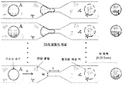

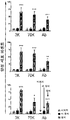
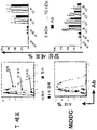


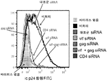
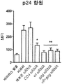

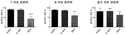


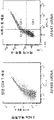
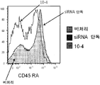


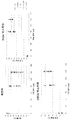
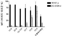


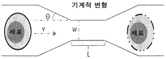

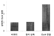
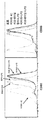
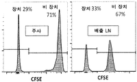
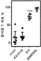


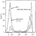
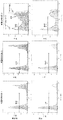
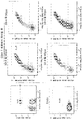
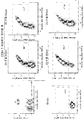


Classifications
-
- C—CHEMISTRY; METALLURGY
- C12—BIOCHEMISTRY; BEER; SPIRITS; WINE; VINEGAR; MICROBIOLOGY; ENZYMOLOGY; MUTATION OR GENETIC ENGINEERING
- C12M—APPARATUS FOR ENZYMOLOGY OR MICROBIOLOGY; APPARATUS FOR CULTURING MICROORGANISMS FOR PRODUCING BIOMASS, FOR GROWING CELLS OR FOR OBTAINING FERMENTATION OR METABOLIC PRODUCTS, i.e. BIOREACTORS OR FERMENTERS
- C12M35/00—Means for application of stress for stimulating the growth of microorganisms or the generation of fermentation or metabolic products; Means for electroporation or cell fusion
- C12M35/04—Mechanical means, e.g. sonic waves, stretching forces, pressure or shear stimuli
-
- A—HUMAN NECESSITIES
- A61—MEDICAL OR VETERINARY SCIENCE; HYGIENE
- A61K—PREPARATIONS FOR MEDICAL, DENTAL OR TOILETRY PURPOSES
- A61K39/00—Medicinal preparations containing antigens or antibodies
-
- A—HUMAN NECESSITIES
- A61—MEDICAL OR VETERINARY SCIENCE; HYGIENE
- A61K—PREPARATIONS FOR MEDICAL, DENTAL OR TOILETRY PURPOSES
- A61K45/00—Medicinal preparations containing active ingredients not provided for in groups A61K31/00 - A61K41/00
- A61K45/06—Mixtures of active ingredients without chemical characterisation, e.g. antiphlogistics and cardiaca
-
- A—HUMAN NECESSITIES
- A61—MEDICAL OR VETERINARY SCIENCE; HYGIENE
- A61K—PREPARATIONS FOR MEDICAL, DENTAL OR TOILETRY PURPOSES
- A61K49/00—Preparations for testing in vivo
- A61K49/001—Preparation for luminescence or biological staining
- A61K49/0013—Luminescence
- A61K49/0017—Fluorescence in vivo
-
- A—HUMAN NECESSITIES
- A61—MEDICAL OR VETERINARY SCIENCE; HYGIENE
- A61P—SPECIFIC THERAPEUTIC ACTIVITY OF CHEMICAL COMPOUNDS OR MEDICINAL PREPARATIONS
- A61P35/00—Antineoplastic agents
-
- A—HUMAN NECESSITIES
- A61—MEDICAL OR VETERINARY SCIENCE; HYGIENE
- A61P—SPECIFIC THERAPEUTIC ACTIVITY OF CHEMICAL COMPOUNDS OR MEDICINAL PREPARATIONS
- A61P37/00—Drugs for immunological or allergic disorders
- A61P37/02—Immunomodulators
-
- A—HUMAN NECESSITIES
- A61—MEDICAL OR VETERINARY SCIENCE; HYGIENE
- A61P—SPECIFIC THERAPEUTIC ACTIVITY OF CHEMICAL COMPOUNDS OR MEDICINAL PREPARATIONS
- A61P43/00—Drugs for specific purposes, not provided for in groups A61P1/00-A61P41/00
-
- C—CHEMISTRY; METALLURGY
- C12—BIOCHEMISTRY; BEER; SPIRITS; WINE; VINEGAR; MICROBIOLOGY; ENZYMOLOGY; MUTATION OR GENETIC ENGINEERING
- C12M—APPARATUS FOR ENZYMOLOGY OR MICROBIOLOGY; APPARATUS FOR CULTURING MICROORGANISMS FOR PRODUCING BIOMASS, FOR GROWING CELLS OR FOR OBTAINING FERMENTATION OR METABOLIC PRODUCTS, i.e. BIOREACTORS OR FERMENTERS
- C12M23/00—Constructional details, e.g. recesses, hinges
- C12M23/02—Form or structure of the vessel
- C12M23/16—Microfluidic devices; Capillary tubes
-
- C—CHEMISTRY; METALLURGY
- C12—BIOCHEMISTRY; BEER; SPIRITS; WINE; VINEGAR; MICROBIOLOGY; ENZYMOLOGY; MUTATION OR GENETIC ENGINEERING
- C12M—APPARATUS FOR ENZYMOLOGY OR MICROBIOLOGY; APPARATUS FOR CULTURING MICROORGANISMS FOR PRODUCING BIOMASS, FOR GROWING CELLS OR FOR OBTAINING FERMENTATION OR METABOLIC PRODUCTS, i.e. BIOREACTORS OR FERMENTERS
- C12M35/00—Means for application of stress for stimulating the growth of microorganisms or the generation of fermentation or metabolic products; Means for electroporation or cell fusion
-
- C—CHEMISTRY; METALLURGY
- C12—BIOCHEMISTRY; BEER; SPIRITS; WINE; VINEGAR; MICROBIOLOGY; ENZYMOLOGY; MUTATION OR GENETIC ENGINEERING
- C12N—MICROORGANISMS OR ENZYMES; COMPOSITIONS THEREOF; PROPAGATING, PRESERVING, OR MAINTAINING MICROORGANISMS; MUTATION OR GENETIC ENGINEERING; CULTURE MEDIA
- C12N15/00—Mutation or genetic engineering; DNA or RNA concerning genetic engineering, vectors, e.g. plasmids, or their isolation, preparation or purification; Use of hosts therefor
- C12N15/09—Recombinant DNA-technology
- C12N15/87—Introduction of foreign genetic material using processes not otherwise provided for, e.g. co-transformation
-
- C—CHEMISTRY; METALLURGY
- C12—BIOCHEMISTRY; BEER; SPIRITS; WINE; VINEGAR; MICROBIOLOGY; ENZYMOLOGY; MUTATION OR GENETIC ENGINEERING
- C12N—MICROORGANISMS OR ENZYMES; COMPOSITIONS THEREOF; PROPAGATING, PRESERVING, OR MAINTAINING MICROORGANISMS; MUTATION OR GENETIC ENGINEERING; CULTURE MEDIA
- C12N5/00—Undifferentiated human, animal or plant cells, e.g. cell lines; Tissues; Cultivation or maintenance thereof; Culture media therefor
- C12N5/06—Animal cells or tissues; Human cells or tissues
- C12N5/0602—Vertebrate cells
- C12N5/0634—Cells from the blood or the immune system
- C12N5/0635—B lymphocytes
-
- C—CHEMISTRY; METALLURGY
- C12—BIOCHEMISTRY; BEER; SPIRITS; WINE; VINEGAR; MICROBIOLOGY; ENZYMOLOGY; MUTATION OR GENETIC ENGINEERING
- C12N—MICROORGANISMS OR ENZYMES; COMPOSITIONS THEREOF; PROPAGATING, PRESERVING, OR MAINTAINING MICROORGANISMS; MUTATION OR GENETIC ENGINEERING; CULTURE MEDIA
- C12N5/00—Undifferentiated human, animal or plant cells, e.g. cell lines; Tissues; Cultivation or maintenance thereof; Culture media therefor
- C12N5/06—Animal cells or tissues; Human cells or tissues
- C12N5/0602—Vertebrate cells
- C12N5/0634—Cells from the blood or the immune system
- C12N5/0636—T lymphocytes
-
- C—CHEMISTRY; METALLURGY
- C12—BIOCHEMISTRY; BEER; SPIRITS; WINE; VINEGAR; MICROBIOLOGY; ENZYMOLOGY; MUTATION OR GENETIC ENGINEERING
- C12N—MICROORGANISMS OR ENZYMES; COMPOSITIONS THEREOF; PROPAGATING, PRESERVING, OR MAINTAINING MICROORGANISMS; MUTATION OR GENETIC ENGINEERING; CULTURE MEDIA
- C12N5/00—Undifferentiated human, animal or plant cells, e.g. cell lines; Tissues; Cultivation or maintenance thereof; Culture media therefor
- C12N5/06—Animal cells or tissues; Human cells or tissues
- C12N5/0602—Vertebrate cells
- C12N5/0634—Cells from the blood or the immune system
- C12N5/0639—Dendritic cells, e.g. Langherhans cells in the epidermis
-
- G—PHYSICS
- G01—MEASURING; TESTING
- G01N—INVESTIGATING OR ANALYSING MATERIALS BY DETERMINING THEIR CHEMICAL OR PHYSICAL PROPERTIES
- G01N33/00—Investigating or analysing materials by specific methods not covered by groups G01N1/00 - G01N31/00
- G01N33/48—Biological material, e.g. blood, urine; Haemocytometers
- G01N33/50—Chemical analysis of biological material, e.g. blood, urine; Testing involving biospecific ligand binding methods; Immunological testing
- G01N33/5005—Chemical analysis of biological material, e.g. blood, urine; Testing involving biospecific ligand binding methods; Immunological testing involving human or animal cells
- G01N33/5008—Chemical analysis of biological material, e.g. blood, urine; Testing involving biospecific ligand binding methods; Immunological testing involving human or animal cells for testing or evaluating the effect of chemical or biological compounds, e.g. drugs, cosmetics
- G01N33/5044—Chemical analysis of biological material, e.g. blood, urine; Testing involving biospecific ligand binding methods; Immunological testing involving human or animal cells for testing or evaluating the effect of chemical or biological compounds, e.g. drugs, cosmetics involving specific cell types
- G01N33/5047—Cells of the immune system
-
- A—HUMAN NECESSITIES
- A61—MEDICAL OR VETERINARY SCIENCE; HYGIENE
- A61K—PREPARATIONS FOR MEDICAL, DENTAL OR TOILETRY PURPOSES
- A61K39/00—Medicinal preparations containing antigens or antibodies
- A61K2039/51—Medicinal preparations containing antigens or antibodies comprising whole cells, viruses or DNA/RNA
- A61K2039/515—Animal cells
- A61K2039/5158—Antigen-pulsed cells, e.g. T-cells
-
- C—CHEMISTRY; METALLURGY
- C12—BIOCHEMISTRY; BEER; SPIRITS; WINE; VINEGAR; MICROBIOLOGY; ENZYMOLOGY; MUTATION OR GENETIC ENGINEERING
- C12N—MICROORGANISMS OR ENZYMES; COMPOSITIONS THEREOF; PROPAGATING, PRESERVING, OR MAINTAINING MICROORGANISMS; MUTATION OR GENETIC ENGINEERING; CULTURE MEDIA
- C12N2501/00—Active agents used in cell culture processes, e.g. differentation
- C12N2501/05—Adjuvants
-
- C—CHEMISTRY; METALLURGY
- C12—BIOCHEMISTRY; BEER; SPIRITS; WINE; VINEGAR; MICROBIOLOGY; ENZYMOLOGY; MUTATION OR GENETIC ENGINEERING
- C12N—MICROORGANISMS OR ENZYMES; COMPOSITIONS THEREOF; PROPAGATING, PRESERVING, OR MAINTAINING MICROORGANISMS; MUTATION OR GENETIC ENGINEERING; CULTURE MEDIA
- C12N2501/00—Active agents used in cell culture processes, e.g. differentation
- C12N2501/998—Proteins not provided for elsewhere
-
- C—CHEMISTRY; METALLURGY
- C12—BIOCHEMISTRY; BEER; SPIRITS; WINE; VINEGAR; MICROBIOLOGY; ENZYMOLOGY; MUTATION OR GENETIC ENGINEERING
- C12N—MICROORGANISMS OR ENZYMES; COMPOSITIONS THEREOF; PROPAGATING, PRESERVING, OR MAINTAINING MICROORGANISMS; MUTATION OR GENETIC ENGINEERING; CULTURE MEDIA
- C12N2510/00—Genetically modified cells
-
- C—CHEMISTRY; METALLURGY
- C12—BIOCHEMISTRY; BEER; SPIRITS; WINE; VINEGAR; MICROBIOLOGY; ENZYMOLOGY; MUTATION OR GENETIC ENGINEERING
- C12N—MICROORGANISMS OR ENZYMES; COMPOSITIONS THEREOF; PROPAGATING, PRESERVING, OR MAINTAINING MICROORGANISMS; MUTATION OR GENETIC ENGINEERING; CULTURE MEDIA
- C12N2527/00—Culture process characterised by the use of mechanical forces, e.g. strain, vibration
Abstract
항원과 같은 화합물을 면역 세포의 시토졸로 우선적으로 전달하는 방법 및 장치. 상기 방법은 표적 면역 세포를 포함하는 세포 현탁액을 미세유체 장치를 통해 통과시키는 단계, 및 현탁액을 전달하고자 하는 화합물(들) 또는 페이로드(payload)와 접촉시키는 단계를 포함한다.A method and apparatus for preferentially delivering a compound, such as an antigen, to a cytosol of an immune cell. The method comprises passing a cell suspension comprising target immune cells through a microfluidic device and contacting the suspension with a compound (s) or payload to be delivered.

Description
관련 relation 출원에 대한 참조Reference to the application
본 출원은 35 U.S.C.§ 119(e) 하에서 2014년 10월 31일자로 출원된 U.S. 가특허원 제62/073,548호에 대한 우선권의 이익을 청구하며, 이는 전문이 본원에 참고로 포함된다.The present application is a continuation-in-part of U. S. Patent Application No. < RTI ID = 0.0 > U. < / RTI > Claims priority 62 / 073,548, which is incorporated herein by reference in its entirety.
연방 후원 연구에 대한 성명서Statement of federal sponsorship research
본 발명은 국립 보건원에 의해 수여된 승인 번호 GM101420, AI112521, AI111595, 및 AI069259 하에서 정부 지원으로 이루어졌다. 정부는 발명에 있어 특정 권한을 갖는다.The invention was made with government support under authorization numbers GM101420, AI112521, AI111595, and AI069259, granted by the National Institutes of Health. The government has certain powers in the invention.
발명의 분야Field of invention
본 발명은 물질에서 세포로의 전달에 관한 것이다.The present invention relates to the transfer of substances to cells.
서열 order 목록에 대한 참조Reference to list
본 출원은 MS-윈도우와 운영 체계 호환성을 갖는 IBM-PC 기기 포맷으로 2015년 10월 30일자로 작성된 2.31 킬로바이트 크기의 "38172-508001WO_Sequence_Listing.txt"의 파일명으로 존재하는 뉴클레오티드 및/또는 아미노산 서열을 참고로 포함하며, 이는 본 출원의 일부로서 2015년 10월 30일자로 제출된 텍스트 파일에 담겨져 있다.This application refers to the nucleotide and / or amino acid sequence present in the filename of "38172-508001WO_Sequence_Listing.txt" of 2.31 kilobytes size, which was created on October 30, 2015 in IBM-PC device format compatible with MS- , Which is contained in a text file submitted as of October 30, 2015, as part of the present application.
세포질로의 다당류, 단백질, 또는 핵산과 같은 거대분자의 전달은 연구 또는 치료 목적을 위해 세포 기능을 일시적 또는 영구적으로 변화시킬 수 있다. 그러나, 일차 면역 세포, 특히 휴지기 림프구로의 세포내 전달을 위한 현존 기술들은 한계를 갖는다. 전기천공법은 상당한 세포 독성을 야기한다. 바이러스 벡터는 휴지기 림프구를 감염시킬 수 없다. 세포 막 투과 (또는 형질도입) 펩티드는 일차 림프구를 효율적으로 형질감염시키지 못한다. 항체-약물 복합체 및 접합체는 각 세포 유형을 위한 특이 항체 및 상이한 페이로드를 운반하기 위한 별개의 설계를 필요로 한다. 게다가, 이들은 생산하는데 많은 비용이 들고 잠재적으로 면역원성이다. 앱타머-siRNA 키메라성 RNA는 어떠한 독성 또는 면역 활성화 없이 생체내에 표적화된 유전자 녹다운을 야기하지만 작은 RNA를 전달하는 데에만 사용되는 것으로 나타났으며, 이들은 관심있는 각 세포에 대한 특정 표적화 앱타머를 확인하는 것을 필요로 한다. 나노입자 및 리포솜 기반 기술에 있어서의 발전이 식세포 항원 제시 세포, 예를 들면, 수지상 세포 및 단핵구/대식세포로의 약물 및 항원의 개선된 세포내 전달을 초래하였지만, 림프구에 대해서는 효과적이지 못하다. 이러한 방법들 중의 대부분은 페이로드의 엔도솜 흡수를 야기하고, 페이로드의 매우 작은 부분(~1-2%로 추정됨) 만이 엔도솜에서 시토졸로 빠져나오며, 여기서 이것은 생물학적 활성을 위한 트래픽을 필요로 한다. 이러한 기술들 중의 다수는 또한 세포에 비생분해성 패킹 또는 전달 물질의 축적을 야기하며, 이것이 세포 기능에 영향을 미칠 수 있다.Delivery of macromolecules such as polysaccharides, proteins, or nucleic acids into the cytoplasm can temporarily or permanently alter cellular function for research or therapeutic purposes. However, existing techniques for intracellular delivery to primary immune cells, particularly dormant lymphocytes, have limitations. Electroporation causes significant cytotoxicity. Viral vectors can not infect resting lymphocytes. Cell membrane permeability (or transduction) peptides do not efficiently transfect primary lymphocytes. Antibody-drug complexes and conjugates require separate designs to carry specific antibodies and different payloads for each cell type. In addition, they are costly to produce and potentially immunogenic. Aptamer-siRNA Chimeric RNAs have been shown to cause targeted gene knockdown in vivo without any toxicity or immune activation, but are used only to deliver small RNAs, which are specific targeting aptamers for each cell of interest It needs to be confirmed. Advances in nanoparticle and liposome-based technologies have led to improved intracellular delivery of drugs and antigens to phagocytic antigen presenting cells, e. G., Dendritic cells and monocytes / macrophages, but are not effective for lymphocytes. Most of these methods result in endosomal absorption of the payload, and only a very small fraction of the payload (estimated at 1-2%) is excreted from the endosome into the cytosol, where it is necessary to have the traffic for biological activity . Many of these techniques also lead to the accumulation of non-biodegradable packing or transducing material in the cells, which can affect cellular function.
따라서, 면역 세포로의 다양한 거대분자의 효율적인 비독성 전달을 가능케 하는 대안적인 기술들이 요구되고 있다.Accordingly, there is a need for alternative techniques that enable efficient non-toxic delivery of various macromolecules to immune cells.
본 발명은 면역 세포로의 화합물 또는 조성물의 전달과 관련된 이전의 문제들에 대한 해결책을 제공한다. 본 발명 이전에는, 화합물, 예컨대, 단백질, 핵산, 탄수화물의 도입이 어려웠고 비능률적으로 일어나고/났으며 독성 화합물 또는 바이러스 벡터와 같은 바람직하지 않은 매개체의 존재를 필요로 하였다. 본 발명에 따르면, 면역 세포 기능을 가공하는 방법은 면역 세포의 세포질을 둘러싼 막의 일시적인 교란에 의한 화합물의 세포내 전달을 포함한다. 예를 들면, 화합물을 면역 세포의 시토졸로 우선적으로 전달하기 위한 바이러스 벡터-비함유 방법은 표적 면역 세포를 포함하는 세포 현탁액을 미세유체 장치(microfluidic device)를 통해 통과시키는 단계 및 현탁액을 전달하고자 하는 화합물(들) 또는 페이로드와 접촉시키는 단계를 포함한다. 장치는 10-60㎛의 협착 길이 및 3-8㎛, 예컨대, 3-4㎛ 또는 4㎛의 협착 폭을 포함한다.The present invention provides a solution to previous problems associated with the delivery of a compound or composition to an immune cell. Prior to the present invention, the introduction of compounds, such as proteins, nucleic acids, carbohydrates, was inefficient and ineffective and required the presence of undesirable mediators such as toxic compounds or viral vectors. According to the present invention, a method of processing immune cell function involves intracellular delivery of the compound by transient disturbance of the membrane surrounding the cytoplasm of the immune cell. For example, a viral vector-free method for preferentially delivering a compound to a cytosol of an immune cell comprises passing a cell suspension comprising a target immune cell through a microfluidic device and passing the suspension through a microfluidic device With the compound (s) or payload. The device comprises a stenotic length of 10-60 μm and a stenotic width of 3-8 μm, eg 3-4 μm or 4 μm.
예를 들면, 장치는 2㎛-10㎛의 직경의 협착을 포함한다. 비처리 T 및 B 세포에 관한 바람직한 구현예에서, 장치는 약 10, 15, 20, 25, 30, 또는 10-30㎛의 길이, 약 3, 3.5, 4, 또는 3-4㎛의 폭, 약 15, 20, 25, 또는 15-25㎛의 깊이, 및/또는 약 5, 6, 7, 8, 9, 10, 11, 12, 13, 14, 15, 또는 5-15도 각도를 갖는 협착을 포함한다.For example, the device includes a stenosis of a diameter of 2 [mu] m to 10 [mu] m. In a preferred embodiment about untreated T and B cells, the device has a length of about 10, 15, 20, 25, 30, or 10-30 microns, a width of about 3, 3.5, 4, 15, 20, 25, or 15-25 microns in depth and / or about 5, 6, 7, 8, 9, 10, 11, 12, 13, 14, 15, .
협착을 통한 통과 후, 면역 세포로 전달되는 화합물의 양은 비-면역 세포로 전달된 것보다 또는, 예컨대, 세포내이입(endocytosis) 단독에 의한 세포 스퀴징의 부재하에서 면역 세포로 전달되는 양에 비해 적어도 10%(예컨대, 20%, 50%, 2배, 5배, 10배, 또는 그 이상) 더 크다. 예를 들면, 면역 세포는 B 세포, T 세포, 대식세포, 또는 수지상 세포를 포함한다. 우선적으로 면역 세포로의 페이로드의 전달을 위해, 예시적인 장치는 세포가 통과하는 30㎛의 협착 길이 및 4㎛의 협착 폭을 포함하는 하나 이상의 채널을 특징으로 한다.After passage through the stenosis, the amount of the compound delivered to the immune cells is greater than that delivered to the non-immune cells or, for example, to the amount delivered to the immune cells in the absence of cell squeezing by the endocytosis alone At least 10% (e.g., 20%, 50%, 2 times, 5 times, 10 times, or more). For example, the immune cells include B cells, T cells, macrophages, or dendritic cells. For delivery of the payload primarily to the immune cells, the exemplary device is characterized by one or more channels comprising a stenotic length of 30 mu m through which the cells pass and a stenotic width of 4 mu m.
섭씨 0 내지 45도의 온도, 예컨대, 0-25℃가 세포 처리 동안 사용된다. 예를 들면, 비처리 T 세포, B 세포 및/또는 단핵구의 처리는 4-8℃의 온도에서, 예컨대, 빙상에서 수행된다. 또 다른 예에서, 수지상 세포, 활성화 T 세포, 및/또는 활성화 B 세포는 20-25℃의 온도에서, 예컨대, 전형적인 주위 실온에서 장치를 사용하여 처리된다.Temperatures of 0 to 45 degrees Celsius, such as 0-25 DEG C, are used during cell processing. For example, treatment of untreated T cells, B cells and / or mononuclear cells is carried out at a temperature of 4-8 ° C, for example on ice sheets. In another example, dendritic cells, activated T cells, and / or activated B cells are treated using the device at a temperature of 20-25 占 폚, e.g., at a typical ambient room temperature.
페이로드는 면역 세포의 세포질로 전달하고자 하는 임의의 분자 또는 화합물을 함유한다. 예를 들면, 화합물은 항원, 예컨대, 질환-관련 항원, 예를 들면, 종양 항원, 바이러스 항원, 박테리아 항원, 또는 진균 항원을 포함한다. 항원은 정제될 수 있거나 다른 성분들의 혼합물일 수 있으며, 예컨대, 항원은 종양 세포 용해물 또는 대상체, 예컨대, 전염병에 걸린 대상체로부터의 생검 감염되거나 질환에 걸친 조직의 용해물과 같은 세포 용해물에 존재한다. 몇몇 예에서, 항원은 전체, 전장 (또는 비가공된) 단백질 항원, 예컨대, 7, 8, 9, 또는 10개 아미노산의 길이를 초과하는 단백질 또는 펩티드를 포함한다. 다른 적하 분자는 핵산, 예를 들면, siRNA, mRNA, miRNA, 암호화 또는 비-암호화 올리고뉴클레오티드 뿐만 아니라 소분자, 예컨대, 소분자 프로브를 포함한다. 핵산, 예를 들면, DNA, 예컨대, 발현 벡터, 예를 들면, 플라스미드가 또한 바이러스 벡터를 필요로 하지 않으면서 이러한 방식으로 전달된다.The payload contains any molecule or compound that is intended to be delivered to the cytoplasm of the immune cell. For example, the compound includes an antigen, such as a disease-associated antigen, for example, a tumor antigen, a viral antigen, a bacterial antigen, or a fungal antigen. The antigen may be purified or a mixture of other components, for example, the antigen may be present in a cell lysate such as a tumor cell lysate or a subject, such as a biopsy from an infected subject, or a lysate of tissue across the disease do. In some instances, the antigen comprises a protein or peptide that is greater than the length of a full, full-length (or unprocessed) protein antigen, such as 7, 8, 9, or 10 amino acids. Other loading molecules include nucleic acids, such as siRNA, mRNA, miRNA, coding or non-coding oligonucleotides, as well as small molecules, such as small molecule probes. Nucleic acids, such as DNA, for example, expression vectors, such as plasmids, are also delivered in this manner without the need for viral vectors.
몇몇 예에서, 면역 세포는 활성화된 상태에 비해 휴지 상태에 있다. 예를 들면, 세포는 다음 마커들의 발현을 특징으로 한다: CD25, KLRG1, CD80, CD86, PD-1, PDL-1, CTLA-4, CD28, CD3, MHC-I, MHC-II, CD62L, CCR7, CX3CR1 및 CXCR5, 이들 각각은 가공(기재된 방법들을 사용하여 분자를 면역 세포로 도입함으로써 증가 또는 감소)될 수 있다. 세포 현탁액은 가공된 세포, 예컨대, 재현탁된 연막 세포(분별된 백혈구 세포) 또는 전혈을 포함한다. 비처리 면역 세포, 예컨대, T 세포는 (활성화된 세포에 비해) CD25, CD80, CD86, PD-1, 및 CTLA-4의 비교적 낮은 수준의 발현 및 비교적 높은 수준의 CCR7을 특징으로 한다.In some instances, immune cells are in a dormant state relative to the activated state. For example, the cells are characterized by the expression of the following markers: CD25, KLRG1, CD80, CD86, PD-1, PDL-1, CTLA-4, CD28, CD3, MHC-I, MHC- , CX3CR1 and CXCR5, each of which can be processed (increased or decreased by introducing molecules into immune cells using the methods described). The cell suspension comprises processed cells, such as resuspended swollen cells (fractionated leukocyte cells) or whole blood. Untreated immune cells, such as T cells, are characterized by relatively low levels of expression of CD25, CD80, CD86, PD-1, and CTLA-4 (relative to activated cells) and relatively high levels of CCR7.
비-면역 세포에 비해 화합물을 면역 세포로 우선적으로 전달하기 위한 장치는 적어도 하나의 미세유체 채널을, 예컨대, 시린지 형태로 포함하거나, 또는 다수의 채널을, 예컨대, 마이크로칩 또는 미세유체 장치의 형태로 포함한다. 예를 들면, 채널은 30㎛의 협착 길이 및 4㎛의 협착 폭을 포함한다.An apparatus for preferentially delivering a compound to an immune cell relative to a non-immune cell comprises at least one microfluidic channel, for example in the form of a syringe, or a plurality of channels, for example in the form of a microchip or microfluidic device . For example, the channel includes a stenosis length of 30 mu m and a stenosis width of 4 mu m.
본 발명은 또한 화합물의 세포내 전달에 의해 면역 세포 기능을 가공하는 방법을 포함하며, 여기서 전달은 면역 세포의 세포질을 둘러싼 막을 일시적으로 교란시키고 항원을 시토졸로 전달함으로써 매개된다. 예를 들면, 항원은 2, 3, 4, 5, 6, 7, 8, 9, 또는 10개 이상의 아미노산의 길이를 포함하고, 여기서, 면역 세포는 항원을 길이가 11개 아미노산 미만인 펩티드로 가공한다. 이때 세포는 면역 세포의 표면 상에 더 짧아지고 가공된 펩티드, 항원의 I종 조직적합 항원 제한 가공 형태를 나타낸다. 예를 들면, CD8+ T 세포 또는 세포독성 T 세포로의 I종 제시를 위해 가공된 MHC/HLA에 대한 펩티드는 8-10개 잔기의 범위이고, CD4+ T 세포 또는 헬퍼 세포로의 MHC/HLA II종 제시를 위해 가공된 펩티드는 14-20개 잔기의 범위이다. 8개 잔기보다 더 짧은 펩티드(예컨대, 2, 3, 4, 5, 6, 또는 7개 잔기)가 다른 T 세포 타입 또는 NK 세포로의 제시를 위해 유용하다.The invention also includes a method of processing immune cell function by intracellular delivery of a compound wherein the delivery is mediated by transiently disrupting the membrane surrounding the cytoplasm of the immune cell and delivering the antigen to the cytosol. For example, an antigen comprises a length of 2, 3, 4, 5, 6, 7, 8, 9, or more than 10 amino acids, wherein the immune cell processes the antigen to a peptide of less than 11 amino acids in length . At this time, the cells show a shortened and processed peptide on the surface of immune cells, a type I histocompatibility antigen restricted processing form of the antigen. For example, peptides for MHC / HLA engineered for the presentation of type I to CD8 + T cells or cytotoxic T cells range in size from 8-10 residues and include MHC / HLA II species to CD4 + T cells or helper cells Peptides processed for presentation are in the range of 14-20 residues. Peptides (e.g., 2, 3, 4, 5, 6, or 7 residues) shorter than 8 residues are useful for presentation to other T cell types or NK cells.
예를 들면, 면역 세포를 2㎛-10㎛의 직경의 협착을 통해 통과시킴으로써 세포 막이 교란된다. 세포 스퀴징에 의해 시토졸로 전달된 항원은 전장의 비가공된 단백질이거나 또는 항원 제시 세포에 의한 항원 제시를 위해 조직적합 항원에 결합하기에 적합한 크기/길이로 가공되어야 하는 펩티드이다. 면역 세포 기능을 가공하는 방법은 항원-부하된 면역 세포를 효과기 T 세포와 접촉시키고 세포독성 T 세포 면역 반응을 활성화시킴을 추가로 포함할 수 있다. 몇몇 예에서, 스퀴징된 항원-부하 면역 세포는 B 세포, 수지상 세포 또는 대식세포를 포함한다. 또 다른 예에서, 스퀴징된 항원-부하 면역 세포는 T 세포를 포함한다. 어느 경우에나, 스퀴징된 항원 부하 면역 세포는 스퀴징, 예컨대, 세포내이입 또는 음세포작용 단독에 의한 흡수의 부재하에서 동일한 항원 또는 페이로드와 접촉된 면역 세포에 비해 적어도 10%, 25%, 50%, 2배, 5배, 10배, 20배, 25배, 50배, 또는 그 이상의 항원 또는 기타의 페이로드 조성물을 포함한다.For example, the cell membrane is disturbed by passing the immune cells through the stenosis of a diameter of 2 탆 - 10 탆. Antigens delivered by cytosine to the cytosol are either unprocessed proteins of the full length or peptides that have to be processed to a size / length suitable for binding to the histocompatibility antigens for antigen presentation by antigen presenting cells. Methods of processing immune cell function can further include contacting antigen-loaded immune cells with effector T cells and activating cytotoxic T cell immune responses. In some instances, the squashed antigen-loaded immune cells include B cells, dendritic cells, or macrophages. In another example, the squashed antigen-loaded immune cells comprise T cells. In any case, the squamous antigen-loaded immune cells are at least 10%, 25%, 30%, 50%, 50% or more as compared to the immune cells contacted with the same antigen or payload in the absence of squeegeeing, 50%, 2-fold, 5-fold, 10-fold, 20-fold, 25-fold, 50-fold, or more antigen or other payload composition.
면역 세포의 기능, 활성, 또는 활성화 상태는 이러한 처리 후 변화된다. 예를 들면, T 세포 상에 항원 제시 표현형을 부여하는 방법은 상기한 바와 같은 미세유체 장치를 통해 T 세포를 통과시킴으로써 항원, 예컨대, 전체 비가공된 단백질 또는 이의 단편을 T 세포의 시토졸로 전달함으로써 수행된다. 예를 들면, 장치는 약 2㎛-10㎛의 직경의 협착을 포함하며, T 세포는 미세유체 장치 통과 후 T 세포의 표면 상에 항원의 I종 조직적합 항원-제한된, 가공된 형태를 포함한다. 방법은 제1 (스퀴징된, 항원-부하) T 세포를 제2 T 세포와 접촉시킴을 추가로 포함할 수 있으며, 제2 T 세포는 I종 조직적합 항원 제한된 세포독성 T 세포 표현형을 포함한다. 예시적인 항원은 하나 이상의 종양 항원, 예컨대, 종양 항원의 혼합물, 예를 들면, 종양 생검 용해물, 또는 바이러스 항원을 포함한다. 이러한 방식으로의 항원-부하 T 세포의 생산이 시험관내 및/또는 생체내에서 사용되어 세포독성 T 세포 반응을 발휘한다. 따라서, 항원에 특이적인 세포독성 T 세포 반응을 활성화시키기 위한 세포-스퀴징된, 항원 부하된 T 세포 반응의 사용이 본 발명에 포함된다. 예를 들면, 이러한 T 세포는 전달/부하되는 항원에 따라 종양 세포를 사멸시키고/시키거나 바이러스-감염된 세포를 사멸시킴으로써 임상적 유용성을 부여한다.The function, activity, or activation state of the immune cell changes after such treatment. For example, a method of imparting an antigen-presenting phenotype on a T cell can be achieved by passing an antigen, such as the entire unprocessed protein or fragment thereof, to a cytosol of the T cell by passing the T cell through the microfluidic device as described above . For example, the device comprises a stenosis of a diameter of about 2 [mu] m to 10 [mu] m, and the T cell comprises a Type I tissue-compatible antigen-restricted, processed form of the antigen on the surface of the T cell after passage through the microfluidic device . The method may further comprise contacting a first (squamous, antigen-loaded) T cell with a second T cell and the second T cell comprises a Type I histocompatibility antigen restricted cytotoxic T cell phenotype . Exemplary antigens include one or more tumor antigens, e. G., Mixtures of tumor antigens, e. G., Tumor biopsy seafood, or viral antigens. The production of antigen-loaded T cells in this manner is used in vitro and / or in vivo to exert cytotoxic T cell responses. Thus, the use of a cell-squashed, antigen loaded T cell response to activate an antigen-specific cytotoxic T cell response is included in the present invention. For example, such T cells confer clinical utility by killing tumor cells and / or killing virus-infected cells according to the delivered / loaded antigen.
본원에 기재된 조성물은 정제된다. 정제된 화합물은 관심 화합물의 적어도 60중량%(건조 중량)이다. 바람직하게는, 제제는 관심 화합물의 적어도 75중량%, 보다 바람직하게는 적어도 90중량%, 가장 바람직하게는 적어도 99중량%이다. 순도는 임의의 표준방법에 의해, 예를 들면, 컬럼 크로마토그래피, 폴리아크릴아미드 겔 전기영동, 또는 HPLC 분석에 의해 측정된다. 예를 들면, 화합물, 예컨대, 단백질 항원은 천연에서 발생하는 하나 이상의 화합물로부터 분리되었다. 세포의 경우에, 정제된 집단이 선택된 세포 유형의 적어도 75%, 85%, 90%, 95%, 98%, 99% 또는 100%이다. 세포 분류기와 같은 장치를 사용하여 크기 또는 세포 표면 마커 발현에 의해 분리함을 포함하여 특정 세포 타입을 정제하거나 증폭하는 방법이 널리 공지되어 있다.The compositions described herein are purified. The purified compound is at least 60% by weight (dry weight) of the compound of interest. Preferably, the formulation is at least 75 wt%, more preferably at least 90 wt%, and most preferably at least 99 wt% of the compound of interest. Purity is measured by any standard method, for example, by column chromatography, polyacrylamide gel electrophoresis, or HPLC analysis. For example, a compound, e. G., A protein antigen, has been isolated from one or more compounds that occur in nature. In the case of cells, the purified population is at least 75%, 85%, 90%, 95%, 98%, 99% or 100% of the selected cell type. Methods for purifying or amplifying certain cell types, including isolation by size or cell surface marker expression using a device such as a cell sorter, are well known.
본 발명의 다른 특징 및 이점은 이의 바람직한 구현예의 하기 설명으로부터 및 청구항으로부터 자명해질 것이다. 본원에 인용된 모든 참조문헌은 참고로 포함된다.Other features and advantages of the present invention will become apparent from the following description of the preferred embodiments thereof and from the claims. All references cited herein are incorporated by reference.
도 1a는 전달 시스템을 보여주는 일련의 다이아그램이다. 시스템을 설정하기 위해, 각각의 미세유체 칩을 폴리카보네이트 유체 저장소와 접속할 수 있는 홀더에 탑재한다. 시스템을 작동시키기 위해, 전달하고자 하는 거대 분자를 세포와 혼합하고, 약 30-150㎕의 용적으로 장치 저장소에 부하한 다음 압력 공급원에 연결하여 미세유체 채널을 통한 유동을 유도한다. 도 1b는 세포 변형 및 페이로드 전달을 보여주는 다이아그램이다. 세포가 채널을 통해 유동함에 따라, 이들은 협착에서 변형하여, 막 교란을 야기한다. 그후 유체 중의 거대분자는 교란된 막을 통해 확산되고 막이 복구된 후 세포에 갇히게 된다. 이러한 도면들은 세포 스퀴징에 의한 전달을 입증한다.
도 2a 및 b는 뮤린 면역 세포로의 덱스트란 및 항체 전달을 보여주는 히스토그램 및 막대 그래프이다. 도 2a는 APC-표지된 IgG1을 전달하기 위해 CellSqueeze 장치에 의해 처리된 T 세포, B 세포 및 골수 세포 (CD11b+)의 대표적인 히스토그램을 보여준다. 도 2b는 전달 효율을 보여준다. 모든 결과는 처리한지 한 시간 내에 유동 세포계측에 의해 측정하였다. 죽은 세포는 요오드화프로피듐 염색에 의해 배제시켰다. 도 2b의 데이터(평균±SD)는 3가지 독립 실험으로부터의 것이다. 비처리 세포는 장치를 거치지 않거나 생체분자에 노출시켰다. '비 장치(no device)' 샘플은 생체분자와 함께 항온처리하였지만, 장치로 처리하지는 않았다. 이러한 대조군은 표면 결합, 세포내이입 및 기타의 백그라운드 효과를 을 설명하기 위한 것이다.
도 3a-d는 인간 면역 세포로의 덱스트란, 항체 및 siRNA의 전달을 보여주는 그래프이다. 도 3a에서, 인간 T 세포 및 MDDC를 캐스케이드 블루 표지된 3kDa 덱스트란, 플루오레세인 표지된 70kDa 덱스트란, 및 APC 표지된 IgG1의 전달에 대해 시험하였다. 30-4 (T 세포) 및 10-7 (MDDC) 장치(좌측) 및 장치 설계에 걸친 레플리케이트(우측)에 대한 대표적인 히스토그램이 나타내어져 있다. 도 3b는 각각 CD4+ T 세포 및 MDDC에서의 CD4 및 DC-SIGN 단백질 수준의 siRNA 매개된 녹다운을 보여준다. 용량 또는 협착 크기에 대한 녹다운 의존성을 평가하기 위해 상이한 siRNA 농도 및 장치 설계를 시험하였다. 도 3c는 인간 조절성 T 세포 또한 30-4 장치에 의한 처리에 대한 반응으로 CD4 발현의 상당한 녹다운을 나타내었음을 보여준다. 죽은 세포는 전달 또는 녹다운 분석에 배제시켰다. 도 3d는 Amaxa에 의한 뉴클레오펙션에 대한 T 세포에서의 장치 성능의 비교를 보여준다. CD4에 대한 siRNA의 전달 후 72 시간 후의 단백질 발현이 두 시스템에 대해 나타내어져 있다. 두 가지 방법에 의한 처리 후의 세포 생존력이 또한 나타내어져 있다.
도 4a-b는 내인성 및 바이러스성 유전자의 표적화된 녹다운에 의한 HIV 감염의 억제를 보여주는 그래프이다. 도 a에서, p24 항원에 대한 세포내 염색이 감염 24시간 후 처리된 인간 CD4+ T 세포에서의 HIV 감염 수준의 지표로서 사용되었다. 이러한 연구에서, vif 및/또는 gag, siRNA는 감염 24 시간 전에 전달되는 반면 CD4 siRNA는 감염 48 시간 전에 전달되었다. 도 4n은 실험 조건의 반복(min. N=4)에 걸쳐 p24 항원 염색의 중간 형광 강도를 보여준다. 데이터는 평균 + 1 표준 오차로서 나타내어 진다.
도 5는 백신화 방법의 다이아그램이다.
도 6은 뮤린 일차 면역 세포로의 3 kDa 및 70 kDa 덱스트란 및 항체의 흡수를 보여주는 일련의 선 그래프이다. 전달 효율 값을 계산하는데 사용되는 게이팅이 나타내어져 있다. 이들 데이터는 도 2에 나타낸 실험에 상응한다. 회색의 채워진 히스토그램은 비처리 세포를 나타내고, 흑색은 물질에 노출되었지만 장치로 처리되지 않은 세포를 나타내며, 채워지지 않은 회색은 표적 생체분자의 존재하에서 장치로 처리된 세포를 나타낸다.
도 7은 도 2에 나타낸 실험에 상응하는 세포 생존력 데이터를 보여주는 일련의 막대 그래프이다. ***는 30-4 장치로 처리된 세포의 생존력을 비 장치 또는 비 처리의 경우와 비교하여 p < 0.001임을 나타낸다. 장치로 처리된 B 세포 및 골수 세포의 생존력에 있어서의 변화는 비처리 또는 비 장치의 경우와 유의적으로 상이하지 않았다.
도 8은 골수-유래 수지상 세포(BMDC)로의 덱스트란 및 항체의 전달을 보여주는 일련의 그래프이다. BMDC는 골수 세포를 GM-CSF 함유 배지 중에서 8일간 배양함으로써 C57BL6 마우스로부터 생성되었다. 캐스케이드 블루-표지된 3 kDa 덱스트란, 플루오레세인-표지된 70 kDa 덱스트란, 및 APC-표지된 IgG1은 두 가지 장치 설계, 10-6 및 30-6을 사용하여 전달되었다.
도 9는 항체 및 덱스트란 전달의 상관성을 보여주는 그래프이다. 물질과 항온처리, 즉, 비 장치(흑색 점; 하부 좌측)와 비교하여 30-4 장치(회색 점; 중심, 상부 우측)를 사용한 T 세포로의 덱스트란 (3 kDa 및 70 kDa) 및 항체 전달.
도 10은 인간 CD4+ T 세포의 생존력을 보여주는 막대 그래프이다. 장치를 통과하는 세포는 비처리 대조군에 비해 감소된 생존력을 갖지만 뉴클레오펙션을 겪은 세포보다는 양호한 생존력을 갖는다. 일원 ANOVA에 이은 Boneferroni's 검정을 사용하여 통계적 유의도를 계산하였다. *는 p < 0.05를 나타내고 ***은 p < 0.001을 나타낸다. 다른 비교 그룹들은 유의적으로 상이한 생존력을 나타내지 않았다(즉, 비처리 또는 30-4에 비해 10-4, 및 뉴클레오펙션에 비해 30-4).
도 11은 인간 MDDC에 대한 상이한 장치 설계 시험으로부터의 전달 (상부) 및 생존력 (하부) 결과를 보여주는 일련의 막대 그래프이다. 캐스케이드 블루 표지된 3kDa 덱스트란, 플루오레세인 표지된 70 kDa 덱스트란, 및 APC 표지된 IgG1 동종 대조군 항체는 6개의 상이한 장치 설계를 사용하여 그리고 Amaxa 뉴클레오펙션을 사용하여 전달되었다. 생존력 및 전달 결과는 처리 직후 측정하였다.
도 12는 Alexa 488 또는 Alexa 647 표지된 siRNA 및 3 kDa 캐스케이드 블루 표지된 덱스트란이 10-4i 장치에 의해 인간 CD4 T 세포로 및 30-5x5i 장치에 의해 뮤린 B 세포로 동시에 전달되었음을 보여주는 일련의 그래프이다. 데이터는 두 가지 물질의 전달이 밀접한 상관성이 있음을 나타낸다. 이 결과는 제안된 확산 전달 메카니즘과 일관되며, 즉, 전달 효율은 화학 구조 보다는 물질 크기에 크게 좌우된다.
도 13은 CD45RA 발현을 보여주는 그래프이다. CD45RA에 대한 siRNA는 10-4 장치에 의해 인간 T 세포로 전달되었다. 녹다운은 처리한지 72시간 후 유동 세포계측에 의해 측정하였다.
도 14는 (전달한지 48시간 후 PCR에 의해 측정된 바와 같은) CD4 mRNA 녹다운을 보여주는 막대 그래프이다.
도 15는 유동 세포계측에 의해 측정되는 바와 같이 처리후 2주에 걸쳐 CD4+ 인간 T 세포에서의 CD4의 발현 수준을 보여주는 일련의 막대 그래프이다. CD3 수준을 또한 대조군 유전자로서 측정하였다.
도 16은 인간 단핵구로의 모형 적하물(model cargo), 덱스트란의 전달을 보여주는 일련의 그래프이다. 단핵구는 인간 혈액으로부터 유도되었다. 캐스케이드 블루 표지된 3 kDa 덱스트란, 및 플루오레세인 표지된 70 kDa 덱스트란은 두 가지 상이한 작동 압력에서 네 개의 상이한 장치 설계를 사용하여 전달되었다. 0psi 케이스는 덱스트란에만 노출되었을 뿐 장치로 처리되지는 않은 대조군에 상응한다. 생존력은 요오드화프로피듐 염색에 의해 측정하였다.
도 17은 인간 B 세포로의 덱스트란의 전달을 보여주는 일련의 그래프이다. B 세포는 인간 혈액으로부터 유도되었다. 캐스케이드 블루 표지된 3 kDa 덱스트란, 및 플루오레세인 표지된 2 MDa 덱스트란은 두 가지 상이한 작동 압력에서 네 개의 상이한 장치 설계를 사용하여 전달되었다. 0psi 케이스는 덱스트란에만 노출되었을 뿐 장치로 처리되지는 않은 대조군에 상응한다. 생존력은 요오드화프로피듐 염색에 의해 측정하였다.
도 18은 처리한지 72 시간 후의 DC-Sign의 측정된 단백질 수준을 보여주는 막대 그래프이다. 단백질 녹다운은 6개의 상이한 장치 설계에 걸쳐 측정하고 뉴클레오펙션과 비교하였다. 뉴클레오펙션은 대조군 siRNA 전달의 경우에도 DC-Sign의 ~50% 비특이 녹다운을 야기하는 것을 보인다는 것을 주지한다. 이것은 전기천공 처리로 인한 오프-타겟 효과(off-target effect)를 나타낼 수 있으며, 즉, 전기장에의 세포의 노출이 세포, 특히 표적 단백질에 손상을 초래하여, 단백질을 표적화하는 siRNA의 부재하에서 발현 수준의 측정된 감소를 야기한다. 이러한 결과는 막 변형 방법이 비특히 (오프-타겟) 효과와 관련된 전기천공/뉴클레오펙션 방법에 비해 더욱 특이적임을 나타낸다.
도 19는 전혈에서 세포로의 불투과성 Alexa-488 표지된 10 kDa 덱스트란 염료의 전달에 관한 데이터를 보여주는 일련의 그래프이다. 형광 표지된 염료를 전혈과 혼합하고, 전혈 + 표지된 염료의 혼합물을 장치로 퍼지게 한 다음 RBC 용해 후 FACS에 의해 혈액 세포로의 전달에 대해 측정하였다. 결과는 염료가 세포로 성공적으로 전달되었으며 "세포 불투과성"을 특징으로 하는 적하 화합물이 세포 스퀴징을 사용하여 면역 세포로 효과적으로 전달된다는 것을 입증한다. 전달 과정이 전혈에서 작동한다는 것이 놀랍다. 전혈은 정제, 예컨대, 적혈구 세포로부터 말초 혈관 단핵 세포의 분멸 또는 분리 없이 가공하기가 매우 어려운데, 본원에 개시된 장치는 전혈 웰에서 화합물을 면역 세포로 전달할 수 있다. 비제한적인 예에 대해, 도 33 및 34 참조.
도 20은 미세유체 막 교란 시스템의 다이아그램이다.
도 21a-b는 Optimem 완충액 중에서 120 psi에서 10-4 칩으로 전달한지 하루 후의 mRNA 발현을 보여주는 점 도표이다.
도 22a는 상이한 칩 각도로 상이한 압력에서 10 kDa Alexa 488-표지된 덱스트란의 전달을 보여주는 다이아그램이고 도 22b는 막대 그래프이다. 괄호 안의 숫자는 협착 각도이다. 칩 각도는 0-180도 범위, 예컨대, 11-105도 범위이다. 도식은 칩 각도를 나타낸다. 깊이 파라미터는 2㎛ 내지 1 mm, 예컨대, ~20μm 범위이고 U.S. 특허 공보 제20140287509호(본원에 참고로 포함됨)에 추가로 기재되어 있다. 예시적인 파라미터는 비처리 T 및 B 세포에 대해 0-30㎛ 길이/3-4㎛ 폭/20㎛ 깊이/11도 각도를 포함한다.
도 23은 NK 세포 및 pDC로의 전사 인자 전달을 보여주는 선 그래프이다. Oct4 mRNA 발현은 마우스 비장 NK 세포로의 Oct4 재조합 단백질의 전달 후 4시간 후에 측정하였다. 이러한 데이터는 전사 인자가 활성이며 내인성 Oct4의 발현을 유도할 수 있음을 나타낸다. Oct4 전사 인자는 iPS 생성에 필요한 네 가지 인자(Oct4, Klf4, cMyc, Sox2) 중의 하나이며 자체로 포지티브 피드백 조절를 발휘한다. 활성 Oct4 단백질의 전달은 증가된 Oct4 mRNA 발현을 산출한다. 활성 전사 인자의 전달은 단백질-기반 방법을 위한 재프로그래밍 공정에서 중대한 단계이다. 이러한 방법은 통합 위험을 최소화시키기 때문에 바이러스 및 DNA 기반 시스템을 능가하는 다수의 이점을 갖는다.
도 24a-b는 스퀴징에 대한 반응으로 어떠한 정상적인 DC 기능의 검출된 억제도 관찰되지 않았음을 보여주는 선 그래프이다. 도 24a: 비장 DC를 스퀴징에 의한, 항원 세포내이입 단독에 의한 처리 후 LPS (1ug/ml) 중에서 배양하였으며, 비처리된 세포는 CD80 및 CD86 발현을 상향조절하는 능력에 있어서 검출 가능한 차이를 보이지 않았다. 비 자극된 세포내이입 조건 및 4℃에서 유지된 비처리 세포가 대조군으로서 사용되었다. 도 24b: CD45 유사유전자형 마우스로부터의 비장 DC를 LPS (1ug/ml)를 갖는 C57BL6 마우스의 발바닥에 주사하고 주사한지 18 시간 후 빼낸 림프절에서 회수하였다. 림프절 회귀성에 있어서는 유의적인 차이가 검출되지 않았다.
도 25a-d는 막 변형 장치 시스템 및 방법의 사용이 다른 항원 전달 접근법에 비해 보다 효과적인 항원 제시를 야기함을 보여주는 그래프이다. 도 25a: 피하 DC 백신접종에 대한 반응으로 입양 전달된 CD8+ OT-I T 세포의 생체내 증식. 장치 처리된 BMDC(0.1 mg/ml의 Ova)는 항원을 세포내이입할 수 있는 것에 비해 T 세포 증식에 있어서 유의적인 증가(P < 0.0001)를 입증하였다. 도 25b: Ova 발현 흑색종 세포주(B16F10)의 세포 용해물로 처리된 BMDC와 공동배양된 CD8+ OT-I T 세포의 시험관내 증식. %는 10-6 CellSqueeze 장치에 의한 처리 전 BMDC 세포 현탁액에 첨가된 세포 용해물 물질의 분율을 나타낸다. CD8 T 세포는 APC와 접촉 전에 카복시플루오로세인 석신이미딜 에스테르 (CFSE)로 표지되었다. 이들이 증식하면, CFSE 염료가 딸 세포 사이에 분포하여 세포당 보다 낮은 형광 강도를 야기한다. 비증식된 T 세포는 높은 CFSE 강도를 보유한다. 도 25c: 항원-특이 IFNγ 분비 CD8 T 세포의 분율을 측정한다. 이러한 실험에서는, 마우스를 입양 전달된 OT-I T 세포의 부재하에서 Ova에 대해 백신접종하였다. 내인성, 항원 특이 반응은 백신접종한지 7일 후 백신접종된 마우스의 비장을 분리하고 이들을 시험관내에서 SIINFEKL 펩티드 (OVA 에피토프)로 재자극함으로써 측정하였다. 항원-특이 CD8 T 세포는 재자극에 대한 반응으로 IFNγ를 분비한다. 도 25d; 두 가지 상이한 장치 설계로 처리한 B 세포와 공동배양된 CD8+ OT-I T 세포의 시험관내 증식(0.1 mg/m1 Ova). CpG가 이러한 실험에서 아주반트로서 사용되었다.
도 26은 생체내 CFSE 증식 검정의 결과를 보여주는 그래프이다: 이 그래프는 항원 특이 CD8 T 세포의 증식을 측정한다. CD8 T 세포가 마우에서 활성화되고 증식됨에 따라 이들이 CFSE 염료를 희석시키고 더 낮은 형광 강도를 갖는다. 이 경우, 장치로 처리된 공여체 T 세포 또는 양성 대조군은 수용체 마우스에서 CD8 T 세포의 훨씬 더 큰 활성화 및 증식을 야기하였다. 이에 반해 세포내이입 대조군은 최소한의 효과를 나타낸다.
도 27은 항원 처리된 야생형 T 세포로의 백신접종에 대한 반응으로 마우스에서의 항원 특이 OT-I T 세포의 증식을 보여주는 일련의 히스토그램이다. T 세포 증식 반응은 CFSE 염색에 의해 측정되며, 염색은 세포가 증식함에 따라 희석된다. 보다 낮은 강도는 보다 큰 반응을 나타내고, 보다 높은 강도 피크는 반응이 없음/덜함을 나타낸다. 각 컬럼은 동일한 마우스로부터 유도된 림프절 및 비장을 갖는 실험의 레플리케이트를 나타낸다. 각 컬럼의 실험은 3마리의 마우스를 포함하였다(총 9마리 마우스).
도 28a-b는 DQ-OVA+3kDa-덱스트란+ T 세포 둘 다에 대한 게이팅을 보여주는 히스토그램이다.
도 29는 항원 제시 세포로서의 마우스 T 세포의 사용을 입증하는 막대 그래프이다. 비가공 알부민 항원을 부하하기 위해 세포 스퀴징에 의해 처리된 T 세포를 OT-1 (SIINFEKL-특이 T 세포주)와 배양하고 활성화 마커, CD25 및 CD69를 평가하였다.
도 30은 항원 제시 세포로서 B 세포의 스퀴즈-매개된 생산을 수행하고 특성화하기 위한 예시적인 과정의 개략도이다.
도 31은 세포-스퀴징된 B 세포는 강력한 CD8+ T 세포 증식을 유도함을 보여주는 일련의 히스토그램이다.
도 32는 세포-스퀴징된 수지상 세포(OVA 전달된)는 감마 인터페론을 분비함을 보여주는 일련의 히스토그램이다.
도 33은 비변형 전혈에서 직접 인간 B 및 T-세포로의 10kDa 물질의 전달을 보여주는 데이터 및 카툰을 보여준다.
도 34는 전혈에서의 생존력 및 전달 효율을 보여주는 그래프이다.Figure 1a is a series of diagrams illustrating the delivery system. To set up the system, each microfluidic chip is mounted in a holder that can be connected to a polycarbonate fluid reservoir. To operate the system, the macromolecules to be delivered are mixed with the cells, loaded into the device reservoir at a volume of about 30-150 [mu] l, and then connected to a pressure source to induce flow through the microfluidic channel. Figure 1B is a diagram showing cell deformation and payload delivery. As the cells flow through the channels, they deform in stenosis, causing membrane perturbation. The macromolecules in the fluid then diffuse through the perturbed membrane and become trapped in the cell after the membrane is restored. These figures demonstrate cell-squeezed delivery.
Figures 2a and b are histograms and histograms showing dextran and antibody delivery to murine immune cells. Figure 2a shows representative histograms of T cells, B cells and bone marrow cells (CD11b + ) treated by the CellSqueeze device to deliver APC-labeled IgG1. 2B shows the transfer efficiency. All results were measured by flow cytometry within one hour of treatment. Dead cells were excluded by iodidopropylamide staining. The data in Figure 2b (mean + - SD) is from three independent experiments. Untreated cells did not go through the device or were exposed to biomolecules. The 'no device' sample was incubated with the biomolecule but not the device. These controls are intended to explain surface binding, intracellular transfer, and other background effects.
Figures 3a-d are graphs showing the delivery of dextran, antibody and siRNA to human immune cells. In Figure 3a, human T cells and MDDCs were tested for delivery of cascade blue labeled 3kDa dextran, fluorescein labeled 70kDa dextran, and APC labeled IgG1. Representative histograms for 30-4 (T cells) and 10-7 (MDDC) devices (left) and replicators (right) over the device design are shown. Figure 3b shows siRNA mediated knockdown of CD4 and DC-SIGN protein levels in CD4 + T cells and MDDC, respectively. Different siRNA concentrations and device designs were tested to assess the knockdown dependence on capacity or stenosis size. Figure 3c shows that human regulatory T cells also displayed significant knockdown of CD4 expression in response to treatment with the 30-4 device. Dead cells were excluded in the transfer or knockdown assay. FIG. 3D shows a comparison of device performance in T cells for amplification by Amaxa. Protein expression 72 hours after delivery of siRNA to CD4 is shown for both systems. Cell viability after treatment by both methods is also shown.
Figures 4a-b are graphs showing inhibition of HIV infection by targeted knockdown of endogenous and viral genes. In Figure < RTI ID = 0.0 > a, < / RTI > intracellular staining for p24 antigen was used as an index of HIV infection level in human CD4 + T cells treated 24 hours after infection. In this study, vif and / or gag, siRNA were delivered 24 hours before infection, whereas CD4 siRNA was delivered 48 hours before infection. Figure 4n shows intermediate fluorescence intensity of p24 antigen staining over the course of the experiment (min. N = 4). The data is expressed as mean + 1 standard error.
5 is a diagram of a vaccination method.
Figure 6 is a series of line graphs showing the uptake of 3 kDa and 70 kDa dextran and antibody to murine primary immunocytes. The gating used to calculate the transfer efficiency value is shown. These data correspond to the experiment shown in Fig. A gray filled histogram represents untreated cells, black represents cells that have been exposed to the material but have not been treated with the device, and unfilled gray represents cells treated with the device in the presence of the target biomolecule.
Figure 7 is a series of bar graphs showing cell viability data corresponding to the experiment shown in Figure 2; *** indicates that the viability of cells treated with the 30-4 device is p < 0.001 compared to non-device or untreated cases. The changes in viability of B cell and bone marrow cells treated with the device were not significantly different from those of the untreated or non-treated cases.
Figure 8 is a series of graphs showing the delivery of dextran and antibody to bone marrow-derived dendritic cells (BMDC). BMDC was generated from C57BL6 mice by culturing bone marrow cells in GM-CSF containing medium for 8 days. Cascade blue-labeled 3 kDa dextran, fluorescein-labeled 70 kDa dextran, and APC-labeled IgG1 were delivered using two device designs, 10-6 and 30-6.
Figure 9 is a graph showing the correlation of antibody and dextran transfer. Dextran (3 kDa and 70 kDa) to T cells using 30-4 instrument (gray dot; center, upper right) compared with the material and incubation, ie, non-device (black dot; lower left) .
Figure 10 is a bar graph showing the viability of human CD4 + T cells. Cells that pass through the device have reduced survival compared to untreated controls but have better viability than cells that underwent nucleofection. Statistical significance was calculated using Boneferroni's test followed by one-way ANOVA. * Indicates p < 0.05 and *** indicates p < 0.001. Other comparison groups did not show significant different viability (i.e., 10-4 compared to untreated or 30-4, and 30-4 relative to nucleofection).
Figure 11 is a series of bar graphs showing the transfer (top) and viability (bottom) results from different device design tests for human MDDC. Cascaded blue-labeled 3 kDa dextran, fluorescein labeled 70 kDa dextran, and APC-labeled IgG1 homogeneous control antibodies were delivered using six different device designs and using Amaxa nucleofection. Survival and delivery results were measured immediately after treatment.
Figure 12 shows a series of graphs showing that Alexa 488 or Alexa 647 labeled siRNA and 3 kDa cascade blue labeled dextran were simultaneously delivered to human CD4 T cells and murine B cells by 30-5x5i device by 10-4i device to be. The data indicate that there is a close correlation between the transfer of the two materials. This result is consistent with the proposed diffusion transport mechanism, ie, the delivery efficiency is highly dependent on the material size rather than the chemical structure.
Figure 13 is a graph showing CD45RA expression. SiRNA for CD45RA was delivered to human T cells by the 10-4 device. The knockdown was measured by flow cytometry after 72 hours of treatment.
Figure 14 is a bar graph showing CD4 mRNA knockdown (as measured by PCR after 48 hours of delivery).
Figure 15 is a series of bar graphs showing the level of expression of CD4 in CD4 + human T cells over two weeks after treatment as measured by flow cytometry. CD3 levels were also measured as control genes.
Figure 16 is a series of graphs showing the delivery of model cargo, dextran, to human mononuclear cells. Mononuclear cells were derived from human blood. The cascaded blue-labeled 3 kDa dextran, and the fluorescein labeled 70 kDa dextran were delivered using four different device designs at two different working pressures. The 0 psi case corresponds to a control that was only exposed to dextran but not to the device. Survival was measured by propidium iodide staining.
Figure 17 is a series of graphs showing the delivery of dextran to human B cells. B cells were derived from human blood. Cascaded blue-labeled 3 kDa dextran, and fluorene-labeled 2 MDa dextran were delivered using four different device designs at two different working pressures. The 0 psi case corresponds to a control that was only exposed to dextran but not to the device. Survival was measured by propidium iodide staining.
Figure 18 is a bar graph showing the measured protein levels of DC-Sign 72 hours after treatment. Protein knockdown was measured over six different device designs and compared to nucleofection. It is noted that the nucleofection shows a ~ 50% non-specific knockdown of DC-Sign even in the case of control siRNA delivery. This may represent an off-target effect due to electroporation, i.e., exposure of the cells to an electric field causes damage to the cell, particularly the target protein, resulting in expression in the absence of siRNA targeting the protein Resulting in a measured decrease in level. These results indicate that the membrane modification method is more specific than the electroporation / nucleofection method associated with the non-specifically (off-target) effect.
Figure 19 is a series of graphs showing data on the transfer of impermeable Alexa-488 labeled 10 kDa dextran dyes from whole blood to cells. Fluorescently labeled dyes were mixed with whole blood and a mixture of whole blood + labeled dyes spread through the device and then measured for transfer to blood cells by FACS after RBC lysis. The results demonstrate that the dye has been successfully delivered to the cell and that the loading compound, characterized by "cell-impermeable" is effectively delivered to the immune cells using cell squeegee. It is surprising that the transfer process works on whole blood. Whole blood is very difficult to process without purification, e. G., The destruction or detachment of peripheral vascular mononuclear cells from red blood cells. The device disclosed herein is capable of delivering compounds to immune cells in whole blood wells. For non-limiting examples, see FIGS. 33 and 34.
20 is a diagram of a microfluidic membrane perturbation system.
Figures 21A-B are point plots showing mRNA expression after one day of delivery at 120 psi to the 10-4 chip in Optimem buffer.
Figure 22a is a diagram showing the delivery of 10 kDa Alexa 488-labeled dextran at different pressures at different chip angles and Figure 22b is a bar graph. The number in parentheses is the stenosis angle. The chip angle ranges from 0 to 180 degrees, for example, from 11 to 105 degrees. The schematic shows the chip angle. Depth parameters range from 2 [mu] m to 1 mm, e.g., ~ 20 [mu] m and are further described in US Patent Publication No. 20140287509 (incorporated herein by reference). Exemplary parameters include 0-30 mu m length / 3-4 mu m width / 20 mu m depth / 11 degree angle for untreated T and B cells.
23 is a line graph showing the transfer of the transcription factor into NK cells and pDC. Oct4 mRNA expression was measured 4 hours after delivery of Oct4 recombinant protein to mouse spleen NK cells. These data indicate that transcription factors are active and can induce the expression of endogenous Oct4. Oct4 transcription factor is one of the four factors required for iPS production (Oct4, Klf4, cMyc, Sox2) and exerts its own positive feedback regulation. Delivery of active Oct4 protein results in increased Oct4 mRNA expression. The transfer of active transcription factors is a critical step in the reprogramming process for protein-based methods. This method has a number of advantages over viruses and DNA-based systems because it minimizes the risk of integration.
Figures 24A-B are line graphs showing that no detected inhibition of any normal DC function was observed in response to squeezing. Figure 24a: Spleen DCs were cultured in LPS (1 ug / ml) after treatment with an intracellular transduction alone by squeegee, and untreated cells showed a detectable difference in the ability to upregulate CD80 and CD86 expression I did not see it. Non-stimulated intracellular entry conditions and untreated cells maintained at 4 < 0 > C were used as controls. Figure 24b: Spleen DC from CD45 quasi-genotype mice were injected into the soles of C57BL6 mice with LPS (1 ug / ml) and harvested at withdrawn lymph nodes 18 hours after injection. There was no significant difference in lymph node regression.
Figures 25a-d are graphs showing that the use of a membrane deformation device system and method results in more effective antigen presentation compared to other antigen delivery approaches. Figure 25a: In vivo proliferation of CD8 + OT-I T cells transduced with adoption in response to subcutaneous DC vaccination. The device-treated BMDC (0.1 mg / ml of Ova) demonstrated a significant increase in T cell proliferation (P <0.0001) compared to the intracellular transfer of the antigen. Figure 25b: In vitro proliferation of CD8 + OT-I T cells co-cultured with BMDC treated with cell lysate of Ova expressing melanoma cell line (B16F10). % Represents the fraction of cell lysate added to the BMDC cell suspension prior to treatment with 10-6 CellSqueeze device. CD8 T cells were labeled with carboxyfluorescein succinimidyl ester (CFSE) prior to contact with APC. When they proliferate, CFSE dyes are distributed among daughter cells, resulting in lower fluorescence intensity per cell. Non-proliferating T cells possess high CFSE intensity. Figure 25c: Determination of the fraction of antigen-specific IFN gamma secreting CD8 T cells. In these experiments, mice were vaccinated against Ova in the absence of adoptive OT-I T cells. The endogenous, antigen specific responses were determined by isolating the spleen of vaccinated
Figure 26 is a graph showing the results of an in vivo CFSE proliferation assay: This graph measures the proliferation of antigen-specific CD8 T cells. As CD8 T cells are activated and proliferated in corn, they dilute the CFSE dye and have lower fluorescence intensity. In this case, the donor T cells or positive controls treated with the device resulted in much greater activation and proliferation of CD8 T cells in the recipient mice. In contrast, the intracellularly transferred control group has minimal effect.
Figure 27 is a series of histograms showing the proliferation of antigen-specific OT-I T cells in mice in response to vaccination with antigen-treated wild-type T cells. The T cell proliferative response is measured by CFSE staining, and the staining is diluted as the cells proliferate. A lower intensity indicates a greater response and a higher intensity peak indicates less / less response. Each column represents an experimental replicate with lymph node and spleen derived from the same mouse. Each column experiment included 3 mice (total 9 mice).
Figures 28a-b are histograms showing gating for both DQ-OVA + 3kDa-dextran + T cells.
29 is a bar graph demonstrating the use of mouse T cells as antigen presenting cells. T cells treated by cell squeezing to load non-processed albumin antigen were cultured with OT-1 (SIINFEKL-specific T cell line) and the activation markers, CD25 and CD69 were evaluated.
Figure 30 is a schematic diagram of an exemplary process for performing and characterizing squeezed-mediated production of B cells as antigen presenting cells.
Figure 31 is a series of histograms showing that cell-squashed B cells induce potent CD8 + T cell proliferation.
Figure 32 is a series of histograms showing that cell-squashed dendritic cells (OVA delivered) secrete gamma interferon.
Figure 33 shows data and cartoons showing the delivery of 10 kDa material directly from unmodified whole blood to human B and T-cells.
34 is a graph showing the viability and the transfer efficiency in whole blood.
벡터-비함유 미세유체 전달 플랫폼(CellSqueeze)이 최소한의 세포독성으로 예컨대, 마우스, 인간의 일차 면역 세포의 시토졸로 직접 거대분자를 전달하는데 사용된다. 이러한 접근법의 근간이 되는 원리는 표적 세포의 신속한 기계적 변형, 또는 스퀴징에 의한 일시적인 막 교란이며, 이것은 유체 매질에서의 거대분자의 확산에 의한 흡수를 가능케 하며 이후에 세포 막 회복이 뒤따른다(문헌 참조; 예컨대, U.S. 특허 공보 제20140287509호, 본원에 참고로 포함됨. 미세유체 설계의 라이브러리를 사용하여, 덱스트란 중합체, 항체 및 작은 간섭 RNA(siRNA)와 같은 시험 화합물의 흡수가 일차 인간 및 뮤린 T 세포, B 세포, 단핵구/대식세포, 및 수지상 세포+로 전달되었다. 결과는 각종 상이한 크기 및 유형의 거대분자를 전달하는 플랫폼의 유용성을 입증한다. 다양한 세포 직경(~8-30㎛), 분명한 형태, 이방성 및 막 가요성을 갖는 상이한 종류의 면역 세포로의 물질의 효율적인 전달은 상이한 세포 유형에 대한 특이적인 조건의 확인을 필요로 한다. 세포 스퀴징을 위해서는, 협착의 폭이 중요한 파라미터이며, 기하적 요소, 속도, 완충액, 및 온도와 같은 기타의 파라미터들 또한 적하물의 전달에 영향을 미칠 수 있다. 비처리 T 또는 B 세포로의 적하물의 전달을 위한 예시적인 협착 폭은 3-4㎛의 범위이며; 활성화된 T 또는 B 세포로의 전달을 위해서는 4-6㎛이고; 수지상 세포로의 전달을 위해서는 6-8㎛이다.A vector-free microfluidic transport platform (CellSqueeze) is used to deliver macromolecules directly into the cytosol of, for example, mice, primary human immune cells with minimal cytotoxicity. The principle underlying this approach is rapid mechanical deformation of the target cell, or transient membrane perturbation by squeezing, which allows absorption by the diffusion of macromolecules in the fluid medium, followed by cell membrane recovery Using a library of microfluidic designs, the absorption of test compounds, such as dextran polymers, antibodies and small interfering RNAs (siRNAs), can be measured in primary human and murine T < RTI ID = 0.0 > Cells, B cells, monocytes / macrophages, and dendritic cells + The results demonstrate the usefulness of platforms delivering macromolecules of various sizes and types. Efficient delivery of substances to different types of immune cells with morphology, anisotropy and membrane flexibility can be achieved by identifying specific conditions for different cell types For cell squeeing, the width of stenosis is an important parameter, and other parameters such as geometric factors, rates, buffers, and temperature can also affect the delivery of the load. An exemplary stenotic width for delivery of the droplets to the cell is in the range of 3-4 [mu] m, 4-6 [mu] m for delivery to activated T or B cells and 6-8 [mu] m for delivery to dendritic cells .
siRNA의 전달은 왕성한 유전자 녹다운을 초래하였다. 게다가, CD4+ T 세포로의 항바이러스성 siRNA의 전달은 HIV 복제를 억제하였으며, 이는 미세유체-기반 전달의 기능적 유용성을 입증한다. 유사하게, 수지상 세포 및 B 세포로의 항원성 단백질의 전달은 시험관내 및 생체내 항원-특이 CD8+ T 세포의 보다 효과적인 항원 제시 및 보다 큰 활성화/증식을 야기하였다. 생존력을 최소한으로 손실하면서 왕성한 세포내 전달을 위한 플랫폼을 제공함으로써, 세포 스퀴징은 연구 및 임상 용도를 위해 면역 세포 기능을 조사하고 조절하기 위한 유연하고 유용한 도구를 나타낸다.Delivery of siRNA resulted in vigorous gene knockdown. In addition, the delivery of anti-viral siRNA to CD4 + T cells inhibited HIV replication, demonstrating the functional utility of microfluid-based delivery. Similarly, the delivery of antigenic proteins to dendritic cells and B cells resulted in more effective antigen presentation and greater activation / proliferation of in vitro and in vivo antigen-specific CD8 + T cells. By providing a platform for vigorous intracellular delivery with minimal loss of viability, cell squezing represents a flexible and useful tool for investigating and modulating immune cell function for research and clinical use.
일차 면역 세포, 특히 휴지기 림프구로의 단백질 및 siRNA와 같은 생체분자의 세포내 전달은 어려웠다. 본원에 기재된 벡터-비함유 미세유체 플랫폼은 세포의 신속한 기계적 변형에 의한 일시적인 막 교란을 야기하며, 이것이 T 세포, B 세포, 단핵구/대식세포, 및 수지상 세포와 같은 면역 세포로의 거대분자의 세포내 전달을 초래한다. 16개 미세유체 장치 설계의 라이브러리를 덱스트란 중합체, siRNA 및 항체를 인간 및 뮤린 면역 세포로 전달하는 능력에 대해 시험하였다. 전달된 물질의 활성은 CD45, DC-SIGN, 및 CD4 단백질의 siRNA-매개된 녹다운을 측정함으로써 확인하였다. 바이러스 벡터도 전기장도 필요로 하지 않는 미세유체 전달은 세포 독성을 덜 가지면서 전기천공에 필적하거나 더 양호한 전달을 초래하였다. 질환 분야에서의 기술의 유용성은 바이러스성 vif 및 gag 유전자에 대해 지시된 siRNA로 처리된 일차 인간 CD4 T 세포에서 HIV 바이러스 복제를 억제함으로써 보여졌다. 따라서, 벡터-비함유 미세유체 전달은 과거에는 가공하기가 어려웠던 세포(예컨대, 일차 면역 세포)로의 거대분자의 시토졸성 전달의 장애물을 극복하는 방법을 제공한다. 따라서, 방법 및 장치는 면역 세포 기능을 가공하는데 유용하다.Intracellular delivery of biomolecules such as proteins and siRNAs into primary immune cells, particularly dormant lymphocytes, has been difficult. The vector-free microfluidic platforms described herein cause transient membrane disruption due to rapid mechanical deformation of the cells, and this can result in the formation of macromolecular cells into immune cells such as T cells, B cells, monocytes / macrophages, and dendritic cells It causes my delivery. A library of 16 microfluidic device designs were tested for their ability to deliver dextran polymers, siRNA and antibodies to human and murine immune cells. The activity of the delivered material was confirmed by measuring the siRNA-mediated knockdown of CD45, DC-SIGN, and CD4 proteins. Microfluidic transport, which does not require viral vectors or electric fields, is comparable to electroporation or better delivery with less cytotoxicity. The utility of the technology in the field of disease has been shown by inhibiting HIV viral replication in primary human CD4 T cells treated with siRNA directed against the viral vif and gag genes. Thus, vector-free microfluidic delivery provides a way to overcome the obstacles to the cytosolic delivery of macromolecules to cells (such as primary immune cells), which in the past were difficult to process. Thus, methods and devices are useful for processing immune cell functions.
특정 측면에서, 본 발명은 면역 세포를 포함하는 세포 현탁액을 미세유체 장치를 통해 통과시키고 현탁액을 화합물과 접촉시킴을 포함하는, 화합물을 면역 세포의 시토졸로 우선적으로 전달하는 방법에 관한 것이며, 여기서, 상기 장치는 약 2㎛, 3㎛, 4㎛, 5㎛, 6㎛, 7㎛, 8㎛, 9㎛, 10㎛ 또는 2㎛-10㎛의 직경의 협착을 포함하고, 면역 세포로 전달되는 화합물의 양은 비-면역 세포로 전달되는 양보다 적어도 10% 더 크다. 특정 측면에서, 본 발명은 면역 세포를 포함하는 세포 현탁액을 미세유체 장치를 통해 통과시키고 현탁액을 화합물과 접촉시킴을 포함하는, 화합물을 면역 세포의 시토졸로 전달하는 방법에 관한 것이며, 여기서, 상기 장치는 약 2㎛, 3㎛, 4㎛, 5㎛, 6㎛, 7㎛, 8㎛, 9㎛, 10㎛ 또는 2㎛-10㎛의 직경의 협착을 포함한다.In particular aspects, the invention relates to a method of preferentially delivering a compound to a cytosol of an immune cell, comprising passing a cell suspension comprising immune cells through a microfluidic device and contacting the suspension with a compound, The device comprises a stent of a diameter of about 2, 3, 4, 5, 6, 7, 8, 9, 10 or 2 탆 - 10 urn, Is at least 10% greater than the amount delivered to non-immune cells. In particular aspects, the invention relates to a method of delivering a compound to a cytosol of an immune cell, comprising passing a cell suspension comprising immune cells through a microfluidic device and contacting the suspension with a compound, wherein the
용어 '세포-스퀴징된'은 세포 현탁액을 협착을 포함하는 미세유체 장치를 통해 통과시킴을 포함하는 방법을 나타낸다. 일부 구현예에서, 세포 현탁액은 미세유체 장치를 통과하기 전, 동시에 또는 후에 화합물과 접촉된다. 일부 구현예에서, 면역 세포는 장치를 통과하지 않고서 화합물과 접촉된 면역 세포와 비교하여 장치를 통과한 후 적어도 10%, 25%, 50%, 2배, 5배, 10배, 20배, 25배, 50배, 또는 그 이상의 화합물을 포함한다.The term " cell-squeezed " refers to a method comprising passing a cell suspension through a microfluidic device comprising stenosis. In some embodiments, the cell suspension is contacted with the compound before, concurrently with, or after passing through the microfluidic device. In some embodiments, the immune cell is at least 10%, 25%, 50%, 2x, 5x, 10x, 20x, 25x, or 25x after passage through the device compared to the immune cells contacted with the compound without passing through the device. Fold, 50-fold, or more compounds.
면역 세포 기능의 가공Processing of immune cell functions
막 완전성을 일시적으로 교란하거나 변형함으로써 물질을 면역 세포의 세포내 공간으로 전달함으로써, 내부 메카니즘이 심의되고 특성화될 수 있으며, 이들의 기능이 여러 용도를 위해 조정하고 변형될 수 있다.Internal mechanisms can be deliberated and characterized by transferring substances into the intracellular space of immune cells by temporarily disturbing or modifying membrane integrity, and their function can be adjusted and modified for various uses.
세포의 기능을 가공하고/하거나 이의 내부 작동을 이해하기 위한 효과적인 방법은 물질(예컨대 생활성 분자)를 세포에 도입하여 세포내 프로세스를 직접 조정하는 것이다. 본원에 기재된 방법은 세포가 들어있는 배지의 내용물 및/또는 표면 수용체에의 결합을 통한 신호전달을 조정하는데 대개 중점을 두는 현존 또는 선행 접근법들에 비해 이점을 포함한다. 면역 세포로의 세포내 전달은 현존 또는 선행 기술들을 사용한 상당한 도전이다. 본원에 기재된 CellSqueeze 플랫폼은 다양한 물질을 면역 세포로 전달하며 생체내 및 시험관내 세포 기능에 영향을 미치는 능력을 입증하였다. 이러한 방법은 임상 용도, 예컨대, 입양 전달 요법을 위해 면역 세포 기능을 프로그래밍(또는 재-프로그래밍)하는데 유용하다. 더욱이, 방법은 약물 표적 및/또는 진단법을 확인하기 위해 면역학적 메카니즘을 시험 및 규명하는데 유용하다.An effective way to process the function of the cell and / or understand its internal action is to introduce the substance (e. G., A bioactive molecule) into the cell and directly regulate the intracellular process. The methods described herein include advantages over existing or prior approaches that are usually focused on modulating signaling through the contents of the medium containing the cells and / or binding to surface receptors. Intracellular delivery to immune cells is a significant challenge with existing or prior art. The CellSqueeze platform described herein demonstrates the ability to deliver a variety of substances to immune cells and affect cellular function in vivo and in vitro . Such methods are useful for clinical applications, such as programming (or re-programming) immune cell function for adoptive delivery therapy. Moreover, the method is useful for testing and identifying immunological mechanisms to identify drug targets and / or diagnostics.
본 발명의 측면은 화합물이 전혈 내에 있으면서 면역 세포(예를 들면 인간 B 및 T 세포)로 전달될 수 있다는 놀라운 발견과 관련된다. 전혈은 정제, 예컨대, 적혈구 세포로부터의 말초 혈액 단핵 세포의 분별 또는 분리 없이 가공하기가 어렵다. 그러나, 본원에 개시된 장치 및 방법은 전혈 내에서 화합물을 면역 세포로 전달한다. 본 발명은 세포 스퀴징 장치를 통해 이종 혼합물(면역 세포, 적혈구, 혈장/혈청)을 통과시키기 전에 전혈로부터 면역 세포를 분리할 필요없이 화합물을 면역 세포로 전달할 수 있다. 이러한 주목할 만한 결과는 중요한 기술적 이익을 가지며, 몇몇 경우에서 환자의 병상 치료 뿐만 아니라 세포-분별, 예컨대, 전장에의 접근 없이 현장에서 세포를 프로세싱하는 능력을 가능케 한다. 예를 들면, 대상체는 전혈을 제거하여 본 발명의 장치를 통해 통과시킨 다음, 예컨대, 연속 공정으로 재주입할 수 있다. 면역 세포의 분리 또는 증폭가 필요하지 않기 때문에, 세포의 가공이 덜 필요하고, 세포가 인공 배지와 같은 배지를 사용하여 가공할 필요가 없다. 게다가, 전혈에서 면역 세포를 처리하는 것은 높은 수준의 생존력을 유지하면서 높은 효율로 수행될 수 있다. 예컨대, 도 33 및 34 참조.Aspects of the present invention relate to the surprising discovery that compounds can be delivered into immune cells (e.g., human B and T cells) while still in the whole blood. Whole blood is difficult to process without purification, such as separation or separation of peripheral blood mononuclear cells from red blood cells. However, the apparatus and methods disclosed herein deliver the compounds into the immune cells in whole blood. The present invention is capable of delivering a compound to an immune cell without the need to separate the immune cells from whole blood before passing a heterogeneous mixture (immune cells, red blood cells, plasma / serum) through a cell squeegee device. This notable result has significant technical benefits and in some cases allows for the ability to process cells on site without cell-sorting, such as access to battlefield, as well as patient medication. For example, the subject may be removed from the whole blood, passed through the device of the present invention, and then reinjected, for example, into a continuous process. Since isolation or amplification of immune cells is not required, processing of the cells is less necessary and the cells do not need to be processed using a medium such as an artificial medium. In addition, treating immune cells in whole blood can be performed with high efficiency while maintaining a high level of viability. See, for example, FIGS. 33 and 34.
선행 구현예와 조합될 수 있는 일부 구현예에서, 세포 현탁액은 포유동물 세포를 포함한다. 일부 구현예에서, 세포 현탁액은 혼합된 세포 집단을 포함한다. 일부 구현예에서, 세포 현탁액은 전혈이다. 일부 구현예에서, 세포 현탁액은 연막 세포를 포함한다. 일부 구현예에서, 세포 현탁액은 림프이다. 일부 구현예에서, 세포 현탁액은 말초 혈액 단핵 세포를 포함한다. 일부 구현예에서, 세포 현탁액은 정제된 세포 집단을 포함한다. 일부 구현예에서, 세포는 일차 세포 또는 세포주이다. 일부 구현예에서, 세포는 혈구이다. 일부 구현예에서, 혈구는 면역 세포이다. 일부 구현예에서, 면역 세포는 림프구이다. 일부 구현예에서, 면역 세포는 T 세포, B 세포, 자연 킬러(NK) 세포, 수지상 세포 (DC), NKT 세포, 비만 세포, 단핵구, 대식세포, 호염구, 호산구, 또는 호중구이다. 일부 구현예에서, 면역 세포는 T 세포 및 B 세포와 같은 적응 면역 세포이다. 일부 구현예에서, 면역 세포 선천성 면역 세포이다. 예시적인 선천성 면역 세포는 선천성 림프상 세포 (ILC1, ILC2, ILC3), 호염구, 호산구, 비만 세포, NK 세포, 호중구, 및 단핵구를 포함한다. 일부 구현예에서, 면역 세포는 기억 세포이다. 일부 구현예에서, 면역 세포는 일차 인간 T 세포이다. 일부 구현예에서, 세포는 마우스, 개, 고양이, 말, 랫트, 염소, 원숭이, 또는 토끼 세포이다. 일부 구현예에서, 세포는 인간 세포이다. 일부 구현예에서, 세포 현탁액은 비-포유동물 세포를 포함한다. 일부 구현예에서, 세포는 닭, 개구리, 곤충, 또는 선충 세포이다.In some embodiments, which may be combined with previous embodiments, the cell suspension comprises mammalian cells. In some embodiments, the cell suspension comprises a mixed population of cells. In some embodiments, the cell suspension is whole blood. In some embodiments, the cell suspension comprises swollen cells. In some embodiments, the cell suspension is lymph. In some embodiments, the cell suspension comprises peripheral blood mononuclear cells. In some embodiments, the cell suspension comprises a population of purified cells. In some embodiments, the cell is a primary cell or cell line. In some embodiments, the cell is a blood cell. In some embodiments, the blood cells are immune cells. In some embodiments, the immune cell is a lymphocyte. In some embodiments, the immune cells are T cells, B cells, natural killer (NK) cells, dendritic cells (DC), NKT cells, mast cells, monocytes, macrophages, basophils, eosinophils, or neutrophils. In some embodiments, the immune cells are adaptive immune cells, such as T cells and B cells. In some embodiments, the immune cell is a congenital immune cell. Exemplary congenital immune cells include congenital lymphoid cells (ILC1, ILC2, ILC3), basophils, eosinophils, mast cells, NK cells, neutrophils, and monocytes. In some embodiments, the immune cell is a memory cell. In some embodiments, the immune cell is a primary human T cell. In some embodiments, the cell is a mouse, dog, cat, horse, rat, goat, monkey, or rabbit cell. In some embodiments, the cell is a human cell. In some embodiments, the cell suspension comprises non-mammalian cells. In some embodiments, the cell is a chicken, frog, insect, or nematode cell.
몇몇 예에서, 면역 세포는 활성화 상태에 비해 휴지 상태에 있으며, 예컨대, 활성화 상태에서의 세포는 일반적으로 휴지 상태에서 동일한 표현형의 세포에 비해 더 큰 직경을 포함한다. 예를 들면, 세포는 다음 마커들의 발현을 특징으로 한다: CD25, KLRG1, CD80, CD86, PD-1, PDL-1, CTLA-4, CD28, CD3, MHC-I, MHC-II, CD62L, CCR7, CX3CR1 및 CXCR5, 이들 각각은 조정될 수 있다(기재된 방법을 사용하여 분자를 면역 세포에 도입함으로써 증가 또는 감소됨). 일부 구현예에서, 하나 이상의 마커의 발현은 면역 세포로의 화합물의 전달에 의해 면역 세포 상에서 증가된다. 일부 구현예에서, 하나 이상의 마커의 발현은 면역 세포로의 화합물의 전달에 의해 면역 세포 상에서 감소된다. 일부 구현예에서, 하나 이상의 마커의 발현은 면역 세포로의 화합물의 전달에 의해 면역 세포 상에서 증가되고 하나 이상의 마커의 발현은 감소된다. 일부 구현예에서, 면역 세포는 비처리 면역 세포이다. 비처리 면역 세포, 예컨대, T 세포는 활성화된 면역 세포에 의한 발현 수준과 비교하여 CD25, CD80, CD86, PD-1, 및 CTLA-4의 비교적 낮은 수준의 발현 및 (활성화된 세포에 비해) 비교적 높은 수준의 CCR7을 특징으로 한다.In some instances, the immune cells are in a dormant state relative to the activated state, e.g., the cells in the activated state generally comprise a larger diameter than the cells of the same phenotype in the dormant state. For example, the cells are characterized by the expression of the following markers: CD25, KLRG1, CD80, CD86, PD-1, PDL-1, CTLA-4, CD28, CD3, MHC-I, MHC- , CX3CR1 and CXCR5, each of which can be adjusted (increased or decreased by introducing molecules into immune cells using the methods described). In some embodiments, the expression of the one or more markers is increased on the immune cells by the delivery of the compound to the immune cells. In some embodiments, the expression of one or more markers is reduced on the immune cells by the delivery of the compound to the immune cells. In some embodiments, the expression of the one or more markers is increased on the immune cells by the delivery of the compound to the immune cells and the expression of the one or more markers is reduced. In some embodiments, the immune cell is an untreated immune cell. Untreated immune cells, such as T cells, express relatively low levels of CD25, CD80, CD86, PD-1, and CTLA-4 compared to expression levels by activated immune cells, It is characterized by a high level of CCR7.
본 주제의 측면은 주 조직적합성 복합체(MHC)에 관한 것이다. MHC의 주 기능은 병원균으로부터 유래된 펩티드 단편에 결합하여 이들을 적합한 T 세포에 의한 인식을 위해 세포 표면 상에 표시하는 것이다. 인간에서, MHC는 인간 백혈구 항원(HLA)이라고도 불린다. MHC I종 (HLA-A, HLA-B, 및 HLA-C)에 상응하는 HLA는 세포 내부로부터 펩티드를 제시한다. 예를 들면, 세포가 바이러스로 감염되면, HLA 시스템이 바이러스의 단편을 세포의 표면으로 가져가서 세포가 면역계에 의해 파괴될 수 있다. 이러한 펩티드는 프로테아좀에서 분해되는 소호된 단백질로부터 생산된다. 일반적으로 MHC-1에 대해, 이러한 특정 펩티드는 길이가 약 8-10개 아미노산인 작은 중합체이다. MHC I종에 의해 제시되는 외부 항원은 세포를 파괴하는 킬러 T-세포 (CD8 양성- 또는 세포독성 T-세포라고도 불림)를 공격한다. MHC II종 (HLA-DP, HLA-DM, HLA-DOA, HLA-DOB, HLA-DQ, 및 HLA-DR)에 상응하는 HLA는 세포의 외부로부터 T-림프구로 항원을 제시한다. 이러한 특정 항원은 T-헬퍼 세포의 증식을 자극하며, 이것이 결국 항체-생성 B-세포를 자극하여 그 특정 항원에 대한 항체를 생산한다. 자가-항원은 조절성 T 세포에 의해 억제된다.Aspects of this subject are related to the main organizational fitness complex (MHC). The main function of MHC is to bind to peptide fragments derived from pathogens and mark them on the cell surface for recognition by suitable T cells. In humans, MHC is also called human leukocyte antigen (HLA). HLA corresponding to MHC species I (HLA-A, HLA-B, and HLA-C) present peptides from within the cell. For example, when a cell is infected with a virus, the HLA system can take a piece of the virus to the surface of the cell and the cell can be destroyed by the immune system. These peptides are produced from the degraded proteins that are degraded in the proteasome. Generally for MHC-1, this particular peptide is a small polymer that is about 8-10 amino acids in length. External antigens presented by MHC I attack killer T-cells (also called CD8-positive or cytotoxic T-cells) that destroy cells. HLA corresponding to MHC II species (HLA-DP, HLA-DM, HLA-DOA, HLA-DOB, HLA-DQ and HLA-DR) presents antigen from outside the cell to T-lymphocytes. This particular antigen stimulates the proliferation of T-helper cells, which in turn stimulate antibody-producing B-cells to produce antibodies against that particular antigen. Self-antigens are inhibited by regulatory T cells.
특정 측면에서, 본 발명은 화합물 또는 조성물을 세포로 전달하는 방법에 관한 것이다. 일부 구현예에서, 화합물은 단일 화합물이다. 일부 구현예에서, 화합물은 화합물의 혼합물이다. 일부 구현예에서, 화합물은 핵산을 포함한다. 일부 구현예에서, 화합물은 핵산이다. 예시적인 핵산은 재조합 핵산, DNA, 재조합 DNA, cDNA, 게놈 DNA, RNA, siRNA, mRNA, saRNA, miRNA, IncRNA, tRNA, 및 shRNA를 포함하지만, 이에 제한되지 않는다. 일부 구현예에서, 핵산은 세포에서의 핵산에 상동성이다. 일부 구현예에서, 핵산은 세포에서의 핵산에 비상동성이다. 일부 구현예에서, 화합물은 플라스미드이다. 일부 구현예에서, 핵산은 치료학적 핵산이다. 일부 구현예에서, 핵산은 치료학적 폴리펩티드를 암호화한다.In particular aspects, the invention relates to a method of delivering a compound or composition to a cell. In some embodiments, the compound is a single compound. In some embodiments, the compound is a mixture of compounds. In some embodiments, the compound comprises a nucleic acid. In some embodiments, the compound is a nucleic acid. Exemplary nucleic acids include, but are not limited to, recombinant nucleic acids, DNA, recombinant DNA, cDNA, genomic DNA, RNA, siRNA, mRNA, saRNA, miRNA, IncRNA, tRNA, and shRNA. In some embodiments, the nucleic acid is homologous to the nucleic acid in the cell. In some embodiments, the nucleic acid is non-homologous to the nucleic acid in the cell. In some embodiments, the compound is a plasmid. In some embodiments, the nucleic acid is a therapeutic nucleic acid. In some embodiments, the nucleic acid encodes a therapeutic polypeptide.
일부 구현예에서 핵산은 리포터 또는 선택 마커를 암호화한다. 예시적인 리포터 마커는 녹색 형광 단백질 (GFP), 적색 형광 단백질 (RFP), 오쿠오린, 베타-갈락토시다제, 우로포르피리노겐(urogen) III 메틸트랜스퍼라제 (UMT), 및 루시퍼라제를 포함하지만, 이에 제한되지 않는다. 예시적인 선택 마커는 블라스티시딘, G41 8/제네티신, 히그로마이신 B, 푸로마이신, 제오신, 아데닌 포스포리보실트랜스퍼라제, 및 티미딘 키나제를 포함하지만, 이에 제한되지 않는다. 일부 구현예에서, 화합물은 MHC 복합체를 암호화하는 핵산이다. 일부 구현예에서, 화합물은 MHC I종 또는 MHC II종 복합체를 암호화하는 핵산이다. 일부 구현예에서, 핵산은 키메라 항원 수용체, 예를 들면, 키메라 T 세포 수용체를 암호화한다. 일부 구현예에서, 핵산은 재조합 T 세포 수용체를 암호화한다. 예를 들면, 키메라 항원 수용체를 암호화하는 핵산은 바이러스-비함유 방식으로, 즉, 세포 스퀴징에 의해 T 세포로 도입되어 CAR-T의 발현을 유지한다. 예를 들면, DNA의 도입은 바이러스 입자를 사용하지 않고 달성된다. 그러나, 핵산 작제물은 바이러스성 게놈 요소를 포함할 수 있으며, 이것이 통합을 도울 수 있거나 염색체외 핵산으로서 유지될 수 있다.In some embodiments, the nucleic acid encodes a reporter or selectable marker. Exemplary reporter markers include, but are not limited to, green fluorescent protein (GFP), red fluorescent protein (RFP), oroin, beta-galactosidase, urogenopyrimidine III methyltransferase (UMT), and luciferase , But is not limited thereto. Exemplary selectable markers include, but are not limited to, blasticidin,
일부 구현예에서, 화합물은 단백질 또는 폴리펩티드를 포함한다. 일부 구현예에서, 화합물은 단백질 또는 폴리펩티드이다. 일부 구현예에서, 단백질 또는 폴리펩티드는 치료 단백질, 항체, 융합 단백질, 항원, 합성 단백질, 리포터 마커, 또는 선택 마커이다. 일부 구현예에서, 단백질은 유전자-편집 단백질 또는 뉴클레아제, 예를 들면, 아연-집게 뉴클레아제(ZFN), 전사 활성화인자-유사 효과기 뉴클레아제 (TALEN), 메가 뉴클레아제, 또는 CRE 재조합효소이다. 일부 구현예에서, 융합 단백질은 키메라 단백질 약물, 예를 들면, 항체 약물 접합체 또는 재조합 융합 단백질, 예를 들면, GST 또는 스트렙타비딘으로 태그된 단백질을 포함할 수 있지만, 이에 제한되지 않는다. 일부 구현예에서, 화합물은 전사 인자이다. 예시적인 전사 인자는 Oct5, Sox2, c-Myc, Klf-4, T-bet, GATA3, FoxP3, 및 RORγt를 포함하지만, 이에 제한되지 않는다. 일부 구현예에서, 핵산은 트랜스포손이다. 트랜스포손, 또는 전이 인자는 자신을 게놈 내의 다른 위치로 삽입시키는 DNA 조각이다.In some embodiments, the compound comprises a protein or polypeptide. In some embodiments, the compound is a protein or polypeptide. In some embodiments, the protein or polypeptide is a therapeutic protein, an antibody, a fusion protein, an antigen, a synthetic protein, a reporter marker, or a selectable marker. In some embodiments, the protein is a gene-editing protein or a nuclease such as Zinc-Tongue Nuclease (ZFN), a transcription activator-like effector nuclease (TALEN), a meganuclease, or a CRE It is a recombinant enzyme. In some embodiments, the fusion protein may include, but is not limited to, a chimeric protein drug, e. G., An antibody drug conjugate or a recombinant fusion protein, e. G., A protein tagged with GST or streptavidin. In some embodiments, the compound is a transcription factor. Exemplary transcription factors include, but are not limited to, Oct5, Sox2, c-Myc, Klf-4, T-bet, GATA3, FoxP3, and RORγt. In some embodiments, the nucleic acid is a transposon. Transposons, or transfer factors, are DNA fragments that insert themselves into other locations in the genome.
일부 구현예에서, 화합물은 항원을 포함한다. 일부 구현예에서, 화합물은 항원이다. 항원은 세포 또는 항체-매개된 면역 반응과 같은 특이 면역 반응을 자극하는 물질이다. 항원은 면역 세포에 의해 발현되는 수용체, 예를 들면, T 세포 수용체(TCR)에 결합하며, 이것은 특정 항원에 또는 항원 제시 분자, 예를 들면, MHC/HLA 이질이합체에 특이적이다. 항원-수용체 결합은 후속적으로 세포 활성화, 사이토킨 생산, 세포 이동, 세포독성 인자 분비, 및 항체 생산과 같은 다운스트림 면역 효과기 경로를 초래하는 세포내 신호전달 경로를 유발한다. 일부 구현예에서, 화합물은 질환-관련 항원을 포함한다. 일부 구현예에서, 항원은 외래 공급원, 예를 들면, 박테리아, 진균류, 바이러스, 또는 알레르겐으로부터 유도된다. 일부 구현예에서, 항원은 내부 공급원, 예를 들면, 종양 세포 또는 자가-단백질(즉, 자가-항원)으로부터 유도된다. 일부 구현예에서, 종양 항원은 종양 용해물 중에 있다. 자가-항원은 유기체 자체 세포에 존재하는 항원이다. 자가-항원은 정상적으로는 면역 반응을 자극하지 않지만, 자가면역 질환, 예를 들면, I형 당뇨병 또는 류마티스 관절염, 다발성 경화증(및 기타의 탈수초 질환)의 맥락에서는 그러할 수 있다. 일부 구현예에서, 항원은 신생항원이다. 신생항원은 정상적인 인간 게놈에는 부재하지만 신규 단백질 서열의 형성을 초래하는 종양-특이 DNA 변형의 결과로서 종양형성 세포 내에서 생성되는 항원이다. 예시적인 바이러스성 항원은 HIV 항원, 에볼라 항원, HPV 항원, 및 EBV 항원을 포함하며, 이것은 혼합물로서 정제 또는 전달되거나, 또는 죽거나 약독화된 바이러스 또는 바이러스 단편으로서 전달된다. 일부 구현예에서, HPV 항원은 HPV16의 종양유전자 E6 및 E7로부터 유도된다. 일부 구현예에서, 화합물은 미지의 병원균으로 감염된 조직으로부터의 세포 용해물을 포함한다. 일부 구현예에서, 항원은 비-단백질 항원, 예를 들면, 지질, 당지질, 또는 다당류이다.In some embodiments, the compound comprises an antigen. In some embodiments, the compound is an antigen. Antigens are substances that stimulate specific immune responses such as cellular or antibody-mediated immune responses. An antigen binds to a receptor expressed by an immune cell, such as a T cell receptor (TCR), which is specific to a particular antigen or to an antigen presenting molecule, such as an MHC / HLA heterodimer. Antigen-receptor binding subsequently induces intracellular signaling pathways leading to downstream immune effector pathways such as cell activation, cytokine production, cell migration, cytotoxic factor secretion, and antibody production. In some embodiments, the compound comprises a disease-associated antigen. In some embodiments, the antigen is derived from a foreign source, such as a bacterium, fungus, virus, or allergen. In some embodiments, the antigen is derived from an internal source, such as a tumor cell or a self-protein (i. E., A self-antigen). In some embodiments, the tumor antigen is in a tumor lysate. A self-antigen is an antigen present in the organism's own cells. Self-antigens do not normally stimulate an immune response, but may be in the context of autoimmune diseases, such as type I diabetes or rheumatoid arthritis, multiple sclerosis (and other dehydrating diseases). In some embodiments, the antigen is a neoplastic antigen. Neoplastic antigens are antigens produced in tumorigenic cells as a result of tumor-specific DNA modifications that are absent in the normal human genome but result in the formation of new protein sequences. Exemplary viral antigens include HIV antigens, ebola antigens, HPV antigens, and EBV antigens, which are delivered as purified or delivered as a mixture, or as a killed or attenuated virus or virus fragment. In some embodiments, the HPV antigen is derived from the oncogenic genes E6 and E7 of HPV16. In some embodiments, the compound comprises cell lysates from tissues infected with unknown pathogens. In some embodiments, the antigen is a non-protein antigen, for example, a lipid, a glycolipid, or a polysaccharide.
일부 구현예에서 단백질 또는 폴리펩티드는 리포터 또는 선택 마커이다. 예시적인 리포터 마커는 녹색 형광 단백질 (GFP), 적색 형광 단백질 (RFP), 오쿠오린, 베타-갈락토시다제, 우로포르피리노겐(urogen) III 메틸트랜스퍼라제 (UMT), 및 루시퍼라제를 포함하지만, 이에 제한되지 않는다. 예시적인 선택 마커는 블라스티시딘, G418/제네티신, 히그로마이신 B, 푸로마이신, 제오신, 아데닌 포스포리보실트랜스퍼라제, 및 티미딘 키나제를 포함하지만, 이에 제한되지 않는다.In some embodiments, the protein or polypeptide is a reporter or a selectable marker. Exemplary reporter markers include, but are not limited to, green fluorescent protein (GFP), red fluorescent protein (RFP), oroin, beta-galactosidase, urogenopyrimidine III methyltransferase (UMT), and luciferase , But is not limited thereto. Exemplary selectable markers include, but are not limited to, blasticidin, G418 / geneticin, hygromycin B, puromycin, myosin, adenine phosphoribosyl transferase, and thymidine kinase.
일부 구현예에서, 화합물은 소분자를 포함한다. 일부 구현예에서, 화합물은 소분자이다. 예시적인 소분자는 형광 마커, 염료, 약제학적 제제, 대사산물, 또는 라디오뉴클레오티드를 포함하지만, 이에 제한되지 않는다. 일부 구현예에서, 약제학적 제제는 치료 약물 및/또는 세포독성제이다.In some embodiments, the compound comprises a small molecule. In some embodiments, the compound is a small molecule. Exemplary small molecules include, but are not limited to, fluorescent markers, dyes, pharmaceutical agents, metabolites, or radio-nucleotides. In some embodiments, the pharmaceutical agent is a therapeutic agent and / or a cytotoxic agent.
일부 구현예에서, 화합물은 나노입자를 포함한다. 나노입자의 예는 금 나노입자, 양자점, 탄소 나노튜브, 나노쉘, 덴드리머, 및 리포솜을 포함한다. 일부 구현예에서, 나노입자는 치료 분자를 함유하거나 이에 (공유적 또는 비공유적으로) 결합된다. 일부 구현예에서, 나노입자는 핵산, 예를 들면, mRNA 또는 cDNA를 함유한다. 일부 구현예에서, 나노입자는 형광 또는 방사성 표지와 같은 표지를 함유한다.In some embodiments, the compound comprises nanoparticles. Examples of nanoparticles include gold nanoparticles, quantum dots, carbon nanotubes, nanoshells, dendrimers, and liposomes. In some embodiments, the nanoparticles contain or are conjugated (covalently or non-covalently) to a therapeutic molecule. In some embodiments, the nanoparticle contains a nucleic acid, e.g., mRNA or cDNA. In some embodiments, the nanoparticles contain labels such as fluorescent or radioactive labels.
본 발명의 측면은 세포내 항체의 개선된 전달에 관한 것이다. 세포내 항체의 비제한적인 예는 1999년 12월 21일자로 허여된 U.S. 특허 제6,004,940호; 2001년 12월 11일자로 허여된 U.S. 특허 제6,329,173호; 2010년 6월 10일자로 공개된 U.S. 특허 공보 제2010/0143371호; 및 2006년 2월 16일자로 공개된 U.S. 특허 공보 제2006/0034834로에 기재되어 있으며, 이들 각각의 내용은 본원에 참고로 포함된다. 세포내 항체의 유용성에 영향을 미치는 한계가 전혈 내의 세포를 포함하여 관심 세포에 이들을 발현시켰다. 본 발명은 이러한 한계를 극복하며, 분리된 항체, 또는 항체를 암호화하는 작제물이 면역 세포의 시토졸로 전달되도록 할 수 있다.Aspects of the invention relate to improved delivery of intracellular antibodies. Non-limiting examples of intracellular antibodies are described in U.S. Pat. Patent No. 6,004,940; U.S. Pat. Patent No. 6,329,173; U.S.A., published June 10, 2010; Patent Publication No. 2010/0143371; And U.S. Pat. Patent Publication No. 2006/0034834, each of which is incorporated herein by reference. The limitations affecting the usefulness of intracellular antibodies have been expressed in cells of interest, including cells in whole blood. The present invention overcomes this limitation and allows the isolated antibody, or construct encoding the antibody, to be delivered to the cytosol of the immune cell.
본 발명은 온전한 단클론성 항체의 전달뿐만 아니라 면역활성 항체 단편, 예를 들면, Fab 또는 (Fab)2 단편; 가공된 단일쇄 Fv 분자; 또는 키메라 분자, 예를 들면, 뮤린 기원의 하나의 항체 및, 예를 들면, 인간 기원의 다른 항체의 나머지 부분의 결합 특이성을 함유하는 항체의 전달도 포함한다. 또 다른 예에서, 키메라 분자는 CD3-제타 막통과 및 엔도도메인에 융합된 단클론성 항체로부터 유래된 단일쇄 가변 단편(scFv)의 융합이다. 이러한 분자는 이의 표적의 scFv에 의한 인식에 대한 반응으로 제타 신호의 전송을 야기한다. 면역글로불린 중쇄 및 경쇄의 가변 영역은 가요성 링커에 의해 융합되어 scFv를 형성한다. 이러한 scFv가 신호 펩티드를 앞세워서 발생기 단백질을 세포질 망상구조 및 후속적인 표면 발현으로 지시한다. 가요성 스페이서는 scFv가 다른 방향으로 배향되어 항원 결합을 가능케 하도록 한다. 막통과 도메인은 세포로 돌출되어 목적하는 신호를 전송하는 신호전달 엔도도메인의 원래 분래로부터 통상적으로 유도되는 전형적인 소수성 알파 나선이다. 이러한 키메라 항원 수용체는 본원에 기재된 미세유체 스퀴즈법을 사용하여 T 세포로 전달된다.The present invention also encompasses the delivery of intact monoclonal antibodies as well as immunologically active antibody fragments, such as Fab or (Fab) 2 fragments; Processed single chain Fv molecules; Or chimeric molecules, such as one antibody of murine origin and, for example, the binding specificity of the remaining portion of another antibody of human origin. In another example, a chimeric molecule is a fusion of a single chain variable fragment (scFv) derived from a monoclonal antibody fused to a CD3-zeta membrane passage and an endo domain. These molecules cause the transmission of the zeta signal in response to recognition by scFv of its target. The variable regions of the immunoglobulin heavy chain and light chain are fused by a flexible linker to form scFv. These scFvs precede the signal peptide to direct the generator protein to cytoplasmic reticulation and subsequent surface expression. The flexible spacer allows the scFv to be oriented in the other direction to enable antigen binding. The transmembrane domain is a typical hydrophobic alpha helix normally derived from the original splice of the signaling endo domain that protrudes into the cell and transmits the desired signal. Such chimeric antigen receptors are delivered to T cells using the microfluidic squeeze method described herein.
일부 구현예에서, 화합물은 키메라 항원 수용체(CAR)를 포함한다. 일부 구현예에서, 화합물은 키메라 항원 수용체(CAR)이다. 일부 구현예에서, CAR은 세포외 인식 도메인(예컨대, 항원-결합 도메인), 막통과 도메인, 및 하나 이상의 세포내 신호전달 도메인의 융합이다. 항원 개입시, CAR의 세포내 신호전달 영역이 면역 세포에서 활성화-관련 반응, 예를 들면, 사이토킨 또는 세포용해 분자의 방출을 개시할 수 있다. 일부 구현예에서, CAR은 키메라 T-세포 항원 수용체이다. 일부 구현예에서, CAR은 종양 항원에 특이적인 항원-결합 도메인을 함유한다. 일부 구현예에서, CAR 항원-결합 도메인은 단일쇄 항체 가변 단편(scFv)이다. 일부 구현예에서, 화합물은 T 세포 기능을 증진시킨다. 일부 구현예에서, T 세포 기능을 증진시키는 화합물은 면역 관문 경로 억제제이다. 예시적인 면역 관문 경로 억제제는 예정사-1 경로 억제제, 예정사 리간드-1 경로 억제제, 및 항-세포독성 T-림프구 항원 4 경로 억제제를 포함하지만, 이에 제한되지 않는다. 예를 들면, 면역 관문 경로 억제제는 PD-1 및 CTLA-4 신호전달에 관여하는 SHP2, 티로신 포스파타제를 표적으로 할 수 있다.In some embodiments, the compound comprises a chimeric antigen receptor (CAR). In some embodiments, the compound is a chimeric antigen receptor (CAR). In some embodiments, the CAR is a fusion of an extracellular recognition domain (e.g., an antigen-binding domain), a transmembrane domain, and one or more intracellular signaling domains. Upon antigen intervention, the intracellular signal transduction domain of CAR can initiate activation-related reactions, such as cytokines or cytolytic molecules, in immune cells. In some embodiments, CAR is a chimeric T-cell antigen receptor. In some embodiments, the CAR contains an antigen-binding domain specific for a tumor antigen. In some embodiments, the CAR antigen-binding domain is a single chain antibody variable fragment (scFv). In some embodiments, the compound enhances T cell function. In some embodiments, the compound that enhances T cell function is an immune-mediated pathway inhibitor. Exemplary immune-mediated pathway inhibitors include, but are not limited to, predicted s-1 pathway inhibitors, prospective four ligand-1 pathway inhibitors, and anti-cytotoxic T-
일부 구현예에서, 화합물은 형광 태그된 분자를 포함한다. 일부 구현예에서, 화합물은 형광 태그된 분자, 예를 들면, 퍼시픽 블루, Alexa 288, Cy5, 또는 캐스케이드 블루와 같은 형광색소로 태그된 분자이다. 일부 구현예에서, 화합물은 방사성 뉴클레오티드, 덱스트란 입자, 자성 비드, 또는 불투과성 염료이다. 일부 구현예에서, 화합물은 PacBlue로 표지된 3 kDa 덱스트란이다. 일부 구현예에서, 화합물은 Alexa488로 표지된 10 kDa 덱스트란 입자이다. 일부 구현예에서, 화합물은 소분자 형광단 태그된 단백질이다. 일부 구현예에서, 화합물은 Alexa647로 태그된 소분자이다. 일부 구현예에서, 화합물은 바이러스 또는 바이러스-유사 입자를 포함한다. 일부 구현예에서, 바이러스는 치료 바이러스이다. 일부 구현예에서, 바이러스는 항암 바이러스이다. 일부 구현예에서, 바이러스 또는 바이러스-유사 입자는 치료 폴리펩티드와 같은 치료 분자를 암호화하는 핵산을 함유한다.In some embodiments, the compound comprises a fluorescently tagged molecule. In some embodiments, the compound is a fluorescently tagged molecule, for example a fluorescently tagged molecule such as Pacific Blue, Alexa 288, Cy5, or Cascade Blue. In some embodiments, the compound is a radioactive nucleotide, a dextran particle, a magnetic bead, or an impermeable dye. In some embodiments, the compound is 3 kDa dextran labeled with PacBlue. In some embodiments, the compound is a 10 kDa dextran particle labeled with Alexa488. In some embodiments, the compound is a small molecule fluorescent tagged protein. In some embodiments, the compound is a small molecule tagged with Alexa647. In some embodiments, the compound comprises a virus or virus-like particle. In some embodiments, the virus is a therapeutic virus. In some embodiments, the virus is an anti-cancer virus. In some embodiments, the virus or virus-like particle contains a nucleic acid encoding a therapeutic molecule, such as a therapeutic polypeptide.
일부 구현예에서, 화합물은 면역관용원성 인자(tolerogenic factor)를 포함한다. 일부 구현예에서, 화합물은 아주반트를 포함한다. 일부 구현예에서, 화합물은 분화 인자를 포함한다. T 세포의 분화 및/또는 활성화/성숙을 촉진시키기 위해 T 세포의 시토졸로 전달하고자 하는 예시적인 분화 인자는 T-박스 전사 인자 T-bet 및 Eomesodermin(Eomes), NFκB 및/또는 포크헤드 박스 P3 (FOXP3)을 포함한다.In some embodiments, the compound comprises an immunogenic tolerogenic factor. In some embodiments, the compound comprises a conjugate. In some embodiments, the compound comprises a differentiating factor. Exemplary differentiation factors that are intended to be delivered to the cytosol of a T cell to promote differentiation and / or activation / maturation of T cells include T-box transcription factor T-bet and Eomesodermin (Eomes), NFkB and / or fork headbox P3 FOXP3).
세포내 전달을 위한 예시적인 화합물 및 조성물은 다음을 포함한다:Exemplary compounds and compositions for intracellular delivery include the following:
· 핵산, 특히: 화학적, 생물학적 또는 달리 변형된 DNA (플라스미드 또는 기타의 올리고) 및 RNA (예컨대 siRNA, mRNA, tRNA, saRNA, lncRNA, miRNA, 안내 RNA). 단백질: 예컨대 항체, 억제제, 효소 (예컨대 키나제), 전사 인자, 리보솜, 항원, 세포 용해물;Nucleic acids, in particular: chemically, biologically or otherwise modified DNA (plasmids or other oligos) and RNA (such as siRNA, mRNA, tRNA, saRNA, lncRNA, miRNA, guiding RNA). Proteins: such as antibodies, inhibitors, enzymes (such as kinases), transcription factors, ribosomes, antigens, cell lysates;
· 펩티드: 긴 (100-10,000개 아미노산) 및 짧은 (1-100개 아미노산) 나노물질: 예컨대 지질계 나노입자, 중합체성 나노입자, 탄소 나노튜브, 양자점, 금속성 나노입자 (금 포함);Peptides: long (100-10,000 amino acids) and short (1-100 amino acids) nanomaterials such as lipid-based nanoparticles, polymeric nanoparticles, carbon nanotubes, quantum dots, metallic nanoparticles (including gold);
· 바이러스: 바이러스성(또는 바이러스-유사) 입자의 세포질 전달은 달리 감염에 내성이 있는 세포를 위한 성공적인 유전자 전달을 초래한다. 복제 불능 바이러스의 사용은 세포 기능을 조절하는 추가의 수단을 나타낸다;Viruses: The cytoplasmic delivery of viral (or virus-like) particles results in successful gene transfer for cells that are otherwise resistant to infection. The use of non-replicable viruses represents an additional means of controlling cell function;
· 기타의 물질: 중합체, 염료, TrisNTA, 소분자 약물, 아주반트, 프로브;· Other substances: polymers, dyes, TrisNTA, small molecule drugs, aztans, probes;
· 상기의 임의의 조합의 혼합물.Mixtures of any combination of the above.
화합물/조성물이 전달되는 예시적인 세포 유형(모든 후천성 및 선천성 면역 세포)은 다음을 포함한다:Exemplary cell types (all acquired and innate immune cells) into which the compound / composition is delivered include:
· 모든 포유류 종, 예컨대, 인간, 마우스, 개, 고양이, 말, 원숭이· All mammalian species such as humans, mice, dogs, cats, horses, monkeys
· B 세포 (예컨대 비처리 B 세포, 형질아세포);B cells (such as untreated B cells, plasma cells);
· T 세포 (예컨대 Th1, Th17, Th2, Treg, CD8, CD4, Trm, Tem, Tcm);T cells (e.g., Th1, Th17, Th2, Treg, CD8, CD4, Trm, Tem, Tcm);
· 수지상 세포 (예컨대 pDC, 단핵구 유래 DC, cDC, CD8+ DC, CD11b+ DC;Dendritic cells (such as pDC, monocyte-derived DC, cDC, CD8 + DC, CD11b + DC;
· 단핵구, 대식세포;Monocytes, macrophages;
· 호중구, NK 세포, 선천성 림프상 세포 (ILC1, ILC2, ILC3), 호염구, 과립구 및 비만 세포.Neutrophils, NK cells, congenital lymphoid cells (ILC1, ILC2, ILC3), basophils, granulocytes and mast cells.
· 전구 세포, (조혈성 줄기 세포, CLP, 중간엽 줄기 세포)· Progenitor cells, (hematopoietic stem cells, CLP, mesenchymal stem cells)
면역 세포 항원 제시 가공Immune cell antigen presentation processing
본 발명의 특정 측면은 면역 세포의 세포질을 둘러싼 막을 일시적으로 교란시키고 항원을 시토졸로 전달함으로써 화합물의 세포내 전달을 포함하여 면역 세포 기능을 가공하는 방법에 관한 것이다. 일부 구현예에서, 항원은 7, 8, 9 또는 10개 이상의 아미노산의 길이를 포함하고, 여기서, 면역 세포는 항원을 프로세싱하고 면역 세포의 표면 상에 항원의 I종 조직적합 항원 제한 가공된 형태를 나타낸다. Certain aspects of the invention relate to methods of processing immune cell function, including intracellular delivery of a compound, by temporarily disrupting the membrane surrounding the cytoplasm of the immune cell and transferring the antigen to the cytosol. In some embodiments, the antigen comprises a length of 7, 8, 9, or more than 10 amino acids, wherein the immune cell is capable of processing the antigen and producing a Type I histocompatibility antigen-restricted form of the antigen on the surface of the immune cell .
본 발명의 특정 측면은 협착을 포함하는 미세유체 장치를 통해 면역 세포를 통과시키고 면역 세포를 화합물과 접촉시킴으로써 화합물의 세포내 전달을 포함하는 면역 세포 기능을 가공하는 방법에 관한 것이다. 일부 구현예에서, 화합물은 항원을 포함하고 면역 세포는 항원을 프로세싱하고 항원을 면역 세포의 표면에 나타낸다. 일부 구현예에서, 면역 세포는 면역 세포의 표면 상에 항원의 I종 조직적합 항원 제한 제한 가공된 형태를 나타낸다. 일부 구현예에서, 면역 세포는 면역 세포의 표면 상에 항원의 II종 조직적합 항원 제한 가공된 형태를 나타낸다. 일부 구현예에서, 세포 막은 2㎛-10㎛ 직경의 협착을 통해 면역 세포를 통과시킴으로써 교란된다. 일부 구현예에서, 항원은 전장의 비가공된 단백질을 포함한다. 일부 구현예에서, 면역 세포는 효과기 T 세포, 예를들면, CD8+ T 세포와 접촉하여 세포독성 T 세포 면역 반응을 활성화시킨다. 일부 구현예에서, 면역 세포는 효과기 T 세포, 예를 들면, CD4+ T 세포와 접촉되어, 헬퍼 T 세포 면역 반응을 활성화시킨다. 일부 구현예에서, 면역 세포는 효과기 T 세포와 접촉되어 면역관용원성 T 세포 면역 반응을 활성화시킨다. 일부 구현예에서, 면역 세포는 B 세포, 수지상 세포 또는 대식세포를 포함한다. 일부 구현예에서, 면역 세포는 T 세포를 포함한다.A particular aspect of the invention relates to a method of processing immune cell function comprising intracellular delivery of a compound by passing the immune cell through a microfluidic device comprising stenosis and contacting the immune cell with the compound. In some embodiments, the compound comprises an antigen and the immune cells process the antigen and display the antigen on the surface of the immune cell. In some embodiments, the immune cells represent a Type I histocompatibility complex restricted-processed form of the antigen on the surface of the immune cells. In some embodiments, the immune cells represent Type II histocompatibility antigen restricted forms of the antigens on the surface of the immune cells. In some embodiments, the cell membrane is disturbed by passing the immune cells through a stenosis of 2 [mu] m to 10 [mu] m in diameter. In some embodiments, the antigen comprises an unprocessed protein of the full length. In some embodiments, the immune cells contact the effector T cells, e. G., CD8 + T cells, to activate the cytotoxic T cell immune response. In some embodiments, the immune cells are contacted with effector T cells, e. G., CD4 + T cells, to activate the helper T cell immune response. In some embodiments, the immune cells are contacted with effector T cells to activate immune tolerant T cell immune responses. In some embodiments, the immune cell comprises B cells, dendritic cells, or macrophages. In some embodiments, the immune cell comprises T cells.
본 발명의 특정 측면은 미세유체 장치를 통해 T 세포를 통과시킴으로써 전체의 비가공된 항원을 T 세포의 시토졸로 전달함을 포함하는, T 세포 상에 항원 제시 표현형을 부여하는 방법에 관한 것이며, 여기서, 장치는 2㎛-10㎛ 직경의 협착을 포함하고 상기 T 세포는 미세유체 장치를 통과한 후 면역 세포의 표면 상에 항원의 I종 조직적합 항원 제한 가공된 형태를 포함한다. 본 발명의 특정 측면은 미세유체 장치를 통해 T 세포를 통과시킴으로써 전체의 비가공된 항원을 T 세포의 시토졸로 전달함을 포함하는, T 세포 상에 항원 제시 표현형을 부여하는 방법에 관한 것이며, 여기서, 장치는 2㎛-10㎛ 직경의 협착을 포함하고 상기 T 세포는 미세유체 장치를 통과한 후 면역 세포의 표면 상에 항원의 II종 조직적합 항원 제한 가공된 형태를 포함한다. 일부 구현예에서, 항원은 종양 항원 또는 바이러스성 항원을 포함한다. 일부 구현예에서, T 세포는 제2 T 세포와 추가로 접촉되며, 제2 T 세포는 I종 조직적합 항원 제한 세포독성 T 세포 표현형을 포함한다. 일부 구현예에서, T 세포는 제2 T 세포와 추가로 접촉되며, 제2 T 세포는 II종 조직적합 항원 제한 헬퍼 T 세포 표현형을 포함한다.A particular aspect of the invention is directed to a method of imparting an antigen-presenting phenotype on a T cell, comprising passing the T cell through a microfluidic device to thereby transfer the entire unprocessed antigen to the cytosol of the T cell, wherein , The device comprises a stenosis of 2 탆 -10 탆 diameter and the T-cell comprises a type I histocompatibility antigen restricted form of the antigen on the surface of the immune cell after passing through the microfluidic device. A particular aspect of the invention is directed to a method of imparting an antigen-presenting phenotype on a T cell, comprising passing the T cell through a microfluidic device to thereby transfer the entire unprocessed antigen to the cytosol of the T cell, wherein , The device comprises stenosis with a diameter of 2 [mu] m to 10 [mu] m, and the T cell comprises a type II histocompatibility antigen restricted form of the antigen on the surface of the immune cell after passing through the microfluidic device. In some embodiments, the antigen comprises a tumor antigen or a viral antigen. In some embodiments, the T cell is further contacted with a second T cell, and the second T cell comprises a Type I histocompatibility antigen restricted cytotoxic T cell phenotype. In some embodiments, the T cell is further contacted with a second T cell, and the second T cell comprises a type II histocompatibility antigen restriction helper T cell phenotype.
본 발명의 특정 측면은 항원에 특이적인 세포독성 T 세포 반응을 활성화시키기 위한 세포-스퀴징되고, 항원 부하된 T 세포의 용도에 관한 것이다. 본 발명의 특정 측면은 항원에 특이적인 헬퍼 T 세포 반응을 활성화시키기 위한 세포-스퀴징되고, 항원 부하된 T 세포의 용도에 관한 것이다. 본 발명의 특정 측면은 항원에 특이적인 면역관용원성 T 세포 반응을 유도하기 위한 세포-스퀴징되고, 항원 부하된 T 세포의 용도에 관한 것이다.Certain aspects of the invention relate to the use of cell-squeegeed, antigen loaded T cells to activate antigen-specific cytotoxic T cell responses. Certain aspects of the invention relate to the use of cell-squashed, antigen loaded T cells to activate antigen-specific helper T cell responses. Certain aspects of the invention relate to the use of cell-squashed, antigen loaded T cells to induce an antigen-specific immune tolerant T cell response.
면역 세포 귀소성 가공Immunocytochemical processing
본 발명의 특정 측면은 미세유체 장치를 통해 면역 세포를 통과시킴으로써 화합물을 T 세포의 시토졸로 전달함을 포함하는, 면역 세포에 귀소 표현형을 부여하는 방법에 관한 것이며, 여기서, 장치는 2㎛-10㎛ 직경의 협착을 포함하고 상기 화합물은 면역 세포에 귀소 표현형의 발현을 부여한다. 예를 들면, 전달된 화합물은 특정 부위로의 귀소를 지시하는 케모카인 수용체의 발현은 증가시키고 상충하는 케모카인 수용체의 발현은 하향조절할 수 있다.A particular aspect of the present invention relates to a method of imparting a homeostatic phenotype to an immune cell, comprising passing the immune cell through a microfluidic device and thereby delivering the compound to a cytosol of the T cell, Lt; / RTI > diameter, and the compound confers expression of the oocyte phenotype in immune cells. For example, the delivered compound may increase the expression of chemokine receptors that direct the locus to a particular site and down-regulate the expression of conflicting chemokine receptors.
일부 구현예에서, 화합물은 핵산을 포함한다. 일부 구현예에서, 화합물은 핵산이다. 예시적인 핵산은 재조합 핵산, DNA, 재조합 DNA, cDNA, 게놈 DNA, RNA, siRNA, mRNA, saRNA, miRNA, IncRNA, tRNA, 및 shRNA를 포함하지만, 이에 제한되지 않는다.In some embodiments, the compound comprises a nucleic acid. In some embodiments, the compound is a nucleic acid. Exemplary nucleic acids include, but are not limited to, recombinant nucleic acids, DNA, recombinant DNA, cDNA, genomic DNA, RNA, siRNA, mRNA, saRNA, miRNA, IncRNA, tRNA, and shRNA.
일부 구현예에서, 화합물은 단백질 또는 폴리펩티드를 포함한다. 일부 구현예에서, 화합물은은 단백질 또는 폴리펩티드이다. 일부 구현예에서, 단백질은 유전자-편집 단백질 또는 뉴클레아제, 예를 들면, 아연-집게 뉴클레아제 (ZFN), 전사 활성화인자-유사 효과기 뉴클레아제 (TALEN), 메가 뉴클레아제, 또는 CRE 재조합효소이다. 일부 구현예에서, 화합물은은 전사 인자이다. 예시적인 전사 인자는 Oct5, Sox2, c-Myc, Klf-4, T-bet, GATA3, FoxP3, 및 RORγt를 포함하지만, 이에 제한되지 않는다. 일부 구현예에서, 전사 인자는 MHC 복합체의 세포 발현을 유도한다.In some embodiments, the compound comprises a protein or polypeptide. In some embodiments, the compound is a silver protein or polypeptide. In some embodiments, the protein is a gene-editing protein or a nuclease such as Zinc-Tongue Nuclease (ZFN), a transcription activator-like effector nuclease (TALEN), a meganuclease, or a CRE It is a recombinant enzyme. In some embodiments, the compound is a silver transcription factor. Exemplary transcription factors include, but are not limited to, Oct5, Sox2, c-Myc, Klf-4, T-bet, GATA3, FoxP3, and RORγt. In some embodiments, the transcription factor induces cellular expression of the MHC complex.
일부 구현예에서, 화합물은 키메라 항원 수용체(CAR)를 포함한다. 일부 구현예에서, 화합물은 키메라 항원 수용체(CAR)이다. 일부 구현예에서, CAR은 세포외 인식 도메인 (예컨대, 항원-결합 도메인), 막통과 도메인, 및 하나 이상의 세포내 신호전달 도메인의 융합이다. 항원 개입시, CAR의 세포내 신호전달 영역이 면역 세포에서 활성화-관련 반응, 예를 들면, 특정 조직 또는 생리학적 위치로의 귀소성을 개시할 수 있다. 일부 구현예에서, CAR은 키메라 T-세포 항원 수용체이다. In some embodiments, the compound comprises a chimeric antigen receptor (CAR). In some embodiments, the compound is a chimeric antigen receptor (CAR). In some embodiments, the CAR is a fusion of an extracellular recognition domain (e.g., an antigen-binding domain), a transmembrane domain, and one or more intracellular signaling domains. Upon antigen intervention, the intracellular signal transduction domain of CAR can initiate activation-related reactions in immune cells, for example, to specific tissue or physiological location. In some embodiments, CAR is a chimeric T-cell antigen receptor.
면역관용원성을 위한 면역 세포 가공Immune Cell Processing for Immune Tolerance
본 발명의 특정 측면은 미세유체 장치를 통해 면역 세포를 통과시킴으로써 화합물을 T 세포의 시토졸로 전달함을 포함하는, 면역 세포에 면역관용원성 표현형을 부여하는 방법에 관한 것이며, 여기서, 장치는 2㎛-10㎛ 직경의 협착을 포함하고 상기 화합물은 면역관용원성 표현형을 갖는 세포로의 면역 세포의 분화를 유도한다. 일부 구현예에서, 화합물은 핵산을 포함한다. 일부 구현예에서, 화합물은 핵산이다. 예시적인 핵산은 재조합 핵산, DNA, 재조합 DNA, cDNA, 게놈 DNA, RNA, siRNA, mRNA, saRNA, miRNA, lncRNA, tRNA, 및 shRNA를 포함하지만, 이에 제한되지 않는다.A particular aspect of the invention relates to a method of imparting an immunotolerant phenotype to an immune cell, comprising passing the immune cell through a microfluidic device and thereby delivering the compound to a cytosol of the T cell, Lt; / RTI > diameter diameter and the compound induces the differentiation of immune cells into cells with an immunostimulatory phenotype. In some embodiments, the compound comprises a nucleic acid. In some embodiments, the compound is a nucleic acid. Exemplary nucleic acids include, but are not limited to, recombinant nucleic acids, DNA, recombinant DNA, cDNA, genomic DNA, RNA, siRNA, mRNA, saRNA, miRNA, lncRNA, tRNA, and shRNA.
본 발명의 특정 측면은 기재된 방법에 따라 변형된 면역 세포를 환자에게 도입함으로써 환자를 치료하는 방법에 관한 것이다. 일부 구현예에서, 면역 세포는 면역억제 요법에서 사용하기 위한 것이다. 일부 구현예에서, 세포를 환자로부터 분리하고, 본원에 기재된 방법에 따라 변형시켜, 다시 환자에게 도입한다. 일부 구현예에서, 면역 관문 억제제를 환자에게 추가로 투여한다.A particular aspect of the invention relates to a method of treating a patient by introducing a modified immune cell into the patient in accordance with the methods described. In some embodiments, the immune cell is for use in immunosuppressive therapy. In some embodiments, the cells are isolated from the patient, modified according to the methods described herein, and introduced back into the patient. In some embodiments, the immunostimulatory agent is administered to the patient in additional.
카미카제 면역 세포 가공Kamikaze Immune Cell Processing
본 발명의 특정 측면은 미세유체 장치를 통해 면역 세포를 통과시킴으로써 자가-증폭 RNA를 T 세포의 시토졸로 전달함을 포함하는, 카미카제 면역 세포를 생성하는 방법에 관한 것이며, 여기서, 장치는 2㎛-10㎛ 직경의 협착을 포함하고 상기 자가-증폭 RNA는 암호화된 단백질의 연속 생산을 위해 암호화된다. 일부 구현예에서, 화합물은 핵산을 포함한다. 일부 구현예에서, 화합물은 핵산이다. 일부 구현예에서, 핵산은 자가-증폭 RNA(saRNA)이다.A particular aspect of the invention relates to a method for generating camichase immune cells, comprising passing an immune cell through a microfluidic device to deliver autologous amplified RNA to a cytosol of a T cell, Lt; RTI ID = 0.0 > 10 < / RTI > um in diameter and the self-amplifying RNA is encoded for continuous production of the encoded protein. In some embodiments, the compound comprises a nucleic acid. In some embodiments, the compound is a nucleic acid. In some embodiments, the nucleic acid is a self-amplifying RNA (saRNA).
백신 개발을 위한 항원 스크리닝Antigen screening for vaccine development
일부 구현예에서, 본원에 기재된 방법에 따라 변형된 면역 세포는 백신 개발을 위해 항원을 스크리닝하는데 사용된다. 예를 들면, 종양 세포 용해물은 본원에 기재된 스퀴즈 방법을 사용하여 항원 제시 세포, T 세포, 또는 B 세포로 전달된다. APC를 환자-유래된 T 세포 또는 T 세포 클론/주와 항온처리하여 백신 후보 항원의 정체를 알아낸다. 또 다른 접근법에서, 종양-용해물 부하된 APC에 의해 가공되고 제시된 항원을 질량 분광법으로 확인한다. 그후 이러한 방식으로 확인된 항원을 백신화에 사용한다.In some embodiments, the immune cells modified according to the methods described herein are used to screen for antigens for vaccine development. For example, a tumor cell lysate is delivered to an antigen presenting cell, T cell, or B cell using the squeeze method described herein. APC is incubated with patient-derived T cells or T cell clones / strains to determine the identity of the vaccine candidate antigen. In another approach, antigen detected and processed by tumor-lysate loaded APC is identified by mass spectrometry. The antigen identified in this way is then used for vaccination.
면역 세포 교환(trafficking)Immune cell trafficking
본 발명의 특정 측면은 본원에 기재된 방법에 따라 T 세포에 표지를 전달하고 표지된 T 세포를 환자에게 투여함을 포함하는, 환자 내에서 T 세포 교환을 알아내는 방법에 관한 것이며, 여기서, 환자 내에서의 T 세포 교환은 표지된 T 세포를 검출함으로써 알아낼 수 있다. 일부 구현예에서, 표지는 형광 표지 또는 방사능 표지이다. 일부 구현예에서, T 세포 교환은 종양에 대한 교환이다. 예를 들면, T 세포를 환자로부터 분리하고, 동위원소를 T 세포로 전달하기 위해 미세유체 장치를 통해 통과시켜 다시 환자에게 주사할 수 있다. 그후, PET 스캔과 같은 이미징법을 사용하여 표지를 검출하고 신체를 통한 T 세포의 교환을 추적할 수 있다.A particular aspect of the invention relates to a method of determining T cell exchange in a patient, comprising delivering a marker to the T cell according to the methods described herein and administering the labeled T cell to the patient, Can be detected by detecting labeled T cells. In some embodiments, the label is a fluorescent label or a radioactive label. In some embodiments, the T cell exchange is an exchange for a tumor. For example, T cells can be isolated from the patient and injected back into the patient through the microfluidic device to deliver the isotope to the T cells. Imaging methods such as PET scans can then be used to detect the markers and trace the exchange of T cells through the body.
백신을 위한 항원 제시Antigen presentation for vaccine
시스템은 단백질을 면역 세포로 우선적으로 전달할 수 있기 때문에, 관심 표적에 대해 환자(인간, 마우스, 비-인간 영장류, etc.)의 백신화에서 사용하기 위해 이러한 세포를 가공하는 것이 유용하다. 특이 항원성 단백질 (또는 항원성 단백질의 혼합물, 단백질+아주반트의 혼합물, 또는 단백질의 단편에 상응하는 펩티드)을 표적 세포 (예컨대, DC, T 세포 또는 B 세포)의 시토졸로 직접 전달함으로써, 항원의 I종 MHC 제시가 유도되며, 이는 후속적으로 표적 질환, 예컨대, 암 또는 병원성 미생물 감염, 예를 들면, 바이러스 감염에 대해 CD8 T 세포 매개된 면역력을 구동한다. 이러한 프로세스에서의 아주반트의 임의 사용은 반응(예컨대 아주반트 인자의 존재하에서 세포의 효능 또는 배양을 증진시키는 물질의 공동-전달)을 증진시킨다. 본 발명 이전에는 가공하기가 어려웠거나 불가능했던 면역 세포를 조절하는 능력은, 특히 암 및 HIV와 같은 오늘날 극복해야 할 질환들에 대해, 본 발명 이전에는 가능하지 않았던 치료적 및 예방적 백신 응용을 가능케 한다. 또 다른 소견은 세포가 더 오랫동안 항원을 제시할 수 있도록 세포 생존을 증진시키는 물질을 공동-전달함; 다수의 항원에 대해 동시에 백신화; 아주반트 효과를 제공하고 면역 반응을 증진시키도록 활성화 인자의 전달 (또는 이의 노출)과 함께 백신화; 및/또는 항원 공급원으로서 세포 용해물을 사용하는 신속-반응 백신을 포함한다. 후자(알려지지 않은 새로운 병원균/질환에 대한 백신화)의 예에서, 감염된 또는 암성 세포를, 예컨대 조직 샘플링 또는 생검에 의해 환자로부터 채취하고, 이러한 세포의 용해물을 상기한 전략을 사용하여 면역 세포에 전달한다. 이러한 접근법은 항원의 정체를 미리 알지 않고도 미지의 질환과 관련된 항원에 대한 면역 반응을 높인다.Since the system can preferentially deliver proteins to immune cells, it is useful to process such cells for use in vaccination of patients (human, mouse, non-human primate, etc.) for the target of interest. By directly delivering a specific antigenic protein (or a mixture of antigenic proteins, a mixture of proteins + adjuvants, or a peptide corresponding to a fragment of a protein) to the cytosol of a target cell (e.g., DC, T or B cells) , Which in turn drives CD8 T cell mediated immunity against a target disease, such as a cancer or pathogenic microbial infection, e. G., A viral infection. The random use of a conjugate in such a process enhances the response (e. G., Co-delivery of the substance to enhance the cell's efficacy or culture in the presence of a multivalent factor). The ability to regulate immune cells that were difficult or impossible to process prior to the present invention enabled therapeutic and prophylactic vaccine applications that were not previously possible prior to the present invention, particularly for diseases that must be overcome today, such as cancer and HIV do. Another observation is that co-transfer of substances promoting cell survival so that the cells can present antigens for a longer time; Simultaneous vaccination against multiple antigens; Vaccination with delivery of an activating factor (or exposure thereof) to provide a < RTI ID = 0.0 > multivalent < / RTI > effect and enhance the immune response; And / or a rapid-reaction vaccine using a cell lysate as an antigen source. In the latter example (vaccination against unknown new pathogens / diseases), infected or cancerous cells are harvested from the patient, for example, by tissue sampling or biopsy, and lysates of such cells are transferred to immune cells . This approach enhances the immune response to an antigen associated with an unknown disease without knowing the identity of the antigen in advance.
백신 vaccine 아주반트Ajvant
아주반트 또는 면역 반응 활성화제/강화제는 백신 항원에 대한 면역 세포, 예컨대, T 세포, 반응의 도출을 촉진시키는데 사용된다. 일부 구현예에서, 면역 세포가 미세유체 장치를 통과한 후 면역 세포를 아주반트와 접촉시킨다. 예를 들면 세포를 미세유체 장치를 통과한지 약 5분 내지 약 2시간 후 또는 그 사이의 임의의 시간 또는 시간 범위에 아주반트와 접촉시킨다. 예를 들면, 세포를 미세유체 장치를 통과한지 약 5분 내지 약 1.5시간, 약 5분 내지 약 1시간, 약 5분 내지 약 45분, 약 5분 내지 약 30분, 약 5분 내지 약 15분, 또는 약 5분 내지 약 10분 후 아주반트와 접촉시킨다. 일부 구현예에서, 세포를 미세유체 장치를 통과한지 약 10분 내지 약 2시간, 약 15분 내지 약 2시간, 약 30분 내지 약 2시간, 약 45분 내지 약 2시간, 약 1시간 내지 약 2시간, 또는 약 1.5시간 내지 약 2시간 후 아주반트와 접촉시킨다. 명반 또는 유중수 에멀젼(예컨대, 프로인트 불완전 아주반트 및 MF59®)과 같은 전통적인 아주반트 이외에, 패턴 인식 수용체(PRR)에 대한 리간드와 같은 기타의 아주반트가 선천성 면역을 유도하고, APC를 표적화하며, 그 결과 후천성 면역 반응에 영향을 미침으로써 작용한다. 거의 모든 PRR 계열의 구성원이 아주반트에 대한 표적이다. 이들은 Toll-유사 수용체 (TLR), NOD-유사 수용체 (NLR), RIG-I-유사 수용체 (RLR) 및 C-타입 렉틴 수용체 (CLR)를 포함한다. 이들은 상이한 전사 인자들의 활성화를 야기하는 이산 어뎁터 분자를 포함하는 경로를 통해 신호전달한다. 전사 인자(NF-κB, IRF3)는 면역 반응의 프라이밍, 확장 및 편극화에 주요한 역할을 하는 사이토킨 및 케모카인의 생산을 유도한다. NLRP3 및 NLRC4와 같은 NLR 계열의 몇몇 구성원의 활성화는 전염증성 사이토킨 IL-1β 및 IL-18의 유도에 관여하는 인플라마솜이라도 불리는 단백질 복합체의 형성을 유발한다. NLRP3 및 NLRC4 인플라마솜은 특정 아주반트에 의해 유도되는 선천성 면역에 연련되지만 이들의 작용 메카니즘은 불명확하다.The adjuvant or immune response activator / enhancer is used to stimulate the elicitation of immune cells, such as T cells, responses to vaccine antigens. In some embodiments, the immune cells are contacted with the antigen after passing through the microfluidic device. For example, the cells are contacted with Ajvant at any time or time range between about 5 minutes and about 2 hours after passing through the microfluidic device or between them. For example, cells may be allowed to pass through the microfluidic device for about 5 minutes to about 1.5 hours, about 5 minutes to about 1 hour, about 5 minutes to about 45 minutes, about 5 minutes to about 30 minutes, about 5 minutes to about 15 Min, or from about 5 minutes to about 10 minutes. In some embodiments, the cells are allowed to pass through the microfluidic device for about 10 minutes to about 2 hours, about 15 minutes to about 2 hours, about 30 minutes to about 2 hours, about 45 minutes to about 2 hours, about 1 hour to about 2
단독으로 또는 다양한 제형으로의 PRR에 대한 천연 리간드 또는 합성 효능제. PRR 활성화는 병원균을 없애는 숙주의 능력을 증가시키는 전염증성 사이토킨/케모카인 및 I형 IFN의 생산을 자극한다. 백신 제형에서 병원균 관련 분자 패턴 (PAMPs)의 삽입은 백신-특이 반응의 유도를 개선 및 촉진시킨다. 명반 또는 전통적인 에멀젼 아주반트와 함께 사용되어, PAMPS는 Th1 반응에 대한 면역 반응을 구동하는데 유용하다.Natural ligands or synthetic agonists for PRR, alone or in various formulations. Activation of PRR stimulates the production of proinflammatory cytokines / chemokines and type I IFNs that increase the host's ability to clear pathogens. Insertion of pathogen-associated molecular patterns (PAMPs) in vaccine formulations improves and promotes the induction of vaccine-specific responses. Used with alum or the traditional emulsion Ajvant, PAMPS is useful for driving an immune response to a Th1 response.
TLR3 및 RLR 리간드. 대부분의 바이러스의 복제 동안 생산되는 이중 가닥 RNA(dsRNA)는 강력한 유발인자이다. 폴리(I:C)와 같은 dsRNA의 합성 유사체가 아주반트로서 유용하다. 이들은 TLR3 및 RIG-I/MDA-5를 통해 작용하여, IL-12 및 I형 IFNs 생산을 유도하고, MHC II종 분자에 대한 항원 교차-제시를 촉진시키고, 세포독성 T 세포의 생성을 향상시킨다.TLR3 and RLR ligands. Double-stranded RNA (dsRNA) produced during replication of most viruses is a powerful inducer. Synthetic analogues of dsRNA such as poly (I: C) are useful as adjuvants. They act through TLR3 and RIG-I / MDA-5, inducing production of IL-12 and type I IFNs, promoting antigen cross-presentation to MHC II species molecules, and enhancing the production of cytotoxic T cells .
TLR4 리간드. TLR4에 대한 리간드인 박테리아성 지다당류(LPS)가 오랫동안 강력한 아주반트로서 인식되어 왔지만, 이들의 발열 활성이 임상적 사용을 방해하였다. 덜 독성인 유도체의 개발물은 모노포스포릴 지질 A (MPLA)를 포함한다. MPLA는 아주반트로서 유용하며 Th1 반응에 대한 면역 반응을 구동하는데 유용하다.TLR4 ligand. Bacterial lipopolysaccharides (LPS), a ligand for TLR4, have long been recognized as potent adjuvants, but their exothermic activity has hampered clinical use. Developments of less toxic derivatives include monophosphoryl lipid A (MPLA). MPLA is useful as a vaccine and is useful for driving immune responses to Th1 responses.
TLR5 리간드. TLR5 리간드, 박테리아성 플라젤린은 강력한 T-세포 항원이며 백신 아주반트로서 가능성을 갖는다. 기타의 TLR 효능제와는 달리, 플라젤린은 강력한 Th1 반응보다는 혼합 Th1 및 Th2 반응을 초래하는 경향이 있다. 플라젤린은 항원와 혼합되거나 재조합 백신 항원에 융합된 아주반트로서 사용될 수 있다.TLR5 ligand. TLR5 ligand, a bacterial plasgelin, is a potent T-cell antigen and has potential as a vaccine adjuvant. Unlike other TLR agonists, plasmids tend to cause mixed Th1 and Th2 responses rather than strong Th1 responses. Plasmulin can be used as an adjuvant that is mixed with an antigen or fused to a recombinant vaccine antigen.
TLR7/8 리간드. 단일 가닥 바이러스성 RNA의 인식에 있어서 특화된 이들 리간드는 백신 아주반트로서도 유용하다. 예를 들면, 이미다조퀴놀린(즉, 이미퀴모드, 가르디퀴모드 및 R848)은 IFN-α, 및 IL-12의 생산을 유도하여 Th1 반응을 촉진시키는 수지상 세포의 다수의 하위세트에서 TLR7/8을 활성화시키는 합성 화합물이다.TLR7 / 8 ligand. These ligands, which are specialized in the recognition of single-stranded viral RNA, are also useful as vaccine adjuvants. For example, imidazoquinolines (i.e., imiquimod, garduxim and R848) induce the production of IFN- [alpha] and IL-12 and induce a Thl response in a large subset of dendritic cells that promote TLR7 / 8 ≪ / RTI >
TLR9 리간드. 특이 CpG 모티프 (CpG ODNs, 예를 들면, ODN 1826 및 ODN 2006)를 함유하는 올리고데옥시뉴클레오티드가 TLR9에 의해 인식된다. 이들은 항체 생산을 증진시킬 뿐만 아니라 Th1 및 Th2 반응에 대한 Th 세포 반응을 구동/촉진시킨다.TLR9 ligand. Oligodeoxynucleotides containing specific CpG motifs (CpG ODNs, e.g. ODN 1826 and ODN 2006) are recognized by TLR9. They not only promote antibody production but also drive / promote Th cell responses to Th1 and Th2 responses.
NOD2 리간드. 무라밀 디펩티드 (MDP)와 같은 박테리아 세포 벽의 단편은 널리 알려져 있는 아주반트이다. MDP는 NOD2 및 NLRP3 인플라마솜의 활성화를 유발한다.NOD2 ligand. A fragment of a bacterial cell wall such as muramyl dipeptide (MDP) is a widely known adjuvant. MDP causes activation of NOD2 and NLRP3 inflammatome.
이러한 종류의 아주반트, 예컨대, PRR 경로의 조절제는 강력한 세포-매개된 면역력을 유도하는 이들의 능력으로 인해 백신에서 유용하다. 바람직한 아주반트는 CpG 올리고데옥시뉴클레오티드, R848, 지다당류 (LPS), rhIL-2, 항-CD40 또는 CD4OL, IL-12, 및/또는 디사이클릭 뉴클레오티드를 포함한다.Modulators of this sort of adjuvants, such as the PRR pathway, are useful in vaccines due to their ability to induce potent cell-mediated immunity. Preferred adjuvants include CpG oligodeoxynucleotides, R848, lipopolysaccharide (LPS), rhIL-2, anti-CD40 or CD4OL, IL-12, and / or dicyclic nucleotides.
면역요법을 위한 T 세포 가공T cell processing for immunotherapy
본 발명의 특정 측면은 기재된 방법에 따라 변형된 면역 세포를 환자에게 도입함으로써 환자를 치료하는 방법에 관한 것이다. 일부 구현예에서, 면역 세포는 면역요법에서 사용하기 위한 것이다. 예를 들면, 다양한 물질을 T 세포로 전달할 수 있게 함으로써, T 세포 기능이 관심 질환을 표적화하도록 가공된다. 예를 들면, 관심 항원을 표적화하는 TCR에 대한 키메라 항원 수용체, 또는 DNA/mRNA를 전달함으로써, 질환 항원에 대해 특이적이고 킬러 (및 또는 헬퍼) T 세포 반응을 신속하게 하는 T 세포가 생성된다. NF-κB, Bcl-2, Bcl- 3, Bcl-xl, CD3/CD28의 상향조절제, CpG, R848, PD-1의 저해제, PDL-1의 저해제, CTLA-4의 저해제와 같은 다른 물질이 전달되어 세포 활성 및 생존을 증진시킨다.A particular aspect of the invention relates to a method of treating a patient by introducing a modified immune cell into the patient in accordance with the methods described. In some embodiments, the immune cell is for use in immunotherapy. For example, by allowing various substances to be delivered to T cells, T cell function is engineered to target the disease of interest. For example, by delivering a chimeric antigen receptor, or DNA / mRNA, to a TCR that targets the antigen of interest, T cells that are specific for disease antigens and that expedite killer (and / or helper) T cell responses are generated. Other substances, such as NF-κB, Bcl-2, Bcl-3, Bcl-xl, CD3 / CD28 upregulators, CpG, R848, PD-1 inhibitors, PDL-1 inhibitors and CTLA- Thereby promoting cell activity and survival.
예를 들면, 최근의 입양 T 세포 입양 전달 요법에서의 한 가지 공통적인 도전과제는 활성화된 T 세포가 종양의 면역억제 미세환경에 노출되어 피로/무력해져서 이들의 효능을 최소화시킨다는 것이다. 기재된 방법을 사용한 세포내 전달을 사용하여, [예컨대 면역억제 유전자 CTLA-4, PD-1, PD-2, PDl-1, PDl-2, 또는 siRNA 매개된 녹다운의 결실, 또는 전사 활성인자-유사 효과기 뉴클레아제 (TALENS), 또는 아연 집게 뉴클레아제 (ZFN) 기반 접근책과 같은 공지된 방법을 사용한 소분자 억제제/항체 기반 억제에 의해] 면역억제 경로를 교란하고, 이에 따라, 이들 T 세포는 종양 환경에서 이들의 고도로 활성화된 킬러 상태를 보유할 수 있게 된다. 게다가, 이러한 접근법은 T 세포가 그 표현형으로의 분화(보다 양호한 장기간 보호를 제공함)를 구동하는 적합한 인자의 공동-전달에 의해 기억 세포로 되도록 유도하는데 사용된다. 종래의 입양 전달에서, 환자에게 도입되는 T 세포는 증식으로 인해 이미 활성화된 반-피로(semi-exhausted) 상태로 있다. 본원에 기재된 방법은 이들의 표현형을 비처리 상태로 재설정하는데 사용된다. 따라서, 세포는 전달후 훨씬 더 많은 생체내 증식이 가능해지고 더 이상 피로한 표현형을 갖지 않게 된다.For example, one common challenge in recent adoptive T cell adoption delivery therapies is that activated T cells are exposed to the tumor's immunosuppressive microenvironment and become fatigued / inactivated, thereby minimizing their efficacy. The expression of the immunostimulatory gene CTLA-4, PD-1, PD-2, PDI-1, PDI-2, or siRNA mediated knock down deletion, or transcription activator-like (By small molecule inhibitor / antibody-based inhibition using known methods such as effector nuclease (TALENS) or zinc tougia nuclease (ZFN) -based approaches), and thus these T cells It is possible to retain their highly active killer state in the tumor environment. In addition, this approach is used to induce T cells to become memory cells by co-transfer of appropriate factors driving differentiation into the phenotype (providing better long-term protection). In conventional adoptive delivery, T cells introduced into the patient are in a semi-exhausted state that is already activated due to proliferation. The methods described herein are used to reset their phenotype to the untreated state. Thus, the cell can no longer have a lot more in vivo proliferation is enabled and no longer tired phenotype after delivery.
일부 구현예에서, 본 발명의 방법은 생체외 항원 특이 T 세포를 생성하는데 사용된다. 예를 들면, 항원을 DC와 같은 면역 세포로 전달하고, 그후 항원 부하된 면역 세포를 환자-유래 T 세포와 배양하여 이들을 시험관내에서 활성화시킨다. 그후, 이러한 T 세포를 환자에게 재주사하기 전에 추가의 자극을 통해 확대시킬 수 있다.In some embodiments, the methods of the invention are used to generate in vitro antigen-specific T cells. For example, antigen is delivered to immune cells such as DC, and antigen-loaded immune cells are then incubated with patient-derived T cells to activate them in vitro . These T cells can then be expanded through additional stimulation before resuscitation into the patient.
면역관용원성을Immune tolerance 위한 항원 제시: Antigen presentation for:
세포-제시된 항원에 대한 면역관용원성을 유도하기 위해, 세포를 대신에 면역관용원성을 유도할 수 있는 항원(들)과 함께 흉선 간질 림포포이에틴, 덱사메타손, 비타민 D, 레티노산, 라파마이신, 아스피린, 형질전환 성장 인자 베타, 인터류킨-10, 또는 혈관활성 장 펩티드와 같은 면역관용원과 추가로 접촉시킨다.Dexamethasone, vitamin D, retinoic acid, rapamycin, and the like, along with antigen (s) capable of inducing immunotolerance in place of cells, to induce immune tolerance to the cell- Such as aspirin, transforming growth factor beta, interleukin-10, or vasoactive intestinal peptide.
종양 및 T 세포 Tumors and T cells 면역관용원성Immunotoxicity
종양 미세환경에서, 종양 반응성 T 세포는 면역관용원화될 수 있다. 이것은 종양 발병과 관련된 기타 세포의 면역관용원성 활성을 포함한, 다수의 억제 메카니즘으로 인한 것이다(문헌 참조; Anderson et al., J Immunol 2007, 178:1268-1276; Probst et al., Nat Immunol 2005, 6:280-286). CTLA-4 및 PD-1과 같은 관문 수용체를 통한 신호전달을 차단하는 항체-기반 약물은 원발성 및 전이성 질환 둘 다에서 항종양 반응을 야기하였다. 관문 차단제에 반응한 악성 종양은 높은 돌연변이 빈도를 가지며 암 항원에 반응성인 T-세포가 침투된다(문헌 참조; Taneja, J Urol 2012, 188:2148-2149; Brahmer et al., N Engl J Med 2012, 366:2455-2465; Wolchok et al., N Engl J Med 2013, 369:122-133). 이러한 접근법이 특정 징후에 대해서는 성공적일 수 있지만, 다수의 억제성 관문 표면 수용체의 존재가 면역요법에 이용가능한 현재 제한된 패널의 기능 차단 항체의 적용을 약화시킬 수 있다. 게다가, 다수의 차단 항체로의 전신 치료에 대한 필요가, 특히 조합하여 사용되는 경우(문헌[참조; Postow et al., J Clin Oncol 2015, 33:1974-1982 and Gao et al., Oncogene 2015]에 리뷰됨), 증가된 독성을 가질 수 있다(문헌 참조; Ribas et al., N Engl J Med 2013, 368:1365-1366; Weber et al., Cancer 2013, 119:1675-1682). 본 발명의 몇몇 측면에서, 환자 성과에 있어서의 극적인 개선은 종양 반응성 T 세포에서만 억제 경로를 저해시킴으로써 달성된다. 비제한적인 예에서, 이것은 종양 침윤 림프구(TIL), 재조합 TCR, 및 키메라 항원 수용체 (CAR) T 세포와 같은 입양 전달 방법에서 사용되는 T-세포 내에서 유전자 녹다운 또는 억제 경로의 녹아웃을 억제하거나 가능케 함으로써 달성될 수 있다.Tumor In a microenvironment, tumor reactive T cells can become immune tolerant. This is due to a number of inhibition mechanisms, including immunostimulatory activity of other cells involved in tumorigenesis (Anderson et al., J Immunol 2007, 178: 1268-1276; Probst et al., Nat Immunol 2005 , 6: 280-286). Antibody-based drugs that block signaling through gating receptors such as CTLA-4 and PD-1 have produced antitumor responses in both primary and metastatic disease. Malignant tumors responding to blockers have a high mutation frequency and are infiltrated with T-cells reactive to cancer antigens (Taneja, J Urol 2012, 188: 2148-2149; Brahmer et al., N Engl J Med 2012 , 366: 2455-2465; Wolchok et al., N Engl J Med 2013, 369: 122-133). While this approach may be successful for certain indications, the presence of multiple inhibitory gating surface receptors may weaken the application of currently limited panel functional blocking antibodies available for immunotherapy. In addition, the need for systemic treatment with multiple blocking antibodies, particularly when used in combination (see Postow et al., J Clin Oncol 2015, 33: 1974-1982 and Gao et al., Oncogene 2015) and may have increased toxicity (Ribas et al., N Engl J Med 2013, 368: 1365-1366; Weber et al., Cancer 2013, 119: 1675-1682). In some aspects of the invention, a dramatic improvement in patient outcome is achieved by inhibiting the inhibitory pathway in tumor responsive T cells only. In a non-limiting example, this can inhibit or enable the knockout of a gene knockdown or suppression pathway in T-cells used in adoptive delivery methods such as tumor invasive lymphocytes (TIL), recombinant TCR, and chimeric antigen receptor (CAR) ≪ / RTI >
SHP2는, T-세포에서 활성화시, TCR 신호전달을 약화시키고, 그 결과 암 세포에 대한 T-세포 반응을 약화시키는 유비쿼터스 티로신 포스파타제이다. 몇몇 면역 관문 수용체에 의한 SHP2를 통한 신호전달은 T-세포 활성을 감소시킨다. SHP2를 활성화시키는 억제성 수용체는 PD-1 (문헌 참조; Yokosuka et al., J Exp Med 2012, 209:1201-1217), CTLA-4 (문헌 참조; Marengere et al., Science 1996, 272:1170-1173), BTLA (문헌 참조; Watanabe et al., Nat Immunol 2003, 4:670-679) 및 LAIR-1 (문헌 참조; Lebbink et al., J Immunol 2004, 172:5535-5543) (문헌[Nirschl et al., Clin Cancer Res 2013, 19:4917-4924]에서도 검토됨)을 포함하지만, 이에 제한되지 않는다. 일부 구현예에서, 종양-반응성 T 세포에서 SHP2의 유전적 비활성화 또는 하향-조절(예를 들면, RNA 간섭을 사용함)은 몇몇 억제성 관문 수용체의 신호전달 차단과 유사한 항종양 반응을 제공한다. 이러한 구현예는 기타의 항종양 면역요법과 함께, SHP-1 및 -2를 포함한 티로신 포스파타제의 약리학적 억제제, 나트륨 스티보글루코네이트(SSG)의 치료적 사용을 능가하는 이점(감소된 부작용)을 가질 수 있다(문헌 참조; Yi et al., Oncotarget 2011, 2:1155-1164; Naing et al., J Cancer 2011, 2:81-89; Pathak et al., J Immunol 2001, 167:3391-3397). SHP2는 T 세포 외인성 역할을 하는 것으로 나타났으며, T-세포 상의 이들 분자의 억제는 이의 기능의 전신 억제와 관련된 임의의 잠재적인 부작용을 특이적으로 없앨 수 있다. 더욱이, T 세포에서 단일 세포내 신호전달 분자의 유전적 교란 또는 하향-조절을 통한 다중 억제 신호전달 경로를 표적화함으로써, T 세포 입양 전달 및 전신 관문 차단의 조합보다 더 큰 효능이 달성된다. 일부 구현예에서, 단백질 또는 핵산, 예컨대, siRNA, 소분자 억제제, 항체, 전자 활성인자-유사 효과기 뉴클레아제, 또는 아연 집게 뉴클레아제가 면역 세포, 예컨대, T 세포로 전달되어, SHP2와 같은 유전자의 발현 또는 유전자 산물의 활성을 조절하여 T 세포의 거동 또는 기능을 변형시킨다. 예를 들면, Shp2 손상된 CD8 T-세포는 비손상 세포보다 더 높은 종양 진행 조절 효능을 갖는다.SHP2 is a ubiquitous tyrosine phosphatase that, upon activation in T-cells, weakens TCR signaling and thereby weakens T-cell responses to cancer cells. Signal transduction via SHP2 by some immunoreceptor receptors decreases T-cell activity. Inhibitory receptors that activate SHP2 include PD-1 (see Yokosuka et al., J Exp Med 2012,209: 1201-1217), CTLA-4 (Marengere et al., Science 1996, 272: 1170 Leibbink et al., J Immunol 2004, 172: 5535-5543) (see literature: Watanabe et al., Nat Immunol 2003, 4: 670-679) and LAIR- Nirschl et al., Clin Cancer Res 2013, 19: 4917-4924). In some embodiments, genetic inactivation or down-regulation (e.g., using RNA interference) of SHP2 in tumor-responsive T cells provides an antitumor response similar to that of some inhibitory gating receptors. This embodiment, together with other anti-tumor immunotherapies, has the advantage (reduced side effects) over the therapeutic use of the pharmacological inhibitor of tyrosine phosphatase, including SSP-1 and -2, sodium stibogluconate (SSG) J Cancer 2011, 2: 81-89; Pathak et al., J Immunol 2001, 167: 3391-3397 (published by Yi et al., Oncotarget 2011, 2: 1155-1164; Naing et al. ). SHP2 has been shown to play a T-cell extrinsic role and inhibition of these molecules on T-cells can specifically eliminate any potential side effects associated with systemic suppression of its function. Moreover, by targeting multiple suppression signaling pathways through genetic perturbation or down-regulation of single intracellular signaling molecules in T cells, greater efficacy is achieved than combinations of T cell adoption delivery and systemic gating. In some embodiments, a protein or nucleic acid, such as an siRNA, a small molecule inhibitor, an antibody, an electroactive factor-like effector nuclease, or a zinc agonist nuclease is delivered to an immune cell, such as a T cell, Modulate the activity or function of the gene product to alter the behavior or function of the T cell. For example, Shp2-deficient CD8 T-cells have a higher tumor progression modulating effect than uninjured cells.
초기 접근법과 관련된 T 세포에서 유전자 발현을 조절하려는 도전과제는 CellSqueeze 장치 및 본원에 기재된 방법을 사용하여 극복되었다. CellSqueeze 장치의 비제한적인 측면들은 문헌[참조; the Proceedings of the National Academy of Sciences (Sharei et al., Proc Natl Acad Sci USA 2013, 110:2082-2087) and Nano Letters (Lee et al., Nano Lett 2012, 12:6322-6327)]에 논의되어 있으며, 이들 각각의 전문은 본원에 참고로 포함된다. 예를 들면, CellSqueeze 기술은 이들이 협착을 통과하여 이들의 막을 일시적으로 교란시키고 세포질로의 표적 물질의 수송을 가능케 함에 따라 세포를 신속하게 변형시킬 수 있는 미세유체 칩을 포함할 수 있다. 더욱이, 전기장 (예컨대, 일부 구현예에서, 장치 및 방법은 전기장에의 세포의 노출 또는 적용을 포함하지 않는다) 또는 잠재적으로 독성인 외인성 물질에 대한 필요성을 없애기 때문에, CellSqueeze 기술은 세포 독성 및 오프-타켓 효과에 대한 가능성을 최소화시킨다. 본원에 기재된 미세유체 설계 및 치료 과정은 다양한 암 증상에 대해 보다 효과적인 가공된 T 세포 요법을 생성할 수 있다.Challenges to regulate gene expression in T cells associated with early approaches have been overcome using the CellSqueeze device and the methods described herein. Non-limiting aspects of the CellSqueeze device are described in [ The Proceedings of the National Academy of Sciences (Sharei et al., Proc . Natl Acad Sci USA 2013, 110: 2082-2087) and Nano Letters (Lee et al., Nano Lett 2012, 12: 6322-6327), each of which is incorporated herein by reference in its entirety. For example, the CellSqueeze technique may include microfluidic chips that can rapidly transform cells as they pass through the stenosis to temporarily disrupt the membranes thereof and allow transport of the target material to the cytoplasm. Moreover, since the electric field (e.g., in some embodiments, devices and methods do not involve exposure or application of the cells to the electric field) or eliminates the need for potentially toxic exogenous materials, the CellSqueeze technology is both cytotoxic and off- Minimizing the potential for target effects. The microfluidic design and treatment procedures described herein can produce more effective processed T cell therapy for a variety of cancerous conditions.
면역억제를 위한 T 세포 가공T cell processing for immunosuppression
자가-면역 질환은 종종 건강한 조직을 손상시키는 자가-반응성 면역 세포를 포함한다. T 세포로의 FoxP3 (및/또는 기타 인자)의 세포내 전달은 조절 T 세포를 생성하여 자가-면역을 반대하는데 사용된다. 이러한 Tregs는 광범위한 전신 면역억제를 위해 또는 자가-항원 특이 T 세포를 Treg 표현형으로 재프로그래밍하기 위해 생성된다. 후자는 자가-면역을 영구화하고 이들의 활성의 국소화된 억제를 야기하는 세포와 동일한 표적 부위로의 Treg 귀소성을 초래한다.Autoimmune diseases often involve self-reactive immune cells that damage healthy tissues. Intracellular delivery of FoxP3 (and / or other factors) to T cells is used to reverse regulatory autoimmunity by creating regulatory T cells. These Tregs are generated for extensive systemic immunosuppression or to reprogram self-antigen specific T cells into Treg phenotypes. The latter results in Treg origin to the same target site as the cells that perpetuate self-immunity and cause localized inhibition of their activity.
자가-증폭 RNA (Self-amplifying RNA ( sasa RNA) RNA)
자가-증폭 RNA는 독특한 능력을 제공하는데, 그 이유는 이들이 관심 단백질을 발현할 뿐만 아니라 숙주 게놈으로의 통합 위험 없이 이들의 서열을 세포질적으로 복제하기 때문이다. 따라서 면역 세포로의 자가-증폭 RNA의 도입은 면역 세포 기능을 가공하는데 유용하다. 몇몇 특이 소견은 카미카제 면역 세포, 단백질 전달의 대안, 또는 세포 기능의 연속적 조절을 포함하며, 이들 각각은 아래에 기재된다.Self-amplifying RNAs provide unique abilities because they not only express the protein of interest, but also replicate their sequences cytoplasmically without risk of integration into the host genome. Thus, the introduction of self-amplifying RNA into immune cells is useful for processing immune cell functions. Some specific findings include Kamikaze immune cells, alternatives to protein delivery, or continuous modulation of cellular function, each of which is described below.
카미카제 면역 세포Kamikaze immune cells
상기한 바와 유사한 백신화 또는 면역억제 계획을 위해, 관심 항원(들)을 암호화하는 saRNA는 목적하는 위치로 귀소하는 면역 세포로 전달된다. 예를 들면, saRNA를 암호화하는 암 항원을 T 세포 (또는 B 세포, 단핵구, DC, 또는 대식세포)로 전달하고 이러한 세포를 환자에게 주사하면 신속한 saRNA 복제로 인해 T 세포에서 항원이 신속하게 생산되고 이러한 T 세포가 최후에는 사멸하게 된다. 표적 항원이 부하된 죽은 T 세포의 이러한 파열(burst)은 감염을 모의하며 표적 조직에서 선천성 세포에 의한 물질의 흡수를 초래하고 표적 항원에 대한 백신 반응을 프라이밍한다. 표적 조직, 아주반트의 존재, 및 환자의 전반적인 염증 상태에 따라, 이러한 계획은 면역관용원성을 유도하는데에도 사용된다. 면역관용원성을 유도하기 위해, 상기한 카미카제 세포를 가공하여 면역관용원성 인자 및 항원을 방출시키거나 면역관용원성 인자, 예를 들면, TGF-베타 및 IL-10과 접촉시킨다. 예를 들면, 카미카제 T 또는 B 세포는 면역관용원성인 인자를 발현하면서(예컨대, TGF-베타 및 IL-10의 분비) 목적하는 항원을 과발현하는 mRNA, DNA 또는 saRNA로 부하될 수 있다. 그후 이들 세포는 사멸을 겪기 전에 림프상 기관으로 이동하고, 이것이 항원 및 면역관용원성 인자를 환경으로 방출하여 표적 항원에 대한 면역관용원성을 유도하는 것을 도울 것이다. 이것은 자가-반응성 효과기 T 세포를 없애고 자가반응성으로부터 보호할 수 있는 T reg의 생산을 자극하도록 도울 것이다. 하나의 비제한적인 예에서, 이러한 접근법은 류마티스 관절염 항원을 사용하여 이용된다.For a vaccination or immunosuppression scheme similar to that described above, the saRNA encoding the antigen (s) of interest is delivered to the immune cells that are located at the desired location. For example, by delivering cancer antigen encoding saRNA to T cells (or B cells, monocytes, DCs, or macrophages) and injecting these cells into patients, rapid saRNA replication results in rapid production of the antigen from T cells These T cells eventually die. This burst of dead T cells loaded with the target antigen simulates the infection and results in the absorption of the substance by the congenital cells in the target tissue and primes the vaccine response against the target antigen. Depending on the target tissue, the presence of Ajvant, and the overall inflammatory state of the patient, this scheme is also used to induce immunotolerance. To induce immunotolerance, the above-described camichase cells are processed to release immunostimulatory agents and antigens or to contact with immunostimulatory agents such as TGF-beta and IL-10. For example, Kamikaze T or B cells may be loaded with mRNA, DNA or saRNA overexpressing the desired antigen while expressing an immunotolerant agent (e.g., secretion of TGF-beta and IL-10). These cells then migrate to the lymphatic organs before undergoing the death, which will help release antigen and immune tolerant agents into the environment and induce immune tolerance to the target antigen. This will help to eliminate self-reactive effector T cells and stimulate the production of T reg which can protect against autoreactive. In one non-limiting example, this approach is used using rheumatoid arthritis antigens.
단백질 전달의 대안An alternative to protein delivery
saRNA, mRNA 또는 발현 벡터, 예컨대, 플라스미드는 백신 치료 vs. 단백질 전달의 펄스의 지속시간 동안 연속적인 단백질 생산을 제공한다.SaRNA, mRNA or expression vectors, such as plasmids, And provides continuous protein production over the duration of the pulse of protein transfer.
세포 기능의 연속 조절Continuous regulation of cell function
예로서 T 세포 입양 전달 요법을 사용하여, 보다 양호한 T 세포 요법의 개발은 면역억제 경로의 억제제를 암호화하는 saRNA를 사용함으로써 달성된다. saRNA는 또한 높은 T 세포 활성화 상태를 유지하기 위해 자극 단백질을 및/또는 생존을 연장시키기 위해 항-아폽토시스 단백질을 발현하는데 사용된다. 저해 억제제의 비제한적인 예는 PD-1, PD-L1, CTLA-4 또는 기타의 관문 억제제를 차단하는 임의의 물질을 포함한다. IL-2, NF-Kb, IL-7, IL-15, IL-12 등을 발현함으로써, 높은 T 세포 활성이 유지될 수 있고 Bcl2 및 Bclx1 같은 단백질이 생존을 연장하는데 도움을 줄 수 있다.Using T cell adoptive transfer therapy as an example, the development of better T cell therapy is achieved by using saRNAs that encode inhibitors of the immunosuppressive pathway. SaRNAs are also used to express stimulatory proteins and / or anti-apoptotic proteins to prolong survival to maintain high T cell activation status. Non-limiting examples of inhibiting inhibitors include any substance that blocks PD-I, PD-L1, CTLA-4 or other gate inhibitors. By expressing IL-2, NF-Kb, IL-7, IL-15 and IL-12, high T cell activity can be maintained and proteins such as Bcl2 and Bclx1 can help prolong survival.
면역 세포 기능의 재프로그래밍Reprogramming immune cell function
면역 세포는 자가조직 세포의 공급원으로서 사용된다. 세포는 수많은 질환 치료 기능을 수행하도록 재프로그래밍된다. 예를 들면, 관절염의 경우, 물질이 T 세포로 전달되어 접합부 귀소 표현형(joint homing pheonotype)의 발현을 유도하고 증상을 완화시킬 수 있는 인자들(예를 들면, IL-4, IL-6, IL-10, IL-13, IL-11, TGFb, 레티노산, 및 관문 자극제)의 방출을 프로그래밍하거나, 또는 파킨슨병의 경우, 물질이 T 세포 또는 B 세포로 전달되어 뇌-귀소 수용체의 발현 및 환자 결과를 개선시키는 인자의 분비를 유도한다. 관절염과 관련하여, 전염증성 사이토킨을 제거하는 것이 또한 유용하며, 예컨대, IL-2에 대해 높은 친화도를 갖는 T 세포는 자가반응성 효과기 세포가 필요로 하는 사이토킨을 흡수한다.Immune cells are used as a source of autologous tissue cells. The cells are reprogrammed to perform a number of disease treatment functions. For example, in the case of arthritis, a substance is delivered to a T cell to induce expression of a joint homing pheonotype, and factors (e.g., IL-4, IL-6, IL -10, IL-13, IL-11, TGFb, retinoic acid, and a portal stimulator), or in the case of Parkinson's disease, the substance is delivered to T cells or B cells, Leading to secretion of factors that improve outcome. With respect to arthritis, it is also useful to remove proinflammatory cytokines, e.g., T cells that have a high affinity for IL-2 will absorb cytokines required by autoreactive effector cells.
하기 재료 및 방법들이 본원에 기재된 데이터를 생성하는데 사용되었다.The following materials and methods were used to generate the data described herein.
CellSqueezeCellSqueeze 미세유체 장치 Microfluidic device
CellSqueeze 플랫폼은 세 가지 주 성분으로 이루어진다: a) 다수의 채널을 병렬식으로 함유하며, 각각이 적어도 하나의 협착점을 함유하는 실리콘 및 유리 미세유체 칩, b) 세포 현탁액을 부하/수집할 수 있는 저장소, c) 저장소를 가압하고 칩을 통한 유체 유동을 촉진시키는 압력 조절 시스템. 전형적인 워크플로우(도 1a)에서, 표적 전달 물질을 목적하는 세포 (현탁액으로)와 혼합하고 이들을 저장소에 부하한다. 그후, 가압 튜브를 저장소에 연결하고, 챔버를 목적하는 수준으로 가압시켜 유체 유동을 개시한다. 처리 후, 세포를 출력 저장소로부터 수집하고 일정 시간, 예컨대, 적어도 1, 2, 3, 4, 5 min. 또는 그 이상 동안 목적하는 온도에서 항온처리하여 추가의 가공 전에 적절한 막 회복을 보장한다.The CellSqueeze platform consists of three main components: a) a silicon and glass microfluidic chip containing multiple channels in parallel, each containing at least one stiction point, b) A reservoir, and c) a pressure regulating system that pressurizes the reservoir and facilitates fluid flow through the chip. In a typical workflow (FIG. 1A), the target delivery material is mixed with the desired cells (as a suspension) and loaded into the reservoir. The pressurization tube is then connected to the reservoir and the chamber is pressurized to the desired level to initiate fluid flow. After treatment, cells are collected from the output reservoir and incubated for a period of time, e.g., at least 1, 2, 3, 4, 5 min. Or more, at the desired temperature to ensure adequate membrane recovery prior to further processing.
CellSqueeze 장치 및 관련 작동 장비는 미국 SQZ Biotechnologies로부터 입수하였다. 장치를 제조자 프로토콜[참조; Sharei, A., N. Cho, S. Mao, E. Jackson, R. Poceviciute, A. Adamo, J. Zoldan, R. Langer, and K. F. Jensen. 2013. Cell squeezing as a robust, microfluidic intracellular delivery platform. Journal of visualized experiments: JoVE: e50980]에 따라 조립하여 사용하였다.The CellSqueeze device and associated operating equipment were obtained from SQZ Biotechnologies, USA. The device is connected to a manufacturer's protocol (cf. Sharei, A., N. Cho, S. Mao, E. Jackson, R. Poceviciute, A. Adamo, J. Zoldan, R. Langer, and K. F. Jensen. 2013. Cell squeezing as a robust, microfluidic intracellular delivery platform. Journal of Visualized Experiments: JoVE: e50980].
예를 들면, 개별 CellSqueeze 장치 및 관련 저장소 시스템을 70% 에탄올에서 보관하여 멸균을 유지시켰다. 각 실험을 위해, 목적하는 CellSqueeze 장치를 저장소에 연결시키고, 세포 샘플과 사용 전에 70 ul의 PBS를 사용하여 시스템을 수세하였다.For example, individual CellSqueeze devices and associated storage systems were kept in 70% ethanol to maintain sterility. For each experiment, the desired CellSqueeze device was connected to the reservoir and the system was washed with 70 ul of PBS before cell samples and use.
전달 실험 동안, 표적 세포, 장치+저장소, 및 수집 플레이트는 빙상에서(T 세포 및 B 세포) 또는 실온에서(수지상 세포) 유지시킨다. 세포 (PBS 또는 배양 배지 중에 2x10⁶-1x10⁷개 세포/ml의 농도로)를 유체 저장소에 가하기 전에 목적하는 농도로 표적 전달 물질과 혼합한다. 압력 튜브를 연결하고, 시스템을 목적하는 작동 압렵으로 설정하고, 샘플을 함유하는 저장소를 가압시켜 유동을 개시한다. 칩을 통해 통과시킨 후, 세포를 수집 저장소로부터 수집하고 96-웰 플레이트로 옮긴다. 이 공정을 임의로 반복한다. 막힘을 최소화하기 위해, 칩에서의 유동 방향을 샘플 간에 교대시킨다. 처리후 배지를 가하기 전 5분 동안 샘플을 빙상에서 항온처리되도록 하고 이들을 추가의 가공을 위해 옮긴다.During the delivery experiment, the target cells, device + reservoir, and collection plate are maintained on ice (T cells and B cells) or at room temperature (dendritic cells). Cells (at a concentration of 2x10 6 -1x10 7 cells / ml in PBS or culture medium) are mixed with the target delivery material at the desired concentration before being added to the fluid reservoir. Connect the pressure tube, set the system to the desired operating pressure, and pressurize the reservoir containing the sample to initiate flow. After passing through the chip, the cells are collected from the collection reservoir and transferred to a 96-well plate. This process is repeated arbitrarily. To minimize clogging, the direction of flow in the chip is alternated between samples. Allow the samples to incubate on ice for 5 minutes before adding the medium after treatment and transfer them for further processing.
CAR T 세포CAR T cells
암-특이 항원을 인식하는 키메라 항원 수용체 (CAR)를 발현하도록 T 세포를 변형시킴으로써, 세포를 프라이밍하여 달리 면역 검출을 벗어나는 종양 세포를 인식하여 사멸시킬 수 있다. 공정은 환자의 T 세포를 추출하고, 이들을 CAR을 위한 유전자로 형질감염시킨 다음 형질감염된 세포를 환자에게 재주입함을 포함한다.By modifying T cells to express a chimeric antigen receptor (CAR) that recognizes cancer-specific antigens, the cells can be primed to recognize and kill tumor cells that deviate from immune detection. The process involves extracting the patient ' s T cells, transfecting them with the gene for CAR, and then injecting the transfected cells into the patient.
이러한 인공 T 세포 수용체(키메라 T 세포 수용체, 키메라 면역수용체, 또는 CAR로도 공지됨)는 조직된 수용체이며, 이것은 임의의 특이성을 면역 효과기 세포에 접목시킨다. 전형적으로, 이들 수용체는 단클론성 항체의 특이성을 T 세포에 접목시키는데 사용된다. 본 발명 이전에, 핵산 암호화 서열의 전달은 전형적으로 레트로바이러스 벡터에 의해 촉진되었다. 본원에 기재된 방법은 바이러스 벡터를 사용하거나 포함하지 않는다. 암호화 서열 또는 단백질 CAR은 바이러스 벡터를 필요로 하지 않으면서 기재된 장치로 세포 스퀴징을 사용하여 T 세포와 같은 면역 세포의 시토졸로 전달된다.These artificial T cell receptors (also known as chimeric T cell receptors, chimeric immunoreceptors, or CARs) are organized receptors, which graft any specificity to immune effector cells. Typically, these receptors are used to conjugate monoclonal antibody specificity to T cells. Prior to the present invention, delivery of nucleic acid encoding sequences was typically facilitated by retroviral vectors. The methods described herein use or do not include viral vectors. The coding sequence or protein CAR is transferred to a cytosol of an immune cell, such as a T cell, using cell squeezing into the described device without the need for a viral vector.
치료적 용도를 위해, 환자의 T 세포를 말초 혈액으로부터 수득(및 임의로 증폭 또는 정제)하고 특정 암-관련 항원에 특이적인 인공 (키메라) 수용체를 발현하도록 변형시킨다. 변형 후, T 세포가 암을 인식하여 사멸시킨다. 예를 들면, 예시적인 CAR은 B-세포-혈액 악성종양에서 발현되는 항원인 CD19를 인식한다. T 세포가 CAR을 발현하도록 변형된 후, 변형된 T 세포를 환자에게 재주입한다. 가공된 세포는 암성 세포를 인식하여 사멸시킨다. 이러한 요법이 ALL, 비-호지킨 림프종, 및 만성 림프성 백혈병(CLL)을 위해 사용되어 왔으며, 혈액-매개 암, 예를 들면, 백혈병, B-세포 악성종양 (예컨대, 급성 림프구성 백혈병 (ALL) 및 만성 림프성 백혈병), 뿐만 아니라 고형 암을 포함한 임의의 유형의 암을 위해 치료법에 적합하다. 본원에 기재된 세포 프로세싱 방법은 CAR T 세포를 생성하기 위한 보다 우수한 공정을 나타낸다.For therapeutic use, the patient ' s T cells are obtained (and optionally amplified or purified) from peripheral blood and transformed to express an artificial (chimeric) receptor specific for a particular cancer-associated antigen. After transformation, T cells recognize and kill cancer. For example, the exemplary CAR recognizes CD19, an antigen expressed in B-cell-hematological malignancies. After the T cells have been modified to express CAR, the modified T cells are reinjected into the patient. The processed cells recognize cancer cells and kill them. Such therapies have been used for ALL, non-Hodgkin's lymphoma, and chronic lymphocytic leukemia (CLL) and have been implicated in the treatment of blood-mediated cancers such as leukemia, B-cell malignant tumors ) And chronic lymphocytic leukemia), as well as solid cancers. The cell processing method described herein represents a better process for producing CAR T cells.
자가조직 T 세포는 종양 세포에서 특정 항원을 인식하는 능력을 가공된 T 세포에 부여하는 CAR 단백질을 발현한다. 이러한 종양-관련 항원은 확인되었으며 당업계에 알려져 있다(아래 표 참조). 그후, 가공된 CAR T 세포를 실험실에서 확대시킨 다음 CAR T 세포의 확대된 개체군을 환자에게 주입한다. T 세포가 환자의 몸에서 증식하여, 이들의 표면에 종양-관련 항원을 갖는 암 세포를 인식하고, 이에 결합하여 이를 사멸시킨다. 임의로, 면역 관문 억제제, 예를 들면, 예정사-1 (PD-1) 억제제 또는 리간드 (PD-L1)의 억제제 및/또는 항-세포독성 T-림프구 항원 4 항-CTLA4 약물이 CAR T 세포와 조합될 수 있다.Autologous T cells express a CAR protein that confers on the processed T cells the ability to recognize specific antigens in tumor cells. These tumor-associated antigens have been identified and are known in the art (see table below). The processed CAR T cells are then expanded in the laboratory, and an expanded population of CAR T cells is injected into the patient. T cells proliferate in the patient's body, recognize cancer cells with tumor-associated antigens on their surface, bind to it and kill it. Optionally, an immunostimulatory agent such as an inhibitor of a prodrug-1 (PD-I) inhibitor or a ligand (PD-L1) and / or an anti-cytotoxic T- lymphocyte antigen 4-CTLA4 drug, Can be combined.
종양의 치료Treatment of tumors
화합물 또는 조성물을 시토졸에 도입하기 위해 상기한 바와 같이 처리된 면역 세포는, 예컨대, 종양-특이 T-세포 매개된 면역 반응을 발취하여 종양을 사멸시키거나 증식을 억제함으로써 종양을 치료하는데 사용된다. 방법은 어떠한 종양 유형에도 적용 가능한데, 그 이유는 장치 및 방법이 면역 세포에 종양-특이/종양 관련 항원 또는 이의 혼합물, 예컨대, 종양 생검 세포 용해물 제제를 도입한다. 예를 들면, 종양 유형은 방광암, 유방암, 결장 및 직장 암, 자궁내막암, 신장암, 백혈병, 폐암, 흑색종, 비-호지킨 림프종, 췌장암, 전립선암, 갑상선암을 포함하며, 이들은 U.S. 인구에서 만연해 있다(참조; American Cancer Society: Cancer Facts and 도s, 2015, Atlanta, Ga: American Cancer Society, 2015. Available online). 가공된 면역 세포를 사용하여 치료되는 또 다른 종양 유형은 뇌(교아종), 간(간세포 암종) 뿐만 아니라 원발성 종양의 부위 또는 조직과는 별개의 신체의 해부학적 부위 또는 조직에서 발생하는 전이성 암을 포함한다.The immune cells treated as described above for introducing the compounds or compositions into the cytosol are used to treat tumors by, for example, excretion of tumor-specific T-cell mediated immune responses to kill the tumor or inhibit proliferation . The method is applicable to any tumor type because the device and method introduce tumor-specific / tumor-associated antigens or mixtures thereof, such as tumor biopsy cell lysate preparations, into the immune cells. For example, tumor types include bladder cancer, breast cancer, colon and rectal cancer, endometrial cancer, kidney cancer, leukemia, lung cancer, melanoma, non-Hodgkin's lymphoma, pancreatic cancer, prostate cancer and thyroid cancer. (See, American Cancer Society: Cancer Facts and S, 2015, Atlanta, Ga: American Cancer Society, 2015. Available online). Another type of tumor that is treated using processed immune cells is metastatic cancer, which occurs in the anatomical region or tissue of the body that is separate from the brain or the liver (hepatocellular carcinoma), as well as the site or tissue of the primary tumor .
바람직한 구현예에서, 종양은 췌장암, 난소암, 흑색종, 폐암, 신경교종 또는 교아종 종양이다. 일부 구현예에서, 특정 환자의 종양이 표적화된다. 예를 들면, 환자로부터의 종양 용해물이 세포 스퀴징을 사용하여 면역 세포로 전달하고자 하는 항원으로서 사용될 수 있다.In a preferred embodiment, the tumor is pancreatic cancer, ovarian cancer, melanoma, lung cancer, glioma, or a glioma tumor. In some embodiments, the tumor of a particular patient is targeted. For example, a tumor lysate from a patient can be used as an antigen to be transferred to immune cells using cell squeegee.
종양 세포 항원Tumor cell antigen
정제된 종양-관련 항원 또는 종양 세포 용해물(항원의 이종 혼합물)이 세포 스퀴징을 사용하여 면역 세포로 전달하고자 하는 항원으로서 사용된다. 종양 세포 용해물은 종양으로 진단받았고/받았거나 병리학적 악성종양에 대해 치료 예정인 대상체로부터 입수된 종양 세포주 또는 종양 생검 조직으로부터 생산된다. 종양 세포 용해물의 생산은 당업계에 공지되어 있다. 이러한 종양 용해물 제제는 면역 세포의 시토졸로의 세포 스퀴즈-기반 전달에서 사용하기에 적합하다.Purified tumor-associated antigens or tumor cell lysates (a heterogeneous mixture of antigens) are used as antigens to be delivered to immune cells using cell squeezing. Tumor cell lysates are produced from tumor cell lines or tumor biopsy tissue obtained from a subject who has been diagnosed and / or has received treatment for a pathologic malignancy. The production of tumor cell lysates is well known in the art. Such oncolytic agents are suitable for use in cell squeeze-based delivery of immune cells to cytosol.
예를 들면, 종양 조직을 대상체로부터 절제하여, 조직을 갈아놓는다. 종양 세포를 가공, 예컨대, 분별, 증폭 또는 정제/분리한다. 비-고형 종양, 혈액-매개 종양(원발성 또는 전이성), 또는 세포주, 증폭된 세포)의 경우, 세포는 체액, 예컨대, 말초 혈액으로부터 입수하며, 임의로 증폭시키거나 정제하여 비-종양 세포로부터 종양 세포를 농축시킨다. 종양 세포(또는 종양 세포에 대해 증폭된 세포의 집단)를 가공하여 종양 세포 용해물을 수득한다(예컨대, 문헌[참조; Hatfield et al., J Immunother. 2008 Sep; 31(7): 620-632.]에 기재된 바와 같음). 예를 들면, 세포를 동결-해동 사이클(예컨대, 1, 2, 3, 4, 5, 10회 또는 그 이상의 동결/해동 사이클)에 적용한다. 임의로, 단계는 환자-특이 종양 세포 항원의 혼합물을 수득하기 위해 고체 부스러기의 제거 및 여과, 예컨대, 0.2 micron 필터를 포함한다.For example, the tumor tissue is excised from the target, and the tissue is ground. The tumor cells are processed, e.g., fractionated, amplified or purified / separated. In the case of non-solid tumors, blood-mediated tumors (primary or metastatic), or cell lines, amplified cells, the cells are obtained from body fluids such as peripheral blood, optionally amplified or purified and isolated from non- ≪ / RTI > Tumor cells (or a population of amplified cells for tumor cells) are processed to obtain tumor cell lysates (see, for example, Hatfield et al., J Immunother. 2008 Sep; 31 (7): 620-632 .]). For example, cells are subjected to a freeze-thaw cycle (e.g., 1, 2, 3, 4, 5, 10 or more freeze / thaw cycles). Optionally, the step includes removal of solid debris and filtration, e.g., a 0.2 micron filter, to obtain a mixture of patient-specific tumor cell antigens.
비-제한적인 예에서, 세포를 액체 질소를 사용하여 다수회(예를 들면, 4회) 연속 동결/해동 사이클을 통해 용해시킨다. 그후, 세포를 1000 g에서 원심분리하기 전 약 10, 15, 또는 20초 동안 초음파처리하여 불용성 부스러기를 제거한다. 그후, 상청액을 용해물 전달에 사용한다. 임의로, 면역 세포로 전달하기 전에 지질 및/또는 핵산을 용해물로부터 제거한다. 전달 전에 아주반트를 임의로 가하여 APC 기능을 증대시킨다.In a non-limiting example, cells are lysed using liquid nitrogen multiple times (e. G., Four times) over a continuous freeze / thaw cycle. The cells are then sonicated for approximately 10, 15, or 20 seconds before centrifugation at 1000 g to remove insoluble debris. The supernatant is then used for lysate delivery. Optionally, the lipids and / or nucleic acids are removed from the lysate prior to delivery to the immune cells. Increases APC functionality by arbitrarily adding arbitrary bands before transmission.
일부 구현예에서, 용해물 중의 암 항원의 비율을 선택적으로 증가시키도록 액틴과 같은 일반 내인성 단백질을 제거한다. 몇몇 실행에서, 순한 계면활성제를 용해된 세포 조성물에 가하여, 단백질이 세포에 의한 제시를 위해 전달되거나 가공하기가 어려울 수 있는 응집물을 형성하는 것을 방지하도록 돕는다. In some embodiments, a common endogenous protein such as actin is removed to selectively increase the proportion of cancer antigen in the lysate. In some implementations, a mild surfactant is added to the dissolved cell composition to help prevent the protein from forming aggregates that may be difficult to deliver or process for presentation by the cell.
자가조직 종양 세포 용해물은 또한 상업적으로 이용가능한 장치/방법, 예컨대, gentleMACS™ 분해기(Miltenyi Biotec GmbH)를 사용하여 생성할 수 있다.Autologous tumor cell lysates can also be produced using commercially available devices / methods such as the gentleMACS (TM) digestor (Miltenyi Biotec GmbH).
종양-관련 항원은 당업계에 공지되어 있으며, 이러한 항원은 생화학적으로 정제하고, 재조합적으로 발현시키고/시키거나 달리 정제/분리할 수 있다. 이러한 항원의 예는 아래 표에 나타내어져 있다.Tumor-associated antigens are known in the art, and such antigens can be biochemically purified, recombinantly expressed and / or otherwise purified / isolated. Examples of such antigens are shown in the table below.

(Buonaguro et al., Clin Vaccine Immunol. 2011 Jan; 18(1): 23-34.)(Buonaguro et al., Clin. Vaccine Immunol. 2011 Jan; 18 (1): 23-34.)
기타의 정제된 종양 항원이 당업계에 공지되어 있고, 예컨대, 문헌[참조; Tumor-Associated Antigens. Edited by Olivier Gires and Barbara Seliger, 2009, WILEY-VCH Verlag GmbH & Co., KGaA, Weinheim]에 기재되어 있다.Other purified tumor antigens are known in the art, see for example, Tumor-Associated Antigens. Edited by Olivier Gires and Barbara Seliger, 2009, WILEY-VCH Verlag GmbH & Co. KGaA, Weinheim.
바이러스 항원Virus antigen
바이러스-관련 항원을 세포 스퀴징을 사용하여 면역 세포에 전달할 수 있다. 바이러스-관련 항원은 당업계에 공지되어 있으며 이러한 항원은 생화학적으로 정제하고, 재조합적으로 발현시키고/시키거나 달리 정제/분리할 수 있다. 바이러스 항원의 예는 아래 표에 나타내어져 있다.Virus-associated antigens can be delivered to immune cells using cell squeegee. Virus-associated antigens are known in the art and such antigens can be biochemically purified, recombinantly expressed and / or otherwise purified / isolated. Examples of viral antigens are shown in the table below.

박테리아 항원Bacterial antigen
박테리아 항원을 또한 세포 스퀴징을 사용하여 면역 세포에 전달할 수 있다. 바람직한 구현예에서, 박테리아 항원은 세포내 박테리아, 예를 들면, 마이코플라스마 종(Mycoplasma sp .), 바이코박테리움 종(Mycobacterium sp.)(예컨대, M. 튜베르쿨로시스) 또는 리스테리아 모노시토게네스(Listeria monocytogenes)와 관련된다.Bacterial antigens can also be delivered to immune cells using cell squeezing. In a preferred embodiment, the bacterial antigen is an intracellular bacterium, e. G. Mycoplasma species sp . ), By M. Te Solarium species (Mycobacterium sp.) (E.g., M. tuberculosis tube suberic cool) or L. monocytogenes cytokines to Ness (Listeria monocytogenes .
하기 재료 및 방법들이 본원에 기재된 데이터를 생성하는데 사용되었다.The following materials and methods were used to generate the data described herein.
마우스 면역 세포 분리Mouse immune cell isolation
T 및 B 세포를 공지된 방법, 예컨대, Stemcell Technologies(Vancouver, Canada)로부터의 세포-특이 분리 키트를 사용하여 제조자의 지침(음성 선택 기술)에 기초하여 야생형 C57BL6/J 마우스의 비장으로부터 분리하였다. 단핵구/대식세포를 1 ml의 티오글리콜레이트 용액을 복강내 주사한지 3일 후 야생형 C57BL6/J 마우스의 복강으로부터 분리하였다. 세포를 Stemcell Technologies(Vancouver, Canada)로부터의 CD11 b 양성 선택 키트를 사용하여 제조자의 지침에 기초하여 정제하였다. 세포를 10% 태아 소 혈청, 1% 항생제/항진균제, 0.5% 베타-머캅토에탄올, 1% 비필수 아미노산, 1 mM 나트륨 피루베이트, 및 10 mM HEPES 완충액(제조원; 모두 (Life Technologies, NY, USA))을 함유하는 글루타민 함유 RPMI 1640 배지에서 배양하였다.T and B cells were isolated from the spleen of wild-type C57BL6 / J mice based on the manufacturer's instructions (negative selection technique) using cell-specific separation kits from Stemcell Technologies (Vancouver, Canada) in a known manner. Mononuclear cells / macrophages were isolated from the abdominal cavity of wild-type C57BL6 / J mice three days after intraperitoneal injection of 1 ml thioglycolate solution. Cells were purified using a CD11b positive selection kit from Stemcell Technologies (Vancouver, Canada) based on the manufacturer's instructions. Cells were treated with 10% fetal bovine serum, 1% antibiotic / antifungal agent, 0.5% beta-mercaptoethanol, 1% nonessential amino acid, 1 mM sodium pyruvate, and 10 mM HEPES buffer )) In glutamine-containing RPMI 1640 medium.
인간 일차 T 세포 및 단핵구 유래 수지상 세포Human primary T cells and monocyte-derived dendritic cells
인간 PBMC를 공지된 방법, 예컨대, Ficoll-Paque (GE Healthcare, Uppsala, Sweden) 밀도 구배 원심분리를 사용하여 전혈로부터 분리하였다. CD4+ T 세포를 CD14 및 CD4 자성 마이크로비드 (MACS Miltenyi Biotec, Auburn, CA)를 사용하여 PBMC의 CD14-음성 분획으로부터 분리하였다. T 세포를 5 ng/ml rhIL-15 (R&D Systems, Minneapolis, MN)로 보충된 10% 인간 혈청(AB) (GemCell, West Sacramento, CA), 100 U/ml 페니실린 및 스트렙토마이신 설페이트 100㎍/ml (H 10 배지)를 함유하는 RPMI 1640 배지(Cellgro, Manassas, VA)에서 배양하여 세포 활성화 없이 세포 생존력을 유지시켰다. 인간 단핵구 유래 수지상 세포 (MDDC)를 항-CD14 자성 마이크로비드(MACS Miltenyi Biotec)를 사용하여 말초 혈액 단핵 세포로부터 선택된 CD14- 양성 단핵구로부터 제조하고 100 ng/ml 인터류킨-4 및 50 ng/ml 과립구-대식세포 집락-자극 인자(R & D Systems)와 6일 동안 배양하였다.Human PBMCs were separated from whole blood using known methods, such as Ficoll-Paque (GE Healthcare, Uppsala, Sweden) density gradient centrifugation. CD4 + T cells were isolated from the CD14-negative fraction of PBMC using CD14 and CD4 magnetic microbeads (MACS Miltenyi Biotec, Auburn, Calif.). T cells were resuspended in 10% human serum (AB) (GemCell, West Sacramento, CA) supplemented with 5 ng / ml rhIL-15 (R & D Systems, Minneapolis, MN), 100 U / ml penicillin and streptomycin sulfate (
세포 형질감염Cell transfection
인간 CD45 siRNA: 센스 5'-AF488 CUGGCUGAAUUUCAGAGCAdTdT-3' (서열번호 1), 인간 CD4 siRNA: 센스 5'-GAUCAAGAGACUCCUCAGUdTdT-3' (서열번호 2) (Alnylam, Cambridge, MA); vif siRNA: 센스 5'-CAGAUGGCAGGUGAUGAUUGT-3', (서열번호 3) gag siRNA: 센스 5'-GAUUGUACUGAGAGACAGGCU-3' (서열번호 4) (Huang et al., 2013, Nature Biotechnology 31: 350-356); (GenePharma, Shanghai, China); 조절 스크램블 siRNA: 5'-GCCAAGCACCGAAGUAAAUUU-3' (서열번호 5), 인간 DC-SIGN siRNA: 센스 5'-GGAACUGGCACGACUCCAUUU -3' (서열번호 6) (Dharmacon, ThermoScientific, Pittsburgh, PA).Human CD45 siRNA: sense 5'-AF488 CUGGCUGAAUUUCAGAGCAdTdT-3 '(SEQ ID NO: 1), human CD4 siRNA: sense 5'-GAUCAAGAGACUCCUCAGUdTdT-3' (SEQ ID NO: 2) (Alnylam, Cambridge, MA); vif siRNA: sense 5'-CAGAUGGCAGGUGAUGAUUGT-3 ', (SEQ ID NO: 3) gag siRNA: sense 5'-GAUUGUACUGAGAGACAGGCU-3' (SEQ ID NO: 4) (Huang et al., 2013, Nature Biotechnology 31: 350-356); (Gene Pharma, Shanghai, China); Regulatory scrambled siRNA: 5'-GCCAAGCACCGAAGUAAAUUU-3 '(SEQ ID NO: 5), human DC-SIGN siRNA: sense 5'-GGAACUGGCACGACUCCAUUU -3' (SEQ ID NO: 6) (Dharmacon, ThermoScientific, Pittsburgh, PA).
뉴클레오펙션Nucleofection
기재된 전기천공 실험에서, Amaxa Nucleofector II (Lonza Inc., Allendale, NJ)를 제조자의 권고에 따라 사용하였다. 인간 T 세포 실험은 인간 T 세포 키트로 인간 비자극된 T 세포, 높은 생존력, U-014에 대한 프로그램을 사용하여 수행하였다. 인간 MDDC를 위해서는 MDDC용 인간 수지상 세포 키트로 인간 수지상 세포 U-002에 대한 프로그램을 사용하였다. 간략하게, 2x10⁶개 세포를 200 pmol의 siRNA를 갖는 100㎕의 뉴클레오펙션 용액에 현탁시키고 기기로 뉴클레오펙션시켰다. 단백질 전달을 시험하기 위해, 본 출원인은 CellSqueeze 및 뉴클레오펙션 실험 둘 다에 대해 APC-표지된 마우스 IgG1 (cl. MOPC-21, Biolegend)를 0.02 mg/mL로 사용하였다. 본 출원인은 또한 3 kDa 캐스케이드 블루 표지된 덱스트란 및 70 kD 플루오레세인 표지된 덱스트란을 0.2 mg/ml (Invitrogen)로 사용하였다.In the described electroporation experiments, the Amaxa Nucleofector II (Lonza Inc., Allendale, NJ) was used according to the manufacturer's recommendations. Human T cell experiments were performed using a program for human non-stimulated T cells, high viability, U-014 as a human T cell kit. For human MDDC, a program for human dendritic cell U-002 was used as a human dendritic cell kit for MDDC. Briefly, 2x10 6 cells were suspended in 100 μl of a nucleoprotein solution with 200 pmol of siRNA and nucleophilized by instrument. To test protein delivery, Applicants used 0.02 mg / mL of APC-labeled mouse IgG1 (cl. MOPC-21, Biolegend) for both CellSqueeze and nucleofection experiments. Applicants also used a 3 kDa cascade blue labeled dextran and a 70 kD fluorescein labeled dextran at 0.2 mg / ml (Invitrogen).
조절 T 세포Regulatory T cells
조절 T 세포 (Tregs)는 공지된 방법을 사용하여 분리하고 확대시켰다. 예를 들면, CD4+ T 세포-풍부 PBMC는 CD4+ T 세포 RosetteSep 증폭 키트(Sigma-Aldrich and STEMCELL Technologies)를 사용하여 밀도 원심분리에 의해 건강한 개체의 말초 혈액으로부터 분리하고 항CD3-PE-Cy7, CD4-FITC, CD25- APC 및 CD127-PE로 표지하였다. CD3+CD4+CD25+CD1271ow Tregs를 FACS Aria 세포 분류기(BD Biosciences)에서 분류하고, 항-CD3/항-CD28-코팅된 마이크로비드(Invitrogen)로 자극시키고 IL-2 (300 U/mL)와 배양하였다.Regulatory T cells (Tregs) were isolated and expanded using known methods. For example, CD4 + T cell-rich PBMCs were isolated from peripheral blood of healthy individuals by density centrifugation using CD4 + T cell RosetteSep amplification kit (Sigma-Aldrich and STEMCELL Technologies) and incubated with anti-CD3-PE-Cy7, CD4- FITC, CD25-APC and CD127-PE. CD3 + CD4 + CD25 + CD1271ow Tregs were sorted in a FACS Aria cell sorter (BD Biosciences) and stimulated with anti-CD3 / anti-CD28-coated microbeads (Invitrogen) and incubated with IL-2 (300 U / Respectively.
siRNA 전달을 위해, 배양 7일째에, Tregs를 세척하고 X-VIVO 15 (Lonza) 배지 단독에서 1.0 x 10⁷개 세포/ml로 재현탁시켰다. 조건당 1.0 x 10⁶개 세포를 사용하였다. CD4 siRNA (5'-GAUCAAGAGACUCCUCAGU-3' (서열번호 7), Alnylam) 및 대조군 siRNA (siGENOME 비표적화 siRNA Pool #1, Thermo Fisher)를 100 psi에서 30-4 칩 설계로 1㎛로 사용하였다.For siRNA delivery, Tregs were washed and resuspended at 1.0 x 10⁷ cells / ml in X-VIVO 15 (Lonza) medium alone on
siRNA 전달한지 2일 후, 세포를 LIVE/DEAD® Fixable Violet 죽은 세포 염색 키트 (Life Technologies) 및 항-CD4-APC로 염색하였다. 데이터를 LSR2 유세포 분석기(BD Biosciences) 상에서 획득하고 FlowJo (Treestar) 상에서 분석하였다.Two days after siRNA delivery, cells were stained with LIVE / DEAD Fixable Violet dead cell staining kit (Life Technologies) and anti-CD4-APC. Data were acquired on an LSR2 flow cytometer (BD Biosciences) and analyzed on a FlowJo (Treestar).
유동 세포계측Flow cytometry
마우스 세포를 다음의 항체로 염색하였다: 항-CD8-퍼시픽 블루, 항-CD4-APC, 항-CD11b-PE (cl. M1/70), 항-CD11c-APC. 요오드화프로피듐을 사용하여 죽은 세포를 배제시켰다. 데이터를 FACS CantoII, LSR II, 또는 LSRFortessa (BD Biosciences)를 사용하여 획득하고 FlowJo (Tree Star, Ashland, OR)를 사용하여 분석하였다.Mouse cells were stained with the following antibodies: anti-CD8-Pacific blue, anti-CD4-APC, anti-CD11b-PE (cl. M1 / 70), anti-CD11c-APC. Dead cells were excluded using propidium iodide. Data were acquired using FACS Canto II, LSR II, or LSRFortessa (BD Biosciences) and analyzed using FlowJo (Tree Star, Ashland, OR).
인간 세포를 다음의 항체로 염색하였다: Biolegend (San Diego, CA)로부터의 항-CD3-APC (cl.OKT3), 항-CD45RA-PE-Cy7 (cl. HI100) 및 항-CD4-AF488 (cl.OKT4) 및 항-DC-SIGN-APC (c1.9E9A8) (R & D Systems, Minneapolis, MN). 죽은 세포는 시톡스 블루(Sytox blue) 및 7-AAD (7-아미노악티노마이신 D) 데드 염색 염료(Invitrogen)를 사용하여 배제시켰다. 데이터를 FACS CantoII (BD Biosciences)를 사용하여 획득하고 FlowJo (Tree Star, Ashland, OR)를 사용하여 분석하였다.Human cells were stained with the following antibodies: anti-CD3-APC (cl.OKT3), anti-CD45RA-PE-Cy7 (cl. HI100) and anti-CD4-AF488 (cl .OKT4) and anti-DC-SIGN-APC (c1.9E9A8) (R & D Systems, Minneapolis, MN). Dead cells were excluded using Sytox blue and 7-AAD (7-aminoactinomycin D) dead dye (Invitrogen). Data were acquired using FACS Canto II (BD Biosciences) and analyzed using FlowJo (Tree Star, Ashland, OR).
HIV 감염 및 HIV infection and 세포내Intracellular p24 항원 염색 p24 antigen staining
일차 CD4+ T 세포를 10-4 칩을 사용하여 5㎛ siRNA로 처리하였다. CD4의 녹다운을 위해, siRNA는 감염 전 48 시간 전에 전달한 반면 siRNA 표적화 바이러스 유전자 vif 및 gag는 감염 전 24 시간 전에 전달하였다. 그후, 세포를 5 μg/mL 피토헤마글루티닌(PHA)으로 밤새 자극시키고 96 웰 플레이트에서 HIV IIIB (400 ng/ml p24)을 갖는 2x10⁵개 세포/웰로 하여 HIVIIIB로 감염시켰다. HIV IIIB는 NIH AIDS Reagent Program으로부터 입수하였으며 바이러스 스톡은 이전에 기재된 바와 같이 제조하였다(18). 폴리브렌을 5㎍/ml로 첨가하고 37℃에서 2 시간 동안 1200 x g에서 원심 접종(spinoculation)함으로써 감염을 증가시켰다(19). 세포내 p24 항원 염색은 세포 투과화를 위한 Fix & Perm 키트(Invitrogen)로 항-p24 KC57-FITC 항체 (Beckman Coulter, Fullerton, CA)를 사용하여 24 시간 후 수행하고 유동 세포계측법으로 분석하였다.Primary CD4 + T cells were treated with 5 [mu] m siRNA using 10-4 chips. For CD4 knockdown, siRNAs were delivered 48 hours before infection whereas siRNA targeting virus genes vif and gag were delivered 24 hours before infection. Cells were then stimulated overnight with 5 μg / mL phytohemagglutinin (PHA) and infected with HIVIIIB at 2 × 10 5 cells / well with HIV IIIB (400 ng / ml p24) in 96-well plates. HIV IIIB was obtained from the NIH AIDS Reagent Program and virus stocks were prepared as previously described (18). Infection was increased by adding polybrene at 5 μg / ml and spinocating at 1200 xg for 2 hours at 37 ° C (19). Intracellular p24 antigen staining was performed after 24 hours using the anti-p24 KC57-FITC antibody (Beckman Coulter, Fullerton, CA) with Fix & Perm kit (Invitrogen) for cell permeabilization and analyzed by flow cytometry.
정량적 RT-Quantitative RT- PCRPCR
총 RNA를 RNeasy Mini 키트(Qiagen)를 사용하여 T 세포로부터 분리하고 복제 DNA는 Superscript III 및 랜덤 헥사머(Invitrogen)를 사용하여 합성하였다. 실시간 PCR은 SsoFast EvaGreen Supemix 및 Bio-Rad CFX96 실시간 PCR 시스템(Bio-Rad Laboratories, Hercules, CA)을 사용하여 수행하였다. 프라이머는 다음과 같았다: Gapdh 전방향: 5'-AGCCACATCGCTCAGACAC -3' (서열번호 8), Gapdh 역방향: 5'-GCCCAATACGACCAAATCC -3' (서열번호 9), CD4 전방향: 5'-GGCAGTGTCTGCTGAGTGAC - 3' (서열번호 10), CD4 역방향: 5'-GACCATGTGGGCAGAACCT - 3' (서열번호 11).Total RNA was isolated from T cells using RNeasy Mini kit (Qiagen) and cloned DNA was synthesized using Superscript III and random hexamers (Invitrogen). Real-time PCR was performed using SsoFast EvaGreen Supemix and Bio-Rad CFX96 real-time PCR system (Bio-Rad Laboratories, Hercules, Calif.). The primers were as follows: Gapdh forward direction: 5'-AGCCACATCGCTCAGACAC -3 '(SEQ ID NO: 8), Gapdh reverse direction: 5'-GCCCAATACGACCAAATCC -3' (SEQ ID NO: 9), CD4 forward direction: 5'-GGCAGTGTCTGCTGAGTGAC- (SEQ ID NO: 10), CD4 reverse direction: 5'-GACCATGTGGGCAGAACCT-3 '(SEQ ID NO: 11).
통계적 분석Statistical analysis
GraphPad Prism 4 소프트웨어(GraphPad Software, San Diego, CA)를 사용하여 다중 그룹을 비교할 경우에는 일원 분산 분석 (ANOVA)과 본페로니 다중 비교 검정을 수행하거나, 2 그룹을 비교할 경우에는 양측 스튜던트 T 검정을 수행하였다. *, ** 및 ***는 본페로니 다중 비교 검정을 사용하는 경우 0.05, 0.01 및 0.001 미만의 P 값을 나타내고, ###는 양측 스튜던트 T 검정을 사용하는 경우 0.001 미만의 P 값을 나타낸다. 데이터는 달리 나타내지 않는 한 평균 ± 1 표준 편차로서 표시된다.When comparing multiple groups using
기계적 막 교란에 의한 전달Transfer by mechanical membrane disturbance
시험된 미세유체 장치는 다양한 협착 길이(10-50㎛), 폭 (4-9㎛) 및 채널당 협착의 수(1-5개 협착)의 45-75개 병렬식 미세유체 채널을 함유하였다. (문헌 참조; Sharei et al., 2013, Proceedings of the National Academy of Sciences of the United States of America 110: 2082-2087; Sharei et al., 2014, Integrative biology: quantitative biosciences from nano to macro 6: 470-475).The microfluidic device tested contained 45-75 parallel microfluidic channels of varying stenosis length (10-50 μm), width (4-9 μm) and number of stenosis per channel (1-5 stenosis). Biotechnology: Quantitative biosciences from nano to macro 6: 470-720 (1986), pp. 475).
미세유체 칩을 작동시키는데 필요한 시스템은 유체 저장소를 실리콘 및 유리 장치에 고정시키는 장착 부재, 및 시스템을 통해 유체를 구동시키는데 사용되는 가스 압력을 조절하는 압력 조절 시스템을 포함하였다. 일부 구현예에서, 장치는 미세유체 채널을 통한 유동을 유도하는 시린지 또는 압력 공급원을 포함한다.The system required to operate the microfluidic chip included a mounting member for securing the fluid reservoir to the silicon and glass apparatus, and a pressure regulating system for regulating the gas pressure used to drive the fluid through the system. In some embodiments, the apparatus includes a syringe or pressure source to induce flow through the microfluidic channel.
작동 과정이 도 1a에 예시되어 있다. 세포가 미세유체 채널을 통해 유동함에 따라(도 1b), 이들이 채널의 협착점에 도달하여(치료하고자 하는 세포의 대부분의 직경의 약 50% 미만) 세포의 신속한 기계적 변형 (표적 세포에 대한 협착점의 예시적인 치수는 다음과 같다: T 세포 (휴지시: 7-8㎛, 활성화시 (7-15㎛), 대식세포 (휴지시 및 활성화시: 10-30㎛), 수지상 세포 (휴지시 및 활성화시: 10-30㎛), 또는 스퀴징을 야기한다. 채널 협착이 적정 크기인 경우, 변형이 세포 막을 일시적으로 교란시킨다(예컨대, 변형 공정에는 0.1㎲-1ms가 소요되지만 막 교란은 5 min 이하 동안 벌어져 있을 수 있다). 그후, 둘러싼 완충액에 존재하는 거대분자가 막 교란을 통해 이동하기에 충분할 정도로 작다면 세포 시토졸로 들어간다. ~5 min 내에, 막은 이의 완전한 상태를 회복하며 세포에 의해 흡수된 거대분자는 세포 시토졸에 갇힌 채로 있다. 선행 연구들에서 협착 길이 (L), 폭 (W) 및 유체 속도 (V, 유체 속도는 작동 압력에 의해 측정됨을 주지한다)가 전달 효율 및 세포 생존력에 영향을 미치는 중요한 파라미터로서 확인되었다(문헌 참조; Sharei et al., 2013, Proceedings of the National Academy of Sciences of the United States of America 110: 2082-2087).The operational process is illustrated in Figure 1A. As the cells flow through the microfluidic channels (Fig. Ib), they reach the stenotic point of the channel (less than about 50% of the diameter of most of the cells to be treated) and cause rapid mechanical deformation of the cells (7-15 占 퐉), macrophages (at rest and at activation: 10-30 占 퐉), dendritic cells (at rest and at rest) If the channel stenosis is of an appropriate size, the deformation temporarily disrupts the cell membrane (eg, the deformation process takes 0.1 μs-1 ms, but the membrane disturbance is 5 min If the macromolecule present in the surrounding buffer is small enough to travel through membrane perturbation, it enters the cell cytosol. Within ~ 5 min, the membrane restores its full state and is absorbed by the cell The macromolecule (L), width (W), and fluid velocity (V, fluid velocity are measured by operating pressure) in prior studies are important parameters affecting transfer efficiency and cell viability (See: Sharei et al., 2013, Proceedings of the National Academy of Sciences 110: 2082-2087).
16개의 상이한 협착 설계의 라이브러리가 상이한 유동 조건하에서 시험되었다. 시험된 장치 설계의 라이브러리. 첫번째 숫자는 협착 길이를 나타내고, -(dash)가 앞에 붙은 그 다음의 숫자는 협착의 폭을 나타낸다. 다수의 동일한 협착이 연속하여 있다면, 이것은 'x' 다음에 협착의 수로 나타내어진다. 예를 들면, 10-5-4-5는 5㎛, 4㎛, 및 5㎛의 폭을 갖는 3개의 10㎛ 길이 협착을 연속해서 함유한다. 10-4x5는 각각 폭이 4㎛인 5개의 10㎛ 길이 협착을 함유한다.A library of 16 different stenotic designs was tested under different flow conditions. Library of device designs tested. The first number represents the stenosis length, and the second number after the - (dash) represents the stenosis width. If multiple identical stenoses are present, this is indicated by the number of stenosis after 'x'. For example, 10-5-4-5 successively contains three 10μm long stenoses with widths of 5μm, 4μm, and 5μm. 10-4x5 contain five 10μm long stenoses, each with a width of 4μm.
(길이 명칭 "10" 뒤의) 폭 명칭 "5-4-5" 에서 각 숫자는 세 개의 상이한 협착점을 나타낸다10-5-4-5
In the width designation "5-4-5" (after the length designation "10"), each number represents three different stenosis points
이러한 변수들은 거대분자 전달을 최적화하고 세포 독성을 최소화하기 위해 (유량을 변화시키기 위한) 압력 및 온도의 변화를 포함하였다. 모든 시험된 완충액(PBS, PBS+2% 혈청, 완전 배양 배지, 및 인간 전혈)은 시스템과 상용성인 것으로 밝혀졌으며, 이는 임의의 생리학적으로 상용성인 유체 또는 용액이 전달 장치 및 공정을 통해 세포를 현탁시키는데 적합함을 나타낸다.These variables included pressure and temperature changes (to change the flow rate) to optimize macromolecular delivery and minimize cytotoxicity. All tested buffers (PBS, PBS + 2% serum, complete culture medium, and human whole blood) have been found to be compatible with the system, which allows any physiologically compatible fluid or solution to pass through the delivery device and process And is suitable for suspension.
30-5x5, 10-4x2, 10-5-4-5, 10-6-4-6, 30-5-4-5, 및 10-4x5 설계를 또한 뮤린 및 인간 T 세포에 대해 시험하였지만, 어느 것도 30-4의 실행보다 우수하지 않았다.30-5x5, 10-4x2, 10-5-4-5, 10-6-4-6, 30-5-4-5, and 10-4x5 designs were also tested for murine and human T cells, It was not even better than 30-4.
일부 구현예에서, 장치는 약 5㎛ 내지 약 50㎛ 또는 그 사이의 임의의 길이 또는 범위의 협착 길이를 포함한다. 예를 들면, 협착 길이는 약 5㎛ 내지 약 40㎛, 약 5㎛ 내지 약 30㎛, 약 5㎛ 내지 약 20㎛, 또는 약 5㎛ 내지 약 10㎛에 이른다. 일부 구현예에서, 협착 길이는 약 10㎛ 내지 약 50㎛, 약 20㎛ 내지 약 50㎛, 약 30㎛ 내지 약 50㎛, 또는 약 40㎛ 내지 약 50㎛에 이른다. 일부 구현예에서, 협착 깊이는 약 2um 내지 약 200um 또는 그 사이의 임의의 깊이 또는 범위에 이른다. 예를 들면, 협착 깊이는 약 2㎛ 내지 약 150㎛, 약 2㎛ 내지 약 100㎛, 약 2㎛ 내지 약 50㎛, 약 2㎛ 내지 약 25㎛, 약 2㎛ 내지 약 15㎛, 또는 약 2㎛ 내지 약 10㎛에 이른다. 일부 구현예에서, 협착 깊이는 약 10㎛ 내지 약 200㎛, 약 25㎛ 내지 약 200㎛, 약 50㎛ 내지 약 200㎛, 약 100㎛ 내지 약 200㎛, 또는 약 150㎛ 내지 약 200㎛에 이른다. 일부 구현예에서, 협착의 입구 또는 출구 부분의 각도는 약 0도 내지 약 90도 또는 그 사이의 임의의 각도 또는 각도 범위에 이른다. 예를 들면, 각도는 약 5, 약 10, 약 15, 약 20, 약 30, 약 40, 약 50, 약 60, 약 70, 약 80, 또는 약 90도 또는 그 이상에 이른다. 일부 구현예에서, 압력은 약 50 psi 내지 약 200 psi 또는 그 사이의 임의의 압력 또는 압력 범위에 이른다. 예를 들면, 압력은 약 50 psi 내지 약 150 psi, 약 50 psi 내지 약 125 psi, 약 50 psi 내지 약 100 psi, 또는 약 50 psi 내지 약 75 psi에 이른다. 일부 구현예에서, 압력은 약 75 psi 내지 약 200 psi, 약 100 psi 내지 약 200 psi, 약 125 psi 내지 약 200 psi, 약 150 psi 내지 약 200 psi, 또는 약 175 psi 내지 약 200 psi에 이른다. 일부 구현예에서, 장치는 약 2㎛ 내지 약 10㎛ 또는 그 사이의 임의의 폭 또는 폭 범위의 협착 폭을 포함한다. 예를 들면, 협착 폭은 약 3㎛, 약 4㎛, 약 5㎛, 약 6㎛, 또는 약 7㎛ 중의 어느 하나일 수 있다.In some embodiments, the device comprises a stenotic length of any length or range from about 5 [mu] m to about 50 [mu] m or therebetween. For example, the constriction length ranges from about 5 占 퐉 to about 40 占 퐉, from about 5 占 퐉 to about 30 占 퐉, from about 5 占 퐉 to about 20 占 퐉, or from about 5 占 퐉 to about 10 占 퐉. In some embodiments, the stenotic length ranges from about 10 microns to about 50 microns, from about 20 microns to about 50 microns, from about 30 microns to about 50 microns, or from about 40 microns to about 50 microns. In some embodiments, the stenotic depth ranges from about 2 um to about 200 um or any depth or range therebetween. For example, the stenotic depth may be from about 2 microns to about 150 microns, from about 2 microns to about 100 microns, from about 2 microns to about 50 microns, from about 2 microns to about 25 microns, from about 2 microns to about 15 microns, Mu m to about 10 mu m. In some embodiments, the stenotic depth ranges from about 10 microns to about 200 microns, from about 25 microns to about 200 microns, from about 50 microns to about 200 microns, from about 100 microns to about 200 microns, or from about 150 microns to about 200 microns . In some embodiments, the angle of the entrance or exit portion of the stenosis ranges from about 0 degrees to about 90 degrees, or any angle or angle range therebetween. For example, the angles may be about 5, about 10, about 15, about 20, about 30, about 40, about 50, about 60, about 70, about 80, or about 90 degrees or more. In some embodiments, the pressure ranges from about 50 psi to about 200 psi, or any pressure or pressure range therebetween. For example, the pressure ranges from about 50 psi to about 150 psi, from about 50 psi to about 125 psi, from about 50 psi to about 100 psi, or from about 50 psi to about 75 psi. In some embodiments, the pressure ranges from about 75 psi to about 200 psi, from about 100 psi to about 200 psi, from about 125 psi to about 200 psi, from about 150 psi to about 200 psi, or from about 175 psi to about 200 psi. In some embodiments, the device comprises a narrowing width in the range of about 2 占 퐉 to about 10 占 퐉 or any width or width therebetween. For example, the narrowed width may be any of about 3 microns, about 4 microns, about 5 microns, about 6 microns, or about 7 microns.
일차 마우스 세포로의 전달Delivery to primary mouse cells
일차 면역 세포로의 세포내 전달을 가능케 하는 이들 플랫폼의 가능성을 평가하기 위해, 마우스 T 세포, B 세포, 및 단핵구/대식세포를 형광 표지된 덱스트란 (3, 및 70, 및 2,000 kDa), 및 항체 (약 150 kDa)의 존재하에서 미세유체 장치를 통해 통과시켰다. 이들 물질은 각각 소분자, 다당류, 및 단백질을 위한 모델로서 선택되었다. 세포 유형당 적어도 네 개의 장치 설계와 두 개의 작동 압력을 시험하였다. 협착 치수의 초기 선택은 세포주, 일차 섬유아세포 및 배아 줄기 세포에서의 작업에 의해 정해졌다. (문헌 참조; Sharei et al., 2013, Proceedings of the National Academy of Sciences of the United States of America 110: 2082-2087). 구체적으로, 협착의 폭은 표적 세포의 평균 직경의 ~50%로 되도록 선택되었다. 30-4 설계 (즉, 협착이 30㎛ 길이 및 4㎛ 폭을 가짐)를 사용한 전달이 림프구 및 골수 세포에 가장 효과적이었다(도 2a-c).To assess the potential of these platforms to allow intracellular delivery to primary immune cells, mouse T cells, B cells, and monocytes / macrophages were incubated with fluorescence labeled dextran (3, and 70, and 2,000 kDa), and Was passed through the microfluidic device in the presence of antibody (about 150 kDa). These materials were selected as models for small molecules, polysaccharides, and proteins, respectively. At least four device designs and two working pressures per cell type were tested. The initial selection of stenotic dimensions was determined by working in cell lines, primary fibroblasts and embryonic stem cells. (See references: Sharei et al., 2013, Proceedings of the National Academy of Sciences, United States of America 110: 2082-2087). Specifically, the width of stenosis was selected to be ~ 50% of the mean diameter of the target cells. Delivery using the 30-4 design (i.e., stenosis with 30 [mu] m length and 4 [mu] m width) was most effective for lymphocytes and bone marrow cells (Fig. 2a-c).
생체분자의 전달 (도 2a-c 및 도 6) 및 세포 생존력 (도 7)은 처리한지 1-2시간 후 유동 세포계측에 의해 측정하였다. 도 2a-c는 항체 전달 (도 2a), 독립적인 실험에서 3 kDa 덱스트란, 70 kDa 덱스트란, 및 항체의 전달 효율 (도 2b), 및 대표적인 중간 강도 데이터 (도 2c)의 대표적인 히스토그램을 보여준다. 전달 효율(백그라운드보다 높은 형광을 갖는 살아있는 세포의 퍼센트로서 정의됨) 및 중간 형광 강도가 전달의 일차 척도로서 사용되었다. 3 kDa 덱스트란은 각각 T 세포, B 세포, 및 골수 세포의 66.2±13.4%, 67.5±13.9%, 및 80.8±3.46%로 전달되었다. 보다 큰 70 kDa 덱스트란 및 Ab의 흡수는 예상대로 3 kDa 덱스트란의 흡수보다 덜 효과적이었으며, 이는 더 작은 분자가 더 효율적으로 전달됨을 시사한다. T 세포의 세포 생존력은, B 세포 또는 골수 세포는 그렇지 않지만, 비처리 세포에 비해 다소 손상되었다. 장치로 처리된 B 세포 및 골수 세포의 생존력에 있어서의 변화는 비처리 또는 비-장치 세포와 유의적으로 상이하지 않았다. 골수 유래된 수지상 세포는 이들의 보다 큰 크기 때문에 6㎛의 보다 큰 협착 폭을 사용하여, 생존력을 제한된 정도로 상실하면서, 덱스트란 및 항체를 흡수하였다(도 8). 덱스트란 (3 kDa 및 70 kDa)과 항체의 동시 전달은 이들 분자의 전달이 비례하며, 즉, 항체를 받아들인 세포는 또한 비교 양의 덱스트란 분자를 받아들임을 보여주었다(도 9).The delivery of biomolecules (Figures 2a-c and Figure 6) and cell viability (Figure 7) were measured by flow cytometry after 1-2 hours of treatment. Figures 2a-c show representative histograms of antibody delivery (Figure 2a), transfer efficiency of 3 kDa dextran, 70 kDa dextran, and antibody in independent experiments (Figure 2b), and representative intensity data (Figure 2c) . Transfer efficiency (defined as a percentage of living cells with fluorescence higher than background) and intermediate fluorescence intensity were used as the primary measure of transmission. 3 kDa dextran was delivered to 66.2 ± 13.4%, 67.5 ± 13.9%, and 80.8 ± 3.46% of T cells, B cells, and bone marrow cells, respectively. The absorption of the larger 70 kDa dextran and Ab was less than expected as the absorption of 3 kDa dextran was expected, suggesting that smaller molecules are delivered more efficiently. The cell viability of T cells was somewhat impaired compared to untreated cells, although B cells or bone marrow cells were not. The changes in viability of B cell and bone marrow cells treated with the device were not significantly different from untreated or non-device cells. Bone marrow-derived dendritic cells absorbed dextran and antibody (Fig. 8), using a larger stenotic width of 6 mu m due to their larger size, with a limited loss of viability. Simultaneous delivery of dextran (3 kDa and 70 kDa) and antibody was proportional to the delivery of these molecules, i.e., the cells that received the antibody also received a comparable amount of dextran molecules (Fig. 9).
인간 면역 세포로의 전달Delivery to human immune cells
이러한 접근법의 인간 면역 세포에의 적용 가능성을 조사하기 위해, T 세포의 경우 4-6㎛ 및 단핵구-유래 수지상 세포 (MDDC)의 경우 6-9㎛ 범위의 협착 폭을 갖는 장치 설계를 시험하였다. 상이한 생체분자에 대한 항체 전달 및 효율 데이터의 대표적인 히스토그램이 도 3a에 나타내어져 있다. 가장 효과적인 설계는 3 kDa 덱스트란을 T 세포 (4㎛ 협착 크기)의 70%±9% 및 MDDC (7㎛ 협착 크기)의 60%±4.5%로 전달하였다(도 3a). 70 kDa 덱스트란은 T 세포의 71%±11% 및 MDDC의 40%±14%로 전달되었으며, 단백질 (항체)은 T 세포의 65%±15% 및 MDDC의 38%±7%로 전달되었다. 형광 표지된 siRNA (CD45RA siRNA - Alexa-Fluor-488)의 전달은 유사한 결과를 산출하였다(도 12, 13).To investigate the applicability of this approach to human immune cells, device designs with stenotic widths ranging from 4-6 microns for T cells and 6-9 microns for monocyte-derived dendritic cells (MDDC) were tested. A representative histogram of antibody delivery and efficiency data for different biomolecules is shown in Figure 3A. The most effective design delivered 3 kDa dextran to 70% of 9% of T cells (4 ㎛ stenosis size) and 60% of 4.5% of MDDC (7 ㎛ stenosis size) (Fig. 3a). 70 kDa dextran was delivered to 71% ± 11% of T cells and 40% ± 14% of MDDC, and protein (antibody) was delivered to 65% ± 15% of T cells and 38% ± 7% of MDDC. Transfer of fluorescently labeled siRNA (CD45RA siRNA-Alexa-Fluor-488) yielded similar results (Figures 12 and 13).
단백질 녹다운을 시험하기 위해, 인간 CD4 또는 CD45RA에 대한 siRNA를 혈액 유래 T 세포로 전달하고 5, 1 및 0.2㎛ siRNA를 사용하여 처리한지 72시간 후 단백질 수준의 용량-의존적 녹다운을 관찰한 반면, 대조군 siRNA는 유의적인 효과를 나타내지 않았다(도 3b, 도 12, 13). CD4 mRNA는 또한 처리한지 48 시간 후 qRT-PCR에 의해 측정하는 경우 감소되었다(도 14). 5㎛ CD4 siRNA로 처리한 T 세포에서 CD4 녹-다운의 지속성을 또한 시험하였다. 녹다운은 ~10일간 지속되었다(도 14). 수지상 세포 마커 DC-SIGN를 표적으로 하는 siRNA의 전달은 또한 상이한 장치 설계를 사용하여 MDDC에서 DC-SIGN 단백질 발현의 상당하고 서열-특이적인 녹다운을 야기하였다. 상이한 장치 설계에서의 녹-다운의 수준은 덱스트란/Ab 전달 효율과 상관성이 있다. CD4 T 세포 및 MDDC에 대한 독립적인 실험으로부터의 단백질 발현 및 편집된 녹다운 데이터의 대표적인 히스토그램이 도 3b에 나타내어져 있다. 접근법은 또한 인간 조절 T 세포 (도 3c), B 세포 및 단핵구 (도 16, 17, 18)에도 적용 가능한 것으로 밝혀졌다.To test for protein knockdown, siRNA for human CD4 or CD45RA was transferred to blood-derived T cells and observed a protein-level dose-dependent knockdown 72 hours after treatment with 5, 1 and 0.2 mu m siRNA, siRNA did not show a significant effect (Fig. 3B, Fig. 12, 13). CD4 mRNA was also reduced when measured by qRT-PCR 48 hours after treatment (Figure 14). The persistence of CD4 rust-down in T cells treated with 5 mu m CD4 siRNA was also tested. The knockdown lasted for ~ 10 days (Fig. 14). Delivery of siRNAs targeting the dendritic cell marker DC-SIGN also caused significant sequence-specific knockdown of DC-SIGN protein expression in MDDC using different device designs. The level of rust-down in different device designs correlates with dextran / Ab transfer efficiency. Representative histograms of protein expression and edited knockdown data from independent experiments on CD4 T cells and MDDC are shown in Figure 3B. The approach has also been found to be applicable to human regulatory T cells (Figure 3c), B cells and monocytes (Figures 16, 17, 18).
미세유체 장치의 성능을 뉴클레오펙션(핵산 전달에 대한 전기천공-기반 접근법)과 비교하기 위해 인간 CD4 T 세포를 CellSqueeze 플랫폼 또는 뉴클레오펙션 기기에 의해 동시에 siRNA로 처리하고, 단백질 녹다운을 72 시간 후 유동 세포계측에 의해 측정하였다. CD4 녹다운의 정도가 둘 사이에 유사하기는 하지만(도 3d), 뉴클레오펙션 후 T 세포 생존력은 미세유체 장치로 처리한 세포에서보다 유의적으로 악화되었다(P<0.05). 표지된 덱스트란 및 단백질의 전달에 대한 두 개의 플랫폼의 효율을 비교하였다. 변형 장치의 성능을 뉴클레오펙션과 비교하면, MDDC의 맥락에서, T 세포에 대해 유사한 결과가 산출되었다. 구체적으로 최상의 미세유체 장치 (T 세포의 경우 30-4, MDDC의 경우 10-6)는 세포 생존력 및 전달에 있어서 이점을 나타내었다(도 18). 두 가지 기술 간의 siRNA 녹다운 효율의 비교 또한 세포 스퀴징이 오프-타켓 효과를 덜 야기하고(도 19) 장기간 생존력을 향상시킴을 나타내었다.To compare the performance of microfluidic devices with nucleophilization (an electroporation-based approach to nucleic acid delivery), human CD4 T cells were treated with siRNA at the same time by a CellSqueeze platform or a nucleofection instrument, and protein knockdown was performed 72 hours later Were measured by flow cytometry. Although the degree of CD4 knockdown was similar between the two (Fig. 3d), T cell viability after nucleofection was significantly worse (P < 0.05) in cells treated with microfluidic devices. The efficiency of the two platforms for the delivery of labeled dextran and protein was compared. Comparing the performance of the transducer with the nucleofection, similar results were obtained for T cells in the context of MDDC. Specifically, the best microfluidic device (30-4 for T cells, 10-6 for MDDC) showed advantages in cell viability and delivery (Fig. 18). Comparison of siRNA knockdown efficiencies between the two techniques also showed that cell squeegee caused less off-target effects (FIG. 19) and improved long-term viability.
일차 인간 T 세포에서 HIV-1 감염의 억제Inhibition of HIV-1 infection in primary human T cells
미세유체 접근법이 바이러스 유전자를 표적화하는 siRNA를 전달함으로써 인간 일차 CD4+ T 세포에서 HIV 감염 및 복제를 억제하는데 유용한지를 측정하기 위해 연구를 수행하였다. 바이러스 복제를 저해하는 것으로 이전에 증명된 vif 및 gag에 대한 siRNA를 HIV로 감염시키기 24 시간 전에 세포에 전달하였다. 양성 대조군으로서, HIV 수용체 CD4를 표적화하는 siRNA를 사용하였다. CD4 siRNA를 감염시키기 48 시간 전에 전달하여 표면 CD4 수준의 감소를 보장하고, 이에 따라 감염을 억제시켰다. HIV 복제를 세포내 p24 항원에 대해 염색함으로써 측정하고, 유동 세포계측에 의해 측정하였다. 독립적인 감염 실험으로부터의 대표적인 히스토그램 및 편집된 결과가 도 4a-b에 나타내어져 있다. p24 항원의 유의적인 감소가 둘 다 별도로 및 조합하여 vif 및 gag 표적화 siRNA로 처리된 T 세포에서 관찰되었다(p<0.01). 억제 효과는 CD4 녹다운에 의해 유도된 것보다 더 컸다(p<0.05) (도 4a-b).Studies have been conducted to determine whether microfluidic approaches are useful in inhibiting HIV infection and replication in human primary CD4 + T cells by delivering siRNA targeting viral genes. SiRNA for vif and gag, previously proven to inhibit viral replication, was delivered to the cells 24 hours before infection with HIV. As a positive control, siRNA targeting HIV receptor CD4 was used. CD4 siRNA was delivered 48 hours prior to infection to ensure a reduction in surface CD4 levels, thereby inhibiting infection. HIV replication was measured by staining for intracellular p24 antigen and by flow cytometry. Representative histograms and edited results from independent infection experiments are shown in Figures 4a-b. Significant reductions in p24 antigen were observed in T cells treated with vif and gag-targeted siRNAs separately and in combination (p < 0.01). The inhibitory effect was greater than that induced by CD4 knockdown (p < 0.05) (Fig. 4a-b).
세포 형태 변경 장치를 사용한 면역 세포로의 화합물의 Of a compound into an immune cell using a cell morphogenetic device 생체외In vitro 시토졸성Cytosolic 전달 relay
면역 세포로의 거대분자의 세포내 전달은 본 발명 이전에는 상당한 도전과제였다. 여기에 나타낸 결과는 소분자 뿐만 아니라 큰 거대분자를 진핵 세포, 예를 들면, 마우스 및 인간 면역 세포로 전달하기 위한 벡터-비함유 막 교란 기술의 유용성을 입증한다. 결과는 특히 임상 사용과 관련하여 놀라운데, 그 이유는 면역 세포가 조성물을 이들 세포의 세포로 전달하는 다른 기술들에 대해서는 저항성이기 때문이다. 본원에 기재된 데이터는 다음을 입증한다: (i) 다양한 생물학적 관련 거대분자(다당류, 단백질, 및 핵산)를 전달하는 능력; (ii) 대부분의 임상적으로-관련된 면역 세포 서브세트(T 세포, B 세포, DC, 단핵구/대식세포)에서의 효율(도 2a-c 및 도 3a-d); (iii) 벡터 물질 및 전기장으로부터의 해방, 이에 따라 식작용 포착(endocytic entrapment) 및 전기천공-수준 독성과 관련된 문제들 중의 일부를 극복함; 및 (iv) 다수 종류의 거대분자를 표적 세포로 동시 전달(도 2c, 9, 12, 13).Intracellular delivery of macromolecules into immune cells has been a significant challenge prior to the present invention. The results presented here demonstrate the utility of vector-free membrane perturbation techniques for delivering small molecules as well as large macromolecules to eukaryotic cells, e. G., Mouse and human immune cells. The results are particularly surprising in relation to clinical use, since immune cells are resistant to other techniques for delivering the composition to the cells of these cells. The data described herein demonstrate that: (i) the ability to deliver a variety of biologically relevant macromolecules (polysaccharides, proteins, and nucleic acids); (ii) efficiency in most clinically-relevant immune cell subsets (T cells, B cells, DC, monocytes / macrophages) (Figures 2a-c and 3a-d); (iii) overcome some of the problems associated with release from vector materials and electric fields, thereby endocytic entrapment and electroporation-level toxicity; And (iv) co-transfer of a large number of macromolecules into target cells (Figures 2c, 9, 12, 13).
이러한 놀랍고도 유의적인 이점은 이전에는 예측하지 못했던 면역 세포 가공 및 임상 응용을 가능케 한다. 예를 들면, 이 시스템은 펩티드 또는 단백질계 치료제를 림프구, 기존 방법, 예컨대 전기천공이 제한된 효능을 보였던 물질로 전달하기 위한 플랫폼으로서 작용한다. 시스템의 동시 전달 특징은 다수의 치료 후보물질을 병행하여 스크리닝하여 스크리닝 공정을 가속화시키고 잠재적으로는 상보적 효과를 확인하는데 또한 유용하다. 게다가, 유사한 크기의 표지된 분자를 트레이서로 사용하면 비표지된 표적 물질의 전달 효율을 독립적으로 모니터링할 수 있다. 기타의 영상화제, 예를 들면, 양자점이 또한 이 시스템을 사용하여 전달될 수 있으며, 따라서 살아있는 세포에서 분자 상호작용의 직접 관찰을 촉진시켜 생물학적 공정에 대해 보다 깊은 이해를 얻을 수 있다.This surprising and significant advantage enables previously unexpected immune cell processing and clinical applications. For example, the system acts as a platform for delivering a peptide or protein-based therapeutic agent to a lymphocyte, an existing method, such as a material that has shown efficacy with limited electric penetration. The simultaneous transfer feature of the system is also useful for screening multiple therapeutic candidates in parallel to accelerate the screening process and potentially to identify complementary effects. In addition, the use of labeled molecules of similar size as tracers can independently monitor the delivery efficiency of unlabeled target material. Other imaging agents, e. G., Quantum dots, can also be delivered using this system, thus facilitating direct observation of molecular interactions in living cells and providing a deeper understanding of biological processes.
비제한적인 예에서, 전달된 면역 세포의 생체내 이미징을 촉진시키기 위해 양자점 또는 자성 나노입자가 전달된다. 예를 들면, MRI는 자성 입자가 부하된 입양 전달된 T 세포의 국소화를 검출할 수 있다.In a non-limiting example, quantum dots or magnetic nanoparticles are delivered to promote in vivo imaging of the delivered immune cells. For example, MRI can detect the localization of adoptive delivered T cells loaded with magnetic particles.
협착 기하학, 온도, 작동 압력 및 완충액 조성과 같은 시스템 파라미터에 대한 전달 성능의 의존성을 면역 세포 또는 면역 세포의 혼합물, 예컨대, 전혈에서의 세포로의 화합물/조성물의 전달에 대해 맞추었다. 예를 들면, 면역 세포에 대한 예시적인 장치 파라미터는 T 세포(30-4 휴지시, 10-4 활성화시), B 세포 (30-4 휴지시, 10-4 활성화시), 대식세포 (10-6), 수지상 세포 (10-6) 뿐만 아니라 혼합물, 예를 들면, 백혈구 분획/연막 세포 또는 전혈 세포를 포함한다. 모두를 위한 협착 설계의 범위는 2-10㎛ 폭, 0.1-90㎛ 길이이다). 따라서, 장치 건축을 개발하고 다른 세포 유형에 대해 확립된 설계 규정을 사용하여 프로토콜을 작동시킴으로써 표적 림프구 및 골수 세포 집단에서 플랫폼의 성능을 최적화할 수 있다. 예를 들면, 더 길고, 더 좁은 협착을 갖는 장치를 제작함으로써 전달 효율을 증가시킬 수 있다. 장치의 처리량은, 현재 100,000-1,000,000개 세포/초에서, 장치당 보다 많은 병렬 채널을 포함시키거나 작동 압력을 증가시킴으로써 또한 증가될 수 있다. 일부 구현예에서, 채널 깊이를 증가시키거나 추가의 채널을 병렬로 추가하여 처리량을 증가시킨다. 작동 압력은 장치의 설계에 따라 변할 수 있다. 특정 구현예에서, 압력은 1 psi 내지 1000 psi이다.The dependence of delivery performance on system parameters such as constriction geometry, temperature, working pressure and buffer composition is tailored to the delivery of the compound / composition to cells in a mixture of immune cells or immune cells, such as whole blood. For example, exemplary device parameters for immune cells include T cells (30-4 at rest, 10-4 activated), B cells (30-4 at rest, 10-4 activated), macrophages (10- 6), dendritic cells (10-6), as well as mixtures, such as leukocyte fraction / smoke cells or whole blood cells. The range of stenotic designs for all is 2-10 [mu] m wide, 0.1-90 [mu] m long. Thus, platform performance can be optimized in target lymphocyte and bone marrow cell populations by developing device architecture and operating protocols using established design rules for other cell types. For example, it is possible to increase the delivery efficiency by making devices with longer and narrower stenosis. The throughput of the device can also be increased by including more parallel channels per device or increasing the working pressure, at 100,000-1,000,000 cells / sec now. In some implementations, increasing the channel depth or adding additional channels in parallel increases the throughput. The operating pressure may vary depending on the design of the device. In certain embodiments, the pressure is between 1 psi and 1000 psi.
vif 및 gag 녹다운 연구는 세포 표현형을 변경시키고 질환-관련 생물학적 공정, 예를 들면, 바이러스성(e,g, HIV) 복제에 영향을 미칠 수 있는 이들 접근책의 가능성을 입증한다(도 4a, b). 이 결과는 질환 치료 및 질환 메카니즘(예컨대 특정 유전자에 대한 바이러스 복제의 의존성)을 연구하는데 있어서의 이러한 접근법의 유용성을 입증할 뿐만 아니라 잠재적으로 독성인 전달 벡터를 사용하지 않으면서 특정 숙주 세포 기능/경로를 표적화함으로써 임상 사용을 위한 면역 세포의 가공을 입증한다. 스퀴징에 의해 전달된 단백질 전사 인자, 예컨대, IRF5를 사용하여 인간 pDC에서 IFN-α 생산을 증가시킬 수 있으며, 따라서 이는 스퀴징에 의해 전달된 단백질이 또한 기능적이며 세포 표현형에 영향을 미칠 수 있음을 입증한다. 따라서, 이러한 세포내 전달 시스템은 세포외 항체 및 사이토킨-기반 접근법을 능가하는 역량으로 면역 세포 가공에 유용하다.vif and gag knockdown studies demonstrate the potential of these approaches to alter cell phenotype and affect disease-related biological processes, such as viral (e, g, HIV) replication (Fig. 4a, b ). This result demonstrates the utility of this approach in studying disease treatment and disease mechanisms (e.g., the dependence of viral replication on a particular gene), as well as demonstrating the utility of a particular host cell function / pathway To demonstrate the processing of immune cells for clinical use. The protein transcription factor delivered by squeezing, such as IRF5, can be used to increase IFN- [alpha] production in human pDCs, so that the protein delivered by squeezing is also functional and can affect cell phenotype . Thus, such an intracellular delivery system is useful for immunocyte processing with the ability to out-reach extracellular antibodies and cytokine-based approaches.
본원에 기재된 데이터는 (본 발명 이전에는) 가공하기가 어렵고 화합물/조성물의 시토졸성 전달을 달성하기가 어려웠던 면역 세포에 대한 시토졸성 전달에 대한 바이러스 벡터-비함유, 미세유체 접근법의 놀라운 효율을 보여준다. 데이터는 T 세포, B 세포, 골수 세포 (CD11b+), 및 수지상 세포로의 다당류, 단백질 및 핵산을 포함한 다양한 거대분자의 전달을 촉진시키는 이러한 접근법의 능력을 추가로 입증한다. 전달된 물질의 기능은 다섯 가지 상이한 유전자를 표적으로 하는 siRNA-기반 녹다운 연구에서 검증되었다. 마지막으로, HIV 감염 연구는 세포 표현형을 변경하고 전염병의 억제를 위해 바이러스 복제에 영향을 미치는 이러한 접근법의 유용성을 강조한다. 본원에 기재된 벡터-비함유 전달 시스템은 면역 세포의 기능을 가공하고/하거나 이들의 표현형, 기능, 활성화 상태를 변경하기 위한 안전하고 신뢰할 수 있으며 효과적인 방법을 제공한다.The data described herein demonstrate the remarkable efficiency of viral vector-free, microfluidic approaches to cytosolic delivery to immune cells that were difficult to process (prior to the present invention) and difficult to achieve cytosolic delivery of compounds / compositions . The data further demonstrate the ability of this approach to promote the delivery of various macromolecules including T cells, B cells, bone marrow cells (CD11b + ), and polysaccharides, proteins and nucleic acids into dendritic cells. The function of the delivered substance was verified in an siRNA-based knockdown study targeting five different genes. Finally, HIV infection studies emphasize the utility of this approach to alter cell phenotype and affect viral replication for the control of infectious diseases. The vector-free delivery system described herein provides a safe, reliable and effective method for processing the functions of immune cells and / or altering their phenotype, function, activation status.
미확인 병원균을 위한 반응 백신 시스템Reactive vaccine system for unidentified pathogens
전염성 병원균은 군인 및 민간인에 비슷하게 심각한 위협을 가한다. 잠재적인 생물학적 공격 및 유행성 바이러스의 진화를 막기 위해, 강력한 신속-반응 백신화 능력을 개발해야 한다. 최신의 기술로, 강한 변종을 분리하고 과학자들이 이를 특성화할 수 있기는 하지만, 효과적인 백신을 개발하기 위해서는 수년이 걸리며 수십억 달러가 소요될 수 있다. 에볼라 및 HIV와 같은 여러 치명적인 병원균은 수십년의 연구에도 불구하고 여전히 현대의 백신화 방법으로 해결할 수 없다. 본 발명은 본원에 기재된 벡터-비함유 세포내 전달 플랫폼을 사용하여 개체의 면역 세포를 직접 가공함으로써 병원균으로부터의 신속-반응, 다중-표적화, 개별화된 보호를 위한 방법 및 장치를 제공한다. 상기 방법은 새로 감염된 개체를 확인한지 수 시간 내에 국소 위험 집단의 효과적인 백신화를 가능케 하며, 아래 표 3에 나타낸 바와 같이 이전의 접근법을 능가하는 몇 가지 이점을 갖는다.Infectious pathogens pose a similarly serious threat to both soldiers and civilians. In order to prevent the evolution of potential biological and epidemic viruses, a strong rapid-response vaccination capability should be developed. With the latest technology, isolating strong strains and allowing scientists to characterize them, it takes years and billions of dollars to develop an effective vaccine. Several deadly pathogens such as Ebola and HIV can not be resolved by modern vaccination methods despite decades of research. The present invention provides methods and apparatus for rapid-response, multi-targeting, and individualized protection from pathogens by directly processing immune cells of an individual using the vector-free intracellular delivery platform described herein. This method allows effective vaccination of locally critical populations within a few hours of identifying newly infected individuals and has several advantages over previous approaches, as shown in Table 3 below.
개체의 APC(예컨대, B 세포, DC, 및 재프로그래밍된 T 세포)의 세포질로의 항원성 단백질의 직접 전달은 바이러스 탈출을 해소하는데 필요한 다중-표적 면역 반응의 유도를 통해 보다 효과적인 보호를 제공하면서 약독화된 바이러스 벡터의 확인 및 개발에 대한 필요성을 일소시킨다. 현장-배치 가능한 백신화 플랫폼은 수 개월/년이 아니라 수 시간 내에 발병을 해결한다.Direct delivery of antigenic proteins to the cytoplasm of APCs (e.g., B cells, DCs, and reprogrammed T cells) of an individual provides more effective protection through induction of the multi-target immune response necessary to resolve viral escape Thereby eliminating the need for identification and development of attenuated viral vectors. Site-deployable vaccination platforms address the disease within hours, not months or years.
병원균이 개체에서 의심되는 경우, 먼저 감염 조직에 대해 생검을 실시한다. 병원균을 함유하는 이 조직을, 분자 성분들(즉, 항원)은 그대로 있고, 반면에 병원균은 용균 공정으로 인해 비활성화되도록 용해시킨다. 그후 이들의 혈액을 채취하고, 혈류로 다시 보내기 전에 이들 세포를 미세유체 세포내 전달 장치(3)를 통해 지나가게 함으로써 이러한 항원 혼합물이 건강한 개체의 면역 세포로 전달된다. 이러한 전달 공정은 병원균 관련 항원을 T 세포, B 세포, 수지상 세포 및 단핵구를 포함한 혈중의 잔류 APCs의 세포질로 도입한다. 그후, 항원 단편은 MHC-I에 의해 제시되고 질환-특이 세포독성 T 세포 CTLs의 활성화를 구동하여 보호를 제공한다. 용해는 감염된 생검을 수득한지 1-2시간 내에 완료될 수 있으며(단일 생검으로부터 다수의 건강한 환자에 대한 용량을 생성함) 건강한 환자의 후속적인 접종에는 일인당 <5 min가 소요된다(도 5). 게다가, 병원균이, 예컨대 HIV 또는 에볼라의 경우, 특성화되었다면, 세포 용해물의 사용을 항원 공급원으로서 화학적으로 정의된 합성 펩티드로 대체할 수 있으며, 따라서, 용해물의 사용을 둘러싼 잠재적인 안전성 우려를 없앨 수 있다.If a pathogen is suspected in an individual, biopsy is first performed on the infected tissue. In this tissue containing pathogens, the molecular components (ie, antigen) remain intact, while pathogens dissolve to be inactivated by the lysis process. These blood components are then transferred to the immune cells of a healthy individual by allowing their blood to pass through the microfluidic
신속 반응 백신 플랫폼은 종래의 비용이 많이 드는 다년간의 개발 공정이 상당한 인명 손실을 초래할 수 있는 경우 다양한 생물학적 위협으로부터의 일차 방어선을 제공한다. 이러한 백신화에 대한 접근법은 종양학 및 전염병 치료에도 적용 가능하다.The rapid response vaccine platform provides a primary line of defense from a variety of biological threats if conventional costly multi-year development processes can result in significant loss of life. This approach to vaccination is also applicable to oncology and infectious disease treatment.
B 세포에서의 세포내 항원 부하를 위한 미세유체 스퀴징Microfluidic squeezing for intracellular antigen loading in B cells
항원 제시 세포(APCs)는 조직으로부터의 외래 또는 자가 단백질 및 펩티드를 포획하고, 적응 면역 세포를 활성화시켜 이러한 항원에 대해 염증성 또는 면역관용원성 면역 반응을 생성하는 (수지상 세포, 대식세포, B-세포를 포함한) 면역 세포의 다양한 서브세트이다. 단백질은 이들 주변의 유체-상 샘플링 또는 외래 미생물 또는 죽은 세포 부스러기의 수용체-매개된 섭취를 통해 생체내에서 APCs에 의해 섭취된다. 섭취된 단백질은 펩티드 단편 (항원)으로 분해되고, 이것이 가공되고 상호자극 신호와 함께 T-세포에 제시되어, APC에 의해 수용된 특정 신호 및 제시된 항원에 기초한 비처리 T-세포 활성화를 지시한다. T-세포 활성화에 있어서의 이러한 중요한 역할 때문에, 항원이 부하되고 생체외 활성화된 정제 APCs를 (예컨대, 입양 T-세포 요법을 위해) 배양액 중에서 기능성 T-세포를 확대시키거나 효과적인 생체내 세포 백신으로서 사용할 수 있다. 미세유체 시스템을 사용하여, APCs의 생체외 가공은 병원성 세포의 표적 사멸이 중요하고 내인성 APC 기능이 활동적으로 억제되는 암 및 HIV와 같은 질환에서 특정 유형의 면역력, 특히 세포독성 T 림프구 (CTL)를 생성하기 위한 대안적인 접근법으로서 효과적인 것으로 나타났다. 유망한 전임상 연구에도 불구하고, 세포계 백신의 임상적 번역은 여러 한계에 의해 제한되었다. 면역 세포의 시토졸로의 항원의 전달을 위한 미세유체 장치 및 관련 방법은 이전 접근법들의 다수의 문제점과 단점을 해결한다.Antigen presenting cells (APCs) capture foreign or self-proteins and peptides from tissues and activate adaptive immune cells to produce an inflammatory or immune tolerant immune response to these antigens (dendritic cells, macrophages, B-cells Lt; RTI ID = 0.0 > immune < / RTI > Proteins are ingested by APCs in vivo through fluid-phase sampling around them or by receptor-mediated uptake of foreign microorganisms or dead cell debris. The ingested protein is degraded into peptide fragments (antigens), which are processed and presented to the T-cell along with the mutual stimulation signal to direct untreated T-cell activation based on the specific signal received and the antigen presented by the APC. Because of this important role in T-cell activation, antigen-loaded and in vitro activated purified APCs can be used to expand functional T-cells in culture (e.g., for adoptive T-cell therapy) or as effective in vivo cell vaccines Can be used. Using a microfluidic system, the in vitro processing of APCs has been shown to be of particular importance for the treatment of certain types of immunity, particularly cytotoxic T lymphocytes (CTL), in diseases such as cancer and HIV, where target killing of pathogenic cells is important and endogenous APC function is actively inhibited As an alternative approach to generate. Despite promising preclinical studies, clinical translations of cell-based vaccines have been limited by several limitations. Microfluidic devices and related methods for the delivery of antigen to immune cells cytosol solve many of the problems and disadvantages of previous approaches.
세포계 백신에 대한 이전의 임상 연구는, CTL을 프라이밍하는데 있어서의 이들의 효율, 및 이들의 매우 활성인 세포외 단백질 흡수 및 항원-가공 능력 때문에, 소위 "전문(professional)" APC라고 하는 수지상 세포 (DC)에 초점을 맞추었다. 그러나, 임상 사용을 위한 플랫폼으로서, DC는 인간 혈중에서의 이들의 상대적 결핍, 복잡한 서브세트 이종성, 짧은 수명, 및 증식 불능에 의해 제한된다. 이러한 문제는 대식세포 및 B-세포를 포함한 다른 세포 유형도 세포계 APC 백신을 위해 고려되도록 한다. B-세포는, 림프구로서의 이들의 독특한 특성 및 DC의 여러 한계를 극복할 수 있는 이들의 잠재력 때문에, 이러한 목적을 위해 바람직한 세포 집단이다. 예를 들면, B-세포는 순환계에서 풍부하고(혈액 mL당 0.5 백만 개 이하의 세포), 세포 활성화시 증식할 수 있으며, 정맥내 투여시 부수적인 림프상 기관으로 효율적으로 귀소한다. Previous clinical studies on cell-based vaccines have focused on dendritic cells (so-called " professional "APCs) because of their efficiency in priming CTLs and their highly active extracellular protein absorption and antigen- DC). However, as a platform for clinical use, DCs are limited by their relative deficiency in human blood, complex subset heterogeneity, short life span, and inability to proliferate. This problem allows other cell types, including macrophages and B-cells, to be considered for cell-based APC vaccines. B-cells are a preferred population of cells for this purpose because of their unique properties as lymphocytes and their potential to overcome various limitations of DC. For example, B-cells are abundant in the circulatory system (less than 0.5 million cells per mL of blood) and can proliferate upon cellular activation, effectively inviting them to ancillary lymphatic organs upon intravenous administration.
APC로서의 B-세포의 이러한 이점은 T-세포의 프라이밍을 위해 항원을 획득하고 가공하는 B-세포의 능력에 있어서의 한계에 의해 상쇄된다. B-세포는 유전적으로 재배열된 B-세포 수용체(BCR)를 발현하며, 이것은 이들의 표적 항원에 결합시, 항원 흡수 및 B-세포 활성화를 촉진시킨다. B-세포는 이들의 BCR을 통해 항원을 내재화하고 일차 T-세포 반응을 프라이밍할 수 있지만, 비-특히 항원(즉, 이들의 BCR에 의해 인식되지 않은 항원)의 흡수는 대식세포 및 DC에 비해 불량하며, 이것이 이들 주변으로부터 항원을 효율적으로 음작용하고 식균한다. 게다가, CTL의 프라이밍은 I종 MHC 분자에 의한 펩티드의 제시를 통해 일어나며, 이것은 정상적으로 시토졸 (여기에 I종 MHC 가공 기계가 주로 존재한다)에 위치한 항원으로만 부하된다. 이와 달리, BCR을 통해 엔돌리소좀으로 흡수된 단백질은 CD4+T-세포에의 제시를 위해 II종 MHC 제시로 지시되는 경향이 있다. 대안적으로, B-세포 및 기타의 전문 APC는 교차 제시를 통해 I종 MHC 분자와 펩티드를 부하할 수 있으며, 이 공정에 의해 I종 펩티드-MHC 복합체가 프로테아좀 가공 또는 액포 단백질 분해를 통해 세포내이입된 항원으로부터 생산되지만, 이 공정은 일반적으로 매우 비효율적이다.This advantage of B-cells as APCs is counteracted by the limitations of the ability of B-cells to acquire and process antigens for the priming of T-cells. B-cells express genetically rearranged B-cell receptors (BCRs), which, upon binding to their target antigen, promote antigen uptake and B-cell activation. Although B-cells can internalize the antigen through their BCR and primate the primary T-cell response, the uptake of non-specifically (ie, antigens not recognized by their BCRs) into non-macrophages and DCs Which is effective for the negative action of the antigen from these surroundings and inoculation. In addition, priming of CTL occurs through the presentation of peptides by MHC molecules of type I, which are normally loaded only with antigens located in the cytosol (where there is predominantly class I MHC processing machinery). In contrast, proteins absorbed by endolysosomes via BCR tend to be directed by the presentation of Class II MHC for presentation to CD4 + T-cells. Alternatively, B-cells and other specialized APCs may be loaded with MHC molecules and peptides of type I through cross-presentation, and by this process the I-type peptide-MHC complexes can be proteasome- Although produced from intracellular antigens, this process is generally very inefficient.
B-세포에서 항원 흡수 및 교차-제시를 증가시키는 방법이 개발되었지만, 이러한 전략들은 대개 식작용 흡수를 위해 특정 수용체를 표적화하거나, 비특이 세포내이입을 증가시키기 위해 유체상 단백질 노출과 함께 B-세포를 활성화시키거나, 항원을 면역-자극 복합체로서 전달하거나, B-세포 기능을 지시하기 위해 융합 단백질을 생성하는 데에 의존한다. 이러한 접근법들은 항원 흡수가 표적 수용체를 통한 신호전달에 의해 매개된 B-세포 상태에 있어서의 다른 변화들에 결부된다는 사실에 의해 제한되며, 이는 항원 부하 및 B-세포 활성화가 별도로 조정될 수 없음을 의미한다. 예를 들면, 휴지 B-세포는 비처리 CD8+T-세포에 대해 면역관용원성이며, 이는 자가면역을 치료하는데 있어서 잠재적으로 유용한 특성인 것으로 나타났으며, B-세포의 활성화가 이러한 적용에서 문제가 있다. 항원을 암호화하는 DNA, RNA33, 또는 바이러스 벡터로의 B-세포의 형질감염이 또한 유망한 것으로 나타났으나, 전기천공의 독성, 바이러스 벡터 패킹 성능, 형질도입 효율, 안정성, 및 항-벡터 면역력과 같은 다수의 사안에 의해 제한되었다. 본원에 기재된 방법은 이전의 접근책들의 이러한 단점에 대한 해결책을 제공한다.Although strategies have been developed to increase antigen uptake and cross-presentation in B-cells, these strategies are usually targeted to specific receptors for pharmacological uptake, or to B-cells with fluid protein protein expres- sion to increase non- , To transfer the antigen as an immunostimulatory complex, or to produce a fusion protein to direct B-cell function. These approaches are limited by the fact that antigen uptake is associated with other changes in B-cell status mediated by signaling through the target receptor, meaning that antigen load and B-cell activation can not be adjusted separately do. For example, resting B-cells have been shown to be immunopotentiating against untreated CD8 + T-cells, which is a potentially useful property in treating autoimmunity, and activation of B- . Transfection of B-cells with antigen encoding DNA, RNA33, or viral vectors has also been shown to be promising, but the toxicity of electroporation, viral vector packing performance, transduction efficiency, stability, and anti-vector immunity It was limited by a number of issues. The method described herein provides a solution to this shortcoming of previous approaches.
B-세포가 미세유체 장치의 미소규모 채널에서 협착을 통해 통과함에 따라 일시적인 혈장 막 교란/교란에 의한 살아있는 B-세포로의 전체 단백질, 즉, 비가공 항원의 직접 시토졸성 전달이 달성된다(기계-교란). 명확하고 당업계에 인식된 모델 항원 난백알부민(OVA)을 사용하여, 이 방법을 통한 전체 단백질의 전달이 휴지 B-세포 조차도 시험관내 및 생체내 둘 다에서 효과기 CTL의 강력한 프라이밍을 유도할 수 있었다. I종 MHC에 의한 전체 단백질 전달 및 항원 제시를 위한 이러한 방법은 B-세포 활성화의 공정으로부터 항원 부하를 분리시켜, 이러한 두 가지 공정들이 면역원성 또는 면역관용원성 백신을 위해 별도로 맞춰지도록 할 수 있게 하는 B-세포에서의 일차 항원 전달법이다. 세포 스퀴징은 시험관내 CTL 확대를 위해 자가조직 B-세포를 프라이밍하는 또 다른 모듈 플랫폼을 제공할 뿐만 아니라 B-세포계 백신의 개발을 촉진시킨다.As the B-cells pass through the stenosis in the microscale channels of the microfluidic device, direct cytosolic delivery of whole proteins, that is, unprocessed antigens, to live B-cells by transient plasma membrane perturbations / disturbances is achieved -derangement). Using clear and art-recognized model antigenic albumin (OVA), the delivery of whole protein via this method could induce potent priming of effector CTL both in vitro and in vivo, even for resting B-cells . This method for total protein delivery and antigen presentation by class I MHC separates the antigen load from the process of B-cell activation, allowing these two processes to be tailored for immunogenic or immuno-tolerant vaccines This is the primary antigen delivery method in B-cells. Cell squeegee not only provides another modular platform for priming autologous B-cells for in vitro CTL expansion, but also facilitates the development of B-cell based vaccines.
다음의 재료 및 방법을 사용하여 항원 제시에 관한 데이터를 생성하였다.The following materials and methods were used to generate data on antigen presentation.
시약. TRITC- 및 캐스케이드 블루-표지된 3 kDa 덱스트란은 Life Technologies로부터 구입하였다. FITC-표지된 40 kDa 덱스트란은 Chondrex로부터 구입하였다. 모델 항원, 저 내독소 난백알부민 단백질은 Worthington Biochemical Corporation으로부터 구입하였다. CpG ODN 1826 (CpG B), CpG ODN 2395 (CpG C), 및 LPS 에쉐리키아 콜리 K12 (LPS)는 모두 Invivogen로부터 구입하였다. 다합체성/메가CD40L은 Adipogen 및 Enzo Life Sciences로부터 구입하였다.reagent. TRITC- and cascaded blue-labeled 3 kDa dextran were purchased from Life Technologies. FITC-labeled 40 kDa dextran was purchased from Chondrex. Model antigen, low endotoxin albumin protein was purchased from Worthington Biochemical Corporation. CpG ODN 1826 (CpG B), CpG ODN 2395 (CpG C), and LPS were all purchased from Invivogen. The polyhedral somatosensory / mega CD40L was purchased from Adipogen and Enzo Life Sciences.
세포 분리. 면역 세포 또는 면역 세포의 서브세트를 분리/정제 또는 증폭하기 위한 방법 및 과정은 당업계에 널리 공지되어 있다. 예를 들면 인간의 경우, 말초 혈액 단핵 세포는 정맥천자에 의해 채취된 전혈 및 표준 프로토콜을 사용하여 분리된 면역 세포의 서브세트, 예컨대, B 세포, T 세포, 수지상 세포, 대식세포로부터 수득된다. 골수가 또한 면역 세포의 공급원으로서 사용된다.Cell separation. Methods and processes for isolating / purifying or amplifying immune cells or subsets of immune cells are well known in the art. For example, in the case of humans, peripheral blood mononuclear cells are obtained from a subset of isolated immune cells such as B cells, T cells, dendritic cells, macrophages using whole blood and standard protocols taken by venipuncture. Bone marrow is also used as a source of immune cells.
본원에 기재된 실시예에서 B-세포 분리를 위해, 비장을 마우스로부터 수확하고 70㎛ 세포 스트래인너를 통해 매쉬시켰다. 적혈구를 용해시키고 휴지 B-세포를 B-세포 분리 키트, 마우스 (Miltenyi Biotec)로 제조자의 지침에 따라 세포 현탁액으로부터 분리하였다. 분리 후, B-세포 현탁액은 유동 세포계측으로 측정하여 >95% B220+이었다. CD8+T-세포 분리를 위해, 비장 및 서혜부 림프절을 수확하여 매쉬시켰다. 적혈구 용해 후, CD8a+T-세포를 CD8a+T-세포 분리 키트, 마우스 (Millenyi Biotec)로 제조자의 지침에 따라 분리하였다. CD4+T-세포 분리는 비장 및 서혜부 림프절 현탁액 상에서 CD4+T-세포 분리 키트, 마우스 (Millenyi Biotec)로 수행하였다. T-세포는 CD8a 또는 CD4 염색 및 유동 세포계측으로 측정하여 항상 >90% 순수하였다. 모든 세포 배양은 1㎕/mL의 55 mM β-머캅토에탄올로 보충된 T-세포 배지(10% FBS, 페니실린-스트렙토마이신, 1X 나트륨 피루베이트를 갖는 RPMI)에서 수행하였다.For B-cell separation in the examples described herein, the spleen was harvested from the mouse and masticated through a 70 [mu] m cell strainer. Red blood cells were lysed and resting B-cells were separated from the cell suspension according to the manufacturer's instructions with a B-cell dissociation kit, mouse (Miltenyi Biotec). After separation, the B-cell suspension was> 95% B220 + as determined by flow cytometry. For CD8 + T-cell isolation, spleen and inguinal lymph nodes were harvested and masticated. After erythrocyte lysis, CD8a + T-cells were isolated with a CD8a + T-cell dissociation kit, mouse (Millenyi Biotec) according to the manufacturer's instructions. CD4 + T-cell separation was performed with a CD4 + T-cell dissociation kit, mouse (Millenyi Biotec) on a spleen and inguinal lymph node suspension. T-cells were always> 90% pure, measured by CD8a or CD4 staining and flow cytometry. All cell cultures were performed in T-cell medium (RPMI with 10% FBS, penicillin-streptomycin, 1X sodium pyruvate) supplemented with 1 μl / ml of 55 mM β-mercaptoethanol.
세포 스퀴징에 의한 단백질 전달. 휴지 B-세포로의 전체 단백질 항원의 전달은 CellSqueeze, 미세유체 장치 및 압력 시스템(SQZ Biotech)을 사용하여 수행하였으며; 사용되는 칩 설계는 30-4×1, 10-4×1, 및 30-5×5를 포함하였으며, 여기서, X-Y×Z는 X㎛ 길이 및 Y㎛ 직경 치수의 Z개 연속 협착 채널을 나타낸다. B-세포를 100㎍/mL의 난백알부민, 0.3 mg/mL TRITC- 또는 퍼시픽 블루-표지된 3 kDa 덱스트란, 또는 0.3 mg/mL FITC-표지된 40 kDa 덱스트란을 가진 배지에 5×10⁶개 세포/mL로 현탁시키고, 빙상에 배치하였다. 미세유체 칩 및 홀더 세트 또한 냉각될 때까지 빙수욕에 두었다. 세포 현탁액을 120 psi에서 200㎕ 분취량으로 장치를 통해 보냈다. 세포내이입 조절 B-세포는 OVA를 갖는 배지에서 동일하게 제조하였지만, 미세유체 장치를 통과하지는 않았다. 항원 부하 후, 세포를 실온에서 5분 동안 휴지시키고, PBS로 2회 세척하였다. 전달 효율을 평가하기 위해, 항원 또는 달리 전달된 것의 흡수는 유동 세포계측(형광-표지된 조성물의 검출)에 의해 측정하였다.Protein delivery by cell squeezing. Transfer of whole protein antigen to resting B cells was performed using CellSqueeze, microfluidic device and pressure system (SQZ Biotech); The chip designs used included 30-4
시험관내 세포 배양, 활성화 & 증식 검정. 기계-천공 B-세포의 시험관내 활성화를 특성화하기 위해, SQZ 장치를 통과한 세포 및 세포내이입 조절 세포를 5㎛ CpG B, 5㎛ CpG C, 또는 100 ng/mL LPS와 함께 5×10⁵개 세포/mL로 96-웰 U-바닥 플레이트에서 항온처리하였다. 유동 세포계측은 CD86, CD40, CD69, I종 MHC, 및 II종 MHC의 세포-표면 수준을 측정하기 위해 24 및 48시간에 수행하였다. 시험관내 증식 검정을 위해, 정제된 SIINFEKL 난백알부민 펩티드 - 특이 OT-I CD8+ (제한된 I종 MHC) 또는 OT-II CD4+T-세포 (제한된 II종 MHC)를 107개 세포/mL로 현탁시키고 10 min 동안 CFSE (5㎛, Life Technologies)로 표지하였다. 1회 세척 후, T-세포 및 B-세포를 96-웰 U-바닥 플레이트에서 200㎕의 T-세포 배지에 1:0.8 비로 플레이팅하였다. CpG B, CpG C, 또는 LPS를 적당한 웰에 가하고, 항-CD3/CD28 Dynabeads (Life Technologies)를 양성 대조군 웰에 1 비드/T-세포로 가하였다. T-세포 증식을 평가하기 위해 사이토킨 분석 및 유동 세포계측을 위한 상청액 수집은 2일 및 4일째에 수행하였다. In vitro cell culture, activation & proliferation assay. Machine-perforated in order to characterize the in vitro activation of B- cells, the SQZ unit cells and cells transfected control cells through the 5㎛ B CpG,
시험관내 증식 검정. -1일째, CFSE(5㎛)로 표지된 106 OT-I Thy1.1 CD8+휴지 T-세포를 C57BL/6 마우스에 안구뒤(r.o.) 주사하였다. 다음날(0일), 동물에게 전날 미세유체 SQZ 장치를 사용하여 OVA로 부하되고 5㎛ CpG B와 밤새 항온처리한 1-3 백만개 CD45.1+B-세포를 r.o. 주사하거나, 또는 주사 직전 기계-교란에 의해 OVA로 부하하지만 어떠한 TLR 리간드에도 노출시키지 않았다. 동물을 4일째에 부검하고 이들의 비장, 서혜부 림프절, 및 경부 림프절을 수확하였다. 기관을 세포 스트래인너를 통해 매쉬시켰다. 단일-세포 현탁액을 마우스 항-CD16/CD32 (eBioscience)와 항온처리하여 비특이 항체 결합을 감소시키고, 항-CD8-APC, 항-B220-PE-Cy7, 항-CD45.1-PerCP-Cy5.5, 및 항-Thy1.1-APC-Cy7로 염색시켰다. 세포 수 및/또는 CFSE 희석도를 측정하기 위한 유동 세포계측은 공지된 방법을 사용하여 수행하였다. In vitro proliferation assay. On day -1, 106 OT-I Thy1.1 CD8 + dormant T-cells labeled with CFSE (5 [mu] m) were injected posteriorly (ro) into C57BL / 6 mice. On the next day (day 0), animals were injected intravenously with 1 to 3 million CD45.1 + B-cells loaded with OVA and incubated overnight with 5 μM CpG B using a microfluidic SQZ device, Loaded with OVA by disturbance but not exposed to any TLR ligand. The animals were autopsied on
기계-교란(미세유체 세포 Mechanical-disturbance (microfluidic cells 스퀴징Squeezing )은 거대분자의 신속하고 효율적인 전달을 가능케 ) Enables rapid and efficient delivery of macromolecules 한다do
B-세포를 항원으로 부하하기 위한 기계-교란의 공정은 본원에 기재된 장치를 사용하여 달성되었다. 살아있는 세포를 실리콘 장치에서 병렬 미세유체 채널을 통해 통과시켰으며; 각 채널에서, 1개 또는 그 이상의 협착이 장치를 통과한 세포의 막에 일시적인 구멍 또는 교란을 야기한다. 주변 유체에 존재하는 거대분자 적재물은 이러한 일시적인 교란/막 교란 동안 세포로 확산되어, 세포내 부하를 야기한다. 기계-교란은 일차 뮤린 B-세포를 포함한 광범위하게 다양한 세포 유형으로의 거대분자의 시토졸성 전달을 촉진시키는데 효과적이고; 효율적인 전달은 30㎛ 길이 및 5㎛ 폭의 치수를 갖는 5개 연속 협착으로 달성되었다. 장치 설계는 B-세포로의 단백질 전달 및 후속적인 항원 제시를 위해 기계-교란 파라미터에 즉각적으로 맞추도록 변경될 수 있다(병렬식 협착 채널의 수를 75개로, 더 긴 입구 영역, 가역성 등을 증가시킴). 파일럿 최적화 실험은 120 psi에서 실행하는 30-4×1 미세유체 칩(30㎛ 길이 및 4㎛ 직경인 채널 당 1개의 협착)이 5×10⁶개 B-세포/mL의 농축 이온에서 효율적인 전달과 높은 세포 생존력 둘 다를 갖는 뮤린 B-세포의 기계-교란을 위해 효과적인 칩 설계임을 보여주었다. 또 다른 구성이 본원, 예컨대, 30-5 x 5 vs. 30-4 x 1에 기재되어 있다. 이러한 압력에서 세포는 초당 대략 1 백만개 세포의 속도로 장치를 통해 흘러간다. 미세유체 스퀴징은 세포내이입에 비해 덱스트란 흡수를 즉각 크게 증진시키며, 내재화는 3 및 40 kDa 덱스트란에 대해 각각 ~65배 및 ~25배 증가시켰다. 이것은 덱스트란 둘 다에 대해 모든 세포의 75-90%로의 전달을 나타내었다; 그에 비해, 적재물의 휴지 B-세포 세포내이입 검출 가능한 양의 10% 미만. 기계-교란 후 회복된 세포의 생존력은, 세포내이입 대조군과 유사하게, ~95%이었다. 이러한 일회용 미세유체 칩을 통과할 수 있는 세포의 최대 수에 대한 용량은 장치당 1-5 백만개 세포에 이르렀다; 그러나, 장치는 응고될 때까지 다중 분취량의 세포(실행 당 1 백만개 세포)로 실행되었으며, 소정의 실험에서 각각의 개별 장치를 통해 실행된 세포의 최대 수는 종종 5 백만개 세포를 상당히 초과하였다. 첫번째 실행에서부터 장치가 막힐 때까지 전달된 세포의 퍼센트에 있어서는 변동성이 낮았으며, 이는 장치내 변동성이 최소임을 나타낸다. 동일한 실험 기간 내의 다수 장치의 전달 성능(장치내 변동성)은 매우 일관되었다. 일부 용도에서는 보다 높은 갯수의 B-세포, 상이한 세포 밀도에서의 미세유체 장치의 효율을 필요로 할 수 있음을 인지해야 한다. 세포내 덱스트란 전달의 효율은 대개 mL당 적어도 50×106개 세포까지는 세포 농도와 무관하였으며, 이것은 강력한 확장성(scalability)에 대한 가능성을 나타낸다.The process of mechanical-perturbation to load B-cells into the antigen was accomplished using the apparatus described herein. Living cells were passed through a parallel microfluidic channel in a silicon device; In each channel, one or more stenoses cause temporary holes or disturbances in the membrane of cells that have passed through the device. The macromolecular load present in the surrounding fluid diffuses into the cell during this transient disturbance / membrane disturbance, resulting in intracellular load. Mechanical-perturbations are effective in promoting cytosolic delivery of macromolecules to a wide variety of cell types, including primary murine B-cells; Efficient delivery was achieved with five successive constriction with dimensions of 30 [mu] m long and 5 [mu] m wide. The device design can be modified to instantly match the mechanical-perturbation parameters for protein delivery to B-cells and subsequent antigen presentation (increasing the number of parallel stenotic channels to 75, longer inlet area, reversibility, etc.) Sikim). Pilot optimization experiments were performed with 30-4 × 1 microfluidic chips (1 stent per 30 μm length and 4 μm diameter) running at 120 psi with efficient delivery at 5 × 10 6 B-cells / mL of concentrated ion Showed an effective chip design for machine-disturbance of murine B-cells with both high cell viability. Other configurations are described herein, such as 30-5
증진된 항원 제시 세포로서 기능하도록 처리된 B 세포를 보여주는 개략도가 도 30에 나타내어져 있다.A schematic diagram showing B cells treated to function as enhanced antigen presenting cells is shown in FIG.
전체 단백질로 Whole protein 스퀴징된Squared 다클론성Polyclonality B-세포는 B-cells 시험관내In vitro 에서in 효과기 Effector 사이토킨을Cytokine 분비하는 항원-특이 CD8+T-세포를 Secreting antigen-specific CD8 + T-cells 확장시킨다Expand
기계-교란이 시토졸성 I종 MHC 항원 가공 및 제시 기기로의 단백질 전달을 촉진시킬 수 있는지를 알아보기 위해, 본 출원인은 상기한 최적 조건을 사용하여, 주변 배지에서 과량의 OVA의 존재하에 미세유체 장치를 통해 세포를 통과시킴으로써 모델 단백질 난백알부민 (OVA)을 휴지, 정제된 다클론성 B-세포에 전달하였다. 그후, 스퀴징된 B-세포를, B-세포-활성화 자극으로서 CpG의 존재 또는 부재하에, MHC I종-제한된 OVA 펩티드 SIINFEKL에 대해 특이적인 형질전환 T-세포 수용체를 갖는 CFSE-표지된 OT-I CD8+ T-세포와 공동-배양하였다. OT-I T-세포에서의 CFSE 희석을 유동 세포계측에 의해 분석하여 B-세포-제시된 항원에 대한 반응으로 T-세포의 증식/확장을 평가하였다. 세포 스퀴징에 의한 B-세포로의 전체 단백질 전달은 시험관내에서 왕성한 MHC I종 항원 제시 및 항원-특이 CD8+T-세포 프라이밍을 가능케 하는 것으로 밝혀졌다. 본원에 기재된 바와 같은 세포 스퀴징은 주로 항원을 시토졸에 지시하며 MHC II종 부하가 일어나는 엔도솜 구획에는 지시하지 않는다. 이와 일관되게, 데이터는 휴지 및 CpG-활성화 기계-천공 B-세포 중 어느 것도 공동 배양한지 4일 후에 OVA-특이 OT-II CD4+T-세포를 확장시킬 수 없음을 나타내었다. B-세포-프라이밍된 CD8+ T-세포 집단의 기능성을 공동-배양 2일 및 4일째의 효과기 분자의 분비를 측정함으로써 검정하였다. 스퀴징에 의해 항원으로 부하된 B-세포는, 휴지 상태이든 활성화되었든 간에, T-세포를 프라이밍하여 상당량의 그랜자임 B, IFN-γ, 및 TNF-α를 분비하는데 반해, 세포내이입에 의해 항원으로 부하된 B-세포는 기저 수준의 사이토킨을 생산하였다.To determine whether mechanical-perturbation could promote protein delivery to cytosolic Class I MHC antigen processing and presentation devices, Applicants found that by using the optimal conditions described above, the microfluidic activity in the presence of excess OVA in the surrounding medium Model protein albumin (OVA) was transferred to resting, purified polyclonal B-cells by passing the cells through the device. The squashed B-cells were then stimulated with CFSE-labeled OT-cells with a transgenic T-cell receptor specific for MHC I species-restricted OVA peptide SIINFEKL, in the presence or absence of CpG as a B-cell- I < / RTI > CD8 + T-cells. CFSE dilution in OT-I T-cells was analyzed by flow cytometry to assess T-cell proliferation / expansion in response to B-cell-presented antigens. Total protein transfer to B-cells by cell squeegee has been shown to allow vigorous MHC-I antigen presentation and antigen-specific CD8 + T-cell priming in vitro. Cellular squeezing as described herein primarily refers to an antigen as a cytosol and does not direct the endosomal compartment in which MHC type II stress occurs. Consistent with this, the data showed that neither the dormant nor CpG-activated mechanical-perforated B-cells were able to expand OVA-specific OT-II CD4 + T-
스퀴징된Squared B-세포는 B-cells 생체내In vivo 에서in 항원-특이 CD8+T-세포를 Antigen-specific CD8 + T-cells 프라이밍한다Priming
시험관내 항원-특이 T-세포 확장 플랫폼 이외에, 기재된 바와 같이 가공된 B-세포가 세포 백신으로서 사용하기 위한 수지상 세포에 대한 대안으로서 유용하다. 생체내 성능을 평가하기 위해, 유사유전자형 마커로서 Thy1.1을 발현하는 CFSE-표지된 OT-I CD8+T-세포를 항원 제시의 리포터로서 수용체 마우스에게 입양 전달하였다. 하루 후, 휴지 B-세포를 기계-교란-매개된 항원 부하 직후 주사하거나, 또는 기계-교란-부하된 B-세포를 주사하였으며, 이를 후속적으로 24 h 동안 활성화시키고 시험관내에서 CpG를 주사하였다. 세포내이입에 의해 항원으로 부하된 휴지 B-세포를 CpG로 활성화시킨 직후 또는 24 h 후 대조군으로서 사용하였다. B-세포 전달한지 4일 후, 마우스를 희생시키고 비장과 서혜부 림프절을 유동 세포계측에 의해 OT-I 증식에 대해 분석하였다. 시험관내 결과와 일관되게, 기계-천공된 B-세포는 입양 전달된 OT-I T-세포의 분열을 유발할 수 있는 반면 세포내이입 대조군은 단지 기초 분열만을 나타내었다. CpG-활성화된 및 휴지 스퀴징된 B-세포 둘 다는 비장에서 OT-I 증식을 야기하였다(각각 SQZ vs. 세포내이입과 비교하여 p < 0.001 및 p = 0.001로 주사된 OT-I T-세포의 ~45% 및 ~35% 분열). CpG-활성화된 및 휴지 스퀴징된 B-세포는 또한 세포내이입 B-세포에 비해 림프절에서 유사하게 증진된 OT-I 증식을 유발하였다(각각 CpG B 및 휴지 스퀴징된 B-세포에 의해 주사된 OT-I T-세포의 ~40% 및 ~35% 분열; 휴지 및 CpG 둘 다에 대해 SQZ vs. 세포내이입과 비교하여 p < 0.001). 세포내이입 대조군은 림프절에서 ~4% 베이스라인 분열을 보였다. 이러한 결과는 미세유체 기계-교란에 의해 부하된 B-세포가 세포 백신에 유용함을 나타내었다. In addition to the in vitro antigen-specific T-cell expansion platform, processed B-cells as described are useful as an alternative to dendritic cells for use as cell vaccines. To assess in vivo performance, CFSE-labeled OT-I CD8 + T-cells expressing Thy1.1 as a pseudotypic marker were delivered to the recipient mice as a reporter for antigen presentation. One day later, resting B-cells were injected immediately after mechanical-disturbed-mediated antigen loading, or machine-disturbed-loaded B-cells were injected, followed by activation for 24 h and CpG in vitro . Resting B-cells loaded with antigen by intracellular entry were used either immediately after CpG activation or 24 h later as a control. Four days after B-cell transfer, mice were sacrificed and spleen and inguinal lymph nodes were analyzed for OT-I proliferation by flow cytometry. Consistent with the in vitro results, the mechano-perforated B-cells could induce cleavage of OT-I T-cells transduced with adoption, while intracellularly transferred controls showed only basal cleavage. Both CpG-activated and horsesubiquitinated B-cells resulted in OT-I proliferation in the spleen (OT-I T-cells injected with p < 0.001 and p = 0.001 compared to SQZ vs. intracellular transfer, respectively ~ 45% and ~ 35% cleavage). CpG-activated and hippocampalized B-cells also caused similarly enhanced OT-I proliferation in lymph nodes compared to intracellularly transferred B-cells (injected by CpG B and hippocampalized B-cells, respectively P < 0.001 compared to SQZ vs. intracellular transfer for both ~ 40% and ~ 35% cleavage of OT-I T-cells; Intracellular transfer controls showed ~ 4% baseline cleavage in the lymph nodes. These results indicate that B-cells loaded by microfluidic mechanical-perturbation are useful for cell vaccination.
결과는 반응성 CD8+ T 세포의 활성화 상태를 평가하는 실험에 의해 추가로 확인되었다(도 29).The results were further confirmed by experiments evaluating the activation status of reactive CD8 + T cells (Figure 29).
따라서, 여기에 기재된 바와 같은 특이 항원으로 부하된 B-세포가 세포-기반 백신을 위한 APC로서 및 항원-특이 T-세포의 확장을 위한 자가조직 시약으로서 유용하며, 특히 말초 혈액으로부터 다수회의 용이한 이용 가능성 및 실질적으로 배양물에서 추가로 확장될 수 있는 능력에 대해, 수지상 세포를 능가하는 상당한 이점을 갖는다. 본 발명 이전에는, B-세포에서의 효율적인 항원 부하를 위한 방법들, 특히 I종 MHC 제시를 위한 방법들이 시험관내 또는 생체내 사용을 위한 APC로서 다클론성 B-세포의 개발에 주요한 장벽이었다. 본원에 기재된 장치 및 방법은 휴지 또는 활성화된 B-세포의 시토졸에 및 비처리 전체 단백질로부터 유래된 펩티드의 MHC I종 제시에 직접 단백질 항원의 왕성한 부하를 야기하는 수동 확산 및 미세유체-기반 기계적 변형을 사용한 간단한 접근법을 나타낸다. 가공, 가공, 또는 변형 없이 비처리 단백질의 전달, 공정의 신속한 특성, 전달 및 기능적 결과의 상대적으로 높은 효율, 및 프로그래밍 자극 또는 수용체 표적화와 같은 세포 생물학과는 분리되어 휴지 세포로 물질을 전달하는 능력을 포함하는, 기계-교란에 의해 부여되는 장점은 무수하다.Thus, B-cells loaded with specific antigens as described herein are useful as APCs for cell-based vaccines and as autologous reagents for the expansion of antigen-specific T-cells, Have a significant advantage over dendritic cells for their availability and ability to be further expanded in cultures. Prior to the present invention, methods for efficient antigen loading in B-cells, particularly methods for presenting Class I MHC, were the major barriers to the development of polyclonal B-cells as APCs for in vitro or in vivo use. The devices and methods described herein are based on passive diffusion and microfluidic-based mechanical methods that cause vigorous loading of protein antigens directly to the cytosol of resting or activated B-cells and to the MHC I species presentation of peptides derived from untreated whole proteins It shows a simple approach using variants. The ability to deliver materials to the dormant cells separately from cell biology such as delivery of untreated proteins, rapid characterization of the process, relatively high efficiency of delivery and functional results, and programming stimulation or receptor targeting without processing, processing, or modification The advantages provided by machine-perturbation are myriad.
전체 비처리 단백질 또는 폴리펩티드를 가공 및 MHC I종 제시를 위해 직접 시토졸로 전달할 수 있는 능력은 비처리 항원 가공 후 다수의 펩티드의 비편향된 제시를 가능케 한다. 미세유체 세포 스퀴징 장치는 또한 종양 용해물, 예컨대, 종양 생검 용해물, 또는 기타의 복합 단백질 공급원을 포함한 단백질의 혼합물을 전달을 가능케 한다. 단백질이 세포 활성화 상태와는 관계없이 MHC I종 가공을 위해 전달된다는 상기한 입증이 이러한 접근법의 주요 장점이며, 접근법은 단백질 부하와 세포 프로그래밍을 분리하여, 각 성분의 독립적인 조절을 촉진시킬 수 있다.The ability to transfer the entire untreated protein or polypeptide to the direct cytosol for processing and presentation of MHC I species allows the non-biased presentation of multiple peptides after untreated antigen processing. Microfluidic cell-scouring devices also allow delivery of a mixture of proteins, including tumor lysates, e.g., tumor biopsy seafood, or other complex protein sources. The above demonstration that proteins are delivered for MHC I processing regardless of cell activation state is a key advantage of this approach and the approach can separate protein loading and cell programming to promote independent regulation of each component .
예시적인 설계는 75개의 병렬식 협착 채널을 함유한다; 그러나, 채널 수의 증가 또는 다수의 장치의 병렬식 작동이 처리량을 극적으로 향상시켰다. 당해 접근법에 의해 가능해진 이러한 사용 용이성 및 신속한 가공(예컨대, < 2 h의 전체 실험 시간 동안 장치를 통한 ~1 x 10⁶개 세포/s의 유량)은 세포-기반 치료제의 제조를 위해 필요한 시간 및 자원 감소와 같은 임상 번역을 위한 이익을 제공한다. An exemplary design includes 75 parallel stenotic channels; However, an increase in the number of channels or parallel operation of multiple devices dramatically improves throughput. Such ease of use and rapid processing enabled by this approach (e.g., <1 x 10 6 cells / s flow rate through the device for a total experiment time of 2 h) will require the time required for the production of the cell- Provides benefits for clinical translation, such as resource reduction.
APC로서 B-세포를 사용하여, 단일-용량 세포 백신을 제조하는데 필요한 환자 혈액 양이 DC-기반 접근법에 비해 대단히 감소된다. 세포내이입 또는 음세포작용과 같은 세포 흡수 공정에 의한 단백질 부하에 필요한 시간이 없어진다. 시험관내 결과는 효과기 CTL의 확장을 위한 대안적인 플랫폼으로서의 B-APC에 대한 상당한 가능성 및 생체내 효과적인 APC로서 또한 기능한 스퀴징된 B-세포를 입증하였다. 방법은 또한 MHC I종 및 II종 항원 제시 경로 둘 다를 공동-부하하여 CD4+T-세포 생성을 돕는데 유용하다. Using B-cells as APC, the amount of patient blood required to make a single-dose cell vaccine is greatly reduced compared to the DC-based approach. The time required for protein loading by the cellular uptake process, such as intracellular entry or eukaryotic action, is lost. In vitro results have demonstrated a significant likelihood for B-APC as an alternative platform for expansion of effector CTLs and also for squamous B-cells functioning as in vivo effective APCs. The method is also useful for co-loading both MHC type I and type II antigen presentation pathways to aid in the production of CD4 + T-cells.
B 세포에 의한 I종-제한된 항원 가공 및 제시Type I-restricted antigen processing and presentation by B cells
따라서, B-세포를 세포 스퀴징에 의해 가공하여 자가조직 항원-제시 세포(APC)를 생성하여 시험관내 및 생체내 둘 다에서 항원-특이 T-세포를 프라이밍하였다. 이러한 미소규모의 세포 스퀴징 공정은 혈장 막에 일시적인 교란 또는 구멍을 생성하여, 기계-교란을 통해 B-세포로의 주변 배지로부터의 전달 단백질의 세포내 전달을 가능케 한다. 휴지 및 활성화 B-세포 둘 다는 오직 항원-특이 CD8+ T-세포만 기계-교란을 통해 전달된 항원을 가공하고 제시하며, CD4+ T-세포에 대해서는 아니다. 스퀴징된 B-세포는 그랜자임 B 및 인터페론-γ를 포함한, 세포용해 기능에 중요한 효과기 사이토킨을 생산하는 다수의 효과기 CD8+ T-세포를 시험관내에서 프라이밍 및 확장시켰다. 스퀴징된 항원-부하 B-세포는 또한 마우스에 입양 전달되는 경우 생체내에서 항원-특이 CD8+ T-세포를 프라이밍할 수 있었다. 이러한 데이터는 기계-교란/막 교란이 B-세포 항원 부하, 항원-특이 CD8+ T-세포의 프라이밍, 및 B-세포 활성화로부터 항원 흡수의 분리를 위한 유용하고 매우 효율적인 방법임을 입증한다.Thus, B-cells were processed by cell squezing to generate autologous antigen-presenting cells (APCs) and primed antigen-specific T-cells both in vitro and in vivo . This micro-scale cell-spiking process creates transient disturbances or holes in the plasma membrane, allowing intracellular delivery of the transfer protein from the surrounding medium to the B-cells via mechanical-perturbation. Both resting and activated B-cells process and present only antigens-specific CD8 + T-cells delivered via mechanical-perturbation, not CD4 + T-cells. The squashed B-cells primed and expanded in vitro many effector CD8 + T-cells, producing granulocyte-B and interferon-gamma, producing effector cytokines important for cytolytic function. The squashed antigen-loaded B-cells were also capable of priming antigen-specific CD8 + T-cells in vivo when delivered to mice. These data demonstrate that mechanical-perturbation / membrane disturbance is a useful and highly efficient method for B-cell antigen loading, priming of antigen-specific CD8 + T-cells, and separation of antigen uptake from B-cell activation.
면역 자극 또는 면역관용원성을 위한 항원 제시 세포로서의 T 세포T cells as antigen-presenting cells for immunostimulation or immune tolerance
본 발명 이전에는, 면역 자극 또는 면역관용원성의 목적을 위한 항원 제시가 일반적으로 항원 제시 세포, 예컨대, B 세포, 수지상 세포 및 대식세포라고 하는 세포의 선택 서브세트의 범위에 있는 것으로 추정하였으며, T 세포를 포함하지 않았는데, 그 이유는 이들이 면역 자극 또는 면역관용원성을 촉진시킨다는 맥락에서 항원을 가공 및 제시하는데 필요한 기전이 부족하다고 추정하였기 때문이다. 장치 및 세포의 스퀴즈-매개된 항원 부하가 T 세포의 시토졸로 직접 항원 단백질을 전달하는 것이 이러한 가공된 세포에 항원 제시 표현형을 부여한다는 놀라운 발견을 가져왔다.Prior to the present invention, antigen presentation for immunostimulatory or immune tolerance purposes was generally assumed to be in the range of a selection subset of cells called antigen presenting cells, such as B cells, dendritic cells and macrophages, and T Cells because they presumed that they lacked the mechanisms necessary to process and present antigens in the context of promoting immune stimulation or immune tolerance. The surprising discovery that the device and the squeeze-mediated antigen loading of the cells transfer the antigen protein directly to the cytosol of the T cells gives these engineered cells an antigen-presenting phenotype.
방법은 이전에 보고된 바 없는 방식으로 생체내 T 세포 면역 반응을 조절한다. 모델 항원, 난백알부민을 CellSqueeze 장치 플랫폼을 사용하여 뮤린 일차 T 세포에 전달하고, 세포를 숙주에 주사하여 CD8+ T 세포 반응을 측정하였다. 항원 부하된 T 세포는 유동 세포계측에 의해 측정되는 바와 같이 상당한 CD8+ T 세포 반응을 생성할 수 있었으며, 이것은 외부 메카니즘, 예컨대, 세포 스퀴징에 의해 항원으로 부하된 T 세포가 정말 이들의 표면 상에 에피토프를 제시하고, 다른 T 세포와 소통하고, 측정 가능한 면역 반응을 생성할 수 있음을 나타낸다. 이러한 반응은 대조군보다 상당히 크며 수지상 세포와 같은 전문 APC을 사용한 실험에서 관찰되는 반응에 필적하였다.The method modulates the in vivo T cell immune response in a manner not previously reported. Model antigens, albumin albumin were transferred to murine primary T cells using the CellSqueeze device platform and the cells were injected into the host to measure the CD8 + T cell response. Antigen loaded T cells were able to generate significant CD8 + T cell responses as measured by flow cytometry, indicating that T cells loaded with an antigen by external mechanisms, such as cell squeezing, Suggesting an epitope, communicating with other T cells, and producing a measurable immune response. This response was significantly greater than in the control and was comparable to that observed in experiments using specialized APCs such as dendritic cells.
이러한 조사결과는 예측하지 못한 것이며 치료 및 연구 용도를 위한 효과적인 항원 제시자로서의 T 세포의 사용을 입증하기 때문에 면역학 및 면역요법 분야에 매우 중요하다. 본 발명 이전에는, DC가 이러한 사용을 위해 널리 받아들어지는 유일한 세포였으며, 상기한 바와 같이, 세포-스퀴즈 접근법은 이러한 목적을 위한 B 세포의 신속하고, 효율적이며, 매우 증진된 생산을 위해 유용하다. T 세포가 DC보다 더 풍부하고 이용 가능하기 때문에, 이들은 더 큰 임상 효능을 제공하고 현재 비용이 많이 들고 생산하기가 어려운 세포 백신 용도에 대해 광범위한 영향을 촉진시킨다. 따라서, T 세포는 본 발명에 이르러 임의 유형의 항원, 목적(면역관용원성 vs. 면역자극), 주사 경로에 대한 제시를 위해, 임상 및 연구 용도를 위해 항원 제시 세포로서 사용될 수 있다.These findings are unpredictable and are very important in the immunology and immunotherapeutic field because they demonstrate the use of T cells as effective antigen presenters for therapeutic and research applications. Prior to the present invention, DC was the only cell widely accepted for such use and, as indicated above, the cell-squeeze approach is useful for the rapid, efficient, highly enhanced production of B cells for this purpose . Because T cells are more abundant and available than DC, they provide greater clinical efficacy and promote widespread impacts on cell vaccine applications that are currently costly and difficult to produce. Thus, T cells can be used as antigen presenting cells for clinical and research applications, for the presentation of any type of antigen, purpose (immune tolerance vs. immunostimulation), injection pathway, to the present invention.
세포-스퀴즈 방법을 사용하여 전체, 비가공 항원으로 처리된 마우스 T 세포가 항원 제시 세포로서 기능한다(도 29). 뮤린 T 세포를 마우스 비장으로부터 분리하고 OVA를 예시적인 10-3 및 30-4 장치 구성을 사용하여 (250, 50, 10ug/ml OVA)의 OVA 농도에서 RPMI에서 T 세포에 전달하였다. 그후, 이들 T 세포를 OT-1 T 세포 (SIINFEKL 펩티드 에피토프-특이)와 배양하고 활성화 마커 CD25 및 CD69를 평가하였다. 결과는 전체, 비가공 항원, 예컨대, 전장 난백알부민을 세포-스퀴징을 사용하여 시토졸성으로 전달하는 T 세포가 놀랍고도 효과적으로 항원을 제시하여 에피토프-특이 효과기 CD8+ T 세포를 활성화시키는 기능을 함을 입증한다.Whole, untreated antigen-treated mouse T cells function as antigen-presenting cells using the cell-squeeze method (Fig. 29). Murine T cells were isolated from mouse spleen and OVA was delivered to T cells in RPMI at OVA concentrations (250, 50, 10 ug / ml OVA) using the exemplary 10-3 and 30-4 instrument configurations. These T cells were then incubated with OT-1 T cells (SIINFEKL peptide epitope-specific) and the activation markers CD25 and CD69 were evaluated. The result is that T cells that deliver whole, non-engineered antigens, e. G., Full-length albumin, into the cytosol using cell-squeegee, exhibit surprising and effective antigens to activate epitope-specific effect CD8 + T cells Prove that.
추가의 데이터는 혈액으로부터 분리된 일차 인간 T 세포로의 DQ-Ova (DQ™ 난백알부민, Catalog # D-12053, Molecular Probes, Inc.)의 전달을 입증한다(도 28a 및 b). DQ ova는 단백질이 가공되면 FITC 채널에서 형광을 띠지만 단백질이 세포에 의해 가공되지 않으면 형광을 띠지 않는 난백알부민 단백질의 화학적으로 접합된 버전이다. 따라서, 장치-처리된 케이스에 대한 FITC 신호의 출현은 세포가 DQ-Ova 항원을 가공하였음을 나타내며, 제조한 항원-제시 T 세포(APC로서의 T 세포)가 상기한 마우스계 이외에 인간계에서도 기능한다는 관측을 추가로 뒷받침한다. 몇몇 실험에서, 퍼시픽 블루 채널에서 형광을 띠는 3kDa 덱스트란 염료를 동시-전달하였다. 결과는 CD4와 CD8 T 세포 간의 Ova 가공에 있어서의 차이 및 세포내이입 대조군에 비해 극적인 개선을 보여준다. 이러한 데이터는 항원이 T세포의 시토졸로 직접 전달되는 경우 항원 가공이 인간 T 세포에 의해 수행됨을 나타낸다. DQ-Ova 항원이 이의 형광 특성을 변화시킴을 보여주는 데이터는 항원이 인간 T 세포에서 가공되며, 따라서, 조직적합 항원에 의해 T 효과기 세포에 제시됨을 보여준다.Additional data demonstrate the delivery of DQ-Ova (DQ ™ albumin albumin, Catalog # D-12053, Molecular Probes, Inc.) to primary human T cells isolated from blood (FIGS. 28A and B). DQ ova is a chemically conjugated version of an albumin protein that fluoresces on a FITC channel when the protein is processed but does not fluoresce unless the protein is processed by the cell. Thus, the appearance of the FITC signal on the device-treated case indicates that the cells have processed the DQ-Ova antigen and that the prepared antigen-presenting T cells (T cells as APCs) . In some experiments, 3kDa dextran dyes that fluoresce in the Pacific Blue channel were co-transferred. The results show a difference in Ova processing between CD4 and CD8 T cells and a dramatic improvement over the intracellular transfer control. This data indicates that antigen processing is performed by human T cells when the antigen is delivered directly to the cytosol of the T cell. Data showing that the DQ-Ova antigen alters its fluorescence properties suggests that the antigen is processed in human T cells and thus presented to T effector cells by histocompatibility antigens.
에피토프-특이 T 세포의 활성화가 또한 생체내에서 입증되었다. 도 26은 생체내 CFSE 증식 검정(항원 특이 CD8+ T 세포의 증식)의 결과를 보여준다. CD8 T 세포가 할성화되고 마우스에서 증식함에 따라, 이들이 CFSE 염료를 희석시켜 보다 낮은 형광 강도를 갖는다. 이 경우, 장치에 의해 처리된 공여체 T 세포 또는 양성 대조군은 수용체 마우스에서 CD8 T 세포의 상당히 더 큰 활성화 및 증식을 초래하였다. 세포내이입 대조군은 이와 달리 최소한의 효과를 보인다.Activation of epitope-specific T cells was also demonstrated in vivo . Figure 26 shows the results of in vivo CFSE proliferation assays (antigen specific CD8 + T cell proliferation). As CD8 T cells become activated and proliferate in mice, they dilute the CFSE dye and have a lower fluorescence intensity. In this case, donor T cells or positive controls treated by the device resulted in significantly greater activation and proliferation of CD8 T cells in the recipient mice. In contrast, intracellular transfer controls have minimal effects.
항원 처리된 야생형 T 세포로의 백신화에 대한 반응으로 마우스에서의 항원 특이 OT-I T 세포의 증식이 또한 생체내에서 입증되었다(도 27). T 세포 증식 반응은 CFSE 염색에 의해 측정하였다. 염색은 세포가 증식함에 따라 희석되며; 따라서, 더 낮은 강도는 더 큰 반응을 나타내고, 더 높은 강도 피크는 반응 없음/덜함을 나타낸다. 각 컬럼은 동일한 마우스로부터 유래된 림프절 및 비장을 사용한 실험의 레플리케이트를 나타낸다. 실험의 각 컬럼은 3마리 마우스를 포함하였다(총 n = 9마리 마우스).Proliferation of antigen-specific OT-I T cells in mice was also demonstrated in vivo in response to vaccination with antigen-treated wild-type T cells (Figure 27). T cell proliferation was measured by CFSE staining. Dyeing is diluted as the cells multiply; Thus, a lower intensity indicates a greater response, and a higher intensity peak indicates less / less response. Each column represents an experimental replicate using lymph nodes and spleens derived from the same mouse. Each column of the experiment included 3 mice (total n = 9 mice).
아래 기재된 재료 및 방법들이 상기한 면역 자극 또는 면역관용원성을 위한 항원 제시 세포로서의 T 세포에 관한 데이터를 생성하는데 사용되었다.The materials and methods described below were used to generate data on T cells as antigen presenting cells for immunostimulatory or immunotolerant applications as described above.
실험 시각표. 다음의 실험 시각표가 사용되었다:Experimental Timeline. The following timeline was used:
0일: CFSE/CellTraceViolet 표지된 OT-1 CD45.1 T 세포를 비처리 B6 숙주에 주사함0 day: CFSE / CellTraceViolet labeled OT-1 CD45.1 T cells injected into untreated B6 host
1일: 항원을 T 세포 APCS (Tc-APCs)에 전달하고, Tc-APC 및 대조군 APC를 B6 숙주에 주사함Day 1: Antigen delivered to T cell APCS (Tc-APCs), Tc-APC and control APC injected into B6 host
5/6일: 면역화된 마우스로부터 비장 및 림프절을 수확하고, 유동 세포계측을 통해 T 세포 증식을 분석함5/6 days: harvest spleen and lymph nodes from immunized mice and analyze T cell proliferation through flow cytometry
T 세포 분리(입양 전달을 위한). 재료: 100 um Falcon 세포 스트래인너; 70 um Falcon 세포 스트래인너; 암모늄-클로라이드-칼륨 (ACK) 용해 완충액; CD8a+ T 세포 분리 키트(Miltenyi Biotec); MACS 세포 분리 컬럼. T 세포 배지(10% FBS, pen strep, 1x 나트륨 피루베이트, 50uM b-머캅토에탄올을 갖는 RPMI); MACS 완충액(PBS 중의 0.5% BSA, 2mM EDTA). 방법: 비장 및 피부 채취 림프절을 C57BL/6J 백그라운드에서 OT-1 CD45.1 Rag 2-/- 마우스로부터 수확하고 습윤 100um 세포 스트래인너를 통해 50 mL Falcon 튜브로 매슁시켰다. 필터를 MACS 완충액으로 세척한 다음 500 rcf에서 4분 동안 회전시켰다. 상청액을 흡입한 다음 3 mL of ACK 용해 완충액을 가하여 적혈구를 용해시켰다. 12 mL의 MACS 완충액으로 반응을 켄칭시킨 다음 500 rcf에서 4분 동안 회전시켰다. 세포 펠렛을 1x10⁷개 세포당 40uL MACS 완충액에 재현탁시킨 다음 70um 세포 스트래인너를 통해 50 mL Falcon 튜브로 여과하였다. 10uL의 CD8a+ T 세포 항체 칵테일을 1x10⁷개 세포 당 가하고, 빙상에서 10분 동안 항온처리하였다. 1x10⁷개 세포당 30uL MACS 완충액을 가한 다음 20uL의 스트렙타비딘 비드를 1x10⁷개 세포당 가하고 빙상에서 15분 동안 항온처리하였다. 세포를 800 rcf에서 5분 동안 회전시키고 상청액을 흡입한 다음 3 mL MACS 완충액에 재현탁시켰다. 세포 분리 컬럼은 70um 세포 스트래인너 상에 3 mL의 MACS 완충액에 이어 3 mL의 세포 현탁액을 통해 15 mL Falcon 튜브에 통과시킴으로써 제조하였다. 컬럼을 3 mL MACS 완충액으로 3x 세척하고, 혼주된 통과액을 500 rcf에서 4분 동안 회전시켰다. 상청액을 흡입하고 증폭된 CD8+ T 세포를 T 세포 배지에 현탁시켰다. CD8a에 대한 염색 및 유동 세포계측으로의 분석에 의해 >90% 순도가 확인되었다.T cell separation (for adoptive delivery). Materials: 100 um Falcon cell strainer; 70 um Falcon cell strainer; Ammonium-chloride-potassium (ACK) lysis buffer; CD8a + T cell isolation kit (Miltenyi Biotec); MACS cell separation column. T cell medium (10% FBS, pen strep, 1x sodium pyruvate, RPMI with 50 uM b-mercaptoethanol); MACS buffer (0.5% BSA in PBS, 2 mM EDTA). Methods: Spleen and skin-harvesting lymph nodes were harvested from OT-1 CD45.1 Rag 2 - / - mice in the C57BL / 6J background and masticated with a 50 mL Falcon tube through a wet 100 μm cell strainer. The filters were washed with MACS buffer and then spun at 500 rcf for 4 minutes. The supernatant was aspirated and 3 mL of ACK lysis buffer was added to dissolve the red blood cells. The reaction was quenched with 12 mL of MACS buffer and then spun at 500 rcf for 4 minutes. The cell pellet was resuspended in 40 uL MACS buffer per 1 x 10⁷ cells and then filtered through a 50 mL Falcon tube through a 70 um cell strainer. 10 uL of CD8a + T cell antibody cocktail was added per 1 x 10 7 cells and incubated on ice for 10 min. 30 uL MACS buffer per 1 x 10 7 cells was added followed by 20 uL streptavidin beads per 1 x 10⁷ cells and incubated on ice for 15 min. The cells were spun at 800 rcf for 5 min, the supernatant was aspirated and resuspended in 3 mL MACS buffer. The cell separation column was prepared by passing 3 mL of MACS buffer onto a 70 μm cell strainer and then passing it through a 15 mL Falcon tube through 3 mL of cell suspension. The column was washed 3x with 3 mL MACS buffer, and the homogenous passing solution was spun at 500 rcf for 4 minutes. The supernatant was aspirated and the amplified CD8 + T cells were suspended in T cell media. ≫ 90% purity was identified by staining for CD8a and analysis by flow cytometry.
CellTrace Violet/CFSE 표지화. 재료: DMSO 중의 CFSE(Life Technologies, Grand Island, NY); DMSO 중의 CellTrace Violet(Life Technologies, Grand Island, NY); 정제된 OT-I CD45.1 CD8+ T 세포; 멸균 PBS; 멸균 태아 소 혈청. 방법: OT-1 CD45.1 CD8+ T 세포를 상기한 바와 같이 증폭시키고 15 mL Falcon 튜브 중에서 2.5 mL의 가온시킨 PBS에 현탁시켰다. DMSO 중의 CellTrace Violet 또는 CFSE를 가온시킨 PBS에 10 uM에서 용해시킨 다음 2.5 mL를 세포 현탁액에 가하였다(5uM 최종 농도). 세포를 37℃ 온수욕에서 10분간 정치시킨 다음 7 mL의 PBS를 가하여 반응을 켄칭시켰다. 2 mL의 FBS를 멸균 유리 피펫을 사용하여 바닥에 층상화시킨 다음 500 rcf에서 4분 동안 회전시켰다. 상청액을 흡입하고 세포를 10 mL의 PBS에서 다시 세척한 다음 500 rcf에서 회전시키고 100ul PBS당 2x10⁷개 세포로 재현탁시켰다.CellTrace Violet / CFSE labeling. Materials: CFSE in DMSO (Life Technologies, Grand Island, NY); CellTrace Violet in DMSO (Life Technologies, Grand Island, NY); Purified OT-I CD45.1 CD8 + T cells; Sterile PBS; Sterile fetal bovine serum. Methods: OT-1 CD45.1 CD8 + T cells were amplified as described above and suspended in 2.5 mL of warm PBS in 15 mL Falcon tubes. CellTrace Violet or CFSE in DMSO was dissolved in warmed PBS at 10 uM and 2.5 mL was added to the cell suspension (final concentration of 5 uM). The cells were allowed to stand for 10 minutes in a warm water bath at 37 DEG C, and then the reaction was quenched by the addition of 7 mL of PBS. 2 mL of FBS was layered on the bottom using a sterile glass pipette and then spun at 500 rcf for 4 minutes. The supernatant was aspirated and the cells were washed again in 10 mL of PBS and then spun at 500 rcf and resuspended to 2x10 7 cells per 100 ul PBS.
T 세포 분리(Tc-APC로서). 재료: 100 um Falcon 세포 스트래인너; 70 um Falcon 세포 스트래인너; ACK 용해 완충액; Pan T 세포 분리 키트(Miltenyi Biotec, San Diego, CA); MACS 세포 분리 컬럼; T 세포 배지(10% FBS, pen-strep, lx 나트륨 피루베이트, 50uM b-머캅토에탄올을 갖는 RPMI); MACS 완충액(PBS 중의 0.5% BSA, 2mM EDTA). 방법: 비장 및 피부 채취 림프절을 C57BL/6J 마우스를 통해 수확하고 습윤 100um 세포 스트래인너를 통해 50mL Falcon 튜브에 매싱시켰다. 필터를 MACS 완충액으로 세척한 다음 500 rcf에서 4분간 회전시켰다. 상청액을 흡입시킨 다음 3 mL의 ACK 용해 완충액을 가하여 적혈구를 용해시켰다. 12 mL의 MACS 완충액으로 반응을 켄칭시킨 다음 500 rcf에서 4분간 회전시켰다. 세포 펠렛을 1x10⁷개 세포당 40uL MACS 완충액에 재현탁시킨 다음 70um 세포 스트래인너를 통해 50 mL Falcon 튜브로 여과시켰다. 1x10⁷개 세포당 20uL의 Pan T 세포 항체 칵테일을 가하고, 빙상에서 10분간 항온처리하였다. 1x10⁷개 세포당 30uL MACS 완충액을 가한 다음 20uL의 스트렙타비딘 비드를 1x10⁷개 세포당 가하고 빙상에서 15분 동안 항온처리하였다. 세포를 800 rcf에서 5분간 회전시키고 상청액을 흡입한 다음 3 mL MACS 완충액에 재현탁시켰다. 세포 분리 컬럼은 3 mL의 MACS 완충액에 이어 3 mL의 세포 현탁액을 통해 70um 세포 스트래인너 상에서 15 mL Falcon 튜브로 통과시킴으로써 제조하였다. 컬럼을 3mL MACS 완충액으로 3x 세척하고, 혼주된 통과물을 500 rcf에서 4분간 회전시켰다. 상청액을 흡입하고 증폭된 T 세포를 T 세포 배지에 현탁시켰다. CD3e에 대한 염색 및 유동 세포계측으로의 분석에 의해 >90% 순도가 확인되었다.T cell separation (as Tc-APC). Materials: 100 um Falcon cell strainer; 70 um Falcon cell strainer; ACK lysis buffer; Pan T Cell Separation Kit (Miltenyi Biotec, San Diego, Calif.); MACS cell separation column; T cell medium (10% FBS, pen-strep, Ix sodium pyruvate, RPMI with 50 uM b-mercaptoethanol); MACS buffer (0.5% BSA in PBS, 2 mM EDTA). Methods: Spleen and skin-harvesting lymph nodes were harvested via C57BL / 6J mice and masticated in a 50 mL Falcon tube through a wet 100-μm cell strainer. The filters were washed with MACS buffer and spun at 500 rcf for 4 min. After the supernatant was inhaled, 3 mL of ACK lysis buffer was added to dissolve the red blood cells. The reaction was quenched with 12 mL of MACS buffer and then spun at 500 rcf for 4 minutes. The cell pellet was resuspended in 40 uL MACS buffer per 1 x 10⁷ cells and then filtered through a 50 mL Falcon tube through a 70 um cell strainer. 20 uL Pan T cell antibody cocktail per 1 x 10 7 cells was added and incubated on ice for 10 min. 1x10 7 cells per 30uL MACS buffer, and then added to the streptavidin beads were added 20uL per 1x10 7 cells incubated for 15 minutes on ice. The cells were spun at 800 rcf for 5 min, the supernatant was aspirated and resuspended in 3 mL MACS buffer. The cell separation column was prepared by passing 3 mL of MACS buffer followed by 3 mL of cell suspension to a 15 mL Falcon tube on a 70 μm cell strainer. The column was washed 3x with 3 mL MACS buffer, and the coherent passage was spun at 500 rcf for 4 minutes. The supernatant was aspirated and the amplified T cells were suspended in T cell media. Dyeing to CD3e and analysis by flow cytometry indicated> 90% purity.
시험관내 T 세포 항원 전달 및 활성화. 재료: 정제된 T 세포; CellSqueeze 30-4 칩; CellSqueeze 장치; 지다당류(LPS); T 세포 배지(10% FBS, pen-strep, lx 나트륨 피루베이트, 50uM b-머캅토에탄올을 갖는 RPMI); OVA 단백질; SIINFEKL 펩티드. 방법: 정제된 T 세포를 1ug/mL LPS와 함께 T 세포 배지에서 빙상에서 30분간 항온처리하였다. T 세포를 T 세포 배지에서 2x 세척하고 500 rcf에서 4분간 회전한 다음 0.01mg/mL-0.1mg/mL OVA 또는 1ug/mL SIINFEKL와 빙상에서 항온처리하였다. CellSqueeze 장치를 표준 프로토콜에 따라 30-4 칩으로 조립한 다음 200uL의 T 세포 배지를 100 PSI에서 각 칩을 통해 흘려 보냈다. T 세포의 절반을 100 PSI에서 CellSqueeze 장치를 통해 통과시키고, 나머지 반은 빙상에 두었다(세포내이입 경우). 통과물을 96 웰 플레이트에서 수집하고 세포 막이 회복되도록 하기 위해 빙상에서 15분간 두었다. 세포를 350 RCF에서 10분간 회전시키고, 상청액을 플리킹시켰다. 세포를 숙주에 주사하기 전에 10분 동안 1ug/mL LPS에서 재활성화시켰다. In vitro T cell antigen delivery and activation. Materials: Purified T cells; CellSqueeze 30-4 chip; CellSqueeze device; Polysaccharides (LPS); T cell medium (10% FBS, pen-strep, Ix sodium pyruvate, RPMI with 50 uM b-mercaptoethanol); OVA protein; SIINFEKL peptides. METHODS: Purified T cells were incubated on ice for 30 minutes in T cell medium with 1 ug / mL LPS. T cells were washed 2x in T cell media, rotated at 500 rcf for 4 minutes and then incubated on ice with 0.01 mg / mL-0.1 mg / mL OVA or 1 ug / mL SIINFEKL. The CellSqueeze device was assembled into a 30-4 chip according to standard protocols and then 200 uL of T cell medium was flowed through each chip at 100 PSI. Half of the T cells were passed through the CellSqueeze device at 100 PSI and the other half was placed on ice (intracellular entry). Passes were collected in 96-well plates and placed on ice for 15 minutes to allow cell membranes to recover. The cells were spun at 350 RCF for 10 minutes and the supernatant was flicked. Cells were reactivated at 1 ug / mL LPS for 10 min prior to injection into the host.
환자 면역 세포의 증폭Amplification of patient immune cells
세포 스퀴징은 후속적으로 환자에게 다시 전달하기 위해 환자-유래 면역 세포를 생체외 증폭 또는 확장시키는데 또한 유용하다. 예를 들면, 항원을 기계-교란을 통해 항원 제시 세포(수지상 세포, B 세포, T 세포, 대식세포)의 시토졸로 전달한다. 이러한 항원 제시 세포는 MHC/HLA 이질이합체가 이들 표면 상에 T 세포를 자극시키고 확장시킨다는 맥락에서 가공된 항원을 가공 및 제시한다. 그후, T 세포의 확장된 집단을 환자에 재주입하여 치료적 면역 반응을 증대시킨다. 예를 들면, 항-종양 반응을 증대시키기 위해 항원은 종양 항원(또는 용해물)이다. 대안적으로, 미생물 전염병을 증대시키기 위해서는 항원 또는 박테리아성 또는 바이러스성 항원(또는 이의 단편 또는 약독화되거나 사멸된 박테리아성 세포 또는 바이러스성 입자). 또 다른 예에서, 항원은, 환자에게 다시 재주입하여 이상, 예컨대, 자가-면역 반응을 하향조절하기 위 해 면역 세포를 면역관용화시킨 다음 면역관용화된 면역 세포의 집단을 확장시키기 위해 면역관용원(상기한 바와 같음)과 함께 제시되는 자가-항원이다.Cell squeegeing is also useful for in vitro amplification or expansion of patient-derived immune cells for subsequent delivery to the patient. For example, the antigen is transferred to a cytosol of antigen-presenting cells (dendritic cells, B cells, T cells, macrophages) via mechanical-perturbation. These antigen presenting cells process and present the engineered antigen in the context that the MHC / HLA heterodimer stimulates and expands T cells on these surfaces. The expanded population of T cells is then reinjected into the patient to augment the therapeutic immune response. For example, an antigen is a tumor antigen (or lysate) to enhance anti-tumor response. Alternatively, antigenic or bacterial or viral antigens (or fragments thereof or attenuated or killed bacterial cells or viral particles) may be used to increase microbial infectious diseases. In another example, the antigen may be re-infused into the patient to further immunologically tolerate the immune cells to down-regulate the auto-immune response, and then immunostimulated to expand the population of immune tolerized immune cells Antigens presented with a circle (as described above).
항원 제시 세포로서의 T 세포의 용도Use of T cells as antigen presenting cells
항체 치료제를 사용한 환자의 T 세포의 활성화에 기초한 암 면역요법이 종양 관련 항원 (TAA)에 대한 기존 T 세포 반응 및 높은 빈도의 돌연변이를 갖는 이들의 증상에서 성공을 보여왔다(문헌 참조; Topalian et al., Cancer Cell 2015; 27: 450-461; Hodi et al., N Engl J Med 2010; 363: 711-723; Topalian et al., Cell 2015; 161: 185-186). 다수의 암이 낮은 빈도의 돌연변이 및 낮은 비율의 자발적인 T 세포 반응을 갖기 때문에, 암 백신의 사용이 TAA에 대한 T-세포 반응을 북돋우는 역할을 할 수 있다(문헌 참조; Melief et al., J Clin Invest 2015; 125: 3401-3412). 암 백신은 단일요법으로(문헌 참조; Ly et al., Cancer Res 2010; 70: 8339-8346; Kantoff et al., N Engl J Med 2010; 363: 411-422), 관문 차단 요법과 함께(문헌 참조; Agarwalla et al. J Immunother 2012; 35: 385-389), 또는 입양 T-세포 요법과 함께(문헌 참조; Lou et al., Cancer Res 2004; 64: 6783-6790) 사용될 가능성을 갖는다.Cancer immunotherapy based on the activation of T cells in patients with antibody therapeutics has been successful in their T-cell response to tumor-associated antigen (TAA) and their symptoms with high frequency mutations (Topalian et al , Cancer Cell 2015; 27: 450-461; Hodi et al., N Engl J Med 2010; 363: 711-723; Topalian et al., Cell 2015; 161: 185-186). Because many cancers have low frequency mutations and low rates of spontaneous T cell responses, the use of cancer vaccines can play a role in stimulating T-cell responses to TAA (Melief et al., J Clin Invest 2015; 125: 3401-3412). Cancer vaccines have been used as monotherapy (Ly et al., Cancer Res 2010; 70: 8339-8346; Kantoff et al., N Engl J Med 2010; 363: 411-422) (Agarwalla et al. J Immunother 2012; 35: 385-389), or with adoptive T-cell therapy (Lou et al., Cancer Res 2004; 64: 6783-6790).
전임상 설정에서, 암 백신화를 위해 전문 항원 제시 세포(APC)를 사용하는 전략은 항원-펄스된 수지상 세포(DC)가 종양에 대한 보호 면역을 생성할 수 있음을 입증하였다(문헌 참조; Celluzzi et al., J Exp Med 1996; 183: 283-287; Mayordomo et al., Nat Med 1995; 1: 1297-1302; 7489412; Flamand et al., Eur J Immunol 1994; 24: 605-610). 다양한 유형의 암에서 수행된 임상 연구(문헌 참조; Nestle et al., Nat Med 1998; 4: 328-332; Hu et al., Cancer Res 1996; 56: 2479-2483; Hsu et al., Nat Med 1996; 2: 52-58; Reichardt et al., Blood 1999; 93: 2411-2419; Morse et al., Clin Cancer Res 1999; 5: 1331-1338; Yu et al., Cancer Res 2001; 61: 842-847)는 단핵구-유래 DC가 인간에서 항원-특이 면역을 유도할 수 있지만 임상 반응은 낮음을 시사한다. 예를 들면, 최초 FDA 승인된 DC-기반 암 백신(Sipuleucel-T, Provenge, Dendreon, Seattle, WA)은 호르몬-무반응 전립선암을 가진 환자에서 4개월 연장된 생존을 입증하였다(문헌 참조; Kantoff et al., N Engl J Med 2010; 363: 411-422; Higano et al., Cancer 2009; 115: 3670-3679). 이 백신이 이렇게 낮은 임상적 유용성을 갖는 이유를 설명하기 위한 가설들 중에서, Sipuleucel-T DC를 항원으로 부하하는데 사용되는 과정은 MHC I종 및 II종 제시 둘 다를 갖는 것이 아니라 MHC II종 항원 제시만을 초래할 수 있다. 이처럼, 낮은 세포독성 T 세포 활성은 이러한 제품의 불량한 효율에 있어서 역할을 할 수 있다.In pre-clinical settings, the strategy of using professional antigen presenting cells (APCs) for cancer vaccination has demonstrated that antigen-pulsed dendritic cells (DCs) can generate protective immunity against tumors (see Celluzzi et al., J Exp Med 1996; 183: 283-287; Mayordomo et al., Nat Med 1995; 1: 1297-1302; 7489412; Flamand et al., Eur J Immunol 1994; 24: 605-610). Cancer Res 1996; 56: 2479-2483; Hsu et al., Nat Med et al., Nat Med 1998; 4: 328-332; 1996; 2: 52-58; Reichardt et al, Blood 1999; 93:. 2411-2419; Morse et al, Clin Cancer Res 1999; 5:. 1331-1338; Yu et al, Cancer Res 2001; 61:. 842 -847) suggests that monocyte-derived DC can induce antigen-specific immunity in humans, but the clinical response is low. For example, the first FDA-approved DC-based cancer vaccine (Sipuleucel-T, Provenge, Dendreon, Seattle, WA) demonstrated prolonged survival for 4 months in patients with hormone-refractory prostate cancer (Kantoff et al., N Engl J Med 2010; 363: 411-422; Higano et al., Cancer 2009; 115: 3670-3679). Among the hypotheses that explain why this vaccine has such a low clinical utility, the process used to load Sipuleucel-T DC as an antigen is not MHC I and II, but MHC II species antigen presentation . As such, low cytotoxic T cell activity can play a role in the poor efficiency of these products.
TAA를 DC에 전달하기 위해 개발된 방법들은 MHC I종 및 II종 조직적합 분자 둘 다에 대한 최적의 항원 제시를 위해 CellSqueeze 플랫폼을 사용한다. 현존 항원 부하 기술의 한계를 해결함으로써, 본원에 기재된 방법은 생체내 보다 강력한 세포독성 및 헬퍼 T 세포 반응을 초래한다. 플랫폼은 세포가 협착을 통과하여 이들의 막을 일시적으로 교란시키고 세포질로의 표적 물질의 수송을 가능케 함으로써 세포를 신속하게 변형시킬 수 있는 미세유체 칩을 포함한다. 전기장 또는 외인성 증강제 또는 담체 물질에 대한 필요를 없앰으로써, 기재된 방법은 세포 독성 및 오프-타켓 효과에 대한 가능성을 최소화시킨다.The methods developed for delivering TAA to DC use the CellSqueeze platform for optimal antigen presentation for both MHC type I and type II tissue compatible molecules. By addressing the limitations of existing antigen load technology, the methods described herein result in more potent cytotoxicity and helper T cell responses in vivo . The platform includes a microfluidic chip capable of rapidly transforming cells by allowing cells to pass through stenosis to temporarily disrupt their membranes and allow transport of target material to the cytoplasm. By eliminating the need for an electric field or exogenous enhancer or carrier material, the described methods minimize the potential for cytotoxicity and off-target effects.
바이러스성 또는 신생 항원 Viral or neoplastic antigen 표적화Targeting
중추 면역관용원성이 자가-항원에 도될 수 있기 때문에, 치료적 백신화가 본 발명에 이르러 바람직하게는 신생 항원(문헌 참조; Gubin et al., J Clin Invest 2015; 125: 3413-3421; Gubin et al., Nature 2014; 515: 577-581; Yadav et al., Nature 2014; 515: 572-576; Quakkelaar et al., Adv Immunol 2012; 114: 77-106; Castle et al., Cancer Res 2012; 72: 1081-1091) 및 바이러스 항원(문헌 참조; Melief et al., J Clin Invest 2015; 125: 3401-3412; Quakkelaar et al., Adv Immunol 2012; 114: 77-106)을 표적화한다.Because central immunity tolerance can also be a self-antigen, therapeutic vaccination has led to the present invention, preferably to a new antigen (Gubin et al., J Clin Invest 2015; 125: 3413-3421; Gubin et al., Nature 2014; 515: 577-581; Yadav et al., Nature 2014; 515: 572-576; Quakkelaar et al., Adv Immunol 2012; 114: 77-106; Castle et al., Cancer Res 2012; 72: 1081-1091) and viral antigens (Melief et al., J Clin Invest 2015; 125: 3401-3412; Quakkelaar et al., Adv Immunol 2012; 114: 77-106).
항원을 확인하고 평가하기 위해, 이러한 두 개의 종양단백질의 전체 길이를 나타내고 전임상 모델에서 뿐만 아니라 전암 상태의 환자에서 반응을 보이는 HPV16의 종양유전자 E6 및 E7의 9개 긴 합성 중첩 펩티드(SLP)의 칵테일(HPV16-SLP)을 면역 반응을 평가하는데 사용한다. CellSqueeze 플랫폼을 사용하여 DC를 HPV16-SLP 칵테일로 부하하며, 이로서 펩티드가 MHC I종 및 II종 제시 경로 둘 다로 채널화되고, 이에 따라 CD4 및 CD8 T 세포의 항원-특이 확장을 유도할 수 있다. 이와 같이, 이러한 접근법은 HLA 특이 짧은 펩티드의 한계를 극복하는 다중-에피토프 반응을 즉각적으로 일으키며 보다 효과적인 치료법을 가능케 한다.To identify and evaluate the antigen, a cocktail of 9 long synthetic overlapping peptides (SLP) of the oncogenic genes E6 and E7 of HPV16, representing the total length of these two tumor proteins and reacting in pre-clinical models as well as in pre-cancerous patients (HPV16-SLP) are used to assess the immune response. The CellSqueeze platform is used to load DC with HPV16-SLP cocktail, whereby the peptide is channeled to both the MHC I and II species-presenting pathways, thereby inducing antigen-specific expansion of CD4 and CD8 T cells. As such, this approach immediately triggers a multi-epitope response that overcomes the limitations of HLA-specific short peptides and enables more effective treatments.
인간 및 마우스 DC로의 HPV16 - SLP의 세포스퀴징: HPV16-SLP 칵테일을 인간 및 마우스 DC로 전달하는데 상이한 CellSqueeze 조건을 시험하였다. 협착 길이, 협착 폭, 협착의 수 및 협착에 접근 각도에 차이가 있는 칩 설계를 평가한다. 공정 파라미터에 대해서는, 세포 농도, 전달 배지, 전달 배지 중의 펩티드 농도, 가공 온도, 작동 압력 및 칩을 통한 통과 횟수를 조사한다. 최적화 공정에 대한 실험적 접근법은 또한 세포로의 펩티드 전달의 양을 설명하기 위한 HPV16-SLP 펩티드 칵테일의 형광 표지화를 포함한다. 더욱이, 펩티드의 최적 가공 및 제시는 유동 세포계측에 의해 TCR 특이 사합체로 제시된 펩티드를 검출함으로써 I종 및 II종의 맥락에서 설명된다. 이러한 접근법을 교차-제시를 위해 HLA 특이 I종 짧은 펩티드로의 DC의 부하와 비교한다. Of human HPV16 - SLP to mouse DC Three squeezing force: to deliver the HPV16-SLP cocktail in human and mouse DC were tested in a different CellSqueeze conditions. Evaluate chip designs with different stenosis length, stenosis width, number of stenosis and approach angle to stenosis. For process parameters, the cell concentration, delivery medium, peptide concentration in the transfer medium, processing temperature, working pressure, and number of passes through the chip are examined. An experimental approach to the optimization process also involves fluorescent labeling of the HPV16-SLP peptide cocktail to account for the amount of peptide delivery into the cell. Furthermore, optimal processing and presentation of peptides is described in the context of species I and II by detecting peptides presented as TCR specific mastomers by flow cytometry. This approach is compared to the loading of DC to HLA-specific I-type short peptides for cross-presentation.
이 연구는, 예컨대, 긴 펩티드의 효율적인 가공 및 APC, 예컨대, 수지상 세포에 의한 항원의 부하를 위한 조건을 포함하는, 인간 및 뮤린 DC를 HPV16-SLP 펩티드 항원 또는 항원의 혼합물, 예컨대, 바이러스 항원의 칵테일로 효율적으로 부하하는 칩 설계 및 전달 조건을 확인한다.This study has demonstrated that human and murine DCs can be used in combination with HPV 16-SLP peptide antigens or mixtures of antigens, such as, for example, viral antigens, for the efficient processing of long peptides and conditions for the loading of APCs, Identify chip design and delivery conditions that efficiently load with cocktail.
항원 특이 T 세포를 확장시키는 Extending antigen-specific T cells HPVHPV 16- 16- SLPSLP 스퀴징된Squared DC의 효능의 Of the efficacy of DC 시험관내In vitro 및 And 생체내In vivo 평가. evaluation.
항원 특이 T 세포를 확장시키는 능력에 대해 몇가지 조건하에서 펩티드 칵테일로 부하된 DC의 기능을 시험한다. 예를 들면, 마우스를 HPV16-SLP 및 CpG로 면역화하여 항원-특이 T 세포를 유도한다. 부스팅한지 8일 후, T 세포를 면역화된 마우스의 비장으로부터 분리하고, 항원-특이 CD4 및 CD8 T 세포 확장 및 사이토킨 생산을 일으키는 부하된 세포의 능력을 알아보기 위해 HPV16-SLP 부하된 DC와 공동-배양하는데 사용한다. 인간 부하된 DC를 시험하기 위해, HPV16에 대해 반응성인 인간 T 세포 클론을 사용하여 몇가지 조건하에서 HPV16-SLP 칵테일로 부하된 인간 DC와 공동-배양한다. 항원-특이 T 세포 확장 및 사이토킨 생산을 평가한다. 인간 및/또는 마우스 부하된 DC가 기능적이며 항원-특이 T 세포를 확장시킬 수 있는지를 시험관내에서 시험한다. HLA 특이 I종 짧은 펩티드로 부하된 DC를 대조군으로서 사용한다. HLA 배칭 T 세포를 반응자(responder)로서 사용한다.For the ability to expand antigen-specific T cells, the function of DC loaded with peptide cocktails is tested under several conditions. For example, mice are immunized with HPV16-SLP and CpG to induce antigen-specific T cells. After 8 days of boosting, T cells were isolated from the spleens of immunized mice and co-cultured with HPV16-SLP loaded DCs to examine the ability of the loaded cells to produce antigen-specific CD4 and CD8 T cell expansion and cytokine production, It is used for cultivation. To test human-loaded DCs, human T cell clones reactive to HPV16 are co-cultured with human DCs loaded with HPV16-SLP cocktails under several conditions. Antigen-specific T cell expansion and cytokine production. It is tested in vitro whether human and / or mouse loaded DCs are functional and capable of expanding antigen-specific T cells. DC loaded with HLA-specific I-type short peptide is used as a control. HLA-B-labeled T cells are used as responder.
그후 생체내에서 HPV-16에 대해 항-종양 반응을 생성하는 CellSqueeze DC-기반 백신의 효능에 대해 시험하기 위해, 마우스를 HPV16-SLP 부하된 DC로 백신화하고 TC1 종양(HPV E6 및 E7, 및 c-H-ras 종양유전자를 갖는 C57BL/6 마우스의 레트로바이러스 형질도입된 폐 섬유아세포)으로 항원투여(challenge)한다. 항원투여된 마우스의 생존이 판독치이다.To test for the efficacy of the CellSqueeze DC-based vaccine to generate an anti-tumor response to HPV-16 in vivo , mice were vaccinated with HPV16-SLP loaded DC and injected with TC1 tumors (HPV E6 and E7, lt; / RTI > transfected lung fibroblasts of C57BL / 6 mice with cH- ras oncogene). Survival of antigen-injected mice is a read-out.
이러한 연구는 CellSqueeze 기술로 생성된 DC-기반 백신이 생체내에서 보호적인 항-종양 T 세포 및/또는 항-바이러스 반응을 일으킴을 보여준다.These studies show that DC-based vaccines generated with the CellSqueeze technology cause protective anti-tumor T cells and / or anti-viral responses in vivo .
기타의 구현예들Other Embodiments
본원에 참고된 특허 및 과학 문헌들은 당업계의 숙련가들에게 이용 가능한 지식을 확립한다. 본원에 인용된 모든 미국 특허 및 공개되거나 공개되지 않은 미국 특허 출원이 참고로 포함된다. 본원에 인용된 모든 공개된 외국 특허 및 특허 출원이 여기에 참고로 포함된다. 본원에 인용된 모든 다른 공개된 참고문헌, 문서, 원고 및 과학 문헌이 여기에 참고로 포함된다. 본 발명이 이의 바람직한 구현예를 참고하여 특히 나타내어지고 기재되어 있지만, 형태 및 상세에 있어서 다양한 변화가 첨부된 청구항에 의해 포함된 발명의 범위를 벗어나지 않으면서 그 안에서 이루어질 수 있음을 당업계의 숙련가들은 이해할 것이다.The patents and scientific literature referred to herein establish the knowledge available to those skilled in the art. All U. S. patents and published or unpublished U.S. patent applications cited herein are incorporated by reference. All disclosed foreign patents and patent applications cited herein are hereby incorporated by reference. All other published references, documents, manuscripts, and scientific literature cited herein are incorporated by reference herein. While the invention has been particularly shown and described with reference to preferred embodiments thereof, it will be understood by those skilled in the art that various changes in form and details may be made therein without departing from the scope of the invention encompassed by the appended claims. I will understand.
<110> Massachusetts Institute of Technology President and Fellows of Harvard College <120> DELIVERY OF BIOMOLECULES TO IMMUNE CELLS <130> 2017-FPA-8084 <150> 62/073,548 <151> 2014-10-31 <160> 11 <170> PatentIn version 3.5 <210> 1 <211> 21 <212> DNA <213> Artificial Sequence <220> <223> Human CD45 siRNA Sense <400> 1 cuggcugaau uucagagcat t 21 <210> 2 <211> 21 <212> DNA <213> Artificial Sequence <220> <223> Human CD4 siRNA Sense <400> 2 gaucaagaga cuccucagut t 21 <210> 3 <211> 21 <212> DNA <213> Artificial Sequence <220> <223> vif siRNA Sense <400> 3 cagauggcag gugaugauug t 21 <210> 4 <211> 21 <212> RNA <213> Artificial Sequence <220> <223> gag siRNA Sense <400> 4 gauuguacug agagacaggc u 21 <210> 5 <211> 21 <212> RNA <213> Artificial Sequence <220> <223> control scrambled siRNA <400> 5 gccaagcacc gaaguaaauu u 21 <210> 6 <211> 21 <212> RNA <213> Artificial Sequence <220> <223> Human DC-SIGN siRNA Sense <400> 6 ggaacuggca cgacuccauu u 21 <210> 7 <211> 19 <212> RNA <213> Artificial Sequence <220> <223> CD4 siRNA <400> 7 gaucaagaga cuccucagu 19 <210> 8 <211> 19 <212> DNA <213> Artificial Sequence <220> <223> Gapdh forward <400> 8 agccacatcg ctcagacac 19 <210> 9 <211> 19 <212> DNA <213> Artificial Sequence <220> <223> Gapdh reverse <400> 9 gcccaatacg accaaatcc 19 <210> 10 <211> 20 <212> DNA <213> Artificial Sequence <220> <223> CD4 forward <400> 10 ggcagtgtct gctgagtgac 20 <210> 11 <211> 19 <212> DNA <213> Artificial Sequence <220> <223> CD4 reverse <400> 11 gaccatgtgg gcagaacct 19 <110> Massachusetts Institute of Technology President and Fellows of Harvard College <120> DELIVERY OF BIOMOLECULES TO IMMUNE CELLS <130> 2017-FPA-8084 <150> 62 / 073,548 <151> 2014-10-31 <160> 11 <170> PatentIn version 3.5 <210> 1 <211> 21 <212> DNA <213> Artificial Sequence <220> <223> Human CD45 siRNA Sense <400> 1 cuggcugaau uucagagcat t 21 <210> 2 <211> 21 <212> DNA <213> Artificial Sequence <220> <223> Human CD4 siRNA Sense <400> 2 gaucaagaga cuccucagut t 21 <210> 3 <211> 21 <212> DNA <213> Artificial Sequence <220> <223> vif siRNA Sense <400> 3 cagauggcag gugaugauug t 21 <210> 4 <211> 21 <212> RNA <213> Artificial Sequence <220> <223> gag siRNA Sense <400> 4 gauuguacug agagacaggc u 21 <210> 5 <211> 21 <212> RNA <213> Artificial Sequence <220> <223> control scrambled siRNA <400> 5 gccaagcacc gaaguaaauu u 21 <210> 6 <211> 21 <212> RNA <213> Artificial Sequence <220> <223> Human DC-SIGN siRNA Sense <400> 6 ggaacuggca cgacuccauu u 21 <210> 7 <211> 19 <212> RNA <213> Artificial Sequence <220> <223> CD4 siRNA <400> 7 gaucaagaga cuccucagu 19 <210> 8 <211> 19 <212> DNA <213> Artificial Sequence <220> <223> Gapdh forward <400> 8 agccacatcg ctcagacac 19 <210> 9 <211> 19 <212> DNA <213> Artificial Sequence <220> <223> Gapdh reverse <400> 9 gcccaatacg accaaatcc 19 <210> 10 <211> 20 <212> DNA <213> Artificial Sequence <220> <223> CD4 forward <400> 10 ggcagtgtct gctgagtgac 20 <210> 11 <211> 19 <212> DNA <213> Artificial Sequence <220> <223> CD4 reverse <400> 11 gaccatgtgg gcagaacct 19
Claims (122)
상기 면역 세포를 포함하는 세포 현탁액을 미세유체 장치를 통해 통과시키고, 상기 현탁액을 상기 화합물과 접촉시키는 단계(여기서, 상기 장치는 2㎛ 내지 10㎛ 직경의 협착을 포함하고, 상기 면역 세포로 전달되는 상기 화합물의 양은 비-면역 세포로 전달되는 것보다 적어도 10% 더 큼)를 포함하는, 방법.A method for preferentially delivering a compound to a cytosol of an immune cell,
Passing a cell suspension comprising said immune cells through a microfluidic device and contacting said suspension with said compound, wherein said device comprises a stenosis of a diameter of 2 [mu] m to 10 [mu] m, Wherein the amount of said compound is at least 10% greater than that delivered to non-immune cells.
상기 면역 세포를 포함하는 세포 현탁액을 미세유체 장치를 통해 통과시키고, 상기 현탁액을 상기 화합물과 접촉시키는 단계(여기서, 상기 장치는 2㎛ 내지 10㎛ 직경의 협착을 포함함)를 포함하는, 방법.A method of delivering a compound to a cytosol of an immune cell,
Passing the cell suspension comprising the immune cells through a microfluidic device, and contacting the suspension with the compound, wherein the device comprises stenosis of 2 [mu] m to 10 [mu] m diameter.
상기 현탁액이 미세유체 장치를 통해 통과하기 전, 동시에, 또는 후에 화합물과 접촉되는 방법.The method according to claim 1 or 2,
Wherein the suspension is contacted with the compound before, concurrently with, or after passing through the microfluidic device.
상기 면역 세포가 B 세포, T 세포, NK 세포, 단핵구, 대식세포, 호중구, 과립구, 선천성 림프상 세포, 또는 수지상 세포를 포함하는 방법.The method according to any one of claims 1 to 3,
Wherein said immune cells comprise B cells, T cells, NK cells, monocytes, macrophages, neutrophils, granulocytes, congenital lymphoid cells, or dendritic cells.
상기 화합물이 질환-관련 항원을 포함하는 방법.The method according to any one of claims 1 to 4,
Wherein said compound comprises a disease-associated antigen.
상기 화합물이 종양 항원, 바이러스 항원, 박테리아 항원, 자가 항원, 또는 진균 항원을 포함하는 방법.The method according to any one of claims 1 to 5,
Wherein said compound comprises a tumor antigen, a viral antigen, a bacterial antigen, an autoantigen, or a fungal antigen.
상기 종양 항원이 간암 항원, 폐암 항원, 방광암 항원, 유방암 항원, 결장암 항원, 직장암 항원, 자궁내막암 항원, 신장암 항원, 백혈병 항원, 폐암 항원, 흑색종 항원, 비-호지킨 림프종 항원, 췌장암 항원, 전립선암 항원, 갑상선암 항원, 난소암 항원, 또는 자궁암 항원인 방법.The method of claim 6,
Wherein said tumor antigen is selected from the group consisting of hepatoma antigen, lung cancer antigen, bladder cancer antigen, breast cancer antigen, colon cancer antigen, rectal cancer antigen, endometrial cancer antigen, renal cancer antigen, leukemia antigen, lung cancer antigen, melanoma antigen, non-Hodgkin lymphoma antigen, , Prostate cancer antigen, thyroid cancer antigen, ovarian cancer antigen, or uterine cancer antigen.
상기 종양 항원이 종양 용해물에 있는 방법.The method of claim 6,
Wherein said tumor antigen is in a tumor lysate.
상기 바이러스 항원이 HIV 항원, 에볼라 항원, HPV 항원, 또는 EBV 항원인 방법.The method of claim 6,
Wherein said viral antigen is an HIV antigen, an ebola antigen, an HPV antigen, or an EBV antigen.
상기 화합물이 키메라 항원 수용체를 포함하는 방법.The method according to any one of claims 1 to 9,
Wherein said compound comprises a chimeric antigen receptor.
상기 키메라 항원 수용체가 키메라 T 세포 수용체인 방법.The method according to any one of claims 1 to 10,
Wherein said chimeric antigen receptor is a chimeric T cell receptor.
상기 화합물이 키메라 항원 수용체를 암호화하는 핵산을 포함하는 방법.The method according to any one of claims 1 to 11,
Wherein said compound comprises a nucleic acid encoding a chimeric antigen receptor.
상기 화합물이 T 세포 기능을 증진시키는 방법.The method according to any one of claims 1 to 12,
Wherein said compound enhances T cell function.
T 세포 기능을 증진시키는 화합물이 면역 관문 경로 억제제인 방법.14. The method of claim 13,
Wherein the compound that enhances T cell function is an immune-mediated pathway inhibitor.
상기 화합물이 미지의 병원균으로 감염된 조직으로부터의 세포 용해물을 포함하는 방법.The method according to any one of claims 1 to 14,
Wherein said compound comprises a cell lysate from tissue infected with unknown pathogens.
상기 화합물이 종양 세포 용해물을 포함하는 방법.The method according to any one of claims 1 to 15,
Wherein said compound comprises a tumor cell lysate.
상기 화합물이 면역관용원성 인자를 포함하는 방법.The method according to any one of claims 1 to 16,
Wherein said compound comprises an immunotolerant agent.
상기 화합물이 아주반트를 포함하는 방법.The method according to any one of claims 1 to 17,
Wherein said compound comprises an aztit.
상기 화합물이 핵산을 포함하는 방법.The method according to any one of claims 1 to 18,
Wherein said compound comprises a nucleic acid.
상기 핵산이 siRNA, mRNA, miRNA, lncRNA, tRNA, saRNA, 또는 shRNA를 암호화하는 방법.The method of claim 19,
Wherein said nucleic acid encodes siRNA, mRNA, miRNA, lncRNA, tRNA, saRNA, or shRNA.
상기 핵산이 플라스미드인 방법.The method of claim 19,
Wherein said nucleic acid is a plasmid.
상기 핵산이 트랜스포손인 방법.The method of claim 19,
Wherein said nucleic acid is a transposon.
상기 화합물이 단백질 또는 펩티드를 포함하는 방법.The method according to any one of claims 1 to 18,
Wherein said compound comprises a protein or a peptide.
상기 단백질이 TALEN 단백질, 아연 집게 뉴클레아제, 메가 뉴클레아제, 또는 CRE 재조합효소를 포함하는 방법.24. The method of claim 23,
Wherein said protein comprises a TALEN protein, a zinc agonist nuclease, a meganuclease, or a CRE recombinant enzyme.
상기 단백질이 전사 인자를 포함하는 방법.27. The method of claim 24,
Wherein said protein comprises a transcription factor.
상기 화합물이 바이러스 또는 바이러스-유사 입자인 방법.The method according to any one of claims 1 to 18,
Wherein said compound is a virus or virus-like particle.
상기 핵산이 MHC 복합체를 암호화하는 방법.The method of claim 20,
Wherein said nucleic acid encodes an MHC complex.
상기 면역 세포가, 장치를 통과하지 않고서 화합물과 접촉하는 면역 세포에 비해, 장치를 통과한 후 화합물의 적어도 10%, 25%, 50%, 2배, 5배, 10배, 20배, 25배, 50배, 또는 그 이상을 포함하는 방법.28. The method according to any one of claims 1 to 27,
25%, 50%, 2-fold, 5-fold, 10-fold, 20-fold, 25-fold of the compound after passing through the device, compared to the immune cells in contact with the compound without passing through the device , 50-fold, or more.
상기 면역 세포가 활성화 상태에 비해 휴지 상태에 있는 방법.29. The method according to any one of claims 1 to 28,
Wherein said immune cells are in a dormant state relative to an activated state.
상기 휴지 상태가 면역 세포 상에 CD25, KLRG1, CD80, CD86, PD-1, PDL-1, CTLA-4, CD28, CD3, MHC-I, MHC-II, CD62L, CCR7, CX3CR1 및 CXCR5 중의 어느 하나로부터 선택된 하나 이상의 마커의 발현을 특징으로 하는 방법.29. The method of claim 29,
Wherein said dormant state is selected from the group consisting of CD25, KLRG1, CD80, CD86, PD-1, PDL-1, CTLA-4, CD28, CD3, MHC-I, MHC-II, CD62L, CCR7, CX3CR1 and CXCR5 Lt; RTI ID = 0.0 > of: < / RTI >
상기 하나 이상의 마커의 발현이 면역 세포로의 화합물의 전달에 의해 조절될 수 있는 방법.32. The method of claim 30,
Wherein expression of said one or more markers can be modulated by delivery of a compound to an immune cell.
상기 조절이 하나 이상의 마커의 감소된 발현인 방법.32. The method of claim 31,
Wherein said modulation is reduced expression of one or more markers.
상기 조절이 하나 이상의 마커의 증가된 발현인 방법.32. The method of claim 31,
Wherein said modulation is increased expression of one or more markers.
상기 조절이 하나 이상의 마커의 증가된 발현 및 하나 이상의 마커의 감소된 발현인 방법.34. The method according to any one of claims 30-33,
Wherein said modulation is increased expression of one or more markers and reduced expression of one or more markers.
상기 면역 세포가 비처리 면역 세포인 방법.29. The method according to any one of claims 1 to 28,
Wherein said immune cells are untreated immune cells.
상기 비처리 면역 세포가, 활성화된 면역 세포에 의한 발현의 수준에 비해, CD25, CD80, CD86, PD-1, 및 CTLA-4 중의 어느 하나로부터 선택된 마커의 보다 낮은 발현 수준, 및 CCR7의 보다 높은 발현을 특징으로 하는 방법.36. The method of claim 35,
Wherein the untreated immune cells have a lower expression level of a marker selected from any one of CD25, CD80, CD86, PD-1, and CTLA-4 and a higher expression level of a CCR7 than the level of expression by activated immune cells ≪ / RTI >
상기 면역 세포가 기억 세포인 방법.29. The method according to any one of claims 1 to 28,
Wherein said immune cell is a memory cell.
상기 세포 현탁액이 전혈을 포함하는 방법.37. The method according to any one of claims 1 to 37,
Wherein said cell suspension comprises whole blood.
상기 세포 현탁액이 연막(buffy coat) 세포를 포함하는 방법.42. The method according to any one of claims 1 to 38,
Wherein said cell suspension comprises buffy coat cells.
상기 세포 현탁액이 혼합 세포 집단을 포함하는 방법.41. The method of any one of claims 1 to 39,
Wherein said cell suspension comprises a population of mixed cells.
상기 세포 현탁액이 정제된 세포 집단을 포함하는 방법.37. The method according to any one of claims 1 to 37,
Wherein said cell suspension comprises a population of purified cells.
상기 세포 현탁액이 포유동물 세포를 포함하는 방법.42. The method of any one of claims 1 to 41,
Wherein said cell suspension comprises mammalian cells.
상기 세포 현탁액이 인간, 마우스, 개, 고양이, 말, 원숭이, 또는 랫트 세포를 포함하는 방법.43. The method of any one of claims 1 to 42,
Wherein said cell suspension comprises a human, mouse, dog, cat, horse, monkey, or rat cell.
상기 세포 현탁액이 비-포유동물 세포를 포함하는 방법.42. The method of any one of claims 1 to 41,
Wherein said cell suspension comprises non-mammalian cells.
상기 세포 현탁액이 닭, 개구리, 곤충, 또는 선충 세포를 포함하는 방법.43. The method of any one of claims 1 to 42 or 44,
Wherein said cell suspension comprises a chicken, frog, insect, or nematode cell.
상기 미세유체 장치가 30㎛의 협착 길이 및 4㎛의 협착 폭을 포함하는 방법.45. The method according to any one of claims 1 to 45,
Wherein the microfluidic device comprises a stenotic length of 30 mu m and a stenotic width of 4 mu m.
0℃ 내지 45℃에서 수행되는 방법.47. The method of any one of claims 1 to 46,
0 > C to 45 < 0 > C.
미세유체 채널을 통해 유동을 유도하는 시린지 또는 압력 공급원을 포함하는 장치.47. The method of any one of claims 49- 52,
And a syringe or pressure source to direct flow through the microfluidic channel.
상기 면역 세포의 세포질을 둘러싼 막을 일시적으로 교란시키고 상기 시토졸에 항원을 전달함으로써 화합물의 세포내 전달을 포함하는, 방법.As a method for processing immune cell function,
Lt; RTI ID = 0.0 > transiently < / RTI > disturbing the membrane surrounding the cytoplasm of said immune cell and delivering the antigen to said cytosol.
상기 항원이 7, 8, 9 또는 10개 이상의 아미노산의 길이를 포함하고, 상기 면역 세포가 항원을 가공하고 상기 면역 세포의 표면 상에 항원의 I종 조직적합 항원 제한 가공된 형태를 표시하는 방법.55. The method of claim 54,
Wherein the antigen comprises a length of 7, 8, 9, or 10 or more amino acids, wherein the immune cells process the antigen and display the Type I histocompatibility complex restricted processing of the antigen on the surface of the immune cell.
상기 면역 세포를 협착을 포함하는 미세유체 장치를 통해 통과시키고 상기 면역 세포를 화합물과 접촉시킴으로써 상기 화합물의 세포내 전달을 포함하는, 방법. As a method for processing immune cell function,
Said intracellular delivery of said compound by passing said immune cell through a microfluidic device comprising stenosis and contacting said immune cell with a compound.
상기 화합물이 항원을 포함하고, 상기 면역 세포가 항원을 가공하고 면역 세포의 표면 상에 항원을 표시하는 방법.56. The method of claim 56,
Wherein said compound comprises an antigen, wherein said immune cell processes the antigen and displays the antigen on the surface of the immune cell.
상기 면역 세포가 면역 세포의 표면 상에 항원의 I종 조직적합 항원 제한 가공된 형태를 표시하는 방법.60. The method of claim 57,
Wherein said immune cells display the type I histocompatibility antigen restricted form of the antigen on the surface of immune cells.
상기 면역 세포가 면역 세포의 표면 상에 항원의 II종 조직적합 항원 제한 가공된 형태를 표시하는 방법.60. The method of claim 57,
Wherein said immune cells display a type II histocompatibility antigen restricted form of the antigen on the surface of immune cells.
상기 화합물이 분화 인자를 포함하는 방법.56. The method of claim 56,
Wherein said compound comprises a differentiating factor.
상기 막이 2㎛ 내지 10㎛ 직경의 협착을 통해 상기 면역 세포를 통과시킴으로써 교란되는 방법.The method of any one of claims 54 to 60,
Wherein said membrane is disturbed by passing said immune cells through a stenosis of 2 [mu] m to 10 [mu] m in diameter.
상기 항원이 전장의 비가공된 단백질을 포함하는 방법.55. The method according to any one of claims 54 to 59,
Wherein said antigen comprises a full-length unprocessed protein.
상기 면역 세포를 효과기 T 세포와 접촉시키고 세포독성 T 세포 면역 반응을 활성화시키는 단계를 추가로 포함하는 방법.The method of any one of claims 54 to 58,
Contacting said immune cell with an effector T cell and activating a cytotoxic T cell immune response.
상기 면역 세포를 효과기 T 세포와 접촉시키고 헬퍼 T 세포 면역 반응을 활성화시키는 단계를 추가로 포함하는 방법.55. The method according to any one of claims 54 to 57 or 59,
Contacting said immune cell with effector T cells and activating a helper T cell immune response.
상기 면역 세포를 효과기 T 세포와 접촉시키고 면역관용원성 T 세포 면역 반응을 활성화시키는 단계를 추가로 포함하는 방법.The method of any one of claims 54 to 60,
Further comprising contacting said immune cells with effector T cells and activating immune tolerant T cell immune responses.
상기 면역 세포가 B 세포, 수지상 세포 또는 대식세포를 포함하는 방법.65. The method according to any one of claims 54-65,
Wherein said immune cells comprise B cells, dendritic cells or macrophages.
상기 면역 세포가 T 세포를 포함하는 방법.65. The method according to any one of claims 54-65,
Wherein said immune cell comprises T cells.
상기 T 세포를 미세유체 장치(여기서, 상기 장치는 2㎛ 내지 10㎛ 직경의 협착을 포함하고, 상기 T 세포는 미세유체 장치 통과 후 면역 세포의 표면 상에 상기 항원의 I종 조직적합 항원 제한 가공된 형태를 포함함)를 통해 통과시킴으로써 전체 비가공된 항원을 상기 T 세포의 시토졸로 전달하는 단계를 포함하는, 방법.A method of imparting an antigen-presenting phenotype on T cells,
The T cells are contacted with a microfluidic device wherein the device comprises stenosis of a diameter between 2 and 10 mu m and the T cells are subjected to microfluidic device post- Wherein the total immobilized antigen is transferred to the cytosol of the T cell by passage through the T cell of the T cell.
상기 T 세포를 미세유체 장치(여기서, 상기 장치는 2㎛ 내지 10㎛ 직경의 협착을 포함하고, 상기 T 세포는 미세유체 장치 통과 후 면역 세포의 표면 상에 상기 항원의 II종 조직적합 항원 제한 가공된 형태를 포함함)를 통해 통과시킴으로써 전체 비가공된 항원을 상기 T 세포의 시토졸로 전달함을 포함하는, .A method of imparting an antigen-presenting phenotype on T cells,
Said T cells are cultured in a microfluidic device wherein said device comprises stenosis of a diameter of 2 탆 to 10 탆 diameters and said T cells are cultured on a surface of an immune cell after passage through a microfluidic device, (S) to a cytosol of said T cell by passing the whole unprocessed antigen through said T cells.
상기 항원이 종양 항원 또는 바이러스 항원을 포함하는 방법.69. The method of claim 68 or 69,
Wherein the antigen comprises a tumor antigen or a viral antigen.
상기 T 세포를 제2 T 세포(여기서, 상기 제2 T 세포는 I종 조직적합 항원 제한된 세포독성 T 세포 표현형을 포함함)와 접촉시키는 단계를 추가로 포함하는 방법.69. The method of claim 68,
Further comprising contacting said T cell with a second T cell, wherein said second T cell comprises a Type I histocompatibility antigen restricted cytotoxic T cell phenotype.
상기 T 세포를 제2 T 세포(여기서, 상기 제2 T 세포는 II종 조직적합 항원 제한된 헬퍼 T 세포 표현형을 포함함)와 접촉시키는 단계를 추가로 포함하는 방법.68. The method of claim 69,
Further comprising contacting said T cell with a second T cell, wherein said second T cell comprises a type II histocompatibility antigen limited helper T cell phenotype.
상기 면역 세포가 면역관용원과 추가로 접촉되는 방법.The method of any one of claims 1 to 69,
Wherein said immune cells are further contacted with an immune tolerant source.
상기 면역관용원이 흉선 간질 림포포이에틴, 덱사메타손, 비타민 D, 레티노산, 라파마이신, 아스피린, 형질전환 성장 인자 베타, 인터류킨-10, 또는 혈관활성 장 펩티드인 방법.78. The method of claim 76,
Wherein said immunostimulatory source is thymocyte stromal cell popoietin, dexamethasone, vitamin D, retinoic acid, rapamycin, aspirin, transforming growth factor beta, interleukin-10, or a vasoactive intestinal peptide.
상기 면역 세포가 아주반트와 추가로 접촉되는 방법.The method of any one of claims 1 to 69,
Wherein said immune cells are further contacted with a vaccine.
상기 면역 세포가 상기 미세유체 장치를 통과한 후 상기 면역 세포가 상기 아주반트와 접촉되는 방법.78. The method of claim 78,
Wherein the immune cells are in contact with the adjuvant after the immune cells have passed through the microfluidic device.
상기 아주반트가 toll-유사 수용체 리간드 또는 효능제, NOD-유사 수용체 효능제, RIG-I-유사 수용체 효능제, C-타입 렉틴 수용체 효능제, 또는 수산화알루미늄을 포함하는 방법.82. The method of claim 78 or 79,
Wherein said Ajvant comprises a toll-like receptor ligand or agonist, a NOD-like receptor agonist, an RIG-I-like receptor agonist, a C-type lectin receptor agonist, or aluminum hydroxide.
상기 아주반트가 CpG 올리고데옥시뉴클레오티드, R848, 지다당류(LPS), rhIL-2, 항-CD40 또는 CD40L, IL-12, 및/또는 디사이클릭 뉴클레오티드를 포함하는 방법.The method of any one of claims 78 through 80,
Wherein said adjuvant comprises CpG oligodeoxynucleotides, R848, lipopolysaccharide (LPS), rhIL-2, anti-CD40 or CD40L, IL-12, and / or dicyclic nucleotides.
상기 면역 세포를 미세유체 장치(여기서, 상기 장치는 2㎛ 내지 10㎛ 직경의 협착(constriction)을 포함하고, 상기 화합물은 상기 면역 세포에 귀소 표현형의 발현을 부여함)를 통해 통과시킴으로써 화합물을 T 세포의 시토졸로 전달하는 단계를 포함하는, 방법.As a method of imparting a homing phenotype to immune cells,
By passing the said immune cells through a microfluidic device, wherein said device comprises a constriction of 2 [mu] m to 10 [mu] m diameter, said compound giving said immune cell the expression of a putative phenotype, Lt; RTI ID = 0.0 > cytosol < / RTI > of the cell.
상기 화합물이 핵산을 포함하는 방법.83. The method of claim 82,
Wherein said compound comprises a nucleic acid.
상기 핵산이 siRNA, mRNA, miRNA, lncRNA, tRNA, saRNA, 또는 shRNA를 암호화하는 방법.The method of claim 83,
Wherein said nucleic acid encodes siRNA, mRNA, miRNA, lncRNA, tRNA, saRNA, or shRNA.
상기 핵산이 플라스미드인 방법.The method of claim 83,
Wherein said nucleic acid is a plasmid.
상기 화합물이 단백질 또는 펩티드를 포함하는 방법.83. The method of claim 82,
Wherein said compound comprises a protein or a peptide.
상기 단백질이 TALEN 단백질, 아연 집게 뉴클레아제, 메가 뉴클레아제, 또는 CRE 재조합효소를 포함하는 방법.The method as claimed in claim 86,
Wherein said protein comprises a TALEN protein, a zinc agonist nuclease, a meganuclease, or a CRE recombinant enzyme.
상기 단백질이 전사 인자인 방법.88. The method of claim 87,
Wherein said protein is a transcription factor.
상기 화합물이 키메라 항원 수용체를 포함하는 방법.83. The method of claim 82,
Wherein said compound comprises a chimeric antigen receptor.
상기 키메라 항원 수용체가 키메라 T 세포 수용체인 방법.The method of claim 89,
Wherein said chimeric antigen receptor is a chimeric T cell receptor.
상기 핵산이 키메라 항원 수용체를 암호화하는 방법.The method of claim 83,
Wherein said nucleic acid encodes a chimeric antigen receptor.
상기 핵산이 재조합 T 세포 수용체를 암호화하는 방법.The method of claim 83,
Wherein said nucleic acid encodes a recombinant T cell receptor.
상기 면역 세포를 미세유체 장치를 통해 통과시킴으로써 화합물을 T 세포의 시토졸로 전달하는 단계(여기서, 상기 장치는 2㎛ 내지 10㎛ 직경의 협착을 포함하고, 상기 화합물은 면역관용원성 표현형을 갖는 세포로의 상기 면역 세포의 분화를 유도함)를 포함하는, 방법.A method of imparting an immune tolerant phenotype to an immune cell,
Delivering the compound to a cytosol of a T cell by passing the immune cell through a microfluidic device, wherein the device comprises a stenosis of 2 [mu] m to 10 [mu] m in diameter, the compound being a cell having an immunostimulatory phenotype Inducing the differentiation of said immune cells.
상기 화합물이 핵산을 포함하는 방법.The method of claim 93,
Wherein said compound comprises a nucleic acid.
상기 핵산이 siRNA, mRNA, miRNA, lncRNA, tRNA, saRNA, 또는 shRNA를 암호화하는 방법.The method of claim 94,
Wherein said nucleic acid encodes siRNA, mRNA, miRNA, lncRNA, tRNA, saRNA, or shRNA.
상기 핵산이 플라스미드인 방법.The method of claim 94,
Wherein said nucleic acid is a plasmid.
상기 화합물이 단백질 또는 펩티드를 포함하는 방법.The method of claim 93,
Wherein said compound comprises a protein or a peptide.
상기 단백질이 TALEN 단백질, 아연 집게 뉴클레아제, 메가 뉴클레아제, 및 CRE 재조합효소를 포함하는 방법.The method of claim 97,
Wherein said protein comprises a TALEN protein, a zinc agonist nuclease, a meganuclease, and a CRE recombinant enzyme.
상기 단백질이 전사 인자인 방법.The method of claim 97,
Wherein said protein is a transcription factor.
상기 면역 세포를 미세유체 장치를 통해 통과시킴으로써 자가-증폭 RNA를 T 세포의 시토졸로 전달하는 단계(여기서, 상기 장치는 2㎛ 내지 10㎛ 직경의 협착을 포함하고, 상기 자가-증폭 RNA는 암호화된 단백질의 연속 생산을 암호화함)를 포함하는, 방법.As a method for producing Kamikaze immune cells,
Transferring said self-amplifying RNA to a cytosol of a T cell by passing said immune cell through a microfluidic device, wherein said device comprises a stenosis of 2 [mu] m to 10 [mu] m diameter, said self- Lt; / RTI > encoding a continuous production of a protein.
상기 면역 세포가 상기 미세유체 장치를 통과하기 전, 동시에, 또는 후에 상기 화합물과 접촉되는, 방법.The method of any one of claims 82 through 100,
Wherein the immune cells are contacted with the compound before, concurrently with, or after passing through the microfluidic device.
상기 면역 세포가 B 세포, T 세포, NK 세포, 단핵구, 대식세포, 호중구, 과립구, 또는 수지상 세포를 포함하는 방법.The method of any one of claims 82 through 101,
Wherein said immune cells comprise B cells, T cells, NK cells, monocytes, macrophages, neutrophils, granulocytes, or dendritic cells.
상기 면역 세포가, 상기 장치를 통과하지 않고 상기 혼합물과 접촉된 면역 세포에 비해, 상기 장치로부터 통과한 후 상기 화합물의 적어도 10%, 25%, 50%, 2배, 5배, 10배, 20배, 25배, 50배, 또는 그 이상을 포함하는, 방법.The method of any one of claims 82 through 102,
25%, 50%, 2-fold, 5-fold, 10-fold, or 20-fold of the compound after passage through the device than the immune cells contacted with the mixture without passing through the device Fold, 25-fold, 50-fold, or more.
상기 세포 현탁액이 전혈을 포함하는 방법.The method of any one of claims 82 through 103,
Wherein said cell suspension comprises whole blood.
상기 세포 현탁액이 연막 세포를 포함하는 방법.The method of any one of claims 82 through 103,
Wherein said cell suspension comprises swollen cells.
상기 세포 현탁액이 혼합 세포 집단을 포함하는 방법.The method of any one of claims 82 through 105,
Wherein said cell suspension comprises a population of mixed cells.
상기 세포 현탁액이 정제된 세포 집단을 포함하는 방법.The method of any one of claims 82 through 103,
Wherein said cell suspension comprises a population of purified cells.
상기 세포 현탁액이 포유동물 세포를 포함하는 방법.The method of any one of claims 82 through 107,
Wherein said cell suspension comprises mammalian cells.
상기 세포 현탁액이 인간, 마우스, 개, 고양이, 말, 원숭이, 또는 랫트 세포를 포함하는 방법.The method of any one of claims 82 through 108,
Wherein said cell suspension comprises a human, mouse, dog, cat, horse, monkey, or rat cell.
상기 세포 현탁액이 비-포유동물 세포를 포함하는 방법.The method of any one of claims 82 through 107,
Wherein said cell suspension comprises non-mammalian cells.
상기 세포 현탁액이 닭, 개구리, 곤충, 또는 선충 세포를 포함하는 방법.The method of any one of claims 82 through 107 or claim 110,
Wherein said cell suspension comprises a chicken, frog, insect, or nematode cell.
상기 미세유체 장치가 30㎛의 협착 길이 및 4㎛의 협착 폭을 포함하는 방법.The method of any one of claims 82 through 110,
Wherein the microfluidic device comprises a stenotic length of 30 mu m and a stenotic width of 4 mu m.
0℃ 내지 45℃에서 수행되는 방법.The method of any one of claims 82 through 112,
0 > C to 45 < 0 > C.
청구항 1 내지 청구항 43, 청구항 46 내지 청구항 109, 청구항 112 또는 청구항 113 중 어느 한 항에 따라 변형된 면역 세포를 상기 환자에게 도입함으로써 상기 환자를 치료하는, 방법.CLAIMS 1. A method of treating a patient,
Wherein said patient is treated by introducing an immune cell modified according to any one of claims 1 to 43, claim 46, 109, 112 or 113 to said patient.
상기 면역 세포가 면역요법에서 사용하기 위한 것인 방법.116. The method of claim 114,
Wherein said immune cell is for use in immunotherapy.
상기 면역 세포가 면역억제 요법에서 사용하기 위한 것인 방법.116. The method of claim 114,
Wherein said immune cells are for use in immunosuppressive therapy.
면역 관문 억제제를 상기 환자에게 투여하는 단계를 추가로 포함하는 방법.116. The method of claim 114,
Further comprising administering an immunostimulant inhibitor to said patient.
상기 세포가 환자로부터 분리되고, 청구항 1 내지 청구항 43, 청구항 46 내지 청구항 109, 청구항 112 또는 청구항 113 중 어느 한 항의 방법에 따라 변형되고, 그리고 상기 환자에게 다시 도입되는, 방법.116. The method of claim 114,
Wherein said cell is isolated from a patient and modified according to the method of any one of claims 1 to 43, claim 46 to claim 109, claim 112 or claim 113 and reintroduced into said patient.
청구항 1 또는 청구항 2의 방법에 따라 T 세포에 표지를 전달하고 상기 표지된 T 세포를 환자에게 투여하는 단계(여기서, 상기 환자 내에서의 T 세포 교환은 상기 표지된 T 세포를 검출함으로써 측정될 수 있음)를 포함하는, 방법.CLAIMS What is claimed is: 1. A method for measuring T cell trafficking in a patient,
Delivering a marker to the T cell according to the method of claim 1 or 2 and administering the labeled T cell to the patient wherein the T cell exchange in the patient can be measured by detecting the labeled T cell Lt; / RTI >
상기 표지가 형광 표지 또는 방사능 표지인 방법.118. The method of claim 120,
Wherein the label is a fluorescent label or a radioactive label.
상기 T 세포 교환이 종양에의 교환인 방법.118. The method of claim 120 or claim 121,
Wherein said T cell exchange is an exchange to a tumor.
Priority Applications (1)
| Application Number | Priority Date | Filing Date | Title |
|---|---|---|---|
| KR1020237021840A KR20230098928A (en) | 2014-10-31 | 2015-10-30 | Delivery of biomolecules to immune cells |
Applications Claiming Priority (3)
| Application Number | Priority Date | Filing Date | Title |
|---|---|---|---|
| US201462073548P | 2014-10-31 | 2014-10-31 | |
| US62/073,548 | 2014-10-31 | ||
| PCT/US2015/058489 WO2016070136A1 (en) | 2014-10-31 | 2015-10-30 | Delivery of biomolecules to immune cells |
Related Child Applications (1)
| Application Number | Title | Priority Date | Filing Date |
|---|---|---|---|
| KR1020237021840A Division KR20230098928A (en) | 2014-10-31 | 2015-10-30 | Delivery of biomolecules to immune cells |
Publications (1)
| Publication Number | Publication Date |
|---|---|
| KR20170074235A true KR20170074235A (en) | 2017-06-29 |
Family
ID=55858434
Family Applications (2)
| Application Number | Title | Priority Date | Filing Date |
|---|---|---|---|
| KR1020177013389A KR20170074235A (en) | 2014-10-31 | 2015-10-30 | Delivery of biomolecules to immune cells |
| KR1020237021840A KR20230098928A (en) | 2014-10-31 | 2015-10-30 | Delivery of biomolecules to immune cells |
Family Applications After (1)
| Application Number | Title | Priority Date | Filing Date |
|---|---|---|---|
| KR1020237021840A KR20230098928A (en) | 2014-10-31 | 2015-10-30 | Delivery of biomolecules to immune cells |
Country Status (12)
| Country | Link |
|---|---|
| US (2) | US11111472B2 (en) |
| EP (1) | EP3212772A4 (en) |
| JP (2) | JP2017533702A (en) |
| KR (2) | KR20170074235A (en) |
| CN (1) | CN107109362A (en) |
| AU (2) | AU2015338893A1 (en) |
| BR (1) | BR112017009050A2 (en) |
| CA (1) | CA2964392A1 (en) |
| IL (1) | IL252005B (en) |
| RU (2) | RU2020139190A (en) |
| SG (2) | SG10201903912XA (en) |
| WO (1) | WO2016070136A1 (en) |
Cited By (1)
| Publication number | Priority date | Publication date | Assignee | Title |
|---|---|---|---|---|
| WO2021242002A1 (en) * | 2020-05-26 | 2021-12-02 | 가톨릭대학교 산학협력단 | Lsp1-deficient t cell |
Families Citing this family (70)
| Publication number | Priority date | Publication date | Assignee | Title |
|---|---|---|---|---|
| NZ564243A (en) * | 2005-06-08 | 2011-03-31 | Dana Farber Cancer Inst Inc | Methods and compositions for the treatment of persistent infections by inhibiting the programmed cell death 1 (PD-1) pathway |
| CN107058101B (en) * | 2011-10-17 | 2021-06-01 | 麻省理工学院 | Intracellular delivery |
| JP6502940B2 (en) | 2013-08-16 | 2019-04-17 | マサチューセッツ インスティテュート オブ テクノロジー | Selective delivery of substances to cells |
| BR112017009050A2 (en) | 2014-10-31 | 2018-01-30 | Massachusetts Inst Technology | delivery of biomolecules to immune cells |
| CN113897285A (en) | 2014-11-14 | 2022-01-07 | 麻省理工学院 | Disruption and field-effected delivery of compounds and compositions into cells |
| WO2016115179A1 (en) | 2015-01-12 | 2016-07-21 | Massachusetts Institute Of Technology | Gene editing through microfluidic delivery |
| US20170151281A1 (en) | 2015-02-19 | 2017-06-01 | Batu Biologics, Inc. | Chimeric antigen receptor dendritic cell (car-dc) for treatment of cancer |
| WO2016168680A1 (en) * | 2015-04-17 | 2016-10-20 | Exocyte Therapeutics Pte Ltd. | Method for developing exosome-based vaccines |
| EP4257675A3 (en) | 2015-07-09 | 2024-01-03 | Massachusetts Institute of Technology | Delivery of materials to anucleate cells |
| CA2993014A1 (en) * | 2015-07-20 | 2017-01-26 | Angiocrine Bioscience, Inc. | Methods and compositions for stem cell transplantation |
| EP3344575B1 (en) | 2015-09-04 | 2020-04-15 | SQZ Biotechnologies Company | Intracellular delivery of biomolecules to cells comprising a cell wall |
| JP2019519483A (en) | 2016-05-03 | 2019-07-11 | スクイーズ バイオテクノロジーズ カンパニー | Intracellular delivery of biomolecules to induce tolerance |
| RU2747878C2 (en) * | 2016-05-03 | 2021-05-17 | ЭсКьюЗед БАЙОТЕКНОЛОДЖИЗ КОМПАНИ | Intracellular delivery of biomolecules for tolerance induction |
| JP2019516768A (en) * | 2016-05-25 | 2019-06-20 | ザ カウンシル オブ ザ クイーンズランド インスティテュート オブ メディカル リサーチ | Immune checkpoint inhibitors and cytotoxic T cells for the treatment of cancer |
| CA3026182A1 (en) | 2016-06-03 | 2017-12-07 | Steven Greenberg | Klrg1 signaling therapy |
| CN106047818A (en) * | 2016-08-05 | 2016-10-26 | 武汉赛云博生物科技有限公司 | Oncofetal antigen-specific TCR gene-modified T cell and cancer inhibition use thereof |
| MX2019002968A (en) * | 2016-09-21 | 2019-10-15 | Amal Therapeutics Sa | Fusion comprising a cell penetrating peptide, a multi epitope and a tlr peptide agonist for treatment of cancer. |
| US20190203230A1 (en) * | 2016-09-28 | 2019-07-04 | Novartis Ag | Porous membrane-based macromolecule delivery system |
| WO2018074980A1 (en) * | 2016-10-21 | 2018-04-26 | Agency For Science, Technology And Research | Device, system and method for intracellular delivery of substances |
| JP7084393B2 (en) * | 2016-11-08 | 2022-06-14 | ジョージア テック リサーチ コーポレイション | Methods for convection-driven intracellular transport |
| CN110494549A (en) * | 2016-12-22 | 2019-11-22 | 阿维塔斯有限公司 | By invertibity infiltration to the carrier-free Intracellular delivery of non-adherent cell |
| WO2018145033A1 (en) * | 2017-02-03 | 2018-08-09 | University Of Pittsburgh-Of The Commonwealth System Of Higher Education | Oncolytic virus therapy |
| EP3589295A4 (en) | 2017-02-28 | 2020-11-04 | Endocyte, Inc. | Compositions and methods for car t cell therapy |
| WO2019018497A1 (en) * | 2017-07-18 | 2019-01-24 | President And Fellows Of Harvard College | Intracellular delivery using microfluidics-assisted cell screening (macs) |
| WO2019037055A1 (en) * | 2017-08-24 | 2019-02-28 | 深圳市博奥康生物科技有限公司 | Shrna of human grd4 gene and application of shrna |
| CA3074323C (en) * | 2017-11-02 | 2023-08-08 | Indee. Pty. Ltd. | Intracellular delivery and method therefore |
| JP2021507689A (en) * | 2017-12-05 | 2021-02-25 | スクイーズ バイオテクノロジーズ カンパニー | Intracellular delivery of biomolecules to modulate antibody production |
| WO2019126146A1 (en) | 2017-12-19 | 2019-06-27 | Xcell Biosciences, Inc. | Methods of modulating cell phenotype by way of regulating the gaseous environment |
| TW201940182A (en) | 2018-01-22 | 2019-10-16 | 美商安德賽特公司 | Methods of use for CAR T cells |
| RU2020132504A (en) * | 2018-03-12 | 2022-04-13 | ЭсКьюЗед БАЙОТЕКНОЛОДЖИЗ КОМПАНИ | INTRACELLULAR DELIVERY OF BIOMOLECULES FOR MODULATION OF THE IMMUNE RESPONSE |
| MA52003A (en) * | 2018-03-12 | 2021-01-20 | Sqz Biotechnologies Co | TREATMENT METHODS FOR DISEASES ASSOCIATED WITH HPV |
| US10869888B2 (en) | 2018-04-17 | 2020-12-22 | Innovative Cellular Therapeutics CO., LTD. | Modified cell expansion and uses thereof |
| US20220348682A1 (en) | 2018-08-30 | 2022-11-03 | Innovative Cellular Therapeutics Holdings, Ltd. | Chimeric antigen receptor cells for treating solid tumor |
| CN113165866A (en) * | 2018-10-04 | 2021-07-23 | Sqz生物技术公司 | Intracellular delivery of biomolecules to enhance antigen presenting cell function |
| EP3882332A4 (en) * | 2018-11-12 | 2022-09-28 | Femtobiomed Inc. | Method and apparatus for controlling intracellular delivery of materials |
| US10918667B2 (en) | 2018-11-20 | 2021-02-16 | Innovative Cellular Therapeutics CO., LTD. | Modified cell expressing therapeutic agent and uses thereof |
| EP3914722A1 (en) | 2019-01-25 | 2021-12-01 | SQZ Biotechnologies Company | Anucleate cell-derived vaccines |
| US20220168331A1 (en) * | 2019-02-18 | 2022-06-02 | Duke University | PRR-Activating and MicroRNA-Inhibiting Molecules and Methods of Using Same |
| CA3131701A1 (en) | 2019-02-28 | 2020-09-03 | Sqz Biotechnologies Company | Delivery of biomolecules to pbmcs to modify an immune response |
| CN113840657B (en) * | 2019-03-12 | 2023-04-14 | 明新特生物科技公司 | Microfluidic system for intracellular delivery of materials and method thereof |
| CA3136296A1 (en) | 2019-04-08 | 2020-10-15 | Sqz Biotechnologies Company | Cartridge for use in a system for delivery of a payload into a cell |
| US20220168739A1 (en) * | 2019-04-11 | 2022-06-02 | The Chinese University Of Hong Kong | Systems and methods for controlled membrane disruption |
| US11026973B2 (en) | 2019-04-30 | 2021-06-08 | Myeloid Therapeutics, Inc. | Engineered phagocytic receptor compositions and methods of use thereof |
| US11685891B2 (en) | 2019-05-17 | 2023-06-27 | Regents Of The University Of Colorado | Precise mechanical disruption for intracellular delivery to cells and small organisms |
| JP2022546592A (en) | 2019-09-03 | 2022-11-04 | マイエロイド・セラピューティクス,インコーポレーテッド | Methods and compositions for genomic integration |
| JP2023517340A (en) * | 2020-03-11 | 2023-04-25 | シャンハイ・ビリーフ-デリバリー・バイオメッド・カンパニー・リミテッド | Novel use of aspirin compounds in increasing nucleic acid expression |
| EP3892720A1 (en) | 2020-04-06 | 2021-10-13 | Innovative Cellular Therapeutics Holdings, Ltd. | Presenting cell and use thereof in cell therapy |
| CN115551540A (en) | 2020-05-11 | 2022-12-30 | 豪夫迈·罗氏有限公司 | Combination therapy using modified PBMCs and immunoconjugates |
| EP4188428A1 (en) | 2020-07-29 | 2023-06-07 | SQZ Biotechnologies Company | Methods to stimulate immune responses to mutant ras using nucleated cells |
| KR20230058388A (en) | 2020-07-29 | 2023-05-03 | 에스큐지 바이오테크놀로지스 컴퍼니 | A method for stimulating an immune response against mutant RAS using nucleated cells |
| EP4208192A2 (en) * | 2020-09-02 | 2023-07-12 | SQZ Biotechnologies Company | Methods to stimulate hla-agnostic immune responses to proteins using nucleated cells |
| IL302639A (en) | 2020-11-04 | 2023-07-01 | Myeloid Therapeutics Inc | Engineered chimeric fusion protein compositions and methods of use thereof |
| CA3201944A1 (en) | 2020-11-18 | 2022-05-27 | Cellfe, Inc. | Methods and systems for mechanoporation-based payload delivery into biological cells |
| CA3203696A1 (en) | 2020-12-29 | 2022-07-07 | Oliver Rosen | Methods for treating cancers with modified pbmcs |
| CN116801719A (en) | 2020-12-29 | 2023-09-22 | Sqz生物技术公司 | Formulations for cryopreservation of PBMC |
| EP4271407A1 (en) | 2020-12-29 | 2023-11-08 | SQZ Biotechnologies Company | Formulations of activating antigen carriers |
| WO2022147443A1 (en) | 2020-12-29 | 2022-07-07 | Sqz Biotechnologies Company | Methods for treating cancers with activating antigen carriers |
| EP4277981A1 (en) * | 2021-01-13 | 2023-11-22 | The General Hospital Corporation | Viscoelastic mechanoporation systems and methods of use thereof |
| EP4314238A1 (en) * | 2021-04-01 | 2024-02-07 | Max-Planck-Gesellschaft zur Förderung der Wissenschaften e.V. | Multistage device and method for intracellular delivery |
| WO2023010090A1 (en) | 2021-07-29 | 2023-02-02 | Sqz Biotechnologies Company | Methods to generate enhanced tumor infiltrating lymphocytes through microfluidic delivery |
| WO2023034941A1 (en) * | 2021-09-01 | 2023-03-09 | VisiCELL Medical Inc. | Microsphere facilitated intracellular delivery and associated methods and devices |
| CN113862112B (en) * | 2021-09-03 | 2023-08-15 | 南昌大学 | Microfluidic centrifugal extrusion cell transfection system and cell transfection method |
| WO2023077148A1 (en) | 2021-11-01 | 2023-05-04 | Tome Biosciences, Inc. | Single construct platform for simultaneous delivery of gene editing machinery and nucleic acid cargo |
| WO2023087009A1 (en) | 2021-11-11 | 2023-05-19 | Sqz Biotechnologies Company | Methods to generate enhanced tumor infiltrating lymphocytes through microfluidic delivery |
| WO2023122764A1 (en) | 2021-12-22 | 2023-06-29 | Tome Biosciences, Inc. | Co-delivery of a gene editor construct and a donor template |
| WO2023205744A1 (en) | 2022-04-20 | 2023-10-26 | Tome Biosciences, Inc. | Programmable gene insertion compositions |
| WO2023225670A2 (en) | 2022-05-20 | 2023-11-23 | Tome Biosciences, Inc. | Ex vivo programmable gene insertion |
| WO2024020587A2 (en) | 2022-07-22 | 2024-01-25 | Tome Biosciences, Inc. | Pleiopluripotent stem cell programmable gene insertion |
| WO2024026492A1 (en) | 2022-07-28 | 2024-02-01 | Sqz Biotechnologies Company | Methods for treating cancer with enhanced antigen presenting cells |
| WO2024026491A2 (en) | 2022-07-28 | 2024-02-01 | Sqz Biotechnologies Company | Enhanced antigen presenting cell formulations |
Family Cites Families (115)
| Publication number | Priority date | Publication date | Assignee | Title |
|---|---|---|---|---|
| DE2502621C3 (en) | 1975-01-23 | 1978-09-14 | Kernforschungsanlage Juelich Gmbh, 5170 Juelich | Measurement of elastic and dielectric properties of the membrane of living cells |
| US4478824A (en) | 1983-08-08 | 1984-10-23 | Franco Robert S | Method for altering red blood cell function and survival |
| FR2569477B1 (en) | 1984-08-24 | 1987-01-02 | Descartes Universite Rene | APPARATUS AND METHOD FOR DETERMINING THE DEFORMABILITY OF RED BLOOD CELLS |
| JP2720161B2 (en) | 1988-02-01 | 1998-02-25 | 株式会社アドバンス | Cell deformability measuring device |
| JP2685544B2 (en) | 1988-11-11 | 1997-12-03 | 株式会社日立製作所 | Blood filter, blood test method, and blood test apparatus |
| JP2532707B2 (en) | 1990-03-08 | 1996-09-11 | 佑二 菊池 | Blood circuit, blood measuring apparatus and blood measuring method using the same |
| SG46225A1 (en) | 1990-04-24 | 1998-02-20 | Flustat Pty Limited | Oral vaccine comprising antigen surface-associated with red blood cells |
| US5658892A (en) | 1993-01-15 | 1997-08-19 | The General Hospital Corporation | Compound delivery using high-pressure impulse transients |
| US6218166B1 (en) | 1994-12-09 | 2001-04-17 | John Wayne Cancer Institute | Adjuvant incorporation into antigen carrying cells: compositions and methods |
| US6461867B1 (en) | 1995-03-08 | 2002-10-08 | The Scripps Research Institute | Synthetic antigen presenting matrix |
| US6051409A (en) | 1995-09-25 | 2000-04-18 | Novartis Finance Corporation | Method for achieving integration of exogenous DNA delivered by non-biological means to plant cells |
| US6133503A (en) | 1995-10-31 | 2000-10-17 | The Regents Of The University Of California | Mammalian artificial chromosomes and methods of using same |
| ATE234622T1 (en) | 1995-12-04 | 2003-04-15 | Puget Sound Blood Ct | PREPARATIONS CONTAINING NON-IMMUNOGENIC PLATES AND RED BLOOD CELLS |
| WO1997035619A1 (en) | 1996-03-28 | 1997-10-02 | Genitrix, L.L.C. | Opsonin-enhanced cells, and methods of modulating an immune response to an antigen |
| US5885470A (en) | 1997-04-14 | 1999-03-23 | Caliper Technologies Corporation | Controlled fluid transport in microfabricated polymeric substrates |
| EP0882448B1 (en) | 1997-05-05 | 2005-01-12 | DIDECO S.r.l. | Method of encapsulating biologically active agents within erythrocytes and apparatus therefor |
| US5842787A (en) | 1997-10-09 | 1998-12-01 | Caliper Technologies Corporation | Microfluidic systems incorporating varied channel dimensions |
| SE9704076D0 (en) | 1997-11-06 | 1997-11-06 | Holdingbolaget Vid Goeteborgs | Method for permeabilization of cell structures and use thereof |
| GB9816583D0 (en) | 1998-07-31 | 1998-09-30 | Univ Ulster | Nucleic acid carrier |
| US6562616B1 (en) | 1999-06-21 | 2003-05-13 | The General Hospital Corporation | Methods and devices for cell culturing and organ assist systems |
| JP2002325572A (en) | 2000-12-25 | 2002-11-12 | Univ Osaka | Method for introducing exogenous substance |
| AU2002252073A1 (en) | 2001-02-22 | 2002-09-12 | The Scepens Eye Research Institute, Inc. | Tolerogenic antigen presenting cells and in treating immune-inflammatory conditions |
| WO2003020039A1 (en) | 2001-08-28 | 2003-03-13 | Rush-Presbyterian-St. Luke's Medical Center | Immune tolerance to predetermined antigens |
| DE60217393T2 (en) | 2001-11-27 | 2007-10-18 | Cellectricon Ab | METHOD FOR THE COMBINED PARALLEL INTRODUCTION OF AGENTS AND ELECTROPORATION FOR CELL STRUCTURES AND USE THEREOF |
| US20030133922A1 (en) | 2002-01-15 | 2003-07-17 | Kasha John R. | Oral tolerance using allogeneic platelets in ITP |
| EP1411351A4 (en) | 2002-06-05 | 2010-07-07 | Panasonic Corp | Extracellular potential measuring device and method for fabricating the same |
| GB0214528D0 (en) | 2002-06-24 | 2002-08-07 | Univ Aberdeen | Materials and methods for induction of immune tolerance |
| JP2006517790A (en) | 2003-01-09 | 2006-08-03 | インヴィトロジェン コーポレーション | Cellular delivery and activation of polypeptide-nucleic acid complexes |
| US20060134067A1 (en) | 2003-02-18 | 2006-06-22 | Maxcyte, Inc. | Loading of cells with antigens by electroporation |
| WO2005123905A1 (en) | 2004-06-17 | 2005-12-29 | Ken Nakata | Cell culturing method by biomechanical stimulus load and its device |
| WO2006010521A1 (en) | 2004-07-27 | 2006-02-02 | Dsm Ip Assets B.V. | Process for making a carbon nanotubes / ultra-high molar mass polyethylene composite fibre |
| FR2873925B1 (en) | 2004-08-05 | 2006-10-13 | Erytech Pharma Soc Par Actions | METHOD AND DEVICE FOR LYSE-RESCALING FOR THE INCORPORATION OF ACTIVE PRINCIPLE, IN PARTICULAR ASPARAGINASE OR INOSITOL HEXAPHOSPHATE, IN ERYTHROCYTES |
| CN100591761C (en) * | 2004-08-19 | 2010-02-24 | 加的夫大学学院咨询有限公司 | Preparation of antigen-presenting human gamma delta t cells and use in immunotherapy |
| US20060134772A1 (en) | 2004-11-18 | 2006-06-22 | The Regents Of The University Of California | System for locating cells and for cellular analysis |
| US8420078B2 (en) | 2005-03-10 | 2013-04-16 | Yeda Research And Development Co. Ltd. | Methods and immunogenic cell preparations for treating antigen-associated diseases |
| US7704743B2 (en) | 2005-03-30 | 2010-04-27 | Georgia Tech Research Corporation | Electrosonic cell manipulation device and method of use thereof |
| US7993821B2 (en) | 2005-08-11 | 2011-08-09 | University Of Washington | Methods and apparatus for the isolation and enrichment of circulating tumor cells |
| WO2007067032A1 (en) | 2005-12-09 | 2007-06-14 | Academisch Medisch Cemtrum Bij De Universiteit Van Amsterdam | Means and methods for influencing the stability of cells |
| WO2007097934A2 (en) | 2006-02-17 | 2007-08-30 | Elusys Therapeutics, Inc. | Methods and compositions for using erythrocytes as carriers for delivery of drugs |
| US8293524B2 (en) | 2006-03-31 | 2012-10-23 | Fluxion Biosciences Inc. | Methods and apparatus for the manipulation of particle suspensions and testing thereof |
| US20070249038A1 (en) | 2006-04-21 | 2007-10-25 | Andrea Adamo | Microfluidic device for single cell targeted operations |
| AU2007284454A1 (en) | 2006-08-17 | 2008-02-21 | Massachusetts Institute Of Technology | Method and apparatus for microfluidic injection |
| US20080311140A1 (en) | 2007-05-29 | 2008-12-18 | Baylor College Of Medicine | Antigen specific immunosuppression by dendritic cell therapy |
| US8841135B2 (en) | 2007-06-20 | 2014-09-23 | University Of Washington | Biochip for high-throughput screening of circulating tumor cells |
| WO2008156021A1 (en) | 2007-06-21 | 2008-12-24 | National University Corporation Nagoya University | Method for introducing foreign substance into cell having cell wall |
| FR2919804B1 (en) | 2007-08-08 | 2010-08-27 | Erytech Pharma | COMPOSITION AND ANTI-TUMOR THERAPEUTIC VACCINE |
| GB0718160D0 (en) | 2007-09-18 | 2007-10-24 | Medical Res Council | Methods |
| EP2057998A1 (en) | 2007-10-31 | 2009-05-13 | Universitätsklinikum Hamburg-Eppendorf | Use of modified cells for the treatment of multiple sclerosis |
| CN101874201B (en) | 2007-11-28 | 2012-08-15 | 柯尼卡美能达精密光学株式会社 | Blood fluidity measurement apparatus and blood fluidity measurement method |
| US20090280518A1 (en) | 2008-05-12 | 2009-11-12 | Massachusetts Institute Of Technology | System for high throughput measurement of mechanical properties of cells |
| JP2010002582A (en) | 2008-06-19 | 2010-01-07 | Konica Minolta Business Technologies Inc | Image forming apparatus |
| JP5137129B2 (en) | 2008-07-23 | 2013-02-06 | 国立大学法人山口大学 | Measuring device for mechanical properties of blood cells |
| EP2321405A4 (en) | 2008-08-08 | 2012-09-12 | Agency Science Tech & Res | Microfluidic continuous flow device |
| US8211656B2 (en) | 2008-08-13 | 2012-07-03 | The Invention Science Fund I, Llc | Biological targeting compositions and methods of using the same |
| US20110091973A1 (en) | 2008-12-09 | 2011-04-21 | Glaser Larry F | Modified and fusion enhanced erythrocytes, cells and uses thereof |
| US9157550B2 (en) | 2009-01-05 | 2015-10-13 | The Board Of Trustees Of The University Of Illinois | Microfluidic systems and methods |
| EP2405959A4 (en) | 2009-03-13 | 2013-10-16 | Univ Tufts | Methods, apparatuses, and kits for introducing genetic material into living cells |
| US9017991B2 (en) | 2009-03-13 | 2015-04-28 | Tufts University | Methods tip assemblies and kits for introducing material into cells |
| EP2408899A4 (en) | 2009-03-20 | 2013-02-27 | Agency Science Tech & Res | Devices for separating cells and methods of using them |
| WO2010129671A2 (en) | 2009-05-08 | 2010-11-11 | Chauncey Sayre | Method of and apparatus for changing one type of cell into another type of cell |
| GB0909754D0 (en) | 2009-06-05 | 2009-07-22 | Magnani Mauro | Drug delivery systems |
| EP2449135B1 (en) | 2009-06-30 | 2016-01-06 | Sangamo BioSciences, Inc. | Rapid screening of biologically active nucleases and isolation of nuclease-modified cells |
| CA2766907A1 (en) * | 2009-07-06 | 2011-01-13 | Novartis Ag | Self replicating rna molecules and uses thereof |
| US9364831B2 (en) | 2009-08-08 | 2016-06-14 | The Regents Of The University Of California | Pulsed laser triggered high speed microfluidic switch and applications in fluorescent activated cell sorting |
| ES2600932T3 (en) | 2009-10-27 | 2017-02-13 | Erytech Pharma | Composition to induce a specific immune tolerance |
| EP2813572B1 (en) | 2009-12-21 | 2016-04-27 | Keygene N.V. | Improved techniques for transfecting protoplasts |
| US9005579B2 (en) | 2010-01-05 | 2015-04-14 | Contrafect Corporation | Methods and compositions for enhanced immunological therapy and targeting of gram-positive bacteria |
| US8844570B2 (en) | 2010-02-01 | 2014-09-30 | California Institute Of Technology | Generating binary states using a microfluidic channel |
| JP5704590B2 (en) | 2010-02-05 | 2015-04-22 | 国立大学法人東京農工大学 | Detection of circulating tumor cells using size-selective microcavity array |
| US20120064505A1 (en) | 2010-03-22 | 2012-03-15 | Massachusetts Institute Of Technology | Measurement of material properties and related methods and compositions |
| RU2013112654A (en) | 2010-08-30 | 2014-10-10 | Президент Энд Феллоуз Ов Харвард Колледж | SHIFT CONTROL RELEASE FOR DAMAGES CAUSED BY STENOSIS AND THROMBOLITIC THERAPIES |
| ES2924027T3 (en) | 2010-11-25 | 2022-10-04 | Imcyse Sa | Immunogenic peptides for use in the prevention and/or treatment of infectious diseases, autoimmune diseases, immune responses to allofactors, allergic diseases, tumors, graft rejection and immune responses against viral vectors used for gene therapy or gene vaccination |
| EP2665540A1 (en) | 2011-01-19 | 2013-11-27 | The University of British Columbia | Apparatus and method for particle separation |
| CN103930550B (en) | 2011-02-25 | 2017-02-15 | 重组股份有限公司 | Genetically modified animals and methods for making the same |
| SG10201601434RA (en) | 2011-02-28 | 2016-03-30 | Harvard College | Cell Culture System |
| US9902990B2 (en) | 2011-05-27 | 2018-02-27 | The University Of British Columbia | Microfluidic cell trap and assay apparatus for high-throughput analysis |
| MX2013013243A (en) | 2011-07-05 | 2014-05-30 | Sotio As | Means and methods for active cellular immunotherapy of cancer by using tumor cells killed by high hydrostatic pressure and dendritic cells. |
| US20130065314A1 (en) | 2011-08-25 | 2013-03-14 | Phytocentric Biotech Inc. | Algal transformation systems, compositions and methods |
| CN107058101B (en) | 2011-10-17 | 2021-06-01 | 麻省理工学院 | Intracellular delivery |
| JP2015520194A (en) | 2012-06-07 | 2015-07-16 | プレジデント・アンド・フェロウズ・オブ・ハーバード・カレッジ | Nanotherapy for drug targeting |
| US9255245B2 (en) | 2012-07-03 | 2016-02-09 | Agilent Technologies, Inc. | Sample probes and methods for sampling intracellular material |
| PT2912175T (en) | 2012-10-23 | 2018-11-05 | Toolgen Inc | Composition for cleaving a target dna comprising a guide rna specific for the target dna and cas protein-encoding nucleic acid or cas protein, and use thereof |
| CN102925337B (en) | 2012-11-08 | 2014-06-18 | 武汉友芝友生物制药有限公司 | Microfluid cell capturing chip and manufacture method thereof |
| US8697359B1 (en) | 2012-12-12 | 2014-04-15 | The Broad Institute, Inc. | CRISPR-Cas systems and methods for altering expression of gene products |
| GB201300052D0 (en) | 2013-01-03 | 2013-02-20 | Transimmune Ag | Method for obtaining immuno-suppressive dendritic cells |
| GB201300049D0 (en) | 2013-01-03 | 2013-02-20 | Transimmune Ag | Method for obtaining immuno-stimulatory dendritic cells |
| WO2014120956A1 (en) | 2013-01-30 | 2014-08-07 | Asta Fluidic Technologies, Inc. | Microfluidic-based fetal red blood cell detection |
| US9725709B2 (en) | 2013-03-12 | 2017-08-08 | OpenCell Technologies, Inc. | Intracellular delivery and transfection methods and devices |
| JP6606058B2 (en) | 2013-03-15 | 2019-11-13 | ザ リージェンツ オブ ザ ユニバーシティ オブ カリフォルニア | High-throughput cargo delivery to living cells using a photothermal platform |
| KR20140115560A (en) | 2013-03-21 | 2014-10-01 | 인하대학교 산학협력단 | Method of modifying characteristics of cells by mechanical stress and device for the same |
| AU2014248119B2 (en) * | 2013-04-03 | 2019-06-20 | Memorial Sloan-Kettering Cancer Center | Effective generation of tumor-targeted T-cells derived from pluripotent stem cells |
| JP6502940B2 (en) | 2013-08-16 | 2019-04-17 | マサチューセッツ インスティテュート オブ テクノロジー | Selective delivery of substances to cells |
| CA2927947A1 (en) | 2013-10-22 | 2015-04-30 | Cellanyx Diagnostics, Llc | Systems, devices and methods for microfluidic culturing, manipulation and analysis of tissues and cells |
| US10137673B2 (en) | 2013-12-31 | 2018-11-27 | Canon U.S. Life Sciences, Inc. | Methods and systems for continuous flow cell lysis in a microfluidic device |
| MA39819A (en) | 2014-04-01 | 2017-02-08 | Rubius Therapeutics Inc | Methods and compositions for immunomodulation |
| EP3800248A3 (en) | 2014-04-18 | 2021-08-04 | Editas Medicine, Inc. | Crispr-cas-related methods, compositions and components for cancer immunotherapy |
| US10947526B2 (en) | 2014-07-03 | 2021-03-16 | Massachusetts Institute Of Technology | Microfluidic assay for rapid optimization of cell electroporation |
| BR112017009050A2 (en) | 2014-10-31 | 2018-01-30 | Massachusetts Inst Technology | delivery of biomolecules to immune cells |
| CN113897285A (en) | 2014-11-14 | 2022-01-07 | 麻省理工学院 | Disruption and field-effected delivery of compounds and compositions into cells |
| US9744533B2 (en) | 2014-12-10 | 2017-08-29 | Berkeley Lights, Inc. | Movement and selection of micro-objects in a microfluidic apparatus |
| CN107429262A (en) | 2015-01-07 | 2017-12-01 | 英迪公司 | A kind of method for being used for machinery and the transfection of hydrodynamic force microfluid and the equipment for it |
| WO2016115179A1 (en) | 2015-01-12 | 2016-07-21 | Massachusetts Institute Of Technology | Gene editing through microfluidic delivery |
| US20180135012A1 (en) | 2015-05-13 | 2018-05-17 | Rubius Therapeutics, Inc. | Membrane-receiver complex therapeutics |
| GB201513278D0 (en) | 2015-07-03 | 2015-09-09 | Transimmune Ag And Yale University | Device and method for obtaining immunostimulatory antigen-presenting cells |
| EP4257675A3 (en) | 2015-07-09 | 2024-01-03 | Massachusetts Institute of Technology | Delivery of materials to anucleate cells |
| EP3344575B1 (en) | 2015-09-04 | 2020-04-15 | SQZ Biotechnologies Company | Intracellular delivery of biomolecules to cells comprising a cell wall |
| EP3344747B1 (en) | 2015-09-04 | 2022-11-09 | SQZ Biotechnologies Company | Intracellular delivery of biomolecules mediated by a surface with pores |
| JP2018538343A (en) | 2015-12-22 | 2018-12-27 | サングイ バイオ ピーティーワイ. エルティーディー | Treatment method using red blood cells |
| KR20180095713A (en) | 2016-01-11 | 2018-08-27 | 루비우스 테라퓨틱스, 아이엔씨. | Compositions and methods related to multimodal therapeutic cell systems for immunological indications |
| CN116218916A (en) | 2016-01-12 | 2023-06-06 | Sqz生物技术公司 | Intracellular delivery of complexes |
| JP2019519483A (en) | 2016-05-03 | 2019-07-11 | スクイーズ バイオテクノロジーズ カンパニー | Intracellular delivery of biomolecules to induce tolerance |
| RU2747878C2 (en) | 2016-05-03 | 2021-05-17 | ЭсКьюЗед БАЙОТЕКНОЛОДЖИЗ КОМПАНИ | Intracellular delivery of biomolecules for tolerance induction |
| US20170326213A1 (en) | 2016-05-16 | 2017-11-16 | Augusta University Research Institute, Inc. | Protein-Coupled Red Blood Cell Compositions and Methods of Their Use |
| CN106244543A (en) | 2016-07-29 | 2016-12-21 | 北京时合生物科技有限公司 | A kind of method that PBMC Differentiation Induction in vitro becomes dendritic cell |
| JP7084393B2 (en) | 2016-11-08 | 2022-06-14 | ジョージア テック リサーチ コーポレイション | Methods for convection-driven intracellular transport |
-
2015
- 2015-10-30 BR BR112017009050A patent/BR112017009050A2/en not_active Application Discontinuation
- 2015-10-30 SG SG10201903912XA patent/SG10201903912XA/en unknown
- 2015-10-30 AU AU2015338893A patent/AU2015338893A1/en not_active Abandoned
- 2015-10-30 JP JP2017521144A patent/JP2017533702A/en active Pending
- 2015-10-30 RU RU2020139190A patent/RU2020139190A/en unknown
- 2015-10-30 KR KR1020177013389A patent/KR20170074235A/en active Application Filing
- 2015-10-30 US US15/523,142 patent/US11111472B2/en active Active
- 2015-10-30 CN CN201580062411.6A patent/CN107109362A/en active Pending
- 2015-10-30 RU RU2017118935A patent/RU2739794C2/en active
- 2015-10-30 EP EP15855640.7A patent/EP3212772A4/en active Pending
- 2015-10-30 KR KR1020237021840A patent/KR20230098928A/en not_active Application Discontinuation
- 2015-10-30 WO PCT/US2015/058489 patent/WO2016070136A1/en active Application Filing
- 2015-10-30 CA CA2964392A patent/CA2964392A1/en active Pending
- 2015-10-30 SG SG11201703044PA patent/SG11201703044PA/en unknown
-
2017
- 2017-04-27 IL IL252005A patent/IL252005B/en unknown
-
2021
- 2021-08-04 US US17/394,125 patent/US20220064584A1/en active Pending
- 2021-09-10 JP JP2021147556A patent/JP2021192630A/en active Pending
-
2022
- 2022-02-23 AU AU2022201222A patent/AU2022201222A1/en active Pending
Cited By (1)
| Publication number | Priority date | Publication date | Assignee | Title |
|---|---|---|---|---|
| WO2021242002A1 (en) * | 2020-05-26 | 2021-12-02 | 가톨릭대학교 산학협력단 | Lsp1-deficient t cell |
Also Published As
| Publication number | Publication date |
|---|---|
| CA2964392A1 (en) | 2016-05-06 |
| EP3212772A1 (en) | 2017-09-06 |
| WO2016070136A1 (en) | 2016-05-06 |
| IL252005A0 (en) | 2017-06-29 |
| JP2017533702A (en) | 2017-11-16 |
| JP2021192630A (en) | 2021-12-23 |
| RU2020139190A (en) | 2021-01-26 |
| SG11201703044PA (en) | 2017-05-30 |
| RU2739794C2 (en) | 2020-12-28 |
| US20180142198A1 (en) | 2018-05-24 |
| RU2017118935A3 (en) | 2019-10-01 |
| KR20230098928A (en) | 2023-07-04 |
| RU2017118935A (en) | 2018-12-03 |
| US11111472B2 (en) | 2021-09-07 |
| SG10201903912XA (en) | 2019-05-30 |
| EP3212772A4 (en) | 2018-10-03 |
| AU2022201222A1 (en) | 2022-03-17 |
| CN107109362A (en) | 2017-08-29 |
| AU2015338893A1 (en) | 2017-05-04 |
| IL252005B (en) | 2022-04-01 |
| BR112017009050A2 (en) | 2018-01-30 |
| US20220064584A1 (en) | 2022-03-03 |
Similar Documents
| Publication | Publication Date | Title |
|---|---|---|
| US20220064584A1 (en) | Delivery of biomolecules to immune cells | |
| JP7452929B2 (en) | Lymphocyte expansion using cytokine compositions for active cellular immunotherapy | |
| US11649436B2 (en) | Exosome for stimulating T cell and pharmaceutical use thereof | |
| Palucka et al. | Dendritic cells: a critical player in cancer therapy? | |
| TW202003025A (en) | Methods for treating HPV-associated diseases | |
| EP2700708B1 (en) | Enhancing the T-cell stimulatory capacity of human antigen presenting cells in vitro and in vivo and its use in vaccination | |
| KR20220031615A (en) | Method for producing T cells by direct sorting and composition thereof | |
| Schijns et al. | Immune adjuvants as critical guides directing immunity triggered by therapeutic cancer vaccines | |
| US20170000881A1 (en) | Enhancing the t-cell stimulatory capacity of human antigen presenting cells in vitro and in vivo and its use in vaccination | |
| US20230046472A1 (en) | Therapeutic cell compositions and methods of manufacturing and use thereof | |
| Pinho et al. | Dendritic-tumor cell hybrids induce tumor-specific immune responses more effectively than the simple mixture of dendritic and tumor cells | |
| Frleta et al. | Influenza virus and poly (I: C) inhibit MHC class I-restricted presentation of cell-associated antigens derived from infected dead cells captured by human dendritic cells | |
| US9821044B2 (en) | CD4 T cell vaccine and use thereof | |
| EP3833743B1 (en) | Recombinant cd1-restricted t cells for treating tuberculosis | |
| US20230340040A1 (en) | Chimeric myd88 receptors | |
| US20230243825A1 (en) | Adaptive nanoparticle platforms for high throughput expansion and detection of antigen-specific t cells | |
| Mann et al. | Investigation of Cellular and Humoral Immune Responses to Tumor-Associated Antigens and Survival of Patients with Advanced Cancers Treated with Combined Radiation and Immunotherapy | |
| WO2024077071A2 (en) | Nanoparticles for delivery of immunoregulatory materials to t cells | |
| Böhm | Adoptive T-cell-receptor transfer to examine human T-cell immunology in vitro |
Legal Events
| Date | Code | Title | Description |
|---|---|---|---|
| AMND | Amendment | ||
| E902 | Notification of reason for refusal | ||
| AMND | Amendment | ||
| E601 | Decision to refuse application | ||
| AMND | Amendment | ||
| X601 | Decision of rejection after re-examination | ||
| A107 | Divisional application of patent |