CN107058101B - Intracellular delivery - Google Patents
Intracellular delivery Download PDFInfo
- Publication number
- CN107058101B CN107058101B CN201710109954.XA CN201710109954A CN107058101B CN 107058101 B CN107058101 B CN 107058101B CN 201710109954 A CN201710109954 A CN 201710109954A CN 107058101 B CN107058101 B CN 107058101B
- Authority
- CN
- China
- Prior art keywords
- cell
- cells
- constriction
- delivery
- payload
- Prior art date
- Legal status (The legal status is an assumption and is not a legal conclusion. Google has not performed a legal analysis and makes no representation as to the accuracy of the status listed.)
- Active
Links
Images


Classifications
-
- C—CHEMISTRY; METALLURGY
- C12—BIOCHEMISTRY; BEER; SPIRITS; WINE; VINEGAR; MICROBIOLOGY; ENZYMOLOGY; MUTATION OR GENETIC ENGINEERING
- C12N—MICROORGANISMS OR ENZYMES; COMPOSITIONS THEREOF; PROPAGATING, PRESERVING, OR MAINTAINING MICROORGANISMS; MUTATION OR GENETIC ENGINEERING; CULTURE MEDIA
- C12N5/00—Undifferentiated human, animal or plant cells, e.g. cell lines; Tissues; Cultivation or maintenance thereof; Culture media therefor
- C12N5/06—Animal cells or tissues; Human cells or tissues
- C12N5/0602—Vertebrate cells
-
- C—CHEMISTRY; METALLURGY
- C12—BIOCHEMISTRY; BEER; SPIRITS; WINE; VINEGAR; MICROBIOLOGY; ENZYMOLOGY; MUTATION OR GENETIC ENGINEERING
- C12M—APPARATUS FOR ENZYMOLOGY OR MICROBIOLOGY; APPARATUS FOR CULTURING MICROORGANISMS FOR PRODUCING BIOMASS, FOR GROWING CELLS OR FOR OBTAINING FERMENTATION OR METABOLIC PRODUCTS, i.e. BIOREACTORS OR FERMENTERS
- C12M23/00—Constructional details, e.g. recesses, hinges
- C12M23/02—Form or structure of the vessel
- C12M23/16—Microfluidic devices; Capillary tubes
-
- C—CHEMISTRY; METALLURGY
- C12—BIOCHEMISTRY; BEER; SPIRITS; WINE; VINEGAR; MICROBIOLOGY; ENZYMOLOGY; MUTATION OR GENETIC ENGINEERING
- C12M—APPARATUS FOR ENZYMOLOGY OR MICROBIOLOGY; APPARATUS FOR CULTURING MICROORGANISMS FOR PRODUCING BIOMASS, FOR GROWING CELLS OR FOR OBTAINING FERMENTATION OR METABOLIC PRODUCTS, i.e. BIOREACTORS OR FERMENTERS
- C12M1/00—Apparatus for enzymology or microbiology
- C12M1/02—Apparatus for enzymology or microbiology with agitation means; with heat exchange means
-
- C—CHEMISTRY; METALLURGY
- C12—BIOCHEMISTRY; BEER; SPIRITS; WINE; VINEGAR; MICROBIOLOGY; ENZYMOLOGY; MUTATION OR GENETIC ENGINEERING
- C12M—APPARATUS FOR ENZYMOLOGY OR MICROBIOLOGY; APPARATUS FOR CULTURING MICROORGANISMS FOR PRODUCING BIOMASS, FOR GROWING CELLS OR FOR OBTAINING FERMENTATION OR METABOLIC PRODUCTS, i.e. BIOREACTORS OR FERMENTERS
- C12M35/00—Means for application of stress for stimulating the growth of microorganisms or the generation of fermentation or metabolic products; Means for electroporation or cell fusion
- C12M35/02—Electrical or electromagnetic means, e.g. for electroporation or for cell fusion
-
- C—CHEMISTRY; METALLURGY
- C12—BIOCHEMISTRY; BEER; SPIRITS; WINE; VINEGAR; MICROBIOLOGY; ENZYMOLOGY; MUTATION OR GENETIC ENGINEERING
- C12M—APPARATUS FOR ENZYMOLOGY OR MICROBIOLOGY; APPARATUS FOR CULTURING MICROORGANISMS FOR PRODUCING BIOMASS, FOR GROWING CELLS OR FOR OBTAINING FERMENTATION OR METABOLIC PRODUCTS, i.e. BIOREACTORS OR FERMENTERS
- C12M35/00—Means for application of stress for stimulating the growth of microorganisms or the generation of fermentation or metabolic products; Means for electroporation or cell fusion
- C12M35/04—Mechanical means, e.g. sonic waves, stretching forces, pressure or shear stimuli
-
- C—CHEMISTRY; METALLURGY
- C12—BIOCHEMISTRY; BEER; SPIRITS; WINE; VINEGAR; MICROBIOLOGY; ENZYMOLOGY; MUTATION OR GENETIC ENGINEERING
- C12N—MICROORGANISMS OR ENZYMES; COMPOSITIONS THEREOF; PROPAGATING, PRESERVING, OR MAINTAINING MICROORGANISMS; MUTATION OR GENETIC ENGINEERING; CULTURE MEDIA
- C12N15/00—Mutation or genetic engineering; DNA or RNA concerning genetic engineering, vectors, e.g. plasmids, or their isolation, preparation or purification; Use of hosts therefor
- C12N15/09—Recombinant DNA-technology
- C12N15/87—Introduction of foreign genetic material using processes not otherwise provided for, e.g. co-transformation
-
- B—PERFORMING OPERATIONS; TRANSPORTING
- B82—NANOTECHNOLOGY
- B82Y—SPECIFIC USES OR APPLICATIONS OF NANOSTRUCTURES; MEASUREMENT OR ANALYSIS OF NANOSTRUCTURES; MANUFACTURE OR TREATMENT OF NANOSTRUCTURES
- B82Y5/00—Nanobiotechnology or nanomedicine, e.g. protein engineering or drug delivery
-
- C—CHEMISTRY; METALLURGY
- C12—BIOCHEMISTRY; BEER; SPIRITS; WINE; VINEGAR; MICROBIOLOGY; ENZYMOLOGY; MUTATION OR GENETIC ENGINEERING
- C12N—MICROORGANISMS OR ENZYMES; COMPOSITIONS THEREOF; PROPAGATING, PRESERVING, OR MAINTAINING MICROORGANISMS; MUTATION OR GENETIC ENGINEERING; CULTURE MEDIA
- C12N5/00—Undifferentiated human, animal or plant cells, e.g. cell lines; Tissues; Cultivation or maintenance thereof; Culture media therefor
- C12N5/06—Animal cells or tissues; Human cells or tissues
Abstract
A microfluidic system capable of causing perturbations in a cell membrane, the system comprising a microfluidic channel defining a chamber, the channel being arranged such that a cell suspended in a buffer can pass therethrough, wherein the microfluidic channel comprises a constriction that deforms the cell, wherein the diameter of the constriction is a function of the diameter of the cell.
Description
The present application is a divisional application entitled "intracellular delivery" filed on day 17, 10/2012, under application number 201280060689.6.
RELATED APPLICATIONS
Priority of united states provisional application No. 61/548,013 filed on day 10, month 17 of 2011 and priority of united states provisional application No. 61/684,301 filed on day 8, month 17 of 2012, the contents of each of which are incorporated herein by reference.
Federally sponsored research statement
The invention is at least partially government supported in accordance with Grant 5 RC1 EB011187-02, granted by the national institutes of health. The government has certain rights in the invention.
Background
Many pharmaceutical companies are mainly dedicated to the development of small molecule drugs.
These drugs are called small molecule drugs because of their relatively small molecular size, which allows them to diffuse freely in vivo to reach their target. These molecules can also slide across other impermeable cell membranes as a significant barrier. However, the new generation of protein, DNA or RNA based therapeutics cannot readily cross cell membranes and therefore require cellular modifications to accelerate delivery.
Established methods use chemical or electrical pulses to disrupt the cell membrane and transfer the material into the cytoplasm. Proper intracellular is a crucial step in the study, development and implementation of next generation therapeutics.
Existing methods are often difficult to develop and produce with high specificity for their particular application. Thus, the prior art does not adequately address the problems with many clinically important cell types (e.g., stem cells and immune cells). Therefore, there is a need for more stable and accurate techniques in order to address the needs of modern biological/medical research.
Disclosure of Invention
The present invention is based on the unexpected discovery that controlled damage, e.g., subjecting a cell to contraction, rapid stretching, rapid compression, or pulses of high shear rate, causes the cell to take up molecules from the surrounding medium into the cytoplasm. Thus, the invention features a vector-containing microfluidic platform for delivering materials, such as compounds or compositions, directly to the cytoplasm of eukaryotic cells. The device is useful as a flexible and widely applicable laboratory tool for delivering desired molecules to target cells. The delivery of molecules into cells using the methods described herein is proportional, for example, linearly proportional or monotonically proportional, to the velocity of the cell through contraction and/or pressure. For example, 50 microliters of cell suspension is passed through the device in a few seconds. The flux ranges between 1 cell per second per channel (or less) to over 1000 cells per second per channel. Although cell velocities can be as high as 10m/s (or higher), cell velocities typically by contraction include 10mm/s to 500 mm/s. Other channels may be placed in parallel to increase the overall throughput of the system.
Absorption of the molecule is diffusion-based rather than endocytosis-i.e. after passage through the device, the payload (the compound delivered to the cell) is present in the cytoplasm rather than in the nuclear endoplasmic. After the cells are treated, little or no of the payload is present in the nuclear endoplasmic. For example, large molecules absorb slower than small molecules. The controlled cell stretching and the speed of movement of the cell during contraction results in preferential delivery of the target molecule while maintaining the viability and integrity of the cell. Cell viability is between 70-100% after treatment, for example, typically 90% after treatment. In contrast, previous methods using only high shear rate delivery for a few seconds or milliseconds have resulted in cells with poor viability after treatment. In contrast to previous techniques, the method of the invention subjects the cells to a shear pulse in the range of 100-1000 Pa for a very short time (approximately 100 microseconds) when the cells undergo contraction. The technique of the present invention is completely different from the previous technique. In the present technique, it is preferred that the cells undergo complete mechanical deformation when undergoing contraction, which may intensify different shear forces compared to the prior art. In a preferred embodiment, the cell is not subjected to an electric current. In another embodiment, a combined approach is used, e.g., mechanical deformation is achieved using the devices described herein, followed or preceded by electroporation (an osmotic transfection in which a current is used to create temporary pores in the cell membrane to allow access of nucleic acids or macromolecules).
A payload is a compound or composition that needs to be delivered into a cell. For example, the payload may include proteins, fluorescent dyes, quantum dots, carbon nanotubes, RNA molecules, DNA molecules, antigens or other macromolecules, nanoparticles, and combinations of substances.
The delivery of molecules to cells is influenced by the magnitude of the device constriction, the length of the constriction, the geographic structure of the entry region and the width of the device channel. Preferably, the width of the constricted portion of the conduit is no more than 4 microns in diameter, and the length of the constricted portion of the conduit is preferably in the range 40 to 50 microns. The length of the constriction will generally not exceed 90 microns. The diameter of the constriction is related to the cell type being treated. As described below, this diameter is less than the cell diameter (e.g., 20-99% of the cell diameter). Many cells are between 5-15 microns in diameter, for example, dendritic cells are 7-8 microns in diameter. For example, when treating a single cell, the diameter of the constriction is 4.5, 5, 5.5, 6 or 6.5 microns. In other embodiments, the constriction size/diameter of the treated human egg is in the range of 6.2 microns and 8.4 microns, although larger or smaller constrictions (the diameter of the human egg cell is approximately 12 microns) may also be used. In another embodiment, embryos (e.g., clusters of 2-3 cells) are treated with constrictions having a diameter between 12 microns and 17 microns.
The devices and methods can be used effectively for vaccine development and production using specialized antigen presenting cells (e.g., dendritic cells). For example, a method of stimulating antigen presentation by: the dendritic cells are subjected to a controlled injury, such as a short-time contraction or a pulse of high shear force, and contacted with a solution comprising the targeted antigen. This method enables the production of highly activated antigen-presenting cells compared to previous stimulation methods. Dendritic cells or other antigen presenting cells are driven through a device comprising a constriction (so the cells are subjected to rapid stretching) and then cultured in a solution containing a payload (e.g., antigen) for vaccine production. After rapid deformation of the cells, the cells are immersed in a cell culture medium containing one or more antigens, but the cells may also be contacted with the antigens before, during, and/or after the rapid deformation event/process.
Optionally, a surfactant (e.g., 0.1-10% w/w, e.g., poloxamer, animal derived serum, albumin) can be used in the running buffer. The delivery of molecules into the cell is not affected by the presence or absence of the surfactant, however, optionally, the surfactant selectively reduces clogging of the device during operation.
The device is composed of silicon, metal (e.g., stainless steel), plastic (e.g., polystyrene), ceramic, or any other material suitable for etching micron-scale structural features and includes one or more channels or conduits for the flow of cells therethrough. Silicon is particularly suitable because a micro-grating method is well established using this material, and thus it is relatively easy to fabricate new devices, altered designs, and the like. In addition, the hardness of silicon is more advantageous, e.g., higher transfer rates, than other soft substrates, such as Polydimethylsiloxane (PDMS). For example, the device comprises 2, 10, 20, 25, 45, 50, 75, 100 or more channels. The device is microfabricated by etching silicon. Pressure is used to move, for example, push cells through a channel or catheter. The cell driver may provide such pressure. Cell drivers include, for example, pressure pumps, gas cylinders, compressors, vacuum pumps, syringes, syringe pumps, peristaltic pumps, manual syringes, pipettes, pistons, capillary drives, and gravity. Instead of channels, cells may be passed through the constriction in the form of a mesh or a closely placed plate. In any case, the width of the constriction through which the cell passes is 20-99% of the width or diameter of the cell being treated in its natural, unstressed state. Temperature can affect absorption of the composition and affect viability. The method is performed at room temperature (e.g., 20 ℃), at physiological temperature (e.g., 39 ℃), at a higher or lower temperature than physiological temperature (e.g., 4 ℃), or within a range between these exemplary temperatures.
Controlled damage to cells by contraction, stretching, and/or high shear rate pulses is followed by culturing the cells in a delivery solution that includes the compound or molecule desired to be introduced into the cells. The controlled injury is characterized by small lesions, e.g., 200 nm in diameter, on the cell membrane. Several minutes after the end of the damage caused by the constriction is the recovery time of the cells. The delivery time is 1-10 minutes or more, for example, 15 minutes, 20 minutes, 30 minutes, 60 minutes or more, and 2-5 minutes is the most preferable when operating at room temperature. Longer incubation times in the delivery solution do not necessarily increase absorption. For example, the data shows that after five minutes, little material is taken up by the cells.
The present invention thus provides a solution to the long standing problem in the field of delivering drugs to cells and to the shortcomings of the previous methods.
With respect to materials delivered to eukaryotic cells, cells can be divided into two main types:
1) cells susceptible to transmission (ETD): the vast majority of chemical and viral processes fall within this category. Cells that are easily transmitted are often not of direct clinical relevance.
2) Difficult To Deliver (DTD) cells: high clinical relevance. Advances in delivery technology have greatly facilitated the development of new therapeutic agents. Such cells include stem cells, primary cells, and immune cells. Due to the tremendous momentum in the coming years of new RNA, stem cell and protein based therapeutics, the DTD delivery market is expected to grow rapidly.
The techniques described herein have proven to be particularly effective in the field of DTD research, although the same techniques can also be used for cells that are easily deliverable. In addition, this method facilitates the delivery of materials (e.g., quantum dots, carbon nanotubes, and antibodies) that cannot otherwise be delivered into cells that are easily delivered or cells that are difficult to deliver.
In summary, in one aspect, embodiments of the present invention can provide a microfluidic system that can cause perturbation of cell membranes, the system comprising a microfluidic channel defining a chamber, the channel being configured such that cells suspended in a buffer can pass therethrough, wherein the microfluidic channel comprises a constriction, wherein the diameter of the constriction is a function of the diameter of the cell
Embodiments of the invention may also provide one or more of the following features. The diameter of the constriction is 20-99% of the diameter of the cell passing therethrough. The cross-section of the channel is selected from the group consisting of: circular, oval, elongated slits, square, hexagonal, and triangular. The constriction includes an inlet portion, a center point, and an outlet portion. The inlet portion defines a constriction angle, wherein the constriction angle preferably reduces channel blockage. The microfluidic system further comprises a series of parallel arranged microfluidic channels, e.g., 2, 5, 10, 20, 40, 45, 50, 75, 100, 500, 1000 or more channels
In summary, in another aspect, embodiments of the invention may also provide a method of delivering a compound into a cell, the method comprising providing a cell in suspension or suspending the cell or a payload in a solution, passing the solution through a microfluidic channel comprising a constriction, the size of the constriction being a function of the diameter of the cell, passing the cell through the constriction to exert pressure on the cell, causing sufficient perturbation of the cell to allow the payload to pass, and after passing the constriction, incubating the cell in the solution for a preset period of time.
Embodiments of the invention may also provide one or more of the following features. The diameter of the constriction is 20-99% of the diameter of the cell passing therethrough. The cross-section of the channel is selected from the group consisting of: circular, oval, elongated slits, square, hexagonal, and triangular. Passing the solution includes passing the solution from the inlet portion, the center point, and the outlet portion of the constriction. The method further includes reducing clogging of the microfluidic channel by adjusting a constriction angle of the inlet portion. The method includes passing the solution through parallel-arranged microfluidic channels.
In summary, in yet another aspect, embodiments of the invention may also provide a method of delivering a compound into a cell, the method comprising providing the cell in solution or suspending the cell in solution, passing the solution through a microfluidic channel comprising a constriction, the size of the constriction being a function of the diameter of the cell, passing the cell through the constriction to exert pressure on the cell, causing a perturbation of the cell, and after passing the constriction, culturing the cell in a solution containing a payload for a predetermined period of time, wherein the perturbation is sufficiently large to allow the payload to pass through.
Embodiments of the invention may also provide one or more of the following features. The diameter of the constriction is 20-99% of the diameter of the cell passing therethrough. The cross-section of the channel is selected from the group consisting of: circular, oval, elongated slits, square, hexagonal, and triangular. "passing through the solution" includes passing the solution from the inlet portion, the center point, and the outlet portion of the constriction. The method further includes reducing clogging of the microfluidic channel by adjusting a constriction angle of the inlet portion. "passing through a solution" includes passing through a solution from a series of microfluidic channels arranged in series or parallel. "culturing" includes culturing the cells for 0.0001 seconds to 20 minutes (or longer). The pressure force is one of a shear force and a compression force.
In summary, in yet another aspect, embodiments of the invention may also provide a method of delivering a compound into a cell, the method comprising providing the cell in solution or suspending the cell in solution, denaturing the cell to cause a perturbation on the cell membrane, and culturing the cell in a solution containing a payload after the cell has deformed.
Embodiments of the invention may also provide one or more of the following features. "deforming cells" includes deforming cells for 1. mu.s to 10ms, for example, 10. mu.s, 50. mu.s, 100. mu.s, 500. mu.s, and 750. mu.s. The incubation is performed for 0.0001 seconds to 20 minutes, for example, for 1 second, 30 seconds, 90 seconds, 270 seconds, and 900 seconds.
Various embodiments of the invention may provide one or more of the following capabilities. Compared with the prior art, more accurate and standardized transmission can be realized. The material may be automatically delivered into the cells. Materials such as proteins, RNA, siRNA, peptides, DNA and permeant dyes can be implanted into cells such as embryonic stem cells or Induced Pluripotent Stem Cells (iPSCs), primary cells or immortalized cell lines. The apparatus and methods described herein may be modified for different cell types and the constriction may be sized for the cell being treated. The methods and apparatus herein may provide significant advantages. For example, the present technique may reduce experimental noise in existing systems as compared to existing techniques. The amount of material delivered by a cell population is sustained. Cells can be processed individually without having to be processed in batches. The present invention also introduces a rather unique opportunity to deliver a wide variety of nanoparticles and proteins into the cytoplasm. Existing methods are very unreliable or ineffective in performing this function.
With respect to delivering sensitive payloads, for example, proteins (particularly large proteins, e.g., greater than 30kDa, 50kDa, 100kDa, 150kDa, 200kDa, 300kDa, 400kDa, 500kDa, or greater), quantum dots, or other electrically sensitive or easily damaged payloads can be reliably delivered, e.g., to cells and maintain the integrity and activity of the sensitive payload. Thus, the devices and methods of the present invention have significant advantages over existing techniques (e.g., electroporation) which electrically shocks the payload composition (thus destroying the payload) and results in reduced cell viability (e.g., typically 505 or more cell death following electroporation). Another advantage of the rapid stretching/deforming method is that the stem cells or precursor cells are randomly selected to absorb the payload without altering the differentiation state or activity of the treated cells. In addition to delivering compositions into the cytoplasm of cells for therapy (e.g., vaccine production), the methods can also be used to introduce molecules, e.g., to introduce macromolecules including detectable labels into labeled intracellular structures (e.g., organelles), or to introduce macromolecules including detectable labels into labeled intracellular components for diagnosis or imaging.
Various embodiments of the invention may provide one or more of the following capabilities. The DNA can be delivered to cells, e.g., stem cells, primary cells, immune cells, etc., for delivery of the dose. Very large plasmid (even whole chromosome) delivery can also be achieved. The present invention also allows for the easy implementation of quantitative delivery of known amounts of gene constructs to cells to study the expression level of genes of interest and their sensitivity to concentration. A known amount of DNA sequence is delivered along with a known amount of an enzyme that enhances DNA recombination, thereby achieving simpler/more efficient stable delivery, homologous recombination, and point-specific mutagenesis. The methods and devices described herein may also be useful for quantitatively delivering RNA for more efficient/conclusive studies. Delivery of small interfering RNA into the cytoplasm of cells can also be readily achieved.
Various embodiments of the invention may provide one or more of the following capabilities. The RNA can be delivered into the cell, thereby quiescing the RNA without the need for liposomes. A known amount of RNA molecule is delivered along with a known amount of Dicer molecule, thereby allowing the RNA to pass through multiple cell lines in a standardized, efficient manner under varying conditions. mRNA can be introduced into cells to study gene expression rules at the post-transcriptional level. Known amounts of RNA markers can be used to study RNA and cell half-life. Universal protein delivery can also be achieved. Known amounts of labeled protein can be delivered to study its half-life in cells. Delivery of the marker protein to the protein site of interest may also be accomplished. Known amounts of marker proteins can be delivered to study protein-protein interactions in the intracellular environment. Delivery of labeled antibodies into living cells can also be achieved for immunostaining and fluorescence-based western blotting.
Various embodiments of the present invention may also provide one or more of the following clinical and research properties. A dose study that quantitatively delivers drugs into cell models for improved screening can be achieved. The present invention can also be developed as a method for high throughput screening of protein activity in cytoplasm to help identify protein therapeutics or understand pathogenesis of disease. These applications have been severely limited because of the lack of effectiveness of existing protein delivery methods. The devices and techniques of the present invention can be used to efficiently deliver drugs intracellularly to specific subsets of circulating blood cells (e.g., lymphocytes), and high-throughput delivery of sugars into cells can improve cryopreservation of cells, particularly oocytes, target cell differentiation may be improved by introducing proteins, mRNA, DNA and/or growth factors, and delivery of genes or protein material may cause re-evolution of the cells, thereby generating iPS cells, transferring DNA and/or recombinase into embryonic stem cells can improve transgenic stem cell lines, transferring DNA and/or recombinase into fertilized eggs can develop into transgenic organs, DC cell activation, induced pluripotent stem cell generation and stem cell differentiation, nanoparticle transfer can also be achieved for diagnosis and/or mechanistic studies, and introduction of quantum dots can also be achieved. Skin cells used in plastic surgery may also be modified using the devices and methods described herein.
Stimulation of antigen presentation using methods of delivering antigens and/or immunostimulatory molecules results in the production of antigen presenting cells, e.g., dendritic cells, while improving the level of activity compared to commonly used stimulation methods, thus resulting in increased levels of T-cell and B-cell mediated immunity to the target antigen. Thus, this method can be used as a method of activating the immune system in response to cancer or infection.
The device of the invention is used in a method of labeling cells for screening, imaging and diagnostic purposes. A method of labeling cells by subjecting the cells to a controlled insult or contacting the cells with a solution comprising a detectable label, wherein the insult comprises a short time contraction or a high shear rate pulse. The detectable label comprises a fluorescent molecule, a radionuclide, a quantum dot, a gold nanoparticle, or a magnetic bead.
Prior to the present invention, it was very difficult to treat stem cells to introduce exogenous compositions. The devices and methods described herein, for example, passing stem cells or precursor cells (e.g., Induced Pluripotent Stem Cells (iPSCs)) through constricted channels, do not induce differentiation, but reliably induce uptake of the composition into the cells. For example, differentiation factors are induced into these cells. After the introduced factor is absorbed, the cells are differentiated according to a differentiation pathway controlled by the introduced factor, and the method used when introducing the factor into the cells does not cause any difficulty.
In addition to single cells, very large cells, such as egg cells (about 200 microns in diameter), cell clusters, such as embryos comprising 2-3 cells, may also be treated to absorb the target composition. The aperture size is adjusted so that the constriction width is just smaller than the cluster size. For example, the width of the channel is 20-99% of the width of the cell cluster.
Isolating/purifying cells or cell clusters, or enriching desired cell types. Dendritic cells or other cells (e.g., immune cells, such as macrophages, B cells, T cells, or stem cells, such as embryonic stem cells or iPS) used in the methods of the invention are purified or enriched. For example, cells can be isolated or enriched by re-initiating expression or other identifiable features on cell surface markers. Dendritic cells are identified and isolated by methods in which they express beta-integrin, CD11c, or other identifiable cell surface markers. With respect to such cells, the term "isolated" means that the cells are substantially free of other cell types or their naturally occurring cellular material.
For example, a cell sample chamber is "substantially pure" when a cell sample of a particular tissue or phenotype comprises 60% of the total cell population. Preferably, the proportion is at least 75%, more preferably at least 90%, most preferably at least 99% or 100% of the cell population. Cell purity is determined by suitable standard methods, for example, by Fluorescence Activated Cell Sorting (FACS).
The payload composition, e.g., polynucleotide, polypeptide, or other agent, is purified and/or isolated. In particular, as used herein, an "isolated" or "purified" nucleic acid molecule, polynucleotide, or protein is substantially free of other cellular material or culture medium when produced using recombinant techniques, chemically synthesized using chemical precursors or other chemicals. The weight of the purified compound is at least 60% (dry weight) of the total weight of the compound of interest, preferably, the ratio is at least 75%, more preferably at least 90%, and most preferably at least 99% of the weight of the compound of interest. For example, the purified compound constitutes at least 90%, 91%, 92%, 93%, 94%, 95%, 98%, 99%, or 100% (weight/weight) of the total weight of the compound of interest. Purity is measured by suitable standard methods, for example, by column chromatography, thin layer chromatography, or High Performance Liquid Chromatography (HPLC) analysis. A purified or isolated polynucleotide (ribonucleic acid (RNA) or deoxyribonucleic acid (DNA)) does not contain genes or sequences on its naturally occurring side chains. Examples of isolated or purified nucleic acid molecules include: (a) a DNA, a part of which is a naturally occurring gene DNA molecule but does not contain, in its side chain, a side chain nucleic acid sequence at both ends of the naturally occurring organ genome molecule; (b) nucleic acid integrated into a plasmid or nucleic acid integrated into the genomic DNA of a prokaryotic or eukaryotic cell, the resulting molecule being completely different from all naturally occurring vectors or genomic DNA; (c) individual molecules, such as cDNA, genomic fragments, fragments resulting from Polymerase Chain Reaction (PCR), or restriction fragments; and (d) a recombinant nucleotide sequence as part of a hybrid gene (i.e., a gene encoding a fusion protein). The isolated nucleic acid molecules of the present invention further include synthetic molecules as well as any nucleic acid having an altered chemical structure and/or having a modified backbone structure.
The suspending solution is any physiological buffer or solution, or a cell-compatible buffer or solution. For example, the suspension solution is cell culture medium or phosphate buffered saline. The payloads are the same or different suspension solutions and may include compositions that are desired to be delivered to the interior of the cell.
Advantages of the device include that no modification of the ideal payload is required and that the payload does not need to be exposed to an electromagnetic field or other form of external force. With respect to electroporation, this method can damage the protein and not deliver it efficiently. The method described herein does not present this important problem; the methods of the invention are particularly suited for delivering sensitive payloads, for example, proteins, especially large proteins (e.g., 40kDa-70kDa, or molecular weights above 120, 130, 150, 200 kDa), large nucleic acid constructs (e.g., plasmids and other constructs including 1kb, 2kb, 5kb or more nucleic acid polymers, even whole chromosomes) 6, large compounds, and quantum dots (e.g., 12 nm in diameter) or other materials known to be electrically sensitive and susceptible to damage upon electrical contact. For example, surface ligands or quantum dots on nanoparticles can be damaged or charged when exposed to an electric field, thereby causing the particles to aggregate, limiting/eliminating their function. Another advantage of the controlled damage method is that the cells are contacted with the delivery composition for a certain period of time. In particular for proteins, the present method contacts the cells with the payload solution after treatment for a relatively short time compared to previous methods, due to the sensitivity of the protein to proteases, temperature and electricity. The microfluidic nature of the device also requires a smaller working volume to conserve valuable raw materials and/or cells. The device may also be used in conjunction with existing delivery methods, such as electroporation, or with liposomes, which results in significantly improved delivery compared to either method alone.
The functional activity of the delivered payload is inversely proportional to the fluid shear force, i.e., physical stress applied to the cell membrane (e.g., stretching the cell membrane) can modulate the absorption of the payload better than shear. Traditional nanoparticle delivery methods allow more material to enter the intracellular environment of the cell; however, conventional methods reduce the activity of the delivered material compared to the methods described herein, since conventional methods can sequester the delivered material in endosomes. The methods described herein result in compounds/compositions that are delivered directly to the cytoplasm such that a lesser amount of payload is delivered into the cell, but the delivered molecules retain a greater amount of functional activity due to their ready access to other cytoplasmic components. For example, previous methods for delivering nanoparticles delivered 2-10 times the delivered material into the cell, but the delivered cell had little or no functional activity due to sequestration of the delivered material in the endosome. By avoiding inclusion bodies, the devices and methods of the present invention can overcome the problems associated with existing intracellular delivery methods.
Other advantages and features include time specification and cell speed of processing that are much faster than previous methods. In addition, other methods do not compress the cell as in the methods described herein, e.g., the size (diameter) of the constriction is determined by the size (diameter) of the cell (is% of the cell diameter). This rapid, powerful but semi-whip, squeezing or deformation results in better uptake of the payload "directly into the cellular fluids" by the cells. The cells are deformed suddenly, that is, deformation occurs substantially between 1 microsecond and 1 millisecond. Generally, an external force generated by excessive deformation of cells can kill the cells, and at the same time, if the external force is insufficient, cell disturbance is not generated. Thus, the present subject matter provides a method and system that can generate enough external force to induce a short-time perturbation, but not generate an external force that causes a permanent perturbation and does not kill the cells.
Any of the methods described above can be performed in vitro, or in vivo. For in vivo applications, the device may be implanted in a vessel lumen, for example, in an embedded vascular stent. These and other features of the present invention, as well as the invention itself, will be more readily understood after a review of the following figures, detailed description, and claims.
Drawings
Fig. 1a is a schematic view of a microfluidic system. The cells are exposed to the delivery material (payload) after passing through the constriction.
Fig. 1b is a schematic view of a microfluidic system. Throughout the process, the cells are contacted with the delivery material (payload) by suspending the cells in a solution comprising the delivery material (payload) (e.g., contacting the cells with the delivery material before or after passing through the constriction).
Fig. 2A is a schematic diagram of one embodiment of a microfluidic system.
Fig. 2B is a schematic diagram of a microfluidic system depicting depth, width and length.
Fig. 3 is a schematic diagram of a microfluidic system.
FIG. 4 is a schematic diagram showing perturbations on the cell wall.
Fig. 5 is a photograph of a microfluidic system.
Fig. 6 is a photograph of a microfluidic system.
Fig. 7 is a photograph of a microfluidic system.
Fig. 8a-8b are graphs showing exemplary results obtained for a microfluidic system.
FIG. 9 is a graph showing exemplary results obtained for cells processed using a microfluidic system.
FIG. 10 is a graph showing exemplary results obtained for cells processed using a microfluidic system.
FIG. 11 is a graph showing exemplary results obtained for cells processed using a microfluidic system.
FIG. 12 is a schematic view of a microfluidic system.
FIG. 13 is a graph showing exemplary results obtained for cells processed using a microfluidic system.
FIG. 14 is a graph showing exemplary results obtained for cells processed using a microfluidic system.
FIG. 15 is a graph showing exemplary results obtained for cells processed using a microfluidic system.
Fig. 16a-16f are exemplary schematic diagrams of microfluidic systems.
FIG. 17 is a flow chart of a method of using a microfluidic system.
FIGS. 18a-18b are graphs showing exemplary results obtained for cells processed using microfluidic systems.
FIG. 19 is an overlay transport and copolfluorescence plot showing z-cut copolfluorescence plots of cells treated with Quantum Dots (QDs) using the subject matter of the present invention
Fig. 20A shows the delivery efficiency of polyimidazolyl ligand (PIL) -coated quantum dots into the cytoplasm of HeLa cells using the subject methods of the invention. Cell viability was determined by flow cytometry to be > 80%.
Fig. 20B shows the inventory of HeLa cells after flat QD535 delivery using the subject matter of the present invention, the viability being determined using flow cytometry after propidium iodide staining.
FIG. 21 shows construct design, absorption and stability in various media.
Fig. 22A shows a live cell confocal micrograph of treated and reference cells.
FIG. 22B shows the intensity of treated cells as a function of time in the green and red channels.
FIG. 23 shows the mean cell fluorescence and viability as determined by flow cytometry.
Fig. 24 shows an epifluorescence plot of individual quantum dots in the cytoplasm of a cell that are not aggregated, and a scintillation trace of three quantum dots that are auto-fluorescent, after processing a 10nM quantum dot solution using the device of the present invention.
FIG. 25 shows the results of an experiment demonstrating that the transmission behavior depends on the cell velocity and constriction design.
Fig. 26 shows a scan of HeLa cells at different horizontal lines measured using a confocal microscope after delivery of pacific blue conjugated 3kDa dextran.
Figure 27 shows a simplified 2D diffusion model that stimulates passive diffusion of material through a perforated cell membrane.
Fig. 28 shows the dual delivery results for the material.
Figure 29 shows data related to siRNA, protein and nanoparticle delivery.
FIG. 30 shows the ability of the inventive subject matter to cross cell.
Figure 31 shows data for nanomaterial and antibody delivery.
Figure 32 shows protein delivery applications.
Fig. 33 is a table of exemplary cell types in which a payload was successfully delivered.
The diagram of figure 34 describes a system in which a microfluidic device is used to process parent blood to deliver payloads, for example, of macromolecules.
Figure 35 shows the delivery efficiency and viability of human embryonic stem cells delivering a delivery payload using a 10 μ ι η -6 μ ι η device.
FIG. 36 shows the generation and characterization of murine and human iPSC cell lines following direct delivery of fusion recoded proteins using the subject matter of the invention
FIG. 37 depicts the primary protein recoding results and describes the expression of the human embryonic stem cell markers Oct4, SSEA-4, Tra-60, Tra-80, Alkaline Phosphatase (AP) in the iPSC clone.
FIG. 38 depicts a device modified by photolithographic techniques by incorporating electrodes across the constriction of the device and introducing a local electric field into the channel by Au deposition to combine cell deformation with electroporation.
Fig. 39 depicts other embodiments of microfluidic systems in which the entry portion has a 90 degree angle of contraction.
Fig. 40A and 40B show a comparison of survival and efficacy of delivery for the device in the exemplary embodiment depicted in fig. 2A and the device in the exemplary embodiment depicted in fig. 39.
Figure 41 shows CD45 expression of activated T cells measured by Alexa488 antibody to CD 45. Treatment of cells with the device in the presence of CD45 resting RNA showed a lower peak in fluorescence intensity and therefore very low expression of CD45 gene.
Figure 42 shows some exemplary fields of application, such as regenerative medicine, immunization; imaging and sensing; and cancer vaccines and cancer research.
FIGS. 43A and 43B are intensity histograms of flow cytometers for a reference population exposed to 3kDa dextran conjugated with cascade blue and a population of cells subjected to a 30 μ ι η -6 μ ι η device and then exposed to 3kDa dextran.
FIG. 44 is a bar graph showing the reduction of GFP in human embryonic stem cells after treatment using a microfluidic system and related methods.
Detailed Description
Embodiments of the present invention provide techniques for imparting material into cells by subjecting the cells to controlled deformation for a predetermined time, causing a perturbation in the cell membrane. The deformation may be caused by, for example, mechanical pressure or pressure caused by shear forces. In one embodiment, a microfluidic system includes a structure that enables control and/or manipulation of fluids by geometrically confining the fluids on a small scale (e.g., volumes below milliliters, such as microliters, nanoliters, or picoliters). The microfluidic system is capable of delivering any payload into a cell. The system consists of one or more microfluidic channels for the passage of cells, said channels comprising a constriction. Preferably, the cells are forced through the system by pressure through microfluidic channels suspended in a liquid medium. As the cell passes through the constriction, the cell membrane is disturbed, causing a short period of disruption of the cell membrane and allowing the payload contained in the surrounding medium to be absorbed. The constriction is a function of the size of the target cell, but is preferably the same as or smaller than the cell diameter. Multiple constrictions may be placed in parallel and/or in series. Perturbing a cell refers to the destruction of a cell that allows extracellular material to enter the cell (e.g., holes, tears, cavities, slits, holes, disruptions, gaps, perforations). The perturbations (e.g., pores or holes) produced by the methods described herein are not produced by the assembly of protein subunits to form multimeric pore structures (e.g., by complement or bacterial hemolysin). Other embodiments are within the scope of the subject matter described herein.
With respect to fig. 1-3, the microfluidic system 5 includes a channel 10 defining a tubular cavity. The microfluidic channel 10 comprises a constriction 15 arranged such that only a single target cell 20 can pass through it at a time. Preferably, the cells 20 are suspended in the solution buffer 25 through the channel 10, the buffer 25 also containing the delivery material 30, but the delivery material may also be added to the solution buffer 25 after the cells 20 pass through the constriction 15. When cell 20 reaches and passes constriction 15, constriction 15 applies pressure (e.g., mechanical pressure) to cell 20, compressing cell 20 (e.g., shown as cell 20)1). The pressure applied to the cell by constriction 15 causes the cell membrane (i.e., cell 20)2) A perturbation (e.g., a hole as shown in fig. 4). Once the cells have passed through the constriction 15, the cells 20 begin to take up material, including the transfer material 30 (i.e., the cells 20), in solution buffer 25 through the pores3). The cell membrane is restored over a period of time, and preferably, at least a portion of the delivery material 30 is trapped inside the cell.
The configuration of the constriction 15 can be tailored to control the contraction of the cell 20 and, thus, the force applied to the cell 20.
Preferably, the converging portion 15 includes an inlet portion 35, a center point 40, and an outlet portion 45. For example, the diameter of the constriction 15 can be varied to regulate the force applied to the cell (and the speed at which the force is applied/released), and the length of the constriction 15 can also be varied to regulate the time at which the force is applied to the cell. In some configurations, physical contraction of the cells is not required, and the desired perturbation can be produced simply by contacting the cells with very high shear and/or compression rates. Typically this is independent of the outer diameter of the microfluidic system and the ratio of the inner diameter to the outer diameter may vary (e.g. greater than 5).
The diameter of the center point 40 may be a function of the diameter of the cell 20. Preferably, the center point 40 is the same as the diameter of the cell 20 or less than the diameter of the cell 20 (e.g., 20-99% of the diameter of the cell). Preferably, the diameter of the center point 40 is 60% -70% of the cell diameter, however, the optimal center point diameter may vary depending on the application and/or cell type. In previous experiments, the center point 40 exemplary diameters were 5-6 microns and 7-8 microns. The diameter of the central point 40 may be larger than the diameter of the cell 20 but is arranged to generate a pressure pulse (e.g. a shear force) on the cell 20. The shear force can be measured using known methods (e.g., Journal of Applied Physics 27,1097 (1956); Murphey et al.).
The angle of contraction of the inlet portion 35 (e.g., as shown in fig. 2A) may be varied (e.g., the rate of diameter reduction). Preferably, the angle of contraction is such as to minimize obstruction of the system 5 during cell passage. The angle of the outlet portion 45 may also vary. For example, the angle of the outlet portion 45 is designed to reduce the likelihood of non-laminar turbulent/turbulent flow (e.g., an angle of 1-80 degrees). The walls of the inlet portion 35 and/or the walls of the outlet portion 45 are preferably straight, but may be other shapes (e.g., the walls are curved).
The cross-sections of the channel 10, the inlet portion 35, the center point 40 and the outlet portion 45 may vary. For example, the various cross-sections may be circular, oval, elongated slits, square, hexagonal, triangular, and the like. The length of the center point 40 may also be varied and adjusted to vary the pressure experienced by the cell 20 as it passes through the constriction 15. At a given flow rate, a longer constriction 15 (e.g., a longer center point 40) will exert pressure on the cell 20 for a longer period of time. The width of the channel 10, the inlet portion 35, the center point 40 and the outlet portion 45 may vary. For example, the width may be adjusted to provide tighter shrinkage and thus improved transfer in the same manner as the change in shrinkage width. The width and length of the device can be varied in design and can be determined in the fabrication of the device, for example, by using a chrome mask in photolithography. The width may be uniform throughout the channel and may be determined by a deep reactive ion etching step at the time of fabrication. The width may be, for example, 15 microns to 20 microns. As used herein, a device size consists of a series of numbers representing length, width, and constriction (e.g., 30 μm-60 μm5 represents a device length of 30 microns, width of 5 microns, and 5 constrictions).
The rate at which the cells 20 pass through the channel 10 can also be varied to control the delivery of the delivery material 30 to the cells 20. For example, adjusting the speed at which the cell 20 passes through the channel 10 can change the time at which pressure is applied to the cell, and can change the speed at which pressure is applied to the cell (e.g., slow application or fast application). The cells 20 are passed through the system 5 at a speed of at least 0.1mm/s, for example 0.1mm/s-5m/s, preferably 10mm/s to 500mm/s, although other speeds are possible. In some embodiments, the cells 20 pass at a velocity greater than 5 m/s.
The channel 10 can be fabricated from different materials, such as silicon, glass, ceramic, crystalline materials, amorphous materials, and polymers (e.g., Polymethylmethacrylate (PMMA), PDMS, Cyclic Olefin Copolymer (COC), etc.). Preferably, the fabrication process is based on clean space and may use, for example, dry etching, wet etching, photolithographic printing, injection molding, laser ablation, SU-8 masking, and the like. An exemplary channel 10 has a length of about 40-50 microns, an uncontracted section of about 50 microns in diameter, and a constricted section of about 4-8 microns in diameter. Preferably, the length of the channel 10 is short enough to avoid clogging. Other dimensions may also be used.
FIG. 39 depicts another embodiment of the microfluidics of the present invention. In a sub-embodiment, the channel 10 includes a preliminary inlet portion 50 that does not constrict the cells 20. The elongated channel portion 55 provides the entrance portion 55 with a converging angle (i.e., α in fig. 2A) of 90 degrees.
Fig. 40A and 40B are two graphs showing a comparison of the survival and delivery efficiency of two exemplary embodiments. Reference 4000 refers to the measured value when using the embodiment shown in fig. 2A, and 4010 refers to the measured value when using the embodiment shown in fig. 39. At the same speed and operating pressure, the embodiment shown in fig. 39 has been shown to have higher delivery efficiency and survival rates. Although with similar shear rates, cell velocities and time under pressure as the conditions of figure 2A.
Several parameters may affect the delivery of the delivery material 30 to the cells 20. For example, the size of the constriction 15, the fluid velocity of the operation (e.g., the time for the cells to pass through the constriction 15), the concentration of the delivery material 30 in the solution buffer 25, and the length of time for which the cells 20 recover/incubate into the solution buffer 25 after constriction can affect the delivery of the delivery material 30 into the cells 20. Other parameters that can affect the delivery of the delivery material 30 into the cells 20 include the velocity of the cells 20 in the constriction 15, the shear rate of the constriction 20, the velocity component perpendicular to the fluid velocity, the cell compression rate, and the contraction time. These parameters may be designed to control the delivery of the delivery material 30. The composition of the solution buffer 25 (e.g., salt concentration, serum concentration, etc.) may also affect the delivery of the delivery material 30. As the cells 20 pass through the constriction 15, the deformation/pressure created by the constriction 15 causes the cells to be damaged for a short period of time causing the material to passively spread through the perturbation. In some embodiments, the cell 20 deforms only for a small period of time, on the order of 100 microseconds, thereby minimizing the chance of triggering apoptotic pathways that are cell signaling mechanisms, although other times are possible (e.g., in the range of nanoseconds to hours). Initial observations indicate that the delivery material 30 is taken up by the cells 20 within minutes after the cells 20 pass through the constriction 15.
The cells 20 may be driven through the channel 10 by a variety of methods. For example, pressure may be applied by a pump (gas cylinder or compressor) on the inlet side, vacuum may be pumped by a vacuum pump on the outlet portion, capillary action through a small tube, and/or the system 5 may be by gravity. Fluid system based displacements (e.g., syringe pumps, peristaltic pumps, manual pipettes or pipettes, pistons, etc.) may also be used. An exemplary fluid rate through a single channel 10 is on the order of 1 microliter per second. In addition, solution buffer 25 may include one or more lubricants (water or other surfactants) to reduce or eliminate clogging of channel 10 and improve survival.
The system 5 is controlled to ensure that the amount of delivery of the delivery material 30 into the cell population is continuous. For example, the system 5 may use a contracted convective delivery mechanism that enables the delivery material 30 to impinge on the permeable cell membrane of the cell 20. By controlling the flow rate of the secondary stream, the amount of delivery material 30 into the cells can preferably be controlled. In addition, controlling the concentration of the delivery material 30 in the solution buffer 25 during cell membrane recovery may also improve delivery of the delivery material 30 into the cell population. Preferably, the system 5 operates entirely in a mechanical system, without the use of electric fields and/or chemical reagents, although other configurations may be used (e.g., electrical and/or optical sensors are used to measure cellular properties, such as fluorescence).
In addition, it is preferred that the delivery of the system 5 is independent of the type of material being delivered. For example, proteins, RNA and DNA can be delivered using the same system without any modification.
In structures with certain cells 20, the cells 20 are cultured in one or more solutions in order for the delivery material to be absorbed by the cells. For example, prior to delivery, the cells 20 can be cultured in a de-polymerizing solution (e.g., Lantrunculin a 0.1 μ g/ml) for 1 hour to de-polymerize the actin cytoskeleton. As an additional example, prior to delivery, cells may be cultured in 10 micromolar colchicine (Sigma) for 2 hours, thereby depolymerizing the microtubule network structure. This method will help to obtain gene expression when delivering DNA.
With respect to fig. 5, a photograph of a parallel configuration of the system 5 is shown. The system 5 may include any number of parallel channels. Preferably, the overall throughput of the system 5 is increased by adding additional parallel channels to the system 5. Fig. 6 shows a photograph of a parallel configuration of the system 5 comprising a filter at the inlet of each channel 10. In addition, fig. 6 also shows the structure of the constricted portion 15, the constricted portion 15 including an inlet portion 35 having a plurality of steps. With respect to fig. 7, fig. 7 shows a prototype photograph of another system 5. As shown in FIG. 7, the prototype size including the culture well was approximately 1 inch x1/4 inch x1/4 inch. Other configurations of system 5 may also include a sorter, a pre-processing/post-processing module, and/or a sensor module (e.g., optical, electrical, and magnetic sensors).
Microfluidic systems and related methods have wide application, as described in more detail in the examples below. FIG. 42 is a schematic diagram depicting some embodiments of application domains. For example, the subject matter of the present invention can be used in regenerative medicine, enabling cells to recombine and stem cells to differentiate. The subject matter of the present invention can be used in the field of immunization, for example to achieve enhancement/inhibition of antigen presentation and immune activity by delivery to dendritic cells, monocytes, T cells, B cells and other lymphocytes. Further, improved delivery of quantum dots, carbon nanotubes, and antibodies into target cells is beneficial for imaging and sensing. In addition, the subject matter of the present invention can be applied in cancer vaccines and research, e.g., for Circulating Tumor Cell (CTC) Fenix and lymph treatment. The method also provides a robust platform for screening for active siRNA and small molecule compounds that can treat disease or control cell behavior.
This concept has been successfully explained by its prototype, in which cells 20 are introduced to absorb cell membrane permeable dyes (e.g., fluorescent dyes with molecular weights between 3kDa and 2 MDa), DNA, proteins, RNA, nanotubes, or nanoparticles present in solution buffer 25. After treatment, the cells 20 were able to recover and proliferate while retaining the delivered material for 72 hours. 11 different cell types were tested in this system, including those listed in FIG. 33, thus demonstrating that the system can exhibit robust effects on different cell types. FIG. 33 is a table including cell types for which the inventive subject matter may be successfully employed. The average cell flux measured using the single channel 10 was approximately 5000-. All experiments were performed at room temperature. However, in some techniques, the temperature may be varied. For example, the method can be performed at room temperature (e.g., 20 ℃), at physiological conditions (e.g., 39 ℃), at a temperature above physiological temperature, or at a reduced temperature (e.g., 4 ℃), or between these exemplary temperatures. Carrying out the method of the invention at reduced temperatures (i.e. temperatures substantially close to 4 ℃ by freezing, ice-bath or other known techniques) enables an unexpected improvement in delivery efficiency and cell viability. Thus, temperature can be adjusted to affect delivery of the composition and cell viability.
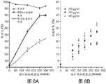
As shown in fig. 8a-b, increasing the speed of the cells through the constriction 15 can increase the percentage and efficiency of delivery of the delivery material 30. It was found that the delivery efficiency varies linearly with cell velocity and that there is a dose-dependent response.
In fig. 9, after passing through the constriction 15, the cells are incubated in solution buffer 25 for a period of time that will generally affect the percentage of the delivery material 30 that is delivered to the cells 20. However, it was shown that after a period of incubation (approximately 2-3 minutes), the percent delivery did not substantially change. From this data, it is believed that the perturbation on the cell 20 caused after the cell 20 passes through the constriction 15 is corrected within about 5 minutes after the cell 20 passes through the constriction 15. In addition, for reference, the corresponding time for the control group was 1 minute.
As shown in fig. 10, it was observed that multiple passes of cell 20 through constriction 15 affected the overall percent delivery, but negatively affected the overall survival rate of cell 20. To generate this data, the cells are passed through constriction 15, collected, and then passed through the device once again in approximately 1 minute.
It is observed that material within the cell can be extracted by the perturbation during the time the cell 20 is perturbed (e.g., after passing through the constriction 15). Thus, it has been found that material can enter and exit the cells 20 when the cells 20 are perturbed. This system 5 can be used as a method of sampling intracellular material without lysing the cells. Preferably, the perturbation of the cell membrane will cause the efflux of macromolecules from the cytoplasm and thus can be used as probes for cytoplasmic compositions.
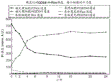
As shown in FIG. 11, HeLa cells stably expressing Green Fluorescent Protein (GFP) were treated in the presence of stationary siRNA (Ambion, USA) for Green Fluorescent Protein (GFP) and then subjected to fluorescence analysis at 48 hours using FACS (FACS Canto II, BD Biotech., USA). the results of FIG. 11 show that gene expression was reduced by 40% or more compared with that of commercially available reagents, such as Lipofectamine2000 (Invitrogen, USA). FIG. 11 also shows the negative siRNA (Scrambled siRNA) reference, as shown, this reduction is not due to the deformation process itself.
As shown in fig. 13-14, the extrusion size can affect the overall transfer efficiency of the transfer material 30. For example, fig. 13-14 show that as the operating pressure is varied (e.g., by varying the length and/or width of the constriction 15), the overall transfer efficiency also varies to some extent (fig. 14 shows the transfer of quantum dots (nanoparticles) under different conditions). Furthermore, as shown in FIGS. 18a-18b, the estimated cell velocity may affect the overall survival rate and overall delivery efficiency of the delivery material 30. For example, FIG. 18a shows that the overall transfer rate varies to some extent as the operating speed varies. In addition, FIG. 18b shows that the viability of the cells changed to some extent with the speed of the operation. These figures show that changes in the length of the constriction will improve delivery and minimally affect survival. In addition, large molecules undergo cell shrinkage at a slower rate than small molecules. The intracellular delivery methods described herein are "universal" and are applicable to a variety of different types of materials and cells. Further, the device produces cell membrane disruptions of a typical size of at least-100 nm, however, other size disruptions are possible.
As shown in FIG. 12, in one embodiment, the amount of delivery material can be predictably controlled by controlling the concentration gradient of the solution buffer 25 and the cytoplasm. A local delivery method may be used in which the cells 20 are exposed to a concentrated macromolecular cloud after the cells 20 have been made pores by shrinkage. However, these local delivery methods should take into account the estimated disturbance recovery time to ensure proper function. In practice, a "microtube" may be added vertically to the channel that delivers a high concentration of payload to the vicinity of the cell membrane (as shown in figure 6A). Preferably, the microtube may be located in the constricted portion 15 and/or in the vicinity of the constricted portion 15. This approach provides a diffusive transport mechanism by convection, thus enabling more accurate cell loading and higher concentrations. Preferably, the injection occurs when the cells 20 are in a high concentration region of the constriction 15. This localized technique is more effective in preserving the effectiveness of the delivery material since it is not necessary to maintain high concentrations in the buffer.
With respect to FIG. 16a, a series of micro-cylinders 100 may be used to apply pressure to the cells 20 to cause agitation. In this implementation, the cells 20 are forced through the constricted tube array to exert pressure on the cells 20.
With respect to fig. 16b, the cell 20 is stressed using the compression plate 105 to cause a perturbation. In this implementation, the compression plate 105 is controlled to apply a pressure to the cells 20 for a predetermined time. The compression plate 105 is arranged so that one or both plates can move to apply pressure to the cells 20. An additional set of compression plates 105 may also be provided to substantially enclose the cells 20.
With respect to fig. 16c, a buffering additive 115 (or bulk material attached to the cell surface) may be used to stimulate the extrusion of cells 20 through the constriction 15, the diameter of the constriction 15 being greater than the diameter of the cells 20. For example, shrinkage can be stimulated by the interfering action of the buffering additives. Examples of buffering additives 115 include microparticles or nanoparticles (e.g., polymer-based, lipid-based, ceramic-based, metal-based). The particles are labeled with a cell-binding ligand (e.g., an antibody, DNA sequence, peptide, or small molecule), although this is not required.
With respect to fig. 16d, the cells 20 are compressed using magnetic beads 120. For example, a magnetic and/or electrostatic force may be used to apply a force to the cell 20, or in the case of FIG. 16e, to push the cell 20. Preferably, the force applied to the cell 20 is sufficient to cause the perturbation.
With respect to fig. 16f, the plurality of liquid streams 125 are controlled in a manner that is capable of causing pressure (or rapid short-time shear forces) on the cells 20. For example, multiple streams of liquid are ejected so as to be brought into proximity or impact with each other. As the cell 20 passes through the plurality of fluid streams 125, a force may be applied to the cell causing a perturbation on the cell membrane of the cell 20. Alternatively, cells can be ejected from narrow slit-like tubes to accelerate delivery.
The system 5 may be a stand-alone system such as the display of fig. 7, but may have other configurations. For example, the system 5 may be implanted in a patient for local intracellular delivery and/or integrated ex vivo in a machine for treating cells prior to their return to the patient.
To achieve the delivery advantages described herein, the microfluidic nature of the system enables one skilled in the art to exercise precise control over different delivery conditions, pre-treatment processes, and subsequent cell characterization. For example, the system may be applied in tandem by a Fluorescence Activated Cell Sorter (FACS). This allows real-time delivery and sorting of cells in one and the same system. A variety of pre-treatment and post-sorting assay techniques can also be used to develop continuous, high-throughput assays for drug screening and diagnosis.
The efficiency of payload delivery to the target cells is determined by contacting the reference population of target cells with the payload and the population processed by the microfluidic system. The reference sample and the cells treated by the device are treated at the same concentration and for at least the same time in the same delivery solution. To compensate for surface binding, endocytosis, and other effects such as autofluorescence, a transmission region is defined such that at most 1-5% of the viable reference cells fall into this region. Thus, the transfer efficiency of the sample corresponds to the percentage of living cells located within this transfer region. For example, fig. 43A is an intensity histogram of a flow cytometer for a reference population exposed to 3kDa dextran conjugated to cascade blue, and fig. 43B is an intensity histogram of a flow cytometer for a cell population subjected to a 30 μ ι η -6 μ ι η device and then exposed to 3kDa dextran. The unshaded areas in fig. 43A and 43B are the transfer areas defined above.
In operation, with respect to fig. 17, and with further reference to fig. 1-3, a process 1000 for implementing the intracellular delivery system 5 includes various steps as shown. However, the process 1000 is exemplary only and not limiting. The process 1000 can be altered by methods such as adding, deleting, changing, or rearranging various steps.
At step 1005, the cells 20 are suspended in the solution buffer 25 separately from the delivery material. Typically, the cell concentration is 104To 109In the range of cells/ml. The concentration of the delivery material is in the range of 10 mg/ml to 0.1 μ g/ml.
The delivery material is added to the cell buffer either before or immediately after delivery is completed using the ideal set-up that leaves the lesion/well open for 1-5 minutes. The solution buffer may be composed of a number of salts, sugars, growth factors, animal derivatives, or other components suitable for cell proliferation, maintaining cell health, or inducing cell signaling pathways. Other materials may also be added to the solution buffer 25. For example, a surfactant (i.e., a copolymer) and/or a bulk material may be added to the solution buffer 25.
At step 1010, a solution buffer 25 comprising cells 20 and delivery material 30 is passed through channel 10 of system 5. The solution buffer 25 passes through the channel 10 by gravity, or may be assisted by other means. For example, pressure may be applied to solution buffer 25 on the inlet side of channel 10 (e.g., using a gas cylinder and/or compressor), and/or vacuum may be applied on the outlet side of channel 10 using a vacuum pump. Additionally, displacement-based fluid systems may also be used.
As the individual cells pass through the constriction 15, the solid structure of the constriction 15 temporarily exerts a force on the cell 20, causing a perturbation of the cell membrane of the cell 20, e.g., creating a hole, thereby delivering the delivery material 30 to the interior of the cell 20. The amount and time of pressure applied to the cell 20 can be varied by adjusting the size of the constriction 15, the speed at which the cell 20 passes through the constriction 15, and/or by adjusting the shape of the constriction 15. In one configuration, approximately 5,000-20,000 cells/second pass through the constriction 15, and each cell is constricted for approximately 100 microseconds.
The system 5 may include one or more channels 10. For example, the system 5 includes 50-100 channels 10, with the channels 10 arranged in parallel. The use of a parallel arrangement reduces the occurrence of blockages in one or more of the channels 10 and increases the overall throughput of the system 5. In addition, the system 5 comprises one or more channels connected in series.
After the cells 20 pass through the constriction 15, the cells are cultured/recovered by leaving the cells in the solution buffer 25 at step 1015. During this process, the cell 20 will absorb some of the transfer material 30 present in the solution buffer 25 by the perturbations present on the cell membrane. One mechanism of this absorption is diffusion-based, since large molecules absorb at a slower rate than small molecules. Preferably, the cells 20 are cultured/recovered in the solution buffer 25 for about 2-5 minutes, although other times are possible. During this time, the cells 20 are cultured/recovered in the solution buffer 25, and the material within the cells 20 may also be released from within the cells into the solution buffer 25. During the incubation/recovery period, certain conditions may be controlled to ensure that the amount of delivery material 30 continues to pass through the cell population. For example, convective transfer mechanisms can be used to impinge the transfer material into the cells being cultured/recovered after contraction.
Optionally, in step 1020, after the cells have been cultured/recovered, the cells are washed to remove the solution buffer. Preferably, the washing is performed after a period of time has elapsed to allow the previously occurring disruption to recover, although the washing solution may be performed at other times to control the amount of the delivery material 30 that is absorbed by the cells.
Example 1 delivery of functionally engineered nanoparticles
Engineered nanoparticles have great potential as live cell imaging tools, therapeutic agent molecule delivery agents, or even as Methods of manipulating live cells by using external means (e.g., light or magnetic fields) (Howarth, m., et al. monovirtual, reduced-size quantum dots for imaging receptors. Nature Methods 5,397-399 (2008)). However, many of these potential applications require delivery of the nanomaterial into the cytoplasm of the cell. Most nanoparticles, such as QDs, need to be passivated by polymers to enable the nanoparticles to dissolve in aqueous vehicles, which often prevents passive diffusion of the nanoparticles across cell membranes.
Microinjection of nanoparticles is considered to be impractical due to the lack of specialized instrumentation and low throughput, while electroporation allows QD deposition within the cell. Therefore, most current attempts to deliver quantum dots into the cytoplasm rely on endocytosis of the quantum dots by the cell and are not phagocytosed by the inclusion bodies. Prior to the present subject matter, it was not possible to deliver quantum dots into the cytoplasm in a satisfactory manner, on a scale. The system of the present invention provides a solution to the delivery problems of previous approaches.
The microfluidic system can be combined with the newly reported new generation of biocompatible QDs ((Liu, w., et al. compact biocompatible quantum dot of dot dots via RAFT-mediated imidazolyl random copolymer ligand compression biocompatible quantum dot) JACS 132,472-483 (2010)). the quantum dots used in example 1 are coated with a polyimidazole ligand consisting of multiple metal chelated imidazole groups and multiple water-soluble passivated poly (vinyl) ethylene glycol (PEG).
To deliver quantum dots to the cytoplasm, cells were dispensed in PBS buffer containing quantum dots. The cell-quantum dot solution was pipetted into the microfluidic device and then passed through the channel under continuous pressure followed by an incubation time of 5 minutes. After the incubation time was over, the excess quantum dots were centrifuged. For the reference population, the cell-quantum dot solution is kept in the microfluidic system, and then the cells are exposed to the quantum dot solution for a sufficient time to allow the intra-cytoplasmic delivery method to be performed.
FIG. 19 is a superposition of transmission and copolyfluorescent images followed by a z-section copolyfluorescent image of quantum dot-delivered treated cells completed using the present subject matter. FIG. 19 shows (upper panel) immediately after treatment (i.e., delivery) is complete and (lower panel) after incubation at 37 ℃ and 5% carbon dioxide for 48 hours. The diffuse staining pattern is confined to the cytoplasm and the nanoparticles appear not to enter the nucleus (dark areas within the cell). The scale is 10 microns. FIG. 19 shows a photograph of a quantum dot coated, specific ligand that does not contain a polyimidazolyl group, but has no other function than providing biocompatibility through a polyethylene glycol group. The confocal fluorescence photograph shows diffuse cytoplasmic QD staining of isolated circular HeLa cells flowing through a microfluidic device in different z-sections of the cells (fig. 19, top panel). The diffuse staining was still present after culturing the cells at 37 ℃ and 5% carbon dioxide and adsorbing for 48 hours (fig. 19, bottom panel). At 48 hours, the diffusive quantum dots fluoresced dim, probably due to cell lysis (fig. 19). Quantum dots (hydraulic diameter-13 nm) were delivered to-40% of the viable cell population at a flux of-10,000 cells/sec. Fig. 20A shows the delivery efficiency of quantum dots coated with PIL into the cytoplasm of HeLa cells using the inventive subject matter. Cell viability as determined by flow cytometry was greater than 80%. Fig. 20B shows viability of flat QD535 delivered to HeLa cells using the subject name of the present invention, as determined using propidium iodide staining and flow cytometry. The viability of the treated cells was determined by flow cytometry using diffusion staining of the co-polymer image, the ability of the cells to adsorb being consistent with the transfer of the quantum dots into the cytoplasm of living cells.
To determine that these fluorescences were indeed produced by quantum dots delivered into the cytoplasm rather than by quantum dots trapped by inclusion bodies, the nanoparticles were designed to have emission curves that vary with their interaction with the reducing environment of the cytoplasm. The reduction potential in the cytoplasm was-260 to-220 mV and was initially generated by maintaining a high concentration (5-10mM) of the tripeptide glutathione.
Thus, the location and chemical accessibility of the delivered nanoparticles is determined by measuring the fluorescence intensity of the QD-dye construct, which changes its emission profile when in contact with the cytoplasmic environment. The QD-dye was constructed to contain green emitting QDs (transmission: 541 nm) that provide carboxy-X-rhodamine (Rox) staining (transmission: 610 nm) as an energy donor, conjugated by a reducing disulfide bond.
FIG. 21 shows the design, absorbance and stability of the constructs in different media. 2100 is a schematic of the PIL before conjugation to the dye and before coating the QDs (left panel), and a schematic of the resulting quantum dot-disulfide-carboxy-X-rhodamine (Rox) construct (right panel) (pictures not shown to scale). 2110 is the absorption spectrum of the quantum dot-disulfide-carboxy-X-rhodamine (Rox) construct. During the course of the experiment, excitation at 488 nm and 405 nm produced unique light absorption. 2120 is the stability of the fluorescence energy of constructs delivered by QDs to carboxoy-X-rhodamine (Rox) in whole medium at 37 ℃ and 5% carbon dioxide, indicating that disulfide bonds are not cleaved in the extracellular environment. Figure 2130 shows disulfide bond cleavage by intracellular reduced glutathione as shown by the recovery of QD fluorescence. 2140 shows the recovery of fluorescence of quantum dots produced by treatment with non-thiol group reducing tris (2-carboxyethyl) phosphine, further confirming the cleavage of disulfide bonds.
The thiol group incorporated into the PIL forms a disulfide bond with the thiolated carboxy-X-rhodamine (Rox). When there are an average of 13C-X-rhodamine (Rox) per quantum dot, the broad absorption spectrum of the purified construct has the absorption characteristics of both quantum dots and C-X-rhodamine (Rox) (2120), with effectively annealed QDs fluorescent (2130). In the cytoplasm, the construct can act as a sensor that is irreversible to a specific reducing environment. When quantum dots are selectively stimulated with laser light while maintaining disulfide bond integrity at 488 nanometers (microscope) or 405 nanometers (flow cytometer), the construct undergoes Fluorescence Resonance Energy Transfer (FRET), whereby the carboxy-X-rhodamine (Rox) emits red light. In solution experiments, the reduced glutathione of the cell cleaves the disulfide bond, releasing the carboxy-X-rhodamine (Rox) dye and restoring the fluorescence of the quantum dots (2140). The non-thiol reducing tris (2-carboxyethyl) phosphine may also restore the fluorescence of the quantum dot, indicating that the release of carboxy-X-rhodamine (Rox) from the quantum dot surface is not due to the PIL shift produced by glutathione. Some disulfide bonds are blocked due to steric hindrance caused by long polyethylene glycol on the PIL, and the carboxy-X-rhodamine (Rox) fluorescence may not completely disappear due to very small amount of non-specific interaction between the dye and the surface of the quantum dot.
The transfer of the quantum dot-disulfide-carboxy-X-rhodamine (Rox) construct into the cytoplasm was confirmed by changes in the construct's fluorescence curve as measured by flow cytometry and confocal microscopy. Upon exposure to a reducing cytoplasmic environment, cleavage of the disulfide bonds disrupts the Fluorescence Resonance Energy Transfer (FRET) process of the quantum dots to the dye. Thus, as unique quantum dots are excited, the fluorescence of the quantum dot channel increases over time while the fluorescence of the carboxy-X-rhodamine (Rox) channel decreases over time. Viable HeLa cells were treated with a microfluidic device in a solution containing high concentrations of quantum dot-disulfide-carboxy-X-rhodamine (Rox), incubated for 5 minutes, and then washed to remove excess quantum dots prior to addition of cell culture medium (i.e., treated cells). The reference cells were not processed with the microfluidic device, but were incubated with the quantum dot-disulfide-carbo-oxo-X-rhodamine (Rox) for 5 minutes and then washed before adding the cell culture medium. The treated cells and the reference cells were observed for both carboxy-X-rhodamine (Rox) and quantum dot channel fluorescence using confocal microscopy and flow cytometry.
Fig. 22A and 22B show living cell confocal micrographs and fluorescence intensity analysis showing cytoplasmic staining and chemical accessibility of the quantum dot surfaces. Fig. 22A shows images of treated cells (top panel) and reference cells (bottom panel). Only the treated cells showed a diffuse green fluorescent appearance. The scale is 10 microns. Fig. 22b shows the variation of intensity as a function of time in the green and red channels. Since at each time point n <20, the fluctuations in the overall mean fluorescence are corrected by calibrating the 0 hour time point.
Under confocal microscopy, the diffuse fluorescence turned from an intense red to an intense green color through the cytoplasm of the treated cells (as shown in figure 22A). The reference cell image showed some non-specific binding outside the cell membrane, as indicated by the ring-packed fluorescence, and no increase in signal from the green channel. This effect is consistent with the predictable degradation of cytoplasmic disulfide bonds that reduces Fluorescence Resonance Energy Transfer (FRET) effects. In fig. 22B, the line graph shows the average quantum dot and the carbon oxygen-X-rhodamine (Rox) channel intensity in each of the treated and reference cells and the auto-fluorescent cells after correcting the cell-to-cell difference by calibrating the total fluorescence in the delivered fluorescent substance. For the treated cells, the images show images within 2-4 hours of incubation, where the fluorescence of the quantum dots rises above the fluorescence of the carboxy-X-rhodamine (Rox). Interestingly, the treated cells stably maintained the carboxy-X-rhodamine (Rox) signal above the autofluorescence level after 9 hours. This is consistent with solution experimental results, where some Fluorescence Resonance Energy Transfer (FRET) remains after reduction. The observed increase in contrast staining and quantum dot signals, as well as the decrease in carboxy-X-rhodamine (Rox) signals, strongly confirm cytoplasmic transfer and subsequent disulfide cleavage. Fluorescence Resonance Energy Transfer (FRET) quenching of carbox-rhodamine (Rox) refers to quantum dot fluorescence in cells, the appearance of which is indistinguishable from autofluorescence. At an earlier time point the reference cells showed some carboxy-X-rhodamine (Rox) fluorescence above its own fluorescence, then steadily decreasing. This is due to the non-specific interaction between the quantum dot-S-S-carboxy-X-rhodamine (Rox) and the cell surface, followed by re-dissolution of the construct in the medium.
FIG. 23 shows the average cell fluorescence intensity and viability as determined by flow cytometry. 2300 is the mean fluorescence intensity of the QDs per cell (left panel) and the mean fluorescence intensity of the carbox-X-rhodamine (Rox) (right panel), indicating that the QDs fluorescence intensity increased only in the treated cells. At the 24 hour time point, the carboxy-X-rhodamine (Rox) fluorescence intensity of the treated nuclear reference cells was at the autofluorescence level. 2310 is a histogram showing the distribution of fluorescence intensity at selected time points in the QD channel (left panel) and the carbo-X-rhodamine (Rox) channel (right panel) for the treated cells and the reference cells. It is estimated that quantum dot transfer occurs in at least 35% of the cell population. The grey areas are used to guide the eye movement over the peaks of the fluorescence intensity histogram. 2320 shows the viability of the reference and treated cells measured using propidium iodide.
The flow cytometry assay shown in figure 23 demonstrated that the quantum dot-disulfide-carboxy-X-rhodamine (Rox) construct can interact with the cytoplasmic environment. Flow cytometry measurements were recorded for all live cells, including delivered cells (-35% of the treated cell population) and untransferred cells. 2100 shows the mean fluorescence intensity of the treated cells and the reference cells. Initially, QD fluorescence of treated cells rose, peaked at approximately 9 hours, and then declined gradually, as opposed to QD fluorescence of reference cells, which is maintained at autofluorescence levels. This is consistent with the intracytoplasmic reduction of disulfide bridge between QDs and dyes inside the treated cells, followed by dilution of the fluorescent construct by cell division. The fluorescence intensity of the treated and reference cells initially increased and then decreased within the first 2 hours. This drop causes the particles (which are bound to the cell surface during culture) to re-dissolve in the culture medium. Since untransferred cells are also present in the treated cell population, the average fluorescence intensity of the treated cell population for C.sub.x-rhodamine (Rox) is similar to that of the reference cell population. The presence of transmitted and untransferred cells in the treated cell population can be distinguished by the histograms of quantum dot and carboxy-X-rhodamine (Rox) intensities shown in 2310. The fluorescence histogram of the treated cells became bimodal with increasing time, but the histogram of the reference cells was also unimodal. The QD fluorescence intensity rises over time for a subset of the treated cell population, further illustrating disruption of FRET processes in the treated and transferred cells. The carboxyx-rhodamine (Rox) fluorescence decreases overall as the bound cell membrane construct is re-dissolved in the culture medium, but the subset of the treated cell population also maintains carboxyx-rhodamine (Rox) fluorescence. This is consistent with the incomplete reduction of the QD-S-S-Rox bonds observed in the confocal micrograph. The treated cell population survived between 10% of the survival of the reference cells at all time points as determined using propidium iodide staining (2130). Cell viability was > 90% relative to the reference group, which is more popular than alternative methods such as electroporation and polymer-based methods, with viability as low as 40-60% after treatment.
Fig. 24 shows an epifluorescence map of individual quantum dots in the cytoplasm of a cell that are not aggregated (top panel), and the scintillation trajectories of three quantum dots that are auto-fluorescent (labeled 2400, 2410, and 2420) after processing a 10nM quantum dot solution using the inventive device. The QD scintillation trace is non-binary due to the long acquisition time (500 ms). The scale is 10 microns.
The quantum dot delivery platform enables single cell imaging by delivering unaggregated quantum dot-disulfide-carboxy-X-rhodamine (Rox) constructs, with observed emission intermittency consistent with that of single quantum dots. In this experiment, the quantum dot-disulfide-carboxy-X-rhodamine (Rox) construct was delivered into the cytoplasm, followed by 10 hours of incubation and imaging in an epifluorescence microscope. A 10 hour incubation time ensures that fluorescence from quantum dots inside the cytoplasm is restored by reduction of disulfide bonds; epi-fluorescence microscopy was used to ensure that enough photons were collected. Some blinking quantum dots can be observed when cells are treated with the subject matter described herein at low quantum dot concentrations (fig. 24). 2400. 2410 and 2420 show the intensity traces of scintillating quantum dots in the cytoplasm, which are non-binary due to long acquisition times (500 ms). Within the collection time (about 1 minute), the movement of the transformed cells was minimal. These data illustrate the single cell within the cytoplasm that can be observed following the delivery of quantum dots as fluorescent labels using the present subject matter.
Example 1 shows delivery of nanoparticles into the cytoplasm using an embodiment of the inventive subject matter. The interaction of the nanoparticle surface with cytoplasmic components was shown by observing the cleavage of quantum dot-disulfide-carboxy-X-rhodamine (Rox) by intracellular reducing agents.
Embodiments of the present subject matter ensure high flux delivery of quantum dots into the cytoplasm without ligands that are cell penetrating or endosomal escape, and maintain cell viability and quantum dot integrity. The transfer efficiency is 35%, which can be further increased by increasing the number of microfluidic constrictions, changing the size of the constrictions, or increasing the number of processing cycles. Unlike most existing cell penetrating peptide or positive charge assisted delivery methods, the present subject matter does not require that the same nanoparticle be subjected to the intracellular delivery process and the cytoplasmic protein targeting process in combination.
By distributing the foregoing requirements, mitigation of cross-reactivity relationships, unequal reaction efficiency of binding schemes, and conjugate stoichiometry may be achieved. Thus, the design of quantum dot constructs has significant flexibility, paving the way for intracellular protein labeling and tracking. This method can be effectively used to deliver many complex designs of fluorescent nanomaterials that target intracellular proteins and organelles through proven protein targeting protocols (such as, but not limited to, streptavidin-biotin, halo-labeled-chloroalkane, and sortase-labeling).
In example 1, all chemicals were from Sigma Aldrich and were used as received unless otherwise indicated. The air sensitive material was treated with an Omni-Lab VAC glove box at an oxygen level of less than 0.2ppm under dry nitrogen conditions. All solvents are either spectroscopic or reagent. Aromatic ring-containing compounds were imaged in TCLs using a hand-held ultraviolet lamp and KMn 04. Amino group containing compounds were imaged in TCL using ninhydrin staining. Flash column chromatography was performed in the Teledyne Isco Combi Flash company. HeLa cells were purchased from ATCC, and then all cell culture media were purchased from Mediatech unless otherwise specifically indicated.
In the case of the example 1, the following,1h NMR spectra were recorded on a Bruker DRX 401 NMR spectrometer. MS-ESI was performed on Bruker Daltonics APEXIV 4.7 FT-ICR-MS. Uv-vis absorption spectra were recorded using an HP 8453 diode array spectrophotometer. Photoluminescence and absorption spectra were recorded using a BioTek Synergy 4 microplate reader. Polymer molecular weights were determined in DMF solution using an Agilent1100 series HPLC/GPC system using three PL gel columns (103, 104, 105A) in series and narrow polyethylene standards. The dye derivatives were purified in a Varian ProStar Prep HPLC system. The modified polymer was purified using a PD-10 column from GE Healthcare loaded with Sephadex (TM) G-25M. The ligand-exchanged quantum dots were purified by centrifugation and then dialyzed against a Millipore Amicon Ultra 30K cut-off centrifugal filter membrane, followed by GFC on an AKTAprime Plus chromatography system (Amersham Biosciences) equipped with a self-assembled Superdex 20010/100 glass column. Flow cytometry measurements were performed on LSR Fortessa (BD Biosciences).
In example 1, the CdSe core having a first absorption peak of 478 nm was synthesized using the previously reported method (1). In general, 0.4 μmol (54.1 mg) of CdO, 0.8 mmol (0.2232 g) of TDPA, 9.6 mmol (3,72) of TOPO were placed in a 25 ml round bottom flask. The solution was degassed at 160 ℃ for 1 hour, then heated to 300 ℃ under argon until the CdO dissolved and a clean homogeneous solution formed. Subsequently, the above solution was placed under vacuum at 160 ℃ to remove the water evolved. The solution was reheated to 360 ℃ under argon and a TOP-Se solution (containing 1.5 ml of 1.5M TOP-Se in 1.5 ml of TOP) was added rapidly until a CdSe core with a first absorption peak of 478 nm was produced.
The CdS shell was deposited on the CdSe core by the modification method of method (2) previously reported. The core was isolated by repeated precipitation from hexane with acetone and then warmed to 180 ℃ in a mixture solvent of oleylamine (3 ml) and octadecene (6 ml). The Cd and S precursor solutions were then continuously introduced at a rate of 4 ml/hour. The Cd precursor consisted of 0.33 mmol Cd oleate and 0.66 mmol oleylamine in a solvent mixture of octadecene (1.5 ml) and TOP (3 ml). The S precursor consisted of 0.3 mmole of hexamethyldisiloxane [ (TMS)2S ] in 6 ml of TOP. A total of 3 monolayers were added, each Cd and S yielding a quantum dot with emission at 541 nm and a quantum dot yield of 60% when diluted in octane. The extinction coefficient of CdSe (CdS) was calculated using the extinction coefficient of the CdSe core described in document (3) and it was assumed that 95% of the CdSe core was retained in the coating step.
In example 1, a silicon chip was prepared using photolithography and deep reactive ion etching techniques. Cleaning (with H)2O2And H2SO4) The resulting etched silicon wafer is freed of residues, oxidized to produce a glass surface and bonded to a high borosilicate wafer prior to dicing into individually packaged devices. Each device was then individually inspected for defects prior to use.
Example 2 delivery of macromolecules
Intracellular delivery of macromolecules is a critical step in therapeutic and research applications. Nanoparticle-mediated DNA and RNA delivery can be useful, for example, for gene therapy, while protein delivery can be used to affect cellular function in the clinic and laboratory. Other materials, such as small molecules, quantum dots, or gold nanoparticles, can be delivered into the cytosol, and uses thereof can include cancer therapy, intracellular labeling, and single cell tracking.
To illustrate the versatility of this technique, model dextran molecules were delivered into several cell types: DC2.4 dendritic cells, neonatal human foreskin fibroblasts (NuFF) and mouse embryonic stem cells (mESC), obtained delivery efficiencies as high as 55%, 65% and 30%, respectively. Initial realization also showed successful delivery in primary lymphocytes, macrophages and dendritic cells from mice. In addition, the present technology does not produce excessive cytotoxicity or induce stem cell differentiation. Indeed, all cell types have survival rates of over 60% even at the highest experimental speeds. The optimum apparatus and operating conditions should be pre-designed for the cell type.
FIG. 25 shows the results of experiments confirming that the transfer depends on the cell velocity and the constriction involved. The constriction dimensions are represented by numbers (e.g., 10-6 microns by 5), where the first number represents the constriction length, the second number represents the constriction width, and the third number (if present) represents the number of constrictions in series in each via. The transfer efficiency is shown at 2500, and the cell viability (as determined using flow cytometry) at 18 hours after treatment is shown at 2510, as a function of cell velocity (40-6 microns (o), 20-6 microns (□), and 10-6 microns (Δ) device design). The 2520 transfer efficiency and 2530 cell viability (determined by flow cytometry) of the initial human fibroblasts (□), DC2.4 dendritic cells (o) and mouse embryonic stem cells (mESC) (Δ) proved to be a function of velocity in a 30-6 micron device. Human fibroblasts and dendritic cells were analyzed 18 hours after delivery. Mouse embryonic stem cells (mESC) were analyzed 1 hour after the transfer was completed. All data points were run in triplicate and error bars represent two standard deviations.
Fig. 26 shows scans 2600 of HeLas cells at different levels after delivery of 3kDa dextran conjugated by pacific blue, as determined under confocal microscopy. Scanning was performed from top to bottom and then from left to right, where the upper left zeta was 6.98 microns and the lower right zeta was-6.7 microns. The scale is 6 microns. The viable cell delivery efficiency of the 10 micron-6 micron (□), 20 micron-6 micron (o), 30 micron-6 micron (Δ), and 40 micron-6 micron (0) devices is shown at 2610. The time axis represents the time elapsed from the start of the treatment of the cells before the contact with the target delivery solution. All results were determined by flow cytometry at 18 hours after treatment. At 2620, the mean intensity of the delivered cell population is calibrated using the autofluorescence of the untreated cells relative to the reference cells. In successive treatment cycles, fluorescein-conjugated 70kDa dextran (horizontal line) and pacific blue-conjugated 3kDa dextran (diagonal line) were delivered to the cells (cycles 1 and 3) and removed from the cells (cycle 2). The reference indicates cells that are in contact with the transfer solution only, but not treated by the present device. All data points were run in triplicate and error bars represent two standard deviations.
The previously described nanoparticle and Cell Penetrating Peptide (CPP) based delivery techniques reveal intracellular pathways, and evidence is shown to exclude the effects of endocytosis on the delivery mechanism of the subject matter of the invention. At 2600, fig. 26 shows a photomicrograph of a co-polymerization of cells treated with pacific blue conjugated 3KDa dextran, with diffuse cytoplasmic staining opposite to the punctate character expected from the endocytosis method. In addition, when the delivery experiments were performed at 4 ℃, the temperature minimized endocytosis, while for both payloads tested (3KDa and 70KDa dextran), the delivery efficiency was minimally affected by temperature. These data show that endocytosis is unlikely to be responsible for the delivery of the present system.
The characteristics of the delivery kinetics over time are determined. The subject delivery material is used in the absence of the delivery material and the cells are then exposed to pacific blue labeled 3KDa dextran at predetermined time intervals after treatment. In this method, the cell membrane is disrupted by passage of the cell through the constriction, but no detectable transmission occurs until we expose it to labeled dextran. Thus, the efficiency of delivery at each time point will affect the percentage of time that the cells maintain porosity after treatment. The present invention captures pore formation/closure kinetics. The results show that almost 90% of the transfer occurs at 1 minute after treatment (2610), regardless of the device design. The observed timescale confirms the hole formation hypothesis that with repair of the cell membrane effect, kinetics report that cell membrane repair occurs approximately 30 seconds after the injury. In contrast, the time scale recommended for endocytosis, e.g., nanoparticle and CPP-mediated delivery mechanisms, is approximately in hours.
Since the material is diffusible in its delivery through the pores in the cell membrane, the material can be delivered into the cell or out of the cell throughout the time the pores are present.
On the other hand, the endocytosis or contraction mechanism must be unidirectional, i.e., only to facilitate the material to be delivered into the cell. To illustrate the bidirectional transport of material into the cell membrane, an experiment was performed that included 3 cycles of transport. In the first cycle, cells were treated in the presence of 3kDa dextran and 70kDa dextran, incubated in dextran solution for 5 minutes, and then washed 2 times with phosphate buffer. One third of the sample was left for later use. In the second cycle, the remaining washed cells were treated again with the device, but this time without any transfer material, and incubation was continued for 5 minutes. Half of the sample was left to stand. In the third cycle, the cells left from the second cycle were treated with the device under the same conditions as those of the first cycle (i.e., in the presence of dextran), incubated for 5 minutes and then washed 2 times with phosphate buffer. After the assay was complete, the cells were analyzed by flow cytometry for 18 hours. The change in the calibrated fluorescence intensity indicates that dextran reticularly diffuses into the cells in the first cycle, out of the cells in the second cycle, and re-enters the cells in the third cycle (2620). These results are consistent with a diffusive transport mechanism.
FIG. 27 shows a simple, 2D diffusion model developed by the COMSOL Multiphysics software package showing the passive diffusion of material into a pore-forming cell membrane. COMSOL multiprophy is a finite element analysis solver and simulation software/FEA software package developed by COMSOL corporation, suitable for a variety of physical and design applications, particularly coupling phenomena or multi-physical fields. At 2700, the transfer/loss of material is expressed as a function of the diffusivity of the cell membrane. When the pore was formed, the material of interest was in either buffer (□) or in the cell (o), the simulation results indicate that the percent transfer/loss of material into the cell is a function of the diffusivity of the cell membrane. At 2710 are represented images of the simulation system and the concentration gradient across the cell membrane as material is transferred from the buffer into the cell
The experimental data/results of fig. 26 qualitatively reproduce the effect of diffusion as the sole means of mass transfer, taking advantage of the literary value of the particles diffusing into and out of the cytoplasm. Furthermore, the technique delivers 10-40% of the delivery material in buffer to the cytoplasm by fitting experimental data to this model. By comparison, the CPP method for delivering proteins is estimated to deliver only 0.1% of the buffer material into the cells.
The particle size (or hydrodynamic radius) affects its diffusivity and its ability to enter pores of a particular pore size in the cell membrane. Thus, this parameter can affect the transfer efficiency in the pore formation/diffusion mechanism. In a series of experiments, dextran with experimental payloads of 3kDa, 10kDa, 70kDa, 500kDa and 2MDa conjugated to fluorescein or pacific blue were delivered, and in addition, a fluorescein-labeled plasmid with a molecular weight estimated to be 3.1MDa was delivered. Model molecules are selected as the delivery material of interest based on their molecular weight similarity. For example, dextran of 3kDa to 10kDa has similar sizes to some short peptides or siRNAs, while dextran in the 70kDa-2MDa range mimics the size of most proteins and some small nanoparticles.
Fig. 28 shows the results of the double transfer of material. At 2800 it is shown that the viable cell transfer efficiency is a function of velocity for HeLa cells treated with pacific blue conjugated 3KDa dextran (□), treated with fluorescein conjugated 70KDa dextran (o) and treated with 2MDa dextran. The experiment was performed using 10 micron to 6 micron by 5 chips. All data points were run in triplicate and error bars represent two standard deviations. 2810 and 2820 show histograms of flow cytometry data overlaid for untreated (red), treated at 700mm/s (green), and treated at 500mm/s (orange), and treated at 300mm/s (bright blue), or HeLa cells exposed only to the delivery material (reference, dark blue). The delivery material consists of pacific blue conjugated 3KDa dextran (2810) and fluorescein conjugated 70KDa dextran (2820).
Experiments have shown that molecules larger than 70kDa have a different transmission curve than 30kDa dextran (2800). The device produces bidirectional transfer, where a 10 micron-6 micron x5 device operates at 500mm/s, for example, to ensure that 90% of living cells receive molecules of 3kDa, and approximately 50% of cells receive molecules greater than 70kDa and 2 MDa.
Histograms corresponding to these flow cytometer data show that dextran delivery of 3kDa produced 2 distinct peaks (2810). In the first subpopulation, the peak shift generated by the endocytosis and surface binding previously described when considering the 0mm/s data point for the reference, resulted in a 2-6 fold increase in mean fluorescence intensity, indicating a gentle level of cell transmission. In the second population, the mean fluorescence intensity of the cells increased 20-100 fold compared to the reference, showing increased levels of transmission. This effect shows that the latter subpopulation is more porous than the former subpopulation and therefore enables an approximately 10-fold increase in the circulation of the material. Indeed, it appears that the percentage of cells with improved delivery capacity can be increased by increasing the severity of the treatment by increasing the rate of operation, as shown by the curves at 300mm/s, 500mm/s and 700 mm/s. Similar characteristics were observed for the larger 70kDa dextran molecule (2820). But this effect is less pronounced due to lower particle diffusivity and possible size exclusion effects that reduce overall throughput. The mean fluorescence intensity of the mildly transmitted cell population (first peak) was only increased by a factor of 1.5-2 compared to the 2-6 fold increase observed with 3 kDa. This effect should be considered as a difference in the 2800 transmission data, since in the case of large molecules, the mildly transmitted cell population is difficult to distinguish from the reference population according to the transmission definition herein. Thus, for larger molecules, such as 70kDa and 2MDa dextran, the skilled person will mainly determine the second cell population where the transmission is significantly improved.
To determine that the material delivered by the rapid mechanical deformation into the cytoplasm was active and bioavailable in the cytoplasm of cells, a series of experiments were performed to deliver the payload GFP resting siRNA to be tested into GFP capable of expressing destabilisation (Ambion, usa). Dose-dependent and sequence-specific GFP inhibition (up to 80%) was observed at 18 hours post-treatment. Gene suppression is consistent with dextran delivery experiments as a response to cell velocity and device design, so higher velocities and multiple constriction designs will result in greater gene suppression. In these experiments, Lipofectamine2000 was used as a positive reference. Prior to performing these experiments, device design and operating parameters were not optimized for siRNA delivery.
Figure 29 shows data for siRNA, protein and nanoparticle delivery. In 2900, gene suppression was interpreted as a function of device type and cell velocity in HeLa cells expressing stabilized GFP 18 hours after delivery of anti-eGFP siRNA with a 10 micron-6 micron x5 chip at a delivery concentration of 5 micromolar. Lipofectamine2000 was used as a positive reference. In 2910, the delivery efficiency of fluorescein-labeled 70kDa dextran and pacific blue-labeled 3kDa dextran for rapid mechanical deformation delivery and electroporation delivery is shown. The concentration of each dextran type in the delivery solution was 0.1 mg/ml. In 2920, the delivery efficiency of fluorescein-labeled fetal bovine serum albumin (o), the pacific blue-conjugated 3kDa dextran delivery efficiency (□), and the cell viability (Δ) were shown as a function of velocity using a 30-6 micron device. A fluorescence micrograph of HeLa cells, in which tubulin antibodies containing an Alexa Fluor488 marker were delivered, is shown in 2930. Scale bar is 5 microns. 2940 and 2950 show projection electron microscope (TEM) photographs of gold nanoparticles (some indicated by arrows) in cells after treatment with a 10 micron-6 micron x5 device and resting for 1 hour. The scale bar is 500 nm. All data points were run in triplicate and error bars represent two standard deviations.
In further embodiments, potential methods of cytoplasmic delivery, e.g., protein delivery and nanoparticle delivery, are developed for previously challenged applications. To compare the performance of the present subject matter with commercially available methods, 3kDa and 70kDa dextrans were used as protein models, and these models were delivered to human fibroblasts using the present subject matter and a Neon electroporation system (Invitrogen). The results show that rapid mechanical deformation provides a 7-fold increase or more in transfer efficiency for these macromolecules (2910). To make these methods suitable for protein delivery, fluorescein-labeled fetal Bovine Serum Albumin (BSA) was delivered to HeLa cells using a device with a channel diameter of 30-6 microns with a delivery efficiency of up to 44% and an activity of over 90% (2920). After treatment using a 30-6 micron device, Alexa Fluor488 labeled tubulin antibody (Bio Legend) was delivered to HeLa cells at an antibody concentration of 0.25 mg/ml (2930). Diffusion staining showed that the material was not encapsulated in inclusion bodies and was therefore suitable for live cell antibody staining of cellular structures in the cytoplasm. Apolipoprotein E was also successfully delivered using this technique.
TEM images (2940 and 2950) of cells left for 1 hour after deformation to deliver nanoparticles show delivery of 15 nm gold nanoparticles coated with PEG 1000. The gold nanoparticles are substantially free of agglutination and are not visually captured by the inclusion bodies. Some cytoplasmic transmission defects were observed in these images. High-throughput delivery has been demonstrated to be possible, and non-cytotoxic delivery of quantum dots into the cytoplasm of cells is an unmet goal in previous technologies. In these experiments, quantum dots with Rox staining were bound to their surface and transferred by rapid mechanical deformation, and observed over time. The data yielded a minimal transfer effect estimated to be 35%, and it was demonstrated that the transferred quantum dots are located in the cytoplasm and are chemically accessible to the intracellular environment.
The cells are rapidly mechanically deformed to form temporary pores as they pass through the microfluidic system. It was shown that data from diffuse cytoplasmic staining (2600 of fig. 26), siRNA functionality data (2900 of fig. 29), and data from the two-way movement of material through pore-forming cell membranes (2620 of fig. 26) support this mechanism. A number of parameters were determined, such as the size of the constriction, the number of constrictions in series, and the cell velocity affecting the delivery and viability (figure 25). Thus, these parameters can be used to optimize device design for different applications, depending on the cell type and the size of the delivery material.
In example 2, these techniques are based on microfluidic systems that can be integrated into a large integrated system consisting of multiple pre-processing steps before delivery and post-processing analysis or classification steps. At an average throughput rate of 10,000 cells/second, the delivery device may, for example, be placed along a flow cytometer to sort the cells or perform other analysis immediately after delivery.
The apparatus, systems, and methods described herein provide many potential advantages over previously existing methods. Similar to the electroporation method and microinjection method, the present invention is a pore-forming based method and therefore does not rely on foreign materials, on chemical changes in the payload, or on intracellular pathways. However, in contrast to electroporation, the present invention does not need to rely on an electric field, which has a very limited effect on protein delivery and can lose some payload or cause cytotoxicity. In fact, current results show that in most applications there is a relatively high survival rate and that sensitive payloads, such as quantum dots, are not damaged. The subject matter of the present invention provides very important advantages in many areas, for example, quantum dots damaged by electroporation is a significant problem when labeling and tracking intracellular materials, and if chemicals are used, the delivery method will be limited to the range of effective surface chemistries.
Example 2 also shows that the device of the invention is capable of delivering proteins into the cytoplasm. Data and model estimates indicate that the subject matter of the present invention can deliver 10-100 times more material per cell than previous methods (e.g., using cell penetrating peptides or electroporation methods). This improved transmission rate can provide a better method for protein-based cell rearrangement, for example, in which the transmission of transcription factors into the cytosol is a major obstacle to the development of reliable iPSC production methods. The skilled artisan can also use the subject matter of the present invention to study disease mechanisms by delivering various proteins/peptides of interest. Indeed, the present subject matter can be used for high throughput screening of peptide libraries because, unlike most CPP or nanoparticle-based technologies, the present subject matter is insensitive to protein structure and chemistry, does not rely on endocytosis pathways, and does not affect protein function.
Because of the ability of the inventive subject matter to deliver primary cells, cytoplasmic delivery by rapid mechanical deformation can be used as an in vitro therapeutic mechanism. In the present method, the device of the present invention is used to treat target cells from a patient's blood or other tissue outside the patient's body and then replaced in the body. This approach has increased protein or nanoparticle therapeutic agent delivery efficiency and is safer than the prior art, since the present invention does not require potentially toxic carrier particles and can alleviate any side effects associated with reticuloendothelial clearance and decogetable delivery.
In example 2, a silicon-based device was prepared in a microfabricated apparatus using photolithography and deep reactive ion etching techniques. In this process, a 6 "silicon wafer of 450 microns thickness was treated with Hexamethyldisilazane (HMDS), spin coated with photoresist (OCG934, FujiFilm corporation) at 3000rpm for 60 seconds, exposed to an ultraviolet lamp (EV1-EVG) through a chrome mask, and had a constricted portion tunnel design, and extended in AZ405(AZ electronics) solution for 100 seconds. Baked at 90 c for 20 minutes and the wafer is etched to a predetermined depth (e.g., 15 microns in this example) using deep reactive ion etching (SPTS technologies). After the etching process is completed, the residual photoresist is removed using the Piranha process (hydrogen peroxide and sulfuric acid). To etch the entrance holes (i.e., entrance and exit), the above process is repeated on the opposite side of the wafer (i.e., the side not including the etch channels) with a different mask that includes the entrance hole pattern and a thicker photoresist AZ9260(AZ electronics).
Any residual impurities were removed using oxygenated plasma and RCA purification. Wet oxidation was used as a means to produce 100-200 nm silicon dioxide prior to sub-electrochemical bonding of the wafer to the Pyrex wafer, and then cut into individual devices. Each device was inspected for defects before use.
Before carrying out the experiment, the device is fixed on a support comprising an inlet reservoir and an outlet reservoir (designed and produced by Firstcout). These reservoirs were attached to the device using a butyl-nitrile rubber-O-ring (McMaster-Carr corporation) to provide a suitable seal. The inlet reservoir was connected to a pressure regulation system using a teflon tank to provide the necessary driving force to push the material through the device. Example 2 pressures can be adjusted up to 70 psi.
Cell cultures including HeLa (ATCC), HeLa expressing Green Fluorescent Protein (GFP), and DC2.4(ATCC) cells in example 2 were cultured in high glucose Dubelco modified basal medium (DMEM, Mediatech) containing 10% Fetal Bovine Serum (FBS) (atlanta biosciences) and 1% penicillin streptomycin (Mediatech). Primary human fibroblasts (NuFF) (Globalstem corporation) were cultured in high glucose DMEM containing 15% FBS. The cells were maintained in an incubator at 37 ℃ in 5% carbon dioxide. When used, adherent cells were suspended by treating them with 0.05% trypsin/ethylenediaminetetraacetic acid (Mediatech Corp.) for 5-10 minutes.
Mouse embryonic stem cells (mESC) were grown on mouse embryonic fibroblasts (Chemicon) in a medium consisting of 85% knockout DMEM, 15% fetal bovine serum, 1mM glutamine, 0.1mM beta mercaptoethanol, and 1000 units/ml Leukemia Inhibitory Factor (LIF) with 1% nonessential amino acid substitution (Millipore, usa). Cells were passaged every 2-3 days using 0.25% trypsin/edta. When treated with the device, mouse embryonic stem cells (mESC) were able to re-colonize and remain in a normal morphology after 2 weeks of treatment.
To perform the experiment of example 2, cells were first suspended in the desired delivery buffer (growth medium, Phosphate Buffered Saline (PBS), or PBS supplemented with 3% FBS and 1% F-68 Pluronic (Sigma), then mixed with the desired delivery material, placed in the device inlet reservoir, which was connected to a compressed air line controlled by a regulator and using a selected pressure (0-70psi) to drive fluid through the device, then the treated cells were collected from the outlet reservoir.
In example 2, in experiments comparing the effect of dextran delivery of different sizes or comparing proteins with the effect of dextran delivery, the molecules of interest can be co-delivered, i.e., using the same experiment, using the same cell population, on the same device, differentiated by their fluorescent label. All data points were run in triplicate and error bars represent two standard deviations.
The experiment of example 2 was performed as described above for delivery of fluorescently labeled dextran molecules (Invitrogen) or apolipoprotein E (Invitrogen), whereby dextran was included in the delivery buffer at 0.1-0.3 mg/ml or apolipoprotein E was included at 1 mg/ml, respectively.
For delivery of fluorescein conjugated BSA (Invitrogen), cells were first incubated in a medium containing 5 mg/ml unlabeled BSA (Sigma) at 37 ℃ for 2 hours and then treated with the apparatus of example 2, wherein the delivery buffer used comprised 1 mg/ml fluorescein conjugated BSA. The pre-incubation step serves to minimize non-specific binding of fluorescently labeled BSA to the cell surface.
Green Fluorescent Protein (GFP) inhibition in example 2 was determined as a percentage of the decrease in mean fluorescence intensity of the cell population relative to the untreated control. Lipofectamine2000 + siRNA particles were prepared by combining 1 microgram of siRNA with 1 microliter of Lipofectamine2000 reagent in 100 microliters of phosphate buffer. After incubation at room temperature for 20 minutes, 20 microliters of the mixture was added to each test well, which contained 20000 cells and 100 microliters of culture medium. Cells were incubated with the particles for 18 hours prior to analysis.
In example 2, gold particles were prepared by conjugating thiol ends, 1000 molecular weight polyethylene glycol (PEG) to the nanoparticle surface, then excess PEG was removed by washing 4 by centrifugation (30 min at 10,000 rcf), and the resulting material was suspended in phosphate buffer to a final concentration of 100 nanomolar. To reflect the transfer of GNP into HeLa cells, cells were suspended in phosphate buffer containing 3% FBS, 1% F-68 pluronic and 47nM GNP; treated with a 10 micron-6 micron chi 5 device and fixed in sodium cacodylate buffer (ph7.4) containing 2.5% (w/v) glutaraldehyde, 3% (w/v) paraformaldehyde, and 5.0% (w/v) sucrose, after overnight fixation, the cells were fixed again in barbiturate-acetic acid buffer containing 1% (w/v) Os04 for 1 hour. The whole cells were then stained overnight with barbiturate-acetate buffer (pH6.0) containing 0.5% uranyl acetate, dehydrated and embedded in Spurr resin. Slices 70 nm thick were cut on Reichert Ultracut E (Leica) using a diamond knife. The flakes were examined with an EM410 electron microscope (Phillips).
In example 2, neonatal human fibroblasts (NuFF) were transfected with fluorescein-labeled 70kDa dextran and pacific blue-labeled 3kDa dextran in a Neon electroporation system (Invitrogen). The cells are then washed and then suspended in an appropriate buffer according to the instructions for use. Cells were treated with 10 μ l tip at a concentration of 10 cells/ml and dextran at a concentration of 0.1 mg/ml. The following three states are used: 1) a 20 millisecond 1700V pulse; 2) three 10 millisecond 1600V pulses; 3) two 20 ms 1400V pulses. The instructions recommend states 1 and 3 as optimal conditions for transfecting human fibroblasts with eGFP plasmid, with a delivery efficiency of 84% and 82%, respectively.
In the confocal image of example 2, the sample was centrifuged at 800 rpm for 4 minutes and then washed 2-3 times with phosphate buffer before imaging. Confocal images of living cells were acquired in a Nikon TE2000-U inverted microscope equipped with CI confocal add-on units and a 60-fold water immersion lens. The fluorescent sample was excited using a 405 nm laser and then detected with a standard DAPI filter (nikon).
In fluorescence microscopy as described in example 2, the samples were centrifuged for 4 minutes at 800 rpm and then washed 2-3 times with phosphate buffer prior to imaging. Imaging was performed with an Axiovert 200(Zeiss) inverted microscope equipped with a neoflurar lens (Zeiss). Fluorescence was excited by an X-cite 120Q mercury lamp (Lumen Dynamics, Inc.). The microscope was equipped with a Hamamatsu C4742-95 camera (Hamamatsu corporation) and images were analyzed with imagej (nih).
In the flow cytometer of example 2, after the transfer assay, to analyze the cells, the cells were washed 2-3 times with phosphate buffer (in 96-well plates, >100 microliters per well). Then, the suspension was resuspended in a phosphate buffer (Sigma) containing 3% FBS, 1% F-68 Pluronic and 10. mu.g/ml propidium iodide. Cells were analyzed on LSR Fortessa (BD biosciences) or facscan (BD biosciences) equipped with a high-throughput sampling robot. The desired fluorophores were excited with 405 nm and 488 nm lasers. Propidium iodide (live/dead stain), fluorescein and pacific blue stain signals were detected using a 695 nm long channel and using 530/30 and 450/50 filters, respectively. Data were analyzed using FACS Diva (BD biosciences) and flowjo (flowjo) software.
Example 3 Stem cells and immune cells
Proteins, nanoparticles, siRNA, DNA and carbon nanotubes were successfully delivered to 11 different cell types, including embryonic stem cells and immune cells. Indeed, the devices and methods of the invention are capable of delivering structurally diverse materials and their availability to primary cells that are difficult to transfect, suggesting that the methods of the invention have broad application in research and clinical settings.
In example 3, each device consists of 45 identical parallel aligned microfluidic channels comprising one or more constrictions etched on a silicon chip and sealed with a borosilicate glass layer. The width and length of each constriction (as described in more detail below) is 4-8 microns and 10-40 microns, respectively. The apparatus of example 3 typically operated at a throughput rate of 20,000 cells/second, each apparatus being capable of producing nearly a million cells processed, and then the apparatus failed due to clogging. The design of the parallel channels is chosen to increase throughput while ensuring uniform treatment of the cells, since a blockage or defect in one channel does not affect the flow rate in the adjacent channel (the device can operate at constant pressure). Before use, the device may first be attached to steel contact surfaces that are connected to the inlet and outlet reservoirs of the silicon device. The mixture of cells and material to be delivered is then placed into an inlet reservoir and at the inlet. The pressure in the inlet reservoir is then adjusted with a pressure regulator and the cells are driven through the device. The treated cells are then collected from the exit reservoir.
Parameters that can affect the efficiency of delivery have been identified (see, e.g., example 2 above), including cell velocity, constriction size, and number of constrictions (thereby altering the shear and compressibility to which the cell is subjected). For example, the efficiency of delivery of 3kDa dextran molecules with membrane impermeable pacific blue markers to living HeLa cells monotonically increased with cell velocity across different constriction designs (e.g., 2500 of fig. 25). The constriction size also affects the transfer; increasing the constriction length from 20 microns to 40 microns increases the delivery efficiency almost twice at all operating speeds (e.g., 2500 in fig. 25) with minimal impact on survival (e.g., 2510 in fig. 25). A similar effect is obtained by reducing the width of the constriction. Increasing the number of constrictions in series also increases the transfer efficiency, so that devices with 5 constrictions of 10 microns length in series perform better than devices designed with only a single 10 micron, 20 micron or 40 micron length at all cell speeds (e.g., 2500 and 2510 in FIG. 25). in these data, the 0mm/s data point corresponds to the reference case, so cells undergo the same treatment as other samples but do not pass through the device, thus reflecting endocytosis or surface binding effects.
To investigate the versatility of the present technology, the ability of the present technology to deliver model dextran molecules to cell types that were traditionally difficult to transfect, particularly immune cells and stem cells, was evaluated. Since fluorescently labeled 70kDa and 3kDa dextrans are of similar size to many proteins and siRNA molecules, respectively, convenient for flow cytometry testing and because they are negatively charged, surface binding effects can be minimized, fluorescently labeled 70kDa and 3kDa dextrans were used in these experiments.
FIG. 30 shows the use of the subject matter of the present invention in different cell types. 3000 shows the efficiency of delivery and survival of neonatal human fibroblasts (NuFF) after delivery of 3kDa and 70kDa dextrans to the neonatal human fibroblasts (NuFF) with a 30 micron-6 micron device. 3010 shows murine dendritic cells isolated from the spleen are treated with a 10-4 micron device to which 3kDa and 70kDa dextrans are delivered. 3020 shows the delivery efficiency and viability of murine embryonic stem cells treated with a 10 micron-6 micron device to deliver 3kDa and 70kDa dextrans. 3030 shows the efficiency of delivery of 3kDa dextran to B cells (CD19+), T cells (TCR-B +) and macrophages (CDl lb) and 3040 shows the efficiency of delivery of 70kDa dextran to B cells (CD19+), T cells (TCR-B +) and macrophages (CDl lb), which were isolated from whole mouse blood by centrifugation and treated with a 30 micron-5 micron and 30 micron-5 micron x5 device at a rate of 1000 mm/s. Dextran of 3kDa and 70kDa was separated with Pacific blue and fluorescein, respectively. All data points were run in triplicate and error bars represent two standard deviations.
Dextran was delivered to neonatal human foreskin fibroblasts (NuFF) (3000), primary murine dendritic cells (3010) and embryonic stem cells (3020) using various device designs. These experiments minimally impaired cell viability (< 25%) (3000, 3010 and 3020) and results from murine embryonic stem cells indicate that this approach does not induce differentiation. In a further study, leukocytes (buffy coat) were separated from blood by centrifugation and processed using the device of the present invention. The differentiation of B cells, T cells and macrophages by antibody staining showed that both 3kDa and 70kDa dextrans were successfully delivered (3030 and 3040).
To demonstrate the ability of the inventive subject matter to address the challenges presented by current delivery methods, a series of experiments were conducted on possible applications, including cell regeneration and carbon microtubule-based sensing. In addition to the use of specific materials described below, the present subject matter shows the ability to successfully deliver a number of payloads tested, for example, apolipoprotein E, bovine serum albumin, and Green Fluorescent Protein (GFP) -plasmids.
Figure 31 shows nanoparticle and antibody delivery data. 3100 shows the delivery efficiency and cell viability of pacific blue labeled 3kDa dextran and Cy5 labeled DNA coated carbon nanotubes delivered using a 10 micron-6 micron χ 5 device. 3110 shows bright field cell images overlaid with raman scattering, in the G-band (red), showing transport of carbon nanotubes into the treated cells (left) and endocytosis (right). The scale is 2 microns. 3120 shows fluorescence micrographs of HeLa cells 18 hours after delivery of Pacific blue labeled 3kDa dextran (middle panel) and Alexa Fluor488 labeled anti-microprotein antibody (right panel). The scale is 3 microns. 3130 shows the delivery efficiency and viability of HeLa cells following treatment with a 10 micron-6 micron χ 5 device delivering Alexa Fluor488 labeled anti-tubulin antibody at a rate of 500 mm/s. The efficiency of delivery at different antibody concentrations was compared to 100. mu.g/ml endocytosis and untreated cells.
Successful delivery of carbon nanotubes (DNA oligonucleotide coating) was confirmed by flow cytometry (3100) and raman spectroscopy (3110). The present technology can also be used to deliver tubulin antibodies (3120 and 3130) resulting in a diffuse distribution of cells consistent with cytoplasmic delivery. The above materials are difficult to deliver into the cytoplasm of cells using current methods, and each material typically requires a specialized modification to facilitate delivery. In example 3, all four materials were delivered to HeLa cells in a 10 micron-6 micron χ 5 device using the same set of conditions.
Efficient delivery of proteins into primary cells can be used for some therapeutic applications. The problem of cellular recoding is not solved by previous CPP-based protein delivery methods. Figure 32 shows protein delivery applications. Western blot for delivery of c-Myc, Klf-4, Oct-4 and Sox-2 to neonatal human foreskin fibroblasts (NuFF) using cell-penetrating peptides with a 10 micron-6 micron device is shown at 3200.
Each of the four proteins has 9 arginine (9R) groups, thereby facilitating absorption. The lysis column corresponds to the contents of cellular proteins, which are washed and lysed, while the soup column corresponds to the proteins in the medium environment. 3210 shows the efficiency of delivery and survival of delivering Pacific blue labeled 3kDa dextran and Alexa Fluor488 labeled ovalbumin to splenic isolated dendritic cells using a 10 micron-4 micron device. All data points were run in triplicate and error bars represent two standard deviations.
The ability to deliver four exemplary transcription factors (Oct4, Sox2, c-Myc, and Klf-4) into human fibroblasts was tested and compared to the ability of the CPP method (3200). The results show that the transfer by rapid mechanical deformation yields significantly higher transfer efficiency for all 4 proteins, except for being independent of endocytosis (endocytosis would allow most of the material to be captured by the inclusion body). This result is consistent with the aforementioned scenario and shows that the subject matter of the present invention delivers 10-100 times better than CPP-related protein delivery.
Antigen presentation by Dendritic Cells (DCs) is another area where the advantages of the present subject matter may be shown. Researchers have developed methods for expressing antigens on dendritic cells at MHC class I receptors, thereby introducing potentially cytotoxic T cell responses. The subject matter of the present invention has direct clinical applications, for example, the preparation of cancer vaccines by means of cytoplasmic delivery of antigenic proteins, since MHC class I presentation is almost absent in cytoplasmic proteins. By accelerating the direct delivery of material into the cytoplasm, the present subject matter can be used as a platform to generate an in vivo cytotoxic T cell response to an antigen. To demonstrate this ability, Alexa 488-labeled ovalbumin was successfully delivered as a model antigen protein to murine dendritic cells from the spleen (3210). Despite the higher endocytosis in this cell type, the device significantly increased the delivery efficiency compared to the endocytosis reference (0 mm/s). Furthermore, this feature is particularly important for antigen delivery based on cellular delivery mechanisms to deliver delivery material into the cytoplasm, since cytoplasmic pressure is critical for presentation of MHC class I antigens.
The system of example 3 is a practical research tool capable of delivering carbon nanotubes, gold nanoparticles and antigens (fig. 31) -three materials that are difficult to deliver using existing technology. The present subject matter significantly expands the ability of the probe to enter intracellular methods by facilitating antibody and quantum dot staining of living cell structures/proteins, and making carbon nanotubes as cytoplasmic molecular probes or chemical sensors. As a robust protein delivery method, the method of the present invention can be used for high throughput screening of peptide/protein libraries, since, unlike most CPP or nanoparticle-based techniques, the present invention is insensitive to protein structure and chemistry, independent of the endocytosis pathway, and does not affect protein functionality.
In addition, the present subject matter can be effectively used for treatment (fig. 32). For example, target cells of a patient may be isolated from blood or other tissue, treated with the device of the present invention to deliver a desired therapeutic agent, and then reintroduced into the body. This approach takes advantage of the increased delivery efficiency of therapeutic macromolecules and the inventive subject matter is safer than the prior art because it eliminates the need for cytotoxic carrier particles and eliminates potential side effects associated with reticuloendothelial clearance and off-target.
Example 4 personalized cancer vaccine
The challenge of current macromolecular intracellular delivery is the inability to better understand disease mechanisms and implement novel therapeutic modalities. Despite recent advances in delivery technology, patient-derived cell processing remains challenging, and current approaches often rely on toxic electric fields or foreign substances. The microfluidic platform and related systems and methods of the present invention rely on mechanical deformation of the cells to accelerate transfer. This controlled physical approach provides answers to the previously described areas of challenge, such as protein-based cellular recoding and quantum dot delivery.
The most effective and direct method of affecting cell behavior is to deliver active agents into the cytoplasm of the cell. Thus, intracellular delivery of macromolecules plays a crucial role in research and development, and can be used in a range from drug delivery to research in biochemical process therapeutic applications. The existing methods have limitations. They are generally less efficient in patient-derived (primary) cells, rely on toxic electric fields or foreign materials, and are not suitable for delivering structurally diverse materials, such as proteins.
A robust delivery platform can address these issues and can bring significant advances in biological research, and can serve as the basis for a new generation of therapeutics (e.g., personalized cancer vaccines). For example, methods for efficient delivery of proteins to immune cells can be used as a cancer vaccine platform.
As described above, the microfluidic devices, associated systems, and methods described herein can accelerate the transfer of material into cells by rapidly deforming the cells through constrictions. The deformation process causes a short disruption of the cell membrane, thus allowing passive diffusion of material from the surrounding buffer into the cytoplasm. The inventive method provides a simpler and robust method delivery, resulting in less toxicity, since no foreign materials and electric fields are required, which prior art methods rely on. Thus, the method can be used as a broad platform for intracellular delivery of macromolecules and offers advantages in many research and clinical applications (e.g., cancer vaccines).
FIG. 34 is a schematic diagram of a system in which a microfluidic device is used to process a patient's blood for macromolecular transfer. One embodiment of the inventive subject matter includes a system wherein Dendritic Cells (DCs) isolated from the patient's blood are processed by the device, activated ex vivo against a particular cancer antigen and then reintroduced into the patient's blood stream. For example, the antigen delivered is a commonly expressed antigen known to be associated with a particular primary or disease unique to the patient found by biopsy. By delivering cancer antigens directly into the cytoplasm of dendritic cells, one of ordinary skill in the art can develop an MHC class I antigen presentation pathway and introduce potent cytotoxic T lymphocyte responses into patients. Activated T cells are then found and all cancer cells expressing the antigen of interest are destroyed. The platform allows flexibility in using existing, disease-specific antigens or antigens derived directly from the patient's tumor, which flexibility allows for treatment of patients resistant to other therapeutic agents. In fact, this approach provides a personalized response targeting the disease with minimal side effects. This embodiment can be performed by a trained technician in a typical hospital laboratory (< l hours before treatment). Patient-operated treatment systems can also be used due to their smaller size and corresponding simplicity.
Cancer vaccine approaches have been shown in murine models. This system was used to successfully deliver ovalbumin (a model protein) to murine dendritic cells and processed and was demonstrated by increased antigen presentation on MHC class I receptors, SIINFEKL peptide. These treated dendritic cells promote in vitro proliferative Cytotoxic T Lymphocyte (CTL) responses. Reintroducing the treated dendritic cells into the animal to generate a CTL response in vivo. The device is effective for delivering antigen to dendritic cells from human blood.
These data show that the device is capable of delivering material to sensitive primary cells, including dendritic cells, without causing excessive cell death. Cell destruction is not a significant problem. Immune responses may be improved by increasing the number of cells treated, increasing the diversity of the amount of antigen delivered, and/or co-delivering an activator (e.g., lipoglycan). No or little cytotoxicity was observed in cells treated with the device of the invention, and the invention is therefore not only feasible but also effective for therapeutic human and animal applications.
Example 5 hematological cancer treatment
As described in example 4, rapid mechanical deformation of cells can improve a robust method of antigen delivery to Dendritic Cells (DCs) and thus can serve as a platform for cell therapy. The system is based on the finding that rapid mechanical deformation of cells produces cell membrane pores for short periods of time, allowing diffusive transfer of material from the surrounding medium into the cells. Unlike prior therapies described previously, the methods of the invention do not rely on conventional fusion proteins, antigen cross presentation, viral vectors, nanoparticles, or endocytosis; the present invention thus enables a great change in vivo efficacy while reducing therapeutic agent consumption. The flexibility and simplicity of this substantially different approach provides a broad platform for the activation of dendritic cells, which induce CD8 responses to a variety of cancer antigens. The system can target hematologic cancers, such as B-cell lymphoma, which is milder to immunotherapy, and it can also target some other cancer types (e.g., melanoma, pancreatic cancer, etc.) and provide an important, personalized new approach to combat these diseases.
Anticancer cell therapy is an attractive option because it can activate the immune system of the patient and produce a more long lasting antigen-specific CD 8T-cell response against the disease. These treatments, such as the recently approved Provenge for the treatment of prostate cancer, have minimal side effects relative to chemotherapy and radiation therapy. However, one of the biggest obstacles to the development of cell therapeutics has been to achieve appropriate antigen presentation by delivering antigen to the cytoplasm.
Traditionally, activation of the CD8 effector response has been distinguished from the CD4 response by the location of entry of foreign proteins into antigen presenting cells (e.g., dendritic cells). Proteins found in the cytoplasm induce a CD8 response and extracellular proteins trapped by endocytosis induce a CD4 response. Since the cross-presentation mechanism of antigen presenting cells is elusive, one skilled in the art must develop a reliable method to deliver the desired antigen directly into the cytoplasm to facilitate therapy with an effective cytotoxic effector CD8 response. A robust, efficient method of delivery into the cytoplasm of dendritic cells can be used as a platform to induce immune responses to various cancer types.
Fig. 8A and 11 show experiments on HeLa cells showing that rapid mechanical deformation of the cells produces short-time lesions/pores in the cell membrane, which enables passive diffusion of material from the surrounding buffer into the cell cytoplasm. These previously unreported phenomena result in cells that are viable and can proliferate normally after treatment. The achievement indicates that larger molecules show lower transfer rates and therefore diffusion mechanisms than smaller molecules. Successful siRNA delivery and diffuse cytoplasmic staining using confocal microscopy also indicated that the delivered material was in the cytoplasm and in an active/accessible state. Traditionally, methods of delivering antigens are generally practiced in cell lines but cannot be translated into primary immune cells. However, the delivery methods described herein do not rely on endocytosis pathways or cellular responses to foreign materials (which vary greatly among different cell types), but rather rely primarily on the bilayer nature of the membrane. Thus, due to its simplicity and novel approach, the technology of the present invention can be more gentle for delivery into various cell lines and primary immune cells, and mainly improves antigen presentation.
Based on ovalbumin delivery, MHC class I presentation of antigen and dendritic cell maturation was analyzed by antibody staining. T cells harvested from OT-1 and OT-II TCR transgenic mice can also be used to determine the proliferative effects of CD8 and CD4 in response to ovalbumin and/or SIINFEKL antigen loading. The system can be optimized to increase the proliferation of CD8 compared to dendritic cell infusion using endocytosis alone.
Primary murine dendritic cells from the spleen of B6 mice were purified using a MACS CDl lc + separator (Miltenyi Biotec, Germany). A device comprising a constriction of 6 micron channel width can be used to pore the cell membrane of 13umHeLa cells. Due to the small size of these dendritic cells, it is also possible to utilize microfabricated and testing devices with channel widths of 3-5 microns. Existing lithographic printing and deep reactive ion etching techniques are modified to enable efficient production of these devices. The efficiency of delivery can be assessed by FACS using fluorescently labeled dextran molecules as model molecules. Subsequently, ovalbumin was used to deliver the cells and western blot experiments were used to confirm that the protein was taken up by the touching cells.
Examination of the dendritic cell maturation process using CD80 and CD86 antibody staining shows that the rapid deformation method of the invention induces cell maturation. If it is thought that it is desirable to induce dendritic cell maturation following delivery, it is contemplated that extracellular TLR agonists, such as Lipopolysaccharide (LPS), may be used. Ovalbumin can be delivered to dendritic cells and antigen presentation quantified using MHC-T SIINFEKL antibodies. In addition, the efficiency of antigen presentation was assessed based on the delivery of TCR-specific peptides, demonstrating the ability of the system to deliver/present proteins or peptides. Subsequently, CD8 and CD4T cells were harvested from TCR transgenic OT-I and OT-II mice, respectively, stained with CFSE and co-cultured with ovalbumin-treated dendritic cells for 5 days. T cell proliferation of both subsets can be modulated by FACS. The device design was optimized to produce increased levels of functional CD8 cell populations compared to traditional in vitro methods (e.g., endocytosis).
One goal of cancer cell therapeutics, which have proven elusive to date due to the inefficiency or lack of flexibility of existing delivery methods in presenting antigen, is to achieve the ability to variously induce antigen-specific CD8 responses. Existing methods suffer from a number of disadvantages, including the reliance on damaging electric fields, the use of foreign materials, protein sequence modifications and/or endocytosis pathways to facilitate antigen delivery. However, the subject matter of the present invention provides a substantially different pathway for cytoplasmic delivery that does not require either of the above-mentioned problems. Furthermore, due to the nature of the pore-forming-diffusion mechanism, the method of the present invention can be used with a wide variety of antigen types and thus can address many target cancers. Even other signaling molecules can be introduced using the same substrate, thereby improving the dendritic cell maturation/activation process, resulting in a more efficient T cell response. This widely used platform is versatile and more robust than any existing antigen delivery/presentation mechanism in the search for cancer vaccines.
Example 5 has a wide impact. Considering the enormous social burden of cancer causing nationwide (estimated 570,000 deaths in the united states in 2011); cancer may afflict a large percentage of the population. The afflicted population benefits from the development of new cell therapeutics that can exploit the patient's immune system's potency to combat disease. The present subject matter is a more effective, personalized treatment platform that can treat a variety of cancer types, such as hematological cancers, for example, leukemia, lymphoma, and multiple myeloma, as well as neoplasms of bone marrow and extramedullary proliferation and myelodysplastic syndromes. For example, the present methods are particularly useful for treating metastatic cancer because they are able to circulate through the blood. For example, cancer with unknown antigenic determinants can be treated by digesting a tumor biopsy sample, delivering the lysate to the patient's dendritic cells, and then introducing the dendritic cells into the body. This can activate T cells in the host against a variety of cancer antigens to achieve effective multi-target therapy. These personalized aspects are of particular importance to people with rare forms of cancer who are difficult to treat with existing therapies due to exposure to harsh environments. By tailoring the treatment to the individual's disease, the present methods can provide timely, effective care, even in the most aggressive cases, such as multi-drug resistant cancer cases. This immune-based therapy may also be very effective in preventing metastasis (cancer metastasis causes 90% of cancer-related deaths), since CD8T cells can readily fix and destroy metastatic cells, while the immunological memory provided by the present therapeutic method can be used to prevent future relapse. In addition, as a research tool, the method of the present invention opens up unprecedented mechanical research into the course of antigen processing, enabling a better understanding of the process of antigen cross-presentation and thus improving the efficiency of existing/alternative immune activation methods.
Example 6 cell recoding
Stem cells play a crucial role in current regenerative medicine research, especially in the field of rapidly developing tissue engineering design. iPSCs have self-renewal capacity, can differentiate into any cell type and are autologous (patient-specific) and therefore more interesting cells. iPSCs have the opportunity to generate multilineage progenitor cells from multifunctional sources that can be combined into distinct but interacting tissue metabolic domains. Furthermore, these cells ultimately render human embryonic stem cells (hESCs) undesirable for clinical use, thereby avoiding many ethical and ethical issues that plague these cell types. Moreover, patient-derived ipscs do not create or minimize hESC-derived immune rejection problems. Therefore, current research is primarily focused on designing efficient, virus-free methods of generating large numbers of iPSCs.
Originally, iPSCs were generated by recoding adult mouse and Human Fibroblasts (HFs) to a plastic state based on retroviral overexpression of the 4 transcription factors Oct3/4, Sox2, c-Myc, and Klf 4. This iPSC not only agreed well with ES cells in terms of overall gene expression, DNA methylation, and histone modification, but was also able to differentiate into all three germ layers. Although iPSC technology has great potential in biomedical research and cell-based therapy, some major obstacles must be overcome in order to achieve its full capacity. For example, most iPSC cell lines are derived from a variety of somatic cells, by retroviral or lentiviral introduction of genes encoding recoding factors, the collection of viral vectors results in a doubling of chromosome segregation, any of which can give rise to genetic dysfunction and/or tumors. In addition, the recoded transgenes (particularly c-Myc and Klf4) are closely associated with tumorigenesis, increasing the likelihood that their residues will be expressed and/or reactivated to produce tumors. Therefore, recently, many laboratories have developed different genomic non-integration methods, such as adenovirus, attachment vectors, mRNAs and micrornas. Notably, it has been shown that ipscs are produced by direct delivery of four recoding factors (Oct 3/4, Sox2, c-Myc, and Klf4) fused directly to Cell Penetrating Peptides (CPPs). Although it has been reported that murine ipscs can be produced by delivering four CPP fusion factors expressed by e.coli, it was shown that human ipscs can be produced using four CPP fusion factors expressed by mammalian cells. However, these studies show that the recoding efficiency of protein-based recoding is very low (< 0.01%). Since protein-based recoding does not include any type of genetic material (DNA or RNA) and carrier vehicle (virus or plasmid), direct delivery of the protein provides a safest recoding process. Protein-based human iPSCs have been shown to be able to efficiently produce functional dopamine neurons without the production of aberrant properties associated with the collection of viral genomes. Using the delivery platform technology of the present invention, the present subject matter can improve the efficiency of protein-based recoding, thus opening up the possibility of generating clinically useful iPS cells to a large extent.
Furthermore, the present method enables more precise control of cellular functions by preventing random processes that control translation and/or transcription in mRNA, plasmid and viral recoding. Thus, direct protein delivery has two basic advantages over other alternative methods, enabling the removal of the risk of mutagenic insertions, and enabling more accurate control of the highly sensitive recoding process. The delivery techniques described herein demonstrate their ability to deliver proteins to HF and stem cells with high efficiency. Experiments were conducted to compare the delivery capacity of the present technology with existing cell penetrating peptide methods, and the experimental results show that the delivery capacity using the present method is significantly increased (based on the simulation results, the delivery capacity may be increased by more than 100-fold). Moreover, the physical pore-forming mechanism no longer requires chemical modification or the use of exogenous compounds, which is required for other alternative protein delivery methods. Since the present method is agnostic to the material being delivered, small molecules, siRNA and other factors can also be co-delivered during recoding. Thus, the present system provides a unique tool to induce cellular recoding by direct protein transfer. This simple mechanism of action (i.e., diffusion through small pores) also allows one skilled in the art to predict and control the amount of transfer with greater accuracy, thereby facilitating optimization studies, improving understanding of the kinetics of recoding, and thus greatly increasing efficiency. Finally, one skilled in the art can use this microfluidic technology as a medical device to generate ipscs for clinical tissue design and cell therapy applications. Moreover, the application of the system is not limited to protein delivery.
These techniques can be included in a general delivery method for delivering a wide range of macromolecules (DNA, RNA, proteins, sugars, and peptides) into almost all cell types. This can be applied to all applications used in the prior art. Existing liposome, nanoparticle and electroporation-based methods, for example, are generally incapable of transfecting certain primary cells (e.g., immune cells or stem cells) and of efficiently delivering proteins and nanoparticles (e.g., quantum dots). The novel methods of the invention can also deliver peptides for therapeutic agent screening and disease mechanism applications, whereas current methods typically require chemical modification or encapsulation. One skilled in the art can also use the methods of the present invention for nanoparticle-based sensing applications to deliver modified quantum dots for organelle labeling and mechanistic disease studies.
Intracellular delivery is the basis for many biological research applications, including the study of gene expression principles, the study of disease mechanisms, and, addressed in this application, the generation of ipscs. Established delivery methods, such as liposomes, polymeric nanoparticles, and electroporation methods often involve the use of exogenous compounds as delivery vehicles (or, in the case of electroporation, electric fields), and are material and/or cell specific. For example, lipofectamine (invitrogen) can deliver DNA and RNA molecules (to a subset of cell lines or primary cells), but cannot form a suitable complex to deliver proteins or other macromolecules. On the other hand, although electroporation is capable of targeting a range of cell types, the success in protein delivery is very limited because the high electric field of electroporation destroys cells. This makes electroporation, for example, highly unsuitable for the multiple transfections required in iPSC generation. Membrane-penetrating peptides are another delivery technology primarily directed to proteins. However, these peptide-based methods have unpredictable effects on the function of the peptide and undergo important protein degradation in the endosome. Thus, the present subject matter describes a general method that is capable of delivering a wide range of macromolecules (DNA, RNA, proteins, peptides, small molecules) with minimal cell death, enabling unprecedented control of cell function on a technological platform to study disease mechanisms, identify macromolecular therapeutic candidates, direct differentiation or recoding of stem cells, and develop diagnostic techniques using reporter cell lines.
The microfluidic devices described herein can serve as a delivery platform for a wide variety of applications. As a microfluidic device, the device of the present invention is capable of precisely controlling the processing conditions at the single cell level. This unique combination of single cell level control and large scale throughput capability allows the device of the present invention to be in a unique position relative to existing delivery methods. To date, the data show that the present system is capable of delivering material to more than 11 different cell types, including cancer cell lines, embryonic stem cells, primary fibroblasts, and primary lymphocytes. Its mechanical pore-forming mechanism is also capable of delivering pre-excited materials such as carbon nanotubes and quantum dots.
Previous work produced ipscs using recombinant proteins and showed very low efficiencies (< 0.01%) and therefore are not suitable for widespread clinical use. However, the devices, systems and methods mentioned here show that they are capable of delivering proteins directly into the cytoplasm with higher efficiency and minimal cell death, thus providing a compelling opportunity to substantially increase recoding efficiency through more efficient delivery. One skilled in the art can accurately control intracellular kinetics by directly determining the amount of available protein. On the other hand, other recoding methods (e.g., viral, plasmid and mRNA expression) determine the level of protein availability based on random effects and are therefore not suitable for kinetic studies. The low efficiency of existing recoding methods indicates that these processes have a high sensitivity to random variations and that only a small range of transcription factor expression levels are recoded. By delivering the protein directly into the cytoplasm, one skilled in the art can accurately control the effectiveness of the protein and thus more uniformly use the specific conditions required for recoding. Once these conditions are identified and optimized, they can be reproduced exactly for each cell undergoing treatment and therefore improve the recoding efficiency significantly.
This technique can improve a variety of intracellular delivery applications. In addition, the present technique fully exploits the nature of mechanical action thus eliminating the potential complications created by the use of chemical reagents or electric fields. The data did not show any substantial change in cell behavior after treatment. Thus, the present system is a robust, high-throughput, high-efficiency, versatile intracellular delivery mechanism and can be specifically used for re-encoding.
Evidence suggests that rapid deformation of the cell through the constriction can induce the formation of pores in the cell membrane for a short period of time, allowing macromolecules to diffuse from the surrounding buffer into the cytosol. The technique has been used in 11 for different cell types including cancer cell lines, primary fibroblasts, primary lymphocytes and embryonic stem cells (which do not differentiate). A prototype was able to handle approximately 20,000 cells/sec and could handle a range of cell concentrations (10)4-108Individual cells/ml). The clogging problem can be greatly alleviated by improving the experimental scheme and chip design, so that each device can process about 1 million cells before clogging and can be cleaned and recycled. In addition, the multi-channel design provides significant repeatability so that plugging of one channel does not affect the performance of the other channels. The pressure drives the flow (at a controlled constant pressure) and the parallel design of the channels ensures that each channel has a uniform flow profile regardless of the percentage of channels in the chip that are plugged.
The device has shown the ability to deliver dextran molecules into human fibroblasts and embryonic stem cells. FIG. 35 shows the potential advantage of cell recoding. The delivery efficiency and viability of human embryonic stem cells after treatment with a 10-6 micron device and delivery of 3kDa dextran is shown at 3500. Western blot experiments were performed at 3510 for c-Myc, Klf-4, Oct-4 and Sox-2 delivered to neonatal human fibroblast (NuFF) cells using cell penetrating peptides and c-Myc, Klf-4, Oct-4 and Sox-2 delivered to neonatal human fibroblast (NuFF) cells using a 10 micron-6 micron device. The lysate (Ly) column corresponds to the protein content of the cells, which are washed and lysed, while the developer column corresponds to the protein content in the medium environment. Confocal microscopy images of neonatal human fibroblasts (NuFF) fixed after delivery of the recoding factor are shown in 3520. The protein was labeled with Alexa 488-conjugated anti-FLAG antibody and nuclear stained with DAPI.
Moreover, the delivery efficiency of the device of the present invention was compared to the 9 arginine (CPP) method currently used for protein-based recoding. Western blot experimental results (3510) showed a significant increase in the amount of c-Myc, Klf4, Oct4 and Sox2 delivered. Confocal microscopy then confirmed that these transcription factors were successfully immobilized in the nucleus (3520). A simple 2-D diffusion model was developed in COMSOL to model the transport mechanism based on the documented values of ion diffusion from the inside and outside of the cytoplasm. Fitting this model to experimental data, it is estimated that this technique is capable of delivering 10-40% of the delivery material present in the buffer into the cytoplasm. In contrast, the CPP method for delivering proteins delivers only 0.1% of the buffer material into the cytoplasm, and thus the method is effective in increasing the amount of delivered recoding material (10-100 fold). Furthermore, greater bioavailability of the delivered transcription factor is ensured.
Figure 36 shows the characterization of the generation of mouse and human iPSC cell lines by direct delivery of the fused recoded protein. At 3600, mouse stem cell culture is started (first image); morphology after 6 protein treatment treatments (second image); established iPS colonies (third image) and AP staining of established iPS colonies (fourth image). 3610 shows ESC marker immunostaining of p-miPSC. Nuclei were stained with DAPI (blue). In 3620, bisulfite sequence analysis of the Oct4 promoter showed that almost all secondary recoding was done in the p-miPSC-1 and p-miPSC-2 lines. Open and closed loops represent non-methylated and methylated CpG, respectively. In 3630, in vivo differentiation capacity is analyzed by injecting p-miPSCs into immunodeficient mice and H & E staining teratomas. The resulting tissue including teratomas showed all three germ layers; ectodermal (neural tube or epidermis), mesodermal (cartilage or muscle) and endodermal (respiratory or intestinal epithelium) lineage cells. In 3640, the p-miPSC-1 (left panel) derived chimeras as well as the E13.5 fetal p-miPSC-2 (right panel) showed high levels of Green Fluorescent Protein (GFP) in the injected p-miPSCs. At 3650 to 3670, human iPSC cell lines, p-hiPSC-01(3660) and p-hiPSC-02(3670) were generated by direct transfer of the four recoding factors of CPP fusion from human biopsies to human fibroblasts (3650).
FIG. 37 depicts the primary protein recoding results. The progression of the morphological change of the fibroblasts to colonies is shown in 3700. White arrows indicate potential reprogrammed cells. Red arrows point to colony formation with iPSCs. Expression of the human embryonic stem cell markers Oct4, SSEA-4, Tra-60, Tra-80, Alkaline Phosphatase (AP) in iPSC colonies is shown at 3710 to 3760. When appropriate, the small squares represent DAPI counterstaining. The scale is 100 microns.
As protein-based human ipscs were generated and characterized, further fully recoded mouse and human ipscs were produced using the previous CPP-fused recoding factor and then tested by all standard methods including secondary analysis, in vivo pluripotency and chimera formation (figure 36). However, even with partially purified proteins, the efficiency of such recoding is low (< 0.1%) and takes longer than the viral recoding method. Thus, an attempt was made to deliver 4 recoded proteins Oct4, Sox2, Klf4, and c-Myc into human fibroblasts using the device of the present invention at a buffer concentration of 80 μ g/ml. Cells were treated 4 times with 48 hours between each transfer. After 14-20 hours of culture in medium, the first recoded hiPSC-like colony appeared. During this time, changes in fibroblast morphology were observed due to the formation of iPSC colonies and the expression of some hESC markers.
Endocrins, cellular membrane crypts and macropotosis are the three most common endocytic internalization mechanisms. To examine whether endocytosis is involved in macromolecule transport following rapid cell deformation, known chemicals can be used to block this mechanism. Specifically, chlorpromazine can be used to inhibit endocytosis mediated by endosomes; inhibiting cell membrane cave-like endocytosis mediated endocytosis using genistein (genisten); and inhibition of megalocytosis using 5- (N-ethyl-N-isopropyl) amirolide (EIPA) (all available from Sigma Aldrich). Before treatment, HeLa cells were incubated with chlorpromazine (10 μ g/ml), genistein (200 μmol) and EIPA (25 μmol) for 2 hours. Cells were then subjected to rapid deformation processing to deliver dextran, dsRED and dsRED-9R proteins. Fluorescence activated cell sorting was used to determine the respective transfer efficiencies that explain the effect of endocytosis inhibition on CPP and device-based transfer mechanisms. Co-localization experiments using inclusion body markers (Invitrogen) with confocal microscopy can also help determine the percentage of material sequestered into the inclusion body.
Rapid cell deformation systems may also be coupled with other established delivery methods (e.g., electroporation) to mitigate endocytosis mechanisms.
The bonded electrode near the pinch may be coupled with deformation and electroporation such that the efficiency of the transfer is sufficient to produce higher system performance than when each method is used alone. In addition, co-delivery with chemical agents (e.g., quinoline chloride (Sigma)), various polymers, or endosome escape peptides can be used to facilitate endosome escape of the delivered material in a rapid cell deformation system.
As the cells pass through the constriction, the cells undergo a brief (approximately 10-100 microseconds) but rapid shearing and compression. It was previously shown that chordal shear can induce pinhole formation. However, this system also induces mechanical compression. To evaluate these parameters, the actin cytoskeleton was disaggregated by culturing HeLa cells and HF cells in 0.1 μ g/ml Lantrunculin a (Invitrogen) for 1 hour prior to delivery.
The rapid deformation device of the present invention allows for the delivery of fluorescently labeled dextran (Invitrogen) to the cell population being treated. Before treatment, the cells were cultured in 10 μm colchicine (Sigma) for 2 hours to disaggregate the microtubule network and these experiments were repeated in this cell. The efficiency of toxin-treated cells delivery relative to untreated reference was determined by fluorescence activated cell sorting. Pore formation is believed to be related to the rate of deformation of the cell corresponding to a given geometry. The role of the cytoskeleton in resisting deformation was previously studied with a device that probed for cell deformability to provide a quantitative measure of deformation rate. In this method, electrodes are placed at both ends of the constriction, and the capacitance between the two electrodes is measured as the cell passes through. The change in capacitance between the constrictions is then related to the cell propagation time (i.e., its deformation rate). In the above experiments, the transfer behavior of this device was related to these previous deformation studies, resulting in a quantitative relationship between deformation rate and pore-forming efficiency.
Similar to the published studies of qualitative sonoporation, samples were taken at defined time intervals after treatment, and Scanning Electron Microscopy (SEM) and Transmission Electron Microscopy (TEM) were performed on the fixed samples to directly measure the expected pore size and distribution over time. The cells can be fixed at room temperature using a 25% glutaraldehyde solution (Sigma Co.). The cell sample was then dehydrated by successive ethanol washes prior to imaging. The fixed samples were directly imaged using ambient sem (esem) techniques. However, this technique is suitable for detecting morphological changes or lesions of the 1-0.5 micron scale, due to its relatively low resolution (about 200 nm). If the ESEM cannot detect any morphological changes, the cells can be coated with a layer of gold 1-10 nm using a vacuum evaporator to increase resolution to the nanometer scale, which is required for direct visualization of finer pore structures using SEM. TEM may also be used as an alternative imaging technique if SEM does not produce the desired results. These techniques allow one skilled in the art to distinguish localized lesions from the uniform pore-forming mechanism of delivery and to determine the average pore size and distribution. The pore size and distribution in cells undergoing rapid cell deformation were compared to those of untreated cells. The local pore distribution on the cell membrane surface shows a damage model, while a more uniform distribution confirms a uniform pore-forming model.
Three-dimensional models of the pore-forming cells were constructed using COMSOL multiphysics software. Using published data on cytoplasm and buffer diffusivity, in combination with appropriate pore size models obtained from theoretical studies, predictive models of delivery can be generated. This model simulates a porous membrane, separating the low diffusivity cytoplasm from the high diffusivity buffer region. Under this model assumption, the pores have a fixed size for a fixed time before resealing. Dynamic pore behavior, such as changes in shape and diameter, can be integrated into complex models by use in conjunction with MatLab or other software. The simulated prediction of the amount delivered is determined using experimental data obtained from fluorescence activated cell sorting and gel electrophoresis (e.g., western blot experiments). These comparisons can be used to tune the model so that it can predict the amount of material being transferred. The predictive ability of this model can simulate the effects of changing pore size, pore opening time, and buffer concentration, and thus can be used as a guide for future science.
Multi-physical field simulations (e.g., COMSOL or CFD-ACE) may also be used to simulate the flow of liquid through the device. These models can be used to more accurately predict flow velocities and shear forces at the inlet section, outlet section, and constriction section. These data can be used to explain the link between shear force and flow velocity and thus more efficient transfer through the constriction. Furthermore, by constructing more models of the device, it is possible to study the pressure distribution between the different channels and to adjust the design of the inlet and outlet sections, thereby ensuring that all channels operate under almost the same conditions to improve the uniformity of the treatment to which the cell population is subjected.
First, the delivery phenomenon is optimized to increase the delivery efficiency and cell survival rate. Population uniformity (i.e., delivering a similar amount of material to each cell) can be used as a secondary optimization parameter. The initial results identified cell velocity, constriction length, constriction width and inlet portion shape as sensitive parameters. On the other hand, the medium composition seems not to be a major factor. A series of devices can be constructed to systematically vary the constriction length and width to 5-50 microns and 4-8 microns, respectively. Different bevels may be used to form the narrow constriction. The experimental efficiency and viability of these devices as measured by fluorescence activated cell sorting was correlated with the model data described above to better understand the effects of shear force and constriction size. This process is repeated for different cell lines, which may respond differently to the treatment. This data can be used to develop devices with the most geometric and operational parameters for a particular cell (or particular subset of cells) delivery.
The photomicrograph of fig. 38 illustrates an alternative device structure. The bright field micrograph shows the preparation for bonding the pinch to the electrode (scale bar is 30 microns). As depicted in fig. 38, the device was modified by coupling the phenomenon of rapid deformation with electroporation. Gold electrodes are integrated on both sides of the constriction by photolithography printing and gold precipitation, thereby introducing a local electric field into the via. Subsequent experiments may determine the operating parameter values (electric field strength, frequency and operating speed) resulting in an improved performance of the inventive method. One skilled in the art can couple two independent pore-forming mechanisms to more precisely control the system and vary multiple parameters to achieve optimal system performance for each cell type.
In addition, the electric field can be used as a driving force to transport larger charged molecules (e.g., DNA) that have a lower diffusion rate. Potential collaborators can be provided with a streamlined and easy-to-handle version of the system suitable for use. Injection molding or hot embossing using polymethylmethacrylate and polycarbonate provides a polymer-based device. The reduced cost then enables these devices to be used as disposable tools, thereby improving sterility and ease of use. In addition, an easy to use system may be provided by simplifying plumbing connections, system installation, and providing pressure regulators.
The fibroblasts were recoded into iPS cells based on the protein and the optimal value of the parameter space for recoding was investigated. The above studies indicate that ipscs can be generated by direct transfer of the recoding factor of CPP fusion from human and mouse tissues (fig. 36), and indicate that these protein-ipscs can differentiate into functional cells (e.g., dopamine neurons) and do not produce the abnormal phenotype associated with viral ipscs. However, since many factors (including proteins) in the culture medium undergo degradation, cannot be delivered, and since degradation in the intracellular content, the recoding efficiency of direct protein delivery is also very low (< 0.01%) and is not suitable for any practical application. By efficiently delivering proteins directly into the cytoplasm, the microfluidic devices described herein can significantly increase protein-based recoding efficiency, thereby avoiding the harsh in vivo environment and burdensome internal escape processes often encountered with free protein or encapsulated protein delivery methods.
The generation of human ipscs can be accelerated using microfluidic-based delivery methods. The device may be used to deliver one or more of the 4 recoded proteins (c-Myc (protein, Genbank accession number NP-002458.2; DNA, Genbank accession number NM-002467.4), Klf4 (protein, Genbank accession number AAH 30811.1; DNA, Genbank accession number NM-004235.4), Oct4 (protein, Genbank accession number ADW 77326.1; DNA, Genbank accession number HQ122675.1), and Sox2 (protein, Genbank accession number NP-003097.1; DNA, Genbank accession number NM-003106.3) to embryonic Human Fibroblasts (HF), in addition to the four factors Yamanaka (MKOS is c-Myc-Klf4-Oct 38-Sox 2), some other factors (e.g., Lin28 (protein, Genbank accession number NM: AAH 6329; Genbank accession number NM-28566.1; Genbank accession number NarP 638; Genbank accession number NarP accession number NarN-49529.1; Genbank accession number NarP 638; Genbank accession number NarN-DNA), glisl (protein, Genbank accession No.: NP-671726.2; DNA, Genbank accession No.: NM-147193.2), and PRDM14 (protein, Genbank accession No.: NP-078780.1; DNA, Genbank accession No.: NM-024504.3) and have been identified as being capable of improving recoding efficiency. Mammalian expression and purification of 6 factors (MKOS + Lin28 and Nanog; MKOSLN) has been well documented and the biological activity of each factor confirmed using reporter experiments. These factors can be expressed in E.coli or mammalian cells. Purified proteins can be used after expression in mammalian cells (HEK293 and CHO) due to lack of post-translational modifications (e.g., phosphorylation, acetylation, and ubiquitination) of the e. Coli expressed proteins (commercially available from Stemgent, cambridge, ma) can be used as a comparison. First, the FLAG-tagged recoding factor expressed in HEK293 cells by transfection may be resuspended in NP40 cell lysis buffer comprising 50mM Tris-HCl, pH7.4, 250mM NaCl, 5mM EDTA, 1% NP-40, and protease inhibitor. After centrifugation, the collected soluble fractions were added with equilibrated anti-FLAG M2 agarose affinity gel. The protein was washed twice with PBS and the resulting FLAG-tagged protein was eluted by the addition of 0.1 mg/ml FLAG peptide (Sigma).
Suspension solutions of human fibroblasts and 4 or 6 purified proteins can be used in the device, and the treated cells are placed in dishes coated with 0.1% gelatin and containing conditioned hESC medium for 1, 2 or 3 days, followed by the next delivery cycle. After repeating the protein delivery cycle (6-16 times) using the microfluidic device of the present invention, the treated cells were placed in Mouse Embryonic Fibroblasts (MEFs) treated with mitomycin C and grown for 3-4 weeks in conventional hESC medium. IPSC colonies were observed 3 weeks after inoculation on MEF. The efficiency of generating ipscs can be compared to the efficiency of protein delivery using the original CPP-fusion recombinant protein. These iPSC candidates were thoroughly examined as described previously, against all reliable iPSC criteria, including molecular and cellular properties, as well as in vitro and in vivo pluripotency. The use of 4 or 6 factors resulted in iPSC lines with improved efficiency. Other factors, such as Esrrb, Glisl and PRDM14, may be further expressed and used in recoding experiments.
The recoded protein and the mRNA and/or microRNA can be delivered in combination. The microfluidic device may be used to deliver not only proteins, but also other macromolecules. To take advantage of this unique property for optimal non-genomic uniform recoding, recoding factors can be used in combination with messenger RNA and/or microrna. In particular, it is significant that iPSC lines can be successfully generated using only micrornas. Indeed, lipofectamine-based microrna transfection can generate iPSC-like colonies. Since micro-RNAs may induce recoding in a different mechanism than recoding factors, recoding efficiency can be further improved by appropriately combining recoded proteins and micro-RNAs by the microfluidic device of the present invention. The combined transmission of protein and mRNA can significantly contribute to the recoding efficiency. Thus, optimal binding of proteins, mRNA, and/or microrna can be delivered using the microfluidic devices of the present invention. Although microrna/mRNA does not provide a reference at levels equivalent to protein, the high throughput optimization study capacity of the device of the present invention has a significant increase over previous methods in efficiency.
The unique properties of the microfluidic devices of the present invention enable the delivery of various amounts of factors in a controlled and repeatable manner. Optimization of the inventive subject matter can facilitate the development of a reliable, efficient tool for delivering proteins into HF. This may explain the optimal amount and frequency of delivery for each re-encoding factor. Unlike mRNA, plasmid or viral methods, the present system does not rely on the stochastic nature of gene expression and/or translation to determine the effective intracellular concentration of transcription factors. Thus, the device can deliver proteins directly into the cytoplasm and place them in unique locations to precisely control the intracellular environment. A series of delivery schedules can be performed to vary the frequency of treatment (once per day, 1 every two days, or once every 3 days) and to vary the protein concentration of each of the four factors separately. In particular, some reports show that high levels of Oct4 are important for efficient recoding, and that different concentrations of Oct4 can be used to detect this effect while keeping the concentrations of other factors the same. Different concentrations of c-Myc were assessed for a given cell type, since it has been found in some cases that high concentrations of c-Myc would produce most transformed colonies rather than iPSC. Furthermore, the effect of more frequent treatment of c-Myc can be detected, since c-Myc has a very short half-life (approximately 30 min) at the appropriate concentration.
Optimization of temporal processing of the re-encoding factors is facilitated using the methods described herein. Each factor has a functional role and participates in the re-encoding process. In at least one and some cases, these factors need to be combined to achieve the desired re-encoding result. For example, c-Myc is known to inhibit expression of differentiation genes. In addition, Klf4 is known to inhibit microRNA let-7, which is associated with differentiation pathways and pluripotency inhibition. Thus, initially, treatment of c-Myc and/or Klf4 according to sub-optimal conditions may temporarily modulate recoding effects. In addition, while Nanog is not required for the generation of ipscs, it is known in the art that Nanog is important for the ultimate determination and maintenance of pluripotency. Thus, the effect of increasing Nanog at the end of the recoding process can be examined. Furthermore, the sequential treatments of microRNAs and proteins were examined and the recoding efficiencies between the treatments and after simultaneous use of the treatments were compared. Due to the unique properties of the microfluidic device of the present invention, such temporally modulated recoding is achievable and is important for further optimizing protein recoding. Recoding efficiency was calculated by dividing the number of colonies on day 28 by the number of HF cells treated. Once completed, regression analysis was used to derive the relative importance of each recoding factor, its optimal concentration, optimal frequency/time of transmission, and thus the optimal solution to generate ipscs. The ability of each factor to control the amount and time of protein delivery to each cell illustrates the functional significance of each factor in the recoding process, thus further improving the understanding of the cellular recoding process and the establishment of pluripotency. In addition, the results of this work can be used to further improve device design to meet the specific needs of recoding and eventually to develop into a clinically useful version.
As described above, the present protocol can be applied to patient-specific adult human fibroblasts in general, using optimized protein re-encoding methods. Since ESCs and ipscs can be efficiently differentiated into functional dopamine neurons and their transplantation effects are studied, iPSC or iPSC cell lines can be generated from human fibroblasts derived from parkinson's disease patients. Once the IPSC cell line is generated and characterized, such cells can be induced to differentiate into dopamine neurons and characterize their cellular, molecular, physiological and electrophysiological properties. The functionality of dopamine neurons is detected in vivo after transplantation into an animal model of parkinson's disease (e.g. a hereditary PD model or aphakia mouse).
Microfluidic-based protein delivery can be used for direct cell transformation, e.g., direct transformation of fibroblasts into other cell types, such as functional neurons, hepatocytes, and blood cells. In the past, manipulation of viral expression using key transcription factors has resulted in significant chromosomal divisions and gene mutations, making the need for development of non-viral, non-integrated genomic transformation methods more pronounced, such as the direct protein delivery methods described herein. Thus, the device can be used for direct cell transformation with microfluidic system-based protein delivery. Since sometimes mammalian expression of certain transcription factors is stimulated, it is easier to test with one or two protein factors. It is then possible to use a single factor (e.g., Oct4 or Sox2) to convert fibroblasts into another cell lineage, thereby producing blood or neural precursors, respectively. These proteins are more easily applied in purified form for cell transformation by microfluidics-based protein delivery.
FIG. 44 is a bar graph showing Green Fluorescent Protein (GFP) inhibition in HESC, measured by Green Fluorescent Protein (GFP) intensity 48 hours after treatment of active siRNA sequences and scrambled references using the microfluidic device and related methods of the invention. FIGS. 45A and 45B show dye intensity and survival of human embryonic stem cells after delivery of a 3kDa blue dye.
Other embodiments are also within the scope and spirit of the invention. For example, due to the nature of software, the functions described above may be implemented using software, hardware, an operating system, a wired communication system, or any combination of these. Feature implementation functions may also be physically located in various locations, including assigned locations, so that the functions may be implemented in different physical locations.
Please note that one or more of the references are incorporated herein by reference. In the event that any material incorporated herein is inconsistent with the present disclosure, the present disclosure controls. Moreover, where necessary, the materials incorporated herein should be disregarded in order to ensure the accuracy of the claims of the present invention.
Moreover, while the invention has been described above, the description may include more than one invention.
Claims (36)
1. A method of delivering a compound or composition into a cell, the method comprising:
providing a cell in a suspension solution;
passing a solution through a microfluidic channel comprising a constriction that deforms a cell;
passing the cell through the constriction thereby applying a deforming force to the cell causing a cell perturbation of sufficient magnitude to allow the payload to pass; and
culturing the cells in a solution containing the payload for a predetermined period of time after the cells pass through the constriction;
wherein the payload comprises one or more of: a polypeptide, a polynucleotide, a detectable label, or a nanoparticle.
2. The method of claim 1, wherein the diameter of the constriction is 20-99% of the diameter of a cell passing therethrough.
3. The method of claim 2, wherein the diameter of the constriction is 60% of the diameter of the cell passing therethrough.
4. The method of claim 1, wherein the cross-section of the channel is selected from the group consisting of: circular, oval, elongated slits, square, hexagonal, and triangular.
5. The method of claim 1, wherein the step of passing the solution comprises passing the solution through an inlet portion, a center point, and an outlet portion of the constriction.
6. The method of claim 5, further comprising reducing clogging of the microfluidic channel by adjusting a constriction angle of the inlet portion.
7. The method of claim 5, further comprising improving delivery and cell viability by adjusting the angle of contraction of the inlet portion.
8. The method of claim 5, further comprising adjusting the constriction angle of the inlet portion to 90 degrees.
9. The method of claim 1, wherein the step of passing the solution comprises passing the solution through a series of microfluidic channels arranged in series or in parallel.
10. The method of claim 1, wherein culturing comprises culturing the cells for 0.0001 seconds to 20 minutes.
11. The method of claim 1, wherein passing the cells through a constriction comprises introducing a liquid stream suspended in a buffer into the constriction, the constriction having a diameter larger than the diameter of the cells passing therethrough, such that the cells are precompressed by the liquid stream.
12. The method of claim 1, wherein the deformation force is one of a shear force and a compression force.
13. A method of delivering a compound into a cell, the method comprising:
the cells are suspended in a solution of the cells,
passing a solution through a microfluidic channel comprising a constriction that deforms a cell;
the size of the constriction is a function of the cell diameter;
passing the cell through the constriction thereby applying a deforming force to the cell causing cell perturbation; and
after the cells pass through the constriction, incubating the cells in a solution containing the payload for a preset period of time, wherein the cell perturbation is large enough to pass the payload;
wherein the payload comprises one or more of: a polypeptide, a polynucleotide, a detectable label, or a nanoparticle.
14. The method of claim 13, wherein the diameter of the constriction is 20-99% of the diameter of a cell passing therethrough.
15. The method of claim 14, wherein the diameter of the constriction is 60% of the diameter of the cell passing therethrough.
16. The method of claim 13, wherein the cross-section of the channel is selected from the group consisting of: circular, oval, elongated slits, square, hexagonal, and triangular.
17. The method of claim 13, wherein the step of passing the solution comprises passing the solution through an inlet portion, a center point, and an outlet portion of the constriction.
18. The method of claim 17, further comprising reducing clogging of the microfluidic channel by adjusting a constriction angle of the inlet portion.
19. The method of claim 17, further comprising improving delivery and cell viability by adjusting the angle of contraction of the inlet portion.
20. The method of claim 17, further comprising adjusting the constriction angle of the inlet portion to 90 degrees.
21. The method of claim 13, wherein the step of passing the solution comprises passing the solution through a series of microfluidic channels arranged in series or in parallel.
22. The method of claim 13, wherein culturing comprises culturing the cells for 0.0001 seconds to 20 minutes.
23. The method of claim 13, wherein the culturing comprises culturing the cells for more than 0.0001 seconds.
24. The method of claim 13, wherein the deformation force is one of a shear force and a compression force.
25. A method of delivering a compound into a cell, the method comprising:
providing a cell in a solution;
deforming the cell using a microfluidic constriction to create a perturbation in the cell membrane; and is
After cell deformation, culturing the cells in a solution containing the payload;
wherein the payload comprises one or more of: a polypeptide, a polynucleotide, a detectable label, or a nanoparticle.
26. The method of claim 25, wherein deforming the cell comprises deforming the cell for 1 microsecond to 1 millisecond.
27. The method of claim 25, wherein the culturing is performed for 0.0001 seconds to 20 minutes.
28. A method of delivering a compound into a cell, the method comprising:
providing a suspension of cells and payload in solution, thereby producing a suspension;
passing the suspension through a microfluidic channel having a constriction sized to cause sudden and short-term deformation of the cell to allow the payload to pass through the cell membrane; and culturing the cells in suspension for a predetermined time after a sudden and short deformation;
wherein the payload comprises one or more of: a polypeptide, a polynucleotide, a detectable label, or a nanoparticle.
29. The method of claim 28, wherein passing the suspension through a microfluidic channel comprises: passing the suspension through a constriction in the microfluidic channel; and the size of the constriction is a function of the cell diameter.
30. The method of claim 28, wherein the size of the constriction is a function of the cell diameter comprises sizing the constriction so that the diameter of the constriction is 20-99% of the cell diameter.
31. The method of claim 28, wherein passing the suspension through a microfluidic channel comprises: the cells are deformed for 1 microsecond to 1 millisecond.
32. The method of any one of claims 1-31, wherein the method is used to deliver an antigen or RNA to a primary immune cell.
33. The method of any one of claims 1-31, wherein the method is used to image a target cell by delivering at least one of quantum dots and carbon nanotubes into the target cell.
34. A method for delivering a payload into a target cell, the method comprising:
deforming the target cell by passing the target cell through the constriction in a microfluidic channel comprising a cell-deforming constriction to create one or more short-time pores in the cell membrane of the target cell, the one or more short-time pores being large enough for the payload to pass through, and
culturing the cells in a solution containing the payload for a predetermined period of time; wherein the deformation is induced by mechanical or shear forces.
35. A method for delivering a payload into a plurality of target cells, the method comprising:
providing a plurality of target cells into a suspension;
passing a solution comprising a plurality of target cells through a plurality of microfluidic channels, each channel comprising at least one constriction that deforms the cells, thereby inducing deformation of each target cell, thereby creating a sufficiently large perturbation to allow the payload to pass through; and
after the cells pass through the channel, the plurality of target cells are cultured in the solution for a predetermined time.
36. The method of any one of claims 1-31 and 34-35, wherein:
(a) the method is used to deliver DNA, RNA, siRNA or protein into primary fibroblasts and stem cells for cellular recoding; or
(b) The payload comprises a polypeptide, wherein the polypeptide is one or more of: an antigen, transcription factor or antibody; or
(c) The payload comprises a polynucleotide, wherein the polynucleotide comprises one or more of: DNA, cDNA, RNA, mRNA or siRNA; or
(d) The payload comprises a detectable label, wherein the detectable label comprises one or more of: fluorescent molecules, radionuclides, quantum dots, gold nanoparticles, or magnetic beads; or
(e) The payload comprises an antigen, wherein the antigen is a cancer antigen; or
(f) The payload comprises a transcription factor, wherein the transcription factor is a recoding factor; or
(g) The payload comprises DNA, wherein the DNA is in a plasmid or construct comprising a nucleic acid polymer of 1kb, 2kb, 5kb or more; or
(h) The payload comprises a polynucleotide, wherein the polynucleotide is a recombinant nucleotide encoding a fusion protein; or
(i) The cell is a primary cell, a stem cell and/or an immune cell; or
(j) The cell is a primary cell, wherein the primary cell is a primary fibroblast; or
(k) The cell is a stem cell, wherein the stem cell is an embryonic stem cell or an Induced Pluripotent Stem Cell (iPSC); or
(l) The cell is an immune cell, wherein the immune cell is a B cell, a T cell, a dendritic cell, a monocyte, or a macrophage.
Applications Claiming Priority (5)
| Application Number | Priority Date | Filing Date | Title |
|---|---|---|---|
| US201161548013P | 2011-10-17 | 2011-10-17 | |
| US61/548,013 | 2011-10-17 | ||
| US201261684301P | 2012-08-17 | 2012-08-17 | |
| US61/684,301 | 2012-08-17 | ||
| CN201280060689.6A CN103987836B (en) | 2011-10-17 | 2012-10-17 | Intracellular delivery |
Related Parent Applications (1)
| Application Number | Title | Priority Date | Filing Date |
|---|---|---|---|
| CN201280060689.6A Division CN103987836B (en) | 2011-10-17 | 2012-10-17 | Intracellular delivery |
Publications (2)
| Publication Number | Publication Date |
|---|---|
| CN107058101A CN107058101A (en) | 2017-08-18 |
| CN107058101B true CN107058101B (en) | 2021-06-01 |
Family
ID=48141314
Family Applications (3)
| Application Number | Title | Priority Date | Filing Date |
|---|---|---|---|
| CN201280060689.6A Active CN103987836B (en) | 2011-10-17 | 2012-10-17 | Intracellular delivery |
| CN202110519751.4A Pending CN113337402A (en) | 2011-10-17 | 2012-10-17 | Intracellular delivery |
| CN201710109954.XA Active CN107058101B (en) | 2011-10-17 | 2012-10-17 | Intracellular delivery |
Family Applications Before (2)
| Application Number | Title | Priority Date | Filing Date |
|---|---|---|---|
| CN201280060689.6A Active CN103987836B (en) | 2011-10-17 | 2012-10-17 | Intracellular delivery |
| CN202110519751.4A Pending CN113337402A (en) | 2011-10-17 | 2012-10-17 | Intracellular delivery |
Country Status (19)
| Country | Link |
|---|---|
| US (3) | US10696944B2 (en) |
| EP (2) | EP3608394A1 (en) |
| JP (2) | JP6219832B2 (en) |
| KR (1) | KR102058568B1 (en) |
| CN (3) | CN103987836B (en) |
| AU (1) | AU2012326203B2 (en) |
| BR (1) | BR112014009346B1 (en) |
| CA (1) | CA2852672C (en) |
| DK (1) | DK2768942T3 (en) |
| ES (1) | ES2764105T3 (en) |
| HR (1) | HRP20200051T1 (en) |
| HU (1) | HUE047507T2 (en) |
| LT (1) | LT2768942T (en) |
| PL (1) | PL2768942T3 (en) |
| PT (1) | PT2768942T (en) |
| RS (1) | RS59898B1 (en) |
| RU (1) | RU2656156C2 (en) |
| SI (1) | SI2768942T1 (en) |
| WO (1) | WO2013059343A1 (en) |
Families Citing this family (158)
| Publication number | Priority date | Publication date | Assignee | Title |
|---|---|---|---|---|
| BR112014009346B1 (en) | 2011-10-17 | 2020-09-15 | Massachusetts Institute Of Technology | INTRACELLULAR DELIVERY |
| US11046596B2 (en) | 2012-10-25 | 2021-06-29 | Hydrus Technology Pty. Ltd. | Electrochemical liquid treatment apparatus |
| US9725709B2 (en) * | 2013-03-12 | 2017-08-08 | OpenCell Technologies, Inc. | Intracellular delivery and transfection methods and devices |
| GB201308946D0 (en) | 2013-05-16 | 2013-07-03 | The Technology Partnership Plc | Method and device |
| ITPD20130220A1 (en) * | 2013-08-02 | 2015-02-03 | Univ Padova | METHOD FOR REPROGRAMMING AND CELLULAR PROGRAMMING BY USING MICROFLUID TECHNOLOGY |
| US10124336B2 (en) * | 2013-08-16 | 2018-11-13 | Massachusetts Institute Of Technology | Selective delivery of material to cells |
| JP6702866B2 (en) | 2013-11-18 | 2020-06-03 | ルビウス セラピューティクス, インコーポレイテッド | Synthetic membrane-receiver complex |
| ES2865825T3 (en) * | 2014-04-01 | 2021-10-18 | Rubius Therapeutics Inc | Methods and compositions for immunomodulation |
| CN106573801B (en) | 2014-05-23 | 2020-12-15 | 海卓斯科技有限公司 | Electrochemical liquid treatment device |
| US11046595B2 (en) | 2014-05-23 | 2021-06-29 | Hydrus Technology Pty. Ltd. | Electrochemical treatment methods |
| US10760040B1 (en) | 2014-07-03 | 2020-09-01 | NanoCav, LLC | Mechanical transfection devices and methods |
| US10947526B2 (en) | 2014-07-03 | 2021-03-16 | Massachusetts Institute Of Technology | Microfluidic assay for rapid optimization of cell electroporation |
| US10081816B1 (en) | 2014-07-03 | 2018-09-25 | Nant Holdings Ip, Llc | Mechanical transfection devices and methods |
| JP2017533702A (en) * | 2014-10-31 | 2017-11-16 | マサチューセッツ インスティテュート オブ テクノロジー | Delivery of biomolecules to immune cells |
| EP3218492A4 (en) * | 2014-11-14 | 2018-10-10 | Massachusetts Institute Of Technology | Disruption and field enabled delivery of compounds and compositions into cells |
| EP3037518B1 (en) | 2014-12-28 | 2020-05-06 | Femtobiomed Inc. | Device for putting material into cell |
| KR102428463B1 (en) * | 2014-12-28 | 2022-08-03 | 주식회사 펨토바이오메드 | Device for Putting Material into Cell |
| EP3037517A1 (en) * | 2014-12-28 | 2016-06-29 | Femtofab Co., Ltd. | Process for modifying a cell by putting material into the cell |
| EP3037515A1 (en) * | 2014-12-28 | 2016-06-29 | Femtofab Co., Ltd. | Modified cell prepared by putting material into the cell without using delivery vehicle |
| CA2973117C (en) * | 2015-01-07 | 2019-04-16 | Indee. Inc. | A method for mechanical and hydrodynamic microfluidic transfection and apparatus therefor |
| CN107250373A (en) * | 2015-01-12 | 2017-10-13 | 麻省理工学院 | The gene editing realized is delivered by microfluid |
| CN107429254B (en) * | 2015-01-30 | 2021-10-15 | 加利福尼亚大学董事会 | Protein delivery in primary hematopoietic cells |
| WO2016168680A1 (en) * | 2015-04-17 | 2016-10-20 | Exocyte Therapeutics Pte Ltd. | Method for developing exosome-based vaccines |
| CA2988996A1 (en) * | 2015-07-09 | 2017-01-12 | Massachusetts Institute Of Technology | Delivery of materials to anucleate cells |
| CA2991459A1 (en) * | 2015-07-24 | 2017-02-02 | Kimberly-Clark Worldwide, Inc. | Methods for lymphatic delivery of active agents |
| HUE056410T2 (en) | 2015-07-24 | 2022-02-28 | Sorrento Therapeutics Inc | Methods for better delivery of active agents to tumors |
| CN105384825B (en) | 2015-08-11 | 2018-06-01 | 南京传奇生物科技有限公司 | A kind of bispecific chimeric antigen receptor and its application based on single domain antibody |
| JP7370529B2 (en) * | 2015-08-31 | 2023-10-30 | 剛士 田邊 | Pluripotent stem cell production system, method for inducing stem cells, suspension culture method for stem cells, suspension culture vessel for stem cells, method for producing induced pluripotent stem cells, and method for producing specific somatic cells from animal cells |
| WO2017041051A1 (en) | 2015-09-04 | 2017-03-09 | Sqz Biotechnologies Company | Intracellular delivery of biomolecules to cells comprising a cell wall |
| ES2930017T3 (en) | 2015-09-04 | 2022-12-05 | Sqz Biotechnologies Co | Intracellular delivery of biomolecules mediated by a pore surface |
| WO2017070169A1 (en) * | 2015-10-19 | 2017-04-27 | The Methodist Hospital | Crispr-cas9 delivery to hard-to-transfect cells via membrane deformation |
| WO2017115268A1 (en) | 2015-12-28 | 2017-07-06 | Novartis Ag | Compositions and methods for the treatment of hemoglobinopathies |
| US20190017072A1 (en) * | 2016-01-12 | 2019-01-17 | Sqz Biotechnologies Company | Intracellular delivery of complexes |
| CN109715207B (en) | 2016-03-29 | 2023-03-31 | 南加利福尼亚大学 | Chimeric antigen receptors for targeting cancer |
| WO2017173373A1 (en) | 2016-03-31 | 2017-10-05 | Massachusetts Institute Of Technology | Flow-through microfluidic methods and devices featuring membrane-perturbing surface interactions for intracellular delivery |
| US11225638B2 (en) | 2016-04-04 | 2022-01-18 | CyteQuest, Inc. | System, device and method for electroporation of cells |
| US10717083B2 (en) | 2016-04-11 | 2020-07-21 | Indian Institute Of Technology, Bombay | Microdevice for separating plasma from human blood |
| EP3452604A1 (en) * | 2016-05-03 | 2019-03-13 | SQZ Biotechnologies Company | Intracellular delivery of biomolecules to induce tolerance |
| CN109475577A (en) * | 2016-05-03 | 2019-03-15 | Sqz生物技术公司 | Intracellular delivery biomolecule is with inducing tolerance |
| US20190290695A1 (en) * | 2016-05-19 | 2019-09-26 | University Of Rochester | Micro-slits for reticulocyte maturation |
| US20190136224A1 (en) * | 2016-05-31 | 2019-05-09 | Massachusetts Institute Of Technology | Hydrodynamically Controlled Electric Fields for High Throughput Transformation & High Throughput Parallel Transformation Platform |
| US10214750B2 (en) | 2016-06-06 | 2019-02-26 | The Charles Stark Draper Laboratory, Inc. | Systems and methods for cell transduction |
| EP3500662B1 (en) * | 2016-08-20 | 2022-10-05 | The Regents Of The University Of California | High-throughput system and method for the temporary permeabilization of cells |
| WO2018047149A1 (en) * | 2016-09-12 | 2018-03-15 | Texas Children's Hospital | Cell filtration as a means of introducing exogenous material into a cell |
| US11307205B2 (en) | 2016-09-19 | 2022-04-19 | University Of Southern California | Non-radioactive cytotoxicity assays |
| SG11201903189PA (en) * | 2016-10-21 | 2019-05-30 | Agency Science Tech & Res | Device, system and method for intracellular delivery of substances |
| US11198127B2 (en) * | 2016-11-08 | 2021-12-14 | Georgia Tech Research Corporation | Methods for convectively-driven intracellular delivery |
| US10011848B2 (en) | 2016-11-09 | 2018-07-03 | City University Of Hong Kong | System and method for delivery of substance into mammalian cells |
| US11332713B2 (en) | 2016-11-16 | 2022-05-17 | KSQ Therapeutics, Inc. | Gene-regulating compositions and methods for improved immunotherapy |
| CN106525273B (en) * | 2016-11-24 | 2018-10-30 | 哈尔滨工业大学 | cell temperature sensor based on quantum dot film and preparation method thereof |
| TW201839136A (en) | 2017-02-06 | 2018-11-01 | 瑞士商諾華公司 | Compositions and methods for the treatment of hemoglobinopathies |
| GB201701946D0 (en) * | 2017-02-06 | 2017-03-22 | Univ Leeds Innovations Ltd | Fluid flow device |
| EP3580556B1 (en) | 2017-02-13 | 2021-04-07 | The Regents of The University of California | Device and method for intracellular delivery of biomolecular cargo via acoustic wave exposure |
| JP2020524001A (en) * | 2017-06-16 | 2020-08-13 | ニーム サイエンティフィック,インコーポレイテッド | Nanoneedle and related apparatus and method |
| WO2019002942A1 (en) * | 2017-06-30 | 2019-01-03 | Avectas Limited | Electrospray catheter |
| US11339364B2 (en) * | 2017-10-26 | 2022-05-24 | Repligen Corporation | Micro alternating tangential flow perfusion filter, micro bioreactor, and methods of use thereof |
| SG11202004457XA (en) | 2017-11-17 | 2020-06-29 | Iovance Biotherapeutics Inc | Til expansion from fine needle aspirates and small biopsies |
| EP3714041A1 (en) | 2017-11-22 | 2020-09-30 | Iovance Biotherapeutics, Inc. | Expansion of peripheral blood lymphocytes (pbls) from peripheral blood |
| CN111683964A (en) | 2017-12-05 | 2020-09-18 | Sqz生物技术公司 | Intracellular delivery of biomolecules to modulate antibody production |
| WO2019126146A1 (en) | 2017-12-19 | 2019-06-27 | Xcell Biosciences, Inc. | Methods of modulating cell phenotype by way of regulating the gaseous environment |
| CN111727237A (en) * | 2017-12-20 | 2020-09-29 | Sqz生物技术公司 | System for delivering a payload to a cell |
| US11713446B2 (en) | 2018-01-08 | 2023-08-01 | Iovance Biotherapeutics, Inc. | Processes for generating TIL products enriched for tumor antigen-specific T-cells |
| BR112020013848A2 (en) | 2018-01-08 | 2020-12-01 | Iovance Biotherapeutics, Inc. | methods for expanding tumor-infiltrating lymphocytes and for treating an individual with cancer, tumor-infiltrating lymphocyte population, and, method for evaluating transcription factors |
| WO2019136459A1 (en) | 2018-01-08 | 2019-07-11 | Iovance Biotherapeutics, Inc. | Processes for generating til products enriched for tumor antigen-specific t-cells |
| WO2019178005A2 (en) | 2018-03-12 | 2019-09-19 | Sqz Biotechnologies Company | Methods for treating hpv-associated diseases |
| BR112020018612A2 (en) | 2018-03-12 | 2020-12-29 | Sqz Biotechnologies Company | INTRACELLULAR DELIVERY OF BIOMOLECULES TO MODIFY THE IMMUNE RESPONSE |
| JP2019154314A (en) * | 2018-03-13 | 2019-09-19 | 国立大学法人九州大学 | Capillary chip and device comprising the same and introduction method of foreign material to cell |
| CA3093915A1 (en) | 2018-03-15 | 2019-09-19 | KSQ Therapeutics, Inc. | Gene-regulating compositions and methods for improved immunotherapy |
| EP3765094A4 (en) | 2018-03-15 | 2021-12-22 | KSQ Therapeutics, Inc. | Gene-regulating compositions and methods for improved immunotherapy |
| JP2021520785A (en) * | 2018-04-04 | 2021-08-26 | ザ ジェネラル ホスピタル コーポレイション | Microfluidic systems and methods for naked mammalian oocytes |
| EP3556845A1 (en) | 2018-04-20 | 2019-10-23 | Cellix Limited | A method and device for transfecting cells |
| US11904081B2 (en) | 2018-05-11 | 2024-02-20 | Lupagen, Inc. | Systems and methods for closed loop, real-time modifications of patient cells |
| US11654434B2 (en) | 2018-06-24 | 2023-05-23 | Nemametrix Inc. | Microinjection chip, device, system and uses thereof for unicellular or multicellular organisms |
| JP2022512593A (en) | 2018-10-04 | 2022-02-07 | スクイーズ バイオテクノロジーズ カンパニー | Intracellular delivery of biomolecules to enhance the function of antigen-presenting cells |
| JP2022509497A (en) * | 2018-10-26 | 2022-01-20 | カイトペン コーポレイション | Devices, systems, and kits for electroporation, and how to use them. |
| KR20210091212A (en) | 2018-11-05 | 2021-07-21 | 이오반스 바이오테라퓨틱스, 인크. | Treatment of NSCLC Patients Refractory to Anti-PD-1 Antibodies |
| US20220033775A1 (en) | 2018-11-05 | 2022-02-03 | Iovance Biotherapeutics, Inc. | Expansion of tils utilizing akt pathways inhibitors |
| JOP20210094A1 (en) | 2018-11-05 | 2023-01-30 | Iovance Biotherapeutics Inc | Processes for production of tumor infiltrating lymphocytes and uses of the same in immunotherapy |
| CN113272420A (en) | 2018-11-05 | 2021-08-17 | 艾欧凡斯生物治疗公司 | Improved selection of tumor-reactive T cells |
| US10731012B2 (en) * | 2018-11-06 | 2020-08-04 | President And Fellows Of Harvard College | Anti-clogging microfluidic multichannel device |
| KR102232757B1 (en) * | 2018-11-22 | 2021-03-26 | (주)엑솔런스바이오테크놀로지 | Delivery device of target material using extracorporeal shock wave |
| WO2020117856A1 (en) * | 2018-12-04 | 2020-06-11 | Cellfe, Inc. | Methods and systems for intracellular delivery |
| US20220193131A1 (en) | 2018-12-19 | 2022-06-23 | Iovance Biotherapeutics, Inc. | Methods of Expanding Tumor Infiltrating Lymphocytes Using Engineered Cytokine Receptor Pairs and Uses Thereof |
| AU2020212601A1 (en) | 2019-01-25 | 2021-09-09 | Sqz Biotechnologies Company | Anucleate cell-derived vaccines |
| EP3931309A4 (en) | 2019-02-28 | 2022-12-21 | SQZ Biotechnologies Company | Delivery of biomolecules to pbmcs to modify an immune response |
| JP2022522473A (en) | 2019-03-01 | 2022-04-19 | アイオバンス バイオセラピューティクス,インコーポレイテッド | Expansion culture of tumor-infiltrating lymphocytes from humoral tumors and their therapeutic use |
| KR102217407B1 (en) | 2019-03-12 | 2021-02-22 | 고려대학교 산학협력단 | Inertia based microfluidic intracellular delivery platforms and intracellular mass transfer method using the same |
| EP3939701A4 (en) * | 2019-03-12 | 2023-05-10 | MxT Biotech | Microfluidic system for intracellular delivery of substances and method therefor |
| CA3158025C (en) | 2019-03-15 | 2023-07-11 | Yi Zhang | Anti-bcma chimeric antigen receptors |
| JP2022527003A (en) | 2019-04-08 | 2022-05-27 | スクイーズ バイオテクノロジーズ カンパニー | Cartridge for use in the system for delivery of payload into cells |
| WO2020207456A1 (en) * | 2019-04-11 | 2020-10-15 | The Chinese University Of Hong Kong | Systems and methods for controlled membrane disruption |
| WO2020232029A1 (en) | 2019-05-13 | 2020-11-19 | Iovance Biotherapeutics, Inc. | Methods and compositions for selecting tumor infiltrating lymphocytes and uses of the same in immunotherapy |
| US20220213422A1 (en) * | 2019-05-15 | 2022-07-07 | Cellfe, Inc. | Methods and systems for intracellular delivery and products thereof |
| WO2020252215A1 (en) * | 2019-06-12 | 2020-12-17 | Cellfe, Inc. | Methods and systems for cell labeling and imaging |
| SG11202112092TA (en) | 2019-06-25 | 2021-11-29 | Inari Agriculture Technology Inc | Improved homology dependent repair genome editing |
| CN110272810A (en) * | 2019-07-01 | 2019-09-24 | 广州世赛生物科技有限公司 | Allogenic material is delivered to device and method and its application in eukaryocyte |
| JP2022540797A (en) * | 2019-07-02 | 2022-09-20 | カイトペン コーポレイション | Devices, systems and kits for electromechanical delivery and methods of their use |
| EP4056270A4 (en) * | 2019-11-06 | 2024-04-10 | Univ Korea Res & Bus Found | Droplet deformation-based method of transferring material into cells and chip for same |
| KR102499546B1 (en) * | 2020-10-07 | 2023-02-14 | 고려대학교 산학협력단 | Method for intracellular delivery via droplet deformation and chip for the same |
| KR102483442B1 (en) * | 2019-11-06 | 2022-12-30 | 고려대학교 산학협력단 | A method and platform for intracellular delivery using droplet microfluidics |
| CA3161104A1 (en) | 2019-12-11 | 2021-06-17 | Cecile Chartier-Courtaud | Processes for the production of tumor infiltrating lymphocytes (tils) and methods of using the same |
| JP2023506842A (en) | 2019-12-18 | 2023-02-20 | ノバルティス アーゲー | Compositions and methods for treating hemoglobinopathies |
| TW202208616A (en) | 2020-05-04 | 2022-03-01 | 美商艾歐凡斯生物治療公司 | Selection of improved tumor reactive t-cells |
| US20230172987A1 (en) | 2020-05-04 | 2023-06-08 | Iovance Biotherapeutics, Inc. | Processes for production of tumor infiltrating lymphocytes and uses of the same in immunotherapy |
| CR20220576A (en) | 2020-05-11 | 2022-12-07 | Hoffmann La Roche | Combination therapy with modified pbmcs and an immunoconjugate |
| CN116134125A (en) * | 2020-05-22 | 2023-05-16 | Sqz生物技术公司 | Instant medical system for automated cell handling |
| EP4158039A1 (en) * | 2020-05-26 | 2023-04-05 | Transcytos, LLC | Devices and methods for transfection |
| KR20230058389A (en) | 2020-07-29 | 2023-05-03 | 에스큐지 바이오테크놀로지스 컴퍼니 | A method for stimulating an immune response against mutant RAS using nucleated cells |
| EP4188425A1 (en) | 2020-07-29 | 2023-06-07 | SQZ Biotechnologies Company | Methods to stimulate immune responses to mutant ras using anucleate cells |
| CN112268934B (en) * | 2020-09-16 | 2022-06-28 | 东南大学 | Detection chip and detection method for circulating tumor cells |
| WO2022076606A1 (en) | 2020-10-06 | 2022-04-14 | Iovance Biotherapeutics, Inc. | Treatment of nsclc patients with tumor infiltrating lymphocyte therapies |
| CA3195019A1 (en) | 2020-10-06 | 2022-04-14 | Maria Fardis | Treatment of nsclc patients with tumor infiltrating lymphocyte therapies |
| CN112275337B (en) * | 2020-10-29 | 2022-03-18 | 上海荧辉医疗器械有限公司 | Microfluidic chip and cell screening device and method |
| AU2021383756A1 (en) | 2020-11-18 | 2023-06-22 | Cellfe, Inc. | Methods and systems for mechanoporation-based payload delivery into biological cells |
| EP4259164A1 (en) | 2020-12-11 | 2023-10-18 | Iovance Biotherapeutics, Inc. | Treatment of cancer patients with tumor infiltrating lymphocyte therapies in combination with braf inhibitors and/or mek inhibitors |
| CA3202483A1 (en) | 2020-12-17 | 2022-06-23 | Maria Fardis | Treatment with tumor infiltrating lymphocyte therapies in combination with ctla-4 and pd-1 inhibitors |
| JP2024500403A (en) | 2020-12-17 | 2024-01-09 | アイオバンス バイオセラピューティクス,インコーポレイテッド | Treatment of cancer with tumor-infiltrating lymphocytes |
| US20220204902A1 (en) * | 2020-12-29 | 2022-06-30 | Sqz Biotechnologies Company | High-throughput microfluidic chip having parallelized constrictions for perturbing cell membranes |
| CN116685404A (en) | 2020-12-29 | 2023-09-01 | Sqz生物技术公司 | Microfluidic chip with increased flux for use in a system for delivering payloads into cells |
| US20220241392A1 (en) | 2020-12-29 | 2022-08-04 | Sqz Biotechnologies Company | Formulations of pbmcs |
| US20220233676A1 (en) | 2020-12-29 | 2022-07-28 | Sqz Biotechnologies Company | Formulations of activating antigen carriers |
| EP4271711A1 (en) | 2020-12-29 | 2023-11-08 | SQZ Biotechnologies Company | Methods for treating cancers with modified pbmcs |
| US20220296691A1 (en) | 2020-12-29 | 2022-09-22 | Sqz Biotechnologies Company | Methods for treating cancer with activating antigen carriers |
| EP4277981A1 (en) * | 2021-01-13 | 2023-11-22 | The General Hospital Corporation | Viscoelastic mechanoporation systems and methods of use thereof |
| TW202241508A (en) | 2021-01-29 | 2022-11-01 | 美商艾歐凡斯生物治療公司 | Cytokine associated tumor infiltrating lymphocytes compositions and methods |
| EP4314238A1 (en) | 2021-04-01 | 2024-02-07 | Max-Planck-Gesellschaft zur Förderung der Wissenschaften e.V. | Multistage device and method for intracellular delivery |
| WO2022212955A1 (en) | 2021-04-02 | 2022-10-06 | Sqz Biotechnologies Company | Reprogramming of cells and uses thereof |
| WO2022220539A1 (en) * | 2021-04-12 | 2022-10-20 | ㈜엠엑스티바이오텍 | Intracellular delivery platform |
| WO2022225981A2 (en) | 2021-04-19 | 2022-10-27 | Iovance Biotherapeutics, Inc. | Chimeric costimulatory receptors, chemokine receptors, and the use of same in cellular immunotherapies |
| EP4337769A1 (en) | 2021-05-10 | 2024-03-20 | SQZ Biotechnologies Company | Methods for delivering genome editing molecules to the nucleus or cytosol of a cell and uses thereof |
| WO2022269518A2 (en) | 2021-06-23 | 2022-12-29 | Novartis Ag | Compositions and methods for the treatment of hemoglobinopathies |
| CA3226111A1 (en) | 2021-07-22 | 2023-01-26 | Iovance Biotherapeutics, Inc. | Method for cryopreservation of solid tumor fragments |
| WO2023009716A1 (en) | 2021-07-28 | 2023-02-02 | Iovance Biotherapeutics, Inc. | Treatment of cancer patients with tumor infiltrating lymphocyte therapies in combination with kras inhibitors |
| WO2023010090A1 (en) | 2021-07-29 | 2023-02-02 | Sqz Biotechnologies Company | Methods to generate enhanced tumor infiltrating lymphocytes through microfluidic delivery |
| CN113862112B (en) * | 2021-09-03 | 2023-08-15 | 南昌大学 | Microfluidic centrifugal extrusion cell transfection system and cell transfection method |
| US20230187042A1 (en) | 2021-10-27 | 2023-06-15 | Iovance Biotherapeutics, Inc. | Systems and methods for coordinating manufacturing of cells for patient-specific immunotherapy |
| WO2023086803A1 (en) | 2021-11-10 | 2023-05-19 | Iovance Biotherapeutics, Inc. | Methods of expansion treatment utilizing cd8 tumor infiltrating lymphocytes |
| WO2023087009A1 (en) | 2021-11-11 | 2023-05-19 | Sqz Biotechnologies Company | Methods to generate enhanced tumor infiltrating lymphocytes through microfluidic delivery |
| CN114181825A (en) * | 2021-11-29 | 2022-03-15 | 南昌大学 | Cell transfection system for microfluidic centrifugal extrusion under action of external electric field |
| DE102021214276A1 (en) * | 2021-12-14 | 2023-06-15 | Robert Bosch Gesellschaft mit beschränkter Haftung | Device and method for splitting three-dimensional agglomerates |
| WO2023133423A1 (en) | 2022-01-05 | 2023-07-13 | Sqz Biotechnologies Company | Modified hematopoietic stem cells and uses thereof |
| CN114316950B (en) * | 2022-01-12 | 2022-08-26 | 广东粤港澳大湾区国家纳米科技创新研究院 | Method for preparing quantum dot material by precursor capsule, quantum dot material, quantum dot composition and quantum dot device |
| WO2023147488A1 (en) | 2022-01-28 | 2023-08-03 | Iovance Biotherapeutics, Inc. | Cytokine associated tumor infiltrating lymphocytes compositions and methods |
| WO2023147486A1 (en) | 2022-01-28 | 2023-08-03 | Iovance Biotherapeutics, Inc. | Tumor infiltrating lymphocytes engineered to express payloads |
| WO2023164466A1 (en) | 2022-02-23 | 2023-08-31 | Sqz Biotechnologies Company | Methods of producing glial cells and uses thereof |
| WO2023196877A1 (en) | 2022-04-06 | 2023-10-12 | Iovance Biotherapeutics, Inc. | Treatment of nsclc patients with tumor infiltrating lymphocyte therapies |
| WO2023220608A1 (en) | 2022-05-10 | 2023-11-16 | Iovance Biotherapeutics, Inc. | Treatment of cancer patients with tumor infiltrating lymphocyte therapies in combination with an il-15r agonist |
| WO2023230141A1 (en) | 2022-05-24 | 2023-11-30 | Sqz Biotechnologies Company | Reprogramming of cells with self-amplifying rna |
| WO2023230253A1 (en) | 2022-05-27 | 2023-11-30 | Sqz Biotechnologies Company | Cartridges and devices for use in a system for delivery of a payload into a cell |
| EP4299739A1 (en) | 2022-06-30 | 2024-01-03 | Inari Agriculture Technology, Inc. | Compositions, systems, and methods for genome editing |
| WO2024005863A1 (en) | 2022-06-30 | 2024-01-04 | Inari Agriculture Technology, Inc. | Compositions, systems, and methods for genome editing |
| EP4299733A1 (en) | 2022-06-30 | 2024-01-03 | Inari Agriculture Technology, Inc. | Compositions, systems, and methods for genome editing |
| WO2024005864A1 (en) | 2022-06-30 | 2024-01-04 | Inari Agriculture Technology, Inc. | Compositions, systems, and methods for genome editing |
| WO2024026490A1 (en) | 2022-07-28 | 2024-02-01 | Sqz Biotechnologies Company | Polynucleotides encoding linked antigens and uses thereof |
| WO2024026492A1 (en) | 2022-07-28 | 2024-02-01 | Sqz Biotechnologies Company | Methods for treating cancer with enhanced antigen presenting cells |
| WO2024026491A2 (en) | 2022-07-28 | 2024-02-01 | Sqz Biotechnologies Company | Enhanced antigen presenting cell formulations |
| WO2024030758A1 (en) | 2022-08-01 | 2024-02-08 | Iovance Biotherapeutics, Inc. | Chimeric costimulatory receptors, chemokine receptors, and the use of same in cellular immunotherapies |
| WO2024068996A1 (en) | 2022-09-30 | 2024-04-04 | Centre Hospitalier Universitaire Vaudois (C.H.U.V.) | Anti-sars-cov-2 antibodies and use thereof in the treatment of sars-cov-2 infection |
Family Cites Families (114)
| Publication number | Priority date | Publication date | Assignee | Title |
|---|---|---|---|---|
| DE2502621C3 (en) | 1975-01-23 | 1978-09-14 | Kernforschungsanlage Juelich Gmbh, 5170 Juelich | Measurement of elastic and dielectric properties of the membrane of living cells |
| US4478824A (en) | 1983-08-08 | 1984-10-23 | Franco Robert S | Method for altering red blood cell function and survival |
| FR2569477B1 (en) * | 1984-08-24 | 1987-01-02 | Descartes Universite Rene | APPARATUS AND METHOD FOR DETERMINING THE DEFORMABILITY OF RED BLOOD CELLS |
| JP2720161B2 (en) | 1988-02-01 | 1998-02-25 | 株式会社アドバンス | Cell deformability measuring device |
| JP2685544B2 (en) * | 1988-11-11 | 1997-12-03 | 株式会社日立製作所 | Blood filter, blood test method, and blood test apparatus |
| JP2532707B2 (en) | 1990-03-08 | 1996-09-11 | 佑二 菊池 | Blood circuit, blood measuring apparatus and blood measuring method using the same |
| US5643577A (en) | 1990-04-24 | 1997-07-01 | The University Of Newcastle Research Associates Limited | Oral vaccine comprising antigen surface-associated with red blood cells |
| US5658892A (en) | 1993-01-15 | 1997-08-19 | The General Hospital Corporation | Compound delivery using high-pressure impulse transients |
| US6218166B1 (en) | 1994-12-09 | 2001-04-17 | John Wayne Cancer Institute | Adjuvant incorporation into antigen carrying cells: compositions and methods |
| AU720201B2 (en) | 1995-03-08 | 2000-05-25 | Scripps Research Institute, The | Antigen presenting system and methods for activation of T-cells |
| US6051409A (en) | 1995-09-25 | 2000-04-18 | Novartis Finance Corporation | Method for achieving integration of exogenous DNA delivered by non-biological means to plant cells |
| EP0874635B1 (en) | 1995-12-04 | 2003-03-19 | Puget Sound Blood Center | Nonimmunogenic platelet and red blood cell compositions |
| BR9708387A (en) | 1996-03-28 | 2000-01-04 | Genitrix Llc | Process for vaccination of a mammal to a selected antigen, host and pathogenic cells, vaccine composition, nucleic acid, and engineered opsonin. |
| US5885470A (en) | 1997-04-14 | 1999-03-23 | Caliper Technologies Corporation | Controlled fluid transport in microfabricated polymeric substrates |
| EP0882448B1 (en) | 1997-05-05 | 2005-01-12 | DIDECO S.r.l. | Method of encapsulating biologically active agents within erythrocytes and apparatus therefor |
| US5842787A (en) | 1997-10-09 | 1998-12-01 | Caliper Technologies Corporation | Microfluidic systems incorporating varied channel dimensions |
| SE9704076D0 (en) | 1997-11-06 | 1997-11-06 | Holdingbolaget Vid Goeteborgs | Method for permeabilization of cell structures and use thereof |
| GB9816583D0 (en) | 1998-07-31 | 1998-09-30 | Univ Ulster | Nucleic acid carrier |
| AU5757100A (en) | 1999-06-21 | 2001-01-09 | General Hospital Corporation, The | Methods and devices for cell culturing and organ assist systems |
| JP2002325572A (en) | 2000-12-25 | 2002-11-12 | Univ Osaka | Method for introducing exogenous substance |
| AU2002252073A1 (en) | 2001-02-22 | 2002-09-12 | The Scepens Eye Research Institute, Inc. | Tolerogenic antigen presenting cells and in treating immune-inflammatory conditions |
| WO2003020039A1 (en) | 2001-08-28 | 2003-03-13 | Rush-Presbyterian-St. Luke's Medical Center | Immune tolerance to predetermined antigens |
| JP2005510237A (en) | 2001-11-27 | 2005-04-21 | セレクトリコン アーベー | Combined continuous drug delivery and electroporation of cellular structure and use thereof |
| US20030133922A1 (en) | 2002-01-15 | 2003-07-17 | Kasha John R. | Oral tolerance using allogeneic platelets in ITP |
| WO2003104788A1 (en) * | 2002-06-05 | 2003-12-18 | 松下電器産業株式会社 | Extracellular potential measuring device and method for fabricating the same |
| GB0214528D0 (en) | 2002-06-24 | 2002-08-07 | Univ Aberdeen | Materials and methods for induction of immune tolerance |
| WO2004063342A2 (en) | 2003-01-09 | 2004-07-29 | Invitrogen Corporation | Cellular delivery and activation polypeptide-nucleic acid complexes |
| US20060134067A1 (en) * | 2003-02-18 | 2006-06-22 | Maxcyte, Inc. | Loading of cells with antigens by electroporation |
| DK1746976T3 (en) * | 2004-05-03 | 2017-04-10 | Merrimack Pharmaceuticals Inc | LIPOSOMES SUITABLE FOR PHARMACEUTICAL SUPPLY |
| JP4116057B2 (en) * | 2004-06-17 | 2008-07-09 | 研 中田 | Biomechanical stimulus loading device |
| CN1981073B (en) | 2004-07-27 | 2011-07-13 | 帝斯曼知识产权资产管理有限公司 | Process for making a carbon nanotubes/ultra-high molar mass polyethylene composite fibre |
| US20060134772A1 (en) * | 2004-11-18 | 2006-06-22 | The Regents Of The University Of California | System for locating cells and for cellular analysis |
| US8420078B2 (en) | 2005-03-10 | 2013-04-16 | Yeda Research And Development Co. Ltd. | Methods and immunogenic cell preparations for treating antigen-associated diseases |
| US7704743B2 (en) | 2005-03-30 | 2010-04-27 | Georgia Tech Research Corporation | Electrosonic cell manipulation device and method of use thereof |
| US7993821B2 (en) | 2005-08-11 | 2011-08-09 | University Of Washington | Methods and apparatus for the isolation and enrichment of circulating tumor cells |
| WO2007067032A1 (en) | 2005-12-09 | 2007-06-14 | Academisch Medisch Cemtrum Bij De Universiteit Van Amsterdam | Means and methods for influencing the stability of cells |
| WO2007097934A2 (en) | 2006-02-17 | 2007-08-30 | Elusys Therapeutics, Inc. | Methods and compositions for using erythrocytes as carriers for delivery of drugs |
| US8293524B2 (en) * | 2006-03-31 | 2012-10-23 | Fluxion Biosciences Inc. | Methods and apparatus for the manipulation of particle suspensions and testing thereof |
| US20070249038A1 (en) * | 2006-04-21 | 2007-10-25 | Andrea Adamo | Microfluidic device for single cell targeted operations |
| JP2010500921A (en) | 2006-08-17 | 2010-01-14 | マサチューセッツ・インスティテュート・オブ・テクノロジー | Method and apparatus for microfluidic injection |
| US20080311140A1 (en) | 2007-05-29 | 2008-12-18 | Baylor College Of Medicine | Antigen specific immunosuppression by dendritic cell therapy |
| US8841135B2 (en) | 2007-06-20 | 2014-09-23 | University Of Washington | Biochip for high-throughput screening of circulating tumor cells |
| EP2169070B1 (en) | 2007-06-21 | 2015-06-10 | National University Corporation Nagoya University | Method for introducing foreign substance into cell having cell wall |
| FR2919804B1 (en) | 2007-08-08 | 2010-08-27 | Erytech Pharma | COMPOSITION AND ANTI-TUMOR THERAPEUTIC VACCINE |
| GB0718160D0 (en) | 2007-09-18 | 2007-10-24 | Medical Res Council | Methods |
| EP2057998A1 (en) | 2007-10-31 | 2009-05-13 | Universitätsklinikum Hamburg-Eppendorf | Use of modified cells for the treatment of multiple sclerosis |
| JP5146463B2 (en) * | 2007-11-28 | 2013-02-20 | コニカミノルタアドバンストレイヤー株式会社 | Blood fluidity measuring device and blood fluidity measuring method |
| US20090280518A1 (en) * | 2008-05-12 | 2009-11-12 | Massachusetts Institute Of Technology | System for high throughput measurement of mechanical properties of cells |
| JP2010002582A (en) | 2008-06-19 | 2010-01-07 | Konica Minolta Business Technologies Inc | Image forming apparatus |
| WO2010010513A2 (en) | 2008-07-21 | 2010-01-28 | Trium Investments (Pty) Limited | Solar panel mounting arrangement |
| JP5137129B2 (en) | 2008-07-23 | 2013-02-06 | 国立大学法人山口大学 | Measuring device for mechanical properties of blood cells |
| US9115340B2 (en) | 2008-08-08 | 2015-08-25 | Agency For Science Technology & Research | Microfluidic continuous flow device |
| US8211656B2 (en) | 2008-08-13 | 2012-07-03 | The Invention Science Fund I, Llc | Biological targeting compositions and methods of using the same |
| US20110091973A1 (en) | 2008-12-09 | 2011-04-21 | Glaser Larry F | Modified and fusion enhanced erythrocytes, cells and uses thereof |
| US9157550B2 (en) | 2009-01-05 | 2015-10-13 | The Board Of Trustees Of The University Of Illinois | Microfluidic systems and methods |
| EP2405959A4 (en) | 2009-03-13 | 2013-10-16 | Univ Tufts | Methods, apparatuses, and kits for introducing genetic material into living cells |
| US9017991B2 (en) | 2009-03-13 | 2015-04-28 | Tufts University | Methods tip assemblies and kits for introducing material into cells |
| CN102439131A (en) | 2009-03-20 | 2012-05-02 | 新加坡科技研究局 | Devices for separating cells and methods of using them |
| WO2010129671A2 (en) | 2009-05-08 | 2010-11-11 | Chauncey Sayre | Method of and apparatus for changing one type of cell into another type of cell |
| GB0909754D0 (en) | 2009-06-05 | 2009-07-22 | Magnani Mauro | Drug delivery systems |
| WO2011005799A2 (en) | 2009-07-06 | 2011-01-13 | Novartis Ag | Self replicating rna molecules and uses thereof |
| US9364831B2 (en) * | 2009-08-08 | 2016-06-14 | The Regents Of The University Of California | Pulsed laser triggered high speed microfluidic switch and applications in fluorescent activated cell sorting |
| US20120207745A1 (en) | 2009-10-27 | 2012-08-16 | Yann Godfrin | Composition to Induce Specific Immune Tolerance |
| CA2783551A1 (en) | 2009-12-21 | 2011-06-30 | Keygene N.V. | Improved techniques for transfecting protoplasts |
| US9005579B2 (en) | 2010-01-05 | 2015-04-14 | Contrafect Corporation | Methods and compositions for enhanced immunological therapy and targeting of gram-positive bacteria |
| US8844570B2 (en) | 2010-02-01 | 2014-09-30 | California Institute Of Technology | Generating binary states using a microfluidic channel |
| JP5704590B2 (en) | 2010-02-05 | 2015-04-22 | 国立大学法人東京農工大学 | Detection of circulating tumor cells using size-selective microcavity array |
| WO2011119492A2 (en) | 2010-03-22 | 2011-09-29 | Massachusetts Institute Of Technology | Methods and compositions related to the measurement of material properties |
| US20140034556A1 (en) | 2011-01-19 | 2014-02-06 | The University Of British Columbia | Apparatus and method for particle separation |
| KR102117921B1 (en) | 2011-02-28 | 2020-06-03 | 프레지던트 앤드 펠로우즈 오브 하바드 칼리지 | Cell culture system |
| CN103732731A (en) | 2011-05-27 | 2014-04-16 | 不列颠哥伦比亚大学 | Microfluidic cell trap and assay apparatus for high-throughput analysis |
| MX2013013243A (en) | 2011-07-05 | 2014-05-30 | Sotio As | Means and methods for active cellular immunotherapy of cancer by using tumor cells killed by high hydrostatic pressure and dendritic cells. |
| US20130065314A1 (en) | 2011-08-25 | 2013-03-14 | Phytocentric Biotech Inc. | Algal transformation systems, compositions and methods |
| BR112014009346B1 (en) * | 2011-10-17 | 2020-09-15 | Massachusetts Institute Of Technology | INTRACELLULAR DELIVERY |
| WO2013185032A1 (en) | 2012-06-07 | 2013-12-12 | President And Fellows Of Harvard College | Nanotherapeutics for drug targeting |
| US9255245B2 (en) | 2012-07-03 | 2016-02-09 | Agilent Technologies, Inc. | Sample probes and methods for sampling intracellular material |
| CN102925337B (en) | 2012-11-08 | 2014-06-18 | 武汉友芝友生物制药有限公司 | Microfluid cell capturing chip and manufacture method thereof |
| US8697359B1 (en) | 2012-12-12 | 2014-04-15 | The Broad Institute, Inc. | CRISPR-Cas systems and methods for altering expression of gene products |
| GB201300049D0 (en) | 2013-01-03 | 2013-02-20 | Transimmune Ag | Method for obtaining immuno-stimulatory dendritic cells |
| GB201300052D0 (en) | 2013-01-03 | 2013-02-20 | Transimmune Ag | Method for obtaining immuno-suppressive dendritic cells |
| WO2014120956A1 (en) | 2013-01-30 | 2014-08-07 | Asta Fluidic Technologies, Inc. | Microfluidic-based fetal red blood cell detection |
| US9725709B2 (en) | 2013-03-12 | 2017-08-08 | OpenCell Technologies, Inc. | Intracellular delivery and transfection methods and devices |
| EP2971011B1 (en) | 2013-03-15 | 2021-11-10 | The Regents of the University of California | High-throughput cargo delivery into live cells using photothermal platforms |
| KR20140115560A (en) | 2013-03-21 | 2014-10-01 | 인하대학교 산학협력단 | Method of modifying characteristics of cells by mechanical stress and device for the same |
| CA3185368A1 (en) | 2013-04-03 | 2014-10-09 | Memorial Sloan-Kettering Cancer Center | Effective generation of tumor-targeted t cells derived from pluripotent stem cells |
| US10124336B2 (en) | 2013-08-16 | 2018-11-13 | Massachusetts Institute Of Technology | Selective delivery of material to cells |
| EP3060648A4 (en) | 2013-10-22 | 2017-05-31 | Cellanyx Diagnostics, LLC | Systems, devices and methods for microfluidic culturing, manipulation and analysis of tissues and cells |
| WO2015103331A1 (en) | 2013-12-31 | 2015-07-09 | Canon U.S. Life Sciences, Inc. | Methods and systems for continuous flow cell lysis in a microfluidic device |
| ES2865825T3 (en) | 2014-04-01 | 2021-10-18 | Rubius Therapeutics Inc | Methods and compositions for immunomodulation |
| SG10201809157VA (en) | 2014-04-18 | 2018-11-29 | Editas Medicine Inc | Crispr-cas-related methods, compositions and components for cancer immunotherapy |
| US10947526B2 (en) | 2014-07-03 | 2021-03-16 | Massachusetts Institute Of Technology | Microfluidic assay for rapid optimization of cell electroporation |
| JP2017533702A (en) | 2014-10-31 | 2017-11-16 | マサチューセッツ インスティテュート オブ テクノロジー | Delivery of biomolecules to immune cells |
| EP3218492A4 (en) | 2014-11-14 | 2018-10-10 | Massachusetts Institute Of Technology | Disruption and field enabled delivery of compounds and compositions into cells |
| WO2016094715A2 (en) | 2014-12-10 | 2016-06-16 | Berkeley Lights, Inc. | Movement and selection of micro-objects in a microfluidic apparatus |
| CA2973117C (en) | 2015-01-07 | 2019-04-16 | Indee. Inc. | A method for mechanical and hydrodynamic microfluidic transfection and apparatus therefor |
| CN107250373A (en) | 2015-01-12 | 2017-10-13 | 麻省理工学院 | The gene editing realized is delivered by microfluid |
| WO2016183482A1 (en) | 2015-05-13 | 2016-11-17 | Rubius Therapeutics, Inc. | Membrane-receiver complex therapeutics |
| GB201513278D0 (en) | 2015-07-03 | 2015-09-09 | Transimmune Ag And Yale University | Device and method for obtaining immunostimulatory antigen-presenting cells |
| CA2988996A1 (en) * | 2015-07-09 | 2017-01-12 | Massachusetts Institute Of Technology | Delivery of materials to anucleate cells |
| WO2017041051A1 (en) | 2015-09-04 | 2017-03-09 | Sqz Biotechnologies Company | Intracellular delivery of biomolecules to cells comprising a cell wall |
| ES2930017T3 (en) | 2015-09-04 | 2022-12-05 | Sqz Biotechnologies Co | Intracellular delivery of biomolecules mediated by a pore surface |
| WO2017106899A2 (en) | 2015-12-22 | 2017-06-29 | Sangui Bio Pty. Ltd | Therapeutic methods using erythrocytes |
| AU2017207738A1 (en) | 2016-01-11 | 2018-07-12 | Rubius Therapeutics, Inc. | Compositions and methods related to multimodal therapeutic cell systems for cancer indications |
| US20190017072A1 (en) | 2016-01-12 | 2019-01-17 | Sqz Biotechnologies Company | Intracellular delivery of complexes |
| CN109475577A (en) | 2016-05-03 | 2019-03-15 | Sqz生物技术公司 | Intracellular delivery biomolecule is with inducing tolerance |
| EP3452604A1 (en) | 2016-05-03 | 2019-03-13 | SQZ Biotechnologies Company | Intracellular delivery of biomolecules to induce tolerance |
| US20170326213A1 (en) | 2016-05-16 | 2017-11-16 | Augusta University Research Institute, Inc. | Protein-Coupled Red Blood Cell Compositions and Methods of Their Use |
| CN106244543A (en) | 2016-07-29 | 2016-12-21 | 北京时合生物科技有限公司 | A kind of method that PBMC Differentiation Induction in vitro becomes dendritic cell |
| US11198127B2 (en) | 2016-11-08 | 2021-12-14 | Georgia Tech Research Corporation | Methods for convectively-driven intracellular delivery |
| US20210346889A1 (en) * | 2018-08-21 | 2021-11-11 | The Regents Of The University Of California | High-throughput system and method for the temporary permeabilization of cells using lipid bilayers |
| EP4056270A4 (en) * | 2019-11-06 | 2024-04-10 | Univ Korea Res & Bus Found | Droplet deformation-based method of transferring material into cells and chip for same |
| EP4277981A1 (en) * | 2021-01-13 | 2023-11-22 | The General Hospital Corporation | Viscoelastic mechanoporation systems and methods of use thereof |
| EP4314238A1 (en) * | 2021-04-01 | 2024-02-07 | Max-Planck-Gesellschaft zur Förderung der Wissenschaften e.V. | Multistage device and method for intracellular delivery |
| CN116964210A (en) * | 2021-04-12 | 2023-10-27 | 明新特生物科技公司 | Intracellular substance delivery platform |
-
2012
- 2012-10-17 BR BR112014009346-6A patent/BR112014009346B1/en active IP Right Grant
- 2012-10-17 RU RU2014119926A patent/RU2656156C2/en active
- 2012-10-17 EP EP19187758.8A patent/EP3608394A1/en active Pending
- 2012-10-17 PT PT128413291T patent/PT2768942T/en unknown
- 2012-10-17 ES ES12841329T patent/ES2764105T3/en active Active
- 2012-10-17 AU AU2012326203A patent/AU2012326203B2/en active Active
- 2012-10-17 CN CN201280060689.6A patent/CN103987836B/en active Active
- 2012-10-17 US US14/352,354 patent/US10696944B2/en active Active
- 2012-10-17 WO PCT/US2012/060646 patent/WO2013059343A1/en active Application Filing
- 2012-10-17 CN CN202110519751.4A patent/CN113337402A/en active Pending
- 2012-10-17 HU HUE12841329A patent/HUE047507T2/en unknown
- 2012-10-17 LT LTEP12841329.1T patent/LT2768942T/en unknown
- 2012-10-17 CA CA2852672A patent/CA2852672C/en active Active
- 2012-10-17 CN CN201710109954.XA patent/CN107058101B/en active Active
- 2012-10-17 RS RS20200038A patent/RS59898B1/en unknown
- 2012-10-17 SI SI201231714T patent/SI2768942T1/en unknown
- 2012-10-17 JP JP2014537184A patent/JP6219832B2/en active Active
- 2012-10-17 KR KR1020147013150A patent/KR102058568B1/en active IP Right Grant
- 2012-10-17 DK DK12841329.1T patent/DK2768942T3/en active
- 2012-10-17 EP EP12841329.1A patent/EP2768942B1/en active Active
- 2012-10-17 PL PL12841329T patent/PL2768942T3/en unknown
-
2017
- 2017-09-28 JP JP2017187368A patent/JP6629272B2/en active Active
-
2018
- 2018-09-25 US US16/141,107 patent/US20190093073A1/en not_active Abandoned
-
2020
- 2020-01-13 HR HRP20200051TT patent/HRP20200051T1/en unknown
- 2020-03-13 US US16/818,021 patent/US20200277566A1/en not_active Abandoned
Also Published As
Similar Documents
| Publication | Publication Date | Title |
|---|---|---|
| CN107058101B (en) | Intracellular delivery | |
| Claridge et al. | Development of extracellular vesicle therapeutics: Challenges, considerations, and opportunities | |
| CN107002089B (en) | Disruption and field-effected delivery of compounds and compositions into cells | |
| Zhao et al. | Microfluidic on-demand engineering of exosomes towards cancer immunotherapy | |
| Chen et al. | Nonviral cell labeling and differentiation agent for induced pluripotent stem cells based on mesoporous silica nanoparticles | |
| Dixit et al. | Massively-parallelized, deterministic mechanoporation for intracellular delivery | |
| US20190275520A1 (en) | Flow-through microfluidic methods and devices featuring membrane-perturbing surface interactions for intracellular delivery | |
| Tay | The benefits of going small: nanostructures for mammalian cell transfection | |
| US11685891B2 (en) | Precise mechanical disruption for intracellular delivery to cells and small organisms | |
| JP2022512762A (en) | Methods for Producing Therapeutic Delivery Platforms | |
| Li et al. | High-Throughput and Efficient Intracellular Delivery Method via a Vibration-Assisted Nanoneedle/Microfluidic Composite System | |
| Hur et al. | Genetically stable and scalable nanoengineering of human primary T cells via cell mechanoporation | |
| WO2014102539A1 (en) | Delivery method using mesoporous silica nanoparticles | |
| US20230130686A1 (en) | Devices and systems for delivery of materials to cells | |
| US20220105202A1 (en) | Universal multi-functional gsh-responsive silica nanoparticles for delivery of biomolecules into cells | |
| WO2019157316A2 (en) | Methods and devices for the isolation of subcellular components | |
| Kim et al. | Expanding CAR-T cell immunotherapy horizons through microfluidics | |
| Sharei et al. | Intracellular delivery of biomolecules by mechanical deformation | |
| Adams | Assessment of magnetic particles for neural stem cell-based therapies | |
| Schneiderc et al. | A vector-free microfluidic platform for intracellular delivery | |
| Patel | NanoScript: A nanoparticle-based biomimetic platform for stem cell reprogramming |
Legal Events
| Date | Code | Title | Description |
|---|---|---|---|
| PB01 | Publication | ||
| PB01 | Publication | ||
| SE01 | Entry into force of request for substantive examination | ||
| SE01 | Entry into force of request for substantive examination | ||
| GR01 | Patent grant | ||
| GR01 | Patent grant |