EP2700708B1 - Enhancing the T-cell stimulatory capacity of human antigen presenting cells in vitro and in vivo and its use in vaccination - Google Patents
Enhancing the T-cell stimulatory capacity of human antigen presenting cells in vitro and in vivo and its use in vaccination Download PDFInfo
- Publication number
- EP2700708B1 EP2700708B1 EP13181345.3A EP13181345A EP2700708B1 EP 2700708 B1 EP2700708 B1 EP 2700708B1 EP 13181345 A EP13181345 A EP 13181345A EP 2700708 B1 EP2700708 B1 EP 2700708B1
- Authority
- EP
- European Patent Office
- Prior art keywords
- cells
- mrna
- dcs
- trimix
- specific
- Prior art date
- Legal status (The legal status is an assumption and is not a legal conclusion. Google has not performed a legal analysis and makes no representation as to the accuracy of the status listed.)
- Active
Links
- 210000001744 T-lymphocyte Anatomy 0.000 title description 327
- 210000000612 antigen-presenting cell Anatomy 0.000 title description 69
- 238000001727 in vivo Methods 0.000 title description 68
- 238000000338 in vitro Methods 0.000 title description 62
- 238000002255 vaccination Methods 0.000 title description 44
- 230000004936 stimulating effect Effects 0.000 title description 22
- 230000002708 enhancing effect Effects 0.000 title description 5
- 108020004999 messenger RNA Proteins 0.000 claims description 461
- 239000000427 antigen Substances 0.000 claims description 239
- 108091007433 antigens Proteins 0.000 claims description 230
- 102000036639 antigens Human genes 0.000 claims description 230
- 206010028980 Neoplasm Diseases 0.000 claims description 182
- 101000934356 Homo sapiens CD70 antigen Proteins 0.000 claims description 161
- 102100025221 CD70 antigen Human genes 0.000 claims description 159
- 108010029697 CD40 Ligand Proteins 0.000 claims description 145
- 102100032937 CD40 ligand Human genes 0.000 claims description 145
- 108020004414 DNA Proteins 0.000 claims description 120
- 108090000765 processed proteins & peptides Proteins 0.000 claims description 108
- 230000003308 immunostimulating effect Effects 0.000 claims description 90
- 238000002347 injection Methods 0.000 claims description 70
- 239000007924 injection Substances 0.000 claims description 70
- 108090000623 proteins and genes Proteins 0.000 claims description 60
- 230000028993 immune response Effects 0.000 claims description 58
- 102000004169 proteins and genes Human genes 0.000 claims description 57
- 229960005486 vaccine Drugs 0.000 claims description 49
- 102000053602 DNA Human genes 0.000 claims description 29
- 230000002601 intratumoral effect Effects 0.000 claims description 28
- 102100036301 C-C chemokine receptor type 7 Human genes 0.000 claims description 19
- 101000716065 Homo sapiens C-C chemokine receptor type 7 Proteins 0.000 claims description 19
- 239000000203 mixture Substances 0.000 claims description 17
- 101000669447 Homo sapiens Toll-like receptor 4 Proteins 0.000 claims description 12
- 102100039360 Toll-like receptor 4 Human genes 0.000 claims description 12
- 230000004224 protection Effects 0.000 claims description 11
- 108091032973 (ribonucleotides)n+m Proteins 0.000 claims description 9
- 229920002873 Polyethylenimine Polymers 0.000 claims description 6
- 210000003705 ribosome Anatomy 0.000 claims description 6
- 125000002091 cationic group Chemical group 0.000 claims description 5
- 238000005538 encapsulation Methods 0.000 claims description 3
- 239000002502 liposome Substances 0.000 claims description 3
- 210000004443 dendritic cell Anatomy 0.000 description 484
- 210000004027 cell Anatomy 0.000 description 193
- ZKVLEFBKBNUQHK-UHFFFAOYSA-N helium;molecular nitrogen;molecular oxygen Chemical compound [He].N#N.O=O ZKVLEFBKBNUQHK-UHFFFAOYSA-N 0.000 description 191
- 101000946843 Homo sapiens T-cell surface glycoprotein CD8 alpha chain Proteins 0.000 description 158
- 102100034922 T-cell surface glycoprotein CD8 alpha chain Human genes 0.000 description 158
- 108010010995 MART-1 Antigen Proteins 0.000 description 89
- 102000016200 MART-1 Antigen Human genes 0.000 description 89
- 102000004127 Cytokines Human genes 0.000 description 88
- 108090000695 Cytokines Proteins 0.000 description 88
- 238000004520 electroporation Methods 0.000 description 73
- 238000000034 method Methods 0.000 description 72
- 230000000638 stimulation Effects 0.000 description 65
- 241000699670 Mus sp. Species 0.000 description 63
- 108010074328 Interferon-gamma Proteins 0.000 description 51
- 235000018102 proteins Nutrition 0.000 description 51
- 102100037850 Interferon gamma Human genes 0.000 description 50
- 108090000331 Firefly luciferases Proteins 0.000 description 49
- 238000002474 experimental method Methods 0.000 description 48
- 210000001165 lymph node Anatomy 0.000 description 47
- 230000028327 secretion Effects 0.000 description 47
- 230000006698 induction Effects 0.000 description 46
- 108010058846 Ovalbumin Proteins 0.000 description 45
- 229940092253 ovalbumin Drugs 0.000 description 44
- 238000000684 flow cytometry Methods 0.000 description 39
- 230000035800 maturation Effects 0.000 description 36
- 108010074032 HLA-A2 Antigen Proteins 0.000 description 35
- 102000025850 HLA-A2 Antigen Human genes 0.000 description 35
- 238000010186 staining Methods 0.000 description 35
- 201000011510 cancer Diseases 0.000 description 32
- 230000000694 effects Effects 0.000 description 32
- 239000013612 plasmid Substances 0.000 description 31
- 108060008682 Tumor Necrosis Factor Proteins 0.000 description 30
- 102100040247 Tumor necrosis factor Human genes 0.000 description 30
- 238000011282 treatment Methods 0.000 description 30
- MZOFCQQQCNRIBI-VMXHOPILSA-N (3s)-4-[[(2s)-1-[[(2s)-1-[[(1s)-1-carboxy-2-hydroxyethyl]amino]-4-methyl-1-oxopentan-2-yl]amino]-5-(diaminomethylideneamino)-1-oxopentan-2-yl]amino]-3-[[2-[[(2s)-2,6-diaminohexanoyl]amino]acetyl]amino]-4-oxobutanoic acid Chemical compound OC[C@@H](C(O)=O)NC(=O)[C@H](CC(C)C)NC(=O)[C@H](CCCN=C(N)N)NC(=O)[C@H](CC(O)=O)NC(=O)CNC(=O)[C@@H](N)CCCCN MZOFCQQQCNRIBI-VMXHOPILSA-N 0.000 description 29
- 230000003053 immunization Effects 0.000 description 26
- 238000002649 immunization Methods 0.000 description 26
- 208000015181 infectious disease Diseases 0.000 description 26
- 238000011065 in-situ storage Methods 0.000 description 25
- 108060008724 Tyrosinase Proteins 0.000 description 24
- 230000004044 response Effects 0.000 description 24
- 102000003425 Tyrosinase Human genes 0.000 description 23
- 230000001965 increasing effect Effects 0.000 description 23
- 201000001441 melanoma Diseases 0.000 description 23
- 101150013553 CD40 gene Proteins 0.000 description 22
- 102100040245 Tumor necrosis factor receptor superfamily member 5 Human genes 0.000 description 22
- 102100035133 Lysosome-associated membrane glycoprotein 1 Human genes 0.000 description 21
- 108010032605 Nerve Growth Factor Receptors Proteins 0.000 description 21
- 102100033725 Tumor necrosis factor receptor superfamily member 16 Human genes 0.000 description 21
- 102100022430 Melanocyte protein PMEL Human genes 0.000 description 20
- 101800001271 Surface protein Proteins 0.000 description 20
- 101001023379 Homo sapiens Lysosome-associated membrane glycoprotein 1 Proteins 0.000 description 19
- 102100022297 Integrin alpha-X Human genes 0.000 description 19
- 230000004913 activation Effects 0.000 description 19
- 208000037265 diseases, disorders, signs and symptoms Diseases 0.000 description 18
- 239000002609 medium Substances 0.000 description 18
- 238000005415 bioluminescence Methods 0.000 description 17
- 230000029918 bioluminescence Effects 0.000 description 17
- 210000001151 cytotoxic T lymphocyte Anatomy 0.000 description 17
- LOKCTEFSRHRXRJ-UHFFFAOYSA-I dipotassium trisodium dihydrogen phosphate hydrogen phosphate dichloride Chemical compound P(=O)(O)(O)[O-].[K+].P(=O)(O)([O-])[O-].[Na+].[Na+].[Cl-].[K+].[Cl-].[Na+] LOKCTEFSRHRXRJ-UHFFFAOYSA-I 0.000 description 17
- 239000002953 phosphate buffered saline Substances 0.000 description 17
- 102000004196 processed proteins & peptides Human genes 0.000 description 17
- 210000001266 CD8-positive T-lymphocyte Anatomy 0.000 description 16
- 238000003556 assay Methods 0.000 description 16
- 238000003384 imaging method Methods 0.000 description 16
- 230000004083 survival effect Effects 0.000 description 16
- VDABVNMGKGUPEY-UHFFFAOYSA-N 6-carboxyfluorescein succinimidyl ester Chemical compound C=1C(O)=CC=C2C=1OC1=CC(O)=CC=C1C2(C1=C2)OC(=O)C1=CC=C2C(=O)ON1C(=O)CCC1=O VDABVNMGKGUPEY-UHFFFAOYSA-N 0.000 description 15
- 102000019034 Chemokines Human genes 0.000 description 15
- 108010012236 Chemokines Proteins 0.000 description 15
- 230000006052 T cell proliferation Effects 0.000 description 15
- 210000004369 blood Anatomy 0.000 description 15
- 239000008280 blood Substances 0.000 description 15
- 230000006870 function Effects 0.000 description 15
- 230000003834 intracellular effect Effects 0.000 description 15
- 230000003612 virological effect Effects 0.000 description 15
- 230000005867 T cell response Effects 0.000 description 14
- 238000009169 immunotherapy Methods 0.000 description 14
- 210000003289 regulatory T cell Anatomy 0.000 description 14
- 230000001580 bacterial effect Effects 0.000 description 13
- 239000011324 bead Substances 0.000 description 13
- 230000003389 potentiating effect Effects 0.000 description 13
- 230000035755 proliferation Effects 0.000 description 13
- 108010017213 Granulocyte-Macrophage Colony-Stimulating Factor Proteins 0.000 description 12
- 102100039620 Granulocyte-macrophage colony-stimulating factor Human genes 0.000 description 12
- 108010002350 Interleukin-2 Proteins 0.000 description 12
- 102000000588 Interleukin-2 Human genes 0.000 description 12
- 241000699666 Mus <mouse, genus> Species 0.000 description 12
- 230000000890 antigenic effect Effects 0.000 description 12
- 238000013459 approach Methods 0.000 description 12
- 239000012634 fragment Substances 0.000 description 12
- 230000008685 targeting Effects 0.000 description 12
- 230000003827 upregulation Effects 0.000 description 12
- 101000914484 Homo sapiens T-lymphocyte activation antigen CD80 Proteins 0.000 description 11
- 102000003814 Interleukin-10 Human genes 0.000 description 11
- 108090000174 Interleukin-10 Proteins 0.000 description 11
- 108010017324 STAT3 Transcription Factor Proteins 0.000 description 11
- 102100024040 Signal transducer and activator of transcription 3 Human genes 0.000 description 11
- 102100027222 T-lymphocyte activation antigen CD80 Human genes 0.000 description 11
- 239000006285 cell suspension Substances 0.000 description 11
- 239000003795 chemical substances by application Substances 0.000 description 11
- 230000003248 secreting effect Effects 0.000 description 11
- 238000001890 transfection Methods 0.000 description 11
- 230000003442 weekly effect Effects 0.000 description 11
- 238000002965 ELISA Methods 0.000 description 10
- 102000002689 Toll-like receptor Human genes 0.000 description 10
- 108020000411 Toll-like receptor Proteins 0.000 description 10
- 241000700605 Viruses Species 0.000 description 10
- 239000002299 complementary DNA Substances 0.000 description 10
- 238000002784 cytotoxicity assay Methods 0.000 description 10
- 231100000263 cytotoxicity test Toxicity 0.000 description 10
- 238000004020 luminiscence type Methods 0.000 description 10
- 239000013642 negative control Substances 0.000 description 10
- 239000013598 vector Substances 0.000 description 10
- LKKMLIBUAXYLOY-UHFFFAOYSA-N 3-Amino-1-methyl-5H-pyrido[4,3-b]indole Chemical compound N1C2=CC=CC=C2C2=C1C=C(N)N=C2C LKKMLIBUAXYLOY-UHFFFAOYSA-N 0.000 description 9
- 108010074708 B7-H1 Antigen Proteins 0.000 description 9
- 101000914514 Homo sapiens T-cell-specific surface glycoprotein CD28 Proteins 0.000 description 9
- 108090001005 Interleukin-6 Proteins 0.000 description 9
- 102000004889 Interleukin-6 Human genes 0.000 description 9
- 102000003945 NF-kappa B Human genes 0.000 description 9
- 108010057466 NF-kappa B Proteins 0.000 description 9
- 108010004729 Phycoerythrin Proteins 0.000 description 9
- 102100024216 Programmed cell death 1 ligand 1 Human genes 0.000 description 9
- 102100027213 T-cell-specific surface glycoprotein CD28 Human genes 0.000 description 9
- 206010053613 Type IV hypersensitivity reaction Diseases 0.000 description 9
- 230000000735 allogeneic effect Effects 0.000 description 9
- 238000004458 analytical method Methods 0.000 description 9
- 201000010099 disease Diseases 0.000 description 9
- 208000035475 disorder Diseases 0.000 description 9
- 108010048367 enhanced green fluorescent protein Proteins 0.000 description 9
- 230000002519 immonomodulatory effect Effects 0.000 description 9
- 210000000987 immune system Anatomy 0.000 description 9
- 230000002401 inhibitory effect Effects 0.000 description 9
- 238000001990 intravenous administration Methods 0.000 description 9
- 239000006228 supernatant Substances 0.000 description 9
- 230000001629 suppression Effects 0.000 description 9
- 101150079396 trpC2 gene Proteins 0.000 description 9
- 230000005951 type IV hypersensitivity Effects 0.000 description 9
- 208000027930 type IV hypersensitivity disease Diseases 0.000 description 9
- 241000894006 Bacteria Species 0.000 description 8
- 102000004388 Interleukin-4 Human genes 0.000 description 8
- 108090000978 Interleukin-4 Proteins 0.000 description 8
- 230000008901 benefit Effects 0.000 description 8
- 230000009089 cytolysis Effects 0.000 description 8
- 239000012636 effector Substances 0.000 description 8
- MHMNJMPURVTYEJ-UHFFFAOYSA-N fluorescein-5-isothiocyanate Chemical compound O1C(=O)C2=CC(N=C=S)=CC=C2C21C1=CC=C(O)C=C1OC1=CC(O)=CC=C21 MHMNJMPURVTYEJ-UHFFFAOYSA-N 0.000 description 8
- 238000002360 preparation method Methods 0.000 description 8
- 102000016607 Diphtheria Toxin Human genes 0.000 description 7
- 108010053187 Diphtheria Toxin Proteins 0.000 description 7
- 101000713602 Homo sapiens T-box transcription factor TBX21 Proteins 0.000 description 7
- 238000011259 co-electroporation Methods 0.000 description 7
- 230000002538 fungal effect Effects 0.000 description 7
- 208000026278 immune system disease Diseases 0.000 description 7
- 238000011534 incubation Methods 0.000 description 7
- 230000001939 inductive effect Effects 0.000 description 7
- 230000005764 inhibitory process Effects 0.000 description 7
- 239000003446 ligand Substances 0.000 description 7
- 238000004519 manufacturing process Methods 0.000 description 7
- 210000001616 monocyte Anatomy 0.000 description 7
- 210000003819 peripheral blood mononuclear cell Anatomy 0.000 description 7
- 230000002829 reductive effect Effects 0.000 description 7
- 238000012340 reverse transcriptase PCR Methods 0.000 description 7
- 238000012546 transfer Methods 0.000 description 7
- 210000004881 tumor cell Anatomy 0.000 description 7
- 208000030507 AIDS Diseases 0.000 description 6
- 208000035473 Communicable disease Diseases 0.000 description 6
- 241000233866 Fungi Species 0.000 description 6
- 101001018097 Homo sapiens L-selectin Proteins 0.000 description 6
- 108010002586 Interleukin-7 Proteins 0.000 description 6
- 102000000704 Interleukin-7 Human genes 0.000 description 6
- 102100033467 L-selectin Human genes 0.000 description 6
- 108700019961 Neoplasm Genes Proteins 0.000 description 6
- 102000048850 Neoplasm Genes Human genes 0.000 description 6
- 230000006044 T cell activation Effects 0.000 description 6
- 239000002671 adjuvant Substances 0.000 description 6
- 108010004469 allophycocyanin Proteins 0.000 description 6
- 230000000259 anti-tumor effect Effects 0.000 description 6
- 230000009286 beneficial effect Effects 0.000 description 6
- -1 caTLR4 Proteins 0.000 description 6
- 230000002596 correlated effect Effects 0.000 description 6
- 230000000139 costimulatory effect Effects 0.000 description 6
- 230000001461 cytolytic effect Effects 0.000 description 6
- 238000011068 loading method Methods 0.000 description 6
- 239000000463 material Substances 0.000 description 6
- 239000012528 membrane Substances 0.000 description 6
- 230000007115 recruitment Effects 0.000 description 6
- 238000002560 therapeutic procedure Methods 0.000 description 6
- 230000009261 transgenic effect Effects 0.000 description 6
- SSOORFWOBGFTHL-OTEJMHTDSA-N (4S)-5-[[(2S)-1-[[(2S)-1-[[(2S)-1-[[(2S)-1-[[(2S)-1-[[(2S)-1-[[(2S)-1-[[(2S)-6-amino-1-[[(2S)-1-[[(2S)-1-[[(2S)-1-[[(2S)-1-[[2-[(2S)-2-[[(2S)-1-[[(2S)-1-[[(2S)-1-[[(2S)-1-[[(2S)-1-[[(2S)-1-[[(2S)-6-amino-1-[[(2S)-1-[[(2S)-1-[[(2S,3S)-1-[[(2S)-1-[[(2S)-1-[[(2S)-6-amino-1-[[(2S)-1-[[(2S)-1-[[(2S)-1-[[(2S)-1-[[(2S)-1-[[(2S)-5-amino-1-[[(2S)-1-[[(2S)-1-[[(2S)-6-amino-1-[[(2S)-6-amino-1-[[(2S)-1-[[(2S)-1-[[(2S)-5-amino-1-[[(2S)-5-carbamimidamido-1-[[(2S)-5-carbamimidamido-1-[[(1S)-4-carbamimidamido-1-carboxybutyl]amino]-1-oxopentan-2-yl]amino]-1-oxopentan-2-yl]amino]-1,5-dioxopentan-2-yl]amino]-5-carbamimidamido-1-oxopentan-2-yl]amino]-5-carbamimidamido-1-oxopentan-2-yl]amino]-1-oxohexan-2-yl]amino]-1-oxohexan-2-yl]amino]-5-carbamimidamido-1-oxopentan-2-yl]amino]-4-methyl-1-oxopentan-2-yl]amino]-1,5-dioxopentan-2-yl]amino]-4-methyl-1-oxopentan-2-yl]amino]-3-hydroxy-1-oxopropan-2-yl]amino]-3-hydroxy-1-oxopropan-2-yl]amino]-3-hydroxy-1-oxopropan-2-yl]amino]-1-oxopropan-2-yl]amino]-1-oxohexan-2-yl]amino]-3-hydroxy-1-oxopropan-2-yl]amino]-1-oxo-3-phenylpropan-2-yl]amino]-3-methyl-1-oxopentan-2-yl]amino]-3-methyl-1-oxobutan-2-yl]amino]-5-carbamimidamido-1-oxopentan-2-yl]amino]-1-oxohexan-2-yl]amino]-3-methyl-1-oxobutan-2-yl]amino]-5-carbamimidamido-1-oxopentan-2-yl]amino]-3-methyl-1-oxobutan-2-yl]amino]-4-methyl-1-oxopentan-2-yl]amino]-1-oxopropan-2-yl]amino]-5-carbamimidamido-1-oxopentan-2-yl]carbamoyl]pyrrolidin-1-yl]-2-oxoethyl]amino]-3-(1H-indol-3-yl)-1-oxopropan-2-yl]amino]-4-methyl-1-oxopentan-2-yl]amino]-1-oxo-3-phenylpropan-2-yl]amino]-5-carbamimidamido-1-oxopentan-2-yl]amino]-1-oxohexan-2-yl]amino]-3-methyl-1-oxobutan-2-yl]amino]-5-carbamimidamido-1-oxopentan-2-yl]amino]-4-methyl-1-oxopentan-2-yl]amino]-1-oxo-3-phenylpropan-2-yl]amino]-3-(1H-imidazol-4-yl)-1-oxopropan-2-yl]amino]-3-methyl-1-oxobutan-2-yl]amino]-4-methyl-1-oxopentan-2-yl]amino]-4-[[(2S)-2-[[(2S)-2-[[(2S)-2,6-diaminohexanoyl]amino]-3-methylbutanoyl]amino]propanoyl]amino]-5-oxopentanoic acid Chemical compound CC[C@H](C)[C@H](NC(=O)[C@@H](NC(=O)[C@H](CCCNC(N)=N)NC(=O)[C@H](CCCCN)NC(=O)[C@@H](NC(=O)[C@H](CCCNC(N)=N)NC(=O)[C@@H](NC(=O)[C@H](CC(C)C)NC(=O)[C@H](C)NC(=O)[C@H](CCCNC(N)=N)NC(=O)[C@@H]1CCCN1C(=O)CNC(=O)[C@H](Cc1c[nH]c2ccccc12)NC(=O)[C@H](CC(C)C)NC(=O)[C@H](Cc1ccccc1)NC(=O)[C@H](CCCNC(N)=N)NC(=O)[C@H](CCCCN)NC(=O)[C@@H](NC(=O)[C@H](CCCNC(N)=N)NC(=O)[C@H](CC(C)C)NC(=O)[C@H](Cc1ccccc1)NC(=O)[C@H](Cc1c[nH]cn1)NC(=O)[C@@H](NC(=O)[C@H](CC(C)C)NC(=O)[C@H](CCC(O)=O)NC(=O)[C@H](C)NC(=O)[C@@H](NC(=O)[C@@H](N)CCCCN)C(C)C)C(C)C)C(C)C)C(C)C)C(C)C)C(C)C)C(=O)N[C@@H](Cc1ccccc1)C(=O)N[C@@H](CO)C(=O)N[C@@H](CCCCN)C(=O)N[C@@H](C)C(=O)N[C@@H](CO)C(=O)N[C@@H](CO)C(=O)N[C@@H](CO)C(=O)N[C@@H](CC(C)C)C(=O)N[C@@H](CCC(N)=O)C(=O)N[C@@H](CC(C)C)C(=O)N[C@@H](CCCNC(N)=N)C(=O)N[C@@H](CCCCN)C(=O)N[C@@H](CCCCN)C(=O)N[C@@H](CCCNC(N)=N)C(=O)N[C@@H](CCCNC(N)=N)C(=O)N[C@@H](CCC(N)=O)C(=O)N[C@@H](CCCNC(N)=N)C(=O)N[C@@H](CCCNC(N)=N)C(=O)N[C@@H](CCCNC(N)=N)C(O)=O SSOORFWOBGFTHL-OTEJMHTDSA-N 0.000 description 5
- UZOVYGYOLBIAJR-UHFFFAOYSA-N 4-isocyanato-4'-methyldiphenylmethane Chemical compound C1=CC(C)=CC=C1CC1=CC=C(N=C=O)C=C1 UZOVYGYOLBIAJR-UHFFFAOYSA-N 0.000 description 5
- 238000011740 C57BL/6 mouse Methods 0.000 description 5
- 102100035793 CD83 antigen Human genes 0.000 description 5
- 101100148606 Caenorhabditis elegans pst-1 gene Proteins 0.000 description 5
- 239000012981 Hank's balanced salt solution Substances 0.000 description 5
- 101000946856 Homo sapiens CD83 antigen Proteins 0.000 description 5
- 108060001084 Luciferase Proteins 0.000 description 5
- 239000005089 Luciferase Substances 0.000 description 5
- 208000031888 Mycoses Diseases 0.000 description 5
- 239000008156 Ringer's lactate solution Substances 0.000 description 5
- 102100036840 T-box transcription factor TBX21 Human genes 0.000 description 5
- 210000003719 b-lymphocyte Anatomy 0.000 description 5
- ZEWYCNBZMPELPF-UHFFFAOYSA-J calcium;potassium;sodium;2-hydroxypropanoic acid;sodium;tetrachloride Chemical compound [Na].[Na+].[Cl-].[Cl-].[Cl-].[Cl-].[K+].[Ca+2].CC(O)C(O)=O ZEWYCNBZMPELPF-UHFFFAOYSA-J 0.000 description 5
- 230000001413 cellular effect Effects 0.000 description 5
- 238000006243 chemical reaction Methods 0.000 description 5
- 239000012678 infectious agent Substances 0.000 description 5
- 238000002955 isolation Methods 0.000 description 5
- 230000000670 limiting effect Effects 0.000 description 5
- 230000002101 lytic effect Effects 0.000 description 5
- 201000006512 mast cell neoplasm Diseases 0.000 description 5
- 208000006971 mastocytoma Diseases 0.000 description 5
- 230000001404 mediated effect Effects 0.000 description 5
- 230000004048 modification Effects 0.000 description 5
- 238000012986 modification Methods 0.000 description 5
- 210000004985 myeloid-derived suppressor cell Anatomy 0.000 description 5
- 238000001543 one-way ANOVA Methods 0.000 description 5
- 230000002516 postimmunization Effects 0.000 description 5
- 230000008569 process Effects 0.000 description 5
- 210000002966 serum Anatomy 0.000 description 5
- 210000004989 spleen cell Anatomy 0.000 description 5
- 208000011580 syndromic disease Diseases 0.000 description 5
- 238000010361 transduction Methods 0.000 description 5
- 230000026683 transduction Effects 0.000 description 5
- 210000003171 tumor-infiltrating lymphocyte Anatomy 0.000 description 5
- JVJGCCBAOOWGEO-RUTPOYCXSA-N (2s)-2-[[(2s)-2-[[(2s)-2-[[(2s)-2-[[(2s)-4-amino-2-[[(2s,3s)-2-[[(2s,3s)-2-[[(2s)-2-azaniumyl-3-hydroxypropanoyl]amino]-3-methylpentanoyl]amino]-3-methylpentanoyl]amino]-4-oxobutanoyl]amino]-3-phenylpropanoyl]amino]-4-carboxylatobutanoyl]amino]-6-azaniumy Chemical compound OC[C@H](N)C(=O)N[C@@H]([C@@H](C)CC)C(=O)N[C@@H]([C@@H](C)CC)C(=O)N[C@@H](CC(N)=O)C(=O)N[C@H](C(=O)N[C@@H](CCC(O)=O)C(=O)N[C@@H](CCCCN)C(=O)N[C@@H](CC(C)C)C(O)=O)CC1=CC=CC=C1 JVJGCCBAOOWGEO-RUTPOYCXSA-N 0.000 description 4
- 208000035143 Bacterial infection Diseases 0.000 description 4
- 102100025248 C-X-C motif chemokine 10 Human genes 0.000 description 4
- 101710098275 C-X-C motif chemokine 10 Proteins 0.000 description 4
- 238000011765 DBA/2 mouse Methods 0.000 description 4
- 206010017533 Fungal infection Diseases 0.000 description 4
- 241000963438 Gaussia <copepod> Species 0.000 description 4
- 102000015696 Interleukins Human genes 0.000 description 4
- 108010063738 Interleukins Proteins 0.000 description 4
- DCXYFEDJOCDNAF-REOHCLBHSA-N L-asparagine Chemical compound OC(=O)[C@@H](N)CC(N)=O DCXYFEDJOCDNAF-REOHCLBHSA-N 0.000 description 4
- 206010025323 Lymphomas Diseases 0.000 description 4
- 241001465754 Metazoa Species 0.000 description 4
- FAPWRFPIFSIZLT-UHFFFAOYSA-M Sodium chloride Chemical compound [Na+].[Cl-] FAPWRFPIFSIZLT-UHFFFAOYSA-M 0.000 description 4
- 208000036142 Viral infection Diseases 0.000 description 4
- 238000002679 ablation Methods 0.000 description 4
- 230000002411 adverse Effects 0.000 description 4
- 230000007503 antigenic stimulation Effects 0.000 description 4
- 208000022362 bacterial infectious disease Diseases 0.000 description 4
- 238000001574 biopsy Methods 0.000 description 4
- 239000000872 buffer Substances 0.000 description 4
- 229940022399 cancer vaccine Drugs 0.000 description 4
- 238000002512 chemotherapy Methods 0.000 description 4
- 238000012937 correction Methods 0.000 description 4
- 238000005138 cryopreservation Methods 0.000 description 4
- XEYBRNLFEZDVAW-ARSRFYASSA-N dinoprostone Chemical compound CCCCC[C@H](O)\C=C\[C@H]1[C@H](O)CC(=O)[C@@H]1C\C=C/CCCC(O)=O XEYBRNLFEZDVAW-ARSRFYASSA-N 0.000 description 4
- 229960002986 dinoprostone Drugs 0.000 description 4
- 238000001943 fluorescence-activated cell sorting Methods 0.000 description 4
- 230000002068 genetic effect Effects 0.000 description 4
- 208000006454 hepatitis Diseases 0.000 description 4
- 231100000283 hepatitis Toxicity 0.000 description 4
- 230000008105 immune reaction Effects 0.000 description 4
- 230000001506 immunosuppresive effect Effects 0.000 description 4
- 230000002757 inflammatory effect Effects 0.000 description 4
- 210000004698 lymphocyte Anatomy 0.000 description 4
- 238000002826 magnetic-activated cell sorting Methods 0.000 description 4
- 230000007246 mechanism Effects 0.000 description 4
- 210000003071 memory t lymphocyte Anatomy 0.000 description 4
- 239000011325 microbead Substances 0.000 description 4
- 230000037361 pathway Effects 0.000 description 4
- 210000005259 peripheral blood Anatomy 0.000 description 4
- 239000011886 peripheral blood Substances 0.000 description 4
- 230000002093 peripheral effect Effects 0.000 description 4
- 238000012545 processing Methods 0.000 description 4
- 230000000770 proinflammatory effect Effects 0.000 description 4
- XEYBRNLFEZDVAW-UHFFFAOYSA-N prostaglandin E2 Natural products CCCCCC(O)C=CC1C(O)CC(=O)C1CC=CCCCC(O)=O XEYBRNLFEZDVAW-UHFFFAOYSA-N 0.000 description 4
- 239000003531 protein hydrolysate Substances 0.000 description 4
- 210000003491 skin Anatomy 0.000 description 4
- UCSJYZPVAKXKNQ-HZYVHMACSA-N streptomycin Chemical compound CN[C@H]1[C@H](O)[C@@H](O)[C@H](CO)O[C@H]1O[C@@H]1[C@](C=O)(O)[C@H](C)O[C@H]1O[C@@H]1[C@@H](NC(N)=N)[C@H](O)[C@@H](NC(N)=N)[C@H](O)[C@H]1O UCSJYZPVAKXKNQ-HZYVHMACSA-N 0.000 description 4
- 238000013518 transcription Methods 0.000 description 4
- 230000035897 transcription Effects 0.000 description 4
- 238000011830 transgenic mouse model Methods 0.000 description 4
- 238000013519 translation Methods 0.000 description 4
- 230000004614 tumor growth Effects 0.000 description 4
- 102100032367 C-C motif chemokine 5 Human genes 0.000 description 3
- 108010055166 Chemokine CCL5 Proteins 0.000 description 3
- IAZDPXIOMUYVGZ-UHFFFAOYSA-N Dimethylsulphoxide Chemical compound CS(C)=O IAZDPXIOMUYVGZ-UHFFFAOYSA-N 0.000 description 3
- 206010015150 Erythema Diseases 0.000 description 3
- 102100020715 Fms-related tyrosine kinase 3 ligand protein Human genes 0.000 description 3
- 101710162577 Fms-related tyrosine kinase 3 ligand protein Proteins 0.000 description 3
- 208000031886 HIV Infections Diseases 0.000 description 3
- 101001057504 Homo sapiens Interferon-stimulated gene 20 kDa protein Proteins 0.000 description 3
- 101001055144 Homo sapiens Interleukin-2 receptor subunit alpha Proteins 0.000 description 3
- 102000003816 Interleukin-13 Human genes 0.000 description 3
- 108090000176 Interleukin-13 Proteins 0.000 description 3
- 102000003812 Interleukin-15 Human genes 0.000 description 3
- 108090000172 Interleukin-15 Proteins 0.000 description 3
- 108050003558 Interleukin-17 Proteins 0.000 description 3
- 102000013691 Interleukin-17 Human genes 0.000 description 3
- 102100026878 Interleukin-2 receptor subunit alpha Human genes 0.000 description 3
- 108010002616 Interleukin-5 Proteins 0.000 description 3
- 102000000743 Interleukin-5 Human genes 0.000 description 3
- 101710151805 Mitochondrial intermediate peptidase 1 Proteins 0.000 description 3
- 108700008625 Reporter Genes Proteins 0.000 description 3
- 108091008874 T cell receptors Proteins 0.000 description 3
- 102000016266 T-Cell Antigen Receptors Human genes 0.000 description 3
- 210000000447 Th1 cell Anatomy 0.000 description 3
- 108700019146 Transgenes Proteins 0.000 description 3
- 230000001464 adherent effect Effects 0.000 description 3
- 230000000844 anti-bacterial effect Effects 0.000 description 3
- 230000000843 anti-fungal effect Effects 0.000 description 3
- 230000000840 anti-viral effect Effects 0.000 description 3
- 229960002685 biotin Drugs 0.000 description 3
- 239000011616 biotin Substances 0.000 description 3
- 238000010367 cloning Methods 0.000 description 3
- 230000016396 cytokine production Effects 0.000 description 3
- 230000003247 decreasing effect Effects 0.000 description 3
- 238000001514 detection method Methods 0.000 description 3
- 229940079593 drug Drugs 0.000 description 3
- 239000003814 drug Substances 0.000 description 3
- 238000000799 fluorescence microscopy Methods 0.000 description 3
- 210000002865 immune cell Anatomy 0.000 description 3
- 230000002163 immunogen Effects 0.000 description 3
- 230000001771 impaired effect Effects 0.000 description 3
- 230000002458 infectious effect Effects 0.000 description 3
- 238000001802 infusion Methods 0.000 description 3
- 238000010212 intracellular staining Methods 0.000 description 3
- 238000010253 intravenous injection Methods 0.000 description 3
- 238000001638 lipofection Methods 0.000 description 3
- 210000000056 organ Anatomy 0.000 description 3
- 230000034190 positive regulation of NF-kappaB transcription factor activity Effects 0.000 description 3
- 239000002243 precursor Substances 0.000 description 3
- 238000003908 quality control method Methods 0.000 description 3
- 238000007390 skin biopsy Methods 0.000 description 3
- 239000000243 solution Substances 0.000 description 3
- 238000007920 subcutaneous administration Methods 0.000 description 3
- 239000003053 toxin Substances 0.000 description 3
- 231100000765 toxin Toxicity 0.000 description 3
- 108700012359 toxins Proteins 0.000 description 3
- 230000032258 transport Effects 0.000 description 3
- YBJHBAHKTGYVGT-ZKWXMUAHSA-N (+)-Biotin Chemical compound N1C(=O)N[C@@H]2[C@H](CCCCC(=O)O)SC[C@@H]21 YBJHBAHKTGYVGT-ZKWXMUAHSA-N 0.000 description 2
- POVNCJSPYFCWJR-USZUGGBUSA-N (4s)-4-[[(2s)-2-[[(2s)-2-amino-3-(4-hydroxyphenyl)propanoyl]amino]-4-methylpentanoyl]amino]-5-[(2s)-2-[[2-[(2s)-2-[[(2s)-1-[[(2s,3r)-1-[[(1s)-1-carboxy-2-methylpropyl]amino]-3-hydroxy-1-oxobutan-2-yl]amino]-3-methyl-1-oxobutan-2-yl]carbamoyl]pyrrolidin-1- Chemical compound C([C@H](N)C(=O)N[C@@H](CC(C)C)C(=O)N[C@@H](CCC(O)=O)C(=O)N1[C@@H](CCC1)C(=O)NCC(=O)N1[C@@H](CCC1)C(=O)N[C@@H](C(C)C)C(=O)N[C@@H]([C@@H](C)O)C(=O)N[C@@H](C(C)C)C(O)=O)C1=CC=C(O)C=C1 POVNCJSPYFCWJR-USZUGGBUSA-N 0.000 description 2
- 102000040650 (ribonucleotides)n+m Human genes 0.000 description 2
- 108010082808 4-1BB Ligand Proteins 0.000 description 2
- IJGRMHOSHXDMSA-UHFFFAOYSA-N Atomic nitrogen Chemical compound N#N IJGRMHOSHXDMSA-UHFFFAOYSA-N 0.000 description 2
- 101710149870 C-C chemokine receptor type 5 Proteins 0.000 description 2
- 102100035875 C-C chemokine receptor type 5 Human genes 0.000 description 2
- 102100021943 C-C motif chemokine 2 Human genes 0.000 description 2
- 101710155857 C-C motif chemokine 2 Proteins 0.000 description 2
- 210000004366 CD4-positive T-lymphocyte Anatomy 0.000 description 2
- 102100025570 Cancer/testis antigen 1 Human genes 0.000 description 2
- 241000222122 Candida albicans Species 0.000 description 2
- 206010007134 Candida infections Diseases 0.000 description 2
- 206010057248 Cell death Diseases 0.000 description 2
- 206010009944 Colon cancer Diseases 0.000 description 2
- 102100039498 Cytotoxic T-lymphocyte protein 4 Human genes 0.000 description 2
- 238000011510 Elispot assay Methods 0.000 description 2
- 102000001398 Granzyme Human genes 0.000 description 2
- 108060005986 Granzyme Proteins 0.000 description 2
- 208000037357 HIV infectious disease Diseases 0.000 description 2
- 108010088729 HLA-A*02:01 antigen Proteins 0.000 description 2
- 108010059234 HLA-DPw4 antigen Proteins 0.000 description 2
- 208000009889 Herpes Simplex Diseases 0.000 description 2
- 101000856237 Homo sapiens Cancer/testis antigen 1 Proteins 0.000 description 2
- 101000578784 Homo sapiens Melanoma antigen recognized by T-cells 1 Proteins 0.000 description 2
- 102000008100 Human Serum Albumin Human genes 0.000 description 2
- 108091006905 Human Serum Albumin Proteins 0.000 description 2
- 206010061598 Immunodeficiency Diseases 0.000 description 2
- 208000029462 Immunodeficiency disease Diseases 0.000 description 2
- ODKSFYDXXFIFQN-BYPYZUCNSA-N L-arginine Chemical compound OC(=O)[C@@H](N)CCCN=C(N)N ODKSFYDXXFIFQN-BYPYZUCNSA-N 0.000 description 2
- 229930064664 L-arginine Natural products 0.000 description 2
- 235000014852 L-arginine Nutrition 0.000 description 2
- ZDXPYRJPNDTMRX-VKHMYHEASA-N L-glutamine Chemical compound OC(=O)[C@@H](N)CCC(N)=O ZDXPYRJPNDTMRX-VKHMYHEASA-N 0.000 description 2
- 229930182816 L-glutamine Natural products 0.000 description 2
- 102000003855 L-lactate dehydrogenase Human genes 0.000 description 2
- 108700023483 L-lactate dehydrogenases Proteins 0.000 description 2
- 108010009254 Lysosomal-Associated Membrane Protein 1 Proteins 0.000 description 2
- 102100028389 Melanoma antigen recognized by T-cells 1 Human genes 0.000 description 2
- 201000009906 Meningitis Diseases 0.000 description 2
- 206010027476 Metastases Diseases 0.000 description 2
- 229930191564 Monensin Natural products 0.000 description 2
- GAOZTHIDHYLHMS-UHFFFAOYSA-N Monensin A Natural products O1C(CC)(C2C(CC(O2)C2C(CC(C)C(O)(CO)O2)C)C)CCC1C(O1)(C)CCC21CC(O)C(C)C(C(C)C(OC)C(C)C(O)=O)O2 GAOZTHIDHYLHMS-UHFFFAOYSA-N 0.000 description 2
- 241001529936 Murinae Species 0.000 description 2
- 241000699660 Mus musculus Species 0.000 description 2
- 101000746372 Mus musculus Granulocyte-macrophage colony-stimulating factor Proteins 0.000 description 2
- 229930182555 Penicillin Natural products 0.000 description 2
- JGSARLDLIJGVTE-MBNYWOFBSA-N Penicillin G Chemical compound N([C@H]1[C@H]2SC([C@@H](N2C1=O)C(O)=O)(C)C)C(=O)CC1=CC=CC=C1 JGSARLDLIJGVTE-MBNYWOFBSA-N 0.000 description 2
- 108010033276 Peptide Fragments Proteins 0.000 description 2
- 102000007079 Peptide Fragments Human genes 0.000 description 2
- 108010076504 Protein Sorting Signals Proteins 0.000 description 2
- 238000002123 RNA extraction Methods 0.000 description 2
- 239000012980 RPMI-1640 medium Substances 0.000 description 2
- 108010044012 STAT1 Transcription Factor Proteins 0.000 description 2
- 102100029904 Signal transducer and activator of transcription 1-alpha/beta Human genes 0.000 description 2
- 108020004459 Small interfering RNA Proteins 0.000 description 2
- 101150045565 Socs1 gene Proteins 0.000 description 2
- 108010090804 Streptavidin Proteins 0.000 description 2
- 108700027336 Suppressor of Cytokine Signaling 1 Proteins 0.000 description 2
- 102100024779 Suppressor of cytokine signaling 1 Human genes 0.000 description 2
- 206010042971 T-cell lymphoma Diseases 0.000 description 2
- 208000027585 T-cell non-Hodgkin lymphoma Diseases 0.000 description 2
- IQFYYKKMVGJFEH-XLPZGREQSA-N Thymidine Chemical compound O=C1NC(=O)C(C)=CN1[C@@H]1O[C@H](CO)[C@@H](O)C1 IQFYYKKMVGJFEH-XLPZGREQSA-N 0.000 description 2
- 208000035056 Tick-Borne disease Diseases 0.000 description 2
- 102100032101 Tumor necrosis factor ligand superfamily member 9 Human genes 0.000 description 2
- 208000035472 Zoonoses Diseases 0.000 description 2
- 230000003213 activating effect Effects 0.000 description 2
- 150000001413 amino acids Chemical group 0.000 description 2
- 230000003321 amplification Effects 0.000 description 2
- 230000030741 antigen processing and presentation Effects 0.000 description 2
- 230000005975 antitumor immune response Effects 0.000 description 2
- 229960001230 asparagine Drugs 0.000 description 2
- KQNZDYYTLMIZCT-KQPMLPITSA-N brefeldin A Chemical compound O[C@@H]1\C=C\C(=O)O[C@@H](C)CCC\C=C\[C@@H]2C[C@H](O)C[C@H]21 KQNZDYYTLMIZCT-KQPMLPITSA-N 0.000 description 2
- JUMGSHROWPPKFX-UHFFFAOYSA-N brefeldin-A Natural products CC1CCCC=CC2(C)CC(O)CC2(C)C(O)C=CC(=O)O1 JUMGSHROWPPKFX-UHFFFAOYSA-N 0.000 description 2
- 201000003984 candidiasis Diseases 0.000 description 2
- 230000006727 cell loss Effects 0.000 description 2
- 230000001684 chronic effect Effects 0.000 description 2
- 238000003568 cytokine secretion assay Methods 0.000 description 2
- 230000001086 cytosolic effect Effects 0.000 description 2
- 229940029030 dendritic cell vaccine Drugs 0.000 description 2
- 230000001419 dependent effect Effects 0.000 description 2
- 238000011161 development Methods 0.000 description 2
- 230000018109 developmental process Effects 0.000 description 2
- 230000004069 differentiation Effects 0.000 description 2
- 206010013023 diphtheria Diseases 0.000 description 2
- 206010014599 encephalitis Diseases 0.000 description 2
- 238000011156 evaluation Methods 0.000 description 2
- 230000007717 exclusion Effects 0.000 description 2
- 230000029142 excretion Effects 0.000 description 2
- 239000013613 expression plasmid Substances 0.000 description 2
- 238000013213 extrapolation Methods 0.000 description 2
- 238000009472 formulation Methods 0.000 description 2
- 102000054766 genetic haplotypes Human genes 0.000 description 2
- 239000001963 growth medium Substances 0.000 description 2
- 230000010247 heart contraction Effects 0.000 description 2
- 230000005745 host immune response Effects 0.000 description 2
- 102000053826 human CD70 Human genes 0.000 description 2
- 208000033519 human immunodeficiency virus infectious disease Diseases 0.000 description 2
- 229960002751 imiquimod Drugs 0.000 description 2
- DOUYETYNHWVLEO-UHFFFAOYSA-N imiquimod Chemical compound C1=CC=CC2=C3N(CC(C)C)C=NC3=C(N)N=C21 DOUYETYNHWVLEO-UHFFFAOYSA-N 0.000 description 2
- 230000036039 immunity Effects 0.000 description 2
- 230000007813 immunodeficiency Effects 0.000 description 2
- 239000000411 inducer Substances 0.000 description 2
- 150000002632 lipids Chemical class 0.000 description 2
- 230000005923 long-lasting effect Effects 0.000 description 2
- 238000003670 luciferase enzyme activity assay Methods 0.000 description 2
- 230000005012 migration Effects 0.000 description 2
- 238000013508 migration Methods 0.000 description 2
- 238000007799 mixed lymphocyte reaction assay Methods 0.000 description 2
- 229960005358 monensin Drugs 0.000 description 2
- GAOZTHIDHYLHMS-KEOBGNEYSA-N monensin A Chemical compound C([C@@](O1)(C)[C@H]2CC[C@@](O2)(CC)[C@H]2[C@H](C[C@@H](O2)[C@@H]2[C@H](C[C@@H](C)[C@](O)(CO)O2)C)C)C[C@@]21C[C@H](O)[C@@H](C)[C@@H]([C@@H](C)[C@@H](OC)[C@H](C)C(O)=O)O2 GAOZTHIDHYLHMS-KEOBGNEYSA-N 0.000 description 2
- 238000012544 monitoring process Methods 0.000 description 2
- 208000025113 myeloid leukemia Diseases 0.000 description 2
- 238000003199 nucleic acid amplification method Methods 0.000 description 2
- 230000036961 partial effect Effects 0.000 description 2
- 229940049954 penicillin Drugs 0.000 description 2
- 239000000047 product Substances 0.000 description 2
- 230000002062 proliferating effect Effects 0.000 description 2
- 230000002035 prolonged effect Effects 0.000 description 2
- 230000009467 reduction Effects 0.000 description 2
- 238000011160 research Methods 0.000 description 2
- 230000019491 signal transduction Effects 0.000 description 2
- 230000011664 signaling Effects 0.000 description 2
- 239000011780 sodium chloride Substances 0.000 description 2
- 238000010561 standard procedure Methods 0.000 description 2
- 229960005322 streptomycin Drugs 0.000 description 2
- 208000006379 syphilis Diseases 0.000 description 2
- 238000012360 testing method Methods 0.000 description 2
- 230000001225 therapeutic effect Effects 0.000 description 2
- 208000008732 thymoma Diseases 0.000 description 2
- 208000016523 tick-borne infectious disease Diseases 0.000 description 2
- 201000008827 tuberculosis Diseases 0.000 description 2
- 230000006433 tumor necrosis factor production Effects 0.000 description 2
- 239000002435 venom Substances 0.000 description 2
- 231100000611 venom Toxicity 0.000 description 2
- 210000001048 venom Anatomy 0.000 description 2
- 108091005957 yellow fluorescent proteins Proteins 0.000 description 2
- 206010048282 zoonosis Diseases 0.000 description 2
- FDKXTQMXEQVLRF-ZHACJKMWSA-N (E)-dacarbazine Chemical compound CN(C)\N=N\c1[nH]cnc1C(N)=O FDKXTQMXEQVLRF-ZHACJKMWSA-N 0.000 description 1
- 208000029483 Acquired immunodeficiency Diseases 0.000 description 1
- 102000007469 Actins Human genes 0.000 description 1
- 108010085238 Actins Proteins 0.000 description 1
- 208000010370 Adenoviridae Infections Diseases 0.000 description 1
- 239000012114 Alexa Fluor 647 Substances 0.000 description 1
- 102000002260 Alkaline Phosphatase Human genes 0.000 description 1
- 108020004774 Alkaline Phosphatase Proteins 0.000 description 1
- 208000007887 Alphavirus Infections Diseases 0.000 description 1
- 206010059313 Anogenital warts Diseases 0.000 description 1
- 208000006400 Arbovirus Encephalitis Diseases 0.000 description 1
- 208000009828 Arbovirus Infections Diseases 0.000 description 1
- 241000829799 Artemisina melana Species 0.000 description 1
- 201000002909 Aspergillosis Diseases 0.000 description 1
- 208000036641 Aspergillus infections Diseases 0.000 description 1
- 238000011725 BALB/c mouse Methods 0.000 description 1
- 241000193738 Bacillus anthracis Species 0.000 description 1
- 208000031729 Bacteremia Diseases 0.000 description 1
- 208000037205 Bacterial Infections and Mycoses Diseases 0.000 description 1
- 208000022844 Bacterial Sexually Transmitted disease Diseases 0.000 description 1
- 208000015898 Bacterial Skin disease Diseases 0.000 description 1
- 102100021663 Baculoviral IAP repeat-containing protein 5 Human genes 0.000 description 1
- 206010044583 Bartonella Infections Diseases 0.000 description 1
- 208000006373 Bell palsy Diseases 0.000 description 1
- DWRXFEITVBNRMK-UHFFFAOYSA-N Beta-D-1-Arabinofuranosylthymine Natural products O=C1NC(=O)C(C)=CN1C1C(O)C(O)C(CO)O1 DWRXFEITVBNRMK-UHFFFAOYSA-N 0.000 description 1
- 206010005003 Bladder cancer Diseases 0.000 description 1
- 206010005949 Bone cancer Diseases 0.000 description 1
- 208000018084 Bone neoplasm Diseases 0.000 description 1
- 208000024956 Borna Disease Diseases 0.000 description 1
- 208000003508 Botulism Diseases 0.000 description 1
- 208000004020 Brain Abscess Diseases 0.000 description 1
- 208000003174 Brain Neoplasms Diseases 0.000 description 1
- 206010006187 Breast cancer Diseases 0.000 description 1
- 208000026310 Breast neoplasm Diseases 0.000 description 1
- 206010006500 Brucellosis Diseases 0.000 description 1
- 208000023706 Bruton agammaglobulinaemia Diseases 0.000 description 1
- 201000010717 Bruton-type agammaglobulinemia Diseases 0.000 description 1
- 208000008371 Bunyaviridae Infections Diseases 0.000 description 1
- 208000026429 Bunyaviridae infectious disease Diseases 0.000 description 1
- 206010073031 Burkholderia infection Diseases 0.000 description 1
- 206010069748 Burkholderia pseudomallei infection Diseases 0.000 description 1
- 108010074051 C-Reactive Protein Proteins 0.000 description 1
- 102100032752 C-reactive protein Human genes 0.000 description 1
- 108091008048 CMVpp65 Proteins 0.000 description 1
- 108010021064 CTLA-4 Antigen Proteins 0.000 description 1
- 229940045513 CTLA4 antagonist Drugs 0.000 description 1
- 101100314454 Caenorhabditis elegans tra-1 gene Proteins 0.000 description 1
- 208000006339 Caliciviridae Infections Diseases 0.000 description 1
- 208000023722 Caliciviridae infectious disease Diseases 0.000 description 1
- 206010051226 Campylobacter infection Diseases 0.000 description 1
- 208000003732 Cat-scratch disease Diseases 0.000 description 1
- 206010007882 Cellulitis Diseases 0.000 description 1
- 208000014912 Central Nervous System Infections Diseases 0.000 description 1
- 201000006082 Chickenpox Diseases 0.000 description 1
- 241001227713 Chiron Species 0.000 description 1
- 208000007190 Chlamydia Infections Diseases 0.000 description 1
- 208000019442 Chlamydiaceae Infections Diseases 0.000 description 1
- 206010008631 Cholera Diseases 0.000 description 1
- 208000032862 Clinical Deterioration Diseases 0.000 description 1
- 208000037384 Clostridium Infections Diseases 0.000 description 1
- 208000022453 Clostridium infectious disease Diseases 0.000 description 1
- 241000223205 Coccidioides immitis Species 0.000 description 1
- 208000001333 Colorectal Neoplasms Diseases 0.000 description 1
- 201000003874 Common Variable Immunodeficiency Diseases 0.000 description 1
- 208000032170 Congenital Abnormalities Diseases 0.000 description 1
- 206010010356 Congenital anomaly Diseases 0.000 description 1
- 208000008034 Contagious Ecthyma Diseases 0.000 description 1
- 208000001528 Coronaviridae Infections Diseases 0.000 description 1
- 208000002077 Coxsackievirus Infections Diseases 0.000 description 1
- 206010011409 Cross infection Diseases 0.000 description 1
- 201000007336 Cryptococcosis Diseases 0.000 description 1
- 241000221204 Cryptococcus neoformans Species 0.000 description 1
- 102100025621 Cytochrome b-245 heavy chain Human genes 0.000 description 1
- 206010011831 Cytomegalovirus infection Diseases 0.000 description 1
- IGXWBGJHJZYPQS-SSDOTTSWSA-N D-Luciferin Chemical compound OC(=O)[C@H]1CSC(C=2SC3=CC=C(O)C=C3N=2)=N1 IGXWBGJHJZYPQS-SSDOTTSWSA-N 0.000 description 1
- 208000004449 DNA Virus Infections Diseases 0.000 description 1
- 230000028937 DNA protection Effects 0.000 description 1
- 241000238557 Decapoda Species 0.000 description 1
- 208000001490 Dengue Diseases 0.000 description 1
- 206010012310 Dengue fever Diseases 0.000 description 1
- 208000007163 Dermatomycoses Diseases 0.000 description 1
- 206010061825 Duodenal neoplasm Diseases 0.000 description 1
- 206010058314 Dysplasia Diseases 0.000 description 1
- 238000008157 ELISA kit Methods 0.000 description 1
- 206010014568 Empyema Diseases 0.000 description 1
- 101710198774 Envelope protein US9 Proteins 0.000 description 1
- 102000004190 Enzymes Human genes 0.000 description 1
- 108090000790 Enzymes Proteins 0.000 description 1
- 102100023688 Eotaxin Human genes 0.000 description 1
- 101710139422 Eotaxin Proteins 0.000 description 1
- 206010015108 Epstein-Barr virus infection Diseases 0.000 description 1
- 201000000297 Erysipelas Diseases 0.000 description 1
- 208000007985 Erythema Infectiosum Diseases 0.000 description 1
- 241000588724 Escherichia coli Species 0.000 description 1
- 206010061126 Escherichia infection Diseases 0.000 description 1
- 201000005866 Exanthema Subitum Diseases 0.000 description 1
- 101150021185 FGF gene Proteins 0.000 description 1
- 206010016228 Fasciitis Diseases 0.000 description 1
- 238000012413 Fluorescence activated cell sorting analysis Methods 0.000 description 1
- 208000003399 Fournier Gangrene Diseases 0.000 description 1
- 206010017553 Furuncle Diseases 0.000 description 1
- 206010017564 Fusobacterium infections Diseases 0.000 description 1
- 201000000628 Gas Gangrene Diseases 0.000 description 1
- 208000032612 Glial tumor Diseases 0.000 description 1
- 206010018338 Glioma Diseases 0.000 description 1
- WQZGKKKJIJFFOK-GASJEMHNSA-N Glucose Natural products OC[C@H]1OC(O)[C@H](O)[C@@H](O)[C@@H]1O WQZGKKKJIJFFOK-GASJEMHNSA-N 0.000 description 1
- 206010018612 Gonorrhoea Diseases 0.000 description 1
- 108010017080 Granulocyte Colony-Stimulating Factor Proteins 0.000 description 1
- 102000004269 Granulocyte Colony-Stimulating Factor Human genes 0.000 description 1
- 206010018693 Granuloma inguinale Diseases 0.000 description 1
- 102100031618 HLA class II histocompatibility antigen, DP beta 1 chain Human genes 0.000 description 1
- 108010045483 HLA-DPB1 antigen Proteins 0.000 description 1
- 102000006354 HLA-DR Antigens Human genes 0.000 description 1
- 108010058597 HLA-DR Antigens Proteins 0.000 description 1
- 206010061192 Haemorrhagic fever Diseases 0.000 description 1
- 208000008913 Hantavirus Infections Diseases 0.000 description 1
- 102100031573 Hematopoietic progenitor cell antigen CD34 Human genes 0.000 description 1
- 102400001369 Heparin-binding EGF-like growth factor Human genes 0.000 description 1
- 101800001649 Heparin-binding EGF-like growth factor Proteins 0.000 description 1
- 208000005176 Hepatitis C Diseases 0.000 description 1
- 206010019799 Hepatitis viral Diseases 0.000 description 1
- 208000004898 Herpes Labialis Diseases 0.000 description 1
- 208000007514 Herpes zoster Diseases 0.000 description 1
- 206010063491 Herpes zoster oticus Diseases 0.000 description 1
- 208000029433 Herpesviridae infectious disease Diseases 0.000 description 1
- 201000002563 Histoplasmosis Diseases 0.000 description 1
- 101000914324 Homo sapiens Carcinoembryonic antigen-related cell adhesion molecule 5 Proteins 0.000 description 1
- 101000914321 Homo sapiens Carcinoembryonic antigen-related cell adhesion molecule 7 Proteins 0.000 description 1
- 101000889276 Homo sapiens Cytotoxic T-lymphocyte protein 4 Proteins 0.000 description 1
- 101000777663 Homo sapiens Hematopoietic progenitor cell antigen CD34 Proteins 0.000 description 1
- 101001057156 Homo sapiens Melanoma-associated antigen C2 Proteins 0.000 description 1
- 101000617725 Homo sapiens Pregnancy-specific beta-1-glycoprotein 2 Proteins 0.000 description 1
- 101001094545 Homo sapiens Retrotransposon-like protein 1 Proteins 0.000 description 1
- 101000984753 Homo sapiens Serine/threonine-protein kinase B-raf Proteins 0.000 description 1
- 101000595467 Homo sapiens T-complex protein 1 subunit gamma Proteins 0.000 description 1
- 101000800116 Homo sapiens Thy-1 membrane glycoprotein Proteins 0.000 description 1
- 101000819111 Homo sapiens Trans-acting T-cell-specific transcription factor GATA-3 Proteins 0.000 description 1
- 208000003352 Hyper-IgM Immunodeficiency Syndrome Diseases 0.000 description 1
- 230000005353 IP-10 production Effects 0.000 description 1
- 208000007924 IgA Deficiency Diseases 0.000 description 1
- 206010021531 Impetigo Diseases 0.000 description 1
- 208000002979 Influenza in Birds Diseases 0.000 description 1
- 206010022004 Influenza like illness Diseases 0.000 description 1
- 108010078049 Interferon alpha-2 Proteins 0.000 description 1
- 108010047761 Interferon-alpha Proteins 0.000 description 1
- 102000006992 Interferon-alpha Human genes 0.000 description 1
- 102100020881 Interleukin-1 alpha Human genes 0.000 description 1
- 108010082786 Interleukin-1alpha Proteins 0.000 description 1
- 102000004890 Interleukin-8 Human genes 0.000 description 1
- 108090001007 Interleukin-8 Proteins 0.000 description 1
- 108010002335 Interleukin-9 Proteins 0.000 description 1
- 102000000585 Interleukin-9 Human genes 0.000 description 1
- YQEZLKZALYSWHR-UHFFFAOYSA-N Ketamine Chemical compound C=1C=CC=C(Cl)C=1C1(NC)CCCCC1=O YQEZLKZALYSWHR-UHFFFAOYSA-N 0.000 description 1
- 208000008839 Kidney Neoplasms Diseases 0.000 description 1
- 206010061259 Klebsiella infection Diseases 0.000 description 1
- 208000024233 Klebsiella infectious disease Diseases 0.000 description 1
- 101150048357 Lamp1 gene Proteins 0.000 description 1
- 206010023927 Lassa fever Diseases 0.000 description 1
- 208000004023 Legionellosis Diseases 0.000 description 1
- 241000713666 Lentivirus Species 0.000 description 1
- 206010024229 Leprosy Diseases 0.000 description 1
- 206010024238 Leptospirosis Diseases 0.000 description 1
- 206010024639 Listeria infections Diseases 0.000 description 1
- 206010024641 Listeriosis Diseases 0.000 description 1
- 208000019178 Ludwig angina Diseases 0.000 description 1
- 208000016604 Lyme disease Diseases 0.000 description 1
- 108010009489 Lysosomal-Associated Membrane Protein 3 Proteins 0.000 description 1
- 102100038213 Lysosome-associated membrane glycoprotein 3 Human genes 0.000 description 1
- 201000005505 Measles Diseases 0.000 description 1
- 102100027252 Melanoma-associated antigen C2 Human genes 0.000 description 1
- 206010059282 Metastases to central nervous system Diseases 0.000 description 1
- 208000005647 Mumps Diseases 0.000 description 1
- 101100370751 Mus musculus Trpc2 gene Proteins 0.000 description 1
- 208000008756 Mycetoma Diseases 0.000 description 1
- 208000031998 Mycobacterium Infections Diseases 0.000 description 1
- 206010028470 Mycoplasma infections Diseases 0.000 description 1
- 208000003926 Myelitis Diseases 0.000 description 1
- 206010028885 Necrotising fasciitis Diseases 0.000 description 1
- 206010029443 Nocardia Infections Diseases 0.000 description 1
- 206010029803 Nosocomial infection Diseases 0.000 description 1
- 108010075205 OVA-8 Proteins 0.000 description 1
- 206010030155 Oesophageal carcinoma Diseases 0.000 description 1
- 208000010195 Onychomycosis Diseases 0.000 description 1
- 206010067152 Oral herpes Diseases 0.000 description 1
- 206010031071 Orf Diseases 0.000 description 1
- 206010031252 Osteomyelitis Diseases 0.000 description 1
- 206010033128 Ovarian cancer Diseases 0.000 description 1
- 206010061535 Ovarian neoplasm Diseases 0.000 description 1
- 229910019142 PO4 Inorganic materials 0.000 description 1
- 206010061902 Pancreatic neoplasm Diseases 0.000 description 1
- 208000009608 Papillomavirus Infections Diseases 0.000 description 1
- 208000002606 Paramyxoviridae Infections Diseases 0.000 description 1
- 206010034016 Paronychia Diseases 0.000 description 1
- 241000029132 Paronychia Species 0.000 description 1
- 208000029082 Pelvic Inflammatory Disease Diseases 0.000 description 1
- 208000037581 Persistent Infection Diseases 0.000 description 1
- 201000005702 Pertussis Diseases 0.000 description 1
- 206010067781 Pharyngeal abscess Diseases 0.000 description 1
- 201000000239 Phlebotomus fever Diseases 0.000 description 1
- 102100021768 Phosphoserine aminotransferase Human genes 0.000 description 1
- 206010035148 Plague Diseases 0.000 description 1
- 208000035109 Pneumococcal Infections Diseases 0.000 description 1
- 208000000474 Poliomyelitis Diseases 0.000 description 1
- 208000001676 Polyomavirus Infections Diseases 0.000 description 1
- 208000010366 Postpoliomyelitis syndrome Diseases 0.000 description 1
- 102100022019 Pregnancy-specific beta-1-glycoprotein 2 Human genes 0.000 description 1
- 206010060862 Prostate cancer Diseases 0.000 description 1
- 108010072866 Prostate-Specific Antigen Proteins 0.000 description 1
- 208000000236 Prostatic Neoplasms Diseases 0.000 description 1
- 208000032536 Pseudomonas Infections Diseases 0.000 description 1
- 206010037151 Psittacosis Diseases 0.000 description 1
- 208000020264 Puerperal Infection Diseases 0.000 description 1
- 206010037660 Pyrexia Diseases 0.000 description 1
- 206010037688 Q fever Diseases 0.000 description 1
- 208000009341 RNA Virus Infections Diseases 0.000 description 1
- 206010037742 Rabies Diseases 0.000 description 1
- 206010038389 Renal cancer Diseases 0.000 description 1
- 206010061603 Respiratory syncytial virus infection Diseases 0.000 description 1
- 206010057190 Respiratory tract infections Diseases 0.000 description 1
- 208000003801 Retropharyngeal Abscess Diseases 0.000 description 1
- 208000034712 Rickettsia Infections Diseases 0.000 description 1
- 206010061495 Rickettsiosis Diseases 0.000 description 1
- 208000000705 Rift Valley Fever Diseases 0.000 description 1
- 206010039207 Rocky Mountain Spotted Fever Diseases 0.000 description 1
- 206010039438 Salmonella Infections Diseases 0.000 description 1
- 206010039491 Sarcoma Diseases 0.000 description 1
- 206010039587 Scarlet Fever Diseases 0.000 description 1
- 206010039915 Selective IgA immunodeficiency Diseases 0.000 description 1
- 206010040047 Sepsis Diseases 0.000 description 1
- 102100027103 Serine/threonine-protein kinase B-raf Human genes 0.000 description 1
- 201000003176 Severe Acute Respiratory Syndrome Diseases 0.000 description 1
- 208000019802 Sexually transmitted disease Diseases 0.000 description 1
- 206010040914 Skin reaction Diseases 0.000 description 1
- 208000011942 Slow Virus disease Diseases 0.000 description 1
- 206010041067 Small cell lung cancer Diseases 0.000 description 1
- 206010041925 Staphylococcal infections Diseases 0.000 description 1
- 208000005718 Stomach Neoplasms Diseases 0.000 description 1
- 206010061372 Streptococcal infection Diseases 0.000 description 1
- 208000037065 Subacute sclerosing leukoencephalitis Diseases 0.000 description 1
- 206010042297 Subacute sclerosing panencephalitis Diseases 0.000 description 1
- 108010002687 Survivin Proteins 0.000 description 1
- 230000037453 T cell priming Effects 0.000 description 1
- 102100036049 T-complex protein 1 subunit gamma Human genes 0.000 description 1
- 210000000173 T-lymphoid precursor cell Anatomy 0.000 description 1
- 108010017842 Telomerase Proteins 0.000 description 1
- 206010043376 Tetanus Diseases 0.000 description 1
- 102100033523 Thy-1 membrane glycoprotein Human genes 0.000 description 1
- 208000024770 Thyroid neoplasm Diseases 0.000 description 1
- 208000002474 Tinea Diseases 0.000 description 1
- 241000130764 Tinea Species 0.000 description 1
- 208000007712 Tinea Versicolor Diseases 0.000 description 1
- 206010056131 Tinea versicolour Diseases 0.000 description 1
- 108010060804 Toll-Like Receptor 4 Proteins 0.000 description 1
- 102100021386 Trans-acting T-cell-specific transcription factor GATA-3 Human genes 0.000 description 1
- 102000040945 Transcription factor Human genes 0.000 description 1
- 108091023040 Transcription factor Proteins 0.000 description 1
- GLNADSQYFUSGOU-GPTZEZBUSA-J Trypan blue Chemical compound [Na+].[Na+].[Na+].[Na+].C1=C(S([O-])(=O)=O)C=C2C=C(S([O-])(=O)=O)C(/N=N/C3=CC=C(C=C3C)C=3C=C(C(=CC=3)\N=N\C=3C(=CC4=CC(=CC(N)=C4C=3O)S([O-])(=O)=O)S([O-])(=O)=O)C)=C(O)C2=C1N GLNADSQYFUSGOU-GPTZEZBUSA-J 0.000 description 1
- 208000034784 Tularaemia Diseases 0.000 description 1
- 208000004062 Tumor Virus Infections Diseases 0.000 description 1
- 208000037386 Typhoid Diseases 0.000 description 1
- 206010064996 Ulcerative keratitis Diseases 0.000 description 1
- 208000007097 Urinary Bladder Neoplasms Diseases 0.000 description 1
- 208000036826 VIIth nerve paralysis Diseases 0.000 description 1
- 241000700618 Vaccinia virus Species 0.000 description 1
- 206010046980 Varicella Diseases 0.000 description 1
- 241000700647 Variola virus Species 0.000 description 1
- 108010073929 Vascular Endothelial Growth Factor A Proteins 0.000 description 1
- 108010019530 Vascular Endothelial Growth Factors Proteins 0.000 description 1
- 102100039037 Vascular endothelial growth factor A Human genes 0.000 description 1
- 206010047400 Vibrio infections Diseases 0.000 description 1
- 108010067390 Viral Proteins Proteins 0.000 description 1
- 108700020467 WT1 Proteins 0.000 description 1
- 101150084041 WT1 gene Proteins 0.000 description 1
- 201000006449 West Nile encephalitis Diseases 0.000 description 1
- 206010057293 West Nile viral infection Diseases 0.000 description 1
- 208000027207 Whipple disease Diseases 0.000 description 1
- 208000027418 Wounds and injury Diseases 0.000 description 1
- 208000016349 X-linked agammaglobulinemia Diseases 0.000 description 1
- 241000021375 Xenogenes Species 0.000 description 1
- 208000003152 Yellow Fever Diseases 0.000 description 1
- 206010048249 Yersinia infections Diseases 0.000 description 1
- 208000025079 Yersinia infectious disease Diseases 0.000 description 1
- 241000607479 Yersinia pestis Species 0.000 description 1
- 206010061418 Zygomycosis Diseases 0.000 description 1
- 230000002159 abnormal effect Effects 0.000 description 1
- 206010000269 abscess Diseases 0.000 description 1
- 201000007691 actinomycosis Diseases 0.000 description 1
- 230000004518 activated T cell apoptosis Effects 0.000 description 1
- 239000012190 activator Substances 0.000 description 1
- 208000011589 adenoviridae infectious disease Diseases 0.000 description 1
- 229940024606 amino acid Drugs 0.000 description 1
- 235000001014 amino acid Nutrition 0.000 description 1
- 208000006730 anaplasmosis Diseases 0.000 description 1
- 238000010171 animal model Methods 0.000 description 1
- 230000001093 anti-cancer Effects 0.000 description 1
- 238000011224 anti-cancer immunotherapy Methods 0.000 description 1
- 230000003466 anti-cipated effect Effects 0.000 description 1
- 230000005809 anti-tumor immunity Effects 0.000 description 1
- 230000006023 anti-tumor response Effects 0.000 description 1
- 238000011394 anticancer treatment Methods 0.000 description 1
- 230000001640 apoptogenic effect Effects 0.000 description 1
- 206010003246 arthritis Diseases 0.000 description 1
- 230000001363 autoimmune Effects 0.000 description 1
- 210000001099 axilla Anatomy 0.000 description 1
- 210000003651 basophil Anatomy 0.000 description 1
- IQFYYKKMVGJFEH-UHFFFAOYSA-N beta-L-thymidine Natural products O=C1NC(=O)C(C)=CN1C1OC(CO)C(O)C1 IQFYYKKMVGJFEH-UHFFFAOYSA-N 0.000 description 1
- 230000000975 bioactive effect Effects 0.000 description 1
- 235000020958 biotin Nutrition 0.000 description 1
- 230000000903 blocking effect Effects 0.000 description 1
- 230000037396 body weight Effects 0.000 description 1
- 210000001185 bone marrow Anatomy 0.000 description 1
- 201000006491 bone marrow cancer Diseases 0.000 description 1
- 208000024833 burkholderia infectious disease Diseases 0.000 description 1
- 238000010804 cDNA synthesis Methods 0.000 description 1
- 238000010805 cDNA synthesis kit Methods 0.000 description 1
- 238000004364 calculation method Methods 0.000 description 1
- 238000002619 cancer immunotherapy Methods 0.000 description 1
- JGPOSNWWINVNFV-UHFFFAOYSA-N carboxyfluorescein diacetate succinimidyl ester Chemical compound C=1C(OC(=O)C)=CC=C2C=1OC1=CC(OC(C)=O)=CC=C1C2(C1=C2)OC(=O)C1=CC=C2C(=O)ON1C(=O)CCC1=O JGPOSNWWINVNFV-UHFFFAOYSA-N 0.000 description 1
- 230000024245 cell differentiation Effects 0.000 description 1
- 230000010261 cell growth Effects 0.000 description 1
- 210000000170 cell membrane Anatomy 0.000 description 1
- 238000001516 cell proliferation assay Methods 0.000 description 1
- 230000007969 cellular immunity Effects 0.000 description 1
- 201000007455 central nervous system cancer Diseases 0.000 description 1
- 208000025222 central nervous system infectious disease Diseases 0.000 description 1
- 201000004308 chancroid Diseases 0.000 description 1
- 230000008859 change Effects 0.000 description 1
- 230000003399 chemotactic effect Effects 0.000 description 1
- 230000000973 chemotherapeutic effect Effects 0.000 description 1
- 208000028512 chlamydia infectious disease Diseases 0.000 description 1
- 208000016532 chronic granulomatous disease Diseases 0.000 description 1
- 238000003776 cleavage reaction Methods 0.000 description 1
- 238000011260 co-administration Methods 0.000 description 1
- 201000003486 coccidioidomycosis Diseases 0.000 description 1
- 208000029742 colonic neoplasm Diseases 0.000 description 1
- 238000011284 combination treatment Methods 0.000 description 1
- 231100000026 common toxicity Toxicity 0.000 description 1
- 230000001010 compromised effect Effects 0.000 description 1
- 230000003750 conditioning effect Effects 0.000 description 1
- 208000029192 congenital agammaglobulinemia Diseases 0.000 description 1
- 239000000470 constituent Substances 0.000 description 1
- 201000005332 contagious pustular dermatitis Diseases 0.000 description 1
- 201000007717 corneal ulcer Diseases 0.000 description 1
- 230000000875 corresponding effect Effects 0.000 description 1
- 108091008034 costimulatory receptors Proteins 0.000 description 1
- 238000012258 culturing Methods 0.000 description 1
- 102000003675 cytokine receptors Human genes 0.000 description 1
- 108010057085 cytokine receptors Proteins 0.000 description 1
- 231100000433 cytotoxic Toxicity 0.000 description 1
- 230000001472 cytotoxic effect Effects 0.000 description 1
- 230000002950 deficient Effects 0.000 description 1
- 208000025729 dengue disease Diseases 0.000 description 1
- 238000000586 desensitisation Methods 0.000 description 1
- 206010012601 diabetes mellitus Diseases 0.000 description 1
- 230000003292 diminished effect Effects 0.000 description 1
- 230000003828 downregulation Effects 0.000 description 1
- 201000000312 duodenum cancer Diseases 0.000 description 1
- 208000000292 ehrlichiosis Diseases 0.000 description 1
- 206010014665 endocarditis Diseases 0.000 description 1
- 206010014801 endophthalmitis Diseases 0.000 description 1
- 208000010227 enterocolitis Diseases 0.000 description 1
- 210000003979 eosinophil Anatomy 0.000 description 1
- 208000028104 epidemic louse-borne typhus Diseases 0.000 description 1
- 230000008029 eradication Effects 0.000 description 1
- 231100000321 erythema Toxicity 0.000 description 1
- 208000020612 escherichia coli infection Diseases 0.000 description 1
- 201000005889 eumycotic mycetoma Diseases 0.000 description 1
- 239000000284 extract Substances 0.000 description 1
- 210000002950 fibroblast Anatomy 0.000 description 1
- 239000012467 final product Substances 0.000 description 1
- 238000007710 freezing Methods 0.000 description 1
- 230000008014 freezing Effects 0.000 description 1
- 238000002825 functional assay Methods 0.000 description 1
- 208000003512 furunculosis Diseases 0.000 description 1
- 206010017758 gastric cancer Diseases 0.000 description 1
- 201000011349 geniculate herpes zoster Diseases 0.000 description 1
- 239000008103 glucose Substances 0.000 description 1
- 208000001786 gonorrhea Diseases 0.000 description 1
- 108010072094 gp100(280-288) melanoma antigen peptide Proteins 0.000 description 1
- 208000027096 gram-negative bacterial infections Diseases 0.000 description 1
- 208000027136 gram-positive bacterial infections Diseases 0.000 description 1
- 239000008187 granular material Substances 0.000 description 1
- 239000003102 growth factor Substances 0.000 description 1
- 230000003394 haemopoietic effect Effects 0.000 description 1
- 208000029629 hantavirus infectious disease Diseases 0.000 description 1
- 201000010536 head and neck cancer Diseases 0.000 description 1
- 208000014829 head and neck neoplasm Diseases 0.000 description 1
- 230000002489 hematologic effect Effects 0.000 description 1
- 208000002557 hidradenitis Diseases 0.000 description 1
- 201000007162 hidradenitis suppurativa Diseases 0.000 description 1
- 208000008025 hordeolum Diseases 0.000 description 1
- 102000045717 human TLR4 Human genes 0.000 description 1
- 206010066130 hyper-IgM syndrome Diseases 0.000 description 1
- 230000001900 immune effect Effects 0.000 description 1
- 230000008629 immune suppression Effects 0.000 description 1
- 230000005847 immunogenicity Effects 0.000 description 1
- 201000007156 immunoglobulin alpha deficiency Diseases 0.000 description 1
- 230000001976 improved effect Effects 0.000 description 1
- 238000011356 in situ immunization Methods 0.000 description 1
- 201000001371 inclusion conjunctivitis Diseases 0.000 description 1
- 238000010348 incorporation Methods 0.000 description 1
- 201000006747 infectious mononucleosis Diseases 0.000 description 1
- 208000027866 inflammatory disease Diseases 0.000 description 1
- 206010022000 influenza Diseases 0.000 description 1
- 239000003112 inhibitor Substances 0.000 description 1
- 230000003993 interaction Effects 0.000 description 1
- 229960003507 interferon alfa-2b Drugs 0.000 description 1
- 229960005386 ipilimumab Drugs 0.000 description 1
- 230000007794 irritation Effects 0.000 description 1
- NLYAJNPCOHFWQQ-UHFFFAOYSA-N kaolin Chemical compound O.O.O=[Al]O[Si](=O)O[Si](=O)O[Al]=O NLYAJNPCOHFWQQ-UHFFFAOYSA-N 0.000 description 1
- 229960003299 ketamine Drugs 0.000 description 1
- 201000010982 kidney cancer Diseases 0.000 description 1
- 230000002147 killing effect Effects 0.000 description 1
- 238000002372 labelling Methods 0.000 description 1
- 230000003902 lesion Effects 0.000 description 1
- 231100000518 lethal Toxicity 0.000 description 1
- 230000001665 lethal effect Effects 0.000 description 1
- 208000032839 leukemia Diseases 0.000 description 1
- 239000007788 liquid Substances 0.000 description 1
- 201000007270 liver cancer Diseases 0.000 description 1
- 208000014018 liver neoplasm Diseases 0.000 description 1
- 238000001325 log-rank test Methods 0.000 description 1
- 201000003453 lung abscess Diseases 0.000 description 1
- 208000001581 lymphogranuloma venereum Diseases 0.000 description 1
- 239000012139 lysis buffer Substances 0.000 description 1
- 108700021021 mRNA Vaccine Proteins 0.000 description 1
- 229940126582 mRNA vaccine Drugs 0.000 description 1
- 101150018863 maa gene Proteins 0.000 description 1
- 208000015486 malignant pancreatic neoplasm Diseases 0.000 description 1
- 239000003550 marker Substances 0.000 description 1
- 238000002483 medication Methods 0.000 description 1
- 210000002752 melanocyte Anatomy 0.000 description 1
- 201000004015 melioidosis Diseases 0.000 description 1
- 230000009401 metastasis Effects 0.000 description 1
- 244000005700 microbiome Species 0.000 description 1
- 239000003607 modifier Substances 0.000 description 1
- 239000003068 molecular probe Substances 0.000 description 1
- 208000008588 molluscum contagiosum Diseases 0.000 description 1
- 208000005871 monkeypox Diseases 0.000 description 1
- 210000005087 mononuclear cell Anatomy 0.000 description 1
- 229940035032 monophosphoryl lipid a Drugs 0.000 description 1
- 238000010172 mouse model Methods 0.000 description 1
- 201000007524 mucormycosis Diseases 0.000 description 1
- 208000010805 mumps infectious disease Diseases 0.000 description 1
- 230000001400 myeloablative effect Effects 0.000 description 1
- OHDXDNUPVVYWOV-UHFFFAOYSA-N n-methyl-1-(2-naphthalen-1-ylsulfanylphenyl)methanamine Chemical compound CNCC1=CC=CC=C1SC1=CC=CC2=CC=CC=C12 OHDXDNUPVVYWOV-UHFFFAOYSA-N 0.000 description 1
- 210000004296 naive t lymphocyte Anatomy 0.000 description 1
- 201000009240 nasopharyngitis Diseases 0.000 description 1
- 230000002956 necrotizing effect Effects 0.000 description 1
- 210000000440 neutrophil Anatomy 0.000 description 1
- 229910052757 nitrogen Inorganic materials 0.000 description 1
- 208000002154 non-small cell lung carcinoma Diseases 0.000 description 1
- 201000000901 ornithosis Diseases 0.000 description 1
- 201000002528 pancreatic cancer Diseases 0.000 description 1
- 208000008443 pancreatic carcinoma Diseases 0.000 description 1
- 244000052769 pathogen Species 0.000 description 1
- 230000001717 pathogenic effect Effects 0.000 description 1
- NBIIXXVUZAFLBC-UHFFFAOYSA-K phosphate Chemical compound [O-]P([O-])([O-])=O NBIIXXVUZAFLBC-UHFFFAOYSA-K 0.000 description 1
- 239000010452 phosphate Substances 0.000 description 1
- 230000035790 physiological processes and functions Effects 0.000 description 1
- 239000013641 positive control Substances 0.000 description 1
- 208000028529 primary immunodeficiency disease Diseases 0.000 description 1
- 230000037452 priming Effects 0.000 description 1
- 208000037821 progressive disease Diseases 0.000 description 1
- 238000007674 radiofrequency ablation Methods 0.000 description 1
- 238000001959 radiotherapy Methods 0.000 description 1
- 208000010563 rat-bite fever Diseases 0.000 description 1
- 102000005962 receptors Human genes 0.000 description 1
- 108020003175 receptors Proteins 0.000 description 1
- 230000000306 recurrent effect Effects 0.000 description 1
- 230000001105 regulatory effect Effects 0.000 description 1
- 208000007865 relapsing fever Diseases 0.000 description 1
- 230000008672 reprogramming Effects 0.000 description 1
- 208000020029 respiratory tract infectious disease Diseases 0.000 description 1
- 201000003068 rheumatic fever Diseases 0.000 description 1
- 208000009146 rhinoscleroma Diseases 0.000 description 1
- 201000005404 rubella Diseases 0.000 description 1
- 206010039447 salmonellosis Diseases 0.000 description 1
- 239000000523 sample Substances 0.000 description 1
- 238000003118 sandwich ELISA Methods 0.000 description 1
- 230000007017 scission Effects 0.000 description 1
- 206010039766 scrub typhus Diseases 0.000 description 1
- 208000029138 selective IgA deficiency disease Diseases 0.000 description 1
- 238000000926 separation method Methods 0.000 description 1
- 231100000004 severe toxicity Toxicity 0.000 description 1
- 230000035939 shock Effects 0.000 description 1
- 238000004088 simulation Methods 0.000 description 1
- 208000017520 skin disease Diseases 0.000 description 1
- 231100000430 skin reaction Toxicity 0.000 description 1
- 230000035483 skin reaction Effects 0.000 description 1
- 208000000587 small cell lung carcinoma Diseases 0.000 description 1
- MIXCUJKCXRNYFM-UHFFFAOYSA-M sodium;diiodomethanesulfonate;n-propyl-n-[2-(2,4,6-trichlorophenoxy)ethyl]imidazole-1-carboxamide Chemical compound [Na+].[O-]S(=O)(=O)C(I)I.C1=CN=CN1C(=O)N(CCC)CCOC1=C(Cl)C=C(Cl)C=C1Cl MIXCUJKCXRNYFM-UHFFFAOYSA-M 0.000 description 1
- 238000001228 spectrum Methods 0.000 description 1
- 210000000952 spleen Anatomy 0.000 description 1
- 230000002269 spontaneous effect Effects 0.000 description 1
- 238000007619 statistical method Methods 0.000 description 1
- 201000011549 stomach cancer Diseases 0.000 description 1
- 230000002311 subsequent effect Effects 0.000 description 1
- 230000008093 supporting effect Effects 0.000 description 1
- 230000008961 swelling Effects 0.000 description 1
- 230000009885 systemic effect Effects 0.000 description 1
- 238000010257 thawing Methods 0.000 description 1
- 229940104230 thymidine Drugs 0.000 description 1
- 201000002510 thyroid cancer Diseases 0.000 description 1
- 201000005882 tinea unguium Diseases 0.000 description 1
- 210000001519 tissue Anatomy 0.000 description 1
- 125000002088 tosyl group Chemical group [H]C1=C([H])C(=C([H])C([H])=C1C([H])([H])[H])S(*)(=O)=O 0.000 description 1
- 230000001988 toxicity Effects 0.000 description 1
- 231100000419 toxicity Toxicity 0.000 description 1
- 206010044325 trachoma Diseases 0.000 description 1
- 239000012096 transfection reagent Substances 0.000 description 1
- 238000011277 treatment modality Methods 0.000 description 1
- 238000011269 treatment regimen Methods 0.000 description 1
- 229950007217 tremelimumab Drugs 0.000 description 1
- 239000000439 tumor marker Substances 0.000 description 1
- 230000037455 tumor specific immune response Effects 0.000 description 1
- 208000029729 tumor suppressor gene on chromosome 11 Diseases 0.000 description 1
- 201000008297 typhoid fever Diseases 0.000 description 1
- 206010061393 typhus Diseases 0.000 description 1
- 241000701161 unidentified adenovirus Species 0.000 description 1
- 201000005112 urinary bladder cancer Diseases 0.000 description 1
- 208000019206 urinary tract infection Diseases 0.000 description 1
- 210000003462 vein Anatomy 0.000 description 1
- 201000001862 viral hepatitis Diseases 0.000 description 1
- 230000009385 viral infection Effects 0.000 description 1
- 210000000605 viral structure Anatomy 0.000 description 1
- 238000005406 washing Methods 0.000 description 1
- BPICBUSOMSTKRF-UHFFFAOYSA-N xylazine Chemical compound CC1=CC=CC(C)=C1NC1=NCCCS1 BPICBUSOMSTKRF-UHFFFAOYSA-N 0.000 description 1
- 229960001600 xylazine Drugs 0.000 description 1
- 201000009482 yaws Diseases 0.000 description 1
- DGVVWUTYPXICAM-UHFFFAOYSA-N β‐Mercaptoethanol Chemical compound OCCS DGVVWUTYPXICAM-UHFFFAOYSA-N 0.000 description 1
Images


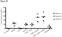

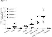


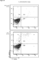


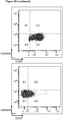
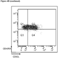
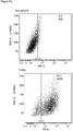


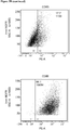


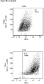


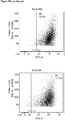


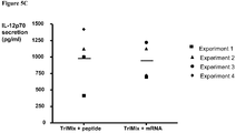


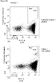
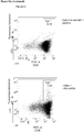


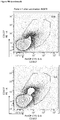

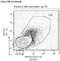
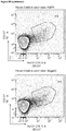
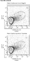
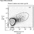

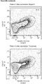


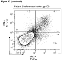

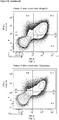


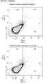

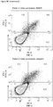


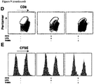


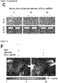


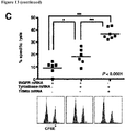

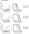


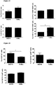


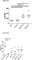


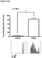
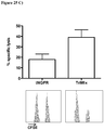

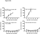

Classifications
-
- A—HUMAN NECESSITIES
- A61—MEDICAL OR VETERINARY SCIENCE; HYGIENE
- A61K—PREPARATIONS FOR MEDICAL, DENTAL OR TOILETRY PURPOSES
- A61K39/00—Medicinal preparations containing antigens or antibodies
- A61K39/39—Medicinal preparations containing antigens or antibodies characterised by the immunostimulating additives, e.g. chemical adjuvants
-
- A—HUMAN NECESSITIES
- A61—MEDICAL OR VETERINARY SCIENCE; HYGIENE
- A61K—PREPARATIONS FOR MEDICAL, DENTAL OR TOILETRY PURPOSES
- A61K39/00—Medicinal preparations containing antigens or antibodies
- A61K39/0005—Vertebrate antigens
- A61K39/0011—Cancer antigens
-
- A—HUMAN NECESSITIES
- A61—MEDICAL OR VETERINARY SCIENCE; HYGIENE
- A61K—PREPARATIONS FOR MEDICAL, DENTAL OR TOILETRY PURPOSES
- A61K39/00—Medicinal preparations containing antigens or antibodies
- A61K39/46—Cellular immunotherapy
- A61K39/461—Cellular immunotherapy characterised by the cell type used
- A61K39/4615—Dendritic cells
-
- A—HUMAN NECESSITIES
- A61—MEDICAL OR VETERINARY SCIENCE; HYGIENE
- A61K—PREPARATIONS FOR MEDICAL, DENTAL OR TOILETRY PURPOSES
- A61K39/00—Medicinal preparations containing antigens or antibodies
- A61K39/46—Cellular immunotherapy
- A61K39/462—Cellular immunotherapy characterized by the effect or the function of the cells
- A61K39/4622—Antigen presenting cells
-
- A—HUMAN NECESSITIES
- A61—MEDICAL OR VETERINARY SCIENCE; HYGIENE
- A61K—PREPARATIONS FOR MEDICAL, DENTAL OR TOILETRY PURPOSES
- A61K39/00—Medicinal preparations containing antigens or antibodies
- A61K39/46—Cellular immunotherapy
- A61K39/464—Cellular immunotherapy characterised by the antigen targeted or presented
- A61K39/4643—Vertebrate antigens
- A61K39/4644—Cancer antigens
- A61K39/464452—Transcription factors, e.g. SOX or c-MYC
- A61K39/464453—Wilms tumor 1 [WT1]
-
- A—HUMAN NECESSITIES
- A61—MEDICAL OR VETERINARY SCIENCE; HYGIENE
- A61K—PREPARATIONS FOR MEDICAL, DENTAL OR TOILETRY PURPOSES
- A61K39/00—Medicinal preparations containing antigens or antibodies
- A61K39/46—Cellular immunotherapy
- A61K39/464—Cellular immunotherapy characterised by the antigen targeted or presented
- A61K39/4643—Vertebrate antigens
- A61K39/4644—Cancer antigens
- A61K39/464454—Enzymes
- A61K39/464456—Tyrosinase or tyrosinase related proteinases [TRP-1 or TRP-2]
-
- A—HUMAN NECESSITIES
- A61—MEDICAL OR VETERINARY SCIENCE; HYGIENE
- A61K—PREPARATIONS FOR MEDICAL, DENTAL OR TOILETRY PURPOSES
- A61K39/00—Medicinal preparations containing antigens or antibodies
- A61K39/46—Cellular immunotherapy
- A61K39/464—Cellular immunotherapy characterised by the antigen targeted or presented
- A61K39/4643—Vertebrate antigens
- A61K39/4644—Cancer antigens
- A61K39/464484—Cancer testis antigens, e.g. SSX, BAGE, GAGE or SAGE
- A61K39/464486—MAGE
-
- A—HUMAN NECESSITIES
- A61—MEDICAL OR VETERINARY SCIENCE; HYGIENE
- A61K—PREPARATIONS FOR MEDICAL, DENTAL OR TOILETRY PURPOSES
- A61K39/00—Medicinal preparations containing antigens or antibodies
- A61K39/46—Cellular immunotherapy
- A61K39/464—Cellular immunotherapy characterised by the antigen targeted or presented
- A61K39/4643—Vertebrate antigens
- A61K39/4644—Cancer antigens
- A61K39/46449—Melanoma antigens
- A61K39/464491—Melan-A/MART
-
- A—HUMAN NECESSITIES
- A61—MEDICAL OR VETERINARY SCIENCE; HYGIENE
- A61K—PREPARATIONS FOR MEDICAL, DENTAL OR TOILETRY PURPOSES
- A61K39/00—Medicinal preparations containing antigens or antibodies
- A61K39/46—Cellular immunotherapy
- A61K39/464—Cellular immunotherapy characterised by the antigen targeted or presented
- A61K39/4643—Vertebrate antigens
- A61K39/4644—Cancer antigens
- A61K39/46449—Melanoma antigens
- A61K39/464492—Glycoprotein 100 [Gp100]
-
- A—HUMAN NECESSITIES
- A61—MEDICAL OR VETERINARY SCIENCE; HYGIENE
- A61K—PREPARATIONS FOR MEDICAL, DENTAL OR TOILETRY PURPOSES
- A61K39/00—Medicinal preparations containing antigens or antibodies
- A61K39/46—Cellular immunotherapy
- A61K39/464—Cellular immunotherapy characterised by the antigen targeted or presented
- A61K39/4643—Vertebrate antigens
- A61K39/4644—Cancer antigens
- A61K39/464499—Undefined tumor antigens, e.g. tumor lysate or antigens targeted by cells isolated from tumor
-
- C—CHEMISTRY; METALLURGY
- C12—BIOCHEMISTRY; BEER; SPIRITS; WINE; VINEGAR; MICROBIOLOGY; ENZYMOLOGY; MUTATION OR GENETIC ENGINEERING
- C12N—MICROORGANISMS OR ENZYMES; COMPOSITIONS THEREOF; PROPAGATING, PRESERVING, OR MAINTAINING MICROORGANISMS; MUTATION OR GENETIC ENGINEERING; CULTURE MEDIA
- C12N5/00—Undifferentiated human, animal or plant cells, e.g. cell lines; Tissues; Cultivation or maintenance thereof; Culture media therefor
- C12N5/06—Animal cells or tissues; Human cells or tissues
- C12N5/0602—Vertebrate cells
- C12N5/0634—Cells from the blood or the immune system
- C12N5/0639—Dendritic cells, e.g. Langherhans cells in the epidermis
-
- A—HUMAN NECESSITIES
- A61—MEDICAL OR VETERINARY SCIENCE; HYGIENE
- A61K—PREPARATIONS FOR MEDICAL, DENTAL OR TOILETRY PURPOSES
- A61K39/00—Medicinal preparations containing antigens or antibodies
- A61K2039/51—Medicinal preparations containing antigens or antibodies comprising whole cells, viruses or DNA/RNA
- A61K2039/515—Animal cells
- A61K2039/5154—Antigen presenting cells [APCs], e.g. dendritic cells or macrophages
-
- A—HUMAN NECESSITIES
- A61—MEDICAL OR VETERINARY SCIENCE; HYGIENE
- A61K—PREPARATIONS FOR MEDICAL, DENTAL OR TOILETRY PURPOSES
- A61K39/00—Medicinal preparations containing antigens or antibodies
- A61K2039/51—Medicinal preparations containing antigens or antibodies comprising whole cells, viruses or DNA/RNA
- A61K2039/515—Animal cells
- A61K2039/5156—Animal cells expressing foreign proteins
-
- A—HUMAN NECESSITIES
- A61—MEDICAL OR VETERINARY SCIENCE; HYGIENE
- A61K—PREPARATIONS FOR MEDICAL, DENTAL OR TOILETRY PURPOSES
- A61K39/00—Medicinal preparations containing antigens or antibodies
- A61K2039/51—Medicinal preparations containing antigens or antibodies comprising whole cells, viruses or DNA/RNA
- A61K2039/515—Animal cells
- A61K2039/5158—Antigen-pulsed cells, e.g. T-cells
-
- A—HUMAN NECESSITIES
- A61—MEDICAL OR VETERINARY SCIENCE; HYGIENE
- A61K—PREPARATIONS FOR MEDICAL, DENTAL OR TOILETRY PURPOSES
- A61K39/00—Medicinal preparations containing antigens or antibodies
- A61K2039/51—Medicinal preparations containing antigens or antibodies comprising whole cells, viruses or DNA/RNA
- A61K2039/53—DNA (RNA) vaccination
-
- A—HUMAN NECESSITIES
- A61—MEDICAL OR VETERINARY SCIENCE; HYGIENE
- A61K—PREPARATIONS FOR MEDICAL, DENTAL OR TOILETRY PURPOSES
- A61K39/00—Medicinal preparations containing antigens or antibodies
- A61K2039/54—Medicinal preparations containing antigens or antibodies characterised by the route of administration
-
- A—HUMAN NECESSITIES
- A61—MEDICAL OR VETERINARY SCIENCE; HYGIENE
- A61K—PREPARATIONS FOR MEDICAL, DENTAL OR TOILETRY PURPOSES
- A61K39/00—Medicinal preparations containing antigens or antibodies
- A61K2039/555—Medicinal preparations containing antigens or antibodies characterised by a specific combination antigen/adjuvant
- A61K2039/55511—Organic adjuvants
- A61K2039/55516—Proteins; Peptides
-
- A—HUMAN NECESSITIES
- A61—MEDICAL OR VETERINARY SCIENCE; HYGIENE
- A61K—PREPARATIONS FOR MEDICAL, DENTAL OR TOILETRY PURPOSES
- A61K39/00—Medicinal preparations containing antigens or antibodies
- A61K2039/57—Medicinal preparations containing antigens or antibodies characterised by the type of response, e.g. Th1, Th2
-
- A—HUMAN NECESSITIES
- A61—MEDICAL OR VETERINARY SCIENCE; HYGIENE
- A61K—PREPARATIONS FOR MEDICAL, DENTAL OR TOILETRY PURPOSES
- A61K2239/00—Indexing codes associated with cellular immunotherapy of group A61K39/46
- A61K2239/31—Indexing codes associated with cellular immunotherapy of group A61K39/46 characterized by the route of administration
-
- A—HUMAN NECESSITIES
- A61—MEDICAL OR VETERINARY SCIENCE; HYGIENE
- A61K—PREPARATIONS FOR MEDICAL, DENTAL OR TOILETRY PURPOSES
- A61K2239/00—Indexing codes associated with cellular immunotherapy of group A61K39/46
- A61K2239/38—Indexing codes associated with cellular immunotherapy of group A61K39/46 characterised by the dose, timing or administration schedule
-
- A—HUMAN NECESSITIES
- A61—MEDICAL OR VETERINARY SCIENCE; HYGIENE
- A61K—PREPARATIONS FOR MEDICAL, DENTAL OR TOILETRY PURPOSES
- A61K2239/00—Indexing codes associated with cellular immunotherapy of group A61K39/46
- A61K2239/46—Indexing codes associated with cellular immunotherapy of group A61K39/46 characterised by the cancer treated
- A61K2239/48—Blood cells, e.g. leukemia or lymphoma
-
- A—HUMAN NECESSITIES
- A61—MEDICAL OR VETERINARY SCIENCE; HYGIENE
- A61K—PREPARATIONS FOR MEDICAL, DENTAL OR TOILETRY PURPOSES
- A61K2239/00—Indexing codes associated with cellular immunotherapy of group A61K39/46
- A61K2239/46—Indexing codes associated with cellular immunotherapy of group A61K39/46 characterised by the cancer treated
- A61K2239/57—Skin; melanoma
-
- C—CHEMISTRY; METALLURGY
- C12—BIOCHEMISTRY; BEER; SPIRITS; WINE; VINEGAR; MICROBIOLOGY; ENZYMOLOGY; MUTATION OR GENETIC ENGINEERING
- C12N—MICROORGANISMS OR ENZYMES; COMPOSITIONS THEREOF; PROPAGATING, PRESERVING, OR MAINTAINING MICROORGANISMS; MUTATION OR GENETIC ENGINEERING; CULTURE MEDIA
- C12N2510/00—Genetically modified cells
-
- Y—GENERAL TAGGING OF NEW TECHNOLOGICAL DEVELOPMENTS; GENERAL TAGGING OF CROSS-SECTIONAL TECHNOLOGIES SPANNING OVER SEVERAL SECTIONS OF THE IPC; TECHNICAL SUBJECTS COVERED BY FORMER USPC CROSS-REFERENCE ART COLLECTIONS [XRACs] AND DIGESTS
- Y02—TECHNOLOGIES OR APPLICATIONS FOR MITIGATION OR ADAPTATION AGAINST CLIMATE CHANGE
- Y02A—TECHNOLOGIES FOR ADAPTATION TO CLIMATE CHANGE
- Y02A50/00—TECHNOLOGIES FOR ADAPTATION TO CLIMATE CHANGE in human health protection, e.g. against extreme weather
- Y02A50/30—Against vector-borne diseases, e.g. mosquito-borne, fly-borne, tick-borne or waterborne diseases whose impact is exacerbated by climate change
Definitions
- the invention is situated in the field of immunotherapy using antigen presenting cells (APCs) from a patient modified either in vitro or in vivo ( in situ ) so they are capable of presenting a target-specific antigen in the patient, leading to a host-mediated immune response to the target-expressing cells.
- APCs antigen presenting cells
- the invention is especially related to increasing the immunostimulatory effect of the APCs either in vitro or in vivo ( in situ ) in view of vaccination of patients suffering from cancer or infectious disorders.
- DCs Dendritic Cells
- APCs such as Dendritic Cells (DCs) have emerged as key players in orchestrating immune responses and in particular in inducing primary responses in patients in general.
- DCs can be generated on a large scale in closed systems, yielding sufficient numbers of cells for use in clinical trials.
- antigens derived from infectious microorganisms and many different tumor-associated antigens, which are either selectively or preferentially expressed by tumor cells have been identified.
- a whole range of strategies to load DCs with such antigens have been designed.
- DCs loaded with a target-specific antigen as APCs are insufficient in eliciting a strong immune response both in vitro and in vivo.
- One cause of this insufficient immunostimulation is the complicated in vitro manipulation of the DCs prior to their use, leading to loss of their characteristic properties such as secretion of cytokines and other factors triggering immune responses.
- Another problem is that artificially made DCs often do not express the necessary cellular markers on their cell-surface needed to activate a T-cell response to the target-specific antigen presented by the DCs thereby overcoming the often occurring T-cell tolerance towards the target-specific antigens.
- Van Lint et al., 2012 (Cancer Res 2012;72;1661-1671 ) for example describes the in vivo use of mRNA encoding CD40L, CD70 and caTLR4 in anti-tumor therapy by intranodal injection.
- a maturation stimulus for example through the stimulation of the DCs with e.g. LPS in order to mature them, this furthermore usually blocks the uptake and presentation of the actual antigens, resulting in poor antigen presentation of the target antigens.
- the inventors have established that the T cell stimulatory capacity of antigenic-peptide pulsed APCs or APCs can be greatly enhanced by providing them with specific molecular adjuvants in the form of a mixture of mRNA or DNA molecules encoding the immunostimulatory factor CD40L and one or more of CD70 and caTLR4, either in vivo or in vitro.
- Said stimulation with immunostimulatory factors can be done in vitro, e.g. through co-electroporation or other means of introducing the mRNA or DNA molecules into the APCs, such as DCs.
- Said stimulation with immunostimulatory factors can also be done in vivo ( in situ ) through intranodal, intradermal, subcutaneous, intratumoral injection or intravenous administration of mRNA or DNA molecules encoding said immunostimulatory factors and optionally the tumor antigen mRNA, DNA or protein.
- Said mRNA or DNA can be naked or can be protected as described below. Said mRNA or DNA can be protected when administered for example intraveneously.
- mRNA encoding activation markers e.g. TriMix
- antigen either administered, ot residing at the place of administering the mRNA encoding activation markers
- Maturation of the DCs and presentation of the antigens go hand in hand instead of excluding each other.
- TriMix mRNA induced a T-cell attracting and stimulatory environment, including recruitment of antigen-specific CD4+ and CD8+ T-cells and cytotoxic T lymphocytes (CTLs) against various TAAs.
- CTLs cytotoxic T lymphocytes
- mRNA vaccination was as efficient in CTL induction and therapy response as vaccination with mRNA electroporated DCs.
- the application provides the proof of concept that such modified APCs pulsed with a target-specific peptide or co-electroporated with mRNA encoding a target-specific antigen can stimulate antigen-specific T cells both in vitro and after vaccination and thus form a promising new approach for anti-tumor, anti-viral, anti-bacterial or anti-fungal immunotherapy.
- the application provides proof of concept that direct administration of mRNAs encoding immunostimulatory factors CD40L, and CD70, caTLR4, or both, induces maturation of the APCs or DCs in situ , enabling them to present target specific antigens and elicit a genuine immuneresponse against the target.
- the inventors found that direct injection of the mRNAs encoding immunostimulatory factors CD40L, and CD70, caTLR4, or both into the tumor not only induced an immuneresponse towards said tumor, but also towards other, peripheral tumors.
- the application thus provides for a method for improving the immunostimulatory characteristics of APCs comprising the introduction of at least two different mRNA or DNA molecules encoding proteins that modify the functionality of the APCs.
- the application hence also provides a method for loading APCs, preferably DCs with at least two different mRNA or DNA molecules encoding proteins that modify the functionality of the APCs. Said loading can be done in vitro through e.g. transfection or electroporation, or in vivo, through direct administration of said mRNA or DNA - either naked or in a protected form - in the subject or patient.
- said proteins are immunostimulatory factors CD40L and one or more of CD70 and caTLR4. More preferable, said proteins are the immunostimulatory factors CD40L, CD70 and caTLR4.
- one or more additional factors are introduced, selected from the group comprising or consisting of: secreted molecules such as IL-12p70, DC migration mediating molecules such as EL-selectin and CCR7, and/orfurther co-stimulatory molecules such as 4-1 BBL.
- secreted molecules such as IL-12p70
- DC migration mediating molecules such as EL-selectin and CCR7
- further co-stimulatory molecules such as 4-1 BBL.
- molecules influencing signaling pathways such as SOCS, STAT3, or A20
- molecules influencing inhibitory pathways such as PD-L1/PD-1/CTLA4
- APCs preferably DCs.
- the antigen-specific stimulations are performed without the addition of any exogenous IL-2 and/or IL-7 to support T-cell proliferation and survival.
- the APCs are additionally stimulated with soluble factors selected from the group comprising TLR ligands, IFN-gamma, TNF-alpha, IL-6, IL-1beta and/or PGE2.
- the method used for in vitro introduction of said mRNA or DNA molecules in APCs or DCs is selected from the group consisting or comprising of: (co)electroporation, viral transduction, lipofection and transfection of mRNA or DNA encoding the immunostimulatory antigens.
- the method used for in vivo ( in situ ) introduction of said different mRNA or DNA molecules in APCs or DCs is done by intranodal injection, intradermal injection, subcutane injection, intratumoral injection or by intravenous administration.
- pretreatment can be done with for example GM-CSF, Flt3L or imiquimod to enhance the effect.
- intravenous administration the use of protected mRNA or DNA molecules is preferred.
- the application provides a method of vaccinating, or inducing an immune-response in a subject, by intranodal, intradermal, subcutaneous, intratumoral injection or intravenous administration of mRNA or DNA encoding the immunostimulatory factor CD40L and one or more of CD70 and caTLR4, optionally in combination with antigens or mRNA or DNA molecules encoding target-specific antigens, e.g. derived from a tumor cell, or from any infectious agent, such as a bacterium, a virus, a fungus, a toxin or venom, etc.
- the application also provides a method of treating an infection in a patient comprising the step of intranodal, intradermal, subcutane, or intratumoral injection intravenous administration of mRNA or DNA encoding the immunostimulatory factor CD40L and one or more of CD70 and caTLR4, optionally in combination with antigens or mRNA or DNA molecules encoding antigens derived from any infectious agent, such as a bacterium, a virus, a fungus, a toxin or venom, etc..
- the administration can be done at the site of infection. Adding the antigens or RNA or DNA molecules encoding them is optional because said antigens are present in circulation or at the site of infection.
- the application also provides a method of anti-cancer treatment of a patient comprising the step of intranodal, intratumoral, intradermal, subcutaneous, or intratumoral injection intravenous administration of mRNA or DNA encoding the immunostimulatory factor CD40L and one or more of CD70 and caTLR4, optionally in combination with antigens or mRNA or DNA molecules encoding tumor antigens.
- Adding the tumor antigens or RNA or DNA molecules encoding them is optional because said antigens are present in the tumor environment.
- said administration is preceded by tumor ablation, such as radiofrequency- or cryo-ablation, thereby freeing tumor-antigens.
- the application further provides a method of improving antigen mRNA-based immunization of a subject, comprising the steps of administering mRNA or DNA encoding the immunostimulatory factor CD40L and one or more of CD70 and caTLR4 to said subject.
- said administration is done in the lymph nodes, e.g. through intranodal injection.
- said administration is done intradermally, subcutaneously or intratumorally, through injection or done through intravenous administration.
- the antigens present at the site of administration will be effectively engulfed by the activated APCs and subsequently presented to the immune system, triggering a specific immune response.
- said immune-response results in treatment of a proliferative disorder.
- said immune-response results in treatment of an infection or an inflammatory disorder.
- the application further provides a vaccine or composition comprising one or more mRNA or DNA molecules encoding the immunostimulatory factor CD40L and one or more of CD70 and caTLR4, optionally in combination with antigens or mRNA or DNA molecules encoding tumor antigens.
- the application thus provides a vaccine comprising one or more mRNA or DNA molecule(s) encoding the functional immunostimulatory proteins CD40L, CD70, and caTLR4 for use in eliciting an immune response towards a tumor or infection causing agent in a subject, wherein said vaccine is administered directly to the tumor environment, respectively the infected area, or wherein said vaccine is administered to the lymph nodes, or through subcutane, intradermal or intravenous administration.
- said administration is done through injection directly into the tumor area or infected area, or through intranodal or intradermal injection
- said vaccine for use further comprises a target-specific antigen.
- said target-specific antigen is selected from the group consisting of: total mRNA isolated from (a) target cell(s), one or more specific target mRNA molecules, protein lysates of (a) target cell(s), specific proteins from (a) target cell(s), a synthetic target-specific peptide or protein, and synthetic mRNA or DNA encoding a target-specific antigen or its derived peptide(s).
- said target-specific antigen is a tumor antigen.
- said administration is preceded by tumor ablation, such as radiofrequency- or cryo-ablation, thereby freeing tumor-antigens.
- said target-specific antigen is a bacterial, viral or fungal antigen.
- said mRNA or DNA molecules encoding the immunostimulatory proteins are part of a single mRNA or DNA molecule.
- the single mRNA or DNA molecule is capable of expressing the two or more proteins simultaneously.
- the mRNA or DNA molecules encoding the immunostimulatory proteins are separated in the single mRNA or DNA molecule by an internal ribosomal entry site (IRES) or a self-cleaving 2a peptide encoding sequence.
- IRS internal ribosomal entry site
- the vaccine for use according to anyone of the embodiments defined herein additionally comprises mRNA or DNA molecules encoding one or more of the proteins selected from the group consisting of: IL-12p70, EL-selectin, CCR7, and 4-1 BBL.
- one or more molecules inhibiting SOCS, A20, PD-L1 or STAT3 expression or function can be included in the vaccine.
- said mRNA is protected.
- the application further provides a method for preparing an immunotherapy agent comprising the steps of:
- the method of modification used in step b) and/or c) is selected from the group of electroporation, viral transduction, lipofection or transfection of mRNA or DNA encoding the immunostimulatory antigens.
- the specific immunostimulatory proteins and the target antigens are introduced through a one-step mechanism.
- co-electroporation of the mRNA or DNA encoding a target-specific antigen with the mRNA or DNA encoding the immunostimulatory factors is used.
- protein or peptide pulsing is used to load the target-specific antigen or its derived antigenic peptides onto the APCs.
- a preferred combination of immunostimulatory factors used in the methods of the application is CD40L and CD70.
- the combination of CD40L, CD70 and caTLR4 immunostimulatory molecules is used, which is called the "TriMix" hereinafter.
- the APCs used in the methods of the application are selected from the group consisting of patient-specific DCs or B-cells; or established dendritic cell lines or B-cell lines.
- the application further provides a vaccine comprising the immunotherapy agent obtained by any of the methods of the application mentioned above, further comprising pharmaceutically acceptable adjuvant(s).
- the immunotherapy agent is directed to a target-specific antigen which can be a tumor antigen, or a bacterial, viral or fungal antigen.
- Said target-specific antigen can be derived from either one of: total mRNA isolated from (a) target cell(s), one or more specific target mRNA molecules, protein lysates of (a) target cell(s), specific proteins from (a) target cell(s), or a synthetic target-specific peptide or protein and synthetic mRNA or DNA encoding a target-specific antigen or its derived peptides.
- the application further encompasses the use of a preparation of APCs obtained by the method of the application or the immunotherapy agent obtained by the method of the application in the manufacture of a vaccine capable of eliciting an immune response in a patient in need thereof.
- the application further provides for a method to screen for new target-specific epitopes that can be used for vaccination of patients, using APCs obtained by the immunostimulation method of the application comprising;
- the application provides for a method of following the effects of the treatment with an anti-target vaccine in a patient; comprising the detection and analysis of the immune response towards the target-specific antigen elicited in the subject previously injected with the anti-target vaccine obtained by any of the methods of the application.
- the patient is suffering from a disease or disorder selected from the group of: cancer, bacterial, viral or fungal infection, e.g. HIV infection or hepatitis.
- the application also provides a kit for improving the immunostimulatory characteristics of APCs comprising a combination of at least two different mRNA or DNA molecules encoding functional immunostimulatory proteins selected from the group consisting of CD40L, CD70, caTLR4, IL-12p70, EL-selectin, CCR7, and/or 4-1 BBL, and optionally comprising molecules inhibiting SOCS, A20, PD-L1 or STAT3 expression or function.
- the kit comprises mRNA or DNA molecules encoding CD40L and CD70.
- the kit of the application can additionally comprise the mRNA or DNA encoding for the caTLR4, resulting in the so-called "TriMix".
- the kit of the application comprises a single mRNA or DNA molecule, wherein said two or more mRNA or DNA molecules encoding the immunostimulatory proteins are combined.
- the single mRNA or DNA molecule is capable of expressing the two or more immunostimulatory proteins simultaneously e.g. the two or more mRNA or DNA molecules encoding the immunostimulatory proteins are linked in the single mRNA or DNA molecule by an internal ribosomal entry site (IRES) or a self-cleaving 2a peptide encoding sequence.
- IRS internal ribosomal entry site
- the application provides an ex vivo method of amplifying antigen-specific T-cells from a patient.
- the patient can be previously vaccinated or not.
- the amplified pool of T-cells can then be used for new or additional vaccination (boosting) of the patient.
- the application thus provides a method for the ex vivo amplification of a pool of T-cells from a patient comprising;
- the application further provides for methods of using the modified APCs of the application for treating neoplasms, cancer, infectious diseases such as viral, bacterial or fungal infections e.g. with HIV and hepatitis, or immunological disorders such as immunodeficiency, SCIPD, or AIDS.
- infectious diseases such as viral, bacterial or fungal infections e.g. with HIV and hepatitis
- immunological disorders such as immunodeficiency, SCIPD, or AIDS.
- the treatment with the vaccine, immunotherapy agent or APCs of the application can be combined or followed by a non-specific treatment of immunomodulation in order to boost the immune system of the patient.
- this can be anti-CTLA4 antibodies or IFN-alpha or other methods of immunomodulation in order to boost the immune system of the patient.
- APCs such as DCs, B-cells, dendritic cell-lines, or B-cell-lines with a maturation signal through mRNA electroporation offers several advantages:
- APCs electroporated with mRNA or DNA encoding two or more immunostimulatory factors e.g. the TriMix of CD40L, CD70 and caTRLA4
- two or more immunostimulatory factors e.g. the TriMix of CD40L, CD70 and caTRLA4
- APCs electroporated with mRNA or DNA encoding two or more immunostimulatory factors e.g. the TriMix of CD40L, CD70 and caTRLA4
- This approach offers several further advantages:
- the maturation and antigen-loading of the APCs can be combined in one simple step. Obviating the peptide pulsing step in the vaccine production thus results in less manipulation of the cells and in less cell-loss and contamination-risk.
- the antigen-encoding plasmid can be genetically modified by adding an HLA class II targeting sequence. This not only routes the antigen to the HLA class II compartments for processing and presentation of HLA class II restricted antigen-derived peptides, but also enhances processing and presentation in the context of HLA class I molecules.
- TriMix APCs i.e. electroporated with mRNA encoding CD40L, CD70 and caTLR4
- TriMix APCs can almost equally well stimulate MelanA-specific T cells when co-electroporated with whole MelanA-encoding mRNA than when being pulsed with MelanA-derived peptide.
- TriMix APCs can stimulate T cells specific for other antigens with a lower precursor frequency both in vitro and in vivo.
- the application further provides for methods of treatment of a subject having cancer, being infected with an infectious agent, or suffering from an immunological disorder, comprising the administration of a vaccine comprising DCs that have been in vitro modified with the immunostimulatory factors such as CD40L, CD70 and/or caTLR4.
- the application also provides for a method of inducing an immuneresponse in a subject or a method of vaccinating a subject against an antigen, comprising the administration of a vaccine comprising DCs that have been in vitro modified with the immunostimulatory factors such as CD40L, CD70 and/or caTLR4.
- Said administration can be done intravenously or intradermally, or through a combination thereof in any one of the methods using in vitro modified DCs according to the application (e.g. TriMix-DCs).
- Said methods of treatment or vaccination using in vitro modified DCs according to the application can be combined with any other chemotherapeutic or otherwise beneficial treatment to said subject.
- the in vivo modification of APCs or DC's is also part of the application and encompasses the direct administration of one or more mRNA or DNA molecule(s) encoding the immunostimulatory factor CD40L and one or more of CD70 and caTLR4.
- mRNA or DNA molecules encoding target-specific antigens can be administred as well.
- mRNA or DNA mentioned in any one of the embodiments defined herein can either be naked mRNA or DNA, or protected mRNA or DNA. Protection of DNA or mRNA increases its stability, yet preserving the ability to use the mRNA or DNA for vaccination purposes, since it is still able to be presented by APCs or DCs.
- Non-limiting examples of mRNA or DNA protection can be: liposome-encapsulation, protamine-protection, (Cationic) Lipid Lipoplexation, lipidic, cationic or polycationic compositions, Mannosylated Lipoplexation, Bubble Liposomation, Polyethylenimine (PEI) protection, liposome-loaded microbubble protection etc.
- the administration of the components of the vaccine, immunotherapy agent, or composition can be done simultaneously or sequentially, i.e. one component can be administered to the subject at the time.
- the mRNA or DNA molecules encoding CD40L, and caTLR4 or CD70 can be administered simultaneously together with the target antigen.
- the antigen can be added after a small time interval.
- each mRNA or DNA molecule encoding an immunostumulatory factor i.e. CD40L, caTLR4, and CD70
- CD40L, caTLR4, and CD70 an immunostumulatory factor
- the DCs or APCs can be in vitro modified by adding the components of the kit or composition simultaneously or sequentially, i.e. one component can be added at the time.
- the mRNA or DNA molecules encoding CD40L, and caTLR4 or CD70 can be added to the APCs or DCs simultaneously together with the target antigen.
- the antigen can be added after a small time interval.
- each mRNA or DNA molecule encoding an immunostumulatory factor i.e. CD40L, caTLR4, and CD70
- CD40L, caTLR4, and CD70 an immunostumulatory factor
- modifying APCs or DCs with the immunostimulatory agents also reduces the activity of regulatory T cells in vitro.
- Regulatory T cells are important mediators of peripheral tolerance, thus preventing the occurrence of autoimmune reponses.
- these regulatory T cells accumulate and dampen the infection- or cancer-specific immune response. Reducing the activity of regulatory T cells at the site of infection or within the tumor environment is therefore highly beneficial, since it will increase the immune response towards the antigens present at the site of infection or within the tumor.
- TriMix mRNA modified human DCs do not induce Treg induction starting from CD4 + CD25- T cells.
- the application provides mRNA or DNA molecules encoding CD40L, and caTLR4, CD70, or both, which can be used to locally reduce the activity of Regulatory T cells, comprising the step of administering said mRNA or DNA molecules to the subject, preferably to the site of infection or to the tumor, or intranodally or intradermally.
- the application therefore further provides for a method of locally reducing the activity of regulatory T cells, comprising the step of administering antigen-presenting or DCs modified by introduction of mRNA or DNA molecules encoding CD40L, and caTLR4, CD70, or both, to the site of infection or to the tumor.
- the application further provides antigen-presenting cells or DCs modified by introduction of mRNA or DNA molecules encoding CD40L, and caTLR4, CD70, or both, for use in locally reducing the activity of regulatory T cells, comprising the step of administering said antigen-presenting cells or DCs to the site of infection or to the tumor.
- the inventors investigated whether the activation state of DCs is a critical factor in determining whether the DCs presenting a target-specific antigen will be potent inducers of an anti-target immune response after vaccination or not.
- the inventors unexpectedly found that the effectiveness of currently used DC vaccination protocols could be significantly improved by providing the DCs with a more potent activation signal and by using a shorter manipulation process.
- the inventors moreover found that using a combination of mRNA or DNA molecules encoding a set of specific immunostimulatory proteins could be used to mature DCs both in vitro and in vivo, i.e. through in situ administration to the subject in e.g. the lymph nodes or directly at the site of infection or in the tumor.
- TriMix stands for a mixture of mRNA molecules encoding CD40L, CD70 and caTLR4 immunostimulatory proteins.
- TriMix DCs or “TriMix APCs” stands for respectively DCs or APCs that have been modified to express the TriMix mixture of mRNA or DNA molecules encoding CD40L, CD70 and caTLR4 immunostimulatory proteins.
- the mRNA or DNA used or mentioned herein can either be naked mRNA or DNA, or protected mRNA or DNA. Protection of DNA or mRNA increases its stability, yet preserving the ability to use the mRNA or DNA for vaccination purposes.
- Non-limiting examples of protection of both mRNA and DNA can be: liposome-encapsulation, protamine-protection, (Cationic) Lipid Lipoplexation, lipidic, cationic or polycationic compositions, Mannosylated Lipoplexation, Bubble Liposomation, Polyethylenimine (PEI) protection, liposome-loaded microbubble protection etc..
- target used throughout the description is not limited to the specific examples that may be described herein. Any infectious agent such as a virus, a bacterium or a fungus may be targeted. In addition any tumor or cancer cell may be targeted.
- target-specific antigen used throughout the description is not limited to the specific examples that may be described herein. It will be clear to the skilled person that the application is related to the induction of immunostimulation in APCs, regardless of the target-specific antigen that is presented. The antigen that is to be presented will depend on the type of target to which one intends to elicit an immune response in a subject. Typical examples of target-specific antigens are expressed or secreted markers that are specific to tumor, bacterial and fungal cells or to specific viral proteins or viral structures. Without wanting to limit the scope of protection of the application, some examples of possible markers are listed below.
- the term "antigen presenting cell” used throughout the description includes all APCs. Specific non limiting examples are DCs, dendritic cell-lines, B-cells, or B-cell-lines.
- the DCs or B-cells can be isolated or generated from the blood of a patient or healthy subject. The patient or subject can have been the subject of prior vaccination or not.
- neoplasms are not intended to be limited to the types of cancer or tumors that may have been exemplified.
- the term therefore encompasses all proliferative disorders such as neoplasma, dysplasia, premalignant or precancerous lesions, abnormal cell growths, benign tumors, malignant tumors, cancer or metastasis, wherein the cancer is selected from the group of: leukemia, non-small cell lung cancer, small cell lung cancer, CNS cancer, melanoma, ovarian cancer, kidney cancer, prostate cancer, breast cancer, glioma, colon cancer, bladder cancer, sarcoma, pancreatic cancer, colorectal cancer, head and neck cancer, liver cancer, bone cancer, bone marrow cancer, stomach cancer, duodenum cancer, oesophageal cancer, thyroid cancer, hematological cancer, and lymphoma.
- Specific antigens for cancer can e.g. be MelanA/M
- infectious disease or "infection” used throughout the description is not intended to be limited to the types of infections that may have been exemplified herein. The term therefore encompasses all infectious agents to which vaccination would be beneficial to the subject.
- Non-limiting examples are the following virus-caused infections or disorders: Acquired Immunodeficiency Syndrome - Adenoviridae Infections - Alphavirus Infections - Arbovirus Infections - Bell Palsy - Borna Disease - Bunyaviridae Infections - Caliciviridae Infections - Chickenpox - Common Cold - Condyloma Acuminata - Coronaviridae Infections - Coxsackievirus Infections - Cytomegalovirus Infections - Dengue - DNA Virus Infections - Contagious Ecthyma, - Encephalitis - Encephalitis, Arbovirus - Encephalitis, Herpes Simplex - Epstein
- bacteria- or fungus-caused infections or disorders Abscess - Actinomycosis - Anaplasmosis - Anthrax - Arthritis, Reactive - Aspergillosis - Bacteremia - Bacterial Infections and Mycoses - Bartonella Infections - Botulism - Brain Abscess - Brucellosis - Burkholderia Infections - Campylobacter Infections - Candidiasis - Candidiasis, Vulvovaginal - Cat-Scratch Disease - Cellulitis - Central Nervous System Infections - Chancroid - Chlamydia Infections - Chlamydiaceae Infections - Cholera - Clostridium Infections - Coccidioidomycosis - Corneal Ulcer - Cross Infection - Cryptococcosis - Dermatomycoses - Diphtheria
- immunological disorder encompasses any immunological disorder, including all disorders or syndromes involving impaired or reduced immunological response.
- disorders or syndromes involving impaired or reduced immune response are so-called Primary Immune Deficiencies such as: congenital defects of the immune system, Selective IgA Deficiency, Common Variable Immunodeficiency, X-Linked Agammaglobulinemia (Bruton type, X-linked infantile, or congenital agammaglobulinemia), Chronic Granulomatous Disease, Hyper-IgM Syndrome, and SCID (the classic "bubble boy” disease).
- acquired immunodeficiencies can occur, such as, but not limited to: AIDS, or to subjects receiving chemotherapy or immunosuppressive medications such as subjects having cancer and subjects that underwent an organ transplant, or for various other conditions.
- AIDS acquired immunodeficiencies
- immunosuppressive medications such as subjects having cancer and subjects that underwent an organ transplant, or for various other conditions.
- diabetes patients might sufferfrom mild immune suppression and elderly people or children and newborns can have a weakened or weaker immune system. All said conditions, syndromes or disorders are meant to be covered by the term "immunological disorders".
- the current application provides new methods of enhancing the immunostimulatory capacities of human DCs through transfection with at least two different mRNA or DNA molecules encoding molecular adjuvants selected from the list of CD40L, CD70, caTLR4, IL-12p70, EL-selectin, CCR7 and/or 4-1BBL; or in combination with the inhibition of the expression or function of SOCS, A20, PD-L1 or STAT3, for example through siRNA transfection.
- the application provides for methods of enhancing the immunostimulatory capacities of human DCs in situ in a subject, by administering mRNA or DNA molecules encoding molecular adjuvants CD40L and CD70, caTLR4, or both to said subject, preferably in the lymph nodes, where DCs reside and mature.
- said mRNA or DNA molecules can be administered intratumorally, subcutane, or intradermally.
- mRNA or DNA molecules encoding any one or more of the following proteins: IL-12p70-, EL-selectin, CCR7 and 4-1BBL can be co-administered.
- CD40L and caTLR4 in monocyte derived immature DCs through mRNA electroporation generates mature, cytokine/chemokine secreting DCs, as has been shown for CD40 and TLR4 ligation through addition of soluble CD40L and LPS.
- CD70 into the DCs provides a co-stimulatory signal to CD27 + naive T-cells by inhibiting activated T-cell apoptosis and by supporting T-cell proliferation.
- TLR4 Toll-Like Receptors
- TLR4 a constitutive active form is known, and could possibly be introduced into the DCs in order to elicit a host immune response.
- caTLR4 is the most potent activating molecule and is therefore preferred.
- mRNA encoding an additional cytokine such as IL-12p70 in the DCs could be beneficial to further increase the cytokine excretion of the DCs, subsequently further stimulating the host immune response.
- co-stimulatory molecules such as 4-1 BBL or a constitutively active form of Akt could also be introduced in the DCs or co-administered in situ .
- inhibitory molecules such as SOCS, A20, PD-L1, STAT3 could be lowered or halted through additional introduction of specific inhibitory molecules such as specific siRNA molecules in the DCs, or can be co-administered in situ.
- the application preferably uses DCs derived from peripheral blood mononuclear cells (PBMCs) directly isolated from the patient's blood, but alternatives such as DCs differentiated out of CD34-positive cells or commercially available dendritic cell-lines could be used as well.
- PBMCs peripheral blood mononuclear cells
- the in vitro method of the application uses either mRNA electroporation, viral transduction (e.g. through lentivirus, adenovirus, or vaccinia virus), mRNA lipofection or DNA transfection to introduce immunostimulatory molecules and target-specific antigens into the DCs.
- mRNA electroporation is especially preferred due to its high efficiency and its wide accepted use in clinical settings in contrast to viral transduction.
- pulsing of the cells with the antigen-specific peptides or with protein can be used as an alternative to mRNA electroporation.
- the introduced mRNA can be a specifically synthesized sequence based on known tumor-specific markers, or can be isolated from (a) tumor cell line(s) or from a tumor-biopsy of the patient.
- the application preferably uses autologous plasma obtained from the patient, but human AB serum, which is commercially available, can also be used.
- the in vivo or in situ methods preferably encompass intranodal injection of the mRNA or DNA molecules encoding the immunostimulatory factors as explained above.
- intratumoral or intradermal injection can be used to target the DCs in vivo.
- said intradermal injection is preceded by intradermal injection with GM-CSF, FLT3L or local treatment with imiquimod.
- the application lies in the combined administration of CD40L and CD70 to DCs, either in vitro or in vivo, thereby leading to increased immunostimulatory effects of the DCs.
- the specific combination of CD40L, CD70 and caTLR4 is administered to the DCs, again in vitro or in vivo, to improve the immunostimulatory effects of the DCs.
- any of the following markers could be administered additionally: IL-12p70, EL-selectin, CCR7, 4-1 BBL for increased expression or SOCS, A20, PD-L1 or STAT3 inhibition.
- a target-specific antigen or its derived epitopes are introduced into the DCs in order to enable them to elicit a T-cell immune response towards the target-specific antigen.
- CD70 expression on mature murine DCs Although strong expression of CD70 on mature murine DCs had been reported after CD40 and TLR ligation alone or in combination, very little is known about the expression of CD70 on human DCs. In our hands, immature DCs, cytokine cocktail matured DCs or DCs electroporated with CD40L and/or TLR4 did not express CD70. Even after combined CD40 ligation through 3T6-associated CD40L and TLR ligation through LPS or dsRNA only a minor percentage of DCs showed CD70 expression. Whether this low CD70 expression by human DCs is a general phenomenon or could be related to our DC generation protocol remains to be established.
- the NF-kappaB activation assay indicates that the mRNA electroporation of our caTLR4 plasmid leads to the expression of a functional protein.
- the CD40L and CD70 plasmids encode functional proteins since CD40L and CD70 electroporated DCs activate the NF-kappaB signaling pathway after CD40 and CD27 ligation, respectively.
- CD40 costimulatory molecules
- CD80 costimulatory molecules
- CD83 costimulatory molecules
- CD86 costimulatory molecules
- HLA class I molecules HLA class I molecules
- CD40 engagement through CD40L electroporation did not impair the upregulation of CD40 expression.
- cytokine secretion level we found a marked upregulation in the secretion of the Th1 cytokine IL-12p70, several pro-inflammatory cytokines (IL-1beta, IL-6, TNF-alpha), hematopoietic growth factors (G-CSF, GM-CSF), IFN-gamma, and IL-10.
- pro-inflammatory cytokines IL-1beta, IL-6, TNF-alpha
- G-CSF hematopoietic growth factors
- GM-CSF hematopoietic growth factors
- IFN-gamma IFN-gamma
- MIP-1 alpha Recruitment of neutrophils
- MIP-1 alpha Recruitment of monocytes and T-cells
- IP-10 IFN-gamma inducible 10 kDa protein; recruitment of monocytes and T-cells
- RANTES Recruitment of T-cells, basophils and eosinophils
- MIP-1 alpha, RANTES and IP-10 are all chemotactic for T-cells, but it has been shown that MIP-1 alpha and RANTES are produced by Th1/Th2-promoting DCs, while IP-10 production is restricted to Th1-promoting DCs.
- CD70 co-electroporation does not induce phenotypical changes or enhanced cytokine/chemokine secretion by DCs, because DCs lack expression of its signaling ligand CD27.
- cytokine and chemokine secretion pattern suggests that DCs electroporated with CD40L and/or caTLR4 mRNA would preferentially induce IFN-gamma producing Th1 cells, a finding that was confirmed in the allogeneic stimulation of CD45RA + CD4 + T-cells. Indeed, T-cells stimulated with DCs electroporated with CD40L and caTLR4, alone or in combination, produced very high amounts of IFN-gamma, but almost no IL-4 and IL-10, secretion of which was not increased in comparison to T-cells stimulated with DCs electroporated with irrelevant mRNA.
- CD70 co-electroporation with CD40L, together or not with caTLR4 induced an additional increase of MelanA-specific T-cells when compared to DCs electroporated with CD40L together or not with caTLR4. This is probably due to a survival-effect induced by the ligation of CD70 on the DCs with CD27 on the T-cells during stimulation.
- Markers can be tumor-specific markers or can be virus-specific, bacterium-specific or fungal specific.
- the application provides for the first time evidence that genetically modified DCs expressing at least two stimulating molecules selected from the lot of CD40L, CD70 and caTLR4, IL-12p70, EL-selectin, CCR7, 4-1 BBL; or in combination with suppression of SOCS, A20, PD-L1 or STAT3 offer a DC based vaccine possessing all the features considered necessary for induction of optimal target-reactive immune responses.
- the combination of stimulating molecules is CD40L and CD70.
- the specific combination of stimulating molecules is the TriMix of CD40L, CD70 and caTLR4.
- the use of the methods of the application has a further advantage over the prior art in that the in vitro manipulation of the DCs is reduced to a minimum in order to prevent the excretion of physiologically relevant cytokines in the in vitro culture medium.
- This is achieved by using a highly efficient one-step transduction method, preferably through mRNA electroporation, enabling the simultaneous introduction of at least two mRNA molecules encoding molecular adjuvants (possibly in combination with a target-specific antigen).
- This enables the DCs to release their natural cytokines in their future environment, be it in vitro for the experiments or in vivo in the patient, leading to an increased T-cell immune response.
- the DCs of the application are useful in methods for identifying new target-specific markers.
- the modified DCs can be used to stimulate T cells from healthy donors or patients having cancer or an infectious disease, who were or were not previously vaccinated with a vaccine containing a target-specific antigen. Subsequently, after one or more stimulations with modified DCs, the target-antigen specific T cells can be identified and the target-antigen derived epitope against which the T cells are responding, can be characterized.
- TriMix DCs can be co-electroporated with (tumor)antigen-encoding mRNA instead of being pulsed with antigenic peptides.
- This approach offers several further advantages.
- First, the maturation and (tumor)antigen-loading of the DCs can be combined in one simple step. Obviating the peptide pulsing step in the vaccine production thus results in less manipulation of the cells and in less cell-loss and contamination-risk.
- the (tumor)antigen-encoding plasmid can be genetically modified by adding an HLA class II targeting sequence. This not only routes the (tumor)antigen to the HLA class II compartments for processing and presentation of HLA class II restricted (tumor)antigen-derived peptides, but also enhances processing and presentation in the context of HLA class I molecules. The same of course holds true for non-tumor antigens such as virus, bacterium or funus derived antigens.
- TriMix DCs were prepared as such or co-electroporated with tumorantigen-mRNA.
- TriMix DCs co-electroporated with tumorantigen mRNA to stimulate both HLA-A2-restricted, MelanA-specific CD8 + T cells and compared it to peptide pulsed TriMix DCs. It was observed that TriMix DCs co-electroporated with sig-MelanA-DCLamp mRNA were indeed able to prime MelanA-specific CD8 + T cells from the blood of healthy donors and that, like their peptide pulsed counterparts, they were much more potent than immature or cytokine cocktail matured DCs.
- TriMix DCs co-electroporated with tumorantigen mRNA were slightly less potent than peptide pulsed TriMix DCs, while after 3 stimulations they were equally potent in 2 out of 4 experiments.
- co-electroporated TriMix DCs seem to induce a lower number of epitope specific T cells than their peptide pulsed counterparts in this setting, this does not necessarily mean that they will be less efficient when used for vaccination purposes, and this for a number of reasons.
- the HLA-A2 restricted immunodominant peptide of MelanA is an epitope for which a very high precursor frequency in the blood exists.
- TriMix DCs co-electroporated with other tumorantigens would be able to induce antigen specific CD8 + T cell responses. Since this work is part of the preclinical assessment of a vaccination study where TriMix DCs co-electroporated with Mage-A3, Mage-C2, Tyrosinase or gp100 mRNA will be injected into melanoma patients, we investigated whether responses specific for these antigens could be induced both in vitro in the blood of unvaccinated melanoma patients and in vivo after vaccination.
- TriMix DCs could indeed stimulate TAA-specific T cells and like for the MelanA antigen, they were more potent than cytokine cocktail matured DCs. Nevertheless, we could only observe specific responses for the HLA-A2 restricted Tyrosinase epitope, as demonstrated by tetramer staining. No responses were observed for the other HLA-A2 restricted Mage-A3, Mage-C2 or gp100 epitopes tested.
- the functional assays did not show that the TriMix DCs had induced T cells specific for other epitopes than the ones tested in tetramer staining, although in these experiments positive results might have been concealed by the relatively high aspecific T cell activation induced by TriMix DCs.
- This aspecific T cell activation seems inherent to TriMix DCs and occurs both in vitro and in vivo. The reason for this observation remains unclear at this point. On the one hand, it might be due to the fact that DCs electroporated with CD40L and caTLR4 secrete quite high amounts of cytokines and chemokines, which might attract and activate T cells in an aspecific manner.
- TriMix DCs are able to induce robust responses for the Mage-A3, Mage-C2 and Tyrosinase antigens through vaccination. Tetramer staining showed that these responses were not directed towards the known HLA-A2 restricted epitopes tested, evidencing the advantage of using full-length tumorantigen mRNA.
- TriMix DCs preferably induce Th1 CD4 + T cells, we had not investigated whether they were also able to process and present HLA class II restricted peptides from electroporated target-specific antigen encoding mRNA.
- the application further shows that TriMix DCs co-electroporated with Mage-A3 linked to a HLA class II targeting sequence can indeed stimulate established HLA-DP4 restricted Mage-A3 specific CD4 + T cells. Moreover, their capacity to do so is similar to the CD4 + T cell stimulatory capacity of peptide pulsed cells.
- TriMix DCs pulsed with a target-specific peptide or co-electroporated with mRNA encoding a target-specific antigen can stimulate antigen-specific T cells both in vitro and after vaccination and thus form a promising new approach for anti-tumor, anti-viral, anti-bacterial or anti-fungal immunotherapy.
- the ultimate goal of the application is to provide an anti-target vaccine that is capable of eliciting or enhancing a host-specific immune response in either a cancer patient or in a patient infected with a virus, bacteria or fungus.
- the DCs are modified with at least two immunostimulatory molecules and a target-specific antigen or target-antigen derived epitope(s) in vitro and reintroduced into the patient intradermally, intravenously, or through a combination thereof.
- the DCs are able to stimulate T-cells and elicit a host-mediated immune response due to their specific immunostimulatory characteristics.
- the DC stimulation can be done in situ , by injecting the immunostimulatory molecules (the TriMix) and the target-specific antigens, intranodally, intratumorally, subcutane, or intradermally in the cancer patient or in a patient infected with a virus, bacteria or fungus.
- the immunostimulatory agents or proteins are capable of in situ maturating the DCs naturally residing in the lymph nodes of the patient, resulting in the DCs presenting the antigens to the immune system and hence provoking an immune response in said subject.
- the immune reaction in the host can then be analyzed through known techniques. Analyzing the increase of inflammatory markers point to the establishment of an immune reaction in the host, probably directed towards the target antigen.
- several known techniques such as intracellular cytokine staining through flow cytometry, ELISPOT or Enzyme Linked Immuno-Sorbent Assays (ELISA) using peptide fragments of the target antigen or the whole antigen in order to capture and detect antigen specific host T cells can be used.
- the immune response can be monitored both in the peripheral blood of the patient or in the skin, after the induction of a delayed type hypersensitivity (DTH)-reaction and subsequent biopsy of the DTH region.”
- DTH delayed type hypersensitivity
- the application further provides for a vaccine comprising:
- said target-specific antigen is a tumor antigen.
- the target-specific antigen is a bacterial, viral or fungal antigen.
- the mRNA or DNA molecule(s) encode(s) the CD40L and CD70 immunostimulatory proteins.
- the mRNA or DNA molecule(s) encode(s) CD40L, CD70, and caTLR4 immunostimulatory proteins.
- Said mRNA or DNA molecules encoding the immunostimulatory proteins can be part of a single mRNA or DNA molecule.
- said single mRNA or DNA molecule is capable of expressing the two or more proteins simultaneously.
- the mRNA or DNA molecules encoding the immunostimulatory proteins are separated in the single mRNA or DNA molecule by an internal ribosomal entry site (IRES) or a self-cleaving 2a peptide encoding sequence.
- IRES internal ribosomal entry site
- the application further encompasses a method of following the effects of the treatment with an anti-cancer vaccine in a cancer patient, comprising the detection and analysis of the immune response towards the tumor-specific antigen elicited in the subject previously injected with the anti-cancer vaccine obtainable or obtained by the methods of the application.
- the application further encompasses a method of following the effects of the treatment with an anti-viral, anti-bacterial or anti-fungal vaccine in a patient respectively infected or at risk of being infected with a virus, bacteria or fungus, comprising the detection and analysis of the immune response towards the target-specific antigen elicited in the subject previously injected with the vaccine obtainable or obtained by the methods of the application.
- the application further provides a kit for improving the immunostimulatory characteristics of APCs comprising a combination of at least two different mRNA or DNA molecules encoding functional immunostimulatory proteins selected from the group consisting of CD40L, CD70, caTLR4, IL-12p70, EL-selectin, CCR7, and/or 4-1 BBL; or in combination with molecules inhibiting SOCS, A20, PD-L1 or STAT3 expression or function.
- the combination comprises mRNA encoding CD40L and CD70.
- the kit comprises the mRNA coding for the CD40L, CD70 and caTLR4 immunostimulatory molecules.
- the two or more mRNA or DNA molecules encoding the immunostimulatory proteins are part of a single mRNA or DNA molecule.
- This single mRNA or DNA molecule is preferably capable of expressing the two or more proteins independently.
- the two or more mRNA or DNA molecules encoding the immunostimulatory proteins are linked in the single mRNA or DNA molecule by an internal ribosomal entry site (IRES), enabling separate translation of each of the two or more mRNA sequences into an amino acid sequence.
- IRS internal ribosomal entry site
- a selfcleaving 2a peptide-encoding sequence is incorporated between the coding sequences of the different immunostimulatory factors.
- the application thus further provides for an mRNA molecule encoding two or more immunostimulatory factors, wherein the two or more immunostimulatory factors are either translated separately from the single mRNA molecule through the use of an IRES between the two or more coding sequences.
- the application provides an mRNA molecule encoding two or more immunostimulatory factors separated by a selfcleaving 2a peptide-encoding sequence, enabling the cleavage of the two protein sequences after translation.
- the application provides an ex vivo method for amplifying antigen-specific T-cells from a patient. This patient could be previously vaccinated or not.
- This ex vivo amplified pool of T-cells can then be used for the purpose of "adoptive cellular transfer".
- the adoptive cellular transfer of autologous immune cells that were amplified ex vivo with the aid of the application could be performed in patients that did or did not undergo a conditioning treatment (such as but not restricted to non-myeloablative chemotherapy) and could be performed with or without concommitant administrations of the application or with or without additional immunomodulatory treatments (such as but not restricted to the administration of cytokines or co-stimulatory signal modifying molecules).
- the application thus provides a method for the ex vivo amplification of a pool of autologous immune cells from a patient comprising;
- the application provides for a method of in vivo amplifying antigen-specific T-cells in a patient comprising the steps of stimulating DCs in situ ( in vivo ) with the TriMix mRNA or DNA mixture and target-specific antigen.
- the application further provides for methods of treating a patient in need thereof with a pool of APCs of the application or with the vaccine of the application.
- the application further provides for methods of using the modified APCs of the application, or of using the vaccine of the application as defined herein for treating cancer or infectious diseases (such as viral, bacterial or fungal infections e.g. HIV and hepatitis virus infections).
- cancer or infectious diseases such as viral, bacterial or fungal infections e.g. HIV and hepatitis virus infections.
- the treatment with APCs of the application can be preceded by, combined with or followed by any non-specific treatment of immunomodulation in order to improve the activity of the application itself or to exploit any synergy between the different treatment modalities (e.g. by improving the immune response to the application through non-specific stimulation of the patient's immune system with cytokines (e.g. interleukin-2 or Interferon alfa-2b) or TLR-ligands; or e.g.
- cytokines e.g. interleukin-2 or Interferon alfa-2b
- TLR-ligands e.g.
- the application also provides for complex treatment regimens in which the application itself and a defined number of other immunomodulatory treatments are used to result in a more active treatment plan (e.g. the sequential use of the application with modality 1 (e.g. a cytokine) followed by the use of the application for ex vivo expansion of vaccinal immune cells followed by an adoptive cellular transfer of these cells followed by a combination treatment of the application with an additional modality (e.g. a costimulatory receptor signal modifier) or any possible combination of concomitant and/or sequential use of the application and additional immunomodulatory treatments.
- modality 1 e.g. a cytokine
- an additional modality e.g. a costimulatory receptor signal modifier
- the inventors next have analysed the possibility to stimulate or mature the DCs in situ ( in vivo ), instead of in vitro. This has the advantage of circumventing the steps of: generating DCs from the patient's blood, keeping them into culture, stimulating them in vitro performing an extensive quality control and cryo-preservation cycle, and re-injecting them into the patient. Unexpectedly, the inventors have been able to show that the TriMix mRNA composition of CD40L, CD70 and caTLR4 is able to stimulate DCs in vivo, when injected intranodally, intratumorally, or intradermally. When co-injected with e.g.
- mRNA molecules encoding target-specific antigens said mRNA was taken up by the DC's, was expressed and cytotoxic T-cells were produced against said target-specific antigen in the treated subject.
- the inventors could demonstrate that antigens that are automatically occurring at the tumor sites or sites of infection were successfully presented by tumor or infection site resident or infiltrating DCs, i.e. without the need of actually administering said target specific antigens. The experimental proof of this is outlined in the examples below.
- the application hence provides for a method for inducing antigen-specific immunity or immune response in a subject, comprising the step of administering to said subject:
- said mRNA or DNA molecules and target-specific antigens are administered to the lymph node(s), or said mRNA or DNA molecules are administered intratumorally or intradermally, e.g. respectively through intranodal, intratumoral, or intradermal injection.
- said mRNA or DNA molecule(s) encode(s) for CD40L and CD70 immunostimulatory proteins, more preferably the mRNA or DNA molecule(s) encode(s) for CD40L, CD70 and caTLR4 immunostimulatory proteins (called the TriMix herein).
- the target-specific antigen is a tumor antigen.
- the target-specific antigen is a bacterial, viral or fungal antigen.
- said target-specific antigen is selected from the group consisting of: total mRNA isolated from (a) target cell(s), one or more specific target mRNA molecules, protein lysates of (a) target cell(s), specific proteins from (a) target cell(s), a synthetic target-specific peptide or protein and synthetic mRNA or DNA encoding a target-specific antigen or its derived peptide(s).
- Said target can be viral, bacterial, fungal, or tumor-cell derived proteins or mRNA.
- the subject to be treated is preferably suffering from a disease or disorder selected from the group consisting of: neoplasma, tumor presence, cancer, melanoma presence, bacterial, viral or fungal infection, HIV infection, hepatitis infection, or immunological disorders such as acquired or not-acquired impaired immune response syndromes or diseases, such as AIDS, SCID, etc..
- a disease or disorder selected from the group consisting of: neoplasma, tumor presence, cancer, melanoma presence, bacterial, viral or fungal infection, HIV infection, hepatitis infection, or immunological disorders such as acquired or not-acquired impaired immune response syndromes or diseases, such as AIDS, SCID, etc.
- Example 1 Generating immature dendritic cells from patient blood mononuclear cells (PBMCs).
- PBMCs patient blood mononuclear cells
- the adherent cells in turn are further cultivated in culture medium comprising dendritic cell differentiation factors such as GM-CSF (in a concentration of about 1000 U/ml) and IL-4 (in a concentration of 500 U/ml) in an appropriate medium (e.g. RPMI1640) supplemented with 1% autologous patient plasma.
- dendritic cell differentiation factors such as GM-CSF (in a concentration of about 1000 U/ml) and IL-4 (in a concentration of 500 U/ml) in an appropriate medium (e.g. RPMI1640) supplemented with 1% autologous patient plasma.
- Day 2 and 4 On days 2 and 4, the medium is again supplemented with GM/CSF and IL-4, in the same amounts as on day 0.
- Immature DCs are harvested from the cultivation chambers and can either be cryopreserved for future use or utilized immediately.
- Cryopreservation is done in an appropriate medium such as 1ml autologous patient plasma complemented with 10% DMSO and 2% glucose. Between 5 and 20 10 6 DCs are frozen per container and freezing is performed according to standard techniques in liquid nitrogen at -192°C.
- Example 2 Modifying the obtained dendritic cells such that they express both a tumorantigen derived peptide and the CD40L, CD70 and TLR4 immunostimulatory factors to obtain an anti-tumor vaccine.
- CD40L was amplified from activated CD4 + T cell cDNA with the following primers: CD40LS 5'-GATGGATCCGTCATGATCGAAACATACAAC-3' (SEQ ID NO:3) and CD40LAS 5'-GCT CGGTACCCATCAGAGTTTGAGTAAGCC-3' (SEQ ID NO:4) and was inserted in the pGEM4Z-A64 plasmid (kindly provided by Dr. N.
- CD70 was amplified from the pIRESneo2-CD70 plasmid (a kind gift from Dr. S. Iwamoto, Department of Biochemistry, Showa University, Japan) with the following primers: CD70S 5'-AAAAGCTTCCACCATGCCGGAGGAGGGTTC-3' (SEQ ID NO:5) and CD70AS 5'-GGGGGGAATTCTCAGGGGCGCACCCAC-3' (SEQ ID NO:6) and was inserted in the pGEM4Z-A64 plasmid as a HindIII-EcoRI fragment.
- caTLR4 was amplified from human mature DC cDNA with the following primers: caTLR4S 5'-GGGGATCCTGTGCTGAGTTTGAATA TCACC-3' (SEQ ID NO:7) and caTLR4AS 5'-GGGAATTCTCAGATAGATGTTCTTCCTG-3' (SEQ ID NO:8).
- caTLR4 cDNA was inserted into the pGEM4Z-sig-LAMP1-A64 as a BamHI-EcoRI fragment, hereby deleting the LAMP1 targeting sequence from the vector.
- the caTLR4 cDNA was also inserted as a BamHI-EcoRI fragment into the pcDNA3 vector containing sig.
- IMDM IMDM containing 1 % heat inactivated AB serum (PAA Laboratories, Linz, Austria), PSG, 0.24 mM L-asparagine and 0.55 mM L-arginine (both from Cambrex) (referred to as stimulation medium) at a concentration of 1 x 10 6 cells/ml for further use.
- stimulation medium 1 % heat inactivated AB serum (PAA Laboratories, Linz, Austria), PSG, 0.24 mM L-asparagine and 0.55 mM L-arginine (both from Cambrex) (referred to as stimulation medium) at a concentration of 1 x 10 6 cells/ml for further use.
- stimulation medium a concentration of 1 x 10 6 cells/ml for further use.
- No GM-CSF, IL-4 or maturation cytokines were added to the DCs after electroporation.
- the HLA-A*0201 restricted MelanA/MART-1 derived peptide corresponding to the optimized immunodominant epitope (aa 26-35; ELAGIGILTV) was purchased from Thermo Electron (Thermo Electron Corporation, Ulm, Germany).
- the HLA-A2 restricted gag peptide (gag-A2 peptide, HXB2 gag peptidecomplete Set, NIH, AIDS Research & Reference Reagent Program, McKesson BioServices Corporation, Rockville, MD) was used as a negative control.
- DC were diluted to a final density of 2 x 10 6 cells/ml in IMDM containing 10 ⁇ g/ml peptide and were incubated for 2 h at 37°C.
- CD40L-PE or CD70-PE Cells were stained using monoclonal antibodies (mAbs) against CD40L-PE or CD70-PE (Beckton Dickinson, BD, San Jose, CA).
- mAbs monoclonal antibodies
- CD40L staining DCs were incubated with Golgi-plug (brefeldin A, BD, San Jose, CA) for 4h, after which an intracellular staining for CD40L was performed using the BD Cytofix/Cytoperm plus kit.
- Immature or cytokine cocktail matured DCs showed no expression of CD70 as detected by FACS, nor did DCs electroporated with CD40L and/or caTLR4 mRNA.
- CD70 expression could be detected in about 5.8 ⁇ 0.3%, 9 ⁇ 3.3% and 11.2 ⁇ 3% of the DCs, respectively.
- DCs electroporated with CD70 mRNA showed a strong and long-lasting expression of CD70 on their membrane ( Figure 1 B) . Twenty-four hours after electroporation 78% of the electroporated DCs expressed CD70, while 96h after electroporation, 67% still expressed CD70. Again, CD70 expression DCs slightly diminished when a combination of two or three different mRNAs was electroporated in comparison with CD70 mRNA alone.
- the genetic constructs used were as follows: the pNFconluc plasmid encoding the firefly luciferase gene driven by a minimal NF-kappaB-responsive promoter was kindly provided by Dr. R. Beyaert (VIB, Ghent University, Belgium).
- the CSCW-GLuc-YFP plasmid, encoding the humanized secreted Gaussia luciferase fused to yellow fluorescent protein was a kind gift from Dr. B.A. Tannous (Massachusetts General Hospital, Boston, MA).
- the GLuc-YFP was subcloned from this plasmid into the pHR-vector.
- CD27 was amplified from EBV-B cell cDNA with the following primers: CD27S 5'-AA AAAGCTTCCACCATGGCACGGCCACATCCCTG-3' (SEQ ID NO:1) and CD27AS 5'-CCCCTCGAG TCAGGGGGAGCAGGCAGG-3' (SEQ ID NO:2) and was inserted in the pCDNA3 vector as a Hindlll-Xhl fragment.
- NF-kappaB luciferase assay 293T cells (1 x 10 5 cells per well) were seeded in 24 well plates. After 24h, cells were transfected with 10 ng of the pNFconluc reporter gene plasmid, 10 ng pHR-GLuc-YFP and with 100 ng of the pcDNA3-caTLR4 or pcDNA3-CD27 expression plasmid when indicated. Transfections were performed in triplicate with the FuGENE 6 transfection reagent (Roche) and the total amounts of plasmid were kept constant by adding empty pcDNA3 plasmid. Following transfection, 1 x 10 5 electroporated DCs were added to the wells when indicated. Cell extracts were prepared 24h later, and reporter gene activity was determined by the luciferase assay system (Promega, Leiden, The Netherlands). Results were normalized for the secreted Gaussia luciferase activity.
- DCs were stained using monoclonal antibodies (mAbs) against CD40-PE, CD80-PE, CD83-FITC, CD86-FITC and HLA-ABC-FITC (all from Pharmingen, San Jose, CA).
- T cells were phenotyped with mAbs against CD4-FITC, CD8-FITC, CD8-APC-Cy7, CD27-APC, CD28-APC, CD45RA-biotin, CD45RO-APC, CD62L-FITC (all from Pharmingen) and CCR7-APC (R&D Systems, Oxford, UK). Biotinylated CD45RA was detected with PerCP conjugated streptavidin.
- Non-reactive isotype-matched mAbs (Pharmingen) were used as controls.
- Naive CD4 + T-cells were isolated from the non-adherent fraction of peripheral blood mononuclear cells by immunomagnetic selection using the CD4 + T-cell Isolation Kit II (Miltenyi Biotec, Bergisch Gladbach, Germany), after which CD45RA + T-cells were positively selected using CD45RA microbeads (Miltenyi Biotec). CD4 + T-cells were consistently >85% pure and >90% CD45RA positive (data not shown).
- 5 x 10 4 naive CD4 + T-cells were co-cultured with 1 x 10 4 allogeneic DCs electroporated with the indicated mRNA. Each coculture was performed in 12-fold in 200 ⁇ l stimulation medium per round-bottom 96 well.
- T-cells After 6 days, stimulated T-cells were harvested, resuspended at a density of 1 x 10 6 T-cells/ml stimulation medium in the presence of 4.7 x 10 4 CD3/CD28 T-cell expander beads (Dynal, Invitrogen) and replated at 200 ⁇ l per 96-well with round bottom. After 24h of incubation at 37°C, the supernatant was harvested and assayed for IFN-gamma (BioSource International, Camarillo, CA), IL-4 (Pierce Biotechnology, Aalst, Belgium) and IL-10 (R&D Systems) content using commercially available ELISA kits. Each coculture was tested in duplicate in ELISA.
- T cells and DCs were obtained from HLA-A2 + healthy donors.
- DCs were electroporated with the indicated mRNA and immediately pulsed with MelanA-A2 peptide for 2h. After washing, peptide-pulsed mRNA electroporated DC were co-cultured with 10 x 10 6 autologous CD8 + T-cells purified through immunomagnetic selection by using CD8 microbeads (Miltenyi). CD8 + T-cells were consistently >90% pure (data not shown). Stimulations were carried out at a DC:T cell ratio of 1:10 in 5 ml stimulation medium per 6 well. CD8 + T-cells were restimulated weekly with the same stimulator DCs as used in the primary stimulation. After 3 rounds of stimulation, CD8 + T-cells were harvested and their antigen specificity and function were determined.
- T cells were stained with 10 nM PE-labeled HLA-A2 tetramers containing either MelanA (ELAGIGILTV) or MAGE-A3 (FLWGPRALV) peptides. Tetramers were prepared in-house. Subsequently, cells were stained with a FITC-labeled anti-CD8 Ab and 1 x 10 5 cells were analyzed by flow cytometry.
- ELAGIGILTV MelanA
- FLWGPRALV MAGE-A3
- T2 cells pulsed with MelanA-A2 or gag-A2 peptide were co-cultured with primed CD8 + T-cells at a responder:stimulator ratio of 10:1 for 2-3h at 37°C.
- Golgi-plug was then added to block cytokine secretion and cells were incubated for an additional 12h at 37°C.
- CD8 + T-cells were then stained with APC-Cy7-conjugated anti-CDB, washed, permeabilized and stained intracellularly with IFN-gamma-PE/TNF-alpha-FITC using the BD Cytofix/Cytoperm plus kit.
- One hundred thousand cells were analyzed by flow cytometry to assess the percentage of cytokine producing CD8 + T-cells.
- 1 x 10 5 primed CD8 + T-cells were restimulated with 4 x 10 4 MelanA-A2 or gag-A2 peptide loaded T2 cells in the presence of Golgi-stop (monensin, BD) and either PE-Cy5-labelled anti-CD107a mAb or an irrelevant isotope control. After 12h of incubation at 37°C, cells were harvested, stained with FITC-labeled anti-CD8 mAb and 1 x 10 5 cells were analyzed by flow cytometry to assess the percentage of CD8 + CD107a + T-cells.
- caTLR4 mRNA electroporated DCs showed a slightly less pronounced phenotypical maturation than CD40L mRNA electroporated DCs whereas the combination of CD40L and caTLR4 mRNA induced the most pronounced phenotypical maturation, which was comparable with the maturation induced by the cytokine cocktail.
- CD70 electroporation or co-electroporation had no effect on the DC's phenotype.
- Melan4 peptide pulsed DCs was stained with MelanA HLA-A2 tetramers and anti-CD6 A MelanA-specific CD6 + T-cells were then identified by flow cytometry. Background staining with MAGE-A3-specific HLA-A2 tetramers which never reacted higher than 0.5%, was substracted. The number of living cells was determined by trypan blue exclusion. ⁇ Absolute number of MelanA-specific CD8 + T-cells was calculated with the following formuls: (number of CD8 + T-cells/100) x % of CD8 + MelanA tetramer + T-cells.
- CD107a mobilization assay ( Figure 4B ), which measures exposure of CD107a, present on the membrane of cytotoxic granules, onto the T-cell surface as a result of degranulation upon antigenic stimulation. It has been shown that CD107a mobilization can be used as a marker for lytic activity.
- the primed CD8 + MelanA-specific T-cells were all CD45RA - CD45RO + CD27 + CD28 + , together with a variable expression of CD62L and CCR7 ( Figure 4D ). Overall, there were no significant differences in the phenotype of the MelanA-specific CD8 + T-cells of the different donors, regardless of which DC type was used for stimulation.
- TriMix DCs can be co-electroporated with TAA mRNA without affecting their electroporation efficiency, mature phenotype and cytokine secretion .
- DCs were stained using the following mAbs: CD40-APC, CD70-PE, CD80-PE, CD83-PE, CD86-PE, HLA-ABC-FITC (all from BD Pharmingen, Erembodegem, Belgium) and HLA-DR (purified from clone L243).
- the anti-HLA-DR antibody was biotin labeled and detected through streptavidin-APC (BD Pharmingen).
- Non-reactive isotype-matched mAbs (BD Pharmingen) were used as controls. Data were collected using a FACSCanto flow cytometer and analyzed using FACSDiva software. Cells were electronically gated according to light scatter properties in order to exclude dead and contaminating cells.
- IL-12p70 secretion by DCs during the first 24h after electroporation was assessed by ELISA using a commercially available kit (eBioscience, Zoersel, Belgium).
- DCs electroporated with a TriMix of CD40L, CD70 and caTLR4 mRNA are typically very efficiently electroporated: on average, about 80% of the DCs express the CD70 molecule on their surface 24h after electroporation. Because we observed that the electroporation efficiency slightly decreased when a combination of three different mRNAs was electroporated in comparison with a single mRNA, we investigated whether adding a fourth mRNA would affect electroporation efficiency. We found that, when TriMix DCs are co-electroporated with TAA mRNA, electroporation efficiency does not alter notably as demonstrated by CD70 expression 24h after electroporation ( Fig. 5A ).
- Example 5 Induction of MelanA-specific CD8 + T cells by TriMix DCs pulsed with peptide or co-electroporated with whole tumorantigen mRNA .
- TriMix DCs pulsed with peptide or co-electroporated with whole tumorantigen mRNA were prepared as described above, as well as the in vitro induction of MelanA specific CD8 + T cells and tetramer staining.
- T cells were phenotyped with the following mAbs: CD8-FITC, CD8-APC-Cy7, CD27-APC, CD28-APC, CD45RA-biotin, CD45RO-APC, CD62L-FITC (all from BD Pharmingen) and CCR7-APC. Biotinylated CD45RA was detected with PerCP conjugated streptavidin (BD Pharmingen). Non-reactive isotype-matched mAbs (BD Pharmingen) were used as controls. Data were collected using a FACSCanto flow cytometer and analyzed using FACSDiva software. Cells were electronically gated according to light scatter properties in order to exclude dead and contaminating cells.
- Intracellular cytokine staining and CD107a / CD137 assay Intracellular cytokine staining and CD107a / CD137 assay.
- CD8 + T cells were restimulated with 2 x 10 4 stimulator cells in the presence of Golgi-plug (brefeldinA, Becton Dickinson, BD, Erembodegem, Belgium). After 12h of incubation at 37°C, CD8 + T cells were then stained with FITC or APC-Cy7-conjugated anti-CD8 mAb, washed, permeabilized and stained intracellularly using the BD Cytofix/Cytoperm plus kit with IFN-gamma-PE/TNF-alpha-APC or IFN-gamma-PE/TNF-alfa-FITC, respectively.
- CD107a/ CD137 assay 1 x 10 5 primed CD8 + T cells were restimulated with 2 x 10 4 stimulator cells in the presence of Golgi-stop (monensin, BD) and PE-Cy5-labelled anti-CD107a mAb (BD Pharmingen). After 12h of incubation at 37°C, cells were harvested and stained with FITC-labeled anti-CD8 mAb and PE-labeled CD137 mAb (both from BD Pharmingen). As stimulator cells, TAP-deficient, HLA-A2 + T2 cells pulsed with peptide or cytokine cocktail matured DCs electroporated with TAA-mRNA were used. Cells were analyzed by flow cytometry using a FACSCanto flow cytometer and FACSDiva software. Cells were electronically gated according to light scatter properties in order to exclude dead and contaminating cells.
- TriMix DCs co-electroporated with full length MelanA-encoding mRNA could prime naive MelanA-specific CD8 + T cells. Therefore, DCs from HLA-A2 + healthy donors were electroporated with TriMix mRNA and either pulsed with the immunodominant MelanA peptide or co-electroporated with MelanA-DCLamp mRNA. The DCs were then cocultured with autologous CD8 + T cells without the addition of exogenous cytokines. Immature and cytokine cocktail matured DCs, electroporated with irrelevant NGFR mRNA and pulsed with MelanA peptide, were used as controls. Cells were stimulated 3 times with a weekly interval.
- CD8 + T cells stimulated 3 times with TriMix DCs pulsed with peptide or co-electroporated with TAA mRNA.
- the main effector mechanisms of stimulated CD8 + T cells i.e. activation, cytolysis and cytokine production, were investigated.
- T cells were restimulated overnight with T2 cells pulsed with MelanA-A2 peptide or gag peptide as a negative control.
- a CD107a mobilization assay combined with a CD137 activation assay ( Fig. 6B ), which respectively measure lytic activity (14) and T cell activation (15) upon antigenic stimulation.
- the primed CD8 + MelanA-specific T cells were all CD45RA - CD45RO + CD27 + CD28 + , together with a variable expression of CD62L and CCR7 (data not shown), suggesting that both central memory T cells (CD62L + and CCR7 + ) and early effector memory T cells (CD62L - and CCR7 - ) have been induced (17).
- CD62L + and CCR7 + central memory T cells
- CD62L - and CCR7 - early effector memory T cells
- Example 6 Stimulation of Mage-A3-specific CD4 + T cells by TriMix DCs pulsed with peptide or co-electroporated with whole TAA mRNA.
- TriMix DCs co-electroporated with TAA mRNA could stimulate established CD4 + T cells. Therefore, TriMix DCs were either pulsed with Mage-A3-DP4 peptide or co-electroporated with MageA3-DCLamp mRNA. Four hours later, the cells were cocultured with Mage-A3-specific, HLA-DP4-restricted T cells for 20h. These T cells are HLA-DP4 (HLA-DPB1*0401) restricted and specific for the Mage-A3 epitope aa 243-258 with sequence KKLLTQHFVQENYLEY.
- TriMix DCs are indeed capable of presenting antigenic epitopes in the context of HLA class II molecules, without remarkable differences between peptide pulsed and TAA co-electroporated cells.
- Example 7 In vitro induction of CD8 + T cells specific for other antigens than MelanA in the blood of unvaccinated melanoma patients.
- the pGEM-sig-MageA3-DCLamp plasmid encoding the full-length Mage-A3 antigen linked to the HLA class II targeting sequence of DC-Lamp (transmembrane/cytoplasmic region) has been described.
- the pGEM-sig-MageC2-DCLamp plasmid contains the full-length MageC2 gene, flanked by the signal sequence and the HLA class II targeting sequence of DC Lamp.
- the pGEM-sig-gp100-Lamp and pGEM-sig-Tyrosinase-Lamp plasmids contain the gp100 and Tyrosinase gene respectively, with their own signal sequence and with their transmembrane and cytosolic regions replaced by the HLA class II targeting sequence of Lamp-1.
- the HLA-A*0201 restricted Mage-A3 (aa 112-120; KVAELVHFL), Mage-C2 (aa 336-344; ALKDVEERV), Tyrosinase (aa 369-377; YMDGTMSQV), gp100 (aa 209-217; ITDQVPFSV) and derived peptides were purchased from Thermo Electron (Ulm, Germany).
- the HLA-A2 restricted gag peptide (gag-A2 peptide, HXB2 gag peptidecomplete Set, NIH, AIDS Research & Reference Reagent Program, McKesson BioServices Corporation, Rockville, MD) was used as a negative control.
- DC or T2 cells were diluted to a final density of 2 x 10 6 cells/ml in IMDM containing 10 ⁇ g/ml peptide and were incubated for 2 h at 37°C.
- CD8 + T cells were isolated from the blood of HLA-A2 + melanoma patients. CD8 + T cells were purified through immunomagnetic selection by using CD8 microbeads (Miltenyi Biotec, Bergisch Gladbach, Germany) and were consistently >90% pure (data not shown).
- CD8 + T cells were cocultured with autologous DCs at a DC:T cell ratio of 1:10 per 6 well in 7.5 ml stimulation medium consisting of IMDM medium containing 1% heat inactivated AB serum (PAA Laboratories, Linz, Austria), 100 U/ml penicillin, 100 ⁇ g/ml streptomycin, 2 mM L-glutamine, 0.24 mM L-asparagine and 0.55 mM L-arginine (all from Cambrex) without any further addition of exogenous cytokines such as IL-2 or IL-7.
- stimulation medium consisting of IMDM medium containing 1% heat inactivated AB serum (PAA Laboratories, Linz, Austria), 100 U/ml penicillin, 100 ⁇ g/ml streptomycin, 2 mM L-glutamine, 0.24 mM L-asparagine and 0.55 mM L-arginine (all from Cambrex) without any further addition of exogenous cytok
- T cells were stained with a FITC-labeled anti-CD8 (BD Pharmingen) and with 10 nM PE-labeled HLA-A2 tetramers (prepared in-house).
- the tetramers contained one of the following HLA-A2 restricted, TAA-derived peptides: FLWGPRALV - SEQ ID NO:9 - or KVAELVHFL - SEQ ID NO:10 - (Mage-A3-derived); ALKDVEERV - SEQ ID NO:11 - (Mage-C2-derived); YMDGTMSQV - SEQ ID NO:12 - (Tyrosinase-derived); ITDQVPFSV-SEQ ID NO:13 - , YLEPGPVTA - SEQ ID NO:14 - or KTWGQYWQV - SEQ ID NO:15 - (gp100-derived); or SLLMWITQC - SEQ ID NO:16 - (NY-ES
- Intracellular cytokine staining and CD107a/CD137 assay were performed as described in example 5.
- Cytokine cocktail matured DCs pulsed with a HLA-A2-restricted, Mage-A3, Mage-C2, Tyrosinase or gp100 derived peptide were used as controls. During the whole stimulation period, no exogenous cytokines like IL-2 or IL-7 to support T cell proliferation and survival were added. After 3 weekly stimulations, the T cells were stained with a panel of tetramers recognizing 7 different HLA-A2 restricted, Mage-A3, Mage-C2, Tyrosinase or gp100-derived epitopes.
- TriMix DCs were co-electroporated with full-length TAA mRNA encoding all possible TAA-derived epitopes, we observed no induction of other Mage-A3, Mage-C2, Tyrosinase or gp100-specific T cells, as assessed by CD137/CD107a and intracellular cytokine staining assays ( Fig. 8B and C and data not shown), although low frequencies of specific T cells might have been concealed by the aspecific T cell activation induced by TriMix DCs.
- Example 8 Induction of CD8 + T cells specific for other antigens than MelanA in the blood of melanoma patients after vaccination with TriMix DCs co-electroporated with TAA mRNA.
- the ultimate goal of the application is of course the provision of an anti-cancer vaccine comprising the manipulated DCs according to the application, presenting tumor-specific antigen-derived epitope in the context of HLA class I or II molecules on their surface, that can be reintroduced into the patient, subsequently eliciting an immune response against the specific tumor marker.
- This immunovaccination procedure comprises the steps of (1) obtaining and manipulation of the DCs as outlined in examples 1 and 7 and (2) injecting the DCs into the subject.
- the subject will either be a mouse model for further analysis of the immunostimulatory effect of the vaccine in vivo, or the subject can be a cancer patient, in order to help establishing a host-mediated immune response towards the tumor-specific antigen.
- a DC preparation preferably comprising 10-100 10 6 DCs, more preferably 10-50 10 6 DCs, resuspended in 250 ⁇ l phosphate-buffered saline (PBS), supplemented with human serum albumin is injected into the subject, preferably intradermally.
- PBS phosphate-buffered saline
- the DCs are able to stimulate T-cells and elicit a host-mediated immune-response due to their specific immunostimulatory characteristics.
- the immune reaction in the host can then be analyzed through standard techniques. Analyzing the increase of inflammatory markers will point to the establishment of an immune reaction in the host, probably directed towards the tumor antigen.
- several known techniques such as intracellular staining through flow cytometry, ELISPOT or Enzyme Linked Immuno-Sorbent Assays (ELISA), using peptide fragments of the tumor antigen or the whole tumor antigen in order to capture and detect tumor-antigen specific host T cells can be used.
- the immune response can be monitored both in the peripheral blood of the patient or in the skin, after induction of a delayed type hypersensitivity (DTH)-reaction and subsequent biopsy of the DTH region.
- DTH delayed type hypersensitivity
- TriMix-DC vaccine Three HLA-A2 + patients (2M/1F) with recurrent stage III or stage IV melanoma were recruited in an ongoing institutional (UZ Brussel) pilot trial with autologous TriMix-DC vaccine for patients with advanced melanoma.
- DCs were electroporated with mRNA encoding one of four tumorantigens (Mage-A3, Mage-C2, Tyrosinase and gp100) and the TriMix-mRNA. After a rest period of one hour, the cells are mixed at equal ratios.
- the first vaccine was administered prior to cryopreservation of the DC-caccine, subsequent vaccines were performed with cells that were thawed at the day of vaccination.
- Vaccines consist of ⁇ 12.5 10 6 TriMix DC per antigen and are administered by 4 bi-weekly intradermal injections at 4 different injection sites (axillar and/or inguinal region).
- TriMix DCs co-electroporated with Mage-A3, Mage-C2, Tyrosinase or gp100 mRNA would be able to induce an antigen-specific CD8 + T cell-response in vivo. Therefore, 2 HLA-A2 + melanoma patients (patients 2 and 3) were vaccinated 4 times at bi-weekly intervals with TriMix DCs. Two weeks after the last vaccination, CD8 + T cells isolated from the blood of these patients were restimulated in vitro with autologous DCs, either with TriMix DCs as prepared for vaccination or with cytokine cocktail matured DCs co-electroporated with tumorantigen mRNA.
- the antigen-specificity and functionality of the T cells was investigated by staining with the HLA-A2 tetramer panel and by the CD137/CD107a and intracellular cytokine staining assays; and this was compared to the response induced in the CD8 + T cells of the same patients, but before vaccination.
- T cells were restimulated overnight with mature DCs electroporated with tumorantigen mRNA or NGFR as irrelevant control after which a CD137/CD107a ( Fig. 8B ) and an intracellular cytokine staining assay ( Fig. 8C ) were performed.
- Example 9 Combining the different mRNAs encoding immunostimulatory factors in a single mRNA molecule for electroporation
- each single factor is encoded by one single mRNA or DNA molecule.
- several factors are linked to each other by means of an IRES (internal ribosomal entry site) sequence or a cleavable 2a peptide-encoding sequence. This way, two or more factors can be encoded by one single mRNA or DNA molecule.
- Example 10 DCs matured through electroporation with TriMix mRNA efficiently stimulate antigen-specific T cells
- TriMix-electroporated DCs displayed a phenotype ( Fig. 9A ), cytokine secretion profile ( Fig. 9B ), and allogeneic Tcell stimulatory capacity ( Fig. 9C ) comparable with that of LPS-activated DCs.
- Fig. 9A TriMix-electroporated DCs displayed a phenotype
- Fig. 9B cytokine secretion profile
- Fig. 9C allogeneic Tcell stimulatory capacity
- TriMix-matured DCs were superior to LPS-matured DCs in stimulation of functional antigen-specific CD8 ⁇ T cells in vivo. This was shown for OVA ( Fig. 9D-F ) and the TAA Trp2 ( Fig. 9G ).
- Example 11 In vivo immunization through intranodal injection of antigen mRNA in combination with the TriMix mRNA composition.
- mice Female, 6- to 12-week-old C57BL/6, DBA/2, and BALB/c mice were purchased from Harlan. Transgenic mice were provided by B. Lambrecht (University of Ghent, Ghent, Belgium) and include OT-I mice that carry a transgenic CD8 T-cell receptor (TCR) specific for the MHC I-restricted ovalbumin (OVA) peptide SIINFEKL, OT-II mice that carry a transgenic CD4 TCR specific for the MHC II-restricted OVA peptide ISQAVHAAHAEINEAGR, and CD11 c-diphtheria toxin receptor (DTR) mice in which CD11c + cells are depleted upon treatment with 4 ng diphtheria toxin (DT)/g mouse (Sigma).
- TCR CD8 T-cell receptor
- OVA ovalbumin
- DTR CD11 c-diphtheria toxin receptor
- mice received an intravenous hydrodynamic injection with 10 microgram of a plasmid encoding Flt3 ligand (a gift from O. Leo, liable Libre de sheep, Brussels, Belgium) in 0.9 NaCl in a final volume equal to 10% of the mouse body weight.
- Animals were treated according to the European guidelines for animal experimentation. Experiments were reviewed by the Ethical committee for use of laboratory animals of the Vrije Universiteit Brussel (Jette, Belgium).
- the melanoma MO4, the T-cell lymphoma EG7-OVA, the mastocytoma P815, and the myeloid leukemia C1498-WT1 were obtained from the American Type Culture Collection, C. Uytttenhove (liable Catholique de Louvain, Brussels, Belgium), and H.E. Kohrt (Stanford University Medical Centre, Stanford, CA), respectively. No full authentication was carried out.
- Cell lines were evaluated for the expression of MHC molecules and antigens (OVA, MO4 and EG7-OVA; P1A, P815; and WT1, C1498-WT1) by reverse transcriptase PCR (RT-PCR) or flow cytometry. Bone marrow-derived DCs were generated as described ( Van Meirvenne et el., 2002, Cancer Gene Ther. 9:787-97 ).
- the vector, pST1 was provided by U. Sahin (Johannes- Gutenberg University, Mainz, Germany).
- the vectors pGEMIi80tOVA, pST1-tyrosinase-DC-LAMP, pST1-sig-WT1-DNLSDC- LAMP, pST1-caTLR4, and pGEM-tNGFR have been described (Benteyn and colleagues; manuscript in preparation; ref. Van Meirvenne et el., 2002, Cancer Gene Ther. 9:787-97 ).
- the sequence encoding firefly luciferase (FLuc) was cloned into pST1 with minor modifications.
- the vector pGEM-Ii80P1A was cloned analogous to the cloning of pGEMIi80tOVA.
- the codon-optimized cDNA encoding mouse CD40L or CD70 were obtained from Geneart and cloned as a Spel-Xhol fragment in the pST1 vector.
- a fragment of the mouse Trp2 gene that encodes SVYDFFVWL was amplified with the following primers: 50-GGGGATCCGGCCATCCTAAGACGG-30 and 30-GGGGGATCCGTGCACACGTCACACTCGTTC-50 and cloned as a BamHI fragment in the BamHI linearized and shrimp alkaline phosphatase-treated pST1-sig-DC-LAMP.
- the sequence encoding enhanced GFP was isolated from p-eGFP-N1 as a Hindlll-Notl fragment and cloned into the Hindlll-Notl digested pST1 vector. All enzymes were purchased from Fermentas. Before in vitro transcription, pGEM and pST1 vectors were linearized with Spel and Sapl, respectively. In vitro transcription was carried out as described ( Van Meirvenne et al., 2002 Cancer Gene Ther. 9:787-98 ). The mRNA was dissolved in PBS, Ca 2+ -containing Hank's balanced salt solution (HBSS, Lonza), or 0.8 Ringer lactate (0.8 RL; Baxter).
- HBSS Ca 2+ -containing Hank's balanced salt solution
- 0.8 Ringer lactate 0.8 RL; Baxter
- DCs were cultured in RPMI-1640 medium supplemented with 5% FCI (Harlan), 50 micromol/L betamercaptoethanol, and 20 ng/mL mouse granulocyte macrophage colony-stimulating factor (GM-CSF; prepared in-house) at a cell density of 10 6 DCs per mL.
- FCI Harden
- GM-CSF mouse granulocyte macrophage colony-stimulating factor
- Electroporation of DCs with mRNA was carried out as described ( Van Meirvenne et el., 2002, Cancer Gene Ther. 9:787-97 ). Where indicated, DCs were activated for 4 hours with 100 ng/mL LPS.
- mice For intranodal delivery of mRNA, C57BL/6 mice were anesthetized with ketamine (70 mg/kg; Ceva) and xylazine (10 mg/kg; Bayer). The inguinal lymph node was surgically exposed and injected with the indicated amount of mRNA (and where indicated 1 ng LPS). Subsequently, the wound was closed. On 3 consecutive days before intradermal delivery of mRNA, mice were injected intradermally with PBS or 20 ng of mouse GM-CSF, after which the mRNA was administered.
- RNA of lymph nodes injected with 0.8 RL, 10 mg antigen mRNA supplemented with 20 mg tNGFR mRNA or TriMix (10 microgram per component) was extracted and converted to cDNA. Quantitative RT-PCR by the TaqMan mouse immune response array (Applied Biosystems) and analysis was conducted according to the manufacturer's instructions.
- Allophycocyanin-conjugated anti-CD11c (HL3), -CCR7 (2H4), and phycoerythrin-conjugated anti-CD40L (MR1) and -CD70 (FR70) antibodies were purchased from Pharmingen.
- the antibodies against CD40 (FGK45), CD80 (16-10A1), and CD86 (GL-1) were prepared in-house. Nonreactive isotype matched antibodies served as controls (Pharmingen). Labeling of DCs was carried out as described ( Van Meirvenne et al., 2002 Cancer Gene Ther. 9:787-98 ). Data were collected using the FACSCanto Flow Cytometer (Becton Dickinson) and analyzed with FACSDiva or FlowJo software.
- IL-6 interleukin-6
- IL-12p70 IL-12p70
- TNF-alpha TNF-alpha
- IFN-gamma IFN-gamma
- Lymph nodes were injected with 10 mg eGFP mRNA, 1 day before isolation. Single-cell suspensions were prepared and stained with a phycoerythrin-conjugated anti-CD11c antibody. Expression of CD11c and eGFP was evaluated with the Evos fl fluorescence microscope.
- mice were immunized intravenously with 5 x 10 5 antigen presenting DCs activated with TriMix or LPS, or intranodally or intradermally with 10 micorgam antigen mRNA supplemented with 30 mg tNGFR mRNA or TriMix (10 microgram per component). Immunization with DCs electroporated with tNGFR mRNA or with tNGFR mRNA as such served as a control. For assessment of therapeutic efficacy, 5 x 10 5 tumor cells were administered subcutaneously in the lower back, 7 days before immunization.
- Spleen cells of immunized mice were stimulated for 24 hours with DCs pulsed for 2 hours with 5 mmol/L SIINFEKL peptide and matured with LPS. GolgiPlug was added 24 hours before intracytoplasmatic staining of IFN-gamma.
- Spleen cells from syngeneic mice were labeled with 10 mmol/L carboxyfluorescein diacetate succinimidyl ester (CFSE) as described ( Van Meirvenne et al., 2002 Cancer Gene Ther. 9:787-98 ). These were pulsed with the peptide SIINFEKL (OVA) or SVYDFFVWL (Trp2; Thermo Electron Cooperation) or a set of overlapping peptides covering WT1 (kind gift from V. Van Tendeloo, University of Antwerp, Edegem, Belgium) or tyrosinase (EMC microcultures) at 5 mmol/L for 2 hours.
- OVA peptide SIINFEKL
- SVYDFFVWL Trp2; Thermo Electron Cooperation
- WT1 kind gift from V. Van Tendeloo, University of Antwerp, Edegem, Belgium
- EMC microcultures tyrosinase
- Peptide-pulsed cells were mixed at a 1:1 ratio with nonpulsed cells, labeled with 0.5 mmol/L CFSE. Specific lysis of target cells was analyzed 18 hours later by flow cytometry. The percentage of killing was calculated as described ( Dullaers et al., 2006, Gene Ther. 13:630-40 ).
- FLuc mRNA was administered intranodally.
- In vivo bioluminescence imaging showed short-term FLuc expression when mRNA was formulated in PBS when compared with high and long FLuc expression when mRNA was formulated in HBSS or 0.8 RL ( Fig. 10B ).
- the latter was unexpected as naked mRNA is believed to have a short extracellular half-life.
- the inventors resected lymph nodes injected with FLuc mRNA 6, 12, and 24 hours after injection. RT-PCR showed the presence of FLuc mRNA up to 12 hours after injection. No FLuc mRNA was detectable at later time points ( Fig. 10C ).
- the inventors To evaluate the effect of TriMix and classical maturation stimuli on the engulfment of mRNA and the induction of an immune stimulatory environment, the inventors first passively pulsed DCs in vitro with FLuc mRNA and these maturation stimuli, showing a reduction in FLuc expression after pulsing of DCs with FLuc mRNA in the presence of LPS, MPL, or polyl:C. This reduction in protein expression was less pronounced when TriMix was codelivered ( Fig. 11A ). In addition, DCs pulsed with TriMix mRNA showed a higher expression of CD40, CD70, CD80, and CD86 than the DCs pulsed with MPL (data not shown), LPS, or polyl:C ( Fig. 11 B) .
- mice received an intranodal injection of 0.8 RL, antigen mRNA combined with tNGFR mRNA, or with TriMix. Lymph nodes were removed 8 hours later, RNA extracted, cDNA synthesized, and quantitative RT-PCR carried out. It summarizes the molecules of which the upregulation was at least 2-fold higher when TriMix was coadministered when compared with antigen mRNA alone. The data show the relative upregulation compared with injection with 0.8 RL alone. The results are shown as mean SEM of 3 experiments.
- CD4 ⁇ T cells Activation of CD4 ⁇ T cells is critical for the induction of long-lasting antitumor immunity ( Beva et al., 2004, Nat. Rev. Immunol. 4:595-602 ). Therefore, the inventors evaluated the expansion of OVA-specific CD4 ⁇ T cells upon intranodal delivery of tNGFR mRNA, OVA mRNA, or combined with TriMix or LPS. Proliferation of CFSE-labeled CD4 ⁇ OT-II cells was evaluated by flow cytometry, showing enhanced proliferation of OT-II cells in mice receiving OVA and TriMix mRNA. Of note, transferred T cells hardly proliferated when LPS was coinjected with OVA mRNA ( Fig. 12A ).
- mice were immunized 1 day after adoptive transfer of CD8 ⁇ OT-I cells. Five days postimmunization, we carried out an H2-kb/SIINFEKL pentamer staining or an in vivo cytotoxicity assay. Both assays showed the enhanced stimulation of OVA-specific CD8 ⁇ T cells when mice were immunized with OVA mRNA and TriMix when compared with mice immunized with OVAmRNA alone or combined with LPS ( Fig. 12B and 12C ).
- mice were immunized with Trp2, WT1, or tyrosinase mRNA alone or combined with TriMix.
- the in vivo cytotoxicity assay showed enhanced lysis of target cells when TriMix was included in the immunization regimen ( Fig. 13A-C ).
- mice bearing MO4 tumors were treated with antigen and TriMix mRNA-modified DCs or antigen and TriMix mRNA as such. Similar results were obtained upon immunization with OVA ( Fig. 6D ) or Trp2 ( Fig. 14E ) as an antigen. Mice treated with tNGFR-electroporated DCs or tNGFR mRNA as such served as controls.
- mice from control groups showed rapid tumor growth, whereas mice immunized with a single intravenous injection of DCs electroporated with antigen and TriMix mRNA or an intranodal injection of antigen and TriMix mRNA showed a reduced tumor growth, hence, prolonged survival.
- These data were extended to the mouse T-cell lymphoma EG7-OVA, the myeloid leukaemia C1498-WT1 in C57BL/6 mice, and the mastocytoma P815 in DBA-2 mice using OVA, WT1, and P1A as the antigen applied for immunization, respectively ( Fig. 14F-H ).
- Example 12 clinical trials with DCs manipulated in vitro and re-introduced intradermally and/or intravenously
- Immature DCs were generated by culturing monocytes in the presence of 1% autologous plasma, 1000 U/mL GM-CSF and 500 U/mL interleukin (IL)-4. Following leukapheresis, monocytes were enriched by plastic adherence. On day 6, DCs were harvested and co-electroporated with TriMix-mRNA (CD40L, CD70, and caTLR4 encoding mRNA) and mRNA encoding 1 of 4 MAAs (MAGE-A3, MAGE-C2, tyrosinase, or gp100) linked to an HLA class II targeting signal.
- TriMix-mRNA CD40L, CD70, and caTLR4 encoding mRNA
- MAAs MAGE-A3, MAGE-C2, tyrosinase, or gp100
- TriMixDC-MEL cellular constituents i.e., DCs expressing one of the four antigens
- DCs were mixed at equal ratios and cryopreserved. DCs were thawed 2-3 hours before injection. An in-process, as well as quality control (QC) of the final product, was performed.
- QC quality control
- the first cohort received 20x10 6 DCs intradermally (id) and 4x10 6 DCs intravenously (iv); the second cohort 12x10 6 DCs id and 12x10 6 DCs iv, the third cohort 4x10 6 DCs id and 20x10 6 DCs iv and the fourth cohort received 24x10 6 DCs iv-only.
- the first four TriMixDC-MEL administrations were administered at a biweekly interval with a 5th administration scheduled 8 weeks after the 4th administration.
- DCs (suspended in 15 ml of physiologic saline solution) were administered iv during a 15 minutes infusion by constant flow rate in a peripheral vein, and/or DCs (suspended in 250 ⁇ l phosphate buffered saline containing 1% human serum albumin) were injected intradermally at 2 different anatomical sites (axilla and/or inguinal region). Each patient was closely monitored for at least 1 hour after the end of the iv-administration. Adverse events (AEs) were monitored continuously and graded using the National Cancer Institute Common Toxicity Criteria, version 4.0.
- TriMixDC-MEL therapy Secondary end points included immunogenicity of the TriMixDC-MEL therapy, tumor response (according to the Response Evaluation Criteria in Solid Tumors [RECIST] v1.1), time-to-progression, and overall survival (assessed by Kaplan-Meier survival probability estimates using IBM SPSS software v19.0).
- TriMixDC-MEL was well tolerated and no severe toxicity (adverse events of grade ⁇ 3 according to the Common Terminology Criteria for Adverse Events) was encountered. All patients experienced grade 2 local skin reactions (irritation, erythema and swelling) at the intradermal injection sites. Post-infusion grade 2 chills were observed in 3 out of the 15 patients. Chills typically started about 15 minutes after the end of the iv-infusion of TriMixDC-MEL, and resolved spontaneously within 30 minutes. In addition, grade 2 flu-like symptoms and fever (38-39°C) that persisted for 2-3 days after the DC-injection were reported by respectively 8 and 3 patients.
- the best objective response by RECIST consisted of a complete response in two patients, a partial response in two patients (for a best objective response rate of 26.6%), and a stable disease in six additional patients (for a disease control rate of 66.6%). Tumor regression was evident in all responding patients at the first evaluation 8 weeks after the first TriMixDC-MEL administration. Continued further regression of FDG-avid metastases was observed up to the last follow-up in both patients with a partial response. All four objective tumor responses were confirmed and are ongoing after a follow-up of respectively 13.2+, 17.8+, 22.6+ and 23.1+ months. Two patients with a stable disease had a clinically meaningful progression-free survival of more than six months (respectively 10 and 18.3+ months). After a median follow-up of 18.2 months (range 11.9-23.7), 8 patients have died. The median PFS and OS are respectively 5.1 months (95%Cl 0-10.4) and 14.4 months.
- a DTH skin biopsy was obtained from 13 patients one week after the 4th DC administration. In 10 patients sufficient T-cells were obtained for assessment of the antigen specificity of the skin infiltrating T-cells (SKILs). In 4 (40%) patients CD8 + T-cells were found with specificity for the melanoma associated antigens (MAA) presented by TriMixDC-MEL (MAA-DC). In 2 additional patients, with insufficient SKILs for direct monitoring, MAA specific CD8 + SKILs could be detected after in vitro restimulation. Thus, in total 60% of the patients had treatment specific CD8 + SKILs.
- a T-cell repertoire with specificity for more than one MAA was found in 4 patients and all four MAA's were recognized by the SKILs from 2 patients (Table 3).
- MAA-DC specific CD4+ T-cells could be detected in 5 out of 12 (42%) patients.
- Example 13 intranodal injection of FLuc mRNA leads to protein expression; and intranodal injection of TriMix mRNA and antigen-encoding mRNA stimulates a specific immune response in subjects.
- a cervical lymph node of a pig was transcutaneously injected with FLuc mRNA dissolved in Ringer lactate. Four hours after injection, the injected lymph node was resected and bioluminescence imaging was performed to obtain bioluminescent pseudo-color images, in which high luminescence [a measure for the amount of FLuc+ cells] is shown by the arrow ( Figure 15A ).
- a human lymph node of a non-heartbeating organ donor was injected with 50 ⁇ g FLuc mRNA dissolved in Ringer Lactate.
- in vivo bioluminescence imaging was performed to obtain bioluminescent pseudo-color images, in which high luminescence [a measure for the amount of FLuc+ cells] is shown by the arrow ( Figure 15B ).
- the LUT [Look up Table] displays the 'min' and 'max' to correlate the luminescence to an absolute amount of counts [relative light units].
- a CMV-specific CD4+ T cell response was observed in the cells derived from the biopsy after injection of TriMix+CMV mRNA, indicating that even through intradermal injection of the TriMix mRNA cocktail in combination with a target antigen, is capable of recruiting antigen-specific T-cells to the site of injection.
- Example 14 Intratumoral injection of TriMix mRNA (and antigens) elicits a specific immune response in subjects.
- Transgenic CD11c-DTR mice transgenic mouse model was generated in which the diphtheria toxin receptor is expressed under the CD11c promoter, cf. Hochweller et al., 2008, Eur J Immunol. Oct;38(10):2776-83 ), which were pre-treated with PBS or DT, received an intratumoral injection with FLuc mRNA.
- In vivo bioluminescence imaging was performed 4 hours after administration of FLuc mRNA. Subsequently single cell suspensions were prepared from the tumors and analyzed by flow cytometry for the presence of CD11 c+ cells. Kinetics of bioluminescence was performed until 11 days after intratumoral injection.
- mice treated with TriMix mRNA also contains a higher number of CD11c+ cells, which have a similar maturation status as CD11c+ cells from tNGFR treated mice.
- the number of CD11c+ cells in tumor draining lymph nodes does not differ between TriMix or tNGFR treated mice, whereas the maturation status of the former is increased.
- the tumor environment of mice treated with TriMix contains a lower number of CD11 b+ cells, in particular CD11 b+ Ly6G+ cells. These cells are immunosuppressive MDSC (myeloid derived suppressor cells).
- TriMix-DCs are able to counteract Treg functions and to reprogram Treg to Th1 cells under certain circumstances.
- Treg Regulatory T cells
- caTLR4 constitutively active TLR4
- CD40L CD40L
- CD70 TriMix-DCs
- CC-DCs cocktail of inflammatory cytokines
- Immature DCs were thawed and electroporated with mRNA encoding a constitutively active form of TLR4 (caTLR4) and CD40L (further referred to as DiMix-DCs), or a combination of DiMix and CD70 encoding mRNA (further referred to as TriMix-DCs)
- the mock electroporated DCs were either kept immature orwere matured for 24 hours using a cocktail of inflammatory cytokines (CC-DCs) containing 100 IU/ml IL-1 ⁇ (home made), 1000 IU/ml IL-6 (Gentaur), 100 IU/ml TNF- ⁇ (Bachem) and 1 ⁇ g/ml PGE2 (Pfizer) as described by Jonuleit et al. 1997 (Eur J Immunol 27:3135-3142 ).
- CC-DCs inflammatory cytokines
- Lymphocytes were purified from fraction 2 and 3 of the elutriation product and were used as a source of T cells. After thawing, CD8+ Teff were sorted on LS columns using MACS CD8+ magnetic beads (Miltenyi Biotec). Treg were sorted as previously described ( Ahmadzadeh and Rosenberg, 2006, Blood 107:2409-2414 .). For each experiment, Treg purity was verified by flow cytometry. For some experiments, Treg were pre-enriched by MACS separation as described above and further sorted by cell sorting on a FACS Aria III (BD Biosciences) to isolate CD4+CD25highCD127low T cells with high purity (>97%).
- Flow cytometric analysis was performed using a FACS Canto flow cytometer or an LSR-Fortessa (both from BD Biosciences).
- FACS Diva (BD Biosciences) and FlowJo (Tree Star Inc.) software was used for acquisition and analysis of flow cytometry data, respectively.
- DC maturation was assessed using the following antibodies: allophycocyanin (APC)-conjugated anti-CD11c and anti-CD40 antibodies, fluorescein isothiocyanate (FITC)-conjugated anti-CD80 and anti-CD86 antibodies, and phycoerythrin (PE)-conjugated anti-CD83, and anti-CD70 antibodies (all from BD Pharmingen).
- APC allophycocyanin
- FITC fluorescein isothiocyanate
- PE phycoerythrin
- the T-cell phenotype was assessed using APC-conjugated anti-CD3, PE-conjugated anti-CD8 (BD Pharmingen) and PE-cyanine 7 (Cy7)-conjugated anti-CD4 antibodies (eBioscience).
- Treg were specifically stained with a combination of PE-Cy7-conjugated anti-CD4 antibodies, PE-conjugated anti-CD25 antibodies (Miltenyi Biotec) and FITC-conjugated anti-CD127 antibodies (eBioscience).
- Intranuclear Foxp3 expression was assessed using an APC-labeled anti-Foxp3 antibody (clone PCH101, eBioscience) in combination with a Foxp3 staining buffer set (eBioscience).
- CD4+CD25- T cells were sorted from the elutriated lymphocyte fraction using the CD4 Multisort Kit (Miltenyi Biotec). The CD4+ fraction was labeled with anti-CD25 microbeads and negative selection was performed using LD MACS columns (Miltenyi Biotec).
- CD4+CD25- T cells were cocultured with differentially maturated autologous DCs at a DC:T cell ratio of 1:10, whereby 10 4 DCs were cultured with 10 5 T cells in IMDM (Gibco), supplemented with 100 U/ml penicillin, 100 ⁇ g/ml streptomycin, 2 mM L-glutamine, amino acids (Cambrex), 1% AB serum and 25 IU/ml IL-2 (Chiron), referred to as complete T-cell medium. Induction of Treg was assessed by flow cytometry after one week, with Treg being defined as CD4+CD25highCD127-Foxp3high T cells. These experiments were repeated in an allogeneic setting using a similar setup.
- CD8+ T cells were washed and resuspended in PBS (Lonza) at a cell density of 2x10⁶ cells/ml, after which an equal volume of a 0.6 ⁇ M solution of carboxyfluorescein succinimidyl ester (CFSE) (Molecular Probes) was added to achieve a final concentration of 0.3 ⁇ M CFSE.
- CFSE carboxyfluorescein succinimidyl ester
- Activated DCs were treated as described above and subsequently cocultured with CFSE-labeled CD8+ T cells at a 1:10 ratio in complete T-cell medium.
- the CD8-fraction was used as a source of CD4+CD25high Treg.
- Treg were immediately added to the cultures at a CD8+ T cell:Treg ratio of 1:1.
- anti-CD3 beads were prepared using tosyl-activated Dynabeads (Invitrogen) and anti-CD3 antibody (clone OKT-3, prepared in house). Beads were used at a bead:CD8+ T cell ratio of 1:1.
- Cultures consisted of 105 CD8+ T cells in 200 ⁇ l complete T-cell medium, using round-bottom 96-well plates (Falcon). After 6 days of coculture, T cells were stained with CD3, CD4 and CD8, and inhibition of CD8+ T-cell proliferation by Treg was measured by flow cytometry.
- DCs were cocultured with naive CD8+ T cells in complete T-cell medium for one week after which DCs were depleted using CD11 c coated magnetic beads and an LD column.
- Purified CD8+ T cells were subsequently labeled with CFSE as described above.
- Treg were purified and cocultured with the preactivated CD8+ T cells at a 1:1 ratio in complete T-cell medium.
- anti-CD3/CD28 Dynabeads Invitrogen
- DCs were cocultured with naive CD4+CD25- T cells.
- Intranuclear expression of Foxp3, T-bet and GATA3 was assessed using APC-conjugated anti-Foxp3 antibodies or AlexaFluor647-conjugated anti-T-bet antibodies respectively (both from eBioscience).
- Supernatants of these cocultures were assessed for TNF- ⁇ , IL-5, IL-13, IL-17, IL-2 and IL-10, on a Bio-Plex 200 System Luminex reader using a custom-made 7-plex bead array (BioRad) following the manufacturer's instructions. Secretion of IFN- ⁇ was measured by ELISA (Thermo Scientific).
- TriMix-DCs could partly alleviate Treg inhibition of CD8+ T cells (cf. Figure 21 ). Note that the effect using the DiMix DCs, matured with CD40L and caTLR4 only, is far less pronounced.
- Treg cocultured in the presence of TriMix-DCs partially lost their suppressive capacity. This finding was associated with a decrease in CD27 and CD25 expression on Treg, as well as an increase in expression of T-bet and secretion of IFN- ⁇ , TNF- ⁇ and IL-10, suggesting a shift of the Treg phenotype towards a T helper 1 (Th1) phenotype (cf. Figure 23 for CD27 expression).
- Th1 T helper 1
- Treg incubated with DiMix- and TriMix-DCs but not for the other conditions
- Fig. 24A We also observed a down-regulation of Foxp3 in DiMix and TriMix DCs ( Fig. 24B ).
- One of the characteristics of Treg is their low secretion of cytokines compared to Teff.
- TriMix-DCs are not only able to counteract Treg functions but moreover are able to reprogram Treg to Th1 cells under certain circumstances.
- Example 16 Intratumoral injection of TriMix mRNA without antigens changes the tumor environment and elicits a specific and systemic anti-tumor immune response in subjects.
- TIDC tumor-infiltrating DCs
- TDLN tumor-draining lymph nodes
- TIL Tumor infiltrating lymphocytes
- Robbins, P.F. et al. Nat. Medicine 2013; 19: 747-752 Tumor infiltrating lymphocytes
- the TIL are however suppressed by many factors within the tumor environment. Chief among these is a subset of lymphocytes called Tregs that play a central role in maintaining immunologic tolerance to normal tissues.
- mice were inoculated subcutaneously with tumor cells (ovalbumin expressing EL-4 thymoma cells (E.G7-OVA)).
- tumor cells ovalbumin expressing EL-4 thymoma cells (E.G7-OVA)
- a first step ( Figure 25 A) , control or TriMix mRNA was injected into the tumor nodules when the tumor reached a volume of about 150 mm3. Three days later, the tumor draining lymph nodes were resected and a single cell suspension was made. The CD11c+ cells present in this cell suspension were enriched by MACS and co-cultured with OT-I CD8+ T cells recognizing an ovalbumin derived epitope. The induction of T cell proliferation and the secretion of interferon-g by these T cells was analyzed. The induction of proliferation and the induction of IFN-g secretion by the OVA-specific T cells by the migrated tumor-resident DCs indicates that the tumor-resident DC have been loaded by tumor antigens present in the tumor microenvironment.
- mice were injected intravenously with OT-I CD8+ T cells recognizing an ovalbumin derived epitope.
- control tNGFR
- TriMix mRNA was injected into the tumor nodules.
- the tumor draining lymph nodes were resected and a single cell suspension was made. The induction of the T cell proliferation was analyzed and indicates that the tumor-resident DC have been loaded by tumor antigens present in the tumor microenvironment and are activated to migrate to the draining lymph nodes.
- mice were inoculated subcutaneously with A20 B lymphoma cells in the left and right flank.
- Control or TriMix mRNA was injected into left the tumor nodules when the tumor reached a volume of about 150 mm3. Tumor volumes were again measured over time ( Figure 27 ).
- in situ immunomodulation via mRNA administration in a peripheral tumor site generates an antitumor immune response within the distant tumor sites, leading to their eradication and increased survival of subjects. Therefore, in situ immunomodulation with TriMix mRNA constitutes a powerful ' in situ DC-vaccine' for which no prior knowledge of tumor antigens is needed.
Description
- The invention is situated in the field of immunotherapy using antigen presenting cells (APCs) from a patient modified either in vitro or in vivo (in situ) so they are capable of presenting a target-specific antigen in the patient, leading to a host-mediated immune response to the target-expressing cells. The invention is especially related to increasing the immunostimulatory effect of the APCs either in vitro or in vivo (in situ) in view of vaccination of patients suffering from cancer or infectious disorders.
- Over the years, APCs such as Dendritic Cells (DCs) have emerged as key players in orchestrating immune responses and in particular in inducing primary responses in patients in general. Nowadays, DCs can be generated on a large scale in closed systems, yielding sufficient numbers of cells for use in clinical trials. Simultaneously, antigens derived from infectious microorganisms and many different tumor-associated antigens, which are either selectively or preferentially expressed by tumor cells have been identified. Also, a whole range of strategies to load DCs with such antigens have been designed. Together, these findings enabled the start of clinical studies with antigen-loaded DCs in cancer patients and in patients suffering from infections. Nonetheless, satisfying immunological responses and clinical outcomes are difficult to achieve.
- One major problem using DCs loaded with a target-specific antigen as APCs is that they are insufficient in eliciting a strong immune response both in vitro and in vivo. One cause of this insufficient immunostimulation is the complicated in vitro manipulation of the DCs prior to their use, leading to loss of their characteristic properties such as secretion of cytokines and other factors triggering immune responses. Another problem is that artificially made DCs often do not express the necessary cellular markers on their cell-surface needed to activate a T-cell response to the target-specific antigen presented by the DCs thereby overcoming the often occurring T-cell tolerance towards the target-specific antigens.
- Different approaches can be used to transfect DCs among which an extensively studied ex vivo approach where DCs are modified ex vivo with subsequent adoptive transfer of transfected DCs to the patient. Direct administration of mRNA and uptake in vivo is a more recent approach that has advantages over the ex vivo approach. The most important advantage is that it allows a decrease in the amount of manipulation steps and time. When antigens are administered in vivo, for example into the lymph nodes through intranodal injection, they are not easily presented by APCs such as DCs in an immunostimulatory fashion. Van Lint et al., 2012 (Cancer Res 2012;72;1661-1671) for example describes the in vivo use of mRNA encoding CD40L, CD70 and caTLR4 in anti-tumor therapy by intranodal injection. When combining the in vivo approach with a maturation stimulus, for example through the stimulation of the DCs with e.g. LPS in order to mature them, this furthermore usually blocks the uptake and presentation of the actual antigens, resulting in poor antigen presentation of the target antigens.
- It is therefore the object of the current invention to provide a solution to the above stated problems.
- The inventors have established that the T cell stimulatory capacity of antigenic-peptide pulsed APCs or APCs can be greatly enhanced by providing them with specific molecular adjuvants in the form of a mixture of mRNA or DNA molecules encoding the immunostimulatory factor CD40L and one or more of CD70 and caTLR4, either in vivo or in vitro.
- Said stimulation with immunostimulatory factors can be done in vitro, e.g. through co-electroporation or other means of introducing the mRNA or DNA molecules into the APCs, such as DCs. Said stimulation with immunostimulatory factors can also be done in vivo (in situ) through intranodal, intradermal, subcutaneous, intratumoral injection or intravenous administration of mRNA or DNA molecules encoding said immunostimulatory factors and optionally the tumor antigen mRNA, DNA or protein. Said mRNA or DNA can be naked or can be protected as described below. Said mRNA or DNA can be protected when administered for example intraveneously.
- It is known that the efficacy of immunization by mRNA administered into lymph nodes of subjects, e.g. through intranodal injection, depends on its uptake and its ability to create a CTL inducing environment. The inventors have shown that these prerequisites are met through the codelivery of mRNA molecule(s) encoding the immunostimulatory factor CD40L and one or more of CD70 and caTLR4, as it allows antigen mRNA uptake, confers a high T-cell stimulatory capacity to DCs, and as such enhances their ability to stimulate antigen-specific immunity.
- With prior art methods, antigen loading and maturation of DCs occurs in a consecutive fashion, which leads to either a reduced antigen loading efficiency or to DCs with a severely reduced cytokine secretion capacity (exhausted DCs). Using the methods and kits according to the present application, mRNA encoding activation markers (e.g. TriMix) and antigen (either administered, ot residing at the place of administering the mRNA encoding activation markers) are taken up simultaneousy by resident immature DCs resulting in a contemporaneous maturation and antigen processing, thus ensuring that both processes occur in optimal circumstances. Maturation of the DCs and presentation of the antigens go hand in hand instead of excluding each other.
- The inventors found that intranodal delivery of target antigen mRNA together with TriMix, (a mix of mRNA encoding CD40 ligand, constitutive active toll-
like receptor 4 and CD70), results in the in situ modification and maturation of DCs hence priming of TAA-specific T-cells. Selective uptake and translation of mRNA in vivo by lymph node resident CD11 c+ cells was shown. This process was hampered by co-delivery of classical maturation stimuli but not by TriMix mRNA. Importantly, TriMix mRNA induced a T-cell attracting and stimulatory environment, including recruitment of antigen- specific CD4+ and CD8+ T-cells and cytotoxic T lymphocytes (CTLs) against various TAAs. In several mouse tumor models, mRNA vaccination was as efficient in CTL induction and therapy response as vaccination with mRNA electroporated DCs. - The application provides the proof of concept that such modified APCs pulsed with a target-specific peptide or co-electroporated with mRNA encoding a target-specific antigen can stimulate antigen-specific T cells both in vitro and after vaccination and thus form a promising new approach for anti-tumor, anti-viral, anti-bacterial or anti-fungal immunotherapy.
- In addition, the application provides proof of concept that direct administration of mRNAs encoding immunostimulatory factors CD40L, and CD70, caTLR4, or both, induces maturation of the APCs or DCs in situ, enabling them to present target specific antigens and elicit a genuine immuneresponse against the target. This was tested either using an co-administration of target specific antigens, or without and it was found that the antigens present in the tumor environment where sufficient to provoke a specific immuneresponse towards said tumor. The inventors found that direct injection of the mRNAs encoding immunostimulatory factors CD40L, and CD70, caTLR4, or both into the tumor not only induced an immuneresponse towards said tumor, but also towards other, peripheral tumors.
- The application thus provides for a method for improving the immunostimulatory characteristics of APCs comprising the introduction of at least two different mRNA or DNA molecules encoding proteins that modify the functionality of the APCs. The application hence also provides a method for loading APCs, preferably DCs with at least two different mRNA or DNA molecules encoding proteins that modify the functionality of the APCs. Said loading can be done in vitro through e.g. transfection or electroporation, or in vivo, through direct administration of said mRNA or DNA - either naked or in a protected form - in the subject or patient.
- Preferably, said proteins are immunostimulatory factors CD40L and one or more of CD70 and caTLR4. More preferable, said proteins are the immunostimulatory factors CD40L, CD70 and caTLR4.
- Optionally, one or more additional factors are introduced, selected from the group comprising or consisting of: secreted molecules such as IL-12p70, DC migration mediating molecules such as EL-selectin and CCR7, and/orfurther co-stimulatory molecules such as 4-1 BBL.
- Optionally, molecules influencing signaling pathways such as SOCS, STAT3, or A20, and/or molecules influencing inhibitory pathways such as PD-L1/PD-1/CTLA4 can be added to the APCs, preferably DCs.
- In a specific embodiment, the antigen-specific stimulations are performed without the addition of any exogenous IL-2 and/or IL-7 to support T-cell proliferation and survival.
- In certain embodiments, the APCs are additionally stimulated with soluble factors selected from the group comprising TLR ligands, IFN-gamma, TNF-alpha, IL-6, IL-1beta and/or PGE2.
- Preferably, the method used for in vitro introduction of said mRNA or DNA molecules in APCs or DCs is selected from the group consisting or comprising of: (co)electroporation, viral transduction, lipofection and transfection of mRNA or DNA encoding the immunostimulatory antigens.
- Preferably, the method used for in vivo (in situ) introduction of said different mRNA or DNA molecules in APCs or DCs is done by intranodal injection, intradermal injection, subcutane injection, intratumoral injection or by intravenous administration. For intradermal injection, or subcutane injection (and possibly also for intratumoral injection), pretreatment can be done with for example GM-CSF, Flt3L or imiquimod to enhance the effect. For intravenous administration, the use of protected mRNA or DNA molecules is preferred.
- The application provides a method of vaccinating, or inducing an immune-response in a subject, by intranodal, intradermal, subcutaneous, intratumoral injection or intravenous administration of mRNA or DNA encoding the immunostimulatory factor CD40L and one or more of CD70 and caTLR4, optionally in combination with antigens or mRNA or DNA molecules encoding target-specific antigens, e.g. derived from a tumor cell, or from any infectious agent, such as a bacterium, a virus, a fungus, a toxin or venom, etc..
- The application also provides a method of treating an infection in a patient comprising the step of intranodal, intradermal, subcutane, or intratumoral injection intravenous administration of mRNA or DNA encoding the immunostimulatory factor CD40L and one or more of CD70 and caTLR4, optionally in combination with antigens or mRNA or DNA molecules encoding antigens derived from any infectious agent, such as a bacterium, a virus, a fungus, a toxin or venom, etc.. Alternatively, the administration can be done at the site of infection. Adding the antigens or RNA or DNA molecules encoding them is optional because said antigens are present in circulation or at the site of infection.
- The application also provides a method of anti-cancer treatment of a patient comprising the step of intranodal, intratumoral, intradermal, subcutaneous, or intratumoral injection intravenous administration of mRNA or DNA encoding the immunostimulatory factor CD40L and one or more of CD70 and caTLR4, optionally in combination with antigens or mRNA or DNA molecules encoding tumor antigens. Adding the tumor antigens or RNA or DNA molecules encoding them is optional because said antigens are present in the tumor environment. Optionally, said administration is preceded by tumor ablation, such as radiofrequency- or cryo-ablation, thereby freeing tumor-antigens.
- The application further provides a method of improving antigen mRNA-based immunization of a subject, comprising the steps of administering mRNA or DNA encoding the immunostimulatory factor CD40L and one or more of CD70 and caTLR4 to said subject. Preferably, said administration is done in the lymph nodes, e.g. through intranodal injection. Alternatively, said administration is done intradermally, subcutaneously or intratumorally, through injection or done through intravenous administration. The antigens present at the site of administration will be effectively engulfed by the activated APCs and subsequently presented to the immune system, triggering a specific immune response. Preferably, said immune-response results in treatment of a proliferative disorder.
- Alternatively, said immune-response results in treatment of an infection or an inflammatory disorder.
- The application further provides a vaccine or composition comprising one or more mRNA or DNA molecules encoding the immunostimulatory factor CD40L and one or more of CD70 and caTLR4, optionally in combination with antigens or mRNA or DNA molecules encoding tumor antigens.
- The application thus provides a vaccine comprising one or more mRNA or DNA molecule(s) encoding the functional immunostimulatory proteins CD40L, CD70, and caTLR4 for use in eliciting an immune response towards a tumor or infection causing agent in a subject, wherein said vaccine is administered directly to the tumor environment, respectively the infected area, or wherein said vaccine is administered to the lymph nodes, or through subcutane, intradermal or intravenous administration.
- Preferably, said administration is done through injection directly into the tumor area or infected area, or through intranodal or intradermal injection
- In some embodiments, said vaccine for use further comprises a target-specific antigen. Preferably, said target-specific antigen is selected from the group consisting of: total mRNA isolated from (a) target cell(s), one or more specific target mRNA molecules, protein lysates of (a) target cell(s), specific proteins from (a) target cell(s), a synthetic target-specific peptide or protein, and synthetic mRNA or DNA encoding a target-specific antigen or its derived peptide(s).
- In a preferred embodiment, said target-specific antigen is a tumor antigen. In an eben more preferred embodiment, said administration is preceded by tumor ablation, such as radiofrequency- or cryo-ablation, thereby freeing tumor-antigens.
- Alternatively, said target-specific antigen is a bacterial, viral or fungal antigen.
- In preferred embodiments, said mRNA or DNA molecules encoding the immunostimulatory proteins are part of a single mRNA or DNA molecule. Sometimes, the single mRNA or DNA molecule is capable of expressing the two or more proteins simultaneously. Sometimes the mRNA or DNA molecules encoding the immunostimulatory proteins are separated in the single mRNA or DNA molecule by an internal ribosomal entry site (IRES) or a self-cleaving 2a peptide encoding sequence.
- In some embodiments, the vaccine for use according to anyone of the embodiments defined herein, additionally comprises mRNA or DNA molecules encoding one or more of the proteins selected from the group consisting of: IL-12p70, EL-selectin, CCR7, and 4-1 BBL. In further embodiments, one or more molecules inhibiting SOCS, A20, PD-L1 or STAT3 expression or function can be included in the vaccine. In some embodiments, said mRNA is protected.
- The application further provides a method for preparing an immunotherapy agent comprising the steps of:
- a) obtaining or providing APCs,
- b) in vitro modifying said pool of APCs of step a) with at least 2 immunostimulatory molecules selected from the group comprising CD40L, CD70, caTLR4, IL-12p70, EL-selectin, CCR7, and/or 4-1 BBL; and/or SOCS, A20, PD-L1 or STAT3 inhibition, and
- c) in vitro modifying the pool of APCs from step b) such that they present target-specific antigen derived epitopes.
- In preferred embodiments, the method of modification used in step b) and/or c) is selected from the group of electroporation, viral transduction, lipofection or transfection of mRNA or DNA encoding the immunostimulatory antigens.
- Preferably, the specific immunostimulatory proteins and the target antigens are introduced through a one-step mechanism. In a preferred embodiment, co-electroporation of the mRNA or DNA encoding a target-specific antigen with the mRNA or DNA encoding the immunostimulatory factors, is used.
- In another embodiment, protein or peptide pulsing is used to load the target-specific antigen or its derived antigenic peptides onto the APCs.
- A preferred combination of immunostimulatory factors used in the methods of the application is CD40L and CD70. In other preferred embodiments, the combination of CD40L, CD70 and caTLR4 immunostimulatory molecules is used, which is called the "TriMix" hereinafter.
- The APCs used in the methods of the application are selected from the group consisting of patient-specific DCs or B-cells; or established dendritic cell lines or B-cell lines.
- The application further provides a vaccine comprising the immunotherapy agent obtained by any of the methods of the application mentioned above, further comprising pharmaceutically acceptable adjuvant(s).
- In a specific embodiment, the immunotherapy agent is directed to a target-specific antigen which can be a tumor antigen, or a bacterial, viral or fungal antigen. Said target-specific antigen can be derived from either one of: total mRNA isolated from (a) target cell(s), one or more specific target mRNA molecules, protein lysates of (a) target cell(s), specific proteins from (a) target cell(s), or a synthetic target-specific peptide or protein and synthetic mRNA or DNA encoding a target-specific antigen or its derived peptides.
- The application further encompasses the use of a preparation of APCs obtained by the method of the application or the immunotherapy agent obtained by the method of the application in the manufacture of a vaccine capable of eliciting an immune response in a patient in need thereof.
- The application further provides for a method to screen for new target-specific epitopes that can be used for vaccination of patients, using APCs obtained by the immunostimulation method of the application comprising;
- a) stimulating T cells from healthy donors or patients (previously vaccinated or not with an anti-target vaccine) with APCs obtained by the immunostimulation method of the application;
- b) identifying T cells specific for the used target-antigen; and
- c) identifying the target-antigen derived epitope for which the T cell is specific.
- In addition, the application provides for a method of following the effects of the treatment with an anti-target vaccine in a patient; comprising the detection and analysis of the immune response towards the target-specific antigen elicited in the subject previously injected with the anti-target vaccine obtained by any of the methods of the application. In preferred embodiments, the patient is suffering from a disease or disorder selected from the group of: cancer, bacterial, viral or fungal infection, e.g. HIV infection or hepatitis.
- The application also provides a kit for improving the immunostimulatory characteristics of APCs comprising a combination of at least two different mRNA or DNA molecules encoding functional immunostimulatory proteins selected from the group consisting of CD40L, CD70, caTLR4, IL-12p70, EL-selectin, CCR7, and/or 4-1 BBL, and optionally comprising molecules inhibiting SOCS, A20, PD-L1 or STAT3 expression or function. In a preferred embodiment, the kit comprises mRNA or DNA molecules encoding CD40L and CD70. In a more preferred embodiment, the kit of the application can additionally comprise the mRNA or DNA encoding for the caTLR4, resulting in the so-called "TriMix". In certain embodiments, the kit of the application comprises a single mRNA or DNA molecule, wherein said two or more mRNA or DNA molecules encoding the immunostimulatory proteins are combined. Preferably, the single mRNA or DNA molecule is capable of expressing the two or more immunostimulatory proteins simultaneously e.g. the two or more mRNA or DNA molecules encoding the immunostimulatory proteins are linked in the single mRNA or DNA molecule by an internal ribosomal entry site (IRES) or a self-cleaving 2a peptide encoding sequence.
- In addition, the application provides an ex vivo method of amplifying antigen-specific T-cells from a patient. The patient can be previously vaccinated or not. The amplified pool of T-cells can then be used for new or additional vaccination (boosting) of the patient. The application thus provides a method for the ex vivo amplification of a pool of T-cells from a patient comprising;
- a) obtaining T-cells from a patient who was vaccinated prior to the isolation or not
- b) bringing the T-cells into contact with the immunotherapy agent of the application, comprising APCs of the application, and
- c) identifying, isolating and expanding T-cells ex vivo that are specific for the antigen presented by the APCs they were contacted with. Optionally, the method comprises the following additional step:
- d) administration of these in vitro stimulated and expanded antigen-specific T cells to the patient in a setting of adoptive T cell transfer.
- The application further provides for methods of using the modified APCs of the application for treating neoplasms, cancer, infectious diseases such as viral, bacterial or fungal infections e.g. with HIV and hepatitis, or immunological disorders such as immunodeficiency, SCIPD, or AIDS. In case of active immunotherapy for cancer or infectious or immunological diseases, the treatment with the vaccine, immunotherapy agent or APCs of the application can be combined or followed by a non-specific treatment of immunomodulation in order to boost the immune system of the patient. In case of cancer treatment, this can be anti-CTLA4 antibodies or IFN-alpha or other methods of immunomodulation in order to boost the immune system of the patient.
- Providing the APCs such as DCs, B-cells, dendritic cell-lines, or B-cell-lines with a maturation signal through mRNA electroporation offers several advantages:
- First there is no need to preincubate the APCs for up to 48 hours with soluble maturation signals like pro-inflammatory cytokines or TLR ligands to achieve activation of the antigen presenting cell, which can render the cells "exhausted" and inferior for vaccination purposes. As a result, APCs electroporated with mRNA or DNA encoding two or more immunostimulatory factors (e.g. the TriMix of CD40L, CD70 and caTRLA4), which can be injected into the patient within a few hours after electroporation, will mature and secrete most of their immunostimulatory cytokines and chemokines in situ.
- Second, it has been postulated that maturation of APCs in situ resembles more closely the physiological process involved in response to pathogen infection, and therefore that in situ maturation may lead to enhanced T cell immunity. Pulsing said APCs with a target-specific peptide results in presentation of said peptide to the immune system of the patient.
- Further, the inventors show that APCs electroporated with mRNA or DNA encoding two or more immunostimulatory factors (e.g. the TriMix of CD40L, CD70 and caTRLA4), can be co-electroporated with antigen-encoding mRNA instead of being pulsed with antigenic peptides. This approach offers several further advantages:
- First, the maturation and antigen-loading of the APCs can be combined in one simple step. Obviating the peptide pulsing step in the vaccine production thus results in less manipulation of the cells and in less cell-loss and contamination-risk.
- Second, by using full length antigen-encoding mRNA all possible antigenic epitopes of the TAA will be presented instead of some selected epitopes. Consequently, this strategy might induce a broader antigen-specific T cell response and it is not dependent on (the knowledge of) each patient's HLA type or on the prior identification of antigen-derived epitopes.
- Third, the antigen-encoding plasmid can be genetically modified by adding an HLA class II targeting sequence. This not only routes the antigen to the HLA class II compartments for processing and presentation of HLA class II restricted antigen-derived peptides, but also enhances processing and presentation in the context of HLA class I molecules.
- It was further established, that TriMix APCs (i.e. electroporated with mRNA encoding CD40L, CD70 and caTLR4) can almost equally well stimulate MelanA-specific T cells when co-electroporated with whole MelanA-encoding mRNA than when being pulsed with MelanA-derived peptide. Moreover, TriMix APCs can stimulate T cells specific for other antigens with a lower precursor frequency both in vitro and in vivo.
- The application further provides for methods of treatment of a subject having cancer, being infected with an infectious agent, or suffering from an immunological disorder, comprising the administration of a vaccine comprising DCs that have been in vitro modified with the immunostimulatory factors such as CD40L, CD70 and/or caTLR4.
- The application also provides for a method of inducing an immuneresponse in a subject or a method of vaccinating a subject against an antigen, comprising the administration of a vaccine comprising DCs that have been in vitro modified with the immunostimulatory factors such as CD40L, CD70 and/or caTLR4.
- Said administration can be done intravenously or intradermally, or through a combination thereof in any one of the methods using in vitro modified DCs according to the application (e.g. TriMix-DCs).
- Said methods of treatment or vaccination using in vitro modified DCs according to the application (e.g. TriMix-DCs) can be combined with any other chemotherapeutic or otherwise beneficial treatment to said subject.
- The in vivo modification of APCs or DC's, is also part of the application and encompasses the direct administration of one or more mRNA or DNA molecule(s) encoding the immunostimulatory factor CD40L and one or more of CD70 and caTLR4.
- Optionally, mRNA or DNA molecules encoding target-specific antigens can be administred as well.
- The mRNA or DNA mentioned in any one of the embodiments defined herein can either be naked mRNA or DNA, or protected mRNA or DNA. Protection of DNA or mRNA increases its stability, yet preserving the ability to use the mRNA or DNA for vaccination purposes, since it is still able to be presented by APCs or DCs. Non-limiting examples of mRNA or DNA protection can be: liposome-encapsulation, protamine-protection, (Cationic) Lipid Lipoplexation, lipidic, cationic or polycationic compositions, Mannosylated Lipoplexation, Bubble Liposomation, Polyethylenimine (PEI) protection, liposome-loaded microbubble protection etc.
- The administration of the components of the vaccine, immunotherapy agent, or composition can be done simultaneously or sequentially, i.e. one component can be administered to the subject at the time. For example, the mRNA or DNA molecules encoding CD40L, and caTLR4 or CD70 can be administered simultaneously together with the target antigen. Alternatively, the antigen can be added after a small time interval. In another embodiment, each mRNA or DNA molecule encoding an immunostumulatory factor (i.e. CD40L, caTLR4, and CD70) can be added sequentially, with a small time interval in between the subsequent administrations, followed or preceeded optionnaly by addition of the target antigen.
- Similarly, the DCs or APCs can be in vitro modified by adding the components of the kit or composition simultaneously or sequentially, i.e. one component can be added at the time. For example, the mRNA or DNA molecules encoding CD40L, and caTLR4 or CD70 can be added to the APCs or DCs simultaneously together with the target antigen. Alternatively, the antigen can be added after a small time interval. In another embodiment, each mRNA or DNA molecule encoding an immunostumulatory factor (i.e. CD40L, caTLR4, and CD70) can be added sequentially to the APCs or DCs, with a small time interval in between the subsequent administrations followed or proceeded optionnaly by addition of the target antigen.
- In addition, the inventors have seen that modifying APCs or DCs with the immunostimulatory agents also reduces the activity of regulatory T cells in vitro. Regulatory T cells are important mediators of peripheral tolerance, thus preventing the occurrence of autoimmune reponses. During chronic infections and cancer these regulatory T cells accumulate and dampen the infection- or cancer-specific immune response. Reducing the activity of regulatory T cells at the site of infection or within the tumor environment is therefore highly beneficial, since it will increase the immune response towards the antigens present at the site of infection or within the tumor. We demonstrated that TriMix mRNA modified human DCs do not induce Treg induction starting from CD4+ CD25- T cells. More importantly, the stimulation of CD8+ T cells by TriMix-DCs was only marginally influenced by the presence of Tregs. In addition, we observed that CD8+ T cells that had been pre-cultured with TriMix-DCs were partially protected against subsequent Treg suppression. Besides desensitization of CD8+ T cells to Tregs, we further showed that Tregs co-cultured in the presence of TriMix-DCs partially lost their suppressive capacity, a phenomenon that was accompanied by a decrease in CD27 and CD25 expression on these Tregs, as well as an increase in the expression of the transcription factor T-bet and secretion of cytokines linked to a TH1 phenotye.
- Thus, the application provides mRNA or DNA molecules encoding CD40L, and caTLR4, CD70, or both, which can be used to locally reduce the activity of Regulatory T cells, comprising the step of administering said mRNA or DNA molecules to the subject, preferably to the site of infection or to the tumor, or intranodally or intradermally.
- Alternatively, the application therefore further provides for a method of locally reducing the activity of regulatory T cells, comprising the step of administering antigen-presenting or DCs modified by introduction of mRNA or DNA molecules encoding CD40L, and caTLR4, CD70, or both, to the site of infection or to the tumor.
- The application further provides antigen-presenting cells or DCs modified by introduction of mRNA or DNA molecules encoding CD40L, and caTLR4, CD70, or both, for use in locally reducing the activity of regulatory T cells, comprising the step of administering said antigen-presenting cells or DCs to the site of infection or to the tumor.
- The following section will describe the application in more detail.
-
-
Figure 1 . Transgene expression after mRNA electroporation. (A) DCs were electroporated with CD40L alone or in combination with CD70 and/or caTLR4. Immediately after electroporation, protein transport was blocked with Golgi-plug and after 4h, cells were stained intracellularly for CD40L. Immature DCs electroporated with irrelevant mRNA were used as negative control. Results are representative for 3 independent experiments. (B) DCs were electroporated with CD70 alone or in combination with CD40L and CD40L together with caTLR4. At several time points after electroporation, DCs were stained for CD70 expression. Immature DCs electroporated with irrelevant mRNA were used as negative control. Results are representative for 3 independent experiments. -
Figure 2 . NF-kappaB activation assay. 293T cells were transfected with the pNFconluc reporter gene plasmid (encoding the firefly luciferase gene driven by a minimal NF-kappaB-responsive promoter) and the pHR-GLuc-YFP plasmid (encoding the humanized secreted Gaussia luciferase fused to yellow fluorescent protein). When indicated, cells were co-transfected with the pcDNA3-caTLR4 or pcDNA3-CD27 expression plasmid. Of note, 293T cells endogenously express CD40. Transfections were performed in triplicate and the total amounts of plasmid were kept constant by adding empty pcDNA3 plasmid. Following transfection, 1 x 10⁵ DCs electroporated with CD40L or CD70 mRNA were added when indicated. After 24h, luciferase activities were determined and normalized on the basis of secreted Gaussia luciferase activity. Results are shown as mean ± SD and are representative for 3 independent experiments. -
Figure 3 . Electroporating immature DCs with CD40L and/or caTLR4 mRNA induces phenotypic maturation, enhanced IL-12 secretion and stimulation of naive CD4+ T-cells to differentiate into IFN-gamma secreting cells. (A) DCs electroporated with different combinations of CD40L, CD70 and caTLR4 mRNA were stained after 24h for costimulatory molecules CD40, CD80, CD83 and CD86 and for HLA class I molecules. Percentage of positive cells and mean fluorescence intensity are indicated. Results are representative for at least 8 independent experiments. (B) IL-12p70 produced within 24h after electroporation was dosed in the supernatant. Each dot represents one individual experiment and the mean is indicated by a horizontal line. (C) Electroporated DCs were used to stimulate allogeneic CD45RA+ naive CD4+ T-cells. Six days later, CD4+ T-cells were restimulated with CD3/CD28 T-cell expander beads. After 24h, IFN-gamma secretion was assessed in the supernatant by ELISA. Each dot represents one individual experiment and the mean is indicated by a horizontal line. -
Figure 4 . Increased induction of HLA-A2 restricted MelanA-specific CD8+ T-cells, cytolytic CD8+ T-cells and IFN-gamma/TNF-alpha secreting CD8+ T-cells by DCs electroporated with different combinations of CD40L, CD70 and caTLR4 mRNA and pulsed with MelanA-A2 peptide. (A) Naive CD8+ T-cells were stimulated 3 times with electroporated, peptide pulsed DCs. Then, T-cells were counted and stained for CD8 and MelanA specificity. Fold increase over immature DCs electroporated with irrelevant mRNA is shown. Each dot represents one individual experiment and the mean is indicated by a horizontal line. (B) Cytolytic activity of MelanA-specific T-cells was determined by a CD107a mobilization assay. Primed T-cells were restimulated with T2 cells pulsed with gag or MelanA peptide in the presence of anti-CD107-PE-Cy5 mAb and Golgi-stop. After overnight culture, cells were harvested, stained with anti-CD8-FITC and analyzed by flow cytometry. T-cells were gated on FSC/SSC characteristics and CD8 positivity. (C) Intracellular IFN-gamma/TNF-alpha production by MelanA primed CD8+ T-cells was measured by flow cytometry. Primed T-cells were restimulated with T2 cells pulsed with gag or MelanA peptide in the presence of Golgi-plug. After overnight culture, T-cells were stained for CD8, IFN-gamma and TNF-alpha positivity. T-cells were gated on FSC/SSC characteristics and CD8 positivity. The percentage of IFN-gamma and/or TNF-alpha secreting cells is given, after subtraction of background response induced by T2 pulsed with gag peptide. Results in panels (B) and (C) are given for Experiment 2 (see Table 2). The percentage of MelanA-A2 tetramer positive cells is indicated. For all other experiments, CD107a positivity and IFN-gamma/TNF-alpha secretion correlated with the percentage of MelanA-specific T-cells present in the culture. (D) Phenotype of MelanA-specific CD8+ T-cells. T-cells were stained for CD8 and MelanA-A2 tetramer positivity in combination with the following T-cell markers: CD45RA, CD45RO, CD27, CD28, CCR7 and CD62L. Results are shown for the MelanA-specific CD8+ T-cells induced by DCs electroporated with CD40L, CD70 and caTLR4 mRNA and are representative for all MelanA-specific CD8+ T-cells, irrespective of which DCs were used for stimulation. -
Figure 5 . Electroporation efficiency, phenotype and IL-12p70 secretion by DCs electroporated with TriMix mRNA alone or in combination with tumorantigen mRNA. (A) DCs were electroporated with TriMix (mRNA encoding CD40L, CD70 and caTLR4) mRNA alone or in combination with tumorantigen mRNA. Twenty-four hours later, electroporation efficiency was investigated by staining for surface CD70 expression. Immature DCs electroporated with irrelevant NGFR mRNA were used as negative control. Results are representative for at least 5 independent experiments. (B) Twenty-four hours after electroporation, DCs were stained for costimulatory molecules CD40, CD80, CD83 and CD86 and for HLA class I and II molecules. Percentage of positive cells and mean fluorescence intensity are indicated. Phenotype is compared to immature and cytokine cocktail matured DCs electroporated with irrelevant NGFR mRNA. Results are representative for at least 5 independent experiments. (C) IL-12p70 produced within 24h after electroporation was dosed in the supernatant. Each dot represents one individual experiment and the mean is indicated by a horizontal line. -
Figure 6 . In vitro induction of HLA-A2 restricted MelanA-specific CD8+ T cells, activated/cytolytic CD8+ T cells and IFN-gamma/TNF-alpha secreting CD8+ T cells by DCs electroporated with TriMix mRNA (mRNA encoding CD40L, CD70 and caTLR4) pulsed with antigenic peptide or co-electroporated with tumorantigen mRNA. (A) Naive CD8+ T cells were stimulated 3 times, with a weekly interval with TriMix DCs, i.e. DCs electroporated with a mixture of mRNA molecules encoding CD40L, CD70 and caTRLA4 immunostimulatory proteins). Every week, T cells were counted, stained for CD8 and MelanA specificity and the absolute number of MelanA-specific CD8+ cells present in the culture was calculated. Relative percentage in comparison with the number of MelanA-specific CD8+ T cells obtained after 3 stimulations with TriMix DCs pulsed with MelanA-A2 peptide (set at 100%) is shown. (B) Activation status and cytolytic activity of MelanA-specific T cells was determined by a CD137/CD107a assay. Primed T cells were restimulated with T2 cells pulsed with gag or MelanA peptide in the presence of anti-CD107-PE-Cy5 mAb and Golgi-stop. After overnight culture, cells were harvested, stained with anti-CD8-FITC, CD137-PE and analyzed by flow cytometry. T cells were gated on FSC/SSC characteristics and CD8 positivity. The percentage of CD137/CD107a double positive cells is given, after subtraction of background response induced by T2 pulsed with gag peptide. (C) Intracellular IFN-gamma /TNF-alpha production by MelanA primed CD8+ T cells was measured by flow cytometry. Primed T cells were restimulated overnight with T2 cells pulsed with gag or MelanA peptide in the presence of Golgi-plug. Then, T cells were stained for CD8, IFN-gamma and TNF-alpha positivity. T cells were gated on FSC/SSC characteristics and CD8 positivity. The percentage of IFN-gamma and/or TNF-alpha secreting cells is given, after subtraction of background response induced by T2 pulsed with gag peptide. Results in panels B and C are given for Experiment 1 (see Table 3). In each experiment, CD137/CD107a positivity and IFN-gamma/TNF-alpha secretion correlated with the percentage of MelanA-specific T cells present in the culture. -
Figure 7 . CD4+ T cell stimulatory capacity of TriMix DCs pulsed with antigenic peptide or co-electroporated with tumorantigen mRNA. DCs were either pulsed with Mage-A3-DP4 peptide or co-electroporated with MageA3-DCLamp mRNA. Four hours later, the cells were cocultured with Mage-A3-specific, HLA-DP4-restricted T cells for 20h. Immature DCs electroporated with irrelevant NGFR mRNA were used as a negative control. IFN-gamma production is shown. Each dot represents one individual experiment and the mean is indicated by a horizontal line. -
Figure 8 . Induction of CD8+ T cells specific for other antigens than MelanA in melanoma patients both in vitro and in vivo. (A) TriMix DCs as prepared for vaccination were used to stimulate CD8+ T cells isolated from the blood of HLA-A2+ melanoma patients prior to vaccination. Cytokine cocktail matured DCs pulsed with HLA-A2 restricted, Mage-A3, Mage-C2, Tyrosinase or gp100-specific peptide were used as control. After 3 weekly stimulations, the cells were stained with a panel of HLA-A2 tetramers loaded with different Mage-A3, Mage-C2, Tyrosinase or gp100-specific peptides and anti-CD8 Ab. TAA-specific CD8+ T cells were then identified by flow cytometry. Background staining with NY-ESO-1-specific HLA-A2 tetramers was subtracted. (B) Activation status and cytolytic activity of CD8+ T cells from melanoma patients before or after vaccination with TriMix DCs was determined by a CD107a/137 assay. CD8+ T cells isolated from the blood of HLA-A2+ melanoma patients before or after vaccination with TriMix DCs were stimulated 2 times in vitro with the same DCs as used for vaccination. One week after the last stimulation, cells were restimulated overnight with mature DCs electroporated with TAA mRNA or NGFR as irrelevant control in the presence of anti-CD107-PE-Cy5 mAb and Golgi-stop. Cells were harvested, stained with anti-CD8-FITC, CD137-PE and analyzed by flow cytometry. T cells were gated on FSC/SSC characteristics and CD8 positivity. The percentage of CD137/CD107a double positive cells is given. (C) Cytokine production of CD8+ T cells from melanoma patients before or after vaccination with TriMix DCs was determined by intracellular cytokine staining. CD8+ T cells isolated from the blood of HLA-A2+ melanoma patients before or after vaccination with TriMix DCs were stimulated 2 times in vitro with the same DCs as used for vaccination. One week after the last stimulation, cells were restimulated overnight with mature DCs electroporated with TAA mRNA or NGFR as irrelevant control in the presence of Golgi-plug. Then, T cells were stained for CD8, IFN-gamma and TNF-alpha positivity. T cells were gated on FSC/SSC characteristics and CD8 positivity. The percentage of IFN-gamma and/or TNF-aplha secreting cells is given. -
Figure 9 . DCs matured through electroporation of TriMix efficiently stimulate antigen-specific T cells. The histogram overlays in (A) show the phenotype of DCs electroporated with tNGFRmRNA and left immature or matured by coelectroporation of TriMix or addition of LPS (n=10). The graphs in (B) showthe cytokines secreted by these DCs (n=6). The graph in (C) depicts the incorporation of 3H thymidine by allogeneic spleen cells cultured with these DCs (n=3). D-F, mice were immunized intravenously with 5 x 10⁵ DCs electroporated with OVA mRNA and matured by coelectroporation of TriMix mRNA or addition of LPS. Five days later, the expansion of functional OVA-specific CD8± T cells was assessed. The results of (D) the pentamer staining, (E) the in vivo cytotoxicity assay, and (F) the intracytoplasmatic staining of IFN-gamma on spleen cells restimulated with SIINFEKL-presenting DCs are shown (n=2). G, mice, immunized with Trp2-presenting DCs, were subjected to an in vivo cytotoxicity assay to evaluate the stimulation of Trp2-specific CD8± T cells (n=2). -
Figure 10 . Formulation and pharmacokinetics of mRNA. A, mouse DCs were pulsed with FLuc mRNA in the indicated buffer. Luminescence was measured 4 hours later. The graph depicts the photon emission (n=4). B and C, mice were injected intranodally with FLuc mRNA. B, in vivo bioluminescence imaging was conducted at the indicated time points (n=4). C, to evaluate the stability of FLuc mRNA in vivo, lymph nodes were isolated 6, 12, and 24 hours after injection and PCR carried out on cDNA synthesized from extracted mRNA (n=4). D, mice received an intranodal injection of eGFP mRNA formulated in 0.8 RL. Four hours later, the lymph node was resected, a single-cell suspension prepared and stained for CD11c. The photograph obtained by fluorescence microscopy shows eGFP (green) expression by CD11c± cells (red, n=4). E, transgenic CD11c-DTR mice, which were pretreated with PBS or DT, received an intranodal injection with FLuc mRNA. In vivo bioluminescence imaging was conducted 4 hours later. Single-cell suspensions were prepared from the lymph nodes and analyzed by flow cytometry for the presence of CD11c± cells (n=3). F, mice, of which the skin was pretreated with PBS or GM-CSF, were injected intradermally with FLuc mRNA. In vivo bioluminescence imaging was conducted 6 hours later (n=3). -
Figure 11 . Intranodal delivery of TriMix generates an immunostimulatory environment. A and B, DCs were pulsed with FLuc mRNA in the presence of activation stimuli after which uptake of mRNA and the DCs' phenotype was analyzed (n=4). The graph in (A) shows the photon emission as mean SEM of 4 experiments. The histogramoverlays in (B) show the expression of CD70, CD40, CD80, andCD86 by DCs pulsed in the absence of a maturation stimulus, in the presence of LPS, poly[I:C], or TriMix. C, mice were injected intranodally with FLuc mRNA alone or combined with TriMix or LPS after which in vivo bioluminescence imaging was conducted (n=5). D, activation of DCs in mice pretreated with Flt3-L and injected with FLuc mRNA alone or combined with LPS or TriMix was evaluated by flow cytometry. The histograms depict the expression of CD40, CD80, and CD86 by CD11c± cells obtained from lymph nodes injected with FLuc mRNA alone or the latter together with TriMix mRNA or LPS (n=3). -
Figure 12 . Intranodal delivery of TriMix but not LPS together with OVAmRNA results in stimulation of OVA-specific CD4± and CD8± T cells. CFSE-labeled CD4± OT-II or CD8± OT-I cells were adoptively transferred 1 day before immunization of mice with tNGFR mRNA, OVA mRNA alone, or combined with TriMix or LPS. The amount of mRNA was kept constant by addition of tNGFR mRNA. Five days postimmunization, stimulation of T cells within the lymph node was analyzed. A, proliferation of CD4± OT-II cells was analyzed by flow cytometry (n=3). B and C, stimulation of CD8± OT-I cells was analyzed by (B) pentamer staining (n=5) and (C) in vivo cytotoxicity assay (n=3). D, stimulation of CTLs after immunization with OVA and TriMix mRNA either delivered intradermally in mice pretreated with GM-CSF or intranodally was analyzed by in vivo cytotoxicity assay (n=2). -
Figure 13 . Inclusion of TriMix in the mRNA vaccine enhances the induction of TAA-specific CTLs. An in vivo cytotoxicity assay was conducted to evaluate the induction of CTLs in mice immunized intranodally with TAA mRNA alone or combined with TriMix. The graphs depict the specific lysis of target cells upon immunization against (A) Trp2 (n=2), (B) WT1 (n=3), and (C) tyrosinase (n=2). -
Figure 14 . In situ Immunization with antigen mRNA and TriMix is as efficient in stimulation of CTLs and in therapy as immunization with ex vivo-modified DCs. A-C, C57BL/6 mice were immunized intravenously with antigen and TriMix mRNA-modified DCs or intranodally with antigen and TriMix mRNA. The in vivo cytotoxicity assay was conducted 5 days later. The graphs show the specific lysis of target cells in peripheral blood upon immunization against (A)OVA (n=2), (B) Trp2 (n=2), or (C) WT1 (n=2). D-H, mice bearing palpable tumors (10 mice per group) were immunized by intravenous injection of antigen and TriMix mRNA-electroporated DCs or by intranodal injection with antigen and TriMix mRNA. The graphs show the tumor growth (left) and survival (right) in the MO4 model after immunization with the antigen OVA (D) or the TAA Trp2 (E), in the EG7-OVA model after immunization with OVA (F), in the C1498-WT1 model after immunization with the TAA WT1 (G) all in C57BL/6 mice, and in the P815 model after immunization with the TAA P1A (H) in DBA-2 mice. -
Figure 15 . Intranodal injection of the FLuc mRNA leads to FLuc protein expression.- A) A cervical lymph node of a pig was transcutaneously injected with FLuc mRNA dissolved in Ringer lactate. Four hours after injection, the injected lymph node was resected and bioluminescence imaging was performed to obtain bioluminescent pseudo-color images, in which high luminescence [a measure for the amount of FLuc+ cells] is shown by the arrow.
- B) A human lymph node of a non-heartbeating organ donor was injected with 50 µg FLuc mRNA dissolved in Ringer Lactate. After 4h of incubation in PBS, in vivo bioluminescence imaging was performed to obtain bioluminescent images, in which high luminescence [a measure for the amount of FLuc+ cells] is shown by the arrow.
-
Figure 16 . Intradermal injection of TriMix mRNA and CMV pp65 mRNA stimulates a specific immune response. TriMix mRNA alone or in combination with pp65 CMV mRNA was injected intradermally in the lower back of a subject. A) 72h after injection, a DTH reaction is visible on both injection places (redness and induration), but more pronounced where the CMV antigen is present. B) A CMV-specific CD4+ T cell response was observed in the cells derived from a skin biopsy after injection of TriMix+CMV mRNA. -
Figure 17 . Intratumoral delivery of TriMix results in the induction of antigen-specific immune responses. CFSE-labeled CD8+ OT-I cells were adoptively transferred 1 day before immunization of mice with tNGFR mRNA, OVA or TriMix mRNA alone, or its combination. Five days postimmunization, stimulation of T cells within the tumor was analyzed. Proliferation of CD8+ OT-I cells was analyzed by flow cytometry. -
Figure 18 . Tumor-resident CD11 c+ cells engulf intratumorally administered mRNA. Transgenic CD11c-DTR mice, which were pre-treated with PBS or DT, received an intratumoral injection with FLuc mRNA. In vivo bioluminescence imaging was performed 4 hours after administration of FLuc mRNA. Subsequently single cell suspensions were prepared from the tumors and analyzed by flow cytometry for the presence of CD11 c+ cells (A). Kinetics of bioluminescence was performed until 11 days after intratumoral injection (B). -
Figure 19 . The tumor environment of mice treated with TriMix mRNA contains a higher number of CD11c+ cells, which have a similar maturation status as CD11c+ cells from tNGFR treated mice (A). In contrast, the number of CD11c+ cells in tumor draining lymph nodes does not differ between TriMix or tNGFR treated mice, whereas the maturation status of the former is increased (B). -
Figure 20 . The tumor environment of mice treated with TriMix contains a lower number of CD11 b+ cells, in particular CD11 b+ Ly6G+ cells. These cells are immunosuppressive MDSC (myeloid derived suppressor cells). -
Figure 21 . Alleviation of Treg inhibition of unstimulated CD8+ T cells Differentially modified DCs (iDCs = immature dendritic cells; CC-DCs = cytokine cocktail maturated DCs; DiMix-DCs = DCs electroporated with caTLR4 and CD40L; TriMix-DCs = DCs electroporated with caTLR4, CD40L and CD70), freshly purified Treg and unstimulated CFSE labeled CD8+ T cells (DC-CD8+ T cell ratio of 1:10 and CD8+ T cell:Treg ratio of 1:1) were cocultured together for 6 days in the presence of anti-CD3 coated beads (left panel). Suppression of proliferation was calculated as 1 - (the % of CD8+ T cell proliferation in the presence of Treg divided by the % of CD8+ T cell proliferation without Treg) x 100. The horizontal bars indicate the mean of 6 independent experiments, one representative experiment shown (right panel) with filled histograms indicating proliferation in the presence of Treg and empty histograms the absence of Treg in the CD8+ T cell cocultures. A one-way ANOVA was used to compare different groups and a Bonferroni correction for post hoc pairwise comparison. -
Figure 22 . Protection against Treg suppression of effector CD8+ T cells Unstimulated CD8+ T cells were first cultured with differentially modified DCs (iDCs = immature dendritic cells; CC-DCs = cytokine cocktail matured DCs; DiMix-DCs = DCs electroporated with caTLR4 and CD40L; TriMix-DCs = DCs electroporated with caTLR4, CD40L and CD70) for 6 days after which the CD8+ T cells were harvested and cocultured with freshly purified Treg for 6 days in the presence of anti-CD3 and anti-CD28 coated beads; cell ratios and calculation of suppression were as decribed inFig. 21 . The horizontal bars indicate the mean of 5 independent experiments, one representative experiment shown with filled histograms indicating the proliferation in the presence of Treg and empty histograms in the absence of Treg in the T-cell cocultures. A one-way ANOVA was used to compare different groups and a Bonferroni correction for post hoc pairwise comparison. -
Figure 23 . Influence of DCs on the suppressive capacity of regulatory T cellsDifferentially modified DCs (iDCs = immature dendritic cells; CC-DCs = cytokine cocktail matured DCs; DiMix-DCs = DCs electroporated with caTLR4 and CD40L; TriMix-DCs = DCs electroporated with caTLR4, CD40L and CD70) were cocultured with Treg for 3 days after which the phenotype and function of the Treg was assessed. A, CD27 expression as % of positive cells is shown in the upper left panel, CD27 MFI is shown in the lower left panel. n = at least 4 for each condition, one representative experiment shown in the right panel. Treg were determined by first gating on CD3+CD4+ cells, and subsequently on the CD25high, CD127- and Foxp3+ population. B, After preculture with DCs for 3 days, Treg were purified and cocultured with CFSE labeled unstimulated CD8+ T cells for 6 days. Suppression was again calculated as inFigure 21 (n=3, one representative experiment shown with filled histograms indicating proliferation in the presence of Treg and empty histograms the absence of Treg in the CD8+ T cell cocultures.). A one-way ANOVA was used to compare different groups and a Bonferroni correction for post hoc pairwise comparison. -
Figure 24 . Differentiation of regulatory T cells towards Th cells upon coculture with DCs After preculture with differentially modified DCs (iDCs = immature dendritic cells; CC-DCs = cytokine cocktail maturated DCs; DiMix-DCs = DCs electroporated with caTLR4 and CD40L; TriMix-DCs = DCs electroporated with caTLR4, CD40L and CD70) for 5 days, Treg were assessed for CD25 expression (A), as well as expression of transcription factor Foxp3 (B) (n = 4, except for control conditions, one representative experiment shown for each panel). Supernatant was taken from these cultures and assessed for the cytokines IFN-γ, TNF-α, IL-5, IL-13, IL-17, IL-2 and IL-10 (C, only IFN-γ, IL-10 and TNF-α shown, from left to right) (n = 3). D, expression of T-bet, MFI in the left panel and % in the right panel of Treg that have already been selected on the CD3+CD4+ CD25highCD127- phenotype. (n = 4, except for extra control conditions, one representative experiment shown for each panel) A one-way ANOVA was used to compare different groups and a Bonferroni correction for post hoc pairwise comparison. -
Figure 25 : Effect of intratumoral TriMix mRNA on tumour environmentC57BI/6 mice were inoculated subcutaneously with tumor cells (ovalbumin expressing EL-4 thymoma cells (E.G7-OVA)). A) Control or TriMix mRNA was injected into the tumor nodules when the tumor reached a volume of about 150 mm . Three days later, the tumor draining lymph nodes were resected and a single cell suspension was made. The CD11 c+ cells present derived epitope. The induction of T cell proliferation and the secretion of interferon-g by these T cells was analyzed. The induction of proliferation and the induction of IFN-g secretion by the OVA-specific T cells by the migrated tumor-resident DCs indicates that the tumor-resident DC have been loaded by tumor antigens present in the tumor microenvironment;- B) When the tumor reached a volume of about 150 mm3, the mice were injected intravenously with OT-I CD8+ T cells recognizing an ovalbumin derived epitope. The next day, control (tNGFR) or TriMix mRNA was injected into the tumor nodules. Five days later, the tumor draining lymph nodes were resected and a single cell suspension was made. The induction of the T cell proliferation was analyzed and indicates that the tumor-resident DC have been loaded by tumor antigens present in the tumor microenvironment and are activated to migrate to the draining lymph nodes;
- C) Control or TriMix mRNA was injected into the tumor nodules when the tumor reached a volume of about 150 mm3. Five days later, the induction of an OVA-specific cytotoxic T cell response was analyzed. It is shown that Intratumoral administration of TriMix mRNA induces a tumor antigen specific immune response.
-
Figure 26 . Effect of intratumoral TriMix mRNA injection on tumor size, both at injected tumor and distal tumor- A) DBA/2 mice were inoculated subcutaneously with P815 mastocytoma cells in the left and right flank. Control or TriMix mRNA was injected into left the tumor nodules when the tumor reached a volume of about 150 mm3. Tumor volumes were measured over time. B) Balb/C mice were inoculated subcutaneously with A20 B lymphoma cells in the left and right flank. Control or TriMix mRNA was injected into left the tumor nodules when the tumor reached a volume of about 150 mm3. Tumor volumes were measured over time.
-
Figure 27 : Effect of intratumoral TriMix mRNA injection on survival
DBA/2 mice were inoculated with P815 mastocytoma cells and Balb/C mice with A20 B lymphoma cells in the left and right flank. Control or TriMix mRNA was injected into left the tumor nodules when the tumor reached a volume of about 150 mm3. The survival of the animals was recorded. - In search for new methods for making anti-cancer vaccines, the inventors investigated whether the activation state of DCs is a critical factor in determining whether the DCs presenting a target-specific antigen will be potent inducers of an anti-target immune response after vaccination or not. The inventors unexpectedly found that the effectiveness of currently used DC vaccination protocols could be significantly improved by providing the DCs with a more potent activation signal and by using a shorter manipulation process. The inventors moreover found that using a combination of mRNA or DNA molecules encoding a set of specific immunostimulatory proteins could be used to mature DCs both in vitro and in vivo, i.e. through in situ administration to the subject in e.g. the lymph nodes or directly at the site of infection or in the tumor.
- Throughout the application, the term "TriMix" stands for a mixture of mRNA molecules encoding CD40L, CD70 and caTLR4 immunostimulatory proteins.
- Throughout the application the term "TriMix DCs" or "TriMix APCs" stands for respectively DCs or APCs that have been modified to express the TriMix mixture of mRNA or DNA molecules encoding CD40L, CD70 and caTLR4 immunostimulatory proteins.
- The mRNA or DNA used or mentioned herein can either be naked mRNA or DNA, or protected mRNA or DNA. Protection of DNA or mRNA increases its stability, yet preserving the ability to use the mRNA or DNA for vaccination purposes. Non-limiting examples of protection of both mRNA and DNA can be: liposome-encapsulation, protamine-protection, (Cationic) Lipid Lipoplexation, lipidic, cationic or polycationic compositions, Mannosylated Lipoplexation, Bubble Liposomation, Polyethylenimine (PEI) protection, liposome-loaded microbubble protection etc..
- The term "target" used throughout the description is not limited to the specific examples that may be described herein. Any infectious agent such as a virus, a bacterium or a fungus may be targeted. In addition any tumor or cancer cell may be targeted.
- The term "target-specific antigen" used throughout the description is not limited to the specific examples that may be described herein. It will be clear to the skilled person that the application is related to the induction of immunostimulation in APCs, regardless of the target-specific antigen that is presented. The antigen that is to be presented will depend on the type of target to which one intends to elicit an immune response in a subject. Typical examples of target-specific antigens are expressed or secreted markers that are specific to tumor, bacterial and fungal cells or to specific viral proteins or viral structures. Without wanting to limit the scope of protection of the application, some examples of possible markers are listed below.
- The term "antigen presenting cell" used throughout the description includes all APCs. Specific non limiting examples are DCs, dendritic cell-lines, B-cells, or B-cell-lines. The DCs or B-cells can be isolated or generated from the blood of a patient or healthy subject. The patient or subject can have been the subject of prior vaccination or not.
- The terms "neoplasms", "cancer" and/or "tumor" used throughout the description are not intended to be limited to the types of cancer or tumors that may have been exemplified. The term therefore encompasses all proliferative disorders such as neoplasma, dysplasia, premalignant or precancerous lesions, abnormal cell growths, benign tumors, malignant tumors, cancer or metastasis, wherein the cancer is selected from the group of: leukemia, non-small cell lung cancer, small cell lung cancer, CNS cancer, melanoma, ovarian cancer, kidney cancer, prostate cancer, breast cancer, glioma, colon cancer, bladder cancer, sarcoma, pancreatic cancer, colorectal cancer, head and neck cancer, liver cancer, bone cancer, bone marrow cancer, stomach cancer, duodenum cancer, oesophageal cancer, thyroid cancer, hematological cancer, and lymphoma. Specific antigens for cancer can e.g. be MelanA/MART1, Cancer-germline antigens, gp100, Tyrosinase, CEA, PSA, Her-2/neu, survivin, telomerase.
- The term "infectious disease" or "infection" used throughout the description is not intended to be limited to the types of infections that may have been exemplified herein. The term therefore encompasses all infectious agents to which vaccination would be beneficial to the subject. Non-limiting examples are the following virus-caused infections or disorders: Acquired Immunodeficiency Syndrome - Adenoviridae Infections - Alphavirus Infections - Arbovirus Infections - Bell Palsy - Borna Disease - Bunyaviridae Infections - Caliciviridae Infections - Chickenpox - Common Cold - Condyloma Acuminata - Coronaviridae Infections - Coxsackievirus Infections - Cytomegalovirus Infections - Dengue - DNA Virus Infections - Contagious Ecthyma, - Encephalitis - Encephalitis, Arbovirus - Encephalitis, Herpes Simplex - Epstein-Barr Virus Infections - Erythema Infectiosum - Exanthema Subitum - Fatigue Syndrome, Chronic - Hantavirus Infections - Hemorrhagic Fevers, Viral - Hepatitis, Viral, Human - Herpes Labialis - Herpes Simplex - Herpes Zoster - Herpes Zoster Oticus - Herpesviridae Infections - HIV Infections - Infectious Mononucleosis-Influenza in Birds - Influenza, Human - Lassa Fever- Measles - Meningitis, Viral - Molluscum Contagiosum - Monkeypox - Mumps - Myelitis - Papillomavirus Infections - Paramyxoviridae Infections - Phlebotomus Fever - Poliomyelitis - Polyomavirus Infections - Postpoliomyelitis Syndrome - Rabies - Respiratory Syncytial Virus Infections - Rift Valley Fever - RNA Virus Infections - Rubella - Severe Acute Respiratory Syndrome - Slow Virus Diseases - Smallpox - Subacute Sclerosing Panencephalitis - Tick-Borne Diseases - Tumor Virus Infections - Warts - West Nile Fever - Virus Diseases - Yellow Fever - Zoonoses - Etc. Specific antigens for viruses can be HIV-gag, -tat, -rev or -nef, or Hepatitis C-antigens.
- Further non-limiting examples are the following bacteria- or fungus-caused infections or disorders: Abscess - Actinomycosis - Anaplasmosis - Anthrax - Arthritis, Reactive - Aspergillosis - Bacteremia - Bacterial Infections and Mycoses - Bartonella Infections - Botulism - Brain Abscess - Brucellosis - Burkholderia Infections - Campylobacter Infections - Candidiasis - Candidiasis, Vulvovaginal - Cat-Scratch Disease - Cellulitis - Central Nervous System Infections - Chancroid - Chlamydia Infections - Chlamydiaceae Infections - Cholera - Clostridium Infections - Coccidioidomycosis - Corneal Ulcer - Cross Infection - Cryptococcosis - Dermatomycoses - Diphtheria - Ehrlichiosis - Empyema, Pleural - Endocarditis, Bacterial - Endophthalmitis - Enterocolitis, Pseudomembranous - Erysipelas - Escherichia coli Infections - Fasciitis, Necrotizing - Fournier Gangrene - Furunculosis - Fusobacterium Infections - Gas Gangrene - Gonorrhea - Gram-Negative Bacterial Infections - Gram-Positive Bacterial Infections - Granuloma Inguinale - Hidradenitis Suppurativa - Histoplasmosis - Hordeolum - Impetigo - Klebsiella Infections - Legionellosis - Leprosy - Leptospirosis - Listeria Infections - Ludwig's Angina - Lung Abscess - Lyme Disease - Lymphogranuloma Venereum - Maduromycosis - Melioidosis - Meningitis, Bacterial - Mycobacterium Infections - Mycoplasma Infections - Mycoses - Nocardia Infections - Onychomycosis - Osteomyelitis - Paronychia - Pelvic Inflammatory Disease - Plague - Pneumococcal Infections - Pseudomonas Infections - Psittacosis - Puerperal Infection - Q Fever - Rat-Bite Fever - Relapsing Fever - Respiratory Tract Infections - Retropharyngeal Abscess - Rheumatic Fever - Rhinoscleroma - Rickettsia Infections - Rocky Mountain Spotted Fever - Salmonella Infections - Scarlet Fever - Scrub Typhus - Sepsis - Sexually Transmitted Diseases, Bacterial - Sexually Transmitted Diseases, Bacterial - Shock, Septic - Skin Diseases, Bacterial - Skin Diseases, Infectious - Staphylococcal Infections - Streptococcal Infections - Syphilis - Syphilis, Congenital - Tetanus - Tick-Borne Diseases - Tinea - Tinea Versicolor- Trachoma - Tuberculosis - Tuberculosis, Spinal - Tularemia - Typhoid Fever- Typhus, Epidemic Louse-Borne - Urinary Tract Infections - Whipple Disease - Whooping Cough - Vibrio Infections - Yaws - Yersinia Infections - Zoonoses - Zygomycosis - Etc.
- The term "immunological disorder" encompasses any immunological disorder, including all disorders or syndromes involving impaired or reduced immunological response. Non-limiting examples of disorders or syndromes involving impaired or reduced immune response are so-called Primary Immune Deficiencies such as: congenital defects of the immune system, Selective IgA Deficiency, Common Variable Immunodeficiency, X-Linked Agammaglobulinemia (Bruton type, X-linked infantile, or congenital agammaglobulinemia), Chronic Granulomatous Disease, Hyper-IgM Syndrome, and SCID (the classic "bubble boy" disease). In addition, acquired immunodeficiencies can occur, such as, but not limited to: AIDS, or to subjects receiving chemotherapy or immunosuppressive medications such as subjects having cancer and subjects that underwent an organ transplant, or for various other conditions. Furthermore, diabetes patients might sufferfrom mild immune suppression and elderly people or children and newborns can have a weakened or weaker immune system. All said conditions, syndromes or disorders are meant to be covered by the term "immunological disorders".
- The current application provides new methods of enhancing the immunostimulatory capacities of human DCs through transfection with at least two different mRNA or DNA molecules encoding molecular adjuvants selected from the list of CD40L, CD70, caTLR4, IL-12p70, EL-selectin, CCR7 and/or 4-1BBL; or in combination with the inhibition of the expression or function of SOCS, A20, PD-L1 or STAT3, for example through siRNA transfection.
- In addition, the application provides for methods of enhancing the immunostimulatory capacities of human DCs in situ in a subject, by administering mRNA or DNA molecules encoding molecular adjuvants CD40L and CD70, caTLR4, or both to said subject, preferably in the lymph nodes, where DCs reside and mature. Alternatively, said mRNA or DNA molecules can be administered intratumorally, subcutane, or intradermally.
- Optionally, additional mRNA or DNA molecules encoding any one or more of the following proteins: IL-12p70-, EL-selectin, CCR7 and 4-1BBL can be co-administered.
- The use of the combination of CD40L and caTLR4 in monocyte derived immature DCs through mRNA electroporation generates mature, cytokine/chemokine secreting DCs, as has been shown for CD40 and TLR4 ligation through addition of soluble CD40L and LPS.
- The introduction of CD70 into the DCs provides a co-stimulatory signal to CD27+ naive T-cells by inhibiting activated T-cell apoptosis and by supporting T-cell proliferation.
- As an alternative to caTLR4, other Toll-Like Receptors (TLR) could be used. For each TLR, a constitutive active form is known, and could possibly be introduced into the DCs in order to elicit a host immune response. In our view however, caTLR4 is the most potent activating molecule and is therefore preferred.
- Introduction of mRNA encoding an additional cytokine such as IL-12p70 in the DCs could be beneficial to further increase the cytokine excretion of the DCs, subsequently further stimulating the host immune response.
- Additional introduction of EL-selectin or CCR7 into the DCs could be beneficial to promote the in vivo migration of the manipulated DCs towards the lymph nodes, the place where the immune response is naturally initiated in the host.
- Further co-stimulatory molecules such as 4-1 BBL or a constitutively active form of Akt could also be introduced in the DCs or co-administered in situ.
- In addition, the expression and/or function of inhibitory molecules such as SOCS, A20, PD-L1, STAT3 could be lowered or halted through additional introduction of specific inhibitory molecules such as specific siRNA molecules in the DCs, or can be co-administered in situ.
- Additional in vitro incubation of the DCs with soluble factors such as TLR ligands, IFN-gamma, TNF-alpha, IL-6, PGE2 and/or IL-1beta could also be utilised for the maturation of the DCs. Alternatively, in situ administration of said factor(s) could be done in order to mature the DCs in vivo in a subject.
- The application preferably uses DCs derived from peripheral blood mononuclear cells (PBMCs) directly isolated from the patient's blood, but alternatives such as DCs differentiated out of CD34-positive cells or commercially available dendritic cell-lines could be used as well.
- The in vitro method of the application uses either mRNA electroporation, viral transduction (e.g. through lentivirus, adenovirus, or vaccinia virus), mRNA lipofection or DNA transfection to introduce immunostimulatory molecules and target-specific antigens into the DCs. mRNA electroporation is especially preferred due to its high efficiency and its wide accepted use in clinical settings in contrast to viral transduction. For introduction of the target-specific antigens, pulsing of the cells with the antigen-specific peptides or with protein can be used as an alternative to mRNA electroporation. The introduced mRNA can be a specifically synthesized sequence based on known tumor-specific markers, or can be isolated from (a) tumor cell line(s) or from a tumor-biopsy of the patient.
- For the production of the DCs, the application preferably uses autologous plasma obtained from the patient, but human AB serum, which is commercially available, can also be used.
- The in vivo or in situ methods preferably encompass intranodal injection of the mRNA or DNA molecules encoding the immunostimulatory factors as explained above. Alternatively, intratumoral or intradermal injection can be used to target the DCs in vivo. In a preferred embodiment, said intradermal injection is preceded by intradermal injection with GM-CSF, FLT3L or local treatment with imiquimod.
- In a preferred embodiment, the application lies in the combined administration of CD40L and CD70 to DCs, either in vitro or in vivo, thereby leading to increased immunostimulatory effects of the DCs. In a further preferred embodiment, the specific combination of CD40L, CD70 and caTLR4 is administered to the DCs, again in vitro or in vivo, to improve the immunostimulatory effects of the DCs. In both of these embodiments, any of the following markers could be administered additionally: IL-12p70, EL-selectin, CCR7, 4-1 BBL for increased expression or SOCS, A20, PD-L1 or STAT3 inhibition. In addition to the molecular adjuvants, a target-specific antigen or its derived epitopes are introduced into the DCs in order to enable them to elicit a T-cell immune response towards the target-specific antigen. Several of the combinations listed above were shown to have unexpectedly high immunostimulatory effects on the DCs.
- Several hurdles had to be taken in order to make the method work. First we assessed transgene expression of CD40L after electroporation into K562 cells and DCs. Although CD40L could be readily detected on the membrane of electroporated K562 cells until 24h after electroporation, we were unable to detect it on the DC membrane. This is probably due to the fact that newly synthesized CD40L protein rapidly encounters CD40 on the DC membrane and is re-internalized, a process that cannot take place in CD40-negative K562 cells. Indeed, when the trans-Golgi trafficking of CD40L was blocked with brefeldin A, we were able to detect CD40L protein intracellularly in the DCs.
- Although strong expression of CD70 on mature murine DCs had been reported after CD40 and TLR ligation alone or in combination, very little is known about the expression of CD70 on human DCs. In our hands, immature DCs, cytokine cocktail matured DCs or DCs electroporated with CD40L and/or TLR4 did not express CD70. Even after combined CD40 ligation through 3T6-associated CD40L and TLR ligation through LPS or dsRNA only a minor percentage of DCs showed CD70 expression. Whether this low CD70 expression by human DCs is a general phenomenon or could be related to our DC generation protocol remains to be established.
- Human DCs were matured with different maturation stimuli and were put in coculture with CD40L expressing 3T6 cells with or without IFN-gamma. Twenty-four and 48 hours later, CD70 expression was assessed, showing very little CD70 upregulation. These data show that even very strong, combined maturation stimuli (including CD40 ligation, TLR ligation and IFN-gamma) are unable to induce upregulation of CD70 on human DCs. This is in clear contrast with the data published on murine DCs, where CD70 is readily upregulated after CD40 and/or TLR ligation. For human DCs, CD70 expression needs to be forced through mRNA electroporation. These experiments clearly show that the mere extrapolation of the mouse immunostimulatory concept to the human situation is in no way straightforward. In contrast, we had to explicitly induce CD70 expression through electroporation of CD70 mRNA in human DCs. Only then we could establish a strong expression that persisted for several days, which should enable the DCs to interact with CD27+ T-cells for a prolonged period of time. From the experimental results outlined herein, it will become clear that TriMix-modified DCs are much more potent in stimulating the immune system than DiMix-modified DCs, modified with CD40L and caTLR4 only, again pointing towards an important contribution of CD70.
- Although it was technically unable to investigate the expression of the caTLR4 protein, the NF-kappaB activation assay indicates that the mRNA electroporation of our caTLR4 plasmid leads to the expression of a functional protein. In parallel we could also show that the CD40L and CD70 plasmids encode functional proteins since CD40L and CD70 electroporated DCs activate the NF-kappaB signaling pathway after CD40 and CD27 ligation, respectively.
- In a further experiment, the inventors investigated the effect of CD40L, CD70 and caTLR4 in vitro electroporation in different combinations on the DC phenotype, its cytokine/chemokine secretion pattern and its ability to stimulate naive CD4+ T-cells. For all three properties tested, the same conclusions can be drawn:
- [1] Both CD40L and caTLR4 electroporation in DCs induced phenotypical maturation, enhanced cytokine/chemokine secretion and these electroporated DCs stimulated naive CD4+ T-cells to become IFN-gamma producing, Th1 type T-cells,
- [2] Combination of CD40L with caTLR4 electroporation boosted the effect even further, while
- [3] CD70 (co-)electroporation had no effect on phenotype & chemokine/cytokine secretion (which is as expected because the DC don't express the ligand of CD70 (CD27).
- On the phenotype level, we observed an enhanced expression of the costimulatory molecules CD40, CD80, CD83, CD86 and of the HLA class I molecules. Of note, CD40 engagement through CD40L electroporation did not impair the upregulation of CD40 expression. On the cytokine secretion level, we found a marked upregulation in the secretion of the Th1 cytokine IL-12p70, several pro-inflammatory cytokines (IL-1beta, IL-6, TNF-alpha), hematopoietic growth factors (G-CSF, GM-CSF), IFN-gamma, and IL-10. On the chemokine secretion level, enhanced secretion of IL-8 (recruitment of neutrophils), MIP-1 alpha (recruitment of monocytes and T-cells), IP-10 (IFN-
gamma inducible 10 kDa protein; recruitment of monocytes and T-cells) and RANTES (recruitment of T-cells, basophils and eosinophils) was observed. MIP-1 alpha, RANTES and IP-10 are all chemotactic for T-cells, but it has been shown that MIP-1 alpha and RANTES are produced by Th1/Th2-promoting DCs, while IP-10 production is restricted to Th1-promoting DCs. CD70 (co-)electroporation does not induce phenotypical changes or enhanced cytokine/chemokine secretion by DCs, because DCs lack expression of its signaling ligand CD27. - The cytokine and chemokine secretion pattern suggests that DCs electroporated with CD40L and/or caTLR4 mRNA would preferentially induce IFN-gamma producing Th1 cells, a finding that was confirmed in the allogeneic stimulation of CD45RA+ CD4+ T-cells. Indeed, T-cells stimulated with DCs electroporated with CD40L and caTLR4, alone or in combination, produced very high amounts of IFN-gamma, but almost no IL-4 and IL-10, secretion of which was not increased in comparison to T-cells stimulated with DCs electroporated with irrelevant mRNA. We did not observe an increased IFN-gamma secretion by CD4+ T-cells stimulated with CD70 (co-)electroporated DCs, demonstrating that DCs expressing human CD70 do not directly instruct for Th1 development and IFN-gamma secretion. Nonetheless, DCs expressing human CD70 might sensitize naive CD4+ T-cells towards Th1 development through the induction of T-bet and IL-12Rbeta2.
- In a following experiment, the inventors analyzed whether DCs electroporated with different combinations of CD40L, CD70 and caTLR4 mRNA exerted costimulatory functions in an antigen-specific setting. We could indeed show that MelanA-A2 peptide pulsed DCs expressing CD40L, CD70 and caTLR4 in different combinations induced increased numbers of MelanA-specific CD8+ T-cells, with the combination of all three molecules yielding the best stimulation. DCs electroporated with CD70 alone did not stimulate an increased number of MelanA-specific CD8+ T-cells in comparison to DCs electroporated with NGFR mRNA. In contrast, CD70 co-electroporation with CD40L, together or not with caTLR4, induced an additional increase of MelanA-specific T-cells when compared to DCs electroporated with CD40L together or not with caTLR4. This is probably due to a survival-effect induced by the ligation of CD70 on the DCs with CD27 on the T-cells during stimulation.
- After having established that CD40L, caTLR4 and CD70 expression by DCs increases their ability to stimulate MelanA-specific CD8+ T-cells, the inventors investigated the functional and phenotypical properties of the stimulated T-cells. In correlation with the increased number of MelanA-specific CD8+ T-cells, more IFN-gamma/TNF-alpha producing cells were generated and a greater number of CD8+ T-cells with a cytolytic capacity could be detected. When analyzing the phenotype of the MelanA-specific CD8+ T-cells, all cells appeared to be CD45RA-CD45RO+CD27+CD28+, together with a variable expression of CD62L and CCR7. This indicates that central memory T-cells (CD62L+ and CCR7+) have been induced as well as early effector memory T-cells or EM1 cells (CD62L- and CCR7-), depending on the nomenclature.
- The results of the experiments listed below in the examples clearly establish a proof-of-principle that DCs co-electroporated with mRNA encoding multiple stimulating proteins and pulsed with antigenic peptide are better T-cell stimulators than immature or cytokine cocktail matured DCs. Moreover, it is possible to co-electropate these DCs with target-specific antigen encoding mRNA, thus providing its full antigenic spectrum. Additional data where DCs were co-electroporated with CD40L, CD70, and caTLR4 mRNA together with mRNA encoding the MelanA antigen linked to the HLA class II targeting signal of DC-LAMP indicate that these cells are also superior in inducing MelanA-specific CD8+ T-cells, leading to a fold increase of 300 in comparison to immature DCs. Data suggest that DCs co-electroporated with CD40L, CD70 and caTLR4 mRNA are also able to prime T cells specific for target-associated antigens other than MelanA, in particular for MAGE-A3, gp100 and tyrosinase; antigens for which lower T cell precursor frequencies have been reported. It is clear that the present application should not be regarded as being limited to the examples used to proof the concept of using the APCs of the application to create an immune response in a subject. Any possible antigen to which an immune response could be beneficial for a subject can be envisaged and is an integral part of the application. Markers can be tumor-specific markers or can be virus-specific, bacterium-specific or fungal specific.
- The application provides for the first time evidence that genetically modified DCs expressing at least two stimulating molecules selected from the lot of CD40L, CD70 and caTLR4, IL-12p70, EL-selectin, CCR7, 4-1 BBL; or in combination with suppression of SOCS, A20, PD-L1 or STAT3 offer a DC based vaccine possessing all the features considered necessary for induction of optimal target-reactive immune responses. In a preferred embodiment of the application the combination of stimulating molecules is CD40L and CD70. In a further preferred embodiment, the specific combination of stimulating molecules is the TriMix of CD40L, CD70 and caTLR4.
- Of importance is that in the methods of the application, all antigen-specific stimulations were performed without the addition of any exogenous IL-2 and/or IL-7 to support T-cell proliferation and survival, which is in contrast to most studies reporting in vitro stimulations. In our opinion, omitting exogenous cytokines creates a less artificial environment and is closer to the situation in vivo. Indeed, it has been shown that addition of 50 IU/ml IL-2 during antigen-specific stimulation had no effect on the number of antigen-specific T-cells induced, but did influence the functional profile of the induced specific T-cells, namely by increasing both the number of lytic and of IFN-gamma/TNF-alpha secreting T-cells, indicating that addition of exogenous cytokines to T-cell stimulations can alter the outcome of monitoring techniques.
- The use of the methods of the application has a further advantage over the prior art in that the in vitro manipulation of the DCs is reduced to a minimum in order to prevent the excretion of physiologically relevant cytokines in the in vitro culture medium. This is achieved by using a highly efficient one-step transduction method, preferably through mRNA electroporation, enabling the simultaneous introduction of at least two mRNA molecules encoding molecular adjuvants (possibly in combination with a target-specific antigen). This enables the DCs to release their natural cytokines in their future environment, be it in vitro for the experiments or in vivo in the patient, leading to an increased T-cell immune response.
- In an additional embodiment, the DCs of the application are useful in methods for identifying new target-specific markers. The modified DCs can be used to stimulate T cells from healthy donors or patients having cancer or an infectious disease, who were or were not previously vaccinated with a vaccine containing a target-specific antigen. Subsequently, after one or more stimulations with modified DCs, the target-antigen specific T cells can be identified and the target-antigen derived epitope against which the T cells are responding, can be characterized.
- It was first shown that human, monocyte derived DCs electroporated with mRNA encoding CD40L, CD70 and caTLR4 mRNA (thus creating TriMix DCs), acquire a mature phenotype, enhance their cytokine and chemokine secretion and have an increased capacity to skew naive CD4+ to a Th1 response and to induce MelanA-specific CD8+ T cells when pulsed with the immunodominant MelanA-A2 peptide.
- Further, the inventors show that TriMix DCs can be co-electroporated with (tumor)antigen-encoding mRNA instead of being pulsed with antigenic peptides. This approach offers several further advantages. First, the maturation and (tumor)antigen-loading of the DCs can be combined in one simple step. Obviating the peptide pulsing step in the vaccine production thus results in less manipulation of the cells and in less cell-loss and contamination-risk. Second, by using full-length tumorantigen-encoding mRNA all possible antigenic epitopes of the (tumor)antigen will be presented instead of some selected epitopes. Consequently, this strategy might induce a broader (tumor)antigen-specific T cell response and it is not dependent on the knowledge of each patient's HLA haplotype or on the prior identification of tumorantigen-derived epitopes. Third, the (tumor)antigen-encoding plasmid can be genetically modified by adding an HLA class II targeting sequence. This not only routes the (tumor)antigen to the HLA class II compartments for processing and presentation of HLA class II restricted (tumor)antigen-derived peptides, but also enhances processing and presentation in the context of HLA class I molecules. The same of course holds true for non-tumor antigens such as virus, bacterium or funus derived antigens.
- The inventors confirmed that there were no differences in electroporation efficiency, maturation potential and cytokine secretion when TriMix DCs were prepared as such or co-electroporated with tumorantigen-mRNA.
- Further, the inventors showed the capacity of TriMix DCs co-electroporated with tumorantigen mRNA to stimulate both HLA-A2-restricted, MelanA-specific CD8+ T cells and compared it to peptide pulsed TriMix DCs. It was observed that TriMix DCs co-electroporated with sig-MelanA-DCLamp mRNA were indeed able to prime MelanA-specific CD8+ T cells from the blood of healthy donors and that, like their peptide pulsed counterparts, they were much more potent than immature or cytokine cocktail matured DCs.
- When compared to peptide pulsed TriMix DCs, the inventors observed that after 1 or 2 stimulations, TriMix DCs co-electroporated with tumorantigen mRNA were slightly less potent than peptide pulsed TriMix DCs, while after 3 stimulations they were equally potent in 2 out of 4 experiments. Although co-electroporated TriMix DCs seem to induce a lower number of epitope specific T cells than their peptide pulsed counterparts in this setting, this does not necessarily mean that they will be less efficient when used for vaccination purposes, and this for a number of reasons. First, when investigating the qualitative functionality of the induced T cells, we consistently observed that the T cells stimulated with co-electroporated TriMix DCs induced more cells secreting both IFN-gamma and TNF-alpha. Moreover, the mean fluorescence intensity of the intracellular IFN-gamma staining was increased, indicating that more cytokine per cell had been produced. These data suggest that these T cells are multifunctional, which has been correlated with a better effector function. Second, as discussed before, by electroporating full-length tumorantigen mRNA linked to a HLA class II targeting signal into the DCs all antigenic epitopes are introduced, including unidentified epitopes and epitopes restricted to all possible HLA haplotypes being HLA class I as well as class II. Therefore, this approach is prone to induce a broader TAA-specific T cell response.
- The HLA-A2 restricted immunodominant peptide of MelanA is an epitope for which a very high precursor frequency in the blood exists. We next evaluated whether TriMix DCs co-electroporated with other tumorantigens would be able to induce antigen specific CD8+ T cell responses. Since this work is part of the preclinical assessment of a vaccination study where TriMix DCs co-electroporated with Mage-A3, Mage-C2, Tyrosinase or gp100 mRNA will be injected into melanoma patients, we investigated whether responses specific for these antigens could be induced both in vitro in the blood of unvaccinated melanoma patients and in vivo after vaccination. We observed that in unvaccinated patients, TriMix DCs could indeed stimulate TAA-specific T cells and like for the MelanA antigen, they were more potent than cytokine cocktail matured DCs. Nevertheless, we could only observe specific responses for the HLA-A2 restricted Tyrosinase epitope, as demonstrated by tetramer staining. No responses were observed for the other HLA-A2 restricted Mage-A3, Mage-C2 or gp100 epitopes tested. Moreover, the functional assays did not show that the TriMix DCs had induced T cells specific for other epitopes than the ones tested in tetramer staining, although in these experiments positive results might have been concealed by the relatively high aspecific T cell activation induced by TriMix DCs. This aspecific T cell activation seems inherent to TriMix DCs and occurs both in vitro and in vivo. The reason for this observation remains unclear at this point. On the one hand, it might be due to the fact that DCs electroporated with CD40L and caTLR4 secrete quite high amounts of cytokines and chemokines, which might attract and activate T cells in an aspecific manner. On the other hand, it has been shown that chronic stimulation of naive T cells by APCs continuously expressing CD70, leads to activation of the T cell pool and conversion into effector-memory cells. In this CD70 transgenic mouse model, the T cell activation eventually led to exhaustion of the naive T cell pool and lethal immunodeficiency. Although we also use APCs continuously expressing CD70, we do not expect this in our vaccination study because the T cell pool is not continuously stimulated with CD70, since the DCs are injected bi-weekly and have a limited lifespan in vivo.
- When compared to the massive induction of MelanA specific T cells by TriMix DCs, the induction of T cells specific for other target-specific antigens in vitro is rather poor. This is most probably due to the low precursor frequency of the latter. Overall, reports on the induction of Mage-A3, Mage-C3, Tyrosinase or gp100 specific CD8+ T cells by DCs are scarce and comparisons with our results are difficult to make because exogenous IL-2 and/or IL-7 are commonly added during these stimulations, which support T cell activation and proliferation and thus create an artificial T cell stimulatory environment.
- Although the responses induced in CD8+ T cells of unvaccinated patients were quite poor, we observed that TriMix DCs are able to induce robust responses for the Mage-A3, Mage-C2 and Tyrosinase antigens through vaccination. Tetramer staining showed that these responses were not directed towards the known HLA-A2 restricted epitopes tested, evidencing the advantage of using full-length tumorantigen mRNA.
- Although it is clear that TriMix DCs preferably induce Th1 CD4+ T cells, we had not investigated whether they were also able to process and present HLA class II restricted peptides from electroporated target-specific antigen encoding mRNA. The application further shows that TriMix DCs co-electroporated with Mage-A3 linked to a HLA class II targeting sequence can indeed stimulate established HLA-DP4 restricted Mage-A3 specific CD4+ T cells. Moreover, their capacity to do so is similar to the CD4+ T cell stimulatory capacity of peptide pulsed cells.
- The application therefore clearly provides the proof of concept that TriMix DCs pulsed with a target-specific peptide or co-electroporated with mRNA encoding a target-specific antigen can stimulate antigen-specific T cells both in vitro and after vaccination and thus form a promising new approach for anti-tumor, anti-viral, anti-bacterial or anti-fungal immunotherapy.
- The ultimate goal of the application is to provide an anti-target vaccine that is capable of eliciting or enhancing a host-specific immune response in either a cancer patient or in a patient infected with a virus, bacteria or fungus. To this end, the DCs are modified with at least two immunostimulatory molecules and a target-specific antigen or target-antigen derived epitope(s) in vitro and reintroduced into the patient intradermally, intravenously, or through a combination thereof. In the patient, the DCs are able to stimulate T-cells and elicit a host-mediated immune response due to their specific immunostimulatory characteristics.
- Alternatively, the DC stimulation can be done in situ, by injecting the immunostimulatory molecules (the TriMix) and the target-specific antigens, intranodally, intratumorally, subcutane, or intradermally in the cancer patient or in a patient infected with a virus, bacteria or fungus. The immunostimulatory agents or proteins are capable of in situ maturating the DCs naturally residing in the lymph nodes of the patient, resulting in the DCs presenting the antigens to the immune system and hence provoking an immune response in said subject.
- The immune reaction in the host can then be analyzed through known techniques. Analyzing the increase of inflammatory markers point to the establishment of an immune reaction in the host, probably directed towards the target antigen. In order to check whether the immune response is specifically directed towards the target antigen presented by the DCs in the vaccine preparation, several known techniques such as intracellular cytokine staining through flow cytometry, ELISPOT or Enzyme Linked Immuno-Sorbent Assays (ELISA) using peptide fragments of the target antigen or the whole antigen in order to capture and detect antigen specific host T cells can be used. The immune response can be monitored both in the peripheral blood of the patient or in the skin, after the induction of a delayed type hypersensitivity (DTH)-reaction and subsequent biopsy of the DTH region."
- The application further provides for a vaccine comprising:
- a) one or more mRNA or DNA molecule(s) encoding functional immunostimulatory protein CD40L, in combination with CD70, caTLR4, or both, and optionally
- b) target-specific antigen. Preferably, said target-specific antigen is selected from the group consisting of: total mRNA isolated from (a) target cell(s), one or more specific target mRNA molecules, protein lysates of (a) target cell(s), specific proteins from (a) target cell(s), a synthetic target-specific peptide or protein and synthetic mRNA or DNA encoding a target-specific antigen or its derived peptide(s).
- In a preferred embodiment of the vaccine, said target-specific antigen is a tumor antigen. Alternatively, the target-specific antigen is a bacterial, viral or fungal antigen.
- In a preferred embodiment of the vaccine of the application, the mRNA or DNA molecule(s) encode(s) the CD40L and CD70 immunostimulatory proteins. In a particularly preferred embodiment of the vaccine of the application, the mRNA or DNA molecule(s) encode(s) CD40L, CD70, and caTLR4 immunostimulatory proteins.
- Said mRNA or DNA molecules encoding the immunostimulatory proteins can be part of a single mRNA or DNA molecule. Preferably, said single mRNA or DNA molecule is capable of expressing the two or more proteins simultaneously. In one embodiment, the mRNA or DNA molecules encoding the immunostimulatory proteins are separated in the single mRNA or DNA molecule by an internal ribosomal entry site (IRES) or a self-cleaving 2a peptide encoding sequence.
- The application further encompasses a method of following the effects of the treatment with an anti-cancer vaccine in a cancer patient, comprising the detection and analysis of the immune response towards the tumor-specific antigen elicited in the subject previously injected with the anti-cancer vaccine obtainable or obtained by the methods of the application.
- In addition, the application further encompasses a method of following the effects of the treatment with an anti-viral, anti-bacterial or anti-fungal vaccine in a patient respectively infected or at risk of being infected with a virus, bacteria or fungus, comprising the detection and analysis of the immune response towards the target-specific antigen elicited in the subject previously injected with the vaccine obtainable or obtained by the methods of the application.
- The application further provides a kit for improving the immunostimulatory characteristics of APCs comprising a combination of at least two different mRNA or DNA molecules encoding functional immunostimulatory proteins selected from the group consisting of CD40L, CD70, caTLR4, IL-12p70, EL-selectin, CCR7, and/or 4-1 BBL; or in combination with molecules inhibiting SOCS, A20, PD-L1 or STAT3 expression or function. In a preferred embodiment, the combination comprises mRNA encoding CD40L and CD70. In a most preferred embodiment, the kit comprises the mRNA coding for the CD40L, CD70 and caTLR4 immunostimulatory molecules.
- In a further embodiment, the two or more mRNA or DNA molecules encoding the immunostimulatory proteins are part of a single mRNA or DNA molecule. This single mRNA or DNA molecule is preferably capable of expressing the two or more proteins independently. In a preferred embodiment, the two or more mRNA or DNA molecules encoding the immunostimulatory proteins are linked in the single mRNA or DNA molecule by an internal ribosomal entry site (IRES), enabling separate translation of each of the two or more mRNA sequences into an amino acid sequence. Alternatively, a selfcleaving 2a peptide-encoding sequence is incorporated between the coding sequences of the different immunostimulatory factors. This way, two or more factors can be encoded by one single mRNA or DNA molecule. Preliminary data where cells were electroporated with mRNA encoding CD40L and CD70 linked by an IRES sequence or a self cleaving 2a peptide shows that this approach is indeed feasible.
- The application thus further provides for an mRNA molecule encoding two or more immunostimulatory factors, wherein the two or more immunostimulatory factors are either translated separately from the single mRNA molecule through the use of an IRES between the two or more coding sequences. Alternatively, the application provides an mRNA molecule encoding two or more immunostimulatory factors separated by a selfcleaving 2a peptide-encoding sequence, enabling the cleavage of the two protein sequences after translation.
- In addition, the application provides an ex vivo method for amplifying antigen-specific T-cells from a patient. This patient could be previously vaccinated or not.
- This ex vivo amplified pool of T-cells can then be used for the purpose of "adoptive cellular transfer". The adoptive cellular transfer of autologous immune cells that were amplified ex vivo with the aid of the application could be performed in patients that did or did not undergo a conditioning treatment (such as but not restricted to non-myeloablative chemotherapy) and could be performed with or without concommitant administrations of the application or with or without additional immunomodulatory treatments (such as but not restricted to the administration of cytokines or co-stimulatory signal modifying molecules). The application thus provides a method for the ex vivo amplification of a pool of autologous immune cells from a patient comprising;
- a) obtaining or providing T-cells from a patient which was vaccinated prior to the isolation or not,
- b) bringing the T-cells ex vivo into contact with APCs or immunotherapy agent obtained by the method according to the application,
- c) identifying, isolating and expanding T-cells ex vivo that are specific for the antigen presented by the APCs they were contacted with (these antigens could either be defined or undefined as would be the case if total tumor RNA would be used as a source of antigen).
- d) administration of these in vitro stimulated and expanded antigen-specific T cells to the patient is a setting of an adoptive T cell transfer treatment protocol involving either or not preconditioning regimens and concommitant immunomodulatory treatment.
- Alternatively, the application provides for a method of in vivo amplifying antigen-specific T-cells in a patient comprising the steps of stimulating DCs in situ (in vivo) with the TriMix mRNA or DNA mixture and target-specific antigen.
- The application further provides for methods of treating a patient in need thereof with a pool of APCs of the application or with the vaccine of the application.
- The application further provides for methods of using the modified APCs of the application, or of using the vaccine of the application as defined herein for treating cancer or infectious diseases (such as viral, bacterial or fungal infections e.g. HIV and hepatitis virus infections). In case of active immunotherapy for cancer or infectious diseases, the treatment with APCs of the application can be preceded by, combined with or followed by any non-specific treatment of immunomodulation in order to improve the activity of the application itself or to exploit any synergy between the different treatment modalities (e.g. by improving the immune response to the application through non-specific stimulation of the patient's immune system with cytokines (e.g. interleukin-2 or Interferon alfa-2b) or TLR-ligands; or e.g. by combination of the application with a co-stimulatory signal modifying drug such as ipilimumab or tremelimumab); or any other form of immunotherapy. The application also provides for complex treatment regimens in which the application itself and a defined number of other immunomodulatory treatments are used to result in a more active treatment plan (e.g. the sequential use of the application with modality 1 (e.g. a cytokine) followed by the use of the application for ex vivo expansion of vaccinal immune cells followed by an adoptive cellular transfer of these cells followed by a combination treatment of the application with an additional modality (e.g. a costimulatory receptor signal modifier) or any possible combination of concomitant and/or sequential use of the application and additional immunomodulatory treatments.
- The inventors next have analysed the possibility to stimulate or mature the DCs in situ (in vivo), instead of in vitro. This has the advantage of circumventing the steps of: generating DCs from the patient's blood, keeping them into culture, stimulating them in vitro performing an extensive quality control and cryo-preservation cycle, and re-injecting them into the patient. Unexpectedly, the inventors have been able to show that the TriMix mRNA composition of CD40L, CD70 and caTLR4 is able to stimulate DCs in vivo, when injected intranodally, intratumorally, or intradermally. When co-injected with e.g. mRNA molecules encoding target-specific antigens, said mRNA was taken up by the DC's, was expressed and cytotoxic T-cells were produced against said target-specific antigen in the treated subject. In addition, the inventors could demonstrate that antigens that are automatically occurring at the tumor sites or sites of infection were successfully presented by tumor or infection site resident or infiltrating DCs, i.e. without the need of actually administering said target specific antigens. The experimental proof of this is outlined in the examples below.
- The application hence provides for a method for inducing antigen-specific immunity or immune response in a subject, comprising the step of administering to said subject:
- a) one or more mRNA or DNA molecule(s) encoding functional immunostimulatory protein CD40L in combination with CD70, caTLR4, or both, and optionally
- b) target-specific antigens.
- Preferably, said mRNA or DNA molecules and target-specific antigens are administered to the lymph node(s), or said mRNA or DNA molecules are administered intratumorally or intradermally, e.g. respectively through intranodal, intratumoral, or intradermal injection.
- Preferably, said mRNA or DNA molecule(s) encode(s) for CD40L and CD70 immunostimulatory proteins, more preferably the mRNA or DNA molecule(s) encode(s) for CD40L, CD70 and caTLR4 immunostimulatory proteins (called the TriMix herein).
- In a preferred embodiment of the in vivo method, the target-specific antigen is a tumor antigen. Alternatively, the target-specific antigen is a bacterial, viral or fungal antigen.
- In any embodiment, said target-specific antigen is selected from the group consisting of: total mRNA isolated from (a) target cell(s), one or more specific target mRNA molecules, protein lysates of (a) target cell(s), specific proteins from (a) target cell(s), a synthetic target-specific peptide or protein and synthetic mRNA or DNA encoding a target-specific antigen or its derived peptide(s). Said target can be viral, bacterial, fungal, or tumor-cell derived proteins or mRNA.
- The subject to be treated is preferably suffering from a disease or disorder selected from the group consisting of: neoplasma, tumor presence, cancer, melanoma presence, bacterial, viral or fungal infection, HIV infection, hepatitis infection, or immunological disorders such as acquired or not-acquired impaired immune response syndromes or diseases, such as AIDS, SCID, etc..
- The application is illustrated by the following non-limiting examples
- Day 0: In vitro manipulation of PBMCs: after the patient underwent a leukapheresis in order to obtain a significant number of PBMCs, the leukapheresis product is thoroughly washed and subsequently seeded into cultivation chambers to allow them to adhere to the plastic of the chambers for two hours at 37°C, in the appropriate medium, such as X-VIVO medium, supplemented with 1% autologous plasma, previously obtained from the same patient. After these two hours, the cultivation chambers are washed with e.g. phosphate saline buffer (PBS) in order to remove the non-adherent cells. The adherent cells in turn are further cultivated in culture medium comprising dendritic cell differentiation factors such as GM-CSF (in a concentration of about 1000 U/ml) and IL-4 (in a concentration of 500 U/ml) in an appropriate medium (e.g. RPMI1640) supplemented with 1% autologous patient plasma.
-
Day 2 and 4: Ondays day 0. - Day 6: Immature DCs are harvested from the cultivation chambers and can either be cryopreserved for future use or utilized immediately.
- Cryopreservation is done in an appropriate medium such as 1ml autologous patient plasma complemented with 10% DMSO and 2% glucose. Between 5 and 20 106 DCs are frozen per container and freezing is performed according to standard techniques in liquid nitrogen at -192°C.
- The cloning of the pGEM4Z-NGFR plasmid encoding a truncated form of the nerve growth factor receptor (extracellular and transmembrane fragment) has previously been described. CD40L was amplified from activated CD4+ T cell cDNA with the following primers: CD40LS 5'-GATGGATCCGTCATGATCGAAACATACAAC-3' (SEQ ID NO:3) and CD40LAS 5'-GCT CGGTACCCATCAGAGTTTGAGTAAGCC-3' (SEQ ID NO:4) and was inserted in the pGEM4Z-A64 plasmid (kindly provided by Dr. N. Schaft, Department of Dermatology, University Hospital of Erlangen, Germany) as a BamHI-KpnI fragment. CD70 was amplified from the pIRESneo2-CD70 plasmid (a kind gift from Dr. S. Iwamoto, Department of Biochemistry, Showa University, Japan) with the following primers: CD70S 5'-AAAAGCTTCCACCATGCCGGAGGAGGGTTC-3' (SEQ ID NO:5) and CD70AS 5'-GGGGGGAATTCTCAGGGGCGCACCCAC-3' (SEQ ID NO:6) and was inserted in the pGEM4Z-A64 plasmid as a HindIII-EcoRI fragment. For the cloning of the pGEM4Z-caTLR4-A64 plasmid, the leader sequence (sig) of LAMP1 was fused to human TLR4, truncated between aa M620 and P621, thus deleting the extracellular, LPS-binding domain and creating the constitutively active form of TLR4. caTLR4 was amplified from human mature DC cDNA with the following primers: caTLR4S 5'-GGGGATCCTGTGCTGAGTTTGAATA TCACC-3' (SEQ ID NO:7) and caTLR4AS 5'-GGGAATTCTCAGATAGATGTTCTTCCTG-3' (SEQ ID NO:8). caTLR4 cDNA was inserted into the pGEM4Z-sig-LAMP1-A64 as a BamHI-EcoRI fragment, hereby deleting the LAMP1 targeting sequence from the vector. In parallel the caTLR4 cDNA was also inserted as a BamHI-EcoRI fragment into the pcDNA3 vector containing sig.
- In vitro transcription of capped mRNA and mRNA electroporation of DCs. Capped mRNA encoding the different immunostimulatory molecules was transcribed from linearized plasmid DNA with T7 polymerase. On
day 6, 4 x 10⁶ DCs obtained as in example 1 were electroporated with 10 µg of each mRNA. Electroporation was performed in 200 µl Optimix solution B (Equibio) in a 4mm electroporation cuvette, using the EQUIBIO Easyject Plus® apparatus. The following conditions were used for electroporation: voltage 300 V,capacitance 150 µF and resistance 99 Ω, resulting in a pulse time of about 5 ms. Immediately after electroporation, cells were transferred into IMDM containing 1 % heat inactivated AB serum (PAA Laboratories, Linz, Austria), PSG, 0.24 mM L-asparagine and 0.55 mM L-arginine (both from Cambrex) (referred to as stimulation medium) at a concentration of 1 x 10⁶ cells/ml for further use. No GM-CSF, IL-4 or maturation cytokines were added to the DCs after electroporation. - The HLA-A*0201 restricted MelanA/MART-1 derived peptide corresponding to the optimized immunodominant epitope (aa 26-35; ELAGIGILTV) was purchased from Thermo Electron (Thermo Electron Corporation, Ulm, Germany). The HLA-A2 restricted gag peptide (gag-A2 peptide, HXB2 gag peptidecomplete Set, NIH, AIDS Research & Reference Reagent Program, McKesson BioServices Corporation, Rockville, MD) was used as a negative control. For peptide pulsing, DC were diluted to a final density of 2 x 10⁶ cells/ml in IMDM containing 10 µg/ml peptide and were incubated for 2 h at 37°C.
- Cells were stained using monoclonal antibodies (mAbs) against CD40L-PE or CD70-PE (Beckton Dickinson, BD, San Jose, CA). For CD40L staining, DCs were incubated with Golgi-plug (brefeldin A, BD, San Jose, CA) for 4h, after which an intracellular staining for CD40L was performed using the BD Cytofix/Cytoperm plus kit.
- When K562 cells were electroporated with CD40L mRNA, over 80% of the cells displayed a strong surface expression of CD40L after 4h. After 24h, more than 40% of the cells still expressed CD40L (data not shown). In contrast, when DCs were electroporated with CD40L mRNA, no membrane expression could be detected. CD40L could be detected intracellularly, but only when Golgi-plug was added immediately after electroporation to prevent trafficking to the cell membrane. Under these conditions, about 60% of the electroporated DCs expressed CD40L during the first 4h after electroporation (
Figure 1A ). The percentage of positive cells slightly decreased when CD40L mRNA was electroporated in combination with one or two other mRNAs. - Immature or cytokine cocktail matured DCs showed no expression of CD70 as detected by FACS, nor did DCs electroporated with CD40L and/or caTLR4 mRNA. When these cells were plated on CD40L expressing 3T6 fibroblasts for 48h, we observed a slight upregulation of CD70 expression in about 3 ± 1.8% (n=2) of the immature DCs and 4.9 ± 2.1% (n=2) of the cytokine cocktail matured DCs (data not shown). When the cells were matured with LPS or by passive pulsing or electroporation with the dsRNA analogue Ampligen, CD70 expression could be detected in about 5.8 ± 0.3%, 9 ± 3.3% and 11.2 ± 3% of the DCs, respectively. On the other hand, DCs electroporated with CD70 mRNA showed a strong and long-lasting expression of CD70 on their membrane (
Figure 1 B) . Twenty-four hours after electroporation 78% of the electroporated DCs expressed CD70, while 96h after electroporation, 67% still expressed CD70. Again, CD70 expression DCs slightly diminished when a combination of two or three different mRNAs was electroporated in comparison with CD70 mRNA alone. - DCs already express TLR4 and commercially available antibodies against TLR4 recognize the extracellular domain, which was deleted in the caTLR4 construct. Therefore, we were unable to assess the expression of caTLR4 after mRNA electroporation.
- The genetic constructs used were as follows: the pNFconluc plasmid encoding the firefly luciferase gene driven by a minimal NF-kappaB-responsive promoter was kindly provided by Dr. R. Beyaert (VIB, Ghent University, Belgium). The CSCW-GLuc-YFP plasmid, encoding the humanized secreted Gaussia luciferase fused to yellow fluorescent protein was a kind gift from Dr. B.A. Tannous (Massachusetts General Hospital, Boston, MA). The GLuc-YFP was subcloned from this plasmid into the pHR-vector. CD27 was amplified from EBV-B cell cDNA with the following primers: CD27S 5'-AA AAAGCTTCCACCATGGCACGGCCACATCCCTG-3' (SEQ ID NO:1) and CD27AS 5'-CCCCTCGAG TCAGGGGGAGCAGGCAGG-3' (SEQ ID NO:2) and was inserted in the pCDNA3 vector as a Hindlll-Xhl fragment.
- For NF-kappaB luciferase assay, 293T cells (1 x 10⁵ cells per well) were seeded in 24 well plates. After 24h, cells were transfected with 10 ng of the pNFconluc reporter gene plasmid, 10 ng pHR-GLuc-YFP and with 100 ng of the pcDNA3-caTLR4 or pcDNA3-CD27 expression plasmid when indicated. Transfections were performed in triplicate with the
FuGENE 6 transfection reagent (Roche) and the total amounts of plasmid were kept constant by adding empty pcDNA3 plasmid. Following transfection, 1 x 10⁵ electroporated DCs were added to the wells when indicated. Cell extracts were prepared 24h later, and reporter gene activity was determined by the luciferase assay system (Promega, Leiden, The Netherlands). Results were normalized for the secreted Gaussia luciferase activity. - DCs were stained using monoclonal antibodies (mAbs) against CD40-PE, CD80-PE, CD83-FITC, CD86-FITC and HLA-ABC-FITC (all from Pharmingen, San Jose, CA).
- T cells were phenotyped with mAbs against CD4-FITC, CD8-FITC, CD8-APC-Cy7, CD27-APC, CD28-APC, CD45RA-biotin, CD45RO-APC, CD62L-FITC (all from Pharmingen) and CCR7-APC (R&D Systems, Oxford, UK). Biotinylated CD45RA was detected with PerCP conjugated streptavidin.
- Non-reactive isotype-matched mAbs (Pharmingen) were used as controls.
- Data were collected using a FACSCanto flow cytometer and analyzed using FACSDiva or CellQuest software. Cells were electronically gated according to light scatter properties in order to exclude dead and contaminating cells.
- The secretion of 27 different cytokines and chemokines by DCs during the first 24h after electroporation was assessed with the Bio-Plex human cytokine 27-Plex A panel according to the manufacturer's instructions (Bio-Rad, Nazareth, Belgium).
- Naive CD4+ T-cells were isolated from the non-adherent fraction of peripheral blood mononuclear cells by immunomagnetic selection using the CD4+ T-cell Isolation Kit II (Miltenyi Biotec, Bergisch Gladbach, Germany), after which CD45RA+ T-cells were positively selected using CD45RA microbeads (Miltenyi Biotec). CD4+ T-cells were consistently >85% pure and >90% CD45RA positive (data not shown). Next, 5 x 10⁴ naive CD4+ T-cells were co-cultured with 1 x 10⁴ allogeneic DCs electroporated with the indicated mRNA. Each coculture was performed in 12-fold in 200 µl stimulation medium per round-bottom 96 well. After 6 days, stimulated T-cells were harvested, resuspended at a density of 1 x 10⁶ T-cells/ml stimulation medium in the presence of 4.7 x 10⁴ CD3/CD28 T-cell expander beads (Dynal, Invitrogen) and replated at 200 µl per 96-well with round bottom. After 24h of incubation at 37°C, the supernatant was harvested and assayed for IFN-gamma (BioSource International, Camarillo, CA), IL-4 (Pierce Biotechnology, Aalst, Belgium) and IL-10 (R&D Systems) content using commercially available ELISA kits. Each coculture was tested in duplicate in ELISA.
- T cells and DCs were obtained from HLA-A2+ healthy donors. DCs were electroporated with the indicated mRNA and immediately pulsed with MelanA-A2 peptide for 2h. After washing, peptide-pulsed mRNA electroporated DC were co-cultured with 10 x 106 autologous CD8+ T-cells purified through immunomagnetic selection by using CD8 microbeads (Miltenyi). CD8+ T-cells were consistently >90% pure (data not shown). Stimulations were carried out at a DC:T cell ratio of 1:10 in 5 ml stimulation medium per 6 well. CD8+ T-cells were restimulated weekly with the same stimulator DCs as used in the primary stimulation. After 3 rounds of stimulation, CD8+ T-cells were harvested and their antigen specificity and function were determined.
- T cells were stained with 10 nM PE-labeled HLA-A2 tetramers containing either MelanA (ELAGIGILTV) or MAGE-A3 (FLWGPRALV) peptides. Tetramers were prepared in-house. Subsequently, cells were stained with a FITC-labeled anti-CD8 Ab and 1 x 10⁵ cells were analyzed by flow cytometry.
- The ability of MelanA primed CD8+ T-cells to produce cytokines upon specific restimulation was investigated using intracellular staining for IFN-gamma and TNF-alpha according to the manufacturer's instructions. T2 cells pulsed with MelanA-A2 or gag-A2 peptide were co-cultured with primed CD8+ T-cells at a responder:stimulator ratio of 10:1 for 2-3h at 37°C. Golgi-plug was then added to block cytokine secretion and cells were incubated for an additional 12h at 37°C. CD8+ T-cells were then stained with APC-Cy7-conjugated anti-CDB, washed, permeabilized and stained intracellularly with IFN-gamma-PE/TNF-alpha-FITC using the BD Cytofix/Cytoperm plus kit. One hundred thousand cells were analyzed by flow cytometry to assess the percentage of cytokine producing CD8+ T-cells.
- 1 x 10⁵ primed CD8+ T-cells were restimulated with 4 x 10⁴ MelanA-A2 or gag-A2 peptide loaded T2 cells in the presence of Golgi-stop (monensin, BD) and either PE-Cy5-labelled anti-CD107a mAb or an irrelevant isotope control. After 12h of incubation at 37°C, cells were harvested, stained with FITC-labeled anti-CD8 mAb and 1 x 10⁵ cells were analyzed by flow cytometry to assess the percentage of CD8+CD107a+ T-cells.
- As shown in
Figure 2 , when compared to DCs electroporated with NGFR mRNA, both DCs electroporated with CD40L and CD70 mRNA led to NF-kappaB activation in 293T cells expressing CD40 or CD27, respectively. Although this type of experiment was not feasible with caTLR mRNA, we could show that 293T cells co-transfected with caTLR4 DNA (encoding the same protein as the caTLR4 mRNA) and NF-kappaB reporter plasmid also led to an activation of the NF-kappaB pathway when compared to 293T cells co-transfected with NF-kappaB reporter plasmid and empty pcDNA3 plasmid (Figure 2 ). These data indicate that the CD40L, CD70 and caTLR4 mRNAs encode functionally active proteins. - We assessed the phenotype of DCs electroporated with the different combinations of CD40L, CD70 and caTLR4 mRNA and compared it to immature (Imm) and cytokine cocktail matured (Mat) DCs electroporated with irrelevant NGFR mRNA as negative and positive controls, respectively. As shown in
Figure 3A , electroporation of immature DCs with CD40L and/or caTLR4 mRNA induced a marked upregulation of the costimulatory molecules CD40, CD80, CD83 and CD86 and of HLA class I molecules. Overall, caTLR4 mRNA electroporated DCs showed a slightly less pronounced phenotypical maturation than CD40L mRNA electroporated DCs whereas the combination of CD40L and caTLR4 mRNA induced the most pronounced phenotypical maturation, which was comparable with the maturation induced by the cytokine cocktail. In contrast, CD70 electroporation or co-electroporation had no effect on the DC's phenotype. - In addition to a phenotypical maturation, electroporation with CD40L or caTLR4 mRNA induced an enhanced secretion of bioactive IL-12p70. Combination of CD40L and caTLR4 boosted the IL-12p70 production even further. Again, CD70 electroporation or co-electroporation had no effect (
Figure 3B ). We also investigated the secretion of several other cytokines and chemokines. Secretion by DCs co-electroporated with CD40L, CD70 and caTLR4 mRNA, compared to immature and cytokine cocktail matured DCs electroporated with irrelevant NGFR mRNA is shown in Table 1. For each cytokine/chemokine listed in Table 1, we found that CD70 (co-)electroporation had no effect, whereas CD40L and caTLR4 electroporation increased cytokine/chemokine secretion, and the combination of both yielded the highest secretion. Furthermore, we observed no secretion of IL-2, IL-4, IL-5, IL-7, IL-9, IL-13, IL-15, IL-17, eotaxin, FGF basic or PDGF by any of our DC preparations.

- Next, it was investigated whether DCs electroporated with different combinations of CD40L, CD70 and caTLR4 mRNA could induce a naive CD4+ T-cell response and whether skewing towards a Th1 or Th2 response was observed. Therefore, electroporated DCs were used to stimulate allogeneic CD45RA+ CD4+ T-cells and after restimulation with CD3/CD28 T-cell expander beads the supernatant was assessed for IL-4, IL-10 and IFN-gamma content. Overall, the stimulated T-cells secreted very low amounts of IL-4 (<50 pg/ml) and IL-10 (<200 pg/ml) and no differences were found between the differently stimulated T-cells (data not shown). On the other hand, DCs electroporated with CD40L and caTLR4 mRNA stimulated the CD4+ T-cells to secrete high amounts of IFN-gamma. Here also, combination of CD40L with caTLR4 boosted the IFN-gamma secretion even further and CD70 (co-)electroporation had no effect (
Figure 3C ), although FACS analysis confirmed that the CD4+CD45RA+ T-cells expressed CD27. - In a next set of experiments, we investigated whether DCs electroporated with different combinations of CD40L, CD70 and caTLR4 mRNA could prime naive MelanA-specific CD8+ T-cells. Therefore, DCs from HLA-A2+ healthy donors were electroporated with different combinations of CD40L, CD70 and caTLR4 mRNA, pulsed with the immunodominant MelanA peptide and co-cultured with autologous CD8+ T-cells. Immature and cytokine cocktail matured DCs, electroporated with irrelevant NGFR mRNA and pulsed with MelanA peptide, were used as controls. After 3 weekly stimulations, the number of remaining cells and the percentage of tetramer positive, MelanA-specific CD8+ T-cells were determined (Table 2). From these data the absolute number of tetramer positive, MelanA-specific CD8+ T-cells (Table 2) and the fold increase over immature DCs electroporated with irrelevant mRNA (
Figure 4A ) were calculated. Our data show that electroporating DCs with CD40L or caTLR4 mRNA alone yielded a higher number of MelanA-specific CD8+ T-cells, which was further increased by combining CD40L with CD70 or caTLR4 electroporation. Combination of all three molecules consistently resulted in the highest increase of antigen-specific T-cell numbers, with a mean fold increase of 573 and 203 over immature or cytokine cocktail matured DCs electroporated with NGFR mRNA, respectively.Table 2. Induction of HLA-A2 restricted MelanA-specific CD8+ T-cells by DCs electroporated with different combinations of CD40L CD70 and caTLR4 mRNA and pulsed with MelanA peptide Results are shown for 4 individual experiments from different healthy donors. % CD8+ MelanA Intramer+ T-cells/number of CD8+ T-cells (103)‡ Absolute number of CD8+ MelanA tetramer+ T-cells (103)‡ Exp 1Exp 2Exp 3Exp 4Exp 1Exp 2Exp 3Exp 4imm NGFR 0.4/3.4 0.5/2.1 0.1/2.8 0.1/1.35 13.7 10.7 2.8 1.35 CD40L 60.2/5.4 6.7/2.1 5.6/2.8 1.1/1.5 3271 141 157 16.5 CD70 3.3/3.6 0.9/1.7 0.2/2.2 0.2/1.65 120 15.4 4.4 3.3 caTLR4 20.8/4.3 40.3/4.0 1.3/2.2 9.3/1.8 892 1596 29.1 167 CD40L+ CD70 65/8.9 17.3/2.0 17.1/4.0 1.8/1.75 5792 348 677 31.5 CD40L+ caTLR4 64/6.7 49.5/4.7 39.9/4.0 16.8/2.2 4301 2317 1612 370 CD40L+ CD70+ caTLR4 ND/ND 60.5/5.5 40.2/6.2 63.2/1.1 ND 3303 2506 695 Mat NGFR 0.7/3.1 1.2/3.4 0.4/3.2 0.1/1.7 21.7 41.0 12.9 1.7 † The T-cell population generated after 3 weekly stimulations with electroporated. Melan4 peptide pulsed DCs was stained with MelanA HLA-A2 tetramers and anti-CD6 A MelanA-specific CD6+ T-cells were then identified by flow cytometry. Background staining with MAGE-A3-specific HLA-A2 tetramers which never reacted higher than 0.5%, was substracted. The number of living cells was determined by trypan blue exclusion. ‡ Absolute number of MelanA-specific CD8+ T-cells was calculated with the following formuls: (number of CD8+ T-cells/100) x % of CD8+ MelanA tetramer+ T-cells. - Finally, we assessed the functional and phenotypical properties of CD8+ T-cells stimulated 3 times with differently electroporated, MelanA-A2 peptide pulsed DCs. The main effector mechanisms of stimulated CD8+ T-cells, i.e. cytolysis and cytokine production, were investigated. First we performed a CD107a mobilization assay (
Figure 4B ), which measures exposure of CD107a, present on the membrane of cytotoxic granules, onto the T-cell surface as a result of degranulation upon antigenic stimulation. It has been shown that CD107a mobilization can be used as a marker for lytic activity. Second we performed intracellular cytokine stainings to enumerate the number of cells secreting IFN-gamma and/or TNF-alpha, both major mediators of the immune response, upon antigenic stimulation (Figure 4C ). For all donors tested we observed that the percentage of MelanA-specific T-cells, correlated with the percentage of lytic T-cells and with the percentage of IFN-gamma/TNF-alpha producing T-cells. On the other hand, we also analyzed the phenotype of the induced MelanA-specific CD8+ T-cells. The primed CD8+ MelanA-specific T-cells were all CD45RA-CD45RO+CD27+CD28+, together with a variable expression of CD62L and CCR7 (Figure 4D ). Overall, there were no significant differences in the phenotype of the MelanA-specific CD8+ T-cells of the different donors, regardless of which DC type was used for stimulation. - The pGEM-CD40L, pGEM-CD70, pGEM-caTLR4 plasmids encoding CD40L, CD70 and the constitutively active form of TLR4 (containing the intracellular and transmembrane fragments of TLR4), respectively; the pGEM-NGFR plasmid encoding a truncated form of the nerve growth factor receptor (NGFR, containing the extracellular and transmembrane fragments); and the pGEM-sig-MelanA-DCLamp plasmid encoding the full-length MelanA antigen, containing the optimized immunodominant MelanA-A2 epitope and linked to the DC-Lamp targeting signal have been described.
- Generation, maturation and cryopreservation of immature and cytokine cocktail matured DCs, capped mRNA production and mRNA electroporation of TriMix DCs pulsed with MelanA peptide have been described above. For co-electroporation with tumorantigen mRNA, DCs were electroporated in the same manner as described in example 2, but 20 µg tumorantigen mRNA was included in the mRNA mixture.
- DCs were stained using the following mAbs: CD40-APC, CD70-PE, CD80-PE, CD83-PE, CD86-PE, HLA-ABC-FITC (all from BD Pharmingen, Erembodegem, Belgium) and HLA-DR (purified from clone L243). The anti-HLA-DR antibody was biotin labeled and detected through streptavidin-APC (BD Pharmingen). Non-reactive isotype-matched mAbs (BD Pharmingen) were used as controls. Data were collected using a FACSCanto flow cytometer and analyzed using FACSDiva software. Cells were electronically gated according to light scatter properties in order to exclude dead and contaminating cells.
- IL-12p70 secretion by DCs during the first 24h after electroporation was assessed by ELISA using a commercially available kit (eBioscience, Zoersel, Belgium).
- DCs electroporated with a TriMix of CD40L, CD70 and caTLR4 mRNA are typically very efficiently electroporated: on average, about 80% of the DCs express the CD70 molecule on their
surface 24h after electroporation. Because we observed that the electroporation efficiency slightly decreased when a combination of three different mRNAs was electroporated in comparison with a single mRNA, we investigated whether adding a fourth mRNA would affect electroporation efficiency. We found that, when TriMix DCs are co-electroporated with TAA mRNA, electroporation efficiency does not alter notably as demonstrated byCD70 expression 24h after electroporation (Fig. 5A ). - After electroporation with TriMix mRNA, immature DCs acquire a mature phenotype and enhance their cytokine secretion as demonstrated by upregulation of costimulatory molecules (CD40, CD80, CD83, CD86) and HLA-molecules, and IL-12p70 secretion, respectively. Here also, when TriMix DCs are co-electroporated with TAA mRNA the mature phenotype (
Fig. 5B ) and cytokine secretion (Fig. 5C ) are not markedly altered. - TriMix DCs pulsed with peptide or co-electroporated with whole tumorantigen mRNA were prepared as described above, as well as the in vitro induction of MelanA specific CD8+ T cells and tetramer staining.
- T cells were phenotyped with the following mAbs: CD8-FITC, CD8-APC-Cy7, CD27-APC, CD28-APC, CD45RA-biotin, CD45RO-APC, CD62L-FITC (all from BD Pharmingen) and CCR7-APC. Biotinylated CD45RA was detected with PerCP conjugated streptavidin (BD Pharmingen). Non-reactive isotype-matched mAbs (BD Pharmingen) were used as controls. Data were collected using a FACSCanto flow cytometer and analyzed using FACSDiva software. Cells were electronically gated according to light scatter properties in order to exclude dead and contaminating cells.
- For intracellular cytokine staining, 2 x10⁵ primed CD8+ T cells were restimulated with 2 x 10⁴ stimulator cells in the presence of Golgi-plug (brefeldinA, Becton Dickinson, BD, Erembodegem, Belgium). After 12h of incubation at 37°C, CD8+ T cells were then stained with FITC or APC-Cy7-conjugated anti-CD8 mAb, washed, permeabilized and stained intracellularly using the BD Cytofix/Cytoperm plus kit with IFN-gamma-PE/TNF-alpha-APC or IFN-gamma-PE/TNF-alfa-FITC, respectively. For the CD107a/ CD137 assay, 1 x 10⁵ primed CD8+ T cells were restimulated with 2 x 10⁴ stimulator cells in the presence of Golgi-stop (monensin, BD) and PE-Cy5-labelled anti-CD107a mAb (BD Pharmingen). After 12h of incubation at 37°C, cells were harvested and stained with FITC-labeled anti-CD8 mAb and PE-labeled CD137 mAb (both from BD Pharmingen). As stimulator cells, TAP-deficient, HLA-A2+ T2 cells pulsed with peptide or cytokine cocktail matured DCs electroporated with TAA-mRNA were used. Cells were analyzed by flow cytometry using a FACSCanto flow cytometer and FACSDiva software. Cells were electronically gated according to light scatter properties in order to exclude dead and contaminating cells.
- We investigated whether TriMix DCs co-electroporated with full length MelanA-encoding mRNA could prime naive MelanA-specific CD8+ T cells. Therefore, DCs from HLA-A2+ healthy donors were electroporated with TriMix mRNA and either pulsed with the immunodominant MelanA peptide or co-electroporated with MelanA-DCLamp mRNA. The DCs were then cocultured with autologous CD8+ T cells without the addition of exogenous cytokines. Immature and cytokine cocktail matured DCs, electroporated with irrelevant NGFR mRNA and pulsed with MelanA peptide, were used as controls. Cells were stimulated 3 times with a weekly interval. After each stimulation round, the number of remaining cells and the percentage of tetramer positive, MelanA-specific CD8+ T cells were determined and the absolute number of tetramer positive, MelanA-specific CD8+ T cells was calculated (Table 3). Furthermore, the relative percentage of MelanA-specific T cells obtained after each stimulation was compared to the absolute number of MelanA-specific CD8+ T cells obtained after 3 weekly stimulations with peptide-pulsed TriMix DCs (set at 100%) (
Fig. 6A ). We observed that, after 1 or 2 stimulations, TriMix DCs co-electroporated with TAA mRNA were slightly less potent than peptide pulsed TriMix DCs, while after 3 stimulations, they were equally potent in 2 out of 4 experiments. - Next, we assessed the functional and phenotypical properties of CD8+ T cells stimulated 3 times with TriMix DCs pulsed with peptide or co-electroporated with TAA mRNA. The main effector mechanisms of stimulated CD8+ T cells, i.e. activation, cytolysis and cytokine production, were investigated. T cells were restimulated overnight with T2 cells pulsed with MelanA-A2 peptide or gag peptide as a negative control. First we performed a CD107a mobilization assay combined with a CD137 activation assay (
Fig. 6B ), which respectively measure lytic activity (14) and T cell activation (15) upon antigenic stimulation. Second we performed intracellular cytokine staining to enumerate the number of cells secreting IFN-gamma and/or TNF-alpha upon antigenic stimulation; both major mediators of the immune response (Fig. 6C ). For all donors tested we observed that the percentage of MelanA-specific T cells, correlated with the percentage of lytic/activated T cells and with the percentage of IFN-gamma/TNF-alpha producing T cells. Overall, no major differences were observed between T cells stimulated with peptide pulsed or TAA co-electroporated DCs, except a slight but reproducible increase in mean fluorescence intensity of IFN-gamma staining and also in percentage of IFN-gamma/TNF-alpha double positive cells, suggesting that T cells primed with co-electroporated TriMix DCs exert more functions at once (16). We also analyzed the phenotype of the induced MelanA-specific CD8+ T cells. The primed CD8+ MelanA-specific T cells were all CD45RA- CD45RO+CD27+CD28+, together with a variable expression of CD62L and CCR7 (data not shown), suggesting that both central memory T cells (CD62L+ and CCR7+) and early effector memory T cells (CD62L- and CCR7-) have been induced (17). Overall, there were no significant differences in the phenotype of the MelanA-specific CD8+ T cells of the different donors, regardless of whether peptide pulsed or TAA co-electroporated DCs were used for stimulation.Table 3. Induction of HLA-A2 restricted MelanA-specific CD8+ T cells by TriMix DCs pulsed with MelanA-A2 peptide or co-electroporated with MelanA-DC Lamp mRNA*. % CD8+ MelanA treatement+ T cells / number of CD8+ T cells (10°)† Absolute number of CD8+ MelanA treatement+ T cells (10°)‡ Exp 1Exp 2Exp 3Exp 4Exp 1Exp 2Exp 3Exp 4Imm + MelanA peptide 0.3/1.2 0.1/1.38 1.2/4.2 0.5/3.8 3.5 1.3 50 19 TriMix + MelanA peptide 22.48.3/ 83.2/1.8 82.4/8.3 8 4922 695 4884 6478 % CD8+ MelanA tetramer+ T cells / number of CD8+ T cells (10°)† Absolute number of CD8+ MelanA tetramer+ T cells (10°)‡ Exp 1Exp 2Exp 3Exp 4Exp 1Exp 2Exp 3Exp 4TriMix + MelanA mRNA 72.5/6.3 25.9/1.6 43.3/5.7 44.8/14.7 4572 401 2455 6594 Mat + MelanA peptide ND 0.1/1.5 ND 2.1/2.4 NO 1.5 ND 52 † The T cell peptides generated after 3 weekly simulations with the DCs was started with MelanA peptide HLA-A2 tetramers and anti-CD8 A. MelanA-specific CD8+ T cells were then identified by flow cytometry. Background staining with MAGE-A3-specific HLA-A2 tetramers was substracted. The number of living cells was determined by tryp blue exclusion. ‡ Absolute number of MelanA-specific CD8+ T cells was calculated with the following formula (number of CD8+ T cells / 100) x % of CD8+ MelanA tetramer+ T cells. * Results are shown for 4 individual experiments from different healthy donors. Abbreviations lines, immature DCs electroporated with irrelevant NGFR mRNA; Mat cytokine cocktail matured DCs electroporated with NGFR mRNA: ND, not done. - Because all TAA-constructs used contain an HLA class II targeting signal, we wanted to investigate whether TriMix DCs co-electroporated with TAA mRNA could stimulate established CD4+ T cells. Therefore, TriMix DCs were either pulsed with Mage-A3-DP4 peptide or co-electroporated with MageA3-DCLamp mRNA. Four hours later, the cells were cocultured with Mage-A3-specific, HLA-DP4-restricted T cells for 20h. These T cells are HLA-DP4 (HLA-DPB1*0401) restricted and specific for the Mage-A3 epitope aa 243-258 with sequence KKLLTQHFVQENYLEY. Immature DCs electroporated with irrelevant NGFR mRNA were used as a negative control. IFN-gamma released in the supernatant during the coculture was measured by ELISA (
Fig. 7 ). We observed that TriMix DCs are indeed capable of presenting antigenic epitopes in the context of HLA class II molecules, without remarkable differences between peptide pulsed and TAA co-electroporated cells. - The pGEM-sig-MageA3-DCLamp plasmid encoding the full-length Mage-A3 antigen linked to the HLA class II targeting sequence of DC-Lamp (transmembrane/cytoplasmic region) has been described. The pGEM-sig-MageC2-DCLamp plasmid contains the full-length MageC2 gene, flanked by the signal sequence and the HLA class II targeting sequence of DC Lamp. The pGEM-sig-gp100-Lamp and pGEM-sig-Tyrosinase-Lamp plasmids contain the gp100 and Tyrosinase gene respectively, with their own signal sequence and with their transmembrane and cytosolic regions replaced by the HLA class II targeting sequence of Lamp-1.
- For co-electroporation with MageA3-DCLamp, MageC2-DCLamp, Tyrosinase-Lamp or gp100-Lamp mRNA, 50 x 106 DCs were electroporated with 20 µg of CD40L, CD70 and caTLR4 mRNA together with 60 µg of TAA-encoding mRNA in a 4mm electroporation cuvette and the following conditions were used for electroporation: voltage 300 V, capacitance 450 µF and resistance 99 Ω in a final volume of 600 µl.
- The HLA-A*0201 restricted Mage-A3 (aa 112-120; KVAELVHFL), Mage-C2 (aa 336-344; ALKDVEERV), Tyrosinase (aa 369-377; YMDGTMSQV), gp100 (aa 209-217; ITDQVPFSV) and derived peptides were purchased from Thermo Electron (Ulm, Germany). The HLA-A2 restricted gag peptide (gag-A2 peptide, HXB2 gag peptidecomplete Set, NIH, AIDS Research & Reference Reagent Program, McKesson BioServices Corporation, Rockville, MD) was used as a negative control. For peptide pulsing, DC or T2 cells were diluted to a final density of 2 x 10⁶ cells/ml in IMDM containing 10 µg/ml peptide and were incubated for 2 h at 37°C.
- CD8+T cells were isolated from the blood of HLA-A2+ melanoma patients. CD8+ T cells were purified through immunomagnetic selection by using CD8 microbeads (Miltenyi Biotec, Bergisch Gladbach, Germany) and were consistently >90% pure (data not shown). Twenty million CD8+ T cells were cocultured with autologous DCs at a DC:T cell ratio of 1:10 per 6 well in 7.5 ml stimulation medium consisting of IMDM medium containing 1% heat inactivated AB serum (PAA Laboratories, Linz, Austria), 100 U/ml penicillin, 100 µg/ml streptomycin, 2 mM L-glutamine, 0.24 mM L-asparagine and 0.55 mM L-arginine (all from Cambrex) without any further addition of exogenous cytokines such as IL-2 or IL-7. As stimulator DCs, DCs matured with the cytokine cocktail containing IL-1 alpha, IL-6, TNF-alpha and PGE2 and pulsed with a HLA-A2-restricted, Mage-A3, Mage-C2, Tyrosinase or gp100 derived peptide (sequences KVAELVHFL, ALKDVEERV, YMDGTMSQV and ITDQVPFSV, respectively; mixed at a 1:1:1:1 ratio); or TriMix DCs as prepared for vaccination were used. CD8+ T cells were restimulated weekly with the same stimulator DCs as used in the primary stimulation. After 2 rounds of stimulation, CD8+ T cells were harvested and their antigen specificity and function were determined.
- T cells were stained with a FITC-labeled anti-CD8 (BD Pharmingen) and with 10 nM PE-labeled HLA-A2 tetramers (prepared in-house). The tetramers contained one of the following HLA-A2 restricted, TAA-derived peptides: FLWGPRALV - SEQ ID NO:9 - or KVAELVHFL - SEQ ID NO:10 - (Mage-A3-derived); ALKDVEERV - SEQ ID NO:11 - (Mage-C2-derived); YMDGTMSQV - SEQ ID NO:12 - (Tyrosinase-derived); ITDQVPFSV-SEQ ID NO:13 - , YLEPGPVTA - SEQ ID NO:14 - or KTWGQYWQV - SEQ ID NO:15 - (gp100-derived); or SLLMWITQC - SEQ ID NO:16 - (NY-ESO-1-derived, negative control). Cells were analyzed by flow cytometry.
- Intracellular cytokine staining and CD107a/CD137 assay were performed as described in example 5.
- Because this work is part of the preclinical assessment of a vaccination study where TriMix DCs co-electroporated with Mage-A3, Mage-C2, Tyrosinase or gp100 mRNA will be injected into melanoma patients, we wanted to investigate whether these DCs are able to induce CD8+ T cells specific for these antigens in vitro in the PBMCs of unvaccinated melanoma patients. Therefore, CD8+ T cells from HLA-A2+ melanoma patients were cocultured with autologous DCs as prepared for vaccination, i.e. electroporated with TriMix mRNA together with one of four tumorantigen mRNAs, and mixed afterwards at equal amounts. Cytokine cocktail matured DCs pulsed with a HLA-A2-restricted, Mage-A3, Mage-C2, Tyrosinase or gp100 derived peptide (also mixed at equal amounts) were used as controls. During the whole stimulation period, no exogenous cytokines like IL-2 or IL-7 to support T cell proliferation and survival were added. After 3 weekly stimulations, the T cells were stained with a panel of tetramers recognizing 7 different HLA-A2 restricted, Mage-A3, Mage-C2, Tyrosinase or gp100-derived epitopes. For all 3 patients tested, we observed that TriMix DCs co-electroporated with TAA mRNA were able to induce HLA-A2-restricted Tyrosinase-specific T cells, while cytokine cocktail matured DCs pulsed with the Tyrosinase-A2 peptide failed to do so (
Fig. 8A ). We did not observe T cells recognizing the other Mage-A3, Mage-C2 or gp100-specific tetramers, neither when TriMix DCs nor cytokine cocktail matured DCs were used for in vitro stimulation (data not shown). Although TriMix DCs were co-electroporated with full-length TAA mRNA encoding all possible TAA-derived epitopes, we observed no induction of other Mage-A3, Mage-C2, Tyrosinase or gp100-specific T cells, as assessed by CD137/CD107a and intracellular cytokine staining assays (Fig. 8B andC and data not shown), although low frequencies of specific T cells might have been concealed by the aspecific T cell activation induced by TriMix DCs. - The ultimate goal of the application is of course the provision of an anti-cancer vaccine comprising the manipulated DCs according to the application, presenting tumor-specific antigen-derived epitope in the context of HLA class I or II molecules on their surface, that can be reintroduced into the patient, subsequently eliciting an immune response against the specific tumor marker. This immunovaccination procedure comprises the steps of (1) obtaining and manipulation of the DCs as outlined in examples 1 and 7 and (2) injecting the DCs into the subject. The subject will either be a mouse model for further analysis of the immunostimulatory effect of the vaccine in vivo, or the subject can be a cancer patient, in order to help establishing a host-mediated immune response towards the tumor-specific antigen. In short, a DC preparation preferably comprising 10-100 106 DCs, more preferably 10-50 106 DCs, resuspended in 250 µl phosphate-buffered saline (PBS), supplemented with human serum albumin is injected into the subject, preferably intradermally.
- In the subject, the DCs are able to stimulate T-cells and elicit a host-mediated immune-response due to their specific immunostimulatory characteristics. The immune reaction in the host can then be analyzed through standard techniques. Analyzing the increase of inflammatory markers will point to the establishment of an immune reaction in the host, probably directed towards the tumor antigen. In order to check whether the immune response is specifically directed towards the tumor antigen presented by the DCs in the vaccine preparation, several known techniques such as intracellular staining through flow cytometry, ELISPOT or Enzyme Linked Immuno-Sorbent Assays (ELISA), using peptide fragments of the tumor antigen or the whole tumor antigen in order to capture and detect tumor-antigen specific host T cells can be used. The immune response can be monitored both in the peripheral blood of the patient or in the skin, after induction of a delayed type hypersensitivity (DTH)-reaction and subsequent biopsy of the DTH region.
- Three HLA-A2+ patients (2M/1F) with recurrent stage III or stage IV melanoma were recruited in an ongoing institutional (UZ Brussel) pilot trial with autologous TriMix-DC vaccine for patients with advanced melanoma. For vaccination purposes, DCs were electroporated with mRNA encoding one of four tumorantigens (Mage-A3, Mage-C2, Tyrosinase and gp100) and the TriMix-mRNA. After a rest period of one hour, the cells are mixed at equal ratios. The first vaccine was administered prior to cryopreservation of the DC-caccine, subsequent vaccines were performed with cells that were thawed at the day of vaccination. Vaccines consist of ± 12.5 106 TriMix DC per antigen and are administered by 4 bi-weekly intradermal injections at 4 different injection sites (axillar and/or inguinal region).
- Next it was investigated whether TriMix DCs co-electroporated with Mage-A3, Mage-C2, Tyrosinase or gp100 mRNA would be able to induce an antigen-specific CD8+ T cell-response in vivo. Therefore, 2 HLA-A2+ melanoma patients (
patients 2 and 3) were vaccinated 4 times at bi-weekly intervals with TriMix DCs. Two weeks after the last vaccination, CD8+ T cells isolated from the blood of these patients were restimulated in vitro with autologous DCs, either with TriMix DCs as prepared for vaccination or with cytokine cocktail matured DCs co-electroporated with tumorantigen mRNA. Again, during the whole stimulation period, no exogenous cytokines were added. After 2 weekly stimulations, the antigen-specificity and functionality of the T cells was investigated by staining with the HLA-A2 tetramer panel and by the CD137/CD107a and intracellular cytokine staining assays; and this was compared to the response induced in the CD8+ T cells of the same patients, but before vaccination. For both patients, we observed no T cells specific for the known HLA-A2 restricted, Mage-A3, Mage-C2, Tyrosinase or gp100-derived epitopes in tetramer staining (data not shown), although we had been able to induce Tyrosinase-A2 specific T cells in the CD8+ T cells of these same patients before vaccination (Fig. 8A ). This was still the case after the T cells had received an extra stimulation round in vitro (data not shown). Because the patients were vaccinated with DCs co-electroporated with full-length tumorantigen mRNA encoding all possible tumorantigen -derived epitopes, we investigated whether a T cell response specific for other epitopes than the known HLA-A2 restricted epitopes had been induced. Therefore, one week after the second restimulation in vitro, T cells were restimulated overnight with mature DCs electroporated with tumorantigen mRNA or NGFR as irrelevant control after which a CD137/CD107a (Fig. 8B ) and an intracellular cytokine staining assay (Fig. 8C ) were performed. Indeed, we observed strong, vaccine-induced responses against other Mage-A3 (patient 2), Mage-C2 (patient 2 and 3) and Tyrosinase-epitopes (patient 2), which were not present before vaccination. Overall, similar results were obtained when TriMix or cytokine cocktail matured DCs were used for restimulation in vitro, except for the fact that the latter induced less aspecific T cells (data not shown). - For transfection of 2 or more mRNA or DNA molecules encoding functional immunostimulatory factors and/or factors inhibiting inhibitory molecules, separate mRNA or DNA preparations can be used as is shown in the above examples. In this case, each single factor is encoded by one single mRNA or DNA molecule. In this alternative example, several factors are linked to each other by means of an IRES (internal ribosomal entry site) sequence or a cleavable 2a peptide-encoding sequence. This way, two or more factors can be encoded by one single mRNA or DNA molecule. Preliminary data where cells were electroporated with mRNA encoding CD40L and CD70 linked by an IRES sequence or a cleavable 2a peptide show that this approach is indeed feasible. Extrapolation of this system to more than two immunostimulatory factors is of course also anticipated by this example.
- As they wanted to investigate the use of TriMix for the in situ modification of mouse DCs, the inventors evaluated whether electroporation of mouse DCs with TriMix results in immunogenic DCs. It was shown that TriMix-electroporated DCs displayed a phenotype (
Fig. 9A ), cytokine secretion profile (Fig. 9B ), and allogeneic Tcell stimulatory capacity (Fig. 9C ) comparable with that of LPS-activated DCs. Importantly, it was shown that TriMix-matured DCs were superior to LPS-matured DCs in stimulation of functional antigen-specific CD8± T cells in vivo. This was shown for OVA (Fig. 9D-F ) and the TAA Trp2 (Fig. 9G ). - Female, 6- to 12-week-old C57BL/6, DBA/2, and BALB/c mice were purchased from Harlan. Transgenic mice were provided by B. Lambrecht (University of Ghent, Ghent, Belgium) and include OT-I mice that carry a transgenic CD8 T-cell receptor (TCR) specific for the MHC I-restricted ovalbumin (OVA) peptide SIINFEKL, OT-II mice that carry a transgenic CD4 TCR specific for the MHC II-restricted OVA peptide ISQAVHAAHAEINEAGR, and CD11 c-diphtheria toxin receptor (DTR) mice in which CD11c+ cells are depleted upon treatment with 4 ng diphtheria toxin (DT)/g mouse (Sigma). Where indicated mice received an intravenous hydrodynamic injection with 10 microgram of a plasmid encoding Flt3 ligand (a gift from O. Leo, Université Libre de Bruxelles, Brussels, Belgium) in 0.9 NaCl in a final volume equal to 10% of the mouse body weight. Animals were treated according to the European guidelines for animal experimentation. Experiments were reviewed by the Ethical committee for use of laboratory animals of the Vrije Universiteit Brussel (Jette, Belgium).
- The melanoma MO4, the T-cell lymphoma EG7-OVA, the mastocytoma P815, and the myeloid leukemia C1498-WT1 were obtained from the American Type Culture Collection, C. Uytttenhove (Université Catholique de Louvain, Brussels, Belgium), and H.E. Kohrt (Stanford University Medical Centre, Stanford, CA), respectively. No full authentication was carried out. Cell lines were evaluated for the expression of MHC molecules and antigens (OVA, MO4 and EG7-OVA; P1A, P815; and WT1, C1498-WT1) by reverse transcriptase PCR (RT-PCR) or flow cytometry. Bone marrow-derived DCs were generated as described (Van Meirvenne et el., 2002, Cancer Gene Ther. 9:787-97).
- The vector, pST1 was provided by U. Sahin (Johannes- Gutenberg University, Mainz, Germany). The vectors pGEMIi80tOVA, pST1-tyrosinase-DC-LAMP, pST1-sig-WT1-DNLSDC- LAMP, pST1-caTLR4, and pGEM-tNGFR have been described (Benteyn and colleagues; manuscript in preparation; ref. Van Meirvenne et el., 2002, Cancer Gene Ther. 9:787-97). The sequence encoding firefly luciferase (FLuc) was cloned into pST1 with minor modifications. The vector pGEM-Ii80P1A was cloned analogous to the cloning of pGEMIi80tOVA. The codon-optimized cDNA encoding mouse CD40L or CD70 were obtained from Geneart and cloned as a Spel-Xhol fragment in the pST1 vector. A fragment of the mouse Trp2 gene that encodes SVYDFFVWL was amplified with the following primers: 50-GGGGATCCGGCCATCCTAAGACGG-30 and 30-GGGGGATCCGTGCACACGTCACACTCGTTC-50 and cloned as a BamHI fragment in the BamHI linearized and shrimp alkaline phosphatase-treated pST1-sig-DC-LAMP. The sequence encoding enhanced GFP (eGFP) was isolated from p-eGFP-N1 as a Hindlll-Notl fragment and cloned into the Hindlll-Notl digested pST1 vector. All enzymes were purchased from Fermentas. Before in vitro transcription, pGEM and pST1 vectors were linearized with Spel and Sapl, respectively. In vitro transcription was carried out as described (Van Meirvenne et al., 2002 Cancer Gene Ther. 9:787-98). The mRNA was dissolved in PBS, Ca2+-containing Hank's balanced salt solution (HBSS, Lonza), or 0.8 Ringer lactate (0.8 RL; Baxter).
- To pulse DCs with mRNA, 5 x 10⁶ DCs were pelleted and incubated for 15 minutes with 10 mg tNGFR or FLuc mRNA in 15 microliter. Where indicated pulsing was carried out in the presence of 1 ng/mL LPS from Escherichia coli serotype 055: B5 (Sigma-Aldrich), 10 mg/mL polyl:C (Sigma), or 100 ng/mL monophosphoryl lipid A (MPL; GlaxoSmithKline). DCs were cultured in RPMI-1640 medium supplemented with 5% FCI (Harlan), 50 micromol/L betamercaptoethanol, and 20 ng/mL mouse granulocyte macrophage colony-stimulating factor (GM-CSF; prepared in-house) at a cell density of 106 DCs per mL. Four hours later, DCs were lysed using the reporter lysis buffer from Promega. D-Luciferin (Xenogen) was added, luminescence measured with the Glomax 96-luminometer, and data analyzed with Glomax software (Promega). Electroporation of DCs with mRNA was carried out as described (Van Meirvenne et el., 2002, Cancer Gene Ther. 9:787-97). Where indicated, DCs were activated for 4 hours with 100 ng/mL LPS.
- For intranodal delivery of mRNA, C57BL/6 mice were anesthetized with ketamine (70 mg/kg; Ceva) and xylazine (10 mg/kg; Bayer). The inguinal lymph node was surgically exposed and injected with the indicated amount of mRNA (and where indicated 1 ng LPS). Subsequently, the wound was closed. On 3 consecutive days before intradermal delivery of mRNA, mice were injected intradermally with PBS or 20 ng of mouse GM-CSF, after which the mRNA was administered.
- RNA was extracted using the SV Total RNA Isolation System (Promega) and converted to cDNA by the RevertAid H-Minus First Strand cDNA Synthesis Kit (Fermentas). The sequence encoding FLuc was amplified with 50-AAGGTGTGGCCCTTCC-30 and 50-CCAAGAATGAAAATAGGGTTG-30, whereas the sequence encoding beta-actin was amplified with 50-TGCTATCCAGGCTGTGCTAT-30 and 50-GATGGAGTTGAAGGTAGTTT-30 using the following PCR program: 94°
C 50", 45(94°C 30", 52°C 30", 72°C 30"), 72°C 100 , hold 4°C. - RNA of lymph nodes injected with 0.8 RL, 10 mg antigen mRNA supplemented with 20 mg tNGFR mRNA or TriMix (10 microgram per component) was extracted and converted to cDNA. Quantitative RT-PCR by the TaqMan mouse immune response array (Applied Biosystems) and analysis was conducted according to the manufacturer's instructions.
- Allophycocyanin-conjugated anti-CD11c (HL3), -CCR7 (2H4), and phycoerythrin-conjugated anti-CD40L (MR1) and -CD70 (FR70) antibodies were purchased from Pharmingen. The antibodies against CD40 (FGK45), CD80 (16-10A1), and CD86 (GL-1) were prepared in-house. Nonreactive isotype matched antibodies served as controls (Pharmingen). Labeling of DCs was carried out as described (Van Meirvenne et al., 2002 Cancer Gene Ther. 9:787-98). Data were collected using the FACSCanto Flow Cytometer (Becton Dickinson) and analyzed with FACSDiva or FlowJo software.
- The ability of electroporated DCs to stimulate allogeneic CD90 purified (Miltenyi Biotec) T cells was assessed in a mixed lymphocyte reaction (Breckpot et al;, 2010, J. Virol. 84:5627-36).
- Supernatants were screened in a sandwich ELISA for the presence of interleukin (IL)-6, IL-12p70, TNF-alpha, or IFN-gamma (eBioscience).
- In vivo bioluminescence imaging was conducted as described (Keyaerts et al., 2008 Eur. J. Nucl. Med Mol Imaging 35:999-1007).
- Lymph nodes were injected with 10 mg eGFP mRNA, 1 day before isolation. Single-cell suspensions were prepared and stained with a phycoerythrin-conjugated anti-CD11c antibody. Expression of CD11c and eGFP was evaluated with the Evosfl fluorescence microscope.
- Mice were immunized intravenously with 5 x 10⁵ antigen presenting DCs activated with TriMix or LPS, or intranodally or intradermally with 10 micorgam antigen mRNA supplemented with 30 mg tNGFR mRNA or TriMix (10 microgram per component). Immunization with DCs electroporated with tNGFR mRNA or with tNGFR mRNA as such served as a control. For assessment of therapeutic efficacy, 5 x 10⁵ tumor cells were administered subcutaneously in the lower back, 7 days before immunization.
- Spleen cells of immunized mice were stimulated for 24 hours with DCs pulsed for 2 hours with 5 mmol/L SIINFEKL peptide and matured with LPS. GolgiPlug was added 24 hours before intracytoplasmatic staining of IFN-gamma.
- The staining of CD8+ T cells with H2-Kb/SIINFEKL pentamers (Immunosource) was carried out as described (Breckpot et al;, 2010, J. Virol. 84:5627-36).
- Spleen cells from syngeneic mice were labeled with 10 mmol/L carboxyfluorescein diacetate succinimidyl ester (CFSE) as described (Van Meirvenne et al., 2002 Cancer Gene Ther. 9:787-98). These were pulsed with the peptide SIINFEKL (OVA) or SVYDFFVWL (Trp2; Thermo Electron Cooperation) or a set of overlapping peptides covering WT1 (kind gift from V. Van Tendeloo, University of Antwerp, Edegem, Belgium) or tyrosinase (EMC microcultures) at 5 mmol/L for 2 hours. Peptide-pulsed cells were mixed at a 1:1 ratio with nonpulsed cells, labeled with 0.5 mmol/L CFSE. Specific lysis of target cells was analyzed 18 hours later by flow cytometry. The percentage of killing was calculated as described (Dullaers et al., 2006, Gene Ther. 13:630-40).
- One day before immunization, 106 purified and CFSE-labeled CD8+ OT-I or CD4+ OT-II spleen cells were transferred to mice by intravenous injection. Five days postimmunization, proliferation of T cells was analyzed in peripheral blood, spleen, and lymph nodes (Dullaers et al., 2006, Gene Ther. 13:630-40).
- A one-way ANOVA followed by the Bonferroni multiple comparison test was conducted. Sample sizes and number of times experiments were repeated are indicated in the figure legends. Number of asterisks in the figures indicates the level of statistical significance as follows: *, P < 0.05; **, P < 0.01; ***, P < 0.001. The results are shown in a scatter plot in which each mouse is depicted as a dot and the mean as a horizontal line or in a column graph or table as the mean +/- SEM. Survival was visualized in a Kaplan-Meier plot. Differences in survival were analyzed by the log-rank test.
- In this experiment, it was evaluated which buffer is best suited to deliver mRNA to DCs. FLuc mRNA was dissolved in PBS, Ca2+-containing HBSS, or 0.8 RL. Luminescence analysis of passively pulsed DCs showed high FLuc expression when the mRNA was dissolved in 0.8 RL or HBSS (
Fig. 10A ). - Next, FLuc mRNA was administered intranodally. In vivo bioluminescence imaging showed short-term FLuc expression when mRNA was formulated in PBS when compared with high and long FLuc expression when mRNA was formulated in HBSS or 0.8 RL (
Fig. 10B ). The latter was unexpected as naked mRNA is believed to have a short extracellular half-life. To analyze the stability of mRNA in vivo upon delivery in 0.8 RL, the inventors resected lymph nodes injected withFLuc mRNA Fig. 10C ). - Next, the role of DCs in the uptake of mRNA in vivo was evaluated. Lymph nodes were injected with
eGFP mRNA 24 hours before their isolation. Single-cell suspensions were prepared and stained for CD11c. Fluorescence microscopy showed a small number of eGFP+ cells. Importantly, all eGFP+ cells were CD11c+, showing uptake and translation of mRNA by DCs (Fig. 10D ). To further evidence a role for DCs, the inventors used CD11c-DTR transgenic mice in which administration of DT results in the depletion of CD11c+ cells. In vivo bioluminescence imaging showed the absence of FLuc expression in mice that were treated with DT before intranodal administration of FLuc mRNA. Mice treated with PBS served as a control (Fig. 10E ). Flow cytometric analysis of the lymph nodes of these mice confirmed that the absence of luminescence was correlated with the depletion of DCs (Fig. 10E ). As delivery of mRNA into the inguinal lymph node is technically challenging, the feasibility of delivering mRNA intradermally was finally examined. Because it was shown in the former experiment that CD11c+ cells are responsible for the DC uptake, the mice were pretreated with an intradermal injection of PBS or GM-CSF on 3 consecutive days before the intradermal injection of FLuc mRNA. In vivo bioluminescence imaging, conducted 6 hours later, showed FLuc expression only in mice pretreated with GM-CSF (Fig. 10F ). - To evaluate the effect of TriMix and classical maturation stimuli on the engulfment of mRNA and the induction of an immune stimulatory environment, the inventors first passively pulsed DCs in vitro with FLuc mRNA and these maturation stimuli, showing a reduction in FLuc expression after pulsing of DCs with FLuc mRNA in the presence of LPS, MPL, or polyl:C. This reduction in protein expression was less pronounced when TriMix was codelivered (
Fig. 11A ). In addition, DCs pulsed with TriMix mRNA showed a higher expression of CD40, CD70, CD80, and CD86 than the DCs pulsed with MPL (data not shown), LPS, or polyl:C (Fig. 11 B) . Next, the uptake of FLuc mRNA was evaluated when delivered as such or together with LPS or TriMix in vivo. It was shown that codelivery of TriMix had a lesser impact on the uptake of mRNA than its codelivery with LPS (Fig. 11 C) . To increase the number of DCs that can be recovered from the injected lymph node for analysis, the mice were pretreated with a hydrodynamic injection of a plasmid encoding FIt3 ligand. In analogy with the data described by Kreiter and colleagues (Kreiter S. et al., 2011, Cancer Res. 71:6132-42), FLuc mRNA injected into these mice resulted in increased luminescence reflecting the specific uptake by the DCs (data not shown). Flow cytometry showed that DCs (CD11c+) from lymph nodes coinjected with TriMix displayed the highest expression of CD40, CD80, and CD86 than DCs isolated from lymph nodes injected with FLuc mRNA alone or combined with LPS (Fig. 11 D) . These findings prompted the unventors to analyze, whether codelivery of TriMix promotes a T-cell-attracting and activating environment, by profiling the expression levels of maturation associated markers by quantitative RT-PCR. The inventors observed upregulation of several markers in lymph nodes injected with FLuc and tNGFR mRNA when compared with lymph nodes injected with 0.8 RL. Importantly, the upregulation of the following markers: MHC II, IL-6, IL-15, IFN-g, MCP-1, IP-10, granzyme B, SOCS1, and STAT1 was at least 2-fold higher when TriMix was codelivered (cf. Table 4).Table 4: Intranodal delivery of TriMix mRNA generates an immunostimulatory environment. Antigen mRNA TriMix mRNA Antigen-presenting molecules MHC II 6.2 ± 2.3 27.9 ± 6.5 Proinflammatory cytokines IL-6 3.7 ± 1.3 9.0 ± 3.0 IL-15 5.9 ± 0.8 16.1 ± 1.5 IFN-γ 2.3 ± 0.1 5.1 ± 0.1 T-cell-attracting molecules MCP-1 1.9 ± 0.2 6.1 ± 1.1 IP-10 10.3 ± 2.3 35.9 ± 5.1 Signaling molecules SOCS1 2.5 ± 0.6 7.1 ± 1.9 STAT1 2.8 ± 0.7 4.3 ± 0.1 Others Granzyme B 9.2 ± 1.7 2.4.4 ± 1.8 - NOTE: Mice received an intranodal injection of 0.8 RL, antigen mRNA combined with tNGFR mRNA, or with TriMix. Lymph nodes were removed 8 hours later, RNA extracted, cDNA synthesized, and quantitative RT-PCR carried out. It summarizes the molecules of which the upregulation was at least 2-fold higher when TriMix was coadministered when compared with antigen mRNA alone. The data show the relative upregulation compared with injection with 0.8 RL alone. The results are shown as mean SEM of 3 experiments.
- Activation of CD4± T cells is critical for the induction of long-lasting antitumor immunity (Beva et al., 2004, Nat. Rev. Immunol. 4:595-602). Therefore, the inventors evaluated the expansion of OVA-specific CD4± T cells upon intranodal delivery of tNGFR mRNA, OVA mRNA, or combined with TriMix or LPS. Proliferation of CFSE-labeled CD4± OT-II cells was evaluated by flow cytometry, showing enhanced proliferation of OT-II cells in mice receiving OVA and TriMix mRNA. Of note, transferred T cells hardly proliferated when LPS was coinjected with OVA mRNA (
Fig. 12A ). Similar results were obtained with CD8± OT-I cells (data not shown). To further evaluate the expansion and function of OVA-specific CD8± T cells, mice were immunized 1 day after adoptive transfer of CD8± OT-I cells. Five days postimmunization, we carried out an H2-kb/SIINFEKL pentamer staining or an in vivo cytotoxicity assay. Both assays showed the enhanced stimulation of OVA-specific CD8± T cells when mice were immunized with OVA mRNA and TriMix when compared with mice immunized with OVAmRNA alone or combined with LPS (Fig. 12B and12C ). Using the model antigen OVA, the inventors finally compared intradermal delivery of OVA and TriMix mRNA in mice pretreated with GM-CSF to its intranodal delivery. Using the in vivo cytotoxicity assay we showed that the lysis of target cells was the highest when the mRNA was delivered intranodally (Fig. 12D ), although intradermal delivery also resulted in good lysis results. - Next, it was assessed whether the results obtained with the antigen OVA (ovalbumin) are representative for other TAAs. Mice were immunized with Trp2, WT1, or tyrosinase mRNA alone or combined with TriMix. The in vivo cytotoxicity assay showed enhanced lysis of target cells when TriMix was included in the immunization regimen (
Fig. 13A-C ). - Next, the efficacy of DC- to mRNA-based immunization was compared, evaluating the induction of antigen-specific CTLs in vivo. Immunization with antigen and TriMix mRNA was proven to be as efficient as immunization with antigen and TriMix mRNA-electroporated DCs for the antigen OVA and the TAAs, Trp2, and WT1 (
Fig. 14A-C ). - In a next step, the therapeutic efficacy of such vaccines was evaluated. First, mice bearing MO4 tumors were treated with antigen and TriMix mRNA-modified DCs or antigen and TriMix mRNA as such. Similar results were obtained upon immunization with OVA (
Fig. 6D ) or Trp2 (Fig. 14E ) as an antigen. Mice treated with tNGFR-electroporated DCs or tNGFR mRNA as such served as controls. Mice from control groups showed rapid tumor growth, whereas mice immunized with a single intravenous injection of DCs electroporated with antigen and TriMix mRNA or an intranodal injection of antigen and TriMix mRNA showed a reduced tumor growth, hence, prolonged survival. These data were extended to the mouse T-cell lymphoma EG7-OVA, the myeloid leukaemia C1498-WT1 in C57BL/6 mice, and the mastocytoma P815 in DBA-2 mice using OVA, WT1, and P1A as the antigen applied for immunization, respectively (Fig. 14F-H ). - This showed that both using vaccines comprising in vitro stimulated DC's as well as using the in vivo DC stimulation strategy resulted in effective vaccination.
- Immature DCs were generated by culturing monocytes in the presence of 1% autologous plasma, 1000 U/mL GM-CSF and 500 U/mL interleukin (IL)-4. Following leukapheresis, monocytes were enriched by plastic adherence. On
day 6, DCs were harvested and co-electroporated with TriMix-mRNA (CD40L, CD70, and caTLR4 encoding mRNA) and mRNA encoding 1 of 4 MAAs (MAGE-A3, MAGE-C2, tyrosinase, or gp100) linked to an HLA class II targeting signal. After electroporation, the four different TriMixDC-MEL cellular constituents (i.e., DCs expressing one of the four antigens) were mixed at equal ratios and cryopreserved. DCs were thawed 2-3 hours before injection. An in-process, as well as quality control (QC) of the final product, was performed. - Patients were allocated to four sequential cohorts receiving increasing doses of TriMixDC-MEL intravenously and/or intradermally. The first cohort received 20x106 DCs intradermally (id) and 4x10⁶ DCs intravenously (iv); the second cohort 12x106 DCs id and 12x106 DCs iv, the third cohort 4x10⁶ DCs id and 20x106 DCs iv and the fourth cohort received 24x106 DCs iv-only. The first four TriMixDC-MEL administrations were administered at a biweekly interval with a 5th administration scheduled 8 weeks after the 4th administration. DCs (suspended in 15 ml of physiologic saline solution) were administered iv during a 15 minutes infusion by constant flow rate in a peripheral vein, and/or DCs (suspended in 250 µl phosphate buffered saline containing 1% human serum albumin) were injected intradermally at 2 different anatomical sites (axilla and/or inguinal region). Each patient was closely monitored for at least 1 hour after the end of the iv-administration. Adverse events (AEs) were monitored continuously and graded using the National Cancer Institute Common Toxicity Criteria, version 4.0.
- Secondary end points included immunogenicity of the TriMixDC-MEL therapy, tumor response (according to the Response Evaluation Criteria in Solid Tumors [RECIST] v1.1), time-to-progression, and overall survival (assessed by Kaplan-Meier survival probability estimates using IBM SPSS software v19.0).
- Between December 2009 and February 2011, 15 eligible patients with advanced pretreated melanoma were recruited. Median age was 51 years (range 41-78). Baseline serum lactate dehydrogenase (LDH), C-reactive protein and lymphocyte counts were normal in the majority of patients. Most (10/15) patients had a PS of 0-1 and AJCC stage IV-M1c disease (8/15, including two patients with previously irradiated brain metastases). Thirteen patients had failed prior therapy with DTIC or TMZ chemotherapy, and 3 patients had failed treatment with a CTLA-4 blocking mAb. Only one patient was previously treated with a selective BRAF V600-inhibitor.
- Patients were enrolled onto 4 cohorts, receiving increasing numbers of DCs by the iv-route and decreasing numbers id. Respectively, two and three patients were enrolled onto the first and second cohort. None of them experienced unexpected treatment-related side effects. Among the first three patients enrolled in the third cohort, one patient experienced an unexpected treatment-related adverse event (chills). Therefore this cohort was expanded with three additional patients. Recruitment to the fourth cohort ended when none of the first four patients experienced treatment-related limiting toxicity.
- Eight patients received all five planned TriMixDC-MEL administrations. Five patients only received the first four biweekly administrations. Two patients could not receive more than three administrations because of clinical deterioration due to progressive disease. No relationship was observed between the number of administrations and the treatment cohort.
- TriMixDC-MEL was well tolerated and no severe toxicity (adverse events of grade ≥3 according to the Common Terminology Criteria for Adverse Events) was encountered. All patients experienced
grade 2 local skin reactions (irritation, erythema and swelling) at the intradermal injection sites.Post-infusion grade 2 chills were observed in 3 out of the 15 patients. Chills typically started about 15 minutes after the end of the iv-infusion of TriMixDC-MEL, and resolved spontaneously within 30 minutes. In addition,grade 2 flu-like symptoms and fever (38-39°C) that persisted for 2-3 days after the DC-injection were reported by respectively 8 and 3 patients. - The best objective response by RECIST consisted of a complete response in two patients, a partial response in two patients (for a best objective response rate of 26.6%), and a stable disease in six additional patients (for a disease control rate of 66.6%). Tumor regression was evident in all responding patients at the first evaluation 8 weeks after the first TriMixDC-MEL administration. Continued further regression of FDG-avid metastases was observed up to the last follow-up in both patients with a partial response. All four objective tumor responses were confirmed and are ongoing after a follow-up of respectively 13.2+, 17.8+, 22.6+ and 23.1+ months. Two patients with a stable disease had a clinically meaningful progression-free survival of more than six months (respectively 10 and 18.3+ months). After a median follow-up of 18.2 months (range 11.9-23.7), 8 patients have died. The median PFS and OS are respectively 5.1 months (95%Cl 0-10.4) and 14.4 months.
- A DTH skin biopsy was obtained from 13 patients one week after the 4th DC administration. In 10 patients sufficient T-cells were obtained for assessment of the antigen specificity of the skin infiltrating T-cells (SKILs). In 4 (40%) patients CD8+ T-cells were found with specificity for the melanoma associated antigens (MAA) presented by TriMixDC-MEL (MAA-DC). In 2 additional patients, with insufficient SKILs for direct monitoring, MAA specific CD8+ SKILs could be detected after in vitro restimulation. Thus, in total 60% of the patients had treatment specific CD8+ SKILs. A T-cell repertoire with specificity for more than one MAA was found in 4 patients and all four MAA's were recognized by the SKILs from 2 patients (Table 3). MAA-DC specific CD4+ T-cells could be detected in 5 out of 12 (42%) patients.
- A cervical lymph node of a pig was transcutaneously injected with FLuc mRNA dissolved in Ringer lactate. Four hours after injection, the injected lymph node was resected and bioluminescence imaging was performed to obtain bioluminescent pseudo-color images, in which high luminescence [a measure for the amount of FLuc+ cells] is shown by the arrow (
Figure 15A ). - Subsequently, a human lymph node of a non-heartbeating organ donor was injected with 50 µg FLuc mRNA dissolved in Ringer Lactate. After 4h of incubation in PBS, in vivo bioluminescence imaging was performed to obtain bioluminescent pseudo-color images, in which high luminescence [a measure for the amount of FLuc+ cells] is shown by the arrow (
Figure 15B ). The LUT [Look up Table] displays the 'min' and 'max' to correlate the luminescence to an absolute amount of counts [relative light units]. - Next, a healthy, CMV-protected volunteer was injected on the lower back with TriMix mRNA or TriMix mRNA together with the pp65 antigen of CMV. 72h later, a DTH reaction is visible on both injection places (redness and induration), but more pronounced where the CMV antigen is present. A skin biopsy was taken from the injection place, and cultured for 2.5 weeks in IL2 containing medium. Then, T cells were screened for CMV-specificity (
Figure 16 ). A CMV-specific CD4+ T cell response was observed in the cells derived from the biopsy after injection of TriMix+CMV mRNA, indicating that even through intradermal injection of the TriMix mRNA cocktail in combination with a target antigen, is capable of recruiting antigen-specific T-cells to the site of injection. - Transgenic CD11c-DTR mice (transgenic mouse model was generated in which the diphtheria toxin receptor is expressed under the CD11c promoter, cf. Hochweller et al., 2008, Eur J Immunol. Oct;38(10):2776-83), which were pre-treated with PBS or DT, received an intratumoral injection with FLuc mRNA. In vivo bioluminescence imaging was performed 4 hours after administration of FLuc mRNA. Subsequently single cell suspensions were prepared from the tumors and analyzed by flow cytometry for the presence of CD11 c+ cells. Kinetics of bioluminescence was performed until 11 days after intratumoral injection. The experiment shows that tumor-resident CD11 c+ cells engulf intratumorally administered mRNA. The tumor environment of mice treated with TriMix mRNA also contains a higher number of CD11c+ cells, which have a similar maturation status as CD11c+ cells from tNGFR treated mice. In contrast, the number of CD11c+ cells in tumor draining lymph nodes does not differ between TriMix or tNGFR treated mice, whereas the maturation status of the former is increased. The tumor environment of mice treated with TriMix contains a lower number of CD11 b+ cells, in particular CD11 b+ Ly6G+ cells. These cells are immunosuppressive MDSC (myeloid derived suppressor cells).
- These data prove two important things (cf.
Figures 18-20 ). First the mRNA that has been injected intratumorally is indeed engulfed by resident APCs. When injected with TriMix mRNA, it is expected that - in analogy to lymph node resident DCs - these DCs would mature and start presenting tumor specific antigens. Here, the antigens would either originate from the tumor environment (cf. Example 16), or be co-injected. These DCs will then be able to elicit a specific immune response locally in the tumor, or in the tumor draining lymph node (as our data show that there are more matre DCs present in the tumor draining lymph node after intratumoral TriMix mRNA injection). Next, it shows that intratumoral TriMix mRNA injection leads to a change in the tumor environment. Indeed, less MDSC are present in the tumor environment. As a result, the immunosuppressive tumor-environment is altered by TriMix injection, and an environment is created that is more prone to the induction of tumor specific immune responses. - It was further shown that the intratumoral delivery of TriMix mRNA and antigen results in the induction of antigen-specific immune responses. CFSE-labeled CD8+ OT-I cells (transgenic CD8+ cells specific for OVA (ovalbumin) antigen) were adoptively transferred 1 day before immunization of mice with tNGFR mRNA, OVA, TriMix mRNA alone, or a combination. Five days postimmunization, stimulation of T cells within the tumor was analyzed. Proliferation of CD8+ OT-I cells was analyzed by flow cytometry.
Figure 17 clearly shows that the intratumoral injection of TriMix mRNA and OVA antigen resulted in a specific immuneresponse towards the OVA antigen. The table below shows that upon TriMix mRNA delivery in the tumor, a clear upregulation of DC maturation- and activation markers such as CCR5, CCR7, CD40, CD86 and increased secretion of cytokines such as IL-6, IL-12 and TNF- could be documented. Moreover, the expression of negative regulators such as IL-10 and STAT3 were reduced.% upregulated TriMix vs tNGFR mRNA % downregulated TriMix vs tNGFR mRNA Activation markers 12 CD80 12 CD86 Pro-inflammatory cylokines IL-6 21 IL-12 52 TNF-a 36 Others CCR5 31 CCR7 12 IL-10 51 STAT3 9 - Regulatory T cells (Treg) counteract anticancer immune responses through a number of mechanisms, limiting DC-based anticancer immunotherapy. Here, the inventors investigated the influence of various DC activation stimuli on the Treg functionality. DCs activated by electroporation with mRNA encoding constitutively active TLR4 (caTLR4), CD40L and CD70 (TriMix-DCs) were compared with DCs maturated in the presence of a cocktail of inflammatory cytokines (CC-DCs) for their ability to counteract Treg on different levels.
- Immature DCs (iDCs) were thawed and electroporated with mRNA encoding a constitutively active form of TLR4 (caTLR4) and CD40L (further referred to as DiMix-DCs), or a combination of DiMix and CD70 encoding mRNA (further referred to as TriMix-DCs) The mock electroporated DCs were either kept immature orwere matured for 24 hours using a cocktail of inflammatory cytokines (CC-DCs) containing 100 IU/ml IL-1β (home made), 1000 IU/ml IL-6 (Gentaur), 100 IU/ml TNF-α (Bachem) and 1 µg/ml PGE2 (Pfizer) as described by Jonuleit et al. 1997 (Eur J Immunol 27:3135-3142).
- Lymphocytes were purified from
fraction - Flow cytometric analysis was performed using a FACS Canto flow cytometer or an LSR-Fortessa (both from BD Biosciences). FACS Diva (BD Biosciences) and FlowJo (Tree Star Inc.) software was used for acquisition and analysis of flow cytometry data, respectively. DC maturation was assessed using the following antibodies: allophycocyanin (APC)-conjugated anti-CD11c and anti-CD40 antibodies, fluorescein isothiocyanate (FITC)-conjugated anti-CD80 and anti-CD86 antibodies, and phycoerythrin (PE)-conjugated anti-CD83, and anti-CD70 antibodies (all from BD Pharmingen). The T-cell phenotype was assessed using APC-conjugated anti-CD3, PE-conjugated anti-CD8 (BD Pharmingen) and PE-cyanine 7 (Cy7)-conjugated anti-CD4 antibodies (eBioscience). Treg were specifically stained with a combination of PE-Cy7-conjugated anti-CD4 antibodies, PE-conjugated anti-CD25 antibodies (Miltenyi Biotec) and FITC-conjugated anti-CD127 antibodies (eBioscience). Intranuclear Foxp3 expression was assessed using an APC-labeled anti-Foxp3 antibody (clone PCH101, eBioscience) in combination with a Foxp3 staining buffer set (eBioscience).
- To assess Treg induction from naive CD4+ T cells, CD4+CD25- T cells were sorted from the elutriated lymphocyte fraction using the CD4 Multisort Kit (Miltenyi Biotec). The CD4+ fraction was labeled with anti-CD25 microbeads and negative selection was performed using LD MACS columns (Miltenyi Biotec). CD4+CD25- T cells were cocultured with differentially maturated autologous DCs at a DC:T cell ratio of 1:10, whereby 104 DCs were cultured with 105 T cells in IMDM (Gibco), supplemented with 100 U/ml penicillin, 100 µg/ml streptomycin, 2 mM L-glutamine, amino acids (Cambrex), 1% AB serum and 25 IU/ml IL-2 (Chiron), referred to as complete T-cell medium. Induction of Treg was assessed by flow cytometry after one week, with Treg being defined as CD4+CD25highCD127-Foxp3high T cells. These experiments were repeated in an allogeneic setting using a similar setup.
- To assess the effect of Treg on the proliferation of naive CD8+ T cells, sorted CD8+ T cells were washed and resuspended in PBS (Lonza) at a cell density of 2x10⁶ cells/ml, after which an equal volume of a 0.6 µM solution of carboxyfluorescein succinimidyl ester (CFSE) (Molecular Probes) was added to achieve a final concentration of 0.3 µM CFSE. Activated DCs were treated as described above and subsequently cocultured with CFSE-labeled CD8+ T cells at a 1:10 ratio in complete T-cell medium. The CD8-fraction was used as a source of CD4+CD25high Treg. Treg were immediately added to the cultures at a CD8+ T cell:Treg ratio of 1:1. For T-cell stimulation, anti-CD3 beads were prepared using tosyl-activated Dynabeads (Invitrogen) and anti-CD3 antibody (clone OKT-3, prepared in house). Beads were used at a bead:CD8+ T cell ratio of 1:1. Cultures consisted of 105 CD8+ T cells in 200 µl complete T-cell medium, using round-bottom 96-well plates (Falcon). After 6 days of coculture, T cells were stained with CD3, CD4 and CD8, and inhibition of CD8+ T-cell proliferation by Treg was measured by flow cytometry.
- To measure effector CD8+ T-cell suppression, DCs were cocultured with naive CD8+ T cells in complete T-cell medium for one week after which DCs were depleted using CD11 c coated magnetic beads and an LD column. Purified CD8+ T cells were subsequently labeled with CFSE as described above. Treg were purified and cocultured with the preactivated CD8+ T cells at a 1:1 ratio in complete T-cell medium. For T- cell stimulation, anti-CD3/CD28 Dynabeads (Invitrogen) were added at a bead:T-cell ratio of 1:125. Six days later, the effect of Treg inhibition on CD8+ T cell proliferation was assessed by flow cytometry.
- To study the interaction between DCs and Treg as well as their subsequent effect on CD8+ T-cell proliferation, differentially matured DCs were cocultured with Treg for 48-72 hours at a 1:1 ratio. Treg were isolated from these cocultures by cell sorting. Sorted Treg were then added to naive, CFSE-labeled CD8+ T cells for six days in complete T-cell medium in the presence of anti-CD3/CD28 Dynabeads at a bead:T-cell ratio of 1:20. After six days, Treg-mediated suppression of CD8+ T-cell proliferation was assessed by flow cytometry. In a second setup, DCs were cocultured with Treg for five days. As a control, DCs were cocultured with naive CD4+CD25- T cells. Intranuclear expression of Foxp3, T-bet and GATA3 was assessed using APC-conjugated anti-Foxp3 antibodies or AlexaFluor647-conjugated anti-T-bet antibodies respectively (both from eBioscience). Supernatants of these cocultures were assessed for TNF-α, IL-5, IL-13, IL-17, IL-2 and IL-10, on a Bio-Plex 200 System Luminex reader using a custom-made 7-plex bead array (BioRad) following the manufacturer's instructions. Secretion of IFN-γ was measured by ELISA (Thermo Scientific).
- It was first demonstrated that there was no difference in the extent of Treg induction from CD4+CD25- T cells for the different DC maturation stimuli.
- Secondly, it was shown that TriMix-DCs could partly alleviate Treg inhibition of CD8+ T cells (cf.
Figure 21 ). Note that the effect using the DiMix DCs, matured with CD40L and caTLR4 only, is far less pronounced. - Thirdly, it was observed that CD8+ T cells that had been precultured with TriMix-DCs, were partially protected against subsequent Treg suppression (cf.
Figure 22 ). Again, the effect is much more pronounced using the TriMix DCs. - Finally, it was shown that Treg cocultured in the presence of TriMix-DCs partially lost their suppressive capacity. This finding was associated with a decrease in CD27 and CD25 expression on Treg, as well as an increase in expression of T-bet and secretion of IFN-γ, TNF-α and IL-10, suggesting a shift of the Treg phenotype towards a T helper 1 (Th1) phenotype (cf.
Figure 23 for CD27 expression). To confirm this, the differentially maturated DCs were cocultured with Treg for five days, after which these Treg were analyzed for expression of Treg markers. Cells were stained for the Th1 transcription factor T-bet. An increase in T-bet expression was observed for Treg incubated with DiMix- and TriMix-DCs but not for the other conditions (Fig. 24A ) We also observed a down-regulation of Foxp3 in DiMix and TriMix DCs (Fig. 24B ). One of the characteristics of Treg is their low secretion of cytokines compared to Teff. A marked increased IFN-γ and TNF-α secretion by the Treg that were cocultured with either DiMix- or TriMix-DCs was observed (Fig. 24C ), pointing towards a reprogramming of Treg towards a Th1 phenotype. - In conclusion, these data suggest that TriMix-DCs are not only able to counteract Treg functions but moreover are able to reprogram Treg to Th1 cells under certain circumstances.
- Dendritic cells (DC) are potent inducers of immune responses. Many tumors contain appreciable numbers of tumor-infiltrating DCs (TIDC), but the stimulatory capacity of these DCs is often compromised (Vicari, A. P. et al. Semin. Cancer Biol. 2002; 12: 33-42). These TIDC, which can cross-present the tumor antigens, are poorly activated because of a lack of danger signals and blockade of their maturation by tumor-derived cytokines such as vascular endothelial growth factor, leading to failed T-cell priming in tumor-draining lymph nodes (TDLN). TIDC have the potential to be manipulated and may therefore represent a promising target for cancer immunotherapy. Cancer cells harbor many immunogenic mutated peptides and continuously release antigens and apoptotic bodies. This spontaneous release can be enhanced by radiotherapy, immunogenic chemotherapy or different kinds of ablative treatments, like cryo or radiofrequency ablation. These released antigens are acquired by the TIDC. Tumor infiltrating lymphocytes (TIL) generally recognize mutated antigenic epitopes (Robbins, P.F. et al. Nat. Medicine 2013; 19: 747-752). Only a small portion of the T cells present in bulk TIL populations from 34 individuals with melanoma appeared in a recent study to recognize melanocyte differentiation antigens and cancer-germline antigens (Kvistborg, P. et al. Oncolmmunology 2012; 1: 409-418). The TIL are however suppressed by many factors within the tumor environment. Chief among these is a subset of lymphocytes called Tregs that play a central role in maintaining immunologic tolerance to normal tissues.
- We therefore performed the following experiment: C57BI/6 mice were inoculated subcutaneously with tumor cells (ovalbumin expressing EL-4 thymoma cells (E.G7-OVA)).
- In a first step (
Figure 25 A) , control or TriMix mRNA was injected into the tumor nodules when the tumor reached a volume of about 150 mm3. Three days later, the tumor draining lymph nodes were resected and a single cell suspension was made. The CD11c+ cells present in this cell suspension were enriched by MACS and co-cultured with OT-I CD8+ T cells recognizing an ovalbumin derived epitope. The induction of T cell proliferation and the secretion of interferon-g by these T cells was analyzed. The induction of proliferation and the induction of IFN-g secretion by the OVA-specific T cells by the migrated tumor-resident DCs indicates that the tumor-resident DC have been loaded by tumor antigens present in the tumor microenvironment. - Next, again when the tumor reached a volume of about 150 mm3, the mice were injected intravenously with OT-I CD8+ T cells recognizing an ovalbumin derived epitope. The next day, control (tNGFR) or TriMix mRNA was injected into the tumor nodules. Five days later, the tumor draining lymph nodes were resected and a single cell suspension was made. The induction of the T cell proliferation was analyzed and indicates that the tumor-resident DC have been loaded by tumor antigens present in the tumor microenvironment and are activated to migrate to the draining lymph nodes.
- Finally, (
Figure 25C ) control or TriMix mRNA was injected into the tumor nodules when the tumor reached a volume of about 150 mm3. Five days later, the induction of an OVA-specific cytotoxic T cell response was analyzed. It is shown that Intratumoral administration of TriMix mRNA induces a tumor antigen specific immune response - In these experiments, it is shown that the intratumoral delivery of Trimix mRNA can reprogram the TIDC. TIDC engulf and translate the administered mRNA in functionally competent proteins (
Figure 25 A) . Intratumoral administration of TriMix mRNA activates the TIDC to migrate towards the TDLN (Figure 25 B) . These in-situ modified TIDC become potent activators of TIL and can cross-present tumor antigens to T cells present in the TDLN (Figure 25 B ). - To further analyse the effect of TriMix on tumor growth and survival, DBA/2 mice were inoculated subcutaneously with P815 mastocytoma cells in the left and right flank. Subsequently, control or TriMix mRNA was injected into left the tumor nodules when the tumor reached a volume of about 150 mm3. Tumor volumes were measured over time (
Figure 26 ). - In a parallel experiment, Balb/C mice were inoculated subcutaneously with A20 B lymphoma cells in the left and right flank. Control or TriMix mRNA was injected into left the tumor nodules when the tumor reached a volume of about 150 mm3. Tumor volumes were again measured over time (
Figure 27 ). - As is shown in these syngeneic tumor models (cf.
Figures 26 and27 ) in situ immunomodulation via mRNA administration in a peripheral tumor site generates an antitumor immune response within the distant tumor sites, leading to their eradication and increased survival of subjects. Therefore, in situ immunomodulation with TriMix mRNA constitutes a powerful 'in situ DC-vaccine' for which no prior knowledge of tumor antigens is needed. -
- <110> Vrije Universiteit Brussel
- <120> Enhancing the T-cell stimulatory capacity of human antigen presenting cells in vitro and in vivo and its use in vaccination
- <130> VUB-038-EP2
- <150>
US 13/593,393
<151> 2012-08-23 - <160> 16
- <170> PatentIn version 3.3
- <210> 1
<211> 34
<212> DNA
<213> Artificial - <220>
<223> primer - <400> 1
aaaaagcttc caccatggca cggccacatc cctg 34 - <210> 2
<211> 27
<212> DNA
<213> Artificial - <220>
<223> primer - <400> 2
cccctcgagt cagggggagc aggcagg 27 - <210> 3
<211> 30
<212> DNA
<213> Artificial - <220>
<223> primer - <400> 3
gatggatccg tcatgatcga aacatacaac 30 - <210> 4
<211> 27
<212> DNA
<213> Artificial - <220>
<223> primer - <400> 4
cggtacccat cagagtttga gtaagcc 27 - <210> 5
<211> 30
<212> DNA
<213> Artificial - <220>
<223> primer - <400> 5
aaaagcttcc accatgccgg aggagggttc 30 - <210> 6
<211> 27
<212> DNA
<213> Artificial - <220>
<223> primer - <400> 6
ggggggaatt ctcaggggcg cacccac 27 - <210> 7
<211> 30
<212> DNA
<213> Artificial - <220>
<223> primer - <400> 7
ggggatcctg tgctgagttt gaatatcacc 30 - <210> 8
<211> 28
<212> DNA
<213> Artificial - <220>
<223> primer - <400> 8
gggaattctc agatagatgt tcttcctg 28 - <210> 9
<211> 9
<212> PRT
<213> Artificial - <220>
<223> synthetic - <400> 9

- <210> 10
<211> 9
<212> PRT
<213> Artificial - <220>
<223> synthetic - <400> 10

- <210> 11
<211> 9
<212> PRT
<213> Artificial - <220>
<223> synthetic - <400> 11

- <210> 12
<211> 9
<212> PRT
<213> Artificial - <220>
<223> synthetic - <400> 12

- <210> 13
<211> 9
<212> PRT
<213> Artificial - <220>
<223> synthetic - <400> 13

- <210> 14
<211> 9
<212> PRT
<213> Artificial - <220>
<223> synthetic - <400> 14

- <210> 15
<211> 9
<212> PRT
<213> Artificial - <220>
<223> synthetic - <400> 15

- <210> 16
<211> 9
<212> PRT
<213> Artificial - <220>
<223> synthetic peptide - <400> 16

Claims (6)
- A vaccine comprising one or more mRNA or DNA molecule(s) encoding all of the functional immunostimulatory proteins CD40L, CD70 and caTLR4 (constitutively active TLR4) for use in eliciting an immune response towards a tumor in a subject, characterized in that said vaccine is administered via intratumoral injection.
- The vaccine for use according to claim 1, further comprising a tumor antigen; or RNA or DNA molecules encoding said tumor antigen.
- The vaccine for use according to any one of claims 1 to 2, wherein said mRNA or DNA molecules encoding the immunostimulatory proteins are part of a single mRNA or DNA molecule; and wherein the single mRNA or DNA molecule is capable of expressing the immunostimulatory proteins simultaneously.
- The vaccine for use according to claim 3, wherein the mRNA or DNA molecules encoding the immunostimulatory proteins are separated in the single mRNA or DNA molecule by an internal ribosomal entry site (IRES) or a self-cleaving 2a peptide encoding sequence.
- The vaccine for use according to anyone of claims 1 to 4, additionally comprising mRNA or DNA molecules encoding one or more of the proteins selected from the group consisting of: IL-12p70, CCR7, and 4-1 BBL.
- The vaccine for use according to anyone of claims 1 to 5, wherein said mRNA is protected by liposome-encapsulation; protamine-protection; lipidic, cationic or polycationic compositions; Bubble Liposomation; Polyethylenimine (PEI) protection; and/or liposome-loaded microbubble protection.
Priority Applications (3)
| Application Number | Priority Date | Filing Date | Title |
|---|---|---|---|
| EP17179582.6A EP3255143A3 (en) | 2012-08-23 | 2013-08-22 | Enhancing the t-cells stimulatory capacity of human antigen presenting cells and their use in vaccination |
| PL13181345T PL2700708T3 (en) | 2012-08-23 | 2013-08-22 | Enhancing the T-cell stimulatory capacity of human antigen presenting cells in vitro and in vivo and its use in vaccination |
| EP18191189.2A EP3453754A1 (en) | 2012-08-23 | 2013-08-22 | Enhancing the t-cells stimulatory capacity of human antigen presenting cells and their use in vaccination |
Applications Claiming Priority (1)
| Application Number | Priority Date | Filing Date | Title |
|---|---|---|---|
| US13/593,393 US20130108663A1 (en) | 2007-09-14 | 2012-08-23 | Enhancing the t-cell stimulatory capacity of human antigen presenting cells in vitro and in vivo and their use in vaccination |
Related Child Applications (3)
| Application Number | Title | Priority Date | Filing Date |
|---|---|---|---|
| EP18191189.2A Division EP3453754A1 (en) | 2012-08-23 | 2013-08-22 | Enhancing the t-cells stimulatory capacity of human antigen presenting cells and their use in vaccination |
| EP17179582.6A Division EP3255143A3 (en) | 2012-08-23 | 2013-08-22 | Enhancing the t-cells stimulatory capacity of human antigen presenting cells and their use in vaccination |
| EP17179582.6A Previously-Filed-Application EP3255143A3 (en) | 2012-08-23 | 2013-08-22 | Enhancing the t-cells stimulatory capacity of human antigen presenting cells and their use in vaccination |
Publications (3)
| Publication Number | Publication Date |
|---|---|
| EP2700708A2 EP2700708A2 (en) | 2014-02-26 |
| EP2700708A3 EP2700708A3 (en) | 2014-04-16 |
| EP2700708B1 true EP2700708B1 (en) | 2017-08-02 |
Family
ID=49028950
Family Applications (3)
| Application Number | Title | Priority Date | Filing Date |
|---|---|---|---|
| EP13181345.3A Active EP2700708B1 (en) | 2012-08-23 | 2013-08-22 | Enhancing the T-cell stimulatory capacity of human antigen presenting cells in vitro and in vivo and its use in vaccination |
| EP17179582.6A Withdrawn EP3255143A3 (en) | 2012-08-23 | 2013-08-22 | Enhancing the t-cells stimulatory capacity of human antigen presenting cells and their use in vaccination |
| EP18191189.2A Pending EP3453754A1 (en) | 2012-08-23 | 2013-08-22 | Enhancing the t-cells stimulatory capacity of human antigen presenting cells and their use in vaccination |
Family Applications After (2)
| Application Number | Title | Priority Date | Filing Date |
|---|---|---|---|
| EP17179582.6A Withdrawn EP3255143A3 (en) | 2012-08-23 | 2013-08-22 | Enhancing the t-cells stimulatory capacity of human antigen presenting cells and their use in vaccination |
| EP18191189.2A Pending EP3453754A1 (en) | 2012-08-23 | 2013-08-22 | Enhancing the t-cells stimulatory capacity of human antigen presenting cells and their use in vaccination |
Country Status (7)
| Country | Link |
|---|---|
| US (1) | US20130108663A1 (en) |
| EP (3) | EP2700708B1 (en) |
| DK (1) | DK2700708T3 (en) |
| ES (1) | ES2643943T3 (en) |
| HU (1) | HUE034907T2 (en) |
| PL (1) | PL2700708T3 (en) |
| PT (1) | PT2700708T (en) |
Cited By (1)
| Publication number | Priority date | Publication date | Assignee | Title |
|---|---|---|---|---|
| US11865159B2 (en) | 2017-02-28 | 2024-01-09 | Sanofi | Therapeutic RNA |
Families Citing this family (11)
| Publication number | Priority date | Publication date | Assignee | Title |
|---|---|---|---|---|
| LT2922554T (en) | 2012-11-26 | 2022-06-27 | Modernatx, Inc. | Terminally modified rna |
| EP2946014A2 (en) | 2013-01-17 | 2015-11-25 | Moderna Therapeutics, Inc. | Signal-sensor polynucleotides for the alteration of cellular phenotypes |
| SG11201707832RA (en) | 2015-04-22 | 2017-11-29 | Curevac Ag | Rna containing composition for treatment of tumor diseases |
| NZ737423A (en) | 2015-06-01 | 2019-08-30 | Medigene Immunotherapies Gmbh | Method for generating antibodies against t cell receptor |
| US10858760B2 (en) | 2015-06-01 | 2020-12-08 | Medigene Immunotherapies Gmbh | T cell receptor library |
| EP3359670B2 (en) | 2015-10-05 | 2024-02-14 | ModernaTX, Inc. | Methods for therapeutic administration of messenger ribonucleic acid drugs |
| RS63135B1 (en) | 2015-12-23 | 2022-05-31 | Modernatx Inc | Methods of using ox40 ligand encoding polynucleotides |
| US10882891B2 (en) * | 2015-12-23 | 2021-01-05 | Medigene Immunotherapies Gmbh | Dendritic cell composition |
| JP2022532038A (en) * | 2019-04-26 | 2022-07-13 | イーザアールエヌーエー イムノセラピーズ エンヴェー | mRNA preparation |
| US20230071518A1 (en) * | 2020-02-14 | 2023-03-09 | Etherna Immunotherapies Nv | INTRANASAL mRNA VACCINES |
| CN114457117B (en) * | 2022-01-11 | 2023-06-02 | 深圳市珈钰生物科技有限公司 | Dendritic cell tumor vaccine and use thereof |
Family Cites Families (6)
| Publication number | Priority date | Publication date | Assignee | Title |
|---|---|---|---|---|
| US20030099932A1 (en) * | 1998-05-12 | 2003-05-29 | Lorens James B. | Retroviral vectors with separation sequences |
| US20030202963A1 (en) * | 2000-10-12 | 2003-10-30 | Cornell Research Foundation, Inc. | Method of treating cancer |
| DE10162480A1 (en) * | 2001-12-19 | 2003-08-07 | Ingmar Hoerr | The application of mRNA for use as a therapeutic agent against tumor diseases |
| WO2004019978A1 (en) * | 2002-08-29 | 2004-03-11 | National University Of Singapore | Recombinant nucleic acid useful for inducing protective immune response against allergens |
| DE10335833A1 (en) * | 2003-08-05 | 2005-03-03 | Curevac Gmbh | Transfection of blood cells with mRNA for immune stimulation and gene therapy |
| PT2201100E (en) * | 2007-09-14 | 2016-06-03 | Univ Brussel Vrije | Enhancing the t-cells stimulatory capacity of human antigen presenting cells and their use in vaccination |
-
2012
- 2012-08-23 US US13/593,393 patent/US20130108663A1/en not_active Abandoned
-
2013
- 2013-08-22 PL PL13181345T patent/PL2700708T3/en unknown
- 2013-08-22 EP EP13181345.3A patent/EP2700708B1/en active Active
- 2013-08-22 EP EP17179582.6A patent/EP3255143A3/en not_active Withdrawn
- 2013-08-22 ES ES13181345.3T patent/ES2643943T3/en active Active
- 2013-08-22 HU HUE13181345A patent/HUE034907T2/en unknown
- 2013-08-22 EP EP18191189.2A patent/EP3453754A1/en active Pending
- 2013-08-22 PT PT131813453T patent/PT2700708T/en unknown
- 2013-08-22 DK DK13181345.3T patent/DK2700708T3/en active
Non-Patent Citations (3)
| Title |
|---|
| KREITER SEBASTIAN ET AL: "Intranodal vaccination with naked antigen-encoding RNA elicits potent prophylactic and therapeutic antitumoral immunity.", CANCER RESEARCH 15 NOV 2010, vol. 70, no. 22, 15 November 2010 (2010-11-15), pages 9031 - 9040, ISSN: 1538-7445 * |
| VAN LINT SANDRA ET AL: "Preclinical evaluation of TriMix and antigen mRNA-based antitumor therapy.", CANCER RESEARCH 1 APR 2012, vol. 72, no. 7, 1 April 2012 (2012-04-01), pages 1661 - 1671, ISSN: 1538-7445 * |
| VICARI ALAIN P ET AL: "Tumour escape from immune surveillance through dendritic cell inactivation.", SEMINARS IN CANCER BIOLOGY FEB 2002, vol. 12, no. 1, February 2002 (2002-02-01), pages 33 - 42, ISSN: 1044-579X * |
Cited By (1)
| Publication number | Priority date | Publication date | Assignee | Title |
|---|---|---|---|---|
| US11865159B2 (en) | 2017-02-28 | 2024-01-09 | Sanofi | Therapeutic RNA |
Also Published As
| Publication number | Publication date |
|---|---|
| PT2700708T (en) | 2017-10-25 |
| PL2700708T3 (en) | 2018-01-31 |
| DK2700708T3 (en) | 2017-10-23 |
| EP3453754A1 (en) | 2019-03-13 |
| EP3255143A2 (en) | 2017-12-13 |
| ES2643943T3 (en) | 2017-11-27 |
| EP3255143A3 (en) | 2018-02-28 |
| EP2700708A3 (en) | 2014-04-16 |
| US20130108663A1 (en) | 2013-05-02 |
| EP3255143A9 (en) | 2018-11-14 |
| HUE034907T2 (en) | 2018-03-28 |
| EP2700708A2 (en) | 2014-02-26 |
Similar Documents
| Publication | Publication Date | Title |
|---|---|---|
| EP2700708B1 (en) | Enhancing the T-cell stimulatory capacity of human antigen presenting cells in vitro and in vivo and its use in vaccination | |
| US11684671B2 (en) | Enhancing the T-cell stimulatory capacity of human antigen presenting cells in vitro and in vivo and its use in vaccination | |
| US8476419B2 (en) | Enhancing the T-cells stimulatory capacity of human antigen presenting cells and their use in vaccination | |
| US20220265722A1 (en) | Engineering and delivery of therapeutic compositions of freshley isolated cells | |
| KR102028340B1 (en) | Method for proliferation of antigen-specific t cells | |
| AU2012230780B2 (en) | Method and compositions for cellular immunotherapy | |
| Bray et al. | Dendritic cell-based vaccines positively impact natural killer and regulatory T cells in hepatocellular carcinoma patients | |
| KR20230098928A (en) | Delivery of biomolecules to immune cells | |
| KR20120093978A (en) | Method for proliferation of antigen-specific t cells | |
| KR20210119468A (en) | Compositions and methods for targeting mutant RAS | |
| Plantinga et al. | Dendritic cell therapy in an allogeneic-hematopoietic cell transplantation setting: an effective strategy toward better disease control? | |
| EP2201100B1 (en) | Enhancing the t-cells stimulatory capacity of human antigen presenting cells and their use in vaccination | |
| US9821044B2 (en) | CD4 T cell vaccine and use thereof | |
| WO2023229013A1 (en) | Production method for proliferative myeloid cells that constitutively produce il-12p70 | |
| Bachar-Lustig et al. | Generation of Non-Alloreactive Antiviral Central Memory CD8 Human Veto T Cells for Cell Therapy | |
| Haanen et al. | Long term in vivo functionality of TCR transduced T cells |
Legal Events
| Date | Code | Title | Description |
|---|---|---|---|
| PUAI | Public reference made under article 153(3) epc to a published international application that has entered the european phase |
Free format text: ORIGINAL CODE: 0009012 |
|
| AK | Designated contracting states |
Kind code of ref document: A2 Designated state(s): AL AT BE BG CH CY CZ DE DK EE ES FI FR GB GR HR HU IE IS IT LI LT LU LV MC MK MT NL NO PL PT RO RS SE SI SK SM TR |
|
| AX | Request for extension of the european patent |
Extension state: BA ME |
|
| PUAL | Search report despatched |
Free format text: ORIGINAL CODE: 0009013 |
|
| AK | Designated contracting states |
Kind code of ref document: A3 Designated state(s): AL AT BE BG CH CY CZ DE DK EE ES FI FR GB GR HR HU IE IS IT LI LT LU LV MC MK MT NL NO PL PT RO RS SE SI SK SM TR |
|
| AX | Request for extension of the european patent |
Extension state: BA ME |
|
| RIC1 | Information provided on ipc code assigned before grant |
Ipc: C12N 5/0784 20100101AFI20140313BHEP |
|
| RBV | Designated contracting states (corrected) |
Designated state(s): AL AT BE BG CH CY CZ DE DK EE ES FI FR GB GR HR HU IE IS IT LI LT LU LV MC MK MT NL NO PL PT RO RS SE SI SK SM TR |
|
| 17P | Request for examination filed |
Effective date: 20141007 |
|
| 17Q | First examination report despatched |
Effective date: 20150126 |
|
| GRAP | Despatch of communication of intention to grant a patent |
Free format text: ORIGINAL CODE: EPIDOSNIGR1 |
|
| INTG | Intention to grant announced |
Effective date: 20170303 |
|
| GRAS | Grant fee paid |
Free format text: ORIGINAL CODE: EPIDOSNIGR3 |
|
| GRAA | (expected) grant |
Free format text: ORIGINAL CODE: 0009210 |
|
| AK | Designated contracting states |
Kind code of ref document: B1 Designated state(s): AL AT BE BG CH CY CZ DE DK EE ES FI FR GB GR HR HU IE IS IT LI LT LU LV MC MK MT NL NO PL PT RO RS SE SI SK SM TR |
|
| REG | Reference to a national code |
Ref country code: AT Ref legal event code: REF Ref document number: 914506 Country of ref document: AT Kind code of ref document: T Effective date: 20170815 Ref country code: CH Ref legal event code: EP |
|
| REG | Reference to a national code |
Ref country code: IE Ref legal event code: FG4D |
|
| REG | Reference to a national code |
Ref country code: DE Ref legal event code: R096 Ref document number: 602013024285 Country of ref document: DE |
|
| REG | Reference to a national code |
Ref country code: FR Ref legal event code: PLFP Year of fee payment: 5 |
|
| REG | Reference to a national code |
Ref country code: DK Ref legal event code: T3 Effective date: 20171018 |
|
| REG | Reference to a national code |
Ref country code: PT Ref legal event code: SC4A Ref document number: 2700708 Country of ref document: PT Date of ref document: 20171025 Kind code of ref document: T Free format text: AVAILABILITY OF NATIONAL TRANSLATION Effective date: 20171016 Ref country code: NL Ref legal event code: FP |
|
| REG | Reference to a national code |
Ref country code: SE Ref legal event code: TRGR |
|
| REG | Reference to a national code |
Ref country code: ES Ref legal event code: FG2A Ref document number: 2643943 Country of ref document: ES Kind code of ref document: T3 Effective date: 20171127 |
|
| REG | Reference to a national code |
Ref country code: NO Ref legal event code: T2 Effective date: 20170802 |
|
| REG | Reference to a national code |
Ref country code: LT Ref legal event code: MG4D |
|
| PG25 | Lapsed in a contracting state [announced via postgrant information from national office to epo] |
Ref country code: HR Free format text: LAPSE BECAUSE OF FAILURE TO SUBMIT A TRANSLATION OF THE DESCRIPTION OR TO PAY THE FEE WITHIN THE PRESCRIBED TIME-LIMIT Effective date: 20170802 Ref country code: LT Free format text: LAPSE BECAUSE OF FAILURE TO SUBMIT A TRANSLATION OF THE DESCRIPTION OR TO PAY THE FEE WITHIN THE PRESCRIBED TIME-LIMIT Effective date: 20170802 |
|
| PG25 | Lapsed in a contracting state [announced via postgrant information from national office to epo] |
Ref country code: BG Free format text: LAPSE BECAUSE OF FAILURE TO SUBMIT A TRANSLATION OF THE DESCRIPTION OR TO PAY THE FEE WITHIN THE PRESCRIBED TIME-LIMIT Effective date: 20171102 Ref country code: IS Free format text: LAPSE BECAUSE OF FAILURE TO SUBMIT A TRANSLATION OF THE DESCRIPTION OR TO PAY THE FEE WITHIN THE PRESCRIBED TIME-LIMIT Effective date: 20171202 Ref country code: RS Free format text: LAPSE BECAUSE OF FAILURE TO SUBMIT A TRANSLATION OF THE DESCRIPTION OR TO PAY THE FEE WITHIN THE PRESCRIBED TIME-LIMIT Effective date: 20170802 Ref country code: LV Free format text: LAPSE BECAUSE OF FAILURE TO SUBMIT A TRANSLATION OF THE DESCRIPTION OR TO PAY THE FEE WITHIN THE PRESCRIBED TIME-LIMIT Effective date: 20170802 |
|
| REG | Reference to a national code |
Ref country code: HU Ref legal event code: AG4A Ref document number: E034907 Country of ref document: HU |
|
| REG | Reference to a national code |
Ref country code: GR Ref legal event code: EP Ref document number: 20170402731 Country of ref document: GR Effective date: 20180330 |
|
| PG25 | Lapsed in a contracting state [announced via postgrant information from national office to epo] |
Ref country code: RO Free format text: LAPSE BECAUSE OF FAILURE TO SUBMIT A TRANSLATION OF THE DESCRIPTION OR TO PAY THE FEE WITHIN THE PRESCRIBED TIME-LIMIT Effective date: 20170802 |
|
| REG | Reference to a national code |
Ref country code: DE Ref legal event code: R097 Ref document number: 602013024285 Country of ref document: DE |
|
| PG25 | Lapsed in a contracting state [announced via postgrant information from national office to epo] |
Ref country code: SK Free format text: LAPSE BECAUSE OF FAILURE TO SUBMIT A TRANSLATION OF THE DESCRIPTION OR TO PAY THE FEE WITHIN THE PRESCRIBED TIME-LIMIT Effective date: 20170802 Ref country code: MC Free format text: LAPSE BECAUSE OF FAILURE TO SUBMIT A TRANSLATION OF THE DESCRIPTION OR TO PAY THE FEE WITHIN THE PRESCRIBED TIME-LIMIT Effective date: 20170802 Ref country code: SM Free format text: LAPSE BECAUSE OF FAILURE TO SUBMIT A TRANSLATION OF THE DESCRIPTION OR TO PAY THE FEE WITHIN THE PRESCRIBED TIME-LIMIT Effective date: 20170802 Ref country code: EE Free format text: LAPSE BECAUSE OF FAILURE TO SUBMIT A TRANSLATION OF THE DESCRIPTION OR TO PAY THE FEE WITHIN THE PRESCRIBED TIME-LIMIT Effective date: 20170802 |
|
| PLBE | No opposition filed within time limit |
Free format text: ORIGINAL CODE: 0009261 |
|
| STAA | Information on the status of an ep patent application or granted ep patent |
Free format text: STATUS: NO OPPOSITION FILED WITHIN TIME LIMIT |
|
| 26N | No opposition filed |
Effective date: 20180503 |
|
| REG | Reference to a national code |
Ref country code: FR Ref legal event code: PLFP Year of fee payment: 6 |
|
| PG25 | Lapsed in a contracting state [announced via postgrant information from national office to epo] |
Ref country code: SI Free format text: LAPSE BECAUSE OF FAILURE TO SUBMIT A TRANSLATION OF THE DESCRIPTION OR TO PAY THE FEE WITHIN THE PRESCRIBED TIME-LIMIT Effective date: 20170802 |
|
| PG25 | Lapsed in a contracting state [announced via postgrant information from national office to epo] |
Ref country code: MT Free format text: LAPSE BECAUSE OF NON-PAYMENT OF DUE FEES Effective date: 20170822 |
|
| PG25 | Lapsed in a contracting state [announced via postgrant information from national office to epo] |
Ref country code: CY Free format text: LAPSE BECAUSE OF NON-PAYMENT OF DUE FEES Effective date: 20170802 |
|
| PG25 | Lapsed in a contracting state [announced via postgrant information from national office to epo] |
Ref country code: MK Free format text: LAPSE BECAUSE OF FAILURE TO SUBMIT A TRANSLATION OF THE DESCRIPTION OR TO PAY THE FEE WITHIN THE PRESCRIBED TIME-LIMIT Effective date: 20170802 |
|
| REG | Reference to a national code |
Ref country code: AT Ref legal event code: UEP Ref document number: 914506 Country of ref document: AT Kind code of ref document: T Effective date: 20170802 |
|
| PG25 | Lapsed in a contracting state [announced via postgrant information from national office to epo] |
Ref country code: AL Free format text: LAPSE BECAUSE OF FAILURE TO SUBMIT A TRANSLATION OF THE DESCRIPTION OR TO PAY THE FEE WITHIN THE PRESCRIBED TIME-LIMIT Effective date: 20170802 |
|
| P01 | Opt-out of the competence of the unified patent court (upc) registered |
Effective date: 20230526 |
|
| PGFP | Annual fee paid to national office [announced via postgrant information from national office to epo] |
Ref country code: NL Payment date: 20230821 Year of fee payment: 11 Ref country code: LU Payment date: 20230821 Year of fee payment: 11 |
|
| PGFP | Annual fee paid to national office [announced via postgrant information from national office to epo] |
Ref country code: TR Payment date: 20230821 Year of fee payment: 11 Ref country code: NO Payment date: 20230824 Year of fee payment: 11 Ref country code: IT Payment date: 20230825 Year of fee payment: 11 Ref country code: IE Payment date: 20230822 Year of fee payment: 11 Ref country code: GB Payment date: 20230822 Year of fee payment: 11 Ref country code: FI Payment date: 20230821 Year of fee payment: 11 Ref country code: CZ Payment date: 20230815 Year of fee payment: 11 Ref country code: CH Payment date: 20230902 Year of fee payment: 11 Ref country code: AT Payment date: 20230822 Year of fee payment: 11 |
|
| PGFP | Annual fee paid to national office [announced via postgrant information from national office to epo] |
Ref country code: SE Payment date: 20230821 Year of fee payment: 11 Ref country code: PT Payment date: 20230810 Year of fee payment: 11 Ref country code: PL Payment date: 20230727 Year of fee payment: 11 Ref country code: HU Payment date: 20230823 Year of fee payment: 11 Ref country code: GR Payment date: 20230822 Year of fee payment: 11 Ref country code: FR Payment date: 20230824 Year of fee payment: 11 Ref country code: DK Payment date: 20230823 Year of fee payment: 11 Ref country code: DE Payment date: 20230821 Year of fee payment: 11 Ref country code: BE Payment date: 20230821 Year of fee payment: 11 |
|
| PGFP | Annual fee paid to national office [announced via postgrant information from national office to epo] |
Ref country code: ES Payment date: 20231027 Year of fee payment: 11 |