JP7080025B2 - 情報処理装置、情報処理方法およびプログラム - Google Patents
情報処理装置、情報処理方法およびプログラム Download PDFInfo
- Publication number
- JP7080025B2 JP7080025B2 JP2017168833A JP2017168833A JP7080025B2 JP 7080025 B2 JP7080025 B2 JP 7080025B2 JP 2017168833 A JP2017168833 A JP 2017168833A JP 2017168833 A JP2017168833 A JP 2017168833A JP 7080025 B2 JP7080025 B2 JP 7080025B2
- Authority
- JP
- Japan
- Prior art keywords
- image
- region
- radiation
- substance
- images
- Prior art date
- Legal status (The legal status is an assumption and is not a legal conclusion. Google has not performed a legal analysis and makes no representation as to the accuracy of the status listed.)
- Active
Links
Images


Classifications
-
- A—HUMAN NECESSITIES
- A61—MEDICAL OR VETERINARY SCIENCE; HYGIENE
- A61B—DIAGNOSIS; SURGERY; IDENTIFICATION
- A61B6/00—Apparatus or devices for radiation diagnosis; Apparatus or devices for radiation diagnosis combined with radiation therapy equipment
- A61B6/42—Arrangements for detecting radiation specially adapted for radiation diagnosis
- A61B6/4208—Arrangements for detecting radiation specially adapted for radiation diagnosis characterised by using a particular type of detector
- A61B6/4241—Arrangements for detecting radiation specially adapted for radiation diagnosis characterised by using a particular type of detector using energy resolving detectors, e.g. photon counting
-
- A—HUMAN NECESSITIES
- A61—MEDICAL OR VETERINARY SCIENCE; HYGIENE
- A61B—DIAGNOSIS; SURGERY; IDENTIFICATION
- A61B6/00—Apparatus or devices for radiation diagnosis; Apparatus or devices for radiation diagnosis combined with radiation therapy equipment
- A61B6/02—Arrangements for diagnosis sequentially in different planes; Stereoscopic radiation diagnosis
- A61B6/03—Computed tomography [CT]
- A61B6/032—Transmission computed tomography [CT]
-
- A—HUMAN NECESSITIES
- A61—MEDICAL OR VETERINARY SCIENCE; HYGIENE
- A61B—DIAGNOSIS; SURGERY; IDENTIFICATION
- A61B6/00—Apparatus or devices for radiation diagnosis; Apparatus or devices for radiation diagnosis combined with radiation therapy equipment
- A61B6/46—Arrangements for interfacing with the operator or the patient
- A61B6/461—Displaying means of special interest
-
- A—HUMAN NECESSITIES
- A61—MEDICAL OR VETERINARY SCIENCE; HYGIENE
- A61B—DIAGNOSIS; SURGERY; IDENTIFICATION
- A61B6/00—Apparatus or devices for radiation diagnosis; Apparatus or devices for radiation diagnosis combined with radiation therapy equipment
- A61B6/46—Arrangements for interfacing with the operator or the patient
- A61B6/467—Arrangements for interfacing with the operator or the patient characterised by special input means
- A61B6/469—Arrangements for interfacing with the operator or the patient characterised by special input means for selecting a region of interest [ROI]
-
- A—HUMAN NECESSITIES
- A61—MEDICAL OR VETERINARY SCIENCE; HYGIENE
- A61B—DIAGNOSIS; SURGERY; IDENTIFICATION
- A61B6/00—Apparatus or devices for radiation diagnosis; Apparatus or devices for radiation diagnosis combined with radiation therapy equipment
- A61B6/48—Diagnostic techniques
- A61B6/481—Diagnostic techniques involving the use of contrast agents
-
- A—HUMAN NECESSITIES
- A61—MEDICAL OR VETERINARY SCIENCE; HYGIENE
- A61B—DIAGNOSIS; SURGERY; IDENTIFICATION
- A61B6/00—Apparatus or devices for radiation diagnosis; Apparatus or devices for radiation diagnosis combined with radiation therapy equipment
- A61B6/48—Diagnostic techniques
- A61B6/482—Diagnostic techniques involving multiple energy imaging
-
- A—HUMAN NECESSITIES
- A61—MEDICAL OR VETERINARY SCIENCE; HYGIENE
- A61B—DIAGNOSIS; SURGERY; IDENTIFICATION
- A61B6/00—Apparatus or devices for radiation diagnosis; Apparatus or devices for radiation diagnosis combined with radiation therapy equipment
- A61B6/50—Apparatus or devices for radiation diagnosis; Apparatus or devices for radiation diagnosis combined with radiation therapy equipment specially adapted for specific body parts; specially adapted for specific clinical applications
- A61B6/503—Apparatus or devices for radiation diagnosis; Apparatus or devices for radiation diagnosis combined with radiation therapy equipment specially adapted for specific body parts; specially adapted for specific clinical applications for diagnosis of the heart
-
- A—HUMAN NECESSITIES
- A61—MEDICAL OR VETERINARY SCIENCE; HYGIENE
- A61B—DIAGNOSIS; SURGERY; IDENTIFICATION
- A61B6/00—Apparatus or devices for radiation diagnosis; Apparatus or devices for radiation diagnosis combined with radiation therapy equipment
- A61B6/50—Apparatus or devices for radiation diagnosis; Apparatus or devices for radiation diagnosis combined with radiation therapy equipment specially adapted for specific body parts; specially adapted for specific clinical applications
- A61B6/504—Apparatus or devices for radiation diagnosis; Apparatus or devices for radiation diagnosis combined with radiation therapy equipment specially adapted for specific body parts; specially adapted for specific clinical applications for diagnosis of blood vessels, e.g. by angiography
-
- A—HUMAN NECESSITIES
- A61—MEDICAL OR VETERINARY SCIENCE; HYGIENE
- A61B—DIAGNOSIS; SURGERY; IDENTIFICATION
- A61B6/00—Apparatus or devices for radiation diagnosis; Apparatus or devices for radiation diagnosis combined with radiation therapy equipment
- A61B6/52—Devices using data or image processing specially adapted for radiation diagnosis
- A61B6/5205—Devices using data or image processing specially adapted for radiation diagnosis involving processing of raw data to produce diagnostic data
-
- A—HUMAN NECESSITIES
- A61—MEDICAL OR VETERINARY SCIENCE; HYGIENE
- A61B—DIAGNOSIS; SURGERY; IDENTIFICATION
- A61B6/00—Apparatus or devices for radiation diagnosis; Apparatus or devices for radiation diagnosis combined with radiation therapy equipment
- A61B6/52—Devices using data or image processing specially adapted for radiation diagnosis
- A61B6/5211—Devices using data or image processing specially adapted for radiation diagnosis involving processing of medical diagnostic data
- A61B6/5217—Devices using data or image processing specially adapted for radiation diagnosis involving processing of medical diagnostic data extracting a diagnostic or physiological parameter from medical diagnostic data
-
- A—HUMAN NECESSITIES
- A61—MEDICAL OR VETERINARY SCIENCE; HYGIENE
- A61B—DIAGNOSIS; SURGERY; IDENTIFICATION
- A61B6/00—Apparatus or devices for radiation diagnosis; Apparatus or devices for radiation diagnosis combined with radiation therapy equipment
- A61B6/52—Devices using data or image processing specially adapted for radiation diagnosis
- A61B6/5258—Devices using data or image processing specially adapted for radiation diagnosis involving detection or reduction of artifacts or noise
-
- G—PHYSICS
- G01—MEASURING; TESTING
- G01N—INVESTIGATING OR ANALYSING MATERIALS BY DETERMINING THEIR CHEMICAL OR PHYSICAL PROPERTIES
- G01N23/00—Investigating or analysing materials by the use of wave or particle radiation, e.g. X-rays or neutrons, not covered by groups G01N3/00 – G01N17/00, G01N21/00 or G01N22/00
- G01N23/02—Investigating or analysing materials by the use of wave or particle radiation, e.g. X-rays or neutrons, not covered by groups G01N3/00 – G01N17/00, G01N21/00 or G01N22/00 by transmitting the radiation through the material
- G01N23/04—Investigating or analysing materials by the use of wave or particle radiation, e.g. X-rays or neutrons, not covered by groups G01N3/00 – G01N17/00, G01N21/00 or G01N22/00 by transmitting the radiation through the material and forming images of the material
- G01N23/046—Investigating or analysing materials by the use of wave or particle radiation, e.g. X-rays or neutrons, not covered by groups G01N3/00 – G01N17/00, G01N21/00 or G01N22/00 by transmitting the radiation through the material and forming images of the material using tomography, e.g. computed tomography [CT]
-
- G—PHYSICS
- G01—MEASURING; TESTING
- G01N—INVESTIGATING OR ANALYSING MATERIALS BY DETERMINING THEIR CHEMICAL OR PHYSICAL PROPERTIES
- G01N23/00—Investigating or analysing materials by the use of wave or particle radiation, e.g. X-rays or neutrons, not covered by groups G01N3/00 – G01N17/00, G01N21/00 or G01N22/00
- G01N23/02—Investigating or analysing materials by the use of wave or particle radiation, e.g. X-rays or neutrons, not covered by groups G01N3/00 – G01N17/00, G01N21/00 or G01N22/00 by transmitting the radiation through the material
- G01N23/06—Investigating or analysing materials by the use of wave or particle radiation, e.g. X-rays or neutrons, not covered by groups G01N3/00 – G01N17/00, G01N21/00 or G01N22/00 by transmitting the radiation through the material and measuring the absorption
- G01N23/083—Investigating or analysing materials by the use of wave or particle radiation, e.g. X-rays or neutrons, not covered by groups G01N3/00 – G01N17/00, G01N21/00 or G01N22/00 by transmitting the radiation through the material and measuring the absorption the radiation being X-rays
-
- G—PHYSICS
- G06—COMPUTING OR CALCULATING; COUNTING
- G06T—IMAGE DATA PROCESSING OR GENERATION, IN GENERAL
- G06T5/00—Image enhancement or restoration
- G06T5/50—Image enhancement or restoration using two or more images, e.g. averaging or subtraction
-
- G—PHYSICS
- G06—COMPUTING OR CALCULATING; COUNTING
- G06T—IMAGE DATA PROCESSING OR GENERATION, IN GENERAL
- G06T5/00—Image enhancement or restoration
- G06T5/70—Denoising; Smoothing
-
- G—PHYSICS
- G06—COMPUTING OR CALCULATING; COUNTING
- G06T—IMAGE DATA PROCESSING OR GENERATION, IN GENERAL
- G06T5/00—Image enhancement or restoration
- G06T5/73—Deblurring; Sharpening
- G06T5/75—Unsharp masking
-
- G—PHYSICS
- G06—COMPUTING OR CALCULATING; COUNTING
- G06T—IMAGE DATA PROCESSING OR GENERATION, IN GENERAL
- G06T5/00—Image enhancement or restoration
- G06T5/90—Dynamic range modification of images or parts thereof
- G06T5/94—Dynamic range modification of images or parts thereof based on local image properties, e.g. for local contrast enhancement
-
- A—HUMAN NECESSITIES
- A61—MEDICAL OR VETERINARY SCIENCE; HYGIENE
- A61B—DIAGNOSIS; SURGERY; IDENTIFICATION
- A61B6/00—Apparatus or devices for radiation diagnosis; Apparatus or devices for radiation diagnosis combined with radiation therapy equipment
- A61B6/42—Arrangements for detecting radiation specially adapted for radiation diagnosis
- A61B6/4208—Arrangements for detecting radiation specially adapted for radiation diagnosis characterised by using a particular type of detector
- A61B6/4233—Arrangements for detecting radiation specially adapted for radiation diagnosis characterised by using a particular type of detector using matrix detectors
-
- G—PHYSICS
- G01—MEASURING; TESTING
- G01N—INVESTIGATING OR ANALYSING MATERIALS BY DETERMINING THEIR CHEMICAL OR PHYSICAL PROPERTIES
- G01N2223/00—Investigating materials by wave or particle radiation
- G01N2223/40—Imaging
- G01N2223/401—Imaging image processing
-
- G—PHYSICS
- G01—MEASURING; TESTING
- G01N—INVESTIGATING OR ANALYSING MATERIALS BY DETERMINING THEIR CHEMICAL OR PHYSICAL PROPERTIES
- G01N2223/00—Investigating materials by wave or particle radiation
- G01N2223/40—Imaging
- G01N2223/419—Imaging computed tomograph
-
- G—PHYSICS
- G01—MEASURING; TESTING
- G01N—INVESTIGATING OR ANALYSING MATERIALS BY DETERMINING THEIR CHEMICAL OR PHYSICAL PROPERTIES
- G01N2223/00—Investigating materials by wave or particle radiation
- G01N2223/60—Specific applications or type of materials
- G01N2223/612—Specific applications or type of materials biological material
-
- G—PHYSICS
- G06—COMPUTING OR CALCULATING; COUNTING
- G06T—IMAGE DATA PROCESSING OR GENERATION, IN GENERAL
- G06T2207/00—Indexing scheme for image analysis or image enhancement
- G06T2207/10—Image acquisition modality
- G06T2207/10072—Tomographic images
- G06T2207/10081—Computed x-ray tomography [CT]
-
- G—PHYSICS
- G06—COMPUTING OR CALCULATING; COUNTING
- G06T—IMAGE DATA PROCESSING OR GENERATION, IN GENERAL
- G06T2207/00—Indexing scheme for image analysis or image enhancement
- G06T2207/10—Image acquisition modality
- G06T2207/10116—X-ray image
-
- G—PHYSICS
- G06—COMPUTING OR CALCULATING; COUNTING
- G06T—IMAGE DATA PROCESSING OR GENERATION, IN GENERAL
- G06T2207/00—Indexing scheme for image analysis or image enhancement
- G06T2207/20—Special algorithmic details
- G06T2207/20016—Hierarchical, coarse-to-fine, multiscale or multiresolution image processing; Pyramid transform
-
- G—PHYSICS
- G06—COMPUTING OR CALCULATING; COUNTING
- G06T—IMAGE DATA PROCESSING OR GENERATION, IN GENERAL
- G06T2207/00—Indexing scheme for image analysis or image enhancement
- G06T2207/30—Subject of image; Context of image processing
- G06T2207/30004—Biomedical image processing
Landscapes
- Health & Medical Sciences (AREA)
- Life Sciences & Earth Sciences (AREA)
- Engineering & Computer Science (AREA)
- Medical Informatics (AREA)
- Physics & Mathematics (AREA)
- General Health & Medical Sciences (AREA)
- Pathology (AREA)
- Nuclear Medicine, Radiotherapy & Molecular Imaging (AREA)
- Radiology & Medical Imaging (AREA)
- Animal Behavior & Ethology (AREA)
- Optics & Photonics (AREA)
- Biophysics (AREA)
- Biomedical Technology (AREA)
- Heart & Thoracic Surgery (AREA)
- Molecular Biology (AREA)
- Surgery (AREA)
- High Energy & Nuclear Physics (AREA)
- Public Health (AREA)
- Veterinary Medicine (AREA)
- Theoretical Computer Science (AREA)
- General Physics & Mathematics (AREA)
- Computer Vision & Pattern Recognition (AREA)
- Dentistry (AREA)
- Human Computer Interaction (AREA)
- Oral & Maxillofacial Surgery (AREA)
- Pulmonology (AREA)
- Chemical & Material Sciences (AREA)
- Analytical Chemistry (AREA)
- Biochemistry (AREA)
- Immunology (AREA)
- Vascular Medicine (AREA)
- Cardiology (AREA)
- Physiology (AREA)
- Toxicology (AREA)
- Image Processing (AREA)
- Apparatus For Radiation Diagnosis (AREA)
Description
放射線画像であり、抽出画像が実効原子番号画像である場合は特許文献1の方法を用いることができない。
図1は、本発明の第1実施形態に係る放射線撮影システム100の構成例を示す図である。放射線撮影システム100は、放射線発生装置104、放射線源101、FPD102、情報処理装置120を有する。尚、放射線撮影システム100の構成を単に放射線撮影装置ともいう。情報処理装置120は、被写体を撮影した放射線画像に基づく情報を処理する。
ステップS301において、物質特性計算部110は、物質特性画像である物質弁別画像、又は実効原子番号画像を生成する。具体的には、物質特性計算部110は、FPD102で撮影された、高エネルギー放射線画像XHと低エネルギー放射線画像XLから以下の(1)式、(2)式に基づいて物質弁別画像を生成する。
ステップS302において、周波数分解部211は、放射線画像および物質特性画像を周波数分解し、放射線画像および物質特性画像において、それぞれ異なる周波数帯域に制限された複数の帯域制限画像(高周波画像)を生成する。本ステップで、周波数分解部211は、放射線画像および物質特性画像を、それぞれ異なる周波数帯域に制限された複数の帯域制限画像および低周波画像に分解する。放射線画像および物質特性画像の両方を周波数分解することにより、両画像における領域の位置関係を維持することができる。
本ステップでは、画像処理部109のゲイン調整部212は、低周波画像に分解された物質特性画像における特定の領域の位置に基づいて、放射線画像の帯域制限画像および低周波画像における特定の領域を強調または減衰させるための調整係数(ゲイン)を設定する。
ステップS304において、周波数再構成部213は、ステップS303でゲイン調整された放射線画像(帯域制限画像HXGn、低周波画像LXGn)を再構成する。具体的には、周波数再構成部213は、図10に示すように、周波数分解された放射線画像のうち、低周波画像LXGn+1をアップサンププリング(↑)し、ローパスフィルター(LPF)でエイリアシング防止処理した拡大画像を生成する。また、周波数再構成部213は、生成した拡大画像に、放射線画像の帯域制限画像HXGnを加算することで、放射線画像の低周波画像LXGnを生成する。周波数分解レベルn+1から周波数分解レベルnである低周波画像LXGnが生成される。
本実施形態では、放射線画像の画素値の分散値および平均値に基づき、物質特性画像を生成する情報処理装置の構成を説明する。以下の説明では、第1実施形態と同様の部分は説明を省略し、第2実施形態に特有な構成部分についてのみ説明を行う。本実施形態の構成は、互いに異なるスペクトルの放射線画像(例えば、第1実施形態で説明した、高エネルギー放射線画像XHや低エネルギー放射線画像XL等)を取得できない場合においても物質特性画像を生成し、生成した物質特性画像を利用して放射線画像における所定の領域を強調又は減衰させることが可能である。
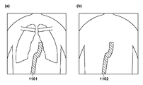
ステップS1301において、平均値取得部1201は、被写体を有する放射線画像の画素値を、被写体が無い放射線画像の画素値で除算した画素値の平均値(平均情報)を示す平均値画像を取得する。具体的には、平均値取得部1201は、FPD102で撮影された、被写体を有する放射線画像M(x,y,t)と、被写体が無い放射線画像M0(x,y,t)と、を用いて平均値画像A(x,y)を取得(生成)する((3)式)。ここで、xとyは画像の画素の座標、tは整数で時系列に撮影された画像のフレーム番号を表す。また、ブラケット「< >t」は時間平均を表す。被写体が無い放射線画像M0の時間平均(平均情報)により、被写体を有する放射線画像Mの時間平均(平均情報)を除算することにより、FPD102のゲイン特性のばらつきを補正することができる。被写体が無い放射線画像M0(x,y,t)は、予め撮影されて記憶部108に記憶されている。平均値取得部1201は、平均値画像を取得する際に、記憶部108から被写体が無い放射線画像M0(x,y,t)を読出して、(3)式の演算処理を行う。
ステップS1302において、分散値取得部1202は、被写体を有する放射線画像の画素値を、被写体が無い放射線画像の画素値で除算した画素値の分散値(分散情報)を示す分散値画像を取得する。具体的には、分散値取得部1202は、FPD102で撮影された、被写体を有する複数の放射線画像M(x,y,t)と、被写体が無い放射線画像M0(x,y,t)と、を用いて分散値画像V(x,y)を取得(生成)する((4)式)。xとyは画像の画素の座標、tは整数で時系列に撮影された画像のフレーム番号を表す。また、ブラケット「< >t」は時間平均を表す。被写体が無い放射線画像M0(x,y,t)は、予め撮影されて記憶部108に記憶されている。分散値取得部1202は、分散値画像を取得する際に、記憶部108から被写体が無い放射線画像M0(x,y,t)を読出して、(4)式の演算処理を行う。

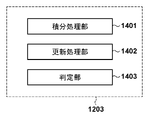
ステップS1303において、物質特性計算部1203の積分処理部1401は、被写体を構成する物質の実効原子番号および面密度を取得するための以下6つのパラメータを算出する。


ステップS1304では、物質特性計算部1203の更新処理部1402は、(13)式に基づいて実効原子番号、面密度を取得する。

ステップS1305において、物質特性計算部1203の判定部1403は、更新処理部1402により更新された実効原子番号および面密度の収束を判定する。判定部1403は、ステップS1304の反復演算により更新された物質の実効原子番号(Zeff)および物質の面密度(σeff)が収束したか判定する。収束判定方法としては種々の方法により判定することが可能である。判定部1403は、例えば、n回目の更新演算結果と、n+1回目の更新演算結果との比較により、両者の差分が所定の閾値以下となった場合、所定の計算精度が得られたものとして、n+1回目の更新演算結果は収束したと判定することが可能である。あるいは、判定部1403は、更新処理部1402による更新演算の反復回数を取得して、所定の反復回数の更新演算が実行された場合に更新演算結果は収束したと判定することも可能である。
本発明は、上述の実施形態の1以上の機能を実現するプログラムを、ネットワーク又は記憶媒体を介してシステム又は装置に供給し、そのシステム又は装置のコンピュータにおける1つ以上のプロセッサーがプログラムを読出し実行する処理でも実現可能である。また、1以上の機能を実現する回路(例えば、ASIC)によっても実現可能である。
104:放射線発生装置、105:制御部、106:モニタ、
107:操作部、108:記憶部、109:画像処理部、
110:物質特性計算部、111:領域強調減衰部、211:周波数分解部、
212:ゲイン調整部、213:周波数再構成部、1201:平均値取得部、
1202:分散値取得部、1203、物質特性計算部、
1401:積分処理部、1402:更新処理部、1403:判定部
Claims (10)
- 被写体の放射線画像を取得する取得手段と、
互いに異なるスペクトルの放射線により取得される複数の放射線画像に基づいて、前記被写体の内部を物質ごとの領域に抽出可能な物質特性画像を生成する生成手段と、
前記放射線画像および前記物質特性画像を、それぞれ異なる周波数帯域に制限された複数の帯域制限画像および複数の低周波画像に分解する分解手段と、
前記複数の低周波画像に分解された前記物質特性画像における特定の領域の位置に基づいて、前記放射線画像の前記複数の低周波画像および前記複数の帯域制限画像における特定の領域を強調又は減衰させる処理を行う画像処理手段と、
を備えることを特徴とする情報処理装置。 - 前記特定の領域を強調又は減衰させた画像を再構成する再構成手段を更に備えることを特徴とする請求項1に記載の情報処理装置。
- 前記画像処理手段は、
前記複数の低周波画像に分解された前記物質特性画像における前記特定の領域の位置に基づいて、前記放射線画像における前記特定の領域以外の領域を強調または減衰させるための調整係数を設定することを特徴とする請求項1に記載の情報処理装置。 - 前記画像処理手段は、
前記放射線画像の帯域制限画像および低周波画像における特定の領域に対して異なる調整係数を設定することを特徴とする請求項1に記載の情報処理装置。 - 前記画像処理手段は、前記特定の領域および前記特定の領域以外の領域に対して異なる調整係数を設定することを特徴とする請求項3に記載の情報処理装置。
- 前記物質特性画像は、前記被写体を構成する物質の実効原子番号の分布を表す実効原子番号画像であることを特徴とする請求項1乃至5のいずれか1項に記載の情報処理装置。
- 前記物質特性画像は、前記被写体を構成する物質を弁別した物質弁別画像であることを特徴とする請求項1乃至5のいずれか1項に記載の情報処理装置。
- 被写体の放射線画像を取得する取得手段と、
互いに異なるスペクトルの放射線により取得される複数の放射線画像に基づいて、前記被写体の内部を物質ごとの領域に抽出可能な実効原子番号画像または物質弁別画像を生成する生成手段と、
前記放射線画像、実効原子番号画像または物質弁別画像を、異なる周波数帯域に制限された複数の帯域制限画像および複数の低周波画像に分解する分解手段と、
前記複数の低周波画像に分解された前記実効原子番号画像または物質弁別画像における特定の領域の位置に基づいて、前記放射線画像の前記複数の低周波画像および複数の帯域制限画像における前記特定の領域を強調または減衰させる処理を行う画像処理手段と、
前記特定の領域を強調又は減衰させた画像を再構成する再構成手段と、
を備えることを特徴とする情報処理装置。 - 被写体を撮影した放射線画像に基づく情報を処理する情報処理方法であって、
前記被写体の放射線画像を取得する取得工程と、
互いに異なるスペクトルの放射線により取得される複数の放射線画像に基づいて、前記被写体の内部を物質ごとの領域に抽出可能な物質特性画像を生成する生成工程と、
前記放射線画像および前記物質特性画像を、それぞれ異なる周波数帯域に制限された複数の帯域制限画像および複数の低周波画像に分解する分解工程と、
前記複数の低周波画像に分解された前記物質特性画像における特定の領域の位置に基づいて、前記放射線画像の前記複数の低周波画像および前記複数の帯域制限画像における特定の領域を強調又は減衰させる処理を行う画像処理工程と、
を有することを特徴とする情報処理方法。 - 請求項9に記載の情報処理方法の各工程をコンピュータに実行させるためのプログラム。
Priority Applications (4)
| Application Number | Priority Date | Filing Date | Title |
|---|---|---|---|
| JP2017168833A JP7080025B2 (ja) | 2017-09-01 | 2017-09-01 | 情報処理装置、情報処理方法およびプログラム |
| EP18850649.7A EP3677183A4 (en) | 2017-09-01 | 2018-07-19 | INFORMATION PROCESSING DEVICE, RADIOGRAPHY DEVICE, INFORMATION PROCESSING PROCESS AND PROGRAM |
| PCT/JP2018/027035 WO2019044241A1 (ja) | 2017-09-01 | 2018-07-19 | 情報処理装置、放射線撮影装置、情報処理方法およびプログラム |
| US16/778,037 US11357455B2 (en) | 2017-09-01 | 2020-01-31 | Information processing apparatus, radiation imaging apparatus, information processing method, and storage medium |
Applications Claiming Priority (1)
| Application Number | Priority Date | Filing Date | Title |
|---|---|---|---|
| JP2017168833A JP7080025B2 (ja) | 2017-09-01 | 2017-09-01 | 情報処理装置、情報処理方法およびプログラム |
Publications (2)
| Publication Number | Publication Date |
|---|---|
| JP2019042161A JP2019042161A (ja) | 2019-03-22 |
| JP7080025B2 true JP7080025B2 (ja) | 2022-06-03 |
Family
ID=65527491
Family Applications (1)
| Application Number | Title | Priority Date | Filing Date |
|---|---|---|---|
| JP2017168833A Active JP7080025B2 (ja) | 2017-09-01 | 2017-09-01 | 情報処理装置、情報処理方法およびプログラム |
Country Status (4)
| Country | Link |
|---|---|
| US (1) | US11357455B2 (ja) |
| EP (1) | EP3677183A4 (ja) |
| JP (1) | JP7080025B2 (ja) |
| WO (1) | WO2019044241A1 (ja) |
Families Citing this family (9)
| Publication number | Priority date | Publication date | Assignee | Title |
|---|---|---|---|---|
| JP7093233B2 (ja) | 2018-06-07 | 2022-06-29 | キヤノン株式会社 | 放射線撮影装置、放射線撮影方法およびプログラム |
| WO2020003744A1 (ja) | 2018-06-27 | 2020-01-02 | キヤノン株式会社 | 放射線撮影装置、放射線撮影方法およびプログラム |
| JP7169853B2 (ja) * | 2018-11-09 | 2022-11-11 | キヤノン株式会社 | 画像処理装置、放射線撮影装置、および画像処理方法 |
| JP7185596B2 (ja) * | 2019-06-11 | 2022-12-07 | キヤノン株式会社 | 画像処理装置および画像処理方法、プログラム |
| JP7246281B2 (ja) | 2019-08-02 | 2023-03-27 | キヤノン株式会社 | 画像処理装置およびその制御方法、放射線撮影装置、プログラム |
| JP7412178B2 (ja) * | 2020-01-07 | 2024-01-12 | キヤノンメディカルシステムズ株式会社 | X線診断装置及び医用画像処理装置 |
| JP7433971B2 (ja) * | 2020-02-19 | 2024-02-20 | キヤノン株式会社 | 画像処理装置、画像処理装置の作動方法及びプログラム |
| JP7619821B2 (ja) | 2021-02-09 | 2025-01-22 | キヤノン株式会社 | 情報処理装置、放射線撮影システム、情報処理方法およびプログラム |
| JP2024025211A (ja) | 2022-08-10 | 2024-02-26 | キヤノン株式会社 | 放射線撮像装置、放射線撮像システム、制御装置、放射線撮像装置の制御方法、および、放射線撮像装置を制御するためのプログラム |
Citations (4)
| Publication number | Priority date | Publication date | Assignee | Title |
|---|---|---|---|---|
| JP2006026198A (ja) | 2004-07-20 | 2006-02-02 | Hitachi Medical Corp | 医用画像処理装置 |
| JP2009118985A (ja) | 2007-11-14 | 2009-06-04 | Ge Medical Systems Global Technology Co Llc | X線ct装置 |
| JP2010131263A (ja) | 2008-12-05 | 2010-06-17 | Toshiba Corp | X線診断装置および画像処理装置 |
| JP2017000675A (ja) | 2015-06-16 | 2017-01-05 | 株式会社日立製作所 | 医用画像処理装置及びx線撮像装置 |
Family Cites Families (18)
| Publication number | Priority date | Publication date | Assignee | Title |
|---|---|---|---|---|
| JPH04156689A (ja) | 1990-10-19 | 1992-05-29 | Fuji Photo Film Co Ltd | 放射線画像処理方法および装置 |
| JPH0969157A (ja) * | 1995-09-01 | 1997-03-11 | Konica Corp | 放射線画像処理方法 |
| EP1341125A3 (en) * | 2002-02-22 | 2005-03-16 | Agfa-Gevaert | Multiscale gradation processing method |
| EP1517269A1 (en) * | 2002-02-22 | 2005-03-23 | Agfa-Gevaert | Method for enhancing the contrast of an image |
| JP2008229122A (ja) * | 2007-03-22 | 2008-10-02 | Fujifilm Corp | 画像成分分離装置、方法、およびプログラム |
| US8218837B2 (en) * | 2008-06-06 | 2012-07-10 | General Electric Company | Material composition detection from effective atomic number computation |
| JP5226590B2 (ja) | 2009-04-02 | 2013-07-03 | キヤノン株式会社 | 画像解析装置、画像処理装置及び画像解析方法 |
| JP5294956B2 (ja) | 2009-04-08 | 2013-09-18 | キヤノン株式会社 | 画像処理装置及び画像処理装置の制御方法 |
| JP5315157B2 (ja) | 2009-07-27 | 2013-10-16 | キヤノン株式会社 | 情報処理装置、ライン状ノイズ低減処理方法、及びプログラム |
| JP5543194B2 (ja) | 2009-12-24 | 2014-07-09 | キヤノン株式会社 | 情報処理装置、処理方法及びプログラム |
| JP6214128B2 (ja) | 2010-11-22 | 2017-10-18 | キヤノン株式会社 | 画像処理装置、画像処理方法、及び記憶媒体 |
| KR101337339B1 (ko) * | 2011-10-21 | 2013-12-06 | 삼성전자주식회사 | 엑스선 영상 장치 및 그 제어방법 |
| JP6122269B2 (ja) | 2011-12-16 | 2017-04-26 | キヤノン株式会社 | 画像処理装置、画像処理方法、及びプログラム |
| JP6016403B2 (ja) | 2012-03-27 | 2016-10-26 | キヤノン株式会社 | 画像処理装置、画像処理方法 |
| JP6312401B2 (ja) | 2012-11-30 | 2018-04-18 | キヤノン株式会社 | 画像処理装置、画像処理方法、及びプログラム |
| JP6497912B2 (ja) | 2014-12-01 | 2019-04-10 | キヤノン株式会社 | 画像処理装置、放射線撮影システム、制御方法、及びプログラム |
| JP6942491B2 (ja) | 2016-03-15 | 2021-09-29 | キヤノン株式会社 | インプリント装置、および物品の製造方法 |
| JP2018110794A (ja) | 2017-01-13 | 2018-07-19 | キヤノン株式会社 | 情報処理装置、放射線撮像装置、情報処理方法およびプログラム |
-
2017
- 2017-09-01 JP JP2017168833A patent/JP7080025B2/ja active Active
-
2018
- 2018-07-19 EP EP18850649.7A patent/EP3677183A4/en not_active Withdrawn
- 2018-07-19 WO PCT/JP2018/027035 patent/WO2019044241A1/ja not_active Ceased
-
2020
- 2020-01-31 US US16/778,037 patent/US11357455B2/en active Active
Patent Citations (4)
| Publication number | Priority date | Publication date | Assignee | Title |
|---|---|---|---|---|
| JP2006026198A (ja) | 2004-07-20 | 2006-02-02 | Hitachi Medical Corp | 医用画像処理装置 |
| JP2009118985A (ja) | 2007-11-14 | 2009-06-04 | Ge Medical Systems Global Technology Co Llc | X線ct装置 |
| JP2010131263A (ja) | 2008-12-05 | 2010-06-17 | Toshiba Corp | X線診断装置および画像処理装置 |
| JP2017000675A (ja) | 2015-06-16 | 2017-01-05 | 株式会社日立製作所 | 医用画像処理装置及びx線撮像装置 |
Also Published As
| Publication number | Publication date |
|---|---|
| JP2019042161A (ja) | 2019-03-22 |
| EP3677183A1 (en) | 2020-07-08 |
| EP3677183A4 (en) | 2021-09-08 |
| US20200163630A1 (en) | 2020-05-28 |
| WO2019044241A1 (ja) | 2019-03-07 |
| US11357455B2 (en) | 2022-06-14 |
Similar Documents
| Publication | Publication Date | Title |
|---|---|---|
| JP7080025B2 (ja) | 情報処理装置、情報処理方法およびプログラム | |
| US10292672B2 (en) | Radiographic image processing device, method, and recording medium | |
| US9949706B2 (en) | Image-processing device, radiographic imaging system, image-processing program, and image-processing method | |
| JP6700273B2 (ja) | 雑音モデルに基づくマルチスケール雑音低減による線量増加のシミュレーション | |
| JP5952251B2 (ja) | 画像処理装置、放射線画像撮影システム、画像処理プログラム、及び画像処理方法 | |
| JP6122269B2 (ja) | 画像処理装置、画像処理方法、及びプログラム | |
| US10064591B2 (en) | System, method and computer readable medium for preview of low-dose x-ray projection and tomographic images | |
| US10332281B2 (en) | System and method for denoising medical images by enforcing low rank spatial-temporal or spatial-spectral image matrices | |
| JP7093233B2 (ja) | 放射線撮影装置、放射線撮影方法およびプログラム | |
| CN115209808B (zh) | 学习完毕模型的制作方法、图像生成方法以及图像处理装置 | |
| CN107533755A (zh) | 用于改进医学图像质量的设备和方法 | |
| US6751284B1 (en) | Method and system for tomosynthesis image enhancement using transverse filtering | |
| WO2020003744A1 (ja) | 放射線撮影装置、放射線撮影方法およびプログラム | |
| WO2020095578A1 (ja) | 情報処理装置および方法、放射線撮影システム | |
| JP2020192268A (ja) | 画像処理装置、画像処理方法及びプログラム | |
| EP4310771B1 (en) | Methods and systems for image denoising | |
| JP2009054013A (ja) | 画像処理装置 | |
| JP7144988B2 (ja) | 放射線撮影装置、放射線撮影方法およびプログラム | |
| JP2021126163A (ja) | 画像処理装置及び画像処理方法 | |
| WO2023002743A1 (ja) | X線撮影システム、および、画像処理方法 | |
| JP2021126162A (ja) | 画像処理装置及び画像処理方法 |
Legal Events
| Date | Code | Title | Description |
|---|---|---|---|
| A621 | Written request for application examination |
Free format text: JAPANESE INTERMEDIATE CODE: A621 Effective date: 20200827 |
|
| RD01 | Notification of change of attorney |
Free format text: JAPANESE INTERMEDIATE CODE: A7421 Effective date: 20210103 |
|
| A521 | Request for written amendment filed |
Free format text: JAPANESE INTERMEDIATE CODE: A523 Effective date: 20210113 |
|
| A131 | Notification of reasons for refusal |
Free format text: JAPANESE INTERMEDIATE CODE: A131 Effective date: 20210726 |
|
| A521 | Request for written amendment filed |
Free format text: JAPANESE INTERMEDIATE CODE: A523 Effective date: 20210924 |
|
| A131 | Notification of reasons for refusal |
Free format text: JAPANESE INTERMEDIATE CODE: A131 Effective date: 20220114 |
|
| A521 | Request for written amendment filed |
Free format text: JAPANESE INTERMEDIATE CODE: A523 Effective date: 20220127 |
|
| TRDD | Decision of grant or rejection written | ||
| A01 | Written decision to grant a patent or to grant a registration (utility model) |
Free format text: JAPANESE INTERMEDIATE CODE: A01 Effective date: 20220425 |
|
| A61 | First payment of annual fees (during grant procedure) |
Free format text: JAPANESE INTERMEDIATE CODE: A61 Effective date: 20220524 |
|
| R151 | Written notification of patent or utility model registration |
Ref document number: 7080025 Country of ref document: JP Free format text: JAPANESE INTERMEDIATE CODE: R151 |