JP6952683B2 - Methods and devices for detecting entities in body samples - Google Patents
Methods and devices for detecting entities in body samples Download PDFInfo
- Publication number
- JP6952683B2 JP6952683B2 JP2018512961A JP2018512961A JP6952683B2 JP 6952683 B2 JP6952683 B2 JP 6952683B2 JP 2018512961 A JP2018512961 A JP 2018512961A JP 2018512961 A JP2018512961 A JP 2018512961A JP 6952683 B2 JP6952683 B2 JP 6952683B2
- Authority
- JP
- Japan
- Prior art keywords
- sample
- pathogen
- candidate
- body sample
- feature
- Prior art date
- Legal status (The legal status is an assumption and is not a legal conclusion. Google has not performed a legal analysis and makes no representation as to the accuracy of the status listed.)
- Active
Links
Images


Classifications
-
- G—PHYSICS
- G01—MEASURING; TESTING
- G01N—INVESTIGATING OR ANALYSING MATERIALS BY DETERMINING THEIR CHEMICAL OR PHYSICAL PROPERTIES
- G01N21/00—Investigating or analysing materials by the use of optical means, i.e. using sub-millimetre waves, infrared, visible or ultraviolet light
- G01N21/84—Systems specially adapted for particular applications
- G01N21/88—Investigating the presence of flaws or contamination
- G01N21/8851—Scan or image signal processing specially adapted therefor, e.g. for scan signal adjustment, for detecting different kinds of defects, for compensating for structures, markings, edges
-
- G—PHYSICS
- G02—OPTICS
- G02B—OPTICAL ELEMENTS, SYSTEMS OR APPARATUS
- G02B21/00—Microscopes
- G02B21/36—Microscopes arranged for photographic purposes or projection purposes or digital imaging or video purposes including associated control and data processing arrangements
- G02B21/365—Control or image processing arrangements for digital or video microscopes
- G02B21/367—Control or image processing arrangements for digital or video microscopes providing an output produced by processing a plurality of individual source images, e.g. image tiling, montage, composite images, depth sectioning, image comparison
-
- G—PHYSICS
- G06—COMPUTING; CALCULATING OR COUNTING
- G06F—ELECTRIC DIGITAL DATA PROCESSING
- G06F18/00—Pattern recognition
- G06F18/20—Analysing
- G06F18/21—Design or setup of recognition systems or techniques; Extraction of features in feature space; Blind source separation
- G06F18/214—Generating training patterns; Bootstrap methods, e.g. bagging or boosting
-
- G—PHYSICS
- G06—COMPUTING; CALCULATING OR COUNTING
- G06T—IMAGE DATA PROCESSING OR GENERATION, IN GENERAL
- G06T7/00—Image analysis
- G06T7/0002—Inspection of images, e.g. flaw detection
- G06T7/0012—Biomedical image inspection
-
- G—PHYSICS
- G06—COMPUTING; CALCULATING OR COUNTING
- G06V—IMAGE OR VIDEO RECOGNITION OR UNDERSTANDING
- G06V20/00—Scenes; Scene-specific elements
- G06V20/60—Type of objects
- G06V20/69—Microscopic objects, e.g. biological cells or cellular parts
- G06V20/693—Acquisition
-
- G—PHYSICS
- G06—COMPUTING; CALCULATING OR COUNTING
- G06V—IMAGE OR VIDEO RECOGNITION OR UNDERSTANDING
- G06V20/00—Scenes; Scene-specific elements
- G06V20/60—Type of objects
- G06V20/69—Microscopic objects, e.g. biological cells or cellular parts
- G06V20/698—Matching; Classification
-
- G—PHYSICS
- G01—MEASURING; TESTING
- G01N—INVESTIGATING OR ANALYSING MATERIALS BY DETERMINING THEIR CHEMICAL OR PHYSICAL PROPERTIES
- G01N21/00—Investigating or analysing materials by the use of optical means, i.e. using sub-millimetre waves, infrared, visible or ultraviolet light
- G01N21/84—Systems specially adapted for particular applications
- G01N21/88—Investigating the presence of flaws or contamination
- G01N21/8851—Scan or image signal processing specially adapted therefor, e.g. for scan signal adjustment, for detecting different kinds of defects, for compensating for structures, markings, edges
- G01N2021/8887—Scan or image signal processing specially adapted therefor, e.g. for scan signal adjustment, for detecting different kinds of defects, for compensating for structures, markings, edges based on image processing techniques
-
- G—PHYSICS
- G06—COMPUTING; CALCULATING OR COUNTING
- G06T—IMAGE DATA PROCESSING OR GENERATION, IN GENERAL
- G06T2207/00—Indexing scheme for image analysis or image enhancement
- G06T2207/10—Image acquisition modality
- G06T2207/10056—Microscopic image
-
- G—PHYSICS
- G06—COMPUTING; CALCULATING OR COUNTING
- G06T—IMAGE DATA PROCESSING OR GENERATION, IN GENERAL
- G06T2207/00—Indexing scheme for image analysis or image enhancement
- G06T2207/30—Subject of image; Context of image processing
- G06T2207/30004—Biomedical image processing
- G06T2207/30024—Cell structures in vitro; Tissue sections in vitro
-
- G—PHYSICS
- G06—COMPUTING; CALCULATING OR COUNTING
- G06T—IMAGE DATA PROCESSING OR GENERATION, IN GENERAL
- G06T2207/00—Indexing scheme for image analysis or image enhancement
- G06T2207/30—Subject of image; Context of image processing
- G06T2207/30004—Biomedical image processing
- G06T2207/30101—Blood vessel; Artery; Vein; Vascular
-
- Y—GENERAL TAGGING OF NEW TECHNOLOGICAL DEVELOPMENTS; GENERAL TAGGING OF CROSS-SECTIONAL TECHNOLOGIES SPANNING OVER SEVERAL SECTIONS OF THE IPC; TECHNICAL SUBJECTS COVERED BY FORMER USPC CROSS-REFERENCE ART COLLECTIONS [XRACs] AND DIGESTS
- Y02—TECHNOLOGIES OR APPLICATIONS FOR MITIGATION OR ADAPTATION AGAINST CLIMATE CHANGE
- Y02A—TECHNOLOGIES FOR ADAPTATION TO CLIMATE CHANGE
- Y02A90/00—Technologies having an indirect contribution to adaptation to climate change
- Y02A90/10—Information and communication technologies [ICT] supporting adaptation to climate change, e.g. for weather forecasting or climate simulation
Description
関連出願の相互参照
本出願は、2015年9月17日に出願された、Eshelによる「Methods of detecting a pathogen in a bodily sample and system thereof」と題する米国特許仮出願第62/219,889号からの優先権を主張する。この出願は、参照により本明細書に組み込まれる。
Cross-reference to related applications This application is filed on September 17, 2015, from US Patent Provisional Application No. 62 / 219,888, entitled "Methods of detecting a pathogen in a body sample and system therapy" by Eschel. Claim the priority of. This application is incorporated herein by reference.
本発明の実施形態の分野
本明細書で開示の主題のいくつかの用途は、一般に、身体試料中の実体の検出、特に、画像処理および分類を使った病原体の自動的検出に関する。
Fields of embodiments of the invention Some uses of the subject matter disclosed herein generally relate to the detection of entities in body samples, especially the automatic detection of pathogens using image processing and classification.
身体試料(例えば、血液試料)中の特定の病原性感染の検出の主な方法は、身体試料の顕微鏡検査、および病原体の存在および濃度の目視確認である。顕微鏡検査の前の染料または色素による身体試料の染色は、多くの場合、顕微鏡画像のコントラストを高め、特定の生物学的構造を有する細胞を視覚的に強調するために使用される。特に、いくつかの蛍光色素は、細胞中の核酸に親和性がある。適切な波長の蛍光により励起されると、核酸は蛍光を発する。したがって、顕微鏡下での検出のために、蛍光色素を使って、細胞の部分の差次染色が行われることもある。例えば、青色光で励起されると、DNAに結合した蛍光色素アクリジンオレンジは、緑色光を発光し、RNAに結合すると、赤色光を発光する。アナプラズマ・マジナーレ、ヘモバルトネラ属、トリパノソーマ類、プラスモジウム属菌種、バベシア属菌種などの血液病原体は全て、アクリジンオレンジで検出されてきた。 The main methods of detecting a particular pathogenic infection in a body sample (eg, a blood sample) are microscopic examination of the body sample and visual confirmation of the presence and concentration of the pathogen. Staining of body samples with dyes or dyes prior to microscopic examination is often used to increase the contrast of microscopic images and visually enhance cells with a particular biological structure. In particular, some fluorescent dyes have an affinity for nucleic acids in cells. When excited by fluorescence of the appropriate wavelength, the nucleic acid fluoresces. Therefore, fluorescent dyes may be used to perform differential staining of cell parts for detection under a microscope. For example, when excited by blue light, the fluorescent dye acridine orange bound to DNA emits green light, and when bound to RNA, it emits red light. All blood pathogens such as Anaplasma maginale, Hemobartonella, Tripanosoma, Plasmodium, and Babesia have been detected in acridine orange.
病原体を検出する主な方法は、顕微鏡の明視野像による目視特定のままであるが、頻度はより少ないにしても、蛍光顕微鏡も同様に使用されてきた。しかし、両方のケースで、マニュアル特定による病原体の病原性感染の検出は、次の2つの主な欠点:多くの環境では(特に地方で)、試験を行う態勢が整っていないこと、および結果の精度が試料を検査する人の技能と試料中の病原体の量の両方に依存していること、が存在する。したがって、身体試料中の病原体の検出を自動化する試みを行った。 The main method of detecting pathogens remains visually identified by the brightfield image of the microscope, but less frequently, fluorescence microscopy has been used as well. However, in both cases, detection of pathogenic infections of pathogens by manual identification has two main drawbacks: in many environments (especially in rural areas), unprepared for testing, and the consequences. There is that accuracy depends on both the skill of the person inspecting the sample and the amount of pathogen in the sample. Therefore, an attempt was made to automate the detection of pathogens in body samples.
本発明のいくつかの用途では、顕微鏡システムの顕微鏡を使って、身体試料(例えば、血液試料)の1つまたは複数の顕微鏡画像が取得される。コンピュータープロセッサーは、画像内で、少なくとも1つの要素を病原体候補(すなわち、病原体であり得、したがって、病原体としての候補であることを示す特性を示す、試料内の構成要素)として特定する。例えば、画像は、試料内でDNAおよび/またはRNAを染色するように構成されている染料または色素により試料が染色されている間に、取得された血液試料の画像であってよく、またコンピュータープロセッサーは、画像中の染色した要素(例えば、蛍光を発している要素)を検出することにより候補を特定し得る。コンピュータープロセッサーは、1つまたは複数の画像から、少なくとも1つの候補としての情報価値の高い、病原体候補に関連する特徴および少なくとも1つの試料としての情報価値の高い、身体試料に関連するコンテキスト情報を示す特徴を抽出する。候補としての情報価値の高い特徴を試料としての情報価値の高い特徴と組み合わせて処理することにより、身体試料の病原性感染症に感染している可能性を分類する。分類に対応した出力は通常、出力装置上に生成される。 In some applications of the present invention, the microscope of the microscopy system is used to obtain one or more microscopic images of a body sample (eg, a blood sample). The computer processor identifies at least one element in the image as a pathogen candidate (ie, a component in the sample that exhibits properties indicating that it can be a pathogen and is therefore a candidate as a pathogen). For example, the image may be an image of a blood sample obtained while the sample is stained with a dye or dye that is configured to stain DNA and / or RNA within the sample, and may be a computer processor. Can identify candidates by detecting stained elements (eg, fluorescent elements) in the image. From one or more images, the computer processor presents at least one candidate information-valued, pathogen candidate-related feature and at least one sample information-valued, body sample-related contextual information. Extract features. By processing a feature with high information value as a candidate in combination with a feature with high information value as a sample, the possibility of being infected with a pathogenic infection of a body sample is classified. The output corresponding to the classification is usually generated on the output device.
いくつかの用途に対しては、候補としての情報価値の高い特徴に対応して、コンピュータープロセッサーは、病原体候補の病原体としての可能性を分類する、第1の分類を実施する。第1の分類に対応して、試料としての情報価値の高い特徴を組み合わせて、コンピュータープロセッサーは、身体試料が病原性感染を含む可能性を分類する、第2の分類を実施する。いくつかの用途に対しては、第1の分類(ここでは、病原体候補の病原体としての可能性が分類される)が、試料としての情報価値の高い特徴と組み合わせて、候補としての情報価値の高い特徴に対応して実施される。いくつかの用途に対しては、コンピュータープロセッサーは、候補としての情報価値の高い特徴を試料としての情報価値の高い特徴と組み合わせて処理することにより、身体試料中の病原性感染を所定の種類の病原性感染(例えば、マラリア原虫、マラリア原虫の所定の菌株、および/または所定の成熟度または成熟範囲のマラリア原虫)として分類する。 For some applications, the computer processor performs a first classification, which classifies the potential pathogens of a candidate pathogen, in response to the informative features of the candidate. Corresponding to the first classification, combining the informative features of the sample, the computer processor performs a second classification, which classifies the likelihood that the body sample contains a pathogenic infection. For some uses, the first classification (here, the potential pathogen potential of a pathogen candidate) is combined with the informative features of the sample for the information value of the candidate. Implemented in response to high characteristics. For some applications, computer processors treat pathogenic infections in body samples of a given type by processing candidate informative features in combination with sample informative features. Classify as a pathogenic infection (eg, malaria protozoan, a given strain of malaria protozoan, and / or a malaria protozoan of a given maturity or range of maturity).
いくつかの用途に対しては、候補としての情報価値の高い特徴は、病原体候補のサイズ(例えば、寸法、長さ、円周、最小幅、最大幅、面積、および/または他の候補または実体に対する候補の相対サイズ)、病原体候補の形状、病原体候補の動き、病原体候補の強度、病原体候補の身体試料内の位置(他の候補または実体に対する候補の近接度、隣接、および/または重なりを含む)、病原体候補と重なり合う細胞の性質、病原体候補の色(強度および染色パターンを含む)、病原体候補のテクスチャ(例えば、輪郭)、および/または病原体候補の境界の鮮明度を含む。候補としての情報価値の高い特徴のさらなる非限定的例は、例えば、Bacheletによる米国特許出願公開第2012/0169863号、および/またはPollakによる米国特許出願公開第2015/0037806号に記載されている。これら両出願は、参照により本明細書に組み込まれる。 For some applications, candidate informative features include the size of the pathogen candidate (eg, dimensions, length, circumference, minimum width, maximum width, area, and / or other candidate or entity. Includes (relative size of candidate to), pathogen candidate shape, pathogen candidate movement, pathogen candidate strength, location of pathogen candidate in body sample (candidate proximity, adjacency, and / or overlap to other candidates or entities) ), The nature of the cells that overlap the pathogen candidate, the color of the pathogen candidate (including intensity and staining pattern), the texture of the pathogen candidate (eg, contour), and / or the sharpness of the boundary of the pathogen candidate. Further non-limiting examples of candidate informative features are described, for example, in Bachelet's US Patent Application Publication No. 2012/0169863 and / or Pollak's US Patent Application Publication No. 2015/0037806. Both of these applications are incorporated herein by reference.
いくつかの用途に対しては、試料としての情報価値の高い特徴は、身体試料中の1つまたは複数の非病原体候補成分のサイズ、身体試料中の1つまたは複数の非病原体候補成分の形状、身体試料中の1つまたは複数の非病原体候補成分の強度、身体試料中の所定の細胞型の細胞量、身体試料中の所定の細胞型の細胞の分布、および/または身体試料中の病原体候補の分布を含む。 For some applications, informative features as a sample are the size of one or more non-pathogen candidate components in a body sample, the shape of one or more non-pathogen candidate components in a body sample. , Intensity of one or more non-pathogen candidate components in a body sample, cell volume of a given cell type in a body sample, distribution of cells of a given cell type in a body sample, and / or pathogens in a body sample Includes candidate distribution.
したがって、本発明のいくつかの用途では、次記を含む装置が提供される:
1つまたは複数の身体試料の顕微鏡画像を取得するように構成された顕微鏡システム;
出力装置;および
次記を行うように構成された少なくとも1つのコンピュータープロセッサー;
1つまたは複数の画像中の少なくとも1つの要素を病原体候補であるとして特定すること、
1つまたは複数の画像から、少なくとも1つの候補としての情報価値の高い、病原体候補に関連する特徴を抽出すること、
1つまたは複数の画像から、少なくとも1つの試料としての情報価値の高い、身体試料に関連するコンテキスト情報を示す特徴を抽出すること、
候補としての情報価値の高い特徴を、試料としての情報価値の高い特徴と組み合わせて処理することにより、身体試料の病原性感染症に感染している可能性を分類すること、および
それに対応した出力を出力装置上に生成すること。
Therefore, in some applications of the present invention, devices are provided that include:
A microscope system configured to acquire microscopic images of one or more body samples;
Output device; and at least one computer processor configured to:
Identifying at least one element in one or more images as a potential pathogen,
Extracting pathogen candidate-related features with high information value as at least one candidate from one or more images,
Extracting features from one or more images that show contextual information related to a body sample that are of high information value as at least one sample.
By processing information-valued features as candidates in combination with information-valued features as samples, it is possible to classify the possibility of being infected with a pathogenic infection of a body sample, and the corresponding output. To be generated on the output device.
いくつかの用途では:
顕微鏡システムは、染料で染色されている身体試料の1つまたは複数の顕微鏡画像を取得するように構成され、および、
少なくとも1つのコンピュータープロセッサーは、少なくとも1つの要素が染色されていることを特定して少なくとも1つの要素を病原体候補として特定することにより、少なくとも1つの要素を病原体候補として特定するように構成される。
For some uses:
The microscopy system is configured to acquire one or more microscopic images of a body sample stained with a dye, and
At least one computer processor is configured to identify at least one element as a pathogen candidate by identifying that at least one element is stained and identifying at least one element as a pathogen candidate.
いくつかの用途では、少なくとも1つのコンピュータープロセッサーは、次記を行うことにより、候補としての情報価値の高い特徴を試料としての情報価値の高い特徴と組み合わせて処理するように構成される:
候補としての情報価値の高い特徴に対応して、病原体候補の病原体としての可能性を分類する、第1の分類を実施すること、および
第1の分類に対応して、試料としての情報価値の高い特徴を組み合わせて、身体試料が病原性感染を含む可能性を分類する、第2の分類を実施すること。
In some applications, at least one computer processor is configured to process candidate informative features in combination with sample informative features by doing the following:
To classify the potential pathogens of pathogen candidates according to their high information value as candidates, to carry out the first classification, and to correspond to the first classification, to classify the information value as a sample. Perform a second classification that combines high features to classify the likelihood that a body sample will contain a pathogenic infection.
いくつかの用途では、少なくとも1つのコンピュータープロセッサーは、次記を行うことにより、候補としての情報価値の高い特徴を試料としての情報価値の高い特徴と組み合わせて処理するように構成される:
候補としての情報価値の高い特徴に対応して、試料としての情報価値の高い特徴と組み合わせて、病原体候補の病原体としての可能性を分類する、第1の分類を実施すること、および
第1の分類に少なくとも部分的に対応して、身体試料が病原性感染を含む可能性を分類する、第2の分類を実施すること。
In some applications, at least one computer processor is configured to process candidate informative features in combination with sample informative features by doing the following:
Performing the first classification, which classifies the potential pathogens of a candidate pathogen in combination with the information-valued features as a sample, corresponding to the information-valued features as a candidate, and the first. Perform a second classification that classifies the likelihood that a body sample contains a pathogenic infection, at least in part, in response to the classification.
いくつかの用途では、少なくとも1つのコンピュータープロセッサーは、1つまたは複数の画像から、病原体候補のサイズ、病原体候補の形状、病原体候補の動き、病原体候補の強度、身体試料中の病原体候補の位置、病原体候補と重なり合う細胞の性質、病原体候補の色、病原体候補のテクスチャ、および病原体候補の境界の鮮明度からなる群より選択される特徴であり、少なくとも1つの候補としての情報価値の高い、病原体候補に関連する特徴を抽出することにより、1つまたは複数の画像から少なくとも1つの候補としての情報価値の高い、病原体候補に関連する特徴を抽出するように構成される。 In some applications, at least one computer processor, from one or more images, the size of the pathogen candidate, the shape of the pathogen candidate, the movement of the pathogen candidate, the strength of the pathogen candidate, the position of the pathogen candidate in the body sample, A characteristic selected from the group consisting of the properties of cells overlapping with the pathogen candidate, the color of the pathogen candidate, the texture of the pathogen candidate, and the sharpness of the boundary of the pathogen candidate, and the pathogen candidate having high information value as at least one candidate. By extracting the features related to the pathogen, it is configured to extract the features related to the pathogen candidate having high information value as at least one candidate from one or more images.
いくつかの用途では、少なくとも1つのコンピュータープロセッサーは、1つまたは複数の画像から、身体試料中の1つまたは複数の非病原体候補成分のサイズ、身体試料中の1つまたは複数の非病原体候補成分の形状、身体試料中の1つまたは複数の非病原体候補成分の強度、身体試料中の所定の細胞型の細胞の量、身体試料中の所定の細胞型の細胞の分布、および身体試料中の病原体候補の分布からなる群より選択される少なくとも1つの試料としての情報価値の高い特徴を抽出することにより、1つまたは複数の画像から、少なくとも1つの試料としての情報価値の高い、身体試料に関連するコンテキスト情報を示す特徴を抽出するように構成される。 In some applications, at least one computer processor, from one or more images, the size of one or more non-pathogenic candidate components in a body sample, one or more non-pathogenic candidate components in a body sample. Shape, the intensity of one or more non-pathogenic candidate components in the body sample, the amount of cells of a given cell type in the body sample, the distribution of cells of a given cell type in the body sample, and the distribution of cells of a given cell type in the body sample. By extracting the information-valued features as at least one sample selected from the group consisting of the distribution of pathogen candidates, one or more images can be used as at least one sample-valued body sample. It is configured to extract features that indicate relevant contextual information.
いくつかの用途では:
顕微鏡システムは、染料で染色されている身体試料の1つまたは複数の顕微鏡画像を取得することにより、身体試料の1つまたは複数の顕微鏡画像を取得するように構成され、および、
少なくとも1つのコンピュータープロセッサーは、1つまたは複数の画像から、少なくとも1つの試料としての情報価値の高い、染料により身体試料の染色の品質を示す特徴を抽出することにより、1つまたは複数の画像から、少なくとも1つの試料としての情報価値の高い、身体試料に関連するコンテキスト情報を示す特徴を抽出するように構成される。
For some uses:
The microscopy system is configured to acquire one or more microscopic images of a body sample by acquiring one or more microscopic images of the body sample stained with a dye, and.
At least one computer processor extracts from one or more images a feature that is highly informative as at least one sample and indicates the quality of dyeing of a body sample with a dye, from one or more images. , It is configured to extract features that indicate contextual information related to the body sample, which has high information value as at least one sample.
いくつかの用途では、少なくとも1つのコンピュータープロセッサーは、1つまたは複数の画像から、少なくとも1つの試料としての情報価値の高い、身体試料中に存在する異物を示す特徴を抽出することにより、1つまたは複数の画像から、少なくとも1つの試料としての情報価値の高い、身体試料に関連するコンテキスト情報を示す特徴を抽出するように構成される。 In some applications, at least one computer processor extracts from one or more images a feature that is highly informative as at least one sample and indicates a foreign body present in a body sample. Alternatively, it is configured to extract features indicating contextual information related to a body sample, which have high information value as at least one sample, from a plurality of images.
いくつかの用途では、身体試料は、血液試料、希釈した血液試料、主に赤血球を含む試料、および主に赤血球を含む希釈した試料からなる群より選択される身体試料を含み、顕微鏡システムは選択身体試料の1つまたは複数の画像を取得するように構成される。 In some applications, the body sample includes a body sample selected from the group consisting of a blood sample, a diluted blood sample, a sample mainly containing red blood cells, and a diluted sample mainly containing red blood cells, and the microscope system is selected. It is configured to acquire one or more images of a body sample.
いくつかの用途では、少なくとも1つのコンピュータープロセッサーは、1つまたは複数の画像から、身体試料中に存在する1つまたは複数の赤血球のサイズを抽出することにより、1つまたは複数の画像から、少なくとも1つの試料としての情報価値の高い、身体試料に関連するコンテキスト情報を示す特徴を抽出するように構成される。 In some applications, at least one computer processor extracts the size of one or more red blood cells present in a body sample from one or more images, thereby at least from one or more images. It is configured to extract features that indicate contextual information related to the body sample, which has high information value as one sample.
いくつかの用途では、少なくとも1つのコンピュータープロセッサーは、1つまたは複数の画像から、身体試料中のハウエルジョリー小体の存在の指標を抽出することにより、1つまたは複数の画像から、少なくとも1つの試料としての情報価値の高い、身体試料に関連するコンテキスト情報を示す特徴を抽出するように構成される。 In some applications, at least one computer processor extracts at least one indicator of the presence of Howell-Jolly bodies in a body sample from one or more images. It is configured to extract features that indicate contextual information related to the body sample, which has high information value as a sample.
いくつかの用途では、少なくとも1つのコンピュータープロセッサーは、1つまたは複数の画像から、身体試料中の血小板の濃度を抽出することにより、1つまたは複数の画像から、少なくとも1つの試料としての情報価値の高い、身体試料に関連するコンテキスト情報を示す特徴を抽出するように構成される。 In some applications, at least one computer processor extracts the concentration of platelets in a body sample from one or more images, thereby providing information value as at least one sample from one or more images. It is configured to extract features that indicate high-level, contextual information related to the body sample.
いくつかの用途では、少なくとも1つのコンピュータープロセッサーは、1つまたは複数の画像から、候補に関連する網状赤血球の数と、候補に関連する成熟赤血球の数との関連性を抽出することにより、1つまたは複数の画像から、少なくとも1つの試料としての情報価値の高い、身体試料に関連するコンテキスト情報を示す特徴を抽出するように構成される。 In some applications, at least one computer processor extracts the association between the number of reticulocytes associated with a candidate and the number of mature red blood cells associated with a candidate from one or more images. It is configured to extract features from one or more images that show contextual information related to the body sample, which is of high information value as at least one sample.
いくつかの用途では、少なくとも1つのコンピュータープロセッサーは、1つまたは複数の画像から、身体試料中の網状赤血球体の濃度を抽出することにより、1つまたは複数の画像から、少なくとも1つの試料としての情報価値の高い、身体試料に関連するコンテキスト情報を示す特徴を抽出するように構成される。 In some applications, at least one computer processor extracts the concentration of reticulocytes in a body sample from one or more images, thereby as at least one sample from one or more images. It is configured to extract features that are highly informative and indicate contextual information related to the body sample.
いくつかの用途では、少なくとも1つのコンピュータープロセッサーは、身体試料内の網状赤血球体の濃度に基づいて、病原性感染の陽性判定の閾値を調節することにより、身体試料の病原性感染症に感染している可能性を分類するように構成される。 In some applications, at least one computer processor infects a pathogenic infection in a body sample by adjusting the threshold for positive determination of the pathogenic infection based on the concentration of reticulated erythrocytes in the body sample. It is configured to classify the possibilities.
いくつかの用途では、少なくとも1つのコンピュータープロセッサーは、候補としての情報価値の高い特徴を、試料としての情報価値の高い特徴と組み合わせて処理することにより、身体試料中の病原性感染を1つまたは複数の所定の種類の病原体を含むとして分類するように構成される。 In some applications, at least one computer processor processes one or more pathogenic infections in a body sample by processing the informative features as candidates in combination with the informative features as samples. It is configured to be classified as containing a plurality of predetermined types of pathogens.
いくつかの用途では、少なくとも1つのコンピュータープロセッサーは、病原性感染を、マラリア原虫、マラリア原虫の所定の菌株、所定の成熟度のマラリア原虫、および所定の成熟範囲のマラリア原虫からなる群より選択される1つまたは複数の病原体カテゴリーを含むとして分類することにより、身体試料中の病原性感染を1つまたは複数の所定の種類の病原体を含むとして分類するように構成される。 In some applications, at least one computer processor selects the pathogenic infection from the group consisting of a pathogenic protozoan, a given strain of malaria protozoan, a given maturity of malaria protozoan, and a given maturity range of malaria protozoan. By classifying as containing one or more pathogen categories, pathogenic infections in body samples are configured to be classified as containing one or more predetermined types of pathogens.
いくつかの用途では:
身体試料は、血液試料、希釈した血液試料、主に赤血球を含む試料、および主に赤血球を含む希釈した試料からなる群より選択される身体試料を含み;
少なくとも1つのコンピュータープロセッサーは、1つまたは複数の画像から、候補に関連する網状赤血球の数と、候補に関連する成熟赤血球の数との関連性を抽出することにより、1つまたは複数の画像から、少なくとも1つの試料としての情報価値の高い、身体試料に関連するコンテキスト情報を示す特徴を抽出するように構成され;および
少なくとも1つのコンピュータープロセッサーは、候補に関連する網状赤血球の数と、候補に関連する成熟赤血球の数との間の関連性に少なくとも部分的に基づいて、所定の種類の病原体を含むとして身体試料中の病原性感染を分類することにより、身体試料中の病原性感染を1つまたは複数の所定の種類の病原体を含むとして分類するように構成される。
For some uses:
Body samples include body samples selected from the group consisting of blood samples, diluted blood samples, samples containing predominantly red blood cells, and diluted samples predominantly containing red blood cells;
At least one computer processor extracts from one or more images the association between the number of reticular red blood cells associated with the candidate and the number of mature red blood cells associated with the candidate from the one or more images. , At least one computer processor is configured to extract features that indicate contextual information related to the body sample, which is highly informative as at least one sample; and at least one computer processor, with the number of reticulated red blood cells associated with the candidate, Pathogenic infections in body samples by classifying pathogenic infections in body samples as containing certain types of pathogens, at least in part, based on their association with the number of mature red blood cells involved. It is configured to be classified as containing one or more predetermined types of pathogens.
いくつかの用途では:
身体試料は、血液試料、希釈した血液試料、主に赤血球を含む試料、および主に赤血球を含む希釈した試料からなる群より選択される身体試料を含み;
少なくとも1つのコンピュータープロセッサーは、1つまたは複数の画像から、身体試料中の赤血球の形状を抽出することにより、1つまたは複数の画像から、少なくとも1つの試料としての情報価値の高い、身体試料に関連するコンテキスト情報を示す特徴を抽出するように構成され、および
少なくとも1つのコンピュータープロセッサーは、身体試料中の赤血球の形状に少なくとも部分的に基づいて、身体試料中の病原性感染を所定の種類の病原性感染として分類することにより、身体試料中の病原性感染を所定の種類の病原体を含むとして分類するように構成される。
For some uses:
Body samples include body samples selected from the group consisting of blood samples, diluted blood samples, samples containing predominantly red blood cells, and diluted samples predominantly containing red blood cells;
At least one computer processor extracts the shape of red blood cells in a body sample from one or more images to obtain a body sample having high information value as at least one sample from one or more images. Configured to extract features that indicate relevant contextual information, and at least one computer processor of a given type of pathogenic infection in a body sample, at least partially based on the shape of the red blood cells in the body sample. By classifying as a pathogenic infection, the pathogenic infection in the body sample is configured to be classified as containing a predetermined type of pathogen.
いくつかの用途では:
身体試料は、血液試料、希釈した血液試料、主に赤血球を含む試料、および主に赤血球を含む希釈した試料からなる群より選択される身体試料を含み;
少なくとも1つのコンピュータープロセッサーは、1つまたは複数の画像から、身体試料中の赤血球のサイズを抽出することにより、1つまたは複数の画像から、少なくとも1つの試料としての情報価値の高い、身体試料に関連するコンテキスト情報を示す特徴を抽出するように構成され、および
少なくとも1つのコンピュータープロセッサーは、身体試料中の赤血球のサイズに少なくとも部分的に基づいて、身体試料中の病原性感染を所定の種類の病原性感染として分類することにより、身体試料中の病原性感染を所定の種類の病原体を含むとして分類するように構成される。
For some uses:
Body samples include body samples selected from the group consisting of blood samples, diluted blood samples, samples containing predominantly red blood cells, and diluted samples predominantly containing red blood cells;
At least one computer processor extracts the size of red blood cells in a body sample from one or more images to obtain a highly informative body sample as at least one sample from one or more images. It is configured to extract features that indicate relevant contextual information, and at least one computer processor of a given type of pathogenic infection in a body sample, at least in part, based on the size of the red blood cells in the body sample. By classifying as a pathogenic infection, the pathogenic infection in the body sample is configured to be classified as containing a predetermined type of pathogenic agent.
本発明のいくつかの用途では、次記を含む方法がさらに提供される:
顕微鏡を使って、1つまたは複数の身体試料の顕微鏡画像を取得すること;
少なくとも1つのコンピュータープロセッサーを使って:
1つまたは複数の画像中の少なくとも1つの要素を病原体候補であるとして特定すること、
1つまたは複数の画像から、少なくとも1つの候補としての情報価値の高い、病原体候補に関連する特徴を抽出すること、
1つまたは複数の画像から、少なくとも1つの試料としての情報価値の高い、身体試料に関連するコンテキスト情報を示す特徴を抽出すること、
候補としての情報価値の高い特徴を試料としての情報価値の高い特徴と組み合わせて処理することにより、身体試料の病原性感染症に感染している可能性を分類すること;および
それに対応した出力を生成すること。
Some uses of the invention further provide methods including:
Taking microscopic images of one or more body samples using a microscope;
With at least one computer processor:
Identifying at least one element in one or more images as a potential pathogen,
Extracting pathogen candidate-related features with high information value as at least one candidate from one or more images,
Extracting features from one or more images that show contextual information related to a body sample that are of high information value as at least one sample.
Classify the likelihood of being infected with a pathogenic infectious disease of a body sample by processing the information-valued features as candidates in combination with the information-valued features as a sample; and the corresponding output. To generate.
本発明のいくつかの用途では、身体試料、出力装置、および1つまたは複数の身体試料の顕微鏡画像を取得するように構成された顕微鏡システムと共に使用するためのコンピューターソフトウェア製品がさらに提供され、該コンピューターソフトウェア製品は、プログラム命令が保存されている非一時的コンピューター可読媒体を含み、該命令は、コンピューターにより読み取られる場合、コンピューターに次のステップを実行させる:1つまたは複数の画像で、少なくとも1つの要素を病原体候補であるとして特定するステップ;1つまたは複数の画像から少なくとも1つの候補としての情報価値の高い、病原体候補に関連する特徴を抽出するステップ;1つまたは複数の画像から少なくとも1つの試料としての情報価値の高い、身体試料に関連するコンテキスト情報を示す特徴を抽出するステップ;候補としての情報価値の高い特徴を試料としての情報価値の高い特徴と組み合わせて処理することにより、身体試料の病原性感染症に感染している可能性を分類するステップ;およびそれに対応した出力を出力装置上に生成するステップ。 Some applications of the present invention further provide computer software products for use with body samples, output devices, and microscope systems configured to acquire microscopic images of one or more body samples. Computer software products include non-transitory computer-readable media in which program instructions are stored, which, when read by the computer, cause the computer to perform the next step: at least one in one or more images. The step of identifying one element as a candidate pathogen; the step of extracting features associated with the candidate pathogen from one or more images that are highly informative as at least one candidate; at least one from one or more images. Steps to extract features that indicate contextual information related to a body sample that are highly informative as one sample; the body by processing the features that are highly informative as candidates in combination with the features that are highly informative as a sample. A step of classifying a sample as likely to be infected with a pathogenic infection; and a step of producing a corresponding output on the output device.
本発明のいくつかの用途では、次記を含む装置がさらに提供される:
1つまたは複数の身体試料の顕微鏡画像を取得するように構成された顕微鏡システム;
出力装置;および
次記を行うように構成された少なくとも1つのコンピュータープロセッサー:
1つまたは複数の画像から、少なくとも1つの試料としての情報価値の高い、身体試料に関連するコンテキスト情報を示す特徴を抽出すること、
抽出した試料としての情報価値の高い特徴に少なくとも部分的に基づいて、
試料キャリア中に配置された身体試料に関連する欠陥が存在することを特定すること、および
欠陥源を分類すること、および
それに対応した、欠陥源を示す出力を出力装置上に生成すること。
In some applications of the present invention, further devices are provided that include:
A microscope system configured to acquire microscopic images of one or more body samples;
Output device; and at least one computer processor configured to do the following:
Extracting features from one or more images that show contextual information related to a body sample that are of high information value as at least one sample.
At least partially based on the informative features of the extracted sample
Identifying the presence of defects associated with the body sample placed in the sample carrier, classifying the source of the defect, and producing a corresponding output indicating the source of the defect on the output device.
いくつかの用途では、少なくとも1つのコンピュータープロセッサーは、発生源を、試料キャリア、試料キャリアの所定の部分、身体試料、および試料を希釈した希釈剤からなる群より選択される少なくとも1つの発生源であるとして分類することにより、欠陥源を分類するように構成される。 In some applications, the source is at least one source selected from the group consisting of a sample carrier, a predetermined portion of the sample carrier, a body sample, and a diluent obtained by diluting the sample. By classifying as being, it is configured to classify the source of the defect.
本発明のいくつかの用途では、次記を含む方法がさらに提供される:
顕微鏡を使って、試料キャリア中に配置された1つまたは複数の身体試料の顕微鏡画像を取得すること;
少なくとも1つのコンピュータープロセッサーを使って:
1つまたは複数の画像から、少なくとも1つの試料としての情報価値の高い、身体試料に関連するコンテキスト情報を示す特徴を抽出すること、
抽出した試料としての情報価値の高い特徴に少なくとも部分的に基づいて、
試料キャリア中に配置された身体試料に関連する欠陥が存在することを特定すること、および
欠陥源を分類すること、および
それに対応した、欠陥源を示す出力を生成すること。
Some uses of the invention further provide methods including:
Using a microscope to obtain microscopic images of one or more body samples placed in a sample carrier;
With at least one computer processor:
Extracting features from one or more images that show contextual information related to a body sample that are of high information value as at least one sample.
At least partially based on the informative features of the extracted sample
To identify the presence of defects associated with the body sample placed in the sample carrier, to classify the source of the defect, and to generate a corresponding output indicating the source of the defect.
本発明のいくつかの用途では、身体試料、出力装置、および1つまたは複数の身体試料の顕微鏡画像を取得するように構成された顕微鏡システムと共に使用するためのコンピューターソフトウェア製品がさらに提供され、該コンピューターソフトウェア製品は、プログラム命令が保存されている非一時的コンピューター可読媒体を含み、該命令は、コンピューターにより読み取られる場合、コンピューターに次のステップを実行させる:1つまたは複数の画像から少なくとも1つの試料としての情報価値の高い、身体試料に関連するコンテキスト情報を示す特徴を抽出するステップ;抽出した試料としての情報価値の高い特徴に少なくとも部分的に基づいて、試料キャリア中に配置された身体試料に関連する欠陥が存在することを特定し、欠陥源を分類するステップ;およびそれに対応した、欠陥源を示す出力を出力装置上に生成するステップ。 Some applications of the present invention further provide computer software products for use with body samples, output devices, and microscope systems configured to acquire microscopic images of one or more body samples. Computer software products include non-temporary computer-readable media in which program instructions are stored, which, when read by the computer, cause the computer to perform the next step: at least one from one or more images. Steps to extract features that are informative as a sample and indicate contextual information related to the body sample; body samples placed in the sample carrier, at least in part, based on the informative features as the extracted sample. The step of identifying the presence of defects associated with and classifying the source of the defect; and the corresponding step of producing an output indicating the source of the defect on the output device.
本発明のいくつかの用途では、身体試料を分類する装置がさらに提供され、該装置は、次記を含む:
1つまたは複数の身体試料の顕微鏡画像を取得するように構成された顕微鏡システム;
出力装置;および
次記を行うように構成された少なくとも1つのコンピュータープロセッサー;
1つまたは複数の画像中の少なくとも1つの要素を所定の実体の候補であるとして特定すること、
1つまたは複数の画像から、少なくとも1つの候補としての情報価値の高い、特定した要素に関連する特徴を抽出すること、
1つまたは複数の画像から、少なくとも1つの試料としての情報価値の高い、身体試料に関連するコンテキスト情報を示す特徴を抽出すること、
候補としての情報価値の高い特徴を試料としての情報価値の高い特徴と組み合わせて処理すること、および
それに対応した出力を出力装置に生成すること。
In some applications of the present invention, a device for classifying body samples is further provided, the device including:
A microscope system configured to acquire microscopic images of one or more body samples;
Output device; and at least one computer processor configured to:
Identifying at least one element in one or more images as a candidate for a given entity,
Extracting features related to a specific element that have high information value as at least one candidate from one or more images,
Extracting features from one or more images that show contextual information related to a body sample that are of high information value as at least one sample.
To process a feature with high information value as a candidate in combination with a feature with high information value as a sample, and to generate an output corresponding to it in an output device.
いくつかの用途では、身体試料は、血液を含む試料を含み、コンピュータープロセッサーは、少なくとも1つの要素を血液中の所定の実体の候補であるとして特定することにより、少なくとも1つの要素を所定の実体の候補であるとして特定するように構成される。 In some applications, the body sample comprises a sample containing blood, and the computer processor identifies at least one element as a candidate for a given entity in the blood. It is configured to identify as a candidate for.
いくつかの用途では、コンピュータープロセッサーは、少なくとも1つの要素を病原体候補として特定することにより、少なくとも1つの要素を所定の実体の候補であるとして特定するように構成される。 In some applications, computer processors are configured to identify at least one element as a candidate for a given entity by identifying at least one element as a candidate pathogen.
本発明のいくつかの用途では、身体試料を分類する方法がさらに提供され、該方法は、次記を含む:
顕微鏡を使って、1つまたは複数の身体試料の顕微鏡画像を取得すること;
少なくとも1つのコンピュータープロセッサーを使って:
1つまたは複数の画像中の少なくとも1つの要素を所定の実体の候補であるとして特定すること、
1つまたは複数の画像から、少なくとも1つの候補としての情報価値の高い、特定した要素に関連する特徴を抽出すること、
1つまたは複数の画像から、少なくとも1つの試料としての情報価値の高い、身体試料に関連するコンテキスト情報を示す特徴を抽出すること、
候補としての情報価値の高い特徴を試料としての情報価値の高い特徴と組み合わせて処理すること、および
それに対応した出力を生成すること。
Some uses of the invention further provide methods for classifying body samples, which methods include:
Taking microscopic images of one or more body samples using a microscope;
With at least one computer processor:
Identifying at least one element in one or more images as a candidate for a given entity,
Extracting features related to a specific element that have high information value as at least one candidate from one or more images,
Extracting features from one or more images that show contextual information related to a body sample that are of high information value as at least one sample.
To process a feature with high information value as a candidate in combination with a feature with high information value as a sample, and to generate a corresponding output.
いくつかの用途では、身体試料は、血液を含む試料を含み、少なくとも1つの要素を所定の実体の候補として特定することは、少なくとも1つの要素を血液中の所定の実体の候補として特定することを含む。 In some applications, a body sample comprises a sample containing blood, and identifying at least one element as a candidate for a given entity means identifying at least one element as a candidate for a given entity in blood. including.
いくつかの用途では、少なくとも1つの要素を所定の実体の候補であるとして特定することは、少なくとも1つの要素を病原体候補として特定することを含む。 In some applications, identifying at least one element as a candidate for a given entity comprises identifying at least one element as a candidate pathogen.
本発明のいくつかの用途では、身体試料、出力装置、および1つまたは複数の身体試料の顕微鏡画像を取得するように構成された顕微鏡システムと共に使用するためのコンピューターソフトウェア製品がさらに提供され、該コンピューターソフトウェア製品は、プログラム命令が保存されている非一時的コンピューター可読媒体を含み、該命令は、コンピューターにより読み取られる場合、コンピューターに次のステップを実行させる:1つまたは複数の画像で、少なくとも1つの要素を所定の実体の候補であるとして特定するステップ;1つまたは複数の画像から少なくとも1つの候補としての情報価値の高い、特定した要素に関連する特徴を抽出するステップ;1つまたは複数の画像から少なくとも1つの試料としての情報価値の高い、身体試料に関連するコンテキスト情報を示す特徴を抽出するステップ;候補としての情報価値の高い特徴を試料としての情報価値の高い特徴と組み合わせて処理するステップ;およびそれに対応した出力を出力装置上に生成するステップ。 Some applications of the present invention further provide computer software products for use with body samples, output devices, and microscope systems configured to acquire microscopic images of one or more body samples. Computer software products include non-temporary computer-readable media in which program instructions are stored, which, when read by the computer, cause the computer to perform the next step: at least one in one or more images. A step of identifying an element as a candidate for a given entity; a step of extracting features associated with the identified element from one or more images that have high information value as at least one candidate; one or more. A step of extracting from an image a feature indicating contextual information related to a body sample, which has a high information value as at least one sample; a feature having a high information value as a candidate is processed in combination with a feature having a high information value as a sample. Step; and the step of producing the corresponding output on the output device.
本発明のいくつかの用途では、次記を含む装置がさらに提供される:
1つまたは複数の身体試料の顕微鏡画像を取得するように構成された顕微鏡システム;
出力装置;および
次記を行うように構成された少なくとも1つのコンピュータープロセッサー;
1つまたは複数の画像中の少なくとも1つの要素を所定の実体の候補として特定すること、
1つまたは複数の画像から、少なくとも1つの候補としての情報価値の高い、候補に関連する特徴を抽出すること、
1つまたは複数の画像から、少なくとも1つの試料としての情報価値の高い、身体試料に関連するコンテキスト情報を示す特徴を抽出すること、
候補としての情報価値の高い特徴を試料としての情報価値の高い特徴と組み合わせて処理すること、および
それに対応して、次記からなる群より選択される動作を行うこと:身体試料中の感染の存在を、十分な信頼度で決定し得ないことを示す出力を出力装置上に生成すること、一部の試料は、再撮像する必要があることを示す出力を出力装置上に生成すること、一部の試料は異なる設定を使って再撮像する必要があることを示す出力を出力装置上に生成すること、一部の試料の再撮像をするように顕微鏡システムを操作すること、異なる設定を使って一部の試料の再撮像をするように顕微鏡システムを操作すること、および顕微鏡システムが顕微鏡画像を取得するフレームレートを調節すること。
In some applications of the present invention, further devices are provided that include:
A microscope system configured to acquire microscopic images of one or more body samples;
Output device; and at least one computer processor configured to:
Identifying at least one element in one or more images as a candidate for a given entity,
Extracting candidate-related features with high information value as at least one candidate from one or more images,
Extracting features from one or more images that show contextual information related to a body sample that are of high information value as at least one sample.
Treating high-information-value features as candidates in combination with high-information-value features as samples, and correspondingly performing actions selected from the group consisting of the following: Infections in body samples Producing an output on the output device that indicates that the presence cannot be determined with sufficient reliability, and that some samples generate an output on the output device that indicates that reimaging is required. Producing output on the output device to indicate that some samples need to be reimaged with different settings, manipulating the microscope system to reimage some samples, different settings Manipulating the microscope system to reimage some samples using it, and adjusting the frame rate at which the microscope system acquires microscopic images.
本発明のいくつかの用途では、次記を含む方法がさらに提供される:
顕微鏡を使って、1つまたは複数の身体試料の顕微鏡画像を取得すること;
少なくとも1つのコンピュータープロセッサーを使って:
1つまたは複数の画像中の少なくとも1つの要素を所定の実体の候補であるとして特定すること、
1つまたは複数の画像から、少なくとも1つの候補としての情報価値の高い、候補に関連する特徴を抽出すること、
1つまたは複数の画像から、少なくとも1つの試料としての情報価値の高い、身体試料に関連するコンテキスト情報を示す特徴を抽出すること、
候補としての情報価値の高い特徴を試料としての情報価値の高い特徴と組み合わせて処理すること、および
それに対応して、次記からなる群より選択される動作を行うこと:身体試料中の感染の存在を、十分な信頼度で決定し得ないことを示す出力を生成すること、一部の試料は、再撮像する必要があることを示す出力を生成すること、一部の試料は異なる設定を使って再撮像する必要があることを示す出力を生成すること、一部の試料の再撮像をするように顕微鏡を操作すること、異なる設定を使って一部の試料の再撮像をするように顕微鏡を操作すること、および顕微鏡により顕微鏡画像を取得するフレームレートを調節すること。
Some uses of the invention further provide methods including:
Taking microscopic images of one or more body samples using a microscope;
With at least one computer processor:
Identifying at least one element in one or more images as a candidate for a given entity,
Extracting candidate-related features with high information value as at least one candidate from one or more images,
Extracting features from one or more images that show contextual information related to a body sample that are of high information value as at least one sample.
Processing high information value features as candidates in combination with high information value features as samples, and correspondingly performing actions selected from the group consisting of the following: Infection in body samples Produce an output indicating that the presence cannot be determined with sufficient reliability, some samples generate an output indicating that they need to be reimaged, some samples have different settings. Use to generate output indicating that some samples need to be reimaged, manipulate the microscope to reimage some samples, and use different settings to reimage some samples. Manipulating the microscope and adjusting the frame rate at which the microscope image is obtained.
本発明のいくつかの用途では、身体試料、出力装置、および1つまたは複数の身体試料の顕微鏡画像を取得するように構成された顕微鏡システムと共に使用するためのコンピューターソフトウェア製品がさらに提供され、該コンピューターソフトウェア製品は、プログラム命令が保存されている非一時的コンピューター可読媒体を含み、該命令は、コンピューターにより読み取られる場合、コンピューターに次のステップを実行させる:1つまたは複数の画像で、少なくとも1つの要素を所定の実体の候補であるとして特定するステップ;1つまたは複数の画像から少なくとも1つの候補としての情報価値の高い、候補に関連する特徴を抽出するステップ;1つまたは複数の画像から少なくとも1つの試料としての情報価値の高い、身体試料に関連するコンテキスト情報を示す特徴を抽出するステップ;候補としての情報価値の高い特徴を試料としての情報価値の高い特徴と組み合わせて処理するステップ;およびそれに対応して、次記からなる群より選択される動作を行うステップ:身体試料中の感染の存在を、十分な信頼度で決定し得ないことを示す出力を出力装置上に生成すること、一部の試料は、再撮像する必要があることを示す出力を出力装置上に生成すること、一部の試料は異なる設定を使って再撮像する必要があることを示す出力を出力装置上に生成すること、一部の試料の再撮像をするように顕微鏡システムを操作すること、異なる設定を使って一部の試料の再撮像をするように顕微鏡システムを操作すること、および顕微鏡システムが顕微鏡画像を取得するフレームレートを調節すること。 Some applications of the present invention further provide computer software products for use with body samples, output devices, and microscope systems configured to acquire microscopic images of one or more body samples. Computer software products include non-temporary computer-readable media in which program instructions are stored, which, when read by the computer, cause the computer to perform the next step: at least one in one or more images. A step of identifying an element as a candidate for a given entity; a step of extracting at least one candidate-related feature from one or more images that has high information value as a candidate; from one or more images. A step of extracting a feature indicating contextual information related to a body sample, which has a high information value as at least one sample; a step of processing a feature having a high information value as a candidate in combination with a feature having a high information value as a sample; And correspondingly, the action selected from the group consisting of the following is performed: Producing an output on the output device indicating that the presence of infection in the body sample cannot be determined with sufficient reliability. , Some samples generate an output on the output device indicating that they need to be reimaged, and some samples output an output on the output device indicating that they need to be reimaged using different settings. To generate, to operate the microscope system to re-image some samples, to operate the microscope system to re-image some samples with different settings, and to operate the microscope system. Adjusting the frame rate for acquiring microscopic images.
本発明のいくつかの用途では、次記を含む装置がさらに提供される:
1つまたは複数の身体試料の顕微鏡画像を取得するように構成された顕微鏡システム;
出力装置;および
次記を行うように構成された少なくとも1つのコンピュータープロセッサー;
画像セットの1つまたは複数の画像中で、要素を1つまたは複数の所定の実体の候補として特定すること、
1つまたは複数の画像から、候補としての情報価値の高い、候補に関連する特徴を抽出すること、
候補としての情報価値の高い特徴から、2つまたはそれを超える試料としての情報価値の高い、身体試料に関連する特徴を抽出すること、
2つまたはそれを超える試料としての情報価値の高い特徴を処理することにより、身体試料の特性を決定すること、および
それに対応した出力を生成すること。
In some applications of the present invention, further devices are provided that include:
A microscope system configured to acquire microscopic images of one or more body samples;
Output device; and at least one computer processor configured to:
Identifying an element as a candidate for one or more predetermined entities in one or more images in an image set,
Extracting candidate-related features with high information value as candidates from one or more images,
Extracting information-valued features related to body samples as two or more samples from information-valued features as candidates,
Determining the characteristics of a body sample by processing two or more information-valued features as a sample, and generating corresponding output.
いくつかの用途では、身体試料は、血液を含む身体試料を含み、コンピュータープロセッサーは、1つまたは複数の候補としての情報価値の高い、血液中の病原体候補に関連する特徴を抽出し、1つまたは複数の候補としての情報価値の高い、血液中の血小板に関連する特徴を抽出することにより、候補としての情報価値の高い、候補に関連する特徴を抽出するように構成される。 In some applications, the body sample comprises a body sample containing blood, and a computer processor extracts informational and informative features in the blood as one or more candidates, one. Alternatively, by extracting the features related to platelets in blood, which have high information value as a plurality of candidates, the features related to the candidates, which have high information value as candidates, are extracted.
いくつかの用途では、身体試料は、血液を含む身体試料を含み、コンピュータープロセッサーは、1つまたは複数の候補としての情報価値の高い、血液中の病原体候補に関連する特徴を抽出し、1つまたは複数の候補としての情報価値の高い、血液中の網状赤血球に関連する特徴を抽出することにより、候補としての情報価値の高い、候補に関連する特徴を抽出するように構成される。 In some applications, the body sample comprises a body sample containing blood, and a computer processor extracts informational and informative features in the blood as one or more candidates, one. Alternatively, by extracting features related to reticulocytes in blood having high information value as a plurality of candidates, features related to candidates having high information value as candidates are extracted.
いくつかの用途では:
身体試料は、血液を含む身体試料を含み、
コンピュータープロセッサーは、要素を病原体候補として特定することにより、画像セットの1つまたは複数の画像中で、要素を1つまたは複数の所定の実体の候補として特定するように構成され、および
コンピュータープロセッサーは、候補としての情報価値の高い特徴から、試料中の病原体候補の数、試料中の病原体候補の種類、バックグラウンド輝度に対する候補の輝度、候補の病原体である確率、閾値を超える病原体である確率を有する候補の数、閾値を超える所定の種類の病原体である確率を有する候補の数、試料中の血小板の数、血小板の輝度、試料中の網状赤血球の数、試料中の病原体に感染した網状赤血球の数、候補の赤血球への近接度、および試料中の赤血球の数、からなる群より選択される2つまたはそれを超える試料としての情報価値の高い特徴を抽出することにより、候補としての情報価値の高い特徴から、2つまたはそれを超える試料としての情報価値の高い、身体試料に関連する特徴を抽出するように構成される。
For some uses:
Body samples include body samples containing blood and
The computer processor is configured to identify an element as a candidate for one or more predetermined entities in one or more images of an image set by identifying the element as a candidate pathogen, and the computer processor From the characteristics with high information value as candidates, the number of pathogen candidates in the sample, the type of pathogen candidate in the sample, the brightness of the candidate with respect to the background brightness, the probability of being a candidate pathogen, and the probability of being a pathogen exceeding the threshold. Number of candidates, number of candidates with a probability of being a pathogen of a given type exceeding the threshold, number of platelets in the sample, brightness of platelets, number of reticular erythrocytes in the sample, reticular erythrocytes infected with the pathogen in the sample Information as a candidate by extracting information-valued features as a sample of two or more selected from the group consisting of the number of, the proximity of the candidate to the erythrocytes, and the number of erythrocytes in the sample. It is configured to extract features associated with a body sample that are of high information value as two or more samples from the high-value features.
本発明のいくつかの用途では、身体試料を分類する方法がさらに提供され、該方法は、次記を含む:
顕微鏡を使って、身体試料の1組の顕微鏡画像を取得すること;
少なくとも1つのコンピュータープロセッサーを使って:
画像セットの1つまたは複数の画像中で、要素を1つまたは複数の所定の実体の候補として特定すること、
1つまたは複数の画像から、候補としての情報価値の高い、候補に関連する特徴を抽出すること、
候補としての情報価値の高い特徴から、2つまたはそれを超える試料としての情報価値の高い、身体試料に関連する特徴を抽出すること、
該2つまたはそれを超える試料としての情報価値の高い特徴を処理することにより、身体試料の特性を決定すること、および
それに対応した出力を生成すること。
Some uses of the invention further provide methods for classifying body samples, which methods include:
Using a microscope to obtain a set of microscopic images of a body sample;
With at least one computer processor:
Identifying an element as a candidate for one or more predetermined entities in one or more images in an image set,
Extracting candidate-related features with high information value as candidates from one or more images,
Extracting information-valued features related to body samples as two or more samples from information-valued features as candidates,
Determining the characteristics of a body sample and producing corresponding output by processing the information-valued features of the two or more samples.
本発明のいくつかの用途では、身体試料、出力装置、および1つまたは複数の身体試料の顕微鏡画像を取得するように構成された顕微鏡システムと共に使用するためのコンピューターソフトウェア製品がさらに提供され、該コンピューターソフトウェア製品は、プログラム命令が保存されている非一時的コンピューター可読媒体を含み、該命令は、コンピューターにより読み取られる場合、コンピューターに次のステップを実行させる:画像セットの1つまたは複数の画像中で、要素を1つまたは複数の所定の実体の候補であるとして特定するステップ;1つまたは複数の画像から候補としての情報価値の高い、候補に関連する特徴を抽出するステップ;候補としての情報価値の高い特徴から2つまたはそれを超える試料としての情報価値の高い、身体試料に関連する特徴を抽出するステップ;2つまたはそれを超える試料としての情報価値の高い特徴を処理することにより、身体試料の特性を決定するステップ;およびそれに対応した出力を出力装置上に生成するステップ。 Some applications of the present invention further provide computer software products for use with body samples, output devices, and microscope systems configured to acquire microscopic images of one or more body samples. Computer software products include non-transitory computer-readable media in which program instructions are stored, which, when read by the computer, cause the computer to perform the next step: in one or more images in an image set. In, a step of identifying an element as a candidate for one or more predetermined entities; a step of extracting features related to a candidate with high information value as a candidate from one or a plurality of images; information as a candidate. Steps to extract information-valued, body-sample-related features as two or more samples from high-value features; by processing the information-valued features as two or more samples. The step of determining the characteristics of a body sample; and the step of producing a corresponding output on the output device.
本発明は、下記の図面と合わせた、次記のより詳細な実施形態の記載から、より完全に理解されるであろう。 The present invention will be more fully understood from the description of the following more detailed embodiments, combined with the drawings below.
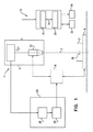
図1を参照すると、これは、本発明のいくつかの用途による病原体検出システム10の機能図である。病原体検出システム10は、記憶装置30に、例えば、コミュニケーションバス31により、動作可能に接続されたプロセッサー28を含む。特定の実施形態では、病原体検出システム100は、所望により顕微鏡システム11を含め得るか、または顕微鏡システム11に動作可能に接続され得る。顕微鏡システム11は通常、撮像モジュール14、合焦変動モジュール16、試料キャリア18および自動焦点システム20を含むデジタル顕微鏡である。いくつかの用途に対しては、顕微鏡システム11は通常、Greenfieldによる米国特許出願公開第2014/0347459号に記載の顕微鏡システムに類似である。この特許は参照により本明細書に組み込まれる。
Referring to FIG. 1, this is a functional diagram of a
通常、撮像モジュール14は、光学ユニット22および画像センサーユニット24を含む。光学ユニット22は、焦点面36と画像平面を共役させることにより、身体試料12(例えば、血液試料)の拡大画像を形成するように構成される。画像センサーユニット24は通常、画像センサー、例えば、電荷結合素子(CCD)、相補型金属酸化膜半導体(CMOS)センサー、および/または拡大画像を検知するように光学ユニット22の画像平面中に配置されたマトリックスセンサーを含む。
Usually, the image pickup module 14 includes an
コンピュータープロセッサー28は通常、画像を受け取り、処理する。コンピュータープロセッサーは記憶装置30と通信し、画像は記憶装置を介してプロセッサーにより受け取られる。ユーザーインターフェース32を介して、使用者(例えば、検査室技術者)はコンピュータープロセッサーに命令を送る。いくつかの用途に対しては、ユーザーインターフェースには、キーボード、マウス、ジョイスティック、タッチスクリーン装置(例えば、スマートフォンまたはタブレットコンピューター)、タッチパッド、トラックボール、音声操作インターフェース、および/または当技術分野で既知の他のタイプのユーザーインターフェースが含まれる。通常、コンピュータープロセッサーは、出力装置34を介して出力を生成する。さらに典型的には、出力装置は、モニターなどのディスプレイを含み、出力はディスプレイ上に表示される出力を含む。いくつかの用途に対しては、プロセッサーは、様々な種類の目視、テキスト、グラフィックス、 触知、音声、および/またはビデオ出力装置、例えば、スピーカー、ヘッドフォン、スマートフォン、またはタブレットコンピューター上に出力を生成する。いくつかの用途に対しては、ユーザーインターフェース32は、入力インターフェースおよび出力インターフェースの両方として機能、すなわち、入力/出力インターフェースとして機能する。いくつかの用途に対しては、プロセッサーは、ディスク、または携帯型USBドライブなどのコンピューター可読媒体(例えば、非一時的コンピューター可読媒体)上に、出力を生成するか、および/または印刷機上に出力を生成する。
The
特定の実施形態では、顕微鏡システム11は、顕微鏡システム11の少なくとも一部のプロセス、例えば、画像取得および/または病原体検出システム10の他の構成要素および病原体検出システム10の外部の構成要素を含む他の構成要素との通信を制御するローカルプロセッサーを含み得る。特定のその他の実施形態では、プロセッサー28は、顕微鏡システム11の1つまたは複数の、例えば、画像取得および/または通信を含むプロセスを制御し得る。必要に応じて、病原体検出システム10は、複数のデジタル顕微鏡を含み得る、またはそれに動作可能に接続され得る。必要に応じて、複数のデジタル顕微鏡中のそれぞれのデジタル顕微鏡は、それ自体のローカルプロセッサーを有する。
In certain embodiments, the
特定の実施形態では、図2を参照して以下で詳述されるように、記憶装置30は、画像情報、プログラムデータおよび/または身体試料中の病原体検出用実行可能プログラム命令を保存するように構成され得る。記憶装置30は、例えば、揮発性記憶装置または不揮発性記憶装置であり得る。特定の実施形態では、記憶装置30は、不揮発性記憶装置、例えば、ハードディスクドライブ、フラッシュメモリー、などである。
In certain embodiments, the
いくつかの用途に対しては、顕微鏡システム11は、1つまたは複数の身体試料の高倍率デジタル画像を取得するように構成される。必要に応じて、1つまたは複数のデジタル画像には、身体試料の異なる部分を含む画像が挙げられる。必要に応じて、画像は重なり合わない(または5%未満だけ、または1%未満だけ重なり合う)。必要に応じて、画像は、重なり合い画像を含み、異なる焦点深度、および/または異なる照明条件で取得される。必要に応じて、1つまたは複数のデジタル画像は、重なり合わない(または5%未満または1%未満だけ重なり合う)画像のセットを含むがそれぞれのセットは、異なる照明条件で取得された別のセットの画像を含む。特定の実施形態では、顕微鏡システム11は、以下でさらに詳述されるように、例えば、明視野、青色光、および紫外線を含む複数の照明条件下で画像を取得するように構成される。
For some applications, the
いくつかの用途では、身体試料12(例えば、血液試料)は、身体試料の複数の部分が撮像されるように、顕微鏡システムにより走査される。いくつかの用途に対しては、身体試料の1つまたは複数の部分から複数の画像が取得され、それぞれの複数の画像は、それぞれの撮像条件下で取得される。例えば、身体試料の部分の2つの画像は、それぞれ、細胞の検出を可能とする撮像条件(例えば、明視野)および染色物の可視化を可能とする撮像条件(例えば、適切な蛍光照射)を使って取得され得る。 In some applications, the body sample 12 (eg, a blood sample) is scanned by a microscopy system so that multiple parts of the body sample are imaged. For some applications, multiple images are obtained from one or more parts of the body sample, each of which is obtained under the respective imaging conditions. For example, the two images of the portion of the body sample use imaging conditions that allow the detection of cells (eg, bright field) and imaging conditions that allow visualization of the stain (eg, appropriate fluorescence irradiation), respectively. Can be obtained.
画像センサーユニット24は、取得デジタル画像を出力装置34(ディスプレイを含み得る)および/または自動焦点システム20に出力し得る。合焦変動モジュール16は、光学ユニット22の焦点面36と試料キャリア18との間の距離を変えるように構成し得る。合焦変動モジュール16は、例えば、試料キャリア18の位置を光学ユニット22の光軸Zに沿って変更し得る、メカニカルインターフェースを介して、マニュアルで、または自動的に操作され得る。あるいはまたはさらに追加して、合焦変動モジュール16は、自動焦点システム20により命令され得る。例えば、合焦変動モジュール16は、(1)光学ユニット22の位置を光軸Zに沿って変更することにより、(2)試料キャリア18の位置を光軸Zの位置に沿って変更することにより(例えば、試料キャリアが置かれているステージを動かすことにより)、(3)焦点面の位置を変更することにより、例えば、光学ユニット22の焦点距離を変えることにより、またはこれらの組み合わせにより、試料キャリア18と焦点面の間の距離を変え得る。
The image sensor unit 24 may output the acquired digital image to the output device 34 (which may include a display) and / or the
試料キャリア18は、プレートを含み得る。試料キャリア18は、身体試料12を収容するように構成され得る。キャリアは、生物試料を保持するために当該技術分野において既知の任意のキャリアであってよい。必要に応じて、キャリアの底面は、相互接触状態の細胞が顕微鏡の焦点面からほぼ同じ距離になるのを可能とするように、実質的に平坦である。例には、キャリアスライド、検査室容器、ディッシュ、プレート、マルチウェルプレート、試験チューブ(例えば、平底を有する)、マイクロ流体セル、カートリッジなどが挙げられる。 The sample carrier 18 may include a plate. The sample carrier 18 may be configured to accommodate the body sample 12. The carrier may be any carrier known in the art to hold the biological sample. If desired, the bottom surface of the carrier is substantially flat so that the cells in contact can be approximately the same distance from the focal plane of the microscope. Examples include carrier slides, laboratory vessels, dishes, plates, multi-well plates, test tubes (eg, with flat bottoms), microfluidic cells, cartridges and the like.
自動焦点システム20は、自動焦点計算モジュール38および自動焦点適応モジュール39を含み得る。自動焦点計算モジュールは、撮像モジュール14により取得された画像を受け取るように、画像センサーユニット24に接続し得る。自動焦点適応モジュールは、合焦変動モジュール16に接続し得、例えば、上述のように、合焦変動モジュール16に命令するように構成され得る。
The
いくつかの用途に対しては、プロセッサー28は、1つまたは複数の機能モジュール、例えば、特徴抽出モジュール、候補分類器、試料分類器、および診断モジュールなどを含む。いくつかの用途に対しては、プロセッサー28は、画像情報中に含まれる特徴を抽出することにより、画像情報を処理するように構成される。通常、プロセッサーは、少なくとも1つの試料としての情報価値の高い特徴および少なくとも1つの候補としての情報価値の高い特徴を抽出するように構成される。いくつかの用途に対しては、以降でさらに詳述されるように、プロセッサーは、少なくとも1つの試料としての情報価値の高い特徴を処理してコンテキスト情報を得るように、および少なくとも1つの候補としての情報価値の高い特徴を処理して候補データを得るようにさらに構成される。
For some applications, the
通常、プロセッサーは、少なくとも部分的に少なくとも1つの候補としての情報価値の高い特徴に基づいて、候補(すなわち、病原体であり得、したがって、病原体の候補であることを示す特性を示す試料中の構成要素)が病原体である可能性を分類するように構成される。さらに通常は、プロセッサーは、少なくとも1つの候補としての情報価値の高い特徴を、少なくとも1つの試料としての情報価値の高い特徴と組み合わせて処理することにより、身体試料が病原性感染症に感染している可能性を分類するように構成される。 Typically, a processor is configured in a sample that exhibits properties indicating that it is a candidate (ie, a potential pathogen and therefore a candidate for a pathogen, at least in part, based on its informative features as at least one candidate. The element) is configured to classify the possibility of being a pathogen. More typically, the processor infects a body sample with a pathogenic infection by processing the informative feature as at least one candidate in combination with the informative feature as at least one sample. It is configured to classify the possibility of being present.
いくつかの用途に対しては、プロセッサーは、分類および/または機械学習アルゴリズム、例えば、サポートベクターマシン、ニューラルネットワーク、単純ベイズアルゴリズム、などを使って、候補が病原体である可能性を分類するように、および/または試料が病原性感染症に感染している可能性を分類するようにプログラムされる。プロセッサーにより使用できる分類および/または機械学習アルゴリズムの種類の追加の例は、Bacheletによる米国特許出願公開第2012/0169863号、および/またはPollakによる米国特許出願公開第2015/0037806号に記載されている。これら両出願は、参照により本明細書に組み込まれる。いくつかの用途に対しては、コンピュータープロセッサーは、身体試料の分析での使用に先立って、身体試料の訓練画像を使って訓練される。 For some applications, the processor may use classification and / or machine learning algorithms, such as support vector machines, neural networks, naive Bayes algorithms, etc., to classify candidates as potential pathogens. , And / or the sample is programmed to classify the likelihood of being infected with a pathogenic infection. Additional examples of classifications and / or types of machine learning algorithms that can be used by the processor are described in U.S. Patent Application Publication No. 2012/0169863 by Bachelet and / or U.S. Patent Application Publication No. 2015/0037806 by Pollak. .. Both of these applications are incorporated herein by reference. For some applications, computer processors are trained with training images of body samples prior to their use in the analysis of body samples.
いくつかの用途に対しては、身体試料が病原性感染症に感染していることが分かっている場合は(または身体試料が病原性感染症に感染している可能性が閾値を超えることが分かっている場合は)、コンピュータープロセッサーは、少なくとも1つの試料としての情報価値の高い特徴に少なくとも基づいて、病原性感染症についての診断情報を抽出するようにさらに構成される。 For some applications, if the body sample is known to be infected with a pathogenic infection (or the likelihood that the body sample is infected with a pathogenic infection may exceed the threshold). If known), the computer processor is further configured to extract diagnostic information about pathogenic infections based on at least one informative feature as a sample.
本明細書で開示の主題の教示は、図1に関連して記載された特定の病原体検出システムに限定されるものではないことに留意されたい。等価および/または修正機能を別の方法に統合または分割でき、ソフトウェア、ファームウェアおよびハードウェアの任意の適切な組み合わせに実装することができる。プロセッサーは、適切にプログラムされたコンピューターとして実装することができる。 It should be noted that the teachings of the subject matter disclosed herein are not limited to the particular pathogen detection system described in connection with FIG. Equivalence and / or modification functionality can be integrated or split in other ways and implemented in any suitable combination of software, firmware and hardware. The processor can be implemented as a properly programmed computer.
図2を参照すると、これは、本発明のいくつかの用途による、身体試料(例えば、血液試料)中の病原性感染の検出方法の一般化したフローチャートを示す。 With reference to FIG. 2, this shows a generalized flow chart of a method of detecting a pathogenic infection in a body sample (eg, a blood sample) according to some uses of the present invention.
第1のステップ200では、身体試料の1つまたは複数の画像が顕微鏡システム11により取得される。1つまたは複数の画像、1つまたは複数の画像の情報を与えてくれるデータ、または1つまたは複数の画像に由来するデータ(本明細書においては、まとめて、「画像情報」と称する)が通常、記憶装置30に保存される。その後、本明細書の以降でさらに詳細に記載されるように、画像情報はプロセッサー28により解析される。本出願では、コンピュータープロセッサーは、1つまたは複数の画像からの特徴の抽出として記載されることに留意されたい。この用語は、1つまたは複数の画像の情報を与えてくれるデータ、または1つまたは複数の画像由来のデータから特徴を抽出することを含むとして解釈されるべきであり、1つまたは複数の画像それら自体から直接に特徴を抽出することに限定されると解釈されるべきではない。
In the
いくつかの用途に対しては、画像情報は、情報を与えてくれる試料の少なくとも1つの高倍率顕微鏡像である。あるいはまたはさらに追加して、画像情報は、例えば、試料の異なる部分の画像、異なる焦点深度でおよび/または異なる照明条件で、および/または異なる時間に取得した試料の同じ部分の画像を含む、情報を与えてくれる複数の画像である。 For some applications, the image information is at least one high magnification microscopic image of the informing sample. Or or in addition, the image information includes, for example, images of different parts of the sample, images of the same part of the sample taken at different depths of focus and / or under different lighting conditions, and / or at different times. It is a plurality of images that give us.
身体試料は、任意の生きている動物由来であってよいが、温血動物由来であるのが好ましい。通常、身体試料は血液試料である。試料は、1つまたは複数の赤血球を含む、任意の血液試料またはその一部であり得る。場合により、試料は、主に赤血球を含む(すなわち、試料中の大部分の細胞(例えば、細胞の少なくとも60%)は赤血球である)。必要に応じて、試料はまた、血小板および白血球の内の少なくとも1種を含む。必要に応じて、血液試料は希釈される。必要に応じて希釈が行われるか、またはそれ以外の試料は、撮像される表面上の細胞の濃度が平方ミリ当たり3,000〜30,000個の細胞(例えば、赤血球)となるように調製される。必要に応じて、血液試料は染色溶液で希釈される。 The body sample may be of any living animal origin, but is preferably of a warm-blooded animal origin. Usually, the body sample is a blood sample. The sample can be any blood sample or part thereof, including one or more red blood cells. In some cases, the sample contains predominantly red blood cells (ie, most cells in the sample (eg, at least 60% of the cells) are red blood cells). If desired, the sample also comprises at least one of platelets and white blood cells. If necessary, the blood sample is diluted. Diluted as needed, or other samples are prepared so that the concentration of cells on the surface to be imaged is 3,000 to 30,000 cells per square millimeter (eg, red blood cells). Will be done. If necessary, the blood sample is diluted with the staining solution.
必要に応じて、試料または染色溶液は、1つまたは複数の好適する色素または染料(必要に応じて、1種または複数の蛍光色素を含む)を含む。いくつかの実施形態では、血液試料は、全血試料、赤血球試料、軟膜試料、血漿試料、血清試料、いずれか他の血液画分由来の試料、またはこれらの任意の組み合わせから選択される。 If desired, the sample or staining solution comprises one or more suitable dyes or dyes, optionally including one or more fluorescent dyes. In some embodiments, the blood sample is selected from a whole blood sample, a red blood cell sample, a buffy coat sample, a plasma sample, a serum sample, a sample derived from any other blood fraction, or any combination thereof.
必要に応じて、試料は試料キャリア18の表面上に単一層を形成する。本開示においては、細胞の単一層に言及する場合、少なくとも50%(時には、少なくとも60%、70%、80%あるいは90%)の細胞がキャリアの底表面と直接接触しており、20%以下(時には、10%以下あるいは5%以下)の細胞が相互に重ね合わさっている(すなわち、上述の%以下の細胞が、部分的にまたは完全に相互の上に位置する)、実質的に単層としての表面上の細胞分布を包含すると理解されたい。さらに、単一層に言及する場合、少なくとも5%(時には、少なくとも10%あるいは少なくとも20%)の細胞が底面上で相互に接触していることを理解されたい。いくつかの用途に対しては、単一層は、Pollakによる米国特許出願公開第9,329,129号に記載の技術により、形成される。この特許は、参照により本明細書に組み込まれる。 If desired, the sample forms a single layer on the surface of the sample carrier 18. In the present disclosure, when referring to a single layer of cells, at least 50% (sometimes at least 60%, 70%, 80% or 90%) of cells are in direct contact with the bottom surface of the carrier and no more than 20%. Cells (sometimes 10% or less or 5% or less) are superposed on each other (ie, the% or less cells described above are partially or completely above each other) and are substantially monolayered. It should be understood to include the cell distribution on the surface as. Further, when referring to a single layer, it should be understood that at least 5% (sometimes at least 10% or at least 20%) of cells are in contact with each other on the bottom surface. For some applications, a single layer is formed by the technique described in US Patent Application Publication No. 9,329,129 by Pollak. This patent is incorporated herein by reference.
いくつかの用途に対しては、撮像の前に、身体試料は1つまたは複数の好適な色素または染料で染色される。必要に応じて、1種または複数の好適な色素または染料は、1種または複数の蛍光色素または染料を含み、染色試料は、1つまたは複数の病原体の検出に好適な照明条件で励起される。本明細書で使用される場合、「好適な色素または染料」という用語は、目的の病原体の検出に有用な、任意の好適な蛍光色素または染料を含む任意の色素または染料を含むように広く解釈されるべきである。本明細書で使用される場合、「好適な蛍光色素または染料」は、1つまたは複数の種類の核酸(例えば、DNAのみ、RNAのみ、DNAとRNAの両方、など)に選択的に結合でき、1つまたは複数の特定の照明条件下で蛍光を発し、それにより、身体試料中の1つまたは複数の種類の核酸を識別可能とする色素または染料を含むように広く解釈されるべきである。好適な蛍光色素または染料には、例えば、DNAに結合するがRNAに結合しない色素または染料、RNAに結合するがDNAに結合しない色素または染料、およびDNAとRNAの両方に結合する色素または染料を含み得る。好適な蛍光色素または染料の非限定的例には、例えば、アクリジンオレンジ、ヘキスト染料、などが挙げられる。 For some applications, the body sample is stained with one or more suitable dyes or dyes prior to imaging. If desired, one or more suitable dyes or dyes include one or more fluorescent dyes or dyes, and the stained sample is excited under lighting conditions suitable for detection of one or more pathogens. .. As used herein, the term "suitable dye or dye" is broadly construed to include any dye or dye, including any suitable fluorescent dye or dye, useful in detecting the pathogen of interest. It should be. As used herein, a "suitable fluorochrome or dye" can selectively bind to one or more types of nucleic acids (eg, DNA only, RNA only, both DNA and RNA, etc.). It should be broadly interpreted to include dyes or dyes that fluoresce under one or more specific lighting conditions, thereby making one or more types of nucleic acids identifiable in body samples. .. Suitable fluorescent dyes or dyes include, for example, dyes or dyes that bind to DNA but not RNA, dyes or dyes that bind to RNA but not DNA, and dyes or dyes that bind to both DNA and RNA. Can include. Non-limiting examples of suitable fluorescent dyes or dyes include, for example, acridine orange, Hoechst dye, and the like.
特定の好適な蛍光色素または染料に蛍光を発光させる特定の照明条件は、本明細書においては「好適な照明条件」と呼ばれ、これは、特定の蛍光色素または染料を励起するために使用する場合、蛍光色素または染料の蛍光を発光させる照明条件を含むように広く解釈されるべきである。特定の実施形態では、励起された色素または染料により発光される蛍光は、所定の波長帯内の蛍光を識別可能にする1つまたは複数の異なる光フィルターの使用により識別可能であり得る。したがって、このようなフィルターを考慮して、好適な照明条件を使用し得る。好適な照明条件の非限定的例には、例えば、明視野、青色光、および紫外線が含まれる。好適な蛍光色素または染料および好適な照明条件の追加の非限定的例は、Bacheletによる米国特許出願公開第2012/0169863号、およびPollakによる米国特許出願公開第2015/0037806号に記載されている。これら両出願は、参照により本明細書に組み込まれる。 Specific lighting conditions that cause a particular suitable fluorescent dye or dye to emit fluorescence are referred to herein as "suitable lighting conditions", which are used to excite a particular fluorescent dye or dye. If so, it should be broadly interpreted to include illumination conditions that cause the fluorescence of the fluorescent dye or dye to illuminate. In certain embodiments, the fluorescence emitted by the excited dye or dye can be identifiable by the use of one or more different optical filters that make the fluorescence within a given wavelength band identifiable. Therefore, suitable lighting conditions can be used in consideration of such a filter. Non-limiting examples of suitable lighting conditions include, for example, brightfield, blue light, and ultraviolet light. Additional non-limiting examples of suitable fluorescent dyes or dyes and suitable lighting conditions are described in US Patent Application Publication No. 2012/0169863 by Bachelet and US Patent Application Publication No. 2015/0037806 by Pollak. Both of these applications are incorporated herein by reference.
上記で詳述したように、特定の実施形態では、試料は、試料中のRNAとDNAの間で区別を可能とする1つまたは複数の色素または染料で染色し得る(すなわち、分染)。分染は、例えば、1つまたは複数の標的特異的色素または染料で試料を染色することにより達成できる。本明細書で使用される場合、標的特異的色素または染料(例えば、RNA特異的またはDNA特異的)は、選択条件下で、その他の細胞成分の存在下で検出し得るように、標的部分を検出可能に染色する色素または染料である。これに関して、標的を検出可能に染色することは、色素または染料が標的にその他の細胞成分より高い親和性で結合すること、および/またはそれが標的と結合した場合、より強力なシグナル(例えば、蛍光)を与えることを意味し得る。いくつかの色素または染料は、2つ以上の標的を染色し得るが、例えば、発光蛍光の波長、および/または色素または染料の励起に使用される波長に基づいて区別し得ることを留意されたい。いくつかの実施形態では、標的特異的色素または染料は、標的に結合時に、その発光波長を元の周波数帯からシフト周波数帯に移動させる蛍光色素または染料である。このような場合には、標的はシフト周波数帯中の発光波長を検出するように構成されたシステムにより検出され得る。 As detailed above, in certain embodiments, the sample can be stained with one or more dyes or dyes that allow the RNA and DNA in the sample to be distinguished (ie, split dyeing). Separation can be achieved, for example, by staining the sample with one or more target-specific dyes or dyes. As used herein, a target-specific dye or dye (eg, RNA-specific or DNA-specific) will detect the target moiety under selective conditions in the presence of other cellular components. A dye or dye that stains detectably. In this regard, detectable staining of the target means that the dye or dye binds to the target with a higher affinity than other cellular components and / or when it binds to the target, a stronger signal (eg, for example). Fluorescence) can be meant to be given. Note that some dyes or dyes can stain more than one target, but can be distinguished, for example, based on the wavelength of emission fluorescence and / or the wavelength used to excite the dye or dye. .. In some embodiments, the target-specific dye or dye is a fluorescent dye or dye that, upon binding to the target, shifts its emission wavelength from the original frequency band to the shift frequency band. In such cases, the target can be detected by a system configured to detect emission wavelengths in the shift frequency band.
実施例1に関連して下記で詳述のように、分染を使ってDNAおよびRNAの相対的位置を決定し得る。必要に応じて、単一色素または染料(例えば、アクリジンオレンジ)を異なる照明条件下で使用して、分染を提供し得る。必要に応じて、任意の核酸(DNAおよびRNA)を検出するように構成された、1つまたは複数のDNA特異的色素または染料(例えば、ヘキスト試薬)および1つまたは複数のその他の色素または染料(例えば、アクリジンオレンジ)を含む、色素または染料の組み合わせが使用される。 Separation can be used to determine the relative positions of DNA and RNA, as detailed below in connection with Example 1. If desired, a single dye or dye (eg, acridine orange) may be used under different lighting conditions to provide split dyeing. One or more DNA-specific dyes or dyes (eg, Hoechst reagents) and one or more other dyes or dyes configured to detect any nucleic acid (DNA and RNA) as needed. A combination of dyes or dyes is used, including (eg, acridine orange).
いくつかの用途に対しては、画像情報は、情報を与えてくれる身体試料の1つまたは複数の視野である。本明細書で使用される場合、「視野」は、身体試料の撮像される一部である。通常、これは、試料を保持する試料キャリアの底部の領域に対応する。画像が高倍率で取得される場合、一回で、全体血液試料の一部のみが撮像できる。したがって、病原体検出システム10は、撮像領域を実質上複数の視野に再分割し、それぞれの視野は別々に撮像され、それにより、情報を与えてくれる身体試料の複数の画像、それぞれの視野の情報を与えてくれるそれぞれの画像が得られる。必要に応じて、撮像視野は重なり合わない、またはそれらの重複度は、領域の5%未満、もしくは1%未満である。特定の実施形態では、撮像されるそれぞれの視野は、1つまたは複数の異なる照明条件下で撮像される。必要に応じて、それぞれの視野の画像は、異なる照明条件で複数回取得される。例えば、視野は、RNA関連蛍光を検出する照明条件で少なくとも1回、DNA関連蛍光を検出する照明条件で少なくとも1回、および明視野で少なくとも1回、撮像され得る。
For some applications, image information is one or more fields of view of an informative body sample. As used herein, the "field of view" is the imaged portion of a body sample. This usually corresponds to the area at the bottom of the sample carrier that holds the sample. When the image is acquired at high magnification, only a part of the whole blood sample can be imaged at one time. Therefore, the
図3を参照すると、これは、非限定的例であるが、本出願のいくつかの用途による、1つまたは複数の好適な蛍光色素で染色し、好適な照明条件下で励起した血液試料の視野からなる画像情報300を示す。観察されるように、色素(単一または複数)に起因して、構成要素302は蛍光を発し、それにより、試料中の他の非蛍光発光構成要素304(この場合、これは赤血球を含む)より明るく見え(または、いくつかの事例では、異なる色)、試料中の染色した領域の識別が可能となり、いくつかのその特徴は、試料中のいくつかの特定の細胞型の情報価値があり得る。
Referring to FIG. 3, this is a non-limiting example of a blood sample stained with one or more suitable fluorescent dyes and excited under suitable lighting conditions according to some applications of the present application. The
特定の実施形態では、画像情報は、候補(すなわち、病原体であり得、従って、病原体の候補であることを示す特性を示す構成要素)および非候補を含む1つまたは複数の試料構成要素の情報を与えてくれる。いくつかの用途に対しては、試料が好適な蛍光色素または染料で染色され、例えば、Bacheletによる米国特許出願公開第2012/0169863号、および/またはPollakによる米国特許出願公開第2015/0037806号(これら両出願は、参照により本明細書に組み込まれる)に記載の好適な照明条件により励起される場合、要素は、蛍光発光が現れている要素に基づいて候補として特定される。あるいはまたはさらに追加して、要素は、そのサイズ、形状、色、その他の要素に対する近接度などの他の判定基準に基づいて候補として特定され得る。本明細書で使用される場合、「非候補」という用語は、候補ではない試料の構成要素を含むように広く解釈されるべきである。 In certain embodiments, the image information is information about one or more sample components, including candidates (ie, components exhibiting properties that can be pathogens and thus are candidates for pathogens) and non-candidates. Gives me. For some applications, the sample is stained with a suitable fluorescent dye or dye, eg, US Patent Application Publication No. 2012/0169863 by Bachelet and / or US Patent Application Publication No. 2015/0037806 by Pollak ( If both of these applications are excited by the preferred illumination conditions described in (incorporated herein by reference), the element is identified as a candidate based on the element in which the fluorescence emission appears. Or, in addition, an element may be identified as a candidate based on other criteria such as its size, shape, color, and proximity to other elements. As used herein, the term "non-candidate" should be broadly construed to include components of a non-candidate sample.
図2を再度参照すると、ステップ201で、プロセッサー28は、1つまたは複数の画像から、画像情報から、および/またはそれらの一部から、身体試料に関連するコンテキスト情報を示す、身体試料の1つまたは複数の試料としての情報価値の高い特徴を抽出する。通常、複数の試料としての情報価値の高い特徴が抽出される。本明細書で使用される場合、「試料としての情報価値の高い特徴」は、特定の候補を対象とせず、また、試料中の病原性感染の存在、可能性、または特性を決定するのに使用でき、いくつかの実施形態では、特定の候補の分類を含む、コンテキスト情報を提供するのに使用できる身体試料の特徴を含む。非限定的例であるが、試料としての情報価値の高い特徴には、例えば、試料中に非候補成分に関連する特徴、または試料中の所定の種類の細胞の量および/または分布に関連する特徴を挙げることができる。試料中の非候補成分に関連する特徴には、例えば、1つまたは複数の非候補のサイズ関連特性(予測サイズ、または1つまたは複数の他の細胞の観察サイズに比較した相対サイズを含む)、1つまたは複数の非候補の形状関連特性(予測形状、または1つまたは複数の他の要素の観察形状に比較した相対的形状を含む)、および1つまたは複数の非候補の強度関連特性(予測強度、または1つまたは複数の他の要素の観察強度に比較した相対的強度を含む)を挙げることができる。本明細書で使用される場合、「予測」値(例えば、サイズ、形状および/または強度の予測値)は、所定の試料に関する画像情報の分析に先立って既知であり得るような値である。このような値には、例えば、既知であるか、または、必要に応じて、特定の条件(例えば、高度、身体試料の処理、など)に応じて、計算できる(例えば、全ての人および/またはその任意のサブグループに対する、例えば、年齢、性別、人種、民族、などを基準に)母集団統計値が挙げられる。
With reference to FIG. 2 again, in
いくつかの用途に対しては、試料としての情報価値の高い特徴には、試料またはその部分中の候補または病原体の分布に関連する特徴が含まれる。例えば、所定の画像(または画像の部分もしくは試料の連続部分を含む画像群)で見つかる候補または病原体の数が、同じ試料の他の部分で見つかる候補または病原体の数よりかなり高い場合には、これは、試料の一部で見つかる高濃度の候補または病原体が、試料の診断に影響を与えるべきではない局所効果の結果である可能性があることを示し得る。例えば、試料の1つの部分では存在するが他の部分では存在しない高濃度の候補または病原体(例えば、赤血球と重なり合う高濃度の候補)は、例えば、調査中の試料に入った別の試料からの血液の落下に由来する汚染を示す可能性がある。 For some applications, informative features as a sample include features related to the distribution of candidates or pathogens within the sample or parts thereof. For example, if the number of candidates or pathogens found in a given image (or group of images containing a portion of the image or a contiguous portion of a sample) is significantly higher than the number of candidates or pathogens found in other parts of the same sample. Can indicate that high concentrations of candidates or pathogens found in parts of the sample may be the result of local effects that should not affect the diagnosis of the sample. For example, a high concentration candidate or pathogen that is present in one part of the sample but not in the other part (eg, a high concentration candidate that overlaps with red blood cells) is, for example, from another sample in the sample under investigation. May indicate contamination resulting from blood drops.
いくつかの用途に対しては、例えば、所定の判定基準(例えば、輝度、焦点、など)で測定して、いくつかの画像情報が不十分な品質であるかどうかを判断するために、前処理段階でステップ201の一部または全てが実施され、不十分な品質の場合には、画像情報のいくつかの部分がさらなる処理から排除され得る(例えば、実施例6に関連して後述するように)。
For some applications, for example, to measure with a given criterion (eg, brightness, focus, etc.) to determine if some image information is of inadequate quality. If some or all of
ステップ202では、コンピュータープロセッサー28は、試料内の1つまたは複数の構成要素を病原体の候補として特定する。本明細書で上記したように、試料が好適な蛍光色素で染色され、例えば、Bacheletによる米国特許出願公開第2012/0169863号、および/またはPollakによる米国特許出願公開第2015/0037806号(これら両出願は、参照により本明細書に組み込まれる)に記載の好適な照明条件により励起される場合、要素は、蛍光発光が現れている要素に基づいて候補として特定され得る。あるいはまたはさらに追加して、要素は、形状、サイズ、その他の要素(例えば、赤血球、またはその他の候補)に対する近接度などの他の判定基準に基づいて候補として特定され得る。
In
ステップ203で、コンピュータープロセッサーは、1つまたは複数の画像から、画像情報から、および/またはそれらの一部から、1つまたは複数の候補としての情報価値の高い、1つまたは複数の特定された候補に関連する特徴を抽出する。通常、それぞれの候補に対して、複数の候補としての情報価値の高い特徴が抽出される。本明細書で使用される場合、「候補としての情報価値の高い特徴」は、候補(または、いくつかの事例では、以下で詳述されるように、候補に極めて近接した構成要素)の、所定の候補が病原体または病原体の一部である可能性を決定する情報を提供するのに使用可能な特徴を含む。
In
非限定的例であるが、候補としての情報価値の高い特徴は、次記に関連する特徴を含み得る:候補のサイズ、候補の形状、候補の動き(例えば、少なくとも2種の少なくとも部分的に重なり合う順々に取得された画像の比較に基づいて)、および/または候補の強度。 Although non-limiting examples, information-valued features as candidates may include features related to: candidate size, candidate shape, candidate movement (eg, at least two, at least in part). (Based on a comparison of the images obtained in sequence), and / or the intensity of the candidates.
いくつかの用途に対しては、候補としての情報価値の高い特徴は、候補のその他の試料成分(例えば、赤血球)に対する相対的位置を含む。あるいはまたはさらに追加して、候補としての情報価値の高い特徴は、少なくとも部分的に候補と重なり合う細胞(例えば、赤血球)の、性質(および、必要に応じて、重なり合いの量も)、例えば、候補と重なり合う細胞のサイズまたは形状を含む。いくつかの用途に対しては、候補と重なり合う細胞の寸法および形状に関連する特徴は、予測サイズまたは予測形状と比較した重なり合う細胞の相対的サイズおよび相対的形状を含む。本明細書で使用される場合、画像情報中で、少なくとも一部の細胞が、少なくとも一部(例えば、候補の少なくとも20%または少なくとも25%)の候補と同じ場所に配置されるように見えると、細胞は、候補と少なくとも部分的に重なり合うと見なされる。 For some applications, candidate informative features include the position of the candidate relative to other sample components (eg, red blood cells). Or, or in addition, an informative feature as a candidate is the nature (and, if necessary, the amount of overlap) of cells (eg, erythrocytes) that at least partially overlap the candidate, eg, candidate. Includes the size or shape of cells that overlap with. For some applications, features related to the size and shape of overlapping cells with the candidate include the predicted size or relative size and shape of the overlapping cells compared to the predicted shape. As used herein, in image information, at least some cells appear to be co-located with at least some (eg, at least 20% or at least 25% of candidates) candidates. , Cells are considered to overlap at least partially with the candidate.
必要に応じて、候補としての情報価値の高い特徴は、候補に極めて近接して見つかる他の構成要素(例えば、病原体候補および/または病原体)の特徴を含み得る。これに関して、「極めて近接」は、任意の好適な尺度に従って、あらかじめ定義できる。例えば、候補に極めて近接している成分は、候補から最大2X離れた距離内に位置する成分を含み得る。ここで、Xは、予測(例えば、平均)赤血球の直径である。したがって、いくつかの実施形態では、候補としての情報価値の高い特徴(候補の特徴、候補と重なり合う細胞の特徴、および/またはその他の成分の特徴を含む)は、候補に近接した距離内の特徴を含み得るか、またはそのような特徴に限定され得る。 If desired, informative features as candidates may include features of other components found very close to the candidate (eg, pathogen candidates and / or pathogens). In this regard, "extreme proximity" can be predefined according to any suitable measure. For example, a component that is very close to the candidate may include a component that is located within a distance of up to 2X from the candidate. Where X is the diameter of the predicted (eg, average) red blood cell. Thus, in some embodiments, information-valued features as candidates, including candidate features, cell features that overlap the candidate, and / or other component features, are features within a close distance to the candidate. Can include or be limited to such features.
いくつかの用途に対しては、画像情報またはその一部は、候補としての情報価値の高い特徴の抽出のために、前処理段階で、少なくとも部分的に処理される。特定の実施形態では、前処理段階は、試料としての情報価値の高い特徴を抽出してコンテキスト情報を得ること、および得られたコンテキスト情報に従って候補としての情報価値の高い特徴を抽出するために使用される画像情報を決定することを含み得る。いくつかの用途に対しては、候補としての情報価値の高い特徴を抽出するために使用される一部の画像情報および試料としての情報価値の高い特徴を抽出するために使用される一部の画像情報は、部分的にまたは完全に重なり合う。 For some uses, the image information or a portion thereof is processed at least partially in the preprocessing stage for the extraction of informationally valuable features as candidates. In certain embodiments, the pretreatment step is used to extract information-valued features as a sample to obtain contextual information, and to extract candidate information-valued features according to the obtained contextual information. It may include determining the image information to be made. For some applications, some image information used to extract high information value features as candidates and some used to extract high information value features as samples. The image information partially or completely overlaps.
ステップ201、202および203は、任意の順序で実施できることに留意されたい。いくつかの用途では、ステップ201、202および203は、単一ステップとして実施されるおよび/または相互に組み合わせられる。いくつかの用途では、ステップ201、202および203の一部または全ては、複数の別々のステップとして実施される。
Note that steps 201, 202 and 203 can be performed in any order. In some applications, steps 201, 202 and 203 are performed as a single step and / or combined with each other. In some applications, some or all of
通常、試料としての情報価値の高い特徴(単一または複数)と組み合わせた、候補としての情報価値の高い特徴(単一または複数)に基づいて、コンピュータープロセッサー28は、身体試料が病原性感染症に感染している可能性を分類する。いくつかの用途に対しては、病原性感染は、図2に示す追加のステップを実装することにより検出される。
いくつかの用途に対しては、少なくともいくつかの候補としての情報価値の高い特徴が抽出されると、ステップ205で、プロセッサー28は、それぞれの候補に対し得た候補データに従って、それぞれの候補が病原体である可能性を分類する。本明細書で使用される場合、「病原体である可能性」という用語は、2値判定(例えば、病原体または非病原体)またはスカラー判定(例えば、所定の候補が病原体であるという推定可能性を反映する数、値)を含むように広く解釈されるべきである。特定の実施形態では、プロセッサー28は、候補としての情報価値の高い特徴と組み合わせて、抽出した試料としての情報価値の高い特徴(例えば、ステップ201で抽出した特徴)を使って、それぞれの候補が病原体である可能性を分類する。これについては、例えば、実施例1および2に関連して、以降でさらに詳述される。これは、ステップ201をステップ205に連結する点線の矢印で示されており、ステップ201がステップ205への任意入力であることを示す。
When at least some information-valued features as candidates are extracted for some applications, in
通常、候補分類(すなわち、ステップ205)の後に、ステップ207で、プロセッサー28は、身体試料が病原性感染症に感染している可能性を分類する。本明細書で使用される場合、「身体試料が感染している可能性」という用語は、2値判定(例えば、感染または非感染)またはスカラー判定(例えば、所定の試料が感染しているという推定可能性を反映する数、値)を含むように広く解釈されるべきである。いくつかの用途に対しては、プロセッサー28は、試料としての情報価値の高い特徴(ステップ201で抽出)と組み合わせて、候補(ステップ205で抽出)の分類に基づいて、試料を分類する。これについては、例えば、実施例1および3に関連して、以降でさらに詳述される。
Usually, after candidate classification (ie, step 205), in
いくつかの用途に対しては、ステップ209で、プロセッサー28は、1つまたは複数の抽出した試料としての情報価値の高い特徴および/または候補としての情報価値の高い特徴に従って、病原性感染を、1つまたは複数の所定の種類の病原体を含むとして分類する。
For some applications, in
病原性感染を1つまたは複数の所定の種類の病原体を含むとして分類することは、ステップ201、203、205、および207の1つまたは複数のステップで得られた情報および/または特徴を使って、および/または1つまたは複数の特徴抽出および分類の追加のステップを実施することにより、実施し得る。いくつかの用途に対しては、病原性感染を分類するために、(a)候補が所定の種類の病原体として分類され、および(b)全体の病原性感染が個々の候補の分類に基づいて分類される。いくつかの用途に対しては、試料としての情報価値の高い特徴が、個々の候補を所定の種類の病原体として分類するために、および/または全体の感染を所定の種類の病原体を含むとして分類するために、使用される。
Classification of pathogenic infections as containing one or more predetermined types of pathogens uses the information and / or features obtained in one or more steps of
いくつかの用途に対しては、例えば、実施例4および5に関して以降でさらに詳述されるように、病原性感染を1つまたは複数の所定の種類の病原体を含むとして分類することは、例えば、試料中に含まれる病原体の種または菌株を決定するために病原性感染を分類することを含む。このような決定は病原体を単一の種もしくは菌株、またはいくつかの可能な種もしくは菌株の群に分類すること(これらの少なくとも1つは試料中に含まれる)、および/または所定の種または菌株を除外すること(試料中に含まれない種として)を含み得るまたはそれに限定し得る。いくつかの用途では、プロセッサー28は、病原性感染を1つまたは複数のマラリア原虫、マラリア原虫の所定の菌株、所定の成熟度のマラリア原虫、および/または所定の成熟範囲のマラリア原虫を含むとして分類する。
For some uses, for example, classifying a pathogenic infection as comprising one or more predetermined types of pathogens, as described in more detail below with respect to Examples 4 and 5, eg. Includes classifying pathogenic infections to determine the species or strain of pathogen contained in a sample. Such a determination classifies a pathogen into a single species or strain, or a group of several possible species or strains (at least one of which is contained in a sample), and / or a given species or Exclusion of strains (as species not included in the sample) may be included or limited thereto. In some applications, the
いくつかの用途に対しては、コンピュータープロセッサーは、使用者に出力を生成し(例えば、出力装置に)、試料が病原体に感染しているか否かを示し、感染の分類を示す。いくつかの用途に対しては、コンピュータープロセッサーが、身体試料中の感染の存在は十分な信頼度で決定されない可能性があることを示す、一部の試料は再撮像の必要があることを示す、および/または一部の試料は異なる設定を使って(例えば、異なる照明を使って、異なる染料を使って、異なるまたは新しい試料調製法を使って、および/または異なる顕微鏡設定を使って)再撮像する必要があることを示す出力を生成する。いくつかの用途に対しては、身体試料中の感染の存在は、十分な信頼度で決定されない可能性があるという判断に対応して、コンピュータープロセッサーは、使用者は適切な使用者による対策をとる必要がある(例えば、新しい試料を調製する、および/または独自の方法を使って試料を試験する、など)ことを示す出力を生成する。あるいはまたはさらに追加して、コンピュータープロセッサーは、一部の試料を再撮像するように顕微鏡システムを自動的に操作する、異なる設定(例えば、異なる焦点、または異なる視野サイズ)を使って一部の試料を再撮像するように顕微鏡システムを操作する、および/または顕微鏡システムが顕微鏡画像を取得するフレームレートを調節する。 For some applications, the computer processor produces an output to the user (eg, in the output device), indicates whether the sample is infected with a pathogen, and indicates the classification of the infection. For some applications, computer processors show that the presence of infections in body samples may not be determined with sufficient confidence, some samples need to be reimaging. , And / or some samples re-used with different settings (eg, with different lighting, with different dyes, with different or new sample preparation methods, and / or with different microscopy settings). Produces an output indicating that an image needs to be taken. For some applications, computer processors respond to the determination that the presence of infection in a body sample may not be determined with sufficient confidence, and the computer processor allows the user to take appropriate user action. Produces output indicating that it needs to be taken (eg, preparing a new sample and / or testing the sample using a proprietary method, etc.). Or, in addition, the computer processor automatically manipulates the microscopy system to reimage some samples, using different settings (eg, different focal points, or different field of view sizes) for some samples. Manipulate the microscope system to re-image, and / or adjust the frame rate at which the microscope system acquires the microscope image.
いくつかの用途に対しては、試料としての情報価値の高い特徴は、必ずしも画像から直接的に得られないことに留意されたい。例えば、試料としての情報価値の高い特徴は、試料中の候補および/またはその他の実体に関する統計情報またはその他の情報、および/または試料の一般的特性を含み得る。一般に、本出願の範囲は、試料を2つのレベルに関し分析することを含み、その1つ目は候補毎のレベル、および2つ目は全体として試料の特性を示す、より一般的なレベルである。 It should be noted that for some applications, informative features as a sample are not always obtained directly from the image. For example, informative features as a sample may include statistical or other information about candidates and / or other entities in the sample, and / or general properties of the sample. In general, the scope of this application includes analyzing the sample in terms of two levels, the first being the per-candidate level and the second being the more general level indicating the characteristics of the sample as a whole. ..
いくつかの用途に対しては、候補レベルの特徴に基づいて、2つまたはそれを超える試料としての情報価値の高い、身体試料に関連する特徴が抽出され、身体試料の特性は、2つまたはそれを超える試料としての情報価値の高い特徴を処理することにより決定される。通常、少なくとも一部の候補は、病原体候補であり、病原体候補に関連する候補としての情報価値の高い特徴が抽出される。いくつかの用途に対しては、網状赤血球および/または血小板などの実体の候補が追加で特定され、これらの候補に関する候補としての情報価値の高い特徴が抽出される。いくつかの用途に対しては、試料としての情報価値の高い特徴は、試料中の病原体候補の数、試料中の病原体候補の種類、バックグラウンド輝度に対する候補の輝度、候補の病原体である確率、閾値を超える病原体である確率を有する候補の数、閾値を超える所定の種類の病原体である確率を有する候補の数、試料中の血小板の数、血小板の輝度、試料中の網状赤血球の数、試料中の病原体に感染した網状赤血球の数、候補の赤血球に対する近接度、および/または試料中の赤血球の数、を含む。 For some applications, based on candidate-level characteristics, highly informative, body-sample-related features as two or more samples are extracted, with two or more body sample characteristics. It is determined by processing features with high information value as a sample beyond that. Usually, at least some candidates are pathogen candidates, and highly informative features as candidates related to the pathogen candidates are extracted. For some applications, additional entity candidates such as reticulocytes and / or platelets are identified and informative features as candidates for these candidates are extracted. For some applications, the informative features of a sample are the number of pathogen candidates in the sample, the type of pathogen candidate in the sample, the brightness of the candidate against background brightness, the probability of being a candidate pathogen, Number of candidates with a probability of being a pathogen exceeding the threshold, number of candidates with a probability of being a pathogen of a predetermined type exceeding the threshold, number of platelets in a sample, brightness of platelets, number of reticular red cells in a sample, sample Includes the number of reticular erythrocytes infected with the pathogen in, the proximity to candidate erythrocytes, and / or the number of erythrocytes in the sample.
本明細書で開示の主題の実施形態では、図2に示したものより少ない、多いおよび/またはそれとは異なる段階を実行し得る。本明細書で開示の主題の実施形態では、図2に示した段階の1つまたは複数の段階を異なる順に実行し得、および/または1つまたは複数の段階群を同時に実行し得る。 In embodiments of the subject matter disclosed herein, less, more, and / or different steps than those shown in FIG. 2 may be performed. In an embodiment of the subject matter disclosed herein, one or more of the stages shown in FIG. 2 may be performed in different order and / or one or more stages may be performed simultaneously.
開示主題をさらによく理解するために、上記で詳述した本方法の特定の非限定的用途の詳細を示す多くの実施例を以降で提供する。 To better understand the disclosed subject matter, many embodiments are provided below that detail specific non-limiting applications of the method detailed above.
実施例1:候補を分類するための、および/または病原性感染を分類するための、試料としての情報価値の高い特徴としての網状赤血球の濃度の使用
上記で詳述したように、特定の実施形態では、試料は、試料中のDNAおよびRNAのそれぞれの位置を識別するために染色される。このような染色としては、例えば、少なくとも1種のDNA特異的色素および少なくとも1種のRNA特異的色素の使用、または少なくとも1つの標的(DNAまたはRNA)特異的色素およびDNAおよびRNAの両方を染色する少なくとも1種の色素が挙げられる。いくつかの用途に対しては、候補が病原体である可能性を分類するために(ステップ205)、病原体の予測パターン(単一または複数)に対応する染色パターン(単一または複数)であるかどうかを判定するために、RNAおよびDNA染色の試料中のそれぞれの位置がプロセッサーにより使用される。
Example 1: Use of reticulocyte concentration as an informative feature as a sample for classifying candidates and / or for classifying pathogenic infections, specific practices, as detailed above. In morphology, the sample is stained to identify the respective location of DNA and RNA in the sample. Such staining may include, for example, the use of at least one DNA-specific dye and at least one RNA-specific dye, or at least one target (DNA or RNA) -specific dye and both DNA and RNA. There is at least one dye that does. For some applications, is the staining pattern (single or plural) corresponding to the predictive pattern of the pathogen (single or plural) to classify the potential pathogens (step 205)? Each position in the sample for RNA and DNA staining is used by the processor to determine if.
図4は、候補400aおよび400bを模式的に示し、それぞれの候補は、RNAに対して染色された領域(RNA部分402)およびDNAに対し染色された領域(DNA部分404)を示す。RNA部分402およびDNA部分404は、それぞれの特定の染色領域の境界を識別するために、例えば、異なる色素および/または異なる照明を使って区別を付けて染色し得る。図4に示すように、候補400aでは、DNA部分404は、RNA部分402と完全に重なり合うが、候補400bでは、DNA部分404は、RNA部分402に部分的に重なり合う。
FIG. 4 schematically shows candidates 400a and 400b, and each candidate shows a region stained for RNA (RNA portion 402) and a region stained for DNA (DNA portion 404).
少なくとも部分的に重なり合ったDNAおよびRNAを有するように見える候補は、病原体である可能性がある。しかし、重なり合ったRNAおよびDNAの染色領域のこの外観は、例えば、異なる細胞型、または相互の上に位置するように見える2つの別の物(この内の1つはDNAを含み、もう一方はRNAを含む)を含む異なる実体または複数実体によっても引き起こされ得る。 Candidates that appear to have at least partially overlapping DNA and RNA can be pathogens. However, this appearance of overlapping RNA and stained regions of DNA is, for example, two different cell types that appear to be located on top of each other, one containing DNA and the other. It can also be triggered by different or multiple entities, including (including RNA).
成熟赤血球は検出可能なDNAまたはRNAを有せず、したがって、核酸用として染色した場合、蛍光発光しない。対照的に、マラリア原虫トロホゾイト(病原体の1種)は赤血球中のDNA含有およびRNA含有物として検出し得る。したがって、いくつかの用途に対しては、病原体を含む赤血球を特定するために、DNAおよびRNAの両方を染色させる染色物質(例えば、アクリジンオレンジ)が使われる。あるいはまたはさらに追加して、DNAのみを染色する染料(例えば、ヘキスト染料)が使われる。 Mature erythrocytes have no detectable DNA or RNA and therefore do not fluoresce when stained for nucleic acids. In contrast, the Plasmodium trohozoite (a type of pathogen) can be detected as DNA- and RNA-containing substances in erythrocytes. Therefore, for some applications, stains that stain both DNA and RNA (eg, acridine orange) are used to identify red blood cells containing pathogens. Alternatively or additionally, a dye that stains only the DNA (eg, Hoechst dye) is used.
ハウエルジョリー小体は、いくつかの不健康な状態の赤血球中で見つかり得るDNA含有物である。いくつかの事例では、試料中のハウエルジョリー小体の存在は、病原体感染の擬陽性判定の確率を高め得る。DNAおよびRNA両方を染色する染料と共にDNA特異的染料が使われる場合でも、ハウエルジョリー小体は病原体感染の擬陽性判定を生ずる場合がある。したがって、いくつかの実施形態では、ハウエルジョリー小体含有赤血球と、病原体含有赤血球との間の識別は有益であり得る。 Howell-Jolly bodies are DNA-containing substances that can be found in some unhealthy red blood cells. In some cases, the presence of Howell-Jolly bodies in the sample can increase the probability of a false positive test for pathogen infection. Even when DNA-specific dyes are used with dyes that stain both DNA and RNA, Howell-Jolly bodies can result in false-positive verdicts for pathogen infection. Therefore, in some embodiments, the distinction between Howell-Jolly body-containing erythrocytes and pathogen-containing erythrocytes can be beneficial.
網状赤血球と呼ばれる若い赤血球が血液中で見つかることがある。これらの細胞は、RNA体のみを含む。赤血球中のハウエルジョリー小体の存在と、正常な量より多い網状赤血球との間の正の相関が知られている。したがって、いくつかの用途に対しては、試料としての情報価値の高い特徴は、血液試料中の網状赤血球の濃度を示す特徴を含む。(マラリア原虫感染もまた、患者の網状赤血球数を上昇させることに留意されたい。しかし、この増加(例えば、約5%の)は通常、高いハウエルジョリー小体数(約10倍の多さであり得る)を有する典型的な患者の増加より遥かに少ない。したがって、高網状赤血球数の判定の閾値は、ヒト(またはその所定の亜集団)の平均値より典型的にはわずかに高い)。 Young red blood cells, called reticulocytes, may be found in the blood. These cells contain only RNA bodies. A positive correlation is known between the presence of Howell-Jolly bodies in erythrocytes and more than normal amounts of reticulocytes. Therefore, for some applications, the informative features as a sample include features that indicate the concentration of reticulocytes in the blood sample. (Note that Plasmodium infection also increases the patient's reticulocyte count, but this increase (eg, about 5%) is usually a high Howell-Jolly body count (about 10 times higher). Much less than the increase in typical patients with (possible). Therefore, the threshold for determining the number of reticular red blood cells is typically slightly higher than the average of humans (or their predetermined subpopulation).
高濃度の網状赤血球の特定に基づいて、病原体候補がハウエルジョリー小体である可能性が増加する。さらには、候補が病原体である可能性が減少し、試料が感染している可能性が減少する。したがって、いくつかの用途に対しては、コンピュータープロセッサーは、網状赤血球の濃度に基づいて、感染の陽性判定の閾値を調節する。例えば、同時に少ない寄生虫血(例えば、200未満の寄生生物/マイクロリットル血液)を有すると検出された多くの網状赤血球は、擬陽性の高い確率を示す(すなわち、試料は非感染である)と解釈し得る。 Based on the identification of high concentrations of reticulocytes, the likelihood that the pathogen candidate is Howell-Jolly is increased. In addition, the chances of the candidate being a pathogen are reduced and the chances of the sample being infected are reduced. Therefore, for some applications, the computer processor adjusts the threshold for positive infections based on the concentration of reticulocytes. For example, many reticulocytes detected to have low parasite blood (eg, less than 200 parasites / microliter blood) at the same time are interpreted as showing a high probability of false positives (ie, the sample is uninfected). Can be.
あるいはまたはさらに追加して、網状赤血球の濃度に基づいて、候補が病原体である可能性を分類する(図2のステップ205)ために、プロセッサーは、単純に赤血球中のDNAおよび/またはRNAの存在にではなく、所定の赤血球候補中のDNAおよび/またはRNAの相対位置に、より多くの重みを割り付ける。あるいはまたはさらに追加して、網状赤血球の濃度に基づいて、試料が感染している可能性を分類する(図2のステップ207)ために、プロセッサーは、細胞内のマラリア原虫候補(これは、ハウエルジョリー小体である可能性がある)にではなく、細胞外のマラリア原虫候補に、より多くの重みを割り付ける。
Alternatively, or in addition, the processor simply presents DNA and / or RNA in the red blood cells to classify the potential pathogens based on the concentration of reticulocytes (
実施例2:候補を分類するための、試料としての情報価値の高い特徴としての、試料中の候補の分布の使用
試料中の候補は、均一に分布していると想定されている。したがって、いくつかの用途に対しては、予測均一分布とは大きく異なる試料中の候補の分布が試料としての情報価値の高い特徴として使用される。例えば、大きな候補クラスターが存在する場合、クラスターは、血液試料の一部ではなく、試料キャリアに関連する外来物であり得るか、または異なる感染試料が解析する試料を汚染した(例えば、試料キャリア上で隣接するチャンバーから溢れることにより)ことを示し得る。候補の不均一分布を検出したことに対応して、局在型クラスター内にある候補は、より低いスコアを与えられる(すなわち、それらは、病原体である可能性が低いとして分類され得る)可能性がある。例えば、試料としての情報価値の高い特徴が候補のクラスター形成を示す場合は、プロセッサーは、任意の所定の候補のクラスター中心(単一または複数)からの距離を候補分類用の特徴として使用し得る。
Example 2: Use of distribution of candidates in a sample as a feature with high information value as a sample for classifying candidates It is assumed that the candidates in a sample are uniformly distributed. Therefore, for some applications, the distribution of candidates in the sample, which is significantly different from the predicted uniform distribution, is used as a feature with high information value as a sample. For example, in the presence of large candidate clusters, the clusters may be foreign substances associated with the sample carrier rather than part of the blood sample, or contaminated the sample analyzed by a different infected sample (eg, on the sample carrier). By overflowing from the adjacent chamber). Candidates within localized clusters are likely to be given lower scores (ie, they can be classified as less likely to be pathogens) in response to detecting a heterogeneous distribution of candidates. There is. For example, if an informative feature as a sample indicates candidate cluster formation, the processor may use the distance from any given candidate cluster center (single or plural) as a candidate classification feature. ..
実施例3:試料を感染しているとして分類するための、試料としての情報価値の高い特徴としての血小板濃度
血小板は通常、小さい細胞外RNA体として現れるが、いくつかの血小板は、試料が撮像される場合、それらは細胞の上または下に配置されるので、細胞と重なり合っているように見える場合がある。通常濃度の血小板は通常、150,000〜400,000個の血小板/全血マイクロリットルである。
Example 3: Platelet concentration as a highly informative feature of the sample for classifying the sample as infected Platelets usually appear as small extracellular RNA bodies, but some platelets are imaged by the sample. If so, they are placed above or below the cells and may appear to overlap the cells. Normal concentrations of platelets are typically 150,000-400,000 platelets / whole blood microliter.
血小板の濃度は、マラリア原虫感染、その重症度およびマラリア原虫の種、ならびにその他の無関係の条件(医学的状態、処置および薬物を含む)により影響を受け得ることが知られている。したがって、いくつかの用途に対しては、試料中の血小板の数および/または濃度が試料としての情報価値の高い特徴として使用され、また、例えば、試料が感染している可能性の分類における入力として使用し得る。 It is known that the concentration of platelets can be affected by Plasmodium infection, its severity and species of Plasmodium, and other unrelated conditions, including medical conditions, treatments and drugs. Therefore, for some applications, the number and / or concentration of platelets in the sample is used as an informative feature of the sample and, for example, an input in the classification of the possibility that the sample is infected. Can be used as.
実施例4:病原体の種の情報を与えてくれる、試料としての情報価値の高い特徴としての血小板濃度
実施例3に関連して述べたように、血小板の数および/または濃度は、病原体の特定の種と相関し得る。例えば、低血小板数は、三日熱マラリア原虫感染より極めて大きな程度で、熱帯熱マラリア原虫感染と相関することが示されている。いくつかの用途に対しては、図2のステップ209に従って、血液試料中の血小板の数および/または濃度は、所定の種類の病原体を含むとしての病原性感染の分類のための入力として使用される。
Example 4: Platelet concentration as a highly informative feature of the sample that provides information on the species of the pathogen As described in connection with Example 3, the number and / or concentration of platelets is the identification of the pathogen. Can correlate with species. For example, low platelet counts have been shown to correlate with Plasmodium falciparum infection to a much greater extent than Plasmodium vivax infection. For some applications, according to step 209 of FIG. 2, the number and / or concentration of platelets in a blood sample is used as an input for the classification of pathogenic infections as containing certain types of pathogens. NS.
実施例5:試料としての情報価値の高い特徴としての赤血球サイズおよび形状
いくつかの病原体は感染細胞の形態を変化させる。例えば、いくつかの病原体(例えば、三日熱マラリア原虫および卵形マラリア原虫の比較的成熟したトロホゾイト)は、感染赤血球を、時には、非感染細胞の約2倍にまでの拡大を引き起こす。その他の病原体(例えば、四日熱マラリア原虫)は、感染赤血球のサイズを低減させる。さらに他の病原体(例えば、熱帯熱マラリア原虫)は、感染細胞を拡大することも、サイズを低減させることもない。いくつかの用途に対しては、血液試料中で感染していると思われる赤血球のサイズが、感染している試料を示す(例えば、図2のステップ207)、および/または病原体の固有の性質を示す(例えば、図2のステップ209)、試料としての情報価値の高い特徴として使用される。
Example 5: Red blood cell size and shape as informative features as a sample Some pathogens alter the morphology of infected cells. For example, some pathogens (eg, the relatively mature trohozoites of Plasmodium vivax and Plasmodium ovale) cause infected erythrocytes, sometimes up to about twice the size of uninfected cells. Other pathogens (eg, Quartan malaria protozoan) reduce the size of infected red blood cells. Yet other pathogens (eg, Plasmodium falciparum) do not expand or reduce the size of infected cells. For some applications, the size of the red blood cells that appear to be infected in the blood sample indicates the infected sample (eg,
例えば、三日熱マラリア原虫および/または卵形マラリア原虫に感染している血液試料は、大きく拡大された感染赤血球を含むことが予測される。他方で、四日熱マラリア原虫により感染した血液試料は、サイズが大きく縮小した感染赤血球を含むことが予測される。したがって、いくつかの用途に対しては、このような拡大および/または縮小細胞の検出が、感染している試料を示す(例えば、図2のステップ207)、および/または病原体の固有の性質を示す(例えば、図2のステップ209)、試料としての情報価値の高い特徴として使用される。 For example, blood samples infected with Plasmodium vivax and / or Plasmodium ovale are expected to contain significantly enlarged infected erythrocytes. On the other hand, blood samples infected with Quartan malaria are expected to contain infected red blood cells that are significantly reduced in size. Thus, for some applications, detection of such enlarged and / or reduced cells indicates an infected sample (eg, step 207 of FIG. 2) and / or the inherent properties of the pathogen. As shown (eg, step 209 of FIG. 2), it is used as a feature with high information value as a sample.
別の例では、卵形マラリア原虫は、感染赤血球を、丸く見える傾向がある非感染赤血球より楕円形にし得る。したがって、1つまたは複数の次の試料としての情報価値の高い特徴は、試料が感染しているおよび/または卵形マラリア原虫に感染していることを示すとして解釈され得る:試料中の楕円形赤血球の存在、楕円形赤血球の予測比率より高い存在、および/または楕円形に見える感染赤血球の存在および/または量。 In another example, Plasmodium ovale can make infected red blood cells more elliptical than uninfected red blood cells, which tend to look round. Therefore, informative features as one or more of the following samples can be interpreted as indicating that the sample is infected and / or infected with Plasmodium ovale: oval in the sample. Presence of red blood cells, higher than expected proportion of oval red blood cells, and / or presence and / or amount of infected red blood cells that appear oval.
楕円形(単一または複数)の特徴(例えば、高さ対幅)を、重み付けする方法での分類に使用し得る。例えば、予測値に近い楕円形特徴に付与される重みを、値が非感染赤血球に対する予測値に近い場合、または感染赤血球に対する予測値より極めて異常である値の場合(例えば、楕円形がロッド状に見える場合)より高くし得る。 Elliptical (single or plural) features (eg, height vs. width) can be used for classification in a weighting manner. For example, if the weight given to an elliptical feature close to the predicted value is close to the predicted value for uninfected red blood cells, or if the value is significantly abnormal than the predicted value for infected red blood cells (for example, the elliptical shape is rod-shaped). Can be higher).
感染赤血球(または感染赤血球群)、または感染の可能性のある赤血球は、いずれかの所定の性質(例えば、サイズ、形状、など)が赤血球母集団とは異なる(または非感染細胞とは異なる)という判定、および/またはこのような差異の程度の判定は通常、任意の許容され得る統計を使用して行われることに留意されたい。例えば2つの群の平均サイズを使用し得る、および/または1つの群の平均サイズを、他の群の所定の百分位数に対して、使用し得る。必要に応じて、複数の統計が使用される。必要に応じて、赤血球に対する(または非感染赤血球に対する)1つまたは複数の値が母集団またはそのサブグループの既知の統計から求められる。いくつかの実施形態では、非感染赤血球のみを使用するのではなく、試料中(またはその一部)の全ての赤血球の1つまたは複数の統計を使用し得る。例えば、これは、試料中の一部の感染赤血球が十分に少ない場合に使用し得る。 Infected erythrocytes (or infectious erythrocytes), or potentially infected erythrocytes, differ in any given property (eg, size, shape, etc.) from the erythrocyte population (or differ from uninfected cells). It should be noted that the determination and / or the degree of such difference is usually made using any acceptable statistic. For example, the average size of two groups can be used, and / or the average size of one group can be used for a given percentile of the other group. Multiple statistics are used as needed. If necessary, one or more values for erythrocytes (or for uninfected erythrocytes) are determined from known statistics of the population or its subgroups. In some embodiments, instead of using only uninfected red blood cells, one or more statistics of all red blood cells in the sample (or part thereof) may be used. For example, this can be used when some infected red blood cells in the sample are sufficiently low.
必要に応じて、感染または感染の可能性のある赤血球の任意の所定の性質(例えば、サイズ、形状、など)が異なるまたは相対的に異なるという判定は、感染赤血球または感染の可能性のある赤血球の所定の性質を、試料中の1つまたは複数の非感染赤血球の性質と比較することよりなされる。従って、1つまたは複数の非感染赤血球の性質はまた、候補が病原体であるという可能性を判定するために、試料が感染している可能性を判定するために、および/または病原体の種を分類するために、試料としての情報価値の高い特徴として使用し得る。 If necessary, the determination that any given properties (eg, size, shape, etc.) of infected or potentially infectious red blood cells are different or relatively different is the determination of infected or potentially infectious red blood cells. Is made by comparing the properties of one or more uninfected red blood cells in a sample. Therefore, the nature of one or more uninfected red blood cells can also be used to determine the likelihood that a candidate is a pathogen, to determine the possibility that a sample is infected, and / or to determine the species of the pathogen. It can be used as an informative feature as a sample for classification.
赤血球の特徴(例えば、赤血球のサイズおよび/または形状に関連する特徴)は、予測値と比較する場合、候補の分類(例えば、図2のステップ205で使用される候補としての情報価値の高い特徴として)にも使用し得ることに留意されたい。非限定的例であるが、予測値よりも相対的に大きいまたは小さい、または予測形状とは異なる形状(例えば、丸の代わりに楕円)を有する赤血球の内部にある(または、それと同じ場所に配置されている)ように見える候補は、病原体である可能性がより高い。
Red blood cell features (eg, features related to red blood cell size and / or shape) are highly informative features as candidates used in the classification of candidates (eg, used in
また、試料中の赤血球の特徴(例えば、赤血球のサイズおよび/または形状に関連する特徴)は、試料の分類にも使用し得る(例えば、図2のステップ207で使用される試料としての情報価値の高い特徴として)ことにも留意されたい。非限定的例であるが、このような特徴は、試料中の赤血球の群に対して適用される統計を含む(例えば、試料中の感染しているように見える赤血球のための統計、試料中の非感染赤血球のための統計、および/または一般的に、試料中の赤血球のための統計)。1つの非限定的例には、試料中の感染しているように見える赤血球(例えば、サイズ)の統計を、予測値(例えば、ヒト赤血球の平均サイズ)に、または一般的に、試料中の赤血球のための対応する統計と比較することが挙げられる。感染しているように見える赤血球が比較値より大きいまたは小さいことが明らかになる場合には、これは、試料が感染しているという指標として使用し得る。
The characteristics of the red blood cells in the sample (eg, characteristics related to the size and / or shape of the red blood cells) can also be used to classify the sample (eg, the information value as the sample used in
棘状赤血球は、間隔を置いて対称的に配置した多くの小さいとげのある突起を有する、異常な細胞膜を有する赤血球である。有棘赤血球はまた、とげのある異常な突起を有するが、それらは不規則で非対称である。 Spinal erythrocytes are erythrocytes with abnormal cell membranes that have many small spiny processes that are symmetrically arranged at intervals. Acanthocytes also have prickly and abnormal protrusions, but they are irregular and asymmetric.
いくつかの事例では、マラリア原虫感染は、棘状赤血球または有棘赤血球の外観を生ずる。このような形状の他の原因は、その他の病態であり得、また、一部の溶液(例えば、色素溶液)への長期曝露の場合でもあり得る。さらに、いくつかのマラリア原虫菌株は、他の菌株より大きな変形を生ずる。例えば、三日熱マラリア原虫は通常、熱帯熱マラリア原虫より大きく変形し、一方、三日熱マラリア原虫および卵形マラリア原虫のそれぞれは通常、四日熱マラリア原虫および熱帯熱マラリア原虫のそれぞれより大きく変形する。したがって、いくつかの用途に対しては、試料中のこのような形状の存在は、感染している試料を示す(例えば、図2のステップ207)、および/または病原体の固有の性質を示す(例えば、図2のステップ209)、試料としての情報価値の高い特徴として使用される。
In some cases, Plasmodium malaria infection results in the appearance of spiny or acanthocytes. Other causes of such shapes can be other pathologies, as well as long-term exposure to some solutions (eg, dye solutions). In addition, some Plasmodium strains undergo greater deformation than other strains. For example, the vivax malaria parasite is usually more deformed than the Plasmodium falciparum, while the vivax malaria parasite and the oval malaria parasite are usually larger than the quartan malaria protozoa and the Plasmodium falciparum respectively. Deform. Thus, for some applications, the presence of such a shape in the sample indicates an infected sample (eg,
マラリア原虫のいくつかの菌株では、赤血球変形の程度と、感染病原体の成熟度との間に正の相関が存在する。したがって、いくつかの用途に対しては、試料中のこのような形状の存在は、検出した病原体の成熟度を示す(例えば、図2のステップ209)試料としての情報価値の高い特徴として使用される。 In some strains of Plasmodium malaria, there is a positive correlation between the degree of erythrocyte deformation and the maturity of the infectious agent. Therefore, for some applications, the presence of such a shape in the sample is used as an informative feature of the sample to indicate the maturity of the detected pathogen (eg, step 209 of FIG. 2). NS.
実施例6:試料としての情報価値の高い特徴としての染色品質の使用
生物試料の染色は、時間依存性プロセスであることが知られている。細胞が色素に曝露されると、色素が細胞に侵入し、細胞内の標的部位(単一または複数)に到達するには時間を要する。この期間中に、染色は、よりシャープに、および/またはよりシャープな強度勾配を伴ってより局在型になり得る(試料が撮像前に洗浄されない場合は特に)。通常、このプロセスは、飽和曲線に従う。第1期では、染色は、ある程度の染色が得られるまで比較的高速で増える(すなわち、高速染色期)。その後、品質はまだ向上するが、別の期間の間、比較的ゆっくりと、である(すなわち、低速染色期)。その後、染色品質は、例えば、光退色(蛍光色素中の)に起因して、および/または標的(単一または複数)から離れて、試料を通ってゆっくり拡散する色素に起因して劣化し得る。
Example 6: Use of Staining Quality as a Highly Informational Feature as a Sample Staining of biological samples is known to be a time-dependent process. When a cell is exposed to the dye, it takes time for the dye to invade the cell and reach the target site (s) within the cell. During this period, staining can become more localized with a sharper and / or sharper intensity gradient (especially if the sample is not washed prior to imaging). Normally, this process follows a saturation curve. In the first phase, staining increases at a relatively high rate until some staining is obtained (ie, the fast staining phase). After that, the quality is still improved, but relatively slowly during another period (ie, the slow staining period). The staining quality can then be degraded, for example, due to photobleaching (in the fluorochrome) and / or due to the dye slowly diffusing through the sample (s) away from the target (s). ..
いくつかの用途に対しては、血液試料などの身体試料の複数の視野(例えば、200またはそれを超える視野、少なくともその一部は2回以上撮像され得る)の撮像は、数分を要し得る(例えば、2〜3分以上)。ヘキスト染色(例えば、マラリア原虫感染の検出に使用される)では、高速染色期は、30分以上を要する場合がある。 For some applications, imaging multiple fields of view of a body sample, such as a blood sample (eg, 200 or more fields of view, at least some of which can be imaged more than once) can take minutes. Obtain (eg, 2-3 minutes or more). For Hoechst staining (eg, used to detect Plasmodium malaria infection), the fast staining period can take 30 minutes or more.
したがって、複数の視野の撮像が、上述の時間尺度で実施される場合、視野間の染色品質のかなりの変動が存在する可能性がある。この変動は、診断結果に影響を与える(例えば、感染の強度値、強度勾配および/または閾値を変化させることにより)可能性がある。いくつかの用途に対しては、染色品質の変動を考慮に入れるために、本明細書で記載の技術に従って、試料の染色品質が、試料としての情報価値の高い特徴として使用される。 Therefore, if multiple visual field imaging is performed on the time scale described above, there may be significant variation in staining quality between visual fields. This variation can affect diagnostic outcomes (eg, by changing infection intensity values, intensity gradients and / or thresholds). For some applications, the staining quality of the sample is used as an informative feature of the sample in accordance with the techniques described herein to account for variations in staining quality.
いくつかの用途に対しては、平均値と比較した画像の相対的染色品質に基づいて、複数の画像にわたる平均染色品質が決定され、また、各イメージに対する染色品質パラメーターが決定される。染色品質パラメーターは、例えば、染色が充分に診断用の情報を与えてくれない場合に、あまりにも早期にまたはあまりにも遅くに取得した画像を、さらなる分析に使用される画像から廃棄するために、試料としての情報価値の高い特徴として使用され得る。あるいはまたはさらに追加して、染色品質パラメーターを使用して、異なる視野または視野群に対する1つまたは複数の閾値を、染色品質パラメーターに基づいて調節し、および/または染色品質パラメーターを候補の分類に導入し、それにより、異なる染色品質を有する複数の視野由来の候補を比較し得る。例えば、候補分類は染色品質を使用して正規化し得る。 For some applications, the average staining quality across multiple images is determined based on the relative staining quality of the images compared to the average, and the staining quality parameters for each image are determined. Stain quality parameters are used, for example, to discard images acquired too early or too late from images used for further analysis if staining does not provide sufficient diagnostic information. It can be used as a feature with high information value as a sample. Or, in addition, use stain quality parameters to adjust one or more thresholds for different fields of view or groups of fields based on stain quality parameters, and / or introduce stain quality parameters into the candidate classification. And thereby, candidates from multiple fields of view with different staining qualities can be compared. For example, candidate classification can be normalized using stain quality.
いくつかの用途に対しては、染色品質パラメーターに対応して、身体試料の画像が取得されるフレームレートが調節される。例えば、染色が低品質であるという検出に対応して、画像をより多くのフレームレートで取得し得る。逆もまた同様である。あるいはまたはさらに追加して、染色品質パラメーターに対応して、それぞれの視野が撮像される回数が調節される。一般に、本発明の範囲は、試料としての情報価値の高い特徴を、画像を取得するフレームレートの調節のために、および/またはそれぞれの撮像視野が撮像される回数を調節するために、使用される画像からいくつかの画像を廃棄するための入力として使用することを含む。 For some applications, the frame rate at which the image of the body sample is acquired is adjusted in response to the staining quality parameters. For example, images can be acquired at higher frame rates in response to the detection of poor quality staining. The reverse is also true. Alternatively or additionally, the number of times each field of view is imaged is adjusted according to the staining quality parameters. Generally, the scope of the present invention is used to adjust the information-value features as a sample for adjusting the frame rate at which an image is acquired and / or for adjusting the number of times each imaging field of view is imaged. Includes using as input to discard some images from the image.
必要に応じて、2種またはそれを超える染料を使用する場合、染色品質変動は、染料間で異なり得る。例えば、ヘキスト染色がまだ高速染色期にあるとき、アクリジンオレンジ染色は完了していることがある。したがって、いくつかの用途に対しては、染色品質パラメーターは、それぞれの染料に対し、別々に処理される、および/または染料間の相対的染色品質は、染色品質パラメーターとして使用し得る。 When two or more dyes are used, if desired, dye quality variations can vary between dyes. For example, acridine orange staining may be complete when Hoechst staining is still in the fast staining phase. Thus, for some applications, dye quality parameters are treated separately for each dye, and / or relative dye quality between dyes can be used as dye quality parameters.
実施例7:網状赤血球に対する、試料としての情報価値の高い特徴としての候補の親和性の使用
三日熱マラリア原虫および卵形マラリア原虫は、成熟赤血球に対するよりも感染網状赤血球に対する高い親和性を有する。逆に、熱帯熱マラリア原虫は、全ての赤血球に等しく感染し、一方、四日熱マラリア原虫は成熟赤血球に対し親和性を有する。したがって、いくつかの用途に対しては、網状赤血球に関連する病原体候補の数と、成熟赤血球に関連する候補の数との間の関連性が決定され、これは、試料としての情報価値の高い特徴として使用される。
Example 7: Use of candidate affinity for reticular erythrocytes as an informative feature as a sample Plasmodium vivax and Plasmodium ovale have a higher affinity for infected reticular erythrocytes than for mature erythrocytes. .. Conversely, Plasmodium falciparum infects all erythrocytes equally, while quartan malaria has an affinity for mature erythrocytes. Therefore, for some applications, the association between the number of reticulocyte-related pathogen candidates and the number of mature erythrocyte-related candidates has been determined, which is highly informative as a sample. Used as a feature.
本明細書で上記したように、いくつかの用途に対しては、図2のステップ209に従って、プロセッサーは、病原体を所定の種類の病原体を含むとして分類する。いくつかの用途に対しては、試料が感染していると判定されると、プロセッサーは、感染赤血球推定成熟度に基づいて、および/または網状赤血球に関連する病原体候補の数と、成熟赤血球に関連する候補の数との間の関連性に基づいて、病原体を所定の種類の病原体を含むとして分類する。例えば、病原体が網状赤血球に親和性を有する場合、これは、検出した病原体が三日熱マラリア原虫である指標として使用し得る。あるいは、全ての成熟度の赤血球(異なる成熟度の赤血球の相対存在量に比例する)中の病原体の実質的に均一分布は、検出した病原体が熱帯熱マラリア原虫である指標として使用し得る。 As mentioned above herein, for some applications, according to step 209 of FIG. 2, the processor classifies pathogens as containing certain types of pathogens. For some applications, once the sample is determined to be infected, the processor will determine the number of pathogen candidates associated with reticulocytes and / or reticulocytes based on the estimated maturity of infected erythrocytes. Based on the association with the number of related candidates, pathogens are classified as containing a given type of pathogen. For example, if the pathogen has an affinity for reticulocytes, this can be used as an indicator that the detected pathogen is Plasmodium vivax. Alternatively, a substantially uniform distribution of pathogens in erythrocytes of all maturities (proportional to the relative abundance of erythrocytes of different maturities) can be used as an indicator that the detected pathogens are Plasmodium falciparum.
実施例8:試料としての情報価値の高い特徴としての汚染の検出の使用
血液試料の画像は、診断に無関係であり、例えば、血液試料の調製または保持に使用した溶液またはツール(例えば、希釈溶液および/または試料の収容に使用した試料チャンバー)中のごみまたは欠陥に起因し得る異物を含む場合がある。このような対象物は、病原体に似ているように見える対象物および/または病原体に似ていない対象物を含み得る。
Example 8: Use of detection of contamination as an informative feature as a sample An image of a blood sample is diagnostically irrelevant and is, for example, a solution or tool used to prepare or retain the blood sample (eg, a diluted solution). And / or the sample chamber used to contain the sample) may contain foreign matter that may result from debris or defects. Such objects may include objects that appear to resemble pathogens and / or objects that do not resemble pathogens.
本発明のいくつかの用途に対しては、コンピュータープロセッサーは、1つまたは複数の画像から、少なくとも1つの試料としての情報価値の高い、身体試料に関連するコンテキスト情報を示す特徴を抽出するように構成される。抽出した試料としての情報価値の高い特徴に少なくとも部分的に基づいて、コンピュータープロセッサーは、試料キャリア中に配置された身体試料に関連する欠陥が存在することを特定し、欠陥源を(例えば、試料キャリア、試料キャリアの所定の部分、試料それ自体、および/または試料を希釈した希釈剤として)分類する。コンピュータープロセッサーは、出力装置上に欠陥源を示す出力を生成する。 For some applications of the present invention, a computer processor may extract from one or more images features that are highly informative as at least one sample and exhibit contextual information related to a body sample. It is composed. Based at least in part on the informative features of the extracted sample, the computer processor identifies the presence of defects associated with the body sample placed in the sample carrier and identifies the source of the defect (eg, the sample). Classify (as a carrier, a predetermined portion of the sample carrier, the sample itself, and / or a diluent obtained by diluting the sample). The computer processor produces an output on the output device that indicates the source of the defect.
例えば、このような出力は、試料内の異物の存在を示す、試料としての情報価値の高い特徴に基づいて生成され得る。異物源は、試料の異物の濃度および分布に影響を与えると予測される。例えば、発生源が血液試料それ自体の場合、検出される異物の量は通常、試料の量に比例する。いくつかの用途に対しては、この特性を有する検出異物に対応して、診断のための閾値が調節される。例えば、試料を感染していると見なすのに十分な試料中の病原体数の閾値は、試料中の異物の濃度と、赤血球の濃度との関連性の関数であり得る。 For example, such an output can be generated based on a highly informative feature of the sample that indicates the presence of foreign matter in the sample. The foreign matter source is expected to affect the concentration and distribution of foreign matter in the sample. For example, if the source is the blood sample itself, the amount of foreign matter detected is usually proportional to the amount of the sample. For some applications, diagnostic thresholds are adjusted for detected foreign bodies with this property. For example, a threshold for the number of pathogens in a sample sufficient to consider the sample to be infected can be a function of the association between the concentration of foreign matter in the sample and the concentration of red blood cells.
いくつかの用途に対しては、プロセッサーは、異物源は、所定のチャンバー、所定のチャンバーセット、所定の視野、または所定の視野セットに限定されることを決定するように構成される。このような事例では、コンピュータープロセッサーは、より低い重みを、影響を受けたチャンバーまたは視野で検出される特徴に割り付け得る、および/または他のチャンバーまたは視野からのデータを、影響を受けたチャンバーおよび視野で検出された特徴を解析するための入力として使用し得る。 For some applications, the processor is configured to determine that the foreign body source is limited to a given chamber, a given chamber set, a given field of view, or a given field of view set. In such cases, the computer processor may assign lower weights to features detected in the affected chamber or field of view, and / or data from other chambers or fields of view, in the affected chamber and field of view. It can be used as an input to analyze the features detected in the field of view.
いくつかの用途に対しては、コンピュータープロセッサーは、異物源が試料を希釈した希釈剤(例えば、複数の試料および/またはチャンバーに共通の特性を有する異物の検出に対応して)であることを検出するように構成される。このような場合には、プロセッサーは、可能性のある異物源を示す出力を生成し得る。いくつかの用途に対しては、コンピュータープロセッサーは、チャンバー間の相互汚染を検出するように、およびこれがその場合に該当することを示す出力を生成するように構成される。 For some applications, computer processors indicate that the source of foreign matter is a diluent that dilutes the sample (eg, corresponding to the detection of foreign matter that has properties common to multiple samples and / or chambers). Configured to detect. In such cases, the processor may produce an output that indicates a possible source of foreign matter. For some applications, the computer processor is configured to detect mutual contamination between chambers and to produce an output indicating that this is the case.
身体試料中の病原体感染の検出に関して、本発明のいくつかの用途を記載してきたが、本発明の範囲は、身体試料中の他の成分または実体の特定に関する類似の技術を実施することを含むことに留意されたい。例えば、本明細書で記載のものと類似の技術は、(a)1つまたは複数の候補としての情報価値の高い、所定の実体の候補である要素に関連する特徴を抽出すること、(b)1つまたは複数の試料としての情報価値の高い、身体試料に関連するコンテキスト情報を示す特徴を抽出すること、および(c)候補としての情報価値の高い特徴を、試料としての情報価値の高い特徴と組み合わせて処理することにより、血液試料中の所定の実体の濃度の検出に使用し得る。いくつかの用途に対しては、試料は、血液を含む試料であり、候補は、血小板、白血球、異常白血球、循環性腫瘍細胞、赤血球、網状赤血球、ハウエルジョリー小体、などの血液中の実体の候補である。いくつかのこのような用途に対して、血球数(例えば、全血球数)は、このような候補を特定し、本明細書に記載のような技術を行うことにより、実施される。 Although some uses of the invention have been described with respect to the detection of pathogen infections in body samples, the scope of the invention includes performing similar techniques for identifying other components or entities in body samples. Please note that. For example, techniques similar to those described herein include (a) extracting features related to elements that are candidates for a given entity with high information value as one or more candidates, (b). ) Extract features that have high information value as one or more samples and indicate contextual information related to body samples, and (c) Features that have high information value as candidates have high information value as samples. When treated in combination with features, it can be used to detect the concentration of a given entity in a blood sample. For some applications, the sample is a sample containing blood and candidates are blood entities such as platelets, white blood cells, abnormal white blood cells, circulating tumor cells, red blood cells, reticulocytes, Howell-Jolly bodies, etc. Candidate for. For some such applications, blood cell counts (eg, total blood cell counts) are performed by identifying such candidates and performing techniques as described herein.
いくつかの用途に対しては、試料は、異なる身体試料であり、本明細書で記載の技術は、試料中の病原体感染を特定するために使用される。例えば、本明細書で記載の技術を使って、喀痰試料中の結核菌を特定し得る。あるいはまたはさらに追加して、試料は異なる身体試料であり、本明細書で記載の技術は、試料中の異常細胞を特定するために使用される。例えば、本明細書で記載の技術を使って、PAPスメア中のまたは尿試料中の癌細胞を特定し得る。 For some applications, the sample is a different body sample, and the techniques described herein are used to identify pathogen infections in the sample. For example, the techniques described herein can be used to identify M. tuberculosis in sputum samples. Alternatively or in addition, the sample is a different body sample and the techniques described herein are used to identify abnormal cells in the sample. For example, the techniques described herein can be used to identify cancer cells in PAP smears or in urine samples.
一般に、血液試料に関連して本発明のいくつかの用途が記載されてきたが、本発明の範囲は、本明細書で記載の装置および方法を種々の試料に適用することを含むことに留意されたい。いくつかの用途に対しては、試料は、血液、唾液、精液、汗、喀痰、膣液、便、母乳、管支肺胞洗浄液検査、胃洗浄、涙および/または鼻汁などの身体試料である。身体試料は、任意の生きている動物由来であってよいが、通常は、温血動物由来である。いくつかの用途に対しては、身体試料は、哺乳動物由来、例えば、人体由来試料である。いくつかの用途に対しては、試料は、限定されないが、イヌ、ネコ、ウマ、雌ウシおよびヒツジを含む任意の飼育動物、動物園の動物および家畜から採取される。あるいはまたはさらに追加して、身体試料は、シカまたはラットを含む病原媒介物としての役割を果たす動物から採取される。 Although some uses of the invention have generally been described in the context of blood samples, it should be noted that the scope of the invention includes applying the devices and methods described herein to various samples. I want to be. For some applications, the sample is a body sample such as blood, saliva, semen, sweat, sputum, vaginal fluid, stool, breast milk, bronchoalveolar lavage test, gastric lavage, tears and / or nasal discharge. .. The body sample may be of any living animal origin, but is usually of warm-blooded animal origin. For some applications, the body sample is from a mammal, eg, a human body. For some uses, samples are taken from any domestic animal, zoo animal and livestock, including but not limited to dogs, cats, horses, cows and sheep. Alternatively or additionally, body samples are taken from animals that act as pathogen mediators, including deer or rats.
いくつかの用途に対しては、本明細書で上記したものに対する類似の技術が非身体試料に適用される。いくつかの用途に対しては、試料は、水(例えば、地下水)試料、表面スワブ、土壌試料、空気試料、またはこれらの任意の組み合わせなどの環境試料である。いくつかの実施形態では、試料は、肉試料、酪農試料、水試料、洗液試料、飲料試料、およびこれらの任意の組み合わせなどの食品試料である。 For some applications, similar techniques to those described above herein apply to non-body samples. For some applications, the sample is an environmental sample such as a water (eg, groundwater) sample, a surface swab, a soil sample, an air sample, or any combination thereof. In some embodiments, the sample is a food sample such as a meat sample, a dairy sample, a water sample, a wash solution sample, a beverage sample, and any combination thereof.
本明細書で記載の本発明の用途は、コンピュータープロセッサー28などのコンピューターまたは任意の命令実行システムによりまたはそれに接続して使用するためのプログラムコードを提供する、コンピューターで使用可能なまたはコンピューター可読媒体(例えば、非一時的コンピューター可読媒体)からアクセス可能なコンピュータープログラム製品の形態を取ることができる。この説明においては、コンピューターで使用可能なまたはコンピューター可読媒体は、命令実行システム、装置、またはデバイスにより、またはそれに接続して使用するためのプログラムを収納し、保存し、伝達し、適用し、または移送することができる任意の装置とすることができる。媒体は、電子、磁気、光学、電磁的、赤外、もしくは半導体システム(または装置もしくはデバイス)または伝搬媒体とすることができる。通常、コンピューターで使用可能なまたはコンピューター可読媒体は、非一時的コンピューターで使用可能なまたはコンピューター可読媒体である。
The uses of the invention described herein are computer-enabled or computer-readable media that provide program code for use by or in connection with a computer such as the
コンピューター可読媒体の例には、半導体または固体記憶装置、磁気テープ、取り外し可能なコンピューターディスケット、ランダムアクセスメモリー(RAM)、読み取り専用メモリー(ROM)、リジッド磁気ディスクおよび光学的ディスクが挙げられる。現用光学ディスクの例には、読取専用コンパクトディスク記憶装置(CD−ROM)、リード/ライトコンパクトディスク(CD−R/W)およびDVDが挙げられる。 Examples of computer-readable media include semiconductor or solid-state storage, magnetic tape, removable computer diskettes, random access memory (RAM), read-only memory (ROM), rigid magnetic disks and optical disks. Examples of working optical discs include read-only compact disc storage devices (CD-ROMs), read / write compact discs (CD-R / W), and DVDs.
プログラムコードの保存および/または実行に好適するデータ処理システムには、記憶素子(例えば、記憶装置29)にシステムバスを介して直接または間接的に接続された少なくとも1つのプロセッサー(例えば、コンピュータープロセッサー28)が含まれる。記憶素子には、実際のプログラムコードの実行中に用いられるローカルメモリー、大容量記憶装置、および実行中に大容量記憶装置からコードを取り出す回数を減らすために、少なくとも一部のプログラムコードの一時的記憶を提供するキャッシュメモリーを含むことができる。システムは、プログラム記憶装置上の本発明の命令を読み出し、これらの命令に従って本発明の実施形態の方法を実施することができる。 A data processing system suitable for storing and / or executing program code includes at least one processor (eg, computer processor 28) directly or indirectly connected to a storage element (eg, storage device 29) via a system bus. ) Is included. The storage element is a temporary memory of at least some of the program code to reduce the number of times the code is retrieved from the local memory, mass storage, and mass storage during execution of the actual program code. It can include cache memory that provides memory. The system can read the instructions of the present invention on the program storage device and implement the method of the embodiment of the present invention according to these instructions.
ネットワークアダプターは、プロセッサーに接続して、プライベートまたはパブリックネットワークを介してプロセッサーがその他のプロセッサーまたはリモート印刷機または記憶装置に接続するのを可能とし得る。モデム、ケーブルモデムおよびイーサネットカードは、ごくわずかの現在利用可能な種類のネットワークアダプターである。 A network adapter may connect to a processor and allow the processor to connect to other processors or remote presses or storage devices over a private or public network. Modems, cable modems and Ethernet cards are just a few of the currently available types of network adapters.
本発明の操作を実行するためのコンピュータープログラムコードは、Java、Smalltalk、C++などのオブジェクト指向プログラミング言語およびCプログラミング言語または類似のプログラミング言語などの従来の手続き型プログラミング言語を含む、1つまたは複数のプログラミング言語の任意の組み合わせで書いてよい。 The computer program code for performing the operations of the present invention includes one or more object-oriented programming languages such as Java, Smalltalk, C ++ and conventional procedural programming languages such as C programming languages or similar programming languages. You can write in any combination of programming languages.
図2に示すフローチャートのブロックおよびフローチャートのブロックの組み合わせは、コンピュータープログラム命令により実装できることは理解されよう。これらのコンピュータープログラム命令を、汎用コンピューター、専用コンピューター、またはその他のプログラム可能データ処理装置のプロセッサーに適用して、専用機械を生成し、それにより、コンピューター(例えば、コンピュータープロセッサー28)またはその他のプログラム可能データ処理装置のプロセッサーを介して実行する命令が、フローチャートに指定された機能/作業および/または本出願に記載したアルゴリズムを実装するための手段を作り出し得る。これらのコンピュータープログラム命令はまた、コンピューターまたはその他のプログラム可能データ処理装置に特定の方法で機能するように指示することができ、それにより、コンピューター可読媒体中に保存された命令は、フローチャートブロックで指定された機能/作業およびアルゴリズムを実装した命令手段を含む製品を生成するコンピューター可読媒体(例えば、非一時的コンピューター可読媒体)に保存し得る。また、コンピュータープログラム命令を、コンピューターまたはその他のプログラム可能データ処理装置にロードし、一連の操作ステップをコンピューターまたはその他のプログラム可能装置に実行させて、コンピューターまたはその他のプログラム可能装置で実行する命令がフローチャートで指定された機能/作業および/または本出願で記載したアルゴリズムを実装するためのプロセスを提供するように、コンピューター実装プロセスを生成し得る。 It will be understood that the flow chart blocks and the combinations of flowchart blocks shown in FIG. 2 can be implemented by computer program instructions. Apply these computer programmatic instructions to the processor of a general purpose computer, dedicated computer, or other programmable data processor to generate a dedicated machine, thereby creating a computer (eg, computer processor 28) or other programmable. Instructions executed through the processor of a data processing device may provide a means for implementing the functions / tasks specified in the flowchart and / or the algorithms described in this application. These computer program instructions can also instruct a computer or other programmable data processor to function in a particular way, whereby instructions stored in a computer-readable medium are designated in a flowchart block. It may be stored on a computer-readable medium (eg, a non-temporary computer-readable medium) that produces a product that includes instructional means that implement the functions / work and algorithms that have been implemented. Also, a flow chart of instructions that load a computer program instruction into a computer or other programmable data processor, cause the computer or other programmable device to perform a series of operational steps, and execute it on the computer or other programmable device. A computer-implemented process may be spawned to provide the functionality / work and / or process for implementing the algorithms described in this application.
コンピュータープロセッサー28は通常、コンピュータープログラム命令でプログラムされて専用コンピューターを生成するハードウェアデバイスである。例えば、図2に関連して記載されたアルゴリズムを実施するようにプログラムされる場合、コンピュータープロセッサー28は通常、試料解析専用コンピュータープロセッサーとして機能する。通常、コンピュータープロセッサー28により実施される本明細書で記載の操作は、現実の物理的物品である記憶装置30の物理的状態を、使用される記憶装置の技術に応じて、異なる磁気極性、電荷、などを有するように変換する。
The
特に別段の定めのない限り、本明細書での考察から明らかなように、本明細書の考察全体を通して使用される、「処理」、「実行」、「取得」、「判定」、「分類」、「保存」、「選択」などの用語は、データを他のデータへと操作するおよび/または変換するコンピューターの作業(単一または複数)および/または処理(単一または複数)を意味し、前記データは、電気的などの物理的な量として表されおよび/または前記データは、物理的な対象物を表す。用語の「コンピューター」および「プロセッサー」は、非限定的例であるが、本出願で開示のシステムを含む任意の種類のデータ処理能力を有するエレクトロニクスデバイスを含むように広く解釈されるべきである。 Unless otherwise specified, as will be apparent from the discussion herein, "processing," "execution," "acquisition," "judgment," and "classification" are used throughout this discussion. Terms such as, "save", "select" mean the work (single or multiple) and / or processing (single or multiple) of a computer that manipulates and / or transforms data into other data. The data is represented as any physical quantity of electricity and / or the data represents a physical object. The terms "computer" and "processor" are non-limiting examples, but should be broadly construed to include electronic devices with any type of data processing capability, including the systems disclosed in this application.
用語の「非一時的」は、一過性の伝搬しているシグナルを除くが、それ以外の、本明細書で開示の主題に好適する任意の揮発性または不揮発性のコンピューター記憶技術を含むように本明細書では使用されると理解されたい。 The term "non-temporary" excludes transient propagating signals, but otherwise includes any volatile or non-volatile computer storage technology suitable for the subject matter disclosed herein. It should be understood that it is used herein.
通常、コンピュータープロセッサーは、出力装置34上に出力を生成する。出力は、処理ユニットからの処理データを受け、得られた構造的特徴に関連する情報および/または存在および必要に応じて病原性感染の固有の性質を決定する関連値を、リスト、表、グラフなどを使って表示するために、制御ユニットのモニター上に、または使用者のスマートフォンディスプレイ上に表示されるグラフ、図形またはテキスト、プリントアウト、音声メッセージを含む任意の許容される形態で提供し得る。出力装置は、出力を印刷するための印刷機に接続されたモニターを含み得る。 Computer processors typically produce output on the output device 34. The output lists, tables, graphs, receives processing data from the processing unit and lists, tables, graphs, information related to the resulting structural features and / or associated values that determine the presence and, if necessary, the unique nature of the pathogenic infection. May be provided in any acceptable form, including graphs, graphics or text, printouts, voice messages displayed on the monitor of the control unit or on the user's smartphone display for display using, etc. .. The output device may include a monitor connected to a printing press for printing the output.
特に、検査した身体試料に関するデータ(例えば、発生源、日付、場所、など)、システム操作の制御条件、使用した色素の種類、取得すべき画像数、画像間の時間間隔、などを入力することを含む、ユーザーインターフェース32を使って、システム10および/またはコンピュータープロセッサー28の操作を制御し得る。
In particular, enter data about the body sample examined (eg, source, date, location, etc.), control conditions for system operation, type of dye used, number of images to be acquired, time interval between images, etc. The
時には、コンピュータープロセッサーによる画像解析は、試料の染色の度合いに基づいて画像輝度の調節または正規化を伴う場合がある。これらは、例えば、画像または画像セット(例えば、特定の試料に対応する)中の1つまたは複数の最高輝度のおよび/または最も薄暗いピクセル値、最高輝度のおよび/または最も薄暗い領域の平均輝度、および/または画像ヒストグラムと特定することに基づき得る。このような特徴は、代表的画像から(正規化されている画像とは限らない)または複数の画像中の統計分析から抽出し得る。正規化のために使用する特徴は、単一または複数画像を基準にしてよく、これは、異なる励起波長を使って取得してもよい(例えば、アクリジンオレンジは、異なる照明波長下では異なる色を与える)。画像輝度は、その他の制御手段、例えば、画像取得成分の暴露時間および/または照射の輝度を使って調節することも可能である。 At times, computer processor image analysis may involve adjusting or normalizing image brightness based on the degree of staining of the sample. These include, for example, one or more of the highest and / or dim pixel values in an image or image set (eg, corresponding to a particular sample), the average brightness of the highest and / or dim areas, And / or may be based on identifying with an image histogram. Such features can be extracted from representative images (not necessarily normalized images) or from statistical analysis in multiple images. The features used for normalization may be relative to a single or multiple images, which may be acquired using different excitation wavelengths (eg, acridine oranges may have different colors under different illumination wavelengths). give). The image brightness can also be adjusted using other control means, such as the exposure time of the image acquisition component and / or the brightness of the irradiation.
顕微鏡システム11の条件は、例えば、1つまたは複数の色素または染料を用いて十分なインキュベーション時間を可能とするための画像取得タイミングならびに種々の色または蛍光スペクトルでの染色試料を撮像するための、励起および/または発光波長の異なる光学的構成での操作の制御などであり得る。
The conditions of the
病原体検出システムの構成要素、すなわち、撮像モジュール14、コンピュータープロセッサー28、出力装置34などは、相互に直接接続され得る(例えば、有線で直接)か、または1つまたは複数の構成要素は、1つまたは複数の他の構成要素から遠隔操作され得る。例えば、撮像モジュールは、イントラネット経由でまたはインターネット経由でコンピュータープロセッサー28にデータを送信し、遠隔地で処理を可能とし得る。
The components of the pathogen detection system, i.e. the imaging module 14, the
本開示の技術の実施に使われ得るシステムの例は、Bacheletによる国際公開第2012/090198号、およびGreenfieldによる米国特許出願公開第2014/0347459号に記載されている。これら両出願は、参照により本明細書に組み込まれる。 Examples of systems that can be used to implement the techniques of the present disclosure are described in Bachelet's International Publication No. 2012/090198 and Greenfield's U.S. Patent Application Publication No. 2014/0347459. Both of these applications are incorporated herein by reference.
したがって、本発明のいくつかの用途に従って、次の発明概念が提供される: Therefore, according to some uses of the invention, the following invention concepts are provided:
発明概念1.下記を含む身体試料の病原性感染を検出する方法:
身体試料に関する画像情報を記憶装置に保存し、少なくとも一部の画像情報が試料中の1つまたは複数の病原体候補の情報を与えてくれること、
記憶装置に動作可能に接続されたプロセッサーにより、画像情報の第1の部分の第1の処理を提供し、該第1の処理が、少なくとも1つの試料としての情報価値の高い特徴を抽出し、抽出した少なくとも1つの、試料としての情報価値の高い特徴を処理すること、および試料に関連するコンテキスト情報を示すコンテキストデータを得ることを含むこと、
プロセッサーにより、画像情報の第2の部分の第2の処理を提供し、該第2の処理が、試料中の少なくとも1つの病原体候補を特定すること;少なくとも1つの、候補としての情報価値の高い、特定された候補に関連する特徴を抽出すること、および少なくとも1つの抽出した、候補としての情報価値の高い特徴を処理して、少なくとも1つの候補の分類特性を示す候補データを得ることを含むこと、
プロセッサーにより、第1の分類を提供し、該第1の分類が、少なくとも1つの特定された候補を、少なくとも得られた候補データに従って病原体または非病原体として分類することを含むこと、
プロセッサーにより、第2の分類を提供し、該第2の分類が、少なくとも第1の分類の結果および得られたコンテキストデータに従って試料を感染または非感染として分類することを含むこと。
この場合身体試料中の病原性感染は、第2の分類の結果に基づいて判定される。
Invention concept 1. How to detect pathogenic infections in body samples, including:
Image information about a body sample is stored in a storage device, and at least some of the image information provides information on one or more potential pathogens in the sample.
A processor operably connected to the storage device provides a first process for the first portion of the image information, the first process extracting information-valued features as at least one sample. Includes processing at least one extracted, informative feature as a sample, and obtaining contextual data indicating contextual information related to the sample.
The processor provides a second process for the second portion of the image information, the second process identifying at least one pathogen candidate in the sample; at least one, highly informative candidate. , Extracting features related to the identified candidate, and processing at least one extracted feature with high information value as a candidate to obtain candidate data showing the classification characteristics of at least one candidate. matter,
The processor provides a first classification, the first classification comprising classifying at least one identified candidate as a pathogen or non-pathogen according to at least the obtained candidate data.
The processor provides a second classification, the second classification comprising classifying the sample as infected or non-infected according to at least the results of the first classification and the context data obtained.
In this case, the pathogenic infection in the body sample is determined based on the result of the second classification.
発明概念2.第1の分類が、得られたコンテキストデータにさらに従って実施される、発明概念1に記載の方法。 Invention concept 2. The method of Invention Concept 1, wherein the first classification is further performed according to the resulting contextual data.
発明概念3.第2の分類が、少なくとも1つの特定された候補に対する得られた候補データにさらに従って実施される、発明概念に記載1の方法。 Invention concept 3. The method of 1 described in the concept of invention, wherein the second classification is further performed according to the candidate data obtained for at least one identified candidate.
発明概念4.プロセッサーにより、少なくとも得られたコンテキストデータに従って、試料中の少なくとも1つの病原体を分類することを提供することをさらに含む、発明概念1〜3のいずれか1つに記載の方法。 Invention concept 4. The method according to any one of Invention Concepts 1-3, further comprising providing the processor to classify at least one pathogen in a sample according to at least the obtained contextual data.
発明概念5.少なくとも1つの病原体を分類することが、少なくとも1つの病原体の種を決定することを含む、発明概念4に記載の方法。 Invention concept 5. The method according to Invention Concept 4, wherein classifying at least one pathogen comprises determining the species of at least one pathogen.
発明概念6.少なくとも1つの候補としての情報価値の高い特徴が、候補のサイズ、候補の形状、候補の動き、候補の強度、試料中の候補の位置、および候補と重なり合う細胞の性質、に関連する特徴からなる群から選択される、発明概念1〜5のいずれか1つに記載の方法。 Invention concept 6. At least one informative feature as a candidate consists of features related to the size of the candidate, the shape of the candidate, the movement of the candidate, the strength of the candidate, the position of the candidate in the sample, and the nature of the cells that overlap the candidate. The method according to any one of Invention Concepts 1-5, selected from the group.
発明概念7.細胞が赤血球であり、性質が、サイズ関連性質および形状関連性質の内の少なくとも1つを含む、発明概念6に記載の方法。 Invention concept 7. The method according to Invention Concept 6, wherein the cell is red blood cell and the property comprises at least one of size-related and shape-related properties.
発明概念8.少なくとも1つの試料としての情報価値の高い特徴が、試料中の1つまたは複数の非候補成分のサイズ、形状、または強度、所定の細胞型の細胞の量、所定の細胞型の細胞の分布、および候補の分布に関連する特徴からなる群から選択される、発明概念1〜7のいずれか1つに記載の方法。 Invention concept 8. Informative features of at least one sample are the size, shape, or strength of one or more non-candidate components in the sample, the amount of cells of a given cell type, the distribution of cells of a given cell type, And the method according to any one of Invention Concepts 1-7, selected from the group consisting of features related to the distribution of candidates.
発明概念9.身体試料が、血液試料、希釈した血液試料、主に赤血球を含む試料、および主に赤血球を含む希釈した試料から選択される、発明概念1〜8のいずれか1つに記載の方法。 Invention concept 9. The method according to any one of Invention Concepts 1-8, wherein the body sample is selected from a blood sample, a diluted blood sample, a sample mainly containing red blood cells, and a diluted sample mainly containing red blood cells.
発明概念10.下記を含む身体試料中の病原体を検出する方法:
身体試料に関する画像情報を記憶装置に保存し、少なくとも一部の画像情報が試料中の1つまたは複数の病原体候補の情報を与えてくれること、
記憶装置に動作可能に接続されたプロセッサーにより、画像情報の第1の部分の第1の処理を提供し、該第1の処理が、少なくとも1つの試料としての情報価値の高い特徴を抽出し、抽出した少なくとも1つの、試料としての情報価値の高い特徴を処理すること、および試料に関連するコンテキスト情報を示すコンテキストデータを得ることを含むこと、
プロセッサーにより、画像情報の第2の部分の第2の処理を提供し、該第2の処理が、試料中の少なくとも1つの病原体候補を特定すること;少なくとも1つの、候補としての情報価値の高い、特定された候補に関連する特徴を抽出すること、および少なくとも1つの抽出した、候補としての情報価値の高い特徴を処理して、少なくとも1つの候補の分類特性を示す候補データを得ることを含むこと、
プロセッサーにより、第1の分類を提供し、該第1の分類が、少なくとも1つの特定された候補を、少なくとも得られた候補データおよび得られたコンテキストデータに従って病原体または非病原体として分類することを含むこと。
Image information about a body sample is stored in a storage device, and at least some of the image information provides information on one or more potential pathogens in the sample.
A processor operably connected to the storage device provides a first process for the first portion of the image information, the first process extracting information-valued features as at least one sample. Includes processing at least one extracted, informative feature as a sample, and obtaining contextual data indicating contextual information related to the sample.
The processor provides a second process for the second portion of the image information, the second process identifying at least one pathogen candidate in the sample; at least one, highly informative candidate. , Extracting features related to the identified candidate, and processing at least one extracted feature with high information value as a candidate to obtain candidate data showing the classification characteristics of at least one candidate. matter,
The processor provides a first classification, the first classification comprising classifying at least one identified candidate as a pathogen or non-pathogen according to at least the obtained candidate data and the obtained contextual data. matter.
発明概念11.プロセッサーにより、第2の分類を提供することをさらに含み、該第2の分類が、少なくとも得られた候補データに従って試料中の少なくとも1つの病原体を分類することを含む、発明概念10に記載の方法。
発明概念12.第2の分類が、少なくとも1つの病原体の種を決定することを含む、発明概念11に記載の方法。
Invention concept 12. The method of
発明概念13.プロセッサーにより、画像情報を前処理することを提供し、該前処理が、画像情報が第1の部分および第2の部分の内の少なくとも1つに含まれることを判定することを含み、該前処理が画像情報から少なくとも1つの試料としての情報価値の高い特徴を抽出すること、および抽出された少なくとも1つの試料としての情報価値の高い特徴を処理して、試料に関連するコンテキスト情報を示すコンテキストデータを得ることを含み、該判定することが得られたコンテキストデータに従って行われることをさらに含む、発明概念10〜12のいずれか1つに記載の方法。 Invention concept 13. The processor provides preprocessing of the image information, the preprocessing comprising determining that the image information is contained in at least one of a first portion and a second portion. A context in which the process extracts information-valued features as at least one sample from the image information, and processes the extracted information-valued features as at least one sample to show contextual information related to the sample. The method according to any one of the invention concepts 10-12, comprising obtaining data and further comprising making the determination according to the obtained contextual data.
発明概念14.少なくとも1つの候補としての情報価値の高い特徴が、候補のサイズ、候補の形状、候補の動き、候補の強度、試料内の候補の位置、および候補と重なり合う細胞の性質に関連する特徴からなる群から選択される、発明概念10〜13のいずれか1つに記載の方法。
Invention concept 14. A group of at least one information-valued feature as a candidate that is related to the size of the candidate, the shape of the candidate, the movement of the candidate, the strength of the candidate, the position of the candidate in the sample, and the nature of the cells that overlap the candidate. The method according to any one of the
発明概念15.細胞が赤血球であり、性質が、サイズ関連性質および形状関連性質の内の少なくとも1つを含む、発明概念14に記載の方法。 Invention concept 15. The method of Invention Concept 14, wherein the cell is red blood cell and the property comprises at least one of size-related and shape-related properties.
発明概念16.少なくとも1つの試料としての情報価値の高い特徴が、試料中の1つまたは複数の非候補成分のサイズ、形状、または強度、所定の細胞型の細胞の量、所定の細胞型の細胞の分布、および候補の分布に関連する特徴からなる群から選択される、発明概念10〜15のいずれか1つに記載の方法。
発明概念17.身体試料が、血液試料、希釈した血液試料、主に赤血球を含む試料、および主に赤血球を含む希釈した試料から選択される、発明概念10〜16のいずれか1つに記載の方法。 Invention concept 17. The method according to any one of Invention Concepts 10-16, wherein the body sample is selected from a blood sample, a diluted blood sample, a sample mainly containing red blood cells, and a diluted sample mainly containing red blood cells.
発明概念18.下記を含む身体試料の病原性感染を検出するシステム:
デジタル顕微鏡に動作可能に接続され、デジタル顕微鏡により取得された画像情報を保存するように構成された記憶装置であって、該画像情報は身体試料に関連し、画像情報の少なくとも一部が試料中の1つまたは複数の病原体候補の情報を与えてくれる、記憶装置、および
記憶装置に動作可能に接続され、下記を行うように構成されたプロセッサー:
少なくとも1つの試料としての情報価値の高い特徴を抽出すること、および抽出した少なくとも1つの、試料としての情報価値の高い特徴を処理し、試料に関連するコンテキスト情報を示すコンテキストデータを得ることを含む第1の処理において、画像情報の第1の部分を処理する、
試料中の少なくとも1つの病原体候補を特定すること;少なくとも1つの、候補としての情報価値の高い、特定された候補に関連する特徴を抽出すること、および少なくとも1つの抽出した、候補としての情報価値の高い特徴を処理して、少なくとも1つの候補の分類特性を示す候補データを得ることを含む、第2の処理において、画像情報の第2の部分を処理する、
第1の分類において、少なくとも1つの特定された候補を、少なくとも得られた候補データに従って病原体または非病原体として分類する、
第2の分類において、少なくとも第1の分類の結果および得られたコンテキストデータに従って試料を感染または非感染として分類する。
この場合身体試料中の病原性感染は、第2の分類の結果に基づいて判定される。
Invention concept 18. A system for detecting pathogenic infections in body samples, including:
A storage device operably connected to a digital microscope and configured to store image information acquired by the digital microscope, the image information being associated with a body sample, at least a portion of the image information being contained in the sample. A storage device that provides information on one or more potential pathogens, and a processor operably connected to the storage device and configured to:
Includes extracting at least one information-valued feature as a sample and processing the extracted at least one information-valued feature as a sample to obtain contextual data indicating contextual information related to the sample. In the first process, the first part of the image information is processed.
Identifying at least one pathogen candidate in a sample; extracting at least one, highly informative, candidate-related feature, and at least one extracted candidate information value. In a second process, the second portion of the image information is processed, which comprises processing the high features of the
In the first classification, at least one identified candidate is classified as a pathogen or non-pathogen according to at least the obtained candidate data.
In the second classification, the samples are classified as infected or non-infected according to at least the results of the first classification and the contextual data obtained.
In this case, the pathogenic infection in the body sample is determined based on the result of the second classification.
発明概念19.第1の分類が、得られたコンテキストデータにさらに従って実施される、発明概念18に記載のシステム。 Invention concept 19. The system according to Invention Concept 18, wherein the first classification is further performed according to the resulting contextual data.
発明概念20.第2の分類が、少なくとも1つの特定された候補に対する得られた候補データにさらに従って実施される、発明概念18または19のいずれか1つに記載のシステム。
発明概念21.プロセッサーが第1と第2の処理に先立って、画像情報を前処理するようにさらに構成され、該前処理が、画像情報が第1の部分および第2の部分の内の少なくとも1つに含まれることを判定することを含み、該前処理が画像情報から少なくとも1つの試料としての情報価値の高い特徴を抽出すること、および抽出された少なくとも1つの試料としての情報価値の高い特徴を処理して、試料に関連するコンテキスト情報を示すコンテキストデータを得ることを含み、該判定することが得られたコンテキストデータに従って行われる、発明概念18〜20のいずれか1つに記載のシステム。 Invention concept 21. The processor is further configured to preprocess the image information prior to the first and second processes, which preprocessing includes the image information in at least one of the first and second parts. The pretreatment involves extracting at least one information-valued feature as a sample from the image information and processing the extracted at least one sample-valued feature. The system according to any one of the invention concepts 18 to 20, wherein the determination is performed according to the obtained context data, including obtaining context data indicating context information related to the sample.
発明概念22.プロセッサーが、少なくとも得られたコンテキストデータに従って、試料中の病原体を分類するようにさらに構成される、発明概念18〜21のいずれか1つに記載のシステム:
発明概念23.少なくとも1つの候補としての情報価値の高い特徴が、候補のサイズ、候補の形状、候補の動き、候補の強度、試料内の候補の位置、および候補と重なり合う細胞の性質に関連する特徴からなる群から選択される、発明概念18〜22のいずれか1つに記載のシステム。 Invention concept 23. A group of at least one information-valued feature as a candidate that is related to the size of the candidate, the shape of the candidate, the movement of the candidate, the strength of the candidate, the position of the candidate in the sample, and the nature of the cells that overlap the candidate. The system according to any one of the invention concepts 18 to 22, selected from.
発明概念24.細胞が赤血球であり、性質が、サイズ関連性質および形状関連性質の内の少なくとも1つを含む、発明概念23に記載のシステム。 Invention concept 24. The system according to Invention Concept 23, wherein the cell is red blood cell and the property comprises at least one of size-related and shape-related properties.
発明概念25.少なくとも1つの試料としての情報価値の高い特徴が、試料中の1つまたは複数の非候補成分のサイズ、形状、または強度、所定の細胞型の細胞の量、所定の細胞型の細胞の分布、および候補の分布に関連する特徴からなる群から選択される、発明概念18〜24のいずれか1つに記載のシステム。 Invention concept 25. Informative features of at least one sample are the size, shape, or strength of one or more non-candidate components in the sample, the amount of cells of a given cell type, the distribution of cells of a given cell type, And the system according to any one of Invention Concepts 18-24, selected from the group consisting of features related to the distribution of candidates.
発明概念26.身体試料が、血液試料、希釈した血液試料、主に赤血球を含む試料、および主に赤血球を含む希釈した試料から選択される、発明概念18〜25のいずれか1つに記載のシステム。 Invention concept 26. The system according to any one of Invention Concepts 18-25, wherein the body sample is selected from a blood sample, a diluted blood sample, a sample mainly containing red blood cells, and a diluted sample mainly containing red blood cells.
発明概念27.下記を含む、コンピューターに身体試料中の病原性感染を検出する方法を実施させるように組み込まれた、コンピューター可読プログラムコードを有する非一時的コンピューターで使用可能な媒体に実装されたコンピュータープログラム製品:
身体試料に関する画像情報を、コンピューターに含まれるか、またはそれに動作可能に接続された記憶装置に保存し、少なくとも一部の画像情報が試料中の1つまたは複数の病原体候補の情報を与えてくれること、
コンピューターに含まれるか、またはそれに動作可能に接続され、および記憶装置に動作可能に接続されたプロセッサーにより、画像情報の第1の部分の第1の処理を提供し、該第1の処理が、少なくとも1つの試料としての情報価値の高い特徴を抽出し、抽出した少なくとも1つの、試料としての情報価値の高い特徴を処理し、試料に関連するコンテキスト情報を示すコンテキストデータを得ることを含むこと、
プロセッサーにより、画像情報の第2の部分の第2の処理を提供し、該第2の処理が、試料中の少なくとも1つの病原体候補を特定すること;少なくとも1つの、候補としての情報価値の高い、特定された候補に関連する特徴を抽出すること、および少なくとも1つの抽出した、候補としての情報価値の高い特徴を処理して、少なくとも1つの候補の分類特性を示す候補データを得ることを含むこと、
プロセッサーにより、第1の分類を提供し、該第1の分類が、少なくとも1つの特定された候補を、少なくとも得られた候補データに従って病原体または非病原体として分類することを含むこと、および
プロセッサーにより、第2の分類を提供し、該第2の分類が、少なくとも第1の分類の結果および得られたコンテキストデータに従って試料を感染または非感染として分類することを含むこと。
Invention concept 27. Computer program products implemented on non-temporary computer-enabled media with computer-readable program code that are incorporated to force a computer to perform methods for detecting pathogenic infections in body samples, including:
Image information about the body sample is stored in a storage device contained in the computer or operably connected to it, and at least some of the image information provides information on one or more potential pathogens in the sample. matter,
A processor included in a computer or operably connected to it and operably connected to a storage device provides a first process of a first portion of image information, said first process. Including extracting at least one information-valued feature as a sample and processing the extracted at least one information-valued feature as a sample to obtain contextual data indicating contextual information related to the sample.
The processor provides a second process for the second portion of the image information, the second process identifying at least one pathogen candidate in the sample; at least one, highly informative candidate. , Extracting features related to the identified candidate, and processing at least one extracted feature with high information value as a candidate to obtain candidate data showing the classification characteristics of at least one candidate. matter,
The processor provides a first classification, the first classification comprising classifying at least one identified candidate as a pathogen or non-pathogen according to at least the obtained candidate data, and by the processor. Provide a second classification, the second classification comprising classifying a sample as infected or non-infected according to at least the results of the first classification and the contextual data obtained.
当業者なら、本発明が上記で具体的に示し、説明してきたものに限定されないことを理解するであろう。むしろ、本発明の範囲には、上記のさまざまな特徴の組み合わせおよび副組み合わせの両方が含まれ、さらに、当業者が前述の説明を読むことによって思いつくと思われる、先行技術にはないこれらの変形例および修正例も含まれる。
Those skilled in the art will appreciate that the invention is not limited to what has been specifically shown and described above. Rather, the scope of the invention includes both combinations and subcombinations of the various features described above, as well as these variations not found in the prior art that would be conceived by those skilled in the art by reading the aforementioned description. Examples and modifications are also included.
Claims (27)
前記顕微鏡システムは、血液試料、希釈した血液試料、主に赤血球を含む試料、および主に赤血球を含む希釈した血液試料から成る群から選択される1つまたは複数の身体試料の顕微鏡画像を取得するように構成され、The microscope system acquires microscopic images of one or more body samples selected from the group consisting of blood samples, diluted blood samples, samples containing predominantly red blood cells, and diluted blood samples predominantly containing red blood cells. Is configured as
前記少なくとも1つのコンピュータープロセッサーは以下の処理を実行するように構成される:The at least one computer processor is configured to perform the following processing:
前記1つまたは複数の顕微鏡画像から、少なくとも1つの要素を病原体候補として特定する処理a;A process for identifying at least one element as a pathogen candidate from the one or more microscopic images a;
前記1つまたは複数の顕微鏡画像から、前記病原体候補と関連する少なくとも1つの候補として情報価値の高い特徴を抽出する処理b;A process for extracting a feature having high information value as at least one candidate related to the pathogen candidate from the one or a plurality of microscopic images b;
前記1つまたは複数の顕微鏡画像から、前記身体試料と関連するコンテキスト情報を示す少なくとも1つの試料としての情報価値の高い特徴を抽出する処理cであって、前記試料としての情報価値の高い特徴とは、前記身体試料中のハウエルジョリー小体の存在の指標、前記身体試料中の血小板濃度、網状赤血球に関連する前記病原体候補の数と成熟赤血球に関連する前記病原体候補の数との関係性、および前記身体試料中の網状赤血球の濃度から成る群から選択される、処理c;A process c for extracting a feature having a high information value as at least one sample showing context information related to the body sample from the one or a plurality of microscope images, and the feature having a high information value as the sample. Is an index of the presence of Howell-Jolly bodies in the body sample, the platelet concentration in the body sample, the relationship between the number of the pathogen candidates related to reticulocytes and the number of the pathogen candidates related to mature erythrocytes, And a process c; selected from the group consisting of the concentration of reticulocytes in the body sample;
前記試料としての情報価値の高い特徴と前記候補としての情報価値の高い特徴を組合わせて処理することによって、前記身体試料が病原体に感染している可能性を判定する処理d;A process for determining the possibility that the body sample is infected with a pathogen by processing a combination of the feature having high information value as the sample and the feature having high information value as the candidate d;
前記処理dに応答して、前記出力装置において出力を生成する処理eA process e that generates an output in the output device in response to the process d.
ことを特徴とする、病原体の検出装置。A pathogen detection device, characterized in that.
前記少なくとも1つのコンピュータープロセッサーは少なくとも1つの構成要素が染色されて特定されることにより、前記少なくとも1つの構成要素を病原体候補として特定するように構成される、The at least one computer processor is configured to identify the at least one component as a pathogen candidate by staining and identifying at least one component.
請求項1に記載の病原体の検出装置。The pathogen detection device according to claim 1.
前記候補としての情報価値の高い特徴に応答して、前記病原体候補の可能性を判定する第1の判定ステップを実行する処理;A process of executing a first determination step of determining the possibility of the pathogen candidate in response to the informationally valuable feature as the candidate;
前記試料としての情報価値の高い特徴と前記第1の判定ステップの組合せに応答して、前記身体試料が病原体に感染している可能性を判定する第2の判定ステップを実行する処理、A process of performing a second determination step to determine the possibility that the body sample is infected with a pathogen in response to a combination of the informationally valuable feature of the sample and the first determination step.
を実行するように構成される、請求項1に記載の病原体の検出装置。The pathogen detection device according to claim 1, which is configured to perform the above.
前記試料としての情報価値の高い特徴と前記候補としての情報価値の高い特徴の組合せに応答して、前記病原体候補が病原体である可能性を判定する第1の判定ステップを実行する処理;A process of executing a first determination step of determining the possibility that the pathogen candidate is a pathogen in response to a combination of the information-valued feature as the sample and the information-valued feature as the candidate;
前記第1の判定ステップに少なくとも部分的に応答して、前記身体試料が病原体に感染している可能性を判定する第2の判定ステップを実行する処理、A process of performing a second determination step in response to at least a partial response to the first determination step to determine the possibility that the body sample is infected with a pathogen.
を実行するように構成される、請求項1に記載の病原体の検出装置。The pathogen detection device according to claim 1, which is configured to perform the above.
前記候補として情報価値の高い特徴は、前記病原体候補のサイズ、前記病原体候補の形状、前記病原体候補の動作、前記病原体候補の強度、前記身体試料中の前記病原体候補の位置、前記病原体候補と重なり合う細胞の性質、前記病原体候補の色、前記病原体候補のテクスチャ、前記病原体候補の境界の鮮明度から成る群から選択される、Features with high information value as the candidate are the size of the pathogen candidate, the shape of the pathogen candidate, the operation of the pathogen candidate, the strength of the pathogen candidate, the position of the pathogen candidate in the body sample, and the overlap with the pathogen candidate. Selected from the group consisting of cell properties, the color of the pathogen candidate, the texture of the pathogen candidate, and the sharpness of the boundary of the pathogen candidate.
請求項1に記載の病原体の検出装置。The pathogen detection device according to claim 1.
身体試料に関する1つまたは複数の顕微鏡画像を取得する工程a;Step of acquiring one or more microscopic images of a body sample a;
血液試料、希釈した血液試料、主に赤血球を含む試料、および主に赤血球を含む希釈した試料から成る群から選択される前記身体試料に顕微鏡を使用する工程b;Step b of using a microscope on the body sample selected from the group consisting of a blood sample, a diluted blood sample, a sample mainly containing red blood cells, and a diluted sample mainly containing red blood cells;
少なくとも1つのコンピュータープロセッサーを用いて、前記1つまたは複数の顕微鏡画像から少なくとも1つの要素を病原体候補であるとして特定する工程c;A step of identifying at least one element as a pathogen candidate from the one or more microscopic images using at least one computer processor c;
前記1つまたは複数の顕微鏡画像から、前記病原体候補に関連する少なくとも1つの候補として情報価値の高い特徴を抽出する工程d;Step d; Extracting information-valued features as at least one candidate related to the pathogen candidate from the one or more microscopic images;
前記1つまたは複数の顕微鏡画像から、前記病原体候補に関連する少なくとも1つの試料としての情報価値の高い特徴を抽出する工程eであって、前記少なくとも1つの試料としての情報価値の高い特徴は前記身体試料に関連するコンテキスト情報を示し、前記少なくとも1つの試料としての情報価値の高い特徴は、前記身体試料中のハウエルジョリー小体の存在を示す指標、前記身体試料中の血小板濃度、網状赤血球に関連する病原体候補の数と、成熟赤血球に関連する病原体候補の数の関係性、および前記身体試料中の網状赤血球の濃度から成る群から選択される、工程e;The step e of extracting a feature having a high information value as at least one sample related to the pathogen candidate from the one or a plurality of microscopic images, wherein the feature having a high information value as the at least one sample is described above. Contextual information related to the body sample is shown, and the highly informative feature as the at least one sample is an index showing the presence of Howell-Jolly bodies in the body sample, the platelet concentration in the body sample, and reticulocytes. Step e; selected from the group consisting of the relationship between the number of related pathogen candidates and the number of pathogen candidates associated with mature erythrocytes, and the concentration of reticulocytes in the body sample.
前記試料としての情報価値の高い特徴と前記候補として情報価値の高い特徴を組合わせて処理することによって、前記身体試料が病原体に感染している可能性を判定する工程f;Step f;
前記工程fに応答して出力を生成する工程g、Step g, which produces an output in response to step f,
を包含する方法。A method of including.
前記工程cは、前記少なくとも1つの要素が染色されたことを特定することによって、前記少なくとも1つの要素が病原体候補であるとして特定する工程を含み、The step c comprises identifying the at least one element as a pathogen candidate by identifying that the at least one element has been stained.
請求項14に記載の方法。14. The method of claim 14.
前記試料としての情報価値の高い特徴と前記第1の判定ステップに応答して、前記身体試料が病原体に感染している可能性を判定する、第2の判定ステップを実行する工程、A step of performing a second determination step of determining the possibility that the body sample is infected with a pathogen in response to the highly informative feature of the sample and the first determination step.
を包含する、請求項14に記載の方法。14. The method of claim 14.
前記第1の判定ステップに少なくとも部分的に応答して、前記身体試料が病原体に感染している可能性を判定する、第2の判定ステップを実行する工程、A step of performing a second determination step, which determines the possibility that the body sample is infected with a pathogen in response to at least a partial response to the first determination step.
を包含する、請求項14に記載の方法。14. The method of claim 14.
前記候補として情報価値の高い特徴は、前記病原体候補のサイズ、前記病原体候補の形状、前記病原体候補の動作、前記病原体候補の強度、前記身体試料中の前記病原体候補の位置、前記病原体候補と重なり合う細胞の性質、前記病原体候補の色、前記病原体候補のテクスチャ、前記病原体候補の境界の鮮明度から成る群から選択される、工程、Features with high information value as the candidate are the size of the pathogen candidate, the shape of the pathogen candidate, the operation of the pathogen candidate, the strength of the pathogen candidate, the position of the pathogen candidate in the body sample, and the overlap with the pathogen candidate. A process selected from the group consisting of cell properties, the color of the pathogen candidate, the texture of the pathogen candidate, and the sharpness of the boundary of the pathogen candidate.
を包含する、請求項14に記載の方法。14. The method of claim 14.
を包含する、請求項14に記載の方法。14. The method of claim 14.
を包含する請求項14に記載の方法。14. The method of claim 14.
を包含する請求項14に記載の方法。14. The method of claim 14.
を包含する請求項14に記載の方法。14. The method of claim 14.
を包含する、請求項22に記載の方法。22. The method of claim 22.
をさらに包含する、請求項14に記載の方法。14. The method of claim 14, further comprising.
をさらに包含する、請求項24に記載の方法。24. The method of claim 24.
前記工程hは、網状赤血球に関連する病原体候補の数と、成熟赤血球に関連する病原体候補の数の関連性に少なくとも部分的に基づいて、前記身体試料中の病原体感染を所定の種類の病原体を有するとして分類する工程を含む、The step h causes pathogen infection in the body sample to a predetermined type of pathogen, at least in part, based on the association between the number of pathogen candidates associated with reticulocytes and the number of pathogen candidates associated with mature erythrocytes. Including the step of classifying as having,
請求項24に記載の方法。24. The method of claim 24.
前記コンピュータソフトウェア製品は、以下のプログラム命令を保存した非一時的なコンピューター可読媒体を備える:The computer software product comprises a non-transitory computer-readable medium containing the following program instructions:
コンピューターが命令を読み取った場合に、前記1つまたは複数の顕微鏡画像において、前記コンピューターに以下のステップを実行させる:When the computer reads the instruction, in the one or more microscope images, the computer is made to perform the following steps:
少なくとも1つの要素を病原体候補であるとして特定するステップa;Step a to identify at least one element as a potential pathogen;
前記1つまたは複数の顕微鏡画像から、前記病原体候補に関連する少なくとも1つの候補として情報価値の高い特徴を抽出するステップb;Step b; Extracting information-valued features as at least one candidate related to the pathogen candidate from the one or more microscopic images;
前記1つまたは複数の顕微鏡画像から、前記身体試料に関連するコンテキスト情報を示す少なくとも1つの試料としての情報価値の高い特徴を抽出するステップcであって、前記試料としての情報価値の高い特徴は、前記身体試料中のハウエルジョリー小体の存在を示す指標、前記身体試料中の血小板濃度、網状赤血球に関連する病原体候補の数と、成熟赤血球に関連する病原体候補の数の関係性、および前記身体試料中の網状赤血球の濃度から成る群から選択される、ステップc;In step c, a feature having a high information value as at least one sample showing context information related to the body sample is extracted from the one or a plurality of microscope images, and the feature having a high information value as the sample is , An indicator of the presence of Howell-Jolly bodies in the body sample, the platelet concentration in the body sample, the relationship between the number of reticulocyte-related pathogen candidates and the number of mature erythrocyte-related pathogen candidates, and said. Selected from the group consisting of the concentration of reticulocytes in the body sample, step c;
前記試料としての情報価値の高い特徴と前記候補として情報価値の高い特徴を組合わせて処理することによって、前記身体試料が前記病原体に感染している可能性を判定する、ステップd;Step d;
前記ステップdに応答して、前記出力装置に出力を生成するステップe、In response to step d, step e, which produces an output in the output device,
ことを特徴とする、コンピュータソフトウェア製品。A computer software product that features that.
Priority Applications (2)
| Application Number | Priority Date | Filing Date | Title |
|---|---|---|---|
| JP2021157849A JP2022023070A (en) | 2015-09-17 | 2021-09-28 | Method and apparatus for detecting entity in body sample |
| JP2024015207A JP2024045407A (en) | 2015-09-17 | 2024-02-02 | Method and apparatus for detecting entities in body samples |
Applications Claiming Priority (3)
| Application Number | Priority Date | Filing Date | Title |
|---|---|---|---|
| US201562219889P | 2015-09-17 | 2015-09-17 | |
| US62/219,889 | 2015-09-17 | ||
| PCT/IL2016/051025 WO2017046799A1 (en) | 2015-09-17 | 2016-09-15 | Methods and apparatus for detecting an entity in a bodily sample |
Related Child Applications (1)
| Application Number | Title | Priority Date | Filing Date |
|---|---|---|---|
| JP2021157849A Division JP2022023070A (en) | 2015-09-17 | 2021-09-28 | Method and apparatus for detecting entity in body sample |
Publications (3)
| Publication Number | Publication Date |
|---|---|
| JP2018534605A JP2018534605A (en) | 2018-11-22 |
| JP2018534605A5 JP2018534605A5 (en) | 2019-10-24 |
| JP6952683B2 true JP6952683B2 (en) | 2021-10-20 |
Family
ID=57138094
Family Applications (3)
| Application Number | Title | Priority Date | Filing Date |
|---|---|---|---|
| JP2018512961A Active JP6952683B2 (en) | 2015-09-17 | 2016-09-15 | Methods and devices for detecting entities in body samples |
| JP2021157849A Pending JP2022023070A (en) | 2015-09-17 | 2021-09-28 | Method and apparatus for detecting entity in body sample |
| JP2024015207A Pending JP2024045407A (en) | 2015-09-17 | 2024-02-02 | Method and apparatus for detecting entities in body samples |
Family Applications After (2)
| Application Number | Title | Priority Date | Filing Date |
|---|---|---|---|
| JP2021157849A Pending JP2022023070A (en) | 2015-09-17 | 2021-09-28 | Method and apparatus for detecting entity in body sample |
| JP2024015207A Pending JP2024045407A (en) | 2015-09-17 | 2024-02-02 | Method and apparatus for detecting entities in body samples |
Country Status (8)
| Country | Link |
|---|---|
| US (6) | US10488644B2 (en) |
| EP (2) | EP3859425B1 (en) |
| JP (3) | JP6952683B2 (en) |
| CN (2) | CN114674825A (en) |
| AU (2) | AU2016322966B2 (en) |
| CA (1) | CA2998829A1 (en) |
| WO (1) | WO2017046799A1 (en) |
| ZA (2) | ZA201802222B (en) |
Families Citing this family (66)
| Publication number | Priority date | Publication date | Assignee | Title |
|---|---|---|---|---|
| US9522396B2 (en) | 2010-12-29 | 2016-12-20 | S.D. Sight Diagnostics Ltd. | Apparatus and method for automatic detection of pathogens |
| US10640807B2 (en) | 2011-12-29 | 2020-05-05 | S.D. Sight Diagnostics Ltd | Methods and systems for detecting a pathogen in a biological sample |
| EP3869257A1 (en) | 2013-05-23 | 2021-08-25 | S.D. Sight Diagnostics Ltd. | Method and system for imaging a cell sample |
| IL227276A0 (en) | 2013-07-01 | 2014-03-06 | Parasight Ltd | A method and system for preparing a monolayer of cells, particularly suitable for diagnosis |
| WO2015029032A1 (en) | 2013-08-26 | 2015-03-05 | Parasight Ltd. | Digital microscopy systems, methods and computer program products |
| EP3186778B1 (en) | 2014-08-27 | 2023-01-11 | S.D. Sight Diagnostics Ltd. | System and method for calculating focus variation for a digital microscope |
| US10625259B1 (en) | 2014-11-26 | 2020-04-21 | Medica Corporation | Automated microscopic cell analysis |
| US10488644B2 (en) | 2015-09-17 | 2019-11-26 | S.D. Sight Diagnostics Ltd. | Methods and apparatus for detecting an entity in a bodily sample |
| US11733150B2 (en) * | 2016-03-30 | 2023-08-22 | S.D. Sight Diagnostics Ltd. | Distinguishing between blood sample components |
| CN115266540A (en) | 2016-05-11 | 2022-11-01 | 思迪赛特诊断有限公司 | Optical measurement performed on a sample |
| EP3455610B1 (en) | 2016-05-11 | 2023-01-04 | S.D. Sight Diagnostics Ltd. | Sample carrier for optical measurements |
| US11450121B2 (en) * | 2017-06-27 | 2022-09-20 | The Regents Of The University Of California | Label-free digital brightfield analysis of nucleic acid amplification |
| WO2019097387A1 (en) | 2017-11-14 | 2019-05-23 | S.D. Sight Diagnostics Ltd | Sample carrier for optical measurements |
| US11047845B1 (en) | 2017-11-15 | 2021-06-29 | Medica Corporation | Control material and methods for cell analyzers |
| CN111565620A (en) | 2017-12-27 | 2020-08-21 | 爱惜康有限责任公司 | Fluorescence imaging in low light environments |
| US11592657B2 (en) | 2018-12-18 | 2023-02-28 | Hemotech Cognition, LLC | Method and system for identifying objects in a blood sample |
| JP6898955B2 (en) * | 2019-01-29 | 2021-07-07 | ケーディーアイコンズ株式会社 | Information processing device |
| US20210073513A1 (en) * | 2019-02-01 | 2021-03-11 | Essen Instruments, Inc. D/B/A Essen Bioscience, Inc. | Method for Classifying Cells |
| US10841504B1 (en) | 2019-06-20 | 2020-11-17 | Ethicon Llc | Fluorescence imaging with minimal area monolithic image sensor |
| US11337596B2 (en) | 2019-06-20 | 2022-05-24 | Cilag Gmbh International | Controlling integral energy of a laser pulse in a fluorescence imaging system |
| US11147436B2 (en) | 2019-06-20 | 2021-10-19 | Cilag Gmbh International | Image rotation in an endoscopic fluorescence imaging system |
| US11633089B2 (en) | 2019-06-20 | 2023-04-25 | Cilag Gmbh International | Fluorescence imaging with minimal area monolithic image sensor |
| US11624830B2 (en) | 2019-06-20 | 2023-04-11 | Cilag Gmbh International | Wide dynamic range using a monochrome image sensor for laser mapping imaging |
| US11213194B2 (en) | 2019-06-20 | 2022-01-04 | Cilag Gmbh International | Optical fiber waveguide in an endoscopic system for hyperspectral, fluorescence, and laser mapping imaging |
| US11903563B2 (en) | 2019-06-20 | 2024-02-20 | Cilag Gmbh International | Offset illumination of a scene using multiple emitters in a fluorescence imaging system |
| US11700995B2 (en) | 2019-06-20 | 2023-07-18 | Cilag Gmbh International | Speckle removal in a pulsed fluorescence imaging system |
| US11432706B2 (en) | 2019-06-20 | 2022-09-06 | Cilag Gmbh International | Hyperspectral imaging with minimal area monolithic image sensor |
| US11716543B2 (en) | 2019-06-20 | 2023-08-01 | Cilag Gmbh International | Wide dynamic range using a monochrome image sensor for fluorescence imaging |
| US11925328B2 (en) | 2019-06-20 | 2024-03-12 | Cilag Gmbh International | Noise aware edge enhancement in a pulsed hyperspectral imaging system |
| US11240426B2 (en) | 2019-06-20 | 2022-02-01 | Cilag Gmbh International | Pulsed illumination in a hyperspectral, fluorescence, and laser mapping imaging system |
| US11389066B2 (en) | 2019-06-20 | 2022-07-19 | Cilag Gmbh International | Noise aware edge enhancement in a pulsed hyperspectral, fluorescence, and laser mapping imaging system |
| US11712155B2 (en) | 2019-06-20 | 2023-08-01 | Cilag GmbH Intenational | Fluorescence videostroboscopy of vocal cords |
| US11937784B2 (en) | 2019-06-20 | 2024-03-26 | Cilag Gmbh International | Fluorescence imaging in a light deficient environment |
| US11793399B2 (en) | 2019-06-20 | 2023-10-24 | Cilag Gmbh International | Super resolution and color motion artifact correction in a pulsed hyperspectral imaging system |
| US11540696B2 (en) | 2019-06-20 | 2023-01-03 | Cilag Gmbh International | Noise aware edge enhancement in a pulsed fluorescence imaging system |
| US11674848B2 (en) | 2019-06-20 | 2023-06-13 | Cilag Gmbh International | Wide dynamic range using a monochrome image sensor for hyperspectral imaging |
| US11412920B2 (en) | 2019-06-20 | 2022-08-16 | Cilag Gmbh International | Speckle removal in a pulsed fluorescence imaging system |
| US11550057B2 (en) | 2019-06-20 | 2023-01-10 | Cilag Gmbh International | Offset illumination of a scene using multiple emitters in a fluorescence imaging system |
| US11589819B2 (en) | 2019-06-20 | 2023-02-28 | Cilag Gmbh International | Offset illumination of a scene using multiple emitters in a laser mapping imaging system |
| US11237270B2 (en) | 2019-06-20 | 2022-02-01 | Cilag Gmbh International | Hyperspectral, fluorescence, and laser mapping imaging with fixed pattern noise cancellation |
| US11412152B2 (en) | 2019-06-20 | 2022-08-09 | Cilag Gmbh International | Speckle removal in a pulsed hyperspectral imaging system |
| US11187658B2 (en) | 2019-06-20 | 2021-11-30 | Cilag Gmbh International | Fluorescence imaging with fixed pattern noise cancellation |
| US11758256B2 (en) | 2019-06-20 | 2023-09-12 | Cilag Gmbh International | Fluorescence imaging in a light deficient environment |
| US11398011B2 (en) | 2019-06-20 | 2022-07-26 | Cilag Gmbh International | Super resolution and color motion artifact correction in a pulsed laser mapping imaging system |
| US11931009B2 (en) | 2019-06-20 | 2024-03-19 | Cilag Gmbh International | Offset illumination of a scene using multiple emitters in a hyperspectral imaging system |
| US11360028B2 (en) | 2019-06-20 | 2022-06-14 | Cilag Gmbh International | Super resolution and color motion artifact correction in a pulsed hyperspectral, fluorescence, and laser mapping imaging system |
| US11471055B2 (en) | 2019-06-20 | 2022-10-18 | Cilag Gmbh International | Noise aware edge enhancement in a pulsed fluorescence imaging system |
| US11671691B2 (en) | 2019-06-20 | 2023-06-06 | Cilag Gmbh International | Image rotation in an endoscopic laser mapping imaging system |
| US11533417B2 (en) | 2019-06-20 | 2022-12-20 | Cilag Gmbh International | Laser scanning and tool tracking imaging in a light deficient environment |
| US11622094B2 (en) | 2019-06-20 | 2023-04-04 | Cilag Gmbh International | Wide dynamic range using a monochrome image sensor for fluorescence imaging |
| US11096565B2 (en) | 2019-06-20 | 2021-08-24 | Cilag Gmbh International | Driving light emissions according to a jitter specification in a hyperspectral, fluorescence, and laser mapping imaging system |
| US11071443B2 (en) | 2019-06-20 | 2021-07-27 | Cilag Gmbh International | Minimizing image sensor input/output in a pulsed laser mapping imaging system |
| US11898909B2 (en) | 2019-06-20 | 2024-02-13 | Cilag Gmbh International | Noise aware edge enhancement in a pulsed fluorescence imaging system |
| CN114787611A (en) | 2019-10-22 | 2022-07-22 | 思迪赛特诊断有限公司 | Taking into account errors of optical measurements |
| EP3817005A1 (en) * | 2019-11-01 | 2021-05-05 | Koninklijke Philips N.V. | System for infection diagnosis |
| MX2022007119A (en) | 2019-12-12 | 2022-07-11 | S D Sight Diagnostics Ltd | Detecting platelets in a blood sample. |
| MX2022007123A (en) | 2019-12-12 | 2022-07-11 | S D Sight Diagnostics Ltd | Analyzing an analyte disposed within a medium. |
| WO2021116960A1 (en) | 2019-12-12 | 2021-06-17 | S.D. Sight Diagnostics Ltd | Classification models for analyzing a sample |
| AU2020400353A1 (en) | 2019-12-12 | 2022-06-02 | S.D. Sight Diagnostics Ltd | Artificial generation of color blood smear image |
| WO2021116957A1 (en) | 2019-12-12 | 2021-06-17 | S.D. Sight Diagnostics Ltd | Off-focus microscopic images of a sample |
| CN112469984B (en) * | 2019-12-31 | 2024-04-09 | 深圳迈瑞生物医疗电子股份有限公司 | Image analysis device and imaging method thereof |
| JP2021162323A (en) | 2020-03-30 | 2021-10-11 | 学校法人順天堂 | Method, device, and computer program for supporting differentiation of disease |
| GB202006144D0 (en) * | 2020-04-27 | 2020-06-10 | Univ Oxford Innovation Ltd | Method of diagnosing a biological entity, and diagnostic device |
| WO2022009104A2 (en) | 2020-07-07 | 2022-01-13 | S.D. Sight Diagnostics Ltd | Focusing a microscope using fluorescent images |
| US11599984B2 (en) | 2021-04-29 | 2023-03-07 | Syscom, Inc. | Methods and apparatus for detecting defects for poultry piece grading |
| WO2023144713A1 (en) | 2022-01-25 | 2023-08-03 | S.D. Sight Diagnostics Ltd | Sample carrier for use with a bodily sample |
Family Cites Families (398)
| Publication number | Priority date | Publication date | Assignee | Title |
|---|---|---|---|---|
| US3203768A (en) | 1961-08-01 | 1965-08-31 | Westinghouse Electric Corp | Apparatus of zone refining and controlling solute segregation in solidifying melts by electromagnetic means |
| US3603156A (en) | 1970-02-04 | 1971-09-07 | Gradko Glass Lab Inc | Disposable dilution system |
| US3676076A (en) | 1970-09-24 | 1972-07-11 | Gradko Glass Lab Inc | Disposable container |
| GB1324323A (en) | 1971-02-05 | 1973-07-25 | Image Analysing Computers Ltd | Automatic focusing of an optical image |
| US3967056A (en) | 1973-02-26 | 1976-06-29 | Minolta Camera Kabushiki Kaisha | Automatic focusing apparatus |
| US3916205A (en) | 1973-05-31 | 1975-10-28 | Block Engineering | Differential counting of leukocytes and other cells |
| JPS5916667B2 (en) | 1975-02-28 | 1984-04-17 | トウアイヨウデンシ カブシキガイシヤ | automatic blood analyzer |
| US4076419A (en) | 1976-07-12 | 1978-02-28 | Kleker Richard G | Method and apparatus for hematology |
| US4097845A (en) * | 1976-11-01 | 1978-06-27 | Rush-Presbyterian-St. Luke's Medical Center | Method of and an apparatus for automatic classification of red blood cells |
| US4209548A (en) | 1976-11-01 | 1980-06-24 | Rush-Presbyterian-St. Luke's Medical Center | Method for the preparation of blood samples for automated analysis |
| US4199748A (en) | 1976-11-01 | 1980-04-22 | Rush-Presbyterian-St. Luke's Medical Center | Automated method and apparatus for classification of cells with application to the diagnosis of anemia |
| DE2910875C2 (en) | 1979-03-20 | 1985-11-14 | Kernforschungszentrum Karlsruhe Gmbh, 7500 Karlsruhe | Auto-focus procedure |
| US4453266A (en) | 1980-04-21 | 1984-06-05 | Rush-Presbyterian-St. Luke's Medical Center | Method and apparatus for measuring mean cell volume of red blood cells |
| ES8307122A1 (en) | 1981-08-27 | 1983-06-16 | Becton Dickinson Co | Sample collection container. |
| US4454235A (en) | 1982-06-01 | 1984-06-12 | Miles Laboratories, Inc. | Capillary tube holder for liquid transfer in immunoassay |
| US4494479A (en) | 1982-10-14 | 1985-01-22 | Coulter Electronics, Inc. | Slide preparation apparatus |
| DE3486488T2 (en) | 1983-01-10 | 2003-04-03 | Gen Probe Inc | Methods for the detection, identification and quantification of organisms and viruses |
| US4580895A (en) | 1983-10-28 | 1986-04-08 | Dynatech Laboratories, Incorporated | Sample-scanning photometer |
| JPS60162955A (en) * | 1984-02-03 | 1985-08-24 | Hitachi Ltd | Automatic analysis device for blood cell |
| WO1985005446A1 (en) | 1984-05-15 | 1985-12-05 | Merck Patent Gmbh | Single dye replacement for romanowsky plural dye variable compositions in human biopsy specimen constituent identifications |
| US4700298A (en) | 1984-09-14 | 1987-10-13 | Branko Palcic | Dynamic microscope image processing scanner |
| JPS61198204A (en) | 1985-02-28 | 1986-09-02 | Disco Abrasive Sys Ltd | Automatic focusing method for microscope |
| US5164598A (en) | 1985-08-05 | 1992-11-17 | Biotrack | Capillary flow device |
| US4761381A (en) | 1985-09-18 | 1988-08-02 | Miles Inc. | Volume metering capillary gap device for applying a liquid sample onto a reactive surface |
| DE3707487A1 (en) | 1986-05-16 | 1987-11-26 | Reichert Optische Werke Ag | METHOD FOR AUTOFOCUSING MICROSCOPES AND MICROSCOPE WITH AN AUTOFOCUSING |
| JPH0774856B2 (en) | 1986-10-16 | 1995-08-09 | オリンパス光学工業株式会社 | Automatic focus adjustment method |
| US4774192A (en) | 1987-01-28 | 1988-09-27 | Technimed Corporation | A dry reagent delivery system with membrane having porosity gradient |
| US4849340A (en) | 1987-04-03 | 1989-07-18 | Cardiovascular Diagnostics, Inc. | Reaction system element and method for performing prothrombin time assay |
| US4849430A (en) | 1988-03-09 | 1989-07-18 | Monsanto Company | Method of inhibiting virus |
| US6262798B1 (en) | 1992-09-29 | 2001-07-17 | Board Of Regents, The University Of Texas System | Method and apparatus for direct spectrophotometric measurements in unaltered whole blood |
| US5001067A (en) | 1989-09-18 | 1991-03-19 | Nova Biomedical Corporation | Determining the concentration of water soluble species in biological fluid |
| US5064282A (en) | 1989-09-26 | 1991-11-12 | Artel, Inc. | Photometric apparatus and method for measuring hemoglobin |
| US5229265A (en) | 1990-03-13 | 1993-07-20 | Litron Laboratories | Process for analyzing clastogenic agents |
| EP0479231B1 (en) | 1990-10-01 | 1996-03-27 | Canon Kabushiki Kaisha | Apparatus and method for measuring specimen |
| BR9305952A (en) * | 1992-02-24 | 1997-10-21 | Coulter Corp | Hematology control product process for using the same and process for manufacturing leukocyte analogues |
| US5331958A (en) | 1992-03-31 | 1994-07-26 | University Of Manitoba | Spectrophotometric blood analysis |
| US5430542A (en) | 1992-04-10 | 1995-07-04 | Avox Systems, Inc. | Disposable optical cuvette |
| US5342790A (en) | 1992-10-30 | 1994-08-30 | Becton Dickinson And Company | Apparatus for indirect fluorescent assay of blood samples |
| EP0613003B1 (en) | 1993-02-22 | 2000-04-26 | Sysmex Corporation | A reagent for detecting malaria infected cells and a detecting method for malaria infected cells using the same |
| US5483055A (en) | 1994-01-18 | 1996-01-09 | Thompson; Timothy V. | Method and apparatus for performing an automatic focus operation for a microscope |
| JP5161052B2 (en) | 2008-12-04 | 2013-03-13 | オリンパス株式会社 | Microscope system, specimen observation method and program |
| US6074789A (en) | 1994-03-08 | 2000-06-13 | Philips Electronics N.A. Corp. | Method for producing phosphor screens, and color cathode ray tubes incorporating same |
| US5590660A (en) | 1994-03-28 | 1997-01-07 | Xillix Technologies Corp. | Apparatus and method for imaging diseased tissue using integrated autofluorescence |
| US5782770A (en) | 1994-05-12 | 1998-07-21 | Science Applications International Corporation | Hyperspectral imaging methods and apparatus for non-invasive diagnosis of tissue for cancer |
| JPH07325247A (en) | 1994-05-31 | 1995-12-12 | Nikon Corp | Automatic focusing device |
| US5932872A (en) | 1994-07-01 | 1999-08-03 | Jeffrey H. Price | Autofocus system for scanning microscopy having a volume image formation |
| EP0834758B1 (en) | 1994-07-01 | 1999-09-01 | Jeffrey H. Price | Continuous volume imaging system for scanning microscopy |
| US5566249A (en) * | 1994-09-20 | 1996-10-15 | Neopath, Inc. | Apparatus for detecting bubbles in coverslip adhesive |
| US5978497A (en) | 1994-09-20 | 1999-11-02 | Neopath, Inc. | Apparatus for the identification of free-lying cells |
| WO1996012981A1 (en) | 1994-10-21 | 1996-05-02 | Kla Instruments Corporation | Autofocusing apparatus and method for high resolution microscope system |
| WO1996013615A1 (en) | 1994-10-27 | 1996-05-09 | Entremed, Inc. | Identification of infection with flow cytometry |
| EP0713087B1 (en) | 1994-11-17 | 1999-04-14 | Chemunex | Apparatus and process for rapid and ultrasensitive detection and counting of microorganisms by fluorescence |
| SE504193C2 (en) | 1995-04-21 | 1996-12-02 | Hemocue Ab | Capillary microcuvette |
| JPH08313340A (en) | 1995-05-23 | 1996-11-29 | Eresu Trading Kk | Ultraviolet ray measuring sheet and sunburn device having ultraviolet ray measuring function |
| US5671288A (en) * | 1995-05-31 | 1997-09-23 | Neopath, Inc. | Method and apparatus for assessing slide and specimen preparation quality |
| US5625706A (en) * | 1995-05-31 | 1997-04-29 | Neopath, Inc. | Method and apparatus for continously monitoring and forecasting slide and specimen preparation for a biological specimen population |
| US5985595A (en) | 1995-06-07 | 1999-11-16 | The University Of Connecticut | Early detection of Borrelia infection |
| JP3640712B2 (en) | 1995-08-11 | 2005-04-20 | 衛 石澤 | Platelet examination method and blood collection tube for platelet examination |
| US5670240A (en) | 1995-11-09 | 1997-09-23 | Flex Products, Inc. | Embossed substrate and photoreceptor device incorporating the same and method |
| US6005964A (en) | 1996-01-24 | 1999-12-21 | The Board Of Trustees Of The University Of Illinois | Automatic machine vision microscope slide inspection system and method |
| KR0165522B1 (en) | 1996-05-23 | 1999-03-20 | 김광호 | Optimal point detector for non-invasive diagnosis of blood constituents and non-invasive diagnostic device using the same |
| GB9620934D0 (en) | 1996-10-08 | 1996-11-27 | Molecular Drives Limited | Multi-well containers |
| EP1947442A3 (en) | 1997-05-05 | 2008-07-30 | ChemoMetec A/S | A method and a system for determination of biological particles in a liquid sample |
| US6074879A (en) | 1997-06-23 | 2000-06-13 | Bayer Corporation | Synthetic polymer particles for use as standards and calibrators in flow cytometry |
| JPH1173903A (en) | 1997-08-28 | 1999-03-16 | Jeol Ltd | Autofocusing method for scanning electron microscope |
| GB2329014A (en) | 1997-09-05 | 1999-03-10 | Colin Campbell | Automated identification of tubercle bacilli |
| US6064474A (en) | 1998-02-06 | 2000-05-16 | Optical Sensors, Inc. | Optical measurement of blood hematocrit incorporating a self-calibration algorithm |
| US20020028471A1 (en) | 1998-02-20 | 2002-03-07 | Oberhardt Bruce J. | Cell analysis methods and apparatus |
| US6723290B1 (en) | 1998-03-07 | 2004-04-20 | Levine Robert A | Container for holding biologic fluid for analysis |
| US5948686A (en) | 1998-03-07 | 1999-09-07 | Robert A. Leuine | Method for performing blood cell counts |
| US6929953B1 (en) | 1998-03-07 | 2005-08-16 | Robert A. Levine | Apparatus for analyzing biologic fluids |
| US6350613B1 (en) | 1998-03-07 | 2002-02-26 | Belton Dickinson & Co. | Determination of white blood cell differential and reticulocyte counts |
| US6235536B1 (en) | 1998-03-07 | 2001-05-22 | Robert A. Levine | Analysis of quiescent anticoagulated whole blood samples |
| US6027695A (en) | 1998-04-01 | 2000-02-22 | Dupont Pharmaceuticals Company | Apparatus for holding small volumes of liquids |
| KR20010083041A (en) | 1998-06-02 | 2001-08-31 | 추후 | Methods and apparatus for confocal interference microscopy using wavenumber domain reflectometry and background amplitude reduction and compensation |
| US6101104A (en) | 1998-06-04 | 2000-08-08 | Hughes Electronics Corporation | Predictive threshold synchronous rectifier control |
| IT1304854B1 (en) | 1998-07-29 | 2001-04-05 | Paolo Fazii | METHOD OF CHROMATIC DIFFERENTIATION OF FLUORESCENCE MICROORGANISMS |
| US6132685A (en) | 1998-08-10 | 2000-10-17 | Caliper Technologies Corporation | High throughput microfluidic systems and methods |
| US6320979B1 (en) | 1998-10-06 | 2001-11-20 | Canon Kabushiki Kaisha | Depth of field enhancement |
| US6496260B1 (en) | 1998-12-23 | 2002-12-17 | Molecular Devices Corp. | Vertical-beam photometer for determination of light absorption pathlength |
| JP2000199845A (en) | 1999-01-05 | 2000-07-18 | Ricoh Co Ltd | Device and method for automatic focusing |
| US6330348B1 (en) * | 1999-01-21 | 2001-12-11 | Resolution Sciences Corporation | Method and apparatus for measurement of microtome performance |
| US8406498B2 (en) | 1999-01-25 | 2013-03-26 | Amnis Corporation | Blood and cell analysis using an imaging flow cytometer |
| US6344340B1 (en) | 1999-03-01 | 2002-02-05 | Novus International, Inc. | Viability assay for sporocyst-forming protozoa |
| WO2000070350A1 (en) | 1999-05-12 | 2000-11-23 | Cme Telemetrix Inc. | METHOD AND APPARATUS FOR RAPID MEASUREMENT OF HbA¿1c? |
| US7034883B1 (en) | 1999-08-10 | 2006-04-25 | Cellavision Ab | Automatic focusing |
| US6711516B2 (en) | 1999-11-23 | 2004-03-23 | Spectromedical Inc. | Method for calibrating spectrophotometric apparatus |
| US6949384B2 (en) | 2001-12-21 | 2005-09-27 | Spectromedical Inc. | Method for monitoring degradation of Hb-based blood substitutes |
| US6611777B2 (en) | 1999-11-23 | 2003-08-26 | James Samsoondar | Method for calibrating spectrophotometric apparatus |
| US7754439B2 (en) | 2003-06-12 | 2010-07-13 | Accupath Diagnostic Laboratories, Inc. | Method and system for the analysis of high density cells samples |
| ATE386815T1 (en) | 2000-01-06 | 2008-03-15 | Caliper Life Sciences Inc | METHODS AND SYSTEMS FOR MONITORING INTRACELLULAR BINDING REACTIONS |
| JP3516918B2 (en) | 2000-01-19 | 2004-04-05 | 株式会社日立国際電気 | Semiconductor device manufacturing method and semiconductor manufacturing apparatus |
| US6836559B2 (en) | 2000-03-09 | 2004-12-28 | The Regents Of The University Of California | Automated video-microscopic imaging and data acquisition system for colloid deposition measurements |
| WO2001078005A2 (en) | 2000-04-11 | 2001-10-18 | Cornell Research Foundation, Inc. | System and method for three-dimensional image rendering and analysis |
| US7668362B2 (en) | 2000-05-03 | 2010-02-23 | Aperio Technologies, Inc. | System and method for assessing virtual slide image quality |
| US6711283B1 (en) | 2000-05-03 | 2004-03-23 | Aperio Technologies, Inc. | Fully automatic rapid microscope slide scanner |
| US6799119B1 (en) | 2000-05-15 | 2004-09-28 | Colorado School Of Mines | Method for detection of biological related materials using biomarkers |
| US7630063B2 (en) | 2000-08-02 | 2009-12-08 | Honeywell International Inc. | Miniaturized cytometer for detecting multiple species in a sample |
| US6554788B1 (en) | 2000-06-02 | 2003-04-29 | Cobe Cardiovascular, Inc. | Hematocrit sampling system |
| EP1290428A1 (en) | 2000-06-02 | 2003-03-12 | Medicometrics APS | Method and system for classifying a biological sample |
| US6632681B1 (en) | 2000-07-24 | 2003-10-14 | Ey Laboratories | Reagent delivery device and method of use |
| US6819408B1 (en) | 2000-09-27 | 2004-11-16 | Becton, Dickinson And Company | Method for obtaining a monolayer of desired particles in a liquid sample |
| US6448024B1 (en) | 2000-10-03 | 2002-09-10 | Roche Diagnostics Corporation | Method, reagent, cartridge, and device for determining fibrinogen |
| US20100261159A1 (en) | 2000-10-10 | 2010-10-14 | Robert Hess | Apparatus for assay, synthesis and storage, and methods of manufacture, use, and manipulation thereof |
| AU2002212914A1 (en) | 2000-11-07 | 2002-05-21 | Cellavision Ab | Autofocusing |
| US7027628B1 (en) | 2000-11-14 | 2006-04-11 | The United States Of America As Represented By The Department Of Health And Human Services | Automated microscopic image acquisition, compositing, and display |
| US7155049B2 (en) | 2001-01-11 | 2006-12-26 | Trestle Acquisition Corp. | System for creating microscopic digital montage images |
| CA2434283A1 (en) | 2001-01-12 | 2002-07-25 | General Hospital Corporation | System and method for enabling simultaneous calibration and imaging of a medium |
| US6887057B2 (en) | 2001-01-30 | 2005-05-03 | Outland Technologies (Usa) Inc. | Minimal contact seal positive displacement device method and apparatus |
| JP4937457B2 (en) | 2001-03-01 | 2012-05-23 | オリンパス株式会社 | Microscope control apparatus, microscope control system, microscope control method, program, and recording medium |
| US6898451B2 (en) | 2001-03-21 | 2005-05-24 | Minformed, L.L.C. | Non-invasive blood analyte measuring system and method utilizing optical absorption |
| US6519355B2 (en) | 2001-03-28 | 2003-02-11 | Alan C. Nelson | Optical projection imaging system and method for automatically detecting cells having nuclear and cytoplasmic densitometric features associated with disease |
| EP1381229B1 (en) | 2001-03-30 | 2008-12-10 | National Institute of Advanced Industrial Science and Technology | Real-time omnifocus microscope camera |
| US6872930B2 (en) | 2003-03-31 | 2005-03-29 | Palantyr Research, Llc | Imaging system and methodology employing reciprocal space optical design |
| US7385168B2 (en) | 2001-07-06 | 2008-06-10 | Palantyr Research, Llc | Imaging system, methodology, and applications employing reciprocal space optical design |
| US7248716B2 (en) | 2001-07-06 | 2007-07-24 | Palantyr Research, Llc | Imaging system, methodology, and applications employing reciprocal space optical design |
| US7151246B2 (en) | 2001-07-06 | 2006-12-19 | Palantyr Research, Llc | Imaging system and methodology |
| US7105795B2 (en) | 2001-07-06 | 2006-09-12 | Palantyr Research, Llc | Imaging system, methodology, and applications employing reciprocal space optical design |
| US6664528B1 (en) | 2001-07-06 | 2003-12-16 | Palantyr Research, Llc | Imaging system and methodology employing reciprocal space optical design |
| US7338168B2 (en) | 2001-07-06 | 2008-03-04 | Palantyr Research, Llc | Particle analyzing system and methodology |
| US7288751B2 (en) | 2001-07-06 | 2007-10-30 | Palantyr Research, Llc | Imaging system, methodology, and applications employing reciprocal space optical design |
| US6884983B2 (en) * | 2002-06-10 | 2005-04-26 | Palantyr Research, Llc | Imaging system for examining biological material |
| US7863552B2 (en) | 2001-07-06 | 2011-01-04 | Palantyr Research Llc | Digital images and related methodologies |
| US7439478B2 (en) | 2001-07-06 | 2008-10-21 | Palantyr Research, Llc | Imaging system, methodology, and applications employing reciprocal space optical design having at least one pixel being scaled to about a size of a diffraction-limited spot defined by a microscopic optical system |
| US7132636B1 (en) | 2001-07-06 | 2006-11-07 | Palantyr Research, Llc | Imaging system and methodology employing reciprocal space optical design |
| JP4751535B2 (en) | 2001-07-26 | 2011-08-17 | シスメックス株式会社 | Fractionation method and blood analyzer using the same |
| CN100354430C (en) | 2001-09-06 | 2007-12-12 | 基因描绘系统有限公司 | Rapid and sensitive detection of cells and viruses |
| US6989891B2 (en) | 2001-11-08 | 2006-01-24 | Optiscan Biomedical Corporation | Device and method for in vitro determination of analyte concentrations within body fluids |
| SE0104443D0 (en) | 2001-12-28 | 2001-12-28 | Hemocue Ab | Analysis method and cuvette for that |
| US7713474B2 (en) | 2002-01-15 | 2010-05-11 | Siemens Healthcare Diagnostics Inc. | Liquid permeable composition in dry reagent devices |
| US7133547B2 (en) | 2002-01-24 | 2006-11-07 | Tripath Imaging, Inc. | Method for quantitative video-microscopy and associated system and computer software program product |
| EP1470551A2 (en) | 2002-01-28 | 2004-10-27 | Burstein Technologies, Inc. | Methods and apparatus for logical triggering |
| EP1428169B1 (en) | 2002-02-22 | 2017-01-18 | Olympus America Inc. | Focusable virtual microscopy apparatus and method |
| US20030161514A1 (en) | 2002-02-28 | 2003-08-28 | Curry Bo U. | Bi-directional scanner control system |
| DE10246777A1 (en) | 2002-03-21 | 2003-10-02 | Endress & Hauser Wetzer Gmbh | Device for identifying sample container and/or for preparing information connected with sample container comprises electronic label assigned to container |
| DE10217404A1 (en) | 2002-04-18 | 2003-11-06 | Leica Microsystems | Autofocus method for a microscope and system for adjusting the focus for a microscope |
| US6658143B2 (en) | 2002-04-29 | 2003-12-02 | Amersham Biosciences Corp. | Ray-based image analysis for biological specimens |
| AU2003221584A1 (en) | 2002-05-01 | 2003-11-17 | Chromos Molecular Systems, Inc. | Methods for delivering nucleic acid molecules into cells and assessment thereof |
| US7272252B2 (en) | 2002-06-12 | 2007-09-18 | Clarient, Inc. | Automated system for combining bright field and fluorescent microscopy |
| KR20060019501A (en) | 2002-07-01 | 2006-03-03 | 더 케네쓰 에스. 워렌 인스티튜트 인코포레이티드 | Recombinant tissue protective cytokines and encoding nucleic acids thereof for protection, restoration, and enhancement of responsive cells, tissues, and organs |
| JP2004061942A (en) | 2002-07-30 | 2004-02-26 | Nikon Corp | Microscope system |
| US7838296B2 (en) | 2002-08-28 | 2010-11-23 | Separation Technology, Inc. | Methods and apparatus for ultrasonic determination of red blood cell indices |
| US7177767B2 (en) | 2002-10-18 | 2007-02-13 | Abaxis, Inc. | Systems and methods for the detection of short and long samples |
| JP3904505B2 (en) | 2002-10-22 | 2007-04-11 | 独立行政法人科学技術振興機構 | Malaria infection diagnostic agent and Plasmodium dye |
| DE10254685A1 (en) | 2002-11-22 | 2004-06-03 | Roche Diagnostics Gmbh | Measuring device for the optical examination of a test element |
| US7490085B2 (en) | 2002-12-18 | 2009-02-10 | Ge Medical Systems Global Technology Company, Llc | Computer-assisted data processing system and method incorporating automated learning |
| US20040132171A1 (en) | 2003-01-06 | 2004-07-08 | Peter Rule | Wearable device for measuring analyte concentration |
| US20040218804A1 (en) | 2003-01-31 | 2004-11-04 | Affleck Rhett L. | Image analysis system and method |
| JP3869810B2 (en) | 2003-02-24 | 2007-01-17 | 株式会社堀場製作所 | Micro blood cell counter |
| US6955872B2 (en) | 2003-03-20 | 2005-10-18 | Coulter International Corp. | Dye compositions which provide enhanced differential fluorescence and light scatter characteristics |
| US7346205B2 (en) | 2003-03-27 | 2008-03-18 | Bartron Medical Imaging, Llc | System and method for rapidly identifying pathogens, bacteria and abnormal cells |
| WO2004088574A1 (en) | 2003-04-02 | 2004-10-14 | Amersham Biosciences Uk Limited | Method of, and computer software for, classification of cells into subpopulations |
| US7706862B2 (en) | 2003-04-17 | 2010-04-27 | Research Foundation Of The City University Of New York | Detecting human cancer through spectral optical imaging using key water absorption wavelengths |
| US7329537B2 (en) | 2003-04-17 | 2008-02-12 | Nexcelom Bioscience, Llc | Micro-pattern embedded plastic optical film device for cell-based assays |
| US7324694B2 (en) | 2003-05-23 | 2008-01-29 | International Remote Imaging Systems, Inc. | Fluid sample analysis using class weights |
| DE10324248A1 (en) | 2003-05-28 | 2005-01-05 | Bayerische Motoren Werke Ag | Plastic carrier and method for its production |
| US20040241677A1 (en) | 2003-05-29 | 2004-12-02 | Lin Jeffrey S | Techniques for automated diagnosis of cell-borne anomalies with digital optical microscope |
| US7425421B2 (en) | 2003-06-26 | 2008-09-16 | Litron Laboratories, Ltd. | Method for the enumeration of micronucleated erythrocyte populations while distinguishing platelets and/or platelet-associated aggregates |
| WO2005027714A2 (en) | 2003-07-12 | 2005-03-31 | Accelr8 Technology Corporation | Sensitive and rapid biodetection |
| KR100573621B1 (en) | 2003-07-18 | 2006-04-25 | 주식회사 디지탈바이오테크놀러지 | Device for counting cells and method for manufacturing the same |
| EP1651947B1 (en) | 2003-07-19 | 2015-11-04 | NanoEnTek, Inc. | Device for counting micro particles |
| SE0302114D0 (en) | 2003-07-21 | 2003-07-21 | Cellavision Ab | Ways to distinguish an object outline |
| WO2005010495A2 (en) | 2003-07-22 | 2005-02-03 | Trestle Corporation | System and method for generating digital images of a microscope slide |
| WO2005017025A2 (en) | 2003-08-15 | 2005-02-24 | The President And Fellows Of Harvard College | Study of polymer molecules and conformations with a nanopore |
| US7998435B2 (en) | 2003-09-19 | 2011-08-16 | Life Technologies Corporation | High density plate filler |
| US20060233671A1 (en) | 2003-09-19 | 2006-10-19 | Beard Nigel P | High density plate filler |
| EP1686494A4 (en) | 2003-11-21 | 2011-07-27 | Univ Kochi Nat Univ Corp | Similar pattern searching apparatus, method of similar pattern searching, program for similar pattern searching, and fractionation apparatus |
| US7030351B2 (en) | 2003-11-24 | 2006-04-18 | Mitutoyo Corporation | Systems and methods for rapidly automatically focusing a machine vision inspection system |
| AU2005210742B2 (en) | 2004-02-09 | 2011-06-09 | Rapid Pathogen Screening Inc. | Method for the rapid diagnosis of targets in human body fluids |
| EP1716444B1 (en) | 2004-02-09 | 2008-12-17 | Philips Intellectual Property & Standards GmbH | Fluorescence microscope arrangement |
| US9176121B2 (en) | 2004-02-13 | 2015-11-03 | Roche Diagnostics Hematology, Inc. | Identification of blood elements using inverted microscopy |
| JP4417143B2 (en) | 2004-03-11 | 2010-02-17 | シスメックス株式会社 | SAMPLE ANALYZER, PROGRAM, AND RECORDING MEDIUM CONTAINING THE PROGRAM |
| US8105554B2 (en) | 2004-03-12 | 2012-01-31 | Life Technologies Corporation | Nanoliter array loading |
| US7925070B2 (en) | 2004-03-30 | 2011-04-12 | Sysmex Corporation | Method for displaying virtual slide and terminal device for displaying virtual slide |
| US20060051778A1 (en) | 2004-03-31 | 2006-03-09 | Kallick Charles A | Diagnosis of systemic lupus erythematosus |
| EP2977757B1 (en) | 2004-04-07 | 2017-09-13 | Abbott Laboratories | Disposable chamber for analyzing biologic fluids |
| WO2005121863A1 (en) | 2004-06-11 | 2005-12-22 | Nicholas Etienne Ross | Automated diagnosis of malaria and other infections |
| DE602005023305D1 (en) | 2004-06-17 | 2010-10-14 | Koninkl Philips Electronics Nv | AUTOFOCUS MECHANISM FOR A SPECTROSCOPIC SYSTEM |
| GB0414201D0 (en) | 2004-06-24 | 2004-07-28 | Fujifilm Electronic Imaging | Method and apparatus for forming a multiple focus stack image |
| US8582924B2 (en) | 2004-06-30 | 2013-11-12 | Carl Zeiss Microimaging Gmbh | Data structure of an image storage and retrieval system |
| US20060002817A1 (en) | 2004-06-30 | 2006-01-05 | Sebastian Bohm | Flow modulation devices |
| US7456377B2 (en) | 2004-08-31 | 2008-11-25 | Carl Zeiss Microimaging Ais, Inc. | System and method for creating magnified images of a microscope slide |
| US20060063185A1 (en) | 2004-09-09 | 2006-03-23 | Edouard Vannier | Methods for detection of pathogens in red blood cells |
| US20060095241A1 (en) | 2004-10-29 | 2006-05-04 | Microsoft Corporation | Systems and methods that utilize machine learning algorithms to facilitate assembly of aids vaccine cocktails |
| WO2006040387A1 (en) | 2004-10-11 | 2006-04-20 | Thermo Fisher Scientific Oy | Method for automatically detecting factors that disturb analysis by a photometer |
| US8045782B2 (en) | 2004-12-07 | 2011-10-25 | Ge Healthcare Niagara, Inc. | Method of, and apparatus and computer software for, implementing image analysis protocols |
| DE602005027907D1 (en) | 2004-12-13 | 2011-06-16 | Bayer Healthcare Llc | TRANSMISSION SPECTROSCOPY SYSTEM FOR USE IN THE DETERMINATION OF ANALYTES IN BOD LIQUID |
| EP2248590B1 (en) | 2004-12-13 | 2012-02-08 | Bayer Healthcare LLC | Self-contained test sensor |
| ZA200705737B (en) | 2004-12-14 | 2008-10-29 | Univ Witwatersrand Jhb | A method for diagnosing and monitoring cellular reservoirs of disease |
| US20070103678A1 (en) | 2005-02-14 | 2007-05-10 | Sterling Bernhard B | Analyte detection system with interferent identification and correction |
| US20060223052A1 (en) | 2005-03-30 | 2006-10-05 | Kimberly-Clark Worldwide, Inc. | Technique for detecting microorganisms |
| PL3026874T3 (en) | 2005-04-01 | 2020-05-18 | Polymer Technology Systems, Inc. | Body fluid testing component for analyte detection |
| US7580120B2 (en) | 2005-04-07 | 2009-08-25 | Sysmex Corporation | Blood analyzer, sample analyzer, and flow cytometer |
| JP2006301270A (en) | 2005-04-20 | 2006-11-02 | Hitachi Kokusai Electric Inc | Device and method for automatic focusing |
| JP4667944B2 (en) | 2005-04-20 | 2011-04-13 | シスメックス株式会社 | Image creation device |
| FR2884920B1 (en) | 2005-04-21 | 2007-08-10 | Horiba Abx Sa Sa | DEVICE AND METHOD FOR MULTIPARAMETRIC ANALYSIS OF MICROSCOPIC ELEMENTS |
| EP1888778B1 (en) | 2005-05-06 | 2012-11-14 | Samsung Electronics Co., Ltd. | Digital bio disc(dbd), dbd driver apparatus, and assay method using the same |
| AT501944B1 (en) | 2005-06-15 | 2007-06-15 | Tissuegnostics Gmbh | PROCESS FOR SEGMENTING LEUKOCYTES |
| US7417213B2 (en) | 2005-06-22 | 2008-08-26 | Tripath Imaging, Inc. | Apparatus and method for rapid microscopic image focusing having a movable objective |
| US7993585B2 (en) | 2005-07-14 | 2011-08-09 | Battelle Memorial Institute | Biological and chemical monitoring |
| US20070031056A1 (en) | 2005-08-02 | 2007-02-08 | Perz Cynthia B | System for and method of focusing in automated microscope systems |
| JP2007040814A (en) | 2005-08-03 | 2007-02-15 | Matsushita Electric Ind Co Ltd | Absorbance measuring sensor and method |
| WO2007035633A2 (en) | 2005-09-16 | 2007-03-29 | President & Fellows Of Harvard College | Screening assays and methods |
| US7609369B2 (en) | 2005-09-24 | 2009-10-27 | Beckman Coulter, Inc. | Methods of detection of iron deficiency and hemochromatosis |
| US7796797B2 (en) | 2005-09-28 | 2010-09-14 | Sysmex Corporation | Apparatus for obtaining an image of a blood cell and method for obtaining an image of a blood cell |
| US7599893B2 (en) | 2005-10-13 | 2009-10-06 | Aureon Laboratories, Inc. | Methods and systems for feature selection in machine learning based on feature contribution and model fitness |
| GB2431537B (en) | 2005-10-20 | 2011-05-04 | Amersham Biosciences Uk Ltd | Method of processing an image |
| US7344890B2 (en) | 2005-11-09 | 2008-03-18 | Beckman Coulter, Inc. | Method for discriminating platelets from red blood cells |
| US7933435B2 (en) | 2005-11-21 | 2011-04-26 | Vala Sciences, Inc. | System, method, and kit for processing a magnified image of biological material to identify components of a biological object |
| WO2007063131A1 (en) | 2005-12-01 | 2007-06-07 | Evotec Technologies Gmbh | A generic assay for monitoring endocytosis |
| EP2016423B1 (en) | 2006-01-20 | 2011-07-13 | Beckman Coulter, Inc. | Methods of detection of iron deficiency |
| SE529536C2 (en) | 2006-01-25 | 2007-09-04 | Hemocue Ab | Method for ensuring the quality of a sample container |
| GB2435925A (en) | 2006-03-09 | 2007-09-12 | Cytokinetics Inc | Cellular predictive models for toxicities |
| SE530244C2 (en) | 2006-05-05 | 2008-04-08 | Hemocue Ab | Method and system for quantitative hemoglobin determination |
| WO2007139201A1 (en) | 2006-05-31 | 2007-12-06 | Olympus Corporation | Organism specimen imaging method and organism specimen imaging device |
| CN101501785A (en) | 2006-06-02 | 2009-08-05 | 维克托·B·克利 | High-speed measurement, analysis and imaging systems and methods for length scales from meter to sub-nanometer |
| JP5040191B2 (en) | 2006-06-29 | 2012-10-03 | 富士通株式会社 | Microinjection apparatus and automatic focus adjustment method |
| AT503862B1 (en) | 2006-07-05 | 2010-11-15 | Arc Austrian Res Centers Gmbh | PATHOGENIC IDENTIFICATION DUE TO A 16S OR 18S RRNA MICROARRAY |
| DE102006031177A1 (en) | 2006-07-06 | 2008-01-10 | Carl Zeiss Microimaging Gmbh | Method and device for producing an image of a thin layer of an object |
| SE530750C2 (en) | 2006-07-19 | 2008-09-02 | Hemocue Ab | A measuring device, a method and a computer program |
| US7542137B2 (en) | 2006-07-24 | 2009-06-02 | University Of Ottawa | Pathogen detection using coherent anti-stokes Raman scattering microscopy |
| US8067245B2 (en) | 2006-07-24 | 2011-11-29 | Medica Corporation | Automated microscope for blood cell analysis |
| US8428331B2 (en) | 2006-08-07 | 2013-04-23 | Northeastern University | Phase subtraction cell counting method |
| WO2008033393A2 (en) | 2006-09-11 | 2008-03-20 | University Of Florida | Isolation, expansion and uses of tumor stem cells |
| WO2008063135A1 (en) | 2006-11-24 | 2008-05-29 | Agency For Science, Technology And Research | Apparatus for processing a sample in a liquid droplet and method of using the same |
| KR100844350B1 (en) | 2007-01-09 | 2008-07-07 | 주식회사 디지탈바이오테크놀러지 | A chip having microchannel for counting specific micro particles among floating micro particle mixture by optical means and a method for counting micro particles using the same |
| US8526704B2 (en) | 2007-01-11 | 2013-09-03 | Intellectual Property Mvm B.V. | Measurement of functional microcirculatory geometry and velocity distributions using automated image analysis |
| US7738094B2 (en) | 2007-01-26 | 2010-06-15 | Becton, Dickinson And Company | Method, system, and compositions for cell counting and analysis |
| CA2677123A1 (en) | 2007-02-02 | 2008-08-07 | Canadian Blood Services | Method of detecting bacterial contamination using dynamic light scattering |
| EP2109856B1 (en) | 2007-02-05 | 2019-01-16 | Siemens Healthcare Diagnostics Inc. | System and method for cell analysis in microscopy |
| GB0704035D0 (en) | 2007-03-02 | 2007-04-11 | Smiths Detection Watford Ltd | Sample preparation apparatus |
| JP5105945B2 (en) | 2007-04-19 | 2012-12-26 | キヤノン株式会社 | Pan head device and control method thereof |
| DE102007021387A1 (en) | 2007-05-04 | 2008-11-06 | Eads Deutschland Gmbh | Detection device for the detection of biological microparticles such as bacteria, viruses, spores, pollen or biological toxins, and detection methods |
| US20080305514A1 (en) | 2007-06-06 | 2008-12-11 | Alcon Research, Ltd. | Method for detecting microbes |
| KR100900511B1 (en) | 2007-07-23 | 2009-06-03 | 주식회사 디지탈바이오테크놀러지 | Chip for analyzing fluids |
| JP5852781B2 (en) | 2007-07-31 | 2016-02-03 | マイクロニクス, インコーポレイテッド | Hygienic swab collection system, microfluidic assay device and method for diagnostic assays |
| CA2604317C (en) * | 2007-08-06 | 2017-02-28 | Historx, Inc. | Methods and system for validating sample images for quantitative immunoassays |
| US7936913B2 (en) | 2007-08-07 | 2011-05-03 | Nextslide Imaging Llc | Network image review in clinical hematology |
| US8878923B2 (en) | 2007-08-23 | 2014-11-04 | General Electric Company | System and method for enhanced predictive autofocusing |
| CN101387599B (en) | 2007-09-13 | 2011-01-26 | 深圳迈瑞生物医疗电子股份有限公司 | Method for distinguishing particle community and particle analyzer |
| BRPI0815961A2 (en) | 2007-09-21 | 2018-02-14 | Dow Global Technologies Inc | isocyanate-terminated prepolymer, elastomer and process for producing an elastomer |
| WO2009041918A1 (en) | 2007-09-26 | 2009-04-02 | Agency For Science, Technology And Research | A method and system for generating an entirely well-focused image of a large three-dimensional scene |
| CA3170924A1 (en) | 2007-10-02 | 2009-04-09 | Labrador Diagnostics Llc | Modular point-of-care devices and uses thereof |
| JP2009109198A (en) | 2007-10-26 | 2009-05-21 | Arkray Inc | Analyzer |
| KR101009447B1 (en) | 2007-11-12 | 2011-01-19 | 바디텍메드 주식회사 | Device for sampling and preprocessing biological fluids and method thereof |
| US8125512B2 (en) | 2007-11-16 | 2012-02-28 | Samsung Electronics Co., Ltd. | System and method for moving object selection in a handheld image capture device |
| EP2227711A4 (en) | 2008-01-02 | 2014-01-22 | Univ California | High numerical aperture telemicroscopy apparatus |
| US20090185734A1 (en) | 2008-01-18 | 2009-07-23 | Hemocue Ab | Apparatus and method for analysis of particles in a liquid sample |
| JP4558047B2 (en) | 2008-01-23 | 2010-10-06 | オリンパス株式会社 | Microscope system, image generation method, and program |
| JP5365011B2 (en) * | 2008-01-29 | 2013-12-11 | 日本電気株式会社 | Pathological diagnosis support apparatus, pathological diagnosis support method, and program |
| CN101971208A (en) | 2008-03-12 | 2011-02-09 | 皇家飞利浦电子股份有限公司 | Correction of spot area in measuring brightness of sample in biosensing device |
| EP2265946B1 (en) | 2008-03-21 | 2012-08-01 | Abbott Point Of Care, Inc. | Method and apparatus for determining the hematocrit of a blood sample utilizing the intrinsic pigmentation of hemoglobin contained within the red blood cells |
| EP2269038B1 (en) | 2008-03-21 | 2016-07-27 | Abbott Point Of Care, Inc. | Method for analyzing individual cells or particulates in blood using fluorescence and absorption of a colorant |
| US7929121B2 (en) | 2008-03-21 | 2011-04-19 | Abbott Point Of Care, Inc. | Method and apparatus for detecting and counting platelets individually and in aggregate clumps |
| CN102027368B (en) | 2008-03-21 | 2014-02-26 | 艾博特健康公司 | Method and apparatus for determining red blood cell indices of a blood sample utilizing the intrinsic pigmentation of hemoglobin contained within the red blood cells |
| JP2009233927A (en) | 2008-03-26 | 2009-10-15 | Toshiba Tec Corp | Manufacturing method for inkjet head |
| KR101561718B1 (en) | 2008-03-27 | 2015-10-19 | 프레지던트 앤드 펠로우즈 오브 하바드 칼리지 | Three-dimensional microfluidic devices |
| US8883491B2 (en) | 2008-04-09 | 2014-11-11 | Nexcelom Bioscience Llc | Systems and methods for counting cells and biomolecules |
| JP2012515931A (en) | 2008-04-25 | 2012-07-12 | ウィンケルマン、ジェイムズ | System and method for determining total blood count and white blood cell percentage |
| US8379944B2 (en) | 2008-04-29 | 2013-02-19 | Siemens Healthcare Diagnostics Inc. | Identification, classification and counting of targets of interest in multispectral image data |
| JP2009268432A (en) | 2008-05-09 | 2009-11-19 | Canon Inc | Method for measuring target nucleic acid |
| CN104939806B (en) | 2008-05-20 | 2021-12-10 | 大学健康网络 | Apparatus and method for fluorescence-based imaging and monitoring |
| WO2009157385A1 (en) | 2008-06-27 | 2009-12-30 | 古河電気工業株式会社 | Method for distinguishing and sorting cells and device therefor |
| DE102008030874A1 (en) | 2008-06-30 | 2010-01-07 | Fraunhofer-Gesellschaft zur Förderung der angewandten Forschung e.V. | Method and device for determining a contour and a center point of an object |
| US8491840B2 (en) | 2008-07-14 | 2013-07-23 | Samsung Electronics Co., Ltd. | Microfluidic device, sample analyzing method using the same, and dilution ratio measuring method |
| US8385971B2 (en) | 2008-08-19 | 2013-02-26 | Digimarc Corporation | Methods and systems for content processing |
| DK2331954T3 (en) | 2008-08-27 | 2020-04-06 | Life Technologies Corp | APPARATUS AND PROCEDURE FOR PROCESSING BIOLOGICAL SAMPLES |
| ES2573945T3 (en) | 2008-09-16 | 2016-06-13 | Novartis Ag | Reproducible quantification of biomarker expression |
| US8280134B2 (en) | 2008-09-22 | 2012-10-02 | Cambridge Research & Instrumentation, Inc. | Multi-spectral imaging including at least one common stain |
| US11865534B2 (en) | 2008-09-24 | 2024-01-09 | First Light Diagnostics, Inc. | Imaging analyzer for testing analytes |
| JP5380973B2 (en) | 2008-09-25 | 2014-01-08 | 株式会社ニコン | Image processing apparatus and image processing program |
| US8760756B2 (en) | 2008-10-14 | 2014-06-24 | Burnham Institute For Medical Research | Automated scanning cytometry using chromatic aberration for multiplanar image acquisition |
| US8842900B2 (en) | 2008-10-28 | 2014-09-23 | Sysmex Corporation | Specimen processing system and blood cell image classifying apparatus |
| WO2010056740A1 (en) | 2008-11-13 | 2010-05-20 | Beckman Coulter, Inc. | Method of correction of particle interference to hemoglobin measurement |
| CN101403650B (en) | 2008-11-21 | 2010-06-23 | 北京理工大学 | Differential confocal combination ultra-long focal length measuring method and apparatus |
| EP2370951B1 (en) | 2008-11-27 | 2016-10-05 | Koninklijke Philips N.V. | Generation of a multicolour image of an unstained biological specimen |
| US20100157086A1 (en) | 2008-12-15 | 2010-06-24 | Illumina, Inc | Dynamic autofocus method and system for assay imager |
| GB0900526D0 (en) | 2009-01-14 | 2009-02-11 | Perkinelmer Ltd | Fluorescence microscopy methods and apparatus |
| JP5426181B2 (en) | 2009-01-21 | 2014-02-26 | シスメックス株式会社 | Specimen processing system, cell image classification apparatus, and specimen processing method |
| EP2216095A1 (en) | 2009-01-27 | 2010-08-11 | Koninklijke Philips Electronics N.V. | Microfluidic device for full blood count |
| JP2011095225A (en) * | 2009-11-02 | 2011-05-12 | Olympus Corp | Apparatus and method for processing image, and microscope system |
| US9778188B2 (en) | 2009-03-11 | 2017-10-03 | Industrial Technology Research Institute | Apparatus and method for detection and discrimination molecular object |
| US8746883B2 (en) | 2009-04-01 | 2014-06-10 | Tearscience, Inc. | Ocular surface interferometery (OSI) devices and systems for imaging, processing, and/or displaying an ocular tear film |
| US9470609B2 (en) | 2009-04-09 | 2016-10-18 | Koninklijke Philips N. V. | Preparation of thin layers of a fluid containing cells for analysis |
| JP5518184B2 (en) | 2009-04-27 | 2014-06-11 | コンスティテューション・メディカル・インコーポレイテッド | System and method for analyzing body fluids |
| JP5424712B2 (en) | 2009-05-21 | 2014-02-26 | キヤノン株式会社 | Image processing apparatus, control method therefor, and program |
| US20100300563A1 (en) | 2009-05-27 | 2010-12-02 | John Ramunas | Modular device and method for moving fluids to and from a sample delivery element |
| US20120064527A1 (en) | 2009-05-27 | 2012-03-15 | Akira Maekawa | Nucleic acid analysis device, nucleic acid analysis apparatus, and nucleic acid analysis method |
| US8891851B2 (en) | 2009-07-15 | 2014-11-18 | Glenn F. Spaulding | Home healthcare management system and hardware |
| US8481303B2 (en) | 2009-10-12 | 2013-07-09 | Corning Incorporated | Microfluidic device for cell culture |
| US8320655B2 (en) | 2009-10-16 | 2012-11-27 | General Electric Company | Process and system for analyzing the expression of biomarkers in cells |
| KR101423896B1 (en) | 2009-10-19 | 2014-07-28 | 벤타나 메디컬 시스템즈, 인코포레이티드 | Device and method for slide caching |
| WO2011056658A1 (en) | 2009-10-27 | 2011-05-12 | Duke University | Multi-photon microscopy via air interface objective lens |
| JP5394887B2 (en) | 2009-10-29 | 2014-01-22 | オリンパス株式会社 | Microscope device and microscope observation method |
| CN102053051A (en) | 2009-10-30 | 2011-05-11 | 西门子公司 | Body fluid analysis system as well as image processing device and method for body fluid analysis |
| US20120058504A1 (en) | 2009-10-30 | 2012-03-08 | Simon Fraser University | Methods and apparatus for dielectrophoretic shuttling and measurement of single cells or other particles in microfluidic chips |
| US9072772B2 (en) | 2009-11-05 | 2015-07-07 | University of Pittsburgh—of the Commonwealth System of Higher Education | Methods of treating disorders associated with protein aggregation |
| US9119573B2 (en) | 2009-12-10 | 2015-09-01 | Siemens Aktiengesellschaft | Stent marker detection using a learning based classifier in medical imaging |
| US8748186B2 (en) | 2009-12-22 | 2014-06-10 | Abbott Laboratories | Method for performing a blood count and determining the morphology of a blood smear |
| WO2011076413A1 (en) | 2009-12-22 | 2011-06-30 | Universität Heidelberg | Means and methods for detecting plasmodia and for screening or diagnosing drug resistance or altered drug responsiveness of plasmodia |
| US8237786B2 (en) | 2009-12-23 | 2012-08-07 | Applied Precision, Inc. | System and method for dense-stochastic-sampling imaging |
| US8837803B2 (en) | 2009-12-31 | 2014-09-16 | Abbott Point Of Care, Inc. | Method and apparatus for determining mean cell volume of red blood cells |
| US8906308B2 (en) | 2010-01-15 | 2014-12-09 | Abbott Laboratories | Method for determining volume and hemoglobin content of individual red blood cells |
| CN102822657B (en) | 2010-01-20 | 2016-01-20 | 耐克思乐生物科学有限责任公司 | Cell count and sample chamber and assembly method thereof |
| US9132423B2 (en) | 2010-01-29 | 2015-09-15 | Micronics, Inc. | Sample-to-answer microfluidic cartridge |
| JP5490568B2 (en) | 2010-02-26 | 2014-05-14 | オリンパス株式会社 | Microscope system, specimen observation method and program |
| US9199233B2 (en) | 2010-03-31 | 2015-12-01 | Abbott Point Of Care, Inc. | Biologic fluid analysis cartridge with deflecting top panel |
| SG174650A1 (en) | 2010-03-31 | 2011-10-28 | Agency Science Tech & Res | A method of monitoring parasite development in blood |
| US8396269B2 (en) | 2010-04-08 | 2013-03-12 | Digital Pathco LLC | Image quality assessment including comparison of overlapped margins |
| US8040517B1 (en) | 2010-04-30 | 2011-10-18 | General Electric Company | Arc flash detection system and method |
| US9442106B2 (en) | 2010-05-08 | 2016-09-13 | Universiteit Twente | Simple and affordable method for immunophenotyping using a microfluidic chip sample preparation with image cytometry |
| US8565503B2 (en) * | 2010-06-04 | 2013-10-22 | Leica Biosystems Imaging, Inc. | System and method to determine slide quality of a digitized microscope slide |
| US9028388B2 (en) | 2010-06-07 | 2015-05-12 | Terumo Bct, Inc. | Multi-unit blood processor with volume prediction |
| WO2012000102A1 (en) | 2010-06-30 | 2012-01-05 | The Governors Of The University Of Alberta | Apparatus and method for microscope-based label-free microflutdic cytometry |
| EP2591434A4 (en) | 2010-07-08 | 2016-07-13 | Life Technologies Corp | Systems and methods for assigning attributes to a plurality of samples |
| US20150032671A9 (en) | 2010-07-23 | 2015-01-29 | General Electric Company | Systems and methods for selecting and analyzing particles in a biological tissue |
| US10139613B2 (en) | 2010-08-20 | 2018-11-27 | Sakura Finetek U.S.A., Inc. | Digital microscope and method of sensing an image of a tissue sample |
| US8933927B2 (en) | 2010-09-02 | 2015-01-13 | Samsung Electronics Co., Ltd. | Display system with image conversion mechanism and method of operation thereof |
| CN103168242B (en) * | 2010-09-02 | 2016-02-03 | 国立大学法人东北大学 | The decision method of pathogenesis of cancer or pathogenesis of cancer risk |
| US8891850B2 (en) | 2010-09-16 | 2014-11-18 | The University Of Kansas | System and methods for digital evaluation of cellblock preparations |
| US8351676B2 (en) | 2010-10-12 | 2013-01-08 | Sony Corporation | Digital image analysis using multi-step analysis |
| US8542274B2 (en) | 2010-10-18 | 2013-09-24 | Olympus America Inc. | Wide field microscopic imaging system and method |
| USD655421S1 (en) | 2010-11-03 | 2012-03-06 | Life Technologies Corporation | Cell counter |
| WO2012075263A1 (en) | 2010-12-03 | 2012-06-07 | Abbott Point Of Care Inc. | Assay devices with integrated sample dilution and dilution verification and methods of using same |
| US9522396B2 (en) | 2010-12-29 | 2016-12-20 | S.D. Sight Diagnostics Ltd. | Apparatus and method for automatic detection of pathogens |
| JP5331828B2 (en) | 2011-01-14 | 2013-10-30 | 株式会社日立ハイテクノロジーズ | Charged particle beam equipment |
| US8712142B2 (en) | 2011-03-14 | 2014-04-29 | The University Of Warwick | Method and apparatus for analysis of histopathology images and its application to cancer diagnosis and grading |
| US9103721B2 (en) | 2011-04-07 | 2015-08-11 | Uwm Research Foundation, Inc. | High speed microscope with spectral resolution |
| US9064301B2 (en) | 2011-04-14 | 2015-06-23 | Abbott Point Of Care, Inc. | Method and apparatus for compressing imaging data of whole blood sample analyses |
| EP3904859B1 (en) | 2011-04-15 | 2023-08-30 | Roche Diagnostics Hematology, Inc. | System and method for determining a platelet volume for a blood sample, computer program and computer readable medium |
| AU2011370959B2 (en) | 2011-06-14 | 2014-07-10 | Hangzhou Everlong Biotechnics, Co. Ltd. | Target-directed, magnetically enhanced system for detoxification of patients |
| TWI493505B (en) | 2011-06-20 | 2015-07-21 | Mstar Semiconductor Inc | Image processing method and image processing apparatus thereof |
| US8897523B2 (en) | 2011-07-09 | 2014-11-25 | Gauss Surgical | System and method for counting surgical samples |
| US10426356B2 (en) | 2011-07-09 | 2019-10-01 | Gauss Surgical, Inc. | Method for estimating a quantity of a blood component in a fluid receiver and corresponding error |
| CN103827889B (en) | 2011-07-19 | 2018-01-02 | 奥维茨奥成像系统公司 | Method and system for the cancer cell in the cell sample that detects and/or classify |
| AU2012287304B2 (en) | 2011-07-22 | 2015-08-13 | Roche Diagnostics Hematology, Inc. | Identifying and measuring reticulocytes |
| JP5959814B2 (en) | 2011-08-08 | 2016-08-02 | ソニー株式会社 | Blood analyzer and blood analysis method |
| EP2757372B1 (en) | 2011-09-16 | 2023-08-23 | AVE Science & Technology Co., Ltd. | Device and method for erythrocyte morphology analysis |
| EP2758765B1 (en) | 2011-09-22 | 2020-08-12 | FOCE Technology International B.V. | Optical platelet counter method |
| US9046473B2 (en) | 2011-09-28 | 2015-06-02 | Abbott Point Of Care, Inc. | Method and apparatus for detecting the presence of intraerythrocytic parasites |
| US9526431B2 (en) | 2011-10-19 | 2016-12-27 | Biovotion Ag | System for noninvasive optical measurements of physiological properties in tissue |
| TWI431264B (en) | 2011-10-20 | 2014-03-21 | Lite On It Corp | Optical detection apparatus and optical measurement system |
| US8885912B2 (en) | 2011-11-03 | 2014-11-11 | General Electric Company | Generate percentage of positive cells for biomarkers by normalizing and autothresholding the image intensity produced by immunohistochemistry technique |
| WO2013101776A2 (en) * | 2011-12-28 | 2013-07-04 | Abbott Laboratories | Accelerated wright-giemsa and may-grünwald staining methods |
| US10640807B2 (en) | 2011-12-29 | 2020-05-05 | S.D. Sight Diagnostics Ltd | Methods and systems for detecting a pathogen in a biological sample |
| CN104094118B (en) | 2011-12-30 | 2016-09-07 | 艾博特健康公司 | Automatically hematoblastic method and apparatus in whole blood sample is identified by micro-image |
| US20130169948A1 (en) | 2011-12-30 | 2013-07-04 | Abbott Point Of Care, Inc. | Method for rapid imaging of biologic fluid samples |
| US9322767B2 (en) | 2012-04-17 | 2016-04-26 | i-calQ, LLC | Device for performing a blood, cell, and/or pathogen count and methods for use thereof |
| US20140315283A1 (en) | 2012-05-09 | 2014-10-23 | David A. Calderwood | Sample cartridge and sample stage |
| FR2991457B1 (en) | 2012-06-01 | 2014-07-18 | Commissariat Energie Atomique | METHOD AND SYSTEM FOR CHARACTERIZING THE SPEED OF DISPLACEMENT OF PARTICLES CONTAINED IN A LIQUID, SUCH AS BLOOD PARTICULATES |
| US8873827B2 (en) | 2012-06-29 | 2014-10-28 | General Electric Company | Determination of spatial proximity between features of interest in biological tissue |
| US8992750B1 (en) | 2012-07-02 | 2015-03-31 | Roche Diagnostics Operations, Inc. | Biosensor and methods for manufacturing |
| US8744165B2 (en) | 2012-07-19 | 2014-06-03 | Sony Corporation | Method and apparatus for stain separation in digital pathology images |
| JP5333635B1 (en) | 2012-08-23 | 2013-11-06 | 富士ゼロックス株式会社 | Image processing apparatus, program, and image processing system |
| JP5464244B2 (en) | 2012-08-24 | 2014-04-09 | 富士ゼロックス株式会社 | Image processing apparatus, program, and image processing system |
| EP2731051A1 (en) | 2012-11-07 | 2014-05-14 | bioMérieux | Bio-imaging method |
| US9400414B2 (en) | 2012-11-19 | 2016-07-26 | Raytheon Company | Methods and apparatus for imaging without retro-reflection using a tilted image plane and structured relay optic |
| AU2013353154B2 (en) | 2012-11-28 | 2016-06-23 | Japan Science And Technology Agency | Cell monitoring device, cell monitoring method and program thereof. |
| WO2014089468A1 (en) | 2012-12-06 | 2014-06-12 | Abbott Point Of Care, Inc. | Imaging biologic fluids using a predetermined distribution |
| US10502666B2 (en) | 2013-02-06 | 2019-12-10 | Alentic Microscience Inc. | Sample processing improvements for quantitative microscopy |
| CA2903809A1 (en) | 2013-03-13 | 2014-10-02 | Tahoe Institute for Rural Health Research, LLC | Portable blood count monitor |
| WO2014159620A1 (en) | 2013-03-14 | 2014-10-02 | Samuels Mark A | Encoded calibration device and systems and methods thereof |
| CA2905564A1 (en) | 2013-03-15 | 2014-09-18 | Richard Harry Turner | A system and methods for the in vitro detection of particles and soluble chemical entities in body fluids |
| US9064304B2 (en) * | 2013-03-18 | 2015-06-23 | General Electric Company | Image quality assessment of microscopy images |
| EP3869257A1 (en) | 2013-05-23 | 2021-08-25 | S.D. Sight Diagnostics Ltd. | Method and system for imaging a cell sample |
| IL227276A0 (en) * | 2013-07-01 | 2014-03-06 | Parasight Ltd | A method and system for preparing a monolayer of cells, particularly suitable for diagnosis |
| DE102013216362A1 (en) * | 2013-08-19 | 2015-02-19 | Siemens Healthcare Diagnostics Products Gmbh | Analysis method for classification support |
| WO2015029032A1 (en) | 2013-08-26 | 2015-03-05 | Parasight Ltd. | Digital microscopy systems, methods and computer program products |
| GB201318084D0 (en) | 2013-10-11 | 2013-11-27 | Isis Innovation | Malaria vaccination |
| CA3036385C (en) | 2013-12-17 | 2022-06-21 | Alentic Microscience Inc. | Dosimeters including lensless imaging systems |
| JP6194791B2 (en) | 2013-12-27 | 2017-09-13 | 富士ゼロックス株式会社 | Image processing apparatus and program |
| EP3155592B1 (en) | 2014-06-10 | 2019-09-11 | Leland Stanford Junior University | Predicting breast cancer recurrence directly from image features computed from digitized immunohistopathology tissue slides |
| EP3186778B1 (en) | 2014-08-27 | 2023-01-11 | S.D. Sight Diagnostics Ltd. | System and method for calculating focus variation for a digital microscope |
| JP6352750B2 (en) | 2014-09-26 | 2018-07-04 | シスメックス株式会社 | Blood analyzer and blood analysis method |
| EP3201311A4 (en) | 2014-09-29 | 2018-06-20 | Chipcare Corporation | A device for optical detection of cells comprising a cartridge and fluidic chip and methods thereof |
| FR3028318A1 (en) | 2014-11-12 | 2016-05-13 | Phuong Lan Tran | METHOD AND DEVICE FOR SELECTIVE, SPECIFIC AND SIMULTANEOUS SORTING OF TARGET RARE CELLS IN A BIOLOGICAL SAMPLE |
| US11478789B2 (en) | 2014-11-26 | 2022-10-25 | Medica Corporation | Automated microscopic cell analysis |
| US20170328924A1 (en) | 2014-11-26 | 2017-11-16 | Ronald Jones | Automated microscopic cell analysis |
| US10061972B2 (en) | 2015-05-28 | 2018-08-28 | Tokitae Llc | Image analysis systems and related methods |
| KR20180091805A (en) | 2015-06-19 | 2018-08-16 | 앤드류 알리안스 에스. 에이. | Apparatus and method for measuring volume of liquid |
| JP6461739B2 (en) | 2015-07-28 | 2019-01-30 | 富士フイルム株式会社 | Image processing apparatus, endoscope system, and method of operating image processing apparatus |
| DE102015113557B4 (en) | 2015-08-17 | 2019-05-02 | Gerresheimer Regensburg Gmbh | Sample device with reference mark |
| US10488644B2 (en) | 2015-09-17 | 2019-11-26 | S.D. Sight Diagnostics Ltd. | Methods and apparatus for detecting an entity in a bodily sample |
| JP6559555B2 (en) | 2015-12-02 | 2019-08-14 | 株式会社日立エルジーデータストレージ | Optical measurement method and apparatus |
| US10416087B2 (en) * | 2016-01-01 | 2019-09-17 | Kla-Tencor Corporation | Systems and methods for defect detection using image reconstruction |
| US11733150B2 (en) | 2016-03-30 | 2023-08-22 | S.D. Sight Diagnostics Ltd. | Distinguishing between blood sample components |
| US10281386B2 (en) | 2016-05-11 | 2019-05-07 | Bonraybio Co., Ltd. | Automated testing apparatus |
| EP3455610B1 (en) * | 2016-05-11 | 2023-01-04 | S.D. Sight Diagnostics Ltd. | Sample carrier for optical measurements |
| CN115266540A (en) * | 2016-05-11 | 2022-11-01 | 思迪赛特诊断有限公司 | Optical measurement performed on a sample |
| WO2018009920A1 (en) | 2016-07-08 | 2018-01-11 | Medica Corporation | Automated microscopic cell analysis |
| WO2018102748A1 (en) | 2016-12-01 | 2018-06-07 | Berkeley Lights, Inc. | Automated detection and repositioning of micro-objects in microfluidic devices |
| US20190054466A1 (en) | 2017-08-17 | 2019-02-21 | Abbott Point Of Care Inc. | Single-use test device for imaging blood cells |
| JP6505792B2 (en) | 2017-08-29 | 2019-04-24 | 富士フイルム株式会社 | LIGHT SOURCE DEVICE FOR ENDOSCOPE AND ENDOSCOPE SYSTEM |
| WO2019097387A1 (en) | 2017-11-14 | 2019-05-23 | S.D. Sight Diagnostics Ltd | Sample carrier for optical measurements |
| WO2019102277A1 (en) | 2017-11-23 | 2019-05-31 | Sigtuple Technologies Private Limited | Method and system for determining hematological parameters in a peripheral blood smear |
| WO2019198094A1 (en) | 2018-04-09 | 2019-10-17 | Sigtuple Technologies Private Limited | Method and system for estimating total count of blood cells in a blood smear |
| JP2019195304A (en) | 2018-05-10 | 2019-11-14 | 学校法人順天堂 | Image analysis method, device, computer program, and generation method of deep learning algorithm |
| CN114787611A (en) | 2019-10-22 | 2022-07-22 | 思迪赛特诊断有限公司 | Taking into account errors of optical measurements |
-
2016
- 2016-09-15 US US15/760,782 patent/US10488644B2/en active Active
- 2016-09-15 EP EP21164814.2A patent/EP3859425B1/en active Active
- 2016-09-15 CN CN202111613655.2A patent/CN114674825A/en active Pending
- 2016-09-15 CA CA2998829A patent/CA2998829A1/en active Pending
- 2016-09-15 WO PCT/IL2016/051025 patent/WO2017046799A1/en active Application Filing
- 2016-09-15 AU AU2016322966A patent/AU2016322966B2/en active Active
- 2016-09-15 CN CN201680053431.1A patent/CN108474934B/en active Active
- 2016-09-15 EP EP16782094.3A patent/EP3350644B1/en active Active
- 2016-09-15 JP JP2018512961A patent/JP6952683B2/en active Active
-
2018
- 2018-04-05 ZA ZA201802222A patent/ZA201802222B/en unknown
-
2019
- 2019-03-27 ZA ZA2019/01881A patent/ZA201901881B/en unknown
- 2019-10-18 US US16/657,473 patent/US10663712B2/en active Active
-
2020
- 2020-04-17 US US16/851,410 patent/US11199690B2/en active Active
- 2020-04-17 US US16/851,686 patent/US11262571B2/en active Active
-
2021
- 2021-09-28 JP JP2021157849A patent/JP2022023070A/en active Pending
- 2021-09-30 US US17/490,767 patent/US11796788B2/en active Active
-
2022
- 2022-01-05 US US17/568,858 patent/US11914133B2/en active Active
- 2022-01-10 AU AU2022200112A patent/AU2022200112B2/en active Active
-
2024
- 2024-02-02 JP JP2024015207A patent/JP2024045407A/en active Pending
Also Published As
| Publication number | Publication date |
|---|---|
| US11914133B2 (en) | 2024-02-27 |
| ZA201901881B (en) | 2021-02-24 |
| CA2998829A1 (en) | 2017-03-23 |
| AU2016322966A1 (en) | 2018-04-19 |
| AU2016322966B2 (en) | 2021-10-14 |
| AU2022200112B2 (en) | 2023-08-24 |
| US10663712B2 (en) | 2020-05-26 |
| US20200049970A1 (en) | 2020-02-13 |
| CN114674825A (en) | 2022-06-28 |
| US20220128811A1 (en) | 2022-04-28 |
| US11262571B2 (en) | 2022-03-01 |
| EP3350644A1 (en) | 2018-07-25 |
| US20200249458A1 (en) | 2020-08-06 |
| US11199690B2 (en) | 2021-12-14 |
| ZA201802222B (en) | 2020-11-25 |
| US10488644B2 (en) | 2019-11-26 |
| JP2024045407A (en) | 2024-04-02 |
| EP3859425B1 (en) | 2024-04-17 |
| US20220019070A1 (en) | 2022-01-20 |
| JP2022023070A (en) | 2022-02-07 |
| EP3859425A1 (en) | 2021-08-04 |
| US11796788B2 (en) | 2023-10-24 |
| WO2017046799A1 (en) | 2017-03-23 |
| US20180246313A1 (en) | 2018-08-30 |
| AU2022200112A1 (en) | 2022-02-03 |
| EP3350644B1 (en) | 2021-04-28 |
| US20200249459A1 (en) | 2020-08-06 |
| CN108474934B (en) | 2022-01-18 |
| JP2018534605A (en) | 2018-11-22 |
| CN108474934A (en) | 2018-08-31 |
Similar Documents
| Publication | Publication Date | Title |
|---|---|---|
| JP6952683B2 (en) | Methods and devices for detecting entities in body samples | |
| US20230159982A1 (en) | Methods and systems for detecting entities in a biological sample | |
| US10445876B2 (en) | Methods and systems for assessing cell morphology | |
| JP5663089B2 (en) | Method and apparatus for automated whole blood sample analysis from microscopic images | |
| EP3375859B1 (en) | Method for constructing classifier, and method for determining life or death of cells using same | |
| WO2017150194A1 (en) | Image processing device, image processing method, and program | |
| US20040241677A1 (en) | Techniques for automated diagnosis of cell-borne anomalies with digital optical microscope | |
| JP2003107081A (en) | Image analyzing method, analyzer, and recording medium | |
| WO2021116960A1 (en) | Classification models for analyzing a sample | |
| Christian Ramarolahy et al. | Classification and generation of microscopy images with plasmodium falciparum via artificial neural networks | |
| BR122020017765B1 (en) | METHODS AND APPARATUS FOR DETECTING AN ENTITY IN A BODY SAMPLE | |
| BR112018005099B1 (en) | METHODS AND APPARATUS TO DETECT AN ENTITY IN A BODY SAMPLE | |
| Jones et al. | Methodical Approaches of Image Analysis on Malarial Parasites Identification and Categorization in Thick Blood Smears-A Review |
Legal Events
| Date | Code | Title | Description |
|---|---|---|---|
| A521 | Request for written amendment filed |
Free format text: JAPANESE INTERMEDIATE CODE: A523 Effective date: 20190910 |
|
| A621 | Written request for application examination |
Free format text: JAPANESE INTERMEDIATE CODE: A621 Effective date: 20190910 |
|
| A977 | Report on retrieval |
Free format text: JAPANESE INTERMEDIATE CODE: A971007 Effective date: 20201026 |
|
| A131 | Notification of reasons for refusal |
Free format text: JAPANESE INTERMEDIATE CODE: A131 Effective date: 20201208 |
|
| A601 | Written request for extension of time |
Free format text: JAPANESE INTERMEDIATE CODE: A601 Effective date: 20210308 |
|
| A521 | Request for written amendment filed |
Free format text: JAPANESE INTERMEDIATE CODE: A523 Effective date: 20210326 |
|
| TRDD | Decision of grant or rejection written | ||
| A01 | Written decision to grant a patent or to grant a registration (utility model) |
Free format text: JAPANESE INTERMEDIATE CODE: A01 Effective date: 20210831 |
|
| A61 | First payment of annual fees (during grant procedure) |
Free format text: JAPANESE INTERMEDIATE CODE: A61 Effective date: 20210928 |
|
| R150 | Certificate of patent or registration of utility model |
Ref document number: 6952683 Country of ref document: JP Free format text: JAPANESE INTERMEDIATE CODE: R150 |