JP5259305B2 - 細胞分析装置及び細胞分析方法 - Google Patents
細胞分析装置及び細胞分析方法 Download PDFInfo
- Publication number
- JP5259305B2 JP5259305B2 JP2008222537A JP2008222537A JP5259305B2 JP 5259305 B2 JP5259305 B2 JP 5259305B2 JP 2008222537 A JP2008222537 A JP 2008222537A JP 2008222537 A JP2008222537 A JP 2008222537A JP 5259305 B2 JP5259305 B2 JP 5259305B2
- Authority
- JP
- Japan
- Prior art keywords
- cell
- epithelial cells
- nucleus
- measurement sample
- fluorescence
- Prior art date
- Legal status (The legal status is an assumption and is not a legal conclusion. Google has not performed a legal analysis and makes no representation as to the accuracy of the status listed.)
- Expired - Fee Related
Links
Images


Landscapes
- Investigating, Analyzing Materials By Fluorescence Or Luminescence (AREA)
- Investigating Or Analysing Biological Materials (AREA)
- Investigating Or Analysing Materials By Optical Means (AREA)
Description
前記特許文献1に記載のフローサイトメトリー法は、10μm前後の大きさの白血球を測定するために用いられている。したがって、特許文献1に記載の分析装置においては、例えば子宮頸部の上皮細胞のように、20〜100μm程度の大きさを有する細胞を高精度に測定することは困難であった。
[細胞分析装置の全体構成]
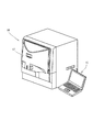
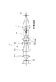
図1は、本発明の一実施の形態に係る細胞分析装置10の斜視説明図である。この細胞分析装置10は、患者から採取した細胞を含む測定試料をフローセルに流し、このフローセルを流れる測定試料にレーザ光を照射し、測定試料からの光(前方散乱光、側方蛍光など)を検出・分析することで、前記細胞に癌・異型細胞が含まれているか否かを判断するのに用いられ、具体的には、子宮頸部の上皮細胞を用いて子宮頸癌をスクリーニングするのに用いられる。細胞分析装置10は、試料の測定などを行う装置本体12と、この装置本体12に接続され、測定結果の分析などを行うシステム制御部13とを備えている。
測定制御部16は、マイクロプロセッサ20、記憶部21、I/Oコントローラ22、センサ信号処理部23、駆動部制御ドライバ24、及び外部通信コントローラ25などを備えている。記憶部21は、ROM、RAMなどからなり、ROMには、駆動部17を制御するための制御プログラム、及び、制御プログラムの実行に必要なデータが格納されている。マイクロプロセッサ20は、制御プログラムをRAMにロードし、又はROMから直接実行することが可能である。
マイクロプロセッサ20が処理したデータや、マイクロプロセッサ20の処理に必要なデータは、外部通信コントローラ25を介してシステム制御部13などの外部の装置との間で送受信される。
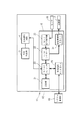
図3は、システム制御部13のブロック図である。システム制御部13は、パーソナルコンピュータなどからなり、本体27と、表示部28と、入力部29とから主に構成されている。本体27は、CPU27aと、ROM27bと、RAM27cと、ハードディスク27dと、読出装置27eと、入出力(I/O)インターフェース27fと、画像出力インターフェース27gと、から主に構成されている。これらの間は、バス27hによって通信可能に接続されている。
また、ハードディスク27dには、細胞分析装置10の測定制御部16への測定オーダ(動作命令)の送信、装置本体12で測定した測定結果の受信及び処理、処理した分析結果の表示などを行う操作プログラムがインストールされている。この操作プログラムは、当該オペレーティングシステム上で動作するものとしている。
画像出力インターフェース27gは、LCD又はCRTなどで構成された表示部28に接続されており、CPU27aから与えられた画像データに応じた映像信号を表示部28に出力するようになっている。表示部28は、入力された映像信号にしたがって、画像(画面)を表示する。
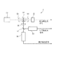
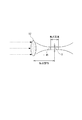
図4は、光学検出部3の構成を示す図である。図において、レンズ系(光学系)52は、光源である半導体レーザ53から放射されたレーザ光を、フローセル51を流れる測定試料に集光し、集光レンズ54は測定試料中の細胞の前方散乱光を散乱光検出器であるフォトダイオード55に集光する。上記レンズ系52は、簡単のために単一のレンズとして図示しているが、より詳細には、図8及び図9に示すように、半導体レーザ53側からコリメータレンズ52a、シリンダーレンズ系(平凸シリンダーレンズ52b+両凹シリンダーレンズ52c)及びコンデンサレンズ系(コンデンサレンズ52d+コンデンサレンズ52e)からなるレンズ群として構成することができる。
なお、光源として、前記半導体レーザに代えてガスレーザを用いることもできるが、低コスト、小型、且つ低消費電力である点で半導体レーザを採用することが好ましく、半導体レーザの採用により製品コストを低減させるとともに、装置の小型化及び省電力化を図ることができる。本実施の形態では、ビームを狭く絞ることに有利な波長の短い青色半導体レーザを用いている。青色半導体レーザは、PI等の蛍光励起波長に対しても有効である。なお、半導体レーザのうち、低コスト且つ長寿命であり、メーカーからの供給が安定している赤色半導体レーザを用いてもよい。
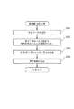
つぎに、細胞分析装置10(図1)を用いた細胞分析方法の実施の形態について説明する。
FSCW/SFLW≦T・・・(1)
SFLW≧M・・・(2)
SFLI≧N・・・(3)
4 信号処理回路
10 細胞分析装置
12 装置本体
13 システム制御部
16 測定制御部
27 本体
28 表示部
29 入力部
51 フローセル
52 レンズ系
53 半導体レーザ
54 集光レンズ
55 フォトダイオード
56 集光レンズ
57 ダイクロイックミラー
58 フォトマルチプライヤ
59 フォトマルチプライヤ
Claims (11)
- 子宮頸部から採取した上皮細胞を分析するための細胞分析装置であって、
核が染色された前記上皮細胞を含む測定試料を流すフローセルと、
このフローセルを流れる測定試料に光を照射する光源部と、
前記測定試料の流れ方向の径が4〜7μmであり、当該測定試料の流れに直交する方向の径が300〜600μmであるビームスポットを、前記フローセルを流れる測定試料上に形成し得る光学系と、
前記測定試料からの光を受光し、前記フローセルを流れる測定試料からの散乱光を検出する散乱光検出器および前記フローセルを流れる測定試料からの蛍光を検出する蛍光検出器を有する受光部と、
前記散乱光検出器から出力される散乱光信号から前記上皮細胞の大きさを反映した値を取得するとともに、前記蛍光検出器から出力される蛍光信号から前記上皮細胞の核の大きさを反映した値を取得する信号処理部と
を備え、
前記信号処理部が、前記上皮細胞の大きさを反映した値として、散乱光信号のパルス幅を取得するとともに、前記上皮細胞の核の大きさを反映した値として、蛍光信号のパルス幅を取得するように構成されていることを特徴とする細胞分析装置。 - 前記信号処理部で得られた、前記上皮細胞の大きさを反映した値及び前記上皮細胞の核の大きさを反映した値に基づいて、前記上皮細胞が異常であるか否かを判定する分析部をさらに備えている請求項1に記載の細胞分析装置。
- 前記分析部が、前記散乱光信号のパルス幅および前記蛍光信号のパルス幅の比を取得するように構成されている請求項2に記載の細胞分析装置。
- 前記信号処理部は、前記蛍光検出器から出力された蛍光信号から、前記上皮細胞の核のDNA量を反映した値を取得し、
前記信号処理部で得られた、前記上皮細胞の核の大きさを反映した値及び前記上皮細胞の核のDNA量を反映した値に基づいて、前記上皮細胞が異常であるか否かを判定する分析部をさらに備えている請求項1に記載の細胞分析装置。 - 測定試料の流れ方向に絞られた光の焦点深度が20〜110μmとなるようにビームスポットが形成される請求項1〜4のいずれか一項に記載の細胞分析装置。
- 前記信号処理部は、前記蛍光検出器から出力された蛍光信号から、前記上皮細胞の核のDNA量を反映した値を取得し、
前記信号処理部で得られた、前記上皮細胞の核のDNA量とその出現頻度の分布を指標とし、前記上皮細胞が異常であるか否かを判定する分析部をさらに備えている請求項1に記載の細胞分析装置。 - 子宮頸部から採取され、核が染色された上皮細胞を含む測定試料をフローセルに流し、前記測定試料に光を照射して、当該測定試料からの光を利用して前記上皮細胞を分析する細胞分析方法であって、
前記測定試料の流れ方向の径が4〜7μmであり、当該測定試料の流れに直交する方向の径が300〜600μmであるビームスポットを、前記フローセルを流れる測定試料上に形成し、前記測定試料から散乱光及び蛍光を検出する検出工程と、
前記散乱光から生成された散乱光信号から前記上皮細胞の大きさを反映した値を取得するとともに、前記蛍光から生成された蛍光信号から前記上皮細胞の核の大きさを反映した値を取得する取得工程と、を備え、
前記上皮細胞の大きさを反映した値として、散乱光信号のパルス幅を取得するとともに、前記上皮細胞の核の大きさを反映した値として、蛍光信号のパルス幅を取得することを特徴とする細胞分析方法。 - 前記上皮細胞の大きさを反映した値及び前記上皮細胞の核の大きさを反映した値に基づいて、前記上皮細胞が異常であるか否かを判定する判定工程を含む請求項7に記載の細胞分析方法。
- 前記判定工程において、前記散乱光信号のパルス幅および前記蛍光信号のパルス幅の比に基づいて判定を行う請求項8に記載の細胞分析方法。
- 前記取得工程において、前記蛍光から生成された蛍光信号から前記上皮細胞の核のDNA量を反映した値を取得し、
前記上皮細胞の核の大きさを反映した値及び前記上皮細胞の核のDNA量を反映した値に基づいて、前記上皮細胞が異常であるか否かを判定する判定工程を含む請求項7に記載の細胞分析方法。 - 前記取得工程において、前記蛍光から生成された蛍光信号から前記上皮細胞の核のDNA量を反映した値を取得し、
前記信号処理部で得られた、前記上皮細胞の核のDNA量とその出現頻度の分布を指標とし、前記上皮細胞が異常であるか否かを判定する判定工程を含む請求項7に記載の細胞分析方法。
Priority Applications (4)
| Application Number | Priority Date | Filing Date | Title |
|---|---|---|---|
| JP2008222537A JP5259305B2 (ja) | 2007-10-03 | 2008-08-29 | 細胞分析装置及び細胞分析方法 |
| CN200810168323.6A CN101403739B (zh) | 2007-10-03 | 2008-09-26 | 细胞分析仪及细胞分析方法 |
| US12/286,702 US7800742B2 (en) | 2007-10-03 | 2008-10-01 | Cell analyzer and cell analyzing method |
| EP08017313.1A EP2045595B1 (en) | 2007-10-03 | 2008-10-01 | Cell analyser and cell analysing method |
Applications Claiming Priority (3)
| Application Number | Priority Date | Filing Date | Title |
|---|---|---|---|
| JP2007259777 | 2007-10-03 | ||
| JP2007259777 | 2007-10-03 | ||
| JP2008222537A JP5259305B2 (ja) | 2007-10-03 | 2008-08-29 | 細胞分析装置及び細胞分析方法 |
Publications (3)
| Publication Number | Publication Date |
|---|---|
| JP2009103687A JP2009103687A (ja) | 2009-05-14 |
| JP2009103687A5 JP2009103687A5 (ja) | 2011-09-29 |
| JP5259305B2 true JP5259305B2 (ja) | 2013-08-07 |
Family
ID=40537815
Family Applications (1)
| Application Number | Title | Priority Date | Filing Date |
|---|---|---|---|
| JP2008222537A Expired - Fee Related JP5259305B2 (ja) | 2007-10-03 | 2008-08-29 | 細胞分析装置及び細胞分析方法 |
Country Status (2)
| Country | Link |
|---|---|
| JP (1) | JP5259305B2 (ja) |
| CN (1) | CN101403739B (ja) |
Families Citing this family (24)
| Publication number | Priority date | Publication date | Assignee | Title |
|---|---|---|---|---|
| WO2010143572A1 (ja) * | 2009-06-12 | 2010-12-16 | オリンパス株式会社 | 被検体情報分析装置及び被検体情報分析方法 |
| US8610085B2 (en) * | 2009-08-20 | 2013-12-17 | Bio-Rad Laboratories, Inc. | High-speed cellular cross sectional imaging |
| JP2012047464A (ja) * | 2010-08-24 | 2012-03-08 | Sony Corp | 微小粒子測定装置及び光軸補正方法 |
| JP5693894B2 (ja) | 2010-08-26 | 2015-04-01 | 日本光電工業株式会社 | 細胞解析装置及び細胞解析方法 |
| JP2013024629A (ja) * | 2011-07-19 | 2013-02-04 | Sysmex Corp | フローサイトメータ |
| JP5138801B2 (ja) * | 2011-07-22 | 2013-02-06 | シスメックス株式会社 | 癌化情報提供方法および装置 |
| JP5876359B2 (ja) * | 2012-03-30 | 2016-03-02 | シスメックス株式会社 | 癌化情報提供方法および癌化情報提供装置 |
| JP5699118B2 (ja) * | 2012-11-14 | 2015-04-08 | シスメックス株式会社 | 細胞分析装置、癌化情報提供方法、dna量ヒストグラムの作成方法および検査方法 |
| JP6001425B2 (ja) * | 2012-11-26 | 2016-10-05 | シスメックス株式会社 | 血球分析方法、血球分析装置およびプログラム |
| JP5892967B2 (ja) | 2013-03-29 | 2016-03-23 | シスメックス株式会社 | 細胞分析装置、細胞測定装置の管理方法およびコンピュータプログラム |
| CN104297213B (zh) * | 2013-07-16 | 2017-10-03 | 成都深迈瑞医疗电子技术研究院有限公司 | 血液细胞分析仪及其异常细胞的识别方法与系统 |
| JP6378599B2 (ja) * | 2013-10-11 | 2018-08-22 | シスメックス株式会社 | 細胞分析方法および細胞分析装置 |
| JP6116502B2 (ja) * | 2014-02-28 | 2017-04-19 | シスメックス株式会社 | 検体分析装置および検体分析方法 |
| RU2016131016A (ru) * | 2014-02-28 | 2018-03-29 | Лайф Текнолоджиз Корпорейшн | Системы, способы и устройства для оптических систем в проточных цитометрах |
| JP6238856B2 (ja) * | 2014-08-25 | 2017-11-29 | シスメックス株式会社 | 尿中異型細胞の分析方法、尿分析装置および体液中異型細胞の分析方法 |
| CN115655825A (zh) * | 2015-09-18 | 2023-01-31 | 紫荆实验室股份有限公司 | 利用表面附着结构的流动池,以及相关的系统和方法 |
| JP6318184B2 (ja) * | 2016-01-20 | 2018-04-25 | シスメックス株式会社 | 細胞分析方法および細胞分析装置 |
| JP6830968B2 (ja) * | 2016-06-07 | 2021-02-17 | エッセン インストゥルメンツ,インコーポレイテッド ディー/ビー/エー エッセン バイオサイエンス,インコーポレイテッド | フローサイトメトリ散乱波形分析を用いてサンプル間の気泡を検出するための方法 |
| JP6249049B2 (ja) * | 2016-06-14 | 2017-12-20 | ソニー株式会社 | 微小粒子測定装置 |
| ES2919343T3 (es) * | 2017-05-25 | 2022-07-26 | Abbott Lab | Métodos y sistemas para el análisis de muestras |
| CN111936905B (zh) | 2018-03-30 | 2023-01-06 | Idexx实验室公司 | 流式细胞仪的激光光学组件 |
| JP7368161B2 (ja) * | 2019-09-30 | 2023-10-24 | シスメックス株式会社 | 送液監視方法および検体測定装置 |
| WO2021207894A1 (zh) * | 2020-04-13 | 2021-10-21 | 深圳迈瑞生物医疗电子股份有限公司 | 细胞分析方法、细胞分析仪及计算机可读存储介质 |
| CN115015178A (zh) * | 2022-08-05 | 2022-09-06 | 天津迈科隆生物科技有限公司 | 一种光学检测装置及血液分析仪 |
Family Cites Families (5)
| Publication number | Priority date | Publication date | Assignee | Title |
|---|---|---|---|---|
| JP3290786B2 (ja) * | 1993-11-26 | 2002-06-10 | シスメックス株式会社 | 粒子分析装置 |
| TW438973B (en) * | 1995-12-19 | 2001-06-07 | Sysmex Corp | Apparatus and method for analyzing solid components in urine |
| JP3815838B2 (ja) * | 1997-03-13 | 2006-08-30 | シスメックス株式会社 | 粒子測定装置 |
| JP4554810B2 (ja) * | 2000-12-21 | 2010-09-29 | シスメックス株式会社 | 粒子分析装置および粒子分析方法 |
| WO2006103920A1 (ja) * | 2005-03-29 | 2006-10-05 | Sysmex Corporation | 癌・異型細胞および凝集粒子を弁別する方法および細胞分析装置 |
-
2008
- 2008-08-29 JP JP2008222537A patent/JP5259305B2/ja not_active Expired - Fee Related
- 2008-09-26 CN CN200810168323.6A patent/CN101403739B/zh not_active Expired - Fee Related
Also Published As
| Publication number | Publication date |
|---|---|
| JP2009103687A (ja) | 2009-05-14 |
| CN101403739B (zh) | 2014-08-06 |
| CN101403739A (zh) | 2009-04-08 |
Similar Documents
| Publication | Publication Date | Title |
|---|---|---|
| JP5259305B2 (ja) | 細胞分析装置及び細胞分析方法 | |
| US7800742B2 (en) | Cell analyzer and cell analyzing method | |
| JP5739959B2 (ja) | 細胞分析装置及び細胞分析方法 | |
| JP5537347B2 (ja) | 粒子分析装置 | |
| US10208352B2 (en) | Providing cervical or oral mucosa canceration information from measurement of cells located toward the basal membrane side of epithelial tissue | |
| JP2010085194A (ja) | 試料分析装置 | |
| EP2784480A2 (en) | Blood cell analyzer and blood cell analyzing method | |
| US20110104744A1 (en) | Cell analyzing apparatus and cell analyzing method | |
| US9354161B2 (en) | Sample analyzing method and sample analyzer | |
| EP2843409B1 (en) | Urine sample analyzing method and sample analyzer | |
| JP5699118B2 (ja) | 細胞分析装置、癌化情報提供方法、dna量ヒストグラムの作成方法および検査方法 | |
| JP2008241644A (ja) | 粒子分析装置 |
Legal Events
| Date | Code | Title | Description |
|---|---|---|---|
| A521 | Request for written amendment filed |
Free format text: JAPANESE INTERMEDIATE CODE: A523 Effective date: 20110817 |
|
| A621 | Written request for application examination |
Free format text: JAPANESE INTERMEDIATE CODE: A621 Effective date: 20110817 |
|
| A977 | Report on retrieval |
Free format text: JAPANESE INTERMEDIATE CODE: A971007 Effective date: 20120704 |
|
| A131 | Notification of reasons for refusal |
Free format text: JAPANESE INTERMEDIATE CODE: A131 Effective date: 20120710 |
|
| A521 | Request for written amendment filed |
Free format text: JAPANESE INTERMEDIATE CODE: A523 Effective date: 20120904 |
|
| A02 | Decision of refusal |
Free format text: JAPANESE INTERMEDIATE CODE: A02 Effective date: 20121016 |
|
| A521 | Request for written amendment filed |
Free format text: JAPANESE INTERMEDIATE CODE: A523 Effective date: 20130116 |
|
| A911 | Transfer to examiner for re-examination before appeal (zenchi) |
Free format text: JAPANESE INTERMEDIATE CODE: A911 Effective date: 20130305 |
|
| TRDD | Decision of grant or rejection written | ||
| A01 | Written decision to grant a patent or to grant a registration (utility model) |
Free format text: JAPANESE INTERMEDIATE CODE: A01 Effective date: 20130402 |
|
| A61 | First payment of annual fees (during grant procedure) |
Free format text: JAPANESE INTERMEDIATE CODE: A61 Effective date: 20130424 |
|
| FPAY | Renewal fee payment (event date is renewal date of database) |
Free format text: PAYMENT UNTIL: 20160502 Year of fee payment: 3 |
|
| R150 | Certificate of patent or registration of utility model |
Ref document number: 5259305 Country of ref document: JP Free format text: JAPANESE INTERMEDIATE CODE: R150 Free format text: JAPANESE INTERMEDIATE CODE: R150 |
|
| R250 | Receipt of annual fees |
Free format text: JAPANESE INTERMEDIATE CODE: R250 |
|
| R250 | Receipt of annual fees |
Free format text: JAPANESE INTERMEDIATE CODE: R250 |
|
| R250 | Receipt of annual fees |
Free format text: JAPANESE INTERMEDIATE CODE: R250 |
|
| R250 | Receipt of annual fees |
Free format text: JAPANESE INTERMEDIATE CODE: R250 |
|
| R250 | Receipt of annual fees |
Free format text: JAPANESE INTERMEDIATE CODE: R250 |
|
| LAPS | Cancellation because of no payment of annual fees |