JP4454474B2 - 医用画像診断支援装置 - Google Patents
医用画像診断支援装置 Download PDFInfo
- Publication number
- JP4454474B2 JP4454474B2 JP2004333069A JP2004333069A JP4454474B2 JP 4454474 B2 JP4454474 B2 JP 4454474B2 JP 2004333069 A JP2004333069 A JP 2004333069A JP 2004333069 A JP2004333069 A JP 2004333069A JP 4454474 B2 JP4454474 B2 JP 4454474B2
- Authority
- JP
- Japan
- Prior art keywords
- organ
- shape
- integrated
- region
- subject
- Prior art date
- Legal status (The legal status is an assumption and is not a legal conclusion. Google has not performed a legal analysis and makes no representation as to the accuracy of the status listed.)
- Expired - Fee Related
Links
Images


Landscapes
- Nuclear Medicine (AREA)
- Image Processing (AREA)
- Image Analysis (AREA)
Description
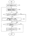
最初のRI集積領域を指定する(S21)。RI集積領域は相対的なもので、周囲より高濃度の領域である。例えば肝臓の場合、肝臓全体としてのRI集積領域があり、さらに肝臓の一部に他より高集積の領域がある場合は、この一部の領域もあと一つのRI集積領域として扱う。
最初の臓器(N=1)を指定する(S22)。
画像中、比較的高濃度であるRI集積領域の形と臓器形(N)とが相似するか否かを判定する(S23)。
RI集積領域に対応するメモリ領域に“病巣領域”の記録をする。又は、モニタ15に表示した計測画像(PET像)において、図4のように異常陰影候補がある部位にマーク40を付ける(S24)。
準備した全ての臓器について調べたか否かの判定をする(S25)。
次の臓器(N)を指定する(S26)。そしてステップS23に戻り、上記と同様の処理を行なう。
全てのRI集積領域について調べたか否かを判定する(S27)。
次の集積領域を指定する。そしてステップS23へ戻り、RI集積領域の形と臓器形(N)とが相似しているか否かを判定する(S28)。
ステップS21と同様、最初のRI集積領域を指定する(S51)。
RI集積領域における集積度Gの時間依存F(t)を求める。
ステップS52で求めたRI集積領域における集積度Gの時間依存F(t)に近似的に一致するガンでないことが既知の集積度の時間依存パターンが存在するか否かを判定する(S53)。
ステップS53において一致する正常パターンがなかったRI集積領域は、異常陰影候補として検出し、記録又は、図4に示すように計測画像にマーク40をつける(S54)。
全てのRI集積領域について調べたか否かを判定する(S55)。
次のRI集積領域を指定する。そしてステップS52へ戻る。
Claims (5)
- PET装置で計測したPETデータを取得するPETデータ取得手段と、
被検体の各臓器の形状を格納する臓器形状格納手段と、
前記PETデータに含まれ、周囲より高濃度の領域であるRI集積領域を抽出するRI集積領域抽出手段と、
前記臓器形状格納手段に格納され、かつ前記被検体におけるRI集積領域が位置する部位の各臓器の形状と、前記抽出されたRI集積領域の形状と、を用い、前記各臓器の体積と前記RI集積領域の体積とを規格化し、当該規格化された前記各臓器の体積及び前記RI集積領域の体積の重心から辺縁までの距離の角度依存曲線が同じ形であるかに基づいて、前記各臓器の形状と前記RI集積領域の形状との相関関係の有無を判定する手段と、
前記相関関係がないと判定された前記RI集積領域を異常陰影候補として検出する異常陰影候補検出手段と、
を備えることを特徴とする医用画像診断支援装置。 - PET装置で計測したPETデータを取得するPETデータ取得手段と、
被検体の各臓器の形状を格納する臓器形状格納手段と、
前記PETデータに含まれ、周囲より高濃度の領域であるRI集積領域を抽出するRI集積領域抽出手段と、
前記臓器形状格納手段に格納され、かつ前記被検体におけるRI集積領域が位置する部位の各臓器の形状と、前記抽出されたRI集積領域の形状と、に基づいて、前記被検体の全身に対する前記RI集積領域の体積の割合と、前記被検体の全身に対する前記各臓器の体積の割合と、が近似的に等しいか否かを判定する手段と、
前記RI集積領域の体積の割合が、前記判定により近似的に等しくないと判定された前記RI集積領域を異常陰影候補として検出する異常陰影候補検出手段と、
を備えることを特徴とする医用画像診断支援装置。 - 前記臓器形状格納手段は、医用画像撮影装置により得られた前記被検体の撮影データに基づいて抽出した前記被検体の各臓器の形状を格納する、
ことを特徴とする請求項1又は2に記載の医用画像診断支援装置。 - 前記臓器形状格納手段は、前記被検体の各臓器の形状に代えて、解剖図、人体モデル又は臓器モデルの少なくとも一つに基づいて抽出した各臓器の標準的な形状を格納する、
ことを特徴とする請求項1又は2に記載の医用画像診断支援装置。 - PET装置で計測したPETデータを取得するPETデータ取得手段と、
前記PETデータに含まれ、周囲より高濃度の領域であるRI集積領域を抽出するRI集積領域抽出手段と、
前記RI集積領域抽出手段で抽出されたRI集積領域の、RI集積度の時間依存を示す被検体RI集積データを取得する被検体RI集積データ取得手段と、
予め調べた、薬剤種毎の病巣領域における前記各薬剤注入後から所定時間経過後のRI集積度の時間依存を示す標準RI集積データ、又は予め調べた、薬剤種毎の非病巣領域における前記各薬剤注入後から所定時間経過後のRI集積度の時間依存を示す標準RI集積データのうちの少なくとも一つを格納する標準RI集積データ格納手段と、
前記被検体RI集積データと前記標準RI集積データとを比較し、それらの比較結果に基づいて異常陰影候補を検出する異常陰影候補検出手段と、
を備えることを特徴とする医用画像診断支援装置。
Priority Applications (1)
| Application Number | Priority Date | Filing Date | Title |
|---|---|---|---|
| JP2004333069A JP4454474B2 (ja) | 2004-11-17 | 2004-11-17 | 医用画像診断支援装置 |
Applications Claiming Priority (1)
| Application Number | Priority Date | Filing Date | Title |
|---|---|---|---|
| JP2004333069A JP4454474B2 (ja) | 2004-11-17 | 2004-11-17 | 医用画像診断支援装置 |
Publications (3)
| Publication Number | Publication Date |
|---|---|
| JP2006145281A JP2006145281A (ja) | 2006-06-08 |
| JP2006145281A5 JP2006145281A5 (ja) | 2007-05-24 |
| JP4454474B2 true JP4454474B2 (ja) | 2010-04-21 |
Family
ID=36625161
Family Applications (1)
| Application Number | Title | Priority Date | Filing Date |
|---|---|---|---|
| JP2004333069A Expired - Fee Related JP4454474B2 (ja) | 2004-11-17 | 2004-11-17 | 医用画像診断支援装置 |
Country Status (1)
| Country | Link |
|---|---|
| JP (1) | JP4454474B2 (ja) |
Families Citing this family (13)
| Publication number | Priority date | Publication date | Assignee | Title |
|---|---|---|---|---|
| JP4640143B2 (ja) * | 2005-12-02 | 2011-03-02 | 株式会社島津製作所 | 画像診断支援装置 |
| JP4609298B2 (ja) * | 2005-12-09 | 2011-01-12 | 株式会社島津製作所 | 画像診断支援装置 |
| US8017915B2 (en) | 2008-03-14 | 2011-09-13 | Reflexion Medical, Inc. | Method and apparatus for emission guided radiation therapy |
| US9968309B2 (en) | 2009-12-08 | 2018-05-15 | Koninklijke Philips N.V. | Method and a correction system for correcting tracer-uptake measurements |
| CN110585607B (zh) | 2011-03-31 | 2022-07-19 | 反射医疗公司 | 用于在发射引导的放射治疗中使用的系统和方法 |
| JP6026089B2 (ja) * | 2011-08-23 | 2016-11-16 | 東芝メディカルシステムズ株式会社 | 医用画像診断装置、画像情報表示装置及び制御プログラム |
| CN109152928B (zh) | 2016-03-09 | 2021-05-28 | 反射医疗公司 | 用于计算辐射治疗的注量图的方法和系统 |
| JP6797557B2 (ja) * | 2016-05-17 | 2020-12-09 | キヤノンメディカルシステムズ株式会社 | 医用画像診断装置、医用画像処理装置および画像表示プログラム |
| WO2018093849A1 (en) | 2016-11-15 | 2018-05-24 | Reflexion Medical, Inc. | Methods for radiation delivery in emission-guided radiotherapy |
| JP7201243B2 (ja) | 2016-11-15 | 2023-01-10 | リフレクション メディカル, インコーポレイテッド | 放出誘導型高エネルギー光子送達のためのシステム |
| EP3664712B1 (en) | 2017-08-09 | 2025-03-12 | RefleXion Medical, Inc. | Systems and methods for fault detection in emission-guided radiotherapy |
| WO2022031750A1 (en) | 2020-08-07 | 2022-02-10 | Reflexion Medical, Inc. | Multi-sensor guided radiation therapy |
| JP7761425B2 (ja) * | 2021-08-26 | 2025-10-28 | キヤノンメディカルシステムズ株式会社 | 核医学診断装置、データ処理方法及びプログラム |
-
2004
- 2004-11-17 JP JP2004333069A patent/JP4454474B2/ja not_active Expired - Fee Related
Also Published As
| Publication number | Publication date |
|---|---|
| JP2006145281A (ja) | 2006-06-08 |
Similar Documents
| Publication | Publication Date | Title |
|---|---|---|
| JP5241397B2 (ja) | 患者の陽電子放出断層撮影データのための減弱値を求める方法 | |
| Boellaard | Need for standardization of 18F-FDG PET/CT for treatment response assessments | |
| CN101273919B (zh) | 使用更新方法和系统的连续图像采集 | |
| Lee et al. | Clinical usefulness of 18F-FDG PET-CT for patients with gallbladder cancer and cholangiocarcinoma | |
| US8488857B2 (en) | Automated diagnosis and alignment supplemented with positron emission tomography (PET) and magnetic resonance (MR) flow estimation | |
| JP4571187B2 (ja) | 病気の進行または治療効果を解析するために関心領域を複数の時点にわたってリンクさせるシステムおよび方法 | |
| EP2399238B1 (en) | Functional imaging | |
| JP4454474B2 (ja) | 医用画像診断支援装置 | |
| CN103040479B (zh) | 可能的灌注缺陷的确定 | |
| JP2012518168A (ja) | 核画像化におけるモデルベースの視野拡大 | |
| JP2008503258A (ja) | マルチモード視覚化を使用して病気の進行または治療効果を監視するシステムおよび方法 | |
| Zhang et al. | Cross-modality PET/CT and contrast-enhanced CT imaging for pancreatic cancer | |
| JP4317412B2 (ja) | 画像処理方法 | |
| US20090041318A1 (en) | Method for recording measured data of a patient while taking account of movement operations, and an associated medical device | |
| Sheng et al. | Diffusion kurtosis imaging and diffusion-weighted imaging in assessment of liver fibrosis stage and necroinflammatory activity | |
| US9675311B2 (en) | Follow up image acquisition planning and/or post processing | |
| US8788012B2 (en) | Methods and apparatus for automatically registering lesions between examinations | |
| Delgado Sánchez-Gracián et al. | Quantitative myocardial perfusion with stress dual-energy CT: iodine concentration differences between normal and ischemic or necrotic myocardium. Initial experience | |
| Hu et al. | Diagnostic performance of total-body 18F-FDG PET/CT with fast 2-min acquisition for liver tumours: comparison with conventional PET/CT | |
| CN101006465B (zh) | 横跨时间点链接vois以分析疾病进展或者对治疗的响应的系统和方法 | |
| US9600875B2 (en) | Tissue surface roughness quantification based on image data and determination of a presence of disease based thereon | |
| JP2008503259A (ja) | 病気の進行または治療効果を解析するために複数の時点をロードするシステムおよび方法 | |
| Roy et al. | Enhancing patient care with modality-based image registration in modern healthcare | |
| Yin et al. | Parametric net influx rate imaging of 68Ga-DOTATATE in patients with neuroendocrine tumors: assessment of lesion detectability | |
| Yavuz et al. | Calculation of recovery coefficients for partial volume effect correction in PET/CT imaging using a customized anthropomorphic body phantom |
Legal Events
| Date | Code | Title | Description |
|---|---|---|---|
| A521 | Written amendment |
Free format text: JAPANESE INTERMEDIATE CODE: A523 Effective date: 20070330 |
|
| A621 | Written request for application examination |
Free format text: JAPANESE INTERMEDIATE CODE: A621 Effective date: 20070330 |
|
| RD02 | Notification of acceptance of power of attorney |
Free format text: JAPANESE INTERMEDIATE CODE: A7422 Effective date: 20090717 |
|
| RD04 | Notification of resignation of power of attorney |
Free format text: JAPANESE INTERMEDIATE CODE: A7424 Effective date: 20090721 |
|
| A131 | Notification of reasons for refusal |
Free format text: JAPANESE INTERMEDIATE CODE: A131 Effective date: 20090908 |
|
| A521 | Written amendment |
Free format text: JAPANESE INTERMEDIATE CODE: A523 Effective date: 20091020 |
|
| TRDD | Decision of grant or rejection written | ||
| A01 | Written decision to grant a patent or to grant a registration (utility model) |
Free format text: JAPANESE INTERMEDIATE CODE: A01 Effective date: 20100202 |
|
| A01 | Written decision to grant a patent or to grant a registration (utility model) |
Free format text: JAPANESE INTERMEDIATE CODE: A01 |
|
| A61 | First payment of annual fees (during grant procedure) |
Free format text: JAPANESE INTERMEDIATE CODE: A61 Effective date: 20100202 |
|
| FPAY | Renewal fee payment (event date is renewal date of database) |
Free format text: PAYMENT UNTIL: 20130212 Year of fee payment: 3 |
|
| R150 | Certificate of patent or registration of utility model |
Free format text: JAPANESE INTERMEDIATE CODE: R150 |
|
| FPAY | Renewal fee payment (event date is renewal date of database) |
Free format text: PAYMENT UNTIL: 20140212 Year of fee payment: 4 |
|
| LAPS | Cancellation because of no payment of annual fees |