JP4454474B2 - Medical image diagnosis support device - Google Patents
Medical image diagnosis support device Download PDFInfo
- Publication number
- JP4454474B2 JP4454474B2 JP2004333069A JP2004333069A JP4454474B2 JP 4454474 B2 JP4454474 B2 JP 4454474B2 JP 2004333069 A JP2004333069 A JP 2004333069A JP 2004333069 A JP2004333069 A JP 2004333069A JP 4454474 B2 JP4454474 B2 JP 4454474B2
- Authority
- JP
- Japan
- Prior art keywords
- organ
- shape
- integrated
- region
- subject
- Prior art date
- Legal status (The legal status is an assumption and is not a legal conclusion. Google has not performed a legal analysis and makes no representation as to the accuracy of the status listed.)
- Expired - Fee Related
Links
Images


Description
本発明は、医用画像診断支援装置に係り、特にPET装置で計測したPETデータから異常陰影候補を検出する技術に関する。 The present invention relates to a medical image diagnosis support apparatus, and more particularly to a technique for detecting abnormal shadow candidates from PET data measured by a PET apparatus.
PET(ポジトロン・エミッション・トモグラフィ)によるガン検査は、ガン細胞が正常細胞よりも数倍から多い場合は20倍近く多くのブドウ糖を取りこむ性質を利用した検査である。ラジオアイソトープ(以下「RI」という)の一種であるFDG(フルオロデオキシグルコース)というブドウ糖によく似た構造の薬剤を注射した後、その集積度を画像化して診断を行う。X線CT装置やMRI装置による検査がガンなどの大きさや形を捉えるのに対し、PET検査はガン細胞などの高まった代謝状態を捉える検査であり、数ミリのガン病巣が発見された例もある。 A cancer test by PET (Positron Emission Tomography) is a test that uses the property of taking up nearly 20 times more glucose when cancer cells are several to more than normal cells. After injection of a drug having a structure similar to glucose called FDG (fluorodeoxyglucose), which is a kind of radioisotope (hereinafter referred to as “RI”), diagnosis is performed by imaging the degree of accumulation. Whereas X-ray CT and MRI inspections capture the size and shape of cancer and the like, PET inspections are an inspection of the increased metabolic state of cancer cells and other cases, and several millimeters of cancer lesions have been discovered. is there.
但し、正常でも活発にブドウ糖代謝を行う臓器や、排泄の際の通り道となる腎臓、尿管、膀胱など、PET検査には向かない部位もある。更に、胃ガンや肝臓ガン、進行の遅い肺ガンの中には、その性質や種類によってあまりブドウ糖を取りこまないものもある。 However, there are some parts that are not suitable for PET examinations, such as organs that actively metabolize glucose even when normal, and the kidneys, ureters, and bladder that become the path of excretion. Furthermore, some stomach cancers, liver cancers, and slow-growing lung cancers do not take up much glucose due to their nature and type.
PET検査では、今までの検診では調べにくかった臓器なども含め、広範囲(頚部から大腿部など)を一度に検査することができる。そのため、良性・悪性の判断、転移や再発の把握などにも効果があると言われている。 In the PET examination, it is possible to examine a wide range (such as the neck and the thigh) at once, including organs that have been difficult to examine by conventional examinations. Therefore, it is said that it is also effective in determining benign / malignant and grasping metastasis and recurrence.
特許文献1には、時間を経て測定された二つの放射線分布画像の減衰補正を行ったものの間での比較を行い、変化の大きいデータを除外する処理を行う技術が開示されている。
しかし、特許文献1のように変化の大きいデータを除外する処理を行った場合、使用する薬剤の種類(核種)に依存して変化の様子は異なるので、ガンである可能性がある異常陰影候補まで除外してしまう危険性があった。
However, when the process of excluding data with a large change as in
本発明は、上記問題に鑑みてなされたものであり、使用する薬剤の種類に依存せずにPET画像から異常陰影候補を検出する医用画像診断支援装置を提供することを目的とする。 The present invention has been made in view of the above problems, and an object of the present invention is to provide a medical image diagnosis support apparatus that detects an abnormal shadow candidate from a PET image without depending on the type of medicine to be used.
前記目的を達成するために本発明は、PET装置で計測したPETデータを取得するPETデータ取得手段と、被検体の各臓器の形状を格納する臓器形状格納手段と、前記PETデータに含まれるRI集積領域を抽出するRI集積領域抽出手段と、前記被検体におけるRI集積領域が位置する部位の各臓器の形状と、前記RI集積領域の形状と、を比較して形状の相似度を算出する相似度算出手段と、前記形状の相似度が所定値よりも低いRI集積領域を異常陰影候補として検出する異常陰影候補検出手段と、を備える。 In order to achieve the above object, the present invention provides a PET data acquisition unit that acquires PET data measured by a PET apparatus, an organ shape storage unit that stores the shape of each organ of a subject, and an RI included in the PET data. Similarity for calculating the similarity of the shape by comparing the RI integrated region extracting means for extracting the integrated region, the shape of each organ in the region where the RI integrated region is located in the subject, and the shape of the RI integrated region Degree calculation means, and abnormal shadow candidate detection means for detecting, as an abnormal shadow candidate, an RI accumulated area having a shape similarity lower than a predetermined value.
また、前記臓器形状格納手段は、医用画像撮影装置により得られた被検体の撮影データに基づいて抽出した前記被検体の各臓器の形状を格納する。 The organ shape storage means stores the shape of each organ of the subject extracted based on the subject's imaging data obtained by the medical imaging device.
また、前記臓器形状格納手段は、解剖図、人体モデル又は臓器モデルの少なくとも一つに基づいて抽出した各臓器の標準的な形状を格納する。 The organ shape storage means stores a standard shape of each organ extracted based on at least one of an anatomical chart, a human body model, or an organ model.
また、PET装置で経時的に計測したPETデータに基づいて生成された被検体におけるRI集積量の経時的変化を示す被検体RI集積データを取得する被検体RI集積データ取得手段と、病巣領域におけるRI集積量の標準的な経時的変化を示す標準RI集積データ又は非病巣領域におけるRI集積量の標準的な経時的変化を示す標準RI集積データのうちの少なくとも一つを格納する標準RI集積データ格納手段と、前記被検体RI集積データと前記標準RI集積データとを比較し、それらの比較結果に基づいて異常陰影候補を検出する異常陰影候補検出手段と、を備える。 In addition, subject RI integrated data acquisition means for acquiring subject RI integrated data indicating temporal changes in the RI integrated amount in the subject generated based on PET data measured over time by a PET apparatus, and in a lesion region Standard RI integrated data for storing at least one of standard RI integrated data indicating a standard time-dependent change of the RI integrated amount or standard RI integrated data indicating a standard time-dependent change of the RI integrated amount in a non-lesion area. Storage means, and abnormal shadow candidate detection means for comparing the subject RI accumulated data and the standard RI accumulated data and detecting an abnormal shadow candidate based on the comparison result.
ここで「被検体RI集積データ取得手段」は、被検体におけるRI集積量の経時的変化を計測した結果を取得する手段のほか、PET装置から経時的に計測したPETデータを取得し、医用画像診断支援装置がそのPETデータに基づいて被検体におけるRI集積量の経時的変化を計測し、その計測結果を出力する手段も含む。 Here, the “subject RI accumulated data obtaining means” obtains PET data measured over time from a PET apparatus, in addition to means for obtaining a result of measuring a temporal change in the amount of RI accumulated in the subject, and obtains a medical image. The diagnosis support apparatus also includes means for measuring a temporal change in the RI accumulation amount in the subject based on the PET data and outputting the measurement result.
本発明によれば、臓器の形状とRI集積領域との形状を比較した結果、又は病巣領域(ガン細胞)又は非病巣領域(非ガン細胞)における経時的なRI集積変化と被検体のRI集積データとを比較した結果に基づいて異常陰影候補を検出する。そのため、使用する核種に依存せずにPET画像から異常陰影候補を検出する医用画像診断支援装置を提供することができる。 According to the present invention, as a result of comparing the shape of the organ and the shape of the RI accumulation region, or the change in RI accumulation over time in the lesion region (cancer cell) or non-lesion region (non-cancer cell) and the RI accumulation of the subject. An abnormal shadow candidate is detected based on the result of comparison with the data. Therefore, it is possible to provide a medical image diagnosis support apparatus that detects an abnormal shadow candidate from a PET image without depending on the nuclide to be used.
以下、添付図面に従って本発明に係る医用画像診断支援装置の好ましい実施の形態について詳説する。なお、発明の実施の形態を説明するための全図において、同一機能を有するものは同一符号を付け、その繰り返しの説明は省略する。 Hereinafter, preferred embodiments of a medical image diagnosis support apparatus according to the present invention will be described in detail with reference to the accompanying drawings. Note that components having the same function are denoted by the same reference symbols throughout the drawings for describing the embodiment of the invention, and the repetitive description thereof is omitted.
図1は、発明の一実施の形態に係る医用画像診断支援システムの構成を示すハードウェア構成図である。 FIG. 1 is a hardware configuration diagram showing the configuration of a medical image diagnosis support system according to an embodiment of the invention.
図1の医用画像診断支援処理システム1は、PET装置2と、PET装置2が計測して得たPET画像等を格納するデータベース3と、被検体を撮影して画像データを生成する医用画像撮影装置5と、PET画像から異常陰影候補を検出する医用画像診断支援装置10とを備え、PET装置2とデータベース3と医用画像撮影装置5と医用画像診断支援装置10とは、LAN4等のネットワークにより互いに接続される。
A medical image diagnosis
医用画像撮影装置5は、X線CT装置、MR装置など、被検体の断層像を撮影可能な装置により構成される。 The medical image capturing apparatus 5 is configured by an apparatus capable of capturing a tomographic image of a subject such as an X-ray CT apparatus and an MR apparatus.
医用画像診断支援装置10は、主として各構成要素の動作を制御する中央処理装置(CPU)11と、装置の制御プログラムが格納されたり、プログラム実行時の作業領域となったりする主メモリ12と、オペレーティングシステム(OS)、周辺機器のデバイスドライブ、後述するPET画像から異常陰影候補を検出する処理等を行うためのプログラムを含む各種アプリケーションソフト等が格納される磁気ディスク13と、表示用データを一時記憶する表示メモリ14と、この表示メモリ14からのデータに基づいて画像を表示するCRTモニタや液晶モニタ等のモニタ15と、キーボード16と、位置入力装置としてのマウス17と、マウス17の状態を検出してモニタ15上のマウスポインタの位置やマウス17の状態等の信号をCPU11に出力するコントローラ18と、上記各構成要素を接続するバス19とを備える。
The medical image
データベース3には、PET装置2で計測して得られたPET画像と、医用画像撮影装置5で撮影した被検体の画像、又、解剖図、人体又は臓器の標本モデルのいずれか一つに基づいて抽出した各臓器の形状と、病巣領域におけるRI集積量の標準的な経時的変化を示す標準RI集積データ又は非病巣領域における集積量の標準的な経時的変化を示す標準RI集積データのうちの少なくとも一つとが格納される。
The
本実施の形態においては、医用画像診断支援装置10は、LAN4を介してデータベース3からPET画像を読み出すが、医用画像診断支援装置10に接続された記憶装置、例えばFDD、CD−RWドライブ、MOドライブ、ZIPドライブから画像データを読み込んでも良い。また、LAN4を経由してPET装置2から直接PET画像を取得してもよい。
In the present embodiment, the medical image diagnosis support
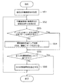
次に、図2乃至4に基づいて、本実施の形態に係る医用画像診断支援装置10を用いてPET画像から異常陰影候補を検出する手順について説明する。図2は、第一の実施の形態を説明するためのフローチャートである。図3は、図2の処理において被検体の断層像から各臓器を抽出する処理を示す模式図、図4は、PET像に含まれる異常陰影候補にマークをつけて表示した画面表示例を示す模式図である。以下図2の各ステップ順に説明する。
Next, a procedure for detecting an abnormal shadow candidate from a PET image using the medical image diagnosis support
(ステップS21)
最初のRI集積領域を指定する(S21)。RI集積領域は相対的なもので、周囲より高濃度の領域である。例えば肝臓の場合、肝臓全体としてのRI集積領域があり、さらに肝臓の一部に他より高集積の領域がある場合は、この一部の領域もあと一つのRI集積領域として扱う。
(Step S21)
The first RI accumulation area is designated (S21). The RI integrated region is a relative region and is a region having a higher concentration than the surroundings. For example, in the case of the liver, there is an RI accumulation region as a whole of the liver, and when there is a region of higher accumulation in another part of the liver, this part of the region is also treated as another RI accumulation region.
(ステップS22)
最初の臓器(N=1)を指定する(S22)。
(Step S22)
The first organ (N = 1) is designated (S22).
臓器の形は、データベース3又は磁気ディスク13に予め記録しておく。臓器の形は、図3に示すように医用画像撮影装置5により被検体の断層像30を撮影しておき、この断層像30から抽出してもよい。または、一般的な解剖図や標準的な人体モデル又は臓器モデルから臓器の形を抽出してもよい。
The shape of the organ is recorded in advance in the
(ステップS23)
画像中、比較的高濃度であるRI集積領域の形と臓器形(N)とが相似するか否かを判定する(S23)。
(Step S23)
It is determined whether or not the shape of the RI integrated region having a relatively high density in the image is similar to the organ shape (N) (S23).
相似には、形が近似的に等しい場合として、a)体積を規格化した後での、重心から辺縁までの距離の角度依存曲線が同じ形である場合と、b)面積を規格化した規格二値画像と二値化した計測画像との比較的高濃度領域との差分値が一定値より小さい場合とがある。 For similarity, assuming that the shapes are approximately equal, a) the angle-dependent curve of the distance from the center of gravity to the edge after normalizing the volume is the same shape, and b) the area is normalized In some cases, a difference value between a standard binary image and a binarized measurement image and a relatively high density region is smaller than a certain value.
また、相似には、割合が近似的に等しい場合として、全身に対する臓器体積の割合が近い場合等がある。 Similarity may be the case where the ratio of the organ volume to the whole body is close, for example, when the ratio is approximately equal.
RI集積領域の形と臓器形(N)とに所定の相関関係が認められない場合には、異常陰影候補であると判定してステップS24へ進む。RI集積領域と臓器形(N)とに所定の相関関係が認められた場合には、異常陰影候補ではないと判定してステップS25へ進む。 When a predetermined correlation is not recognized between the shape of the RI accumulation region and the organ shape (N), it is determined that the candidate is an abnormal shadow candidate, and the process proceeds to step S24. If a predetermined correlation is recognized between the RI accumulation area and the organ shape (N), it is determined that the RI shadow area is not an abnormal shadow candidate, and the process proceeds to step S25.
(ステップS24)
RI集積領域に対応するメモリ領域に“病巣領域”の記録をする。又は、モニタ15に表示した計測画像(PET像)において、図4のように異常陰影候補がある部位にマーク40を付ける(S24)。
(Step S24)
The “lesion area” is recorded in the memory area corresponding to the RI integration area. Alternatively, in the measurement image (PET image) displayed on the
(ステップS25)
準備した全ての臓器について調べたか否かの判定をする(S25)。
(Step S25)
It is determined whether all prepared organs have been examined (S25).
全ての臓器について調べていない場合はステップS26へ進む。全ての臓器について調べた場合はステップS27へ進む。 If all the organs have not been examined, the process proceeds to step S26. If all the organs have been examined, the process proceeds to step S27.
(ステップS26)
次の臓器(N)を指定する(S26)。そしてステップS23に戻り、上記と同様の処理を行なう。
(Step S26)
The next organ (N) is designated (S26). Then, the process returns to step S23 and the same processing as described above is performed.
(ステップS27)
全てのRI集積領域について調べたか否かを判定する(S27)。
(Step S27)
It is determined whether or not all the RI integrated regions have been examined (S27).
全てのRI集積領域について調べた場合には、異常陰影候補検出処理を終了する。まだ調べていない集積領域がある場合にはステップS28へ進む。 When all the RI accumulation regions have been examined, the abnormal shadow candidate detection process ends. If there is an accumulation area that has not been examined yet, the process proceeds to step S28.
(ステップS28)
次の集積領域を指定する。そしてステップS23へ戻り、RI集積領域の形と臓器形(N)とが相似しているか否かを判定する(S28)。
(Step S28)
Specify the next accumulation area. Then, the process returns to step S23, and it is determined whether or not the shape of the RI accumulation region and the organ shape (N) are similar (S28).
次に図5乃至9に基づいて医用画像診断支援装置10の第二の実施の形態について説明する。図5は、図1に示した医用画像診断支援装置10を使ってPET画像から異常陰影候補を抽出する第二の実施の形態を示すフローチャート、図6は、集積度Gの時間依存F(t)を格納した状態を示す模式図、図7は、薬剤種別に臓器の流出半減期を格納した状態を示す模式図、図8は、計測画像と標準半透明画像(投影像、MIP画像、3D画像)とを同期回転表示した画面表示例を示す模式図、図9は、図8で表示する標準半透明画像を生成する処理を示す模式図である。以下図5の各ステップ順に沿って説明する。
Next, a second embodiment of the medical image
(ステップS51)
ステップS21と同様、最初のRI集積領域を指定する(S51)。
(Step S51)
As in step S21, the first RI accumulation region is designated (S51).
(ステップS52)
RI集積領域における集積度Gの時間依存F(t)を求める。
(Step S52)
The time dependence F (t) of the degree of integration G in the RI integration region is obtained.
RI集積領域における集積度Gの時間依存F(t)は、RI集積領域のRI残量の経時的変化を測定することにより求める。集積度Gの時間依存F(t)は、医用画像診断支援装置10が、経時的に計測したPETデータに基づいて算出する。
The time dependence F (t) of the integration degree G in the RI integrated region is obtained by measuring the change over time of the remaining amount of RI in the RI integrated region. The time dependence F (t) of the integration degree G is calculated based on the PET data measured by the medical image
(ステップS53)
ステップS52で求めたRI集積領域における集積度Gの時間依存F(t)に近似的に一致するガンでないことが既知の集積度の時間依存パターンが存在するか否かを判定する(S53)。
(Step S53)
It is determined whether or not there is a time-dependent pattern of the integration degree that is known to be not a cancer that approximately matches the time dependence F (t) of the integration degree G in the RI integration area obtained in step S52 (S53).
ガンでないことが既知の集積度Gの時間依存F(t)は、図6に示すように、薬剤種毎に各臓器における薬剤注入後から所定時間tn経過後の集積度dnを予め調べておき、データベース3や磁気デイスク13などに記録しておく。図6に例示した時間依存と集積領域における集積度Gとを用いて判定することにより、集積度Gの変化を時間を追って観察することができる。
As shown in FIG. 6, the time dependence F (t) of the accumulation degree G that is known not to be cancer is obtained by checking in advance the accumulation degree dn after a predetermined time tn from the injection of the drug in each organ for each drug type. The data is recorded in the
また、一般に、被検体における病巣領域のうち、炎症部位とガン部位とは集積度Gの経時変化が異なるため、集積度Gの経時変化を観察することにより炎症部位とガン部位とを区別することができる。 In general, among the lesion areas in the subject, the inflammatory site and the cancer site differ in the accumulation degree G over time, so that the inflammatory site and the cancer site are distinguished by observing the temporal change in the accumulation degree G. Can do.
また、図7のように、薬剤種毎に、注入量、注入方法、薬剤種毎に決定される放射性半減期、臓器、臓器毎の排泄に伴う流出による半減期(以下「流出半減期」という)を対応付けて、予めデータベース3や磁気デイスク13などに記録しておく。図7に例示した集積度Gの時間依存F(t)とを用いて判定することにより、RI集積領域における集積度Gが最も高い時刻から、集積度Gが半減するまでの時刻と、臓器毎の流出半減期とを比較することができる。
In addition, as shown in FIG. 7, for each drug type, the injection amount, the injection method, the radioactive half-life determined for each drug type, the half-life due to excretion associated with excretion for each organ, organ (hereinafter referred to as “flow-out half-life” ) Are previously recorded in the
正常パターン(非病巣領域における集積度の時間依存パターン)がなければ、ステップS54へ進む。正常パターンが存在すればステップS55へ進む。 If there is no normal pattern (a time-dependent pattern of accumulation in the non-focal area), the process proceeds to step S54. If a normal pattern exists, the process proceeds to step S55.
(ステップS54)
ステップS53において一致する正常パターンがなかったRI集積領域は、異常陰影候補として検出し、記録又は、図4に示すように計測画像にマーク40をつける(S54)。
(Step S54)
The RI integrated region in which there is no matching normal pattern in step S53 is detected as an abnormal shadow candidate and recorded or marked 40 in the measurement image as shown in FIG. 4 (S54).
(ステップS55)
全てのRI集積領域について調べたか否かを判定する(S55)。
(Step S55)
It is determined whether or not all the RI integrated regions have been examined (S55).
全てのRI集積領域について調べた場合には、異常陰影候補検出処理を終了する。まだ調べていないRI集積領域がある場合にはステップS56へ進む。 When all the RI accumulation regions have been examined, the abnormal shadow candidate detection process ends. If there is an RI accumulation area that has not been examined yet, the process proceeds to step S56.
(ステップS56)
次のRI集積領域を指定する。そしてステップS52へ戻る。
(Step S56)
Specifies the next RI accumulation area. Then, the process returns to step S52.
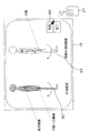
次に図8に基づいて、画面表示例を説明する。図8は、左側に計測画像80と、右側に図9に示す医用画像撮影装置5により生成された撮影データに基づいて再構成された3次元画像を多方向から投影して投影面1、2、3上に作成した投影像81、または再構成された3次元画像90に基づいて生成されたMIP像からなる標準半透明画像81とを並べて表示する。モニタ15上に表示された回転アイコン82をクリックすることにより、計測画像80と標準透明画像81とを同じ角度ずつ回転表示させることができる。
Next, a screen display example will be described with reference to FIG. FIG. 8 shows
上記図5で示した第二の実施の形態においては、病巣領域(異常陰影)でないことが既知の集積度の時間依存パターンを格納しておき、RI集積領域の集積度Gの時間依存F(t)と比較をしたが、病巣領域(異常陰影)であることが既知の集積度の時間依存パターンを格納しておき、RI集積領域の集積度の時間依存と比較をして所定の相関が認められた場合に異常陰影候補として検出してもよい。 In the second embodiment shown in FIG. 5 described above, a time-dependent pattern of the integration degree that is known not to be a lesion area (abnormal shadow) is stored, and a time dependency F ( t), the time-dependent pattern of the accumulation degree known to be a lesion area (abnormal shadow) is stored, and the predetermined correlation is obtained by comparing with the time dependence of the accumulation degree of the RI accumulation area. If it is recognized, it may be detected as an abnormal shadow candidate.
更に、医用画像診断支援装置10は、各RI集積領域に対して図2及び図5に示す処理を行い、どちらの処理においても異常陰影候補として検出された場合にのみ、異常陰影候補としてRI集積領域にマークをつけたり、記録したりするように構成してもよい。これにより、異常陰影候補の検出がより正確に行える。
Furthermore, the medical image
1…医用画像診断支援システム、2…PET装置、3…データベース、4…LAN、5…医用画像撮影装置、10…医用画像診断支援装置、11…CPU、12…主メモリ、13…磁気ディスク、14…表示メモリ、15…モニタ、16…キーボード、17…マウス、18…コントローラ、19…共通バス
DESCRIPTION OF
Claims (5)
被検体の各臓器の形状を格納する臓器形状格納手段と、
前記PETデータに含まれ、周囲より高濃度の領域であるRI集積領域を抽出するRI集積領域抽出手段と、
前記臓器形状格納手段に格納され、かつ前記被検体におけるRI集積領域が位置する部位の各臓器の形状と、前記抽出されたRI集積領域の形状と、を用い、前記各臓器の体積と前記RI集積領域の体積とを規格化し、当該規格化された前記各臓器の体積及び前記RI集積領域の体積の重心から辺縁までの距離の角度依存曲線が同じ形であるかに基づいて、前記各臓器の形状と前記RI集積領域の形状との相関関係の有無を判定する手段と、
前記相関関係がないと判定された前記RI集積領域を異常陰影候補として検出する異常陰影候補検出手段と、
を備えることを特徴とする医用画像診断支援装置。 PET data acquisition means for acquiring PET data measured by a PET apparatus;
Organ shape storage means for storing the shape of each organ of the subject;
RI integrated region extraction means for extracting an RI integrated region that is included in the PET data and is a region having a higher concentration than the surroundings ;
Stored in the organ shape storage unit, and the use and shape of each organ site RI integrated region is located in a subject, and a shape of the extracted RI accumulation area, the the volume of the respective organ RI Normalizing the volume of the integrated region, and based on whether the normalized volume of each organ and the angle-dependent curve of the distance from the center of gravity of the volume of the RI integrated region to the edge are the same Means for determining the presence or absence of a correlation between the shape of the organ and the shape of the RI integrated region ;
Abnormal shadow candidate detection means for detecting the RI accumulation region determined to have no correlation as an abnormal shadow candidate;
A medical image diagnosis support apparatus comprising:
被検体の各臓器の形状を格納する臓器形状格納手段と、
前記PETデータに含まれ、周囲より高濃度の領域であるRI集積領域を抽出するRI集積領域抽出手段と、
前記臓器形状格納手段に格納され、かつ前記被検体におけるRI集積領域が位置する部位の各臓器の形状と、前記抽出されたRI集積領域の形状と、に基づいて、前記被検体の全身に対する前記RI集積領域の体積の割合と、前記被検体の全身に対する前記各臓器の体積の割合と、が近似的に等しいか否かを判定する手段と、
前記RI集積領域の体積の割合が、前記判定により近似的に等しくないと判定された前記RI集積領域を異常陰影候補として検出する異常陰影候補検出手段と、
を備えることを特徴とする医用画像診断支援装置。 PET data acquisition means for acquiring PET data measured by a PET apparatus;
Organ shape storage means for storing the shape of each organ of the subject;
RI integrated region extraction means for extracting an RI integrated region that is included in the PET data and is a region having a higher concentration than the surroundings;
Based on the shape of each organ stored in the organ shape storage means and where the RI accumulation region of the subject is located, and the shape of the extracted RI accumulation region, the whole body of the subject Means for determining whether or not the volume ratio of the RI integrated region and the volume ratio of each organ relative to the whole body of the subject are approximately equal;
An abnormal shadow candidate detecting means for detecting, as an abnormal shadow candidate, the RI integrated region determined by the determination that the volume ratio of the RI integrated region is not approximately equal;
A medical image diagnosis support apparatus comprising:
ことを特徴とする請求項1又は2に記載の医用画像診断支援装置。 The organ shape storage means stores the shape of each organ of the subject extracted based on the subject imaging data obtained by a medical imaging device.
The medical image diagnosis support apparatus according to claim 1, wherein the medical image diagnosis support apparatus is a medical image diagnosis support apparatus.
ことを特徴とする請求項1又は2に記載の医用画像診断支援装置。 The medical image diagnosis support apparatus according to claim 1, wherein the medical image diagnosis support apparatus is a medical image diagnosis support apparatus.
前記PETデータに含まれ、周囲より高濃度の領域であるRI集積領域を抽出するRI集積領域抽出手段と、RI integrated region extraction means for extracting an RI integrated region that is included in the PET data and is a region having a higher concentration than the surroundings;
前記RI集積領域抽出手段で抽出されたRI集積領域の、RI集積度の時間依存を示す被検体RI集積データを取得する被検体RI集積データ取得手段と、Subject RI integrated data acquisition means for acquiring subject RI integrated data indicating the time dependence of the RI integration degree of the RI integrated region extracted by the RI integrated region extraction means;
予め調べた、薬剤種毎の病巣領域における前記各薬剤注入後から所定時間経過後のRI集積度の時間依存を示す標準RI集積データ、又は予め調べた、薬剤種毎の非病巣領域における前記各薬剤注入後から所定時間経過後のRI集積度の時間依存を示す標準RI集積データのうちの少なくとも一つを格納する標準RI集積データ格納手段と、Standard RI accumulation data indicating the time dependence of the RI accumulation degree after a predetermined time has elapsed after the injection of each drug in the lesion area for each drug type examined in advance, or each of the above in the non-focal area for each drug type examined in advance Standard RI integrated data storage means for storing at least one of standard RI integrated data indicating the time dependence of the RI integration degree after a predetermined time has elapsed since the drug injection;
前記被検体RI集積データと前記標準RI集積データとを比較し、それらの比較結果に基づいて異常陰影候補を検出する異常陰影候補検出手段と、An abnormal shadow candidate detecting means for comparing the subject RI integrated data and the standard RI integrated data and detecting an abnormal shadow candidate based on the comparison result;
を備えることを特徴とする医用画像診断支援装置。A medical image diagnosis support apparatus comprising:
Priority Applications (1)
| Application Number | Priority Date | Filing Date | Title |
|---|---|---|---|
| JP2004333069A JP4454474B2 (en) | 2004-11-17 | 2004-11-17 | Medical image diagnosis support device |
Applications Claiming Priority (1)
| Application Number | Priority Date | Filing Date | Title |
|---|---|---|---|
| JP2004333069A JP4454474B2 (en) | 2004-11-17 | 2004-11-17 | Medical image diagnosis support device |
Publications (3)
| Publication Number | Publication Date |
|---|---|
| JP2006145281A JP2006145281A (en) | 2006-06-08 |
| JP2006145281A5 JP2006145281A5 (en) | 2007-05-24 |
| JP4454474B2 true JP4454474B2 (en) | 2010-04-21 |
Family
ID=36625161
Family Applications (1)
| Application Number | Title | Priority Date | Filing Date |
|---|---|---|---|
| JP2004333069A Expired - Fee Related JP4454474B2 (en) | 2004-11-17 | 2004-11-17 | Medical image diagnosis support device |
Country Status (1)
| Country | Link |
|---|---|
| JP (1) | JP4454474B2 (en) |
Families Citing this family (9)
| Publication number | Priority date | Publication date | Assignee | Title |
|---|---|---|---|---|
| JP4640143B2 (en) * | 2005-12-02 | 2011-03-02 | 株式会社島津製作所 | Diagnostic imaging support device |
| JP4609298B2 (en) * | 2005-12-09 | 2011-01-12 | 株式会社島津製作所 | Diagnostic imaging support device |
| US8017915B2 (en) | 2008-03-14 | 2011-09-13 | Reflexion Medical, Inc. | Method and apparatus for emission guided radiation therapy |
| WO2011070484A2 (en) | 2009-12-08 | 2011-06-16 | Koninklijke Philips Electronics N.V. | A method and a correction system for correcting tracer-uptake measurements |
| CN103650095B (en) | 2011-03-31 | 2016-12-07 | 反射医疗公司 | For the system and method used in launching the radiotherapy guided |
| JP6026089B2 (en) * | 2011-08-23 | 2016-11-16 | 東芝メディカルシステムズ株式会社 | Medical image diagnostic apparatus, image information display apparatus, and control program |
| EP3426345B1 (en) | 2016-03-09 | 2021-06-23 | RefleXion Medical, Inc. | Fluence map generation methods for radiotherapy |
| JP6797557B2 (en) * | 2016-05-17 | 2020-12-09 | キヤノンメディカルシステムズ株式会社 | Medical image diagnostic equipment, medical image processing equipment and image display program |
| WO2018093849A1 (en) | 2016-11-15 | 2018-05-24 | Reflexion Medical, Inc. | Methods for radiation delivery in emission-guided radiotherapy |
-
2004
- 2004-11-17 JP JP2004333069A patent/JP4454474B2/en not_active Expired - Fee Related
Also Published As
| Publication number | Publication date |
|---|---|
| JP2006145281A (en) | 2006-06-08 |
Similar Documents
| Publication | Publication Date | Title |
|---|---|---|
| Si-Mohamed et al. | Spectral photon-counting computed tomography (SPCCT): in-vivo single-acquisition multi-phase liver imaging with a dual contrast agent protocol | |
| Boellaard | Need for standardization of 18F-FDG PET/CT for treatment response assessments | |
| EP2120702B1 (en) | Automated diagnosis and alignment supplemented with pet/mr flow estimation | |
| Kaza et al. | Update of dual-energy CT applications in the genitourinary tract | |
| US9597041B2 (en) | Sequential image acquisition with updating method and system | |
| JP5068519B2 (en) | Machine-readable medium and apparatus including routines for automatically characterizing malignant tumors | |
| EP2399238B1 (en) | Functional imaging | |
| US8788012B2 (en) | Methods and apparatus for automatically registering lesions between examinations | |
| JP2008503253A (en) | System and method for linking regions of interest across multiple time points to analyze disease progression or therapeutic effect | |
| US8155408B2 (en) | Standardized normal database having anatomical phase information | |
| JP2012518168A (en) | Model-based field expansion in nuclear imaging | |
| JP2008503258A (en) | System and method for monitoring disease progression or therapeutic effect using multi-mode visualization | |
| JP4317412B2 (en) | Image processing method | |
| JP2006255412A (en) | Method and system for monitoring tumor burden | |
| US8180128B2 (en) | Method for recording measured data of a patient while taking account of movement operations, and an associated medical device | |
| Bachar et al. | Image quality and localization accuracy in C‐arm tomosynthesis‐guided head and neck surgery | |
| US20110164797A1 (en) | Method and system of processing multi-energy x-ray images | |
| JP2008080121A (en) | Method and system for discriminating region in image | |
| JP2008503259A (en) | System and method for loading multiple time points to analyze disease progression or therapeutic effect | |
| JP2014518125A (en) | Follow-up image acquisition plan and / or post-processing | |
| JP4454474B2 (en) | Medical image diagnosis support device | |
| Delgado Sánchez-Gracián et al. | Quantitative myocardial perfusion with stress dual-energy CT: iodine concentration differences between normal and ischemic or necrotic myocardium. Initial experience | |
| US9600875B2 (en) | Tissue surface roughness quantification based on image data and determination of a presence of disease based thereon | |
| Scherer et al. | Dynamic quantitative iodine myocardial perfusion imaging with dual-layer CT using a porcine model | |
| US20150025359A1 (en) | Method for evaluation and comparison of a chronological sequence of combined medical imaging examinations and also a medical imaging system which is designed for executing the inventive method |
Legal Events
| Date | Code | Title | Description |
|---|---|---|---|
| A521 | Written amendment |
Free format text: JAPANESE INTERMEDIATE CODE: A523 Effective date: 20070330 |
|
| A621 | Written request for application examination |
Free format text: JAPANESE INTERMEDIATE CODE: A621 Effective date: 20070330 |
|
| RD02 | Notification of acceptance of power of attorney |
Free format text: JAPANESE INTERMEDIATE CODE: A7422 Effective date: 20090717 |
|
| RD04 | Notification of resignation of power of attorney |
Free format text: JAPANESE INTERMEDIATE CODE: A7424 Effective date: 20090721 |
|
| A131 | Notification of reasons for refusal |
Free format text: JAPANESE INTERMEDIATE CODE: A131 Effective date: 20090908 |
|
| A521 | Written amendment |
Free format text: JAPANESE INTERMEDIATE CODE: A523 Effective date: 20091020 |
|
| TRDD | Decision of grant or rejection written | ||
| A01 | Written decision to grant a patent or to grant a registration (utility model) |
Free format text: JAPANESE INTERMEDIATE CODE: A01 Effective date: 20100202 |
|
| A01 | Written decision to grant a patent or to grant a registration (utility model) |
Free format text: JAPANESE INTERMEDIATE CODE: A01 |
|
| A61 | First payment of annual fees (during grant procedure) |
Free format text: JAPANESE INTERMEDIATE CODE: A61 Effective date: 20100202 |
|
| FPAY | Renewal fee payment (event date is renewal date of database) |
Free format text: PAYMENT UNTIL: 20130212 Year of fee payment: 3 |
|
| R150 | Certificate of patent or registration of utility model |
Free format text: JAPANESE INTERMEDIATE CODE: R150 |
|
| FPAY | Renewal fee payment (event date is renewal date of database) |
Free format text: PAYMENT UNTIL: 20140212 Year of fee payment: 4 |
|
| LAPS | Cancellation because of no payment of annual fees |