JP4351041B2 - 傷治癒のための処置および組成物 - Google Patents
傷治癒のための処置および組成物 Download PDFInfo
- Publication number
- JP4351041B2 JP4351041B2 JP2003503264A JP2003503264A JP4351041B2 JP 4351041 B2 JP4351041 B2 JP 4351041B2 JP 2003503264 A JP2003503264 A JP 2003503264A JP 2003503264 A JP2003503264 A JP 2003503264A JP 4351041 B2 JP4351041 B2 JP 4351041B2
- Authority
- JP
- Japan
- Prior art keywords
- apc
- protein
- agent
- wound
- wound healing
- Prior art date
- Legal status (The legal status is an assumption and is not a legal conclusion. Google has not performed a legal analysis and makes no representation as to the accuracy of the status listed.)
- Expired - Lifetime
Links
Images


Classifications
-
- A—HUMAN NECESSITIES
- A61—MEDICAL OR VETERINARY SCIENCE; HYGIENE
- A61K—PREPARATIONS FOR MEDICAL, DENTAL OR TOILETRY PURPOSES
- A61K38/00—Medicinal preparations containing peptides
- A61K38/16—Peptides having more than 20 amino acids; Gastrins; Somatostatins; Melanotropins; Derivatives thereof
- A61K38/43—Enzymes; Proenzymes; Derivatives thereof
- A61K38/46—Hydrolases (3)
- A61K38/48—Hydrolases (3) acting on peptide bonds (3.4)
- A61K38/482—Serine endopeptidases (3.4.21)
- A61K38/4866—Protein C (3.4.21.69)
-
- A—HUMAN NECESSITIES
- A61—MEDICAL OR VETERINARY SCIENCE; HYGIENE
- A61K—PREPARATIONS FOR MEDICAL, DENTAL OR TOILETRY PURPOSES
- A61K9/00—Medicinal preparations characterised by special physical form
- A61K9/0012—Galenical forms characterised by the site of application
- A61K9/0048—Eye, e.g. artificial tears
-
- A—HUMAN NECESSITIES
- A61—MEDICAL OR VETERINARY SCIENCE; HYGIENE
- A61P—SPECIFIC THERAPEUTIC ACTIVITY OF CHEMICAL COMPOUNDS OR MEDICINAL PREPARATIONS
- A61P17/00—Drugs for dermatological disorders
- A61P17/02—Drugs for dermatological disorders for treating wounds, ulcers, burns, scars, keloids, or the like
-
- A—HUMAN NECESSITIES
- A61—MEDICAL OR VETERINARY SCIENCE; HYGIENE
- A61P—SPECIFIC THERAPEUTIC ACTIVITY OF CHEMICAL COMPOUNDS OR MEDICINAL PREPARATIONS
- A61P19/00—Drugs for skeletal disorders
- A61P19/04—Drugs for skeletal disorders for non-specific disorders of the connective tissue
-
- A—HUMAN NECESSITIES
- A61—MEDICAL OR VETERINARY SCIENCE; HYGIENE
- A61P—SPECIFIC THERAPEUTIC ACTIVITY OF CHEMICAL COMPOUNDS OR MEDICINAL PREPARATIONS
- A61P19/00—Drugs for skeletal disorders
- A61P19/08—Drugs for skeletal disorders for bone diseases, e.g. rachitism, Paget's disease
-
- A—HUMAN NECESSITIES
- A61—MEDICAL OR VETERINARY SCIENCE; HYGIENE
- A61P—SPECIFIC THERAPEUTIC ACTIVITY OF CHEMICAL COMPOUNDS OR MEDICINAL PREPARATIONS
- A61P29/00—Non-central analgesic, antipyretic or antiinflammatory agents, e.g. antirheumatic agents; Non-steroidal antiinflammatory drugs [NSAID]
-
- A—HUMAN NECESSITIES
- A61—MEDICAL OR VETERINARY SCIENCE; HYGIENE
- A61P—SPECIFIC THERAPEUTIC ACTIVITY OF CHEMICAL COMPOUNDS OR MEDICINAL PREPARATIONS
- A61P43/00—Drugs for specific purposes, not provided for in groups A61P1/00-A61P41/00
-
- A—HUMAN NECESSITIES
- A61—MEDICAL OR VETERINARY SCIENCE; HYGIENE
- A61P—SPECIFIC THERAPEUTIC ACTIVITY OF CHEMICAL COMPOUNDS OR MEDICINAL PREPARATIONS
- A61P9/00—Drugs for disorders of the cardiovascular system
- A61P9/10—Drugs for disorders of the cardiovascular system for treating ischaemic or atherosclerotic diseases, e.g. antianginal drugs, coronary vasodilators, drugs for myocardial infarction, retinopathy, cerebrovascula insufficiency, renal arteriosclerosis
Description
凝固は、脱顆粒した血小板から様々な成長因子およびサイトカインを遊離することによって、止血を制御し、治癒を始める。炎症段階中では、血小板凝集および凝固により、血漿タンパク質および血液細胞を捕捉し、様々な型の細胞の流入を誘導する基質が形成される。好中球が、到達する最初の細胞であり、混入している細菌を食菌し、フィブリンクロットを消化し、そしてマクロファージを引き寄せ、繊維芽細胞とケラチン生成細胞を活性化するメディエーターを遊離する(非特許文献3)。マクロファージは、病原体を消化し、傷から異物を取り除き、そして、サイトカイン/成長因子(例えば、繊維芽細胞および内皮細胞を刺激するインターロイキン−1(IL−1)、上皮成長因子(EGF)、血管内皮成長因子(VEGF)、形質転換成長因子(TGF−β)、および塩基性繊維芽細胞成長因子(bFGF))を分泌する。全体的に、炎症段階は、感染から守り、傷治癒の遊走および増殖段階を促進するのに重要である。
これらの段階は、細胞の遊走と増殖を含む。リンパ球とマクロファージが関与するが、主な細胞の種類は、上皮細胞、繊維芽細胞、および内皮細胞である。損傷数時間内に、主にケラチン生成細胞から成る上皮被覆が、遊走を始め、表皮を覆い始める、再上皮化として知られる工程。それらが完全に傷を覆うと、それらは分化して、層を形成し、基底膜と共に新しい表皮を形成する。血管新生(すなわち、新しい血管の形成)が、この段階で生じ、残存するために発達している組織に栄養素を供給する。繊維芽細胞は、傷部位に遊走し、最終的に傷に抗張力を付与するコラーゲンとプロテオグリカンを生成する。再構築段階が進むにつれて、顆粒化組織が、瘢痕組織を形成するコラーゲンとエラスチン繊維の網状組織によって置換される。
皮膚の傷治癒の低下、および/または、皮膚潰瘍が、抹消動脈閉塞疾患、深部静脈血栓症、糖尿病、床ずれ、および、やけどの患者に生じる(非特許文献4)。徹底的な研究に関わらず、傷治癒の低下に関わる分子機序が、十分に理解されていない。
慢性的な傷は、最初に、乾燥痂皮の創面切除、適切な場合には抗生物質処置、および、通常の外傷用医薬材料等を用いた手当てを含む処置によって管理される(非特許文献2)。ヒドロゲル類、親水性コロイド類あるいはアルギナート類などの他の外傷用医薬材料も使用できる。静脈潰瘍は、加圧療法によって処置されるが、動脈あるいは糖尿病性の潰瘍は、外傷用医薬材料の通常の変更を必要とする。床ずれは、損傷部位での圧力の軽減によって治癒が促される。動脈性潰瘍のためのレーザー処置、高圧酸素および電気刺激など、いくつかの別の物理的装置も、傷治癒を促すために使用される(非特許文献2、非特許文献9、非特許文献10)。
Bello, Y.M. and T.J. Phillips. 2000. Recent advances in wound healing. JAMA 283:716-718. Braddock, M., C.J. Campbell, and D. Zuder. 1999. Current Therapies for wound healing: electrical stimulation, biological therapeutics, and the potential for gene therapy. Int J Dermatol 38:808-817. Hunt, T.K., H. Hopf, and Z. Hussain. 2000. Physiology of wound healing. Adv Skin Wound Care 13:6-11. Bello, Y.M. and T.J. Phillips. 2000. Therapeutic dressings. Adv Dermatol 16:253-271. Yager, D.R. and B.C. Nwomeh. 1999. The proteolytic environment of chronic wounds. Wound Repair Regen 7:433-441 Trengove, N.J., M.C. Stacey, S.MacAuley, N. Bennett, J. Gibson, F. Burslem, G. Murphy, and G. Schultz. 1999. Analysis of the acute and chronic wound environments: the role of proteases and their inhibitors. Wound Repair Regen 7:442-452. Kahari, V.M. and U. Saarialhokere. 1997. Matrix metalloproteinases in skin. Experimental Dermatology 6:199-213. Singer, A.J. and R.A. Clark. 1999. Cutaneous wound healing. N Engl J Med 341:738-746. McDaniel, D.H., K. Ash, J. Lord, J. Newman, and M. Zukowski. 1998. Accelerates laser resurfacing wound healing using a triad of topical antioxidants. Dermatol Surg 24: 661-664. Stone, A. 1998. Hyperbaric oxygen treatment for wounds. Plast Reconstr Surg 101:1738-1739. Brem, H., J. Balledux, T. Bloom, M.D. Kerstein, and L. Hollier. 2000. Healing of diabetic foot ulcers and pressure ulcers with human skin equivalent: a new paradigm in wound healing. Arch Surg 135:627-634. Falanga, V. and M. Sabolinski. 1999. A bilayered living skin construct(APLIGRAF) accelerates complete closure of hard-to-heal venous ulcers. Wound Repair Regen 7:201-207. Gillam, A.J. and C.C. Da Camara. 1993. Treatment of wounds with procuren. Ann Pharmacother 27:1201-1203. Mazue, G., F.Bertolero, C. Jacob, P. Sarmientos, and R. Roncucci. 1991. Preclinical and clinical studies with recombinant human basic fibroblast growth factor. Ann N Y Acad Sci 638:329-340. Esmon, C.T., W. Ding, K. Yasuhiko, J.M. Gu, G. Ferrell, L.M. Regen, D.J. Stearns-Kurosawa, S. Kurosawa, T. Mather, Z. Laszik, and N.L. Esmon. 1997. The protein C pathway: new insights. Thromb Haem 78:70-74. Boffa, M.C. and M. Karmochkine. 1998. Thrombomodulin: an overview and potential implications in vascular disorders. Lupus 7:Suppl, 2-5. Esmon, C.T., J.M.Gu, J. Xu, D. Qu, D.J. Stearns-Kurosawa, and S. Kurosawa. 1999. Regulation and functions of the protein C anticoagulant pathway. Haematologica 84:363-368. Baker, W.F. and B. and. 1999. Treatment of hereditary and acquired thrombophilic disorders. Semin.Thromb.Hemostasis. 25:387-405. Morales, D.E., K.A. Mcgowen, D.S. Grant, S. Maheshwari, D. Bhartiya, M.C. Cid, H.K. kleinman, and H.W. Schnaper. 1995. Estrogen promotes angiogenic activity in human umbilical vein endothelial cells in vitro and in a murine model. Circulation 91:755-763. Nguyen, M., J. Arkell, and C.J. Jackson. 2000. Activated protein C directly activates human endothelial gelatinase A. J Biol Chem 275:9095-9098. Murphy, G. 1995. Matrix metalloproteinases and their inhibitors. Acta orthop Scand Suppl 266:55-60. Fang, J., Y. Shing, D. Wiederschain, L. Yan, C. Butterfield, G. Jackson, J. Harper, G. Tamvakopoulos, and M.A. Moses. 2000. Matrix metalloproteinase-2 is required for the switch to the angiogenic phenotype in tumor model. Proc Natl Acad Sci USA 97:3884-3889. Kondo, K., H. Kinoshita, H. Ishikura, T. Miyoshi, T. Hirose, Y. Matsumori, and Y. Monden. 2001. Activation of matrix metalloproteinase-2 is correlated with invasiveness in thymic epithelial tumors. J Surg Oncol 76: 169-175. Kozaci, L.D., D.J. Buttle, and A.P. Hollander. 1997. Degradation of Type ii collagen, but not proteoglycan, correlates with matrix metalloproteinase activity in cartilage explant cultures. Arthr Rheum 40:164-174. Cheung, P.Y., G. Sawicki, M. Wozniak, W. Wang, M.W. Radomski, and R. Schulz. 2000. Matrix mettalloproteinase-2 contributes to ischemia-reperfusion injury in the heart. Circulation 101:1833-1839. Fernandez-Patron, C., M.W. Radomski, and S.T. Davidge. 1999. Vascular matrix metalloproteinase-2 cleaves big endothelin-1 yielding a novel vasoconstrictor. Circ Res 85:906-911. Sawicki, G., E. Salas, J. Murat, H. Misztalane, and M.W. Radomski. 1997. Release of gelatinase A during platelet activation mediates aggregation. Nature 386:616-619. Kirshenbaun, K., Zuckermann, R.N., and Dill, K.A. 1999. Designing polymers that mimic biomolecules. Curr Opin Stract Biol 9:530-535. Sidhu, S.S., Lowman, H.B., Cunningham, B.C., and Wells, J.A. 2000 Phage display for selection of novel binding peptides. Methods Enzymol 328:333-363. Cunningham, B.C., and Wells, J.A. 1997 Minimized proteins. Carr Opin Struct Biol 7:457-462. Drolet, D.W., Jenison, R.D., Smith, D.E., Pratt, D., and Hicke, B.J. 1999 A high throughout platform for systematic evolution of ligands by exponential enrichment (SELEX). Comb Chem High Throughout Screen 2:271-278. Bissantz, C., Folkers, G., and Rognan, D. 2000 Protein-based virtual screening of chemical databases. 1 Evaluation of different docking/scoring combinations. J Med Chem 43:4759-4767. Houghten, R.A, Wilson, D.B., and Pinilla, C. 2000 Drug Discovery and vaccine development using mixture-based synthetic combinatorial libraries. Drug Discovery Today 5:276-285. Ribatti, D., A. Gualandris, M. Bastaki, A. Vacca, M. Iurlaro, L. Roncali, and M. Presta. 1997. New model for the study of angiogenesis and antiangiogenesis in the chick embryo chorioallantoic membrane: the gelantin sponge/chorioallantoic membrane assay. J Vasc Res 34:455-463. Taniyama Y., Morishita R., Hiraoka K., Aoki M., Nakagami H., Yamasaki K., Matsumoto K., Nakamura T., Kaneda Y., and Ogihara T. 2001 Therapeutic angiogenesis induced by human hepatocyte growth factor gene in rat diabetic hind limb ischemia model: molecular mechanisms of delayed angiogenesis in diabetes. Circulation 104:2344-2350. Maniatis, T.等、Molecular Cloning: a laboratory manual、第二版、Cold Spring Harbor Laboratory出版社
(i)APC、
(ii)APCの機能性断片、
(iii)APC擬似化合物、および
(iv)タンパク質C、
の1つ以上を含む作用剤の有効量を、随意に、薬学的に許容可能な担体と混合して、前記被験者に投与することを含む方法を提供する。
(i)APC、
(ii)APCの機能性断片、
(iii)APC擬似化合物、および
(iv)タンパク質C、
のうち1つ以上を含むある量の作用剤を含む薬剤を提供する。
(i)APC、
(ii)APCの機能性断片、
(iii)APC擬似化合物、および
(iv)タンパク質C、
の1つ以上を含むある量の作用剤を組み込んだ送達システム(例えば、ゲル、スポンジ、ガーゼ、またはメッシュ)であって、傷への適用に適し、適用後、傷治癒を促進する送達システムを提供する。
(i)APC、
(ii)APCの機能性断片、
(iii)APC擬似化合物、および
(iv)タンパク質C、
の1つ以上を含む作用剤の、被験者における傷治癒を促進するための薬剤を調製するための使用を提供する。
(i)APC、
(ii)APCの機能性断片、
(iii)APC擬似化合物、および
(iv)タンパク質C、
の1つ以上を含む作用剤の、送達システムを調製するための使用であって、前記送達システムが傷への適用に適し、適用後、傷治癒を促進する使用を提供する。
(i)血圧、脈管炎、動脈および静脈の疾患を伴う皮膚潰瘍などの皮膚潰瘍(例えば、糖尿病をわずらっている患者、老齢の患者において、静脈機能不全および脳血管発作を伴い、皮膚上の直接的な圧力または剪断力および摩擦により生じる床ずれ、もしくは局所的な組織傷害の結果生じる)、
(ii)やけど、
(iii)口の傷(例えば、歯肉炎によって引き起こされる)、
(iv)眼の傷(例えば、傷害、手術またはレーザー治療により生じた角膜の傷)、
(v)皮膚以外の傷(例えば、胃/食道潰瘍、膣潰瘍、および内傷、または手術(プラスチック手術を含む)、
(vi)虚血−再灌流傷害(例えば、心筋梗塞の結果生じる)、
(vii)リューマチ性関節炎および変形性関節炎など、骨格筋疾患において生じる骨および軟骨の損傷、および
(viii)ワルファリン関連皮膚壊死
からなる群から選択された傷に適用される。
APCによる内皮細胞傷害の修復の促進
活性化タンパク質C(APC)は、以前に記載された(非特許文献19)ように、in vitroアッセイの修飾を用いて、内皮細胞の創傷の修復を促進する性能について、試験した。簡単には、新生児の包皮由来の融合性微小血管内皮細胞(FSE)を、5日間、培養培地(50μg/mlヘパリン、50μg/ml内皮細胞培養補助剤、および5%ヒト血清を加えたバイオリッチ(Biorich))中、24穴培養プレートで培養した。内皮単層細胞は、ピペットチップで穴の直径を端から端まで一なですることにより、傷つけた。次いで、培地と取り除かれた細胞を吸引し、プレートを、ハンクス(Hanks)緩衝液で洗った。新鮮な培養培地を、様々な濃度のAPC、または強力な腫瘍促進血管新生因子、ホルボールミリスタートアセタート(PMA)(10ng/ml)と共に、プレートに加えて、細胞を37℃で培養した。24時間後、傷の幅を顕微鏡的に視覚化し、APCの異なる用量での結果は画像処理を用いて定量化し、用量応答曲線を作製した(図1)。75nMAPCの存在下で培養した細胞は、24時間以内に、ほとんど完全に傷を閉合し、APCを用いずに培養した細胞よりも2倍以上の遊走反応を示した。100nMでは、さらには増強しなかった。APC(75μM)は、およそPMAと同程度の活性を有していた。
APCによる血管新生の促進
ゼラチナーゼAを活性化し、内皮細胞創傷を促進するAPCの性能に鑑みて、APCが血管新生を促進し得るかどうかについて調べた。APCを、ゼラチンスポンジ(Gelfoam)を用いたニワトリ胚子漿尿膜(CAM)アッセイに加えた。スポンジは、およそ2mm×2mmに切断した。リン酸緩衝生理食塩水(PBS)中5μgのAPC、またはAPC単独をゼラチンスポンジに加え、次いで9日目のCAMの上に、以前記載したように(非特許文献34)置いた。CAMは、毎日視察し、14日目に撮影し、組織学的薄切のために固定した。肉眼的には、14日目に、APCで処理したゼラチンスポンジは、スポーク・ホイール(spoke−wheel)パターンでスポンジに向かって内部に急速に成育する血管により取り囲まれた(データ省略)。これに対して、PBSで処置したゼラチンスポンジは、取り囲む血管が形成されなかった。組織学的切片により、APCで処理されたスポンジが、多くの新しい血管に浸潤されている(血管新生)ことが示された(図2)。さらに、APCで処理されたスポンジ中に、広範囲にわたる繊維芽細胞の遊走があった。また、顕著な上皮細胞層の増殖があり、ゼラチンスポンジ上で外胚葉が完全に成育していた。スポンジの外縁で、層化と退縮が、この再上皮化に伴った。また、内胚葉も、いくつかの切片で層化し、絨毛形成することが示され、絨毛から落ちた細胞の存在を示した(省略)。APCで処理されたスポンジに対して、PBS対照スポンジでは、再上皮化、内皮細胞または繊維化細胞の浸潤のいずれの証拠もほとんどなかった。
APCによる傷治癒の促進
血管内皮細胞の遊走を刺激し、再上皮化、繊維芽細胞の浸潤および血管新生を高めるAPCの性能に鑑みて、ラットモデルにおいて傷治癒を増進する能力について、APCを調べた。スプラーグ・ドーリー(Sprague−Dawley)ラットを、麻酔し、8mmのパンチ生検を用いて、ラットの背中に、その下にある背部側部の骨格筋膜を露出するように、4つの十分に深い傷を切開した。滅菌したガーゼで均一に圧迫することにより、止血した。APCは、等張性、滅菌、発熱物質不含生理食塩溶液中に希釈し、各切開は、滅菌、発熱物質不含生理食塩溶液、または20μgのAPCを含む生理食塩水50μlの局所塗布により、処置した。傷は、包帯をせずに開けたままにしておき、ラットは、1かご当たり1匹ずつかごに入れた。傷の閉合は、40時間、4日および7日後に、視覚的に評価した。各時点で、傷を、ニコン・クールピクス(Nikon Coolpix)950を用いて、フレームにおける距離校正目盛りと共に、デジタル方式で撮影した。傷の面積を、画像解析(Scion Image)によって算出した。40時間後に、対照と比べて、APC処理された傷における傷の閉合に、顕著な視覚的向上があった。4日目において、画像解析の結果により、対照と比べて、APC処理された傷の大きさに有意な減少が示された(図3)。この違いは、7日目に、維持されていた(図3c、**p<0.01、***p<0.001)。
APCによる傷治癒の促進
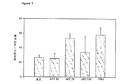
ラットモデルにおける傷治癒を増進する能力について、APCをさらに調べた。スプラーグ・ドーリーラットを麻酔し、8mmのパンチ生検を用いて、ラットの背中について、その下にある背部側部の骨格筋膜を露出するように、4つの十分に深い傷を切開した。滅菌したガーゼで均一に圧迫することにより、止血した。APCは、等張性、滅菌、発熱物質不含生理食塩溶液中に希釈し、各切開は、滅菌、発熱物質不含生理食塩溶液、または以下:0μgAPC(対照、ラット3匹、傷12箇所)、10μgAPC(ラット3匹、傷12箇所)、40μgAPC(ラット4匹、傷16箇所)、70μgAPC(ラット3匹、傷12箇所)、または100μgAPC(ラット3匹、傷12箇所)を含む生理食塩水50μlの局所塗布により、直ちに処置した。傷は、包帯をせずに開けたままにしておき、ラットは、1かご当たり1匹ずつかごに入れた。傷の大きさは、1、3、5、7、および9日後に、画像解析により測定した。各時点で、傷を、ニコン・クールピクス995を用いて、デジタル方式で撮影した。傷の面積を、画像解析(Scion Image)によって算出した。結果を図4に示す。1日後に、10または40μgAPCで処置した傷の大きさに有意な減少があった。対照と70または100μgAPCで処置したラットとの間には、差異が無かった。傷の大きさにおける有意な減少は、40μgAPCで最も顕著であり、1、3、7、および9日目で見られた(**p<0.01、*p<0.05、コスタット(Costat)を用いたスチューデントのt検定)。
APCによる傷治癒の促進
ラットモデルにおける傷治癒を増進する能力について、APCをさらに調べた。スプラーグ・ドーリーラットを、実施例4で記載したように、傷つけた。APCは、等張性、滅菌、発熱物質不含生理食塩溶液中に希釈し、各切開は、滅菌、発熱物質不含生理食塩溶液、または、40μgAPCを含む生理食塩水50μlの局所塗布により、処置した。48時間後、傷は、40μgAPCの2回目の塗布により処置した。傷は、包帯をせずに開けたままにしておき、ラットは、1かご当たり1匹ずつかごに入れた。傷の閉合は、1、2、3、4、5、7、および9日後に、視覚的に評価した。各時点で、傷を、ニコン・クールピクス995を用いて、デジタル方式で撮影した。傷の面積を、画像解析(Scion Image)によって算出した。結果を図5に示す。対照よりもAPCで処置したラットの治癒が早く(p<0.01)、1日後に、傷の大きさに有意な差異があった。この差異は、2、3、および7日においても認められた(**p<0.01、*p<0.05、コスタット(Costat)を用いたスチューデントのt検定)。
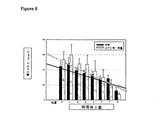
糖尿病ラットにおけるAPCによる傷治癒の促進
糖尿病ラットモデルにおける傷治癒を増進する能力について、APCをさらに調べた。糖尿病モデルは、ゆっくりと傷治癒することが十分に記載されているモデルであるため、選択した(非特許文献35)。糖尿病は、スプラーグ・ドーリーラットにおいて、ストレプトゾトシンの腹膜内注射の標準的な方法を用いて、誘発させた。1週間後、ラットの血中グルコース濃度は、20mMより大きく、糖尿病であることを示していた。糖尿病ラットは、前記実施例4で記載したように、8mmパンチ生検を用いて傷つけて、直ちに、20μgAPC(ラット2匹、傷7箇所)で処置し、または、試験剤で処置しなかった(対照、ラット1匹、傷4箇所)。傷は、包帯をせずに開けたままにしておき、ラットは、1かご当たり1匹ずつかごに入れた。傷の閉合は、1、2、3、4、5、7、および9日後に、視覚的に評価した。各時点で、傷を、ニコン・クールピクス995を用いて、デジタル方式で撮影した。傷の面積を、画像解析(Scion Image)によって算出した。結果を図6に示す。対照とAPCで処置したラットとの間で、傷治癒の速度(回帰直線の傾斜)に有意な差異があり、後者がより早く治癒した(p<0.01)。
内皮細胞の創傷を修復するAPCの性能は、ラットにおける傷治癒を促進するだけでなく、再上皮化、繊維芽細胞の浸潤および血管新生を促進し、APCが有効な傷治癒剤であるであろうことを示す。
Claims (21)
- 被験者における皮膚の傷治癒を促進するための薬剤であり、傷への直接適用によって投与するための薬剤であって、薬学的に許容可能な担体と混合した、
(i)活性化タンパク質C(APC)および/または
(ii)タンパク質C、
を含む有効量の作用剤を含む薬剤。 - APCおよび/またはタンパク質Cを200μg/mlから800μg/mlの濃度で提供する、請求項1に記載の薬剤。
- 作用剤が、ヒトAPCである、請求項1または2に記載の薬剤。
- 作用剤が、ヒトタンパク質Cである、請求項1または2に記載の薬剤。
- さらにタンパク質Cの活性化剤を含む、請求項1、2および4のいずれ一項に記載の薬剤。
- 活性化剤が、トロンビン、カリクレイン、および、トロンボモジュリンからなる群から選択される、請求項5に記載の薬剤。
- 皮膚の傷が、皮膚潰瘍またはやけどである、請求項1または2に記載の薬剤。
- (i)活性化タンパク質C(APC)および/または
(ii)タンパク質C、
を含む有効量の作用剤を組み込んだ送達システムであって、被験者における皮膚の傷へ直接適用し、適用後、皮膚の傷治癒を促進するための送達システム。 - APCおよび/またはタンパク質Cを200μg/mlから800μg/mlの濃度で提供する、請求項8に記載の送達システム。
- 作用剤が、ヒトAPCである、請求項8または9に記載の送達システム。
- 作用剤が、ヒトタンパク質Cである、請求項8または9に記載の送達システム。
- さらに、タンパク質Cの活性化剤を含む、請求項8、9および11のいずれか一項に記載の送達システム。
- 活性化剤が、トロンビン、カリクレイン、および、トロンボモジュリンからなる群から選択される、請求項12に記載の送達システム。
- 送達システムが、ゲル、スポンジ、ガーゼまたはメッシュである、請求項8から13のいずれか一項に記載の送達システム。
- (i)活性化タンパク質C(APC)および/または
(ii)タンパク質C、
を含む有効量の作用剤の、被験者における皮膚の傷治癒を促進するための薬剤であり、傷への直接適用によって投与するための薬剤を調製するための使用。 - APCおよび/またはタンパク質Cを200μg/mlから800μg/mlの濃度で提供する、請求項15に記載の使用。
- 作用剤が、ヒトAPCである、請求項15または16に記載の使用。
- 作用剤が、ヒトタンパク質Cである、請求項15または16に記載の使用。
- 薬剤が、タンパク質Cの活性化剤をさらに含む、請求項15、16および18のいずれか一項に記載の使用。
- 活性化剤が、トロンビン、カリクレイン、および、トロンボモジュリンからなる群から選択される、請求項19に記載の使用。
- 皮膚の傷が、皮膚潰瘍またはやけどである、請求項15から20のいずれか一項に記載の使用。
Applications Claiming Priority (5)
| Application Number | Priority Date | Filing Date | Title |
|---|---|---|---|
| AUPR5637A AUPR563701A0 (en) | 2001-06-13 | 2001-06-13 | Treatment and composition for wound healing |
| AUPR5637 | 2001-06-13 | ||
| AUPS1433A AUPS143302A0 (en) | 2002-04-02 | 2002-04-02 | Treatment and composition for wound healing |
| AUPS1433 | 2002-04-02 | ||
| PCT/AU2002/000751 WO2002100445A1 (en) | 2001-06-13 | 2002-06-11 | Treatment and composition for wound healing |
Publications (4)
| Publication Number | Publication Date |
|---|---|
| JP2004532892A JP2004532892A (ja) | 2004-10-28 |
| JP2004532892A5 JP2004532892A5 (ja) | 2006-01-05 |
| JP4351041B2 true JP4351041B2 (ja) | 2009-10-28 |
| JP2004532892A6 JP2004532892A6 (ja) | 2009-12-24 |
Family
ID=25646727
Family Applications (1)
| Application Number | Title | Priority Date | Filing Date |
|---|---|---|---|
| JP2003503264A Expired - Lifetime JP4351041B2 (ja) | 2001-06-13 | 2002-06-11 | 傷治癒のための処置および組成物 |
Country Status (8)
| Country | Link |
|---|---|
| US (3) | US20040176288A1 (ja) |
| EP (1) | EP1404412B1 (ja) |
| JP (1) | JP4351041B2 (ja) |
| AT (1) | ATE550073T1 (ja) |
| AU (1) | AU2002302192B2 (ja) |
| CA (1) | CA2450240A1 (ja) |
| ES (1) | ES2384099T3 (ja) |
| WO (1) | WO2002100445A1 (ja) |
Families Citing this family (14)
| Publication number | Priority date | Publication date | Assignee | Title |
|---|---|---|---|---|
| ES2358645T3 (es) | 2002-09-16 | 2011-05-12 | Agennix Incorporated | Composiciones de lactoferrina y métodos de tratamiento de la úlcera diabética. |
| US7183381B2 (en) | 2004-10-26 | 2007-02-27 | Agennix, Inc. | Composition of lactoferrin related peptides and uses thereof |
| EP1841442A4 (en) * | 2005-01-07 | 2009-12-09 | Northern Sydney And Central Co | TREATMENT OF AUTOIMMUNE DISEASES AND INFLAMMATORY DISEASES |
| AU2006206187B2 (en) * | 2005-01-24 | 2011-03-10 | Board Of Regents, The University Of Texas System | Fc-fusion constructs binding to phosphatidylserine and their therapeutic use |
| BRPI0613227A2 (pt) * | 2005-06-09 | 2011-01-04 | Chiang Chuan Chi | uma composição para cicatrização de ferida e uso desta |
| EP1971365A4 (en) * | 2005-12-20 | 2010-01-13 | Swiss American Products Inc | PROTEASE COMPOSITIONS FOR THE TREATMENT OF DAMAGED TISSUE |
| GR1005700B (el) * | 2006-09-01 | 2007-10-22 | (40%) ����� �������� | Τροπος αντιμετωπισης της 1ης φασης του εγκαυματοσσε μη σηπτικους ασθενεις με συνεχη ενδοφλεβια χορηγηση ενεργοποιημενης πρωτεινης-c για 48 ωρες με στοχο τη μειωση της τελικης εγκαυματικης βλαβης καιαυξηση της ταχυτητας επουλωσης κατα τα πρωτα επτα μετεγκαυματικα 24ωρα |
| EP2014296A1 (en) * | 2007-07-10 | 2009-01-14 | PAION Deutschland GmbH | Novel strategies for increasing the reperfusion in obstructed blood vessel |
| AR067345A1 (es) * | 2007-07-10 | 2009-10-07 | Paion Deutschland Gmbh | Uso de trombomodulina para la preparacion de un medicamento trombolitico |
| GB2457965B8 (en) * | 2008-07-01 | 2011-02-16 | Renovo Ltd | Methods and systems for determining efficacy of medicaments. |
| WO2013151910A1 (en) * | 2012-04-02 | 2013-10-10 | Saint Louis University | Methods and compositions for reducing the incidence of post-surgical adhesions |
| US20150150954A1 (en) | 2012-07-04 | 2015-06-04 | The University Of Sydney | Treatment of inflammatory skin disorders |
| DK3137102T3 (da) * | 2014-04-16 | 2021-10-11 | Zz Biotech Llc | Apc til anvendelse til behandling af unormal kutan ardannelse |
| PL3131572T3 (pl) * | 2014-04-16 | 2022-02-07 | Zz Biotech Llc | Analog apc do zastosowania w zaleczaniu rany |
Family Cites Families (16)
| Publication number | Priority date | Publication date | Assignee | Title |
|---|---|---|---|---|
| US5571786A (en) * | 1990-08-16 | 1996-11-05 | Immuno Aktiengesellschaft | Use of protein C or the activation peptide of protein C for preparing a pharmaceutical preparation |
| US6197325B1 (en) * | 1990-11-27 | 2001-03-06 | The American National Red Cross | Supplemented and unsupplemented tissue sealants, methods of their production and use |
| US5648380A (en) * | 1991-03-01 | 1997-07-15 | Warner-Lambert Company | Anti-inflammatory wound healing compositions and methods for preparing and using same |
| AT397615B (de) | 1991-05-14 | 1994-05-25 | Immuno Ag | Arzneimittel enthaltend protein c |
| US5279956A (en) * | 1991-06-24 | 1994-01-18 | The Scripps Research Institute | Activated protein C polypeptides and anti-peptide antibodies, diagnostic methods and systems for inhibiting activated protein C |
| AU4784693A (en) * | 1992-07-24 | 1994-02-14 | Oklahoma Medical Research Foundation | Blockade of protein c activation reduces microvascular surgical blood loss |
| US5583102A (en) * | 1993-12-03 | 1996-12-10 | University Of Iowa Research Foundation | Human thrombomodulin in wound healing |
| WO1995030429A1 (en) * | 1994-05-04 | 1995-11-16 | Board Of Trustees Of The University Of Arkansas | Novel ophthalmologic uses of protein c |
| JP3802104B2 (ja) | 1995-05-31 | 2006-07-26 | 財団法人化学及血清療法研究所 | 脊髄損傷に伴う神経障害の予防・治療剤 |
| SE9600216D0 (sv) * | 1996-01-18 | 1996-01-18 | Hans Arne Hansson | Styrning av läkningsprocesser |
| HUP0001237A3 (en) * | 1997-10-20 | 2002-01-28 | Lilly Co Eli | Methods for treating vascular disorders |
| JP3578627B2 (ja) * | 1998-05-15 | 2004-10-20 | 株式会社ホギメディカル | 創傷患部の治癒を促進する組織シーラント |
| WO2000037022A2 (en) | 1998-12-21 | 2000-06-29 | Eli Lilly And Company | Combination therapy for the treatment of sepsis |
| WO2001056532A2 (en) | 2000-02-04 | 2001-08-09 | The Scripps Research Institute | Neuroprotective, antithrombotic and anti-inflammatory uses of activated protein c (apc) |
| DE60111620T2 (de) * | 2000-03-28 | 2006-05-18 | Eli Lilly And Co., Indianapolis | Aktiviertes protein c zur behandlung von pankreatitis |
| US7204981B2 (en) * | 2000-03-28 | 2007-04-17 | Eli Lilly And Company | Methods of treating diseases with activated protein C |
-
2002
- 2002-06-11 WO PCT/AU2002/000751 patent/WO2002100445A1/en active Application Filing
- 2002-06-11 ES ES02729651T patent/ES2384099T3/es not_active Expired - Lifetime
- 2002-06-11 EP EP02729651A patent/EP1404412B1/en not_active Expired - Lifetime
- 2002-06-11 CA CA002450240A patent/CA2450240A1/en not_active Abandoned
- 2002-06-11 JP JP2003503264A patent/JP4351041B2/ja not_active Expired - Lifetime
- 2002-06-11 AT AT02729651T patent/ATE550073T1/de active
- 2002-06-11 AU AU2002302192A patent/AU2002302192B2/en not_active Expired
- 2002-06-13 US US10/480,586 patent/US20040176288A1/en not_active Abandoned
-
2009
- 2009-07-17 US US12/504,898 patent/US8728512B2/en not_active Expired - Fee Related
-
2014
- 2014-04-09 US US14/248,790 patent/US20140219991A1/en not_active Abandoned
Also Published As
| Publication number | Publication date |
|---|---|
| US8728512B2 (en) | 2014-05-20 |
| EP1404412B1 (en) | 2012-03-21 |
| EP1404412A1 (en) | 2004-04-07 |
| US20130280234A1 (en) | 2013-10-24 |
| WO2002100445A1 (en) | 2002-12-19 |
| AU2002302192B2 (en) | 2007-08-16 |
| US20040176288A1 (en) | 2004-09-09 |
| WO2002100445A8 (en) | 2006-10-26 |
| US20140219991A1 (en) | 2014-08-07 |
| EP1404412A4 (en) | 2006-03-01 |
| CA2450240A1 (en) | 2002-12-19 |
| ES2384099T3 (es) | 2012-06-29 |
| JP2004532892A (ja) | 2004-10-28 |
| ATE550073T1 (de) | 2012-04-15 |
Similar Documents
| Publication | Publication Date | Title |
|---|---|---|
| US8728512B2 (en) | Treatment and composition for wound healing | |
| JP2004532892A6 (ja) | 傷治癒のための処置および組成物 | |
| AU2002302192A1 (en) | Treatment and composition for wound healing | |
| JP2020023519A (ja) | 異常な皮膚瘢痕化の治療 | |
| JP2004538320A (ja) | 慢性皮膚潰瘍の治癒を促進する方法 | |
| Stechmiller et al. | 052 Effect of Negative Pressure Wound Therapy on the Expression of TNF‐α, IL‐1β, MMP‐2, MMP‐3, and TIMP‐1 in Wound Fluid of Adults with Pressure Ulcers | |
| Scott et al. | 051 Topical Oxygen Alters Angiogenesis‐Related Growth Factor Expression in Chronic Diabetic Foot Ulcers | |
| Krötzsch et al. | 055 A Comparative Evaluation of Maltodextrin/Ascorbic Acid Vs. Standard Care for the Treatment of Venous Leg Ulcers | |
| Ennis et al. | 036 MIST Ultrasound: The results of a multicenter, randomized, double‐blind, sham‐controlled trial of the healing of diabetic foot ulcers | |
| Mooney et al. | 031 Mechanical Characteristics of Fibroblast–Fibrin Constructs: Effect of Fibrinogen and Thrombin Concentration | |
| Monroe et al. | 075 Effect of Polyhydrated Ionogen (PHI) on Viability and Matrix Metalloproteinase Levels in Medium of Cultured Cells | |
| Falanga et al. | 035 Evaluation of BiLayered Cell Therapy for Full Thickness Excision Wounds: A MultiCenter, Prospective, Randomized, Controlled Trial | |
| Huemer et al. | 022 Extracorporal Shock Wave is More Effective than Gene Therapy with TGF‐Beta to Reduce Ischemic Necrosis in a Rat Epigastric Skin Flap Model | |
| Pirayesh et al. | 054 The Efficacy of PolyHydrated Ionogens in Achieving Stable Wound Closure in Recalcitrant Diabetic Foot Ulcers: A Multicenter Phase 1 Pilot Study | |
| Man et al. | 065 Blockade of Rage with Lentiviral Gene Therapy Accelerates Healing in Diabetic Wounds Over Time | |
| Cullen et al. | 076 A Clinical Study Investigating the Temporal Changes in Proteolytic Activity in Wounds Treated with Promogran | |
| Higgins | 087 PAI‐1 Gene Expression in Wound‐Edge Keratinocytes is Upstream Stimulatory Factor‐Dependent and Required for Cell Migration | |
| Childress et al. | 053 Nitric Oxide Metabolites in Wound Fluid of Adults with Pressure Ulcers on VAC® Therapy | |
| Yao et al. | 056 Activation of Sterol Regulatory Element‐Binding Proteins ((SREBPs) is Critical in IL‐8‐Induced Angigiogenesi | |
| Guthrie | 050 Amplification of the Hypoxic Wound Signal by Treprostinil Sodium (Remodulin®) as Measured by PeriWound Tcpo2 and Laser Doppler Signals | |
| Okwueze et al. | 042 Role of Erbb3 and Epidermal Growth Factor‐Like Ligands on Wound Healing | |
| Kong et al. | 044 Up‐Regulated p38, PCNA, and Decreased Smooth Muscle Actin in Cyclophilin C‐Associated Protein Null Mice During Skin Wound Healing | |
| Sandulache et al. | 048 Interleukin‐1β Regulates the Secretion of but not Expression of Receptors for Prostaglandin E2 | |
| Ho et al. | 072 Wound Healing Properties of Reconstituted Freeze‐Dried Platelets | |
| Falanga et al. | 014 Epiboly in Bioengineered Skin as a Novel Method for Studying Epidermal Migration and Determining Construct Viability |
Legal Events
| Date | Code | Title | Description |
|---|---|---|---|
| A521 | Request for written amendment filed |
Free format text: JAPANESE INTERMEDIATE CODE: A523 Effective date: 20050610 |
|
| A621 | Written request for application examination |
Free format text: JAPANESE INTERMEDIATE CODE: A621 Effective date: 20050610 |
|
| A131 | Notification of reasons for refusal |
Free format text: JAPANESE INTERMEDIATE CODE: A131 Effective date: 20080826 |
|
| A601 | Written request for extension of time |
Free format text: JAPANESE INTERMEDIATE CODE: A601 Effective date: 20081126 |
|
| A602 | Written permission of extension of time |
Free format text: JAPANESE INTERMEDIATE CODE: A602 Effective date: 20081203 |
|
| A521 | Request for written amendment filed |
Free format text: JAPANESE INTERMEDIATE CODE: A523 Effective date: 20090226 |
|
| A521 | Request for written amendment filed |
Free format text: JAPANESE INTERMEDIATE CODE: A821 Effective date: 20090226 |
|
| A131 | Notification of reasons for refusal |
Free format text: JAPANESE INTERMEDIATE CODE: A131 Effective date: 20090331 |
|
| A521 | Request for written amendment filed |
Free format text: JAPANESE INTERMEDIATE CODE: A523 Effective date: 20090529 |
|
| TRDD | Decision of grant or rejection written | ||
| A01 | Written decision to grant a patent or to grant a registration (utility model) |
Free format text: JAPANESE INTERMEDIATE CODE: A01 Effective date: 20090623 |
|
| A01 | Written decision to grant a patent or to grant a registration (utility model) |
Free format text: JAPANESE INTERMEDIATE CODE: A01 |
|
| A61 | First payment of annual fees (during grant procedure) |
Free format text: JAPANESE INTERMEDIATE CODE: A61 Effective date: 20090723 |
|
| FPAY | Renewal fee payment (event date is renewal date of database) |
Free format text: PAYMENT UNTIL: 20120731 Year of fee payment: 3 |
|
| R150 | Certificate of patent or registration of utility model |
Free format text: JAPANESE INTERMEDIATE CODE: R150 Ref document number: 4351041 Country of ref document: JP Free format text: JAPANESE INTERMEDIATE CODE: R150 |
|
| FPAY | Renewal fee payment (event date is renewal date of database) |
Free format text: PAYMENT UNTIL: 20120731 Year of fee payment: 3 |
|
| R154 | Certificate of patent or utility model (reissue) |
Free format text: JAPANESE INTERMEDIATE CODE: R154 |
|
| FPAY | Renewal fee payment (event date is renewal date of database) |
Free format text: PAYMENT UNTIL: 20120731 Year of fee payment: 3 |
|
| FPAY | Renewal fee payment (event date is renewal date of database) |
Free format text: PAYMENT UNTIL: 20130731 Year of fee payment: 4 |
|
| R250 | Receipt of annual fees |
Free format text: JAPANESE INTERMEDIATE CODE: R250 |
|
| R250 | Receipt of annual fees |
Free format text: JAPANESE INTERMEDIATE CODE: R250 |
|
| R250 | Receipt of annual fees |
Free format text: JAPANESE INTERMEDIATE CODE: R250 |
|
| R250 | Receipt of annual fees |
Free format text: JAPANESE INTERMEDIATE CODE: R250 |
|
| R250 | Receipt of annual fees |
Free format text: JAPANESE INTERMEDIATE CODE: R250 |
|
| R250 | Receipt of annual fees |
Free format text: JAPANESE INTERMEDIATE CODE: R250 |
|
| R250 | Receipt of annual fees |
Free format text: JAPANESE INTERMEDIATE CODE: R250 |
|
| R250 | Receipt of annual fees |
Free format text: JAPANESE INTERMEDIATE CODE: R250 |
|
| R250 | Receipt of annual fees |
Free format text: JAPANESE INTERMEDIATE CODE: R250 |
|
| R250 | Receipt of annual fees |
Free format text: JAPANESE INTERMEDIATE CODE: R250 |
|
| EXPY | Cancellation because of completion of term |