JP2019037650A - 画像取得装置およびその制御方法 - Google Patents
画像取得装置およびその制御方法 Download PDFInfo
- Publication number
- JP2019037650A JP2019037650A JP2017163475A JP2017163475A JP2019037650A JP 2019037650 A JP2019037650 A JP 2019037650A JP 2017163475 A JP2017163475 A JP 2017163475A JP 2017163475 A JP2017163475 A JP 2017163475A JP 2019037650 A JP2019037650 A JP 2019037650A
- Authority
- JP
- Japan
- Prior art keywords
- light
- optical path
- path length
- adjustment
- focus
- Prior art date
- Legal status (The legal status is an assumption and is not a legal conclusion. Google has not performed a legal analysis and makes no representation as to the accuracy of the status listed.)
- Pending
Links
Images


Classifications
-
- G—PHYSICS
- G01—MEASURING; TESTING
- G01B—MEASURING LENGTH, THICKNESS OR SIMILAR LINEAR DIMENSIONS; MEASURING ANGLES; MEASURING AREAS; MEASURING IRREGULARITIES OF SURFACES OR CONTOURS
- G01B9/00—Measuring instruments characterised by the use of optical techniques
- G01B9/02—Interferometers
- G01B9/0209—Low-coherence interferometers
- G01B9/02091—Tomographic interferometers, e.g. based on optical coherence
-
- A—HUMAN NECESSITIES
- A61—MEDICAL OR VETERINARY SCIENCE; HYGIENE
- A61B—DIAGNOSIS; SURGERY; IDENTIFICATION
- A61B3/00—Apparatus for testing the eyes; Instruments for examining the eyes
- A61B3/10—Objective types, i.e. instruments for examining the eyes independent of the patients' perceptions or reactions
- A61B3/102—Objective types, i.e. instruments for examining the eyes independent of the patients' perceptions or reactions for optical coherence tomography [OCT]
-
- A—HUMAN NECESSITIES
- A61—MEDICAL OR VETERINARY SCIENCE; HYGIENE
- A61B—DIAGNOSIS; SURGERY; IDENTIFICATION
- A61B3/00—Apparatus for testing the eyes; Instruments for examining the eyes
- A61B3/10—Objective types, i.e. instruments for examining the eyes independent of the patients' perceptions or reactions
- A61B3/12—Objective types, i.e. instruments for examining the eyes independent of the patients' perceptions or reactions for looking at the eye fundus, e.g. ophthalmoscopes
- A61B3/1225—Objective types, i.e. instruments for examining the eyes independent of the patients' perceptions or reactions for looking at the eye fundus, e.g. ophthalmoscopes using coherent radiation
-
- A—HUMAN NECESSITIES
- A61—MEDICAL OR VETERINARY SCIENCE; HYGIENE
- A61B—DIAGNOSIS; SURGERY; IDENTIFICATION
- A61B3/00—Apparatus for testing the eyes; Instruments for examining the eyes
- A61B3/10—Objective types, i.e. instruments for examining the eyes independent of the patients' perceptions or reactions
- A61B3/14—Arrangements specially adapted for eye photography
-
- G—PHYSICS
- G01—MEASURING; TESTING
- G01B—MEASURING LENGTH, THICKNESS OR SIMILAR LINEAR DIMENSIONS; MEASURING ANGLES; MEASURING AREAS; MEASURING IRREGULARITIES OF SURFACES OR CONTOURS
- G01B9/00—Measuring instruments characterised by the use of optical techniques
- G01B9/02—Interferometers
- G01B9/02015—Interferometers characterised by the beam path configuration
- G01B9/02029—Combination with non-interferometric systems, i.e. for measuring the object
- G01B9/0203—With imaging systems
-
- G—PHYSICS
- G01—MEASURING; TESTING
- G01B—MEASURING LENGTH, THICKNESS OR SIMILAR LINEAR DIMENSIONS; MEASURING ANGLES; MEASURING AREAS; MEASURING IRREGULARITIES OF SURFACES OR CONTOURS
- G01B9/00—Measuring instruments characterised by the use of optical techniques
- G01B9/02—Interferometers
- G01B9/02034—Interferometers characterised by particularly shaped beams or wavefronts
- G01B9/02038—Shaping the wavefront, e.g. generating a spherical wavefront
- G01B9/02039—Shaping the wavefront, e.g. generating a spherical wavefront by matching the wavefront with a particular object surface shape
-
- G—PHYSICS
- G01—MEASURING; TESTING
- G01B—MEASURING LENGTH, THICKNESS OR SIMILAR LINEAR DIMENSIONS; MEASURING ANGLES; MEASURING AREAS; MEASURING IRREGULARITIES OF SURFACES OR CONTOURS
- G01B9/00—Measuring instruments characterised by the use of optical techniques
- G01B9/02—Interferometers
- G01B9/02055—Reduction or prevention of errors; Testing; Calibration
- G01B9/02062—Active error reduction, i.e. varying with time
- G01B9/02063—Active error reduction, i.e. varying with time by particular alignment of focus position, e.g. dynamic focussing in optical coherence tomography
-
- G—PHYSICS
- G01—MEASURING; TESTING
- G01B—MEASURING LENGTH, THICKNESS OR SIMILAR LINEAR DIMENSIONS; MEASURING ANGLES; MEASURING AREAS; MEASURING IRREGULARITIES OF SURFACES OR CONTOURS
- G01B9/00—Measuring instruments characterised by the use of optical techniques
- G01B9/02—Interferometers
- G01B9/02055—Reduction or prevention of errors; Testing; Calibration
- G01B9/02062—Active error reduction, i.e. varying with time
- G01B9/02067—Active error reduction, i.e. varying with time by electronic control systems, i.e. using feedback acting on optics or light
-
- G—PHYSICS
- G01—MEASURING; TESTING
- G01N—INVESTIGATING OR ANALYSING MATERIALS BY DETERMINING THEIR CHEMICAL OR PHYSICAL PROPERTIES
- G01N21/00—Investigating or analysing materials by the use of optical means, i.e. using sub-millimetre waves, infrared, visible or ultraviolet light
- G01N21/17—Systems in which incident light is modified in accordance with the properties of the material investigated
-
- A—HUMAN NECESSITIES
- A61—MEDICAL OR VETERINARY SCIENCE; HYGIENE
- A61B—DIAGNOSIS; SURGERY; IDENTIFICATION
- A61B3/00—Apparatus for testing the eyes; Instruments for examining the eyes
- A61B3/10—Objective types, i.e. instruments for examining the eyes independent of the patients' perceptions or reactions
- A61B3/14—Arrangements specially adapted for eye photography
- A61B3/15—Arrangements specially adapted for eye photography with means for aligning, spacing or blocking spurious reflection ; with means for relaxing
- A61B3/152—Arrangements specially adapted for eye photography with means for aligning, spacing or blocking spurious reflection ; with means for relaxing for aligning
Landscapes
- Health & Medical Sciences (AREA)
- Life Sciences & Earth Sciences (AREA)
- Physics & Mathematics (AREA)
- General Health & Medical Sciences (AREA)
- General Physics & Mathematics (AREA)
- Radiology & Medical Imaging (AREA)
- Nuclear Medicine, Radiotherapy & Molecular Imaging (AREA)
- Engineering & Computer Science (AREA)
- Biomedical Technology (AREA)
- Biophysics (AREA)
- Surgery (AREA)
- Animal Behavior & Ethology (AREA)
- Medical Informatics (AREA)
- Public Health (AREA)
- Veterinary Medicine (AREA)
- Heart & Thoracic Surgery (AREA)
- Ophthalmology & Optometry (AREA)
- Molecular Biology (AREA)
- Automation & Control Theory (AREA)
- Optics & Photonics (AREA)
- Chemical & Material Sciences (AREA)
- Analytical Chemistry (AREA)
- Biochemistry (AREA)
- Immunology (AREA)
- Pathology (AREA)
- Eye Examination Apparatus (AREA)
Abstract
【解決手段】画像取得装置であって、光を出射する光源601と、光源からの光を参照光605と測定光606とに分割する分割手段631と、測定光を照射した被検査物607からの戻り光608と参照光とを合波して得た合波光642に基づいて、被検査物の断層画像を形成する画像形成手段と、測定光のフォーカスを調整するフォーカス調整手段617−2と、参照光の光路長を調整する光路長調整手段617−1と、フォーカス調整手段によるフォーカス調整に連動して、光路長調整手段による参照光の光路長の調整を制御する制御手段とを備える。
【選択図】図1
Description
本発明の第1の実施形態について図面を参照しながら、以下に詳細に説明する。
本実施形態に係る光干渉断層画像取得システムの一態様としての断層画像取得システム500について、図1を用いて説明する。本実施形態では、光学系の全体を主に複数の反射部材であるミラーを用いた反射光学系で構成している。
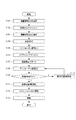
次に、図2のフローチャートを参照して、本実施形態の眼底画像取得システム500において、眼底の断層画像取得を行う撮影手順を説明する。
本発明の第2の実施形態について図面を参照しながら、以下に詳細に説明する。
本実施形態に係る光干渉断層画像取得システムの一態様としての眼底画像取得システム501について、図3を用いて説明する。当該画像取得システム501の基本構成は、第1の実施形態に係る眼底画像取得システム500と同様である。ただし、眼底画像取得システム500に対して、被検査物である被検眼の眼底観察のための走査型レーザー検眼鏡(SLO:Scanning Laser Ophthalmoscope)が追加されている点で異なっている。
次に、図4のフローチャートを参照して、本実施形態の眼底画像取得システム501において、被検眼の眼底の断層画像取得を行う撮影手順を説明する。
本発明の第3の実施形態について図面を参照しながら、以下に詳細に説明する。
本実施形態に係る光干渉断層画像取得システムの一態様としての眼底画像取得システム502について、図5を用いて説明する。当該眼底画像取得システム502の基本構成は、第1の実施形態に係る眼底画像取得システム500と同様である。ただし、第1の実施形態で参照光路に配置されていたミラー614−15の替わりに、ミラー614−1およびミラー614−4を用いている点で異なっている。
以上、本発明の好ましい実施形態について説明したが、本発明はこれらの実施形態に限定されないことはいうまでもなく、その要旨の範囲内で種々の変形および変更が可能である。
前述した実施形態において、被検眼607に照射される光が広帯域な光を生成する広帯域光源を用いたスペクトラルドメイン方式のOCTを用いた。本発明はこれに限定されるものではなく、例えば波長を掃引する光を生成する光源を用いた波長掃引型のOCTに適用しても良い。
605 参照光
606 測定光
607 被検眼
614−11 ミラー
614−12 ミラー
617−1 電動ステージ
617−2 電動ステージ
Claims (11)
- 光を出射する光源と、
前記光源からの光を参照光と測定光とに分割する分割手段と、
前記測定光を照射した被検査物からの戻り光と前記参照光とを合波して得た合波光に基づいて、前記被検査物の断層画像を形成する画像形成手段と、
前記測定光のフォーカスを調整するフォーカス調整手段と、
前記参照光の光路長を調整する光路長調整手段と、
前記フォーカス調整手段による前記フォーカス調整に連動して、前記光路長調整手段による前記参照光の光路長の調整を制御する制御手段とを備えることを特徴とする画像取得装置。 - 前記制御手段は、
前記フォーカス調整に伴う前記測定光の光路長の調整量に応じて、前記参照光の光路長を調整するように前記光路長調整手段を制御することを特徴とする請求項1に記載の画像取得装置。 - 前記形成された断層画像を、表示手段の画像表示領域に表示する表示制御手段を更に備え、
前記制御手段は、前記フォーカス調整により、前記断層画像の所定の画像が前記画像表示領域の所定の位置から離れる場合は、前記測定光の光路長の調整に基づいて前記光路長調整手段を制御することを特徴とする請求項1又は2に記載の画像取得装置。 - 前記被検査物にフォーカス調整された観察光を照射することにより、前記被検査物を観察する観察光学系をさらに備え、
前記制御手段は、前記観察光学系のフォーカス調整により前記フォーカス調整手段を制御することを特徴とする請求項1乃至3の何れか1項に記載の画像取得装置。 - 前記フォーカス調整手段は測定光学系と観察光学系の共通の光路に配置されることを特徴とする請求項4に記載の画像取得装置。
- 前記光路長調整手段は、複数の反射部材を含み、
前記フォーカス調整手段は、少なくとも1組の反射部材を含み、
前記制御手段は、前記少なくとも1組の反射部材の移動量に応じて、前記光路長調整手段の前記複数の反射部材を移動させるように制御することを特徴とする請求項1乃至5の何れか1項に記載の画像取得装置。 - 前記フォーカス調整手段と前記光路長調整手段は第1のステージで構成され、
前記光路長調整手段の前記複数の反射部材を搭載する前記第1のステージに搭載された第2のステージをさらに備え、
前記光路長調整手段は、前記参照光の光路長の調整が指示された場合は、前記参照光の光路長を前記第2のステージの移動により調整することを特徴とする請求項6に記載の画像取得装置。 - 光を出射する光源と、
前記光源からの光を参照光と測定光とに分割する分割手段と、
前記測定光を照射した被検査物からの戻り光と前記参照光とを合波して得た合波光に基づいて前記被検査物の断層画像を形成する画像形成手段と、
前記測定光のフォーカスを調整するフォーカス調整手段と、
前記参照光の光路長を調整する光路長調整手段と、
前記測定光のフォーカス調整を指示するフォーカス指示手段と、
前記参照光の光路長の調整を指示する光路長指示手段と、
前記フォーカス指示手段により前記測定光のフォーカス調整が指示された場合、前記フォーカス調整手段による調整と前記光路長調整手段による調整を連動させて制御し、前記光路長指示手段により前記参照光の光路長の調整が指示された場合、前記光路長調整手段による調整を前記フォーカス調整手段による調整と連動させずに制御する制御手段とを備えることを特徴とする画像取得装置。 - 前記制御手段は、前記フォーカス調整に伴う前記測定光の光路長の調整量に応じて、前記参照光の光路長を調整するように前記光路長調整手段を制御することを特徴とする請求項8に記載の画像取得装置。
- 前記形成された断層画像を表示手段の画像表示領域に表示する表示制御手段を更に備え、
前記制御手段は、前記フォーカス調整により、前記断層画像に含まれる所定の画像が前記画像表示領域の所定の位置に近づく場合は、前記測定光の光路長の調整に基づいた前記光路長調整手段の制御を行わないことを特徴とする請求項8又は9に記載の画像取得装置。 - 光源からの光を参照光と測定光とに分割する分割工程と、
前記測定光のフォーカスを調整するフォーカス調整と、前記参照光の光路長を調整する光路長調整とを連動するように制御する制御工程と、
前記測定光を照射した被検査物からの戻り光と前記参照光とを合波して得た合波光に基づいて、前記被検査物の断層画像を形成する画像形成工程とを備えることを特徴とする画像取得装置の制御方法。
Priority Applications (3)
| Application Number | Priority Date | Filing Date | Title |
|---|---|---|---|
| JP2017163475A JP2019037650A (ja) | 2017-08-28 | 2017-08-28 | 画像取得装置およびその制御方法 |
| PCT/JP2018/030043 WO2019044457A1 (ja) | 2017-08-28 | 2018-08-10 | 画像取得装置およびその制御方法 |
| US16/796,655 US11134841B2 (en) | 2017-08-28 | 2020-02-20 | Image acquisition apparatus and method for controlling the same |
Applications Claiming Priority (1)
| Application Number | Priority Date | Filing Date | Title |
|---|---|---|---|
| JP2017163475A JP2019037650A (ja) | 2017-08-28 | 2017-08-28 | 画像取得装置およびその制御方法 |
Publications (2)
| Publication Number | Publication Date |
|---|---|
| JP2019037650A true JP2019037650A (ja) | 2019-03-14 |
| JP2019037650A5 JP2019037650A5 (ja) | 2020-08-20 |
Family
ID=65525393
Family Applications (1)
| Application Number | Title | Priority Date | Filing Date |
|---|---|---|---|
| JP2017163475A Pending JP2019037650A (ja) | 2017-08-28 | 2017-08-28 | 画像取得装置およびその制御方法 |
Country Status (3)
| Country | Link |
|---|---|
| US (1) | US11134841B2 (ja) |
| JP (1) | JP2019037650A (ja) |
| WO (1) | WO2019044457A1 (ja) |
Families Citing this family (2)
| Publication number | Priority date | Publication date | Assignee | Title |
|---|---|---|---|---|
| EP3695261B1 (en) * | 2017-10-09 | 2023-06-07 | Nederlandse Organisatie voor toegepast- natuurwetenschappelijk onderzoek TNO | Refocusing device |
| WO2020257582A1 (en) * | 2019-06-20 | 2020-12-24 | Duke University | Systems and methods for multiplexing and alternating light of different wavelengths in an ophthalmoscope |
Citations (5)
| Publication number | Priority date | Publication date | Assignee | Title |
|---|---|---|---|---|
| JP2004191114A (ja) * | 2002-12-10 | 2004-07-08 | Naohiro Tanno | 光コヒーレンストモグラフィーにおける光干渉計一体駆動による検知点走査方法及び光コヒーレンストモグラフィー装置 |
| US20080024767A1 (en) * | 2006-07-28 | 2008-01-31 | Peter Seitz | Imaging optical coherence tomography with dynamic coherent focus |
| JP2015221091A (ja) * | 2014-05-22 | 2015-12-10 | 株式会社トプコン | 眼科装置 |
| JP2016049368A (ja) * | 2014-09-01 | 2016-04-11 | 株式会社ニデック | 眼科撮影装置 |
| WO2016121249A1 (ja) * | 2015-01-30 | 2016-08-04 | 浜松ホトニクス株式会社 | 干渉光学装置、干渉観察装置および干渉観察方法 |
Family Cites Families (16)
| Publication number | Priority date | Publication date | Assignee | Title |
|---|---|---|---|---|
| EP2278266A3 (en) * | 2004-11-24 | 2011-06-29 | The General Hospital Corporation | Common-Path Interferometer for Endoscopic OCT |
| EP1887312A1 (en) | 2006-07-28 | 2008-02-13 | Heliotis AG | Imaging optical coherence tomography with dynamic coherent Focus |
| US8395781B2 (en) * | 2007-07-12 | 2013-03-12 | Volcano Corporation | Automatic calibration systems and methods of use |
| US8125645B2 (en) * | 2008-03-31 | 2012-02-28 | Fujifilm Corporation | Optical tomographic imaging system, tomographic image acquiring method, and optical tomographic image forming method |
| JP5331395B2 (ja) * | 2008-07-04 | 2013-10-30 | 株式会社ニデック | 光断層像撮影装置 |
| JP5794664B2 (ja) * | 2011-01-20 | 2015-10-14 | キヤノン株式会社 | 断層画像生成装置及び断層画像生成方法 |
| JP5210443B1 (ja) * | 2012-01-26 | 2013-06-12 | キヤノン株式会社 | 光断層撮像装置および制御方法 |
| JP6007527B2 (ja) | 2012-03-13 | 2016-10-12 | 株式会社ニデック | 眼底撮影装置 |
| JP6186215B2 (ja) | 2013-09-04 | 2017-08-23 | 株式会社日立エルジーデータストレージ | 光計測装置及び光断層観察方法 |
| JP6429447B2 (ja) * | 2013-10-24 | 2018-11-28 | キヤノン株式会社 | 情報処理装置、比較方法、位置合わせ方法及びプログラム |
| JP2016041221A (ja) * | 2014-08-19 | 2016-03-31 | 株式会社トプコン | 眼科撮影装置およびその制御方法 |
| JP6808383B2 (ja) * | 2015-09-30 | 2021-01-06 | キヤノン株式会社 | 光干渉断層撮影装置、その制御方法および光干渉断層撮影システム |
| US11452442B2 (en) * | 2016-06-15 | 2022-09-27 | Oregon Health & Science University | Systems and methods for automated widefield optical coherence tomography angiography |
| JP6731868B2 (ja) * | 2017-02-17 | 2020-07-29 | 株式会社Screenホールディングス | 撮像方法および撮像装置 |
| JP6929684B2 (ja) * | 2017-04-06 | 2021-09-01 | キヤノン株式会社 | 眼科撮影装置及びその制御方法 |
| WO2019117036A1 (ja) * | 2017-12-14 | 2019-06-20 | キヤノン株式会社 | 撮像装置及びその制御方法 |
-
2017
- 2017-08-28 JP JP2017163475A patent/JP2019037650A/ja active Pending
-
2018
- 2018-08-10 WO PCT/JP2018/030043 patent/WO2019044457A1/ja active Application Filing
-
2020
- 2020-02-20 US US16/796,655 patent/US11134841B2/en active Active
Patent Citations (5)
| Publication number | Priority date | Publication date | Assignee | Title |
|---|---|---|---|---|
| JP2004191114A (ja) * | 2002-12-10 | 2004-07-08 | Naohiro Tanno | 光コヒーレンストモグラフィーにおける光干渉計一体駆動による検知点走査方法及び光コヒーレンストモグラフィー装置 |
| US20080024767A1 (en) * | 2006-07-28 | 2008-01-31 | Peter Seitz | Imaging optical coherence tomography with dynamic coherent focus |
| JP2015221091A (ja) * | 2014-05-22 | 2015-12-10 | 株式会社トプコン | 眼科装置 |
| JP2016049368A (ja) * | 2014-09-01 | 2016-04-11 | 株式会社ニデック | 眼科撮影装置 |
| WO2016121249A1 (ja) * | 2015-01-30 | 2016-08-04 | 浜松ホトニクス株式会社 | 干渉光学装置、干渉観察装置および干渉観察方法 |
Also Published As
| Publication number | Publication date |
|---|---|
| US11134841B2 (en) | 2021-10-05 |
| US20200187773A1 (en) | 2020-06-18 |
| WO2019044457A1 (ja) | 2019-03-07 |
Similar Documents
| Publication | Publication Date | Title |
|---|---|---|
| EP2371273B1 (en) | Method of operating an optical tomographic image photographing apparatus | |
| JP4819478B2 (ja) | 眼科撮影装置 | |
| US7824035B2 (en) | Ophthalmic photographing apparatus | |
| KR101670698B1 (ko) | 광간섭 단층촬상장치 및 그 제어 방법 | |
| KR20130086969A (ko) | 광 간섭 단층 촬상 장치, 광 간섭 단층 촬상 장치 제어 방법 및 저장 매체 | |
| JP6552200B2 (ja) | 光断層撮像装置、その制御方法、及びプログラム | |
| US10568504B2 (en) | Ophthalmologic apparatus | |
| JP4949504B2 (ja) | 眼科撮影装置 | |
| JP5997457B2 (ja) | 撮像装置及び撮像装置の制御方法 | |
| US20200297209A1 (en) | Imaging apparatus and control method therefor | |
| JP2011245183A (ja) | 眼底撮影装置 | |
| JP7027698B2 (ja) | 眼科撮影装置 | |
| US11134841B2 (en) | Image acquisition apparatus and method for controlling the same | |
| JP6901264B2 (ja) | 眼科装置 | |
| JP5255711B2 (ja) | 眼科撮影装置 | |
| JP7129162B2 (ja) | 眼底撮像装置 | |
| JP2018023675A (ja) | 光断層撮像装置 | |
| JP5319010B2 (ja) | 眼科撮影装置 | |
| JP7123626B2 (ja) | 眼底撮影装置およびその制御方法 | |
| JP6779674B2 (ja) | Oct装置 | |
| JP7086688B2 (ja) | 画像撮影装置およびその制御方法 | |
| JP7179523B2 (ja) | 眼底撮像装置、眼底撮像方法およびプログラム | |
| JP5306554B2 (ja) | 眼科撮影装置 | |
| JP6916011B2 (ja) | 眼科装置 | |
| JP7029324B2 (ja) | 光源制御装置、眼科装置、及び光源制御方法 |
Legal Events
| Date | Code | Title | Description |
|---|---|---|---|
| A521 | Request for written amendment filed |
Free format text: JAPANESE INTERMEDIATE CODE: A523 Effective date: 20200708 |
|
| A621 | Written request for application examination |
Free format text: JAPANESE INTERMEDIATE CODE: A621 Effective date: 20200708 |
|
| A131 | Notification of reasons for refusal |
Free format text: JAPANESE INTERMEDIATE CODE: A131 Effective date: 20210824 |
|
| A521 | Request for written amendment filed |
Free format text: JAPANESE INTERMEDIATE CODE: A523 Effective date: 20211018 |
|
| A02 | Decision of refusal |
Free format text: JAPANESE INTERMEDIATE CODE: A02 Effective date: 20220315 |