JP2010142641A - 赤外蛍光を観察するための手術用顕微鏡、顕微鏡検査方法、および手術用顕微鏡の使用 - Google Patents
赤外蛍光を観察するための手術用顕微鏡、顕微鏡検査方法、および手術用顕微鏡の使用 Download PDFInfo
- Publication number
- JP2010142641A JP2010142641A JP2009286310A JP2009286310A JP2010142641A JP 2010142641 A JP2010142641 A JP 2010142641A JP 2009286310 A JP2009286310 A JP 2009286310A JP 2009286310 A JP2009286310 A JP 2009286310A JP 2010142641 A JP2010142641 A JP 2010142641A
- Authority
- JP
- Japan
- Prior art keywords
- light
- camera
- beam splitter
- surgical microscope
- input port
- Prior art date
- Legal status (The legal status is an assumption and is not a legal conclusion. Google has not performed a legal analysis and makes no representation as to the accuracy of the status listed.)
- Granted
Links
Images
Classifications
-
- G—PHYSICS
- G02—OPTICS
- G02B—OPTICAL ELEMENTS, SYSTEMS OR APPARATUS
- G02B21/00—Microscopes
- G02B21/0004—Microscopes specially adapted for specific applications
- G02B21/0012—Surgical microscopes
-
- G—PHYSICS
- G02—OPTICS
- G02B—OPTICAL ELEMENTS, SYSTEMS OR APPARATUS
- G02B21/00—Microscopes
- G02B21/0004—Microscopes specially adapted for specific applications
- G02B21/002—Scanning microscopes
- G02B21/0024—Confocal scanning microscopes (CSOMs) or confocal "macroscopes"; Accessories which are not restricted to use with CSOMs, e.g. sample holders
- G02B21/0052—Optical details of the image generation
- G02B21/0064—Optical details of the image generation multi-spectral or wavelength-selective arrangements, e.g. wavelength fan-out, chromatic profiling
-
- G—PHYSICS
- G02—OPTICS
- G02B—OPTICAL ELEMENTS, SYSTEMS OR APPARATUS
- G02B21/00—Microscopes
- G02B21/0004—Microscopes specially adapted for specific applications
- G02B21/002—Scanning microscopes
- G02B21/0024—Confocal scanning microscopes (CSOMs) or confocal "macroscopes"; Accessories which are not restricted to use with CSOMs, e.g. sample holders
- G02B21/0052—Optical details of the image generation
- G02B21/0076—Optical details of the image generation arrangements using fluorescence or luminescence
-
- G—PHYSICS
- G02—OPTICS
- G02B—OPTICAL ELEMENTS, SYSTEMS OR APPARATUS
- G02B21/00—Microscopes
- G02B21/16—Microscopes adapted for ultraviolet illumination ; Fluorescence microscopes
-
- G—PHYSICS
- G02—OPTICS
- G02B—OPTICAL ELEMENTS, SYSTEMS OR APPARATUS
- G02B21/00—Microscopes
- G02B21/18—Arrangements with more than one light path, e.g. for comparing two specimens
- G02B21/20—Binocular arrangements
- G02B21/22—Stereoscopic arrangements
-
- G—PHYSICS
- G02—OPTICS
- G02B—OPTICAL ELEMENTS, SYSTEMS OR APPARATUS
- G02B27/00—Optical systems or apparatus not provided for by any of the groups G02B1/00 - G02B26/00, G02B30/00
- G02B27/10—Beam splitting or combining systems
- G02B27/1006—Beam splitting or combining systems for splitting or combining different wavelengths
- G02B27/1013—Beam splitting or combining systems for splitting or combining different wavelengths for colour or multispectral image sensors, e.g. splitting an image into monochromatic image components on respective sensors
-
- G—PHYSICS
- G02—OPTICS
- G02B—OPTICAL ELEMENTS, SYSTEMS OR APPARATUS
- G02B27/00—Optical systems or apparatus not provided for by any of the groups G02B1/00 - G02B26/00, G02B30/00
- G02B27/10—Beam splitting or combining systems
- G02B27/14—Beam splitting or combining systems operating by reflection only
- G02B27/144—Beam splitting or combining systems operating by reflection only using partially transparent surfaces without spectral selectivity
-
- G—PHYSICS
- G02—OPTICS
- G02B—OPTICAL ELEMENTS, SYSTEMS OR APPARATUS
- G02B27/00—Optical systems or apparatus not provided for by any of the groups G02B1/00 - G02B26/00, G02B30/00
- G02B27/10—Beam splitting or combining systems
- G02B27/14—Beam splitting or combining systems operating by reflection only
- G02B27/145—Beam splitting or combining systems operating by reflection only having sequential partially reflecting surfaces
-
- G—PHYSICS
- G02—OPTICS
- G02B—OPTICAL ELEMENTS, SYSTEMS OR APPARATUS
- G02B27/00—Optical systems or apparatus not provided for by any of the groups G02B1/00 - G02B26/00, G02B30/00
- G02B27/10—Beam splitting or combining systems
- G02B27/14—Beam splitting or combining systems operating by reflection only
- G02B27/149—Beam splitting or combining systems operating by reflection only using crossed beamsplitting surfaces, e.g. cross-dichroic cubes or X-cubes
Landscapes
- Physics & Mathematics (AREA)
- General Physics & Mathematics (AREA)
- Optics & Photonics (AREA)
- Chemical & Material Sciences (AREA)
- Analytical Chemistry (AREA)
- Spectroscopy & Molecular Physics (AREA)
- Health & Medical Sciences (AREA)
- General Health & Medical Sciences (AREA)
- Surgery (AREA)
- Microscoopes, Condenser (AREA)
- Investigating, Analyzing Materials By Fluorescence Or Luminescence (AREA)
- Optical Elements Other Than Lenses (AREA)
- Color Television Image Signal Generators (AREA)
Abstract
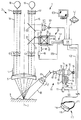
【解決手段】赤外蛍光を観察するための手術用顕微鏡は、3つのチップ35、36、37を有するカメラシステム25を含み、対象物9から発する赤外光は、ダイクロイックビームスプリッタ33を介して3つのカメラチップのうちの1つのみに供給される。
【選択図】図1
Description
本発明は、赤外蛍光を観察するための手術用顕微鏡、および赤外蛍光を観察するための顕微鏡検査方法に関する。
蛍光色素は、特定の種類の組織、組織構造、組織機能、組織灌流の視覚化や他の目的などのさまざまな目的で、医療用途および生物学において用いられる。ここでは、蛍光色素またはそのような蛍光色素の前駆体が、組織試料または検査中の患者に塗布される。色素または前駆体はそれぞれ、組織試料または患者の特定の種類の組織および組織構造に蓄積する。組織は、蛍光を励起するための励起光で照らされ、ここで蛍光色素内に発生する蛍光によって生成された蛍光光が検出されて、特定の組織構造、組織の種類および組織灌流を視覚化し得る。手術用顕微鏡を用いて、対象物を励起光で照らし、蛍光光を検出することができる。
本発明は、上記の技術的課題を考慮して達成された。
本発明のさらなる目的は、蛍光画像および標準の光画像を検出可能な顕微鏡検査方法を提供することである。
以下に説明する例示的な実施例では、機能および構造が同様の構成部品はできる限り同様の参照番号で示す。
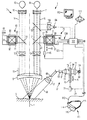
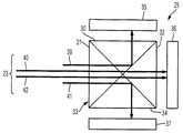
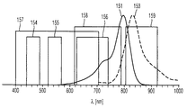
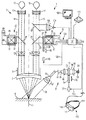
図4に示される手術用顕微鏡は、上述の図1および図3を参照して説明した手術用顕微鏡と同様の構成を有し、同様の機能を提供する。図4の手術用顕微鏡は、カメラシステム25の上流でビーム経路内に配置されたフィルタプレート26が第1のブロックフィルタ27および第2のブロックフィルタ28を含むという点で、上述の手術用顕微鏡と主に異なる。第1のブロックフィルタ27は、赤外光がフィルタ27を通るのを阻止するように構成される。第1のフィルタ27は、対象物9の標準の光画像がカメラシステム25で検出される手術用顕微鏡1の第1の動作モードにおいて、対象物9をカメラチップ35、36および37上に撮像するビーム23内に位置決めされる。このため、第1のブロックフィルタ27は、カメラシステム25によって検出される画像の形成に赤外光が寄与しないように、赤外光を阻止する。
Claims (17)
- 赤外蛍光を観察するための手術用顕微鏡であって、
入力ポート、ダイクロイックビームスプリッタ、ならびに第1、第2および第3のカメラチップを有するカメラシステムを備え、前記ダイクロイックビームスプリッタは、前記入力ポートで受けた赤色光を、前記ビームスプリッタの第1の出力ポートを介して前記第1のカメラチップに主に導くように、かつ前記入力ポートで受けた緑色光を、前記ビームスプリッタの第2の出力ポートを介して前記第2のカメラチップに主に導くように、かつ前記入力ポートで受けた青色光を、前記ビームスプリッタの第3の出力ポートを介して前記第3のカメラチップに主に導くように、かつ前記入力ポートで受けた赤外光を、前記第1、第2および第3のカメラチップのうちの1つのみに主に導くように構成され、前記手術用顕微鏡はさらに、
対象領域を前記カメラシステムの前記カメラチップ上に光学的に撮像するように構成された顕微鏡光学部品と、
前記カメラシステムの前記カメラチップによって検出された光強度に基づいて画像を表示するように構成された表示システムとを備える、手術用顕微鏡。 - 前記ビームスプリッタは、前記入力ポートで受けた前記赤外光を、前記第2および第3のカメラチップの一方のみに主に導くように構成される、請求項1に記載の手術用顕微鏡。
- 赤外光を実質的に阻止し、前記対象領域と前記ダイクロイックビームスプリッタの前記入力ポートとの間の撮像ビーム経路の内側および外側に選択的に位置決めされ得る第1のブロックフィルタをさらに備える、請求項1または2に記載の手術用顕微鏡。
- 赤色光、緑色光および青色光の少なくとも1つを実質的に阻止し、前記対象領域と前記ダイクロイックビームスプリッタの前記入力ポートとの間の撮像ビーム経路の内側および外側に選択的に位置決めされ得る第2のブロックフィルタをさらに備える、請求項1から3の1つに記載の手術用顕微鏡。
- 前記第1および第2のブロックフィルタに結合され、所与の時間に前記第1および第2のブロックフィルタの一方のみが前記撮像ビーム経路内に位置決めされるように構成された作動システムをさらに備える、請求項3と組合された請求項4に記載の手術用顕微鏡。
- 少なくとも1本の照明光ビームを前記対象領域に導くように構成された照明システムと、
赤色光、緑色光および青色光の少なくとも1つを実質的に阻止し、前記照明システムのビーム経路の内側および外側に選択的に位置決めされ得る第3のブロックフィルタとをさらに備える、請求項1から5の1つに記載の手術用顕微鏡。 - 前記第1および第3のブロックフィルタに結合され、所与の時間に前記第1および第3のブロックフィルタの一方のみが前記撮像ビーム経路内および前記照明システムの前記ビーム経路内にそれぞれ位置決めされるように構成された作動システムをさらに備える、請求項3と組合された請求項6に記載の手術用顕微鏡。
- 前記ダイクロイックビームスプリッタはさらに、前記第1のカメラチップに主に導かれる前記赤色光が前記ダイクロイックビームスプリッタの前記入力ポートに供給される場合、第1の検出器によって検出される光強度と、第2の検出器によって検出される光強度および第3の検出器によって検出される光強度の合計との比率が1.8、2.5および3.0のうちの1つよりも大きく、
前記ダイクロイックビームスプリッタはさらに、前記第2のカメラチップに主に導かれる前記緑色光が前記ダイクロイックビームスプリッタの前記入力ポートに供給される場合、前記第2の検出器によって検出される光強度と、前記第1の検出器によって検出される光強度および前記第3の検出器によって検出される光強度の合計との比率が1.8、2.5および3.0のうちの1つよりも大きく、
前記ダイクロイックビームスプリッタはさらに、前記第3のカメラチップに主に導かれる前記青色光が前記ダイクロイックビームスプリッタの前記入力ポートに供給される場合、前記第3の検出器によって検出される光強度と、前記第1の検出器によって検出される光強度および前記第2の検出器によって検出される光強度の合計との比率が1.8、2.5および3.0のうちの1つよりも大きい、請求項1から7の1つに記載の手術用顕微鏡。 - 前記ダイクロイックビームスプリッタはさらに、前記第1、第2および第3のカメラチップのうちの1つのみに主に導かれる前記赤外光が前記ダイクロイックビームスプリッタの前記入力ポートに供給される場合、前記第1、第2および第3のカメラチップのうちの前記1つによって検出される光強度と、その他の前記第1、第2および第3のカメラチップによって検出される光強度の合計との比率が1.8、2.5および3.0のうちの1つよりも大きい、請求項1から8の1つに記載の手術用顕微鏡。
- 前記赤外光は、波長範囲が800nmから930nmの光を含む、請求項1から9の1つに記載の手術用顕微鏡。
- 前記青色光は、波長範囲が440nmから490nmの光を含み、前記緑色光は、波長範囲が520mから570nmの光を含み、前記赤色光は、波長範囲が625nmから740nmの光を含む、請求項1から10の1つに記載の手術用顕微鏡。
- 顕微鏡検査方法であって、
顕微鏡を第1の動作モードで動作させるステップを備え、
前記第1の動作モードは、
赤色光、緑色光および青色光を含む光を対象物に供給するステップと、
前記対象物から発する赤色光を第1のカメラチップに供給するステップと、
前記対象物から発する緑色光を第2のカメラチップに供給するステップと、
前記対象物から発する青色光を第3のカメラチップに供給するステップとを含み、前記方法はさらに、
前記顕微鏡を第2の動作モードで動作させるステップを備え、
前記第2の動作モードは、
前記対象物において蛍光を励起するステップと、
前記蛍光によって生成された赤外光を、前記対象物から、前記第1、第2および第3のカメラチップのうちの1つのみに主に供給するステップとを含む、顕微鏡検査方法。 - 前記第1の動作モードにおいてのみ、赤外光が前記第1、第2および第3のカメラチップのうちの1つに供給されるのを阻止するステップをさらに備える、請求項12に記載の顕微鏡検査方法。
- 前記第2の動作モードにおいて、波長が700nmよりも大きく805nmよりも小さい光が前記第1、第2および第3のカメラチップのうちの1つに供給されるのを阻止するステップをさらに備える、請求項12または13に記載の顕微鏡検査方法。
- 前記第2の動作モードにおいて、赤色光、緑色光および青色光のうちの1つが前記第1、第2および第3のカメラチップのうちの1つに供給されるのを阻止するステップをさらに備える、請求項12から14の1つに記載の顕微鏡検査方法。
- 前記第2の動作モードにおいて、赤色光、緑色光および青色光のうち2つ以下のものを前記対象物に供給するステップをさらに備える、請求項12から15の1つに記載の顕微鏡検査方法。
- 請求項12から16の1つに記載の方法を実行するための、請求項1から11の1つに記載の手術用顕微鏡の使用。
Applications Claiming Priority (2)
| Application Number | Priority Date | Filing Date | Title |
|---|---|---|---|
| DE102008062650.3 | 2008-12-17 | ||
| DE102008062650.3A DE102008062650B9 (de) | 2008-12-17 | 2008-12-17 | Operationsmikroskop zur Beobachtung einer Infrarot-Fluoreszenz und Verfahren hierzu |
Related Child Applications (1)
| Application Number | Title | Priority Date | Filing Date |
|---|---|---|---|
| JP2014182157A Division JP6062405B2 (ja) | 2008-12-17 | 2014-09-08 | 赤外蛍光を観察するための手術用顕微鏡、顕微鏡検査方法、および手術用顕微鏡の使用 |
Publications (3)
| Publication Number | Publication Date |
|---|---|
| JP2010142641A true JP2010142641A (ja) | 2010-07-01 |
| JP2010142641A5 JP2010142641A5 (ja) | 2013-01-31 |
| JP5646844B2 JP5646844B2 (ja) | 2014-12-24 |
Family
ID=41582131
Family Applications (3)
| Application Number | Title | Priority Date | Filing Date |
|---|---|---|---|
| JP2009286310A Active JP5646844B2 (ja) | 2008-12-17 | 2009-12-17 | 赤外蛍光を観察するための手術用顕微鏡 |
| JP2014182157A Active JP6062405B2 (ja) | 2008-12-17 | 2014-09-08 | 赤外蛍光を観察するための手術用顕微鏡、顕微鏡検査方法、および手術用顕微鏡の使用 |
| JP2016165862A Pending JP2017021356A (ja) | 2008-12-17 | 2016-08-26 | 赤外蛍光を観察するための手術用顕微鏡、顕微鏡検査方法、および手術用顕微鏡の使用 |
Family Applications After (2)
| Application Number | Title | Priority Date | Filing Date |
|---|---|---|---|
| JP2014182157A Active JP6062405B2 (ja) | 2008-12-17 | 2014-09-08 | 赤外蛍光を観察するための手術用顕微鏡、顕微鏡検査方法、および手術用顕微鏡の使用 |
| JP2016165862A Pending JP2017021356A (ja) | 2008-12-17 | 2016-08-26 | 赤外蛍光を観察するための手術用顕微鏡、顕微鏡検査方法、および手術用顕微鏡の使用 |
Country Status (5)
| Country | Link |
|---|---|
| US (1) | US8659651B2 (ja) |
| EP (1) | EP2199842B1 (ja) |
| JP (3) | JP5646844B2 (ja) |
| DE (1) | DE102008062650B9 (ja) |
| ES (1) | ES2563284T3 (ja) |
Cited By (7)
| Publication number | Priority date | Publication date | Assignee | Title |
|---|---|---|---|---|
| JP2013531538A (ja) * | 2010-07-02 | 2013-08-08 | インテュイティブ サージカル オペレーションズ, インコーポレイテッド | 選択的照明スペクトルよりなる背景外科画像を有する蛍光画像化方法及びシステム |
| JP2014104365A (ja) * | 2012-11-29 | 2014-06-09 | Sony Corp | 手術用無線ルーペ |
| JP2016161946A (ja) * | 2015-03-04 | 2016-09-05 | カール・ツアイス・メディテック・アーゲー | 手術用顕微鏡 |
| JP2017006620A (ja) * | 2015-06-23 | 2017-01-12 | 三鷹光器株式会社 | 手術用立体観察装置 |
| US10682198B2 (en) | 2010-07-02 | 2020-06-16 | Intuitive Surgical Operations, Inc. | Method and system for fluorescent imaging with background surgical image composed of selective illumination spectra |
| KR20200111370A (ko) * | 2019-03-19 | 2020-09-29 | 한국광기술원 | 증강현실 구현을 위한 빔 스플리터 및 이를 포함한 증강현실 광학계 |
| JP2023539941A (ja) * | 2020-09-08 | 2023-09-20 | ミュニック サージカル イメージング ゲセルシャフト ミット ベシュレンクテル ハフツング | 少なくとも2つのカメラ装置を有する手術用顕微鏡 |
Families Citing this family (44)
| Publication number | Priority date | Publication date | Assignee | Title |
|---|---|---|---|---|
| DE102008029661A1 (de) * | 2008-06-24 | 2009-12-31 | Khs Ag | Redundante Inspektion |
| DE102008062650B9 (de) * | 2008-12-17 | 2021-10-28 | Carl Zeiss Meditec Ag | Operationsmikroskop zur Beobachtung einer Infrarot-Fluoreszenz und Verfahren hierzu |
| DE102010033825B9 (de) * | 2010-08-09 | 2024-04-18 | Carl Zeiss Meditec Ag | Fluoreszenzbeobachtungssystem und Filtersatz |
| DE102010044502A1 (de) * | 2010-09-06 | 2012-03-08 | Leica Microsystems (Schweiz) Ag | Sonderbeleuchtungs-Video-Operations-Stereomikroskop |
| DE102010044503B4 (de) * | 2010-09-06 | 2017-11-02 | Leica Microsystems (Schweiz) Ag | Fluoreszenz-Operations-Stereomikroskop |
| DE102010063960A1 (de) * | 2010-12-22 | 2012-06-28 | Carl Zeiss Microlmaging Gmbh | Kamera mit einem Farbbildsensor sowie Aufnahmeverfahren mit einer solchen Kamera |
| DE102011016138A1 (de) * | 2011-03-30 | 2012-10-04 | Karl Storz Gmbh & Co. Kg | Vorrichtung zur Fluoreszenzdiagnose |
| DE102011017046A1 (de) * | 2011-04-14 | 2012-10-18 | Till Photonics Gmbh | Umschaltbare Mikroskopanordnung mit mehreren Detektoren |
| JP2013003495A (ja) * | 2011-06-21 | 2013-01-07 | Mitaka Koki Co Ltd | 顕微鏡システム |
| DE102012000675A1 (de) | 2012-01-17 | 2013-07-18 | Bundesrepublik Deutschland, vertr.d.d. Bundesministerium für Wirtschaft und Technologie, d.vertr.d.d. Präsidenten der Physikalisch-Technischen Bundesanstalt | Medizinisches Fluoreszenz-Untersuchungssystem |
| US9371555B2 (en) * | 2012-06-01 | 2016-06-21 | Concordia Laboratories Inc. | Lighting systems and methods of using lighting systems for in vitro potency assay for photofrin |
| WO2015042700A1 (en) | 2013-09-24 | 2015-04-02 | Motion Engine Inc. | Mems components and method of wafer-level manufacturing thereof |
| WO2015013827A1 (en) | 2013-08-02 | 2015-02-05 | Motion Engine Inc. | Mems motion sensor for sub-resonance angular rate sensing |
| WO2015151812A1 (ja) | 2014-04-03 | 2015-10-08 | 株式会社日立ハイテクノロジーズ | 蛍光分析器 |
| WO2015154173A1 (en) | 2014-04-10 | 2015-10-15 | Motion Engine Inc. | Mems pressure sensor |
| US11674803B2 (en) | 2014-06-02 | 2023-06-13 | Motion Engine, Inc. | Multi-mass MEMS motion sensor |
| CA3004760A1 (en) | 2014-12-09 | 2016-06-16 | Motion Engine Inc. | 3d mems magnetometer and associated methods |
| DE102014118382B4 (de) | 2014-12-11 | 2020-07-02 | Carl Zeiss Meditec Ag | Optisches Beobachtungsgerät und Verfahren zum Betreiben eines optischen Beobachtungsgerätes. |
| WO2016112463A1 (en) | 2015-01-15 | 2016-07-21 | Motion Engine Inc. | 3d mems device with hermetic cavity |
| US10295815B2 (en) | 2015-02-09 | 2019-05-21 | Arizona Board Of Regents On Behalf Of The University Of Arizona | Augmented stereoscopic microscopy |
| DE102015202605B4 (de) | 2015-02-12 | 2017-03-09 | Carl Zeiss Meditec Ag | Visualisierungssystem |
| CN104793326A (zh) * | 2015-04-30 | 2015-07-22 | 麦克奥迪实业集团有限公司 | 一种具有同轴照明效果的体视显微镜 |
| CN107923851A (zh) * | 2015-06-02 | 2018-04-17 | 国立大学法人旭川医科大学 | 观察辅助装置、信息处理方法以及程序 |
| US10063850B2 (en) * | 2015-06-23 | 2018-08-28 | Mitaka Kohki Co., Ltd. | Surgical stereoscopic observation apparatus |
| WO2017011643A1 (en) | 2015-07-14 | 2017-01-19 | Tsme, Llc | Surgical optical system with heads up display |
| DE102015216570A1 (de) * | 2015-08-31 | 2016-11-03 | Carl Zeiss Meditec Ag | Mikroskopiesystem |
| DE102016104439A1 (de) * | 2015-08-31 | 2017-03-02 | Carl Zeiss Meditec Ag | Bildaufnahmeanordnung, optisches Beobachtungsgerät und Verfahren zum Aufnehmen von Bildern |
| DE102017109021B4 (de) * | 2016-05-11 | 2022-10-27 | Carl Zeiss Meditec Ag | System für das stereoskopische Visualisieren eines Objektbereichs sowie Objektbereich-Visualisierungsverfahren |
| US10299880B2 (en) * | 2017-04-24 | 2019-05-28 | Truevision Systems, Inc. | Stereoscopic visualization camera and platform |
| JP7293227B2 (ja) | 2017-12-12 | 2023-06-19 | アルコン インコーポレイティド | 顕微鏡の短小鏡筒内での近赤外光イメージング及び可視光イメージングの結合 |
| RU2687800C1 (ru) * | 2018-04-18 | 2019-05-16 | Илья Николаевич Дмитриев | Цифровой стереомикроскоп |
| DE102018110643B3 (de) | 2018-05-03 | 2019-07-25 | Carl Zeiss Meditec Ag | Stereo-Mikroskop und Verfahren zur Stereomikroskopie |
| JPWO2019225693A1 (ja) * | 2018-05-24 | 2021-06-24 | 学校法人慶應義塾 | 手術用観察システム |
| KR102698248B1 (ko) | 2018-08-10 | 2024-08-23 | 삼성디스플레이 주식회사 | 표시 패널 검사 장치 |
| WO2021106611A1 (ja) * | 2019-11-28 | 2021-06-03 | ソニーグループ株式会社 | 手術顕微鏡システム、および顕微鏡カメラアダプタ |
| EP4009095A1 (en) * | 2020-12-02 | 2022-06-08 | Leica Instruments (Singapore) Pte. Ltd. | Hybrid microscope |
| KR102549929B1 (ko) * | 2021-08-05 | 2023-06-30 | 주식회사 메타플바이오 | 3차원 광 검출 장치 |
| DE102022116292A1 (de) * | 2022-06-30 | 2024-01-04 | Karl Storz Se & Co. Kg | Strahlteilungsvorrichtung für einen distalen Endabschnitt eines Endoskops, Objektivsystem und Endoskop |
| DE102022125852A1 (de) * | 2022-10-06 | 2024-04-11 | Karl Storz Se & Co. Kg | Medizinische Bildgebungsvorrichtung und Verfahren zur medizinischen Bildgebung |
| US12332507B2 (en) * | 2022-11-18 | 2025-06-17 | Designs For Vision, Inc. | Examination/visualization/collection system with light enhancement |
| US12038630B1 (en) | 2022-11-18 | 2024-07-16 | Designs For Vision, Inc. | Telescopic image capture/recording device |
| EP4403089A1 (de) * | 2023-01-17 | 2024-07-24 | Erbe Elektromedizin GmbH | Visualisierungseinrichtung |
| WO2024158609A1 (en) * | 2023-01-23 | 2024-08-02 | University Of Pittsburgh-Of The Commonwealth System Of Higher Education | Hyper-throughput, multiplex, single molecule imaging platform |
| DE102024107271B3 (de) * | 2024-03-14 | 2025-05-22 | Jenoptik Optical Systems Gmbh | Lichtleiteinrichtung und Verfahren zum Betreiben einer Lichtleiteinrichtung |
Citations (2)
| Publication number | Priority date | Publication date | Assignee | Title |
|---|---|---|---|---|
| JP2004520105A (ja) * | 2000-12-19 | 2004-07-08 | ハイシャン ゼン、 | 蛍光及び反射率撮像と分光のための方法及び装置並びに多数の測定装置による電磁放射線の同時測定のための方法及び装置 |
| WO2007085496A1 (de) * | 2006-01-30 | 2007-08-02 | Carl Zeiss Surgical Gmbh | Mikroskopiesystem |
Family Cites Families (30)
| Publication number | Priority date | Publication date | Assignee | Title |
|---|---|---|---|---|
| US4045772A (en) * | 1974-04-29 | 1977-08-30 | Geometric Data Corporation | Automatic focusing system |
| JPH0282876A (ja) * | 1988-09-20 | 1990-03-23 | Sony Corp | カラーモード・赤外モード共用ビデオカメラ |
| GB2254417A (en) * | 1991-04-05 | 1992-10-07 | Bijan Jouza | Photodynamic laser detection for cancer diagnosis |
| CA2042075C (en) * | 1991-05-08 | 2001-01-23 | Branko Palcic | Endoscopic imaging system |
| JPH06141247A (ja) * | 1992-10-22 | 1994-05-20 | Sony Corp | 固体撮像装置 |
| GB9302799D0 (en) * | 1993-02-12 | 1993-03-31 | Medical Res Council | Detecting means for a scanning optical microscope |
| US5590660A (en) * | 1994-03-28 | 1997-01-07 | Xillix Technologies Corp. | Apparatus and method for imaging diseased tissue using integrated autofluorescence |
| JPH08275182A (ja) * | 1995-03-28 | 1996-10-18 | Canon Inc | カラーモード・赤外モード共用型テレビカメラ |
| US5682038A (en) * | 1995-04-06 | 1997-10-28 | Becton Dickinson And Company | Fluorescent-particle analyzer with timing alignment for analog pulse subtraction of fluorescent pulses arising from different excitation locations |
| US5808329A (en) * | 1996-07-15 | 1998-09-15 | Raytheon Company | Low light level imager with extended wavelength response employing atomic bonded (fused) semiconductor materials |
| DE69834963T2 (de) * | 1997-04-09 | 2007-01-25 | 1192062 Alberta Ltd., Calgary | Farbumsetzendes UV-Mikroskop |
| EP1403675B1 (en) | 1997-04-09 | 2006-06-14 | 1192062 Alberta Limited | Colour translating UV microscope |
| US5986271A (en) * | 1997-07-03 | 1999-11-16 | Lazarev; Victor | Fluorescence imaging system |
| US6198147B1 (en) | 1998-07-06 | 2001-03-06 | Intel Corporation | Detecting infrared and visible light |
| WO2002007587A2 (en) * | 2000-07-14 | 2002-01-31 | Xillix Technologies Corporation | Compact fluorescent endoscopy video system |
| US6899675B2 (en) * | 2002-01-15 | 2005-05-31 | Xillix Technologies Corp. | Fluorescence endoscopy video systems with no moving parts in the camera |
| JP2003270129A (ja) * | 2002-03-18 | 2003-09-25 | Yokogawa Electric Corp | 画像測定装置 |
| DE10362402B3 (de) | 2002-08-28 | 2022-03-03 | Carl Zeiss Meditec Ag | Mikroskopiesystem und Mikroskopieverfahren |
| DE10251151B4 (de) * | 2002-10-31 | 2021-01-28 | Leica Microsystems Cms Gmbh | Konfokales Rastermikroskop zum Abbilden eines Lumineszenzobjekts und Verfahren zum Abbilden eines Lumineszenzobjekts |
| US8473035B2 (en) | 2003-09-15 | 2013-06-25 | Beth Israel Deaconess Medical Center | Medical imaging systems |
| DE102005005253B4 (de) * | 2004-02-11 | 2018-06-28 | Carl Zeiss Ag | Fluoreszenzbeobachtungssystem und Fluoreszenzbeobachtungsverfahren |
| US8556806B2 (en) * | 2004-09-24 | 2013-10-15 | Vivid Medical, Inc. | Wavelength multiplexing endoscope |
| US7508507B2 (en) * | 2004-10-12 | 2009-03-24 | Leica Microsystems Cms Gmbh | Device for selecting and detecting at least two spectral regions of a light beam |
| US7507565B2 (en) * | 2005-03-11 | 2009-03-24 | Stanford University | Bioluminescence resonance energy transfer (BRET) systems and methods of use thereof |
| US7526116B2 (en) * | 2006-01-19 | 2009-04-28 | Luigi Armogida | Automated microscopic sperm identification |
| JP4971816B2 (ja) * | 2007-02-05 | 2012-07-11 | 三洋電機株式会社 | 撮像装置 |
| EP2075616A1 (de) * | 2007-12-28 | 2009-07-01 | Möller-Wedel GmbH | Vorrichtung mit einer Kamera und einer Einrichtung zum Abbilden und Projizieren des aufgenommen Bildes |
| DE102008004596A1 (de) | 2008-01-16 | 2008-06-19 | Carl Zeiss Surgical Gmbh | Fluoreszenzmikroskop und Beleuchtungsystem hierfür |
| US7645971B2 (en) * | 2008-01-18 | 2010-01-12 | Ffei Limited | Image scanning apparatus and method |
| DE102008062650B9 (de) * | 2008-12-17 | 2021-10-28 | Carl Zeiss Meditec Ag | Operationsmikroskop zur Beobachtung einer Infrarot-Fluoreszenz und Verfahren hierzu |
-
2008
- 2008-12-17 DE DE102008062650.3A patent/DE102008062650B9/de active Active
-
2009
- 2009-12-16 EP EP09015577.1A patent/EP2199842B1/en active Active
- 2009-12-16 US US12/639,804 patent/US8659651B2/en active Active
- 2009-12-16 ES ES09015577.1T patent/ES2563284T3/es active Active
- 2009-12-17 JP JP2009286310A patent/JP5646844B2/ja active Active
-
2014
- 2014-09-08 JP JP2014182157A patent/JP6062405B2/ja active Active
-
2016
- 2016-08-26 JP JP2016165862A patent/JP2017021356A/ja active Pending
Patent Citations (2)
| Publication number | Priority date | Publication date | Assignee | Title |
|---|---|---|---|---|
| JP2004520105A (ja) * | 2000-12-19 | 2004-07-08 | ハイシャン ゼン、 | 蛍光及び反射率撮像と分光のための方法及び装置並びに多数の測定装置による電磁放射線の同時測定のための方法及び装置 |
| WO2007085496A1 (de) * | 2006-01-30 | 2007-08-02 | Carl Zeiss Surgical Gmbh | Mikroskopiesystem |
Cited By (15)
| Publication number | Priority date | Publication date | Assignee | Title |
|---|---|---|---|---|
| US11717375B2 (en) | 2010-07-02 | 2023-08-08 | Intuitive Surgical Operations, Inc. | Methods and systems for alternate image display |
| US9211058B2 (en) | 2010-07-02 | 2015-12-15 | Intuitive Surgical Operations, Inc. | Method and system for fluorescent imaging with background surgical image composed of selective illumination spectra |
| JP2013531538A (ja) * | 2010-07-02 | 2013-08-08 | インテュイティブ サージカル オペレーションズ, インコーポレイテッド | 選択的照明スペクトルよりなる背景外科画像を有する蛍光画像化方法及びシステム |
| US12201485B2 (en) | 2010-07-02 | 2025-01-21 | Intuitive Surgical Operations, Inc. | Methods and systems for alternate image display |
| JP2018140186A (ja) * | 2010-07-02 | 2018-09-13 | インテュイティブ サージカル オペレーションズ, インコーポレイテッド | 手術システム及び強調画像表示生成方法 |
| US10682198B2 (en) | 2010-07-02 | 2020-06-16 | Intuitive Surgical Operations, Inc. | Method and system for fluorescent imaging with background surgical image composed of selective illumination spectra |
| JP2014104365A (ja) * | 2012-11-29 | 2014-06-09 | Sony Corp | 手術用無線ルーペ |
| US9729831B2 (en) | 2012-11-29 | 2017-08-08 | Sony Corporation | Wireless surgical loupe |
| JP2016161946A (ja) * | 2015-03-04 | 2016-09-05 | カール・ツアイス・メディテック・アーゲー | 手術用顕微鏡 |
| JP2017006620A (ja) * | 2015-06-23 | 2017-01-12 | 三鷹光器株式会社 | 手術用立体観察装置 |
| KR102321416B1 (ko) | 2019-03-19 | 2021-11-03 | 한국광기술원 | 증강현실 구현을 위한 빔 스플리터 및 이를 포함한 증강현실 광학계 |
| KR20200111370A (ko) * | 2019-03-19 | 2020-09-29 | 한국광기술원 | 증강현실 구현을 위한 빔 스플리터 및 이를 포함한 증강현실 광학계 |
| JP2023539941A (ja) * | 2020-09-08 | 2023-09-20 | ミュニック サージカル イメージング ゲセルシャフト ミット ベシュレンクテル ハフツング | 少なくとも2つのカメラ装置を有する手術用顕微鏡 |
| JP7690024B2 (ja) | 2020-09-08 | 2025-06-09 | ミュニック サージカル イメージング ゲセルシャフト ミット ベシュレンクテル ハフツング | 少なくとも2つのカメラ装置を有する手術用顕微鏡 |
| US12669693B2 (en) | 2020-09-08 | 2026-06-30 | Munich Surgical Imaging GmbH | Operating microscope having at least two camera devices |
Also Published As
| Publication number | Publication date |
|---|---|
| DE102008062650B9 (de) | 2021-10-28 |
| DE102008062650A1 (de) | 2010-07-15 |
| ES2563284T3 (es) | 2016-03-14 |
| JP2017021356A (ja) | 2017-01-26 |
| US8659651B2 (en) | 2014-02-25 |
| US20100182418A1 (en) | 2010-07-22 |
| DE102008062650B4 (de) | 2021-08-12 |
| JP5646844B2 (ja) | 2014-12-24 |
| JP6062405B2 (ja) | 2017-01-18 |
| EP2199842A1 (en) | 2010-06-23 |
| EP2199842B1 (en) | 2015-12-16 |
| JP2015061598A (ja) | 2015-04-02 |
Similar Documents
| Publication | Publication Date | Title |
|---|---|---|
| JP6062405B2 (ja) | 赤外蛍光を観察するための手術用顕微鏡、顕微鏡検査方法、および手術用顕微鏡の使用 | |
| CN112043240B (zh) | 用于荧光诊断的光源、系统和用于荧光诊断的方法 | |
| US11744439B2 (en) | Micro CMOS scopes for medical imaging | |
| JP5245832B2 (ja) | 顕微鏡システム | |
| US8730601B2 (en) | Fluorescence observation system and set of filters | |
| US11690500B2 (en) | Stereo-endoscope | |
| US20120056996A1 (en) | Special-illumination surgical video stereomicroscope | |
| CN105136753B (zh) | 滤光系统、荧光观测系统和执行荧光观测的方法 | |
| JP2010142641A5 (ja) | ||
| JP7112372B2 (ja) | プロトポルフィリンixを観察するためのフィルタセット、システムおよび方法 | |
| WO2013187215A1 (ja) | 撮像装置、顕微鏡装置及び内視鏡装置 | |
| CN106999021A (zh) | 用于多光谱成像的方法和部件 | |
| WO2012167041A2 (en) | Multi-wavelength multi-lamp radiation sources and systems and apparatuses incorporating same | |
| US20180364470A1 (en) | Microscopy system and microscopy method for recording fluorescence images and white-light images | |
| JP6636788B2 (ja) | 光学観察デバイスおよび光学観察デバイスを操作するための方法 | |
| US20240268647A1 (en) | Medical multi-dye fluorescence imaging system and method | |
| JP2005010296A (ja) | 蛍光顕微鏡 | |
| JP7690024B2 (ja) | 少なくとも2つのカメラ装置を有する手術用顕微鏡 | |
| US11173065B2 (en) | Eye surgery method and eye surgery system | |
| US20260020754A1 (en) | Illumination device, imaging device having an illumination device, imaging system, method for generating illumination light, and method for operating an imaging device | |
| US20240268638A1 (en) | Attachment optics and system for fluorescence imaging in open surgery |
Legal Events
| Date | Code | Title | Description |
|---|---|---|---|
| A521 | Request for written amendment filed |
Free format text: JAPANESE INTERMEDIATE CODE: A523 Effective date: 20121207 |
|
| A621 | Written request for application examination |
Free format text: JAPANESE INTERMEDIATE CODE: A621 Effective date: 20121207 |
|
| A711 | Notification of change in applicant |
Free format text: JAPANESE INTERMEDIATE CODE: A712 Effective date: 20130214 |
|
| A977 | Report on retrieval |
Free format text: JAPANESE INTERMEDIATE CODE: A971007 Effective date: 20131003 |
|
| A131 | Notification of reasons for refusal |
Free format text: JAPANESE INTERMEDIATE CODE: A131 Effective date: 20131008 |
|
| A601 | Written request for extension of time |
Free format text: JAPANESE INTERMEDIATE CODE: A601 Effective date: 20140107 |
|
| A602 | Written permission of extension of time |
Free format text: JAPANESE INTERMEDIATE CODE: A602 Effective date: 20140110 |
|
| A601 | Written request for extension of time |
Free format text: JAPANESE INTERMEDIATE CODE: A601 Effective date: 20140207 |
|
| A602 | Written permission of extension of time |
Free format text: JAPANESE INTERMEDIATE CODE: A602 Effective date: 20140213 |
|
| A601 | Written request for extension of time |
Free format text: JAPANESE INTERMEDIATE CODE: A601 Effective date: 20140304 |
|
| A602 | Written permission of extension of time |
Free format text: JAPANESE INTERMEDIATE CODE: A602 Effective date: 20140307 |
|
| A521 | Request for written amendment filed |
Free format text: JAPANESE INTERMEDIATE CODE: A523 Effective date: 20140404 |
|
| A02 | Decision of refusal |
Free format text: JAPANESE INTERMEDIATE CODE: A02 Effective date: 20140507 |
|
| A521 | Request for written amendment filed |
Free format text: JAPANESE INTERMEDIATE CODE: A523 Effective date: 20140908 |
|
| A911 | Transfer to examiner for re-examination before appeal (zenchi) |
Free format text: JAPANESE INTERMEDIATE CODE: A911 Effective date: 20140916 |
|
| TRDD | Decision of grant or rejection written | ||
| A01 | Written decision to grant a patent or to grant a registration (utility model) |
Free format text: JAPANESE INTERMEDIATE CODE: A01 Effective date: 20141014 |
|
| A61 | First payment of annual fees (during grant procedure) |
Free format text: JAPANESE INTERMEDIATE CODE: A61 Effective date: 20141106 |
|
| R150 | Certificate of patent or registration of utility model |
Ref document number: 5646844 Country of ref document: JP Free format text: JAPANESE INTERMEDIATE CODE: R150 |
|
| R250 | Receipt of annual fees |
Free format text: JAPANESE INTERMEDIATE CODE: R250 |
|
| R250 | Receipt of annual fees |
Free format text: JAPANESE INTERMEDIATE CODE: R250 |
|
| R250 | Receipt of annual fees |
Free format text: JAPANESE INTERMEDIATE CODE: R250 |
|
| R250 | Receipt of annual fees |
Free format text: JAPANESE INTERMEDIATE CODE: R250 |
|
| R250 | Receipt of annual fees |
Free format text: JAPANESE INTERMEDIATE CODE: R250 |
|
| R250 | Receipt of annual fees |
Free format text: JAPANESE INTERMEDIATE CODE: R250 |
|
| R250 | Receipt of annual fees |
Free format text: JAPANESE INTERMEDIATE CODE: R250 |
|
| R250 | Receipt of annual fees |
Free format text: JAPANESE INTERMEDIATE CODE: R250 |
|
| R250 | Receipt of annual fees |
Free format text: JAPANESE INTERMEDIATE CODE: R250 |