JP2005296690A - Stent coating and method for drug release - Google Patents
Stent coating and method for drug release Download PDFInfo
- Publication number
- JP2005296690A JP2005296690A JP2005204065A JP2005204065A JP2005296690A JP 2005296690 A JP2005296690 A JP 2005296690A JP 2005204065 A JP2005204065 A JP 2005204065A JP 2005204065 A JP2005204065 A JP 2005204065A JP 2005296690 A JP2005296690 A JP 2005296690A
- Authority
- JP
- Japan
- Prior art keywords
- polymer composition
- stent
- amount
- biologically active
- medical device
- Prior art date
- Legal status (The legal status is an assumption and is not a legal conclusion. Google has not performed a legal analysis and makes no representation as to the accuracy of the status listed.)
- Granted
Links
Images


Classifications
-
- A—HUMAN NECESSITIES
- A61—MEDICAL OR VETERINARY SCIENCE; HYGIENE
- A61L—METHODS OR APPARATUS FOR STERILISING MATERIALS OR OBJECTS IN GENERAL; DISINFECTION, STERILISATION OR DEODORISATION OF AIR; CHEMICAL ASPECTS OF BANDAGES, DRESSINGS, ABSORBENT PADS OR SURGICAL ARTICLES; MATERIALS FOR BANDAGES, DRESSINGS, ABSORBENT PADS OR SURGICAL ARTICLES
- A61L33/00—Antithrombogenic treatment of surgical articles, e.g. sutures, catheters, prostheses, or of articles for the manipulation or conditioning of blood; Materials for such treatment
- A61L33/0005—Use of materials characterised by their function or physical properties
- A61L33/0011—Anticoagulant, e.g. heparin, platelet aggregation inhibitor, fibrinolytic agent, other than enzymes, attached to the substrate
-
- A—HUMAN NECESSITIES
- A61—MEDICAL OR VETERINARY SCIENCE; HYGIENE
- A61L—METHODS OR APPARATUS FOR STERILISING MATERIALS OR OBJECTS IN GENERAL; DISINFECTION, STERILISATION OR DEODORISATION OF AIR; CHEMICAL ASPECTS OF BANDAGES, DRESSINGS, ABSORBENT PADS OR SURGICAL ARTICLES; MATERIALS FOR BANDAGES, DRESSINGS, ABSORBENT PADS OR SURGICAL ARTICLES
- A61L27/00—Materials for grafts or prostheses or for coating grafts or prostheses
- A61L27/14—Macromolecular materials
- A61L27/22—Polypeptides or derivatives thereof, e.g. degradation products
- A61L27/227—Other specific proteins or polypeptides not covered by A61L27/222, A61L27/225 or A61L27/24
-
- A—HUMAN NECESSITIES
- A61—MEDICAL OR VETERINARY SCIENCE; HYGIENE
- A61L—METHODS OR APPARATUS FOR STERILISING MATERIALS OR OBJECTS IN GENERAL; DISINFECTION, STERILISATION OR DEODORISATION OF AIR; CHEMICAL ASPECTS OF BANDAGES, DRESSINGS, ABSORBENT PADS OR SURGICAL ARTICLES; MATERIALS FOR BANDAGES, DRESSINGS, ABSORBENT PADS OR SURGICAL ARTICLES
- A61L31/00—Materials for other surgical articles, e.g. stents, stent-grafts, shunts, surgical drapes, guide wires, materials for adhesion prevention, occluding devices, surgical gloves, tissue fixation devices
- A61L31/08—Materials for coatings
- A61L31/10—Macromolecular materials
-
- A—HUMAN NECESSITIES
- A61—MEDICAL OR VETERINARY SCIENCE; HYGIENE
- A61L—METHODS OR APPARATUS FOR STERILISING MATERIALS OR OBJECTS IN GENERAL; DISINFECTION, STERILISATION OR DEODORISATION OF AIR; CHEMICAL ASPECTS OF BANDAGES, DRESSINGS, ABSORBENT PADS OR SURGICAL ARTICLES; MATERIALS FOR BANDAGES, DRESSINGS, ABSORBENT PADS OR SURGICAL ARTICLES
- A61L31/00—Materials for other surgical articles, e.g. stents, stent-grafts, shunts, surgical drapes, guide wires, materials for adhesion prevention, occluding devices, surgical gloves, tissue fixation devices
- A61L31/14—Materials characterised by their function or physical properties, e.g. injectable or lubricating compositions, shape-memory materials, surface modified materials
- A61L31/16—Biologically active materials, e.g. therapeutic substances
-
- A—HUMAN NECESSITIES
- A61—MEDICAL OR VETERINARY SCIENCE; HYGIENE
- A61F—FILTERS IMPLANTABLE INTO BLOOD VESSELS; PROSTHESES; DEVICES PROVIDING PATENCY TO, OR PREVENTING COLLAPSING OF, TUBULAR STRUCTURES OF THE BODY, e.g. STENTS; ORTHOPAEDIC, NURSING OR CONTRACEPTIVE DEVICES; FOMENTATION; TREATMENT OR PROTECTION OF EYES OR EARS; BANDAGES, DRESSINGS OR ABSORBENT PADS; FIRST-AID KITS
- A61F2/00—Filters implantable into blood vessels; Prostheses, i.e. artificial substitutes or replacements for parts of the body; Appliances for connecting them with the body; Devices providing patency to, or preventing collapsing of, tubular structures of the body, e.g. stents
- A61F2/82—Devices providing patency to, or preventing collapsing of, tubular structures of the body, e.g. stents
- A61F2/86—Stents in a form characterised by the wire-like elements; Stents in the form characterised by a net-like or mesh-like structure
- A61F2/90—Stents in a form characterised by the wire-like elements; Stents in the form characterised by a net-like or mesh-like structure characterised by a net-like or mesh-like structure
-
- A—HUMAN NECESSITIES
- A61—MEDICAL OR VETERINARY SCIENCE; HYGIENE
- A61F—FILTERS IMPLANTABLE INTO BLOOD VESSELS; PROSTHESES; DEVICES PROVIDING PATENCY TO, OR PREVENTING COLLAPSING OF, TUBULAR STRUCTURES OF THE BODY, e.g. STENTS; ORTHOPAEDIC, NURSING OR CONTRACEPTIVE DEVICES; FOMENTATION; TREATMENT OR PROTECTION OF EYES OR EARS; BANDAGES, DRESSINGS OR ABSORBENT PADS; FIRST-AID KITS
- A61F2210/00—Particular material properties of prostheses classified in groups A61F2/00 - A61F2/26 or A61F2/82 or A61F9/00 or A61F11/00 or subgroups thereof
- A61F2210/0014—Particular material properties of prostheses classified in groups A61F2/00 - A61F2/26 or A61F2/82 or A61F9/00 or A61F11/00 or subgroups thereof using shape memory or superelastic materials, e.g. nitinol
-
- A—HUMAN NECESSITIES
- A61—MEDICAL OR VETERINARY SCIENCE; HYGIENE
- A61F—FILTERS IMPLANTABLE INTO BLOOD VESSELS; PROSTHESES; DEVICES PROVIDING PATENCY TO, OR PREVENTING COLLAPSING OF, TUBULAR STRUCTURES OF THE BODY, e.g. STENTS; ORTHOPAEDIC, NURSING OR CONTRACEPTIVE DEVICES; FOMENTATION; TREATMENT OR PROTECTION OF EYES OR EARS; BANDAGES, DRESSINGS OR ABSORBENT PADS; FIRST-AID KITS
- A61F2250/00—Special features of prostheses classified in groups A61F2/00 - A61F2/26 or A61F2/82 or A61F9/00 or A61F11/00 or subgroups thereof
- A61F2250/0058—Additional features; Implant or prostheses properties not otherwise provided for
- A61F2250/0067—Means for introducing or releasing pharmaceutical products into the body
-
- A—HUMAN NECESSITIES
- A61—MEDICAL OR VETERINARY SCIENCE; HYGIENE
- A61L—METHODS OR APPARATUS FOR STERILISING MATERIALS OR OBJECTS IN GENERAL; DISINFECTION, STERILISATION OR DEODORISATION OF AIR; CHEMICAL ASPECTS OF BANDAGES, DRESSINGS, ABSORBENT PADS OR SURGICAL ARTICLES; MATERIALS FOR BANDAGES, DRESSINGS, ABSORBENT PADS OR SURGICAL ARTICLES
- A61L2300/00—Biologically active materials used in bandages, wound dressings, absorbent pads or medical devices
- A61L2300/20—Biologically active materials used in bandages, wound dressings, absorbent pads or medical devices containing or releasing organic materials
- A61L2300/23—Carbohydrates
- A61L2300/236—Glycosaminoglycans, e.g. heparin, hyaluronic acid, chondroitin
-
- A—HUMAN NECESSITIES
- A61—MEDICAL OR VETERINARY SCIENCE; HYGIENE
- A61L—METHODS OR APPARATUS FOR STERILISING MATERIALS OR OBJECTS IN GENERAL; DISINFECTION, STERILISATION OR DEODORISATION OF AIR; CHEMICAL ASPECTS OF BANDAGES, DRESSINGS, ABSORBENT PADS OR SURGICAL ARTICLES; MATERIALS FOR BANDAGES, DRESSINGS, ABSORBENT PADS OR SURGICAL ARTICLES
- A61L2300/00—Biologically active materials used in bandages, wound dressings, absorbent pads or medical devices
- A61L2300/40—Biologically active materials used in bandages, wound dressings, absorbent pads or medical devices characterised by a specific therapeutic activity or mode of action
- A61L2300/42—Anti-thrombotic agents, anticoagulants, anti-platelet agents
-
- A—HUMAN NECESSITIES
- A61—MEDICAL OR VETERINARY SCIENCE; HYGIENE
- A61L—METHODS OR APPARATUS FOR STERILISING MATERIALS OR OBJECTS IN GENERAL; DISINFECTION, STERILISATION OR DEODORISATION OF AIR; CHEMICAL ASPECTS OF BANDAGES, DRESSINGS, ABSORBENT PADS OR SURGICAL ARTICLES; MATERIALS FOR BANDAGES, DRESSINGS, ABSORBENT PADS OR SURGICAL ARTICLES
- A61L2300/00—Biologically active materials used in bandages, wound dressings, absorbent pads or medical devices
- A61L2300/60—Biologically active materials used in bandages, wound dressings, absorbent pads or medical devices characterised by a special physical form
- A61L2300/602—Type of release, e.g. controlled, sustained, slow
-
- A—HUMAN NECESSITIES
- A61—MEDICAL OR VETERINARY SCIENCE; HYGIENE
- A61L—METHODS OR APPARATUS FOR STERILISING MATERIALS OR OBJECTS IN GENERAL; DISINFECTION, STERILISATION OR DEODORISATION OF AIR; CHEMICAL ASPECTS OF BANDAGES, DRESSINGS, ABSORBENT PADS OR SURGICAL ARTICLES; MATERIALS FOR BANDAGES, DRESSINGS, ABSORBENT PADS OR SURGICAL ARTICLES
- A61L2300/00—Biologically active materials used in bandages, wound dressings, absorbent pads or medical devices
- A61L2300/60—Biologically active materials used in bandages, wound dressings, absorbent pads or medical devices characterised by a special physical form
- A61L2300/606—Coatings
-
- A—HUMAN NECESSITIES
- A61—MEDICAL OR VETERINARY SCIENCE; HYGIENE
- A61L—METHODS OR APPARATUS FOR STERILISING MATERIALS OR OBJECTS IN GENERAL; DISINFECTION, STERILISATION OR DEODORISATION OF AIR; CHEMICAL ASPECTS OF BANDAGES, DRESSINGS, ABSORBENT PADS OR SURGICAL ARTICLES; MATERIALS FOR BANDAGES, DRESSINGS, ABSORBENT PADS OR SURGICAL ARTICLES
- A61L2300/00—Biologically active materials used in bandages, wound dressings, absorbent pads or medical devices
- A61L2300/60—Biologically active materials used in bandages, wound dressings, absorbent pads or medical devices characterised by a special physical form
- A61L2300/62—Encapsulated active agents, e.g. emulsified droplets
- A61L2300/622—Microcapsules
Landscapes
- Health & Medical Sciences (AREA)
- Life Sciences & Earth Sciences (AREA)
- Public Health (AREA)
- Veterinary Medicine (AREA)
- Epidemiology (AREA)
- Animal Behavior & Ethology (AREA)
- General Health & Medical Sciences (AREA)
- Surgery (AREA)
- Chemical & Material Sciences (AREA)
- Heart & Thoracic Surgery (AREA)
- Vascular Medicine (AREA)
- Medicinal Chemistry (AREA)
- Engineering & Computer Science (AREA)
- Materials Engineering (AREA)
- Dermatology (AREA)
- Hematology (AREA)
- Oral & Maxillofacial Surgery (AREA)
- Transplantation (AREA)
- Biomedical Technology (AREA)
- Molecular Biology (AREA)
- Materials For Medical Uses (AREA)
- Media Introduction/Drainage Providing Device (AREA)
- Prostheses (AREA)
Abstract
【課題】 隙間のある格子構造を有する、金属製ステントプロテーゼのコーティ
ング及び方法を提供すること。
【解決手段】 コーティングは、比較的に薄い層を有し、この層は生物学的に安
定なエラストマー材料からなる。このエラストマー材料には、所定量の生物学的
に活性な物質、特にヘパリンが分散しており、また、非血栓性の表面を有する。
一実施態様では、表面に、フルオロシリコーンで被覆することにより、電気陰性
度が高い種からなる部位が設けられている。これにより、溶離、特に、初期の放
出速度及び非血栓性の制御を補助する。ヘパリンのための非血栓性の外側層、例
えば、ポリエチレングリコールと共有結合しているものが開示される。
【選択図】 図1PROBLEM TO BE SOLVED: To provide a coating and method for a metal stent prosthesis having a lattice structure with a gap.
The coating has a relatively thin layer, which consists of a biologically stable elastomeric material. The elastomeric material is dispersed with a predetermined amount of biologically active substance, in particular heparin, and has a non-thrombogenic surface.
In one embodiment, the surface is provided with a part of a species having a high electronegativity by coating with fluorosilicone. This helps to control elution, particularly the initial release rate and non-thrombotic properties. Non-thrombotic outer layers for heparin, such as those covalently bonded to polyethylene glycol, are disclosed.
[Selection] Figure 1
Description
本件特許出願は、1995年9月11日に出願された関連出願第08/526
,273号の一部継続出願であるとともに、1995年4月19日に出願された
関連出願08/424,884号の一部継続出願でもある。本件特許出願に包含
されておらず、かつ、これらの親出願に包含されている全ての事項は、如何なる
目的においても、本明細書に援用されている。本件と発明者及び譲受人が共通し
、かつ、同日に出願された、出願第08/ 号、発明の名称、「薬剤放出ス
テントコーティング方法」、並びに、上記した出願の一部継続出願に対して、相
互に参照される。本件特許出願に包含されていない事項については、如何なる目
的においても参照文献として本明細書に援用される。
This patent application is related to the related application No. 08/526 filed on Sep. 11, 1995.
, 273, and a continuation-in-part of related application 08 / 424,884 filed on April 19, 1995. All matters not included in the present patent application and included in these parent applications are incorporated herein for any purpose. For the application 08 / No., The title of the invention, the “method of drug-releasing stent coating”, and the continuation-in-part of the above-mentioned application, which are common to the present inventor and the assignee and were filed on the same day , Cross-referenced. Matters not included in this patent application are incorporated herein by reference for any purpose.
本発明は、一般に、生物学的に安定なエラストマーコーティングを移植片の表
面上に提供することに関し、当該コーティングは、当該コーティング中で制御さ
れた放出特性を有する生物学的に活性な物質を包含するものである。本発明は、
特に、生物学的に活性な物質の時間制御された放出の間または放出後に非血栓性
の表面を提供することに関する。本発明は、特に、生体管腔内への移植、例えば
血管移植のための治療において拡張可能なステントプロテーゼ(prosthe
sis)上のコーティングに関して記載される。
The present invention generally relates to providing a biologically stable elastomeric coating on the surface of an implant, the coating comprising a biologically active material having controlled release characteristics in the coating. To do. The present invention
In particular, it relates to providing a non-thrombogenic surface during or after time-controlled release of a biologically active substance. The present invention particularly relates to a stent prosthesis that can be expanded in the treatment for implantation into a biological lumen, such as a vascular graft.
sis) on the coating.
外科的または他の関連する侵襲性の工程において、血管、尿管またはその他の
アクセスが困難な箇所にステント器具を挿入、拡張させることは、再狭窄を防ぎ
、管もしくは管腔壁支持体若しくは補強体を提供するために、そしてその他の治
療上の若しくは健康回復のための機能のために、長期治療の通常の形態となって
きている。典型的にはそのようなプロテーゼは、血管カテーテル若しくは同様の
管腔貫通器具を用いて所期の位置に適用されて、ステントが所期の位置に運ばれ
る。その後、そこにおいてステントは放出されて拡張するか、またはin si
tuで拡張する。これらの器具は、一般に、永久移植片として設計されており、
移植部位において接触する血管またはその他の組織に埋め込まれてもよいもので
ある。
In surgical or other related invasive processes, inserting and expanding a stent device into a blood vessel, ureter or other difficult-to-access location prevents restenosis and supports the tube or lumen wall support or reinforcement In order to provide the body and other therapeutic or health-recovery functions, it has become a common form of long-term treatment. Typically, such a prosthesis is applied to the intended location using a vascular catheter or similar lumen penetrating device, and the stent is brought to the intended location. Thereafter, the stent is released and expands there or in si.
Extend with tu. These instruments are generally designed as permanent implants,
It may be implanted in a blood vessel or other tissue that makes contact at the implantation site.
自己拡張するステントの一つの型は、幾つかのスレッド(thread) 要素であっ
て個々が柔軟なものから形成される柔軟な管状体を有する。各々のスレッド要素
は螺旋状に伸びており、管状体の中心線が各々のスレッド要素の共通軸として作
用する。これらのスレッド要素は、同じ方向に巻き付けられているが、互いに軸
方向にずれて配置されている。そして、やはり軸に沿って配置されているが、反
対方向に巻き付けられている同様の数の要素と交差して出会っている。この配置
により、弛緩の際安定した広がりを与える弾力性のある網目状の管状構造が提供
される。軸方向の張力は、伸張並びに対応する直径方向の収縮を生じさせ、これ
によりステントはカテーテル器具上に配置されて細い伸張した器具として管状シ
ステムを通して運ばれる。in situで張力がいったん弛緩すると、器具は
少なくとも実質的にその原型に戻る。網目状の柔軟性管状体を含むクラスのプロ
テーゼは、Wallstenの米国特許第4,655,771号および4,954,
126号、並びにWallstenらの米国特許第5,061,275号に図解お
よび記載されている。
One type of self-expanding stent has a flexible tubular body formed from several thread elements, each of which is flexible. Each thread element extends in a spiral, and the centerline of the tubular body acts as a common axis for each thread element. These thread elements are wound in the same direction, but are offset from each other in the axial direction. It also meets a similar number of elements that are also arranged along the axis but wound in the opposite direction. This arrangement provides an elastic mesh-like tubular structure that provides a stable spread during relaxation. Axial tension causes stretching as well as corresponding diametrical contraction, whereby the stent is placed on the catheter device and carried through the tubular system as a thin stretched device. Once the tension is relaxed in situ, the instrument will at least substantially return to its original shape. A class of prostheses comprising a reticulated flexible tubular body is described by Wallsten US Pat. Nos. 4,655,771 and 4,954,
126 and U.S. Pat. No. 5,061,275 to Wallsten et al.
移植されたステントは血栓崩壊剤等の医療用試薬を運搬するために使用されて
きた。Froixの米国特許第5,163,952号はステント自体の材料中に医
療用試薬を含んで運搬するために設計された、温度依存性形状記憶拡張プラスチ
ック性ステント器具を開示している。Pinchukは、米国特許第5,092,
877号において、薬剤の運搬に関連するコーティングを有していてもよいポリ
マー性材料からなるステントを開示している。生物分解性若しくは生物吸収性ポ
リマーを用いたクラスの器具に関するその他の特許は、Tangらの米国特許第
4,916,193号およびMacGregorの米国特許第4,994,071号
を含む。
Implanted stents have been used to carry medical reagents such as thrombolytic agents. Froix U.S. Pat. No. 5,163,952 discloses a temperature dependent shape memory expandable plastic stent device designed for delivery of medical reagents in the material of the stent itself. Pinchuk is described in US Pat. No. 5,092,
No. 877 discloses a stent made of a polymeric material that may have a coating associated with drug delivery. Other patents relating to a class of devices using biodegradable or bioabsorbable polymers include Tang et al. US Pat. No. 4,916,193 and MacGregor US Pat. No. 4,994,071.
Sahatjianの特許である、米国特許第5,304,121号はヒドロゲ
ルポリマー、並びに細胞増殖抑制因子もしくはヘパリン等の予め選択された薬剤
から成るステントに適用されるコーティングを開示している。治療用物質を送達
する被覆された血管内ステントを製造するためのさらなる方法は、1995年1
1月7日に発行された、Bergらの米国特許第5,464,650号、ならびに
これに対応する1994年11月9日に公開された欧州特許出願第0,623,3
54 A1号に記載されている。この開示では、ポリマーコーティング材料は溶
剤に溶解され、治療用物質は溶剤中に分散される;溶剤は塗布後に蒸発する。
Sahatjian's patent, US Pat. No. 5,304,121, discloses a coating applied to a stent comprising a hydrogel polymer and a preselected drug such as a cytostatic factor or heparin. A further method for producing a coated endovascular stent for delivering a therapeutic substance is described in 1995 1
Berg et al., US Pat. No. 5,464,650, issued Jan. 7, and the corresponding European patent application 0,623,3 published Nov. 9, 1994.
54 A1. In this disclosure, the polymer coating material is dissolved in a solvent and the therapeutic substance is dispersed in the solvent; the solvent evaporates after application.
Michael N. Helmus(本発明の共同発明者)による文献「医療器具設計−シス
テムアプローチ:中心静脈カテーテル」、材料および方法の工学技術学会の発展
のための第22回国際会議(1990)は、ヘパリンを放出するためのポリマー
/薬剤/膜システムに関する。これらのポリマー/薬剤/膜システムは機能する
ために2つの異なる型の層を必要とする。
The document “Medical Device Design-System Approach: Central Venous Catheter” by Michael N. Helmus (co-inventor of the present invention), 22nd International Conference for the Development of Engineering Society of Materials and Methods (1990), is heparin. Relates to a polymer / drug / membrane system for releasing These polymer / drug / membrane systems require two different types of layers to function.
血液がin vivoで外来物体の表面と接触すると、血栓形成反応が誘導さ
れる傾向があること、そして宿主血液と接触する外来器具の表面積が増加するほ
ど、これらの表面での凝血および血塊形成の傾向が増加することが認識されてき
ている。これにより、この現象を減ずるために酸素吸入器具等の血液と接する表
面上に、ヘパリン等の固定化された全身性抗凝血剤もしくは血栓溶解剤の使用が
もたらされた。このような手法はWintersらの米国特許第5,182,31
7号、第5,262,451および第5,338,770号に記載されており、これ
らの文献において、活性物質のアミン官能基はシロキサン表面上のポリエチレン
酸化物(PEO)を用いて共有結合されている。
The contact of blood with the surfaces of foreign objects in vivo tends to induce a thrombus formation reaction, and the greater the surface area of the foreign device in contact with the host blood, the more clots and clots form on these surfaces An increasing trend has been recognized. This has led to the use of immobilized systemic anticoagulants or thrombolytic agents such as heparin on surfaces that come into contact with blood, such as oxygen inhalation devices, to reduce this phenomenon. Such an approach is described by Winters et al. US Pat. No. 5,182,31.
7, 5,262,451 and 5,338,770, in which the amine functionality of the active substance is covalently bonded using polyethylene oxide (PEO) on the siloxane surface. Has been.
他のアプローチは、ラーム(Larm)に付与された米国特許第4,613,665
号に記載されている。このアプローチでは、ヘパリンは、プラスチック製表面材
料に共有結合で化学的に固定されており、この表面材料は、非血栓性を付与する
ために1級アミノ基を含有する。へパリンを固定する他のアプローチは、下記の
文献に記載されている:Barbucci, et al.「ヘパリン化可能な新たな材料による
商業的に入手できる材料のコーティング」(Coating of commercially available
materials with a new heparinizable material), Journal of Biomedical Mat
erials Research, 第25巻、1259-1274(1991);Hubbell, J.A., 「材料の薬学
的な修飾」(Pharmacologic Modification of Materials) Cardiovascular Path
ology, 第2巻、第3号(Suppl.), 121S-127S(1993);Gravlee, G.P., 「ヘパリ
ンで被覆された心肺バイパス回路」(Heparin-Coated Cardiopulmonary Bypss Ci
rcuites), Journal of Cardiothroacic and Vascular Anesthesia, Vol 8, No 2, pp 213-222 (1994)。
Another approach is U.S. Pat. No. 4,613,665 to Larm.
In the issue. In this approach, heparin is chemically fixed covalently to a plastic surface material, which surface material contains primary amino groups to impart non-thrombogenic properties. Other approaches to immobilizing heparin are described in the following literature: Barbucci, et al. “Coating of commercially available materials with new materials that can be heparinized”.
materials with a new heparinizable material), Journal of Biomedical Mat
erials Research, Vol. 25, 1259-1274 (1991); Hubbell, JA, “Pharmacologic Modification of Materials” Cardiovascular Path
, Vol. 2, No. 3 (Suppl.), 121S-127S (1993); Gravlee, GP, "Heparin-coated cardiopulmonary bypass circuit" (Heparin-Coated Cardiopulmonary Bypss Ci
rcuites), Journal of Cardiothroacic and Vascular Anesthesia,
ステントについては、ポリマー製ステントは有効であるが、近似する厚さ及び
網目の金属製ステントと比べて、機械特性が劣る場合がある。金属製血管ステン
トであって均等な比較的に細い金属で網目のものは、内側に向かう周方向の圧力
に耐える強度を顕著に大きくすることができる。ポリマー材料で同等の強度を得
るためには、側壁を更に厚い構造とし、かつ、フィラメントの織物組織(weave)
を更に重く、また、織物組織の密度を更に高くする必要がある。従って、ステン
ト内部を流れるために確保される断面積が減少することになり、及び/又は、織
物組織にある隙間のスペースの量が減少することにもなる。カテーテル送達系を
用いてポリマー製ステントを配置し、送達することは、一般的にはより困難であ
る。
For stents, polymer stents are effective but may have poor mechanical properties compared to similar thickness and mesh metal stents. A metal vascular stent, which is an even relatively thin metal and mesh, can significantly increase the strength to withstand circumferential pressure toward the inside. In order to obtain the same strength with polymer materials, the side walls are made thicker and the weave of the filaments
Needs to be heavier and the density of the woven fabric to be higher. Thus, the cross-sectional area reserved for flow through the stent will be reduced and / or the amount of interstitial space in the fabric tissue will be reduced. Placing and delivering polymer stents using a catheter delivery system is generally more difficult.
例えば、網目状金属製ステントのようなある種のステントは、特定の用途で好
まれる場合がある。しかし、本発明のコーティング及びコーティング修飾方法は
、このように限定されるものではなく、様々なプロテーゼに用いることができる
。従って、ステントの場合には、本発明は、例えば、自己拡張的でないステント
に適用することができ、ここで、自己拡張的でないステントには、バルーンで拡
張させることができるものも含まれる。また、本発明は、如何なる種類のポリマ
ー製ステントにも適用することができる。本発明により利益を得ることができる
医療器具としては、例えば、血液交換装置、血管アクセスポート、中心静脈カテ
ーテル、心臓血管カテーテル、対外サーキット、血管移植、ポンプ、心臓弁、心
臓血管縫合等が挙げられる。詳細な実施態様にかかわらず、本発明の適用範囲は
、移植のデザイン、移植位置又は構成要素の材料に関し、制限されるものではな
い。更に、本発明は、移植可能な他の種類のプロテーゼに用いることもできる。
For example, certain types of stents, such as mesh metal stents, may be preferred for certain applications. However, the coating and coating modification method of the present invention are not limited in this way, and can be used for various prostheses. Thus, in the case of a stent, the invention can be applied, for example, to a stent that is not self-expanding, where non-self-expanding stents include those that can be expanded with a balloon. The present invention can also be applied to any type of polymer stent. Medical devices that can benefit from the present invention include, for example, blood exchange devices, vascular access ports, central venous catheters, cardiovascular catheters, external circuits, vascular grafts, pumps, heart valves, cardiovascular sutures and the like. . Regardless of the detailed embodiment, the scope of the present invention is not limited with respect to the implant design, implant location or component materials. Furthermore, the present invention can be used for other types of implantable prostheses.
従って、本発明の第1の目的は、生物学的に活性な物質を長期間に渡って有効
に制御して送達することができるコーティングと、患部に配置されたステントプ
ロテーゼとして使用されるステントを被覆する方法とを提供することである。
Accordingly, a first object of the present invention is to provide a coating capable of effectively controlling and delivering a biologically active substance over a long period of time, and a stent used as a stent prosthesis placed in an affected area. And a method of coating.
本発明の他の目的は、生物学的に安定な疎水性エラストマーを用いた、コーテ
ィング及びステントプロテーゼを被覆する方法を提供することである。ここで、
コーティングの疎水性エラストマーは、生物学的に活性な物質を包含する。
Another object of the present invention is to provide a coating and method for coating a stent prosthesis using a biologically stable hydrophobic elastomer. here,
The hydrophobic elastomer of the coating includes a biologically active material.
本発明の更なる目的は、多層コーティング及び生物学的に活性な物質を送達す
る方法を提供することであり、ここで、活性な物質の割合は、層ごとに異なって
いてもよい。
A further object of the present invention is to provide a multilayer coating and a method of delivering biologically active material, wherein the proportion of active material may vary from layer to layer.
本発明の更なる目的は、多層コーティング及び生物学的に活性な物質をコーテ
ィングから送達する方法を提供することであり、ここで、コーティングは非血栓
性の表面を有する。
It is a further object of the present invention to provide a multilayer coating and a method for delivering biologically active material from the coating, wherein the coating has a non-thrombogenic surface.
本発明の更なる目的は、生物学的に活性な物質、例えば、ヘパリンを送達する
多層コーティングであって、フルオロシリコーン上部層(top layer)を有するも
のを提供することである。
It is a further object of the present invention to provide a multilayer coating that delivers a biologically active substance, such as heparin, having a fluorosilicone top layer.
本発明の更なる目的は、生物学的に活性な物質、例えば、ヘパリンを送達する
多層コーティングであって、固定化されたポリエチレングリコール(PEG)を
有する表面を有するものを提供することである。
It is a further object of the present invention to provide a multilayer coating that delivers a biologically active substance, such as heparin, having a surface with immobilized polyethylene glycol (PEG).
本発明のその他の目的及び利点は、当業者が明細書及びこれに含まれる特許請
求の範囲を理解することにより更に明らかになる。
Other objects and advantages of the invention will become more apparent to those skilled in the art after understanding the specification and the claims included therein.
本発明は、比較的に薄い層状のコーティングを提供するものであり、このコー
ィテングは、生物学的に安定なエラストマー材料からなる。また、このエラスト
マー材料は、生物学的に活性な物質の所定量をその内部に包含するとともに、非
血栓性の表面を有する。かかる表面は、例えば、ステントのような、患部に配置
することができる(deployable)プロテーゼの表面のコーティングに有用である。
The present invention provides a relatively thin layered coating, the coating being made of a biologically stable elastomeric material. The elastomeric material also contains a predetermined amount of biologically active material therein and has a non-thrombogenic surface. Such a surface is useful for coating the surface of a prosthesis that can be deployed in an affected area, such as, for example, a stent.
被覆されるステントは、自己拡張的であり、かつ、開口端を有する筒状のステ
ントプロテーゼが好ましい。ポリマー材料を含む他の材料を用いることができる
が、好ましい実施態様では、この筒状体は、自己拡張的である、隙間のある網目
状に形成され、単一の又は複数のフィラメントの金属ワイヤからなる。この金属
ワイヤは、陥没することなく曲がるものであり、容易に軸方向に変形して、細長
い形状になり、血管カテーテルを介して管腔に挿入され、その部位(in situ)で
除去された際には、所定の径に安定的に拡張する。
The stent to be coated is preferably a tubular stent prosthesis that is self-expanding and has an open end. Although other materials, including polymeric materials, can be used, in a preferred embodiment, the cylinder is formed into a self-expanding, interstitial network, single or multiple filament metal wires Consists of. This metal wire bends without sinking, easily deforms axially into an elongated shape, is inserted into the lumen via a vascular catheter, and removed in situ In this case, it is stably expanded to a predetermined diameter.
この方法において、初期のコーティングは、ポリマー材料と、微細に分割され
た(finely divided)生物学的に活性な物質との混合物、溶液又は懸濁液として塗
布されることが好ましい。ここで、かかる物質は、有機媒体に分散していてもよ
い。また、溶媒又は媒体の溶液又は部分的な溶液であってもよく、ここで、この
媒体は、ポリマー及び/又は生物学的に活性な物質の媒体である。本件特許出願
の目的において、「微細に分割された」という用語は、任意の種類又はサイズの
包含されている材料を意味し、かかる材料は、懸濁液、コロイド及び微粒子混合
物から溶解した分子である。活性な材料は、担体材料の内部に分散しており、こ
の担体材料は、ポリマー、溶媒、又は、この両者であってもよい。コーティング
は、複数の比較的に薄い層として塗布されることが好ましく、これらの層は、順
次、比較的に素早い順序で塗布されていることが好ましく、経方向に拡張した状
態のステントに塗布されていることが好ましい。
In this method, the initial coating is preferably applied as a mixture, solution or suspension of polymeric material and a finely divided biologically active substance. Here, such a substance may be dispersed in an organic medium. It can also be a solution or a partial solution of a solvent or medium, where the medium is a polymer and / or biologically active substance medium. For the purposes of this patent application, the term “finely divided” means an included material of any kind or size, such material being molecules dissolved from suspensions, colloids and particulate mixtures. is there. The active material is dispersed within the carrier material, which may be a polymer, a solvent, or both. The coating is preferably applied as a plurality of relatively thin layers, which are preferably sequentially applied in a relatively quick sequence and applied to the stent in a longitudinally expanded state. It is preferable.
多くの用途において、層状のコーティングは、アンダーコート(undercoat)及
びトップコート(topcoat)を有することを意味し、また、アンダーコート及びト
ップコートを有することに特徴がある。トップコートに対するアンダーコートの
コーティングの厚さの比は、所望される効果及び/又は溶離(elution)系により
、異なるものである。典型的には、これらは互いに異なる組成を有しており、ア
ンダーコートはほとんど又は全ての活性材料を包含し、非血栓性の表面はトップ
コートに見出される。
In many applications, a layered coating means having an undercoat and a topcoat and is characterized by having an undercoat and a topcoat. The ratio of the thickness of the undercoat coating to the topcoat varies depending on the desired effect and / or elution system. Typically they have different compositions, the undercoat includes most or all of the active material, and a non-thrombogenic surface is found in the topcoat.
コーティングは、浸せき又は噴霧により塗布することができ、何れの場合であ
ても、比較的に高い蒸気圧を有する揮発性溶媒材料を用いて、所望する粘度を得
て、コーティング層の厚さを素早く確定する。経方向に拡張しているステントを
回転させつつ、空気ブラシ装置を用いて、相反して(reciprocally)噴霧コーティ
ングをする方法が好ましい。このコーティング方法により、ステントの隙間のあ
る構造のフィラメントの全表面に付着して、形状が適合することができるととも
に、フィラメントの全表面を被覆することができる。同時に、このコーティング
方法により、コーティング装置で、網目状又は他のパターンである隙間のある格
子構造を維持することができる。
The coating can be applied by dipping or spraying, in any case using a volatile solvent material with a relatively high vapor pressure to obtain the desired viscosity and quickly increasing the thickness of the coating layer. Determine. A method of reciprocally spray coating using an air brush device while rotating the stent expanding in the warp direction is preferred. By this coating method, it adheres to the whole surface of the filament of the structure with a gap of a stent, and while being able to adapt a shape, the whole surface of a filament can be coat | covered. At the same time, this coating method can maintain a grid structure with gaps in a mesh or other pattern in the coating apparatus.
このコーティングは、室温にて、所定の時間(例えば、1時間以上)換気装置
に曝露し、溶媒媒体を蒸発させる。ある種のアンダーコート材料の場合には、こ
の後に、室温又は上昇した温度にて、ポリマー材料を硬化させる。硬化とは、加
熱、及び/又は化学薬品を用いることにより、物理化学的変化を惹起させて、エ
ラストマー又はポリマー材料を、最終的な即ち有用な状態に変換することと定義
される。例えば、ポリウレタン熱可塑性エラストマーをアンダーコート材料に用
いることができ、室温にて溶媒を蒸発させることができる。これにより、更に硬
化させることなく、アンダーコート材料を制御された薬剤の放出に有用にするこ
とができる。
The coating is exposed to a ventilator at room temperature for a predetermined time (eg, 1 hour or more) to evaporate the solvent medium. For certain undercoat materials, this is followed by curing of the polymer material at room temperature or elevated temperature. Curing is defined as the use of heat and / or chemicals to cause physicochemical changes to convert the elastomer or polymer material to its final or useful state. For example, a polyurethane thermoplastic elastomer can be used for the undercoat material and the solvent can be evaporated at room temperature. This can make the undercoat material useful for controlled drug release without further curing.
硬化のために適用することができる換気時間及び温度は、関与する特定のポリ
マー及び用いられる特定の薬剤によって決定される。例えば、シリコーン材料、
即ち、ポリシロキサン材料(例えば、ポリジメチルシロキサン)を好ましく用い
ることができる。ウレタンプレポリマーもまた用いることができる。ポリウレタ
ン熱可塑性エラストマーと異なって、これらの材料のいくつかは、コーティング
組成物中のプレポリマーとして塗布されるので、熱により硬化することが必須と
なる。好ましいシリコーン種は、比較的に低い硬化温度を有するものであり、例
えば、室温で加硫できる(RTV)材料として知られているものである。ポリジメチ
ルシロキサン材料のいくつかは、硬化することができ、例えば、約90℃にて、
例えば、16時間という期間、空気に曝露することにより硬化することができる
。硬化工程は、アンダーコート若しくはいくつか下部層(lower layer)を塗布し
た後、及び、上部層(top layer)を塗布した後の双方で行ってもよいし、あるい
は、コーティングが完了した後に一回だけ硬化工程を行ってもよい。
The ventilation time and temperature that can be applied for curing is determined by the particular polymer involved and the particular agent used. For example, silicone material,
That is, a polysiloxane material (for example, polydimethylsiloxane) can be preferably used. Urethane prepolymers can also be used. Unlike polyurethane thermoplastic elastomers, some of these materials are applied as prepolymers in the coating composition, so it is essential to cure by heat. Preferred silicone species are those that have a relatively low cure temperature, for example, those known as room temperature vulcanizable (RTV) materials. Some of the polydimethylsiloxane materials can be cured, for example at about 90 ° C.
For example, it can be cured by exposure to air for a period of 16 hours. The curing process may be performed both after applying the undercoat or several lower layers and after applying the top layer, or once after the coating is complete. Only the curing step may be performed.
被覆されたステントは、硬化後の処理工程に曝され、かかる処理工程は、不活
性ガスプラズマ処理及び滅菌化を含み、滅菌化としては、例えば、ガンマ線照射
、ETO処理、電子線処理、蒸気処理が挙げられる。
The coated stent is exposed to a post-cure processing step, which includes an inert gas plasma treatment and sterilization, such as gamma irradiation, ETO treatment, electron beam treatment, steam treatment. Is mentioned.
プラズマ処理においては、拘束されていない被覆されたステントを反応室に置
き、系を窒素ガスでパージし、20〜50ミリトルの真空にする。次いで、不活
性ガス(アルゴン、ヘリウム又はこれらの混合物を、プラズマ処理のため、反応
室に導入する。一方法では、アルゴン(Ar)ガスを用い、200〜400ワッ
トの出力範囲、分当たり150〜650標準ミリリットルの流速(この流速は、
約100〜450ミリトルに相当する。)、及び、30秒乃至約5分の曝露時間
という条件で処理する。プラズマ処理の後、ステントを即座に除去することがで
き、又は、アルゴン雰囲気に更に所定の時間、典型的には5分間、保持すること
がでできる。
In plasma treatment, an unconstrained coated stent is placed in a reaction chamber, the system is purged with nitrogen gas, and a vacuum of 20-50 millitorr is applied. An inert gas (argon, helium or a mixture thereof is then introduced into the reaction chamber for plasma treatment. In one method, argon (Ar) gas is used, with an output range of 200-400 watts, 150- A flow rate of 650 standard milliliters (this flow rate is
This corresponds to about 100 to 450 millitorr. ) And an exposure time of 30 seconds to about 5 minutes. After the plasma treatment, the stent can be removed immediately or it can be held in an argon atmosphere for a further predetermined time, typically 5 minutes.
本発明では、トップコート、即ち、表面コーティングは、数種の方法の何れか
で塗布されてもよく、これにより、血栓形成性を更に制御することができ、更に
、所望により、放出プロフィール、特に、初期の顕著に高い放出速度であってヘ
パリンの溶離に関連するものを制御することができる。
In the present invention, the topcoat, i.e. the surface coating, may be applied in any of several ways, so that the thrombus formation can be further controlled and, if desired, the release profile, in particular It is possible to control the initial significantly higher release rate associated with heparin elution.
一実施態様では、フルオロシリコーンからなる外部層(outer layer)が、トッ
プコートとして、アンダーコートに塗布される。外部層もまたヘパリンを含有し
てもよい。他の実施態様では、ポリエチレングリコール(PEG)が、コーティ
ングの表面で固定化される。この方法では、下部層(underlayer)を不活性ガスプ
ラズマ処理し、その後、即座にアンモニア(NH3)プラズマ処理し、表面をア
ミノ化する。本明細書では、アミノ化とは、表面にイミノ基及び他のニトロ基含
有(nitro containing)種を生成することを意味する。その後、還元剤、例えば、
シアノボロヒドリドナトリウムにより、求電子的に活性化されたポリエチレング
リコール(PEG)溶液に即座に浸せきする。
In one embodiment, an outer layer of fluorosilicone is applied to the undercoat as a topcoat. The outer layer may also contain heparin. In other embodiments, polyethylene glycol (PEG) is immobilized on the surface of the coating. In this method, the underlayer is treated with an inert gas plasma, and then immediately treated with ammonia (NH 3 ) plasma to aminate the surface. As used herein, amination means the generation of imino and other nitro containing species on the surface. Then a reducing agent, for example
Immediate immersion in an electrophilically activated polyethylene glycol (PEG) solution with sodium cyanoborohydride.
コーティング及び硬化されたステントであって修飾された外部層又は表面を有
するものを、最終的には、ガンマ線照射、通常は2.5乃至3.5ミリラドによ
り、滅菌化する。アルゴン(Ar)プラズマ処理されたステントは、曝露された
際に拘束されていようといまいと、照射後に十分に弾力性を享受する。もっとも
、予めアルゴンプラズマ処理されていないステントが拘束された状態でガンマ線
滅菌化された場合には、弾力性を失い、十分な又は適当な速度で回復しない。
Coated and cured stents having a modified outer layer or surface are ultimately sterilized by gamma irradiation, usually 2.5 to 3.5 millirads. Argon (Ar) plasma treated stents are fully elastic after irradiation, whether constrained when exposed. However, if a stent that has not been previously treated with argon plasma is sterilized with gamma rays in a restrained state, it loses elasticity and does not recover at a sufficient or appropriate speed.
ステントコーティングの下部層(underlayer)を形成するエラストマー材料は、
特定の特性を有するべきである。下部層は、疎水性、かつ、生物学的に安定なエ
ラストマー材料であって分解しないものから構成されるべきであることが好まし
い。表面層の材料は、組織の拒絶反応及び組織の炎症を最小限にし、かつ、ステ
ントが移植された部位に隣接する組織により内部に包み込まれることが可能なも
のが好ましい。曝露される材料は、接触する血液により凝固する傾向が小さくな
るようにデザインされているべきであり、表面はそれに応じて修飾されているこ
とが好ましい。従って、上記材料の下部層には、フルオロシリコーン外側コーテ
ィング層が設けられていることが好ましく、この外側コーティング層は、埋め込
まれている生物活性物質、例えば、ヘパリンを含有してもよいし、含有しなくて
もよい。あるいは、外部コーティングは、ポリエチレングリコール(PEG)、
多糖、リン脂質又はこれらの組み合わせから実質的に構成されていてもよい。
The elastomeric material that forms the underlayer of the stent coating is
Should have certain characteristics. The lower layer should preferably be composed of a hydrophobic and biologically stable elastomeric material that does not degrade. The material of the surface layer is preferably one that minimizes tissue rejection and tissue inflammation and can be encapsulated internally by the tissue adjacent to the site where the stent is implanted. The exposed material should be designed so that it is less prone to clotting by the blood it contacts, and the surface is preferably modified accordingly. Accordingly, it is preferred that the lower layer of the material is provided with a fluorosilicone outer coating layer, which may or may contain an embedded bioactive substance, such as heparin. You don't have to. Alternatively, the outer coating is polyethylene glycol (PEG),
It may consist essentially of polysaccharides, phospholipids or combinations thereof.
アンダーコート又は下部層(underlayer)に一般的に適しているポリマーとして
は、シリコーン(例えば、ポリシロキサン及び置換ポリシロキサン)、ポリウレ
タン、熱可塑性エラストマー一般、エチレン−酢酸ビニル共重合体、ポリオレフ
ィンエラストマー、ポリアミドエラストマー及びEPDMゴムが挙げられる。上
記材料は、本発明が考慮される環境では、疎水性と考えられる。表面層の材料と
しては、フルオロシリコーン、ポリエチレングリコール(PEG)、多糖、リン
脂質、並びに、これらの組み合わせが挙げられる。
Polymers generally suitable for undercoats or underlayers include silicones (eg, polysiloxanes and substituted polysiloxanes), polyurethanes, thermoplastic elastomers in general, ethylene-vinyl acetate copolymers, polyolefin elastomers, polyamides Examples include elastomers and EPDM rubber. Such materials are considered hydrophobic in an environment where the present invention is contemplated. Surface layer materials include fluorosilicone, polyethylene glycol (PEG), polysaccharides, phospholipids, and combinations thereof.
包含される活性物質としては、ヘパリンが好ましい。しかし、包含される薬剤
としては、抗血栓剤、抗凝血剤、抗生物質抗血小板剤、血栓崩壊剤、抗増殖剤、
ステロイド系抗炎症剤、非ステロイド系抗炎症剤、過形成、特に、再狭窄の抑制
剤、平滑筋細胞抑制剤、成長因子、成長因子抑制剤、細胞癒着抑制剤、細胞癒着
促進剤、健康な新内膜組織(内皮細胞の再生を含む。)の形成促進剤が挙げられ
る。肯定的な作用は、特定の細胞(例えば、平滑筋)又は組織形成(例えば、繊
維性筋性組織)の抑制で得られる場合がある一方、異なる細胞移動(例えば、内
皮)及び組織形成(例えば、新内膜組織)の促進で得られる場合もある。
Heparin is preferred as the active substance included. However, the drugs included include antithrombotic agents, anticoagulants, antibiotic antiplatelet agents, thrombolytic agents, antiproliferative agents,
Steroidal anti-inflammatory agent, non-steroidal anti-inflammatory agent, hyperplasia, especially restenosis inhibitor, smooth muscle cell inhibitor, growth factor, growth factor inhibitor, cell adhesion inhibitor, cell adhesion promoter, healthy Examples thereof include an agent for promoting formation of neointimal tissue (including regeneration of endothelial cells). A positive effect may be obtained with the inhibition of specific cells (eg smooth muscle) or tissue formation (eg fibrous muscular tissue), while different cell migration (eg endothelium) and tissue formation (eg In some cases, it can be obtained by promoting new intimal tissue.
網目状ステントを得るための好適な材料としては、ステンレス鋼、タンタル、
チタン合金及びある種のコバルト合金が挙げられる。ここで、チタン合金には、
ニチノール(nitinol)、即ち、ニッケルチタン合金材料であり、熱により形状記
憶するものが含まれる。また、コバルト合金には、コバルト−クロム−ニッケル
合金、例えば、Elgiloy(登録商標)及びPhynox(登録商標)が含まれる。ステ
ントそのものの製造その他の側面についての詳細は、既に参照した米国特許第4
,655,771号及び第4,954,126号(両特許はWallstenに付与され
ている。)、並びに、米国特許第5,061,275(Wallsten el alに付与さ
れている。)に記載されており、これらの文献は本明細書に参照文献として援用
されている。
Suitable materials for obtaining a mesh stent include stainless steel, tantalum,
Mention may be made of titanium alloys and certain cobalt alloys. Here, the titanium alloy
Nitinol, a nickel titanium alloy material, which includes a shape memory by heat. Cobalt alloys also include cobalt-chromium-nickel alloys such as Elgiloy (R) and Phynox (R). For details on the manufacture and other aspects of the stent itself, see US Pat.
, 655,771 and 4,954,126 (both patents are assigned to Wallsten) and US Pat. No. 5,061,275 (assigned to Wallsten el al). These documents are incorporated herein by reference.
ポリマーコーティング材料の様々な組み合わせが、関心のある生物学的に活性
な物質とともに用いることができ、移植されるべき本発明のステントに被覆され
た場合には、所望する効果を奏することができる。治療材料の充填には多くの種
類がある。生物学的に活性な物質を表面コーティングに組み込む機構、及び、発
現させる機構は、表面コーティングポリマー及び組み込まれる活性物質の両者の
性質に依存する。放出機構は、組み込みのモードにも依存する。生物活性物質は
、粒子と粒子との間の通路を通って溶離してもよいし、内部に封じ込めている物
質を通る輸送又は拡散により投与されてもよい。
Various combinations of polymer coating materials can be used with the biologically active material of interest and can have the desired effect when coated on the stent of the present invention to be implanted. There are many types of treatment material filling. The mechanism for incorporating and developing the biologically active material into the surface coating depends on the nature of both the surface coating polymer and the active material to be incorporated. The release mechanism also depends on the built-in mode. The bioactive substance may elute through the passage between the particles or may be administered by transport or diffusion through the substance contained within.
本明細書において、「溶離」とは、物質が体液に直接接触することによる抽出
又は放出であって、コーティングの外部に接続する、粒子と粒子との間の通路を
通るものが関与する放出過程と定義される。「輸送」又は「拡散」とは、放出さ
れる物質が他の物質を通過する放出機構を含むものと定義される。
As used herein, “elution” refers to extraction or release by direct contact of a substance with a bodily fluid, and a release process involving the passage between particles connected to the exterior of the coating. Is defined. “Transport” or “diffusion” is defined as including a release mechanism through which a released substance passes through another substance.
所望する放出速度プロフィールは、コーティングの厚さ、生物活性物質の経方
向の(即ち、層から層への)分布、混合方法、生物活性物質の量、異なる層にお
ける異なるマトリックスポリマー材料の組み合わせ及びポリマー材料の架橋密度
を変化させることにより、調整することができる。架橋密度は、実際に起きる架
橋の量と、マトリックスの相対的な気密度であって用いられる特定の架橋剤によ
り生じるものとに関連する。硬化過程において、架橋の量が確定し、従って、ポ
リマー材料の架橋密度も確定する。架橋されたマトリックスから放出される生物
活性物質、例えば、ヘパリンでは、架橋構造の密度が増大する場合には、放出時
間が長期になり、バースト効果が減少する。
Desired release rate profiles include coating thickness, bioactive agent longitudinal distribution (ie, layer to layer), mixing method, amount of bioactive agent, combination of different matrix polymer materials in different layers and polymers It can be adjusted by changing the crosslink density of the material. The crosslink density is related to the amount of cross-linking that actually occurs and the relative air density of the matrix that is caused by the specific crosslinker used. During the curing process, the amount of cross-linking is determined, and thus the cross-linking density of the polymer material is also determined. For bioactive substances released from a cross-linked matrix, such as heparin, if the density of the cross-linked structure increases, the release time becomes longer and the burst effect decreases.
薬剤が担持されていないシリコーンの薄い上部層には利点があり、薬剤の溶離
を更に制御することができる。しかし、ヘパリンの場合において、例えば、トッ
プコート又は表面コーティングが修飾されたときには、初期のヘパリン放出プロ
フィールを更に制御することができ、又は、表面を更に非血栓性にすることによ
り顕著な利点が得られることは理解されるべきである。
There is an advantage to a thin top layer of silicone with no drug loaded, and drug elution can be further controlled. However, in the case of heparin, for example when the topcoat or surface coating is modified, the initial heparin release profile can be further controlled, or a significant advantage can be obtained by making the surface more non-thrombogenic. It should be understood that
本発明では、ステントコーティングは、生体内、in situで、関心のある身体
管腔に時間制御された送達をするために生物学的に活性な物質を組み込んでいる
。かかるステントコーティングでは、調製されたコーティング溶液又は懸濁液を
噴霧することにより、多くの薄い層が得られ、かかる層の内部に生物学的に活性
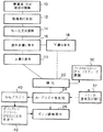
な物質が組み込まれている。かかる方法の工程は、図1に概説されている。コー
ティング溶液又は懸濁液を、後述するようにして、10で調製する。架橋剤を添
加する場合には、所望量の架橋剤を懸濁液又は溶液に、12で添加し、次いで、
その混合物を攪拌し、均一なコーティング組成物を14で得る。その後、この組
成物を塗布容器又は塗布装置に写し、この塗布容器又は塗布装置は、16の吹き
付け塗装のための容器であってもよい。コーティング溶液の典型的な調製例であ
って、ヘパリン及びデキサメタゾン(dexamethasone)に用いられるものについて
、次に述べる。
ヘパリンアンダーコーティング組成物の一般的製造
シリコーンは溶媒(キシレン)中ポリマー先駆体の混合物のとして入手した。
例えば、キシレン中の35%固体シリコーン重量をApplied Silic
one,Part#40,000から入手した。最初に、シリコーン−キシレン
混合物を秤量した。固体シリコーン含量をVendorの分析法に従って測定し
た。予め算出した量の微粉状(finely divided)ヘパリン(2〜6ミクロン)をシ
リコーン中に加えて、次に、テトラヒドロフラン(THF)HPCL等級(Al
drich又はEM)を加えた。例えば、37.5%ヘパリンコーティングに関
して、Wsilicone=5g;固体%=35%;Whep=5x0.35x0.375
/(0.625)=1.05g。このコーティング溶液中のTHF必要量(44
ml)は、37.5%ヘパリンコーティング溶液に関して式:Wsilicone/Whe
p=0.04を用いて算出した。最後に、Pasteur P−ピペットを用い
て製造者の架橋剤溶液を加えた。架橋剤の添加量は放出速度プロフィールをもた
らすように定められた。典型的に、各5gのシリコーン−キシレン混合物に5滴
の架橋剤溶液を加えた。懸濁液が均質なミルク状になるまで、撹拌ロッドを用い
て溶液を撹拌した。次に、コーティング溶液をエアブラシによる塗布のための条
件でペイントジャー中に移した。
デキサメタゾンアンダーコーティング組成物の一般的製造
Metler秤上のビーカー中にシリコーン(上記のような35%溶液)を秤
り入れた。0.35を乗じたシリコーン重量と、デキサメタゾンの望ましい割合
(1〜40%)とによって、デキサメタゾン遊離アルコール又はアセテート形の
重量を算出し、必要量を秤量した。例:Wsilicone=5g;10%デキサメタゾ
ンコーティングに関して、Wdex=5x0.35x0.1/(0.9)=0.1
94gと、このコーティング溶液中のTHF必要量を算出した。10%デキサメ
タゾンコーティング溶液に関して、Wsilicone solid/VTHF=0.06。例:
Wsilicone=5g;VTHF=5x0.35/0.06=29ml。デキサメタゾ
ンを分析用秤上のビーカー中に秤り入れ、THFの総量の1/2を加えた。この
溶液をデキサメタゾンが確実に完全に溶解するように充分に撹拌した。次に、撹
拌済みDEX−THF溶液をシリコーン容器に移した。ビーカーを残りのTHF
で洗浄して、これをシリコーン容器に移した。Pasteur P−ピペットを
用いて、架橋剤溶液を加えた。典型的に、各5gのシリコーンに対して5滴の架
橋剤溶液を用いた。
In the present invention, the stent coating incorporates a biologically active material for in-vivo, in situ, time-controlled delivery to the body lumen of interest. In such a stent coating, a thin coating layer is obtained by spraying the prepared coating solution or suspension, and a biologically active substance is incorporated inside the layer. The steps of such a method are outlined in FIG. A coating solution or suspension is prepared at 10 as described below. If a crosslinker is added, the desired amount of crosslinker is added to the suspension or solution at 12 and then
The mixture is stirred and a uniform coating composition is obtained at 14. Thereafter, the composition is transferred to an application container or application device, which may be a container for 16 spray coatings. A typical preparation of the coating solution, which is used for heparin and dexamethasone, is described next.
General Preparation of Heparin Undercoating Composition Silicone was obtained as a mixture of polymer precursors in a solvent (xylene).
For example, 35% solid silicone weight in xylene is applied Silic.
from one, Part # 40,000. First, the silicone-xylene mixture was weighed. The solid silicone content was measured according to Vendor's analytical method. A pre-calculated amount of finely divided heparin (2-6 microns) is added into the silicone and then tetrahydrofuran (THF) HPCL grade (Al
drich or EM). For example, for a 37.5% heparin coating, W silicone = 5 g;% solids = 35%; W hep = 5 × 0.35 × 0.375
/(0.625)=1.05g. The required amount of THF in this coating solution (44
ml) for the 37.5% heparin coating solution: W silicone / W he
Calculation was made using p = 0.04. Finally, the manufacturer's crosslinker solution was added using a Pasteur P-pipette. The amount of cross-linking agent added was determined to provide a release rate profile. Typically, 5 drops of crosslinker solution was added to each 5 g of silicone-xylene mixture. The solution was stirred using a stir rod until the suspension became homogeneous milky. The coating solution was then transferred into a paint jar at conditions for application with an airbrush.
General Preparation of Dexamethasone Undercoating Composition Silicone (35% solution as described above) was weighed into a beaker on a Mettler scale. The weight of the dexamethasone free alcohol or acetate form was calculated by the silicone weight multiplied by 0.35 and the desired proportion of dexamethasone (1-40%), and the required amount was weighed. Example: W silicone = 5 g; for 10% dexamethasone coating, W dex = 5 × 0.35 × 0.1 / (0.9) = 0.1
94 g and the required amount of THF in this coating solution were calculated. For a 10% dexamethasone coating solution, W silicone solid / V THF = 0.06. Example:
W silicone = 5 g; V THF = 5 × 0.35 / 0.06 = 29 ml. Dexamethasone was weighed into a beaker on an analytical balance and 1/2 of the total amount of THF was added. The solution was stirred thoroughly to ensure complete dissolution of dexamethasone. Next, the stirred DEX-THF solution was transferred to a silicone container. Beak the remaining THF
And transferred to a silicone container. The crosslinker solution was added using a Pasteur P-pipette. Typically, 5 drops of crosslinker solution was used for each 5 g of silicone.
ステント(stent)へのコーティング物質の塗布は全てのコーティング物質に関
して同じであり、上記例におけるように調製したヘパリン懸濁液とデキサメタゾ
ン懸濁液に関しても同じであった。塗布すべき懸濁液を図1の16において塗布
デバイスに移した。典型的に、レギュレーター(Norgren、0〜160p
si)から圧縮空気源を供給される、例えばBadger Model 150
のような、エアブラシに取り付けられたペイントジャーを用いた。ブラシのホー
スをレギュレーターの下流の圧縮空気源に取り付けてから、空気を供給した。圧
力を約15〜25psiに調節し、ノズル状態をトリガーを押し下げることによ
ってチェックした。
The application of the coating material to the stent was the same for all coating materials, and the same for the heparin and dexamethasone suspensions prepared as in the above examples. The suspension to be applied was transferred to the application device at 16 in FIG. Typically, regulators (Norgren, 0-160p)
si) supplied with compressed air source, eg Badger Model 150
A paint jar attached to an air brush was used. The brush hose was attached to a compressed air source downstream of the regulator before supplying air. The pressure was adjusted to about 15-25 psi and the nozzle condition was checked by depressing the trigger.
吹き付けのためにステントを固定するためには任意の適当な方法を用いること
ができ、実験室では回転固定具(rotating fixture)を用いて成功を収めることが
できた。関連ステントの両端を2個の弾性保持体、通常はアリゲータークリップ
によって固定具に固定し、ステントが弛緩した非伸長状態に留まるようにクリッ
プ間の距離を調節した。次に、ローターに動力を与えて、回転速度を所望の塗布
速度を与えるように、即ち、約40rpmに調節した。
Any suitable method could be used to fix the stent for spraying, and it was successful in the laboratory with a rotating fixture. Both ends of the associated stent were secured to the fixture by two elastic retainers, usually alligator clips, and the distance between the clips was adjusted so that the stent remained in a relaxed, unstretched state. The rotor was then powered and the rotational speed was adjusted to give the desired coating speed, i.e. about 40 rpm.
実質的に水平な面内でステントを回転させながら、ノズルからステントまでの
距離が約2〜4インチになるように、吹き付けノズルを調節し、ブラシをステン
トに沿ってステントの遠位端部から近位端部まで向け、次に近位端部から遠位端
部まで向けて、約3回のステント回転中に1回の吹き付けサイクルが生ずるよう
な速度でのスイーピング運動(sweeping motion)で動かした。典型的に、1分間
未満、通常は1/2分間の小休止が層の間に設けられた。もちろん、塗布層の数
は特定の用途によって変化しており、今後も変化する。例えば、図1の18にお
けるような典型的な結合層(tie-layer)では、突出面1cm2につき3〜4mgの
ヘパリンの塗布レベルのために、20サイクルのコーティング塗布が必要であり
、3.5mm直径x14.5cm長さのステントに対して約30mlの溶液が消
耗される。
While rotating the stent in a substantially horizontal plane, the spray nozzle is adjusted so that the distance from the nozzle to the stent is about 2-4 inches, and the brush is moved along the stent from the distal end of the stent. Move to the proximal end, then from the proximal end to the distal end, with a sweeping motion at such a speed that one spray cycle occurs during about 3 stent rotations It was. Typically, a short break of less than 1 minute, usually 1/2 minute, was provided between the layers. Of course, the number of coating layers changes depending on the specific application and will change in the future. For example, a typical tie-layer as in 18 of FIG. 1 requires 20 cycles of coating application due to the application level of 3-4 mg heparin per cm 2 of protruding surface. About 30 ml of solution is consumed for a 5 mm diameter x 14.5 cm long stent.
層状構造体を修飾するために組成物の粘度とスプレイノズルの流量とを必要に
応じて調節することができるので、もちろん、モーターの回転速度も調節可能で
ある。一般に、上記ミックスに関しては、ステントのサイズに依存して、30〜
50rpmの範囲内の回転速度と1分間につきコーティング組成物4〜10ml
の範囲内のスプレイノズルの流量とにおいて、最良の結果が得られている。より
精緻なコンピューター制御塗布装置が実験室において実行可能であると実証され
たプロセスを上首尾に自動化することが考えられる。
Since the viscosity of the composition and the flow rate of the spray nozzle can be adjusted as necessary in order to modify the layered structure, it is of course possible to adjust the rotational speed of the motor. In general, for the above mix, depending on the size of the stent,
Rotational speed in the range of 50 rpm and 4-10 ml of coating composition per minute
The best results have been obtained with spray nozzle flow rates in the range of. It is conceivable to successfully automate processes that have been proven to be feasible in the laboratory with more sophisticated computer controlled coating equipment.
幾つかの塗布層が、18におけるようなアンダーコートと呼ばれるものを構成
する。1つのプロセスでは、生物活性物質とマトリックスポリマー物質と架橋剤
とに関して同じ組成でも異なる組成でもあることができる付加的な上部アンダー
コートを、例えば、20におけるような上部層として塗布することができる。上
部層(top layer)の塗布はアンダーコートと同じ塗布方法に従うが、層の数と厚
さとは任意である。もちろん、如何なる層の厚さもステントの回転速度と吹き付
け条件とを調節することによって調節することができる。一般に、総塗布厚さは
吹き付けサイクル数又は全コートを構成する薄いコート数によって制御される。
Several coating layers constitute what is called an undercoat as in 18. In one process, an additional top undercoat, which can be the same or different composition with respect to the bioactive material, matrix polymer material, and crosslinker, can be applied as a top layer, such as at 20, for example. The application of the top layer follows the same application method as the undercoat, but the number and thickness of the layers are arbitrary. Of course, the thickness of any layer can be adjusted by adjusting the rotational speed of the stent and the spraying conditions. Generally, the total coating thickness is controlled by the number of spray cycles or the number of thin coats that make up the entire coat.
図1の22に示すように、塗布済みステントにその後に硬化工程を受けさせ、
この工程ではプレポリマーと架橋剤とが配位結合して、生物学的に活性な物質を
含有する硬化ポリマーマトリックスを生成する。硬化プロセスは溶媒のキシレン
、THF等の蒸発と、ポリマーの硬化及び架橋とを含む。ある一定のシリコーン
物質は比較的低温(即ち、室温〜50℃)において室温加硫(RTV)プロセス
と呼ばれるプロセスで硬化されることができる。しかし、さらに典型的には、硬
化プロセスは高温硬化物質を含み、塗布済みステントを約90℃以上のオーブン
(oven)に約16時間入れる。デキサメタゾン含有塗布済みステントに関しては1
50℃程度の高温にまで温度を高めることができる。もちろん、特定のシリコー
ン、架橋剤及び生物学的活性物質に応じて、時間と温度とを変化することができ
る。
As shown at 22 in FIG. 1, the coated stent is subsequently subjected to a curing step,
In this process, the prepolymer and the crosslinker are coordinated to produce a cured polymer matrix containing a biologically active material. The curing process involves evaporation of the solvent xylene, THF, etc., and curing and crosslinking of the polymer. Certain silicone materials can be cured at a relatively low temperature (ie, room temperature to 50 ° C.) in a process called a room temperature vulcanization (RTV) process. More typically, however, the curing process includes a high temperature curing material and the coated stent is placed in an oven above about 90 ° C.
Put in (oven) for about 16 hours. 1 for dexamethasone-containing coated stents
The temperature can be increased to a high temperature of about 50 ° C. Of course, depending on the particular silicone, crosslinker and biologically active material, the time and temperature can be varied.
上述したやり方で塗布され、硬化されたステントは、将来の移植用に包装する
前に、滅菌されなければならない。滅菌のためには、特にヘパリン含有コーティ
ングに関しては、γ線が好ましい方法である;しかし、本発明の方法に従って塗
布され、硬化されたステントへγ線滅菌を適用すると、これらの塗布され、硬化
されたステントに24におけるような前処理工程を最初に施さないかぎり、これ
らのステントはカテーテルを用いて血管その他の管腔に供給する場合に、本来の
状態(original posture)を回復するのが非常に緩慢になる可能性がある。
Stents applied and cured in the manner described above must be sterilized prior to packaging for future implantation. For sterilization, especially for heparin-containing coatings, gamma radiation is the preferred method; however, when gamma sterilization is applied to a stent that has been applied and cured according to the method of the present invention, these applied and cured Unless the stents are first subjected to a pretreatment step, such as at 24, these stents are very likely to recover their original posture when delivered to a blood vessel or other lumen using a catheter. May be slow.
前処理工程は、まだ拘束されない形態で塗布され、硬化されたステントのアル
ゴンプラズマ処理を含む。この方法によると、例えばPlasma Scien
ce 350(Himont/Plasma Science,カリフォルニア
州、フォスターシティ)のようなプラズマ表面処理系の室にステントを入れる。
この系は、反応室と、13.56mHzで作動し、0〜500wattの出力の
無線周波数(RF)固体状態発電機であってマイクロプロセッサー制御系を備え
ているものと、完全真空ポンプパッケージとを備える。反応室は16.75イン
チ(42.55cm)x13.5インチ(34.3cm)x17.5インチ(4
4.45cm)深さの妨害されない作用容積(work volume)を有する。
The pretreatment step involves argon plasma treatment of the stent applied and cured in an unconstrained form. According to this method, for example, Plasma Science
The stent is placed in a chamber of a plasma surface treatment system such as ce 350 (Himont / Plasma Science, Foster City, Calif.).
This system consists of a reaction chamber, a radio frequency (RF) solid state generator operating at 13.56 mHz, with an output of 0-500 watts, equipped with a microprocessor control system, and a complete vacuum pump package. Prepare. The reaction chamber is 16.75 inches (42.55 cm) x 13.5 inches (34.3 cm) x 17.5 inches (4
4.45 cm) deep unobstructed work volume.
プラズマプロセスでは、拘束されない塗布済みステントを反応室に入れ、系を
窒素でパージし、真空を20〜50mTorrまで適用する。その後に、プラズ
マ処理のために、不活性ガス(アルゴン、ヘリウム、又はこれらの混合物)を反
応室に入れる。非常に好ましい操作方法は、アルゴンガスの使用と、200〜4
00wattの出力範囲、100〜450mTorrに相当する150〜650
標準ml/分の流量及び30秒間〜約5分間の暴露時間における操作とから成る
。プラズマ処理の直後にステントを取り出すか又はアルゴン雰囲気中に付加的な
時間、典型的には5分間留めることもできる。
In the plasma process, an unconstrained coated stent is placed in a reaction chamber, the system is purged with nitrogen, and a vacuum is applied to 20-50 mTorr. Thereafter, an inert gas (argon, helium, or a mixture thereof) is placed in the reaction chamber for plasma treatment. A highly preferred method of operation is the use of argon gas and 200-4
Output range of 00 watt, 150 to 650 corresponding to 100 to 450 mTorr
Operation at a standard flow rate of ml / min and an exposure time of 30 seconds to about 5 minutes. The stent can be removed immediately after plasma treatment or can be left in an argon atmosphere for an additional time, typically 5 minutes.
この後に、26に示すように、ステントを2.5〜3.5Mradにおけるγ
線滅菌にさらすことができる。半径方向において拘束されない状態又は半径方向
において拘束された状態のステントに対して放射を実施することができる。
After this, as shown at 26, the stent is gamma at 2.5-3.5 Mrad.
Can be exposed to wire sterilization. Radiation can be performed on a stent in a radially unconstrained state or a radially constrained state.
しかし、プラズマ処理前に又は滅菌処理方法の直前に付加的な幾つかの処理方
法の1つによって表面を改質することが好ましく、このような方法の幾つかは以
下の実施例に関連して説明する。
However, it is preferred to modify the surface prior to plasma treatment or by one of several additional treatment methods just prior to the sterilization treatment method, some of which are related to the following examples. explain.
実施例1.ヘパリンコーティングを溶離するフルオロシリコーン表面処理
ステントのアンダーコートを上述したように多重塗布層として塗布し、その後
に22に説明するように硬化させた。このアンダーコートのヘパリン含量は37
.5%であり、コーティング厚さは約30〜40μであった。30においてフル
オロシリコーン懸濁液(Applied Silicone#40032)から
、一定量のフルオロシリコーン懸濁液を秤量し、関係式:VTHF=1.2xフル
オロシリコーン懸濁液の重量に従ってテトラヒドロフラン(THF)を加えるこ
とによって、フルオロシリコーン(FSi)スプレイ溶液を製造した。この溶液
を充分によく撹拌し、18におけるアンダーコートプロセスの塗布方法を用いて
、32においてステント上に吹き付け塗布し、塗布済みステントを90℃におい
て16時間硬化させた。塗布済みステントを上述したγ線滅菌の前に上述した方
法により工程22〜26に従ってアルゴンプラズマ処理する。
Example 1. An undercoat of a fluorosilicone surface treated stent eluting the heparin coating was applied as a multiple coating layer as described above and then cured as described at 22. This undercoat has a heparin content of 37.
. The coating thickness was about 30-40μ. Weigh out a certain amount of fluorosilicone suspension from fluorosilicone suspension (Applied Silicone # 40032) at 30 and add tetrahydrofuran (THF) according to the relationship: V THF = 1.2 x weight of fluorosilicone suspension This produced a fluorosilicone (FSi) spray solution. This solution was stirred well and spray coated onto the stent at 32 using the undercoat process coating method at 18 and the coated stent was cured at 90 ° C. for 16 hours. The coated stent is argon plasma treated according to steps 22-26 by the method described above prior to the above-described gamma sterilization.
図7は、フルオロシリコーントップコート有りと如何なるトップコートも無し
のリン酸塩緩衝剤系中のヘパリン放出速度のプロットである。トップコートの厚
さは約10〜15μである。図7のグラフには現れないが、FSi無しのコーテ
ィングの放出速度は最初、FSi有りのコーティングの約25倍の大きさである
ことに注目すべきである。これは、もちろん、明らかにグラフの規模からはみ出
る。しかし、FSi上部層(top layer)又は拡散バリヤーを有するコーティング
が第1日後から第1週間を通して第10日目まで初期放出速度の低下と溶離速度
の上昇とを示すことが注目に値する。さらに、フルオロシリコーン(FSi)ト
ップコートは、フッ素化の高い電気陰性度のために、生物学的活性物質の溶離中
及び後に非血栓性(non-thrombogenic)の表面品質を維持する。さらに、ヘパリン
自体の負の電荷のために、フルオロシリコーントップコートの電気陰性度は、ヘ
パリン放出速度プロフィールが変化する少なくとも部分的な原因である。
FIG. 7 is a plot of the rate of heparin release in a phosphate buffer system with and without a fluorosilicone topcoat. The thickness of the topcoat is about 10-15μ. Although not shown in the graph of FIG. 7, it should be noted that the release rate of the coating without FSi is initially about 25 times as large as the coating with FSi. This obviously goes beyond the scale of the graph. However, it is noteworthy that the coating with the FSi top layer or diffusion barrier shows a decrease in the initial release rate and an increase in the elution rate from the first day to the tenth day through the first week. Furthermore, the fluorosilicone (FSi) topcoat maintains a non-thrombogenic surface quality during and after elution of the biologically active material due to the high electronegativity of fluorination. Furthermore, due to the negative charge of heparin itself, the electronegativity of the fluorosilicone topcoat is at least partially responsible for the change in the heparin release rate profile.
図8は、16.7%封埋ヘパリン含有フルオロシリコーン(FSi)トップコ
ートとフルオロシリコーン(FSi)のみを含むトップコートとのプロットであ
る。アンダーコートは、図7で用いたアンダーコートと同じものであり、約37
.5%ヘパリンを含有し、約37〜40μ厚さである。これらの溶離速度は、ヘ
パリンを含まないFSi上部層と全く匹敵するものであり、ヘパリン放出の初期
バーストを大きく低下させるか、さもなくば、FSi上部層中のヘパリンは試験
期間にわたってやや大きい放出を示す。
実施例2.薬物溶離アンダーコート上のポリエチレングリコール(PEG)の固
定
アンダーコートをステント上に塗布し、実施例1におけるように22において
硬化させた。次に、ステントを24におけるようにアルゴンガスプラズマによっ
て処理し、40においてアンモニウムガスプラズマによって処理した。アルゴン
ガスプラズマ処理の装置とプロセスとは上述した通りであった。アンモニウムプ
ラズマ処理をアルゴンガスプラズマ処理の直後に実施して、コーティングの表面
をアミノ化した。アンモニウム流量は100〜700cm3/分間(ccM)の
範囲内、好ましくは500〜600ccMの範囲内であった。無線周波数プラズ
マの出力は50〜500wattの範囲内、好ましくは〜200wattであっ
た。プロセス時間は30秒間〜10分間の範囲内、好ましくは〜5分間である。
FIG. 8 is a plot of a 16.7% embedded heparin containing fluorosilicone (FSi) topcoat and a topcoat containing only fluorosilicone (FSi). The undercoat is the same as the undercoat used in FIG.
. Contains 5% heparin and is approximately 37-40μ thick. These elution rates are quite comparable to the FSi top layer without heparin, greatly reducing the initial burst of heparin release, or heparin in the FSi top layer has a slightly higher release over the test period. Show.
Example 2 Solidification of polyethylene glycol (PEG) on drug eluting undercoat
A constant undercoat was applied over the stent and cured at 22 as in Example 1. The stent was then treated with an argon gas plasma as at 24 and treated with an ammonium gas plasma at 40. The argon gas plasma treatment apparatus and process were as described above. An ammonium plasma treatment was performed immediately after the argon gas plasma treatment to aminate the surface of the coating. The ammonium flow rate was in the range of 100-700 cm 3 / min (ccM), preferably in the range of 500-600 ccM. The output of the radio frequency plasma was in the range of 50 to 500 watts, preferably ˜200 watts. The process time is in the range of 30 seconds to 10 minutes, preferably ˜5 minutes.
アミノ化の直後に、ステントを42において求電子的活性化ポリエチレングリ
コール(PEG)溶液中に浸漬した。PEGはタンパク質吸収の阻害剤であるこ
とが知られている。求電子的活性化ポリエチレングリコールの例は、PEGニト
ロフェニルカーボネート類、PEGトリクロロフェニルカーボネート類、PEG
トレシレート、PEGグリシジルエーテル、PEGイソシアネート等であり、任
意に1端部がメトキシ基で終了している。PEGの分子量は約1000〜600
0の範囲であり、好ましくは約3000である。簡単なアンモニウムアミノ化は
エラストマーポリマー表面(例えばシリコーン)上に多量の第1アミン及び第2
アミンを生じない。その代わりに、イミン(>C=N−H)と他のより酸化性の
ニトロ含有基とが表面を支配する。PEG上の官能基がイミンと、恐らく、表面
上の他のニトロ含有種とも反応して、PEGを表面に固定することができるよう
に、反応媒質中に例えばNaBH3CNのような還元剤を加えることが、一般に
必要である。NaBH3CNの典型的な濃度は約2mg/mlである。PEGと
その誘導体とは水及び多くの極性の芳香族溶媒中に溶解するので、コーティング
に用いる溶媒はPEGの溶媒でなければならないが、浸出による薬物の可能な損
失を防ぐためにアンダーコート中の薬剤の溶媒であってはならない。ヘパリン溶
離性(eluting-heparin)コーティングの場合には、ホルムアミドとメチルエチル
ケトンとの混合溶媒又はホルムアミドとアセトンとの混合溶媒(好ましくは、ホ
ルムアミド30:メチルエチルケトン又はアセトン70の容量比で)が、ヘパリ
ンを溶解しないので、好ましい溶媒である。PEGの濃度、反応時間、反応温度
及びpH値は用いるPEGの種類に依存する。ヘパリン溶離性コーティングの場
合には、(30/70)ホルムアミド/メチルエチルケトン中の5%PEGトレ
シレートが用いられて成功を収めている。反応時間は室温において3時間であっ
た。次に、PEGを表面に共有結合させた。次に、この実施態様の滅菌のために
既述したようにγ線を用いた。
Immediately after amination, the stent was immersed in an electrophilic activated polyethylene glycol (PEG) solution at 42. PEG is known to be an inhibitor of protein absorption. Examples of electrophilic activated polyethylene glycols are PEG nitrophenyl carbonates, PEG trichlorophenyl carbonates, PEG
Tresylate, PEG glycidyl ether, PEG isocyanate, etc., optionally terminating at one end with a methoxy group. The molecular weight of PEG is about 1000 to 600
A range of 0, preferably about 3000. Simple ammonium amination can result in large amounts of primary and secondary amines on elastomeric polymer surfaces (eg silicone).
Does not produce amines. Instead, imine (> C = N—H) and other more oxidative nitro-containing groups dominate the surface. In order to allow the functional group on the PEG to react with the imine and possibly other nitro-containing species on the surface to immobilize the PEG on the surface, a reducing agent such as NaBH 3 CN is added in the reaction medium. It is generally necessary to add. A typical concentration of NaBH 3 CN is about 2 mg / ml. Since PEG and its derivatives are soluble in water and many polar aromatic solvents, the solvent used for coating must be a solvent for PEG, but the drug in the undercoat to prevent possible loss of drug due to leaching It must not be a solvent. For heparin-eluting coatings, a mixed solvent of formamide and methyl ethyl ketone or a mixed solvent of formamide and acetone (preferably in a volume ratio of formamide 30: methyl ethyl ketone or acetone 70) dissolves heparin. Therefore, it is a preferred solvent. The PEG concentration, reaction time, reaction temperature and pH value depend on the type of PEG used. In the case of heparin eluting coatings, 5% PEG tresylate in (30/70) formamide / methyl ethyl ketone has been used successfully. The reaction time was 3 hours at room temperature. Next, PEG was covalently attached to the surface. Next, gamma radiation was used as previously described for sterilization of this embodiment.
凝固防止性物質ヘパリンに関して、アンダーコート中の割合は公称約30〜5
0%であり、トップコート中の割合は約0〜30%活性物質である。トップコー
トとアンダーコートとのコーティング厚さの比は約1:10から1:2まで変化
し、好ましくは約1:6から1:3までの範囲内である。
For the anticoagulant heparin, the proportion in the undercoat is nominally about 30-5.
0% and the proportion in the topcoat is about 0-30% active substance. The coating thickness ratio of the topcoat to the undercoat varies from about 1:10 to 1: 2, and is preferably in the range of about 1: 6 to 1: 3.
ステンティングプロセス中に医師は抗血小板/凝固防止薬物のボラス注射を患
者に投与するので、バースト効果を抑制することも薬物担持の減少を可能にし、
換言すると、コーティング層の厚さの低下を可能にする。この結果、ステント中
に封埋された薬物は無駄なく完全に用いられることができる。できるだけ薄いコ
ーティング形状において第1日目放出を調節し、第2日目と第3日目の放出を最
大にすることは、急性又は亜急性の血栓を軽減する。
During the stenting process, doctors administer bolus injections of anti-platelet / anticoagulant drugs to the patient, so suppressing the burst effect can also reduce drug loading,
In other words, it is possible to reduce the thickness of the coating layer. As a result, the drug embedded in the stent can be completely used without waste. Adjusting the first day release and maximizing the second and third day release in the thinnest possible coating configuration alleviates acute or subacute thrombus.
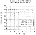
図4は同じ厚さのコーティングに関する薬物担持の一般的効果を示す。初期溶
離速度は、図5に示すように、薬物担持量と共に上昇する。放出速度も同じ担持
量ではコーティングの厚さと共に上昇するが、図6の同じ薬物担持量と同様なア
ンダーコート厚さによって示されるように、トップコートの厚さに逆比例する傾
向がある。
FIG. 4 shows the general effect of drug loading on the same thickness coating. The initial elution rate increases with the amount of drug loaded, as shown in FIG. The release rate also increases with the coating thickness at the same loading, but tends to be inversely proportional to the topcoat thickness, as shown by the undercoat thickness similar to the same drug loading in FIG.
しかし、現在までに集められたデータから明らかであることは、本発明のプロ
セスが特定のステント用途の必要条件を満たすために望ましい方法で薬物溶離速
度を制御することを可能にすることである。同様な方法で、ステントコーティン
グを2種類以上の薬物の組合せを用いて、薬物放出順序及び速度を制御して、ス
テントコーティングを製造することが可能である。例えば、複数の増殖防止性薬
をアンダーコート中で組合せ、複数の抗血小板薬をトップコート中で組み合わせ
ることができる。このようにして、抗血小板薬、例えばヘパリンを最初に溶離さ
せ、次に増殖防止薬を溶離させて、移植したステントの安全な封入(encapsulati
on)をより良好に可能にすることができる。
However, it is clear from the data collected to date that the process of the present invention allows the drug elution rate to be controlled in a desirable manner to meet the requirements of a particular stent application. In a similar manner, a stent coating can be produced using a combination of two or more drugs with controlled drug release sequence and rate. For example, multiple antiproliferative drugs can be combined in the undercoat and multiple antiplatelet drugs can be combined in the topcoat. In this way, an antiplatelet drug, e.g., heparin, is eluted first, followed by an antiproliferative drug to ensure safe encapsulation of the implanted stent.
on) can be made better.
ヘパリン濃度測定はアズレA染料をヘパリンの希薄溶液によって錯化すること
によって作成した標準曲線を用いておこなった。標準曲線を周知の方法でコンパ
イルするために16種類の標準を用いた。
The heparin concentration was measured using a standard curve prepared by complexing Azure A dye with a dilute solution of heparin. Sixteen standards were used to compile standard curves in a well-known manner.
溶離試験に関しては、ステントを約37℃のインキュベーター内のpH7.4
のリン酸塩緩衝液中に浸漬した。この溶液の定期的なサンプリングをおこなって
、ヘパリン溶離量を測定した。各サンプリング後に、各ステントをヘパリンを含
まない緩衝剤溶液中に入れた。
For the elution test, the stent is placed in an incubator at about 37 ° C. with a pH of 7.4.
In a phosphate buffer solution. The solution was periodically sampled to measure the heparin elution amount. After each sampling, each stent was placed in a buffer solution without heparin.
上述したように、エラストマー物質の許容可能なヘパリン担持は変動するが、
シリコーン物質の場合には、ヘパリンは層の総重量の60%を越えることができ
る。しかし、一般的に最も有利に用いられる担持は層の総重量の約10%〜45
%の範囲内である。デキサメタゾンの場合には、担持は層の総重量の50%以上
ほどの量になることができるが、好ましくは約0.4%〜45%の範囲内である
。
As noted above, the acceptable heparin loading of the elastomeric material varies,
In the case of silicone materials, heparin can exceed 60% of the total weight of the layer. However, generally the most advantageously used supports are from about 10% to 45% of the total weight of the layer.
%. In the case of dexamethasone, the loading can be as much as 50% or more of the total weight of the layer, but is preferably in the range of about 0.4% to 45%.
金属ステントに適用可能な薄い表面コーティング構造中への生物学的活性物質
の組み込みの機構が本発明の重要な態様であることは、理解されるであろう。先
行技術薬物溶離デバイスに関連した、比較的肉厚のポリマー溶離ステント又は任
意の膜オーバーレイの必要性は、生物学的活性物質の運搬のための生分解性又は
再吸収性ビヒクルの使用の必要性と同様に無い。この方法は明らかに長時間投与
を可能にし、ステント自体の独立した機械的又は治療的利益に対する妨害を最小
にする。
It will be appreciated that the mechanism of incorporation of biologically active material into a thin surface coating structure applicable to a metal stent is an important aspect of the present invention. The need for relatively thick polymer eluting stents or optional membrane overlays associated with prior art drug eluting devices necessitates the use of biodegradable or resorbable vehicles for the delivery of biologically active substances. Like no. This method clearly allows for long-term administration and minimizes interference with the independent mechanical or therapeutic benefits of the stent itself.
コーティング物質は、特定の塗布方法、コーティング/薬物組合せ及び薬物注
入機構によって設計される。コーティング中の生物学的活性物質の特定の形状及
び放出機構の考察がこの方法に非常に良好な結果を生じさせる。このようにして
、コーティング構造からの生物学的活性物質の投与を、多様な用途に適合するよ
うに調節することができる。
The coating material is designed by the specific application method, coating / drug combination and drug infusion mechanism. Consideration of the specific shape and release mechanism of the biologically active substance in the coating gives this method very good results. In this way, the administration of the biologically active substance from the coating structure can be adjusted to suit a variety of applications.
上記例は放出されるべき2種類の異なる薬物担持又は割合の生物学的活性物質
を示すが、これは決して本発明に関する限定ではなく、所望の放出プロフィール
を得るために、任意の数の層と担持の組合せを用いることができるように意図さ
れる。例えば、層の担持の漸次グレーディング及び変化を利用して、例えば、薄
い層には高い担持を用いることができる。薬物担持を全く有さない層も用いるこ
とができる。例えば、シリコーンの非担持層の間にヘパリンを含有する交互層を
挿入したコーティングによって又はコーティングの一部に他の物質を用いること
によって、パルス状(pulsatile)ヘパリン放出系が得られる。換言すると、本発
明は数え切れない数の組合せを可能にし、その結果、移植されたステントに関す
る生物学的活性物質の放出の制御に関して非常に大きなフレキシビリティを生じ
る。各塗布層は典型的に約0.5ミクロンから15ミクロンの厚さである。吹き
付け塗布される層の総数は、もちろん、広範囲に、10層未満から50層より多
くまで、変化することができ、一般には、20〜40層が包含される。コーティ
ング全体の厚さも広範囲に変化することができるが、一般には約10〜200ミ
クロンであることができる。
While the above examples show two different drug loadings or proportions of biologically active material to be released, this is by no means a limitation on the present invention and any number of layers and layers may be used to obtain the desired release profile. It is contemplated that a combination of supports can be used. For example, high grading and variation of layer loading can be used, for example, high loading can be used for thin layers. A layer with no drug loading can also be used. For example, a pulsatile heparin release system can be obtained by coating with alternating layers containing heparin between unsupported layers of silicone or by using other materials as part of the coating. In other words, the present invention allows for an infinite number of combinations, resulting in a great deal of flexibility in terms of controlling the release of biologically active material with respect to the implanted stent. Each coated layer is typically about 0.5 to 15 microns thick. The total number of layers applied by spraying can, of course, vary widely, from less than 10 layers to more than 50 layers, and generally includes 20-40 layers. The total thickness of the coating can also vary widely, but can generally be from about 10 to 200 microns.
コーティングのポリマーは薄層としてステント物質に付着することができる、
任意の相容性の生物的に安定なエラストマー物質であるが、生物学的活性物質の
放出が一般には疎水性物質によっては予測可能に制御されうることが判明してい
るので、疎水性物質が好ましい。好ましい物質はシリコーンゴムエラストマーと
生物的に安定なポリウレタンが特に好ましい。
The coating polymer can adhere to the stent material as a thin layer,
Any compatible biostable elastomeric material, but it has been found that the release of biologically active material can generally be predictably controlled by hydrophobic materials, so hydrophobic materials preferable. Preferred materials are silicone rubber elastomers and biologically stable polyurethanes.
本発明を特許法に従い、かつ当業者に新規な原理を適用し、実施例の実施態様
を必要に応じて構成して用いるために必要な情報を与えるためにかなり詳細に説
明した。しかし、本発明が特に異なるデバイスによって実施されうること、及び
種々な変更が本発明自体の範囲から逸脱せずになされうることを理解すべきであ
る。
The present invention has been described in considerable detail in order to provide the information necessary to comply with the patent law and to apply those novel principles to those skilled in the art and to configure and use the embodiments of the examples as required. However, it is to be understood that the invention can be practiced with particularly different devices and that various modifications can be made without departing from the scope of the invention itself.
本発明の実施態様は次の通りである。
1.外表面を有する移植可能な医療器具であって、
前記外表面の少なくとも一部が、疎水性エラストマー材料からなる形状が適合したコーティングで被覆されており、
前記疎水性エラストマー材料が、前記疎水性エラストマー材料から時間制御された送達をするために、生物学的に活性な物質の所定量をその内部に包含しており、
生物学的に活性な物質について時間制御された送達がされた後に、非血栓性の表面を提供する手段であって形状が適合したコーティングに関連するものを有する移植可能な医療器具。
2.形状が適合したコーティングが、疎水性エラストマー材料中に微細に分割された生物学的に活性な物質の所定量を包含する上記1に記載の医療器具。
3.微細に分割された生物学的に活性な物質が、約15ミクロン未満の平均粒子径を有する上記2に記載の医療器具。
4.微細に分割された生物学的に活性な物質が、約10ミクロン未満の平均粒子径を有し、形状が適合したコーティングの約25〜60重量%の薬剤を担持する上記2に記載の医療器具。
5.形状が適合したコーティングが、エラストマー材料中に分子レベルで分散した生物学的に活性な物質の所定量を包含する上記1に記載の医療器具。
6.生物学的に活性な物質が、抗血栓剤、抗凝血剤、抗血小板物質剤、血栓崩壊剤、抗増殖剤、ステロイド系抗炎症剤、非ステロイド系抗炎症剤、過形成、特に、再狭窄の抑制剤、平滑筋細胞抑制剤、成長因子、成長因子抑制剤、細胞癒着抑制剤、細胞癒着促進剤、健康な新内膜組織の形成促進剤、及びこれらの組み合わせからなる群から選ばれる上記1に記載の医療器具。
7.疎水性エラストマー材料が、シリコーン、ポリウレタン、エチレン−酢酸ビニル共重合体、ポリオレフィンエラストマー、ポリアミドエラストマー、及び、EPDMゴム、並
びに、これらの組み合わせからなる群から選ばれる上記1に記載の医療器具。
8.形状が適合したコーティングが、エラストマー材料からなる複数の層を包含し、前記エラストマー材料が、その内部に、生物学的に活性な物質の所定量を包含する上記1に記載の医療器具。
9.形状が適合したコーティングに関連する手段が、外層を有し、この外側層が、形状が適合したコーティングを少なくとも部分的に被覆し、かつ、非血栓性のポリマー材料を包含する上記1に記載の医療器具。
10.非血栓性のポリマー材料が、フルオロシリコーン、ポリエチレングリコール、多糖、及び、リン脂質、並びに、これらの組み合わせからなる群から選ばれる上記9に記載の医療器具。
11.非血栓性のポリマー材料が、形状が適合したコーィングに付着したフルオロシリコーンコーティングを包含する上記10に記載の医療器具。
12.非血栓性のポリマー材料が、アミノ化された形状が適合したコーティングに共有結合により結合しているポリエチレングリコールを包含する上記10に記載の医療器具。
13.移植可能な医療器具が金属で形成されている上記1に記載の医療器具。
14.開口端と、隙間のある格子からなる側壁構造と、を有する管状金属体と;
前記側壁構造の表面に設けられた、連続的な形状が適合したコーティングと;
を有する人体に移植可能な拡張可能なステントであって、
前記形状が適合したコーティングは、疎水性エラストマー材料を包含し、前記疎水性エラストマー材料が、前記疎水性エラストマー材料から時間制御された送達をするために、生物学的に活性な物質の所定量をその内部に包含しており、
前記コーティングは、隙間のある格子を保持するように、前記側壁構造に適合し、前記コーティングは、非血栓性の外表面を有するステント。
15.生物学的に活性な物質がヘパリンである上記14に記載のステント。
16.コーティングの外側層が、フルオロシリコーン及びポリエチレングリコール(PEG)からなる群から選ばれた材料を包含する上記15に記載のステント。
17.前記外側層が、フルオロシリコーンである上記16に記載のステント。
18.前記外側層が、所望により、微細に分割されたヘパリンの所定量を更に含む上記17に記載のステント。
19.前記外側層が、ポリエチレングリコールを含む上記16に記載のステント。
20.開口端と側壁とを有する管状金属体と;
前記側壁の表面に設けられた、連続的な形状が適合したコーティングと; を有する血管の管腔に移植するためのステントであって、
前記コーティングは、疎水性エラストマー材料からなるアンダーコートを更に有し、前記疎水性エラストマー材料が、前記疎水性エラストマー材料から時間制御された送達をするために、微細に分割されたヘパリンの所定量をその内部に包含しており、
前記コーティングは、所定量のフルオロシリコーンを含有するトップコートを更に有するステント。
21.前記トップコートが、所望により、微細に分割されたヘパリンの所定量を更に含有する上記20に記載のステント。
22.開口端と側壁とを有する管状金属体と;
前記側壁の表面に設けられた、連続的な形状が適合したコーティングと; を有する血管の管腔に移植するためのステントであって、
前記コーティングは、疎水性エラストマー材料からなるアンダーコートを有し、前記疎水性エラストマー材料が、前記疎水性エラストマー材料から時間制御された送達をするために、微細に分割されたヘパリンの所定量をその内部に包含しており、
前記コーティングは、所定量のポリエチレングリコールを含有するトップコートを有するステント。
23.移植可能なステントプロテーゼを層で被覆する方法であって、
前記層は、疎水性エラストマー材料を包含し、前記疎水性エラストマー材料が、前記疎
水性エラストマー材料から時間制御された送達をするために、生物学的に活性な物質の所定量をその内部に包含しており、
(a) トップコートを塗布する工程と、
ここで、前記トップコートは、溶媒混合物中の未硬化のポリマー性材料と、微細に分割された生物学的に活性な物質の所定量とを含有する組成からなり;
(b) 前記ポリマー性材料を硬化する工程と;
(c) トップコートを塗布する工程と、
ここで、前記トップコートは、特性を有する組成からなり、前記特性は、非血栓性の表面を付与し、かつ、生物学的に活性な物質の修飾した送達を可能ならしめることである;を有する方法。
24.エラストマー材料がシリコーンであり、かつ、生物学的に活性な物質がヘパリンである上記23に記載の方法。
25.前記トップコートが、フルオロシリコーンを包含する上記24に記載の方法。
26.前記トップコートが、所定量のヘパリンを包含する上記25に記載の方法。
27.前記トップコートが、ポリエチレングリコールを包含する上記24に記載の方法。28.(d) 硬化後の前記トップコートを、不活性ガスプラズマで処理し、次いで、アンモニアプラズマで処理する工程と;
(e) ポリエチレングリコール(PEG)の溶液から、ポリエチレングリコールからなる外側コーティングを塗布する工程と;
を更に有する上記27に記載の方法。
29.前記溶液中のポリエチレングリコール(PEG)が、PEGニトロフェニルカーボネート類、PEGトリクロロフェニルカーボネート類、PEGトレシレート、PEGグリシジルエーテル、PEGイソシアネート及びこれらの組み合わせから選ばれた上記28に記載の方法。
30.ポリエチレングリコールが、求電子的に活性である上記28に記載の方法。
31.ポリエチレングリコールが、メトキシ基末端を有する上記28に記載の方法。
Embodiments of the present invention are as follows.
1. An implantable medical device having an outer surface,
At least a portion of the outer surface is coated with a conformable coating made of a hydrophobic elastomeric material;
The hydrophobic elastomer material includes a predetermined amount of a biologically active substance therein for time-controlled delivery from the hydrophobic elastomer material;
An implantable medical device having a means for providing a non-thrombogenic surface associated with a conformal coating after time-controlled delivery of a biologically active substance.
2. The medical device of
3. 3. The medical device of 2 above, wherein the finely divided biologically active substance has an average particle size of less than about 15 microns.
4). The medical device of
5). The medical device of
6). Biologically active substances are antithrombotics, anticoagulants, antiplatelet agents, thrombolytic agents, antiproliferative agents, steroidal antiinflammatory agents, nonsteroidal antiinflammatory agents, hyperplasias, Selected from the group consisting of stenosis inhibitors, smooth muscle cell inhibitors, growth factors, growth factor inhibitors, cell adhesion inhibitors, cell adhesion promoters, healthy neointimal tissue formation promoters, and
7). 2. The medical device according to 1 above, wherein the hydrophobic elastomer material is selected from the group consisting of silicone, polyurethane, ethylene-vinyl acetate copolymer, polyolefin elastomer, polyamide elastomer, EPDM rubber, and combinations thereof.
8). The medical device of
9. The means of
10. 10. The medical device according to 9 above, wherein the non-thrombogenic polymer material is selected from the group consisting of fluorosilicone, polyethylene glycol, polysaccharides, phospholipids, and combinations thereof.
11. 11. The medical device of
12 The medical device of
13. 2. The medical device according to 1 above, wherein the implantable medical device is made of metal.
14 A tubular metal body having an open end and a side wall structure made of a lattice with a gap;
A continuously conformable coating provided on the surface of the sidewall structure;
An expandable stent implantable in a human body having
The shape-matched coating includes a hydrophobic elastomeric material, and the hydrophobic elastomeric material dispenses a predetermined amount of biologically active material for time-controlled delivery from the hydrophobic elastomeric material. Contained within it,
A stent that conforms to the sidewall structure so that the coating retains a gap lattice and the coating has a non-thrombogenic outer surface.
15. 15. The stent according to 14 above, wherein the biologically active substance is heparin.
16. 16. A stent according to claim 15 wherein the outer layer of the coating comprises a material selected from the group consisting of fluorosilicone and polyethylene glycol (PEG).
17. 17. The stent according to 16 above, wherein the outer layer is fluorosilicone.
18. 18. The stent of claim 17, wherein the outer layer further comprises a predetermined amount of finely divided heparin, if desired.
19. 17. The stent according to 16 above, wherein the outer layer comprises polyethylene glycol.
20. A tubular metal body having an open end and a sidewall;
A stent adapted to be implanted in the lumen of a blood vessel having a continuous conformable coating provided on the surface of the side wall;
The coating further comprises an undercoat made of a hydrophobic elastomeric material, the hydrophobic elastomeric material containing a predetermined amount of finely divided heparin for time-controlled delivery from the hydrophobic elastomeric material. Contained within it,
The stent further comprising a topcoat containing a predetermined amount of fluorosilicone.
21. 21. The stent according to 20 above, wherein the top coat further contains a predetermined amount of finely divided heparin as desired.
22. A tubular metal body having an open end and a sidewall;
A stent adapted to be implanted in the lumen of a blood vessel having a continuous conformable coating provided on the surface of the side wall;
The coating has an undercoat made of a hydrophobic elastomeric material, and the hydrophobic elastomeric material dispenses a predetermined amount of finely divided heparin for time-controlled delivery from the hydrophobic elastomeric material. Contained within,
The stent has a topcoat containing a predetermined amount of polyethylene glycol.
23. A method of coating an implantable stent prosthesis with a layer comprising:
The layer includes a hydrophobic elastomer material, and the hydrophobic elastomer material includes therein a predetermined amount of a biologically active substance for time-controlled delivery from the hydrophobic elastomer material. And
(A) applying a top coat;
Wherein the topcoat comprises a composition containing an uncured polymeric material in a solvent mixture and a predetermined amount of finely divided biologically active material;
(B) curing the polymeric material;
(C) applying a top coat;
Wherein the topcoat comprises a composition having properties, the properties being to impart a non-thrombogenic surface and to enable modified delivery of biologically active substances; How to have.
24. 24. The method of claim 23, wherein the elastomeric material is silicone and the biologically active substance is heparin.
25. 25. The method of
26. 26. The method of
27. 25. The method of 24 above, wherein the top coat comprises polyethylene glycol. 28. (D) treating the topcoat after curing with an inert gas plasma and then with an ammonia plasma;
(E) applying an outer coating of polyethylene glycol from a solution of polyethylene glycol (PEG);
28. The method according to 27 above, further comprising:
29. 29. The method according to 28, wherein the polyethylene glycol (PEG) in the solution is selected from PEG nitrophenyl carbonates, PEG trichlorophenyl carbonates, PEG tresylate, PEG glycidyl ether, PEG isocyanate, and combinations thereof.
30. 29. The method according to 28 above, wherein the polyethylene glycol is electrophilically active.
31. 29. The method according to 28 above, wherein the polyethylene glycol has a methoxy group end.
下記の図において、近似する引用番号は、近似する部分を示す。
Claims (46)
該ステントの少なくとも一部に適用され、該ステントの開口格子状側壁構造を保つように該開口格子状側壁構造に適合する第一ポリマー組成物の第一量(該第一ポリマー組成物は、第一生体安定性ポリマーおよび生物学的活性材料を含む);および
該第一量の少なくとも一部に適用され、該ステントの開口格子状側壁構造を保つように該ステントに適合する第二ポリマー組成物の第二量(該第二ポリマー組成物は、該第一生体安定性ポリマーとは異なる第二生体安定性ポリマーを含み、該第二ポリマー組成物は、非血栓形成性であって、前記生物学的活性材料または他の溶離性材料を実質的に含まない)を含み、
使用時に、前記生物学的活性材料が、第二速度とは異なる第一速度でステントから血管に放出され、前記第二ポリマー組成物の第二量が前記第一ポリマー組成物の第一量に適用されなければ、前記第二速度が、ステントからの同じ生物学的活性材料の放出速度である、
医療用装置。 A metal intravascular stent including an open grid-like sidewall structure and designed for permanent implantation in a patient's blood vessel;
A first amount of a first polymer composition applied to at least a portion of the stent and conforming to the open grid sidewall structure to maintain the open grid sidewall structure of the stent (the first polymer composition comprises: A biostable polymer and a biologically active material); and a second polymer composition applied to at least a portion of the first amount and adapted to the stent to retain the open lattice sidewall structure of the stent A second amount of the second polymer composition, wherein the second polymer composition comprises a second biostable polymer different from the first biostable polymer, the second polymer composition being non-thrombogenic, Substantially free of biologically active materials or other eluting materials)
In use, the biologically active material is released from the stent into the blood vessel at a first rate different from the second rate, and a second amount of the second polymer composition is reduced to a first amount of the first polymer composition. If not applied, the second rate is the rate of release of the same biologically active material from the stent.
Medical device.
A method of treating restenosis comprising implanting the medical device of claim 1 into a blood vessel of a patient.
該ステントの少なくとも一部に適用され、該ステントの開口格子状側壁構造を保つように該開口格子状側壁構造に適合する第一ポリマー組成物の第一量(該第一ポリマー組成物は、エチレン酢酸ビニルコポリマー材料および抗生物質を含む);および
該第一量の少なくとも一部に適用され、該ステントの開口格子状側壁構造を保つように該ステントに形状的に適合する第二ポリマー組成物の第二量(該第二ポリマー組成物は、該第一ポリマー組成物のエチレン酢酸ビニルコポリマー材料とは異なる第二生体安定性ポリマーを含み、該第二ポリマー組成物は、非血栓形成性であって、前記第一ポリマー組成物の抗生物質または他の溶離性材料を実質的に含まない)を含む医療用装置であって、使用時に、前記抗生物質が、第二速度とは異なる第一速度でステントから血管に放出され、前記第二ポリマー組成物の第二量が前記第一ポリマー組成物の第一量に適用されなければ、前記第二速度が、ステントからの同じ抗生物質の放出速度である、前記医療用装置。 A metal intravascular stent comprising an open grid-like sidewall structure and designed to be permanently implanted in a patient's blood vessel;
A first amount of a first polymer composition applied to at least a portion of the stent and adapted to the open lattice sidewall structure of the stent to maintain the open lattice sidewall structure of the stent (the first polymer composition is ethylene A second polymer composition applied to at least a portion of the first amount and conformally adapted to the stent so as to maintain an open lattice sidewall structure of the stent. A second amount (the second polymer composition comprises a second biostable polymer different from the ethylene vinyl acetate copolymer material of the first polymer composition, the second polymer composition being non-thrombogenic. Wherein the antibiotic is substantially free of an antibiotic or other eluting material of the first polymer composition), wherein in use, the antibiotic is different from the second rate. If a second rate is released from the stent into the blood vessel at a rate and the second amount of the second polymer composition is not applied to the first amount of the first polymer composition, then the second rate is the same antibiotic from the stent. The medical device, which is a release rate.
第一ポリマー組成物の第一量を該ステントの少なくとも一部に適用し、該ステントの開口格子状側壁構造を保つように前記第一ポリマー組成物を該開口格子状側壁構造に適合させ(該第一ポリマー組成物は、第一生体安定性ポリマーおよび生物学的活性材料を含む);そして
第二ポリマー組成物の第二量を該第一量の少なくとも一部に適用し、該ステントの開口格子状側壁構造を保つように前記第二ポリマー組成物を該ステントに適合させることを含み(該第二ポリマー組成物は、該第一生体安定性ポリマーとは異なる第二生体安定性ポリマーを含み、該第二ポリマー組成物は、非血栓形成性であって、前記生物学的活性材料または他の溶離性材料を実質的に含まない)、使用時に、前記生物学的活性材料が、第二速度とは異なる第一速度でステントから血管に放出され、前記第二ポリマー組成物の第二量が前記第一ポリマー組成物の第一量に適用されなければ、前記第二速度が、ステントからの同じ生物学的活性材料の放出速度である、生物学的活性材料を患者の血管に送達するための医療用装置を製造する方法。 Providing a metallic intravascular stent that includes an open lattice sidewall structure and is designed to be permanently implanted in a blood vessel;
A first amount of the first polymer composition is applied to at least a portion of the stent, and the first polymer composition is adapted to the open lattice sidewall structure to maintain the open lattice sidewall structure of the stent (the The first polymer composition comprises a first biostable polymer and a biologically active material); and a second amount of the second polymer composition is applied to at least a portion of the first amount to open the stent Adapting the second polymer composition to the stent to maintain a lattice sidewall structure (the second polymer composition comprising a second biostable polymer different from the first biostable polymer). The second polymer composition is non-thrombogenic and substantially free of the biologically active material or other eluting material), and in use, the biologically active material is At a first speed different from the speed If the second amount of the second polymer composition is released from the stent into the blood vessel and the second amount of the second polymer composition is not applied to the first amount of the first polymer composition, then the second rate is the same biologically active material from the stent. A method of manufacturing a medical device for delivering biologically active material to a patient's blood vessel, which is a release rate.
第一ポリマー組成物の第一量を該ステントの少なくとも一部に適用し、該ステントの開口格子状側壁構造を保つように前記第一ポリマー組成物を該開口格子状側壁構造に適合させ(該第一ポリマー組成物は、エチレン酢酸ビニルコポリマー材料および抗生物質を含む);そして
第二ポリマー組成物の第二量を該第一量の少なくとも一部に適用し、該ステントの開口格子状側壁構造を保つように前記第二ポリマー組成物を該ステントに適合させることを含み(該第二ポリマー組成物は、該第一ポリマー組成物のエチレン酢酸ビニルコポリマー材料とは異なる第二生体安定性ポリマーを含み、該第二ポリマー組成物は、非血栓形成性であって、前記第一ポリマー組成物の抗生物質または他の溶離性材料を実質的に含まない)、使用時に、前記抗生物質が、第二速度とは異なる第一速度でステントから血管に放出され、前記第二ポリマー組成物の第二量が前記第一ポリマー組成物の第一量に適用されなければ、前記第二速度が、ステントからの同じ抗生物質の放出速度である、生物学的活性材料を患者の血管に送達するための医療用装置を製造する方法。 Providing a metallic intravascular stent that includes an open lattice sidewall structure and is designed to be permanently implanted in a blood vessel;
A first amount of the first polymer composition is applied to at least a portion of the stent, and the first polymer composition is adapted to the open lattice sidewall structure to maintain the open lattice sidewall structure of the stent (the A first polymer composition comprises an ethylene vinyl acetate copolymer material and an antibiotic); and a second amount of a second polymer composition is applied to at least a portion of the first amount to form an open lattice sidewall structure of the stent Adapting the second polymer composition to the stent to maintain a second biostable polymer that is different from the ethylene vinyl acetate copolymer material of the first polymer composition. And wherein the second polymer composition is non-thrombogenic and substantially free of antibiotics or other eluting materials of the first polymer composition) If the substance is released from the stent into the blood vessel at a first rate different from the second rate, and the second amount of the second polymer composition is not applied to the first amount of the first polymer composition, then the second A method of manufacturing a medical device for delivering a biologically active material to a patient's blood vessel, wherein the rate is the rate of release of the same antibiotic from the stent.
Applications Claiming Priority (1)
| Application Number | Priority Date | Filing Date | Title |
|---|---|---|---|
| US08/663,518 US6120536A (en) | 1995-04-19 | 1996-06-13 | Medical devices with long term non-thrombogenic coatings |
Related Parent Applications (1)
| Application Number | Title | Priority Date | Filing Date |
|---|---|---|---|
| JP2003306617A Division JP2004000754A (en) | 1996-06-13 | 2003-08-29 | Stent coating for releasing medicine and method therefor |
Publications (2)
| Publication Number | Publication Date |
|---|---|
| JP2005296690A true JP2005296690A (en) | 2005-10-27 |
| JP4215752B2 JP4215752B2 (en) | 2009-01-28 |
Family
ID=24662170
Family Applications (5)
| Application Number | Title | Priority Date | Filing Date |
|---|---|---|---|
| JP15688297A Withdrawn JPH1052502A (en) | 1996-06-13 | 1997-06-13 | Stent coating for releasing medicine and method therefor |
| JP2003306617A Withdrawn JP2004000754A (en) | 1996-06-13 | 2003-08-29 | Stent coating for releasing medicine and method therefor |
| JP2005204065A Expired - Fee Related JP4215752B2 (en) | 1996-06-13 | 2005-07-13 | Stent coating and method for drug release |
| JP2006286386A Ceased JP2007021247A (en) | 1996-06-13 | 2006-10-20 | Stent coating and method for drug release |
| JP2009156687A Ceased JP2009213928A (en) | 1996-06-13 | 2009-07-01 | Drug release stent coating and process |
Family Applications Before (2)
| Application Number | Title | Priority Date | Filing Date |
|---|---|---|---|
| JP15688297A Withdrawn JPH1052502A (en) | 1996-06-13 | 1997-06-13 | Stent coating for releasing medicine and method therefor |
| JP2003306617A Withdrawn JP2004000754A (en) | 1996-06-13 | 2003-08-29 | Stent coating for releasing medicine and method therefor |
Family Applications After (2)
| Application Number | Title | Priority Date | Filing Date |
|---|---|---|---|
| JP2006286386A Ceased JP2007021247A (en) | 1996-06-13 | 2006-10-20 | Stent coating and method for drug release |
| JP2009156687A Ceased JP2009213928A (en) | 1996-06-13 | 2009-07-01 | Drug release stent coating and process |
Country Status (5)
| Country | Link |
|---|---|
| US (1) | US6120536A (en) |
| EP (3) | EP0832655B1 (en) |
| JP (5) | JPH1052502A (en) |
| CA (1) | CA2207659C (en) |
| DE (3) | DE29724864U1 (en) |
Cited By (23)
| Publication number | Priority date | Publication date | Assignee | Title |
|---|---|---|---|---|
| US7390688B2 (en) | 2005-02-21 | 2008-06-24 | Casio Computer Co.,Ltd. | Semiconductor device and manufacturing method thereof |
| JP2012522589A (en) * | 2009-04-01 | 2012-09-27 | ミシェル テクノロジーズ,インコーポレイテッド | Covered stent |
| US8758429B2 (en) | 2005-07-15 | 2014-06-24 | Micell Technologies, Inc. | Polymer coatings containing drug powder of controlled morphology |
| US8795762B2 (en) | 2010-03-26 | 2014-08-05 | Battelle Memorial Institute | System and method for enhanced electrostatic deposition and surface coatings |
| US8834913B2 (en) | 2008-12-26 | 2014-09-16 | Battelle Memorial Institute | Medical implants and methods of making medical implants |
| US8852625B2 (en) | 2006-04-26 | 2014-10-07 | Micell Technologies, Inc. | Coatings containing multiple drugs |
| US8900651B2 (en) | 2007-05-25 | 2014-12-02 | Micell Technologies, Inc. | Polymer films for medical device coating |
| US9433516B2 (en) | 2007-04-17 | 2016-09-06 | Micell Technologies, Inc. | Stents having controlled elution |
| US9486431B2 (en) | 2008-07-17 | 2016-11-08 | Micell Technologies, Inc. | Drug delivery medical device |
| US9510856B2 (en) | 2008-07-17 | 2016-12-06 | Micell Technologies, Inc. | Drug delivery medical device |
| US9539593B2 (en) | 2006-10-23 | 2017-01-10 | Micell Technologies, Inc. | Holder for electrically charging a substrate during coating |
| US9737642B2 (en) | 2007-01-08 | 2017-08-22 | Micell Technologies, Inc. | Stents having biodegradable layers |
| US9789233B2 (en) | 2008-04-17 | 2017-10-17 | Micell Technologies, Inc. | Stents having bioabsorbable layers |
| US10117972B2 (en) | 2011-07-15 | 2018-11-06 | Micell Technologies, Inc. | Drug delivery medical device |
| US10188772B2 (en) | 2011-10-18 | 2019-01-29 | Micell Technologies, Inc. | Drug delivery medical device |
| US10232092B2 (en) | 2010-04-22 | 2019-03-19 | Micell Technologies, Inc. | Stents and other devices having extracellular matrix coating |
| US10272606B2 (en) | 2013-05-15 | 2019-04-30 | Micell Technologies, Inc. | Bioabsorbable biomedical implants |
| US10464100B2 (en) | 2011-05-31 | 2019-11-05 | Micell Technologies, Inc. | System and process for formation of a time-released, drug-eluting transferable coating |
| US10835396B2 (en) | 2005-07-15 | 2020-11-17 | Micell Technologies, Inc. | Stent with polymer coating containing amorphous rapamycin |
| US11039943B2 (en) | 2013-03-12 | 2021-06-22 | Micell Technologies, Inc. | Bioabsorbable biomedical implants |
| US11369498B2 (en) | 2010-02-02 | 2022-06-28 | MT Acquisition Holdings LLC | Stent and stent delivery system with improved deliverability |
| US11426494B2 (en) | 2007-01-08 | 2022-08-30 | MT Acquisition Holdings LLC | Stents having biodegradable layers |
| US11904118B2 (en) | 2010-07-16 | 2024-02-20 | Micell Medtech Inc. | Drug delivery medical device |
Families Citing this family (603)
| Publication number | Priority date | Publication date | Assignee | Title |
|---|---|---|---|---|
| CA1322628C (en) * | 1988-10-04 | 1993-10-05 | Richard A. Schatz | Expandable intraluminal graft |
| US5843089A (en) | 1990-12-28 | 1998-12-01 | Boston Scientific Corporation | Stent lining |
| US6464713B2 (en) * | 1990-06-28 | 2002-10-15 | Peter M. Bonutti | Body tissue fastening |
| US6908466B1 (en) | 1990-06-28 | 2005-06-21 | Bonutti Ip, Llc | Surgical devices having a biodegradable material with a therapeutic agent |
| US5811447A (en) | 1993-01-28 | 1998-09-22 | Neorx Corporation | Therapeutic inhibitor of vascular smooth muscle cells |
| US6515009B1 (en) | 1991-09-27 | 2003-02-04 | Neorx Corporation | Therapeutic inhibitor of vascular smooth muscle cells |
| US6099562A (en) * | 1996-06-13 | 2000-08-08 | Schneider (Usa) Inc. | Drug coating with topcoat |
| US5837313A (en) * | 1995-04-19 | 1998-11-17 | Schneider (Usa) Inc | Drug release stent coating process |
| US7550005B2 (en) * | 1995-06-07 | 2009-06-23 | Cook Incorporated | Coated implantable medical device |
| US7896914B2 (en) | 1995-06-07 | 2011-03-01 | Cook Incorporated | Coated implantable medical device |
| US7611533B2 (en) | 1995-06-07 | 2009-11-03 | Cook Incorporated | Coated implantable medical device |
| US7846202B2 (en) | 1995-06-07 | 2010-12-07 | Cook Incorporated | Coated implantable medical device |
| US7867275B2 (en) | 1995-06-07 | 2011-01-11 | Cook Incorporated | Coated implantable medical device method |
| US6774278B1 (en) | 1995-06-07 | 2004-08-10 | Cook Incorporated | Coated implantable medical device |
| US7070590B1 (en) | 1996-07-02 | 2006-07-04 | Massachusetts Institute Of Technology | Microchip drug delivery devices |
| US5718717A (en) | 1996-08-19 | 1998-02-17 | Bonutti; Peter M. | Suture anchor |
| IT1289815B1 (en) * | 1996-12-30 | 1998-10-16 | Sorin Biomedica Cardio Spa | ANGIOPLASTIC STENT AND RELATED PRODUCTION PROCESS |
| US10028851B2 (en) | 1997-04-15 | 2018-07-24 | Advanced Cardiovascular Systems, Inc. | Coatings for controlling erosion of a substrate of an implantable medical device |
| US6240616B1 (en) | 1997-04-15 | 2001-06-05 | Advanced Cardiovascular Systems, Inc. | Method of manufacturing a medicated porous metal prosthesis |
| US8172897B2 (en) | 1997-04-15 | 2012-05-08 | Advanced Cardiovascular Systems, Inc. | Polymer and metal composite implantable medical devices |
| US6273913B1 (en) | 1997-04-18 | 2001-08-14 | Cordis Corporation | Modified stent useful for delivery of drugs along stent strut |
| US8790391B2 (en) | 1997-04-18 | 2014-07-29 | Cordis Corporation | Methods and devices for delivering therapeutic agents to target vessels |
| US6776792B1 (en) | 1997-04-24 | 2004-08-17 | Advanced Cardiovascular Systems Inc. | Coated endovascular stent |
| US20050216059A1 (en) | 2002-09-05 | 2005-09-29 | Bonutti Peter M | Method and apparatus for securing a suture |
| US6890546B2 (en) | 1998-09-24 | 2005-05-10 | Abbott Laboratories | Medical devices containing rapamycin analogs |
| US20030129215A1 (en) * | 1998-09-24 | 2003-07-10 | T-Ram, Inc. | Medical devices containing rapamycin analogs |
| US7399480B2 (en) * | 1997-09-26 | 2008-07-15 | Abbott Laboratories | Methods of administering tetrazole-containing rapamycin analogs with other therapeutic substances using medical devices |
| US7637948B2 (en) | 1997-10-10 | 2009-12-29 | Senorx, Inc. | Tissue marking implant |
| US8668737B2 (en) | 1997-10-10 | 2014-03-11 | Senorx, Inc. | Tissue marking implant |
| US6045551A (en) | 1998-02-06 | 2000-04-04 | Bonutti; Peter M. | Bone suture |
| US6024918A (en) * | 1998-03-13 | 2000-02-15 | Medtronic, Inc. | Method for attachment of biomolecules to surfaces of medical devices |
| US7208010B2 (en) | 2000-10-16 | 2007-04-24 | Conor Medsystems, Inc. | Expandable medical device for delivery of beneficial agent |
| US6241762B1 (en) | 1998-03-30 | 2001-06-05 | Conor Medsystems, Inc. | Expandable medical device with ductile hinges |
| EP1222941B2 (en) * | 1998-03-30 | 2009-04-22 | Conor Medsystems, Inc. | Flexible medical device |
| US7208011B2 (en) | 2001-08-20 | 2007-04-24 | Conor Medsystems, Inc. | Implantable medical device with drug filled holes |
| US7223571B2 (en) * | 1998-04-02 | 2007-05-29 | The Board Of Regents Of The Universtiy Of Oklahoma | Targeted glycosaminoglycan polymers by polymer grafting and methods of making and using same |
| US20060188966A1 (en) * | 1998-04-02 | 2006-08-24 | Deangelis Paul L | Natural, chimeric and hybrid glycosaminoglycan polymers and methods of making and using same |
| US8088604B2 (en) | 1998-04-02 | 2012-01-03 | The Board Of Regents Of The University Of Oklahoma | Production of defined monodisperse heparosan polymers and unnatural polymers with polysaccharide synthases |
| US20080108110A1 (en) * | 1998-04-02 | 2008-05-08 | Deangelis Paul L | Targeted glycosaminoglycan polymers by polymer grafting and methods of making and using same |
| US7307159B2 (en) * | 2001-05-08 | 2007-12-11 | The Board Of Regents Of The University Of Oklahoma | Heparin/heparosan synthase from P. multocida and methods of making and using same |
| US7713297B2 (en) | 1998-04-11 | 2010-05-11 | Boston Scientific Scimed, Inc. | Drug-releasing stent with ceramic-containing layer |
| US20030040790A1 (en) | 1998-04-15 | 2003-02-27 | Furst Joseph G. | Stent coating |
| US20020099438A1 (en) | 1998-04-15 | 2002-07-25 | Furst Joseph G. | Irradiated stent coating |
| US20070087028A1 (en) * | 1998-04-16 | 2007-04-19 | Robert Falotico | Intraluminal devices for the prevention and treatment of vascular disease |
| US7658727B1 (en) * | 1998-04-20 | 2010-02-09 | Medtronic, Inc | Implantable medical device with enhanced biocompatibility and biostability |
| WO1999055396A1 (en) * | 1998-04-27 | 1999-11-04 | Surmodics, Inc. | Bioactive agent release coating |
| US6153252A (en) * | 1998-06-30 | 2000-11-28 | Ethicon, Inc. | Process for coating stents |
| US8070796B2 (en) | 1998-07-27 | 2011-12-06 | Icon Interventional Systems, Inc. | Thrombosis inhibiting graft |
| US7967855B2 (en) | 1998-07-27 | 2011-06-28 | Icon Interventional Systems, Inc. | Coated medical device |
| US6248127B1 (en) | 1998-08-21 | 2001-06-19 | Medtronic Ave, Inc. | Thromboresistant coated medical device |
| WO2000012564A1 (en) * | 1998-08-31 | 2000-03-09 | Nof Corporation | High-purity polysaccharide containing hydrophobic groups and process for producing the same |
| WO2000015271A1 (en) * | 1998-09-14 | 2000-03-23 | Kawasumi Laboratories, Inc. | Intravascular stent |
| US7455853B2 (en) * | 1998-09-24 | 2008-11-25 | Abbott Cardiovascular Systems Inc. | Medical devices containing rapamycin analogs |
| US20040197868A1 (en) * | 2001-05-08 | 2004-10-07 | Deangelis Paul L. | Heparin/heparosan synthase from P. multocida, soluble and single action catalysts thereof and methods of making and using same |
| EP1554990A3 (en) * | 1999-01-29 | 2008-08-06 | Medtronic, Inc. | Implantable medical device with enhanced biocompatibility and biostability |
| US7651505B2 (en) | 2002-06-17 | 2010-01-26 | Senorx, Inc. | Plugged tip delivery for marker placement |
| US20090030309A1 (en) | 2007-07-26 | 2009-01-29 | Senorx, Inc. | Deployment of polysaccharide markers |
| US6862470B2 (en) | 1999-02-02 | 2005-03-01 | Senorx, Inc. | Cavity-filling biopsy site markers |
| US7983734B2 (en) | 2003-05-23 | 2011-07-19 | Senorx, Inc. | Fibrous marker and intracorporeal delivery thereof |
| US8498693B2 (en) | 1999-02-02 | 2013-07-30 | Senorx, Inc. | Intracorporeal marker and marker delivery device |
| US6725083B1 (en) | 1999-02-02 | 2004-04-20 | Senorx, Inc. | Tissue site markers for in VIVO imaging |
| US9820824B2 (en) | 1999-02-02 | 2017-11-21 | Senorx, Inc. | Deployment of polysaccharide markers for treating a site within a patent |
| US8361082B2 (en) | 1999-02-02 | 2013-01-29 | Senorx, Inc. | Marker delivery device with releasable plug |
| KR100336508B1 (en) * | 1999-03-06 | 2002-05-15 | 정명호 | Heparin coating way of mac stent for a blood vessel |
| US6368658B1 (en) * | 1999-04-19 | 2002-04-09 | Scimed Life Systems, Inc. | Coating medical devices using air suspension |
| US6575991B1 (en) | 1999-06-17 | 2003-06-10 | Inrad, Inc. | Apparatus for the percutaneous marking of a lesion |
| US6258121B1 (en) | 1999-07-02 | 2001-07-10 | Scimed Life Systems, Inc. | Stent coating |
| US6447516B1 (en) | 1999-08-09 | 2002-09-10 | Peter M. Bonutti | Method of securing tissue |
| US6368343B1 (en) | 2000-03-13 | 2002-04-09 | Peter M. Bonutti | Method of using ultrasonic vibration to secure body tissue |
| FI19991852A7 (en) | 1999-09-01 | 2001-03-02 | Yli Urpo Antti | Novel multilayer material comprising an active ingredient and its preparation |
| US20040029952A1 (en) * | 1999-09-03 | 2004-02-12 | Yung-Ming Chen | Ethylene vinyl alcohol composition and coating |
| US7807211B2 (en) | 1999-09-03 | 2010-10-05 | Advanced Cardiovascular Systems, Inc. | Thermal treatment of an implantable medical device |
| US7682647B2 (en) * | 1999-09-03 | 2010-03-23 | Advanced Cardiovascular Systems, Inc. | Thermal treatment of a drug eluting implantable medical device |
| US20070032853A1 (en) | 2002-03-27 | 2007-02-08 | Hossainy Syed F | 40-O-(2-hydroxy)ethyl-rapamycin coated stent |
| US6287628B1 (en) * | 1999-09-03 | 2001-09-11 | Advanced Cardiovascular Systems, Inc. | Porous prosthesis and a method of depositing substances into the pores |
| US6379381B1 (en) | 1999-09-03 | 2002-04-30 | Advanced Cardiovascular Systems, Inc. | Porous prosthesis and a method of depositing substances into the pores |
| US6759054B2 (en) | 1999-09-03 | 2004-07-06 | Advanced Cardiovascular Systems, Inc. | Ethylene vinyl alcohol composition and coating |
| US6790228B2 (en) | 1999-12-23 | 2004-09-14 | Advanced Cardiovascular Systems, Inc. | Coating for implantable devices and a method of forming the same |
| US6663606B1 (en) | 1999-10-28 | 2003-12-16 | Scimed Life Systems, Inc. | Biocompatible medical devices |
| US8808272B2 (en) * | 1999-10-28 | 2014-08-19 | Boston Scientific Scimed, Inc. | Biocompatible medical devices |
| US6638259B1 (en) * | 1999-10-28 | 2003-10-28 | Scimed Life Systems, Inc. | Biocompatible medical devices |
| US6800073B2 (en) | 1999-10-28 | 2004-10-05 | Scimed Life Systems, Inc. | Biocompatible pharmaceutical articles |
| US6733513B2 (en) | 1999-11-04 | 2004-05-11 | Advanced Bioprosthetic Surfaces, Ltd. | Balloon catheter having metal balloon and method of making same |
| AU770395B2 (en) | 1999-11-17 | 2004-02-19 | Boston Scientific Limited | Microfabricated devices for the delivery of molecules into a carrier fluid |
| US8458879B2 (en) | 2001-07-03 | 2013-06-11 | Advanced Bio Prosthetic Surfaces, Ltd., A Wholly Owned Subsidiary Of Palmaz Scientific, Inc. | Method of fabricating an implantable medical device |
| US8313523B2 (en) | 2003-05-07 | 2012-11-20 | Advanced Bio Prosthetic Surfaces, Ltd. | Metallic implantable grafts and method of making same |
| US6379383B1 (en) | 1999-11-19 | 2002-04-30 | Advanced Bio Prosthetic Surfaces, Ltd. | Endoluminal device exhibiting improved endothelialization and method of manufacture thereof |
| US6936066B2 (en) | 1999-11-19 | 2005-08-30 | Advanced Bio Prosthetic Surfaces, Ltd. | Complaint implantable medical devices and methods of making same |
| US7300457B2 (en) | 1999-11-19 | 2007-11-27 | Advanced Bio Prosthetic Surfaces, Ltd. | Self-supporting metallic implantable grafts, compliant implantable medical devices and methods of making same |
| US6908624B2 (en) * | 1999-12-23 | 2005-06-21 | Advanced Cardiovascular Systems, Inc. | Coating for implantable devices and a method of forming the same |
| US6635073B2 (en) | 2000-05-03 | 2003-10-21 | Peter M. Bonutti | Method of securing body tissue |
| US8932330B2 (en) | 2000-03-13 | 2015-01-13 | P Tech, Llc | Method and device for securing body tissue |
| US9138222B2 (en) | 2000-03-13 | 2015-09-22 | P Tech, Llc | Method and device for securing body tissue |
| US7094251B2 (en) | 2002-08-27 | 2006-08-22 | Marctec, Llc. | Apparatus and method for securing a suture |
| ATE362382T1 (en) * | 2000-03-15 | 2007-06-15 | Orbusneich Medical Inc | COATING WHICH STIMULATES ADHESION OF ENDOTHELIAL CELLS |
| US8109994B2 (en) * | 2003-01-10 | 2012-02-07 | Abbott Cardiovascular Systems, Inc. | Biodegradable drug delivery material for stent |
| US7875283B2 (en) | 2000-04-13 | 2011-01-25 | Advanced Cardiovascular Systems, Inc. | Biodegradable polymers for use with implantable medical devices |
| US6527801B1 (en) | 2000-04-13 | 2003-03-04 | Advanced Cardiovascular Systems, Inc. | Biodegradable drug delivery material for stent |
| US6776796B2 (en) | 2000-05-12 | 2004-08-17 | Cordis Corportation | Antiinflammatory drug and delivery device |
| US7300662B2 (en) | 2000-05-12 | 2007-11-27 | Cordis Corporation | Drug/drug delivery systems for the prevention and treatment of vascular disease |
| US8236048B2 (en) | 2000-05-12 | 2012-08-07 | Cordis Corporation | Drug/drug delivery systems for the prevention and treatment of vascular disease |
| US8252044B1 (en) | 2000-11-17 | 2012-08-28 | Advanced Bio Prosthestic Surfaces, Ltd. | Device for in vivo delivery of bioactive agents and method of manufacture thereof |
| US7682648B1 (en) | 2000-05-31 | 2010-03-23 | Advanced Cardiovascular Systems, Inc. | Methods for forming polymeric coatings on stents |
| US6585765B1 (en) | 2000-06-29 | 2003-07-01 | Advanced Cardiovascular Systems, Inc. | Implantable device having substances impregnated therein and a method of impregnating the same |
| US6562898B2 (en) * | 2000-07-05 | 2003-05-13 | Katsuhisa Masumoto | Resin composition and manufacturing method therefor |
| US6506408B1 (en) * | 2000-07-13 | 2003-01-14 | Scimed Life Systems, Inc. | Implantable or insertable therapeutic agent delivery device |
| US6451373B1 (en) | 2000-08-04 | 2002-09-17 | Advanced Cardiovascular Systems, Inc. | Method of forming a therapeutic coating onto a surface of an implantable prosthesis |
| ATE547080T1 (en) | 2000-08-30 | 2012-03-15 | Univ Johns Hopkins | DEVICES FOR INTRAOCULAR DRUG DELIVERY |
| US7118592B1 (en) | 2000-09-12 | 2006-10-10 | Advanced Cardiovascular Systems, Inc. | Covered stent assembly for reduced-shortening during stent expansion |
| US6805898B1 (en) * | 2000-09-28 | 2004-10-19 | Advanced Cardiovascular Systems, Inc. | Surface features of an implantable medical device |
| US6953560B1 (en) | 2000-09-28 | 2005-10-11 | Advanced Cardiovascular Systems, Inc. | Barriers for polymer-coated implantable medical devices and methods for making the same |
| US20020111590A1 (en) | 2000-09-29 | 2002-08-15 | Davila Luis A. | Medical devices, drug coatings and methods for maintaining the drug coatings thereon |
| AU9486901A (en) | 2000-09-29 | 2002-04-08 | Cordis Corp | Coated medical devices |
| US20060222756A1 (en) * | 2000-09-29 | 2006-10-05 | Cordis Corporation | Medical devices, drug coatings and methods of maintaining the drug coatings thereon |
| US7261735B2 (en) * | 2001-05-07 | 2007-08-28 | Cordis Corporation | Local drug delivery devices and methods for maintaining the drug coatings thereon |
| US20070276473A1 (en) * | 2000-09-29 | 2007-11-29 | Llanos Gerard H | Medical Devices, Drug Coatings and Methods for Maintaining the Drug Coatings Thereon |
| ES2243556T3 (en) | 2000-10-16 | 2005-12-01 | Conor Medsystems, Inc. | EXPANDABLE MEDICAL DEVICE TO PROVIDE A BENEFICIAL AGENT. |
| US6506437B1 (en) | 2000-10-17 | 2003-01-14 | Advanced Cardiovascular Systems, Inc. | Methods of coating an implantable device having depots formed in a surface thereof |
| US6979347B1 (en) | 2000-10-23 | 2005-12-27 | Advanced Cardiovascular Systems, Inc. | Implantable drug delivery prosthesis |
| US6558733B1 (en) | 2000-10-26 | 2003-05-06 | Advanced Cardiovascular Systems, Inc. | Method for etching a micropatterned microdepot prosthesis |
| US6783793B1 (en) | 2000-10-26 | 2004-08-31 | Advanced Cardiovascular Systems, Inc. | Selective coating of medical devices |
| US6758859B1 (en) | 2000-10-30 | 2004-07-06 | Kenny L. Dang | Increased drug-loading and reduced stress drug delivery device |
| US7807210B1 (en) | 2000-10-31 | 2010-10-05 | Advanced Cardiovascular Systems, Inc. | Hemocompatible polymers on hydrophobic porous polymers |
| WO2002038080A2 (en) | 2000-11-07 | 2002-05-16 | Advanced Bio Prosthetic Surfaces, Ltd. | Endoluminal stent, self-fupporting endoluminal graft and methods of making same |
| CA2446993C (en) | 2000-11-20 | 2009-06-02 | Senorx, Inc. | Tissue site markers for in vivo imaging |
| US6824559B2 (en) * | 2000-12-22 | 2004-11-30 | Advanced Cardiovascular Systems, Inc. | Ethylene-carboxyl copolymers as drug delivery matrices |
| US7504125B1 (en) * | 2001-04-27 | 2009-03-17 | Advanced Cardiovascular Systems, Inc. | System and method for coating implantable devices |
| US6663662B2 (en) | 2000-12-28 | 2003-12-16 | Advanced Cardiovascular Systems, Inc. | Diffusion barrier layer for implantable devices |
| US6641607B1 (en) | 2000-12-29 | 2003-11-04 | Advanced Cardiovascular Systems, Inc. | Double tube stent |
| US6635082B1 (en) | 2000-12-29 | 2003-10-21 | Advanced Cardiovascular Systems Inc. | Radiopaque stent |
| DE10144144B4 (en) * | 2001-01-02 | 2004-12-30 | Rehau Ag + Co | Process for the manufacture of a stent with a polymeric covering |
| US20040073294A1 (en) | 2002-09-20 | 2004-04-15 | Conor Medsystems, Inc. | Method and apparatus for loading a beneficial agent into an expandable medical device |
| US6780424B2 (en) | 2001-03-30 | 2004-08-24 | Charles David Claude | Controlled morphologies in polymer drug for release of drugs from polymer films |
| US6712845B2 (en) | 2001-04-24 | 2004-03-30 | Advanced Cardiovascular Systems, Inc. | Coating for a stent and a method of forming the same |
| US6887857B2 (en) * | 2001-04-27 | 2005-05-03 | Scimed Life Systems, Inc. | Microparticle protection of therapeutic agents |
| US6613083B2 (en) | 2001-05-02 | 2003-09-02 | Eckhard Alt | Stent device and method |
| US8182527B2 (en) | 2001-05-07 | 2012-05-22 | Cordis Corporation | Heparin barrier coating for controlled drug release |
| US8580290B2 (en) * | 2001-05-08 | 2013-11-12 | The Board Of Regents Of The University Of Oklahoma | Heparosan-based biomaterials and coatings and methods of production and use thereof |
| US6656506B1 (en) | 2001-05-09 | 2003-12-02 | Advanced Cardiovascular Systems, Inc. | Microparticle coated medical device |
| WO2002090540A1 (en) * | 2001-05-10 | 2002-11-14 | The Salk Institute For Biological Studies | Ethylene insensitive plants |
| US6685745B2 (en) * | 2001-05-15 | 2004-02-03 | Scimed Life Systems, Inc. | Delivering an agent to a patient's body |
| US6743462B1 (en) * | 2001-05-31 | 2004-06-01 | Advanced Cardiovascular Systems, Inc. | Apparatus and method for coating implantable devices |
| US7201940B1 (en) | 2001-06-12 | 2007-04-10 | Advanced Cardiovascular Systems, Inc. | Method and apparatus for thermal spray processing of medical devices |
| US7175873B1 (en) | 2001-06-27 | 2007-02-13 | Advanced Cardiovascular Systems, Inc. | Rate limiting barriers for implantable devices and methods for fabrication thereof |
| US8741378B1 (en) | 2001-06-27 | 2014-06-03 | Advanced Cardiovascular Systems, Inc. | Methods of coating an implantable device |
| US7247313B2 (en) * | 2001-06-27 | 2007-07-24 | Advanced Cardiovascular Systems, Inc. | Polyacrylates coatings for implantable medical devices |
| US6695920B1 (en) | 2001-06-27 | 2004-02-24 | Advanced Cardiovascular Systems, Inc. | Mandrel for supporting a stent and a method of using the mandrel to coat a stent |
| US6565659B1 (en) | 2001-06-28 | 2003-05-20 | Advanced Cardiovascular Systems, Inc. | Stent mounting assembly and a method of using the same to coat a stent |
| US7246321B2 (en) * | 2001-07-13 | 2007-07-17 | Anoto Ab | Editing data |
| US7682669B1 (en) | 2001-07-30 | 2010-03-23 | Advanced Cardiovascular Systems, Inc. | Methods for covalently immobilizing anti-thrombogenic material into a coating on a medical device |
| US7056338B2 (en) | 2003-03-28 | 2006-06-06 | Conor Medsystems, Inc. | Therapeutic agent delivery device with controlled therapeutic agent release rates |
| US7842083B2 (en) | 2001-08-20 | 2010-11-30 | Innovational Holdings, Llc. | Expandable medical device with improved spatial distribution |
| KR100455343B1 (en) * | 2001-08-29 | 2004-11-12 | 학교법인 인하학원 | Covering composition for drug releasing stent and drug releasing stent manufactured using same |
| US8303651B1 (en) | 2001-09-07 | 2012-11-06 | Advanced Cardiovascular Systems, Inc. | Polymeric coating for reducing the rate of release of a therapeutic substance from a stent |
| US20080145402A1 (en) * | 2001-09-10 | 2008-06-19 | Abbott Cardiovascular Systems Inc. | Medical Devices Containing Rapamycin Analogs |
| US7285304B1 (en) | 2003-06-25 | 2007-10-23 | Advanced Cardiovascular Systems, Inc. | Fluid treatment of a polymeric coating on an implantable medical device |
| US7989018B2 (en) | 2001-09-17 | 2011-08-02 | Advanced Cardiovascular Systems, Inc. | Fluid treatment of a polymeric coating on an implantable medical device |
| US20030060873A1 (en) * | 2001-09-19 | 2003-03-27 | Nanomedical Technologies, Inc. | Metallic structures incorporating bioactive materials and methods for creating the same |
| US7776379B2 (en) * | 2001-09-19 | 2010-08-17 | Medlogics Device Corporation | Metallic structures incorporating bioactive materials and methods for creating the same |
| US6863683B2 (en) | 2001-09-19 | 2005-03-08 | Abbott Laboratoris Vascular Entities Limited | Cold-molding process for loading a stent onto a stent delivery system |
| US6908622B2 (en) | 2001-09-24 | 2005-06-21 | Boston Scientific Scimed, Inc. | Optimized dosing for drug coated stents |
| US7195640B2 (en) | 2001-09-25 | 2007-03-27 | Cordis Corporation | Coated medical devices for the treatment of vulnerable plaque |
| US7223282B1 (en) | 2001-09-27 | 2007-05-29 | Advanced Cardiovascular Systems, Inc. | Remote activation of an implantable device |
| US6753071B1 (en) | 2001-09-27 | 2004-06-22 | Advanced Cardiovascular Systems, Inc. | Rate-reducing membrane for release of an agent |
| US7108701B2 (en) | 2001-09-28 | 2006-09-19 | Ethicon, Inc. | Drug releasing anastomosis devices and methods for treating anastomotic sites |
| US20030065377A1 (en) | 2001-09-28 | 2003-04-03 | Davila Luis A. | Coated medical devices |
| US20030077310A1 (en) | 2001-10-22 | 2003-04-24 | Chandrashekhar Pathak | Stent coatings containing HMG-CoA reductase inhibitors |
| US8740973B2 (en) | 2001-10-26 | 2014-06-03 | Icon Medical Corp. | Polymer biodegradable medical device |
| US7682387B2 (en) | 2002-04-24 | 2010-03-23 | Biosensors International Group, Ltd. | Drug-delivery endovascular stent and method for treating restenosis |
| US6939376B2 (en) | 2001-11-05 | 2005-09-06 | Sun Biomedical, Ltd. | Drug-delivery endovascular stent and method for treating restenosis |
| US7585516B2 (en) | 2001-11-12 | 2009-09-08 | Advanced Cardiovascular Systems, Inc. | Coatings for drug delivery devices |
| US6719765B2 (en) | 2001-12-03 | 2004-04-13 | Bonutti 2003 Trust-A | Magnetic suturing system and method |
| US6709514B1 (en) | 2001-12-28 | 2004-03-23 | Advanced Cardiovascular Systems, Inc. | Rotary coating apparatus for coating implantable medical devices |
| US8685427B2 (en) * | 2002-07-31 | 2014-04-01 | Boston Scientific Scimed, Inc. | Controlled drug delivery |
| US7008397B2 (en) | 2002-02-13 | 2006-03-07 | Percardia, Inc. | Cardiac implant and methods |
| US9155544B2 (en) | 2002-03-20 | 2015-10-13 | P Tech, Llc | Robotic systems and methods |
| US7022334B1 (en) | 2002-03-20 | 2006-04-04 | Advanced Cardiovascular Systems, Inc. | Therapeutic composition and a method of coating implantable medical devices |
| US7919075B1 (en) | 2002-03-20 | 2011-04-05 | Advanced Cardiovascular Systems, Inc. | Coatings for implantable medical devices |
| US9375203B2 (en) | 2002-03-25 | 2016-06-28 | Kieran Murphy Llc | Biopsy needle |
| US20030204248A1 (en) * | 2002-03-25 | 2003-10-30 | Murphy Kieran P. | Device viewable under an imaging beam |
| US20030181810A1 (en) * | 2002-03-25 | 2003-09-25 | Murphy Kieran P. | Kit for image guided surgical procedures |
| US7927368B2 (en) * | 2002-03-25 | 2011-04-19 | Kieran Murphy Llc | Device viewable under an imaging beam |
| US20040038303A1 (en) * | 2002-04-08 | 2004-02-26 | Unger Gretchen M. | Biologic modulations with nanoparticles |
| US7261734B2 (en) | 2002-04-23 | 2007-08-28 | Boston Scientific Scimed, Inc. | Resorption-controllable medical implants |
| US20030204168A1 (en) * | 2002-04-30 | 2003-10-30 | Gjalt Bosma | Coated vascular devices |
| DE10223310A1 (en) * | 2002-05-24 | 2003-12-11 | Biotronik Mess & Therapieg | Process for coating implants with a polysaccharide layer |
| US7097850B2 (en) * | 2002-06-18 | 2006-08-29 | Surmodics, Inc. | Bioactive agent release coating and controlled humidity method |
| US7217426B1 (en) | 2002-06-21 | 2007-05-15 | Advanced Cardiovascular Systems, Inc. | Coatings containing polycationic peptides for cardiovascular therapy |
| US7011842B1 (en) | 2002-06-21 | 2006-03-14 | Advanced Cardiovascular Systems, Inc. | Polycationic peptide coatings and methods of making the same |
| US8506617B1 (en) | 2002-06-21 | 2013-08-13 | Advanced Cardiovascular Systems, Inc. | Micronized peptide coated stent |
| US7396539B1 (en) | 2002-06-21 | 2008-07-08 | Advanced Cardiovascular Systems, Inc. | Stent coatings with engineered drug release rate |
| US7056523B1 (en) | 2002-06-21 | 2006-06-06 | Advanced Cardiovascular Systems, Inc. | Implantable medical devices incorporating chemically conjugated polymers and oligomers of L-arginine |
| US7070798B1 (en) | 2002-06-21 | 2006-07-04 | Advanced Cardiovascular Systems, Inc. | Coatings for implantable medical devices incorporating chemically-bound polymers and oligomers of L-arginine |
| US6994867B1 (en) | 2002-06-21 | 2006-02-07 | Advanced Cardiovascular Systems, Inc. | Biocompatible carrier containing L-arginine |
| US7794743B2 (en) | 2002-06-21 | 2010-09-14 | Advanced Cardiovascular Systems, Inc. | Polycationic peptide coatings and methods of making the same |
| US7005137B1 (en) | 2002-06-21 | 2006-02-28 | Advanceed Cardiovascular Systems, Inc. | Coating for implantable medical devices |
| US7033602B1 (en) | 2002-06-21 | 2006-04-25 | Advanced Cardiovascular Systems, Inc. | Polycationic peptide coatings and methods of coating implantable medical devices |
| DK1521603T3 (en) | 2002-07-12 | 2011-04-18 | Cook Inc | Coated medical device |
| US7294329B1 (en) | 2002-07-18 | 2007-11-13 | Advanced Cardiovascular Systems, Inc. | Poly(vinyl acetal) coatings for implantable medical devices |
| US7622146B2 (en) * | 2002-07-18 | 2009-11-24 | Advanced Cardiovascular Systems, Inc. | Rate limiting barriers for implantable devices and methods for fabrication thereof |
| US7491233B1 (en) | 2002-07-19 | 2009-02-17 | Advanced Cardiovascular Systems Inc. | Purified polymers for coatings of implantable medical devices |
| WO2004010900A1 (en) * | 2002-07-25 | 2004-02-05 | Avantec Vascular Corporation | Devices delivering therapeutic agents and methods regarding the same |
| US8016881B2 (en) * | 2002-07-31 | 2011-09-13 | Icon Interventional Systems, Inc. | Sutures and surgical staples for anastamoses, wound closures, and surgical closures |
| US20040024448A1 (en) * | 2002-08-05 | 2004-02-05 | Chang James W. | Thermoplastic fluoropolymer-coated medical devices |
| US7951392B2 (en) * | 2002-08-16 | 2011-05-31 | Boston Scientific Scimed, Inc. | Microarray drug delivery coatings |
| US7363074B1 (en) * | 2002-08-20 | 2008-04-22 | Advanced Cardiovascular Systems, Inc. | Coatings comprising self-assembled molecular structures and a method of delivering a drug using the same |
| US7438925B2 (en) * | 2002-08-26 | 2008-10-21 | Biovention Holdings Ltd. | Drug eluting coatings for medical implants |
| US7732535B2 (en) * | 2002-09-05 | 2010-06-08 | Advanced Cardiovascular Systems, Inc. | Coating for controlled release of drugs from implantable medical devices |
| US20040054104A1 (en) * | 2002-09-05 | 2004-03-18 | Pacetti Stephen D. | Coatings for drug delivery devices comprising modified poly(ethylene-co-vinyl alcohol) |
| EP2263709A1 (en) * | 2002-09-06 | 2010-12-22 | Abbott Laboratories | Medical device having hydration inhibitor |
| US7201935B1 (en) | 2002-09-17 | 2007-04-10 | Advanced Cardiovascular Systems, Inc. | Plasma-generated coatings for medical devices and methods for fabricating thereof |
| US7438722B1 (en) | 2002-09-20 | 2008-10-21 | Advanced Cardiovascular Systems, Inc. | Method for treatment of restenosis |
| US7758636B2 (en) | 2002-09-20 | 2010-07-20 | Innovational Holdings Llc | Expandable medical device with openings for delivery of multiple beneficial agents |
| US20040059409A1 (en) * | 2002-09-24 | 2004-03-25 | Stenzel Eric B. | Method of applying coatings to a medical device |
| AU2003270817B2 (en) | 2002-09-26 | 2009-09-17 | Vactronix Scientific, Llc | High strength vacuum deposited nitionol alloy films, medical thin film graft materials and method of making same |
| US7232573B1 (en) * | 2002-09-26 | 2007-06-19 | Advanced Cardiovascular Systems, Inc. | Stent coatings containing self-assembled monolayers |
| US8202530B2 (en) * | 2002-09-27 | 2012-06-19 | Advanced Cardiovascular Systems, Inc. | Biocompatible coatings for stents |
| US20060100695A1 (en) * | 2002-09-27 | 2006-05-11 | Peacock James C Iii | Implantable stent with modified ends |
| US6702850B1 (en) * | 2002-09-30 | 2004-03-09 | Mediplex Corporation Korea | Multi-coated drug-eluting stent for antithrombosis and antirestenosis |
| US7135038B1 (en) * | 2002-09-30 | 2006-11-14 | Advanced Cardiovascular Systems, Inc. | Drug eluting stent |
| US8337937B2 (en) * | 2002-09-30 | 2012-12-25 | Abbott Cardiovascular Systems Inc. | Stent spin coating method |
| US7404979B1 (en) * | 2002-09-30 | 2008-07-29 | Advanced Cardiovascular Systems Inc. | Spin coating apparatus and a method for coating implantable devices |
| US7087263B2 (en) * | 2002-10-09 | 2006-08-08 | Advanced Cardiovascular Systems, Inc. | Rare limiting barriers for implantable medical devices |
| US20040148016A1 (en) * | 2002-11-07 | 2004-07-29 | Klein Dean A. | Biocompatible medical device coatings |
| US20040142014A1 (en) * | 2002-11-08 | 2004-07-22 | Conor Medsystems, Inc. | Method and apparatus for reducing tissue damage after ischemic injury |
| US7022372B1 (en) | 2002-11-12 | 2006-04-04 | Advanced Cardiovascular Systems, Inc. | Compositions for coating implantable medical devices |
| US8034361B2 (en) * | 2002-11-12 | 2011-10-11 | Advanced Cardiovascular Systems, Inc. | Stent coatings incorporating nanoparticles |
| US6896965B1 (en) | 2002-11-12 | 2005-05-24 | Advanced Cardiovascular Systems, Inc. | Rate limiting barriers for implantable devices |
| US20060036158A1 (en) | 2003-11-17 | 2006-02-16 | Inrad, Inc. | Self-contained, self-piercing, side-expelling marking apparatus |
| US6982004B1 (en) | 2002-11-26 | 2006-01-03 | Advanced Cardiovascular Systems, Inc. | Electrostatic loading of drugs on implantable medical devices |
| US7211150B1 (en) | 2002-12-09 | 2007-05-01 | Advanced Cardiovascular Systems, Inc. | Apparatus and method for coating and drying multiple stents |
| US7776926B1 (en) | 2002-12-11 | 2010-08-17 | Advanced Cardiovascular Systems, Inc. | Biocompatible coating for implantable medical devices |
| US7758880B2 (en) | 2002-12-11 | 2010-07-20 | Advanced Cardiovascular Systems, Inc. | Biocompatible polyacrylate compositions for medical applications |
| US7074276B1 (en) | 2002-12-12 | 2006-07-11 | Advanced Cardiovascular Systems, Inc. | Clamp mandrel fixture and a method of using the same to minimize coating defects |
| US7094256B1 (en) | 2002-12-16 | 2006-08-22 | Advanced Cardiovascular Systems, Inc. | Coatings for implantable medical device containing polycationic peptides |
| US8435550B2 (en) | 2002-12-16 | 2013-05-07 | Abbot Cardiovascular Systems Inc. | Anti-proliferative and anti-inflammatory agent combination for treatment of vascular disorders with an implantable medical device |
| US20060002968A1 (en) | 2004-06-30 | 2006-01-05 | Gordon Stewart | Anti-proliferative and anti-inflammatory agent combination for treatment of vascular disorders |
| US7758881B2 (en) | 2004-06-30 | 2010-07-20 | Advanced Cardiovascular Systems, Inc. | Anti-proliferative and anti-inflammatory agent combination for treatment of vascular disorders with an implantable medical device |
| US20040167572A1 (en) * | 2003-02-20 | 2004-08-26 | Roth Noah M. | Coated medical devices |
| EP1449546B1 (en) * | 2003-02-21 | 2010-10-27 | Sorin Biomedica Cardio S.R.L. | A process for producing stents and corresponding stent |
| US7255891B1 (en) | 2003-02-26 | 2007-08-14 | Advanced Cardiovascular Systems, Inc. | Method for coating implantable medical devices |
| US6926919B1 (en) | 2003-02-26 | 2005-08-09 | Advanced Cardiovascular Systems, Inc. | Method for fabricating a coating for a medical device |
| US8715771B2 (en) * | 2003-02-26 | 2014-05-06 | Abbott Cardiovascular Systems Inc. | Coated stent and method of making the same |
| US7563483B2 (en) | 2003-02-26 | 2009-07-21 | Advanced Cardiovascular Systems Inc. | Methods for fabricating a coating for implantable medical devices |
| US7063884B2 (en) | 2003-02-26 | 2006-06-20 | Advanced Cardiovascular Systems, Inc. | Stent coating |
| US7288609B1 (en) * | 2003-03-04 | 2007-10-30 | Advanced Cardiovascular Systems, Inc. | Coatings for drug delivery devices based on poly (orthoesters) |
| US20050049672A1 (en) * | 2003-03-24 | 2005-03-03 | Murphy Kieran P. | Stent delivery system and method using a balloon for a self-expandable stent |
| AU2004226327A1 (en) | 2003-03-28 | 2004-10-14 | Innovational Holdings, Llc | Implantable medical device with beneficial agent concentration gradient |
| AU2003228014B2 (en) * | 2003-04-04 | 2009-11-05 | Nobil Bio Ricerche S.R.L. | Vascular stent |
| US20080215137A1 (en) * | 2003-04-30 | 2008-09-04 | Boston Scientific Scimed, Inc. | Therapeutic driving layer for a medical device |
| US7497864B2 (en) | 2003-04-30 | 2009-03-03 | Marctec, Llc. | Tissue fastener and methods for using same |
| US7563454B1 (en) * | 2003-05-01 | 2009-07-21 | Advanced Cardiovascular Systems, Inc. | Coatings for implantable medical devices |
| US8791171B2 (en) | 2003-05-01 | 2014-07-29 | Abbott Cardiovascular Systems Inc. | Biodegradable coatings for implantable medical devices |
| US8246974B2 (en) | 2003-05-02 | 2012-08-21 | Surmodics, Inc. | Medical devices and methods for producing the same |
| WO2004098565A2 (en) | 2003-05-02 | 2004-11-18 | Surmodics, Inc. | Implantable controlled release bioactive agent delivery device |
| US7279174B2 (en) | 2003-05-08 | 2007-10-09 | Advanced Cardiovascular Systems, Inc. | Stent coatings comprising hydrophilic additives |
| US7323209B1 (en) | 2003-05-15 | 2008-01-29 | Advanced Cardiovascular Systems, Inc. | Apparatus and method for coating stents |
| US20040236416A1 (en) * | 2003-05-20 | 2004-11-25 | Robert Falotico | Increased biocompatibility of implantable medical devices |
| US7877133B2 (en) | 2003-05-23 | 2011-01-25 | Senorx, Inc. | Marker or filler forming fluid |
| US7186789B2 (en) | 2003-06-11 | 2007-03-06 | Advanced Cardiovascular Systems, Inc. | Bioabsorbable, biobeneficial polyester polymers for use in drug eluting stent coatings |
| DE10329260A1 (en) * | 2003-06-23 | 2005-01-13 | Biotronik Meß- und Therapiegeräte GmbH & Co. Ingenieurbüro Berlin | Stent with a coating system |
| US20050118344A1 (en) | 2003-12-01 | 2005-06-02 | Pacetti Stephen D. | Temperature controlled crimping |
| US7645504B1 (en) | 2003-06-26 | 2010-01-12 | Advanced Cardiovascular Systems, Inc. | Coatings for implantable medical devices comprising hydrophobic and hydrophilic polymers |
| JP2005046611A (en) * | 2003-07-01 | 2005-02-24 | Medtronic Vascular Inc | Adhesive layer activated by energy for stent coated with polymer containing medicament |
| US7875285B1 (en) | 2003-07-15 | 2011-01-25 | Advanced Cardiovascular Systems, Inc. | Medicated coatings for implantable medical devices having controlled rate of release |
| US7056591B1 (en) * | 2003-07-30 | 2006-06-06 | Advanced Cardiovascular Systems, Inc. | Hydrophobic biologically absorbable coatings for drug delivery devices and methods for fabricating the same |
| US7169404B2 (en) * | 2003-07-30 | 2007-01-30 | Advanced Cardiovasular Systems, Inc. | Biologically absorbable coatings for implantable devices and methods for fabricating the same |
| US7645474B1 (en) | 2003-07-31 | 2010-01-12 | Advanced Cardiovascular Systems, Inc. | Method and system of purifying polymers for use with implantable medical devices |
| US7431959B1 (en) | 2003-07-31 | 2008-10-07 | Advanced Cardiovascular Systems Inc. | Method and system for irradiation of a drug eluting implantable medical device |
| US20050033417A1 (en) | 2003-07-31 | 2005-02-10 | John Borges | Coating for controlled release of a therapeutic agent |
| US7785512B1 (en) | 2003-07-31 | 2010-08-31 | Advanced Cardiovascular Systems, Inc. | Method and system of controlled temperature mixing and molding of polymers with active agents for implantable medical devices |
| US7785653B2 (en) | 2003-09-22 | 2010-08-31 | Innovational Holdings Llc | Method and apparatus for loading a beneficial agent into an expandable medical device |
| US7441513B1 (en) | 2003-09-26 | 2008-10-28 | Advanced Cardiovascular Systems, Inc. | Plasma-generated coating apparatus for medical devices and a method of coating deposition |
| US7198675B2 (en) | 2003-09-30 | 2007-04-03 | Advanced Cardiovascular Systems | Stent mandrel fixture and method for selectively coating surfaces of a stent |
| US7318932B2 (en) * | 2003-09-30 | 2008-01-15 | Advanced Cardiovascular Systems, Inc. | Coatings for drug delivery devices comprising hydrolitically stable adducts of poly(ethylene-co-vinyl alcohol) and methods for fabricating the same |
| US7704544B2 (en) * | 2003-10-07 | 2010-04-27 | Advanced Cardiovascular Systems, Inc. | System and method for coating a tubular implantable medical device |
| DE10348022A1 (en) * | 2003-10-15 | 2005-05-25 | Imtm Gmbh | New dipeptidyl peptidase IV inhibitors for the functional influence of different cells and for the treatment of immunological, inflammatory, neuronal and other diseases |
| US7208172B2 (en) * | 2003-11-03 | 2007-04-24 | Medlogics Device Corporation | Metallic composite coating for delivery of therapeutic agents from the surface of implantable devices |
| US7329413B1 (en) | 2003-11-06 | 2008-02-12 | Advanced Cardiovascular Systems, Inc. | Coatings for drug delivery devices having gradient of hydration and methods for fabricating thereof |
| US7435256B2 (en) * | 2003-11-06 | 2008-10-14 | Boston Scientific Scimed, Inc. | Method and apparatus for controlled delivery of active substance |
| US7261946B2 (en) * | 2003-11-14 | 2007-08-28 | Advanced Cardiovascular Systems, Inc. | Block copolymers of acrylates and methacrylates with fluoroalkenes |
| US20050273002A1 (en) | 2004-06-04 | 2005-12-08 | Goosen Ryan L | Multi-mode imaging marker |
| US9114198B2 (en) | 2003-11-19 | 2015-08-25 | Advanced Cardiovascular Systems, Inc. | Biologically beneficial coatings for implantable devices containing fluorinated polymers and methods for fabricating the same |
| US8192752B2 (en) | 2003-11-21 | 2012-06-05 | Advanced Cardiovascular Systems, Inc. | Coatings for implantable devices including biologically erodable polyesters and methods for fabricating the same |
| US7560492B1 (en) | 2003-11-25 | 2009-07-14 | Advanced Cardiovascular Systems, Inc. | Polysulfone block copolymers as drug-eluting coating material |
| US7807722B2 (en) * | 2003-11-26 | 2010-10-05 | Advanced Cardiovascular Systems, Inc. | Biobeneficial coating compositions and methods of making and using thereof |
| US20060085062A1 (en) * | 2003-11-28 | 2006-04-20 | Medlogics Device Corporation | Implantable stent with endothelialization factor |
| US20050119723A1 (en) * | 2003-11-28 | 2005-06-02 | Medlogics Device Corporation | Medical device with porous surface containing bioerodable bioactive composites and related methods |
| EP1691721B1 (en) * | 2003-12-05 | 2015-04-22 | Kieran P. Murphy | Device viewable under an imaging beam |
| US7220816B2 (en) | 2003-12-16 | 2007-05-22 | Advanced Cardiovascular Systems, Inc. | Biologically absorbable coatings for implantable devices based on poly(ester amides) and methods for fabricating the same |
| US20050137677A1 (en) * | 2003-12-17 | 2005-06-23 | Rush Scott L. | Endovascular graft with differentiable porosity along its length |
| US8652502B2 (en) * | 2003-12-19 | 2014-02-18 | Cordis Corporation | Local vascular delivery of trichostatin A alone or in combination with sirolimus to prevent restenosis following vascular injury |
| US20050249776A1 (en) * | 2003-12-19 | 2005-11-10 | Chen Chao C | Coated aneurysmal repair device |
| US8747881B2 (en) | 2003-12-19 | 2014-06-10 | Cordis Corporation | Intraluminal medical devices in combination with therapeutic agents |
| US7435788B2 (en) | 2003-12-19 | 2008-10-14 | Advanced Cardiovascular Systems, Inc. | Biobeneficial polyamide/polyethylene glycol polymers for use with drug eluting stents |
| US8309112B2 (en) * | 2003-12-24 | 2012-11-13 | Advanced Cardiovascular Systems, Inc. | Coatings for implantable medical devices comprising hydrophilic substances and methods for fabricating the same |
| US7563324B1 (en) | 2003-12-29 | 2009-07-21 | Advanced Cardiovascular Systems Inc. | System and method for coating an implantable medical device |
| US7303758B2 (en) | 2004-01-20 | 2007-12-04 | Cordis Corporation | Local vascular delivery of mycophenolic acid in combination with rapamycin to prevent restenosis following vascular injury |
| US7211108B2 (en) * | 2004-01-23 | 2007-05-01 | Icon Medical Corp. | Vascular grafts with amphiphilic block copolymer coatings |
| US20050171596A1 (en) * | 2004-02-03 | 2005-08-04 | Furst Joseph G. | Stents with amphiphilic copolymer coatings |
| US20050175667A1 (en) * | 2004-02-10 | 2005-08-11 | Wenda Carlyle | Use of endothelin antagonists to prevent restenosis |
| US7806924B2 (en) * | 2004-02-18 | 2010-10-05 | Cordis Corporation | Implantable structures for local vascular delivery of cladribine in combination with rapamycin for restenosis |
| US20050187608A1 (en) * | 2004-02-24 | 2005-08-25 | O'hara Michael D. | Radioprotective compound coating for medical devices |
| US8828416B2 (en) | 2004-03-09 | 2014-09-09 | Cordis Corporation | Local vascular delivery of topotecan in combination with rapamycin to prevent restenosis following vascular injury |
| US20080039873A1 (en) | 2004-03-09 | 2008-02-14 | Marctec, Llc. | Method and device for securing body tissue |
| US8685431B2 (en) | 2004-03-16 | 2014-04-01 | Advanced Cardiovascular Systems, Inc. | Biologically absorbable coatings for implantable devices based on copolymers having ester bonds and methods for fabricating the same |
| US7695731B2 (en) | 2004-03-22 | 2010-04-13 | Cordis Corporation | Local vascular delivery of etoposide in combination with rapamycin to prevent restenosis following vascular injury |
| US8551512B2 (en) | 2004-03-22 | 2013-10-08 | Advanced Cardiovascular Systems, Inc. | Polyethylene glycol/poly(butylene terephthalate) copolymer coated devices including EVEROLIMUS |
| US7875282B2 (en) * | 2004-03-22 | 2011-01-25 | Cordis Corporation | Coated medical device for local vascular delivery of Panzem® in combination with rapamycin to prevent restenosis following vascular injury |
| US7550443B2 (en) * | 2004-03-26 | 2009-06-23 | Surmodics, Inc. | Process and systems for biocompatible surfaces |
| US7550444B2 (en) * | 2004-03-26 | 2009-06-23 | Surmodics, Inc. | Composition and method for preparing biocompatible surfaces |
| US20050214339A1 (en) | 2004-03-29 | 2005-09-29 | Yiwen Tang | Biologically degradable compositions for medical applications |
| US7846940B2 (en) * | 2004-03-31 | 2010-12-07 | Cordis Corporation | Solution formulations of sirolimus and its analogs for CAD treatment |
| US8778014B1 (en) | 2004-03-31 | 2014-07-15 | Advanced Cardiovascular Systems, Inc. | Coatings for preventing balloon damage to polymer coated stents |
| US7989490B2 (en) * | 2004-06-02 | 2011-08-02 | Cordis Corporation | Injectable formulations of taxanes for cad treatment |
| US8003122B2 (en) | 2004-03-31 | 2011-08-23 | Cordis Corporation | Device for local and/or regional delivery employing liquid formulations of therapeutic agents |
| US20050220841A1 (en) | 2004-04-06 | 2005-10-06 | Dewitt David M | Coating compositions for bioactive agents |
| US8007737B2 (en) | 2004-04-14 | 2011-08-30 | Wyeth | Use of antioxidants to prevent oxidation and reduce drug degradation in drug eluting medical devices |
| US20050232965A1 (en) | 2004-04-15 | 2005-10-20 | Robert Falotico | Local administration of a combination of rapamycin and 17 beta-estradiol for the treatment of vulnerable plaque |
| US7553377B1 (en) | 2004-04-27 | 2009-06-30 | Advanced Cardiovascular Systems, Inc. | Apparatus and method for electrostatic coating of an abluminal stent surface |
| US7820732B2 (en) | 2004-04-30 | 2010-10-26 | Advanced Cardiovascular Systems, Inc. | Methods for modulating thermal and mechanical properties of coatings on implantable devices |
| US8293890B2 (en) | 2004-04-30 | 2012-10-23 | Advanced Cardiovascular Systems, Inc. | Hyaluronic acid based copolymers |
| US9561309B2 (en) * | 2004-05-27 | 2017-02-07 | Advanced Cardiovascular Systems, Inc. | Antifouling heparin coatings |
| US7563780B1 (en) | 2004-06-18 | 2009-07-21 | Advanced Cardiovascular Systems, Inc. | Heparin prodrugs and drug delivery stents formed therefrom |
| WO2006002112A1 (en) * | 2004-06-18 | 2006-01-05 | Surmodics, Inc. | Devices, articles, coatings, and methods for controlled active agent release |
| US8568469B1 (en) | 2004-06-28 | 2013-10-29 | Advanced Cardiovascular Systems, Inc. | Stent locking element and a method of securing a stent on a delivery system |
| US20050287184A1 (en) | 2004-06-29 | 2005-12-29 | Hossainy Syed F A | Drug-delivery stent formulations for restenosis and vulnerable plaque |
| US8241554B1 (en) | 2004-06-29 | 2012-08-14 | Advanced Cardiovascular Systems, Inc. | Method of forming a stent pattern on a tube |
| USD516723S1 (en) | 2004-07-06 | 2006-03-07 | Conor Medsystems, Inc. | Stent wall structure |
| US8778256B1 (en) | 2004-09-30 | 2014-07-15 | Advanced Cardiovascular Systems, Inc. | Deformation of a polymer tube in the fabrication of a medical article |
| US7731890B2 (en) | 2006-06-15 | 2010-06-08 | Advanced Cardiovascular Systems, Inc. | Methods of fabricating stents with enhanced fracture toughness |
| US8747878B2 (en) | 2006-04-28 | 2014-06-10 | Advanced Cardiovascular Systems, Inc. | Method of fabricating an implantable medical device by controlling crystalline structure |
| US8747879B2 (en) | 2006-04-28 | 2014-06-10 | Advanced Cardiovascular Systems, Inc. | Method of fabricating an implantable medical device to reduce chance of late inflammatory response |
| US7971333B2 (en) | 2006-05-30 | 2011-07-05 | Advanced Cardiovascular Systems, Inc. | Manufacturing process for polymetric stents |
| US8357391B2 (en) | 2004-07-30 | 2013-01-22 | Advanced Cardiovascular Systems, Inc. | Coatings for implantable devices comprising poly (hydroxy-alkanoates) and diacid linkages |
| US7494665B1 (en) | 2004-07-30 | 2009-02-24 | Advanced Cardiovascular Systems, Inc. | Polymers containing siloxane monomers |
| US7311980B1 (en) | 2004-08-02 | 2007-12-25 | Advanced Cardiovascular Systems, Inc. | Polyactive/polylactic acid coatings for an implantable device |
| US9283099B2 (en) | 2004-08-25 | 2016-03-15 | Advanced Cardiovascular Systems, Inc. | Stent-catheter assembly with a releasable connection for stent retention |
| US7648727B2 (en) | 2004-08-26 | 2010-01-19 | Advanced Cardiovascular Systems, Inc. | Methods for manufacturing a coated stent-balloon assembly |
| US7244443B2 (en) | 2004-08-31 | 2007-07-17 | Advanced Cardiovascular Systems, Inc. | Polymers of fluorinated monomers and hydrophilic monomers |
| US20070250157A1 (en) * | 2004-09-08 | 2007-10-25 | Kaneka Corporation | Stent For Placement In Body |
| WO2006027994A1 (en) * | 2004-09-08 | 2006-03-16 | Kaneka Corporation | Indwelling stent |
| US7229471B2 (en) | 2004-09-10 | 2007-06-12 | Advanced Cardiovascular Systems, Inc. | Compositions containing fast-leaching plasticizers for improved performance of medical devices |
| US8110211B2 (en) | 2004-09-22 | 2012-02-07 | Advanced Cardiovascular Systems, Inc. | Medicated coatings for implantable medical devices including polyacrylates |
| US8173062B1 (en) | 2004-09-30 | 2012-05-08 | Advanced Cardiovascular Systems, Inc. | Controlled deformation of a polymer tube in fabricating a medical article |
| US8043553B1 (en) | 2004-09-30 | 2011-10-25 | Advanced Cardiovascular Systems, Inc. | Controlled deformation of a polymer tube with a restraining surface in fabricating a medical article |
| US7875233B2 (en) | 2004-09-30 | 2011-01-25 | Advanced Cardiovascular Systems, Inc. | Method of fabricating a biaxially oriented implantable medical device |
| US7166680B2 (en) | 2004-10-06 | 2007-01-23 | Advanced Cardiovascular Systems, Inc. | Blends of poly(ester amide) polymers |
| US9173647B2 (en) | 2004-10-26 | 2015-11-03 | P Tech, Llc | Tissue fixation system |
| US20060089646A1 (en) | 2004-10-26 | 2006-04-27 | Bonutti Peter M | Devices and methods for stabilizing tissue and implants |
| US9463012B2 (en) | 2004-10-26 | 2016-10-11 | P Tech, Llc | Apparatus for guiding and positioning an implant |
| US9271766B2 (en) | 2004-10-26 | 2016-03-01 | P Tech, Llc | Devices and methods for stabilizing tissue and implants |
| US8603634B2 (en) | 2004-10-27 | 2013-12-10 | Abbott Cardiovascular Systems Inc. | End-capped poly(ester amide) copolymers |
| US7390497B2 (en) | 2004-10-29 | 2008-06-24 | Advanced Cardiovascular Systems, Inc. | Poly(ester amide) filler blends for modulation of coating properties |
| US7481835B1 (en) | 2004-10-29 | 2009-01-27 | Advanced Cardiovascular Systems, Inc. | Encapsulated covered stent |
| US8419656B2 (en) * | 2004-11-22 | 2013-04-16 | Bard Peripheral Vascular, Inc. | Post decompression marker introducer system |
| US7214759B2 (en) | 2004-11-24 | 2007-05-08 | Advanced Cardiovascular Systems, Inc. | Biologically absorbable coatings for implantable devices based on polyesters and methods for fabricating the same |
| US7588642B1 (en) | 2004-11-29 | 2009-09-15 | Advanced Cardiovascular Systems, Inc. | Abluminal stent coating apparatus and method using a brush assembly |
| US8609123B2 (en) | 2004-11-29 | 2013-12-17 | Advanced Cardiovascular Systems, Inc. | Derivatized poly(ester amide) as a biobeneficial coating |
| US7892592B1 (en) | 2004-11-30 | 2011-02-22 | Advanced Cardiovascular Systems, Inc. | Coating abluminal surfaces of stents and other implantable medical devices |
| US20060122560A1 (en) * | 2004-12-07 | 2006-06-08 | Robert Burgmeier | Medical devices and processes for preparing same |
| US20060127443A1 (en) * | 2004-12-09 | 2006-06-15 | Helmus Michael N | Medical devices having vapor deposited nanoporous coatings for controlled therapeutic agent delivery |
| US20060129225A1 (en) * | 2004-12-15 | 2006-06-15 | Kopia Gregory A | Device for the delivery of a cardioprotective agent to ischemic reperfused myocardium |
| US7632307B2 (en) | 2004-12-16 | 2009-12-15 | Advanced Cardiovascular Systems, Inc. | Abluminal, multilayer coating constructs for drug-delivery stents |
| US7604818B2 (en) | 2004-12-22 | 2009-10-20 | Advanced Cardiovascular Systems, Inc. | Polymers of fluorinated monomers and hydrocarbon monomers |
| US7419504B2 (en) | 2004-12-27 | 2008-09-02 | Advanced Cardiovascular Systems, Inc. | Poly(ester amide) block copolymers |
| US8007775B2 (en) | 2004-12-30 | 2011-08-30 | Advanced Cardiovascular Systems, Inc. | Polymers containing poly(hydroxyalkanoates) and agents for use with medical articles and methods of fabricating the same |
| US7202325B2 (en) | 2005-01-14 | 2007-04-10 | Advanced Cardiovascular Systems, Inc. | Poly(hydroxyalkanoate-co-ester amides) and agents for use with medical articles |
| US9050393B2 (en) * | 2005-02-08 | 2015-06-09 | Bruce N. Saffran | Medical devices and methods for modulation of physiology using device-based surface chemistry |
| US9089323B2 (en) | 2005-02-22 | 2015-07-28 | P Tech, Llc | Device and method for securing body tissue |
| US8323333B2 (en) | 2005-03-03 | 2012-12-04 | Icon Medical Corp. | Fragile structure protective coating |
| US9107899B2 (en) | 2005-03-03 | 2015-08-18 | Icon Medical Corporation | Metal alloys for medical devices |
| JP5335244B2 (en) | 2005-03-03 | 2013-11-06 | アイコン メディカル コーポレーション | Medical member using improved metal alloy |
| US7540995B2 (en) | 2005-03-03 | 2009-06-02 | Icon Medical Corp. | Process for forming an improved metal alloy stent |
| WO2006110197A2 (en) | 2005-03-03 | 2006-10-19 | Icon Medical Corp. | Polymer biodegradable medical device |
| US7381048B2 (en) | 2005-04-12 | 2008-06-03 | Advanced Cardiovascular Systems, Inc. | Stents with profiles for gripping a balloon catheter and molds for fabricating stents |
| US10357328B2 (en) | 2005-04-20 | 2019-07-23 | Bard Peripheral Vascular, Inc. and Bard Shannon Limited | Marking device with retractable cannula |
| US20060240059A1 (en) * | 2005-04-22 | 2006-10-26 | Cardiac Pacemakers, Inc. | Lubricious eluting polymer blend and coating made from the same |
| US7795467B1 (en) | 2005-04-26 | 2010-09-14 | Advanced Cardiovascular Systems, Inc. | Bioabsorbable, biobeneficial polyurethanes for use in medical devices |
| US8778375B2 (en) | 2005-04-29 | 2014-07-15 | Advanced Cardiovascular Systems, Inc. | Amorphous poly(D,L-lactide) coating |
| US20060257355A1 (en) * | 2005-05-10 | 2006-11-16 | Abiomed, Inc. | Impregnated polymer compositions and devices using them |
| US7637941B1 (en) | 2005-05-11 | 2009-12-29 | Advanced Cardiovascular Systems, Inc. | Endothelial cell binding coatings for rapid encapsulation of bioerodable stents |
| US7291166B2 (en) | 2005-05-18 | 2007-11-06 | Advanced Cardiovascular Systems, Inc. | Polymeric stent patterns |
| US7622070B2 (en) | 2005-06-20 | 2009-11-24 | Advanced Cardiovascular Systems, Inc. | Method of manufacturing an implantable polymeric medical device |
| US7823533B2 (en) | 2005-06-30 | 2010-11-02 | Advanced Cardiovascular Systems, Inc. | Stent fixture and method for reducing coating defects |
| US8021676B2 (en) | 2005-07-08 | 2011-09-20 | Advanced Cardiovascular Systems, Inc. | Functionalized chemically inert polymers for coatings |
| CA2551995C (en) * | 2005-07-13 | 2013-12-03 | Baxter International Inc. | Improved lubricious or/and wettable or/and anti-thrombin elastomeric gland materials in luer activated devices |
| US7785647B2 (en) | 2005-07-25 | 2010-08-31 | Advanced Cardiovascular Systems, Inc. | Methods of providing antioxidants to a drug containing product |
| US7735449B1 (en) | 2005-07-28 | 2010-06-15 | Advanced Cardiovascular Systems, Inc. | Stent fixture having rounded support structures and method for use thereof |
| US20070026042A1 (en) * | 2005-07-29 | 2007-02-01 | Narayanan Pallasssana V | System for treating aneurysmal disease |
| US7658880B2 (en) | 2005-07-29 | 2010-02-09 | Advanced Cardiovascular Systems, Inc. | Polymeric stent polishing method and apparatus |
| US7297758B2 (en) | 2005-08-02 | 2007-11-20 | Advanced Cardiovascular Systems, Inc. | Method for extending shelf-life of constructs of semi-crystallizable polymers |
| US9101949B2 (en) | 2005-08-04 | 2015-08-11 | Eilaz Babaev | Ultrasonic atomization and/or seperation system |
| US7476245B2 (en) | 2005-08-16 | 2009-01-13 | Advanced Cardiovascular Systems, Inc. | Polymeric stent patterns |
| US7896539B2 (en) | 2005-08-16 | 2011-03-01 | Bacoustics, Llc | Ultrasound apparatus and methods for mixing liquids and coating stents |
| US9248034B2 (en) | 2005-08-23 | 2016-02-02 | Advanced Cardiovascular Systems, Inc. | Controlled disintegrating implantable medical devices |
| US20070048350A1 (en) | 2005-08-31 | 2007-03-01 | Robert Falotico | Antithrombotic coating for drug eluting medical devices |
| US8052658B2 (en) * | 2005-10-07 | 2011-11-08 | Bard Peripheral Vascular, Inc. | Drug-eluting tissue marker |
| US8784860B2 (en) * | 2005-10-27 | 2014-07-22 | Cordis Corporation | Local administration of a combination of rapamycin and cilostazol for the treatment of vascular disease |
| US20070173787A1 (en) * | 2005-11-01 | 2007-07-26 | Huang Mark C T | Thin-film nitinol based drug eluting stent |
| US20070116736A1 (en) * | 2005-11-23 | 2007-05-24 | Argentieri Dennis C | Local vascular delivery of PI3 kinase inhibitors alone or in combination with sirolimus to prevent restinosis following vascular injury |
| US7591841B2 (en) | 2005-12-16 | 2009-09-22 | Advanced Cardiovascular Systems, Inc. | Implantable devices for accelerated healing |
| US7976891B1 (en) | 2005-12-16 | 2011-07-12 | Advanced Cardiovascular Systems, Inc. | Abluminal stent coating apparatus and method of using focused acoustic energy |
| US7867547B2 (en) | 2005-12-19 | 2011-01-11 | Advanced Cardiovascular Systems, Inc. | Selectively coating luminal surfaces of stents |
| US7638156B1 (en) | 2005-12-19 | 2009-12-29 | Advanced Cardiovascular Systems, Inc. | Apparatus and method for selectively coating a medical article |
| US20070156230A1 (en) | 2006-01-04 | 2007-07-05 | Dugan Stephen R | Stents with radiopaque markers |
| US7951185B1 (en) | 2006-01-06 | 2011-05-31 | Advanced Cardiovascular Systems, Inc. | Delivery of a stent at an elevated temperature |
| US20070173925A1 (en) | 2006-01-25 | 2007-07-26 | Cornova, Inc. | Flexible expandable stent |
| US8496657B2 (en) | 2006-02-07 | 2013-07-30 | P Tech, Llc. | Methods for utilizing vibratory energy to weld, stake and/or remove implants |
| US11253296B2 (en) | 2006-02-07 | 2022-02-22 | P Tech, Llc | Methods and devices for intracorporeal bonding of implants with thermal energy |
| US11278331B2 (en) | 2006-02-07 | 2022-03-22 | P Tech Llc | Method and devices for intracorporeal bonding of implants with thermal energy |
| US7967820B2 (en) | 2006-02-07 | 2011-06-28 | P Tech, Llc. | Methods and devices for trauma welding |
| US20070196428A1 (en) | 2006-02-17 | 2007-08-23 | Thierry Glauser | Nitric oxide generating medical devices |
| US7601383B2 (en) | 2006-02-28 | 2009-10-13 | Advanced Cardiovascular Systems, Inc. | Coating construct containing poly (vinyl alcohol) |
| US7713637B2 (en) | 2006-03-03 | 2010-05-11 | Advanced Cardiovascular Systems, Inc. | Coating containing PEGylated hyaluronic acid and a PEGylated non-hyaluronic acid polymer |
| WO2007105067A1 (en) * | 2006-03-14 | 2007-09-20 | Arterial Remodeling Technologies, S.A. | Method of monitoring positioning of polymeric stents |
| US20070224235A1 (en) | 2006-03-24 | 2007-09-27 | Barron Tenney | Medical devices having nanoporous coatings for controlled therapeutic agent delivery |
| US8187620B2 (en) | 2006-03-27 | 2012-05-29 | Boston Scientific Scimed, Inc. | Medical devices comprising a porous metal oxide or metal material and a polymer coating for delivering therapeutic agents |
| US7964210B2 (en) | 2006-03-31 | 2011-06-21 | Abbott Cardiovascular Systems Inc. | Degradable polymeric implantable medical devices with a continuous phase and discrete phase |
| US20070254003A1 (en) * | 2006-05-01 | 2007-11-01 | Pu Zhou | Non-sticky coatings with therapeutic agents for medical devices |
| US11246638B2 (en) | 2006-05-03 | 2022-02-15 | P Tech, Llc | Methods and devices for utilizing bondable materials |
| US7985441B1 (en) | 2006-05-04 | 2011-07-26 | Yiwen Tang | Purification of polymers for coating applications |
| US8304012B2 (en) | 2006-05-04 | 2012-11-06 | Advanced Cardiovascular Systems, Inc. | Method for drying a stent |
| US8003156B2 (en) | 2006-05-04 | 2011-08-23 | Advanced Cardiovascular Systems, Inc. | Rotatable support elements for stents |
| US9114194B2 (en) * | 2006-05-12 | 2015-08-25 | W. L. Gore & Associates, Inc. | Immobilized biologically active entities having high biological activity following mechanical manipulation |
| US8021677B2 (en) | 2006-05-12 | 2011-09-20 | Gore Enterprise Holdings, Inc. | Immobilized biologically active entities having a high degree of biological activity |
| US8496953B2 (en) * | 2006-05-12 | 2013-07-30 | W. L. Gore & Associates, Inc. | Immobilized biologically active entities having a high degree of biological activity following sterilization |
| US8986713B2 (en) * | 2006-05-12 | 2015-03-24 | W. L. Gore & Associates, Inc. | Medical device capable of being compacted and expanded having anti-thrombin III binding activity |
| US20080279909A1 (en) * | 2006-05-12 | 2008-11-13 | Cleek Robert L | Immobilized Biologically Active Entities Having A High Degree of Biological Activity Following Sterilization |
| US7761968B2 (en) | 2006-05-25 | 2010-07-27 | Advanced Cardiovascular Systems, Inc. | Method of crimping a polymeric stent |
| US20130331926A1 (en) | 2006-05-26 | 2013-12-12 | Abbott Cardiovascular Systems Inc. | Stents With Radiopaque Markers |
| US7775178B2 (en) | 2006-05-26 | 2010-08-17 | Advanced Cardiovascular Systems, Inc. | Stent coating apparatus and method |
| US7951194B2 (en) | 2006-05-26 | 2011-05-31 | Abbott Cardiovascular Sysetms Inc. | Bioabsorbable stent with radiopaque coating |
| US7842737B2 (en) | 2006-09-29 | 2010-11-30 | Abbott Cardiovascular Systems Inc. | Polymer blend-bioceramic composite implantable medical devices |
| US7959940B2 (en) | 2006-05-30 | 2011-06-14 | Advanced Cardiovascular Systems, Inc. | Polymer-bioceramic composite implantable medical devices |
| US8343530B2 (en) | 2006-05-30 | 2013-01-01 | Abbott Cardiovascular Systems Inc. | Polymer-and polymer blend-bioceramic composite implantable medical devices |
| US20070282434A1 (en) * | 2006-05-30 | 2007-12-06 | Yunbing Wang | Copolymer-bioceramic composite implantable medical devices |
| US9561351B2 (en) | 2006-05-31 | 2017-02-07 | Advanced Cardiovascular Systems, Inc. | Drug delivery spiral coil construct |
| US8568764B2 (en) | 2006-05-31 | 2013-10-29 | Advanced Cardiovascular Systems, Inc. | Methods of forming coating layers for medical devices utilizing flash vaporization |
| US8486135B2 (en) | 2006-06-01 | 2013-07-16 | Abbott Cardiovascular Systems Inc. | Implantable medical devices fabricated from branched polymers |
| US8034287B2 (en) | 2006-06-01 | 2011-10-11 | Abbott Cardiovascular Systems Inc. | Radiation sterilization of medical devices |
| US8703167B2 (en) | 2006-06-05 | 2014-04-22 | Advanced Cardiovascular Systems, Inc. | Coatings for implantable medical devices for controlled release of a hydrophilic drug and a hydrophobic drug |
| US8778376B2 (en) | 2006-06-09 | 2014-07-15 | Advanced Cardiovascular Systems, Inc. | Copolymer comprising elastin pentapeptide block and hydrophilic block, and medical device and method of treating |
| US8114150B2 (en) | 2006-06-14 | 2012-02-14 | Advanced Cardiovascular Systems, Inc. | RGD peptide attached to bioabsorbable stents |
| US8603530B2 (en) | 2006-06-14 | 2013-12-10 | Abbott Cardiovascular Systems Inc. | Nanoshell therapy |
| US8048448B2 (en) | 2006-06-15 | 2011-11-01 | Abbott Cardiovascular Systems Inc. | Nanoshells for drug delivery |
| US8535372B1 (en) | 2006-06-16 | 2013-09-17 | Abbott Cardiovascular Systems Inc. | Bioabsorbable stent with prohealing layer |
| US8333000B2 (en) | 2006-06-19 | 2012-12-18 | Advanced Cardiovascular Systems, Inc. | Methods for improving stent retention on a balloon catheter |
| US8017237B2 (en) | 2006-06-23 | 2011-09-13 | Abbott Cardiovascular Systems, Inc. | Nanoshells on polymers |
| US9072820B2 (en) | 2006-06-26 | 2015-07-07 | Advanced Cardiovascular Systems, Inc. | Polymer composite stent with polymer particles |
| US8128688B2 (en) | 2006-06-27 | 2012-03-06 | Abbott Cardiovascular Systems Inc. | Carbon coating on an implantable device |
| US8815275B2 (en) | 2006-06-28 | 2014-08-26 | Boston Scientific Scimed, Inc. | Coatings for medical devices comprising a therapeutic agent and a metallic material |
| US7794776B1 (en) | 2006-06-29 | 2010-09-14 | Abbott Cardiovascular Systems Inc. | Modification of polymer stents with radiation |
| EP2032091A2 (en) | 2006-06-29 | 2009-03-11 | Boston Scientific Limited | Medical devices with selective coating |
| US7740791B2 (en) | 2006-06-30 | 2010-06-22 | Advanced Cardiovascular Systems, Inc. | Method of fabricating a stent with features by blow molding |
| US8066753B2 (en) | 2006-07-06 | 2011-11-29 | Robert Kipperman | Specialized catheter and method for placement in a bifurcated vessel |
| US7824438B2 (en) * | 2006-07-06 | 2010-11-02 | Robert Kipperman | Method for placement of a stent assembly in a bifurcated vessel |
| US9028859B2 (en) | 2006-07-07 | 2015-05-12 | Advanced Cardiovascular Systems, Inc. | Phase-separated block copolymer coatings for implantable medical devices |
| US7823263B2 (en) | 2006-07-11 | 2010-11-02 | Abbott Cardiovascular Systems Inc. | Method of removing stent islands from a stent |
| US7757543B2 (en) | 2006-07-13 | 2010-07-20 | Advanced Cardiovascular Systems, Inc. | Radio frequency identification monitoring of stents |
| US7998404B2 (en) | 2006-07-13 | 2011-08-16 | Advanced Cardiovascular Systems, Inc. | Reduced temperature sterilization of stents |
| US8685430B1 (en) | 2006-07-14 | 2014-04-01 | Abbott Cardiovascular Systems Inc. | Tailored aliphatic polyesters for stent coatings |
| US7794495B2 (en) | 2006-07-17 | 2010-09-14 | Advanced Cardiovascular Systems, Inc. | Controlled degradation of stents |
| US7886419B2 (en) | 2006-07-18 | 2011-02-15 | Advanced Cardiovascular Systems, Inc. | Stent crimping apparatus and method |
| US8506984B2 (en) * | 2006-07-26 | 2013-08-13 | Cordis Corporation | Therapeutic agent elution control process |
| US8016879B2 (en) | 2006-08-01 | 2011-09-13 | Abbott Cardiovascular Systems Inc. | Drug delivery after biodegradation of the stent scaffolding |
| US8703169B1 (en) | 2006-08-15 | 2014-04-22 | Abbott Cardiovascular Systems Inc. | Implantable device having a coating comprising carrageenan and a biostable polymer |
| US9173733B1 (en) | 2006-08-21 | 2015-11-03 | Abbott Cardiovascular Systems Inc. | Tracheobronchial implantable medical device and methods of use |
| US7923022B2 (en) | 2006-09-13 | 2011-04-12 | Advanced Cardiovascular Systems, Inc. | Degradable polymeric implantable medical devices with continuous phase and discrete phase |
| JP2010503469A (en) | 2006-09-14 | 2010-02-04 | ボストン サイエンティフィック リミテッド | Medical device having drug-eluting film |
| US20080075753A1 (en) * | 2006-09-25 | 2008-03-27 | Chappa Ralph A | Multi-layered coatings and methods for controlling elution of active agents |
| KR100748038B1 (en) | 2006-10-09 | 2007-08-09 | 아주대학교산학협력단 | Bioactive substance immobilized artificial blood vessel |
| US20080097588A1 (en) | 2006-10-18 | 2008-04-24 | Conor Medsystems, Inc. | Systems and Methods for Producing a Medical Device |
| US8064987B2 (en) * | 2006-10-23 | 2011-11-22 | C. R. Bard, Inc. | Breast marker |
| US7981150B2 (en) | 2006-11-09 | 2011-07-19 | Boston Scientific Scimed, Inc. | Endoprosthesis with coatings |
| US8871242B2 (en) | 2006-11-17 | 2014-10-28 | National Cerebral And Cardiovascular Center | Blood anticoagulant material, coating material and indwelling device comprising the same, and treatment using blood anticoagulant material |
| WO2008073965A2 (en) | 2006-12-12 | 2008-06-19 | C.R. Bard Inc. | Multiple imaging mode tissue marker |
| US8597673B2 (en) | 2006-12-13 | 2013-12-03 | Advanced Cardiovascular Systems, Inc. | Coating of fast absorption or dissolution |
| US8099849B2 (en) | 2006-12-13 | 2012-01-24 | Abbott Cardiovascular Systems Inc. | Optimizing fracture toughness of polymeric stent |
| US8401622B2 (en) | 2006-12-18 | 2013-03-19 | C. R. Bard, Inc. | Biopsy marker with in situ-generated imaging properties |
| CA2673528A1 (en) * | 2006-12-20 | 2008-07-24 | Boston Scientific Limited | Stent with a coating for delivering a therapeutic agent |
| EP2125058B1 (en) * | 2007-02-07 | 2014-12-03 | Cook Medical Technologies LLC | Medical device coatings for releasing a therapeutic agent at multiple rates |
| US8617185B2 (en) | 2007-02-13 | 2013-12-31 | P Tech, Llc. | Fixation device |
| US8070797B2 (en) | 2007-03-01 | 2011-12-06 | Boston Scientific Scimed, Inc. | Medical device with a porous surface for delivery of a therapeutic agent |
| US8431149B2 (en) | 2007-03-01 | 2013-04-30 | Boston Scientific Scimed, Inc. | Coated medical devices for abluminal drug delivery |
| US8646444B2 (en) * | 2007-03-27 | 2014-02-11 | Electrolux Home Products, Inc. | Glide rack |
| US20080241215A1 (en) | 2007-03-28 | 2008-10-02 | Robert Falotico | Local vascular delivery of probucol alone or in combination with sirolimus to treat restenosis, vulnerable plaque, aaa and stroke |
| US8067054B2 (en) | 2007-04-05 | 2011-11-29 | Boston Scientific Scimed, Inc. | Stents with ceramic drug reservoir layer and methods of making and using the same |
| US8262723B2 (en) | 2007-04-09 | 2012-09-11 | Abbott Cardiovascular Systems Inc. | Implantable medical devices fabricated from polymer blends with star-block copolymers |
| JP5267457B2 (en) * | 2007-04-12 | 2013-08-21 | 株式会社カネカ | Stent |
| US8147769B1 (en) | 2007-05-16 | 2012-04-03 | Abbott Cardiovascular Systems Inc. | Stent and delivery system with reduced chemical degradation |
| US7976915B2 (en) | 2007-05-23 | 2011-07-12 | Boston Scientific Scimed, Inc. | Endoprosthesis with select ceramic morphology |
| US9056155B1 (en) | 2007-05-29 | 2015-06-16 | Abbott Cardiovascular Systems Inc. | Coatings having an elastic primer layer |
| US7829008B2 (en) | 2007-05-30 | 2010-11-09 | Abbott Cardiovascular Systems Inc. | Fabricating a stent from a blow molded tube |
| US7959857B2 (en) | 2007-06-01 | 2011-06-14 | Abbott Cardiovascular Systems Inc. | Radiation sterilization of medical devices |
| US8202528B2 (en) | 2007-06-05 | 2012-06-19 | Abbott Cardiovascular Systems Inc. | Implantable medical devices with elastomeric block copolymer coatings |
| US8293260B2 (en) | 2007-06-05 | 2012-10-23 | Abbott Cardiovascular Systems Inc. | Elastomeric copolymer coatings containing poly (tetramethyl carbonate) for implantable medical devices |
| US8425591B1 (en) | 2007-06-11 | 2013-04-23 | Abbott Cardiovascular Systems Inc. | Methods of forming polymer-bioceramic composite medical devices with bioceramic particles |
| US8133553B2 (en) | 2007-06-18 | 2012-03-13 | Zimmer, Inc. | Process for forming a ceramic layer |
| US8309521B2 (en) | 2007-06-19 | 2012-11-13 | Zimmer, Inc. | Spacer with a coating thereon for use with an implant device |
| US8109904B1 (en) | 2007-06-25 | 2012-02-07 | Abbott Cardiovascular Systems Inc. | Drug delivery medical devices |
| US8048441B2 (en) | 2007-06-25 | 2011-11-01 | Abbott Cardiovascular Systems, Inc. | Nanobead releasing medical devices |
| US7901452B2 (en) | 2007-06-27 | 2011-03-08 | Abbott Cardiovascular Systems Inc. | Method to fabricate a stent having selected morphology to reduce restenosis |
| US7955381B1 (en) | 2007-06-29 | 2011-06-07 | Advanced Cardiovascular Systems, Inc. | Polymer-bioceramic composite implantable medical device with different types of bioceramic particles |
| US8002823B2 (en) | 2007-07-11 | 2011-08-23 | Boston Scientific Scimed, Inc. | Endoprosthesis coating |
| US7942926B2 (en) | 2007-07-11 | 2011-05-17 | Boston Scientific Scimed, Inc. | Endoprosthesis coating |
| US7780095B2 (en) | 2007-07-13 | 2010-08-24 | Bacoustics, Llc | Ultrasound pumping apparatus |
| US7753285B2 (en) | 2007-07-13 | 2010-07-13 | Bacoustics, Llc | Echoing ultrasound atomization and/or mixing system |
| EP2187988B1 (en) | 2007-07-19 | 2013-08-21 | Boston Scientific Limited | Endoprosthesis having a non-fouling surface |
| DE102007034364A1 (en) | 2007-07-24 | 2009-01-29 | Biotronik Vi Patent Ag | Degradable metal stent with active ingredient-containing coating |
| US8815273B2 (en) | 2007-07-27 | 2014-08-26 | Boston Scientific Scimed, Inc. | Drug eluting medical devices having porous layers |
| US7931683B2 (en) | 2007-07-27 | 2011-04-26 | Boston Scientific Scimed, Inc. | Articles having ceramic coated surfaces |
| WO2009018340A2 (en) | 2007-07-31 | 2009-02-05 | Boston Scientific Scimed, Inc. | Medical device coating by laser cladding |
| CA2693310C (en) | 2007-08-02 | 2018-11-27 | Arresto Biosciences, Inc. | Lox and loxl2 inhibitors, antibodies and uses thereof |
| WO2009020520A1 (en) | 2007-08-03 | 2009-02-12 | Boston Scientific Scimed, Inc. | Coating for medical device having increased surface area |
| US20090074831A1 (en) | 2007-09-18 | 2009-03-19 | Robert Falotico | LOCAL VASCULAR DELIVERY OF mTOR INHIBITORS IN COMBINATION WITH PEROXISOME PROLIFERATORS-ACTIVATED RECEPTOR STIMULATORS |
| US8608049B2 (en) | 2007-10-10 | 2013-12-17 | Zimmer, Inc. | Method for bonding a tantalum structure to a cobalt-alloy substrate |
| US8216632B2 (en) | 2007-11-02 | 2012-07-10 | Boston Scientific Scimed, Inc. | Endoprosthesis coating |
| US8029554B2 (en) | 2007-11-02 | 2011-10-04 | Boston Scientific Scimed, Inc. | Stent with embedded material |
| US7938855B2 (en) | 2007-11-02 | 2011-05-10 | Boston Scientific Scimed, Inc. | Deformable underlayer for stent |
| DE102007059755A1 (en) | 2007-12-10 | 2009-06-18 | Biotronik Vi Patent Ag | Implants with membrane diffusion controlled drug release |
| US8361538B2 (en) | 2007-12-19 | 2013-01-29 | Abbott Laboratories | Methods for applying an application material to an implantable device |
| US8211489B2 (en) * | 2007-12-19 | 2012-07-03 | Abbott Cardiovascular Systems, Inc. | Methods for applying an application material to an implantable device |
| US8311610B2 (en) | 2008-01-31 | 2012-11-13 | C. R. Bard, Inc. | Biopsy tissue marker |
| US9603980B2 (en) | 2008-02-26 | 2017-03-28 | CARDINAL HEALTH SWITZERLAND 515 GmbH | Layer-by-layer stereocomplexed polymers as drug depot carriers or coatings in medical devices |
| US9925209B2 (en) | 2008-03-19 | 2018-03-27 | The Board Of Regents Of The University Of Oklahoma | Heparosan-polypeptide and heparosan-polynucleotide drug conjugates and methods of making and using same |
| US9687559B2 (en) * | 2008-03-19 | 2017-06-27 | The Board Of Regents Of The University Of Oklahoma | Heparosan polymers and methods of making and using same for the enhancement of therapeutics |
| US10646370B2 (en) | 2008-04-01 | 2020-05-12 | Donaldson Company, Inc. | Enclosure ventilation filter and assembly method |
| ES2423504T3 (en) | 2008-04-22 | 2013-09-20 | Boston Scientific Scimed, Inc. | Medical devices that have a coating of inorganic material |
| US8932346B2 (en) | 2008-04-24 | 2015-01-13 | Boston Scientific Scimed, Inc. | Medical devices having inorganic particle layers |
| US8273404B2 (en) * | 2008-05-19 | 2012-09-25 | Cordis Corporation | Extraction of solvents from drug containing polymer reservoirs |
| US8449603B2 (en) | 2008-06-18 | 2013-05-28 | Boston Scientific Scimed, Inc. | Endoprosthesis coating |
| US20100016890A1 (en) * | 2008-07-17 | 2010-01-21 | Steve Tsai | Spool Dip And Overcoat Process For Medical Devices |
| US9327061B2 (en) | 2008-09-23 | 2016-05-03 | Senorx, Inc. | Porous bioabsorbable implant |
| US8038770B2 (en) * | 2008-12-01 | 2011-10-18 | Eaton Corporation | Separator for degassing fluid |
| US8231980B2 (en) | 2008-12-03 | 2012-07-31 | Boston Scientific Scimed, Inc. | Medical implants including iridium oxide |
| US8670818B2 (en) | 2008-12-30 | 2014-03-11 | C. R. Bard, Inc. | Marker delivery device for tissue marker placement |
| DE102009005792B4 (en) * | 2009-01-22 | 2019-06-19 | Feg Textiltechnik Forschungs- Und Entwicklungsgesellschaft Mbh | Medical implant with surface coating |
| US20100209475A1 (en) * | 2009-02-19 | 2010-08-19 | Biomet Manufacturing Corp. | Medical implants having a drug delivery coating |
| CA2753592A1 (en) | 2009-02-24 | 2010-09-02 | P Tech, Llc | Methods and devices for utilizing bondable materials |
| US8071156B2 (en) | 2009-03-04 | 2011-12-06 | Boston Scientific Scimed, Inc. | Endoprostheses |
| US8287937B2 (en) | 2009-04-24 | 2012-10-16 | Boston Scientific Scimed, Inc. | Endoprosthese |
| US9309347B2 (en) | 2009-05-20 | 2016-04-12 | Biomedical, Inc. | Bioresorbable thermoset polyester/urethane elastomers |
| US8888840B2 (en) * | 2009-05-20 | 2014-11-18 | Boston Scientific Scimed, Inc. | Drug eluting medical implant |
| US20110319987A1 (en) | 2009-05-20 | 2011-12-29 | Arsenal Medical | Medical implant |
| US8137396B2 (en) | 2009-05-20 | 2012-03-20 | 480 Biomedical, Inc | Medical implant |
| US8992601B2 (en) | 2009-05-20 | 2015-03-31 | 480 Biomedical, Inc. | Medical implants |
| US9265633B2 (en) | 2009-05-20 | 2016-02-23 | 480 Biomedical, Inc. | Drug-eluting medical implants |
| DE102009032119A1 (en) * | 2009-06-26 | 2010-12-30 | Koslar, Björn H. | Hemo-compatible-coated stent for fixation in body of patient, has outside layer made of fluorine polymer plastic having unclosed structure, middle layer made of plastic and coating, where outside layer has open-porous structure |
| CN102470196A (en) * | 2009-08-27 | 2012-05-23 | 泰尔茂株式会社 | Medical device for drug delivery |
| US8591932B2 (en) * | 2009-09-17 | 2013-11-26 | W. L. Gore & Associates, Inc. | Heparin entities and methods of use |
| US8372133B2 (en) * | 2009-10-05 | 2013-02-12 | 480 Biomedical, Inc. | Polymeric implant delivery system |
| EP2501431B1 (en) | 2009-11-19 | 2020-01-08 | Wellinq Medical B.V. | Narrow profile composition-releasing expandable medical balloon catheter |
| EP2327380B1 (en) | 2009-11-30 | 2015-06-17 | Biotronik VI Patent AG | Stent having functional elements |
| US8568471B2 (en) | 2010-01-30 | 2013-10-29 | Abbott Cardiovascular Systems Inc. | Crush recoverable polymer scaffolds |
| US8808353B2 (en) | 2010-01-30 | 2014-08-19 | Abbott Cardiovascular Systems Inc. | Crush recoverable polymer scaffolds having a low crossing profile |
| US8398916B2 (en) | 2010-03-04 | 2013-03-19 | Icon Medical Corp. | Method for forming a tubular medical device |
| US8685433B2 (en) | 2010-03-31 | 2014-04-01 | Abbott Cardiovascular Systems Inc. | Absorbable coating for implantable device |
| CA2799786A1 (en) | 2010-06-09 | 2011-12-15 | Semprus Biosciences Corp. | Non-fouling, anti-microbial, anti-thrombogenic graft compositions |
| US20120216908A1 (en) | 2011-02-25 | 2012-08-30 | Abbott Cardiovascular Systems Inc. | Methods Of Drug Loading A Hollow Stent By Immersion |
| US9238514B2 (en) | 2011-02-25 | 2016-01-19 | Abbott Cardiovascular Systems Inc. | Vacuum chamber and apparatus for loading material into a stent strut |
| US8936827B2 (en) | 2011-02-25 | 2015-01-20 | Abbott Cardiovascular Systems Inc. | Methods of loading a hollow stent with a drug or drug formulation |
| US8927047B2 (en) | 2011-02-25 | 2015-01-06 | Abbott Cardiovascular Systems Inc. | Methods of drug loading a hollow stent with a high viscosity formulation |
| US8757219B2 (en) | 2011-02-25 | 2014-06-24 | Abbott Cardiovascular Systems Inc. | Suction pump and apparatus for loading material into a stent strut |
| US8733408B2 (en) | 2011-02-25 | 2014-05-27 | Abbott Cardiovascular Systems Inc. | Cover sleeve and apparatus for loading material into a stent strut |
| US9585780B2 (en) | 2011-02-25 | 2017-03-07 | Abbott Cardiovascular Systems Inc. | Pressure chamber and apparatus for loading material into a stent strut |
| EP2683420B1 (en) | 2011-03-11 | 2018-05-23 | W. L. Gore & Associates, Inc. | Improvements to immobilised biological entities |
| US8726483B2 (en) | 2011-07-29 | 2014-05-20 | Abbott Cardiovascular Systems Inc. | Methods for uniform crimping and deployment of a polymer scaffold |
| MX2014007203A (en) | 2011-12-14 | 2015-04-14 | Semprus Biosciences Corp | Imbibing process for contact lens surface modification. |
| MX2014007211A (en) | 2011-12-14 | 2015-04-14 | Semprus Biosciences Corp | Silicone hydrogel contact lens modified using lanthanide or transition metal oxidants. |
| EP2791214A4 (en) | 2011-12-14 | 2015-11-04 | Semprus Biosciences Corp | Redox processes for contact lens modification |
| US9004682B2 (en) | 2011-12-14 | 2015-04-14 | Semprus Biosciences Corporation | Surface modified contact lenses |
| EP2791211A4 (en) | 2011-12-14 | 2015-08-05 | Semprus Biosciences Corp | Multistep uv process to create surface modified contact lenses |
| CA2869128C (en) | 2012-03-30 | 2022-07-26 | The Board Of Regents Of The University Of Oklahoma | High molecular weight heparosan polymers and methods of production and use thereof |
| EP4173599A1 (en) | 2012-05-14 | 2023-05-03 | C. R. Bard, Inc. | Uniformly expandable stent |
| CA2874824C (en) | 2012-06-01 | 2021-10-26 | Surmodics, Inc. | Apparatus and methods for coating balloon catheters |
| US9827401B2 (en) | 2012-06-01 | 2017-11-28 | Surmodics, Inc. | Apparatus and methods for coating medical devices |
| US10076377B2 (en) | 2013-01-05 | 2018-09-18 | P Tech, Llc | Fixation systems and methods |
| USD723165S1 (en) | 2013-03-12 | 2015-02-24 | C. R. Bard, Inc. | Stent |
| WO2014176545A1 (en) | 2013-04-25 | 2014-10-30 | Innovative Surface Technologies, Inc. | Coatings for controlled release of highly water soluble drugs |
| EP2994174A1 (en) | 2013-05-06 | 2016-03-16 | Abbott Cardiovascular Systems Inc. | A hollow stent filled with a therapeutic agent formulation |
| EP2803372A1 (en) * | 2013-05-16 | 2014-11-19 | Universiteit Twente | Process for the preparation of an object supporting a lipid bilayer |
| USD715442S1 (en) | 2013-09-24 | 2014-10-14 | C. R. Bard, Inc. | Tissue marker for intracorporeal site identification |
| USD716450S1 (en) | 2013-09-24 | 2014-10-28 | C. R. Bard, Inc. | Tissue marker for intracorporeal site identification |
| USD716451S1 (en) | 2013-09-24 | 2014-10-28 | C. R. Bard, Inc. | Tissue marker for intracorporeal site identification |
| USD715942S1 (en) | 2013-09-24 | 2014-10-21 | C. R. Bard, Inc. | Tissue marker for intracorporeal site identification |
| MY178538A (en) | 2013-12-20 | 2020-10-15 | Philip Morris Products Sa | Smoking article including flavour granules having permeable outer layer |
| KR101602402B1 (en) * | 2014-04-29 | 2016-03-10 | 주식회사 엠아이텍 | Stent |
| KR20150131716A (en) * | 2014-05-16 | 2015-11-25 | 박도현 | Stent |
| WO2015199816A1 (en) | 2014-06-24 | 2015-12-30 | Icon Medical Corp. | Improved metal alloys for medical devices |
| CA2959660A1 (en) | 2014-09-03 | 2016-03-10 | Genesegues, Inc. | Therapeutic nanoparticles and related compositions, methods, and systems |
| US9999527B2 (en) | 2015-02-11 | 2018-06-19 | Abbott Cardiovascular Systems Inc. | Scaffolds having radiopaque markers |
| US9700443B2 (en) | 2015-06-12 | 2017-07-11 | Abbott Cardiovascular Systems Inc. | Methods for attaching a radiopaque marker to a scaffold |
| US10058393B2 (en) | 2015-10-21 | 2018-08-28 | P Tech, Llc | Systems and methods for navigation and visualization |
| EP3419682B1 (en) | 2016-02-24 | 2024-04-03 | Innovative Surface Technologies, Inc. | Crystallization inhibitor compositions for implantable urological devices |
| WO2017151548A1 (en) | 2016-03-04 | 2017-09-08 | Mirus Llc | Stent device for spinal fusion |
| EP3752239A4 (en) | 2018-02-13 | 2021-11-03 | Kieran P. Murphy | ADMINISTRATION SYSTEM FOR ADMINISTERING A DELAY FORM AT A TARGET SITE UNDER IMAGE GUIDANCE, AND THE METHODS AND USES OF IT |
| EP3801310B1 (en) | 2018-06-11 | 2025-07-30 | Boston Scientific Scimed Inc. | Sphincterotomes and methods for using sphincterotomes |
| WO2020112816A1 (en) | 2018-11-29 | 2020-06-04 | Surmodics, Inc. | Apparatus and methods for coating medical devices |
| US11819590B2 (en) | 2019-05-13 | 2023-11-21 | Surmodics, Inc. | Apparatus and methods for coating medical devices |
| CN116367796A (en) | 2020-08-31 | 2023-06-30 | 波士顿科学国际有限公司 | Self-expanding stent with covering |
| US12496612B2 (en) | 2021-01-08 | 2025-12-16 | Surmodics, Inc. | Coating application system and methods for coating rotatable medical devices |
| EP4719286A2 (en) * | 2023-05-24 | 2026-04-08 | Imperative Care, Inc. | Vascular implants and coatings |
Family Cites Families (53)
| Publication number | Priority date | Publication date | Assignee | Title |
|---|---|---|---|---|
| US3932627A (en) * | 1974-02-04 | 1976-01-13 | Rescue Products, Inc. | Siver-heparin-allantoin complex |
| US4292965A (en) * | 1978-12-29 | 1981-10-06 | The Population Council, Inc. | Intravaginal ring |
| SE8200751L (en) * | 1982-02-09 | 1983-08-10 | Olle Larm | PROCEDURE FOR COVALENT COUPLING FOR MANUFACTURE OF CONJUGATE AND REQUIRED PRODUCTS |
| SE445884B (en) * | 1982-04-30 | 1986-07-28 | Medinvent Sa | DEVICE FOR IMPLANTATION OF A RODFORM PROTECTION |
| WO1986007541A1 (en) * | 1985-06-19 | 1986-12-31 | Yasushi Zyo | Composition which can impart antithrombotic ability and medical apparatus to be in contact with blood |
| SE453258B (en) * | 1986-04-21 | 1988-01-25 | Medinvent Sa | ELASTIC, SELF-EXPANDING PROTEST AND PROCEDURE FOR ITS MANUFACTURING |
| US4886062A (en) * | 1987-10-19 | 1989-12-12 | Medtronic, Inc. | Intravascular radially expandable stent and method of implant |
| US4916193A (en) * | 1987-12-17 | 1990-04-10 | Allied-Signal Inc. | Medical devices fabricated totally or in part from copolymers of recurring units derived from cyclic carbonates and lactides |
| US5185408A (en) * | 1987-12-17 | 1993-02-09 | Allied-Signal Inc. | Medical devices fabricated totally or in part from copolymers of recurring units derived from cyclic carbonates and lactides |
| US5262451A (en) * | 1988-06-08 | 1993-11-16 | Cardiopulmonics, Inc. | Multifunctional thrombo-resistant coatings and methods of manufacture |
| US5338770A (en) * | 1988-06-08 | 1994-08-16 | Cardiopulmonics, Inc. | Gas permeable thrombo-resistant coatings and methods of manufacture |
| US5182317A (en) * | 1988-06-08 | 1993-01-26 | Cardiopulmonics, Inc. | Multifunctional thrombo-resistant coatings and methods of manufacture |
| US5092877A (en) * | 1988-09-01 | 1992-03-03 | Corvita Corporation | Radially expandable endoprosthesis |
| US5226913A (en) * | 1988-09-01 | 1993-07-13 | Corvita Corporation | Method of making a radially expandable prosthesis |
| US5053048A (en) * | 1988-09-22 | 1991-10-01 | Cordis Corporation | Thromboresistant coating |
| US5162430A (en) * | 1988-11-21 | 1992-11-10 | Collagen Corporation | Collagen-polymer conjugates |
| US4994071A (en) * | 1989-05-22 | 1991-02-19 | Cordis Corporation | Bifurcating stent apparatus and method |
| KR920000459B1 (en) * | 1989-12-13 | 1992-01-14 | 재단법인 한국화학연구소 | Artificial vascular tube |
| US5304121A (en) * | 1990-12-28 | 1994-04-19 | Boston Scientific Corporation | Drug delivery system making use of a hydrogel polymer coating |
| CA2049973C (en) * | 1990-02-28 | 2002-12-24 | Rodney G. Wolff | Intralumenal drug eluting prosthesis |
| US5545208A (en) * | 1990-02-28 | 1996-08-13 | Medtronic, Inc. | Intralumenal drug eluting prosthesis |
| WO1991017724A1 (en) * | 1990-05-17 | 1991-11-28 | Harbor Medical Devices, Inc. | Medical device polymer |
| US5258020A (en) * | 1990-09-14 | 1993-11-02 | Michael Froix | Method of using expandable polymeric stent with memory |
| US5163952A (en) * | 1990-09-14 | 1992-11-17 | Michael Froix | Expandable polymeric stent with memory and delivery apparatus and method |
| US5180366A (en) * | 1990-10-10 | 1993-01-19 | Woods W T | Apparatus and method for angioplasty and for preventing re-stenosis |
| WO1992015286A1 (en) * | 1991-02-27 | 1992-09-17 | Nova Pharmaceutical Corporation | Anti-infective and anti-inflammatory releasing systems for medical devices |
| US5356433A (en) * | 1991-08-13 | 1994-10-18 | Cordis Corporation | Biocompatible metal surfaces |
| WO1993006792A1 (en) * | 1991-10-04 | 1993-04-15 | Scimed Life Systems, Inc. | Biodegradable drug delivery vascular stent |
| US5500013A (en) * | 1991-10-04 | 1996-03-19 | Scimed Life Systems, Inc. | Biodegradable drug delivery vascular stent |
| US5366504A (en) * | 1992-05-20 | 1994-11-22 | Boston Scientific Corporation | Tubular medical prosthesis |
| JPH05184666A (en) * | 1992-01-10 | 1993-07-27 | Kato Hatsujo Kaisha Ltd | Guide wire and its manufacture |
| US5599352A (en) * | 1992-03-19 | 1997-02-04 | Medtronic, Inc. | Method of making a drug eluting stent |
| EP0597593A1 (en) * | 1992-10-30 | 1994-05-18 | Medtronic, Inc. | Thromboresistant articles |
| US5578075B1 (en) * | 1992-11-04 | 2000-02-08 | Daynke Res Inc | Minimally invasive bioactivated endoprosthesis for vessel repair |
| US5449382A (en) * | 1992-11-04 | 1995-09-12 | Dayton; Michael P. | Minimally invasive bioactivated endoprosthesis for vessel repair |
| US5342348A (en) * | 1992-12-04 | 1994-08-30 | Kaplan Aaron V | Method and device for treating and enlarging body lumens |
| EP0604022A1 (en) * | 1992-12-22 | 1994-06-29 | Advanced Cardiovascular Systems, Inc. | Multilayered biodegradable stent and method for its manufacture |
| US5419760A (en) * | 1993-01-08 | 1995-05-30 | Pdt Systems, Inc. | Medicament dispensing stent for prevention of restenosis of a blood vessel |
| WO1994021308A1 (en) * | 1993-03-18 | 1994-09-29 | Cedars-Sinai Medical Center | Drug incorporating and releasing polymeric coating for bioprosthesis |
| BE1006819A7 (en) * | 1993-03-24 | 1994-12-13 | Dsb Nv | Polyurethane coated prostheses (stents) FOR THE TREATMENT OF VESSEL CHOKES. |
| ES2114964T3 (en) * | 1993-04-23 | 1998-06-16 | Schneider Europ Ag | ENDOPROTESIS WITH A COAT OF ELASTIC MATERIAL COATING AND METHOD FOR APPLYING THE COAT ON ENDOPROTESIS. |
| EP0695152A1 (en) * | 1993-04-23 | 1996-02-07 | Schneider (Usa) Inc. | Covered stent and stent delivery device |
| US5464650A (en) * | 1993-04-26 | 1995-11-07 | Medtronic, Inc. | Intravascular stent and method |
| US5380299A (en) * | 1993-08-30 | 1995-01-10 | Med Institute, Inc. | Thrombolytic treated intravascular medical device |
| CA2188563C (en) * | 1994-04-29 | 2005-08-02 | Andrew W. Buirge | Stent with collagen |
| JP3631777B2 (en) * | 1994-06-03 | 2005-03-23 | テルモ株式会社 | Drug administration catheter |
| DE4422336A1 (en) * | 1994-06-27 | 1996-01-04 | Basf Ag | Use of leukotriarylmethanes for marking hydrocarbons |
| JP3625495B2 (en) * | 1994-07-22 | 2005-03-02 | テルモ株式会社 | Luminal organ treatment device |
| US5637113A (en) * | 1994-12-13 | 1997-06-10 | Advanced Cardiovascular Systems, Inc. | Polymer film for wrapping a stent structure |
| US5605696A (en) * | 1995-03-30 | 1997-02-25 | Advanced Cardiovascular Systems, Inc. | Drug loaded polymeric material and method of manufacture |
| DE69623455T2 (en) * | 1995-04-19 | 2003-01-16 | Schneider (Usa) Inc., Plymouth | COATED DILATATOR FOR DISPOSING A MEDICINAL PRODUCT |
| US5837313A (en) * | 1995-04-19 | 1998-11-17 | Schneider (Usa) Inc | Drug release stent coating process |
| JPH11276596A (en) * | 1998-03-31 | 1999-10-12 | Kawasumi Lab Inc | Guide wire |
-
1996
- 1996-06-13 US US08/663,518 patent/US6120536A/en not_active Expired - Lifetime
-
1997
- 1997-06-10 DE DE29724864U patent/DE29724864U1/en not_active Expired - Lifetime
- 1997-06-10 EP EP97109380A patent/EP0832655B1/en not_active Revoked
- 1997-06-10 DE DE29724730U patent/DE29724730U1/en not_active Expired - Lifetime
- 1997-06-10 EP EP04020284A patent/EP1493456A3/en not_active Withdrawn
- 1997-06-10 DE DE69730475T patent/DE69730475T2/en not_active Revoked
- 1997-06-10 EP EP04017320A patent/EP1479401A3/en not_active Withdrawn
- 1997-06-12 CA CA002207659A patent/CA2207659C/en not_active Expired - Fee Related
- 1997-06-13 JP JP15688297A patent/JPH1052502A/en not_active Withdrawn
-
2003
- 2003-08-29 JP JP2003306617A patent/JP2004000754A/en not_active Withdrawn
-
2005
- 2005-07-13 JP JP2005204065A patent/JP4215752B2/en not_active Expired - Fee Related
-
2006
- 2006-10-20 JP JP2006286386A patent/JP2007021247A/en not_active Ceased
-
2009
- 2009-07-01 JP JP2009156687A patent/JP2009213928A/en not_active Ceased
Cited By (40)
| Publication number | Priority date | Publication date | Assignee | Title |
|---|---|---|---|---|
| US7390688B2 (en) | 2005-02-21 | 2008-06-24 | Casio Computer Co.,Ltd. | Semiconductor device and manufacturing method thereof |
| US11911301B2 (en) | 2005-07-15 | 2024-02-27 | Micell Medtech Inc. | Polymer coatings containing drug powder of controlled morphology |
| US8758429B2 (en) | 2005-07-15 | 2014-06-24 | Micell Technologies, Inc. | Polymer coatings containing drug powder of controlled morphology |
| US10835396B2 (en) | 2005-07-15 | 2020-11-17 | Micell Technologies, Inc. | Stent with polymer coating containing amorphous rapamycin |
| US10898353B2 (en) | 2005-07-15 | 2021-01-26 | Micell Technologies, Inc. | Polymer coatings containing drug powder of controlled morphology |
| US9827117B2 (en) | 2005-07-15 | 2017-11-28 | Micell Technologies, Inc. | Polymer coatings containing drug powder of controlled morphology |
| US9737645B2 (en) | 2006-04-26 | 2017-08-22 | Micell Technologies, Inc. | Coatings containing multiple drugs |
| US11850333B2 (en) | 2006-04-26 | 2023-12-26 | Micell Medtech Inc. | Coatings containing multiple drugs |
| US8852625B2 (en) | 2006-04-26 | 2014-10-07 | Micell Technologies, Inc. | Coatings containing multiple drugs |
| US9415142B2 (en) | 2006-04-26 | 2016-08-16 | Micell Technologies, Inc. | Coatings containing multiple drugs |
| US11007307B2 (en) | 2006-04-26 | 2021-05-18 | Micell Technologies, Inc. | Coatings containing multiple drugs |
| US9539593B2 (en) | 2006-10-23 | 2017-01-10 | Micell Technologies, Inc. | Holder for electrically charging a substrate during coating |
| US11426494B2 (en) | 2007-01-08 | 2022-08-30 | MT Acquisition Holdings LLC | Stents having biodegradable layers |
| US9737642B2 (en) | 2007-01-08 | 2017-08-22 | Micell Technologies, Inc. | Stents having biodegradable layers |
| US10617795B2 (en) | 2007-01-08 | 2020-04-14 | Micell Technologies, Inc. | Stents having biodegradable layers |
| US9433516B2 (en) | 2007-04-17 | 2016-09-06 | Micell Technologies, Inc. | Stents having controlled elution |
| US9775729B2 (en) | 2007-04-17 | 2017-10-03 | Micell Technologies, Inc. | Stents having controlled elution |
| US9486338B2 (en) | 2007-04-17 | 2016-11-08 | Micell Technologies, Inc. | Stents having controlled elution |
| US8900651B2 (en) | 2007-05-25 | 2014-12-02 | Micell Technologies, Inc. | Polymer films for medical device coating |
| US10350333B2 (en) | 2008-04-17 | 2019-07-16 | Micell Technologies, Inc. | Stents having bioabsorable layers |
| US9789233B2 (en) | 2008-04-17 | 2017-10-17 | Micell Technologies, Inc. | Stents having bioabsorbable layers |
| US9510856B2 (en) | 2008-07-17 | 2016-12-06 | Micell Technologies, Inc. | Drug delivery medical device |
| US9981071B2 (en) | 2008-07-17 | 2018-05-29 | Micell Technologies, Inc. | Drug delivery medical device |
| US9486431B2 (en) | 2008-07-17 | 2016-11-08 | Micell Technologies, Inc. | Drug delivery medical device |
| US10350391B2 (en) | 2008-07-17 | 2019-07-16 | Micell Technologies, Inc. | Drug delivery medical device |
| US8834913B2 (en) | 2008-12-26 | 2014-09-16 | Battelle Memorial Institute | Medical implants and methods of making medical implants |
| US9981072B2 (en) | 2009-04-01 | 2018-05-29 | Micell Technologies, Inc. | Coated stents |
| JP2012522589A (en) * | 2009-04-01 | 2012-09-27 | ミシェル テクノロジーズ,インコーポレイテッド | Covered stent |
| US10653820B2 (en) | 2009-04-01 | 2020-05-19 | Micell Technologies, Inc. | Coated stents |
| US11369498B2 (en) | 2010-02-02 | 2022-06-28 | MT Acquisition Holdings LLC | Stent and stent delivery system with improved deliverability |
| US8795762B2 (en) | 2010-03-26 | 2014-08-05 | Battelle Memorial Institute | System and method for enhanced electrostatic deposition and surface coatings |
| US9687864B2 (en) | 2010-03-26 | 2017-06-27 | Battelle Memorial Institute | System and method for enhanced electrostatic deposition and surface coatings |
| US10232092B2 (en) | 2010-04-22 | 2019-03-19 | Micell Technologies, Inc. | Stents and other devices having extracellular matrix coating |
| US11904118B2 (en) | 2010-07-16 | 2024-02-20 | Micell Medtech Inc. | Drug delivery medical device |
| US10464100B2 (en) | 2011-05-31 | 2019-11-05 | Micell Technologies, Inc. | System and process for formation of a time-released, drug-eluting transferable coating |
| US10729819B2 (en) | 2011-07-15 | 2020-08-04 | Micell Technologies, Inc. | Drug delivery medical device |
| US10117972B2 (en) | 2011-07-15 | 2018-11-06 | Micell Technologies, Inc. | Drug delivery medical device |
| US10188772B2 (en) | 2011-10-18 | 2019-01-29 | Micell Technologies, Inc. | Drug delivery medical device |
| US11039943B2 (en) | 2013-03-12 | 2021-06-22 | Micell Technologies, Inc. | Bioabsorbable biomedical implants |
| US10272606B2 (en) | 2013-05-15 | 2019-04-30 | Micell Technologies, Inc. | Bioabsorbable biomedical implants |
Also Published As
| Publication number | Publication date |
|---|---|
| JP4215752B2 (en) | 2009-01-28 |
| EP0832655A3 (en) | 1998-12-16 |
| JP2004000754A (en) | 2004-01-08 |
| EP0832655A2 (en) | 1998-04-01 |
| DE29724864U1 (en) | 2004-11-04 |
| CA2207659A1 (en) | 1997-12-13 |
| DE69730475T2 (en) | 2005-11-17 |
| DE69730475D1 (en) | 2004-10-07 |
| EP0832655B1 (en) | 2004-09-01 |
| US6120536A (en) | 2000-09-19 |
| EP1479401A2 (en) | 2004-11-24 |
| CA2207659C (en) | 2004-01-27 |
| EP1493456A2 (en) | 2005-01-05 |
| EP1479401A3 (en) | 2005-07-27 |
| MX9704436A (en) | 1998-07-31 |
| JPH1052502A (en) | 1998-02-24 |
| EP1493456A3 (en) | 2005-07-27 |
| DE29724730U1 (en) | 2003-05-22 |
| JP2009213928A (en) | 2009-09-24 |
| JP2007021247A (en) | 2007-02-01 |
Similar Documents
| Publication | Publication Date | Title |
|---|---|---|
| JP4215752B2 (en) | Stent coating and method for drug release | |
| US6099562A (en) | Drug coating with topcoat | |
| US6358556B1 (en) | Drug release stent coating | |
| US20060089705A1 (en) | Drug release coated stent | |
| CA2470709C (en) | Stent with drug release coating | |
| CA2598946A1 (en) | Drug release stent coating process | |
| MXPA97004436A (en) | Coating and procedure to cover a farm release fixer |
Legal Events
| Date | Code | Title | Description |
|---|---|---|---|
| A621 | Written request for application examination |
Free format text: JAPANESE INTERMEDIATE CODE: A621 Effective date: 20050713 |
|
| A131 | Notification of reasons for refusal |
Free format text: JAPANESE INTERMEDIATE CODE: A131 Effective date: 20070717 |
|
| A521 | Request for written amendment filed |
Free format text: JAPANESE INTERMEDIATE CODE: A523 Effective date: 20071017 |
|
| A02 | Decision of refusal |
Free format text: JAPANESE INTERMEDIATE CODE: A02 Effective date: 20080502 |
|
| A521 | Request for written amendment filed |
Free format text: JAPANESE INTERMEDIATE CODE: A523 Effective date: 20080730 |
|
| A911 | Transfer to examiner for re-examination before appeal (zenchi) |
Free format text: JAPANESE INTERMEDIATE CODE: A911 Effective date: 20080904 |
|
| TRDD | Decision of grant or rejection written | ||
| A01 | Written decision to grant a patent or to grant a registration (utility model) |
Free format text: JAPANESE INTERMEDIATE CODE: A01 Effective date: 20081006 |
|
| A01 | Written decision to grant a patent or to grant a registration (utility model) |
Free format text: JAPANESE INTERMEDIATE CODE: A01 |
|
| A61 | First payment of annual fees (during grant procedure) |
Free format text: JAPANESE INTERMEDIATE CODE: A61 Effective date: 20081104 |
|
| R150 | Certificate of patent or registration of utility model |
Free format text: JAPANESE INTERMEDIATE CODE: R150 |
|
| FPAY | Renewal fee payment (event date is renewal date of database) |
Free format text: PAYMENT UNTIL: 20111114 Year of fee payment: 3 |
|
| FPAY | Renewal fee payment (event date is renewal date of database) |
Free format text: PAYMENT UNTIL: 20111114 Year of fee payment: 3 |
|
| RD02 | Notification of acceptance of power of attorney |
Free format text: JAPANESE INTERMEDIATE CODE: R3D02 |
|
| FPAY | Renewal fee payment (event date is renewal date of database) |
Free format text: PAYMENT UNTIL: 20111114 Year of fee payment: 3 |
|
| FPAY | Renewal fee payment (event date is renewal date of database) |
Free format text: PAYMENT UNTIL: 20121114 Year of fee payment: 4 |
|
| FPAY | Renewal fee payment (event date is renewal date of database) |
Free format text: PAYMENT UNTIL: 20121114 Year of fee payment: 4 |
|
| RD04 | Notification of resignation of power of attorney |
Free format text: JAPANESE INTERMEDIATE CODE: R3D04 |
|
| LAPS | Cancellation because of no payment of annual fees |