US10028851B2 - Coatings for controlling erosion of a substrate of an implantable medical device - Google Patents
Coatings for controlling erosion of a substrate of an implantable medical device Download PDFInfo
- Publication number
- US10028851B2 US10028851B2 US11/173,713 US17371305A US10028851B2 US 10028851 B2 US10028851 B2 US 10028851B2 US 17371305 A US17371305 A US 17371305A US 10028851 B2 US10028851 B2 US 10028851B2
- Authority
- US
- United States
- Prior art keywords
- polymer
- erosion
- region
- coating
- stent
- Prior art date
- Legal status (The legal status is an assumption and is not a legal conclusion. Google has not performed a legal analysis and makes no representation as to the accuracy of the status listed.)
- Expired - Fee Related, expires
Links
Images


Classifications
-
- A—HUMAN NECESSITIES
- A61—MEDICAL OR VETERINARY SCIENCE; HYGIENE
- A61F—FILTERS IMPLANTABLE INTO BLOOD VESSELS; PROSTHESES; DEVICES PROVIDING PATENCY TO, OR PREVENTING COLLAPSING OF, TUBULAR STRUCTURES OF THE BODY, e.g. STENTS; ORTHOPAEDIC, NURSING OR CONTRACEPTIVE DEVICES; FOMENTATION; TREATMENT OR PROTECTION OF EYES OR EARS; BANDAGES, DRESSINGS OR ABSORBENT PADS; FIRST-AID KITS
- A61F2/00—Filters implantable into blood vessels; Prostheses, i.e. artificial substitutes or replacements for parts of the body; Appliances for connecting them with the body; Devices providing patency to, or preventing collapsing of, tubular structures of the body, e.g. stents
- A61F2/82—Devices providing patency to, or preventing collapsing of, tubular structures of the body, e.g. stents
- A61F2/86—Stents in a form characterised by the wire-like elements; Stents in the form characterised by a net-like or mesh-like structure
- A61F2/90—Stents in a form characterised by the wire-like elements; Stents in the form characterised by a net-like or mesh-like structure characterised by a net-like or mesh-like structure
- A61F2/91—Stents in a form characterised by the wire-like elements; Stents in the form characterised by a net-like or mesh-like structure characterised by a net-like or mesh-like structure made from perforated sheet material or tubes, e.g. perforated by laser cuts or etched holes
- A61F2/915—Stents in a form characterised by the wire-like elements; Stents in the form characterised by a net-like or mesh-like structure characterised by a net-like or mesh-like structure made from perforated sheet material or tubes, e.g. perforated by laser cuts or etched holes with bands having a meander structure, adjacent bands being connected to each other
-
- A—HUMAN NECESSITIES
- A61—MEDICAL OR VETERINARY SCIENCE; HYGIENE
- A61F—FILTERS IMPLANTABLE INTO BLOOD VESSELS; PROSTHESES; DEVICES PROVIDING PATENCY TO, OR PREVENTING COLLAPSING OF, TUBULAR STRUCTURES OF THE BODY, e.g. STENTS; ORTHOPAEDIC, NURSING OR CONTRACEPTIVE DEVICES; FOMENTATION; TREATMENT OR PROTECTION OF EYES OR EARS; BANDAGES, DRESSINGS OR ABSORBENT PADS; FIRST-AID KITS
- A61F2/00—Filters implantable into blood vessels; Prostheses, i.e. artificial substitutes or replacements for parts of the body; Appliances for connecting them with the body; Devices providing patency to, or preventing collapsing of, tubular structures of the body, e.g. stents
- A61F2/82—Devices providing patency to, or preventing collapsing of, tubular structures of the body, e.g. stents
- A61F2/86—Stents in a form characterised by the wire-like elements; Stents in the form characterised by a net-like or mesh-like structure
- A61F2/90—Stents in a form characterised by the wire-like elements; Stents in the form characterised by a net-like or mesh-like structure characterised by a net-like or mesh-like structure
- A61F2/91—Stents in a form characterised by the wire-like elements; Stents in the form characterised by a net-like or mesh-like structure characterised by a net-like or mesh-like structure made from perforated sheet material or tubes, e.g. perforated by laser cuts or etched holes
-
- A—HUMAN NECESSITIES
- A61—MEDICAL OR VETERINARY SCIENCE; HYGIENE
- A61L—METHODS OR APPARATUS FOR STERILISING MATERIALS OR OBJECTS IN GENERAL; DISINFECTION, STERILISATION OR DEODORISATION OF AIR; CHEMICAL ASPECTS OF BANDAGES, DRESSINGS, ABSORBENT PADS OR SURGICAL ARTICLES; MATERIALS FOR BANDAGES, DRESSINGS, ABSORBENT PADS OR SURGICAL ARTICLES
- A61L31/00—Materials for other surgical articles, e.g. stents, stent-grafts, shunts, surgical drapes, guide wires, materials for adhesion prevention, occluding devices, surgical gloves, tissue fixation devices
- A61L31/08—Materials for coatings
- A61L31/10—Macromolecular materials
-
- A—HUMAN NECESSITIES
- A61—MEDICAL OR VETERINARY SCIENCE; HYGIENE
- A61L—METHODS OR APPARATUS FOR STERILISING MATERIALS OR OBJECTS IN GENERAL; DISINFECTION, STERILISATION OR DEODORISATION OF AIR; CHEMICAL ASPECTS OF BANDAGES, DRESSINGS, ABSORBENT PADS OR SURGICAL ARTICLES; MATERIALS FOR BANDAGES, DRESSINGS, ABSORBENT PADS OR SURGICAL ARTICLES
- A61L31/00—Materials for other surgical articles, e.g. stents, stent-grafts, shunts, surgical drapes, guide wires, materials for adhesion prevention, occluding devices, surgical gloves, tissue fixation devices
- A61L31/14—Materials characterised by their function or physical properties, e.g. injectable or lubricating compositions, shape-memory materials, surface modified materials
- A61L31/146—Porous materials, e.g. foams or sponges
-
- A—HUMAN NECESSITIES
- A61—MEDICAL OR VETERINARY SCIENCE; HYGIENE
- A61L—METHODS OR APPARATUS FOR STERILISING MATERIALS OR OBJECTS IN GENERAL; DISINFECTION, STERILISATION OR DEODORISATION OF AIR; CHEMICAL ASPECTS OF BANDAGES, DRESSINGS, ABSORBENT PADS OR SURGICAL ARTICLES; MATERIALS FOR BANDAGES, DRESSINGS, ABSORBENT PADS OR SURGICAL ARTICLES
- A61L31/00—Materials for other surgical articles, e.g. stents, stent-grafts, shunts, surgical drapes, guide wires, materials for adhesion prevention, occluding devices, surgical gloves, tissue fixation devices
- A61L31/14—Materials characterised by their function or physical properties, e.g. injectable or lubricating compositions, shape-memory materials, surface modified materials
- A61L31/148—Materials at least partially resorbable by the body
-
- A—HUMAN NECESSITIES
- A61—MEDICAL OR VETERINARY SCIENCE; HYGIENE
- A61F—FILTERS IMPLANTABLE INTO BLOOD VESSELS; PROSTHESES; DEVICES PROVIDING PATENCY TO, OR PREVENTING COLLAPSING OF, TUBULAR STRUCTURES OF THE BODY, e.g. STENTS; ORTHOPAEDIC, NURSING OR CONTRACEPTIVE DEVICES; FOMENTATION; TREATMENT OR PROTECTION OF EYES OR EARS; BANDAGES, DRESSINGS OR ABSORBENT PADS; FIRST-AID KITS
- A61F2/00—Filters implantable into blood vessels; Prostheses, i.e. artificial substitutes or replacements for parts of the body; Appliances for connecting them with the body; Devices providing patency to, or preventing collapsing of, tubular structures of the body, e.g. stents
- A61F2/82—Devices providing patency to, or preventing collapsing of, tubular structures of the body, e.g. stents
- A61F2/86—Stents in a form characterised by the wire-like elements; Stents in the form characterised by a net-like or mesh-like structure
- A61F2/90—Stents in a form characterised by the wire-like elements; Stents in the form characterised by a net-like or mesh-like structure characterised by a net-like or mesh-like structure
- A61F2/91—Stents in a form characterised by the wire-like elements; Stents in the form characterised by a net-like or mesh-like structure characterised by a net-like or mesh-like structure made from perforated sheet material or tubes, e.g. perforated by laser cuts or etched holes
- A61F2/915—Stents in a form characterised by the wire-like elements; Stents in the form characterised by a net-like or mesh-like structure characterised by a net-like or mesh-like structure made from perforated sheet material or tubes, e.g. perforated by laser cuts or etched holes with bands having a meander structure, adjacent bands being connected to each other
- A61F2002/91533—Stents in a form characterised by the wire-like elements; Stents in the form characterised by a net-like or mesh-like structure characterised by a net-like or mesh-like structure made from perforated sheet material or tubes, e.g. perforated by laser cuts or etched holes with bands having a meander structure, adjacent bands being connected to each other characterised by the phase between adjacent bands
-
- A—HUMAN NECESSITIES
- A61—MEDICAL OR VETERINARY SCIENCE; HYGIENE
- A61F—FILTERS IMPLANTABLE INTO BLOOD VESSELS; PROSTHESES; DEVICES PROVIDING PATENCY TO, OR PREVENTING COLLAPSING OF, TUBULAR STRUCTURES OF THE BODY, e.g. STENTS; ORTHOPAEDIC, NURSING OR CONTRACEPTIVE DEVICES; FOMENTATION; TREATMENT OR PROTECTION OF EYES OR EARS; BANDAGES, DRESSINGS OR ABSORBENT PADS; FIRST-AID KITS
- A61F2230/00—Geometry of prostheses classified in groups A61F2/00 - A61F2/26 or A61F2/82 or A61F9/00 or A61F11/00 or subgroups thereof
- A61F2230/0002—Two-dimensional shapes, e.g. cross-sections
- A61F2230/0004—Rounded shapes, e.g. with rounded corners
- A61F2230/0013—Horseshoe-shaped, e.g. crescent-shaped, C-shaped, U-shaped
-
- A—HUMAN NECESSITIES
- A61—MEDICAL OR VETERINARY SCIENCE; HYGIENE
- A61F—FILTERS IMPLANTABLE INTO BLOOD VESSELS; PROSTHESES; DEVICES PROVIDING PATENCY TO, OR PREVENTING COLLAPSING OF, TUBULAR STRUCTURES OF THE BODY, e.g. STENTS; ORTHOPAEDIC, NURSING OR CONTRACEPTIVE DEVICES; FOMENTATION; TREATMENT OR PROTECTION OF EYES OR EARS; BANDAGES, DRESSINGS OR ABSORBENT PADS; FIRST-AID KITS
- A61F2250/00—Special features of prostheses classified in groups A61F2/00 - A61F2/26 or A61F2/82 or A61F9/00 or A61F11/00 or subgroups thereof
- A61F2250/0014—Special features of prostheses classified in groups A61F2/00 - A61F2/26 or A61F2/82 or A61F9/00 or A61F11/00 or subgroups thereof having different values of a given property or geometrical feature, e.g. mechanical property or material property, at different locations within the same prosthesis
- A61F2250/0019—Special features of prostheses classified in groups A61F2/00 - A61F2/26 or A61F2/82 or A61F9/00 or A61F11/00 or subgroups thereof having different values of a given property or geometrical feature, e.g. mechanical property or material property, at different locations within the same prosthesis differing in hardness, e.g. Vickers, Shore, Brinell
-
- A—HUMAN NECESSITIES
- A61—MEDICAL OR VETERINARY SCIENCE; HYGIENE
- A61F—FILTERS IMPLANTABLE INTO BLOOD VESSELS; PROSTHESES; DEVICES PROVIDING PATENCY TO, OR PREVENTING COLLAPSING OF, TUBULAR STRUCTURES OF THE BODY, e.g. STENTS; ORTHOPAEDIC, NURSING OR CONTRACEPTIVE DEVICES; FOMENTATION; TREATMENT OR PROTECTION OF EYES OR EARS; BANDAGES, DRESSINGS OR ABSORBENT PADS; FIRST-AID KITS
- A61F2250/00—Special features of prostheses classified in groups A61F2/00 - A61F2/26 or A61F2/82 or A61F9/00 or A61F11/00 or subgroups thereof
- A61F2250/0014—Special features of prostheses classified in groups A61F2/00 - A61F2/26 or A61F2/82 or A61F9/00 or A61F11/00 or subgroups thereof having different values of a given property or geometrical feature, e.g. mechanical property or material property, at different locations within the same prosthesis
- A61F2250/003—Special features of prostheses classified in groups A61F2/00 - A61F2/26 or A61F2/82 or A61F9/00 or A61F11/00 or subgroups thereof having different values of a given property or geometrical feature, e.g. mechanical property or material property, at different locations within the same prosthesis differing in adsorbability or resorbability, i.e. in adsorption or resorption time
Definitions
- This invention relates to implantable medical devices, such as stents, that have coatings that control erosion of bioabsorbable substrates of the devices.
- This invention relates generally to implantable medical devices having a range of mechanical and therapeutic requirements during use.
- the invention relates to radially expandable endoprostheses that are adapted to be implanted in a bodily lumen.
- An “endoprosthesis” corresponds to an artificial device that is placed inside the body.
- a “lumen” refers to a cavity of a tubular organ such as a blood vessel.
- a stent is an example of such an endoprosthesis.
- Stents are generally cylindrically shaped devices which function to hold open and sometimes expand a segment of a blood vessel or other anatomical lumen such as urinary tracts and bile ducts. Stents are often used in the treatment of atherosclerotic stenosis in blood vessels.
- Step refers to a narrowing or constriction of the diameter of a bodily passage or orifice.
- stents reinforce body vessels and prevent restenosis following angioplasty in the vascular system.
- Restenosis refers to the reoccurrence of stenosis in a blood vessel or heart valve after it has been treated (as by balloon angioplasty or valvuloplasty) with apparent success.
- plaque has been associated with stenosis and restenosis. While treatments of plaque-induced stenosis and restenosis have advanced significantly over the last few decades, the morbidity and mortality associated with vascular plaques have remained significant. Recent work suggests that plaque may generally fall into one of two different general types: standard stenotic plaques and vulnerable plaques. Stenotic plaque, which is sometimes referred to as thrombosis-resistant plaque, can generally be treated effectively by the known intravascular lumen opening techniques. Although plaques induce stenosis, these atherosclerotic plaques themselves are often a benign and are an effectively treatable disease.
- plaque As plaque matures, narrowing of a blood vessel by a proliferation of smooth muscle cells, matrix synthesis, and lipid accumulation may result in formation of a plaque which is quite different than a standard stenotic plaque. Such atherosclerotic plaque becomes thrombosis-prone, and can be highly dangerous. This thrombosis-prone or vulnerable plaque may be a frequent cause of acute coronary syndrome. Both restenosis and vulnerable plaque may be treated by administering to a patient an active agent or a suitable combination of active agents through the use of an implantable medical device, such as a stent.
- an implantable medical device such as a stent.
- the treatment of a diseased site or lesion with a stent involves both delivery and deployment of the stent.
- Delivery refers to introducing and transporting the stent, through a bodily lumen to a region, such as a lesion, in a vessel that requires treatment.
- Delivery corresponds to the expanding of the stent within the lumen at the treatment region. Delivery and deployment of a stent are accomplished by positioning the stent about one end of a catheter, inserting the end of the catheter through the skin into a bodily lumen, advancing the catheter in the bodily lumen to a desired treatment location, expanding the stent at the treatment location, and removing the catheter from the lumen.
- the stent In the case of a balloon expandable stent, the stent is mounted about a balloon disposed on the catheter. Mounting the stent typically involves compressing or crimping the stent onto the balloon. The stent is then expanded by inflating the balloon. The balloon may then be deflated and the catheter withdrawn.
- the stent In the case of a self-expanding stent, the stent may be secured to the catheter via a retractable sheath or a sock. When the stent is in a desired bodily location, the sheath may be withdrawn allowing the stent to self-expand.
- Stents have been made of many materials including metals and polymers.
- Polymer materials include both biostable and biodegradable polymer materials.
- Metallic stents are typically formed from biostable metals. Bioerodible metal stents have been described previously. U.S. Pat. No. 6,287,332 B1 to Bolz et al., U.S. Pat. Appl. Pub. Ser. No. 2002/0004060 A1 to Heublein et. al.
- the stent must be able to satisfy several mechanical requirements.
- the stent must be capable of withstanding the structural loads, namely radial compressive forces, imposed on the stent as it supports the walls of a vessel lumen. This requires a sufficient degree of strength and rigidity or stiffness.
- the stent should be longitudinally flexible to allow it to be maneuvered through a tortuous vascular path and to enable it to conform to a deployment site that may not be linear or may be subject to flexure.
- the material from which the stent is constructed must allow the stent to undergo expansion which typically requires substantial deformation of localized portions of the stent's structure.
- a stent Once expanded, the stent must maintain its size and shape throughout its service life despite the various forces that may come to bear thereon, including the cyclic loading induced by the beating heart. Therefore, a stent must be capable of exhibiting relatively high toughness which corresponds to high strength and rigidity, as well as flexibility.
- a stent may be biodegradable.
- the presence of a stent in a body may be necessary for a limited period of time until its intended function of, for example, maintaining vascular patency and/or drug delivery is accomplished. Therefore, stents fabricated from biodegradable, bioabsorbable, and/or bioerodible materials such as bioabsorbable polymers should be configured to completely erode only after the clinical need for them has ended.
- a stent should also be capable of satisfying the mechanical requirements discussed above during the desired treatment time.
- a biodegradable stent maintain its mechanical stability during a desired treatment period.
- some erodible metals degrade much faster than a desired treatment time.
- a stent erodes too quickly, large pieces of the stent may detach from the eroding stent and cause embolization in a vessel.
- polymers that exhibit a high degree of bulk eroding behavior can experience a substantial deterioration in mechanical properties that could lead to failure prior to the end of the treatment period. Therefore, there is a need to control erosion of biodegradable stents to maintain structural stability.
- Certain embodiments of the present invention may be directed to an implantable medical device that may include a bioabsorbable polymeric substrate region and a bioabsorbable polymeric coating region above the substrate region.
- the coating region may have a lower average erosion rate or a longer half-life than the substrate region.
- an implantable medical device may include a bioabsorbable substrate region and a coating region above the substrate region for controlling erosion of the substrate region.
- the coating region may include a bioabsorbable polymer and a nonbioactive pore forming agent dispersed or mixed within the bioabsorbable polymer.
- Some embodiments of the present invention may be directed to an implantable medical device including a bioabsorbable substrate region and a coating region above the substrate region.
- the coating region may include a porous bioabsorbable polymeric matrix that allows transport of bodily fluids through pores of the porous matrix to the substrate region.
- Additional embodiments of the present invention may be directed to a method of fabricating an implantable medical device including forming a bioabsorbable coating region above a bioabsorbable substrate region.
- the coating region may be configured to limit exposure of the substrate region to bodily fluids.
- the method may further include forming pores in the coating region. The pores may be configured to allow diffusion of the bodily fluids to the substrate region.
- Certain other embodiments of the present invention may be directed to a method of forming a bioabsorbable coating region over a bioabsorbable substrate region.
- the coating region may be configured to reduce, inhibit, or delay erosion of the substrate region.
- the method may include controlling a thickness of the coating region to allow a specified amount of erosion of the substrate region during a selected time period.
- Further embodiments of the present invention may include a method of forming a bioabsorbable coating region over a bioabsorbable substrate region, the coating region being configured to reduce, inhibit, or delay erosion of the substrate region.
- the method may include controlling a degree of crystallinity of the coating region to allow a specified amount of erosion of the substrate region during a selected time period.
- FIG. 1 depicts an example of a stent.
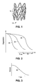
- FIGS. 2 and 3 depict degradation as a function of time for a polymer.
- FIG. 4 depicts a schematic illustration of a cross-section of a strut.
- FIGS. 5 and 6 depict erosion profiles of a stent.
- FIGS. 7 and 8A depict a schematic illustration of a pore forming agent in a coating region.
- FIG. 8B depicts a schematic illustration of pores formed in a coating region.
- FIG. 9 depicts a schematic plot of the rate of crystallization of a polymer as a function of temperature.
- implantable medical device is intended to include, but is not limited to, self-expandable stents, balloon-expandable stents, stent-grafts, grafts (e.g., aortic grafts), artificial heart valves, cerebrospinal fluid shunts, pacemaker electrodes, and endocardial leads (e.g., FINELINE and ENDOTAK, available from Guidant Corporation, Santa Clara, Calif.).
- the structural pattern of the device can be of virtually any design.
- a stent may include a pattern or network of interconnecting structural elements or struts.
- FIG. 1 depicts a three-dimensional view of a stent 10 which shows struts 15 .
- the pattern shown in FIG. 1 should not be limited to what has been illustrated as other stent patterns are easily applicable with the method of the invention.
- a stent such as stent 10 may be fabricated from a tube by forming a pattern with a technique such as laser cutting or chemical etching.
- biodegradable implantable medical devices that include biodegradable coatings over a biodegradable substrate.
- the substrate may be, for example, struts as shown in FIG. 1 .
- the stent may be intended to remain in the body for a duration of time until its intended function of, for example, maintaining vascular patency and/or drug delivery is accomplished.
- the duration can be in a range from about a month to a few years. However, the duration is typically in a range from about six to twelve months.
- biodegradable polymers used in coating applications after the process of degradation, erosion, absorption, and/or resorption has been completed, no polymer will remain on the stent. In some embodiments, very negligible traces or residue may be left behind.
- a variety of methods of coating can be used in practicing the present invention including, but not limited to, spraying, dipping, brushing, pouring, dripping, spinning, roller coating, meniscus coating, powder coating and various inking approaches such as inkjet-type application.
- the method of coating is spraying.
- additional process steps are necessary such as, for example, the application of heat or energy to the implantable medical device and/or coating.
- Embodiments of the coatings described herein may be configured to reduce the overall rate of erosion, or more generally, control erosion of the substrate during a desired treatment period.
- the substrate may be allowed to maintain its mechanical strength in order to serve the purpose of maintaining vascular patency.
- reducing the rate of erosion of the substrate may prevent large pieces of the stent from detaching from the eroding stent and causing embolization in a vessel.
- One preferred type of erosion profile in treatments involving struts of biodegradable stents may be separated into two time periods.
- An initial period may include a slow or minimal degradation for as long as mechanical support for the vessel is desired.
- the slow degradation may then be followed by a period of rapid degradation occurring approximately after the stent is no longer required.
- a stent configuration that may achieve such a profile may include a slow eroding, flexible outer region (e.g., a coating) and a fast eroding, stiff, strong inner region (a strut substrate) that provides mechanical support as long as support is desired.
- a stent may have low form factor (e.g., thinner struts) to reduce bodily fluid flow disruption in tend not to be radiopaque.
- the struts may be significantly thicker than struts in metal stents.
- a polymer-fabricated stent composed of poly(L-lactic acid) may require struts more than 50% thicker than a metallic stent.
- a metallic stent fabricated from a bioerodible metal, such as magnesium erodes too quickly to remain intact for the typical treatment time of six to twelve months.
- polymers can be biostable, bioabsorbable, biodegradable, or bioerodible.
- Biostable refers to polymers that are not biodegradable.
- biodegradable, bioabsorbable, and bioerodible, as well as degraded, eroded, and absorbed are used interchangeably and refer to polymers that are capable of being completely eroded or absorbed when exposed to bodily fluids such as blood and can be gradually resorbed, absorbed and/or eliminated by the body.
- biodegradable devices Several characteristics or parameters of the degradation process are important in designing biodegradable devices. These include an average erosion rate of a device, the erosion profile, the half-life of the degrading polymer, and mechanical stability of a device during the degradation process.
- the selected time interval may be between the onset of degradation and another selected time. Other selected times, for example, may be the time for about 25%, 50%, 75%, or 100% (complete erosion) of the device to erode. Complete erosion may correspond approximately to the time required for treatment by the device.
- the “half-life” of a degrading polymer refers to the length of time for the molecular weight of the polymer to fall to one half of its original value. See e.g., J. C. Middleton and A. J. Tipton, Biomaterials, Vol. 21 (23) (2000) pp. 2335-2346.
- an “erosion profile” refers to the functional dependence of the instantaneous erosion rate on time.
- biodegradable polymers span a continuum from polymers having a relatively constant instantaneous erosion rate with time during a degradation process to polymers with an instantaneous erosion rate that is strongly dependent on time.
- the former case corresponds to surface eroding polymers, while the latter case refers to bulk eroding polymers.
- the concepts of surface eroding and bulk eroding are limiting extremes. Real systems typically behave somewhere in between surface erosion and bulk erosion.
- Biodegradation of polymers generally refers to changes in physical and chemical properties that occur in a polymer upon exposure to bodily fluids as in a vascular environment.
- the changes in properties may include a decrease in molecular weight, deterioration of mechanical properties, and decrease in mass due to erosion or absorption.
- Mechanical properties may correspond to strength and modulus of the polymer. Deterioration of the mechanical properties of the polymer decreases the ability of a stent, for example, to provide mechanical support in a vessel.
- the decrease in molecular weight may be caused by, for example, hydrolysis and/or metabolic processes. Hydrolysis is a chemical process in which a molecule is cleaved into two parts by the addition of a molecule of water.
- the degree of bulk degradation of a polymer is strongly dependent on the diffusivity, and hence the diffusion rate of water in the polymer.
- the “diffusion rate” or flux, “J,” of a species in a material is defined as the number of randomly moving molecules that pass through a unit area per second.
- the time frame of degradation of a polymer part is dependent on water diffusion, hydrolysis, decrease in molecular weight, and erosion.
- a surface eroding polymer typically has relatively low water diffusivity.
- surface erosion is a heterogeneous process in which degradation and erosion tend to occur at or near a surface of the polymer exposed to the bodily fluids.
- a surface eroding erosion profile is more desirable since the disintegration of the device occurs less abruptly than for a bulk eroding polymer.
- a more gradual release of degraded material in a vascular system has a lower risk of embolization caused by a piece of the device breaking away.
- a surface eroding polymer also tends to delay, inhibit, or prevent degradation of regions or layers below a surface eroding layer or region.
- a surface eroding polymer may be preferable because there tends to be little or no change in mechanical properties of remaining polymer that has not eroded.
- a device composed of a surface eroding polymer may still tend to weaken as it loses mass.
- one or more coating regions composed of bulk eroding polymers over a bulk eroding or fast eroding substrate region can simulate the erosion profile of a surface eroding polymer.
- polymer erosion spans a continuum from bulk eroding to surface eroding.
- Bulk erosion refers to degradation of a polymer substantially throughout the bulk of a polymer part exposed to bodily fluids.
- the erosion profile of bulk eroding polymers typically consists of a relatively slow instantaneous erosion rate for a period of time after initial exposure to bodily fluids followed by a sharp increase in the instantaneous erosion rate.
- the degradation behavior of a polymer is strongly linked to the diffusivity of water in the polymer. As the diffusivity of water increases in a polymer, the bulk eroding behavior of the polymer increases.
- a bulk eroding polymer may be capable of absorbing less than about 3% by weight, or more narrowly, less than about 1% by weight. Water diffusivity in the polymer increases as the polymer region degrades.
- FIG. 2 illustrates degradation as a function of time for a bulk eroding polymer part.
- a curve 20 represents the decrease in molecular weight that occurs within the bulk of the polymer material. The decrease in molecular weight causes deterioration in mechanical properties of the polymer, which is shown by a curve 25 .
- a curve 30 represents the cumulative erosion versus time of the polymer.
- bulk eroding polymers include, but are not limited to, poly(L-lactide), poly(glycolide), poly(D,L-lactide), poly(trimethylene carbonate), polycaprolactone, and copolymers thereof.
- poly(L-lactide), poly(glycolide), poly(D,L-lactide), poly(trimethylene carbonate), polycaprolactone, and copolymers thereof During a course of treatment with a biodegradable polymeric stent, the polymer degrades resulting in a decrease in the molecular weight of the polymer and deterioration of mechanical properties.
- the instantaneous absorption or erosion rate of a surface eroding polymer is constant or relatively constant with time.
- This erosion behavior is due to the fact that surface erosion is a heterogeneous process in which degradation and erosion tend to occur at or near a surface of the polymer exposed to the bodily fluids.
- changes in the various properties may occur over the same or similar time frames since degradation is limited to a region at or near an exposed surface.
- a curve 40 depicts the cumulative erosion as a function of time for a surface-eroding polymer part.
- the erosion rate is dependent on the surface area of the eroding part. Since degradation is heterogeneous, the decrease in molecular weight and deterioration of the mechanical properties occur at or near the surface of a surface-eroding polymer part. In the bulk of the polymer part or away from the surface of a surface-eroding polymer part, the molecular weight and mechanical properties are unchanged or substantially unchanged.
- Representative examples of surface eroding polymers include, but are not limited to, polyorthoesters, polyanhydrides and copolymers thereof.
- polymers that may be used to fabricate an implantable medical device using the methods disclosed herein include, but are not limited to, poly(N-acetylglucosamine) (Chitin), Chitoson, poly(hydroxyvalerate), poly(lactide-co-glycolide), poly(hydroxybutyrate), poly(hydroxybutyrate-co-valerate), polyorthoester, polyanhydride, poly(glycolic acid), poly(glycolide), poly(L-lactic acid), poly(L-lactide), poly(D,L-lactic acid), poly(D,L-lactide), poly(caprolactone), poly(trimethylene carbonate), polyester amide, poly(glycolic acid-co-trimethylene carbonate), co-poly(ether-esters) (e.g.
- PEO/PLA polyphosphazenes
- biomolecules such as fibrin, fibrinogen, cellulose, starch, collagen and hyaluronic acid
- polyurethanes silicones
- polyesters polyolefins, polyisobutylene and ethylene-alphaolefin copolymers
- acrylic polymers and copolymers other than polyacrylates vinyl halide polymers and copolymers (such as polyvinyl chloride), polyvinyl ethers (such as polyvinyl methyl ether), polyvinylidene halides (such as polyvinylidene chloride), polyacrylonitrile, polyvinyl ketones, polyvinyl aromatics (such as polystyrene), polyvinyl esters (such as polyvinyl acetate), acrylonitrile-styrene copolymers, ABS resins, polyamides (such as Nylon 66 and polycaprolactam), polycarbonates, polyoxymethylenes, polyimides,
- polymers that may be especially well suited for use in fabricating an implantable medical device according to the methods disclosed herein include ethylene vinyl alcohol copolymer (commonly known by the generic name EVOH or by the trade name EVAL), poly(butyl methacrylate), poly(vinylidene fluoride-co-hexafluororpropene) (e.g., SOLEF 21508, available from Solvay Solexis PVDF, Thorofare, N.J.), polyvinylidene fluoride (otherwise known as KYNAR, available from ATOFINA Chemicals, Philadelphia, Pa.), ethylene-vinyl acetate copolymers, and polyethylene glycol.
- EVAL ethylene vinyl alcohol copolymer
- poly(vinylidene fluoride-co-hexafluororpropene) e.g., SOLEF 21508, available from Solvay Solexis PVDF, Thorofare, N.J.
- Biostable metals refer to metals that are not bioerodible. Biostable metals have negligible erosion or corrosion rates when exposed to bodily fluids.
- metal erosion or corrosion involves a chemical reaction between a metal surface and its environment. Erosion or corrosion in a wet environment, such as a vascular environment, results in removal of metal atoms from the metal surface. The metal atoms at the surface lose electrons and become actively charged ions that leave the metal to form salts in solution.
- biodegradable metals that may be used to fabricate an implantable medical device may include, but are not limited to, magnesium, zinc, and iron.
- a bioerodible metal stent may be completely eroded when exposed to bodily fluids, such as blood, between about a week and about three months, or more narrowly, between about one month and about two months.
- Embodiments of implantable medical devices described herein may include a biodegradable coating region above a biodegradable substrate region. “Above” a region is defined as higher than or over a region measured along an axis normal to a region, but not necessarily in contact with the region.
- the coating region may be configured to control the average erosion rate of the substrate region by controlling exposure of the substrate region to bodily fluids. The exposure of the portions of the substrate regions below the coating region is influenced by the transport of bodily fluids through the coating region to the substrate region.
- embodiments of the implantable medical device may include relatively distinct regions that have different erosion profiles when exposed to bodily fluids. In this way the erosion profile of the stent may be customized to various treatments.
- Embodiments may include a substrate region that is metallic, a bulk eroding polymer, or a substantially or completely surface eroding polymer. Additionally, the coating region may include a bulk eroding polymer or a substantially or completely surface eroding polymer.
- the substrate region may be a radially expandable stent including a pattern of struts.
- the cross-sectional shape of the struts can be circular, square, rectangular, oval, or any other shape.
- a metallic substrate may be a cylindrical or substantially cylindrical coil or mesh of metallic wire.
- a metallic substrate may be a pattern of struts formed on a metallic tube by cutting or etching.
- the coating region may include a bioactive agent.
- a “bioactive agent” is a moiety that is mixed, blended, bonded or linked to a polymer coating, or to a polymer from which a stent is made, and provides a therapeutic effect, a prophylactic effect, both a therapeutic and a prophylactic effect, or other biologically active effect upon release from the stent.
- the bioactive agents of the present invention may remain linked to a portion of the polymer or be released from the polymer.
- a bioabsorbable polymer and the degradants of bioabsorbable polymers tend not to provide a therapeutic effect, a prophylactic effect, both a therapeutic and a prophylactic effect, or other biologically active effect upon release from the stent.
- the polymer region may be configured to release the active agent for a selected amount of time.
- the release may occur through the break-up of the polymer and/or via migration of the active agent out of the polymer.
- the selected amount of time may correspond approximately to a desired treatment time of a stent.
- the substrate region may have pores that are configured to include an active agent.
- the metallic region can be formed by sintering particles, fibers, and wires of material.
- the substrate region may have a faster average erosion rate or lower half-life when exposed to bodily fluids than the coating region when exposed to bodily fluids.
- the coating region may be configured to delay, inhibit, or prevent erosion of the substrate region in a manner that allows the substrate region to provide mechanical support to a bodily lumen.
- the coating region may be configured to delay, inhibit, or prevent erosion of the substrate region for a selected time period.
- the selected time period may be at least a portion of the time period that the substrate region is desired to provide mechanical support. It may be desirable for a substrate region to provide mechanical support for a majority of, all of, or longer than a desired treatment time of the stent.
- the substrate region may erode when the substrate region is exposed to bodily fluids due to degradation of the coating region.
- the substrate region may be exposed to bodily fluids by complete erosion of the coating region over a portion of the substrate region and/or diffusion of bodily fluids through the coating region.
- a substrate region may start to erode when the coating region is only partially degraded and/or eroded. Partially means less than 50% of the coating, or alternatively less than 40%, 30%, 20%, 10%, or 5%.
- the substrate region may start to erode when the coating region is completely (greater than 99%) degraded and/or eroded or when a majority of the coating is degraded and/or eroded.
- Majority includes over 50%, 60%, 70%, 80%, 90%, or alternatively, over 95% of the coating region
- a coating region composed of a bulk eroding polymer may allow a substantial amount of bodily fluids to diffuse through the coating and erode the substrate region.
- the substrate region may be completely or almost completely eroded before the coating region is completely eroded.
- a coating region may be a surface eroding polymer or a substantially surface eroding polymer.
- a surface eroding polymer may be selected that has a water diffusivity that inhibits or prevents erosion of the substrate region for a selected time period.
- the substrate region may be configured to erode when erosion of the polymer region exposes a portion of the substrate region to bodily fluids.
- FIG. 4 depicts a schematic illustration of an embodiment of a cross-section of a strut 100 of a stent that includes a coating region 105 and a substrate region 110 .
- Coating region 105 is composed primarily of a biodegradable polymer.
- Substrate region 110 can be an erodible metal or a biodegradable polymer.
- FIGS. 5 and 6 illustrate examples of erosion as a function of time for such a stent.
- FIG. 5 depicts erosion for a stent with a bulk eroding coating region.
- a curve 115 represents the cumulative erosion vs. time of the coating region and curve 120 represents the cumulative erosion of the substrate region.
- the substrate region may be a fast eroding metallic region or bulk eroding polymer.
- FIG. 6 depicts erosion for a stent with a coating region 105 composed of a substantially surface eroding polymer.
- a curve 125 represents the cumulative erosion vs. time of the coating region and a curve 130 represents the cumulative erosion vs. time of the substrate region.
- a time 140 corresponds to an approximate time of implantation of the stent in a vessel. From time 140 to approximately a time 145 in FIG. 5 and between time 140 and approximately time 150 in FIG. 6 , there is minimal erosion of the substrate region.
- time 145 represents the onset of substantial erosion of the substrate region with a bulk eroding polymer coating region.
- time 150 represents the onset of substantial erosion of the substrate region with a surface eroding polymer coating region.
- Coating region 105 delays the onset of substantial erosion to a later time as compared to a strut with no coating region.
- the stent may be no longer required for treatment.
- the coating and substrate regions may be completely or almost completely eroded and the substrate regions may no longer provide mechanical support.
- the erosion of the substrate region rises sharply during time periods 155 and 160 due to degradation and/or erosion of the coating regions.
- a comparison of curve 120 to curve 130 illustrates the sharper erosion profile of the substrate when a surface eroding polymer is used rather than a bulk eroding polymer for the coating region.
- Curve 120 is less steep than curve 130 because the diffusion of water in the bulk eroding polymer coating is substantially greater than through a surface eroding polymer coating.
- the substrate region with the bulk eroding polymer coating is exposed to a substantial amount of bodily fluids prior to complete erosion of the coating from a portion of the substrate region, i.e, prior to direct exposure of the substrate region to bodily fluids.
- FIGS. 5 and 6 illustrate qualitatively how the coating region alters the erosion profile of the substrate region.
- the coating region alters the approximate time of the onset of erosion (time 145 in FIG. 5 and time 150 in FIG. 6 ) and the subsequent time period of substantial erosion (time period 155 in FIG. 5 and time period 160 in FIG. 6 ).
- the rate of erosion is also altered.
- the coating region may be configured to control exposure of the substrate region to bodily fluids and achieve a specified degree of erosion of the substrate region.
- a coating region may be used to alter or control the exposure of the substrate region, and hence, its erosion profile.
- the thickness of the coating region is directly related to the times of the onset of erosion and a time period of substantial erosion. Increasing the thickness of the coating region alters the erosion profile of the substrate region primarily by increasing the time for water to diffuse through a coating region to the substrate region. As illustrated by Fick's first law of diffusion, as the thickness increases, the flux of fluid diffusing through the coating increases. The time for complete erosion of the coating region over a portion of the substrate region is also altered. For instance, time periods 155 and 160 are shifted to later times as the thickness of the coating region increases.
- the thickness of the coating region may be controlled to control erosion of the substrate region.
- a method may include controlling a thickness of the coating region to allow a specified amount of erosion of the substrate region during a selected time period.
- the specified amount of erosion may be an amount of erosion that allows the substrate region to maintain mechanical support to a bodily lumen.
- the specified amount of erosion may be 10%, 40%, 60%, 80%, or 90% of the amount of erosion that renders the substrate region unable to continue to support a bodily lumen.
- the erosion profile of the substrate region is also altered by changing the diffusion rate or flux of bodily fluids or water through the coating region.
- the diffusion rate of water may be changed by modifying the chemical composition of the coating region, e.g., by using a different polymer.
- the bulk eroding polymers discussed above tend to have relatively high water diffusivities and the surface eroding polymers have relatively low water diffusivities.
- Another way of changing the diffusion rate of water in the coating region is to introduce porosity into the coating region.
- Introducing porosity into a coating region allows variation of the erosion profile of the substrate region without substantially changing the chemical composition of the coating region. This may be an advantage since there may be reasons other than diffusion rate of water in a coating material to use a particular coating composition, e.g., biocompatibility, processing issues, etc.
- a porous coating is also desirable because the pores facilitate transport of eroded material out of a stented area.
- a build-up of eroded material within a coating may inhibit transport of water through a coating, and thus, inhibit degradation. Increasing removal of eroded material increases the diffusion of water through the coating, and thus the degradation rate.
- the erosion profile of the coating region, and consequently the substrate region may be tuned or modified in various ways. Introducing porosity into a coating region composed of a polymer with bulk eroding properties may tend to increase the average erosion rate or decrease the half life of the substrate region.
- curve 180 in FIG. 5 and curve 185 in FIG. 6 may represent the erosion profiles of the substrate region when the coating region has porosity.
- a layer is “porous” when it has a void-to-volume percentage that ranges from about 40% to about 90%, from about 70% to about 80%, or any range therein. In some embodiments, a layer is “non-porous” when it has a void-to-volume percentage that ranges from about 0% to about 5%, from about 1% to about 3%, or any range therein.
- the “void-to-volume percentage” is defined as the volume of the pores divided by the total volume of the layer including the volume of the pores.
- the void-to-volume percentage can be measured using standard test method BSR/AAMI/ISO 7198, which has been adopted in-whole as a revision of ANSI/AAMI VP20-1994 (Cardiovascular Implants—Vascular Prosthesis section 8.2.1.2, Method for Gravimetric Determination of Porosity, Am. Nat'l Stds. Inst./Assoc. for the Adv. of Med. Instr.)
- an implantable medical device may include a coating region that includes a porous bioabsorbable matrix above a substrate region.
- “Above” a region is defined as higher than or over a surface or layer measured along an axis normal to a surface, but not necessarily in contact with the surface or layer.
- the polymeric matrix may allow transport of bodily fluids through pores of the porous matrix to the substrate region. Porosity can be introduced in the coating region by any method known to one of skill in the art.
- the porous matrix may be formed by phase inversion precipitation of a bioabsorbable polymer.
- a polymer may be mixed with two miscible solvents to form a solution.
- One of the solvents (solvent A) should be less volatile than the other solvent (solvent B).
- solvent B solvent
- the polymer should be less soluble in solvent A.
- the solution can then be applied to a portion of the surface of the implantable medical device. Next, when the solvents are allowed to evaporate, the polymer slowly precipitates as solvent B is essentially removed from the coating. As a result, after complete drying, the polymer matrix becomes porous.
- the size of the pores can be controlled by the choice of polymers and solvents and the relative concentrations of the solutions.
- the depth of a porous matrix into the coating region can be controlled by using the phase inversion technique after a portion of the coating region has been applied to the surface of the device. Pores in the range of about 0.1 microns to about 1 micron in diameter may be suitable.
- the porous matrix can be formed by using a sintering process.
- Sintering is a process of fabrication where particles are bonded together by partially melting some of the particles.
- a bioabsorbable polymeric powder or particles can be applied to the surface of the device and then pressed together.
- the particles can be about 1 micron to about 10 microns.
- the polymeric particles can be heated to temperatures slightly below or about the melting point of the polymer. Without entirely melting all of the particles, the particles bond to each other at their respective surfaces. Space remains between the lattice of the particles to form porous cavities.
- a coating region including a bioabsorbable polymeric porous matrix may be formed with a pore forming agent or porogen.
- the coating region may include a bioabsorbable polymer and a pore forming agent dispersed or mixed within the bioabsorbable polymer.
- a pore forming agent in the form of particles and/or fibers may be added to a polymeric material used to form a coating region of an implantable medical device. Pores within the polymeric material of the coating region may be formed when at least a portion of the pore forming agent is dissolved or eroded by a fluid.
- the fluid may be a suitable solvent (e.g., water) or bodily fluids that dissolve or erode the pore forming agent.
- a tortuous porous network may be formed in the coating region that allows diffusion of bodily fluids to the substrate region.
- pores may be formed in the coating region through dissolution and/or erosion of at least some of the pore forming agent after exposure of the coating region to a solvent in vitro.
- pore forming agent may be removed through dissolution and/or erosion of the pore forming agent when the coating region is exposed to bodily fluids after implantation of the device.
- non-bioactive pore forming agents may include, but are not limited to, salts, sugars, and water-soluble polymers.
- Water-soluble polymers may include, for example, polymeric salts, polyvinyl alcohol, polyethylene glycol, polyethylene oxide, glucose, dextran, dextrose, lactose, gamma globulin, ambumin, and combinations thereof. Such particles may be removed in vivo, for example, by washing in water or a very dilute acid bath.
- non-polymeric salts include, but are not limited to, sodium chloride, phosphate salts, carbonate salts, sodium bicarbonate, and combinations thereof.
- Other pore forming agents may include urea and amino acids.
- the coating region may include a bioabsorbable polymer and a pore forming agent dispersed or mixed within the bioabsorbable polymer.
- the pore forming agent may include a second bioabsorbable polymer mixed, dispersed, or blended within the coating region.
- the second bioabsorbable polymer may be in the form of particles and/or fibers. It is desirable for the second bioabsorbable polymer to have a higher average erosion rate or a shorter half-life than the bioabsorbable polymer of the coating region.
- the coating region may include a continuous phase of a slower eroding bioabsorbable polymer and a dispersed phase of a faster eroding bioabsorbable polymer.
- a pore formation rate after implantation may be tuned by selecting a second bioabsorbable polymer with a particular average erosion rate or half life.
- some embodiments may include a pore forming agent that includes an erodible metal mixed or dispersed with the coating region.
- properties of the porous matrix of the coating region may influence the erosion profile of the coating region, and hence, the erosion profile of the substrate region.
- properties of the porous matrix include, but are not limited to, the pore size distribution and porosity. Porosity may be defined as the ratio of the void volume to the total volume of the coating region.
- the erosion profile of the coating region and/or substrate region may be tuned or controlled by controlling the pore size distribution and porosity of the coating region.
- the pore size distribution and porosity depend on variables including, but not limited to, the number concentration of particles and/or fibers (number of particles and/or fibers per unit volume) of pore forming agent and the particle and/or fiber size of pore forming agents.
- the pore size distribution porosity can be controlled by screening the particles and/or fibers according to size and adding particles of a predetermined size to the materials. For example, increasing the particle size of pore forming agent tends to increase both the porosity and pore size distribution. In addition, increasing the number concentration of particles tends to increase the porosity.
- FIGS. 7 and 8A -B illustrate that variables such as particle size and concentration can influence the characteristics of a porous network formed by pore forming agents.
- FIG. 7 depicts a portion of a coating region 200 over a substrate region 205 .
- Coating region 200 has a relatively low concentration of particles 210 of pore forming agent. At relatively low concentrations, of particles 210 may be dissolved or eroded by fluid (solvent or bodily fluids) to form isolated pores or voids that are not connected by other pores.
- fluid solvent or bodily fluids
- FIG. 8A depicts a coating region 220 over a substrate region 225 with a relatively high concentration of particles 230 of pore forming agent.
- the distances between neighboring particles are smaller than distances between neighboring particles for a majority of particles 210 .
- FIG. 8B it is expected that if the concentration of particles 230 is high enough, as particles 230 dissolve or erode, interconnected pores or voids 235 are formed. Thus, a tortuous porous network may be formed in the coating region.
- the formation of a porous network tends to occur in a relatively short time frame. It is expected that the diffusion rate or flux of fluid through the coating region may be relatively constant after initial exposure to fluid. After formation of an interconnected porous network, the diffusion rate of fluid may tend to increase substantially during a short time frame.
- the concentration of pore forming particles can be tuned to obtain a desired time of formation of the interconnected network after exposure of the coating region to bodily fluids.
- the diffusion rate or flux of fluid within a coating region may also be controlled through selection of the chemical properties of the pore forming agent, in particular, the osmotic behavior of water soluble pore forming agents.
- “Osmosis” refers to the diffusion of molecules from a region of higher concentration of an osmotically active agent to a place of lower concentration until the concentration in both regions is equal.
- “Osmotic pressure” is the pressure exerted by a solution necessary to prevent osmosis into that solution when it is separated from the pure solvent by a semipermeable membrane that allows diffusion of only the solvent.
- An osmotically active agent dispersed in the coating region may increase the osmotic pressure differential between the coating region and fluid regions adjacent to the coating region.
- increasing the concentration of the osmotically active agents in the coating region increases the osmotic pressure differential which results in increased diffusion of water or bodily fluids into the coating region.
- the increased diffusion rate may then increase the degradation rate of the coating region.
- the osmotic pressure differential tends to drive fluid into the pores or voids formed by dissolution of particles or pore forming agent.
- a pore forming agent with a higher osmotic pressure may tend to result in formation of a coating region with a larger pore size distribution and a greater porosity.
- the pore size distribution and porosity of a coating region may be controlled by selection of a pore forming agent based on its osmotic pressure.
- the diffusion rate of bodily fluids, and hence the erosion of the substrate region may be controlled with the degree of crystallinity of a bioabsorbable polymeric coating region.
- An embodiment of a method of fabricating an implantable medical device may include controlling a degree of crystallinity of the coating region to allow a specified amount of erosion of the substrate region during a selected time period.
- the diffusion rate of a fluid through a polymer decreases as the degree of crystallinity increases. Therefore, it is expected that the diffusion rate of water and bodily fluids is lower in crystalline and semi-crystalline polymers than in amorphous polymers.
- the erosion rate of a substrate region may be controlled by modifying the degree of crystallinity of the coating region.
- the crystallinity of a polymer may be modified by heating the polymer.
- Heating a polymer can alter the degree of crystallinity and/or size of crystalline regions in a polymer material.
- the degree of crystallinity may be altered by heating the polymer to within a particular temperature range.
- Heating a polymer material to a temperature below the glass transition temperature, T g of the polymer does not significantly alter the molecular structure, and hence, the mechanical properties of the material. Below T g , energy barriers to segmental motion of the chains of a polymer inhibit or prevent alteration of molecular structure of a polymer material.
- crystallization may occur in a polymer material that is heated to a temperature between T g and the melting temperature, T m , of the polymer.
- T g melting temperature
- T m melting temperature
- FIG. 9 depicts a schematic plot of the rate of crystallization of a polymer as a function of temperature.
- FIG. 9 shows that the rate of polymer crystallization increases as the temperature is increased from below the T g of the polymer or is decreased from above the T m of the polymer. The rate of crystallization reaches a maximum 300 somewhere between the T g and the T m .
- FIG. 9 shows that effectively no crystallization occurs below the T g or above the T m .
- an amorphous polymer may be formed by heating a polymer material. Above the T m , a polymer material is a disordered melt and cannot crystallize and any crystallinity present is destroyed. Quenching a polymer heated to above the T m to a temperature below the T g of the polymer may result in the formation of a solid amorphous polymer. The resulting amorphous polymer material may have a lower modulus and be a more flexible or a less stiff material than before heating.
- a coating region may be heated in a variety of ways including, but limited to heating in an oven or blowing hot air on the coating.
- a coating may be quenched, for example, in a water bath.
Abstract
Description
Average erosion rate=(m 2 −m 1)/(t 2 −t 1)
where “m” refers to mass of the device, “t” refers to a time during erosion, and m1 and m2 are the masses of the device at t1 and t2 during erosion. For instance, the selected time interval may be between the onset of degradation and another selected time. Other selected times, for example, may be the time for about 25%, 50%, 75%, or 100% (complete erosion) of the device to erode. Complete erosion may correspond approximately to the time required for treatment by the device.
Claims (2)
Priority Applications (2)
| Application Number | Priority Date | Filing Date | Title |
|---|---|---|---|
| US11/173,713 US10028851B2 (en) | 1997-04-15 | 2005-06-30 | Coatings for controlling erosion of a substrate of an implantable medical device |
| PCT/US2006/025937 WO2007005806A1 (en) | 2005-06-30 | 2006-06-30 | Coatings for controlling erosion of a substrate of an implantable medical device |
Applications Claiming Priority (6)
| Application Number | Priority Date | Filing Date | Title |
|---|---|---|---|
| US08/837,993 US6240616B1 (en) | 1997-04-15 | 1997-04-15 | Method of manufacturing a medicated porous metal prosthesis |
| US09/797,313 US20010013166A1 (en) | 1997-04-15 | 2001-03-01 | Method of manufacturing a medicated porous metal prosthesis |
| US10/235,033 US6723120B2 (en) | 1997-04-15 | 2002-09-03 | Medicated porous metal prosthesis |
| US10/767,296 US7699890B2 (en) | 1997-04-15 | 2004-01-28 | Medicated porous metal prosthesis and a method of making the same |
| US10/880,025 US8172897B2 (en) | 1997-04-15 | 2004-06-28 | Polymer and metal composite implantable medical devices |
| US11/173,713 US10028851B2 (en) | 1997-04-15 | 2005-06-30 | Coatings for controlling erosion of a substrate of an implantable medical device |
Related Parent Applications (1)
| Application Number | Title | Priority Date | Filing Date |
|---|---|---|---|
| US10/880,025 Continuation-In-Part US8172897B2 (en) | 1997-04-15 | 2004-06-28 | Polymer and metal composite implantable medical devices |
Publications (2)
| Publication Number | Publication Date |
|---|---|
| US20050283229A1 US20050283229A1 (en) | 2005-12-22 |
| US10028851B2 true US10028851B2 (en) | 2018-07-24 |
Family
ID=37056938
Family Applications (1)
| Application Number | Title | Priority Date | Filing Date |
|---|---|---|---|
| US11/173,713 Expired - Fee Related US10028851B2 (en) | 1997-04-15 | 2005-06-30 | Coatings for controlling erosion of a substrate of an implantable medical device |
Country Status (2)
| Country | Link |
|---|---|
| US (1) | US10028851B2 (en) |
| WO (1) | WO2007005806A1 (en) |
Cited By (4)
| Publication number | Priority date | Publication date | Assignee | Title |
|---|---|---|---|---|
| US11497595B2 (en) | 2018-11-02 | 2022-11-15 | Boston Scientific Scimed, Inc. | Biodegradable stent |
| US11690807B2 (en) * | 2018-05-24 | 2023-07-04 | Celanese Eva Performance Polymers Llc | Implantable device for sustained release of a macromolecular drug compound |
| US11690806B2 (en) | 2018-05-24 | 2023-07-04 | Celanese Eva Performance Polymers Llc | Implantable device for sustained release of a macromolecular drug compound |
| US11925570B2 (en) | 2018-12-19 | 2024-03-12 | Boston Scientific Scimed, Inc. | Stent including anti-migration capabilities |
Families Citing this family (74)
| Publication number | Priority date | Publication date | Assignee | Title |
|---|---|---|---|---|
| WO2003002243A2 (en) | 2001-06-27 | 2003-01-09 | Remon Medical Technologies Ltd. | Method and device for electrochemical formation of therapeutic species in vivo |
| US9114198B2 (en) | 2003-11-19 | 2015-08-25 | Advanced Cardiovascular Systems, Inc. | Biologically beneficial coatings for implantable devices containing fluorinated polymers and methods for fabricating the same |
| JP2007517550A (en) | 2004-01-02 | 2007-07-05 | アドヴァンスド カーディオヴァスキュラー システムズ, インコーポレイテッド | Medical device coated with high density lipoprotein |
| US8367099B2 (en) | 2004-09-28 | 2013-02-05 | Atrium Medical Corporation | Perforated fatty acid films |
| US9801982B2 (en) | 2004-09-28 | 2017-10-31 | Atrium Medical Corporation | Implantable barrier device |
| US9012506B2 (en) | 2004-09-28 | 2015-04-21 | Atrium Medical Corporation | Cross-linked fatty acid-based biomaterials |
| EP1811935B1 (en) | 2004-09-28 | 2016-03-30 | Atrium Medical Corporation | Heat cured gel and method of making |
| US9000040B2 (en) | 2004-09-28 | 2015-04-07 | Atrium Medical Corporation | Cross-linked fatty acid-based biomaterials |
| US8312836B2 (en) | 2004-09-28 | 2012-11-20 | Atrium Medical Corporation | Method and apparatus for application of a fresh coating on a medical device |
| US9592324B2 (en) | 2006-11-06 | 2017-03-14 | Atrium Medical Corporation | Tissue separating device with reinforced support for anchoring mechanisms |
| US20060088596A1 (en) | 2004-09-28 | 2006-04-27 | Atrium Medical Corporation | Solubilizing a drug for use in a coating |
| US8048141B2 (en) * | 2004-12-07 | 2011-11-01 | Boston Scientific Scimed, Inc. | Medical device that signals lumen loss |
| US20070050009A1 (en) * | 2005-08-30 | 2007-03-01 | Aiden Flanagan | Bioabsorbable stent |
| US9278161B2 (en) | 2005-09-28 | 2016-03-08 | Atrium Medical Corporation | Tissue-separating fatty acid adhesion barrier |
| US9427423B2 (en) | 2009-03-10 | 2016-08-30 | Atrium Medical Corporation | Fatty-acid based particles |
| EP1933991A4 (en) | 2005-10-15 | 2012-05-02 | Atrium Medical Corp | Hydrophobic cross-linked gels for bioabsorbable drug carrier coatings |
| US8840660B2 (en) | 2006-01-05 | 2014-09-23 | Boston Scientific Scimed, Inc. | Bioerodible endoprostheses and methods of making the same |
| US8089029B2 (en) | 2006-02-01 | 2012-01-03 | Boston Scientific Scimed, Inc. | Bioabsorbable metal medical device and method of manufacture |
| US7910152B2 (en) * | 2006-02-28 | 2011-03-22 | Advanced Cardiovascular Systems, Inc. | Poly(ester amide)-based drug delivery systems with controlled release rate and morphology |
| US8048150B2 (en) | 2006-04-12 | 2011-11-01 | Boston Scientific Scimed, Inc. | Endoprosthesis having a fiber meshwork disposed thereon |
| US8323676B2 (en) | 2008-06-30 | 2012-12-04 | Abbott Cardiovascular Systems Inc. | Poly(ester-amide) and poly(amide) coatings for implantable medical devices for controlled release of a protein or peptide and a hydrophobic drug |
| EP3047860A1 (en) | 2006-07-20 | 2016-07-27 | OrbusNeich Medical, Inc. | Bioabsorbable polymeric composition for a medical device |
| US9265866B2 (en) * | 2006-08-01 | 2016-02-23 | Abbott Cardiovascular Systems Inc. | Composite polymeric and metallic stent with radiopacity |
| EP2054537A2 (en) | 2006-08-02 | 2009-05-06 | Boston Scientific Scimed, Inc. | Endoprosthesis with three-dimensional disintegration control |
| DE102006038236A1 (en) * | 2006-08-07 | 2008-02-14 | Biotronik Vi Patent Ag | Biodegradable stent with an active coating |
| US20080057103A1 (en) * | 2006-08-21 | 2008-03-06 | Wouter Roorda | Methods of using medical devices for controlled drug release |
| US8293318B1 (en) | 2006-08-29 | 2012-10-23 | Abbott Cardiovascular Systems Inc. | Methods for modulating the release rate of a drug-coated stent |
| US8808726B2 (en) | 2006-09-15 | 2014-08-19 | Boston Scientific Scimed. Inc. | Bioerodible endoprostheses and methods of making the same |
| EP2068964B1 (en) * | 2006-09-15 | 2017-11-01 | Boston Scientific Limited | Medical devices and methods of making the same |
| WO2008034066A1 (en) * | 2006-09-15 | 2008-03-20 | Boston Scientific Limited | Bioerodible endoprostheses and methods of making the same |
| EP2210625B8 (en) * | 2006-09-15 | 2012-02-29 | Boston Scientific Scimed, Inc. | Bioerodible endoprosthesis with biostable inorganic layers |
| EP2076296A2 (en) | 2006-09-15 | 2009-07-08 | Boston Scientific Scimed, Inc. | Endoprosthesis with adjustable surface features |
| EP2068963B1 (en) * | 2006-09-18 | 2011-10-26 | Boston Scientific Limited | Endoprostheses |
| CA2663762A1 (en) | 2006-09-18 | 2008-03-27 | Boston Scientific Limited | Endoprostheses |
| US7959942B2 (en) | 2006-10-20 | 2011-06-14 | Orbusneich Medical, Inc. | Bioabsorbable medical device with coating |
| EP2073754A4 (en) | 2006-10-20 | 2012-09-26 | Orbusneich Medical Inc | Bioabsorbable polymeric composition and medical device background |
| US9492596B2 (en) | 2006-11-06 | 2016-11-15 | Atrium Medical Corporation | Barrier layer with underlying medical device and one or more reinforcing support structures |
| ES2356274T3 (en) | 2006-12-28 | 2011-04-06 | Boston Scientific Limited | BIODEGRADABLE ENDOPROTESIS AND MANUFACTURING PROCEDURES OF THE SAME. |
| CN101636187B (en) * | 2007-01-30 | 2013-08-28 | 汉莫堤克股份有限公司 | Biodegradable vascular support |
| CA2673914C (en) * | 2007-01-30 | 2013-10-08 | Hemoteq Ag | Biodegradable vascular support |
| DE102007004589A1 (en) * | 2007-01-30 | 2008-07-31 | Orlowski, Michael, Dr. | Reabsorbable implant stent for blood vessels, urinary passages, respiratory system, biliary tract or digestive tract, comprises magnesium alloy containing magnesium, calcium or yattrium |
| US20080249608A1 (en) * | 2007-04-04 | 2008-10-09 | Vipul Dave | Bioabsorbable Polymer, Bioabsorbable Composite Stents |
| US8252361B2 (en) * | 2007-06-05 | 2012-08-28 | Abbott Cardiovascular Systems Inc. | Implantable medical devices for local and regional treatment |
| US8052745B2 (en) | 2007-09-13 | 2011-11-08 | Boston Scientific Scimed, Inc. | Endoprosthesis |
| US8998978B2 (en) * | 2007-09-28 | 2015-04-07 | Abbott Cardiovascular Systems Inc. | Stent formed from bioerodible metal-bioceramic composite |
| US7939096B2 (en) * | 2008-02-12 | 2011-05-10 | Boston Scientific Scimed, Inc. | Medical implants with polysaccharide drug eluting coatings |
| US20090240323A1 (en) * | 2008-03-20 | 2009-09-24 | Medtronic Vascular, Inc. | Controlled Degradation of Magnesium Stents |
| US9259515B2 (en) * | 2008-04-10 | 2016-02-16 | Abbott Cardiovascular Systems Inc. | Implantable medical devices fabricated from polyurethanes with grafted radiopaque groups |
| US7998192B2 (en) | 2008-05-09 | 2011-08-16 | Boston Scientific Scimed, Inc. | Endoprostheses |
| US8236046B2 (en) | 2008-06-10 | 2012-08-07 | Boston Scientific Scimed, Inc. | Bioerodible endoprosthesis |
| US7985252B2 (en) | 2008-07-30 | 2011-07-26 | Boston Scientific Scimed, Inc. | Bioerodible endoprosthesis |
| US20100047319A1 (en) * | 2008-08-21 | 2010-02-25 | Michael Huy Ngo | Biodegradable Poly(Ester-Amide) And Poly(Amide) Coatings For Implantable Medical Devices With Enhanced Bioabsorption Times |
| US20100057219A1 (en) * | 2008-08-29 | 2010-03-04 | Kwangyeol Lee | Device surface design for better cell adhesion |
| US8382824B2 (en) | 2008-10-03 | 2013-02-26 | Boston Scientific Scimed, Inc. | Medical implant having NANO-crystal grains with barrier layers of metal nitrides or fluorides |
| US20100092535A1 (en) * | 2008-10-10 | 2010-04-15 | Medtronic Vascular, Inc. | Nanoporous Drug Delivery System |
| EP2403546A2 (en) | 2009-03-02 | 2012-01-11 | Boston Scientific Scimed, Inc. | Self-buffering medical implants |
| US20100249944A1 (en) * | 2009-03-31 | 2010-09-30 | Thomas Jonathan D | Multizone Implants |
| US9592043B2 (en) * | 2009-03-31 | 2017-03-14 | Covidien Lp | Multizone implants |
| US20100249832A1 (en) * | 2009-03-31 | 2010-09-30 | Joshua Stopek | Multizone Implants |
| US20100249854A1 (en) * | 2009-03-31 | 2010-09-30 | Thomas Jonathan D | Multizone Implants |
| US20110038910A1 (en) | 2009-08-11 | 2011-02-17 | Atrium Medical Corporation | Anti-infective antimicrobial-containing biomaterials |
| US8668732B2 (en) | 2010-03-23 | 2014-03-11 | Boston Scientific Scimed, Inc. | Surface treated bioerodible metal endoprostheses |
| US10322213B2 (en) | 2010-07-16 | 2019-06-18 | Atrium Medical Corporation | Compositions and methods for altering the rate of hydrolysis of cured oil-based materials |
| US20130138219A1 (en) * | 2011-11-28 | 2013-05-30 | Cook Medical Technologies Llc | Biodegradable stents having one or more coverings |
| US9867880B2 (en) | 2012-06-13 | 2018-01-16 | Atrium Medical Corporation | Cured oil-hydrogel biomaterial compositions for controlled drug delivery |
| US9603728B2 (en) | 2013-02-15 | 2017-03-28 | Boston Scientific Scimed, Inc. | Bioerodible magnesium alloy microstructures for endoprostheses |
| US9522220B2 (en) | 2013-10-29 | 2016-12-20 | Boston Scientific Scimed, Inc. | Bioerodible magnesium alloy microstructures for endoprostheses |
| CA2973155A1 (en) | 2015-03-11 | 2016-09-15 | Boston Scientific Scimed, Inc. | Bioerodible magnesium alloy microstructures for endoprostheses |
| US10596660B2 (en) | 2015-12-15 | 2020-03-24 | Howmedica Osteonics Corp. | Porous structures produced by additive layer manufacturing |
| WO2019033121A1 (en) | 2017-08-11 | 2019-02-14 | Elixir Medical Corporation | Uncaging stent |
| US11622872B2 (en) | 2016-05-16 | 2023-04-11 | Elixir Medical Corporation | Uncaging stent |
| CN113143536B (en) | 2016-05-16 | 2022-08-30 | 万能医药公司 | Opening support |
| US11628517B2 (en) | 2017-06-15 | 2023-04-18 | Howmedica Osteonics Corp. | Porous structures produced by additive layer manufacturing |
| AU2018256556B2 (en) | 2017-11-03 | 2024-04-04 | Howmedica Osteonics Corp. | Flexible construct for femoral reconstruction |
Citations (372)
| Publication number | Priority date | Publication date | Assignee | Title |
|---|---|---|---|---|
| US3687135A (en) | 1969-08-20 | 1972-08-29 | Genrikh Borisovich Stroganov | Magnesium-base alloy for use in bone surgery |
| US3839743A (en) | 1972-04-21 | 1974-10-08 | A Schwarcz | Method for maintaining the normal integrity of blood |
| US3855638A (en) | 1970-06-04 | 1974-12-24 | Ontario Research Foundation | Surgical prosthetic device with porous metal coating |
| US3900632A (en) | 1970-02-27 | 1975-08-19 | Kimberly Clark Co | Laminate of tissue and random laid continuous filament web |
| US4101984A (en) | 1975-05-09 | 1978-07-25 | Macgregor David C | Cardiovascular prosthetic devices and implants with porous systems |
| US4104410A (en) | 1973-12-21 | 1978-08-01 | Malecki George J | Processing of green vegetables for color retention in canning |
| US4110497A (en) | 1976-07-02 | 1978-08-29 | Snyder Manufacturing Co., Ltd. | Striped laminate and method and apparatus for making same |
| US4321711A (en) | 1978-10-18 | 1982-03-30 | Sumitomo Electric Industries, Ltd. | Vascular prosthesis |
| US4346028A (en) | 1979-12-14 | 1982-08-24 | Monsanto Company | Asbestiform crystalline calcium sodium or lithium phosphate, preparation and compositions |
| US4355426A (en) | 1975-05-09 | 1982-10-26 | Macgregor David C | Porous flexible vascular graft |
| US4374669A (en) | 1975-05-09 | 1983-02-22 | Mac Gregor David C | Cardiovascular prosthetic devices and implants with porous systems |
| US4405319A (en) * | 1980-04-08 | 1983-09-20 | Renal Systems, Inc. | Porous titanium coating for blood access device |
| EP0108171A1 (en) | 1982-09-15 | 1984-05-16 | Meadox Medicals, Inc. | Synthetic woven double-velour graft |
| US4458366A (en) | 1975-05-09 | 1984-07-10 | Macgregor David C | Artificial implantable blood pump |
| EP0144534A2 (en) | 1983-09-20 | 1985-06-19 | Allied Corporation | prosthetic devices |
| US4596574A (en) | 1984-05-14 | 1986-06-24 | The Regents Of The University Of California | Biodegradable porous ceramic delivery system for bone morphogenetic protein |
| US4599085A (en) | 1979-07-11 | 1986-07-08 | Neodontics, Inc. | Bone implant member for prostheses and bone connecting elements and process for the production thereof |
| US4612009A (en) | 1984-06-19 | 1986-09-16 | Ceskoslovenska Akademie Ved | Biodegradable implant and a method for preparation thereof |
| US4633873A (en) | 1984-04-26 | 1987-01-06 | American Cyanamid Company | Surgical repair mesh |
| US4656083A (en) | 1983-08-01 | 1987-04-07 | Washington Research Foundation | Plasma gas discharge treatment for improving the biocompatibility of biomaterials |
| US4693721A (en) | 1984-10-17 | 1987-09-15 | Paul Ducheyne | Porous flexible metal fiber material for surgical implantation |
| US4718907A (en) | 1985-06-20 | 1988-01-12 | Atrium Medical Corporation | Vascular prosthesis having fluorinated coating with varying F/C ratio |
| US4722335A (en) | 1986-10-20 | 1988-02-02 | Vilasi Joseph A | Expandable endotracheal tube |
| US4723549A (en) | 1986-09-18 | 1988-02-09 | Wholey Mark H | Method and apparatus for dilating blood vessels |
| US4729871A (en) | 1985-06-21 | 1988-03-08 | Hiroshi Kawaguchi | Process for preparing porous metal plate |
| US4732152A (en) | 1984-12-05 | 1988-03-22 | Medinvent S.A. | Device for implantation and a method of implantation in a vessel using such device |
| US4733665A (en) | 1985-11-07 | 1988-03-29 | Expandable Grafts Partnership | Expandable intraluminal graft, and method and apparatus for implanting an expandable intraluminal graft |
| US4740207A (en) | 1986-09-10 | 1988-04-26 | Kreamer Jeffry W | Intralumenal graft |
| US4743252A (en) | 1986-01-13 | 1988-05-10 | Corvita Corporation | Composite grafts |
| JPS63160645A (en) | 1986-12-24 | 1988-07-04 | オリンパス光学工業株式会社 | Body stay tube |
| US4768507A (en) | 1986-02-24 | 1988-09-06 | Medinnovations, Inc. | Intravascular stent and percutaneous insertion catheter system for the dilation of an arterial stenosis and the prevention of arterial restenosis |
| US4800882A (en) | 1987-03-13 | 1989-01-31 | Cook Incorporated | Endovascular stent and delivery system |
| US4816339A (en) | 1987-04-28 | 1989-03-28 | Baxter International Inc. | Multi-layered poly(tetrafluoroethylene)/elastomer materials useful for in vivo implantation |
| US4818559A (en) | 1985-08-08 | 1989-04-04 | Sumitomo Chemical Company, Limited | Method for producing endosseous implants |
| WO1989003232A1 (en) | 1987-10-09 | 1989-04-20 | Bukh Meditec A/S | A medical device for introduction into a body cavity |
| US4850999A (en) | 1980-05-24 | 1989-07-25 | Institute Fur Textil-Und Faserforschung Of Stuttgart | Flexible hollow organ |
| US4877030A (en) | 1988-02-02 | 1989-10-31 | Andreas Beck | Device for the widening of blood vessels |
| US4878906A (en) | 1986-03-25 | 1989-11-07 | Servetus Partnership | Endoprosthesis for repairing a damaged vessel |
| US4879135A (en) | 1984-07-23 | 1989-11-07 | University Of Medicine And Dentistry Of New Jersey | Drug bonded prosthesis and process for producing same |
| US4886062A (en) | 1987-10-19 | 1989-12-12 | Medtronic, Inc. | Intravascular radially expandable stent and method of implant |
| US4902289A (en) | 1982-04-19 | 1990-02-20 | Massachusetts Institute Of Technology | Multilayer bioreplaceable blood vessel prosthesis |
| WO1990001969A1 (en) | 1988-08-24 | 1990-03-08 | Slepian Marvin J | Biodegradable polymeric endoluminal sealing |
| EP0364787A1 (en) | 1988-10-04 | 1990-04-25 | EXPANDABLE GRAFTS PARTNERSHIP a Texas General Partnership | Expandable intraluminal graft |
| WO1990004982A1 (en) | 1988-11-10 | 1990-05-17 | Biocon Oy | Biodegradable surgical implants and devices |
| WO1990006094A1 (en) | 1988-12-08 | 1990-06-14 | Brigham And Women's Hospital | Prosthesis of foam and collagen |
| EP0397500A2 (en) | 1989-05-10 | 1990-11-14 | United States Surgical Corporation | Synthetic semiabsorbable yarn, fabric and tubular prosthesis |
| US4977901A (en) | 1988-11-23 | 1990-12-18 | Minnesota Mining And Manufacturing Company | Article having non-crosslinked crystallized polymer coatings |
| JPH0314516A (en) | 1989-06-09 | 1991-01-23 | Kyoji Sei | Structure containing percutaneous absorption drug impregnated therein |
| US4994298A (en) | 1988-06-07 | 1991-02-19 | Biogold Inc. | Method of making a biocompatible prosthesis |
| US5019090A (en) | 1988-09-01 | 1991-05-28 | Corvita Corporation | Radially expandable endoprosthesis and the like |
| US5028597A (en) | 1986-04-07 | 1991-07-02 | Agency Of Industrial Science And Technology | Antithrombogenic materials |
| US5059211A (en) | 1987-06-25 | 1991-10-22 | Duke University | Absorbable vascular stent |
| US5062829A (en) | 1989-03-17 | 1991-11-05 | Carter Holt Harvey Plastic Products Group Limited | Relates to devices for administering a substance such as a drug or chemical or the like |
| WO1991017789A1 (en) | 1990-05-18 | 1991-11-28 | Stack Richard S | Bioabsorbable stent |
| WO1991017744A1 (en) | 1990-05-14 | 1991-11-28 | Jernberg Gary R | Surgical implant and method incorporating chemotherapeutic agents |
| US5078736A (en) * | 1990-05-04 | 1992-01-07 | Interventional Thermodynamics, Inc. | Method and apparatus for maintaining patency in the body passages |
| EP0464755A1 (en) | 1990-06-29 | 1992-01-08 | Nissho Corporation | Artificial tubular organ |
| US5084065A (en) | 1989-07-10 | 1992-01-28 | Corvita Corporation | Reinforced graft assembly |
| US5085629A (en) | 1988-10-06 | 1992-02-04 | Medical Engineering Corporation | Biodegradable stent |
| GB2247696A (en) | 1990-08-28 | 1992-03-11 | Meadox Medicals Inc | Woven synthetic vascular graft |
| US5100429A (en) | 1989-04-28 | 1992-03-31 | C. R. Bard, Inc. | Endovascular stent and delivery system |
| US5104410A (en) | 1990-10-22 | 1992-04-14 | Intermedics Orthopedics, Inc | Surgical implant having multiple layers of sintered porous coating and method |
| US5108417A (en) | 1990-09-14 | 1992-04-28 | Interface Biomedical Laboratories Corp. | Anti-turbulent, anti-thrombogenic intravascular stent |
| US5108755A (en) | 1989-04-27 | 1992-04-28 | Sri International | Biodegradable composites for internal medical use |
| US5112457A (en) | 1990-07-23 | 1992-05-12 | Case Western Reserve University | Process for producing hydroxylated plasma-polymerized films and the use of the films for enhancing the compatiblity of biomedical implants |
| US5123917A (en) | 1990-04-27 | 1992-06-23 | Lee Peter Y | Expandable intraluminal vascular graft |
| WO1992010218A1 (en) | 1990-12-06 | 1992-06-25 | W.L. Gore & Associates, Inc. | Implantable bioabsorbable article |
| EP0493788A1 (en) | 1990-12-29 | 1992-07-08 | MUDR. MILAN KRAJICEK CSc. | Three-layer vascular prosthesis |
| JPH04215768A (en) | 1990-02-08 | 1992-08-06 | Pfizer Hospital Prod Group Inc | Expansion type stent |
| US5156623A (en) | 1990-04-16 | 1992-10-20 | Olympus Optical Co., Ltd. | Sustained release material and method of manufacturing the same |
| US5163952A (en) | 1990-09-14 | 1992-11-17 | Michael Froix | Expandable polymeric stent with memory and delivery apparatus and method |
| US5163951A (en) | 1990-12-27 | 1992-11-17 | Corvita Corporation | Mesh composite graft |
| US5163958A (en) * | 1989-02-02 | 1992-11-17 | Cordis Corporation | Carbon coated tubular endoprosthesis |
| US5167614A (en) | 1991-10-29 | 1992-12-01 | Medical Engineering Corporation | Prostatic stent |
| US5171262A (en) | 1989-06-15 | 1992-12-15 | Cordis Corporation | Non-woven endoprosthesis |
| US5192311A (en) | 1988-04-25 | 1993-03-09 | Angeion Corporation | Medical implant and method of making |
| US5197977A (en) | 1984-01-30 | 1993-03-30 | Meadox Medicals, Inc. | Drug delivery collagen-impregnated synthetic vascular graft |
| WO1993006792A1 (en) | 1991-10-04 | 1993-04-15 | Scimed Life Systems, Inc. | Biodegradable drug delivery vascular stent |
| EP0554082A1 (en) | 1992-01-31 | 1993-08-04 | Advanced Cardiovascular Systems, Inc. | Expandable stent |
| US5234457A (en) | 1991-10-09 | 1993-08-10 | Boston Scientific Corporation | Impregnated stent |
| US5279594A (en) | 1990-05-23 | 1994-01-18 | Jackson Richard R | Intubation devices with local anesthetic effect for medical use |
| EP0578998A1 (en) | 1992-07-08 | 1994-01-19 | Strecker, Ernst Peter, Dr.-med.Prof. | Implantable percutaneous endoprosthesis |
| US5282860A (en) | 1991-10-16 | 1994-02-01 | Olympus Optical Co., Ltd. | Stent tube for medical use |
| US5290271A (en) | 1990-05-14 | 1994-03-01 | Jernberg Gary R | Surgical implant and method for controlled release of chemotherapeutic agents |
| US5289831A (en) | 1989-03-09 | 1994-03-01 | Vance Products Incorporated | Surface-treated stent, catheter, cannula, and the like |
| US5306294A (en) | 1992-08-05 | 1994-04-26 | Ultrasonic Sensing And Monitoring Systems, Inc. | Stent construction of rolled configuration |
| WO1994013268A1 (en) | 1992-12-04 | 1994-06-23 | Kaplan Aaron V | Method and device for treating and enlarging body lumens |
| EP0604022A1 (en) | 1992-12-22 | 1994-06-29 | Advanced Cardiovascular Systems, Inc. | Multilayered biodegradable stent and method for its manufacture |
| US5328471A (en) | 1990-02-26 | 1994-07-12 | Endoluminal Therapeutics, Inc. | Method and apparatus for treatment of focal disease in hollow tubular organs and other tissue lumens |
| US5330500A (en) | 1990-10-18 | 1994-07-19 | Song Ho Y | Self-expanding endovascular stent with silicone coating |
| US5342621A (en) | 1992-09-15 | 1994-08-30 | Advanced Cardiovascular Systems, Inc. | Antithrombogenic surface |
| US5342395A (en) | 1990-07-06 | 1994-08-30 | American Cyanamid Co. | Absorbable surgical repair devices |
| DE4407079A1 (en) | 1993-03-03 | 1994-09-29 | Endotech Ltd | Intraluminal clamping device and transplant |
| WO1994021196A2 (en) | 1993-03-18 | 1994-09-29 | C.R. Bard, Inc. | Endovascular stents |
| US5356433A (en) | 1991-08-13 | 1994-10-18 | Cordis Corporation | Biocompatible metal surfaces |
| EP0621017A1 (en) | 1993-04-23 | 1994-10-26 | Advanced Cardiovascular Systems, Inc. | Ratcheting stent |
| EP0623354A1 (en) | 1993-04-26 | 1994-11-09 | Medtronic, Inc. | Intravascular stents |
| US5370684A (en) | 1986-12-12 | 1994-12-06 | Sorin Biomedica S.P.A. | Prosthesis of polymeric material coated with biocompatible carbon |
| US5370682A (en) | 1993-04-26 | 1994-12-06 | Meadox Medicals, Inc. | Solid woven tubular prosthesis |
| US5383925A (en) | 1992-09-14 | 1995-01-24 | Meadox Medicals, Inc. | Three-dimensional braided soft tissue prosthesis |
| US5385580A (en) | 1990-08-28 | 1995-01-31 | Meadox Medicals, Inc. | Self-supporting woven vascular graft |
| US5389106A (en) | 1993-10-29 | 1995-02-14 | Numed, Inc. | Impermeable expandable intravascular stent |
| US5399666A (en) | 1994-04-21 | 1995-03-21 | E. I. Du Pont De Nemours And Company | Easily degradable star-block copolymers |
| WO1995011817A1 (en) | 1993-10-27 | 1995-05-04 | Hoffman & Co. Elektrokohle Ges. Mbh | Rubbing contact component and process for connecting an electrical connection lead to a rubbing contact component |
| US5419760A (en) | 1993-01-08 | 1995-05-30 | Pdt Systems, Inc. | Medicament dispensing stent for prevention of restenosis of a blood vessel |
| US5433909A (en) | 1992-03-13 | 1995-07-18 | Atrium Medical Corporation | Method of making controlled porosity expanded polytetrafluoroethylene products |
| US5437834A (en) | 1992-10-08 | 1995-08-01 | Kyocera Corporation | Porous living body repairing member, and a method of imparting elasticity to it |
| EP0665023A1 (en) | 1993-07-21 | 1995-08-02 | Otsuka Pharmaceutical Factory, Inc. | Medical material and process for producing the same |
| US5443458A (en) | 1992-12-22 | 1995-08-22 | Advanced Cardiovascular Systems, Inc. | Multilayered biodegradable stent and method of manufacture |
| US5443500A (en) | 1989-01-26 | 1995-08-22 | Advanced Cardiovascular Systems, Inc. | Intravascular stent |
| US5455040A (en) | 1990-07-26 | 1995-10-03 | Case Western Reserve University | Anticoagulant plasma polymer-modified substrate |
| WO1995029647A2 (en) | 1994-04-29 | 1995-11-09 | Scimed Life Systems, Inc. | Stent with collagen |
| EP0687008A2 (en) | 1994-06-06 | 1995-12-13 | Motorola, Inc. | Method and apparatus for improving interfacial adhesion between a polymer and a metal |
| US5477864A (en) | 1989-12-21 | 1995-12-26 | Smith & Nephew Richards, Inc. | Cardiovascular guidewire of enhanced biocompatibility |
| US5502158A (en) | 1988-08-08 | 1996-03-26 | Ecopol, Llc | Degradable polymer composition |
| EP0709068A2 (en) | 1994-10-27 | 1996-05-01 | Medinol Ltd. | X-ray visible stent |
| US5514379A (en) | 1992-08-07 | 1996-05-07 | The General Hospital Corporation | Hydrogel compositions and methods of use |
| US5518730A (en) | 1992-06-03 | 1996-05-21 | Fuisz Technologies Ltd. | Biodegradable controlled release flash flow melt-spun delivery system |
| US5522894A (en) | 1984-12-14 | 1996-06-04 | Draenert; Klaus | Bone replacement material made of absorbable beads |
| US5527337A (en) | 1987-06-25 | 1996-06-18 | Duke University | Bioabsorbable stent and method of making the same |
| US5540712A (en) | 1992-05-01 | 1996-07-30 | Nitinol Medical Technologies, Inc. | Stent and method and apparatus for forming and delivering the same |
| US5545408A (en) | 1991-10-21 | 1996-08-13 | Peptide Technology Limited | Biocompatible implant for the timing of ovulation in mares |
| JPH08213026A (en) | 1994-11-28 | 1996-08-20 | Katayama Tokushu Kogyo Kk | Metallic porous body for battery electrode substrate, battery plate, and manufacture thereof |
| US5554120A (en) | 1994-07-25 | 1996-09-10 | Advanced Cardiovascular Systems, Inc. | Polymer blends for use in making medical devices including catheters and balloons for dilatation catheters |
| US5556413A (en) | 1994-03-11 | 1996-09-17 | Advanced Cardiovascular Systems, Inc. | Coiled stent with locking ends |
| WO1996028115A1 (en) | 1995-03-10 | 1996-09-19 | Impra, Inc. | Endoluminal encapsulated stent and methods of manufacture and endoluminal delivery |
| US5571187A (en) | 1992-02-27 | 1996-11-05 | Zimmer, Inc. | Implant having a metallic porous surface |
| US5578046A (en) | 1994-02-10 | 1996-11-26 | United States Surgical Corporation | Composite bioabsorbable materials and surgical articles made thereform |
| US5578073A (en) | 1994-09-16 | 1996-11-26 | Ramot Of Tel Aviv University | Thromboresistant surface treatment for biomaterials |
| US5591199A (en) | 1995-06-07 | 1997-01-07 | Porter; Christopher H. | Curable fiber composite stent and delivery system |
| US5591607A (en) | 1994-03-18 | 1997-01-07 | Lynx Therapeutics, Inc. | Oligonucleotide N3→P5' phosphoramidates: triplex DNA formation |
| US5593403A (en) | 1994-09-14 | 1997-01-14 | Scimed Life Systems Inc. | Method for modifying a stent in an implanted site |
| US5599301A (en) | 1993-11-22 | 1997-02-04 | Advanced Cardiovascular Systems, Inc. | Motor control system for an automatic catheter inflation system |
| US5605696A (en) | 1995-03-30 | 1997-02-25 | Advanced Cardiovascular Systems, Inc. | Drug loaded polymeric material and method of manufacture |
| US5605693A (en) | 1991-10-18 | 1997-02-25 | Seare, Jr.; William J. | Methods of making a porous device |
| US5607467A (en) | 1990-09-14 | 1997-03-04 | Froix; Michael | Expandable polymeric stent with memory and delivery apparatus and method |
| US5607463A (en) | 1993-03-30 | 1997-03-04 | Medtronic, Inc. | Intravascular medical device |
| US5607442A (en) | 1995-11-13 | 1997-03-04 | Isostent, Inc. | Stent with improved radiopacity and appearance characteristics |
| JPH0985028A (en) | 1995-09-25 | 1997-03-31 | Sintokogio Ltd | Filter for treating carbon particle in exhaust gas and carbon particle device using the filter |
| US5629077A (en) | 1994-06-27 | 1997-05-13 | Advanced Cardiovascular Systems, Inc. | Biodegradable mesh and film stent |
| US5630840A (en) | 1993-01-19 | 1997-05-20 | Schneider (Usa) Inc | Clad composite stent |
| US5632771A (en) | 1993-07-23 | 1997-05-27 | Cook Incorporated | Flexible stent having a pattern formed from a sheet of material |
| US5632840A (en) | 1994-09-22 | 1997-05-27 | Advanced Cardiovascular System, Inc. | Method of making metal reinforced polymer stent |
| US5632779A (en) | 1989-07-25 | 1997-05-27 | Smith & Nephew, Inc. | Zirconium oxide and zirconium nitride coated vascular grafts |
| US5637113A (en) | 1994-12-13 | 1997-06-10 | Advanced Cardiovascular Systems, Inc. | Polymer film for wrapping a stent structure |
| US5641443A (en) | 1993-01-29 | 1997-06-24 | Impra, Inc. | Method of forming dual porosity PTFE tubes by extrusion of concentric preforms |
| US5667767A (en) | 1995-07-27 | 1997-09-16 | Micro Therapeutics, Inc. | Compositions for use in embolizing blood vessels |
| US5667796A (en) | 1993-11-30 | 1997-09-16 | Otten; Klaus | Method for producing ceramic implant materials, preferably ceramic implant materials including hydroxyl apatite |
| US5670558A (en) | 1994-07-07 | 1997-09-23 | Terumo Kabushiki Kaisha | Medical instruments that exhibit surface lubricity when wetted |
| US5697967A (en) | 1992-03-19 | 1997-12-16 | Medtronic, Inc. | Drug eluting stent |
| US5707385A (en) | 1994-11-16 | 1998-01-13 | Advanced Cardiovascular Systems, Inc. | Drug loaded elastic membrane and method for delivery |
| US5711763A (en) | 1991-02-20 | 1998-01-27 | Tdk Corporation | Composite biological implant of a ceramic material in a metal substrate |
| US5713949A (en) | 1996-08-06 | 1998-02-03 | Jayaraman; Swaminathan | Microporous covered stents and method of coating |
| WO1998004415A1 (en) | 1996-07-31 | 1998-02-05 | Imperial Chemical Industries Plc | Cassette casing for thermal transfer printing dye ribbon |
| US5716981A (en) | 1993-07-19 | 1998-02-10 | Angiogenesis Technologies, Inc. | Anti-angiogenic compositions and methods of use |
| US5725567A (en) | 1990-02-28 | 1998-03-10 | Medtronic, Inc. | Method of making a intralumenal drug eluting prosthesis |
| US5726297A (en) | 1994-03-18 | 1998-03-10 | Lynx Therapeutics, Inc. | Oligodeoxyribonucleotide N3' P5' phosphoramidates |
| US5728751A (en) | 1996-11-25 | 1998-03-17 | Meadox Medicals, Inc. | Bonding bio-active materials to substrate surfaces |
| US5733330A (en) | 1997-01-13 | 1998-03-31 | Advanced Cardiovascular Systems, Inc. | Balloon-expandable, crush-resistant locking stent |
| US5733326A (en) | 1996-05-28 | 1998-03-31 | Cordis Corporation | Composite material endoprosthesis |
| US5733925A (en) | 1993-01-28 | 1998-03-31 | Neorx Corporation | Therapeutic inhibitor of vascular smooth muscle cells |
| US5733564A (en) | 1993-04-14 | 1998-03-31 | Leiras Oy | Method of treating endo-osteal materials with a bisphosphonate solution |
| US5741881A (en) | 1996-11-25 | 1998-04-21 | Meadox Medicals, Inc. | Process for preparing covalently bound-heparin containing polyurethane-peo-heparin coating compositions |
| US5746691A (en) | 1997-06-06 | 1998-05-05 | Global Therapeutics, Inc. | Method for polishing surgical stents |
| US5756476A (en) | 1992-01-14 | 1998-05-26 | The United States Of America As Represented By The Department Of Health And Human Services | Inhibition of cell proliferation using antisense oligonucleotides |
| US5756457A (en) | 1993-08-26 | 1998-05-26 | Genetics Institute, Inc. | Neural regeneration using human bone morphogenetic proteins |
| US5755771A (en) | 1994-11-03 | 1998-05-26 | Divysio Solutions Ulc | Expandable stent and method of delivery of same |
| US5759192A (en) | 1994-11-28 | 1998-06-02 | Advanced Cardiovascular Systems, Inc. | Method and apparatus for direct laser cutting of metal stents |
| WO1998023228A1 (en) | 1996-11-25 | 1998-06-04 | Alza Corporation | Directional drug delivery stent |
| US5765682A (en) | 1994-10-13 | 1998-06-16 | Menlo Care, Inc. | Restrictive package for expandable or shape memory medical devices and method of preventing premature change of same |
| US5769883A (en) | 1991-10-04 | 1998-06-23 | Scimed Life Systems, Inc. | Biodegradable drug delivery vascular stent |
| US5769884A (en) | 1996-06-27 | 1998-06-23 | Cordis Corporation | Controlled porosity endovascular implant |
| US5788558A (en) | 1995-11-13 | 1998-08-04 | Localmed, Inc. | Apparatus and method for polishing lumenal prostheses |
| US5800516A (en) | 1996-08-08 | 1998-09-01 | Cordis Corporation | Deployable and retrievable shape memory stent/tube and method |
| US5800512A (en) | 1996-01-22 | 1998-09-01 | Meadox Medicals, Inc. | PTFE vascular graft |
| US5824049A (en) | 1995-06-07 | 1998-10-20 | Med Institute, Inc. | Coated implantable medical device |
| US5830879A (en) | 1995-10-02 | 1998-11-03 | St. Elizabeth's Medical Center Of Boston, Inc. | Treatment of vascular injury using vascular endothelial growth factor |
| US5830461A (en) | 1992-11-25 | 1998-11-03 | University Of Pittsburgh Of The Commonwealth System Of Higher Education | Methods for promoting wound healing and treating transplant-associated vasculopathy |
| US5830178A (en) | 1996-10-11 | 1998-11-03 | Micro Therapeutics, Inc. | Methods for embolizing vascular sites with an emboilizing composition comprising dimethylsulfoxide |
| US5833651A (en) | 1996-11-08 | 1998-11-10 | Medtronic, Inc. | Therapeutic intraluminal stents |
| US5836962A (en) | 1993-10-20 | 1998-11-17 | Schneider (Europe) Ag | Endoprosthesis |
| US5837313A (en) | 1995-04-19 | 1998-11-17 | Schneider (Usa) Inc | Drug release stent coating process |
| US5840083A (en) | 1989-01-27 | 1998-11-24 | F.B. Rice & Co. | Implant device having biocompatiable membrane coating |
| US5843172A (en) | 1997-04-15 | 1998-12-01 | Advanced Cardiovascular Systems, Inc. | Porous medicated stent |
| WO1998056312A1 (en) | 1997-06-13 | 1998-12-17 | Scimed Life Systems, Inc. | Stents having multiple layers of biodegradable polymeric composition |
| US5854207A (en) | 1995-12-12 | 1998-12-29 | Stryker Corporation | Compositions and therapeutic methods using morphogenic proteins and stimulatory factors |
| US5853408A (en) | 1992-08-20 | 1998-12-29 | Advanced Cardiovascular Systems, Inc. | In-vivo modification of the mechanical properties of surgical devices |
| US5856814A (en) | 1995-08-02 | 1999-01-05 | Canon Kk | Driving method for display apparatus |
| US5855618A (en) | 1996-09-13 | 1999-01-05 | Meadox Medicals, Inc. | Polyurethanes grafted with polyethylene oxide chains containing covalently bonded heparin |
| US5855612A (en) | 1995-05-12 | 1999-01-05 | Ohta Inc. | Biocompatible titanium implant |
| US5858746A (en) | 1992-04-20 | 1999-01-12 | Board Of Regents, The University Of Texas System | Gels for encapsulation of biological materials |
| DE19731021A1 (en) | 1997-07-18 | 1999-01-21 | Meyer Joerg | In vivo degradable metallic implant |
| US5865814A (en) | 1995-06-07 | 1999-02-02 | Medtronic, Inc. | Blood contacting medical device and method |
| US5868781A (en) | 1996-10-22 | 1999-02-09 | Scimed Life Systems, Inc. | Locking stent |
| US5874101A (en) | 1997-04-14 | 1999-02-23 | Usbiomaterials Corp. | Bioactive-gel compositions and methods |
| US5874165A (en) | 1996-06-03 | 1999-02-23 | Gore Enterprise Holdings, Inc. | Materials and method for the immobilization of bioactive species onto polymeric subtrates |
| US5874109A (en) | 1994-07-27 | 1999-02-23 | The Trustees Of The University Of Pennsylvania | Incorporation of biological molecules into bioactive glasses |
| US5876743A (en) | 1995-03-21 | 1999-03-02 | Den-Mat Corporation | Biocompatible adhesion in tissue repair |
| US5877263A (en) | 1996-11-25 | 1999-03-02 | Meadox Medicals, Inc. | Process for preparing polymer coatings grafted with polyethylene oxide chains containing covalently bonded bio-active agents |
| US5879713A (en) | 1994-10-12 | 1999-03-09 | Focal, Inc. | Targeted delivery via biodegradable polymers |
| US5879398A (en) | 1995-02-14 | 1999-03-09 | Zimmer, Inc. | Acetabular cup |
| US5888533A (en) | 1995-10-27 | 1999-03-30 | Atrix Laboratories, Inc. | Non-polymeric sustained release delivery system |
| US5891192A (en) | 1997-05-22 | 1999-04-06 | The Regents Of The University Of California | Ion-implanted protein-coated intralumenal implants |
| WO1999016386A1 (en) | 1997-09-30 | 1999-04-08 | Scimed Life Systems, Inc. | Stent drug delivery system |
| US5897955A (en) | 1996-06-03 | 1999-04-27 | Gore Hybrid Technologies, Inc. | Materials and methods for the immobilization of bioactive species onto polymeric substrates |
| US5906759A (en) | 1996-12-26 | 1999-05-25 | Medinol Ltd. | Stent forming apparatus with stent deforming blades |
| US5916584A (en) | 1994-10-25 | 1999-06-29 | Daratech Proprietary Limited | Controlled release container with core and outer shell |
| US5922005A (en) | 1994-10-27 | 1999-07-13 | Medinol Ltd. | Stent fabrication method |
| US5942209A (en) | 1996-03-11 | 1999-08-24 | Focal, Inc. | Method of local radiotherapy by polymerizing a material in vivo to form a hydrogel |
| WO1999042147A1 (en) | 1998-02-23 | 1999-08-26 | Massachusetts Institute Of Technology | Biodegradable shape memory polymers |
| US5945029A (en) | 1996-09-03 | 1999-08-31 | Fraunhofer-Gesellschaft Zur Forderung Der Angewandten Forschung E.V. | Ceramic monofilament or multifilament fiber and processes for their production and use |
| US5954744A (en) | 1995-06-06 | 1999-09-21 | Quanam Medical Corporation | Intravascular stent |
| US5957975A (en) | 1997-12-15 | 1999-09-28 | The Cleveland Clinic Foundation | Stent having a programmed pattern of in vivo degradation |
| US5965720A (en) | 1994-03-18 | 1999-10-12 | Lynx Therapeutics, Inc. | Oligonucleotide N3'→P5' phosphoramidates |
| US5971954A (en) | 1990-01-10 | 1999-10-26 | Rochester Medical Corporation | Method of making catheter |
| US5976182A (en) | 1997-10-03 | 1999-11-02 | Advanced Cardiovascular Systems, Inc. | Balloon-expandable, crush-resistant locking stent and method of loading the same |
| US5980564A (en) | 1997-08-01 | 1999-11-09 | Schneider (Usa) Inc. | Bioabsorbable implantable endoprosthesis with reservoir |
| US5980928A (en) | 1997-07-29 | 1999-11-09 | Terry; Paul B. | Implant for preventing conjunctivitis in cattle |
| US5980972A (en) | 1996-12-20 | 1999-11-09 | Schneider (Usa) Inc | Method of applying drug-release coatings |
| US5981568A (en) | 1993-01-28 | 1999-11-09 | Neorx Corporation | Therapeutic inhibitor of vascular smooth muscle cells |
| US5986169A (en) | 1997-12-31 | 1999-11-16 | Biorthex Inc. | Porous nickel-titanium alloy article |
| DE19856983A1 (en) | 1998-06-25 | 1999-12-30 | Biotronik Mess & Therapieg | Implantable, bioresorbable vascular wall support, in particular coronary stent |
| US6010445A (en) | 1997-09-11 | 2000-01-04 | Implant Sciences Corporation | Radioactive medical device and process |
| US6010529A (en) | 1996-12-03 | 2000-01-04 | Atrium Medical Corporation | Expandable shielded vessel support |
| EP0970711A2 (en) | 1998-06-30 | 2000-01-12 | Ethicon, Inc. | Process for coating stents |
| US6015541A (en) | 1997-11-03 | 2000-01-18 | Micro Therapeutics, Inc. | Radioactive embolizing compositions |
| US6027779A (en) | 1993-08-18 | 2000-02-22 | W. L. Gore & Associates, Inc. | Thin-wall polytetrafluoroethylene tube |
| US6033582A (en) | 1996-01-22 | 2000-03-07 | Etex Corporation | Surface modification of medical implants |
| WO2000012147A1 (en) | 1998-09-02 | 2000-03-09 | Scimed Life Systems, Inc. | Drug delivery device for stent |
| US6042875A (en) | 1997-04-30 | 2000-03-28 | Schneider (Usa) Inc. | Drug-releasing coatings for medical devices |
| US6051648A (en) | 1995-12-18 | 2000-04-18 | Cohesion Technologies, Inc. | Crosslinked polymer compositions and methods for their use |
| US6056993A (en) | 1997-05-30 | 2000-05-02 | Schneider (Usa) Inc. | Porous protheses and methods for making the same wherein the protheses are formed by spraying water soluble and water insoluble fibers onto a rotating mandrel |
| US6060451A (en) | 1990-06-15 | 2000-05-09 | The National Research Council Of Canada | Thrombin inhibitors based on the amino acid sequence of hirudin |
| US6066156A (en) | 1999-03-11 | 2000-05-23 | Advanced Cardiovascular Systems, Inc. | Temperature activated adhesive for releasably attaching stents to balloons |
| US6071266A (en) | 1996-04-26 | 2000-06-06 | Kelley; Donald W. | Lubricious medical devices |
| US6080488A (en) | 1995-02-01 | 2000-06-27 | Schneider (Usa) Inc. | Process for preparation of slippery, tenaciously adhering, hydrophilic polyurethane hydrogel coating, coated polymer and metal substrate materials, and coated medical devices |
| US6080177A (en) | 1991-03-08 | 2000-06-27 | Igaki; Keiji | Luminal stent, holding structure therefor and device for attaching luminal stent |
| US6083258A (en) | 1998-05-28 | 2000-07-04 | Yadav; Jay S. | Locking stent |
| US6093463A (en) | 1997-12-12 | 2000-07-25 | Intella Interventional Systems, Inc. | Medical devices made from improved polymer blends |
| US6096070A (en) | 1995-06-07 | 2000-08-01 | Med Institute Inc. | Coated implantable medical device |
| US6095817A (en) | 1999-02-24 | 2000-08-01 | Sulzer Calcitek Inc. | Dental implant having multiple textured surfaces |
| US6099562A (en) | 1996-06-13 | 2000-08-08 | Schneider (Usa) Inc. | Drug coating with topcoat |
| US6110188A (en) | 1998-03-09 | 2000-08-29 | Corvascular, Inc. | Anastomosis method |
| US6113629A (en) | 1998-05-01 | 2000-09-05 | Micrus Corporation | Hydrogel for the therapeutic treatment of aneurysms |
| US6117979A (en) | 1997-08-18 | 2000-09-12 | Medtronic, Inc. | Process for making a bioprosthetic device and implants produced therefrom |
| US6120904A (en) | 1995-02-01 | 2000-09-19 | Schneider (Usa) Inc. | Medical device coated with interpenetrating network of hydrogel polymers |
| US6120536A (en) | 1995-04-19 | 2000-09-19 | Schneider (Usa) Inc. | Medical devices with long term non-thrombogenic coatings |
| US6121027A (en) | 1997-08-15 | 2000-09-19 | Surmodics, Inc. | Polybifunctional reagent having a polymeric backbone and photoreactive moieties and bioactive groups |
| US6127173A (en) | 1997-09-22 | 2000-10-03 | Ribozyme Pharmaceuticals, Inc. | Nucleic acid catalysts with endonuclease activity |
| US6125523A (en) | 1998-11-20 | 2000-10-03 | Advanced Cardiovascular Systems, Inc. | Stent crimping tool and method of use |
| US6129928A (en) | 1997-09-05 | 2000-10-10 | Icet, Inc. | Biomimetic calcium phosphate implant coatings and methods for making the same |
| US6129761A (en) | 1995-06-07 | 2000-10-10 | Reprogenesis, Inc. | Injectable hydrogel compositions |
| WO2000064506A1 (en) | 1999-04-23 | 2000-11-02 | Agion Technologies, L.L.C. | Stent having antimicrobial agent |
| US6143370A (en) | 1997-08-27 | 2000-11-07 | Northeastern University | Process for producing polymer coatings with various porosities and surface areas |
| US6150630A (en) | 1996-01-11 | 2000-11-21 | The Regents Of The University Of California | Laser machining of explosives |
| US6159951A (en) | 1997-02-13 | 2000-12-12 | Ribozyme Pharmaceuticals Inc. | 2'-O-amino-containing nucleoside analogs and polynucleotides |
| US6165212A (en) | 1993-10-21 | 2000-12-26 | Corvita Corporation | Expandable supportive endoluminal grafts |
| US6165210A (en) | 1994-04-01 | 2000-12-26 | Gore Enterprise Holdings, Inc. | Self-expandable helical intravascular stent and stent-graft |
| US6171609B1 (en) | 1995-02-15 | 2001-01-09 | Neorx Corporation | Therapeutic inhibitor of vascular smooth muscle cells |
| WO2001001890A1 (en) | 1999-07-02 | 2001-01-11 | Boston Scientific Limited | Stent coating |
| US6174330B1 (en) | 1997-08-01 | 2001-01-16 | Schneider (Usa) Inc | Bioabsorbable marker having radiopaque constituents |
| US6177523B1 (en) | 1999-07-14 | 2001-01-23 | Cardiotech International, Inc. | Functionalized polyurethanes |
| US6183505B1 (en) | 1999-03-11 | 2001-02-06 | Medtronic Ave, Inc. | Method of stent retention to a delivery catheter balloon-braided retainers |
| US6187045B1 (en) | 1999-02-10 | 2001-02-13 | Thomas K. Fehring | Enhanced biocompatible implants and alloys |
| US6210715B1 (en) | 1997-04-01 | 2001-04-03 | Cap Biotechnology, Inc. | Calcium phosphate microcarriers and microspheres |
| US6224626B1 (en) | 1998-02-17 | 2001-05-01 | Md3, Inc. | Ultra-thin expandable stent |
| US6240616B1 (en) | 1997-04-15 | 2001-06-05 | Advanced Cardiovascular Systems, Inc. | Method of manufacturing a medicated porous metal prosthesis |
| US6245103B1 (en) | 1997-08-01 | 2001-06-12 | Schneider (Usa) Inc | Bioabsorbable self-expanding stent |
| US6248344B1 (en) | 1997-04-21 | 2001-06-19 | Heimo Ylänen | Composite and its use |
| US6251142B1 (en) | 1996-12-10 | 2001-06-26 | Sorin Biomedica Cardio S.P.A. | Implantation device and a kit including the device |
| US6251135B1 (en) | 1997-08-01 | 2001-06-26 | Schneider (Usa) Inc | Radiopaque marker system and method of use |
| US6273913B1 (en) | 1997-04-18 | 2001-08-14 | Cordis Corporation | Modified stent useful for delivery of drugs along stent strut |
| US6281262B1 (en) | 1998-11-12 | 2001-08-28 | Takiron Co., Ltd. | Shape-memory, biodegradable and absorbable material |
| US6284333B1 (en) | 1997-09-10 | 2001-09-04 | Scimed Life Systems, Inc. | Medical devices made from polymer blends containing low melting temperature liquid crystal polymers |
| US6287332B1 (en) | 1998-06-25 | 2001-09-11 | Biotronik Mess- Und Therapiegeraete Gmbh & Co. Ingenieurbuero Berlin | Implantable, bioresorbable vessel wall support, in particular coronary stent |
| US6287337B1 (en) | 1996-12-03 | 2001-09-11 | Atrium Medical Corporation | Multi-stage prosthesis |
| US6290721B1 (en) | 1992-03-31 | 2001-09-18 | Boston Scientific Corporation | Tubular medical endoprostheses |
| US6293966B1 (en) | 1997-05-06 | 2001-09-25 | Cook Incorporated | Surgical stent featuring radiopaque markers |
| US6303901B1 (en) | 1997-05-20 | 2001-10-16 | The Regents Of The University Of California | Method to reduce damage to backing plate |
| US6312459B1 (en) | 1999-06-30 | 2001-11-06 | Advanced Cardiovascular Systems, Inc. | Stent design for use in small vessels |
| US20010044652A1 (en) | 1999-10-14 | 2001-11-22 | Moore Brian Edward | Stents with multi-layered struts |
| US6327772B1 (en) | 1996-01-30 | 2001-12-11 | Medtronic, Inc. | Method for fabricating a planar eversible lattice which forms a stent when everted |
| US20020002399A1 (en) | 1999-12-22 | 2002-01-03 | Huxel Shawn Thayer | Removable stent for body lumens |
| US20020038145A1 (en) | 2000-06-05 | 2002-03-28 | Jang G. David | Intravascular stent with increasing coating retaining capacity |
| US20020045668A1 (en) * | 2000-07-17 | 2002-04-18 | Wenbin Dang | Compositions for sustained release of analgesic agents, and methods of making and using the same |
| US6375826B1 (en) | 2000-02-14 | 2002-04-23 | Advanced Cardiovascular Systems, Inc. | Electro-polishing fixture and electrolyte solution for polishing stents and method |
| US6379381B1 (en) | 1999-09-03 | 2002-04-30 | Advanced Cardiovascular Systems, Inc. | Porous prosthesis and a method of depositing substances into the pores |
| US6388043B1 (en) | 1998-02-23 | 2002-05-14 | Mnemoscience Gmbh | Shape memory polymers |
| US6387121B1 (en) | 1996-10-21 | 2002-05-14 | Inflow Dynamics Inc. | Vascular and endoluminal stents with improved coatings |
| US20020062148A1 (en) | 1997-02-26 | 2002-05-23 | Charles C. Hart | Kinetic stent |
| US6395326B1 (en) | 2000-05-31 | 2002-05-28 | Advanced Cardiovascular Systems, Inc. | Apparatus and method for depositing a coating onto a surface of a prosthesis |
| US20020065553A1 (en) | 2000-11-28 | 2002-05-30 | Scimed Life System, Inc. | Method for manufacturing a medical device having a coated portion by laser ablation |
| US6409761B1 (en) | 1996-04-26 | 2002-06-25 | G. David Jang | Intravascular stent |
| US20020082679A1 (en) | 2000-12-22 | 2002-06-27 | Avantec Vascular Corporation | Delivery or therapeutic capable agents |
| US6423092B2 (en) | 1999-12-22 | 2002-07-23 | Ethicon, Inc. | Biodegradable stent |
| US20020111590A1 (en) | 2000-09-29 | 2002-08-15 | Davila Luis A. | Medical devices, drug coatings and methods for maintaining the drug coatings thereon |
| US20020116050A1 (en) | 1999-01-15 | 2002-08-22 | Scimed Life Systems, Inc. | Stents with temporary retaining bands |
| US20020138133A1 (en) | 1999-11-09 | 2002-09-26 | Scimed Life Systems, Inc. | Stent with variable properties |
| US6461632B1 (en) | 1998-10-19 | 2002-10-08 | Synthes (U.S.A.) | Hardenable ceramic hydraulic cement |
| US6464720B2 (en) | 1997-09-24 | 2002-10-15 | Cook Incorporated | Radially expandable stent |
| US20020161114A1 (en) | 1999-07-20 | 2002-10-31 | Gunatillake Pathiraja A. | Shape memory polyurethane or polyurethane-urea polymers |
| US20020165601A1 (en) | 2001-05-04 | 2002-11-07 | Clerc Claude O. | Bioabsorbable stent-graft and covered stent |
| US6479565B1 (en) | 1999-08-16 | 2002-11-12 | Harold R. Stanley | Bioactive ceramic cement |
| US6485512B1 (en) | 2000-09-27 | 2002-11-26 | Advanced Cardiovascular Systems, Inc. | Two-stage light curable stent and delivery system |
| US6492615B1 (en) | 2000-10-12 | 2002-12-10 | Scimed Life Systems, Inc. | Laser polishing of medical devices |
| US6494908B1 (en) | 1999-12-22 | 2002-12-17 | Ethicon, Inc. | Removable stent for body lumens |
| US6495156B2 (en) | 2000-05-12 | 2002-12-17 | Merck Patent Gmbh | Biocements having improved compressive strength |
| US6511748B1 (en) | 1998-01-06 | 2003-01-28 | Aderans Research Institute, Inc. | Bioabsorbable fibers and reinforced composites produced therefrom |
| US20030033001A1 (en) | 2001-02-27 | 2003-02-13 | Keiji Igaki | Stent holding member and stent feeding system |
| US6527801B1 (en) | 2000-04-13 | 2003-03-04 | Advanced Cardiovascular Systems, Inc. | Biodegradable drug delivery material for stent |
| US6537589B1 (en) | 2000-04-03 | 2003-03-25 | Kyung Won Medical Co., Ltd. | Calcium phosphate artificial bone as osteoconductive and biodegradable bone substitute material |
| US6540777B2 (en) | 2001-02-15 | 2003-04-01 | Scimed Life Systems, Inc. | Locking stent |
| US6554854B1 (en) | 1999-12-10 | 2003-04-29 | Scimed Life Systems, Inc. | Process for laser joining dissimilar metals and endoluminal stent with radiopaque marker produced thereby |
| US20030093107A1 (en) | 2001-09-28 | 2003-05-15 | Edward Parsonage | Medical devices comprising nanocomposites |
| US6565599B1 (en) | 2000-12-28 | 2003-05-20 | Advanced Cardiovascular Systems, Inc. | Hybrid stent |
| US6569193B1 (en) | 1999-07-22 | 2003-05-27 | Advanced Cardiovascular Systems, Inc. | Tapered self-expanding stent |
| US6569191B1 (en) | 2000-07-27 | 2003-05-27 | Bionx Implants, Inc. | Self-expanding stent with enhanced radial expansion and shape memory |
| US20030100865A1 (en) | 1999-11-17 | 2003-05-29 | Santini John T. | Implantable drug delivery stents |
| US6572672B2 (en) | 1998-11-06 | 2003-06-03 | Nanoproducts Corporation | Nanotechnology for biomedical products |
| US20030105530A1 (en) | 2001-12-04 | 2003-06-05 | Inion Ltd. | Biodegradable implant and method for manufacturing one |
| US6574851B1 (en) | 2000-07-31 | 2003-06-10 | Advanced Cardiovascular Systems, Inc. | Stent made by rotational molding or centrifugal casting and method for making the same |
| US6585755B2 (en) | 2001-06-29 | 2003-07-01 | Advanced Cardiovascular | Polymeric stent suitable for imaging by MRI and fluoroscopy |
| US6592617B2 (en) | 1996-04-30 | 2003-07-15 | Boston Scientific Scimed, Inc. | Three-dimensional braided covered stent |
| US6592614B2 (en) | 1996-01-05 | 2003-07-15 | Medtronic Ave, Inc. | Cuffed endoluminal prosthesis |
| WO2003063733A1 (en) | 2002-01-31 | 2003-08-07 | Radi Medical Systems Ab | Stent |
| US20030153972A1 (en) | 2002-02-14 | 2003-08-14 | Michael Helmus | Biodegradable implantable or insertable medical devices with controlled change of physical properties leading to biomechanical compatibility |
| US6610087B1 (en) | 1999-11-16 | 2003-08-26 | Scimed Life Systems, Inc. | Endoluminal stent having a matched stiffness region and/or a stiffness gradient and methods for providing stent kink resistance |
| US6613072B2 (en) | 1994-09-08 | 2003-09-02 | Gore Enterprise Holdings, Inc. | Procedures for introducing stents and stent-grafts |
| US20030171053A1 (en) | 1999-11-24 | 2003-09-11 | University Of Washington | Medical devices comprising small fiber biomaterials, and methods of use |
| US6626939B1 (en) | 1997-12-18 | 2003-09-30 | Boston Scientific Scimed, Inc. | Stent-graft with bioabsorbable structural support |
| US20030187495A1 (en) | 2002-04-01 | 2003-10-02 | Cully Edward H. | Endoluminal devices, embolic filters, methods of manufacture and use |
| US6635269B1 (en) | 1997-11-24 | 2003-10-21 | Morphoplant Gmbh | Immobilizing mediator molecules via anchor molecules on metallic implant materials containing oxide layer |
| US20030208259A1 (en) | 2000-06-29 | 2003-11-06 | Pentech Medical Devices Ltd. | Polymeric stents and other surgical articles |
| US6645243B2 (en) | 1997-01-09 | 2003-11-11 | Sorin Biomedica Cardio S.P.A. | Stent for angioplasty and a production process therefor |
| US20030209835A1 (en) | 2002-05-10 | 2003-11-13 | Iksoo Chun | Method of forming a tubular membrane on a structural frame |
| EP1362603A2 (en) | 2002-05-14 | 2003-11-19 | Terumo Kabushiki Kaisha | Coated stent for release of active agents |
| US20030226833A1 (en) | 2001-02-15 | 2003-12-11 | Scimed Life Systems, Inc. | Laser cutting of stents and other medical devices |
| US6664335B2 (en) | 2000-11-30 | 2003-12-16 | Cardiac Pacemakers, Inc. | Polyurethane elastomer article with “shape memory” and medical devices therefrom |
| US6666214B2 (en) | 1995-08-03 | 2003-12-23 | Psimedica Limited | Biomaterial |
| US6667049B2 (en) | 1999-06-14 | 2003-12-23 | Ethicon, Inc. | Relic process for producing bioresorbable ceramic tissue scaffolds |
| US20030236565A1 (en) | 2002-06-21 | 2003-12-25 | Dimatteo Kristian | Implantable prosthesis |
| US6669723B2 (en) | 1998-03-04 | 2003-12-30 | Scimed Life Systems, Inc. | Stent having variable properties and method of its use |
| US6676697B1 (en) | 1996-09-19 | 2004-01-13 | Medinol Ltd. | Stent with variable features to optimize support and method of making such stent |
| US6679980B1 (en) | 2001-06-13 | 2004-01-20 | Advanced Cardiovascular Systems, Inc. | Apparatus for electropolishing a stent |
| US6689375B1 (en) | 1999-11-09 | 2004-02-10 | Coripharm Medizinprodukte Gmbh & Co. Kg | Resorbable bone implant material and method for producing the same |
| US20040034409A1 (en) | 2002-08-13 | 2004-02-19 | Biotronik Mess-Und Therapiegeraete Gmbh & Co. | Stent with polymeric coating |
| US6695920B1 (en) | 2001-06-27 | 2004-02-24 | Advanced Cardiovascular Systems, Inc. | Mandrel for supporting a stent and a method of using the mandrel to coat a stent |
| US6706273B1 (en) | 1999-08-14 | 2004-03-16 | Ivoclar Vivadent Ag | Composition for implantation into the human and animal body |
| US6709379B1 (en) | 1998-11-02 | 2004-03-23 | Alcove Surfaces Gmbh | Implant with cavities containing therapeutic agents |
| WO2004023985A2 (en) | 2002-09-13 | 2004-03-25 | Linvatec Corporation | Drawn expanded stent |
| US6719989B1 (en) | 1999-09-08 | 2004-04-13 | Pentax Corporation | Sustained release drug carrier, and method of manufacturing sustained release drug carrier |
| US20040088038A1 (en) | 2002-10-30 | 2004-05-06 | Houdin Dehnad | Porous metal for drug-loaded stents |
| US20040093077A1 (en) | 2002-08-06 | 2004-05-13 | Jason White | Stent with micro-latching hinge joints |
| US6746773B2 (en) | 2000-09-29 | 2004-06-08 | Ethicon, Inc. | Coatings for medical devices |
| US6752826B2 (en) | 2001-12-14 | 2004-06-22 | Thoratec Corporation | Layered stent-graft and methods of making the same |
| US6753007B2 (en) | 1999-02-02 | 2004-06-22 | Wright Medical Technology, Inc. | Controlled release composite |
| US20040127970A1 (en) | 2002-12-30 | 2004-07-01 | Saunders Richard J. | Polymer link hybrid stent |
| US6764505B1 (en) | 2001-04-12 | 2004-07-20 | Advanced Cardiovascular Systems, Inc. | Variable surface area stent |
| US20040143317A1 (en) | 2003-01-17 | 2004-07-22 | Stinson Jonathan S. | Medical devices |
| US20040167610A1 (en) | 2003-02-26 | 2004-08-26 | Fleming James A. | Locking stent |
| US6818063B1 (en) | 2002-09-24 | 2004-11-16 | Advanced Cardiovascular Systems, Inc. | Stent mandrel fixture and method for minimizing coating defects |
| DE10357747A1 (en) | 2003-06-13 | 2005-01-05 | Mnemoscience Gmbh | Stents for vascular or non-vascular applications by minimally invasive implantation include a biodegradable shape-memory polymer material |
| US6846323B2 (en) | 2003-05-15 | 2005-01-25 | Advanced Cardiovascular Systems, Inc. | Intravascular stent |
| US6867248B1 (en) | 1997-05-12 | 2005-03-15 | Metabolix, Inc. | Polyhydroxyalkanoate compositions having controlled degradation rates |
| US20050079200A1 (en) * | 2003-05-16 | 2005-04-14 | Jorg Rathenow | Biocompatibly coated medical implants |
| US20050209680A1 (en) | 1997-04-15 | 2005-09-22 | Gale David C | Polymer and metal composite implantable medical devices |
| US20050261760A1 (en) | 2004-05-20 | 2005-11-24 | Jan Weber | Medical devices and methods of making the same |
| US20050266040A1 (en) * | 2004-05-28 | 2005-12-01 | Brent Gerberding | Medical devices composed of porous metallic materials for delivering biologically active materials |
| US20060052824A1 (en) * | 2003-06-16 | 2006-03-09 | Ransick Mark H | Surgical implant |
| US20060229711A1 (en) | 2005-04-05 | 2006-10-12 | Elixir Medical Corporation | Degradable implantable medical devices |
| US20060271168A1 (en) | 2002-10-30 | 2006-11-30 | Klaus Kleine | Degradable medical device |
| US20090306765A1 (en) * | 2008-06-10 | 2009-12-10 | Boston Scientific Scimed, Inc. | Bioerodible Endoprosthesis |
-
2005
- 2005-06-30 US US11/173,713 patent/US10028851B2/en not_active Expired - Fee Related
-
2006
- 2006-06-30 WO PCT/US2006/025937 patent/WO2007005806A1/en active Application Filing
Patent Citations (436)
| Publication number | Priority date | Publication date | Assignee | Title |
|---|---|---|---|---|
| US3687135A (en) | 1969-08-20 | 1972-08-29 | Genrikh Borisovich Stroganov | Magnesium-base alloy for use in bone surgery |
| US3900632A (en) | 1970-02-27 | 1975-08-19 | Kimberly Clark Co | Laminate of tissue and random laid continuous filament web |
| US3855638A (en) | 1970-06-04 | 1974-12-24 | Ontario Research Foundation | Surgical prosthetic device with porous metal coating |
| US3839743A (en) | 1972-04-21 | 1974-10-08 | A Schwarcz | Method for maintaining the normal integrity of blood |
| US4104410A (en) | 1973-12-21 | 1978-08-01 | Malecki George J | Processing of green vegetables for color retention in canning |
| US4355426A (en) | 1975-05-09 | 1982-10-26 | Macgregor David C | Porous flexible vascular graft |
| US4458366C1 (en) | 1975-05-09 | 2001-02-20 | David C Macgregor | Artificial implantable blood pump |
| US4458366A (en) | 1975-05-09 | 1984-07-10 | Macgregor David C | Artificial implantable blood pump |
| US4374669A (en) | 1975-05-09 | 1983-02-22 | Mac Gregor David C | Cardiovascular prosthetic devices and implants with porous systems |
| US4101984A (en) | 1975-05-09 | 1978-07-25 | Macgregor David C | Cardiovascular prosthetic devices and implants with porous systems |
| US4110497A (en) | 1976-07-02 | 1978-08-29 | Snyder Manufacturing Co., Ltd. | Striped laminate and method and apparatus for making same |
| US4321711A (en) | 1978-10-18 | 1982-03-30 | Sumitomo Electric Industries, Ltd. | Vascular prosthesis |
| US4599085A (en) | 1979-07-11 | 1986-07-08 | Neodontics, Inc. | Bone implant member for prostheses and bone connecting elements and process for the production thereof |
| US4346028A (en) | 1979-12-14 | 1982-08-24 | Monsanto Company | Asbestiform crystalline calcium sodium or lithium phosphate, preparation and compositions |
| US4405319A (en) * | 1980-04-08 | 1983-09-20 | Renal Systems, Inc. | Porous titanium coating for blood access device |
| US4850999A (en) | 1980-05-24 | 1989-07-25 | Institute Fur Textil-Und Faserforschung Of Stuttgart | Flexible hollow organ |
| US4902289A (en) | 1982-04-19 | 1990-02-20 | Massachusetts Institute Of Technology | Multilayer bioreplaceable blood vessel prosthesis |
| EP0108171A1 (en) | 1982-09-15 | 1984-05-16 | Meadox Medicals, Inc. | Synthetic woven double-velour graft |
| US4656083A (en) | 1983-08-01 | 1987-04-07 | Washington Research Foundation | Plasma gas discharge treatment for improving the biocompatibility of biomaterials |
| EP0144534A2 (en) | 1983-09-20 | 1985-06-19 | Allied Corporation | prosthetic devices |
| US5197977A (en) | 1984-01-30 | 1993-03-30 | Meadox Medicals, Inc. | Drug delivery collagen-impregnated synthetic vascular graft |
| US4633873A (en) | 1984-04-26 | 1987-01-06 | American Cyanamid Company | Surgical repair mesh |
| US4596574A (en) | 1984-05-14 | 1986-06-24 | The Regents Of The University Of California | Biodegradable porous ceramic delivery system for bone morphogenetic protein |
| US4612009A (en) | 1984-06-19 | 1986-09-16 | Ceskoslovenska Akademie Ved | Biodegradable implant and a method for preparation thereof |
| US4879135A (en) | 1984-07-23 | 1989-11-07 | University Of Medicine And Dentistry Of New Jersey | Drug bonded prosthesis and process for producing same |
| US4693721A (en) | 1984-10-17 | 1987-09-15 | Paul Ducheyne | Porous flexible metal fiber material for surgical implantation |
| US4732152A (en) | 1984-12-05 | 1988-03-22 | Medinvent S.A. | Device for implantation and a method of implantation in a vessel using such device |
| US5522894A (en) | 1984-12-14 | 1996-06-04 | Draenert; Klaus | Bone replacement material made of absorbable beads |
| US4718907A (en) | 1985-06-20 | 1988-01-12 | Atrium Medical Corporation | Vascular prosthesis having fluorinated coating with varying F/C ratio |
| US4729871A (en) | 1985-06-21 | 1988-03-08 | Hiroshi Kawaguchi | Process for preparing porous metal plate |
| US4818559A (en) | 1985-08-08 | 1989-04-04 | Sumitomo Chemical Company, Limited | Method for producing endosseous implants |
| US4733665B1 (en) | 1985-11-07 | 1994-01-11 | Expandable Grafts Partnership | Expandable intraluminal graft,and method and apparatus for implanting an expandable intraluminal graft |
| US4776337A (en) | 1985-11-07 | 1988-10-11 | Expandable Grafts Partnership | Expandable intraluminal graft, and method and apparatus for implanting an expandable intraluminal graft |
| US4733665C2 (en) | 1985-11-07 | 2002-01-29 | Expandable Grafts Partnership | Expandable intraluminal graft and method and apparatus for implanting an expandable intraluminal graft |
| US4776337B1 (en) | 1985-11-07 | 2000-12-05 | Cordis Corp | Expandable intraluminal graft and method and apparatus for implanting an expandable intraluminal graft |
| US4739762B1 (en) | 1985-11-07 | 1998-10-27 | Expandable Grafts Partnership | Expandable intraluminal graft and method and apparatus for implanting an expandable intraluminal graft |
| US4733665A (en) | 1985-11-07 | 1988-03-29 | Expandable Grafts Partnership | Expandable intraluminal graft, and method and apparatus for implanting an expandable intraluminal graft |
| US4739762A (en) | 1985-11-07 | 1988-04-26 | Expandable Grafts Partnership | Expandable intraluminal graft, and method and apparatus for implanting an expandable intraluminal graft |
| US4743252A (en) | 1986-01-13 | 1988-05-10 | Corvita Corporation | Composite grafts |
| US4768507A (en) | 1986-02-24 | 1988-09-06 | Medinnovations, Inc. | Intravascular stent and percutaneous insertion catheter system for the dilation of an arterial stenosis and the prevention of arterial restenosis |
| US4878906A (en) | 1986-03-25 | 1989-11-07 | Servetus Partnership | Endoprosthesis for repairing a damaged vessel |
| US5028597A (en) | 1986-04-07 | 1991-07-02 | Agency Of Industrial Science And Technology | Antithrombogenic materials |
| US4740207A (en) | 1986-09-10 | 1988-04-26 | Kreamer Jeffry W | Intralumenal graft |
| US4723549A (en) | 1986-09-18 | 1988-02-09 | Wholey Mark H | Method and apparatus for dilating blood vessels |
| US4722335A (en) | 1986-10-20 | 1988-02-02 | Vilasi Joseph A | Expandable endotracheal tube |
| US5370684A (en) | 1986-12-12 | 1994-12-06 | Sorin Biomedica S.P.A. | Prosthesis of polymeric material coated with biocompatible carbon |
| JPS63160645A (en) | 1986-12-24 | 1988-07-04 | オリンパス光学工業株式会社 | Body stay tube |
| US4800882A (en) | 1987-03-13 | 1989-01-31 | Cook Incorporated | Endovascular stent and delivery system |
| US4816339A (en) | 1987-04-28 | 1989-03-28 | Baxter International Inc. | Multi-layered poly(tetrafluoroethylene)/elastomer materials useful for in vivo implantation |
| US5306286A (en) | 1987-06-25 | 1994-04-26 | Duke University | Absorbable stent |
| US5059211A (en) | 1987-06-25 | 1991-10-22 | Duke University | Absorbable vascular stent |
| US5527337A (en) | 1987-06-25 | 1996-06-18 | Duke University | Bioabsorbable stent and method of making the same |
| WO1989003232A1 (en) | 1987-10-09 | 1989-04-20 | Bukh Meditec A/S | A medical device for introduction into a body cavity |
| US4886062A (en) | 1987-10-19 | 1989-12-12 | Medtronic, Inc. | Intravascular radially expandable stent and method of implant |
| US4877030A (en) | 1988-02-02 | 1989-10-31 | Andreas Beck | Device for the widening of blood vessels |
| US5192311A (en) | 1988-04-25 | 1993-03-09 | Angeion Corporation | Medical implant and method of making |
| US4994298A (en) | 1988-06-07 | 1991-02-19 | Biogold Inc. | Method of making a biocompatible prosthesis |
| US5502158A (en) | 1988-08-08 | 1996-03-26 | Ecopol, Llc | Degradable polymer composition |
| US5834582A (en) | 1988-08-08 | 1998-11-10 | Chronopol, Inc. | Degradable polymer composition |
| WO1990001969A1 (en) | 1988-08-24 | 1990-03-08 | Slepian Marvin J | Biodegradable polymeric endoluminal sealing |
| US5019090A (en) | 1988-09-01 | 1991-05-28 | Corvita Corporation | Radially expandable endoprosthesis and the like |
| EP0364787A1 (en) | 1988-10-04 | 1990-04-25 | EXPANDABLE GRAFTS PARTNERSHIP a Texas General Partnership | Expandable intraluminal graft |
| US5085629A (en) | 1988-10-06 | 1992-02-04 | Medical Engineering Corporation | Biodegradable stent |
| WO1990004982A1 (en) | 1988-11-10 | 1990-05-17 | Biocon Oy | Biodegradable surgical implants and devices |
| US4977901A (en) | 1988-11-23 | 1990-12-18 | Minnesota Mining And Manufacturing Company | Article having non-crosslinked crystallized polymer coatings |
| WO1990006094A1 (en) | 1988-12-08 | 1990-06-14 | Brigham And Women's Hospital | Prosthesis of foam and collagen |
| US5443500A (en) | 1989-01-26 | 1995-08-22 | Advanced Cardiovascular Systems, Inc. | Intravascular stent |
| US5840083A (en) | 1989-01-27 | 1998-11-24 | F.B. Rice & Co. | Implant device having biocompatiable membrane coating |
| US5163958A (en) * | 1989-02-02 | 1992-11-17 | Cordis Corporation | Carbon coated tubular endoprosthesis |
| US5289831A (en) | 1989-03-09 | 1994-03-01 | Vance Products Incorporated | Surface-treated stent, catheter, cannula, and the like |
| US5062829A (en) | 1989-03-17 | 1991-11-05 | Carter Holt Harvey Plastic Products Group Limited | Relates to devices for administering a substance such as a drug or chemical or the like |
| US5108755A (en) | 1989-04-27 | 1992-04-28 | Sri International | Biodegradable composites for internal medical use |
| US5100429A (en) | 1989-04-28 | 1992-03-31 | C. R. Bard, Inc. | Endovascular stent and delivery system |
| EP0397500A2 (en) | 1989-05-10 | 1990-11-14 | United States Surgical Corporation | Synthetic semiabsorbable yarn, fabric and tubular prosthesis |
| JPH0314516A (en) | 1989-06-09 | 1991-01-23 | Kyoji Sei | Structure containing percutaneous absorption drug impregnated therein |
| US5171262A (en) | 1989-06-15 | 1992-12-15 | Cordis Corporation | Non-woven endoprosthesis |
| US5084065A (en) | 1989-07-10 | 1992-01-28 | Corvita Corporation | Reinforced graft assembly |
| US5632779A (en) | 1989-07-25 | 1997-05-27 | Smith & Nephew, Inc. | Zirconium oxide and zirconium nitride coated vascular grafts |
| US5477864A (en) | 1989-12-21 | 1995-12-26 | Smith & Nephew Richards, Inc. | Cardiovascular guidewire of enhanced biocompatibility |
| US5971954A (en) | 1990-01-10 | 1999-10-26 | Rochester Medical Corporation | Method of making catheter |
| JPH04215768A (en) | 1990-02-08 | 1992-08-06 | Pfizer Hospital Prod Group Inc | Expansion type stent |
| US5234456A (en) | 1990-02-08 | 1993-08-10 | Pfizer Hospital Products Group, Inc. | Hydrophilic stent |
| US5328471A (en) | 1990-02-26 | 1994-07-12 | Endoluminal Therapeutics, Inc. | Method and apparatus for treatment of focal disease in hollow tubular organs and other tissue lumens |
| US5725567A (en) | 1990-02-28 | 1998-03-10 | Medtronic, Inc. | Method of making a intralumenal drug eluting prosthesis |
| US5997468A (en) | 1990-02-28 | 1999-12-07 | Medtronic, Inc. | Intraluminal drug eluting prosthesis method |
| US5156623A (en) | 1990-04-16 | 1992-10-20 | Olympus Optical Co., Ltd. | Sustained release material and method of manufacturing the same |
| US5123917A (en) | 1990-04-27 | 1992-06-23 | Lee Peter Y | Expandable intraluminal vascular graft |
| US5078736A (en) * | 1990-05-04 | 1992-01-07 | Interventional Thermodynamics, Inc. | Method and apparatus for maintaining patency in the body passages |
| WO1991017744A1 (en) | 1990-05-14 | 1991-11-28 | Jernberg Gary R | Surgical implant and method incorporating chemotherapeutic agents |
| US5290271A (en) | 1990-05-14 | 1994-03-01 | Jernberg Gary R | Surgical implant and method for controlled release of chemotherapeutic agents |
| WO1991017789A1 (en) | 1990-05-18 | 1991-11-28 | Stack Richard S | Bioabsorbable stent |
| US5279594A (en) | 1990-05-23 | 1994-01-18 | Jackson Richard R | Intubation devices with local anesthetic effect for medical use |
| US6060451A (en) | 1990-06-15 | 2000-05-09 | The National Research Council Of Canada | Thrombin inhibitors based on the amino acid sequence of hirudin |
| EP0464755A1 (en) | 1990-06-29 | 1992-01-08 | Nissho Corporation | Artificial tubular organ |
| US5236447A (en) | 1990-06-29 | 1993-08-17 | Nissho Corporation | Artificial tubular organ |
| US5342395A (en) | 1990-07-06 | 1994-08-30 | American Cyanamid Co. | Absorbable surgical repair devices |
| US5112457A (en) | 1990-07-23 | 1992-05-12 | Case Western Reserve University | Process for producing hydroxylated plasma-polymerized films and the use of the films for enhancing the compatiblity of biomedical implants |
| US5455040A (en) | 1990-07-26 | 1995-10-03 | Case Western Reserve University | Anticoagulant plasma polymer-modified substrate |
| US5385580A (en) | 1990-08-28 | 1995-01-31 | Meadox Medicals, Inc. | Self-supporting woven vascular graft |
| GB2247696A (en) | 1990-08-28 | 1992-03-11 | Meadox Medicals Inc | Woven synthetic vascular graft |
| US5607467A (en) | 1990-09-14 | 1997-03-04 | Froix; Michael | Expandable polymeric stent with memory and delivery apparatus and method |
| US5108417A (en) | 1990-09-14 | 1992-04-28 | Interface Biomedical Laboratories Corp. | Anti-turbulent, anti-thrombogenic intravascular stent |
| US5163952A (en) | 1990-09-14 | 1992-11-17 | Michael Froix | Expandable polymeric stent with memory and delivery apparatus and method |
| US5330500A (en) | 1990-10-18 | 1994-07-19 | Song Ho Y | Self-expanding endovascular stent with silicone coating |
| US5104410A (en) | 1990-10-22 | 1992-04-14 | Intermedics Orthopedics, Inc | Surgical implant having multiple layers of sintered porous coating and method |
| WO1992010218A1 (en) | 1990-12-06 | 1992-06-25 | W.L. Gore & Associates, Inc. | Implantable bioabsorbable article |
| US5163951A (en) | 1990-12-27 | 1992-11-17 | Corvita Corporation | Mesh composite graft |
| EP0493788A1 (en) | 1990-12-29 | 1992-07-08 | MUDR. MILAN KRAJICEK CSc. | Three-layer vascular prosthesis |
| US5711763A (en) | 1991-02-20 | 1998-01-27 | Tdk Corporation | Composite biological implant of a ceramic material in a metal substrate |
| US6080177A (en) | 1991-03-08 | 2000-06-27 | Igaki; Keiji | Luminal stent, holding structure therefor and device for attaching luminal stent |
| US5356433A (en) | 1991-08-13 | 1994-10-18 | Cordis Corporation | Biocompatible metal surfaces |
| US6074659A (en) | 1991-09-27 | 2000-06-13 | Noerx Corporation | Therapeutic inhibitor of vascular smooth muscle cells |
| WO1993006792A1 (en) | 1991-10-04 | 1993-04-15 | Scimed Life Systems, Inc. | Biodegradable drug delivery vascular stent |
| US5769883A (en) | 1991-10-04 | 1998-06-23 | Scimed Life Systems, Inc. | Biodegradable drug delivery vascular stent |
| US5234457A (en) | 1991-10-09 | 1993-08-10 | Boston Scientific Corporation | Impregnated stent |
| US5282860A (en) | 1991-10-16 | 1994-02-01 | Olympus Optical Co., Ltd. | Stent tube for medical use |
| US5605693A (en) | 1991-10-18 | 1997-02-25 | Seare, Jr.; William J. | Methods of making a porous device |
| US5545408A (en) | 1991-10-21 | 1996-08-13 | Peptide Technology Limited | Biocompatible implant for the timing of ovulation in mares |
| US5167614A (en) | 1991-10-29 | 1992-12-01 | Medical Engineering Corporation | Prostatic stent |
| US5756476A (en) | 1992-01-14 | 1998-05-26 | The United States Of America As Represented By The Department Of Health And Human Services | Inhibition of cell proliferation using antisense oligonucleotides |
| US5593434A (en) | 1992-01-31 | 1997-01-14 | Advanced Cardiovascular Systems, Inc. | Stent capable of attachment within a body lumen |
| US5423885A (en) | 1992-01-31 | 1995-06-13 | Advanced Cardiovascular Systems, Inc. | Stent capable of attachment within a body lumen |
| EP0554082A1 (en) | 1992-01-31 | 1993-08-04 | Advanced Cardiovascular Systems, Inc. | Expandable stent |
| US5571187A (en) | 1992-02-27 | 1996-11-05 | Zimmer, Inc. | Implant having a metallic porous surface |
| US5433909A (en) | 1992-03-13 | 1995-07-18 | Atrium Medical Corporation | Method of making controlled porosity expanded polytetrafluoroethylene products |
| US5697967A (en) | 1992-03-19 | 1997-12-16 | Medtronic, Inc. | Drug eluting stent |
| US6290721B1 (en) | 1992-03-31 | 2001-09-18 | Boston Scientific Corporation | Tubular medical endoprostheses |
| US5858746A (en) | 1992-04-20 | 1999-01-12 | Board Of Regents, The University Of Texas System | Gels for encapsulation of biological materials |
| US5540712A (en) | 1992-05-01 | 1996-07-30 | Nitinol Medical Technologies, Inc. | Stent and method and apparatus for forming and delivering the same |
| US5518730A (en) | 1992-06-03 | 1996-05-21 | Fuisz Technologies Ltd. | Biodegradable controlled release flash flow melt-spun delivery system |
| EP0578998A1 (en) | 1992-07-08 | 1994-01-19 | Strecker, Ernst Peter, Dr.-med.Prof. | Implantable percutaneous endoprosthesis |
| US5306294A (en) | 1992-08-05 | 1994-04-26 | Ultrasonic Sensing And Monitoring Systems, Inc. | Stent construction of rolled configuration |
| US5514379A (en) | 1992-08-07 | 1996-05-07 | The General Hospital Corporation | Hydrogel compositions and methods of use |
| US5853408A (en) | 1992-08-20 | 1998-12-29 | Advanced Cardiovascular Systems, Inc. | In-vivo modification of the mechanical properties of surgical devices |
| US5383925A (en) | 1992-09-14 | 1995-01-24 | Meadox Medicals, Inc. | Three-dimensional braided soft tissue prosthesis |
| US5342621A (en) | 1992-09-15 | 1994-08-30 | Advanced Cardiovascular Systems, Inc. | Antithrombogenic surface |
| US5437834A (en) | 1992-10-08 | 1995-08-01 | Kyocera Corporation | Porous living body repairing member, and a method of imparting elasticity to it |
| US5492768A (en) | 1992-10-08 | 1996-02-20 | Kyocera Corporation | Porous living body repairing member, and a method of imparting elasticity to it |
| US5830461A (en) | 1992-11-25 | 1998-11-03 | University Of Pittsburgh Of The Commonwealth System Of Higher Education | Methods for promoting wound healing and treating transplant-associated vasculopathy |
| US6103230A (en) | 1992-11-25 | 2000-08-15 | University Of Pittsburgh Of The Commonwealth System Of Higher Education | Methods for promoting wound healing and treating transplant-associated vasculopathy |
| US5342348A (en) | 1992-12-04 | 1994-08-30 | Kaplan Aaron V | Method and device for treating and enlarging body lumens |
| WO1994013268A1 (en) | 1992-12-04 | 1994-06-23 | Kaplan Aaron V | Method and device for treating and enlarging body lumens |
| EP0604022A1 (en) | 1992-12-22 | 1994-06-29 | Advanced Cardiovascular Systems, Inc. | Multilayered biodegradable stent and method for its manufacture |
| US5443458A (en) | 1992-12-22 | 1995-08-22 | Advanced Cardiovascular Systems, Inc. | Multilayered biodegradable stent and method of manufacture |
| US5419760A (en) | 1993-01-08 | 1995-05-30 | Pdt Systems, Inc. | Medicament dispensing stent for prevention of restenosis of a blood vessel |
| US5630840A (en) | 1993-01-19 | 1997-05-20 | Schneider (Usa) Inc | Clad composite stent |
| US5981568A (en) | 1993-01-28 | 1999-11-09 | Neorx Corporation | Therapeutic inhibitor of vascular smooth muscle cells |
| US5733925A (en) | 1993-01-28 | 1998-03-31 | Neorx Corporation | Therapeutic inhibitor of vascular smooth muscle cells |
| US5811447A (en) | 1993-01-28 | 1998-09-22 | Neorx Corporation | Therapeutic inhibitor of vascular smooth muscle cells |
| US5641443A (en) | 1993-01-29 | 1997-06-24 | Impra, Inc. | Method of forming dual porosity PTFE tubes by extrusion of concentric preforms |
| DE4407079A1 (en) | 1993-03-03 | 1994-09-29 | Endotech Ltd | Intraluminal clamping device and transplant |
| WO1994021196A2 (en) | 1993-03-18 | 1994-09-29 | C.R. Bard, Inc. | Endovascular stents |
| US5607463A (en) | 1993-03-30 | 1997-03-04 | Medtronic, Inc. | Intravascular medical device |
| US5733564A (en) | 1993-04-14 | 1998-03-31 | Leiras Oy | Method of treating endo-osteal materials with a bisphosphonate solution |
| US5618299A (en) | 1993-04-23 | 1997-04-08 | Advanced Cardiovascular Systems, Inc. | Ratcheting stent |
| EP0621017A1 (en) | 1993-04-23 | 1994-10-26 | Advanced Cardiovascular Systems, Inc. | Ratcheting stent |
| US5441515A (en) | 1993-04-23 | 1995-08-15 | Advanced Cardiovascular Systems, Inc. | Ratcheting stent |
| US5464650A (en) | 1993-04-26 | 1995-11-07 | Medtronic, Inc. | Intravascular stent and method |
| US5370682A (en) | 1993-04-26 | 1994-12-06 | Meadox Medicals, Inc. | Solid woven tubular prosthesis |
| JPH0833718A (en) | 1993-04-26 | 1996-02-06 | Medtronic Inc | Stent in blood vessel, and method |
| US5624411A (en) | 1993-04-26 | 1997-04-29 | Medtronic, Inc. | Intravascular stent and method |
| EP0623354A1 (en) | 1993-04-26 | 1994-11-09 | Medtronic, Inc. | Intravascular stents |
| US5716981A (en) | 1993-07-19 | 1998-02-10 | Angiogenesis Technologies, Inc. | Anti-angiogenic compositions and methods of use |
| EP0665023A1 (en) | 1993-07-21 | 1995-08-02 | Otsuka Pharmaceutical Factory, Inc. | Medical material and process for producing the same |
| US5632771A (en) | 1993-07-23 | 1997-05-27 | Cook Incorporated | Flexible stent having a pattern formed from a sheet of material |
| US6027779A (en) | 1993-08-18 | 2000-02-22 | W. L. Gore & Associates, Inc. | Thin-wall polytetrafluoroethylene tube |
| US5756457A (en) | 1993-08-26 | 1998-05-26 | Genetics Institute, Inc. | Neural regeneration using human bone morphogenetic proteins |
| US5836962A (en) | 1993-10-20 | 1998-11-17 | Schneider (Europe) Ag | Endoprosthesis |
| US6165212A (en) | 1993-10-21 | 2000-12-26 | Corvita Corporation | Expandable supportive endoluminal grafts |
| WO1995011817A1 (en) | 1993-10-27 | 1995-05-04 | Hoffman & Co. Elektrokohle Ges. Mbh | Rubbing contact component and process for connecting an electrical connection lead to a rubbing contact component |
| US5389106A (en) | 1993-10-29 | 1995-02-14 | Numed, Inc. | Impermeable expandable intravascular stent |
| US5599301A (en) | 1993-11-22 | 1997-02-04 | Advanced Cardiovascular Systems, Inc. | Motor control system for an automatic catheter inflation system |
| US5667796A (en) | 1993-11-30 | 1997-09-16 | Otten; Klaus | Method for producing ceramic implant materials, preferably ceramic implant materials including hydroxyl apatite |
| US5578046A (en) | 1994-02-10 | 1996-11-26 | United States Surgical Corporation | Composite bioabsorbable materials and surgical articles made thereform |
| US5725549A (en) | 1994-03-11 | 1998-03-10 | Advanced Cardiovascular Systems, Inc. | Coiled stent with locking ends |
| US5556413A (en) | 1994-03-11 | 1996-09-17 | Advanced Cardiovascular Systems, Inc. | Coiled stent with locking ends |
| US5599922A (en) | 1994-03-18 | 1997-02-04 | Lynx Therapeutics, Inc. | Oligonucleotide N3'-P5' phosphoramidates: hybridization and nuclease resistance properties |
| US6169170B1 (en) | 1994-03-18 | 2001-01-02 | Lynx Therapeutics, Inc. | Oligonucleotide N3′→N5′Phosphoramidate Duplexes |
| US5591607A (en) | 1994-03-18 | 1997-01-07 | Lynx Therapeutics, Inc. | Oligonucleotide N3→P5' phosphoramidates: triplex DNA formation |
| US5631135A (en) | 1994-03-18 | 1997-05-20 | Lynx Therapeutics, Inc. | Oligonucleotide N3'→P5' phosphoramidates: hybridization and nuclease resistance properties |
| US5726297A (en) | 1994-03-18 | 1998-03-10 | Lynx Therapeutics, Inc. | Oligodeoxyribonucleotide N3' P5' phosphoramidates |
| US5837835A (en) | 1994-03-18 | 1998-11-17 | Lynx Therapeutics, Inc. | Oligonucleotide N3'-P5' phosphoramidates: hybridization and nuclease resistance properties |
| US5965720A (en) | 1994-03-18 | 1999-10-12 | Lynx Therapeutics, Inc. | Oligonucleotide N3'→P5' phosphoramidates |
| US6165210A (en) | 1994-04-01 | 2000-12-26 | Gore Enterprise Holdings, Inc. | Self-expandable helical intravascular stent and stent-graft |
| US5399666A (en) | 1994-04-21 | 1995-03-21 | E. I. Du Pont De Nemours And Company | Easily degradable star-block copolymers |
| US5693085A (en) | 1994-04-29 | 1997-12-02 | Scimed Life Systems, Inc. | Stent with collagen |
| WO1995029647A2 (en) | 1994-04-29 | 1995-11-09 | Scimed Life Systems, Inc. | Stent with collagen |
| EP0687008A2 (en) | 1994-06-06 | 1995-12-13 | Motorola, Inc. | Method and apparatus for improving interfacial adhesion between a polymer and a metal |
| US5629077A (en) | 1994-06-27 | 1997-05-13 | Advanced Cardiovascular Systems, Inc. | Biodegradable mesh and film stent |
| US5766710A (en) | 1994-06-27 | 1998-06-16 | Advanced Cardiovascular Systems, Inc. | Biodegradable mesh and film stent |
| US5670558A (en) | 1994-07-07 | 1997-09-23 | Terumo Kabushiki Kaisha | Medical instruments that exhibit surface lubricity when wetted |
| US5554120A (en) | 1994-07-25 | 1996-09-10 | Advanced Cardiovascular Systems, Inc. | Polymer blends for use in making medical devices including catheters and balloons for dilatation catheters |
| US5874109A (en) | 1994-07-27 | 1999-02-23 | The Trustees Of The University Of Pennsylvania | Incorporation of biological molecules into bioactive glasses |
| US6613072B2 (en) | 1994-09-08 | 2003-09-02 | Gore Enterprise Holdings, Inc. | Procedures for introducing stents and stent-grafts |
| US5593403A (en) | 1994-09-14 | 1997-01-14 | Scimed Life Systems Inc. | Method for modifying a stent in an implanted site |
| US5578073A (en) | 1994-09-16 | 1996-11-26 | Ramot Of Tel Aviv University | Thromboresistant surface treatment for biomaterials |
| US5632840A (en) | 1994-09-22 | 1997-05-27 | Advanced Cardiovascular System, Inc. | Method of making metal reinforced polymer stent |
| US5649977A (en) | 1994-09-22 | 1997-07-22 | Advanced Cardiovascular Systems, Inc. | Metal reinforced polymer stent |
| US5879713A (en) | 1994-10-12 | 1999-03-09 | Focal, Inc. | Targeted delivery via biodegradable polymers |
| US5765682A (en) | 1994-10-13 | 1998-06-16 | Menlo Care, Inc. | Restrictive package for expandable or shape memory medical devices and method of preventing premature change of same |
| US5916584A (en) | 1994-10-25 | 1999-06-29 | Daratech Proprietary Limited | Controlled release container with core and outer shell |
| US5922005A (en) | 1994-10-27 | 1999-07-13 | Medinol Ltd. | Stent fabrication method |
| EP0709068A2 (en) | 1994-10-27 | 1996-05-01 | Medinol Ltd. | X-ray visible stent |
| US5755771A (en) | 1994-11-03 | 1998-05-26 | Divysio Solutions Ulc | Expandable stent and method of delivery of same |
| US5707385A (en) | 1994-11-16 | 1998-01-13 | Advanced Cardiovascular Systems, Inc. | Drug loaded elastic membrane and method for delivery |
| US5780807A (en) | 1994-11-28 | 1998-07-14 | Advanced Cardiovascular Systems, Inc. | Method and apparatus for direct laser cutting of metal stents |
| US5759192A (en) | 1994-11-28 | 1998-06-02 | Advanced Cardiovascular Systems, Inc. | Method and apparatus for direct laser cutting of metal stents |
| JPH08213026A (en) | 1994-11-28 | 1996-08-20 | Katayama Tokushu Kogyo Kk | Metallic porous body for battery electrode substrate, battery plate, and manufacture thereof |
| US5637113A (en) | 1994-12-13 | 1997-06-10 | Advanced Cardiovascular Systems, Inc. | Polymer film for wrapping a stent structure |
| US5700286A (en) | 1994-12-13 | 1997-12-23 | Advanced Cardiovascular Systems, Inc. | Polymer film for wrapping a stent structure |
| US6080488A (en) | 1995-02-01 | 2000-06-27 | Schneider (Usa) Inc. | Process for preparation of slippery, tenaciously adhering, hydrophilic polyurethane hydrogel coating, coated polymer and metal substrate materials, and coated medical devices |
| US6120904A (en) | 1995-02-01 | 2000-09-19 | Schneider (Usa) Inc. | Medical device coated with interpenetrating network of hydrogel polymers |
| US5879398A (en) | 1995-02-14 | 1999-03-09 | Zimmer, Inc. | Acetabular cup |
| US6171609B1 (en) | 1995-02-15 | 2001-01-09 | Neorx Corporation | Therapeutic inhibitor of vascular smooth muscle cells |
| WO1996028115A1 (en) | 1995-03-10 | 1996-09-19 | Impra, Inc. | Endoluminal encapsulated stent and methods of manufacture and endoluminal delivery |
| US5876743A (en) | 1995-03-21 | 1999-03-02 | Den-Mat Corporation | Biocompatible adhesion in tissue repair |
| US5605696A (en) | 1995-03-30 | 1997-02-25 | Advanced Cardiovascular Systems, Inc. | Drug loaded polymeric material and method of manufacture |
| US5837313A (en) | 1995-04-19 | 1998-11-17 | Schneider (Usa) Inc | Drug release stent coating process |
| US20020004101A1 (en) | 1995-04-19 | 2002-01-10 | Schneider (Usa) Inc. | Drug coating with topcoat |
| US6120536A (en) | 1995-04-19 | 2000-09-19 | Schneider (Usa) Inc. | Medical devices with long term non-thrombogenic coatings |
| US5855612A (en) | 1995-05-12 | 1999-01-05 | Ohta Inc. | Biocompatible titanium implant |
| US5954744A (en) | 1995-06-06 | 1999-09-21 | Quanam Medical Corporation | Intravascular stent |
| US6129761A (en) | 1995-06-07 | 2000-10-10 | Reprogenesis, Inc. | Injectable hydrogel compositions |
| US5865814A (en) | 1995-06-07 | 1999-02-02 | Medtronic, Inc. | Blood contacting medical device and method |
| US6096070A (en) | 1995-06-07 | 2000-08-01 | Med Institute Inc. | Coated implantable medical device |
| US5591199A (en) | 1995-06-07 | 1997-01-07 | Porter; Christopher H. | Curable fiber composite stent and delivery system |
| US5873904A (en) | 1995-06-07 | 1999-02-23 | Cook Incorporated | Silver implantable medical device |
| US5766204A (en) | 1995-06-07 | 1998-06-16 | Metastent Incorporated | Curable fiber composite stent and delivery system |
| US5824049A (en) | 1995-06-07 | 1998-10-20 | Med Institute, Inc. | Coated implantable medical device |
| US5667767A (en) | 1995-07-27 | 1997-09-16 | Micro Therapeutics, Inc. | Compositions for use in embolizing blood vessels |
| US5851508A (en) | 1995-07-27 | 1998-12-22 | Microtherapeutics, Inc. | Compositions for use in embolizing blood vessels |
| US5856814A (en) | 1995-08-02 | 1999-01-05 | Canon Kk | Driving method for display apparatus |
| US6666214B2 (en) | 1995-08-03 | 2003-12-23 | Psimedica Limited | Biomaterial |
| JPH0985028A (en) | 1995-09-25 | 1997-03-31 | Sintokogio Ltd | Filter for treating carbon particle in exhaust gas and carbon particle device using the filter |
| US5830879A (en) | 1995-10-02 | 1998-11-03 | St. Elizabeth's Medical Center Of Boston, Inc. | Treatment of vascular injury using vascular endothelial growth factor |
| US5888533A (en) | 1995-10-27 | 1999-03-30 | Atrix Laboratories, Inc. | Non-polymeric sustained release delivery system |
| US5788558A (en) | 1995-11-13 | 1998-08-04 | Localmed, Inc. | Apparatus and method for polishing lumenal prostheses |
| US5607442A (en) | 1995-11-13 | 1997-03-04 | Isostent, Inc. | Stent with improved radiopacity and appearance characteristics |
| US5916870A (en) | 1995-12-12 | 1999-06-29 | Stryker Corporation | Compositions and therapeutic methods using morphogenic proteins and stimulatory factors |
| US5854207A (en) | 1995-12-12 | 1998-12-29 | Stryker Corporation | Compositions and therapeutic methods using morphogenic proteins and stimulatory factors |
| US5948428A (en) | 1995-12-12 | 1999-09-07 | Stryker Corporation | Compositions and therapeutic methods using morphogenic proteins and stimulatory factors |
| US6048964A (en) | 1995-12-12 | 2000-04-11 | Stryker Corporation | Compositions and therapeutic methods using morphogenic proteins and stimulatory factors |
| US6051648A (en) | 1995-12-18 | 2000-04-18 | Cohesion Technologies, Inc. | Crosslinked polymer compositions and methods for their use |
| US6166130A (en) | 1995-12-18 | 2000-12-26 | Cohesion Technologies, Inc. | Method of using crosslinked polymer compositions in tissue treatment applications |
| US6592614B2 (en) | 1996-01-05 | 2003-07-15 | Medtronic Ave, Inc. | Cuffed endoluminal prosthesis |
| US6150630A (en) | 1996-01-11 | 2000-11-21 | The Regents Of The University Of California | Laser machining of explosives |
| US5800512A (en) | 1996-01-22 | 1998-09-01 | Meadox Medicals, Inc. | PTFE vascular graft |
| US6033582A (en) | 1996-01-22 | 2000-03-07 | Etex Corporation | Surface modification of medical implants |
| US6327772B1 (en) | 1996-01-30 | 2001-12-11 | Medtronic, Inc. | Method for fabricating a planar eversible lattice which forms a stent when everted |
| US5942209A (en) | 1996-03-11 | 1999-08-24 | Focal, Inc. | Method of local radiotherapy by polymerizing a material in vivo to form a hydrogel |
| US6409761B1 (en) | 1996-04-26 | 2002-06-25 | G. David Jang | Intravascular stent |
| US6071266A (en) | 1996-04-26 | 2000-06-06 | Kelley; Donald W. | Lubricious medical devices |
| US6592617B2 (en) | 1996-04-30 | 2003-07-15 | Boston Scientific Scimed, Inc. | Three-dimensional braided covered stent |
| US5733326A (en) | 1996-05-28 | 1998-03-31 | Cordis Corporation | Composite material endoprosthesis |
| US5914182A (en) | 1996-06-03 | 1999-06-22 | Gore Hybrid Technologies, Inc. | Materials and methods for the immobilization of bioactive species onto polymeric substrates |
| US5874165A (en) | 1996-06-03 | 1999-02-23 | Gore Enterprise Holdings, Inc. | Materials and method for the immobilization of bioactive species onto polymeric subtrates |
| US5897955A (en) | 1996-06-03 | 1999-04-27 | Gore Hybrid Technologies, Inc. | Materials and methods for the immobilization of bioactive species onto polymeric substrates |
| US6099562A (en) | 1996-06-13 | 2000-08-08 | Schneider (Usa) Inc. | Drug coating with topcoat |
| US5769884A (en) | 1996-06-27 | 1998-06-23 | Cordis Corporation | Controlled porosity endovascular implant |
| WO1998004415A1 (en) | 1996-07-31 | 1998-02-05 | Imperial Chemical Industries Plc | Cassette casing for thermal transfer printing dye ribbon |
| US5713949A (en) | 1996-08-06 | 1998-02-03 | Jayaraman; Swaminathan | Microporous covered stents and method of coating |
| US5800516A (en) | 1996-08-08 | 1998-09-01 | Cordis Corporation | Deployable and retrievable shape memory stent/tube and method |
| US5945029A (en) | 1996-09-03 | 1999-08-31 | Fraunhofer-Gesellschaft Zur Forderung Der Angewandten Forschung E.V. | Ceramic monofilament or multifilament fiber and processes for their production and use |
| US5855618A (en) | 1996-09-13 | 1999-01-05 | Meadox Medicals, Inc. | Polyurethanes grafted with polyethylene oxide chains containing covalently bonded heparin |
| US6676697B1 (en) | 1996-09-19 | 2004-01-13 | Medinol Ltd. | Stent with variable features to optimize support and method of making such stent |
| US5830178A (en) | 1996-10-11 | 1998-11-03 | Micro Therapeutics, Inc. | Methods for embolizing vascular sites with an emboilizing composition comprising dimethylsulfoxide |
| US6387121B1 (en) | 1996-10-21 | 2002-05-14 | Inflow Dynamics Inc. | Vascular and endoluminal stents with improved coatings |
| US5868781A (en) | 1996-10-22 | 1999-02-09 | Scimed Life Systems, Inc. | Locking stent |
| US5833651A (en) | 1996-11-08 | 1998-11-10 | Medtronic, Inc. | Therapeutic intraluminal stents |
| US6228845B1 (en) | 1996-11-08 | 2001-05-08 | Medtronic, Inc. | Therapeutic intraluminal stents |
| US5877263A (en) | 1996-11-25 | 1999-03-02 | Meadox Medicals, Inc. | Process for preparing polymer coatings grafted with polyethylene oxide chains containing covalently bonded bio-active agents |
| US6096525A (en) | 1996-11-25 | 2000-08-01 | Meadox Medicals, Inc. | Bonding bio-active materials to expanded polytetrafluoroethylene or polyethylene terephthalate via an isocyanate-terminated spacer |
| US5741881A (en) | 1996-11-25 | 1998-04-21 | Meadox Medicals, Inc. | Process for preparing covalently bound-heparin containing polyurethane-peo-heparin coating compositions |
| WO1998023228A1 (en) | 1996-11-25 | 1998-06-04 | Alza Corporation | Directional drug delivery stent |
| US5728751A (en) | 1996-11-25 | 1998-03-17 | Meadox Medicals, Inc. | Bonding bio-active materials to substrate surfaces |
| US6107416A (en) | 1996-11-25 | 2000-08-22 | Scimed Life Systems, Inc. | Polymer coatings grafted with polyethylene oxide chains containing covalently bonded bio-active agents |
| US6010529A (en) | 1996-12-03 | 2000-01-04 | Atrium Medical Corporation | Expandable shielded vessel support |
| US6287337B1 (en) | 1996-12-03 | 2001-09-11 | Atrium Medical Corporation | Multi-stage prosthesis |
| US6251142B1 (en) | 1996-12-10 | 2001-06-26 | Sorin Biomedica Cardio S.P.A. | Implantation device and a kit including the device |
| US5980972A (en) | 1996-12-20 | 1999-11-09 | Schneider (Usa) Inc | Method of applying drug-release coatings |
| US5906759A (en) | 1996-12-26 | 1999-05-25 | Medinol Ltd. | Stent forming apparatus with stent deforming blades |
| US6645243B2 (en) | 1997-01-09 | 2003-11-11 | Sorin Biomedica Cardio S.P.A. | Stent for angioplasty and a production process therefor |
| US5766239A (en) | 1997-01-13 | 1998-06-16 | Advanced Cardiovascular Systems, Inc. | Balloon-expandable, crush resistant locking stent and method of loading the same |
| US5733330A (en) | 1997-01-13 | 1998-03-31 | Advanced Cardiovascular Systems, Inc. | Balloon-expandable, crush-resistant locking stent |
| US6159951A (en) | 1997-02-13 | 2000-12-12 | Ribozyme Pharmaceuticals Inc. | 2'-O-amino-containing nucleoside analogs and polynucleotides |
| US20020062148A1 (en) | 1997-02-26 | 2002-05-23 | Charles C. Hart | Kinetic stent |
| US6210715B1 (en) | 1997-04-01 | 2001-04-03 | Cap Biotechnology, Inc. | Calcium phosphate microcarriers and microspheres |
| US5874101A (en) | 1997-04-14 | 1999-02-23 | Usbiomaterials Corp. | Bioactive-gel compositions and methods |
| US20050209680A1 (en) | 1997-04-15 | 2005-09-22 | Gale David C | Polymer and metal composite implantable medical devices |
| US5843172A (en) | 1997-04-15 | 1998-12-01 | Advanced Cardiovascular Systems, Inc. | Porous medicated stent |
| US6240616B1 (en) | 1997-04-15 | 2001-06-05 | Advanced Cardiovascular Systems, Inc. | Method of manufacturing a medicated porous metal prosthesis |
| US20010013166A1 (en) * | 1997-04-15 | 2001-08-16 | Yan John Y. | Method of manufacturing a medicated porous metal prosthesis |
| US6723120B2 (en) | 1997-04-15 | 2004-04-20 | Advanced Cardiovascular Systems, Inc. | Medicated porous metal prosthesis |
| US6273913B1 (en) | 1997-04-18 | 2001-08-14 | Cordis Corporation | Modified stent useful for delivery of drugs along stent strut |
| US6248344B1 (en) | 1997-04-21 | 2001-06-19 | Heimo Ylänen | Composite and its use |
| US6042875A (en) | 1997-04-30 | 2000-03-28 | Schneider (Usa) Inc. | Drug-releasing coatings for medical devices |
| US6293966B1 (en) | 1997-05-06 | 2001-09-25 | Cook Incorporated | Surgical stent featuring radiopaque markers |
| US6867248B1 (en) | 1997-05-12 | 2005-03-15 | Metabolix, Inc. | Polyhydroxyalkanoate compositions having controlled degradation rates |
| US6303901B1 (en) | 1997-05-20 | 2001-10-16 | The Regents Of The University Of California | Method to reduce damage to backing plate |
| US5891192A (en) | 1997-05-22 | 1999-04-06 | The Regents Of The University Of California | Ion-implanted protein-coated intralumenal implants |
| US6056993A (en) | 1997-05-30 | 2000-05-02 | Schneider (Usa) Inc. | Porous protheses and methods for making the same wherein the protheses are formed by spraying water soluble and water insoluble fibers onto a rotating mandrel |
| US5746691A (en) | 1997-06-06 | 1998-05-05 | Global Therapeutics, Inc. | Method for polishing surgical stents |
| WO1998056312A1 (en) | 1997-06-13 | 1998-12-17 | Scimed Life Systems, Inc. | Stents having multiple layers of biodegradable polymeric composition |
| US20020004060A1 (en) | 1997-07-18 | 2002-01-10 | Bernd Heublein | Metallic implant which is degradable in vivo |
| DE19731021A1 (en) | 1997-07-18 | 1999-01-21 | Meyer Joerg | In vivo degradable metallic implant |
| WO1999003515A2 (en) | 1997-07-18 | 1999-01-28 | Meyer, Jörg | Metallic implant which is degradable in vivo |
| US5980928A (en) | 1997-07-29 | 1999-11-09 | Terry; Paul B. | Implant for preventing conjunctivitis in cattle |
| US5980564A (en) | 1997-08-01 | 1999-11-09 | Schneider (Usa) Inc. | Bioabsorbable implantable endoprosthesis with reservoir |
| US6174330B1 (en) | 1997-08-01 | 2001-01-16 | Schneider (Usa) Inc | Bioabsorbable marker having radiopaque constituents |
| US20040111149A1 (en) | 1997-08-01 | 2004-06-10 | Stinson Jonathan S. | Bioabsorbable marker having radiopaque constituents |
| US6719934B2 (en) | 1997-08-01 | 2004-04-13 | Boston Scientific Scimed, Inc. | Process for making bioabsorbable self-expanding stent |
| US6251135B1 (en) | 1997-08-01 | 2001-06-26 | Schneider (Usa) Inc | Radiopaque marker system and method of use |
| US6245103B1 (en) | 1997-08-01 | 2001-06-12 | Schneider (Usa) Inc | Bioabsorbable self-expanding stent |
| US6121027A (en) | 1997-08-15 | 2000-09-19 | Surmodics, Inc. | Polybifunctional reagent having a polymeric backbone and photoreactive moieties and bioactive groups |
| US6117979A (en) | 1997-08-18 | 2000-09-12 | Medtronic, Inc. | Process for making a bioprosthetic device and implants produced therefrom |
| US6143370A (en) | 1997-08-27 | 2000-11-07 | Northeastern University | Process for producing polymer coatings with various porosities and surface areas |
| US6129928A (en) | 1997-09-05 | 2000-10-10 | Icet, Inc. | Biomimetic calcium phosphate implant coatings and methods for making the same |
| US6284333B1 (en) | 1997-09-10 | 2001-09-04 | Scimed Life Systems, Inc. | Medical devices made from polymer blends containing low melting temperature liquid crystal polymers |
| US6010445A (en) | 1997-09-11 | 2000-01-04 | Implant Sciences Corporation | Radioactive medical device and process |
| US6127173A (en) | 1997-09-22 | 2000-10-03 | Ribozyme Pharmaceuticals, Inc. | Nucleic acid catalysts with endonuclease activity |
| US6464720B2 (en) | 1997-09-24 | 2002-10-15 | Cook Incorporated | Radially expandable stent |
| US5972027A (en) * | 1997-09-30 | 1999-10-26 | Scimed Life Systems, Inc | Porous stent drug delivery system |
| WO1999016386A1 (en) | 1997-09-30 | 1999-04-08 | Scimed Life Systems, Inc. | Stent drug delivery system |
| US5976182A (en) | 1997-10-03 | 1999-11-02 | Advanced Cardiovascular Systems, Inc. | Balloon-expandable, crush-resistant locking stent and method of loading the same |
| US6015541A (en) | 1997-11-03 | 2000-01-18 | Micro Therapeutics, Inc. | Radioactive embolizing compositions |
| US6635269B1 (en) | 1997-11-24 | 2003-10-21 | Morphoplant Gmbh | Immobilizing mediator molecules via anchor molecules on metallic implant materials containing oxide layer |
| US6093463A (en) | 1997-12-12 | 2000-07-25 | Intella Interventional Systems, Inc. | Medical devices made from improved polymer blends |
| US5957975A (en) | 1997-12-15 | 1999-09-28 | The Cleveland Clinic Foundation | Stent having a programmed pattern of in vivo degradation |
| US20040098095A1 (en) | 1997-12-18 | 2004-05-20 | Burnside Diane K. | Stent-graft with bioabsorbable structural support |
| US6626939B1 (en) | 1997-12-18 | 2003-09-30 | Boston Scientific Scimed, Inc. | Stent-graft with bioabsorbable structural support |
| US5986169A (en) | 1997-12-31 | 1999-11-16 | Biorthex Inc. | Porous nickel-titanium alloy article |
| US6511748B1 (en) | 1998-01-06 | 2003-01-28 | Aderans Research Institute, Inc. | Bioabsorbable fibers and reinforced composites produced therefrom |
| US6224626B1 (en) | 1998-02-17 | 2001-05-01 | Md3, Inc. | Ultra-thin expandable stent |
| US6388043B1 (en) | 1998-02-23 | 2002-05-14 | Mnemoscience Gmbh | Shape memory polymers |
| WO1999042147A1 (en) | 1998-02-23 | 1999-08-26 | Massachusetts Institute Of Technology | Biodegradable shape memory polymers |
| US6720402B2 (en) | 1998-02-23 | 2004-04-13 | Mnemoscience Gmbh | Shape memory polymers |
| US6160084A (en) | 1998-02-23 | 2000-12-12 | Massachusetts Institute Of Technology | Biodegradable shape memory polymers |
| US6669723B2 (en) | 1998-03-04 | 2003-12-30 | Scimed Life Systems, Inc. | Stent having variable properties and method of its use |
| US6110188A (en) | 1998-03-09 | 2000-08-29 | Corvascular, Inc. | Anastomosis method |
| US6113629A (en) | 1998-05-01 | 2000-09-05 | Micrus Corporation | Hydrogel for the therapeutic treatment of aneurysms |
| US6083258A (en) | 1998-05-28 | 2000-07-04 | Yadav; Jay S. | Locking stent |
| US6287332B1 (en) | 1998-06-25 | 2001-09-11 | Biotronik Mess- Und Therapiegeraete Gmbh & Co. Ingenieurbuero Berlin | Implantable, bioresorbable vessel wall support, in particular coronary stent |
| DE19856983A1 (en) | 1998-06-25 | 1999-12-30 | Biotronik Mess & Therapieg | Implantable, bioresorbable vascular wall support, in particular coronary stent |
| EP0970711A2 (en) | 1998-06-30 | 2000-01-12 | Ethicon, Inc. | Process for coating stents |
| US6153252A (en) | 1998-06-30 | 2000-11-28 | Ethicon, Inc. | Process for coating stents |
| WO2000012147A1 (en) | 1998-09-02 | 2000-03-09 | Scimed Life Systems, Inc. | Drug delivery device for stent |
| US6461632B1 (en) | 1998-10-19 | 2002-10-08 | Synthes (U.S.A.) | Hardenable ceramic hydraulic cement |
| US6709379B1 (en) | 1998-11-02 | 2004-03-23 | Alcove Surfaces Gmbh | Implant with cavities containing therapeutic agents |
| US6572672B2 (en) | 1998-11-06 | 2003-06-03 | Nanoproducts Corporation | Nanotechnology for biomedical products |
| US6281262B1 (en) | 1998-11-12 | 2001-08-28 | Takiron Co., Ltd. | Shape-memory, biodegradable and absorbable material |
| US6125523A (en) | 1998-11-20 | 2000-10-03 | Advanced Cardiovascular Systems, Inc. | Stent crimping tool and method of use |
| US20020116050A1 (en) | 1999-01-15 | 2002-08-22 | Scimed Life Systems, Inc. | Stents with temporary retaining bands |
| US6753007B2 (en) | 1999-02-02 | 2004-06-22 | Wright Medical Technology, Inc. | Controlled release composite |
| US6187045B1 (en) | 1999-02-10 | 2001-02-13 | Thomas K. Fehring | Enhanced biocompatible implants and alloys |
| US6539607B1 (en) | 1999-02-10 | 2003-04-01 | University Of North Carolina At Charlotte | Enhanced biocompatible implants and alloys |
| US6095817A (en) | 1999-02-24 | 2000-08-01 | Sulzer Calcitek Inc. | Dental implant having multiple textured surfaces |
| US6183505B1 (en) | 1999-03-11 | 2001-02-06 | Medtronic Ave, Inc. | Method of stent retention to a delivery catheter balloon-braided retainers |
| US6245076B1 (en) | 1999-03-11 | 2001-06-12 | Advanced Cardiovascular Systems, Inc. | Temperature activated adhesive for releasably attaching stents to balloons |
| US6066156A (en) | 1999-03-11 | 2000-05-23 | Advanced Cardiovascular Systems, Inc. | Temperature activated adhesive for releasably attaching stents to balloons |
| WO2000064506A1 (en) | 1999-04-23 | 2000-11-02 | Agion Technologies, L.L.C. | Stent having antimicrobial agent |
| US6667049B2 (en) | 1999-06-14 | 2003-12-23 | Ethicon, Inc. | Relic process for producing bioresorbable ceramic tissue scaffolds |
| US6312459B1 (en) | 1999-06-30 | 2001-11-06 | Advanced Cardiovascular Systems, Inc. | Stent design for use in small vessels |
| WO2001001890A1 (en) | 1999-07-02 | 2001-01-11 | Boston Scientific Limited | Stent coating |
| US6177523B1 (en) | 1999-07-14 | 2001-01-23 | Cardiotech International, Inc. | Functionalized polyurethanes |
| US20020161114A1 (en) | 1999-07-20 | 2002-10-31 | Gunatillake Pathiraja A. | Shape memory polyurethane or polyurethane-urea polymers |
| US6569193B1 (en) | 1999-07-22 | 2003-05-27 | Advanced Cardiovascular Systems, Inc. | Tapered self-expanding stent |
| US6706273B1 (en) | 1999-08-14 | 2004-03-16 | Ivoclar Vivadent Ag | Composition for implantation into the human and animal body |
| US6479565B1 (en) | 1999-08-16 | 2002-11-12 | Harold R. Stanley | Bioactive ceramic cement |
| US6379381B1 (en) | 1999-09-03 | 2002-04-30 | Advanced Cardiovascular Systems, Inc. | Porous prosthesis and a method of depositing substances into the pores |
| US6719989B1 (en) | 1999-09-08 | 2004-04-13 | Pentax Corporation | Sustained release drug carrier, and method of manufacturing sustained release drug carrier |
| US20010044652A1 (en) | 1999-10-14 | 2001-11-22 | Moore Brian Edward | Stents with multi-layered struts |
| US20020138133A1 (en) | 1999-11-09 | 2002-09-26 | Scimed Life Systems, Inc. | Stent with variable properties |
| US6689375B1 (en) | 1999-11-09 | 2004-02-10 | Coripharm Medizinprodukte Gmbh & Co. Kg | Resorbable bone implant material and method for producing the same |
| US6610087B1 (en) | 1999-11-16 | 2003-08-26 | Scimed Life Systems, Inc. | Endoluminal stent having a matched stiffness region and/or a stiffness gradient and methods for providing stent kink resistance |
| US20030100865A1 (en) | 1999-11-17 | 2003-05-29 | Santini John T. | Implantable drug delivery stents |
| US6656162B2 (en) | 1999-11-17 | 2003-12-02 | Microchips, Inc. | Implantable drug delivery stents |
| US20030171053A1 (en) | 1999-11-24 | 2003-09-11 | University Of Washington | Medical devices comprising small fiber biomaterials, and methods of use |
| US6554854B1 (en) | 1999-12-10 | 2003-04-29 | Scimed Life Systems, Inc. | Process for laser joining dissimilar metals and endoluminal stent with radiopaque marker produced thereby |
| US6423092B2 (en) | 1999-12-22 | 2002-07-23 | Ethicon, Inc. | Biodegradable stent |
| US6494908B1 (en) | 1999-12-22 | 2002-12-17 | Ethicon, Inc. | Removable stent for body lumens |
| US20020002399A1 (en) | 1999-12-22 | 2002-01-03 | Huxel Shawn Thayer | Removable stent for body lumens |
| US6375826B1 (en) | 2000-02-14 | 2002-04-23 | Advanced Cardiovascular Systems, Inc. | Electro-polishing fixture and electrolyte solution for polishing stents and method |
| US6537589B1 (en) | 2000-04-03 | 2003-03-25 | Kyung Won Medical Co., Ltd. | Calcium phosphate artificial bone as osteoconductive and biodegradable bone substitute material |
| US20030105518A1 (en) | 2000-04-13 | 2003-06-05 | Debashis Dutta | Biodegradable drug delivery material for stent |
| US6527801B1 (en) | 2000-04-13 | 2003-03-04 | Advanced Cardiovascular Systems, Inc. | Biodegradable drug delivery material for stent |
| US6495156B2 (en) | 2000-05-12 | 2002-12-17 | Merck Patent Gmbh | Biocements having improved compressive strength |
| US6395326B1 (en) | 2000-05-31 | 2002-05-28 | Advanced Cardiovascular Systems, Inc. | Apparatus and method for depositing a coating onto a surface of a prosthesis |
| US20020038145A1 (en) | 2000-06-05 | 2002-03-28 | Jang G. David | Intravascular stent with increasing coating retaining capacity |
| US20030208259A1 (en) | 2000-06-29 | 2003-11-06 | Pentech Medical Devices Ltd. | Polymeric stents and other surgical articles |
| US20020045668A1 (en) * | 2000-07-17 | 2002-04-18 | Wenbin Dang | Compositions for sustained release of analgesic agents, and methods of making and using the same |
| US6569191B1 (en) | 2000-07-27 | 2003-05-27 | Bionx Implants, Inc. | Self-expanding stent with enhanced radial expansion and shape memory |
| US6574851B1 (en) | 2000-07-31 | 2003-06-10 | Advanced Cardiovascular Systems, Inc. | Stent made by rotational molding or centrifugal casting and method for making the same |
| US6485512B1 (en) | 2000-09-27 | 2002-11-26 | Advanced Cardiovascular Systems, Inc. | Two-stage light curable stent and delivery system |
| US20020111590A1 (en) | 2000-09-29 | 2002-08-15 | Davila Luis A. | Medical devices, drug coatings and methods for maintaining the drug coatings thereon |
| US6746773B2 (en) | 2000-09-29 | 2004-06-08 | Ethicon, Inc. | Coatings for medical devices |
| US6492615B1 (en) | 2000-10-12 | 2002-12-10 | Scimed Life Systems, Inc. | Laser polishing of medical devices |
| US6517888B1 (en) | 2000-11-28 | 2003-02-11 | Scimed Life Systems, Inc. | Method for manufacturing a medical device having a coated portion by laser ablation |
| US20020065553A1 (en) | 2000-11-28 | 2002-05-30 | Scimed Life System, Inc. | Method for manufacturing a medical device having a coated portion by laser ablation |
| US6664335B2 (en) | 2000-11-30 | 2003-12-16 | Cardiac Pacemakers, Inc. | Polyurethane elastomer article with “shape memory” and medical devices therefrom |
| US20020082679A1 (en) | 2000-12-22 | 2002-06-27 | Avantec Vascular Corporation | Delivery or therapeutic capable agents |
| US6565599B1 (en) | 2000-12-28 | 2003-05-20 | Advanced Cardiovascular Systems, Inc. | Hybrid stent |
| US6540777B2 (en) | 2001-02-15 | 2003-04-01 | Scimed Life Systems, Inc. | Locking stent |
| US20030226833A1 (en) | 2001-02-15 | 2003-12-11 | Scimed Life Systems, Inc. | Laser cutting of stents and other medical devices |
| US20030033001A1 (en) | 2001-02-27 | 2003-02-13 | Keiji Igaki | Stent holding member and stent feeding system |
| US6764505B1 (en) | 2001-04-12 | 2004-07-20 | Advanced Cardiovascular Systems, Inc. | Variable surface area stent |
| US20020165601A1 (en) | 2001-05-04 | 2002-11-07 | Clerc Claude O. | Bioabsorbable stent-graft and covered stent |
| US6679980B1 (en) | 2001-06-13 | 2004-01-20 | Advanced Cardiovascular Systems, Inc. | Apparatus for electropolishing a stent |
| US6695920B1 (en) | 2001-06-27 | 2004-02-24 | Advanced Cardiovascular Systems, Inc. | Mandrel for supporting a stent and a method of using the mandrel to coat a stent |
| US6585755B2 (en) | 2001-06-29 | 2003-07-01 | Advanced Cardiovascular | Polymeric stent suitable for imaging by MRI and fluoroscopy |
| US20030093107A1 (en) | 2001-09-28 | 2003-05-15 | Edward Parsonage | Medical devices comprising nanocomposites |
| US20030105530A1 (en) | 2001-12-04 | 2003-06-05 | Inion Ltd. | Biodegradable implant and method for manufacturing one |
| US6752826B2 (en) | 2001-12-14 | 2004-06-22 | Thoratec Corporation | Layered stent-graft and methods of making the same |
| US7011678B2 (en) * | 2002-01-31 | 2006-03-14 | Radi Medical Systems Ab | Biodegradable stent |
| WO2003063733A1 (en) | 2002-01-31 | 2003-08-07 | Radi Medical Systems Ab | Stent |
| US20030153972A1 (en) | 2002-02-14 | 2003-08-14 | Michael Helmus | Biodegradable implantable or insertable medical devices with controlled change of physical properties leading to biomechanical compatibility |
| US20030187495A1 (en) | 2002-04-01 | 2003-10-02 | Cully Edward H. | Endoluminal devices, embolic filters, methods of manufacture and use |
| US20030209835A1 (en) | 2002-05-10 | 2003-11-13 | Iksoo Chun | Method of forming a tubular membrane on a structural frame |
| EP1362603A2 (en) | 2002-05-14 | 2003-11-19 | Terumo Kabushiki Kaisha | Coated stent for release of active agents |
| US20030236565A1 (en) | 2002-06-21 | 2003-12-25 | Dimatteo Kristian | Implantable prosthesis |
| US20040093077A1 (en) | 2002-08-06 | 2004-05-13 | Jason White | Stent with micro-latching hinge joints |
| US20040034409A1 (en) | 2002-08-13 | 2004-02-19 | Biotronik Mess-Und Therapiegeraete Gmbh & Co. | Stent with polymeric coating |
| WO2004023985A2 (en) | 2002-09-13 | 2004-03-25 | Linvatec Corporation | Drawn expanded stent |
| US6818063B1 (en) | 2002-09-24 | 2004-11-16 | Advanced Cardiovascular Systems, Inc. | Stent mandrel fixture and method for minimizing coating defects |
| US20040088038A1 (en) | 2002-10-30 | 2004-05-06 | Houdin Dehnad | Porous metal for drug-loaded stents |
| US20060271168A1 (en) | 2002-10-30 | 2006-11-30 | Klaus Kleine | Degradable medical device |
| US20040127970A1 (en) | 2002-12-30 | 2004-07-01 | Saunders Richard J. | Polymer link hybrid stent |
| US20040143317A1 (en) | 2003-01-17 | 2004-07-22 | Stinson Jonathan S. | Medical devices |
| US20040167610A1 (en) | 2003-02-26 | 2004-08-26 | Fleming James A. | Locking stent |
| US6846323B2 (en) | 2003-05-15 | 2005-01-25 | Advanced Cardiovascular Systems, Inc. | Intravascular stent |
| US20050079200A1 (en) * | 2003-05-16 | 2005-04-14 | Jorg Rathenow | Biocompatibly coated medical implants |
| DE10357747A1 (en) | 2003-06-13 | 2005-01-05 | Mnemoscience Gmbh | Stents for vascular or non-vascular applications by minimally invasive implantation include a biodegradable shape-memory polymer material |
| US20060052824A1 (en) * | 2003-06-16 | 2006-03-09 | Ransick Mark H | Surgical implant |
| US20050261760A1 (en) | 2004-05-20 | 2005-11-24 | Jan Weber | Medical devices and methods of making the same |
| US20050266040A1 (en) * | 2004-05-28 | 2005-12-01 | Brent Gerberding | Medical devices composed of porous metallic materials for delivering biologically active materials |
| US20060229711A1 (en) | 2005-04-05 | 2006-10-12 | Elixir Medical Corporation | Degradable implantable medical devices |
| US20090306765A1 (en) * | 2008-06-10 | 2009-12-10 | Boston Scientific Scimed, Inc. | Bioerodible Endoprosthesis |
Non-Patent Citations (45)
| Title |
|---|
| Anonymous, Bioabsorbable stent mounted on a catheter having optical coherence tomography capabilities, Research Disclosure, Sep. 2004, pp. 1159-1162. |
| Ansari, End-to-end tubal anastomosis using an absorbable stent, Fertility and Sterility, vol. 32(2), pp. 197-201 (Aug. 1979). |
| Ansari, Tubal Reanastomosis Using Absorbable Stent, International Journal of Fertility, vol. 23, No. 4, pp. 242-243 (1978). |
| Bull, Parylene Coating for Medical Applications, Medical Product Manufacturing News 18,1 pg. (Mar. 1993). |
| Casper et al., Fiber-Reinforced Absorbable Composite for Orthopedic Surgery, Polymeric Materials Science and Engineering, 53: pp. 497-501 (1985). |
| De Scheerder et al., Biocompatibility of Polymer-Coated Oversized Metallic Stents Implanted in Normal Porcine Coronary Arteries, Atherosclerosis 114:105-114 (1995). |
| Detweiler et al., Gastrointestinal Sutureless Anastomosis Using Fibrin Glue: Reinforcement of the Sliding Absorbable Intraluminal Nontoxic Stent and Development of a Stent Placement Device, Journal of Investigative Surgery, vol. 9(2), pp. 111-130 (Mar./Apr. 1996). |
| Detweiler et al., Sliding, Absorbable, Reinforced Ring and an Axially Driven Stent Placement Device for Sutureless Fibrin Glue Gastrointestinal Anastomisis, Journal of Investigative Surgery, vol. 9(6), pp. 495-504 (Nov./Dec. 1996). |
| Detweiler et al., Sutureless Anastomosis of the Small Intestine and the Colon in Pigs Using an Absorbable Intraluminal Stent and Fibrin Glue, Journal of Investigative Surgery, vol. 8(2), pp. 129-140 (Mar. 1995). |
| Detweiler et al., Sutureless Cholecystojejunostomy in Pigs Using an Absorbable Intraluminal Stent and Fibrin Glue, Journal of Investigative Surgery, vol. 9(1), pp. 13-26 (Jan./Feb. 1996). |
| Devanathan et al., Polymeric Conformal Coatings for Implantable Electronic Devices, IEEE Transactions on Biomedical Engineering, vol. BME-27(11), pp. 671-675 (1980). |
| Elbert et al., Conjugate Addition Reactions Combined with Free-Radical Cross-Linking for the Design of Materials for Tissue Engineering, Biomacromolecules 2, pp. 430-441 (2001). |
| Feng-Chun et al., Assessment of Tissue Blood Flow Following Small Artery Welding with an Intraluminal Dissolvable Stent, Microsurgery, vol. 19(3), pp. 148-152 (1999). |
| Hahn et al., Biocompatibility of Glow-Discharge-Polymerized Films and Vacuum-Deposited Parylene, J Applied Polymer Sci, 38, pp. 55-64 (1984). |
| Hahn et al., Glow Discharge Polymers as Coatings for Implanted Devices, ISA, pp. 109-111 (1981). |
| He et al., Assessment of Tissue Blood Flow Following Small Artery Welding with an Intraluminal Dissolvable Stent, Microsurgery, vol. 19(3), pp. 148-152 (1999). |
| International Search Report for PCT/US2007/011177, dated Aug. 11, 2008, 13 pgs. |
| Kelley et al., Totally Resorbable High-Strength Composite Material, Advances in Biomedical Polymers, 35, pp. 75-85 (1987). |
| Kubies et al., Microdomain Structure in polylactide-block-poly(ethylene oxide) copolymer films, Biomaterials 21, pp. 529-536 (2000). |
| Kutryk et al., Coronary Stenting: Current Perspectives, a companion to the Handbook of Coronary Stents pp. 1-16 (1999). |
| Lambert et al., Localized Arterial Wall Drug Delivery From a Polymer-Coated Removable Metallic Stent, Circulation 90(2):1003-1011 (Aug. 1994). |
| Lambert et al., Localized Arterial Wall Drug Delivery From a Polymer-Coated Removable Metallic Stent, Circulation 90(2)1003-1011 (Aug. 1994). |
| Martin et al., Enhancing the biological activity of immobilized osteopontin using a type-1 collagen affinity coating, J. Biomed. Mater Res 70A, pp. 10-19 (2004). |
| Mauduit et al., Hydrolytic degradation of films prepared from blends of high and low moledular weight poly(DL-lactic acid)s, J. Biomed. Mater. Res. v. 30, pp. 201-207 (1996). |
| Middleton et al., Synthetic biodegradable polymers as orthopedic devices, Biomaterials, vol. 21, pp. 2335-2346 (2000). |
| Muller et al., Advances in Coronary Angioplasty: Endovascular Stents, Coron. Arter. Dis., 1(4), pp. 438-448 (Jul./Aug. 1990). |
| Nichols et al., Electrical Insulation of Implantable Devices by Composite Polymer Coatings, ISA Transactions, 26(4), pp. 15-18 (1987). |
| Peuster et al., A novel approach to temporary stenting: degradable cardiovascular stents produced from corrodible metal-results 6-18 months after implantation into New Zealand white rabbits, Heart 86, pp. 563-569 (2001). |
| Pietrzak et al., Bioabsorbable Fixation Devices: Status for the Craniomaxillofacial Surgeon, J. Craniofaxial Surg. 2, pp. 92-96 (1997). |
| Pietrzak et al., Bioresorbable implants-practical considerations, Bone v. 19, No. 1, Supplement Jul. 1996: 109S-119S. |
| Pietrzak et al., Bioresorbable implants—practical considerations, Bone v. 19, No. 1, Supplement Jul. 1996: 109S-119S. |
| Properties and Selection: Nonferrous Alloys and Special-Purpose Materials, taken from: Housh S., Mikucki B. ASM Handbook vol. 2, last udated Oct. 24, 2008, Web Article: http://mg.tripod.com/asm_prop.htm. |
| Redman, Clinical Experience with Vasovasostomy Utilizing Absorbable Intravasal Stent, Urology, vol. 20(1), pp. 59-61 (Jul. 1982). |
| Rust et al., The Effect of Absorbable Stenting on Postoperative Stenosis of the Surgically Enlarged Maxillary Sinus Ostia in a Rabbit Animal Model, Archives of Otolaryngology, vol. 122(12) pp. 1395-1397 (Dec. 1996). |
| Schatz, A View of Vascular Stents, Circulation, 79(2), pp. 445-457 (Feb. 1989). |
| Schmidt et al., Long-Term Implants of Parylene-C Coated Microelectrodes, Med & Biol Eng & Comp, 26(1), pp. 96-101 (Jan. 1988). |
| Search Report for PCT/US2006/025937 filed Jun. 30, 2006, dated Nov. 9, 2006, 18 pgs. |
| Song et al., Electrodeposition of hydroxyapatite coating on AZ91D magnesium alloy for biomaterial application, Mat. Let. 62, pp. 3276-3279 (2008). |
| Spagnuolo et al., Gas 1 is induced by VE-cadherin and vascular endothelial growth factor and inhibits endothelial cell apoptosis, Blood 103, pp. 3005-3012 (2004). |
| Tamai et al., Initial and 6-Month Results of Biodegradable Poly-I-Lactic Acid Coronary Stents in Humans, Circulation, pp. 399-404 (Jul. 25, 2000). |
| Tsuji et al., Biodegradable Polymeric Stents, Current Interventional Cardiology Reports 3, pp. 10-17 (2001). |
| U.S. Appl. No. 10/317,435, filed Dec. 11, 2002, Hossainy et al. |
| Völkel et al., Targeting of immunoliposomes to endothelial cells using a single-chain Fv fragment directed against human endoglin (CD105), Biochimica et Biophysica Acta 1663, pp. 158-166 (2004). |
| von Recum et al., Degradation of polydispersed poly(L-lactic acid) to modulate lactic acid release, Biomaterials 16, pp. 441-445 (1995). |
| Yau et al., Modern Size-Exclusion Liquid Chromatography, Wiley-Interscience Publication, IX-XV (1979). |
Cited By (5)
| Publication number | Priority date | Publication date | Assignee | Title |
|---|---|---|---|---|
| US11690807B2 (en) * | 2018-05-24 | 2023-07-04 | Celanese Eva Performance Polymers Llc | Implantable device for sustained release of a macromolecular drug compound |
| US11690806B2 (en) | 2018-05-24 | 2023-07-04 | Celanese Eva Performance Polymers Llc | Implantable device for sustained release of a macromolecular drug compound |
| US11951215B2 (en) | 2018-05-24 | 2024-04-09 | Celanese Eva Performance Polymers Llc | Implantable device for sustained release of a macromolecular drug compound |
| US11497595B2 (en) | 2018-11-02 | 2022-11-15 | Boston Scientific Scimed, Inc. | Biodegradable stent |
| US11925570B2 (en) | 2018-12-19 | 2024-03-12 | Boston Scientific Scimed, Inc. | Stent including anti-migration capabilities |
Also Published As
| Publication number | Publication date |
|---|---|
| US20050283229A1 (en) | 2005-12-22 |
| WO2007005806A1 (en) | 2007-01-11 |
Similar Documents
| Publication | Publication Date | Title |
|---|---|---|
| US10028851B2 (en) | Coatings for controlling erosion of a substrate of an implantable medical device | |
| US8172897B2 (en) | Polymer and metal composite implantable medical devices | |
| US7329366B1 (en) | Method of polishing implantable medical devices to lower thrombogenecity and increase mechanical stability | |
| US7794495B2 (en) | Controlled degradation of stents | |
| US8623073B2 (en) | Degradable metal stent having agent-containing coating | |
| JP5632401B2 (en) | Temporary intraluminal stent and methods of making and using the same | |
| US6939375B2 (en) | Apparatus and methods for controlled substance delivery from implanted prostheses | |
| US7901452B2 (en) | Method to fabricate a stent having selected morphology to reduce restenosis | |
| US7951185B1 (en) | Delivery of a stent at an elevated temperature | |
| US20080009938A1 (en) | Stent with a radiopaque marker and method for making the same | |
| US8298466B1 (en) | Method for fabricating medical devices with porous polymeric structures | |
| US9101698B2 (en) | Bioabsorbable stent with radiopaque layer and method of fabrication | |
| US7018405B2 (en) | Intravascular delivery of methylprednisolone | |
| EP2911710B1 (en) | Fully absorbable intraluminal devices and methods of manufacturing the same | |
| US20070142898A1 (en) | Intravascular delivery of mizoribine | |
| US20110313510A1 (en) | Polymer Metal and Composite Implantable Medical Devices | |
| US20020065551A1 (en) | Method for immobilizing poly(HEMA) on stents | |
| Schwartz et al. | Bioabsorbable, drug-eluting, intracoronary stents: design and future applications | |
| AU2011265474B2 (en) | Temporal intraluminal stent, methods of making and using |
Legal Events
| Date | Code | Title | Description |
|---|---|---|---|
| AS | Assignment |
Owner name: ADVANCED CARDIOVASCULAR SYSTEMS, INC., CALIFORNIA Free format text: ASSIGNMENT OF ASSIGNORS INTEREST;ASSIGNORS:DUGAN, STEVE;GALE, DAVID C.;REEL/FRAME:016755/0582 Effective date: 20050608 |
|
| STCF | Information on status: patent grant |
Free format text: PATENTED CASE |
|
| FEPP | Fee payment procedure |
Free format text: MAINTENANCE FEE REMINDER MAILED (ORIGINAL EVENT CODE: REM.); ENTITY STATUS OF PATENT OWNER: LARGE ENTITY |
|
| LAPS | Lapse for failure to pay maintenance fees |
Free format text: PATENT EXPIRED FOR FAILURE TO PAY MAINTENANCE FEES (ORIGINAL EVENT CODE: EXP.); ENTITY STATUS OF PATENT OWNER: LARGE ENTITY |
|
| STCH | Information on status: patent discontinuation |
Free format text: PATENT EXPIRED DUE TO NONPAYMENT OF MAINTENANCE FEES UNDER 37 CFR 1.362 |
|
| FP | Lapsed due to failure to pay maintenance fee |
Effective date: 20220724 |