EP4556567A2 - Rational entworfene synthetische peptid-shuttle-wirkstoffe zur verabreichung von polypeptidfrachten aus einem extrazellulären raum an das zytosol und/oder den kern einer eukaryotischen zielzelle, verwendungen davon, verfahren und kits im zusammenhang damit - Google Patents
Rational entworfene synthetische peptid-shuttle-wirkstoffe zur verabreichung von polypeptidfrachten aus einem extrazellulären raum an das zytosol und/oder den kern einer eukaryotischen zielzelle, verwendungen davon, verfahren und kits im zusammenhang damit Download PDFInfo
- Publication number
- EP4556567A2 EP4556567A2 EP25155395.4A EP25155395A EP4556567A2 EP 4556567 A2 EP4556567 A2 EP 4556567A2 EP 25155395 A EP25155395 A EP 25155395A EP 4556567 A2 EP4556567 A2 EP 4556567A2
- Authority
- EP
- European Patent Office
- Prior art keywords
- peptide
- cells
- amino acids
- residues
- shuttle
- Prior art date
- Legal status (The legal status is an assumption and is not a legal conclusion. Google has not performed a legal analysis and makes no representation as to the accuracy of the status listed.)
- Pending
Links
Images


Classifications
-
- A—HUMAN NECESSITIES
- A61—MEDICAL OR VETERINARY SCIENCE; HYGIENE
- A61K—PREPARATIONS FOR MEDICAL, DENTAL OR TOILETRY PURPOSES
- A61K47/00—Medicinal preparations characterised by the non-active ingredients used, e.g. carriers or inert additives; Targeting or modifying agents chemically bound to the active ingredient
- A61K47/50—Medicinal preparations characterised by the non-active ingredients used, e.g. carriers or inert additives; Targeting or modifying agents chemically bound to the active ingredient the non-active ingredient being chemically bound to the active ingredient, e.g. polymer-drug conjugates
- A61K47/51—Medicinal preparations characterised by the non-active ingredients used, e.g. carriers or inert additives; Targeting or modifying agents chemically bound to the active ingredient the non-active ingredient being chemically bound to the active ingredient, e.g. polymer-drug conjugates the non-active ingredient being a modifying agent
- A61K47/62—Medicinal preparations characterised by the non-active ingredients used, e.g. carriers or inert additives; Targeting or modifying agents chemically bound to the active ingredient the non-active ingredient being chemically bound to the active ingredient, e.g. polymer-drug conjugates the non-active ingredient being a modifying agent the modifying agent being a protein, peptide or polyamino acid
- A61K47/64—Drug-peptide, drug-protein or drug-polyamino acid conjugates, i.e. the modifying agent being a peptide, protein or polyamino acid which is covalently bonded or complexed to a therapeutically active agent
-
- A—HUMAN NECESSITIES
- A61—MEDICAL OR VETERINARY SCIENCE; HYGIENE
- A61K—PREPARATIONS FOR MEDICAL, DENTAL OR TOILETRY PURPOSES
- A61K39/00—Medicinal preparations containing antigens or antibodies
- A61K39/395—Antibodies; Immunoglobulins; Immune serum, e.g. antilymphocytic serum
-
- A—HUMAN NECESSITIES
- A61—MEDICAL OR VETERINARY SCIENCE; HYGIENE
- A61K—PREPARATIONS FOR MEDICAL, DENTAL OR TOILETRY PURPOSES
- A61K47/00—Medicinal preparations characterised by the non-active ingredients used, e.g. carriers or inert additives; Targeting or modifying agents chemically bound to the active ingredient
- A61K47/30—Macromolecular organic or inorganic compounds, e.g. inorganic polyphosphates
- A61K47/42—Proteins; Polypeptides; Degradation products thereof; Derivatives thereof, e.g. albumin, gelatin or zein
-
- A—HUMAN NECESSITIES
- A61—MEDICAL OR VETERINARY SCIENCE; HYGIENE
- A61K—PREPARATIONS FOR MEDICAL, DENTAL OR TOILETRY PURPOSES
- A61K48/00—Medicinal preparations containing genetic material which is inserted into cells of the living body to treat genetic diseases; Gene therapy
-
- A—HUMAN NECESSITIES
- A61—MEDICAL OR VETERINARY SCIENCE; HYGIENE
- A61P—SPECIFIC THERAPEUTIC ACTIVITY OF CHEMICAL COMPOUNDS OR MEDICINAL PREPARATIONS
- A61P35/00—Antineoplastic agents
-
- C—CHEMISTRY; METALLURGY
- C07—ORGANIC CHEMISTRY
- C07K—PEPTIDES
- C07K14/00—Peptides having more than 20 amino acids; Gastrins; Somatostatins; Melanotropins; Derivatives thereof
-
- C—CHEMISTRY; METALLURGY
- C07—ORGANIC CHEMISTRY
- C07K—PEPTIDES
- C07K14/00—Peptides having more than 20 amino acids; Gastrins; Somatostatins; Melanotropins; Derivatives thereof
- C07K14/435—Peptides having more than 20 amino acids; Gastrins; Somatostatins; Melanotropins; Derivatives thereof from animals; from humans
- C07K14/46—Peptides having more than 20 amino acids; Gastrins; Somatostatins; Melanotropins; Derivatives thereof from animals; from humans from vertebrates
- C07K14/47—Peptides having more than 20 amino acids; Gastrins; Somatostatins; Melanotropins; Derivatives thereof from animals; from humans from vertebrates from mammals
- C07K14/4701—Peptides having more than 20 amino acids; Gastrins; Somatostatins; Melanotropins; Derivatives thereof from animals; from humans from vertebrates from mammals not used
- C07K14/4702—Regulators; Modulating activity
- C07K14/4705—Regulators; Modulating activity stimulating, promoting or activating activity
-
- C—CHEMISTRY; METALLURGY
- C07—ORGANIC CHEMISTRY
- C07K—PEPTIDES
- C07K16/00—Immunoglobulins [IGs], e.g. monoclonal or polyclonal antibodies
-
- C—CHEMISTRY; METALLURGY
- C07—ORGANIC CHEMISTRY
- C07K—PEPTIDES
- C07K16/00—Immunoglobulins [IGs], e.g. monoclonal or polyclonal antibodies
- C07K16/18—Immunoglobulins [IGs], e.g. monoclonal or polyclonal antibodies against material from animals or humans
-
- C—CHEMISTRY; METALLURGY
- C07—ORGANIC CHEMISTRY
- C07K—PEPTIDES
- C07K16/00—Immunoglobulins [IGs], e.g. monoclonal or polyclonal antibodies
- C07K16/46—Hybrid immunoglobulins
-
- C—CHEMISTRY; METALLURGY
- C07—ORGANIC CHEMISTRY
- C07K—PEPTIDES
- C07K17/00—Carrier-bound or immobilised peptides; Preparation thereof
- C07K17/02—Peptides being immobilised on, or in, an organic carrier
- C07K17/06—Peptides being immobilised on, or in, an organic carrier attached to the carrier via a bridging agent
-
- C—CHEMISTRY; METALLURGY
- C07—ORGANIC CHEMISTRY
- C07K—PEPTIDES
- C07K19/00—Hybrid peptides, i.e. peptides covalently bound to nucleic acids, or non-covalently bound protein-protein complexes
-
- C—CHEMISTRY; METALLURGY
- C07—ORGANIC CHEMISTRY
- C07K—PEPTIDES
- C07K7/00—Peptides having 5 to 20 amino acids in a fully defined sequence; Derivatives thereof
- C07K7/04—Linear peptides containing only normal peptide links
- C07K7/08—Linear peptides containing only normal peptide links having 12 to 20 amino acids
-
- C—CHEMISTRY; METALLURGY
- C12—BIOCHEMISTRY; BEER; SPIRITS; WINE; VINEGAR; MICROBIOLOGY; ENZYMOLOGY; MUTATION OR GENETIC ENGINEERING
- C12N—MICROORGANISMS OR ENZYMES; COMPOSITIONS THEREOF; PROPAGATING, PRESERVING, OR MAINTAINING MICROORGANISMS; MUTATION OR GENETIC ENGINEERING; CULTURE MEDIA
- C12N15/00—Mutation or genetic engineering; DNA or RNA concerning genetic engineering, vectors, e.g. plasmids, or their isolation, preparation or purification; Use of hosts therefor
- C12N15/09—Recombinant DNA-technology
- C12N15/10—Processes for the isolation, preparation or purification of DNA or RNA
- C12N15/102—Mutagenizing nucleic acids
-
- C—CHEMISTRY; METALLURGY
- C12—BIOCHEMISTRY; BEER; SPIRITS; WINE; VINEGAR; MICROBIOLOGY; ENZYMOLOGY; MUTATION OR GENETIC ENGINEERING
- C12N—MICROORGANISMS OR ENZYMES; COMPOSITIONS THEREOF; PROPAGATING, PRESERVING, OR MAINTAINING MICROORGANISMS; MUTATION OR GENETIC ENGINEERING; CULTURE MEDIA
- C12N15/00—Mutation or genetic engineering; DNA or RNA concerning genetic engineering, vectors, e.g. plasmids, or their isolation, preparation or purification; Use of hosts therefor
- C12N15/09—Recombinant DNA-technology
- C12N15/11—DNA or RNA fragments; Modified forms thereof; Non-coding nucleic acids having a biological activity
- C12N15/113—Non-coding nucleic acids modulating the expression of genes, e.g. antisense oligonucleotides; Antisense DNA or RNA; Triplex- forming oligonucleotides; Catalytic nucleic acids, e.g. ribozymes; Nucleic acids used in co-suppression or gene silencing
-
- C—CHEMISTRY; METALLURGY
- C12—BIOCHEMISTRY; BEER; SPIRITS; WINE; VINEGAR; MICROBIOLOGY; ENZYMOLOGY; MUTATION OR GENETIC ENGINEERING
- C12N—MICROORGANISMS OR ENZYMES; COMPOSITIONS THEREOF; PROPAGATING, PRESERVING, OR MAINTAINING MICROORGANISMS; MUTATION OR GENETIC ENGINEERING; CULTURE MEDIA
- C12N15/00—Mutation or genetic engineering; DNA or RNA concerning genetic engineering, vectors, e.g. plasmids, or their isolation, preparation or purification; Use of hosts therefor
- C12N15/09—Recombinant DNA-technology
- C12N15/11—DNA or RNA fragments; Modified forms thereof; Non-coding nucleic acids having a biological activity
- C12N15/62—DNA sequences coding for fusion proteins
-
- C—CHEMISTRY; METALLURGY
- C12—BIOCHEMISTRY; BEER; SPIRITS; WINE; VINEGAR; MICROBIOLOGY; ENZYMOLOGY; MUTATION OR GENETIC ENGINEERING
- C12N—MICROORGANISMS OR ENZYMES; COMPOSITIONS THEREOF; PROPAGATING, PRESERVING, OR MAINTAINING MICROORGANISMS; MUTATION OR GENETIC ENGINEERING; CULTURE MEDIA
- C12N15/00—Mutation or genetic engineering; DNA or RNA concerning genetic engineering, vectors, e.g. plasmids, or their isolation, preparation or purification; Use of hosts therefor
- C12N15/09—Recombinant DNA-technology
- C12N15/87—Introduction of foreign genetic material using processes not otherwise provided for, e.g. co-transformation
- C12N15/90—Stable introduction of foreign DNA into chromosome
- C12N15/902—Stable introduction of foreign DNA into chromosome using homologous recombination
- C12N15/907—Stable introduction of foreign DNA into chromosome using homologous recombination in mammalian cells
-
- C—CHEMISTRY; METALLURGY
- C12—BIOCHEMISTRY; BEER; SPIRITS; WINE; VINEGAR; MICROBIOLOGY; ENZYMOLOGY; MUTATION OR GENETIC ENGINEERING
- C12N—MICROORGANISMS OR ENZYMES; COMPOSITIONS THEREOF; PROPAGATING, PRESERVING, OR MAINTAINING MICROORGANISMS; MUTATION OR GENETIC ENGINEERING; CULTURE MEDIA
- C12N9/00—Enzymes; Proenzymes; Compositions thereof; Processes for preparing, activating, inhibiting, separating or purifying enzymes
- C12N9/14—Hydrolases (3)
- C12N9/16—Hydrolases (3) acting on ester bonds (3.1)
- C12N9/22—Ribonucleases [RNase]; Deoxyribonucleases [DNase]
-
- A—HUMAN NECESSITIES
- A61—MEDICAL OR VETERINARY SCIENCE; HYGIENE
- A61K—PREPARATIONS FOR MEDICAL, DENTAL OR TOILETRY PURPOSES
- A61K38/00—Medicinal preparations containing peptides
-
- C—CHEMISTRY; METALLURGY
- C07—ORGANIC CHEMISTRY
- C07K—PEPTIDES
- C07K2319/00—Fusion polypeptide
- C07K2319/01—Fusion polypeptide containing a localisation/targetting motif
-
- C—CHEMISTRY; METALLURGY
- C07—ORGANIC CHEMISTRY
- C07K—PEPTIDES
- C07K2319/00—Fusion polypeptide
- C07K2319/01—Fusion polypeptide containing a localisation/targetting motif
- C07K2319/02—Fusion polypeptide containing a localisation/targetting motif containing a signal sequence
-
- C—CHEMISTRY; METALLURGY
- C07—ORGANIC CHEMISTRY
- C07K—PEPTIDES
- C07K2319/00—Fusion polypeptide
- C07K2319/01—Fusion polypeptide containing a localisation/targetting motif
- C07K2319/03—Fusion polypeptide containing a localisation/targetting motif containing a transmembrane segment
-
- C—CHEMISTRY; METALLURGY
- C07—ORGANIC CHEMISTRY
- C07K—PEPTIDES
- C07K2319/00—Fusion polypeptide
- C07K2319/01—Fusion polypeptide containing a localisation/targetting motif
- C07K2319/07—Fusion polypeptide containing a localisation/targetting motif containing a mitochondrial localisation signal
-
- C—CHEMISTRY; METALLURGY
- C07—ORGANIC CHEMISTRY
- C07K—PEPTIDES
- C07K2319/00—Fusion polypeptide
- C07K2319/01—Fusion polypeptide containing a localisation/targetting motif
- C07K2319/09—Fusion polypeptide containing a localisation/targetting motif containing a nuclear localisation signal
-
- C—CHEMISTRY; METALLURGY
- C07—ORGANIC CHEMISTRY
- C07K—PEPTIDES
- C07K2319/00—Fusion polypeptide
- C07K2319/01—Fusion polypeptide containing a localisation/targetting motif
- C07K2319/10—Fusion polypeptide containing a localisation/targetting motif containing a tag for extracellular membrane crossing, e.g. TAT or VP22
-
- C—CHEMISTRY; METALLURGY
- C07—ORGANIC CHEMISTRY
- C07K—PEPTIDES
- C07K2319/00—Fusion polypeptide
- C07K2319/70—Fusion polypeptide containing domain for protein-protein interaction
-
- C—CHEMISTRY; METALLURGY
- C12—BIOCHEMISTRY; BEER; SPIRITS; WINE; VINEGAR; MICROBIOLOGY; ENZYMOLOGY; MUTATION OR GENETIC ENGINEERING
- C12N—MICROORGANISMS OR ENZYMES; COMPOSITIONS THEREOF; PROPAGATING, PRESERVING, OR MAINTAINING MICROORGANISMS; MUTATION OR GENETIC ENGINEERING; CULTURE MEDIA
- C12N2310/00—Structure or type of the nucleic acid
- C12N2310/10—Type of nucleic acid
- C12N2310/20—Type of nucleic acid involving clustered regularly interspaced short palindromic repeats [CRISPR]
Definitions
- the present description relates to synthetic peptide shuttle agents useful for delivering a variety of polypeptide cargos from an extracellular space to the cytosol and/or nucleus of target eukaryotic cells. More specifically, the present description relates to parameters useful in the rational design of such synthetic peptide shuttle agents.
- the ability to directly deliver active proteins such as transcription factors or artificial nucleases, inside these cells, may advantageously circumvent the safety concerns and regulatory hurdles associated with more risky gene transfer methods.
- methods of directly delivering active genome editing complexes in immune cells in order to improve immunotherapy would be highly desirable.
- Protein transduction approaches involving fusing a recombinant protein cargo directly to a cell-penetrating peptide require large amounts of the recombinant protein and often fail to deliver the cargo to the proper subcellular location, leading to massive endosomal trapping and eventual degradation.
- a cell-penetrating peptide e.g., HIV transactivating protein TAT
- endosomal membrane-disrupting peptides have been developed to try to facilitate the escape of endosomally-trapped cargos to the cytosol.
- endosomolytic peptides have been used to alleviate endosomal entrapment of cargos that have already been delivered intracellularly, and do not by themselves aid in the initial step of shuttling the cargos intracellularly across the plasma membrane (Salomone et al., 2012; Salomone et al., 2013; Erazo-Oliveras et al., 2014; Fasoli et al., 2014).
- Salomone et al., 2012 described a chimeric peptide CM 18 -TAT 11 , resulting from the fusion of the Tat 11 cell penetrating motif to the CM18 hybrid (residues 1-7 of Cecropin-A and 2-12 of Melittin).
- This peptide was reported to be rapidly internalized by cells (due to its TAT motif) and subsequently responsible for destabilizing the membranes of endocytic vesicles (due to the membrane disruptive abilities of the CM18 peptide).
- CM 18 -TAT 11 fused to the fluorescent label Atto-633 (molecular weight of 774 Da; 21% of the MW of the peptide) was reported to facilitate the escape of endosomally trapped TAT 11 -EGFP to the cytosol (see Figure 3 of Salomone et al., 2012), the CM 18 -TAT 11 peptide (alone or conjugated to Atto-633) was not shown to act as a shuttle agent that can increase delivery of a polypeptide cargo from an extracellular space to inside of the cell- i.e., across the plasma membrane.
- Salomone et al., 2012 compared co-treatment (simultaneous treatment of TAT 11 -EGFP and CM 18 -TAT 11 -Atto-633) versus time-shifted treatment (i.e., incubation of cells with TAT 11 -EGFP alone, fluorescence imaging, and then incubation of the same cells with the CM 18 -TAT 11 -Atto-633 peptide alone, and again fluorescence imaging), and the authors reported that "both yielded the same delivery results" (see page 295 of Salomone et al., 2012, last sentence of first paragraph under the heading "2.9 Cargo delivery assays").
- a plurality of different peptides was screened with the goal of identifying polypeptide-based shuttle agents that can deliver independent polypeptide cargos intracellularly to the cytosol/nucleus of eukaryotic cells.
- these large-scale screening efforts led to the surprising discovery that certain domain-based peptide shuttle agents increase the transduction efficiency of polypeptide cargos in eukaryotic cells, by increasing the number and/or proportion of cells that ultimately internalize the polypeptide cargos, and also enable the internalized cargos to gain access to the cytosol/nuclear compartment (thus avoiding or reducing cargo endosomal entrapment).
- domain-base shuttle agents comprise an endosome leakage domain (ELD) operably linked to a cell penetrating domain (CPD), and optionally one or more histidine-rich domains.
- ELD endosome leakage domain
- CPD cell penetrating domain
- histidine-rich domains optionally one or more histidine-rich domains.
- the above screening efforts also revealed some peptides having no or low polypeptide cargo transduction activity, excessive toxicity, and/or other undesirable properties (e.g., poor solubility and/or stability).
- These empirical data were used herein to identify physiochemical properties of successful, less successful, and failed peptides in order to arrive at a set of design parameters that enable the rational design and/or identification of peptides having protein transduction activity.
- the present description relates to methods for delivering polypeptide cargos from an extracellular space to the cytosol and/or nucleus of a target eukaryotic cell by contacting the cell with the polypeptide cargo in the presence of a peptide shuttle agent as described herein, at a concentration sufficient to increase the polypeptide cargo's transduction efficiency, as compared to in the absence of the shuttle agent.
- the present description relates to parameters that may be used in the rational design of such synthetic peptide shuttle agents, peptide shuttle agents that satisfy one or more of these design parameters, as well as methods and compositions relating to the use of the synthetic peptide shuttle agents for delivery of a variety of polypeptide cargos from an extracellular space to the cytosol and/or nucleus of target eukaryotic cells.
- the present description also relates to machine-learning or computer-assisted approaches that may be used to generate peptide variants that respect one or more of the design parameters described herein.
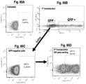
- the present description also relates to co-transducing a polypeptide cargo of interest and a marker protein as a means to identify and/or enrich transduced cells. It was surprisingly discovered that a strikingly high proportion of target eukaryotic cells that were successfully transduced with a polypeptide cargo of interest, were also successfully transduced with a marker protein. Conversely, a strikingly high proportion of cells that were not transduced with the polypeptide cargo of interest, were also not transduced with the marker protein.
- Isolating cells positive for the marker protein resulted in a significant increase in the proportion of cells that were successfully transduced with the polypeptide cargo of interest, and the correlation was found to be concentration dependent in that cell populations exhibiting the highest fluorescence of the marker protein also tended to exhibit the highest proportion of transduction with the polypeptide cargo of interest. It was also discovered that cells that were unsuccessfully transduced following a first round of transduction with a polypeptide cargo of interest, may be isolated and re-transduced with the polypeptide cargo of interest in subsequent rounds of transduction.
- the present description relates to methods comprising co-transduction of a polypeptide cargo of interest with a marker protein, wherein the marker protein may be used to isolate or enrich cells transduced with a polypeptide cargo of interest.
- the present description also relates to methods comprising repeated successive transduction experiments performed on, for example, cells that were not successfully transduced with a marker protein following a first or previous transduction reaction. Such methods present attractive approaches for increasing transduction efficiency in valuable cell populations (e.g., patient-derived cells for cell therapy), and/or in cell populations that are inherently more difficult to transduce.
- the words “comprising” (and any form of comprising, such as “comprise” and “comprises”), “having” (and any form of having, such as “have” and “has”), "including” (and any form of including, such as “includes” and “include”) or “containing” (and any form of containing, such as “contains” and “contain”) are inclusive or openended and do not exclude additional, unrecited elements or method steps.
- protein or “polypeptide” or “peptide” means any peptide-linked chain of amino acids, which may or may not comprise any type of modification (e.g., chemical or post-translational modifications such as acetylation, phosphorylation, glycosylation, sulfatation, sumoylation, prenylation, ubiquitination, etc.).
- modification e.g., chemical or post-translational modifications such as acetylation, phosphorylation, glycosylation, sulfatation, sumoylation, prenylation, ubiquitination, etc.
- protein/polypeptide/peptide modifications are envisaged so long as the modification does not destroy the protein transduction activity of the shuttle agents described herein
- a “domain” or “protein domain” generally refers to a part of a protein having a particular functionality or function. Some domains conserve their function when separated from the rest of the protein, and thus can be used in a modular fashion. The modular characteristic of many protein domains can provide flexibility in terms of their placement within the shuttle agents of the present description. However, some domains may perform better when engineered at certain positions of the shuttle agent (e.g., at the N- or C-terminal region, or therebetween). The position of the domain within its endogenous protein is sometimes an indicator of where the domain should be engineered within the shuttle agent and of what type/length of linker should be used.
- Standard recombinant DNA techniques can be used by the skilled person to manipulate the placement and/or number of the domains within the shuttle agents of the present description in view of the present disclosure.
- assays disclosed herein, as well as others known in the art can be used to assess the functionality of each of the domains within the context of the shuttle agents (e.g., their ability to facilitate cell penetration across the plasma membrane, endosome escape, and/or access to the cytosol).
- Standard methods can also be used to assess whether the domains of the shuttle agent affect the activity of the cargo to be delivered intracellularly.
- operably linked refers to the ability of the domains to carry out their intended function(s) (e.g., cell penetration, endosome escape, and/or subcellular targeting) within the context of the shuttle agents of the present description.
- operably linked is meant to define a functional connection between two or more domains without being limited to a particular order or distance between same.
- synthetic used in expressions such as “synthetic peptide” or “synthetic polypeptide” is intended to refer to non-naturally occurring molecules that can be produced in vitro (e.g., synthesized chemically and/or produced using recombinant DNA technology).
- the purities of various synthetic preparations may be assessed by, for example, high-performance liquid chromatography analysis and mass spectroscopy.
- Chemical synthesis approaches may be advantageous over cellular expression systems (e.g., yeast or bacteria protein expression systems), as they may preclude the need for extensive recombinant protein purification steps (e.g., required for clinical use).
- the peptides or shuttle agent of the present description may be chemically synthesized (e.g., solid- or liquid phase peptide synthesis), as opposed to expressed from a recombinant host cell.
- the peptides or shuttle agent of the present description may lack an N-terminal methionine residue.
- a person of skill in the art may adapt a synthetic peptide or shuttle agent of the present description by using one or more modified amino acids (e.g., non-naturally-occurring amino acids), or by chemically modifying the synthetic peptide or shuttle agent of the present description, to suit particular needs of stability or other needs.
- modified amino acids e.g., non-naturally-occurring amino acids
- polypeptide-based when used here in the context of a shuttle agent of the present description, is intended to distinguish the presently described shuttle agents from non-polypeptide or non-protein-based shuttle agents such as lipid- or cationic polymer-based transduction agents, which are often associated with increased cellular toxicity and may not be suitable for use in human therapy.
- independent is generally intended refer to molecules or agents which are not covalently bound to one another.
- independent polypeptide cargo is intended to refer to a polypeptide cargo to be delivered intracellularly that is not covalently bound (e.g., not fused) to a shuttle agent of the present description.
- having shuttle agents that are independent of (not fused to) a polypeptide cargo may be advantageous by providing increased shuttle agent versatility -- e.g., not being required to re-engineer a new fusion protein for different polypeptide cargoes, and/or being able to readily vary the ratio of shuttle agent to cargo (as opposed to being limited to a 1:1 ratio in the case of a fusion protein).
- the expression “is or is from” or “is from” comprises functional variants of a given protein domain (e.g., CPD or ELD), such as conservative amino acid substitutions, deletions, modifications, as well as variants or function derivatives, which do not abrogate the activity of the protein domain.
- a given protein domain e.g., CPD or ELD
- domain-based peptide shuttle agents comprising an endosome leakage domain (ELD) operably linked to a cell penetrating domain (CPD), and optionally one or more histidine-rich domains, can increase the transduction efficiency of an independent polypeptide cargo in eukaryotic cells, such that the cargo gains access to the cytosol/nuclear compartment (e.g., see Examples 1-15).
- ELD endosome leakage domain
- CPD cell penetrating domain
- histidine-rich domains optionally one or more histidine-rich domains
- the above screening efforts also revealed some peptides having no or low polypeptide cargo transduction power, excessive toxicity, and/or other undesirable properties (e.g., poor solubility and/or stability).
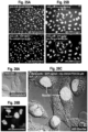
- the screened peptides were evaluated individually according to their transduction performance, with due consideration to, for example: their solubility/stability/ease of synthesis; their ability to facilitate escape of endosomally-trapped calcein (e.g., see Example 2); their ability to deliver one or more types of independent polypeptide cargos intracellularly, as evaluated by flow cytometry (e.g., see Examples 3-6 and 8-15) in different types of cells and cell lines (e.g., primary, immortalized, adherent, suspension, etc.) as well as under different transduction protocols; their ability to deliver polypeptide cargos to the cytosol and/or nucleus, as evaluated by fluorescence microscopy (e.g., for fluorescently labelled cargos), increased transcriptional activity (e.g., for transcription factor cargos), or genome editing capabilities (e.g., for nuclease cargos or genome-editing complexes such as CRISPR/Cas9 or CRISPR/Cpf1) (e.
- each peptide was assigned a score corresponding to a given cell line and fluorescently-labelled polypeptide cargo, which combines both transduction efficiency and cellular toxicity data into a single numerical value.
- the transduction scores were calculated by simply multiplying the highest percentage transduction efficiency observed by flow cytometry for a given peptide, cargo and cell type by the percentage cellular viability for the peptide in the tested cell line.
- the peptides were then sorted according to their transduction scores as a screening tool to stratify peptides as successful, less successful, or failed shuttle agents.
- the design parameters described herein were further validated by testing a plurality of synthetic peptides whose amino acid sequences were generated using a machine learning algorithm (e.g., see Example C.1), the algorithm having been "trained” using transduction efficiency and cellular toxicity data of domain-based peptides (but not the design parameters described herein).
- the peptides generated by the machine learning algorithm demonstrating the highest transduction scores were generally peptides that satisfied all of the design parameters described herein, thereby substantiating their use in actively designing and/or predicting the transduction activity of new peptide shuttle agents (e.g., tailored to particular polypeptide cargos and/or types of cells).
- computer-assisted random peptide sequence generation followed by descriptors filtering was used to generate a list of 10 000 peptide variants that respect nearly all of the design parameters described herein (see Example C.2).
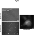
- Rationally-designed peptide shuttle agents are shown herein to facilitate escape of an endosomally trapped fluorescent dye, suggesting endosomolytic activity (e.g., see Example D).
- polypeptide cargos e.g., fluorescent proteins, transcription factors, antibodies, as well as entire CRISPR-associated genome editing complexes, with or without a DNA template
- E-G the ability of rationally-designed peptide shuttle agents to transduce a variety of polypeptide cargos (e.g., fluorescent proteins, transcription factors, antibodies, as well as entire CRISPR-associated genome editing complexes, with or without a DNA template) in a variety of different cell types (both adherent and suspension) is also shown herein (e.g., see Examples E-G).
- the present description relates to a method for delivering a polypeptide cargo from an extracellular space to the cytosol and/or nucleus of a target eukaryotic cell.
- the method comprises contacting the target eukaryotic cell with the polypeptide cargo in the presence of a shuttle agent at a concentration sufficient to increase the transduction efficiency of said polypeptide cargo, as compared to in the absence of the shuttle agent.
- the shuttle agent relates to a peptide that satisfies one or more of the following parameters.
- peptide shuttle agents of the present description respect at least one, at least two, at least three, at least four, at least five, at least six, at least seven, at least eight, at least nine, at least ten, at least eleven, at least twelve, at leave thirteen, at least fourteen, or all of parameters (1) to (15) described herein.
- peptide shuttle agents of the present description respect all of parameters (1) to (3), and at least six, at least seven, at least eight, at least nine, at least ten, at least eleven, or all of parameters (4) to (15) described herein.
- a peptide shuttle agent of the present description comprises two histidine-rich domains: a first histidine-rich domain towards the N terminus, and a second histidine-rich domain towards the C terminus, only the first histidine-rich domain may be included in the calculation/assessment of parameters (1) to (15) described herein.
- a machine-learning or computer-assisted design approach may be implemented to generate peptides that respect one or more of parameters (1) to (15) described herein.
- Some parameters, such as parameters (1) and (5)-(15), may be more amenable to implementation in a computer-assisted design approach, while structural parameters, such as parameters (2), (3) and (4), may be more amenable to a manual design approach.
- peptides that respect one or more of parameters (1) to (15) may be generated by combining computer-assisted and manual design approaches.
- consensus sequences i.e., commonly found patterns of alternance of hydrophobic, cationic, hydrophilic, alanine and glycine amino acids.
- the presence of these consensus sequences are likely to give rise to structural parameters (2), (3) and (4) being respected (i.e., amphipathic alpha-helix formation, a positively-charged face, and a highly hydrophobic core of 12%-50%).
- these and other consensus sequences may be employed in machine-learning and/or computer-assisted design approaches to generate peptides that respect one or of parameters (1)-(15).
- peptide shuttle agents described herein may comprise or consist of the amino acid sequence of:
- peptide shuttle agents of the present description may comprise or consist of any one of the amino acid sequences of SEQ ID NOs: 104, 105, 107, 108, 110-131, 133-135, 138, 140, 142, 145, 148, 151, 152, 169-242, and 243-10 242.
- peptide shuttle agents of the present description may comprise the amino acid sequence motifs of SEQ ID NOs: 158 and/or 159, which were found in each of peptides FSD5, FSD16, FSD18, FSD19, FSD20, FSD22, and FSD23.
- peptide shuttle agents of the present description may comprise the amino acid sequence motif of SEQ ID NO: 158 operably linked to the amino acid sequence motif of SEQ ID NO: 159.
- peptide shuttle agents of the present description may comprise or consist of a peptide which is at least 50%, 55%, 60%, 65%, 70%, 75%, 80%, 85%, 90%, or 95% identical to the amino acid sequence of any one of SEQ ID NOs: 104, 105, 107, 108, 110-131, 133-135, 138, 140, 142, 145, 148, 151, 152, 169-242, and 243-10 242, or a functional variant of any one of SEQ ID NOs: 104, 105, 107, 108, 110-131, 133-135, 138, 140, 142, 145, 148, 151, 152, 169-242, and 243-10 242.
- a “functional variant” refers to a peptide having polypeptide cargo transduction activity, which differs from the reference peptide by one or more conservative amino acid substitutions.
- a “conservative amino acid substitution” is one in which one amino acid residue is replaced with another amino acid residue having a similar side chain.
- Families of amino acid residues having similar side chains have been well defined in the art, including basic side chains (e.g., lysine, arginine, histidine), acidic side chains (e.g., aspartic acid, glutamic acid), uncharged polar side chains (e.g., glycine, asparagine, glutamine, serine, threonine, tyrosine, cysteine), nonpolar side chains (e.g., alanine, valine, leucine, isoleucine, proline, phenylalanine, methionine, tryptophan), beta-branched side chains (e.g., threonine, valine, isoleucine) and aromatic side chains (e.g., tyrosine, phenylalanine, tryptophan, histidine).
- basic side chains e.g., lysine, arginine, histidine
- acidic side chains e.g., aspartic
- peptide shuttle agents of the present description may comprise or consist of the amino acid sequence of any one of SEQ ID NOs: 57-59, 66-72, or 82-102, or a functional variant thereof having at least 65%, 66%, 67%, 68%, 69%, 70%, 71%, 72%, 73%, 74%, 75%, 76%, 77%, 78%, 79%, 80%, 81% 82%, 83%, 84%, 85%, 86%, 87%, 88%, 89%, 90%, 91%, 92%, 93%, 94%, or 95% identity to any one of SEQ ID NOs: 57-59, 66-72, or 82-102.
- peptide shuttle agents of the present description do not comprise one or more of the amino acid sequences of any one of SEQ ID NOs: 57-59, 66-72, or 82-102,
- shuttle agents of the present description may comprise oligomers (e.g., dimers, trimers, etc.) of peptides described herein. Such oligomers may be constructed by covalently binding the same or different types of shuttle agent monomers (e.g., using disulfide bridges to link cysteine residues introduced into the monomer sequences). In some embodiments, shuttle agents of the present description may comprise an N-terminal and/or a C-terminal cysteine residue.
- peptide shuttle agents of the present description may further comprise one or more histidine-rich domains.
- the histidine-rich domain may be a stretch of at least 2, at least 3, at least 4, at least 5, or at least 6 amino acids comprising at least 30%, at least 35%, at least 40%, at least 45%, at least 50%, at least 55%, at least 60%, at least 65%, at least 70%, at least 75%, at least 80%, at least 85%, or at least 90% histidine residues.
- the histidine-rich domain may comprise at least 2, at least 3, at least 4 at least 5, at least 6, at least 7, at least 8, or at least 9 consecutive histidine residues.
- the histidine-rich domain in the shuttle agent may act as a proton sponge in the endosome through protonation of their imidazole groups under acidic conditions of the endosomes, providing another mechanism of endosomal membrane destabilization and thus further facilitating the ability of endosomally-trapped cargos to gain access to the cytosol.
- the histidine-rich domain may be located at or towards the N and/or C terminus of the peptide shuttle agent.
- peptide shuttle agents of the present description may comprise one or more suitable linkers (e.g., flexible polypeptide linkers).
- linkers may separate two or more amphipathic alpha-helical motifs (e.g., see the shuttle agent FSD18 in Figure 49D ).
- linkers can be used to separate two more domains (CPDs, ELDs, or histidine-rich domains) from one another.
- linkers may be formed by adding sequences of small hydrophobic amino acids without rotatory potential (such as glycine) and polar serine residues that confer stability and flexibility. Linkers may be soft and allow the domains of the shuttle agents to move.
- prolines may be avoided since they can add significant conformational rigidity.
- the linkers may be serine/glycine-rich linkers (e.g., GGS, GGSGGGS, GGSGGGSGGGS, or the like).
- the use shuttle agents comprising a suitable linker may be advantageous for delivering an independent polypeptide cargo to suspension cells, rather than to adherent cells.
- the linker may comprise or consist of: -Gn- ; -Sn- ; -(GnSn)n- ; -(GnSn)nGn- ; -(GnSn)nSn- ; -(GnSn)nGn(GnSn)n- ; or -(GnSn)nSn(GnSn)n- , wherein G is the amino acid Gly; S is the amino acid Ser; and n is an integer from 1 to 5.
- ELDs Endosome leakage domains
- peptide shuttle agents of the present description may comprise an endosome leakage domain (ELD) for facilitating endosome escape and access to the cytoplasmic compartment.
- ELD endosome leakage domain
- endosome leakage domain refers to a sequence of amino acids which confers the ability of endosomally-trapped macromolecules to gain access to the cytoplasmic compartment.
- endosome leakage domains are short sequences (often derived from viral or bacterial peptides), which are believed to induce destabilization of the endosomal membrane and liberation of the endosome contents into the cytoplasm.
- endosomolytic peptide is intended to refer to this general class of peptides having endosomal membrane-destabilizing properties. Accordingly, in some embodiments, synthetic peptide or polypeptide-based shuttle agents of the present description may comprise an ELD which is an endosomolytic peptide. The activity of such peptides may be assessed for example using the calcein endosome escape assays described in Example 2.
- the ELD may be a peptide that disrupts membranes at acidic pH, such as pH-dependent membrane active peptide (PMAP) or a pH-dependent lytic peptide.
- PMAP pH-dependent membrane active peptide
- the peptides GALA and INF-7 are amphiphilic peptides that form alpha helixes when a drop in pH modifies the charge of the amino acids which they contain. More particularly, without being bound by theory, it is suggested that ELDs such as GALA induce endosomal leakage by forming pores and flip-flop of membrane lipids following conformational change due to a decrease in pH (Kakudo, Chaki et al., 2004, Li, Nicol et al., 2004).
- ELDs such as INF-7 induce endosomal leakage by accumulating in and destabilizing the endosomal membrane (El-Sayed, Futaki et al., 2009). Accordingly, in the course of endosome maturation, the concomitant decline in pH causes a change in the conformation of the peptide and this destabilizes the endosome membrane leading to the liberation of the endosome contents.
- the same principle is thought to apply to the toxin A of Pseudomonas (Varkouhi, Scholte et al., 2011).
- the ELD may be an antimicrobial peptide (AMP) such as a linear cationic alpha-helical antimicrobial peptide (AMP).
- AMP antimicrobial peptide
- these peptides play a key role in the innate immune response due to their ability to strongly interact with bacterial membranes. Without being bound by theory, these peptides are thought to assume a disordered state in aqueous solution, but adopt an alpha-helical secondary structure in hydrophobic environments. The latter conformation thought to contribute to their typical concentration-dependent membrane-disrupting properties. When accumulated in endosomes at certain concentrations, some antimicrobial peptides may induce endosomal leakage.
- the ELD may be an antimicrobial peptide (AMP) such as Cecropin-A/Melittin hybrid (CM series) peptide.
- AMP antimicrobial peptide
- CM series Cecropin-A/Melittin hybrid
- Cecropins are a family of antimicrobial peptides with membrane-perturbing abilities against both Gram-positive and Gram-negative bacteria.
- Cecropin A (CA) the first identified antibacterial peptide, is composed of 37 amino acids with a linear structure.
- Melittin (M) a peptide of 26 amino acids, is a cell membrane lytic factor found in bee venom.
- Cecropin-melittin hybrid peptides have been shown to produce short efficient antibiotic peptides without cytotoxicity for eukaryotic cells (i.e., non-hemolytic), a desirable property in any antibacterial agent.
- These chimeric peptides were constructed from various combinations of the hydrophilic N-terminal domain of Cecropin A with the hydrophobic N-terminal domain of Melittin, and have been tested on bacterial model systems.
- Two 26-mers, CA(1-13)M(1-13) and CA(1-8) M(1-18) (Boman et al., 1989), have been shown to demonstrate a wider spectrum and improved potency of natural Cecropin A without the cytotoxic effects of melittin.
- synthetic peptide or polypeptide-based shuttle agents of the present description may comprise an ELD which is or is from CM series peptide variants, such as those described above.
- the ELD may be the CM series peptide CM18 composed of residues 1-7 of Cecropin-A (KWKLFKKIGAVLKVLTTG) fused to residues 2-12 of Melittin (YGRKKRRQRRR), [C(1-7)M(2-12)].
- CM18 was shown to independently cross the plasma membrane and destabilize the endosomal membrane, allowing some endosomally-trapped cargos to be released to the cytosol (Salomone et al., 2012).
- the ELD may be CM18 having the amino acid sequence of SEQ ID NO: 1, or a variant thereof having at least 70%, 71%, 72%, 73%, 74%, 75%, 76%, 77%, 78%, 79%, 80%, 81%, 82%, 83%, 84%, 85%, 86%, 87%, 88%, 89%, 85%, 90%, 91%, 92%, 93%, 94%, or 95% identity to SEQ ID NO: 1 and having endosomolytic activity.
- the ELD may be a peptide derived from the N terminus of the HA2 subunit of influenza hemagglutinin (HA), which may also cause endosomal membrane destabilization when accumulated in the endosome.
- HA hemagglutinin
- synthetic peptide or polypeptide-based shuttle agents of the present description may comprise an ELD which is or is from an ELD set forth in Table I, or a variant thereof having endosome escape activity and/or pH-dependent membrane disrupting activity.
- Table I Examples of endosome leakage domains Name Amino acid sequence SEQ ID NO: Reference(s) CM18 KWKLFKKIGAVLKVLTTG 1 (Salomone, Cardarelli et al., 2012) Diphtheria toxin T domain (DT) 2 (Uherek, Fominaya et al., 1998, Glover, Ng et al., 2009) GALA WEAALAEALAEALAEHLAEALAEALEALAA 3 (Parente, Nir et al., 1990) (Li, Nicol et al., 2004) PEA 4 (Fominaya and Wels 1996) INF-7 GLFEAIEGFIENGWEGMIDGWYGC 5 (El-Sayed, Fut
- shuttle agents of the present description may comprise one or more ELD or type of ELD. More particularly, they can comprise at least 2, at least 3, at least 4, at least 5, or more ELDs. In some embodiments, the shuttle agents can comprise between 1 and 10 ELDs, between 1 and 9 ELDs, between 1 and 8 ELDs, between 1 and 7 ELDs, between 1 and 6 ELDs, between 1 and 5 ELDs, between 1 and 4 ELDs, between 1 and 3 ELDs, etc.
- the order or placement of the ELD relative to the other domains (CPD, histidine-rich domains) within the shuttle agents of the present description may be varied provided the shuttling ability of the shuttle agent is retained.
- the ELD may be a variant or fragment of any one those listed in Table I , and having endosomolytic activity.
- the ELD may comprise or consist of the amino acid sequence of any one of SEQ ID NOs: 1-15, 63, or 64, or a sequence which is at least 70%, 71%, 72%, 73%, 74%, 75%, 76%, 77%, 78%, 79%, 80%, 81%, 82%, 83%, 84%, 85%, 86%, 87%, 88%, 89%, 85%, 90%, 91%, 92%, 93%, 94%, or 95% identical to any one of SEQ ID NOs: 1-15, 63, or 64, and having endosomolytic activity.
- shuttle agents of the present description do not comprise one or more of the amino acid sequence of any one of SEQ ID NOs: 1-15, 63, or 64.
- CPDs Cell penetration domains
- the shuttle agents of the present description may comprise a cell penetration domain (CPD).
- CPD cell penetration domain
- the expression "cell penetration domain” refers to a sequence of amino acids which confers the ability of a macromolecule (e.g., peptide or protein) containing the CPD to be transduced into a cell.
- the CPD may be (or may be from) a cell-penetrating peptide or the protein transduction domain of a cell-penetrating peptide.
- Cell-penetrating peptides can serve as carriers to successfully deliver a variety of cargos intracellularly (e.g., polynucleotides, polypeptides, small molecule compounds or other macromolecules/compounds that are otherwise membrane-impermeable).
- Cell-penetrating peptides often include short peptides rich in basic amino acids that, once fused (or otherwise operably linked) to a macromolecule, mediate its internalization inside cells (Shaw, Catchpole et al., 2008).
- the first cell-penetrating peptide was identified by analyzing the cell penetration ability of the HIV-1 trans-activator of transcription (Tat) protein (Green and Loewenstein 1988, Vives, Brodin et al., 1997).
- This protein contains a short hydrophilic amino acid sequence, named "TAT", which promotes its insertion within the plasma membrane and the formation of pores. Since this discovery, many other cell-penetrating peptides have been described.
- the CPD can be a cell-penetrating peptide as listed in Table II, or a variant thereof having cell-penetrating activity.
- cell-penetrating peptides are thought to interact with the cell plasma membrane before crossing by pinocytosis or endocytosis.
- TAT peptide its hydrophilic nature and charge are thought to promote its insertion within the plasma membrane and the formation of a pore (Herce and Garcia 2007).
- Alpha helix motifs within hydrophobic peptides (such as SP) are also thought to form pores within plasma membranes (Veach, Liu et al., 2004).
- shuttle agents of the present description may comprise one or more CPD or type of CPD. More particularly, they may comprise at least 2, at least 3, at least 4, or at least 5 or more CPDs. In some embodiments, the shuttle agents can comprise between 1 and 10 CPDs, between 1 and 6 CPDs, between 1 and 5 CPDs, between 1 and 4 CPDs, between 1 and 3 CPDs, etc.
- the CPD may be TAT having the amino acid sequence of SEQ ID NO: 17, or a variant thereof having at least 70%, 71%, 72%, 73%, 74%, 75%, 76%, 77%, 78%, 79%, 80%, 81% 82%, 83%, 84%, 85%, 86%, 87%, 88%, 89%, 90%, 91%, 92%, 93%, 94%, or 95% identity to SEQ ID NO: 17 and having cell penetrating activity; or Penetratin having the amino acid sequence of SEQ ID NO: 18, or a variant thereof having at least 70%, 71%, 72%, 73%, 74%, 75%, 76%, 77%, 78%, 79%, 80%, 81% 82%, 83%, 84%, 85%, 86%, 87%, 88%, 89%, 85%, 86%, 87%, 88%, 89%, 90%, 91%, 92%, 93%, 94%, or 95% identity to SEQ ID NO: 18 and having cell penetrating activity
- the CPD may be PTD4 having the amino acid sequence of SEQ ID NO: 65, or a variant thereof having at least 70%, 71%, 72%, 73%, 74%, 75%, 76%, 77%, 78%, 79%, 80%, 81% 82%, 83%, 84%, 85%, 86%, 87%, 88%, 89%, 90%, 91%, 92%, 93%, 94%, or 95% identity to SEQ ID NO: 65.
- the order or placement of the CPD relative to the other domains (ELD, histidine-rich domains) within the shuttle agents of the present description may be varied provided the shuttling ability of the shuttle agent is retained.
- the CPD may be a variant or fragment of any one those listed in Table II , and having cell penetrating activity.
- the CPD may comprise or consist of the amino acid sequence of any one of SEQ ID NOs: 16-27 or 65, or a sequence which is at least 70%, 71%, 72%, 73%, 74%, 75%, 76%, 77%, 78%, 79%, 80%, 81%, 82%, 83%, 84%, 85%, 86%, 87%, 88%, 89%, 85%, 90%, 91%, 92%, 93%, 94%, or 95% identical to any one of SEQ ID NOs: 16-27 or 65, and having cell penetrating activity.
- shuttle agents of the present description do not comprise any one of the amino acid sequences of SEQ ID NOs: 16-27 or 65.
- peptide shuttle agents of the present description may be useful for delivering a polypeptide cargo (e.g., an independent polypeptide cargo) from an extracellular space to the cytosol and/or nucleus of a target eukaryotic cell, wherein the synthetic peptide shuttle agent is used at a concentration sufficient to increase the transduction efficiency of said polypeptide cargo, as compared to in the absence of said synthetic peptide shuttle agent.
- the polypeptide cargo may be fused to one or more CPDs to further facilitate intracellular delivery.
- the CPD fused to the polypeptide cargo may be the same or different from a CPD that may be present in the shuttle agent of the present description.
- fusion proteins may be constructed using standard recombinant technology.
- the independent polypeptide cargo may be fused, complexed with, or covalently bound to a second biologically active cargo (e.g., a biologically active polypeptide or compound).
- a second biologically active cargo e.g., a biologically active polypeptide or compound.
- the polypeptide cargo may comprise a subcellular targeting domain.
- the polypeptide cargo must be delivered to the nucleus for it to carry out its intended biological effect.
- the cargo is a polypeptide intended for nuclear delivery (e.g., a transcription factor).
- nuclear localization signals e.g., nuclear localization signals
- the NLS sequences are recognized by proteins (importins ⁇ and ⁇ ), which act as transporters and mediators of translocation across the nuclear envelope.
- NLSs are generally enriched in charged amino acids such as arginine, histidine, and lysine, conferring a positive charge which is partially responsible for their recognition by importins.
- the polypeptide cargo may comprise an NLS for facilitating nuclear delivery, such as one or more of the NLSs as listed in Table III , or a variant thereof having nuclear targeting activity.
- the polypeptide cargo may comprise its natural NLS.
- recombinant proteins are exposed to protein trafficking system of eukaryotic cells. Indeed, all proteins are synthetized in the cell's cytoplasm and are then redistributed to their final subcellular localization by a system of transport based on small amino acid sequences recognized by shuttle proteins (Karniely and Pines 2005, Stojanovski, Bohnert et al., 2012). In addition to NLSs, other localization sequences can mediate subcellular targeting to various organelles following intracellular delivery of the polypeptide cargos of the present description.
- polypeptide cargos of the present description may comprise a subcellular localization signal for facilitating delivery of the shuttle agent and cargo to specific organelles, such as one or more of the sequences as listed in Table IV, or a variant thereof having corresponding subcellular targeting activity.
- the cargo can be a biologically active compound such as a biologically active (recombinant) polypeptide (e.g., a transcription factor, a cytokine, or a nuclease) intended for intracellular delivery.
- a biologically active polypeptide e.g., a transcription factor, a cytokine, or a nuclease
- biologically active refers to the ability of a compound to mediate a structural, regulatory, and/or biochemical function when introduced in a target cell.
- the cargo may be a recombinant polypeptide intended for nuclear delivery, such as a transcription factor.
- the transcription factor can be HOXB4 (Lu, Feng et al., 2007), NUP98-HOXA9 (Takeda, Goolsby et al., 2006), Oct3/4, Sox2, Sox9, Klf4, c-Myc (Takahashi and Yamanaka 2006), MyoD (Sung, Mun et al., 2013), Pdx1, Ngn3 and MafA (Akinci, Banga et al., 2012), Blimp-1 (Lin, Chou et al., 2013), Eomes, T-bet (Gordon, Chaix et al., 2012), FOXO3A (Warr, Binnewies et al., 2013), NF-YA (Dolfini, Minuzzo et al., 2012), SALL4 (Aguila, Liao et al.
- the cargo may be a recombinant polypeptide intended for nuclear delivery, such as a nuclease useful for genome editing technologies.
- the nuclease may be an RNA-guided endonuclease, a CRISPR endonuclease, a type I CRISPR endonuclease, a type II CRISPR endonuclease, a type III CRISPR endonuclease, a type IV CRISPR endonuclease, a type V CRISPR endonuclease, a type VI CRISPR endonuclease, CRISPR associated protein 9 (Cas9), Cpf1 (Zetsche et al., 2015), CasX and/or CasY (Burstein et al., 2016) a zinc-finger nuclease (ZFN), a Transcription activator-like effector nuclease (TALEN) (
- the nuclease may be a catalytically dead endonuclease, such as a catalytically dead CRISPR associated protein 9 (dCas9), dCpf1, dCasX, dCasY, or any combination thereof. Other nucleases not explicitly mentioned here may nevertheless be encompassed in the present description.
- the nuclease may be fused to a nuclear localization signal (e.g., Cas9-NLS; Cpf1-NLS; ZFN-NLS; TALEN-NLS).
- the nuclease may be complexed with a nucleic acid (e.g., one or more guide RNAs, a crRNA, a tracrRNAs, or both a crRNA and a tracrRNA).
- a nucleic acid e.g., one or more guide RNAs, a crRNA, a tracrRNAs, or both a crRNA and a tracrRNA.
- the nuclease may possess DNA or RNA-binding activity, but may lack the ability to cleave DNA.
- the shuttle agents of the present description may be used for intracellular delivery (e.g., nuclear delivery) of one or more CRISPR endonucleases, for example one or more of the CRISPR endonucleases described below.
- Type I and its subtypes A, B, C, D, E, F and I including their respective Cas1, Cas2, Cas3, Cas4, Cas6, Cas7 and Cas8 proteins, and the signature homologs and subunits of these Cas proteins including Cse1, Cse2, Cas7, Cas5, and Cas6e subunits in E. coli (type I-E) and Csy1, Csy2, Csy3, and Cas6f in Pseudomonas aeruginosa (type I-F) (Wiedenheft et al., 2011; Makarova et al, 2011).
- Type II and its subtypes A, B, C including their respective Cas1, Cas2 and Cas9 proteins, and the signature homologs and subunits of these Cas proteins including Csn complexes (Makarova et al, 2011).
- Type III and its subtypes A, B and MTH326-like module including their respective Cas1, Cas2, Cas6 and Cas10 proteins, and the signature homologs and subunits of these Cas proteins including Csm and CMR complexes (Makarova et al, 2011).
- Type IV represents the Csf3 family of Cas proteins. Members of this family show up near CRISPR repeats in Acidithiobacillus ferrooxidans ATCC 23270, Azoarcus sp.
- Type V includes the enzyme C2c2, which reported shares little homology to known sequences.
- the shuttle agents of the present description may be used in conjunction with one or more of the nucleases, endonucleases, RNA-guided endonuclease, CRISPR endonuclease described above, for a variety of applications, such as those described herein.
- CRISPR systems interact with their respective nucleic acids, such as DNA binding, RNA binding, helicase, and nuclease motifs (Makarova et al, 2011; Barrangou & Marraffini, 2014).
- CRISPR systems may be used for different genome editing applications including:
- nucleases such as Cpf1, Cas9, and variants of such nucleases or others, are encompassed by the present description. It should be understood that, in one aspect, the present description may broadly cover any cargo having nuclease activity, such an RNA-guided endonuclease, or variants thereof (e.g., those that can bind to DNA or RNA, but have lost their nuclease activity; or those that have been fused to a transcription factor).
- the polypeptide cargo may be a cytokine such as a chemokine, an interferon, an interleukin, a lymphokine, or a tumour necrosis factor.
- the polypeptide cargo may be a hormone or growth factor.
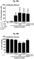
- the cargo may be an antibody (e.g., a labelled antibody, a therapeutic antibody, an anti-apoptotic antibody, an antibody that recognizes an intracellular antigen).
- the cargo can be a detectable label (fluorescent polypeptide or reporter enzyme) that is intended for intracellular delivery, for example, for research and/or diagnostic purposes.
- the cargo may be a globular protein or a fibrous protein. In some embodiments, the cargo may have a molecule weight of any one of about 5, 10, 15, 20, 25, 30, 35, 40, 45, to 50 to about 150, 200, 250, 300, 350, 400, 450, 500 kDa or more. In some embodiments, the cargo may have a molecule weight of between about 20 to 200 kDa.
- the polypeptide cargo may be a peptide cargo, such as peptide that recognizes an intracellular molecule.
- the polypeptide cargo may be an enzyme and/or an enzyme inhibitor.
- peptide shuttle agents of the present description may be useful for delivering a polypeptide cargo from an extracellular space to the cytosol and/or nucleus of different types of target eukaryotic cells, wherein the synthetic peptide shuttle agent is used at a concentration sufficient to increase the transduction efficiency of said polypeptide cargo, as compared to in the absence of said synthetic peptide shuttle agent.
- the target eukaryotic cells may be an animal cell, a mammalian cell, or a human cell.
- the target eukaryotic cells may be a stem cell (e.g., embryonic stem cells, pluripotent stem cells, induced pluripotent stem cells, neural stem cells, mesenchymal stem cells, hematopoietic stem cells, peripheral blood stem cells), a primary cell (e.g., myoblast, fibroblast), or an immune cell (e.g., NK cell, T cell, dendritic cell, antigen presenting cell).
- stem cell e.g., embryonic stem cells, pluripotent stem cells, induced pluripotent stem cells, neural stem cells, mesenchymal stem cells, hematopoietic stem cells, peripheral blood stem cells
- a primary cell e.g., myoblast, fibroblast
- an immune cell e.g., NK cell, T cell, dendritic cell, antigen presenting cell
- the shuttle agents of the present description may be non-toxic to the intended target eukaryotic cells at concentrations up to 50 ⁇ M, 45 ⁇ M, 40 ⁇ M, 35 ⁇ M, 30 ⁇ M, 25 ⁇ M, 20 ⁇ M, 15 ⁇ M, 10 ⁇ M, 9 ⁇ M, 8 ⁇ M, 7 ⁇ M, 6 ⁇ M, 5 ⁇ M, 4 ⁇ M, 3 ⁇ M, 2 ⁇ M, 1 ⁇ M, 0.5 ⁇ Mm 0.1 ⁇ M, or 0.05 ⁇ M.
- Cellular toxicity of shuttle agents of the present description may be measured using any suitable method.
- transduction protocols may be adapted (e.g., concentrations of shuttle and/or cargo used, shuttle/cargo exposure times, exposure in the presence or absence of serum), to reduce or minimize toxicity of the shuttle agents, and/or to improve/maximize transfection efficiency.
- shuttle agents of the present description may be readily metabolizable by intended target eukaryotic cells.
- the shuttle agents may consist entirely or essentially of peptides or polypeptides, for which the target eukaryotic cells possess the cellular machinery to metabolize/degrade.
- the intracellular half-life of the synthetic peptides and polypeptide-based shuttle agents of the present description is expected to be much lower than the half-life of foreign organic compounds such as fluorophores.
- fluorophores can be toxic and must be investigated before they can be safely used clinically (Alford et al., 2009).
- shuttle agents of the present description may be suitable for clinical use.
- the shuttle agents of the present description may avoid the use of domains or compounds for which toxicity is uncertain or has not been ruled out.
- the present description relates to a composition
- a composition comprising a cocktail of at least 2, at least 3, at least 4, or at least 5 different types of the synthetic peptides or polypeptide-based shuttle agents as defined herein.
- combining different types of synthetic peptides or peptide shuttle agents may provide increased versatility for delivering different polypeptide cargos intracellularly.
- combining lower concentrations of different types of shuttle agents may help reduce cellular toxicity associated with using a single type of shuttle agent (e.g., at higher concentrations).
- the present description relates to methods for delivering a polypeptide cargo from an extracellular space to the cytosol and/or nucleus of a target eukaryotic cell.
- the methods comprise contacting the target eukaryotic cell with the polypeptide cargo in the presence of a shuttle agent at a concentration sufficient to increase the transduction efficiency of said polypeptide cargo, as compared to in the absence of said shuttle agent.
- contacting the target eukaryotic cell with the polypeptide cargo in the presence of the shuttle agent results in an increase in the transduction efficiency of said polypeptide cargo by at least 10-fold, 20-fold, 30-fold, 40-fold, 50-fold, or 100-fold, as compared to in the absence of said shuttle agent.
- the present description relates to a method for increasing the transduction efficiency of a polypeptide cargo to the cytosol of a target eukaryotic cell.
- increasing transduction efficiency refers to the ability of a shuttle agent of the present description to improve the percentage or proportion of a population of target cells into which a cargo of interest (e.g., a polypeptide cargo) is delivered intracellularly across the plasma membrane. Immunofluorescence microscopy, flow cytometry, and other suitable methods may be used to assess cargo transduction efficiency.
- a shuttle agent of the present description may enable a transduction efficiency of at least 25%, 30%, 35%, 40%, 45%, 50%, 55%, 60%, 65%, 70%, 75%, 80%, or 85%, for example as measure by immunofluorescence microscopy, flow cytometry, FACS, and other suitable methods.
- a shuttle agent of the present description may enable one of the aforementioned transduction efficiencies together wish a cell viability of at least 25%, 30%, 35%, 40%, 45%, 50%, 55%, 60%, 65%, 70%, 75%, 80%, 85%, 90%, or 95%, for example as measure by the assay described in Example 3.3a, or by another suitable assay known in the art.
- shuttle agents of the present description may facilitate the delivery of a cargo of interest (e.g., a polypeptide cargo) to the cytosol of target cells.
- a cargo of interest e.g., a polypeptide cargo
- efficiently delivering an extracellular cargo to the cytosol of a target cell using peptides can be challenging, as the cargo often becomes trapped in intracellular endosomes after crossing the plasma membrane, which may limit its intracellular availability and may result in its eventual metabolic degradation.
- use of the protein transduction domain from the HIV-1 Tat protein has been reported to result in massive sequestration of the cargo into intracellular vesicles.
- shuttle agents of the present description may facilitate the ability of endosomally-trapped cargo to escape from the endosome and gain access to the cytoplasmic compartment.
- the expression “to the cytosol” in the phrase “increasing the transduction efficiency of an independent polypeptide cargo to the cytosol,” is intended to refer to the ability of shuttle agents of the present description to allow an intracellularly delivered cargo of interest to escape endosomal entrapment and gain access to the cytoplasmic compartment. After a cargo of interest has gained access to the cytosol, it may be subsequently targeted to various subcellular compartments (e.g., nucleus, nucleolus, mitochondria, peroxisome). In some embodiments, the expression “to the cytosol” is thus intended to encompass not only cytosolic delivery, but also delivery to other subcellular compartments that first require the cargo to gain access to the cytoplasmic compartment.
- the methods of the present description are in vitro methods. In other embodiments, the methods of the present description are in vivo methods.
- the methods of the present description may comprise contacting the target eukaryotic cell with the shuttle agent, or composition as defined herein, and the polypeptide cargo.
- the shuttle agent, or composition may be pre-incubated with the polypeptide cargo to form a mixture, prior to exposing the target eukaryotic cell to that mixture.
- the type of shuttle agent may be selected based on the amino acid sequence of the polypeptide cargo to be delivered intracellularly. In other embodiments, the type of shuttle agent may be selected to take into account the amino acid sequence of the polypeptide cargo to be delivered intracellularly, the type of cell, the type of tissue, etc.
- the method may comprise multiple treatments of the target cells with the shuttle agent, or composition (e.g., 1, 2, 3, 4 or more times per day, and/or on a pre-determined schedule). In such cases, lower concentrations of the shuttle agent, or composition may be advisable (e.g., for reduced toxicity).
- the cells may be suspension cells or adherent cells.
- the person of skill in the art will be able to adapt the teachings of the present description using different combinations of shuttles, domains, uses and methods to suit particular needs of delivering a polypeptide cargo to particular cells with a desired viability.
- the methods of the present description may apply to methods of delivering a polypeptide cargo intracellularly to a cell in vivo. Such methods may be accomplished by parenteral administration or direct injection into a tissue, organ, or system.
- the shuttle agent, or composition, and the polypeptide cargo may be exposed to the target cell in the presence or absence of serum.
- the method may be suitable for clinical or therapeutic use.
- the present description relates to a kit for delivering a polypeptide cargo from an extracellular space to the cytosol and/or nucleus of a target eukaryotic cell. In some embodiments, the present description relates to a kit for increasing the transduction efficiency of a polypeptide cargo to the cytosol of a target eukaryotic cell.
- the kit may comprise the shuttle agent, or composition as defined herein, and a suitable container.
- the target eukaryotic cells may be an animal cell, a mammalian cell, or a human cell.
- the target eukaryotic cells may be a stem cell (e.g., embryonic stem cells, pluripotent stem cells, induced pluripotent stem cells, neural stem cells, mesenchymal stem cells, hematopoietic stem cells, peripheral blood stem cells), a primary cell (e.g., myoblast, fibroblast), or an immune cell (e.g., NK cell, T cell, dendritic cell, antigen presenting cell).
- the present description relates to an isolated cell comprising a synthetic peptide or polypeptide-based shuttle agent as defined herein.
- the cell may be a protein-induced pluripotent stem cell. It will be understood that cells that are often resistant or not amenable to protein transduction may be interesting candidates for the synthetic peptides or polypeptide-based shuttle agents of the present description.
- the present description relates to a method for producing a synthetic peptide shuttle agent that delivers a polypeptide cargo from an extracellular space to the cytosol and/or nucleus of a target eukaryotic cell, the method comprising synthesizing a peptide which is:
- the present description relates to a method for identifying a shuttle agent that delivers a polypeptide cargo from an extracellular space to the cytosol and/or nucleus of a target eukaryotic cell, the method comprising: (a) synthesizing a peptide which is the peptide as defined herein; (b) contacting the target eukaryotic cell with the polypeptide cargo in the presence of said peptide; (c) measuring the transduction efficiency of the polypeptide cargo in the target eukaryotic cell; and (d) identifying the peptide as being a shuttle agent that transduces the polypeptide cargo, when an increase in the transduction efficiency of said polypeptide cargo in the target eukaryotic cell is observed.
- the present description relates to a genome editing system comprising: (a) the shuttle agent as defined herein; (b) a CRISPR-associated endonuclease; and (c) one or more guide RNAs.
- the genome editing system may further comprise a linear DNA template for controlling the genome editing.
- the shuttle agents, synthetic peptides, compositions, and methods described herein may be used for transducing genome-editing complexes (e.g., the CRISPR-based genome editing complexes) to genetically engineer cells for improved cell therapy, as compared to native cells or unengineered cells.
- genome-editing complexes e.g., the CRISPR-based genome editing complexes
- Such improvements may include, for example, reducing the immunogenicity of the engineered cells and/or improving the activity/efficacy of the engineered cells.
- NK natural killer cells
- the present description relates to the use of the shuttle agents, synthetic peptides, compositions, and methods described herein for transducing genome-editing complexes (e.g., the CRISPR-based genome editing complexes) to genetically engineer NK (or other immune cells that would benefit from the same modifications) for improved cell-based immunotherapy.
- genome-editing complexes e.g., the CRISPR-based genome editing complexes
- the present description may relate to the intracellular delivery of one or more CRISPR-based genome editing complexes that comprise a guide RNA and/or linear DNA template targeting the CBLB gene, c-CBL gene, GSK3 gene, ILT2 gene, CISH gene, NKG2a gene, B2M gene, or any combination thereof.
- Such gene targets may potentiate NK-mediated cellular cytotoxicity following knockout, as discussed below.
- NKG2A KLRC1, CD159A, Killer cell lectin-like receptor C1
- CD94/NKG2A acts as an MHC class-I specific NK inhibitory receptor (Braud et al., 1998; Lee et al., 1998). It is expressed by a subset of NK cells known as CD56 bright CD16 dim ( ⁇ 10% of peripheral NK), which are typically less cytotoxic (Cooper et al., 2001; Poli et al., 2009).
- NKG2A ligands are the non-classical MHC class-I HLA-E molecules that are expressed in every human cell. The recognition of HLA-E by the NKG2A receptor is part of the "self-tolerance" mechanism (also including KIR receptors), resulting in negative modulation of NK cell cytotoxicity (Lee et al., 1998).
- NKG2A-KO NK cells during adoptive cell therapy may counteract the presence of HLA-E molecules (membrane-bound or solubles) in tumor microenvironment.
- HLA-E molecules membrane-bound or solubles
- NK cells expanded from IL15 or IL21-expressing K562 feeder cells lead to a high percentage of NKG2A pos cells (Denman et al., 2012), and it may be desirable to knockout this inhibitory receptor during the expansion process.
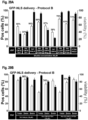
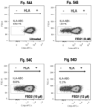
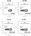
- the results in Example G.9 demonstrate that NKG2A-KO NK92 cells are significantly more cytotoxic against IFN-gamma-treated HeLa cells.
- ILT2 Ig-Like transcript 2 gene
- ILT2 is an inhibitory receptor expressed on several immune cells, including NK cells (Kirwan et al., 2005).
- the ligands for this receptor are HLA-G molecules, which are naturally expressed only in thymus and trophoblasts.
- NKL ILT2 -cells are more potent than parental NKL against HLA-G-overexpressing K562 cells (Wu et al., 2015).
- overexpression of HLA-G in OVCAR-3 cancer cells impaired NK cell-mediated cytotoxicity (Lin et al., 2007).
- HLA-E expression of HLA-G on cancer cells is generally associated with poor prognosis.
- TAM receptors are proposed to negatively regulate NK cells, Cbl-b knockout should rather be associated to a decrease in NK cell activity. Therefore, TAM receptors may be considered as a good target to enhance NK cells but unlikely via Cbl-b knockout.
- GSK3B glycogen synthase kinase beta
- GSK3b is a Ser/Thr kinase involved in several cellular functions, such as proliferation, apoptosis, inflammatory response, stress, and others (Patel et al., 2017). Inhibition of GSK3b (using small inhibitors) in NK cells leads to increase cytotoxicity (likely through IFN-g, TNF- ⁇ production, 2B4 stimulation and up-regulation of LFA-1) against AML (OCI-AML3) (Parameswaran et al., 2016; Aoukaty et al., 2005). We have recently demonstrated that the GSK3 ⁇ inhibitor, SB216763, enhances the cytotoxic activity of NK92 against HeLa cells (data not shown). This effect is increased by co-incubation with IL-15.
- CIS protein is a member of the suppressor of cytokine signaling (SOCS) proteins, which bind to phosphorylated JAKs and inhibit JAK-STAT signaling pathways.
- SOCS cytokine signaling
- cytokines such as IL2 and IL15
- CISH knockout IL15-hypersensitive NK cells
- disrupting the B2M gene encoding ⁇ 2 microglobulin (B2M), a component of MHC class I molecules may substantially reduce the immunogenicity of every cell expressing MHC class I.
- the genome of NK cells can be modified after the delivery of a genome editing system as described herein. More specifically, the cytotoxicity of NK cells can be improved after the delivery of a genome editing system targeting specific putative targets that may potentiate NK-mediated cellular cytotoxicity such as the NKG2A, ILT2, c-Cbl, Cbl-b, GSK3B and CISH genes.
- Example I of the present description shows that co-transducing a polypeptide cargo of interest (e.g., a CRISPR-endonuclease) and an independent marker protein (e.g., GFP) in a population of target eukaryotic cells may not necessarily increase the overall transduction efficiency of the polypeptide cargo of interest.
- a polypeptide cargo of interest e.g., a CRISPR-endonuclease
- an independent marker protein e.g., GFP
- the present description relates to a method for enriching eukaryotic cells transduced with a polypeptide cargo of interest.
- the method may comprise (a) co-transducing a target eukaryotic cell population with a polypeptide cargo of interest and a marker protein; and (b) isolating or concentrating eukaryotic cells transduced with the marker protein, thereby enriching eukaryotic cells transduced with the polypeptide cargo of interest.
- the marker protein may not be covalently bound to the polypeptide cargo of interest (e.g., the marker protein is independent from the polypeptide cargo of interest), the marker protein may be covalently bound to the polypeptide cargo of interest, the marker protein may be non-covalently bound to the polypeptide cargo of interest (e.g., electrostatically and/or conformationally bound via a protein domain-protein domain interaction), or the marker protein is covalently bound to the polypeptide cargo of interest via a cleavable linker (e.g., a linker peptide comprising an enzyme cleavage site, such as an enzyme expressed in endosomes, lysosomes, or in the cytosol).
- the intracellular concentration of the transduced marker protein may be positively correlated with the intracellular concentration of the transduced polypeptide cargo of interest.
- the marker protein may comprise a detectable label.
- detectable label refers to a molecule or particle that enables a person of skill in the art to identify and separate cells comprising the label from cells lacking the label.
- the marker protein may be a fluorescent protein, a fluorescently-labeled protein, a bioluminescent protein, an isotopically-labelled protein, a magnetically-labeled protein, or another detectable label that enables separation of cells containing the marker protein from cells lacking the marker protein, or enables separation of cells based on the level of intracellular marker protein.
- eukaryotic cells transduced with the marker protein may be isolated or concentrated using flow cytometry, fluorescence-activated cell sorting (FACS), magnetic-activated cell sorting (MACS), or other known cell separation/sorting technologies. Isolating or enriching successfully transduced cells may be particularly advantageous, for example, for polypeptide cargoes such as functional CRISPR-based genome-editing complexes, which may be associated with relatively lower transduction efficiencies.
- FACS fluorescence-activated cell sorting
- MCS magnetic-activated cell sorting
- Example I It was also surprisingly disclosed herein in Example I that cells that were unsuccessfully transduced following a first round of transduction with a polypeptide cargo of interest, may be isolated and re-transduced with the polypeptide cargo of interest in subsequent rounds of transduction. These results suggest that, although the cells unsuccessfully transduced following a first round of transduction with a polypeptide cargo of interest are not necessarily refractory to subsequent transductions.
- the present description relates to a method for enriching eukaryotic cells transduced with a polypeptide cargo of interest, the method comprising: (a) co-transducing a target eukaryotic cell population with a polypeptide cargo of interest and a marker protein; (b) isolating eukaryotic cells transduced with the marker protein from cells lacking the marker protein, thereby producing a marker protein-positive cell population and a marker protein-negative cell population.
- steps (a) and (b) may be repeated, one or more times, on the marker protein-negative cell population, on the marker protein-positive cell population, or on both the marker protein-negative and the marker protein-positive cell populations.
- the method for enriching eukaryotic cells transduced with a polypeptide cargo of interest may be automated, for example, by isolating cells negative for transduction with the marker protein and re-transducing the marker protein-negative cell population.
- the present description relates to a method for enriching eukaryotic cells transduced with a polypeptide cargo of interest, the method comprising: (a) co-transducing a target eukaryotic cell population with a polypeptide cargo of interest and a marker protein; and (b) isolating eukaryotic cells transduced with the marker protein based on their intracellular concentration of the marker protein.
- the marker protein described herein may be a protein that stimulates cell proliferation (e.g., a growth factor or a transcription factor), a protein that stimulates cell differentiation, a protein that promotes cell survival, an anti-apoptotic protein, or a protein having another biological activity.
- a protein that stimulates cell proliferation e.g., a growth factor or a transcription factor
- a protein that stimulates cell differentiation e.g., a protein that stimulates cell differentiation
- a protein that promotes cell survival e.g., an anti-apoptotic protein, or a protein having another biological activity.
- the polypeptide cargo of interest and the marker protein may be co-transduced by contacting the target eukaryotic cell with the polypeptide cargo and the marker protein in the presence of a peptide transduction agent, wherein the peptide transduction agent is present at a concentration sufficient to increase the transduction efficiency of the polypeptide cargo and the marker protein, as compared to in the absence of said peptide transduction agent.
- the peptide transduction agent may be an endosomolytic peptide.
- the peptide transduction agent is or comprises a domain-based peptide shuttle agent, or a rationally designed peptide shuttle agent as defined herein, in PCT patent application publication No.
- the aforementioned domain-based peptide shuttle agent may be a synthetic peptide comprising an endosome leakage domain (ELD) operably linked to a cell penetrating domain (CPD), or an ELD operably linked to a histidine-rich domain and a CPD.
- ELD endosome leakage domain
- CPD cell penetrating domain
- the aforementioned domain-based peptide shuttle agent (a) comprises a minimum length of 20, 21 , 22, 23, 24, 25, 26, 27, 28, 29, or 30 amino acid residues and a maximum length of 35, 40, 45, 50, 55, 65, 70, 75, 80, 85, 90, 95, 100, 105, 110, 115, 120, 125, 130, 135, 140, 145, or 150 amino acid residues; (b) has a predicted net charge of at least +4, +5, +6, +7, +8, +9, +10, +11 , +12, +13, +14, or +15 at physiological pH; (c) is soluble in aqueous solution; or (d) any combination of (a) to (c).
- the ELD, CPD, histidine-rich domain, linker domain are as defined herein.
- the target eukaryotic cells comprise or consist of animal cells, mammalian cells, human cells, stem cells, primary cells, immune cells, T cells, NK cells, dendritic cells, or other types or sub-types of cells.
- the polypeptide cargo of interest may be: (i) the polypeptide cargo as defined herein; and/or (ii) one or more CRISPR-associated endonucleases alone or with one or more corresponding guide RNA and/or linear DNA templates as defined herein.
- the present description may relate to the following items:
- Table 1.1 Reagents Material Company City, province-State, Country RPMI 1640 media Sigma-Aldrich Oakville, ON, Canada DMEM Sigma-Aldrich Oakville, ON, Canada Alpha MEM Stem Cell Technology Oakville, ON, Canada Fetal bovine serum (FBS) NorthBio Toronto, ON, Canada Horse serum Invitrogen Burlington, ON, Canada L-glutamine-Penicillin-Streptomycin Sigma-Aldrich Oakville, ON, Canada Trypsin-EDTA solution Sigma-Aldrich Oakville, ON, Canada Inositol Sigma-Aldrich Oakville, ON, Canada Folic acid Sigma-Aldrich Oakville, ON, Canada pEGFP-C1 CLONTECH Laboratories Palo Alto, CA, USA FITC-Antibody ⁇ -tubulin Abcam ab64503 Cambridge, MA, USA ITS Invitrogen/41400-045 Burlington, ON, Canada FGF 2 Feldan Bio/1D-07-017 Quebec, QC, Canada Dexamethas
- HeLa, HEK293A, HEK293T, THP-1, CHO, NIH3T3, CA46, Balb3T3, HT2, KMS-12, DOHH2, REC-1, HCC-78, NCI-H196 and HT2 cells were obtained from American Type Culture Collection (Manassas, VA, USA) and cultured following the manufacturer's instructions. Myoblasts are primary human cells kindly provided by Professor J.P. Tremblay (liable Laval, Quebec, Canada).
- Table 1.2 Cell lines and culture conditions Cell lines Description ATCC/others Culture media Serum Additives HeLa (adherent cells) Human cervical carcinoma cells ATCC TM CCL-2 DMEM 10% FBS L-glutamine 2 mM Penicillin 100 units Streptomycin 100 ⁇ g/mL HEK 293A (adherent cells) Human embryonic ATCC TM CRL-1573 DMEM 10% FBS L-glutamine 2 mM Epithelial kidney cells Penicillin 100 units Streptomycin 100 ⁇ g/mL HEK 293T (adherent cells) Human embryonic ATCC TM CRL-3216 DMEM 10% FBS L-glutamine 2 mM Epithelial kidney cells Penicillin 100 units Streptomycin 100 ⁇ g/mL THP-1 (suspension cells) Acute monocytic leukemia ATCC TM TIB202 RPMI 1640 10% FBS ⁇ - mercaptoethanol 0.05 mM L-glutamine 2 mM Penicillin 100 units Strept
- Fusion proteins were expressed in bacteria ( E. coli BL21DE3) under standard conditions using an isopropyl ⁇ -D-1-thiogalactopyranoside (IPTG) inducible vector containing a T5 promoter.
- Culture media contained 24 g yeast extract, 12 g tryptone, 4 mL glycerol, 2.3 g KH 2 PO 4 , and 12.5 g K 2 HPO 4 per liter.
- Bacterial broth was incubated at 37°C under agitation with appropriate antibiotic (e.g., ampicillin). Expression was induced at optical density (600 nm) between 0.5 and 0.6 with a final concentration of 1 mM IPTG for 3 hours at 30°C.
- Bacteria were recuperated following centrifugation at 5000 RPM and bacterial pellets were stored at -20°C.
- Bacterial pellets were resuspended in Tris buffer (Tris 25 mM pH 7.5, NaCl 100mM, imidazole 5 mM) with phenylmethylsulfonyl fluoride (PMSF) 1 mM, and lysed by passing 3 times through the homogenizer Panda 2K TM at 1000 bar. The solution was centrifuged at 15000 RPM, 4°C for 30 minutes. Supernatants were collected and filtered with a 0.22 ⁇ M filtration device.
- Tris buffer Tris 25 mM pH 7.5, NaCl 100mM, imidazole 5 mM
- PMSF phenylmethylsulfonyl fluoride
- Solubilized proteins were loaded, using a FPLC (AKTA Explorer 100R), on HisTrap TM FF column previously equilibrated with 5 column volumes (CV) of Tris buffer. The column was washed with 30 column volumes (CV) of Tris buffer supplemented with 0.1% Triton TM X-114 followed with 30 CV of Tris buffer with imidazole 40 mM. Proteins were eluted with 5 CV of Tris buffer with 350 mM Imidazole and collected. Collected fractions corresponding to specific proteins were determined by standard denaturing SDS-PAGE.
- Purified proteins were diluted in Tris 20 mM at the desired pH according to the protein's pl and loaded on an appropriate ion exchange column (Q Sepharose TM or SP Sepharose TM ) previously equilibrated with 5 CV of Tris 20 mM, NaCl 30 mM. The column was washed with 10 CV of Tris 20 mM, NaCl 30 mM and proteins were eluted with a NaCl gradient until 1 M on 15 CV. Collected fractions corresponding to specific proteins were determined by standard denaturing SDS-PAGE. Purified proteins were then washed and concentrated in PBS 1X on Amicon Ultra TM centrifugal filters 10,000 MWCO. Protein concentration was evaluated using a standard Bradford assay.
- Peptide shuttle agents facilitate escape of endosomally-trapped calcein
- Microscopy-based and flow cytometry-based fluorescence assays were developed to study endosome leakage and to determine whether the addition of the shuttle agents facilitates endosome leakage of the polypeptide cargo. These methods are described in Example 2 of PCT/CA2016/050403 .
- Calcein is a membrane-impermeable fluorescent molecule that is readily internalized by cells when administered to the extracellular medium. Its fluorescence is pH-dependent and calcein self-quenches at higher concentrations. Once internalized, calcein becomes sequestered at high concentrations in cell endosomes and can be visualized by fluorescence microscopy as a punctate pattern. Following endosomal leakage, calcein is released to the cell cytoplasm and this release can be visualized by fluorescence microscopy as a diffuse pattern.
- FIG. 1A A typical result is shown in Figure 1A , in which untreated HEK293A cells loaded with calcein ("100 ⁇ M calcein”) show a low intensity, punctate fluorescent pattern when visualized by fluorescence microscopy (upper left panel).
- HeLa cells treated with a shuttle agent that facilitates endosomal escape of calcein (“100 ⁇ M calcein + CM18-TAT 5 ⁇ M”) show a higher intensity, more diffuse fluorescence pattern in a greater proportion of cells (upper right panel).
- Calcein fluorescence is optimal at physiological pH (e.g., in the cytosol), as compared to the acidic environment of the endosome.
- the plate was incubated at 37°C for 2.5 hours.
- the cells were washed with 1x PBS and detached using trypsinization. Trypsinization was stopped by addition of appropriate growth media, and calcein fluorescence was quantified using flow cytometry (Accuri C6, Becton, Dickinson and Company (BD)).
- Untreated calcein-loaded cells were used as a control to distinguish cells having a baseline of fluorescence due to endosomally-trapped calcein from cells having increased fluorescence due to release of calcein from endosomes.
- Fluorescence signal means (“mean counts") were analyzed for endosomal escape quantification. In some cases, the "Mean Factor" was calculated, which corresponds to the fold-increase of the mean counts relative to control (untreated calcein-loaded cells).
- the events scanned by flow cytometry corresponding to cells (size and granularity) were analyzed. The cellular mortality was monitored with the percentage of cells in the total events scanned. When it became lower than the control, it was considered that the number of cellular debris was increasing due to toxicity and the assay was discarded.
- FIG. 1B A typical result is shown in Figure 1B , in which an increase in fluorescence intensity (right-shift) is observed for calcein-loaded HeLa cells treated with a shuttle agent that facilitates endosomal escape ("Calcein 100 ⁇ M +CM18-TAT 5 ⁇ M", right panel), as compared to untreated calcein-loaded HeLa cells ("Calcein 100 ⁇ M", left panel).
- the increase in calcein fluorescence is caused by the increase in pH associated with the release of calcein from the endosome (acidic) to the cytoplasm (physiological).
- HeLa cells were cultured and tested in the endosomal escape assays as described in Example 2.1.
- the results of flow cytometry analyses are summarized below. In each case, the flow cytometry results were also confirmed by fluorescence microscopy (data not shown).
- Tables 2.1 and 2.2 show that treating calcein-loaded HeLa cells with the shuttle agents CM18-Penetratin-Cys and CM18-TAT-Cys (having the domain structure ELD-CPD) results in increased mean cellular calcein fluorescence intensity, as compared to untreated control cells or cells treated with single-domain peptides used alone (CM18, TAT-Cys, Penetratin-Cys) or together (CM18 + TAT-Cys, CM18 +Penetratin-Cys).
- HEK293A cells were cultured and tested in the endosomal escape assays as described in Example 2.1.
- the results of flow cytometry analyses are summarized below in Table 2.8 and in Figure 1B .
- Table 2.8: CM18-TAT-Cys in HEK293A cells Domains Peptide Cells Concentration ( ⁇ M) Mean counts (n 2) Stand. dev.
- Peptide shuttle agents increase GFP transduction efficiency
- the cells were incubated with the cargo protein/shuttle agent mixture at 37°C for the indicated time (e.g., 1, 5 or 60 min). After the incubation, the cells were quickly washed three times with freshly prepared PBS and/or heparin (0.5 mg/mL) previously warmed at 37°C. The washes with heparin were required for human THP-1 blood cells to avoid undesired cell membrane-bound protein background in subsequent analyses (microscopy and flow cytometry). The cells were finally incubated in 50 ⁇ L of fresh medium with serum at 37°C before analysis.
- Cargo protein(s) were then added to the peptides and, if necessary, sterile PBS or cell culture medium (serum-free) was added to obtain the desired concentrations of shuttle agent and cargo in a sufficient final volume to cover the cells (e.g., 10 to 100 ⁇ L per well for a 96-well plate).
- the peptides /cargo mixture was then immediately used for experiments. At least three controls were included for each experiment, including: (1) peptides alone (e.g., at highest concentration tested); (2) cargo alone; and (3) without any cargo or shuttle agent.
- the media in wells was removed, cells were washed once with PBS previously warmed at 37°C, and the cells were incubated with the cargo protein/peptide mixture at 37°C for the desired length of time.
- the peptide/cargo mixture in wells was removed, the cells were washed once with PBS, and fresh complete medium was added. Before analysis, the cells were washed once with PBS one last time and fresh complete medium was added.
- suspension cells in exponential growth phase were harvested and plated in a 96-well plate (20,000 cells per well). The cells were incubated overnight in appropriate growth media containing serum (see Example 1). The next day, in separate sterile 1.5-mL tubes, peptides were diluted in sterile distilled water at room temperature (if the cargo is or comprised a nucleic acid, nuclease-free water was used).
- Cargo protein(s) were then added to the peptides and, if necessary, sterile PBS or cell culture medium (serum-free) was added to obtain the desired concentrations of shuttle agent and cargo in a sufficient final volume to resuspend the cells (e.g., 10 to 100 ⁇ L per well in a 96-well plate).
- the shuttle agent/peptide was then immediately used for experiments. At least three controls were included for each experiment, including: (1) peptide alone (e.g., at highest concentration tested); (2) cargo alone; and (3) without any cargo or shuttle agent.
- the cells were centrifuged for 2 minutes at 400g, the medium was then removed and the cells were resuspended in PBS previously warmed at 37°C.
- the cells were centrifuged again 2 minutes at 400g, the PBS removed, and the cells were resuspended with the cargo protein/peptide mixture at 37°C for the desired length of time. After that, 200 ⁇ L of complete medium was added directly on the cells. Cells were centrifuged for 2 minutes at 400g and the medium was removed. The pellet was resuspended and washed in 200 ⁇ L of PBS previously warmed at 37°C. After another centrifugation, the PBS was removed and the cells were resuspended in 50 ⁇ L of trypsin-EDTA solution for 2 min. 200 of complete medium was directly added and cells were centrifuged for 2 minutes at 400g. The medium was removed and the cells were resuspended in 200 ⁇ L of complete medium.
- the delivery of fluorescent protein cargo in cytosolic and nuclear cell compartments was observed with an Olympus IX70 TM microscope (Japan) equipped with a fluorescence lamp (Model U-LH100HGAPO) and different filters.
- the Olympus filter U-MF2 TM (C54942-Exc495/Em510) was used to observe GFP and FITC-labeled antibody fluorescent signals.
- the Olympus filter HQ-TR TM V-N41004-Exc555-60/Em645-75) was used to observe mCherry TM and GFP antibody fluorescent signals.
- the Olympus filter U-MWU2 TM (Exc330/Em385) was used to observe DAPI or Blue Hoechst fluorescent signals.
- the cells incubated in 50 ⁇ L of fresh medium were directly observed by microscopy (Bright-field and fluorescence) at different power fields (4x to 40x).
- the cells were observed using a CoolSNAP-PRO TM camera (Series A02D874021) and images were acquired using the Image-Proplus TM software.
- Adherent cells were plated on a sterile glass strip at 1.5x10 5 cells per well in a 24-plate well and incubated overnight at 37°C.
- For fixation cells were incubated in 500 ⁇ L per well of formaldehyde (3.7% v/v) for 15 minutes at room temperature, and washed 3 times for 5 minutes with PBS.
- For permeabilization cells were incubated in 500 ⁇ L per well of Triton TM X-100 (0.2%) for 10 minutes at room temperature, and washed 3 times for 5 minutes with PBS.
- For blocking cells were incubated in 500 ⁇ L per well of PBS containing 1% BSA (PBS/BSA) for 60 minutes at room temperature.
- Primary mouse monoclonal antibody was diluted PBS/BSA (1%). Cells were incubated in 30 ⁇ L of primary antibody overnight at 4°C. Cells were washed 3 times for 5 minutes with PBS. Secondary antibody was diluted in PBS/BSA (1%) and cells were incubated in 250 ⁇ L of secondary antibody 30 minutes at room temperature in the dark. Cells were washed 3 times for 5 minutes with PBS. Glass strips containing the cells were mounted on microscope glass slides with 10 ⁇ L of the mounting medium Fluoroshield TM with DAPI.
- the fluorescence of GFP was quantified using flow cytometry (Accuri C6, Becton, Dickinson and Company (BD)). Untreated cells were used to establish a baseline in order to quantify the increased fluorescence due to the internalization of the fluorescent protein in treated cells. The percentage of cells with a fluorescence signal above the maximum fluorescence of untreated cells, "mean %” or “Pos cells (%)", is used to identify positive fluorescent cells. "Relative fluorescence intensity (FL1-A)” corresponds to the mean of all fluorescence intensities from each cell with a fluorescent signal after fluorescent protein delivery with the shuttle agent. Also, the events scanned by flow cytometry corresponding to cells (size and granularity) were analyzed. The cellular toxicity (% cell viability) was monitored comparing the percentage of cells in the total events scanned of treated cells comparatively to untreated cells.
- Resazurin is a sodium salt colorant that is converted from blue to pink by mitochondrial enzymes in metabolically active cells. This colorimetric conversion, which only occurs
- resazurin in viable cells can be measured by spectroscopy analysis in order to quantify the percentage of viable cells.
- the stock solution of resazurin was prepared in water at 1 mg/100 mL and stored at 4°C. 25 ⁇ L of the stock solution was added to each well of a 96-well plate, and cells were incubated at 37°C for one hour before spectrometry analysis. The incubation time used for the resazurin enzymatic reaction depended on the quantity of cells and the volume of medium used in the wells.
- the GFP-encoding gene was cloned in a T5 bacterial expression vector to express a GFP protein containing a 6x histidine tag and a serine/glycine rich linker in the N-terminal end, and a serine/glycine rich linker and a stop codon (-) at the C-terminal end.
- Recombinant GFP protein was purified as described in Example 1.4.
- HeLa cells were cultured and tested in the protein transduction assay described in Example 3.1. Briefly, GFP recombinant protein was co-incubated with 0, 3 or 5 ⁇ M of CM18-TAT, and then exposed to HeLa cells for 1 hour. The cells were observed by bright field and fluorescence microscopy as described in Example 3.2. The results presented in Figure 5 show that GFP was delivered intracellularly to HeLa cells in the presence of the shuttle agent CM18-TAT.
- CM18-TAT CM18-TAT-Cys or dimerized CM18-TAT-Cys
- dCM18-TAT-Cys dimerized CM18-TAT-Cys
- Table 3.1 and Figure 6A show the results of flow cytometry analysis of the fluorescence intensity of HeLa cells transduced with GFP (5 ⁇ M) without or with 5, 3, 1, and 0.5 ⁇ M of CM18-TAT-Cys. Corresponding cellular toxicity data are presented in Table 3.1 and in Figure 6B . These results suggest that the shuttle agent CM18-TAT-Cys increases the transduction efficiency of GFP in a dose-dependent manner.
- Table 3.2 Dose response (GFP), data from Figures 7A and 7B Shuttle Cells Conc. of shuttle agent ( ⁇ M) Conc.
- Table 3.2 and Figure 7 show the results of flow cytometry analysis of the fluorescence intensity of HeLa cells transduced with different concentrations of GFP (1 to 10 ⁇ M) without or with 5 ⁇ M of CM18-TAT-Cys ( Figure 7A ) or 2 .5 ⁇ M dCM18-TAT-Cys ( Figure 7B ).
- HeLa cells were cultured and tested in the protein transduction assay described in Example 3.1. Briefly, GFP recombinant protein (5 ⁇ M) was co-incubated with different concentrations and combinations of CM18-TAT-Cys, CM18-Penetratin-Cys, and dimers of each (dCM18-TAT-Cys, dCM18-Penetratin-Cys), and then exposed to HeLa cells for 1 hour. The cells were subjected to flow cytometry analysis as described in Example 3.3. The results are shown in Table 3.3 and Figure 8 , as well as in Table 3.4 and Figure 9 . Table 3.3: Data in Figure 8 No.
- HeLa cells were cultured and tested in the protein transduction assay described in Example 3.1. Briefly, GFP recombinant protein (5 ⁇ M) was co-incubated with 5 ⁇ M of each of the following peptide(s): TAT-Cys; CM18; Penetratin-Cys; TAT-Cys + CM18; Penetratin-Cys + CM18; and CM18-TAT-Cys, and then exposed to HeLa cells for 1 hour. GFP fluorescence was visualized by bright field and fluorescence microscopy. The microscopy results (data not shown) showed that GFP was successfully delivered intracellularly using CM18-TAT-Cys.
- Peptide shuttle agents increase TAT-GFP transduction efficiency
- Example 3 showed the ability of shuttle agents to deliver GFP intracellularly.
- the experiments presented in this example show that the shuttle agents can also increase the intracellular delivery of a GFP cargo protein that is fused to a CPD (TAT-GFP).
- TAT-GFP protein was purified as described in Example 1.4.
- HeLa cells were cultured and tested in the protein transduction assay described in Example 3.1. Briefly, TAT-GFP recombinant protein (5 ⁇ M) was co-incubated with 3 ⁇ M of CM18-TAT-Cys and then exposed to HeLa cells for 1 hour. Cells and GFP fluorescence were visualized by bright field and fluorescence microscopy (as described in Example 3.2) at 10x and 40x magnifications, and sample results are shown in Figure 10 . The microscopy results revealed that in the absence of CM18-TAT-Cys, TAT-GFP shows a low intensity, endosomal distribution as reported in the literature.
- TAT-GFP is delivered to the cytoplasm and to the nucleus in the presence of the shuttle agent CM18-TAT-Cys.
- the TAT peptide itself may act as a nuclear localization signal (NLS), explaining the nuclear localization of TAT-GFP.
- Peptide shuttle agents increase GFP-NLS transduction efficiency and nuclear localization
- the experiments in Examples 3 and 4 showed the ability of shuttle agents to deliver GFP and TAT-GFP intracellularly.
- the experiments presented in this example show that the shuttle agents can facilitate nuclear delivery of a GFP protein cargo fused to a nuclear localization signal (NLS).
- NLS nuclear localization signal
- Example 3.4 Construction was performed as described in Example 3.4, except that an optimized NLS sequence was cloned between the GFP sequence and the stop codon (-). The NLS sequence is separated from the GFP sequence and the stop codon by two serine/glycine rich linkers.
- the recombinant GFP-NLS protein was purified as described in Example 1.4.
- HeLa cells were cultured and tested in the protein transduction assay described in Example 3.1. Briefly, GFP-NLS recombinant protein (5 ⁇ M) was co-incubated with 5 ⁇ M of CM18-TAT-Cys, and then exposed to HeLa cells. GFP fluorescence was visualized by bright field and fluorescence microscopy after 5 minutes (as described in Example 3.2) at 10x, 20x and 40x magnifications, and sample results are shown in Figure 12 . The microscopy results revealed that GFP-NLS is efficiently delivered to the nucleus in the presence of the shuttle agent CM18-TAT-Cys, after only 5 minutes of incubation.
- HeLa cells were cultured and tested in the protein transduction assay described in Example 3.1.
- GFP-NLS recombinant protein (5 ⁇ M) was co-incubated with 0, 0.5, 1, 3, or 5 ⁇ M of CM18-TAT-Cys, and then exposed to HeLa cells for 1 hour. The cells were subjected to flow cytometry analysis as described in Example 3.3. Results are shown in Table 5.1 and Figure 13A . Corresponding cellular toxicity data are presented in Figure 13B .
- HeLa cells were cultured and tested in the protein transduction assay described in Example 3.1.
- GFP-NLS recombinant protein (5 ⁇ M) was co-incubated with different concentrations and combinations of CM18-TAT-Cys, CM18-Penetratin-Cys, and dimers of each (dCM18-TAT-Cys, dCM18-Penetratin-Cys), and then exposed to HeLa cells for 1 hour.
- the cells were subjected to flow cytometry analysis as described in Example 3.3. The results are shown in Tables 5.2 and 5.3, and in Figures 14 and 15 .
- HeLa cells were cultured and tested in the protein transduction assay described in Example 3.1.
- GFP-NLS recombinant protein (5 ⁇ M) was co-incubated with either CM18-TAT-Cys (3.5 ⁇ M) alone or with dCM18-Penetratin-Cys (1 ⁇ M).
- Cells were incubated for 5 minutes or 1 hour in plain DMEM media ("DMEM”) or DMEM media containing 10% FBS (“FBS”), before being subjected to flow cytometry analysis as described in Example 3.3.
- DMEM plain DMEM media
- FBS 10% FBS
- THP-1 cells which is an acute monocytic leukemia cell line that grows in suspension.
- THP-1 cells were cultured (see Example 1) and tested in the protein transduction assay described in Example 3.1.
- GFP-NLS recombinant protein (5 ⁇ M) was co-incubated with or without 1 ⁇ M CM18-TAT-Cys, and exposed to the THP-1 cells for 5 minutes, before being subjected to flow cytometry analysis as described in Example 3.3.
- Table 5.5 Data in Figure 17A and 17B Shuttle Cells Shuttle Conc.
- the experiments in Examples 3-5 showed the ability of shuttle agents to increase the transduction efficiency of GFP, TAT-GFP, and GFP-NLS.
- the experiments presented in this example show that the shuttle agents can also deliver a larger protein cargo: an FITC-labeled anti-tubulin antibody.
- the FITC-labeled anti-tubulin antibody was purchased from (Abcam, ab64503) and has an estimated molecular weight of 150 KDa. The delivery and microscopy protocols are described in Example 3.
- FITC-labeled anti-tubulin antibody (0.5 ⁇ M) was co-incubated with 5 ⁇ M of CM18-TAT-Cys and exposed to HeLa cells for 1 hour. Antibody delivery was visualized by bright field (20x) and fluorescence microscopy (20x and 40x). As shown in Figure 18 , fluorescent tubulin fibers in the cytoplasm were visualized, demonstrating the functionality of the antibody inside the cell.
- HeLa cells were cultured and tested in the protein transduction assay described in Example 3.1.
- FITC-labeled anti-tubulin antibody (0.5 ⁇ M) was co-incubated with 3.5 ⁇ M of CM18-TAT-Cys, CM18-Penetratin-Cys or dCM18-Penetratin-Cys, or a combination of 3.5 ⁇ M of CM18-TAT-Cys and 0.5 ⁇ M of dCM18-Penetratin-Cys, and exposed to HeLa cells for 1 hour.
- the cells were subjected to flow cytometry analysis as described in Example 3.3. Results are shown in Table 6.1 and Figure 19A .
- CM18-TAT-Cys and dCM18-Penetratin-Cys allowed for higher intracellular delivery as compared with CM18-TAT-Cys alone, and with less cell toxicity as compared to CM18-Penetratin-Cys and dCM18-Penetratin-Cys (see Figure 19A and 19B ).
- CM18-TAT-Cys enables intracellular plasmid DNA delivery but poor plasmid expression
- CM18-TAT-Cys shuttle agent The ability of the CM18-TAT-Cys shuttle agent to deliver plasmid DNA intracellularly was tested in this example on HEK293A cells using a plasmid encoding GFP.
- mammalian cells HEK293A
- HEK293A mammalian cells
- the cells were incubated overnight in appropriate growth media containing FBS.
- pEGFP labeled with a Cy5 TM fluorochrome was mixed for 10 min at 37°C with CM18-TAT-Cys (0.05, 0.5, or 5 ⁇ M) in fresh PBS at a final 100 ⁇ L volume.
- the media in wells was removed and the cells were quickly washed three times with PBS and 500 ⁇ L of warm media without FBS was added.
- the pEGFP and CM18-TAT-Cys solution was added to the cells and incubated at 37°C for 4 hours. After the incubation, cells were washed with PBS and fresh media containing FBS was added. Cells were incubated at 37°C before being subjected to flow cytometry analysis as described in Example 3.
- Plasmid DNA (pEGFP) was labeled with a Cy5 TM dye following the manufacturer's instructions (Mirus Bio LLC). Cy5 TM Moiety did not influence transfection efficiency when compared to unlabelled plasmid using standard transfection protocol (data not shown).
- Flow cytometry analysis allowed quantification of Cy5 TM emission, corresponding to DNA intracellular delivery, and GFP emission, corresponding to successful nuclear delivery, DNA transcription and protein expression. The results are shown in Table 7.1 and in Figure 20 .
- Salomone et al., 2013 reported the use of a CM18-TAT11 hybrid peptide to deliver plasmid DNA intracellularly. They used the luciferase enzyme reporter assay to assess transfection efficiency, which may not be ideal for quantifying the efficiency of cytoplasmic/nuclear delivery, as the proportion of plasmid DNA that is successfully released from endosomes and delivered to the nucleus may be overestimated due to the potent activity of the luciferase enzyme.
- CM18-TAT-Cys Following the poor transfection efficiency of the peptide CM18-TAT-Cys (0.1%, see Table 7.1) observed in HEK293A cells, the experiment was repeated with CM18-TAT-Cys in another cell line (HeLa), along with other peptides listed in Table 1.3, Table B1, and Table C1.
- HeLa cells in exponential growth phase were harvested and plated in a 96-well plate (10,000 cells per well). The cells were incubated overnight in appropriate growth media containing FBS. The next day, in separate sterile 1.5 mL tubes, the peptide to be tested and the polynucleotide cargo (pEGFP-C1) were mixed for 10 min at 37°C in serum-free medium at a final volume of 50 ⁇ L. The media in wells was removed and the cells were quickly washed one time with PBS at 37°C.
- GFP-NLS (5 ⁇ M; see Example 5) was co-incubated with 5 ⁇ M of CM18-TAT-Cys or His-CM18-TAT and exposed to HeLa cells for 1 hour. Nuclear fluorescence of intracellularly delivered GFP-NLS was confirmed by fluorescence microscopy (data not shown), indicating successful delivery of GFP-NLS to the nucleus.
- combining the shuttle agents with a further independent synthetic peptide containing a histidine-rich domain fused to a CPD may provide a similar advantage for protein transduction, with the added advantage of allowing the concentration of the histidine-rich domain to be varied or controlled independently from the concentration of the shuttle agent.
- the histidine-rich domain may act as a proton sponge in the endosome, providing another mechanism of endosomal membrane destabilization.
- Protocol A Protein transduction assay for delivery in cell culture medium
- the cells were washed three times with PBS and/or heparin (0.5 mg/mL) previously warmed at 37°C.
- the washes with heparin were used for human THP-1 blood cells to avoid undesired cell membrane-bound protein background in subsequent analyses (microscopy and flow cytometry).
- the cells were finally incubated in 50 ⁇ L of fresh medium with serum at 37°C before analysis.
- Protocol B Protein transduction assay for adherent cells in PBS
- shuttle agents were diluted in sterile distilled water at room temperature (if the cargo is or comprised a nucleic acid, nuclease-free water was used). Cargo protein(s) were then added to the shuttle agents and, if necessary, sterile PBS was added to obtain the desired concentrations of shuttle agent and cargo in a sufficient final volume to cover the cells (e.g., 10 to 100 ⁇ L per well for a 96-well plate).
- the shuttle agent/cargo mixture was then immediately used for experiments. At least three controls were included for each experiment, including: (1) shuttle agent alone (e.g., at highest concentration tested); (2) cargo alone; and (3) without any cargo or shuttle agent.
- the media in wells was removed, cells were washed once with PBS previously warmed at 37°C, and the shuttle agent/cargo mixture was then added to cover all cells for the desired length of time.
- the shuttle agent/cargo mixture in wells was removed, the cells were washed once with PBS, and fresh complete medium was added. Before analysis, the cells were washed once with PBS and fresh complete medium was added.
- Protocol C Protein transduction assay for suspension cells in PBS
- suspension cells in exponential growth phase were harvested and plated in a 96-well plate (20,000 cells per well). The cells were incubated overnight in appropriate growth media containing serum (see Example 1). The next day, in separate sterile 1.5-mL tubes, shuttle agents were diluted in sterile distilled water at room temperature (if the cargo is or comprised a nucleic acid, nuclease-free water was used).
- the cells were centrifuged again 2 minutes at 400g, the PBS removed, and the cells were resuspended in the shuttle agent/cargo mixture. After the desired incubation time, 100 ⁇ L of complete medium was added directly on the cells. Cells were centrifuged for 2 minutes at 400g and the medium was removed. The pellet was resuspended and washed in 200 ⁇ L of PBS previously warmed at 37°C. After another centrifugation, the PBS was removed and the cells were resuspended in 100 ⁇ L of complete medium. The last two steps were repeated one time before analysis.
- HeLa cells were cultured and tested in the protein transduction assays using Protocol A or B as described in Example 9.1. Briefly, GFP-NLS recombinant protein (5 ⁇ M; see Example 5.1) was co-incubated with 10 ⁇ M of His-CM18-PTD4 and exposed to HeLa cells for 1 hour using Protocol A, or was co-incubated with 35 ⁇ M of His-CM18-PTD4 and exposed to HeLa cells for 10 seconds using Protocol B. The cells were subjected to flow cytometry analysis as described in Example 3.3. Results are shown in Table 9.1 and Figure 22A . ("Pos cells (%)" is the percentage of cells emanating a GFP signal).
- Protocol B As compared to Protocol A.
- GFP-NLS recombinant protein (5 ⁇ M; see Example 5.1) was co-incubated with 35 ⁇ M of His-CM18-PTD4, and then exposed to HeLa cells for 10 seconds using Protocol B as described in Example 9.1. The cells were then subjected to fluorescence microscopy analysis as described in Examples 3.2 and 3.2a.
- Fig. 23A, 23B and 23C show nuclei labelling (DAPI) at 4x, 20x and 40x magnifications, respectively, while the lower panels show corresponding GFP-NLS fluorescence.
- Fig. 23C white triangle windows indicate examples of areas of co-labelling between nuclei (DAPI) and GFP-NLS signals.
- Fig. 23D the upper and bottom panels show sample bright field images of the HeLa cells, and the middle panel shows the results of a corresponding FACS analysis (performed as described in Example 3.3), which indicates the percentage of cells in a 96-plate with a GFP signal. No significant GFP fluorescence was observed in negative control samples (i.e., cells exposed to GFP-NLS without any shuttle agent; data not shown).
- Figure 24 shows bright field ( Fig. 24A ) and fluorescent images ( Fig. 24B ).
- the inset in Fig. 24B shows the results of a corresponding FACS analysis (performed as described in Example 3.3), which indicates the percentage of cells in a 96-plate well with a GFP signal. No significant GFP fluorescence was observed in negative control samples (i.e., cells exposed to GFP-NLS without any shuttle agent; data not shown).
- Figure 26 shows sample images captured with confocal microscopy at 63x magnification of living cells.
- Fig. 26A shows a bright field image
- Fig. 26B shows the corresponding fluorescent GFP-NLS.
- Fig. 26C is an overlay between the images in Fig. 26A and Fig. 26B .
- No significant GFP-NLS fluorescence was observed in negative control samples (i.e., cells exposed to GFP-NLS without any shuttle agent; data not shown).
- FITC-labeled anti-tubulin antibody (0.5 ⁇ M; Abcam, ab64503) was co-incubated with 50 ⁇ M of His-CM18-PTD4, and then exposed to HeLa cells for 10 seconds using Protocol B as described in Example 9.1. The cells were then subjected to fluorescence microscopy analysis as described in Examples 3.2 and 3.2a, wherein the FITC fluorescence of the anti-tubulin antibody in the HeLa cells was immediately visualized by bright field and fluorescence microscopy at 20x magnification after the final washing step. Sample results are shown in Figures 24C and 24D . No significant FITC fluorescence was observed in negative control samples (i.e., cells exposed to the FITC-labeled anti-tubulin antibody without any shuttle agent; data not shown).
- Examples 9.4 and 9.4a show that GFP-NLS and FITC-labeled anti-tubulin antibody cargos are successfully transduced and delivered to the nucleus and/or the cytosol of HeLa cells in the presence of the shuttle agent His-CM18-PTD4.
- GFP-NLS recombinant protein (5 ⁇ M; see Example 5.1) was co-incubated with 50 ⁇ M of His-CM18-PTD4, and then exposed to HeLa cells for 10 seconds using Protocol B as described in Example 9.1. After a washing step, the GFP fluorescence of the HeLa cells was immediately visualized by fluorescence microscopy (Example 3.2) at 20x magnification after different intervals of time. Typical results are shown in Figure 27 , in which fluorescence microscopy images were captured after 45, 75, 100, and 120 seconds (see Fig. 27A, 27B, 27C and 27D , respectively).
- FIG. 27A diffuse cellular GFP fluorescence was generally observed after 45 seconds, with areas of lower GFP fluorescence in the nucleus in many cells. These results suggest predominantly cytoplasmic and low nuclear distribution of the GPF-NLS delivered intracellularly via the shuttle agent after 45 seconds.
- Figures 27B-27D show the gradual redistribution of GFP fluorescence to the cell nuclei at 75 seconds ( Fig. 27B ), 100 seconds ( Fig. 27C and 120 seconds ( Fig. 27D ) following exposure to the His-CM18-PTD4 shuttle agent and GFP-NLS cargo. No significant cellular GFP fluorescence was observed in negative control samples (i.e., cells exposed to GFP-NLS without any shuttle agent; data not shown).
- Example 9.5 show that GFP-NLS is successfully delivered to the nucleus of HeLa cells in the presence of the shuttle agent His-CM18-PTD4 by 2 minutes.
- mCherry TM -NLS recombinant protein was constructed, expressed and purified from a bacterial expression system as described in Example 1.4.
- White triangle windows indicate examples of areas of co-labelling between GFP-NLS and mCherry TM fluorescence signals in cell nuclei. No significant cellular GFP or mCherry TM fluorescence was observed in negative control samples (i.e., cells exposed to GFP-NLS or mCherry TM without any shuttle agent; data not shown).
- THP-1 cells were cultured and tested in the protein transduction assays using Protocols A and C as described in Example 9.1.
- GFP-NLS (5 ⁇ M; see Example 5.1) was co-incubated with 1 ⁇ M of His-CM18-PTD4 and exposed to THP-1 cells for 1 hour (Protocol A), or was co-incubated with 5 ⁇ M of His-CM18-PTD4 and exposed to THP-1 cells for 15 seconds (Protocol C).
- the cells were subjected to flow cytometry analysis as described in Example 3.3. Results are shown in Table 9.3 and in Figure 31 .
- GFP-NLS recombinant protein (5 ⁇ M; see Example 5.1) was co-incubated with 5 ⁇ M of His-CM18-PTD4, and then exposed to THP-1 cells for 15 seconds using Protocol C as described in Example 9.1. The cells were subjected to microscopy visualization as described in Example 3.2.
- FIG. 32 For the sample results shown in Figure 32 , GFP fluorescence of the HeLa cells was immediately visualized by bright field (upper panels) and fluorescence (lower panels) microscopy at 4x, 10x and 40x magnifications ( Fig. 32A-32C , respectively) after the final washing step.
- White triangle windows in Fig. 32C indicate examples of areas of co-labelling between bright field and fluorescence images.
- Fig. 32D shows typical results of a corresponding FACS analysis (performed as described in Example 3.3), which indicates the percentage of cells in a 96-plate well with a GFP signal. Additional results are shown in Figure 33 , in which Fig. 33A and 33B show bright field images, and Fig. 33C and 33D show corresponding fluorescence images.
- White triangle windows indicate examples of areas of co-labelling between Fig. 33A and 33C , as well as Fig. 33B and 33D .
- the right-most panel shows typical results of a corresponding FACS analysis (performed as described in Example 3.3), which indicates the percentage of cells in a 96-plate well with a GFP signal.
- HeLa cells were cultured and tested in the protein transduction assays using Protocol B as described in Example 9.1. Briefly, GFP-NLS recombinant protein (5 ⁇ M; see Example 5.1) was co-incubated with 50 ⁇ M of different shuttle agents and exposed to the HeLa cells for 10 seconds. The cells were subjected to flow cytometry analysis as described in Example 3.3. Results are shown in Table 10.1 and Figure 29A . "Pos cells (%)" is the mean percentages of all cells that emanate a GFP signal. The negative control (“Ctrl”) corresponds to cells that were incubated with GFP-NLS recombinant protein (5 ⁇ M) without any shuttle agent. Table 10.1: Data from Figure 29A Protocol Shuttle agent Cells Conc.
- HeLa cells were cultured and tested in the protein transduction assays using Protocol B as described in Example 9.1. Briefly, GFP-NLS recombinant protein (5 ⁇ M; see Example 5.1) was co-incubated with 10 ⁇ M of TAT-KALA, His-CM18-PTD4, or His-C(LLKK) 3 C-PTD4 for 1, 2, or 5 minutes. After the final washing step, the cells were subjected to flow cytometry analysis as described in Example 3.3. Results are shown in Table 10.2 and Figure 29B . "Pos cells (%)" is the mean percentages of all cells that emanate a GFP signal.
- the negative control corresponds to cells that were incubated with GFP-NLS recombinant protein (5 ⁇ M) without any shuttle agent.
- HeLa cells were cultured and tested in the protein transduction assays using Protocol C as described in Example 9.1. Briefly, GFP-NLS recombinant protein (5 ⁇ M; see Example 5.1) was co-incubated with 5 ⁇ M of TAT-KALA, His-CM18-PTD4, or His-C(LLKK) 3 C-PTD4 for 1, 2, or 5 minutes. After the final washing step, the cells were subjected to flow cytometry analysis as described in Example 3.3. Results are shown in Table 10.3 and Figure 29C . The negative control (“Ctrl”) corresponds to cells that were incubated with GFP-NLS recombinant protein (5 ⁇ M) without any shuttle agent. Table 10.3: Data from Figure 29C Protocol Shuttle agent Cells Conc.
- HeLa cells were cultured and tested in the protein transduction assays using Protocol B as described in Example 9.1. Briefly, GFP-NLS recombinant protein (5 ⁇ M; see Example 5.1) was co-incubated with 50 ⁇ M of different shuttle agents (see Table 1.3 for amino acid sequences and properties) and exposed to the HeLa cells for 10 seconds. The cells were subjected to flow cytometry analysis as described in Example 3.3. Results are shown in Tables 10.3a & 10.3b and Figure 29E & 29F . "Pos cells (%)" is the mean percentages of all cells that emanate a GFP signal.
- the negative control corresponds to cells that were incubated with GFP-NLS recombinant protein (5 ⁇ M) without any shuttle agent.
- HeLa cells were cultured and tested in the protein transduction assays using Protocol B as described in Example 9.1. Briefly, GFP-NLS recombinant protein (5 ⁇ M; see Example 5.1) was co-incubated with 10 ⁇ M of TAT-KALA, His-CM18-PTD4, or His-C(LLKK) 3 C-PTD4 for 1, 2, or 5 minutes. After the final washing step, the cells were subjected to flow cytometry analysis as described in Example 3.3. Results are shown in Tables 10.3c & 10.3b and Figure 29G and 29H . "Pos cells (%)" is the mean percentages of all cells that emanate a GFP signal.
- the negative control corresponds to cells that were incubated with GFP-NLS recombinant protein (5 ⁇ M) without any shuttle agent.
- the shuttle agent CM18-PTD4 was used as a model to demonstrate the modular nature of the individual protein domains, as well as their ability to be modified. More particularly, the presence or absence of: an N-terminal cysteine residue (“Cys”); different flexible linkers between the ELD and CPD domains ("L1": GGS; “L2”: GGSGGGS; and “L3”: GGSGGGSGGGS) and different lengths, positions, and variants to histidine-rich domains; were studied.
- Cys N-terminal cysteine residue
- L1 GGS
- L2 GGSGGGS
- L3 GGSGGGSGGGS
- HeLa cells were cultured and tested in the protein transduction assays using Protocol B as described in Example 9.1. Briefly, GFP-NLS recombinant protein (5 ⁇ M; see Example 5.1 ) was co-incubated with 20 ⁇ M of different shuttle peptide variants (see Table 1.3 for amino acid sequences and properties) of the shuttle agent His-CM18-PTD4 for 1 minute. After the final washing step, the cells were subjected to flow cytometry analysis as described in Example 3.3. Results are shown in Table 10.3e and Figure 29I . "Pos cells (%)" is the mean percentages of all cells that emanate a GFP signal.
- the negative control corresponds to cells that were incubated with GFP-NLS recombinant protein (5 ⁇ M) without any shuttle agent.
- CM18-PTD4 variations in a given shuttle
- CM18-PTD4 an N-terminal cysteine residue
- GFP-NLS transduction efficiency decreased GFP-NLS transduction efficiency by 11% (from 47.6% to 36.6%), but increased cell viability from 33.9% to 78.7%.
- histidine-rich domain(s) did not result in a complete loss of transduction efficiency and cell viability of His-CM18-PTD4 (see 3His-CM18-PTD4, 12His-CM18-PTD4, HA-CM18-PTD4, 3HA-CM18-PTD4, CM18-His-PTD4, and His-CM18-PTD4-His).
- adding a second histidine-rich domain at the C terminus of His-CM18-PTD4 i.e., His-CM18-PTD4-His
- HeLa cells were cultured and tested in the protein transduction assays using Protocol B as described in Example 9.1. Briefly, GFP-NLS recombinant protein (5 ⁇ M; see Example 5.1) was co-incubated with 50 ⁇ M of different single-domain peptides (TAT; PTD4; Penetratin; CM18; C(LLKK) 3 C; KALA) or the two-domain peptide His-PTD4 (lacking an ELD), and exposed to the HeLa cells for 10 seconds. After the final washing step, the cells were subjected to flow cytometry analysis as described in Example 3.3. Results are shown in Table 10.4 and Figure 29D . "Pos cells (%)" is the mean percentages of all cells that emanate a GFP signal.
- the negative control corresponds to cells that were incubated with GFP-NLS recombinant protein (5 ⁇ M) without any single-domain peptide or shuttle agent.
- GFP-NLS recombinant protein (5 ⁇ M; see Example 5.1 ) was co-incubated with 50 ⁇ M of shuttle agent, and then exposed to HeLa cells for 10 seconds using Protocol B as described in Example 9.1. The cells were visualized by microscopy as described in Example 3.2, after an incubation time of 2 minutes.
- the insets in the bottom row panels show the results of corresponding FACS analyses (performed as described in Example 3.3 ), which indicates the percentage of cells in a 96-plate well with a GFP signal. No significant cellular GFP fluorescence was observed in negative control samples (i.e., cells exposed to GFP-NLS without any shuttle agent; data not shown).
- THP-1 cells were cultured and tested in the protein transduction assays using Protocol C as described in Example 9.1. Briefly, GFP-NLS recombinant protein (5 ⁇ M; see Example 5.1) was co-incubated with 1 ⁇ M of TAT-KALA, His-CM18-PTD4, or His-C(LLKK) 3 C-PTD4 for 15, 30, 60, or 120 seconds. After the final washing step, the cells were subjected to flow cytometry analysis as described in Example 3.3. The mean percentages of cells emanating a GFP signal ("Pos cells (%)") are shown in Table 10.4a and in Figure 34A . The mean fluorescence intensity is shown in Table 10.5 and Figure 34B .
- the negative control corresponds to cells that were incubated with GFP-NLS recombinant protein (5 ⁇ M) without any shuttle agent.
- THP-1 cells were cultured and tested in the protein transduction assay using Protocol A as described in Example 9.1, but with the following modifications.
- GFP-NLS recombinant protein (5, 2.5, or 1 ⁇ M; see Example 5.1) was co-incubated with 0.5 or 0.8 ⁇ M of His-CM18-PTD4, or with 0.8 ⁇ M of His-C(LLKK) 3 C-PTD4, and then exposed to THP-1 cells each day for 150 min in the presence of cell culture medium containing serum. Cells were washed and subjected to flow cytometry analysis as described in Example 3.3 after 1 or 3 days of repeated exposure to the shuttle agent/cargo. The results are shown in Table 11.1 and in Figure 35A, 35B, 35C and 35F .
- the negative control corresponds to cells that were incubated with GFP-NLS recombinant protein (5 ⁇ M) without any shuttle agent.
- Example 11 show that repeated daily (or chronic) treatments with relatively low concentrations of His-CM 18-PTD4 or His-C(LLKK) 3 C-PTD4 in the presence of serum result in intracellular delivery of GFP-NLS in THP-1 cells.
- the results also suggest that the dosages of the shuttle agents and the cargo can be independently adjusted to improve cargo transduction efficiency and/or cell viability.
- His-CM18-PTD4 increases transduction efficiency and nuclear delivery of GFP-NLS in a plurality of cell lines
- the ability of the shuttle agent His-CM18-PTD4 to deliver GFP-NLS to the nuclei of different adherent and suspension cells using Protocols B (adherent cells) or C (suspension cells) as described in Example 9.1 was examined.
- GFP-NLS (5 ⁇ M; see Example 5.1) was co-incubated with 35 ⁇ M of His-CM18-PTD4 and exposed to adherent cells for 10 seconds (Protocol B), or was co-incubated with 5 ⁇ M of His-CM18-PTD4 and exposed to suspension cells for 15 seconds (Protocol C). Cells were washed and subjected to flow cytometry analysis as described in Example 3.3. Results are shown in Table 12.1 and Figure 36 . "Pos cells (%)" is the mean percentages of all cells that emanate a GFP signal. Table 12.1: Data from Figure 36 Shuttle agent Protocol Conc. of shuttle ( ⁇ M) Conc.
- GFP-NLS recombinant protein (5 ⁇ M; see Example 5.1 ) was co-incubated with 35 ⁇ M of His-CM18-PTD4 and exposed to adherent cells for 10 seconds using Protocol A, or was co-incubated with 5 ⁇ M of His-CM18-PTD4 and exposed to suspension cells for 15 seconds using Protocol B, as described in Example 9.1. After washing the cells, GFP fluorescence was visualized by bright field and fluorescence microscopy. Sample images captured at 10x magnifications showing GFP fluorescence are shown for 293T ( Fig. 37A ), Balb3T3 ( Fig. 37B ), CHO ( Fig. 37C ), Myoblasts ( Fig. 37D ), Jurkat ( Fig.
- GFP-NLS nuclear localization of the GFP-NLS was further confirmed in fixed and permeabilized myoblasts using cell immuno-labelling as described in Example 3.2a.
- GFP-NLS was labeled using a primary mouse monoclonal anti-GFP antibody (Feldan, #A017) and a secondary goat anti-mouse Alexa TM -594 antibody (Abcam #150116). Nuclei were labelled with DAPI.
- Sample results for primary human myoblast cells are shown in Figure 38 , in which GFP immuno-labelling is shown in Fig. 38A , and an overlay of the GFP immuno-labelling and DAPI labelling is shown in Fig. 38B . No significant cellular GFP labelling was observed in negative control samples (i.e., cells exposed to GFP-NLS without any shuttle agent; data not shown).
- His-CM18-PTD4 enables transduction of a CRISPR/Cas9-NLS system and genome editing in Hela cells
- Cas9-NLS recombinant protein was constructed, expressed and purified from a bacterial expression system as described in Example 1.4.
- This assay enables one to visually identify cells that have been successfully delivered an active CRISPR/Cas9 complex.
- the assay involves transfecting cells with an expression plasmid DNA encoding the fluorescent proteins mCherry TM and GFP, with a STOP codon separating their two open reading frames. Transfection of the cells with the expression plasmid results in mCherry TM expression, but no GFP expression ( Figure 39B ).
- a CRISPR/Cas9 complex which has been designed/programmed to cleave the plasmid DNA at the STOP codon, is then delivered intracellularly to the transfected cells expressing mCherry TM ( Figure 39D ).
- DNA plasmids for different experimental conditions are diluted in DMEM (50 ⁇ L) in separate sterile 1.5-mL tubes, vortexed and briefly centrifuged.
- DMEM fetal calf serum
- Fastfect TM transfection reagent was diluted in DMEM (50 ⁇ L) with no serum and no antibiotics at a ratio of 3:1 (3 ⁇ L of Fastfect TM transfection reagent for 1 ⁇ g of DNA) and then quickly vortexed and briefly centrifuged. The Fastfect TM DMEM mixture was then added to the DNA mix and quickly vortexed and briefly centrifuged.
- the Fastfect TM /DMEM/DNA mixture is then incubated for 15-20 min at room temperature, before being added to the cells (100 ⁇ L per well).
- the cells are then incubated at 37°C and 5% CO 2 for 5h.
- the media is then changed for complete medium (with serum) and further incubated at 37°C and 5% CO 2 for 24-48h.
- the cells are then visualized under fluorescent microscopy to view the mCherry TM signal.
- RNAs were designed to target a nucleotide sequence of the EMX1 gene, containing a STOP codon between the mCherry TM and GFP coding sequences in the plasmid of Example 13.2.
- the sequences of the crRNA and tracrRNA used were as follows: - crRNA [SEQ ID NO: 75]: 5'-GAGUCCGAGCAGAAGAAGAAGUUUUAGAGCUAUGCUGUUUUG-3' - tracrRNA [SEQ ID NO: 76]: 5'-AAACAGCAUAGCAAGUUAAAAUAAGGCUAGUCCGUUAUCAACUUGAAAAAGUGGCACCGAGUCGGUGCU-3'
- HeLa cells were cultured and subjected to the transfection plasmid surrogate assay as described in Example 13.2 ).
- the HeLa cells were transfected with a plasmid surrogate encoding the mCherry TM protein as shown in Figure 39A .
- a mix of Cas9-NLS recombinant protein (2 ⁇ M; see Example 13.1 ) and RNAs (crRNA & tracrRNA; 2 ⁇ M; see above) were co-incubated with 50 ⁇ M of His-CM18-PTD4, and the mixture (CRISPR/Cas9 complex) was exposed to HeLa cells for 10 seconds using Protocol B as described in Example 9.1 .
- Double-stranded plasmid DNA cleavage by the CRISPR/Cas9 complex at the STOP codon between the mCherry TM and GFP coding sequences ( Figure 39B ), and subsequent non-homologous repair by the cell in some cases results in removal of the STOP codon ( Figure 39C ), thereby allowing expression of both the mCherry TM and GFP fluorescent proteins in the same cell on Day 3 ( Figure 39D-39E ).
- White triangle windows in Fig. 39D and 39E indicate examples of areas of co-labelling between mCherry TM and GFP.
- HeLa cells were cultured and co-transfected with three plasmids: the plasmid surrogate (as described in Example 13.2 ) and other expression plasmids encoding the Cas9-NLS protein (Example 13.1 ) and the crRNA/tracrRNAs (Example 13.3).
- Typical fluorescence microscopy results are shown in Figure 40A-D . Panels A and B show cells 24 hours post-transfection, while panels C and D show cells 72 hours post-transfection.
- Figure 40E-40H shows the results of a parallel transfection plasmid surrogate assay performed using 35 ⁇ M of the shuttle His-CM18-PTD4, as described for Figure 39 .
- Fig. 40E and 40F show cells 24 hours post-transduction, while panels G and H show cells 48 hours post-transduction.
- Fig. 40E and 40G show mCherry TM fluorescence
- Fig. 40F and 40H show GFP fluorescence, the latter resulting from removal of the STOP codon by the transduced CRISPR/Cas9-NLS complex and subsequent non-homologous repair by the cell. No significant cellular GFP fluorescence was observed in negative control samples (i.e., cells exposed to CRISPR/Cas9-NLS complex without any shuttle agent; data not shown).
- T7 endonuclease I can be used to detect on-target CRISPR/Cas genome editing events in cultured cells.
- genomic DNA from target cells is amplified by PCR.
- the PCR products are then denatured and reannealed to allow heteroduplex formation between wild-type DNA and CRISPR/Cas-mutated DNA.
- T7E1 which recognizes and cleaves mismatched DNA, is used to digest the heteroduplexes.
- the resulting cleaved and full-length PCR products are visualized by gel electrophoresis.
- the T7E1 assay was performed with the Edit-R TM Synthetic crRNA Positive Controls (Dharmacon #U-007000-05) and the T7 Endonuclease I (NEB, Cat #M0302S). After the delivery of the CRISPR/Cas complex, cells were lysed in 100 ⁇ L of Phusion TM High-Fidelity DNA polymerase (NEB #M0530S) laboratory with additives. The cells were incubated for 15-30 minutes at 56°C, followed by deactivation for 5 minutes at 96°C. The plate was briefly centrifuged to collect the liquid at bottom of the wells. 50- ⁇ L PCR samples were set up for each sample to be analyzed.
- PCR samples were heated to 95°C for 10 minutes and then slowly (>15 minutes) cooled to room temperature. PCR product ( ⁇ 5 ⁇ L) was then separated on an agarose gel (2%) to confirm amplification. 15 ⁇ L of each reaction was incubated with T7E1 nuclease for 25 minutes at 37°C. Immediately, the entire reaction volume was run with the appropriate gel loading buffer on an agarose gel (2%).
- a mix composed of a Cas9-NLS recombinant protein (25 nM; Example 13.1 ) and crRNA/tracrRNA (50 nM; see below) targeting a nucleotide sequence of the PPIB gene were co-incubated with 10 ⁇ M of His-CM18-PTD4 or His-C(LLKK) 3 C-PTD4, and incubated with HeLa cells for 16h in medium without serum using Protocol A as described in Example 9.1.
- the sequences of the crRNA and tracrRNAs constructed and their targets were: - Feldan tracrRNA [SEQ ID NO: 77]: 5'- APACAGCAUAGCAAGUUAAAAUAAGGCUAGUCCGUUAUCAACUUGAAAAAGUGGCACCGAGUCGGUGCU-3' - PPIB crRNA [SEQ ID NO: 78]: 5'-GUGUAUUUUGACCUACGAAUGUUUUUAGAGCUAUGCUGUUUUG-3' - Dharmacon tracrRNA [SEQ ID NO: 79]: 5'-AACAGCAUAGCAAGUUAAAAUAAGGCUAGUCCGUUAUCAACUUGAAAAAGUGGCACCGAGUCGGUGCUUUUUU-3'
- HeLa cells were washed with PBS and incubated in medium with serum for 48h. HeLa cells were harvested to proceed with the T7E1 protocol assay as described in Example 13.4.
- Figure 41A shows an agarose gel with the PPIB DNA sequences after PCR amplification.
- Lane A shows the amplified PPIB DNA sequence in HeLa cells without any treatment (i.e., no shuttle or Cas9/RNAs complex).
- Lanes B The two bands framed in white box #1 are the cleavage product of the PPIB DNA sequence by the CRIPR/Cas9 complex after the delivery of the complex with the shuttle His-C(LLKK) 3 C-PTD4.
- Lane C These bands show the amplified PPIB DNA sequence after incubation of the HeLa cells with the Cas9/RNAs complex without shuttle (negative control).
- Lane D The bands framed in white box #2 show the amplified PPIB DNA sequence after incubation of the HeLa cells with the Cas9/RNAs complex in presence of a lipidic transfection agent (DharmaFect TM transfection reagent # T-20XX-01) (positive control). Similar results were obtained using the shuttle His-CM18-PTD4 (data not shown).
- a lipidic transfection agent DharmaFect TM transfection reagent # T-20XX-01
- Figure 41B shows an agarose gel with the PPIB DNA sequences after PCR amplification.
- the left panel shows the cleavage product of the amplified PPIB DNA sequence by the CRIPR/Cas9 complex after the delivery of the complex with the shuttle agent His-CM18-PTD4 in HeLa cells.
- the right panel shows amplified DNA sequence before the T7E1 digestion procedure as a negative control.
- Figure 41C shows an agarose gel with the PPIB DNA sequences after PCR amplification.
- the left panel shows the amplified PPIB DNA sequence after incubation of the HeLa cells with the Cas9/RNAs complex in presence of a lipidic transfection agent (DharmaFect TM transfection reagent # T-20XX-01) (positive control).
- the right panel shows amplified DNA sequence before the T7E1 digestion procedure as a negative control.
- a mix composed of a Cas9-NLS recombinant protein (2.5 ⁇ M; Example 13.1) and crRNA/tracrRNA (2 ⁇ M; see below) targeting a nucleotide sequence of the HPTR gene were co-incubated with 35 ⁇ M of His-CM18-PTD4, His-CM18-PTD4-His, His-C(LLKK)3C-PTD4, or EB1-PTD4, and incubated with HeLa or Jurkat cells for 2 minutes in PBS using Protocol B as described in Example 9.1.
- the sequences of the crRNA and tracrRNAs constructed and their targets were: - Feldan tracrRNA [SEQ ID NO: 77]: 5'- AAACAGCAUAGCAAGUUAAAAUAAGGCUAGUCCGUUAUCAACUUGAAAAAGUGGCACCGAGUCGGUGCU-3' - HPRT crRNA [SEQ ID NO: 103]: 5'- AAUUAUGGGGAUUACUAGGAGUUUUAGAGCUAUGCU-3'
- Figure 46 shows an agarose gel with the HPTR DNA sequences after PCR amplification and the cleavage product of the amplified HPTR DNA sequence by the CRISPR/Cas9 complex after the delivery of the complex with the different shuttle agents.
- Fig. 46A shows the results with the shuttle agents: His-CM18-PTD4, His-CM18-PTD4-His, and His-C(LLKK)3C-PTD4 in HeLa cells.
- Fig. 48B shows the results with His-CM18-PTD4 and His-CM18-L2-PTD4 in Jurkat cells.
- Negative controls show amplified HPTR DNA sequence after incubation of the cells with the CRISPR/Cas9 complex without the presence of the shuttle agent.
- Positive controls (lane 5 in Fig. 46A and 46B ) show the amplified HPTR DNA sequence after incubation of the cells with the Cas9/RNAs complex in presence of a lipidic transfection agent (Lipofectamine ® RNAiMAX TM Transfection Reagent ThermoFisher Product No. 13778100).
- a lipidic transfection agent Lipofectamine ® RNAiMAX TM Transfection Reagent ThermoFisher Product No. 13778100.
- His-CM18-PTD4 enables transduction of the transcription factor HOXB4 in THP-1 cells
- Human HOXB4 recombinant protein was constructed, expressed and purified from a bacterial expression system as described in Example 1.4.
- the initiator methionine and the 6x Histidine tag are shown in bold.
- Control and treated cells are transferred to separate sterile 1.5-mL tubes and centrifuged for 5 minutes at 300g.
- the cell pellets are resuspended in appropriate buffer to lyse the cells.
- RNAase-free 70% ethanol is then added followed by mixing by pipetting.
- the lysates are transferred to an RNeasy TM Mini spin column and centrifuged 30 seconds at 13000 RPM. After several washes with appropriate buffers and centrifugation steps, the eluates are collected in sterile 1.5-mL tubes on ice, and the RNA quantity in each tube is then quantified with a spectrophotometer.
- DNase treatment 2 ⁇ g of RNA is diluted in 15 ⁇ L of RNase-free water.
- Newly synthesized cDNA is transferred in sterile 1.5-mL tubes and diluted in 2 ⁇ L of nuclease-free water. 18 ⁇ L per well of a qPCR machine (CFX-96 TM ) mix is then added in a PCR plate for analysis.
- CFX-96 TM qPCR machine
- THP-1 cells were cultured and tested in the protein transduction assay using Protocol A as described in Example 9.1. Briefly, THP-1 cells were plated at 30 000 cells/well one day before transduction. HOXB4-WT recombinant protein (0.3, 0.9, or 1.5 ⁇ M; Example 14.1) was co-incubated with different concentrations of His-CM18-PTD4 (0, 0.5, 7.5, 0.8 or 1 ⁇ M) and then exposed to THP-1 cells for 2.5 hours in the presence of serum.
- RNA levels of a target gene as a marker for HOXB4 activity, which was then normalized to the target gene mRNA levels detected in the negative control cells (no treatment), to obtain a "Fold over control" value.
- Total RNA levels (ng/ ⁇ L) were also measured as a marker for cell viability. Results are shown in Table 14.1 and Figure 42 .
- Table 14.1 Data from Figure 42 Cargo / shuttle agent ( Fig. 41 ) Cells Conc. of shuttle ( ⁇ M) Conc. of HOX84-WT ( ⁇ M) Fold over control (mean ⁇ St. Dev) Total RNA in ng/ ⁇ L (mean ⁇ St.
- THP-1 cells were cultured and tested in the protein transduction assay using Protocol A as described in Example 9.1. Briefly, THP-1 cells were plated at 30 000 cells/well one day before the first time course experiment. HOXB4-WT recombinant protein (1.5 ⁇ M; Example 14.1) was co-incubated with His-CM18-PTD4 (0.8 ⁇ M) and then exposed to THP-1 cells for 0, 2.5, 4, 24 or 48 hours in presence of serum.
- Example 14.2 The cells were subjected to real time-PCR analysis as described in Example 14.2 to measure mRNA levels of a target gene as a marker for HOXB4 activity, which was then normalized to the target gene mRNA levels detected in the negative control cells (no treatment), to obtain a "Fold over control" value. Total RNA levels (ng/ ⁇ L) were also measured as a marker for cell viability. Results are shown in Table 14.2 and Figure 43 .
- Table 14.2 Data from Figure 43 Cargo / shuttle agent ( Fig. 43 ) Cells Conc. of shuttle ( ⁇ M) Conc. of HOXB4-WT ( ⁇ M) Exposure time (hours) Fold over control (mean ⁇ St.
- THP-1 cells were cultured and tested in the protein transduction assay using Protocol A as described in Example 9.1. Briefly, THP-1 cells were plated at 30 000 cells/well one day before the first time course experiment. HOXB4-WT recombinant protein (0.3 ⁇ M; Example 14.1) was co-incubated with His-CM18-PTD4 (0.8 ⁇ M) and then exposed to THP-1 cells for 0, 0.5, 1, 2, 2.5, 3 or 4 hours in presence of serum.
- Example 14.3 Data from Figure 44 Cargo / shuttle agent ( Fig. 42 ) Cells Conc. of shuttle ( ⁇ M) Conc. of HOXB4-WT ( ⁇ M) time Exposure to (hours) Fold over control (mean ⁇ St.
- HOXB4-WT transcription factor (25 ⁇ M; Example 14.1) was co-incubated with 35 ⁇ M of His-CM18-PTD4 and exposed to HeLa cells for 10 seconds using Protocol B as described in Example 9.1. After a 30-minute incubation to allow transduced HOXB4-WT to accumulate in the nucleus, the cells were fixed, permeabilized and immuno-labelled as described in Example 3.2a.
- HOXB4-WT was labelled using a primary mouse anti-HOXB4 monoclonal antibody (Novus Bio #NBP2-37257) diluted 1/500, and a secondary anti-mouse antibody Alexa TM -594 (Abcam #150116) diluted 1/1000.
- Nuclei were labelled with DAPI.
- the cells were visualized by bright field and fluorescence microscopy at 20x and 40x magnifications as described in Example 3.2, and sample results are shown in Figure 45 .
- Co-localization was observed between nuclei labelling ( Fig. 45A and 45C ) and HOXB4-WT labelling ( Fig. 45B and 45D ), indicating that HOXB4-WT was successfully delivered to the nucleus after 30 min in the presence of the shuttle agent His-CM18-PTD4.
- White triangle windows show examples of areas of co-localization between the nuclei (DAPI) and HOXB4-WT immuno-labels.
- THP-1 cells were cultured and tested in the protein transduction assay using Protocol A as described in Example 9.1. Briefly, THP-1 cells were plated at 30 000 cells/well one day before the first time course experiment. HOXB4-WT recombinant protein (1.5 ⁇ M; Example 14.1) co-incubated with the shuttle agents His-CM18-PTD4, TAT-KALA, EB1-PTD4, His-C(LLKK)3C-PTD4 and His-CM18-PTD4-His at 0.8 ⁇ M, and then exposed to THP-1 cells for 2.5 hours in presence of serum.
- HOXB4-WT recombinant protein 1.5 ⁇ M; Example 14.1 co-incubated with the shuttle agents His-CM18-PTD4, TAT-KALA, EB1-PTD4, His-C(LLKK)3C-PTD4 and His-CM18-PTD4-His at 0.8 ⁇ M, and then exposed to THP-1 cells for 2.5 hours in presence of serum.
- RNA levels of a target gene as a marker for HOXB4 activity, which was then normalized to target gene mRNA levels detected in the negative control cells (no treatment), to obtain a "Fold over control" value.
- Total RNA levels (ng/ ⁇ L) were also measured as a marker for cell viability. Results are shown in Table 14.4 and Figure 47 .
- shuttle agent His-CM18-PTD4 was diluted in sterile distilled water at room temperature.
- GFP-NLS used as cargo protein, was then added to the shuttle agent and, if necessary, sterile PBS was added to obtain the desired concentrations of shuttle agent and cargo in a sufficient final volume for injection in rat brain (e.g., 5 ⁇ L per each injection brain site).
- the shuttle agent/cargo mixture was then immediately used for experiments.
- One negative control was included for the experiment, which corresponds to the injection of the GFP-NLS alone.
- Antero-posterior (AP), lateral (L), and dorso-ventral (DV) coordinates were taken relative to the bregma: (a) AP +0.48 mm, L ⁇ 3 mm, V - 5 mm; (b) AP - 2 mm, L ⁇ 1.3 mm, V - 1.5 mm; (c) AP - 2.6 mm, L ⁇ 1.5 mm, V - 1.5 mm.
- the infused volume of the shuttle/cargo mix or cargo alone was 5 ⁇ L per injection site and the injection was performed for 10 minutes. After that, experimenter waited 1 min before removing the needle from the brain. All measures were taken before, during, and after surgery to minimize animal pain and discomfort. Animals were sacrificed by perfusion with paraformaldehyde (4%) 2 h after surgery, and brain were collected and prepared for microscopy analysis. Experimental procedures were approved by the Animal Care Committee in line with guidelines from the Canadian Council on Animal Care.
- Fig. 48A Dorso-ventral rat brain slices were collected and analysed by fluorescence microscopy and results are shown in at 4x ( Fig. 48A ), 10x ( Fig. 48C ) and 20x ( Fig. 48D ) magnifications.
- the injection site is located in the deepest layers of the parietal cortex (PCx).
- PCx parietal cortex
- the GFP-NLS diffused in cell nuclei of the PCx, of the Corpus Callus (Cc) and of the striatum (Str) (White curves mean limitations between brains structures).
- Fig. 48B shows the stereotaxic coordinates of the injection site (black arrows) from the rat brain atlas of Franklin and Paxinos.
- This experiment demonstrated the cell delivery of the cargo GFP-NLS after its stereotaxic injection in the rat parietal cortex in the presence of the shuttle agent His-CM18-PTD4.
- Results show the delivery of the GFP-NLS in the nucleus of cells from the deeper layers of the parietal cortex (injection site) to the corpus callus and the dorsal level of the striatum (putamen).
- the negative control in which GFP-NLS is only detectable locally around the injection site.
- This experiment shows that shuttle agent induced nuclear delivery of the cargo in the injection site (parietal cortex) and its diffusion through both neighboring brain areas (corpus callus and striatum rat brain).
- a plurality of different peptides was initially screened with the goal of identifying polypeptide-based shuttle agents that can deliver independent polypeptide cargos intracellularly to the cytosol/nucleus of eukaryotic cells.
- domain-based peptide shuttle agents comprising an endosome leakage domain (ELD) operably linked to a cell penetrating domain (CPD), and optionally one or more histidine-rich domains, can increase the transduction efficiency of an independent polypeptide cargo in eukaryotic cells, such that the cargo gains access to the cytosol/nuclear compartment.
- ELD endosome leakage domain
- CPD cell penetrating domain
- histidine-rich domains optionally one or more histidine-rich domains
- transduction power and cellular toxicity of each peptide for a given fluorescently-labelled cargo (GFP, GFP-NLS, or fluorescently-labelled antibodies) and cell line were combined into a single "transduction score" as a further screening tool, which was calculated as follows: [(Highest percentage transduction efficiency observed by flow cytometry for a given peptide in a cell type) x (Percentage viability for the peptide in the tested cell line)] /1000, giving an overall transduction score between 0 and 10 for a given cell type and polypeptide cargo.
- These analyses identified domain-based peptides having transduction scores ranging from about 8 (e.g., for successful domain-based peptide shuttle agents) to as low as 0.067 (e.g., for single-domain negative control peptides).
- the sixteen other amino acid residues (other than A, L, K, and R) represented generally between 10-45% of the shuttle agents (parameter 15).
- Successful shuttle agents generally had a predicted isoelectric point (pl) of between 8-13 (parameter 7), and a predicted net charge greater than or equal to +4 (parameter 6), with dCM18-TAT-Cys having a predicted net charge of as high as +26.
- Hydrophobic residues (A, C, G, I, L, M, F, P, W, Y, V) composed generally between 35-65% of the shuttle agents, and neutral hydrophilic residues (N, Q, S, T) represented generally from 0-30% (parameters 8 and 9).
- the most successful shuttle agents e.g., transduction scores above 5.0
- the most successful shuttle agents generally had few parameters outside the ranges set forth in Table A1.
- significant increases in transduction efficiency were also observed for shuttle agents in which several parameters were not satisfied, depending for example on the extent to which the unsatisfied parameters fall outside the recommended range, and/or on whether other parameters fall closer to the middle of a recommended range.
- shuttle agents having several parameters which fall within "optimal" ranges may compensate for other parameters falling outside of the recommended ranges.
- peptides shorter than 20 amino acids did not show any significant transduction ability (e.g. transduction scores less than 0.4), regardless of how many other parameters were satisfied.
- VSVG-PTD4 (score of 0.35) failed to satisfy six parameters, while JST-PTD4 (score of 0.083) failed to satisfy ten parameters.
- KALA (score of 0.12) failed to satisfy four parameters, with parameters 11 and 14 far exceeding the recommended ranges, reflecting an overabundance of A/L residues and a large imbalance between the percentages of A/L and L/R residues. It is to be understood that the transduction score ranges appearing the Table A2 are arbitrarily selected, and that other ranges can be selected and are within the scope of the present description. Table A1.
- Amphipathic alpha-helix Peptide shuttle agent comprises a predicted amphipathic alpha-helix conformation. (Based on 3D modeling using PEP-FOLD, an online resource for de novo peptide structure prediction: http://bioserv.rpbs.univ-paris-diderot.fr/services/PEP-FOLD/). Yes 3 Positively-charged surface Predicted amphipathic alpha-helix conformation comprises a positively-charged hydrophilic face rich in R and/or K residues.
- Predicted amphipathic alpha-helix conformation comprises a highly hydrophobic core composed of spatially adjacent L, I, F, V, W, and/or M residues representing a percentage of the overall peptide sequence (calculated excluding histidine-rich domains).
- This parameter was calculated by first arranging the amino acids of the peptide in an opened cylindrical representation, then delineating an area of contiguous highly hydrophobic residues (L, I, F, V, W, M), as shown in Figure 49A , right panel.
- the number of highly hydrophobic residues comprised in this delineated highly hydrophobic core was then divided by the total amino acid length of the peptide, excluding N- and/or C-terminal histidine-rich domains. For example, for the peptide shown in Figure 49A , there are 8 residues in the delineated highly hydrophobic core, and 25 total residues in the peptide (excluding the terminal 12 histidines). Thus, the highly hydrophobic core is 32% (8/25).
- Hydrophobic moment Calculated hydrophobic moment ( ⁇ ; calculated while excluding histidine-rich domains), using the online helical wheel projection program available from: http://rzlab.ucr.edu/scripts/wheel/wheel.cgi 3.5-11 6 Net charge Predicted net charge at physiological pH (calculated from the side chains of K, R, D, and E residues). ⁇ +4 7 pl Predicted isoelectric point (pl). (Calculated with the Prot Param software available at: http://web.expasy.org/protparam/).
- hydrophobic residues Overall percentage of hydrophobic residues (A, C, G, I, L, M, F, P, W, Y, V) in the peptide shuttle agent 35-65% 9 % neutral hydrophilic residues Overall percentage of neutral hydrophilic residues (N, Q, S, T) in the peptide shuttle agent. 0-30% 10 % A, L, K, R Overall percentage of residues in the peptide shuttle agent which are A, L, K, or R. 35-85% 11 % A or L Overall percentage of residues in the peptide shuttle agent which are A or L. (Number of A + L residues)/(Total number of residues), with there being at least 5% of L in the peptide.
- the peptides listed in Table B1 were tested for their ability to transduce GFP-NLS cargo (see Example 3.4 ) in HeLa cells, using the protein transduction assay as generally described in Example 3.1a.
- GFP-NLS recombinant protein (10 ⁇ M) was co-incubated with 10 ⁇ M of the peptides and then exposed to HeLa cells for 1 min. The cells were subjected to flow cytometry analysis as described in Example 3.3. Results are shown in Tables B2 and B3.
- a "transduction score"-based analysis was also performed as discussed is Example A, and the results are shown in Table B4.
- Successful nuclear delivery of the transduced GFP-NLS was confirmed by fluorescence microscopy as described in Example 3.2 (data not shown).
- Peptides FSD1-FSD5 were initially designed based on the successful domain-based shuttle agent His-CM18-PTD4-His, with peptides FSD1-FSD4 being designed to intentionally unrespect one or more parameters set forth in Table A1, and FSD5 being designed to respect all fifteen parameters.
- Table B2 peptides FSD1-FSD4 displayed transduction efficiencies ranging from 2.45% to 37.6%. In contrast, the peptide FSD5 displayed high transduction efficiency (70.5%) and low toxicity (cell viability of 86%).
- FIGs 49A and 49B Helical wheel projections and side opened cylindrical representations of FSD5 and VSVG-PTD4 shown in Figures 49A and 49B (adapted from: http://rzlab.ucr.edu/scripts/wheel/wheel.cgi) illustrate the amphipathic nature of FSD5, as compared to VSVG-PTD4.
- the geometrical shape of each amino acid residue corresponds to its biochemical property based on the side chain of the residue (i.e., hydrophobicity, charge, or hydrophilicity).
- One of the main differences between the two opened cylindrical representations of FSD5 and VSVG-PTD4 is the presence of a highly hydrophobic core in FSD5 (outlined in Figure 49A , left and right panels), which is not present in VSVG-PTD4.
- FSN1-FSN8 were designed to explore the effects on transduction efficiency when one or more of the design parameters set forth in Table A1 are not respected.
- FSN7 displayed only 3.56% transduction efficiency and is predicted by PEP-FOLD to form two beta-sheets and no alpha-helices ( Figure 49F ).
- Table B1 The design parameters set forth in Table A1 are not respected.
- the primary amino acid sequences of peptides FSD5, FSD16, FSD18, FSD19, FSD20, FSD22, and FSD23 are related, as shown in the alignment below.
- the peptides listed in Table C1 were designed using an algorithm described in an article by Sébastien Giguère et al. entitled “Machine Learning Assisted Design of Highly Active Peptides for Drug Discovery " (Giguère et al., 2014).
- This computational prediction method is founded on the use of algorithms based on the Kernel and machine learning methods (Shawe-Taylor J. and Cristianini N., 2004). These algorithms aim to sort peptides with maximal bioactivity depending on a biological effect of interest.
- the composition of the groups was based on a "transduction score" calculated as described in Example A.
- Group 1 was composed of peptides demonstrating efficient cell delivery with low toxicity
- Group 2 was composed of peptides demonstrating efficient cell delivery but with elevated toxicity
- Group 3 was composed of peptides that did not demonstrate any significant polypeptide cargo transduction ability.
- the scores of the peptides in each group were used as starting data points for the generation of further peptide variants.
- the algorithm was programmed to use the peptide sequences and the scores of the peptides of Group 1 as the positive references for the prediction of peptide variants with efficient transduction ability.
- the sequences and the scores of Groups 2 and 3 were included as negative controls in the algorithm to delineate the search field.
- the peptide variants generated by the algorithm were limited to those having a length of 35 amino acids.
- the prediction method generated sixteen sequences (FSD27 to FSD42). After analyzing the sequences of these sixteen sequences with respect to the design parameters set forth in Table A1, only peptides FSD27, FSD34 and FSD40 satisfied all of the design parameters (see Table C2).
- FSD28 No predicted amphiphilic alpha-helical structure - Highly hydrophobic core ⁇ 12% of the total surface - Net charge below +4 (+2) - Low percentage K/R residues (11.5%) - Difference between % A/L and % K/R greater than 10% (17.1%)
- FSD29 No predicted amphiphilic alpha-helical structure - Highly hydrophobic core ⁇ 12% of the total surface - Low hydrophobic moment (3.3)
- FSD30 Highly hydrophobic core ⁇ 12% of the total surface - Low hydrophobic moment (2.6)
- FSD31 Highly hydrophobic core ⁇ 12% of the total surface
- FSD32 No predicted amphiphilic alpha-helical structure - Highly hydrophobic core ⁇ 12% of the total surface
- FSD33 Highly hydrophobic core ⁇ 12% of the total surface - Low hydrophobic moment (2.1) - Less than 5% leucines (2.9%) - Difference between % A/L and % K/R greater than 10%
- FSD35 Highly hydrophobic core ⁇ 12% of the total surface - Low hydrophobic moment (2.8) - High % of A/L (48.6%) - Difference between % A/L and % K/R greater than 10% (22.8%) - Low % of total non-A/L/K/R residues (2.85%)
- FSD36 No predicted amphiphilic alpha-helical structure - Low hydrophobic moment (3.3) - Highly hydrophobic core ⁇ 12% of the total surface - Difference between % A/L and % K/R greater than 10% (11.4%)
- FSD37 No predicted amphiphilic alpha-helical structure
- FSD38 - Difference between % A/L and % K/R greater than 10% (20%)
- FSD39 Highly hydrophobic core ⁇ 12% of the total surface FSD40 No out of limit parameters.
- FSD41 High % of A/L (46%) - Difference between % A/L and % K/R greater than 10% (23%)
- HeLa cells were cultured and tested in the protein transduction assay described in Example 3.1a.
- GFP-NLS recombinant protein (10 ⁇ M) was co-incubated with 10 ⁇ M of the peptide and then exposed to HeLa cells for 1 min. The cells were subjected to flow cytometry analysis as described in Example 3.3. Results are shown in Table C3.
- a computer-assisted design approach was employed to demonstrate the feasibility of designing and generating peptide variants that respect most or all of the design parameters set forth in Table A1.
- this approach involved manually considering and comparing the primary amino acid sequences of structurally different yet successful peptide shuttle agents to identify general consensus sequences that lead to structural parameters (2), (3) and (4) being respected (i.e., amphipathic alpha-helix formation, a positively-charged face, and a highly hydrophobic core of 12%-50%).
- the approach involved computer-assisted random peptide sequence generation followed by descriptors filtering, implementing the consensus sequences, and one or more of the design parameters (1) and (5)-(15), in order to generate a list of peptide variants that respect nearly all of the design parameters. This is discussed in more detail below.
- peptides selected for comparison included the following eleven peptides: His-CM18-PTD4; EB1-PTD4; His-C(LLKK) 3 C-PTD4; FSD5; FSD10; FSD19; FSD20; FSD21; FSD44; FSD46; and FSD63, but the analyses were not limited to only these eleven peptides.
- Fig. 49G shows an alignment of the eleven exemplary peptides using PRALINE, wherein the "Consistency" scores at the bottom of each aligned residue position represents the degree of conservation at that residue position (zero being the least conserved, and ten being the most conserved).
- Calcein endosomal escape assays were performed as generally described in Example 2 and characterized fluorescence microscopy (data not shown) and by flow cytometry (results for FSD5 are shown below). FSD18 displayed similar results to FSD5 (data not shown).
- Example 3.1a adherent cells
- Example 3.1b suspension cells
- the results from the flow cytometry are shown in the tables below. Successful delivery of GFP-NLS to the nucleus of cells was verified by fluorescence microscopy (data not shown).
- Table E1 GFP-NLS transduction in HeLa cells Cells Peptide Peptide conc. ( ⁇ M) Conc.
- Protein transduction assays were conducted as generally described in Example 3.1, using the peptide FSD5 and an antibody as cargo after 1 min incubation time, before being characterized by fluorescence microscopy (Example 3.2).
- Figure 50 shows the results of the cytoplasmic transduction of Goat Anti-Mouse IgG H&L (Alexa Fluor ® 488) and Goat Anti-Rabbit IgG H&L (Alexa Fluor ® 594) antibodies delivered in HeLa cells by the peptide FSD5 (8 ⁇ M) for 1 min and visualized by fluorescence microscopy at 20x magnification for the Alexa Fluor 594 Ab ( Fig. 50A ); and at 10x and 20x magnification for the Alexa Fluor 488 Ab ( Fig. 50B and 50C , respectively).
- the bright field and fluorescence images of living cells are shown in upper and lower panels, respectively.
- FSD peptides can also deliver functional antibodies: an anti-NUP98 antibody which labels the nuclear membrane, and two anti-Active Caspase3 antibodies that bind and inactivate the pro-apoptotic Caspase 3 protein.
- an anti-NUP98 antibody which labels the nuclear membrane
- two anti-Active Caspase3 antibodies that bind and inactivate the pro-apoptotic Caspase 3 protein.
- the delivery, microscopy and cell immune-labelling protocols are described in Example 3.
- Anti-NUP98 antibody (10 ⁇ g) was co-incubated with 7.5 ⁇ M of FSD19 and exposed to HeLa cells for 4 hours. Cells are washed, fixed with paraformaldehyde 4%, permeabilized with 0.1% Triton TM and labeled with a fluorescently labeled (Alexa TM Fluor 488) goat anti-rat antibody. Antibody bound to the perinuclear membrane and cell nuclei were visualized by fluorescence microscopy at 20x (upper panels) and 40x (lower panels). As shown in Fig. 50D , green fluorescent signal emanated from the nuclear membrane (left panels) and overlapped with Hoechst staining (right panels), demonstrating that the anti-NUP98 antibody retained its functionality inside the cell following its transduction.
- a monoclonal (mAb) and a polyclonal (pAb) anti-Active Caspase 3 antibodies (2 ⁇ g) were independently co-incubated with THP-1 and Jurkat cells for 5 min in the presence of FSD23 at 7.5 ⁇ M.
- the anti-apoptotic effect of each antibody was assessed via the level of Caspase 3-activated apoptosis with an ELISA cleaved PARP assay and quantified by spectrometry as described below.
- Anti-TNF antibody was used as a non-specific negative control and actinomycin D as a cytotoxic inducer of apoptosis.
- actinomycin D the delivery of each anti-Active Caspase 3 mAb and pAb resulted in the reduction of the basal level of apoptosis compared to the "anti-TNF" control, in which the delivery of the anti-TNF antibody had no discernable impact on cell viability.
- actinomycin D (+"
- the resulting apoptosis was reduced after the delivery of both anti-Active Caspase 3 antibodies with FSD23 compared to the "anti-TNF” control.
- Rationally-designed peptide shuttle agents enable transduction of CRISPR-based genome editing complexes
- Cas9-NLS recombinant protein was prepared as described in Example 13.1.
- a mix composed of a Cas9-NLS recombinant protein and crRNA/tracrRNA (see below) targeting a nucleotide sequence of the HPTR genes were co-incubated with different concentrations of FSD5, FSD8, FSD10 or FSD18 and incubated with HeLa, HCC-78, NIC-H196 or REC-1 cells for 2 min in PBS, or 48 h in medium with serum, using the transduction protocols as generally described in Example 3.1a. Cells were then washed with PBS and harvested to proceed with the T7E1 protocol assay as described in Example 13.4.
- the sequences of the crRNA and tracrRNAs constructed and their targets were: - Feldan tracrRNA [SEQ ID NO: 77]: 5'- AAACAGCAUAGCAAGUUAAAAUAAGGCUAGUCCGUUAUCAACUUGAAAAAGUGGCACCGAGUCGGUGCU-3' - HPRT crRNA [SEQ ID NO: 103]: 5'- AAUUAUGGGGAUUACUAGGAGUUUUAGAGCUAUGCU-3'
- Figures 51A-51F show the results of the cleavage of the targeted genomic HPRT DNA sequence with the CRISPR/Cas9 (2.5 ⁇ M) and the crRNA/tracrRNA (2 ⁇ M) in the absence ("- ctrl") or presence of the shuttle agents FSD5, FSD8, FSD10 or FSD18 used at different concentrations, exposure times, and in different types of cells: HeLa ( Fig. 51A and 51B ); NK ( Fig. 51C ); NIC-196H ( Fig. 51D ); HCC-78 ( Fig. 51E ) and REC-1 cells ( Fig. 51F ), after separation by agarose gel electrophoresis. In some cases, gel lanes were loaded in duplicate.
- Thin dashed arrows indicate the bands corresponding to the target gene, and thicker solid arrows indicate the bands corresponding to the cleavage products of this target gene, which indicate the successful transduction of functional CRISPR/Cas9-NLS genome editing complexes.
- We used a Bio-Rad ImageLab TM software (Version 5.2.1, Bio-Rad, http://www.bio-rad.com/en-ca/product/image-lab-software?tab Download) to quantify the relative signal intensities of each of the different bands directly on gels.
- a mix was prepared containing: a Cas9-NLS recombinant protein (2.5 ⁇ M) (see Example 13.1 ); the crRNA/tracrRNA (2 ⁇ M) targeting a nucleotide sequence of the HPTR genes (see above); the peptide shuttle agent FSD5 (15 ⁇ M); and either 0 ng or 500 ng of a short linear template DNA (72 bp; see below).
- Short DNA template [SEQ ID NO: 154]: 5'-TGAAATGGAGAGCTAAATTATGGGGATTACAAGCTTGATAGCGAAGGGGCAGCAATGAGTTGACACTACAGA-3'
- Figure 51G shows the cleavage of the targeted HPRT genomic sequence by the CRISPR/Cas9 complex transduced by FSD5 (15 ⁇ M), in the absence ("No template") or presence (+500 ng) of the short DNA template.
- Thin dashed arrows indicate the bands corresponding to the target gene, and thicker solid arrows indicate the bands corresponding to the cleavage products of this target gene, which indicate the successful transduction of fully functional genome editing complexes.
- the numerical value in italics at the bottom of each lane is the sum of the relative signals (%) of only the two cleavage product bands (thicker solid arrows).
- a mix was prepared containing: a Cas9-NLS recombinant protein (2.5 ⁇ M) (see Example 13.1 ); the crRNA/tracrRNA (2 ⁇ M) targeting a nucleotide sequence of the HPTR genes (see above); the peptide shuttle agent FSD5 (15 ⁇ M); and either 0 ng or 500 ng of a long linear template DNA encoding GFP (1631 bp; see below).
- - GFP coding DNA template [SEQ ID NO: 156]:
- Figure 51I shows the cleavage of the targeted genomic HPRT genomic sequence by the CRISPR/Cas9 complex transduced by FSD5 (15 ⁇ M), in the absence ("No template”) or presence ("+ 500ng") of the long DNA template.
- the cleavage products are indicated with thick solid arrows.
- a mix composed of a Cpf1-NLS recombinant protein (2.5 ⁇ M) and crRNA (2 ⁇ M; see below) targeting a nucleotide sequence of the DNMT1 gene was co-incubated with different concentrations of FSD18 and incubated with HeLa or NK cells for 2 min in HeLa cells, or 90 sec in NK cells in PBS or in medium without serum using transduction protocols as described in Example 3.1a.
- the sequences of the crRNA used was as follows: - DNMT1 crRNA [SEQ ID NO: 157]: 5'- AAUUUCUACUGUUGUAGAUCUGAUGGUCCAUGUCUGUUACUC -3'
- Thin dashed arrows indicate the bands corresponding to the target gene, and thick solid arrows indicate the bands corresponding to the cleavage products of this target gene, which indicate the successful transduction of fully functional CRISPR/Cpf1-NLS genome editing complexes.
- the numerical value in italics at the bottom of each lane is the sum of the relative signals (%) of only the two cleavage product bands (thick solid arrows).
- the CRISPRMAX TM technology is a commercially available lipofectamine-based transfection reagent optimized for CRISPR-Cas9 protein delivery.
- an equivalent reagent does not presently exist for the transduction of CRISPR-Cpf1.
- FSD18 enabled a robust cleavage of the DNMT1 target in HeLa cells, and a lower but observable cleavage in NK cells.
- Rationally-designed peptide shuttle agents enable single or multiple gene targeting, and/or co-delivery of different CRISPR-based genome editing complexes
- CRISPR/Cas9 and CRISPR/Cpf1 complexes were delivered together in HeLa cells to edit the HPRT and DNMT1 genes, respectively, or to edit the B2M gene in two different loci of exon 2.
- CRISPR/Cpf1 complexes each carrying a specific crRNA, to edit two exons in the B2M gene in NK cells.
- Cas9-NLS recombinant protein was prepared as described in Example 13.1.
- Cpf1-NLS recombinant protein was prepared as described in Example G.2.
- a mix composed of a Cas9-NLS recombinant protein with its respective crRNA/tracrRNA, or a Cpf1-NLS recombinant protein with its respective single guide crRNA(s) (see below) targeting a nucleotide sequence of the B2M gene was co-incubated with different concentrations of the peptides FSD10, FSD18, FSD19, FSD21, FSD22, or FSD23 and incubated with HeLa, THP-1 or NK cells for 90 sec in PBS, or for 1h in medium without serum, or for 48 h in medium with serum, using the transduction protocols as generally described in Example 3.1a. Cells were then washed with PBS and harvested to proceed with the T7E1 protocol assay as described in Example 13.4.
- the sequences of the crRNAs and tracrRNAs constructed and their targets were: - Feldan tracrRNA [SEQ ID NO: 77]: 5'- AAACAGCAUAGCAAGUUAAAAUAAGGCUAGUCCGUUAUCAACUUGAAAAAGUGGCACCGAGUCGGUGCU -3' - Cas9-flanked B2M-crRNA [SEQ ID NO: 160]: 5'- GAGTAGCGCGAGCACAGCTAGUUUUAGAGCUAUGCUGUUUUG -3' - Cpf1-flanked B2M crRNA-1 [SEQ ID NO: 161]: 5'- AAUUUCUACUGUUGUAGAUAUCCAUCCGACAUUGAAGUU -3' - Cpf1-flanked B2M crRNA-2 [SEQ ID NO: 162]: 5'- AAUUUCUACUCUUGUAGAUCCGAUAUUCCUCAGGUACUCCA -3'
- Figures 52A-52D show the results of the cleavage of the targeted genomic B2M DNA sequence after the delivery of CRISPR/Cpf1 (1.33 ⁇ M) with crRNA-1 or crRNA-2 (2 ⁇ M) in the absence ("- ctrl") or in the presence of the peptides FSD10, FSD18, FSD19, FSD21 or FSD23 used at different concentrations, exposure times, and in different types of cells: THP-1 ( Fig. 52A ), and NK ( Figs. 52B, 52C, 52D ), after separation by agarose gel electrophoresis.
- Figure 52D shows cleavage products of the genomic B2M exon 2 DNA sequence after the delivery of a CRISPR/Cpf1 complex carrying a specific single guide RNA (crRNA-1 or crRNA-2) in presence of FSD18 or FSD21, respectively.
- Figure 52E shows the cleavage product of the genomic B2M exon 2 DNA sequence with CRISPR/Cas9 (2.5 ⁇ M) and crRNA (2 ⁇ M) in the absence (“- ctrl”) or in the presence of the peptide FSD22 used at 10 ⁇ M for 1h in HeLa cells, after separation by agarose gel electrophoresis. Gel lanes were loaded in duplicate.
- Thin dashed arrows indicate the bands corresponding to the target gene, and thick solid arrows indicate the bands corresponding to the cleavage products of this target gene, which indicate the successful transduction of fully functional CRISPR genome editing complexes.
- We used a Bio-Rad Imagel.ab TM software (Version 5.2.1, Bio-Rad, http://www.bio-rad.com/en-ca/product/image-lab-software?tab Download) to quantify the relative signal intensities of each of the different bands directly on the gels.
- G.4 Different rationally-designed peptide shuttle agents deliver CRISPR/Cpf1 systems for GSK3, CBLB and DNMT1 gene editing in NK, THP-1 and primary myoblasts cells.
- Cpf1-NLS recombinant protein was prepared as described in Example G.2.
- a mix composed of a Cpf1-NLS recombinant protein with a single guide crRNA (see below) targeting a nucleotide sequence of the GSK3, CBLB or DNMT1 genes was co-incubated with different concentrations of FSD10, FSD18, FSD19 or FSD23 and incubated with NK cells for 48h in medium with serum, and in THP-1 or in primary myoblasts cells for 90 sec in PBS, using the transduction protocols as generally described in Example 3.1a. Cells were then washed with PBS and harvested to proceed with the T7E1 protocol assay as described in Example 13.4.
- the sequences of the crRNA constructed and their targets were: - GSK3 crRNA [SEQ ID NO: 163]: 5'- AAUUUCUACUCUUGUAGAUCUUUCUUCCUUUAGGAGACA -3' - CBLB crRNA [SEQ ID NO: 164]: 5'- AAUUUCUACUCUUGUAGAUAAGAACUAAAAUUCCAGAUG -3' - DNMT1 crRNA [SEQ ID NO: 157]: 5'- AAUUUCUACUGUUGUAGAUCUGAUGGUCCAUGUCUGUUACUC -3'
- Figures 52F-52I show the results of the cleavage of the targeted genomic GSK3, CBLB and DNMT1 DNA sequences with the CRISPR/Cpf1 (1.33 ⁇ M) and crRNA (2 ⁇ M) in absence ("- ctrl") or presence of the shuttle agents FSD10, FSD18, FSD19 or FSD23 used at different concentrations, exposure times, and in different types of cells: NK ( Fig. 52F and 52G ), THP-1 ( Fig. 52H ) and primary myoblasts ( Fig. 52I ) after separation by agarose gel electrophoresis. Gel lanes were loaded in duplicate. Thin dashed arrows indicate the bands corresponding to the target gene, and thick solid arrows indicate the bands corresponding to the cleavage products of this target gene, which indicate the successful transduction of fully functional CRISPR genome editing complexes.
- Cpf1-NLS recombinant protein was prepared as described in Example G.2.
- a mix composed of a Cpf1-NLS recombinant protein with a single guide crRNA (see below) targeting a nucleotide sequence of the NKG2A gene was co-incubated with different concentrations of FSD10, FSD21, FSD22 or FSD23 and incubated with NK and NK-92 cells for 90 sec in PBS, using the transduction protocols as generally described in Example 3.1a. Cells were then washed with PBS and harvested to proceed with the T7E1 protocol assay as described in Example 13.4.
- the sequences of the crRNA constructed and their targets were: - NKG2A crRNA [SEQ ID NO: 165]: 5'- AAUUUCUACUCUUGUAGAUGGGGCAGAUUCAGGUCUGAG -3'
- Figures 52J-52N show the results of the cleavage of the targeted genomic NKG2A DNA sequence with the CRISPR/Cpf1 (1.33 ⁇ M) and crRNA (2 ⁇ M) in absence ("- ctrl") or presence of the shuttle agents FSD10, FSD21, FSD22 or FSD23 used at different concentrations, exposure times, and in NK and NK-92 cells after separation by agarose gel electrophoresis. Gel lanes were loaded in duplicate. Thin dashed arrows indicate the bands corresponding to the target gene, and thick solid arrows indicate the bands corresponding to the cleavage products of this target gene, which indicate the successful transduction of fully functional CRISPR genome editing complexes.
- Cas9-NLS recombinant protein was prepared as described in Example 13.1.
- Cpf1-NLS recombinant protein was prepared as described in Example G.2.
- the sequences of the crRNA and tracrRNAs constructed and their targets were: - Feldan tracrRNA [SEQ ID NO: 77]: 5'- AAACAGCAUAGCAAGUUAAAAUAAGGCUAGUCCGUUAUCAACUUGAAAAAGUGGCACCGAGUCGGUGCU-3' - HPRT crRNA [SEQ ID NO: 103]: 5'- AAUUAUGGGGAUUACUAGGAGUUUUAGAGCUAUGCU-3' - DNMT1 crRNA [SEQ ID NO: 157]: 5'-AAUUUCUACUGUUCUAGAUCUGAUGCUCCAUGUCUGUUACUC -3' - Cas9-flanked B2M-crRNA [SEQ ID NO: 160]: 5'- GAGUAGCGCGAGCACAGCTAGUUUUAGAGCUAUGCUGUUUUG -3' - Cpf1-flanked B2M crRNA-1 [SEQ ID NO: 161]: 5'- AAUUUCUACU
- Figures 53A-53C show the results of the cleavage of the targeted genomic DNMT1, HPRT and B2M DNA sequences with different CRISPR systems in the absence ("- ctrl") or in the presence of the shuttle agents FSD10, FSD18, FSD21 or FSD23 used at different concentrations, exposure times, and in HeLa and NK cells after separation by agarose gel electrophoresis.
- Figure 53A shows DNMT1 (left panel) and HPRT (right panel) DNA cleavage products from the same genomic DNA extract after the co-delivery of a DNMT1-targeting CRISPR/Cpf1 (1.25 ⁇ M) complex and a HPRT-targeting CRISPR/Cas9 (1.25 ⁇ M) complex in HeLa cells.
- Figure 53B shows the cleavage products of the B2M exon 2 from the same genomic DNA extract after the co-delivery of a CRISPR/Cpf1 (1.25 ⁇ M) and a CRISPR/Cas9 (1.25 ⁇ M) in HeLa cells.
- FIG. 53C shows the results of the cleavage of the B2M exon 2 from genomic extracts after the co-delivery of CRISPR/Cpf1 (1.33 ⁇ M) complexes, each one carrying a specific single guide crRNA-1 or crRNA-2 (2 ⁇ M) in presence of FSD10 (upper panel), FSD21 (middle panel) or FSD23 (bottom panel).
- NK cells were exposed to CRISPR/Cpf1 with crRNA-1 or CRISPR/Cpf1 with crRNA-2, or both complexes.
- T cells used herein were obtained from healthy human blood collected in heparinized tubes. T cells were isolated using a Ficoll TM technique (Ficoll-Paque TM GE or Lymphoprep TM Stem Cell Technologies). Briefly, blood was mixed with the Ficoll TM solution in conical tubes (50 mL) and centrifuged at 2280 rpm for 20 minutes. Mononuclear cells were harvested and transferred in another conical tube (50 mL) before washing with PBS and centrifugation at 1100 rpm for 10 minutes. Cells were resuspended in 5 mL of PBS containing 20% FBS.
- Ficoll TM technique Ficoll-Paque TM GE or Lymphoprep TM Stem Cell Technologies
- T cells were counted and then incubated in a culture medium composed by RPMI advanced (cat: 12633012 ThermoFisher), 10% FBS, 1% Penstrep (15140122 ThermoFisher), 1% L-glutamine (25030081 ThermoFisher) IL-2 30U/ml).
- T cells were enriched with a Human T cell Enrichment Kit (StemCell # cat: 19051) by negative selection following the manufacturer instructions.
- the enriched T cells were validated using a specific anti-CD3 antibody (Biolegend # cat: 300438). At this step, collected cells were typically around 99% T cells.
- T cells were activated by adding IL-2 at 30 U/mL and the anti-CD28 antibody (Thermo Fisher # cat: 16-0289-85) in complete medium for 5 days prior to experimentation. The activation of T cell expansion was then double-checked with both anti-CD25 and anti-CD137 antibodies.
- a mix composed of a Cpf1-NLS recombinant protein with respective single guide crRNA(s) targeting a nucleotide sequence of the B2M gene was co-incubated with different concentrations of FSD21 or FSD18 peptide shuttle agents and incubated with T cells for 90 seconds in PBS using the transduction protocols as generally described in Example 3.1a.
- Each of the B2M crRNAs were designed to mediate CRISPR/Cpf1-based cleavage of the B2M gene, the phenotypic effects of which can be seen by the disruption of cell surface HLA, which is detectable by flow cytometry using a fluorescent APC Mouse Anti-Human HLA-ABC antibody.
- the cells were then resuspended in 100 ⁇ L PBS containing 1% FBS and 4 ⁇ L of APC Mouse Anti-Human HLA-ABC antibody before an incubation period of 20 minutes, in the dark, at ambient temperature. Then, 1 mL of PBS containing 1% FBS was added to the suspension followed by a 1200 rpm centrifugation of 5 minutes. Finally, the pellet was resuspended in 100 to 200 ⁇ L of PBS containing 1% FBS before flow cytometry analysis.
- Figs. 54A-54D and 55A-55D show delivery of CRISPR/Cpf1 genome editing complexes via the shuttle peptides FSD21 and FSD18, respectively.
- "untreated" negative control cells which were not exposed to CRISPR/Cpf1 or shuttle peptide, exhibited no significant genome editing (lack of HLA-negative cells).
- Figs. 54B-54D show that FSD21 concentrations of 8, 10 and 12 ⁇ M resulted in 9.87%, 8.68%, and 12.2% of HLA-negative cells, indicating successful nuclear delivery of functional CRISPR/Cpf1 genome editing complexes and subsequent genome editing.
- 55B-55D show that FSD18 concentrations of 8, 10 and 12 ⁇ M resulted in 8.0%, 9.43%, and 7.9% of HLA-negative cells, indicating successful nuclear delivery of functional CRISPR/Cpf1 genome editing complexes and subsequent genome editing.
- Cpf1-NLS recombinant protein was prepared as described in Example G.2.
- a mix composed of a Cpf1-NLS recombinant protein with a single guide crRNA (see below) targeting one of three chosen nucleotide sequences of the B2M gene was co-incubated with (3 ⁇ M) of FSD18 and incubated with THP-1 cells for 90 seconds in PBS, using the transduction protocols as generally described in Example 3.1a.
- the same experiments were performed using a mix composed of a Cpf1-NLS recombinant protein with three guide crRNA (see below), each targeting three different nucleotide sequences of the B2M gene.
- Flow cytometry experiments were performed as described in Example G.7. Also, to proceed with the T7E1 protocol assay as described in Example 13.4, cells were washed with PBS and harvested.
- the sequences of the crRNA constructed and their targets were: - B2M crRNA-E [SEQ ID NO: 166]: 5'- AAUUUCUACUCUUGUAGAUAUCCAUCCGACAUUGAAGUU -3' - B2M crRNA-J [SEQ ID NO: 167]: 5'- AAUUUCUACUCUUGUAGAUCCGAUAUUCCUCAGGUACUCCA -3' - B2M crRNA-G [SEQ ID NO: 168]: 5'- AAUUUCUACUCUUGUAGAUUUAGAGUCUCGUGAUGUUUAAG -3'
- Flow cytometry results based on cell size and granularity using respectively the Forward Scatter (FSC) and the Side Scatter (SSC) parameters show that the viability of the transduced THP-1 cells was not substantially affected by the presence of CRISPR/Cpf1 systems comprising the guide crRNAs (RNA-E, RNA-G, RNA-J) used separately or in combination (data not shown).
- FSC Forward Scatter
- SSC Side Scatter
- Fig. 56A "untreated" negative control cells, which were not exposed to CRISPR/Cpf1 or shuttle peptide, exhibited no significant genome editing (lack of HLA-negative cells).
- Figs. 56B-56D show that each guide crRNA (RNA-E, RNA-G, RNA-J) used separately provided comparable HLA KO efficiencies, while Fig. 56E shows the combination the three guides crRNA enhanced the HLA KO efficiency by almost a factor of two.
- G.9 Increased cytotoxicity of NK cells genome-edited to inactive the NKG2A gene
- NK-92 cells Genome editing was performed in NK-92 cells to evaluate whether inactivation of the endogenous NKG2A gene could increase the cytotoxicity of the NK-92 cells. Briefly, one million NK-92 cells were incubated with Cpf1-NLS (1.5 ⁇ M) gRNA complex targeting the NKG2A gene and with FSD23 (6 ⁇ M) for 90 sec. After transduction, cells were incubated in complete medium with IL-2 (20 ng/mL) for 48 h at 37°C. NK-92 cells were then immunolabelled with a phycoerythrin (PE)-labelled anti-NKG2A antibody (Miltenyi Biotec # CD159a) following the manufacturer recommendations.
- PE phycoerythrin
- NK-92 cells were then analyzed with FACS and scored as a function of their anti-NKG2A detection (PE fluorescence) level and the results are shown in Fig. 57A .
- unlabelled wild-type NK-92 cells ('unlabelled WT cells") had no antibody signal, and labelled wild-type NK-92 cells (“labelled WT cells”) had full immunolabelling signal.
- labelled WT cells labelled wild-type NK-92 cells
- the NKG2A receptor encoded by the NKG2A gene in NK cells normally binds HLA-E epitopes expressed on the surface of potential target cells, which inhibits the cytotoxic activity of the NK cells (effector).
- effector target cell binding
- HeLa cells were treated with interferons (50 ng/mL) to increase their HLA-E cell surface expression.
- Calcein-AM ThermoFisher # C3099
- Calcein-AM a non-fluorescent, hydrophobic compound that easily permeates intact live cells.
- the hydrolysis of Calcein-AM by intracellular esterases produces Calcein, a hydrophilic, strongly fluorescent compound that is well-retained in the cell cytoplasm.
- HeLa cells with intracellular Calcein were then centrifuged and incubated in complete medium before being exposed to WT or NKG2A-KO NK cells in a 96-well plate for 4 hrs at 37°C.
- Fig. 57B presents the percentage of lysis of the target HeLa cells (measured by Calcein release) as a function of different ratios of effector NK cells to target HeLa cells (E:T ratio).
- NK-92 NKG2A-KO knock out of the NKG2A receptor expression on the surface of NK-92 cells
- NK92-WT wild-type NK-92 cells
- NKG2A-KO NK-92 effector cells killed 10-15% more target HeLa cells than WT NK-92 cells at the different effector:target ratios (E:T ratios) tested.
- Cpf1-NLS recombinant protein was prepared as described in Example G.2.
- a mix composed of a Cpf1-NLS recombinant protein with a single guide crRNA (see below) targeting a nucleotide sequence of the B2M or the NKG2A genes was co-incubated with 20 ⁇ M or 6 ⁇ M of the indicated peptide and incubated with HeLa or NK-92 cells, respectively, for 1 min in PBS, using the transduction protocols as generally described in Example 3.1a. Cells were then washed with PBS and harvested to proceed with the T7E1 protocol assay as described in Example 13.4.
- the crRNA constructed and their targets were: B2M crRNA-G ( SEQ ID NO: 168 ) and NKG2A crRNA ( SEQ ID NO: 165 ).
- the peptide FSD67 which failed to respect parameter (5) (low hydrophobic moment), failed to transduce CRISPR/Cpf1.
- Human HOXB4 recombinant protein ( Example 14.1 ) was constructed, expressed and purified from a bacterial expression system as described in Example 1.4. THP-1 cells were cultured and tested in the protein transduction assay as generally described in Example 3.1b. Briefly, THP-1 cells were plated at 30 000 cells/well one day before transduction. HOXB4-WT recombinant protein (300 nM or 50 nM) was co-incubated with FSD10 or FSD18 (1 ⁇ M) and then exposed to THP-1 cells for 30 min in the presence of serum.
- the cells were subjected to real time-PCR analysis as described in Example 14.2 to measure the mRNA levels of a target gene as a marker for HOXB4 activity, which was then normalized to the target gene mRNA levels detected in the negative control cells (no treatment), to obtain a "Fold over control" value.
- Total RNA levels (ng/ ⁇ L) were also measured as a marker for cell viability. Results are shown below.
- Example 9.6 i.e., co-transduction of the fluorescent proteins GFP-NLS and mCherry-NLS
- Example G.6 i.e., co-transduction of the genome editing complexes CRISPR/Cas9 and CRISPR/Cpf1.
- Example I demonstrate the successful co-transduction of a fluorescent protein marker (e.g., GFP-NLS) and a genome editing complex (e.g., CRISPR/Cpf1).
- a fluorescent protein marker e.g., GFP-NLS
- a genome editing complex e.g., CRISPR/Cpf1
- Isolating cells positive for the fluorescent protein marker resulted in a significant increase in the proportion of successfully genome-edited cells.
- the correlation was found to be concentration specific in that cell populations exhibiting the highest fluorescence of the protein marker also exhibited the highest proportion of successful genome-editing.
- Cpf1-NLS recombinant protein was prepared as described in Example G.2 and GFP-NLS recombinant protein was prepared as described in Example 5.1,
- T cells used herein were obtained from healthy human blood collected in heparinized tubes. T cells were isolated using a Ficoll TM technique (Ficoll-Paque TM GE or Lymphoprep Stem Cell Technologies). Briefly, blood was mixed with the Ficoll TM solution in conical tubes (50 mL) and centrifuged at 2280 rpm for 20 minutes. Mononuclear cells were harvested and transferred in another conical tube (50 mL) before washing with PBS and centrifugation at 1100 rpm for 10 minutes. Cells were resuspended in 5 mL of PBS containing 20% FBS.
- Ficoll TM technique Ficoll-Paque TM GE or Lymphoprep Stem Cell Technologies
- T cells were counted and then incubated in a culture medium composed by RPMI advanced (cat: 12633012 ThermoFisher), 10% FBS, 1% Penstrep (15140122 ThermoFisher), 1% L-glutamine (25030081 ThermoFisher) IL-2 30U/ml).
- T cells were enriched with a Human T cell Enrichment Kit (StemCell # cat: 19051) by negative selection following the manufacturer instructions.
- the enriched T cells were validated using a specific anti-CD3 antibody (Biolegend # cat: 300438). At this step, collected cells were typically around 99% T cells.
- T cells were activated by adding IL-2 at 30 U/mL and the anti-CD28 antibody (Thermo Fisher # cat: 16-0289-85) in complete medium for 5 days prior to experimentation. The activation of T cell expansion was then double-checked with both anti-CD25 and anti-CD137 antibodies.
- T cells were transduced with a CRISPR/Cpf1 complex comprising a guide RNA (B2M crRNA-E, SEQ ID NO: 166 ) designed to cleave and inactivate the B2M gene as generally described in Example G.2 , resulting in the inactivation of cell surface HLA in genome-edited cells.
- B2M crRNA-E guide RNA
- SEQ ID NO: 166 guide RNA
- GFP-NLS (20 ⁇ M) was added to the mix containing FSD18 at 15 ⁇ M and the CRISPR/Cpf1-NLS complex just before incubation with cells for 90 seconds. Untreated cells were used as negative control. After transduction, cells were washed and resuspended in culture media. Untreated cells and cells treated with the CRISPR/Cpf1-NLS complex were incubated in complete media for 48 hours before flow cytometry and T7E1 analysis. A first part of the cells treated with CRISPR/Cpf1-NLS and GFP-NLS complex were incubated for 48 hours in T cell medium before flow cytometry and T7E1 analysis.
- the second part of cells were centrifugated and resuspended in PBS with 1% serum.
- GFP positive (+) and GFP negative (-) cells were separated using cell sorters based on the fluorescence signal and fractions were collected and incubated for 48 hours in T cell medium before flow cytometry and T7E1 analysis.
- Quadrant 1 represents cells that are HLA-positive (non-genome edited) and GFP-negative
- quadrant 2 represents cells that are HLA-positive (non-genome edited) and GFP-positive
- quadrant 3 represents cells that are HLA-negative (successfully genome edited) and GFP-positive
- quadrant 4 represents cells that are HLA-negative (successfully genome edited) and GFP-negative.
- Fig. 58A As shown in Fig. 58A , "untreated" negative control cells not exposed to the peptide shuttle agent, GFP, nor CRISPR/Cpf1 resulted in 99% of cells in Q1 (non-genome edited, GFP-negative).
- Fig. 58B shows that cells exposed to the peptide FSD18 (15 ⁇ M) and CRISPR/Cpf1 in the absence of GFP-NLS resulted in 10.1% of cells in Q4-i.e., being HLA-negative (successfully genome-edited) and GFP-negative.
- Fig. 58A shows that cells exposed to the peptide FSD18 (15 ⁇ M) and CRISPR/Cpf1 in the absence of GFP-NLS resulted in 10.1% of cells in Q4-i.e., being HLA-negative (successfully genome-edited) and GFP-negative.
- Fig. 58C shows that cells exposed to both GFP-NLS and CRISPR/Cpf1 in the presence of the peptide FSD18, resulted in 65.7% (54.4% + 11.3%) GFP-positive cells in Q2 + Q3, and 11.7% (0.441% + 11.3%) HLA-negative cells (successfully genome-edited) in Q3 + Q4. Comparing Figs. 58B and 58C , the presence or absence of GFP was not found to significantly increase overall genome editing efficiency (10.1% versus 11.7% of HLA-negative cells). Surprisingly, it was observed that over 96% of genome-edited cells (HLA-negative) were GFP-positive as well [ Fig.
- Fluorescence-activated cell sorting (Fig. 58D ) of cells based on their GFP-fluorescence into a GFP-negative fraction ( Fig. 58E ) and a GFP-positive fraction ( Fig. 58F ) resulted in an increase in the proportion of genome-edited (HLA-negative) cells to 29.7% in the GFP-positive fraction ( Fig. 58F ). Strikingly, the proportion of genome-edited (HLA-negative) cells in the GFP-negative fraction ( Fig. 58E ) was less than 0.1%.
- Example I.1 The experiment described in Example I.1 was repeated on activated T cells using 12 ⁇ M or 15 ⁇ M of the peptide FSD18. Since both concentrations of FSD18 produced similar results, only the results using 15 ⁇ M of FSD18 are shown herein.
- Flow cytometry results based on cell size and granularity using respectively the Forward Scatter (FSC) and the Side Scatter (SSC) parameters showed that co-delivery of GFP-NLS and CRISPR/Cpf1 systems under the tested conditions did not significantly affect the viability of the transduced T cells (data not shown).
- FSC Forward Scatter
- SSC Side Scatter
- Figs. 59A and 59B show the results of "untreated" negative control cells not exposed to the peptide shuttle agent, GFP, nor CRISPR/Cpf1, which were analyzed by flow cytometry for GFP fluorescence ( Fig. 59A ) and cell surface HLA expression ( Fig. 59B ).
- T cells were co-transduced with both GFP-NLS and CRISPR/Cpf1 via the peptide FSD18 (15 ⁇ M) and sorted based on their fluorescence signal, and cell fractions were collected and incubated for 48 hours.
- the resulting fraction of cells sorted for GFP fluorescence distribution is shown in Fig. 59C .
- Fig. 59C indicate the fraction of cells that were considered to be GFP-positive ("GFP+”; 93.2%) and the sub-fraction of cells that were considered as exhibiting high GFP fluorescence ("GFP high”; 33.1%).
- Fluorescence-activated cell sorting analysis was performed to quantify the level of cell surface HLA expression in cells considered to be GFP-positive ( Fig. 59D ) as compared to cells considered as exhibiting high GFP fluorescence ( Fig. 59E ).
- 21 .8% of GFP-positive cells were HLA-negative (successfully genome-edited), whereas this value rose to a striking 41.6% amongst cells exhibiting high GFP fluorescence ( Fig. 59E ).
- the proportion of successfully genome-edited (HLA-negative) cells increased with the fluorescence level of the fluorescent protein marker (GFP-NLS).
- T cells obtained as described in Example I.1 were subjected to a first transduction by co-incubation of FSD18 (10 ⁇ M) and GFP-NLS (20 ⁇ M) for 90 sec before washing and incubation at 37°C.
- the "untreated" negative control cells showed no GFP signal ( Fig. 60A ).
- Cell sorting was performed 18 h after GFP-NLS transduction.
- GFP-positive and GFP-negative cells were separated using cell sorters based on the fluorescence signal (see Fig. 60B ), and GFP-negative cells were harvested and isolated (see Fig. 60C ).
- a second GFP-NLS transduction was performed on the GFP-negative T cell population using the same protocol as the first transduction.
- GFP-NLS transduction was analyzed by flow cytometry as previously described 18 h later and GFP-positive cells were scored by flow cytometry. The results from this second transduction are shown in Fig. 60D , in which the GFP-NLS transduction efficiency was found to be 60.6%.
Landscapes
- Health & Medical Sciences (AREA)
- Life Sciences & Earth Sciences (AREA)
- Chemical & Material Sciences (AREA)
- Genetics & Genomics (AREA)
- Organic Chemistry (AREA)
- Engineering & Computer Science (AREA)
- Molecular Biology (AREA)
- Bioinformatics & Cheminformatics (AREA)
- General Health & Medical Sciences (AREA)
- Biomedical Technology (AREA)
- Zoology (AREA)
- Wood Science & Technology (AREA)
- Biochemistry (AREA)
- Biotechnology (AREA)
- General Engineering & Computer Science (AREA)
- Biophysics (AREA)
- Medicinal Chemistry (AREA)
- Microbiology (AREA)
- Proteomics, Peptides & Aminoacids (AREA)
- Physics & Mathematics (AREA)
- Plant Pathology (AREA)
- Immunology (AREA)
- Animal Behavior & Ethology (AREA)
- Veterinary Medicine (AREA)
- Public Health (AREA)
- Pharmacology & Pharmacy (AREA)
- Epidemiology (AREA)
- Mycology (AREA)
- Gastroenterology & Hepatology (AREA)
- Cell Biology (AREA)
- Crystallography & Structural Chemistry (AREA)
- Chemical Kinetics & Catalysis (AREA)
- General Chemical & Material Sciences (AREA)
- Nuclear Medicine, Radiotherapy & Molecular Imaging (AREA)
- Toxicology (AREA)
- Inorganic Chemistry (AREA)
- Peptides Or Proteins (AREA)
- Medicines That Contain Protein Lipid Enzymes And Other Medicines (AREA)
- Micro-Organisms Or Cultivation Processes Thereof (AREA)
- Medicinal Preparation (AREA)
Applications Claiming Priority (5)
| Application Number | Priority Date | Filing Date | Title |
|---|---|---|---|
| US201662407232P | 2016-10-12 | 2016-10-12 | |
| US201762535015P | 2017-07-20 | 2017-07-20 | |
| US15/666,139 US9982267B2 (en) | 2016-10-12 | 2017-08-01 | Rationally-designed synthetic peptide shuttle agents for delivering polypeptide cargos from an extracellular space to the cytosol and/or nucleus of a target eukaryotic cell, uses thereof, methods and kits relating to same |
| PCT/CA2017/051205 WO2018068135A1 (en) | 2016-10-12 | 2017-10-11 | Rationally-designed synthetic peptide shuttle agents for delivering polypeptide cargos from an extracellular space to the cytosol and/or nucleus of a target eukaryotic cell, uses thereof, methods and kits relating to same |
| EP17860971.5A EP3526235A4 (de) | 2016-10-12 | 2017-10-11 | Rational entworfene synthetische peptid-shuttle-wirkstoffe zur verabreichung von polypeptidfrachten aus einem extrazellulären raum an das zytosol und/oder den kern einer eukaryotischen zielzelle, verwendungen davon, verfahren und kits im zusammenhang damit |
Related Parent Applications (1)
| Application Number | Title | Priority Date | Filing Date |
|---|---|---|---|
| EP17860971.5A Division EP3526235A4 (de) | 2016-10-12 | 2017-10-11 | Rational entworfene synthetische peptid-shuttle-wirkstoffe zur verabreichung von polypeptidfrachten aus einem extrazellulären raum an das zytosol und/oder den kern einer eukaryotischen zielzelle, verwendungen davon, verfahren und kits im zusammenhang damit |
Publications (2)
| Publication Number | Publication Date |
|---|---|
| EP4556567A2 true EP4556567A2 (de) | 2025-05-21 |
| EP4556567A3 EP4556567A3 (de) | 2025-10-29 |
Family
ID=61905066
Family Applications (2)
| Application Number | Title | Priority Date | Filing Date |
|---|---|---|---|
| EP25155395.4A Pending EP4556567A3 (de) | 2016-10-12 | 2017-10-11 | Rational entworfene synthetische peptid-shuttle-wirkstoffe zur verabreichung von polypeptidfrachten aus einem extrazellulären raum an das zytosol und/oder den kern einer eukaryotischen zielzelle, verwendungen davon, verfahren und kits im zusammenhang damit |
| EP17860971.5A Withdrawn EP3526235A4 (de) | 2016-10-12 | 2017-10-11 | Rational entworfene synthetische peptid-shuttle-wirkstoffe zur verabreichung von polypeptidfrachten aus einem extrazellulären raum an das zytosol und/oder den kern einer eukaryotischen zielzelle, verwendungen davon, verfahren und kits im zusammenhang damit |
Family Applications After (1)
| Application Number | Title | Priority Date | Filing Date |
|---|---|---|---|
| EP17860971.5A Withdrawn EP3526235A4 (de) | 2016-10-12 | 2017-10-11 | Rational entworfene synthetische peptid-shuttle-wirkstoffe zur verabreichung von polypeptidfrachten aus einem extrazellulären raum an das zytosol und/oder den kern einer eukaryotischen zielzelle, verwendungen davon, verfahren und kits im zusammenhang damit |
Country Status (9)
| Country | Link |
|---|---|
| US (1) | US12286632B2 (de) |
| EP (2) | EP4556567A3 (de) |
| JP (3) | JP7177047B2 (de) |
| KR (1) | KR102626671B1 (de) |
| CN (2) | CN118909050A (de) |
| AU (2) | AU2017341736B2 (de) |
| CA (1) | CA3038839A1 (de) |
| IL (1) | IL265920B2 (de) |
| WO (1) | WO2018068135A1 (de) |
Families Citing this family (38)
| Publication number | Priority date | Publication date | Assignee | Title |
|---|---|---|---|---|
| US9738687B2 (en) | 2015-04-10 | 2017-08-22 | Feldan Bio Inc. | Polypeptide-based shuttle agents for improving the transduction efficiency of polypeptide cargos to the cytosol of target eukaryotic cells, uses thereof, methods and kits relating to same |
| CA3038839A1 (en) | 2016-10-12 | 2018-04-19 | Feldan Bio Inc. | Rationally-designed synthetic peptide shuttle agents for delivering polypeptide cargos from an extracellular space to the cytosol and/or nucleus of a target eukaryotic cell, uses thereof, methods and kits relating to same |
| US11629170B2 (en) | 2016-10-12 | 2023-04-18 | Feldan Bio Inc. | Rationally-designed synthetic peptide shuttle agents for delivering polypeptide cargos from an extracellular space to the cytosol and/or nucleus of a target eukaryotic cell, uses thereof, methods and kits relating to same |
| US20200362355A1 (en) | 2017-06-15 | 2020-11-19 | The Regents Of The University Of California | Targeted non-viral dna insertions |
| IL274179B2 (en) | 2017-10-27 | 2024-02-01 | Univ California | Targeted replacement of endogenous T cell receptors |
| US11732009B2 (en) | 2018-06-08 | 2023-08-22 | Glympse Bio, Inc. | Activity sensor with tunable analyte |
| US11028425B2 (en) | 2018-06-08 | 2021-06-08 | Glympse Bio, Inc. | Diagnosis and monitoring of liver disease |
| CN109232717B (zh) * | 2018-08-31 | 2021-08-20 | 东北农业大学 | 一种针对革兰氏阴性菌靶向抗菌肽及制作方法与应用 |
| CN109232719B (zh) * | 2018-09-21 | 2021-06-29 | 中国科学院理化技术研究所 | 一种pH响应的抗菌肽及其制备方法和应用 |
| JP2022502094A (ja) | 2018-10-08 | 2022-01-11 | アドルフ・メークル・インスティチュート、ユニバーシティー・オブ・フリブール | 細孔サイズ、膜親和性、安定性、および抗菌活性を高めるための、細孔形成ペプチドのオリゴヌクレオチドベースの調整 |
| CA3121027A1 (en) * | 2018-11-28 | 2020-06-04 | Board Of Regents, The University Of Texas System | Multiplex genome editing of immune cells to enhance functionality and resistance to suppressive environment |
| PH12021552003A1 (en) * | 2019-02-15 | 2022-12-05 | Editas Medicine Inc | Modified natural killer (nk) cells for immunotherapy |
| CN113939597A (zh) | 2019-02-28 | 2022-01-14 | 佐治亚科技研究公司 | 用于生物活性逻辑门控分析的组合物和方法 |
| CA3134500A1 (en) * | 2019-03-22 | 2020-10-01 | Spotlight Therapeutics | Targeted active gene editing agent and methods of use |
| CA3040645A1 (en) * | 2019-04-18 | 2020-10-18 | Feldan Bio Inc. | Peptide-based non-proteinaceous cargo delivery |
| CA3153700A1 (en) * | 2019-09-09 | 2021-03-18 | Scribe Therapeutics Inc. | Compositions and methods for use in immunotherapy |
| US20230116223A1 (en) * | 2020-01-29 | 2023-04-13 | Jenthera Therapeutics Inc. | Nuclease-scaffold composition delivery platform |
| CN111518168B (zh) * | 2020-03-30 | 2022-02-08 | 东北农业大学 | 一种衍生自肉食杆菌素的抗菌肽及其制备方法和应用 |
| CN115397843A (zh) * | 2020-04-15 | 2022-11-25 | 杭州卫本医药科技有限公司 | 用于筛选药物的靶标多肽和筛选方法 |
| US20230203097A1 (en) * | 2020-05-28 | 2023-06-29 | Mito Quest Co., Ltd. | Novel anti-inflammatory peptide and use thereof |
| JP2023542867A (ja) | 2020-09-11 | 2023-10-12 | グリンプス バイオ, インコーポレイテッド | 疾患の検出/診断、病期分類、モニタリング及び処置のための生体外でのプロテアーゼ活性検出 |
| AU2021362834A1 (en) * | 2020-10-18 | 2023-06-01 | Feldan Bio Inc. | Peptide-based transduction of non-anionic polynucleotide analogs for gene expression modulation |
| US20230399661A1 (en) * | 2020-10-22 | 2023-12-14 | Feldan Bio Inc. | Shuttle agent peptides of minimal length and variants thereof adapted for transduction of cas9-rnp and other nucleoprotein cargos |
| WO2022120423A1 (en) * | 2020-12-08 | 2022-06-16 | James Cook University | Peptides and uses thereof |
| IL307352A (en) * | 2021-03-29 | 2023-11-01 | Feldan Bio Inc | Biological conjugates of a synthetic peptide transporter for intracellular cargo transport |
| CN113214355B (zh) * | 2021-04-09 | 2022-02-25 | 东北农业大学 | 一种专杀真菌的抗菌肽gl4w及其制备方法和应用 |
| CN114177306B (zh) * | 2021-07-16 | 2024-01-30 | 吉林医药学院 | 一种iNGR/R9双重修饰的阿霉素靶向脂质体及抗肿瘤活性评价 |
| EP4416292A2 (de) | 2021-10-14 | 2024-08-21 | Arsenal Biosciences, Inc. | Immunzellen mit koexprimierten shrnas und logik-gate-systemen |
| US20230158136A1 (en) * | 2021-11-24 | 2023-05-25 | Versitech Limited | Viral nucleic acid molecules, and compositions and methods of use thereof |
| CN114716569B (zh) * | 2022-04-13 | 2023-11-10 | 浙江大学 | 一种携带目标蛋白自主进入真核细胞的重组蛋白、重组表达载体和重组菌及应用 |
| CN116178519B (zh) * | 2022-08-09 | 2025-04-22 | 成都医学院 | 一种蜂毒肽糖肽及其制备方法和用途 |
| WO2024155711A1 (en) * | 2023-01-17 | 2024-07-25 | Research Institute At Nationwide Children's Hospital | Methods of targeting glycogen synthase kinase 3 beta in nk cells |
| WO2024157895A1 (ja) | 2023-01-27 | 2024-08-02 | 東レ株式会社 | ポリアミド複合断面繊維、ポリアミドマルチフィラメント及びポリアミド複合断面仮撚加工糸 |
| CN115974972B (zh) * | 2023-02-07 | 2023-08-29 | 安徽农业大学 | 一种防治猕猴桃溃疡病菌的抗菌肽、制备方法及其应用 |
| CN116375805B (zh) * | 2023-03-27 | 2023-09-08 | 中山大学附属口腔医院 | 一种兼具抗菌和成骨活性的多肽、其制剂及应用 |
| WO2024219474A1 (ja) * | 2023-04-21 | 2024-10-24 | 株式会社Logomix | Kir遺伝子クラスター領域、lilr遺伝子クラスター領域およびklr遺伝子クラスター領域のいずれか1以上の領域において欠失を有する細胞およびその製造方法 |
| WO2025106604A1 (en) * | 2023-11-14 | 2025-05-22 | The University Of North Carolina At Chapel Hill | Compositions and methods relating to antiviral therapeutics |
| CN120271677A (zh) * | 2025-06-06 | 2025-07-08 | 中国医学科学院病原生物学研究所 | 一种细胞新型毒力因子的功能鉴定及其应用 |
Citations (1)
| Publication number | Priority date | Publication date | Assignee | Title |
|---|---|---|---|---|
| WO2016161516A1 (en) | 2015-04-10 | 2016-10-13 | Feldan Bio Inc. | Polypeptide-based shuttle agents for improving the transduction efficiency of polypeptide cargos to the cytosol of target eukaryotic cells, uses thereof, methods and kits relating to same |
Family Cites Families (23)
| Publication number | Priority date | Publication date | Assignee | Title |
|---|---|---|---|---|
| US20030104622A1 (en) * | 1999-09-01 | 2003-06-05 | Robbins Paul D. | Identification of peptides that facilitate uptake and cytoplasmic and/or nuclear transport of proteins, DNA and viruses |
| FR2849603B1 (fr) | 2003-01-07 | 2006-09-08 | Centre Nat Rech Scient | Composition pour le transport intracellulaire de macromolecules ou particules biologiques |
| US7084248B2 (en) | 2004-07-14 | 2006-08-01 | Gene Tools, Llc | Peptide composition and method for delivering substances into the cytosol of cells |
| US7579318B2 (en) | 2005-12-06 | 2009-08-25 | Centre De La Recherche De La Scientifique | Cell penetrating peptides for intracellular delivery of molecules |
| CA2632451C (en) | 2005-12-06 | 2015-11-03 | Centre National De La Recherche Scientifique | Cell penetrating peptides for intracellular delivery of molecules |
| AU2008211854C1 (en) * | 2007-01-29 | 2014-01-23 | Procell Therapeutics Inc. | Novel macromolecule transduction domains and methods for identification and uses thereof |
| US9526784B2 (en) | 2013-09-06 | 2016-12-27 | President And Fellows Of Harvard College | Delivery system for functional nucleases |
| WO2015038662A1 (en) * | 2013-09-10 | 2015-03-19 | The Texas A&M University System | Compositions and methods for the delivery of molecules into live cells |
| EP3059242A4 (de) * | 2013-10-17 | 2017-04-19 | Seoul National University R&DB Foundation | Alpha-helix-zellenpenetrierendes peptidmultimer, herstellungsverfahren dafür und verwendung davon |
| IL289736B2 (en) | 2013-12-12 | 2025-09-01 | Massachusetts Inst Technology | Administration, use and therapeutic applications of CRISPR–Cas gene editing systems and gene editing preparations |
| CA2933134A1 (en) * | 2013-12-13 | 2015-06-18 | Cellectis | Cas9 nuclease platform for microalgae genome engineering |
| WO2016052442A1 (ja) | 2014-09-29 | 2016-04-07 | 国立大学法人京都大学 | 細胞質送達ペプチド |
| CA2996982A1 (en) | 2015-09-23 | 2017-03-30 | Universite Laval | Modification of the dystrophin gene and uses thereof |
| WO2017175072A1 (en) * | 2016-04-08 | 2017-10-12 | Feldan Bio Inc. | Peptide shuttle based gene disruption |
| US20190248854A1 (en) | 2016-09-23 | 2019-08-15 | UNIVERSITé LAVAL | Methods of modifying the dystrophin gene and restoring dystrophin expression and uses thereof |
| US11629170B2 (en) | 2016-10-12 | 2023-04-18 | Feldan Bio Inc. | Rationally-designed synthetic peptide shuttle agents for delivering polypeptide cargos from an extracellular space to the cytosol and/or nucleus of a target eukaryotic cell, uses thereof, methods and kits relating to same |
| US9982267B2 (en) | 2016-10-12 | 2018-05-29 | Feldan Bio Inc. | Rationally-designed synthetic peptide shuttle agents for delivering polypeptide cargos from an extracellular space to the cytosol and/or nucleus of a target eukaryotic cell, uses thereof, methods and kits relating to same |
| CA3038839A1 (en) | 2016-10-12 | 2018-04-19 | Feldan Bio Inc. | Rationally-designed synthetic peptide shuttle agents for delivering polypeptide cargos from an extracellular space to the cytosol and/or nucleus of a target eukaryotic cell, uses thereof, methods and kits relating to same |
| EP3597223A1 (de) | 2018-07-17 | 2020-01-22 | Centre National De La Recherche Scientifique | Peptide zur verwendung als zellenpenetrierende peptide |
| CA3040645A1 (en) | 2019-04-18 | 2020-10-18 | Feldan Bio Inc. | Peptide-based non-proteinaceous cargo delivery |
| AU2021362834A1 (en) | 2020-10-18 | 2023-06-01 | Feldan Bio Inc. | Peptide-based transduction of non-anionic polynucleotide analogs for gene expression modulation |
| US20230399661A1 (en) | 2020-10-22 | 2023-12-14 | Feldan Bio Inc. | Shuttle agent peptides of minimal length and variants thereof adapted for transduction of cas9-rnp and other nucleoprotein cargos |
| IL307352A (en) | 2021-03-29 | 2023-11-01 | Feldan Bio Inc | Biological conjugates of a synthetic peptide transporter for intracellular cargo transport |
-
2017
- 2017-10-11 CA CA3038839A patent/CA3038839A1/en active Pending
- 2017-10-11 AU AU2017341736A patent/AU2017341736B2/en active Active
- 2017-10-11 IL IL265920A patent/IL265920B2/en unknown
- 2017-10-11 EP EP25155395.4A patent/EP4556567A3/de active Pending
- 2017-10-11 EP EP17860971.5A patent/EP3526235A4/de not_active Withdrawn
- 2017-10-11 CN CN202410666609.6A patent/CN118909050A/zh active Pending
- 2017-10-11 CN CN201780076076.4A patent/CN110291100B/zh active Active
- 2017-10-11 WO PCT/CA2017/051205 patent/WO2018068135A1/en not_active Ceased
- 2017-10-11 JP JP2019519648A patent/JP7177047B2/ja active Active
- 2017-10-11 KR KR1020197013023A patent/KR102626671B1/ko active Active
-
2022
- 2022-11-10 JP JP2022180534A patent/JP2023010788A/ja active Pending
- 2022-12-06 US US18/062,234 patent/US12286632B2/en active Active
- 2022-12-08 AU AU2022283748A patent/AU2022283748B2/en active Active
-
2024
- 2024-12-26 JP JP2024231097A patent/JP2025062600A/ja active Pending
Patent Citations (2)
| Publication number | Priority date | Publication date | Assignee | Title |
|---|---|---|---|---|
| WO2016161516A1 (en) | 2015-04-10 | 2016-10-13 | Feldan Bio Inc. | Polypeptide-based shuttle agents for improving the transduction efficiency of polypeptide cargos to the cytosol of target eukaryotic cells, uses thereof, methods and kits relating to same |
| US9738687B2 (en) | 2015-04-10 | 2017-08-22 | Feldan Bio Inc. | Polypeptide-based shuttle agents for improving the transduction efficiency of polypeptide cargos to the cytosol of target eukaryotic cells, uses thereof, methods and kits relating to same |
Non-Patent Citations (147)
| Title |
|---|
| AGUILA, J. R.W. LIAOJ. YANGC. AVILAN. HAGAGL. SENZELY. MA: "SALL4 is a robust stimulator for the expansion of hematopoietic stem cells.", BLOOD, vol. 118, no. 3, 2011, pages 576 - 585, XP055092565, DOI: 10.1182/blood-2011-01-333641 |
| AKINCI, E.A. BANGAL. V. GREDERJ. R. DUTTONJ. M. SLACK: "Reprogramming of pancreatic exocrine cells towards a beta (beta) cell character using Pdx1, Ngn3 and MafA", BIOCHEM J, vol. 442, no. 3, 2012, pages 539 - 550, XP002725446, DOI: 10.1042/BJ20111678 |
| ALFORD ET AL.: "Toxicity of organic fluorophores used in molecular imaging: literature review", MOL IMAGING, vol. 8, no. 6, 2009, pages 341 - 54 |
| AMAND, H. L.B. NORDENK. FANT: "Functionalization with C-terminal cysteine enhances transfection efficiency of cell-penetrating peptides through dimer formation.", BIOCHEM BIOPHYS RES COMMUN, vol. 418, no. 3, 2012, pages 469 - 474, XP028458299, DOI: 10.1016/j.bbrc.2012.01.041 |
| ANDREU, D., UBACH, J., BOMAN, A., WAHLIN, B., WADE, D., MERRIFIELD, R. B., AND BOMAN, H. G.: "Shortened cecropin A-melittin hybrids. Significant size reduction retains potent antibiotic activity.", FEBS LETTERS, vol. 296, 1992, pages 190 - 194, XP002962042, DOI: 10.1016/0014-5793(92)80377-S |
| AOUKATY, A. & TAN, R.: "Role for glycogen synthase kinase-3 in NK cell cytotoxicity and X-linked lymphoproliferative disease.", J IMMUNOL, vol. 174, 2005, pages 4551 - 8, XP055399878, DOI: 10.4049/jimmunol.174.8.4551 |
| BARRANGOU, R.LUCIANO A. MARRAFFINI: "CRISPR-Cas Systems: Prokaryotes Upgrade to Adaptive Immunity", CELL, vol. 54, 2014, pages 234 - 244, XP055190330, DOI: 10.1016/j.molcel.2014.03.011 |
| BEJARANO, L. A.C. GONZALEZ: "Motif trap: a rapid method to clone motifs that can target proteins to defined subcellular localisations.", J CELL SCI, vol. 112, no. 23, 1999, pages 4207 - 4211, XP002290454 |
| BIKARD: " Programmable repression and activation of bacterial gene expression using an engineered CRISPR-Cas system.", NUCLEIC ACIDS RES., vol. 41, pages 7429 - 7437, XP055195374, DOI: 10.1093/nar/gkt520 |
| BOMAN, H. G., WADE, D., BOMAN, I. A., WAHLIN, B., AND MERRIFIELD, R. B.: "Antibacterial and antimalarial properties of peptides that are cecropin-melittin hybrids.", FEBS LETTERS, vol. 259, 1989, pages 103 - 106, XP025653438, DOI: 10.1016/0014-5793(89)81505-4 |
| BRAUD, V. M.ALLAN, D. S.O'CALLAGHAN, C. A.SODERSTROM, K.D'ANDREA, A.OGG, G. S.LAZETIC, S.YOUNG, N. T.BELL, J. I.PHILLIPS, J. H.: "HLA-E binds to natural killer cell receptors CD94/NKG2A, B and C", NATURE, vol. 391, 1998, pages 795 - 9 |
| BUGANIM: "The Developmental Potential of iPSCs Is Greatly Influenced by Reprogramming Factor Selection", STEM CELL, vol. 15, 2014, pages 295 - 309, XP055490983, DOI: 10.1016/j.stem.2014.07.003 |
| BURSTEIN ET AL.: "New CRISPR-Cas systems from uncultivated microbes.", NATURE, vol. 542, no. 7640, 2011, pages 237 - 241, XP055533438, DOI: 10.1038/nature21059 |
| CHAN, C, K. AND D. A. JANS: "Enhancement of polylysine-mediated transferrinfection by nuclear localization sequences: polylysine does not function as a nuclear localization sequence.", HUM GENE THER, vol. 10, no. 10, 1999, pages 1695 - 1702 |
| CHAN, C, K.D. A. JANS: "Enhancement of MSH receptor- and GAL4-mediated gene transfer by switching the nuclear import pathway", GENE THER, vol. 8, no. 2, 2001, pages 166 - 171, XP037773115, DOI: 10.1038/sj.gt.3301366 |
| CONG ET AL.: "Multiplex genome engineering using CRISPR/Cas systems", SCIENCE, vol. 339, 2013, pages 819 - 823 |
| COOPER, M. A.FEHNIGER, T. A.CALIGIURI, M. A.: "The biology of human natural killer-cell subsets", TRENDS IMMUNOL, vol. 22, 2001, pages 633 - 40, XP004319773, DOI: 10.1016/S1471-4906(01)02060-9 |
| COX ET AL.: "Therapeutic genome editing: prospects and challenges", NATURE MEDICINE, vol. 21, 2015, pages 121 - 131, XP055285107, DOI: 10.1038/nm.3793 |
| DE KRUIJF, E. M., SAJET, A., VAN NES, J. G., NATANOV, R., PUTTER, H., SMIT, V. T., LIEFERS, G. J., VAN DEN ELSEN, P. J., VAN DE VE: "HLA-E and HLA-G expression in classical HLA class I-negative tumors is of prognostic value for clinical outcome of early breast cancer patients", J IMMUNOL, vol. 185, 2010, pages 7452 - 9 |
| DELCONTE, R. B.KOLESNIK, T. B.DAGLEY, L. F.RAUTELA, J.SHI, W.PUTZ, E. M.STANNARD, K.ZHANG, J. G.TEH, C.FIRTH, M.: "CIS is a potent checkpoint in NK cell-mediated tumor immunity", NAT IMMUNOL, vol. 17, 2016, pages 816 - 24, XP055769133, DOI: 10.1038/ni.3470 |
| DENMAN, C. J., SENYUKOV, V. V., SOMANCHI, S. S., PHATARPEKAR, P. V., KOPP, L. M., JOHNSON, J. L., SINGH, H., HURTON, L., MAITI, S.: "Membrane-bound IL-21 promotes sustained ex vivo proliferation of human natural killer cells", PLOS ONE, vol. 7, 2012, pages e30264 |
| DOLFINI, D.M. MINUZZOG. PAVESIR. MANTOVANI: "The short isoform of NF-YA belongs to the embryonic stem cell transcription factor circuitry", STEM CELLS, vol. 30, no. 11, 2012, pages 2450 - 2459 |
| DRIN, G.S. COTTINE. BLANCA. R. REESJ. TEMSAMANI: "Studies on the internalization mechanism of cationic cell-penetrating peptides.", J BIOL CHEM, vol. 278, no. 33, 2003, pages 31192 - 31201, XP002983988, DOI: 10.1074/jbc.M303938200 |
| EISENBERG ET AL.: "The helical hydrophobic moment: a measure of the amphiphilicity of a helix", NATURE, vol. 299, 1982, pages 311 - 374 |
| EL-ANDALOUSSI, S.H. J. JOHANSSONT. HOLMU. LANGEL: "A novel cell-penetrating peptide, M918, for efficient delivery of proteins and peptide nucleic acids.", MOL THER, vol. 15, no. 10, 2007, pages 1820 - 1826 |
| ELMQUIST, A.M. LINDGRENT. BARTFAIU. LANGEL: "VE-cadherin-derived cell-penetrating peptide, pVEC, with carrier functions.", EXP CELL RES, vol. 269, no. 2, 2001, pages 237 - 244, XP003013956, DOI: 10.1006/excr.2001.5316 |
| EL-SAYED, A.S. FUTAKIH. HARASHIMA: "Delivery of macromolecules using arginine-rich cell-penetrating peptides: ways to overcome endosomal entrapment.", AAPS J, vol. 11, no. 1, 2009, pages 13 - 22, XP035718989, DOI: 10.1208/s12248-008-9071-2 |
| ERAZO-OLIVERAS ET AL.: "Protein delivery into live cells by incubation with an endosomolytic agent.", NAT METHODS., no. 8, 2014, pages 861 - 7, XP055325309, DOI: 10.1038/nmeth.2998 |
| FANARA, P.M. R. HODELA. H. CORBETTA. E. HODEL: "Quantitative analysis of nuclear localization signal (NLS)-importin alpha interaction through fluorescence depolarization, Evidence for auto-inhibitory regulation of NLS binding", J BIOL CHEM, vol. 275, no. 28, 2000, pages 21218 - 21223, XP002299162, DOI: 10.1074/jbc.M002217200 |
| FASOLI A ET AL.: "Mechanistic insight into CM18-Tat11 peptide membrane-perturbing action by whole-cell patch-clamp recording.", MOLECULES., vol. 19, no. 7, 2014, pages 9228 - 39 |
| FAWELL, S.J. SEERYY. DAIKHC. MOOREL. L. CHENB. PEPINSKYJ. BARSOUM: "Tat-mediated delivery of heterologous proteins into cells.", PROC NATL ACAD SCI U S A, vol. 91, no. 2, 1994, pages 664 - 668, XP002931403, DOI: 10.1073/pnas.91.2.664 |
| FOMINAYA, J.C. UHEREKW. WELS: "A chimeric fusion protein containing transforming growth factor-alpha mediates gene transfer via binding to the EGF receptor.", GENE THER, vol. 5, no. 4, 1998, pages 521 - 530, XP002934101, DOI: 10.1038/sj.gt.3300614 |
| FOMINAYA, J.W. WELS: "Target cell-specific DNA transfer mediated by a chimeric multidomain protein. Novel non-viral gene delivery system.", J BIOL CHEM, vol. 271, no. 18, 1996, pages 10560 - 10568, XP002133110, DOI: 10.1074/jbc.271.31.18638 |
| FONOUDI, H.M. YEGANEHF. FATTAHIZ. GHAZIZADEHH. RASSOULIM. ALIKHANIB. A. MOJARADH. BAHARVANDG. H. SALEKDEHN. AGHDAMI: "ISL1 protein transduction promotes cardiomyocyte differentiation from human embryonic stem cells.", PLOS ONE, vol. 8, no. 1, 2013, pages e55577 |
| GAO ET AL.: "DNA-guided genome editing using the Natronobacterium gregoryi Argonaute", NATURE BIOTECHNOLOGY, vol. 34, 2016, pages 768 - 773, XP055518128, DOI: 10.1038/nbt.3547 |
| GIGUERE ET AL.: "Machine learning assisted design of highly active peptides for drug discovery", PLOS COMPUT BIOL., vol. 11, no. 4, 7 April 2015 (2015-04-07), pages e1004074 |
| GILBERT ET AL.: "CRISPR-mediated modular RNA-guided regulation of transcription in eukaryotes", CELL, vol. 154, pages 442 - 451, XP055115843, DOI: 10.1016/j.cell.2013.06.044 |
| GILMORE, T. D.H. M. TEMIN: "v-rel oncoproteins in the nucleus and in the cytoplasm transform chicken spleen cells.", J VIROL, vol. 62, no. 3, 1988, pages 703 - 714, XP009140298 |
| GLOVER, D. J.S. M. NGA. MECHLERL. L. MARTIND. A. JANS: "Multifunctional protein nanocarriers for targeted nuclear gene delivery in nondividing cells.", FASEB J, vol. 23, no. 9, 2009, pages 2996 - 3006 |
| GORDON, S. M.J. CHAIXL. J. RUPPJ. WUS. MADERAJ. C. SUNT. LINDSTENS. L. REINER: "The transcription factors T-bet and Eomes control key checkpoints of natural killer cell maturation.", IMMUNITY, vol. 36, no. 1, 2012, pages 55 - 67, XP028453107, DOI: 10.1016/j.immuni.2011.11.016 |
| GOTTSCHALK, S.J. T. SPARROWJ. HAUERM. P. MIMSF. E. LELANDS. L. WOOL. C. SMITH: "A novel DNA-peptide complex for efficient gene transfer and expression in mammalian cells.", GENE THER, vol. 3, no. 5, 1996, pages 448 - 457 |
| GOULD, S. J.G. A. KELLERN. HOSKENJ. WILKINSONS. SUBRAMANI: "A conserved tripeptide sorts proteins to peroxisomes.", J CELL BIOL, vol. 108, no. 5, 1989, pages 1657 - 1664, XP002123390, DOI: 10.1083/jcb.108.5.1657 |
| GREEN, M.P. M. LOEWENSTEIN: "Autonomous functional domains of chemically synthesized human immunodeficiency virus tat trans-activator protein.", CELL, vol. 55, no. 6, 1988, pages 1179 - 1188, XP024244889, DOI: 10.1016/0092-8674(88)90262-0 |
| GRIMES, M. L., J. ZHOU, E. C. BEATTIE, E. C. YUEN, D. E. HALL, J. S. VALLETTA, K. S. TOPP, J. H. LAVAIL, N. W. BUNNETT AND W. C. M: "Endocytosis of activated TrkA: evidence that nerve growth factor induces formation of signaling endosomes.", J NEUROSCI, vol. 16, no. 24, 1996, pages 7950 - 7964 |
| GUO, Z. Y., LV, Y. G., WANG, L., SHI, S. J., YANG, F., ZHENG, G. X., WEN, W. H. & YANG, A. G.: "Predictive value of HLA-G and HLA-E in the prognosis of colorectal cancer patients.", CELL IMMUNOL, vol. 293, 2015, pages 10 - 6 |
| HALLBRINK, M.A. FLORENA. ELMQUISTM. POOGAT. BARTFAIU. LANGEL: "Cargo delivery kinetics of cell-penetrating peptides.", BIOCHIM BIOPHYS ACTA, vol. 1515, no. 2, 2001, pages 101 - 109, XP004323203, DOI: 10.1016/S0005-2736(01)00398-4 |
| HERCE, H. D.A. E. GARCIA: "Molecular dynamics simulations suggest a mechanism for translocation of the HIV-1 TAT peptide across lipid membranes.", PROC NATL ACAD SCI U S A, vol. 104, no. 52, 2007, pages 20805 - 20810, XP002489697, DOI: 10.1073/pnas.0706574105 |
| HO ET AL.: "Synthetic protein transduction domains: enhanced transduction potential in vivo.", CANCER RESEARCH, vol. 61, 2001, pages 474 - 477 |
| HORNG, T., BEZBRADICA, J. S. & MEDZHITOV, R.: "NKG2D signaling is coupled to the interleukin 15 receptor signaling pathway.", NAT IMMUNOL, vol. 8, 2007, pages 1345 - 52, XP002736855, DOI: 10.1038/ni1524 |
| HURT, E. C.B. PESOLD-HURTK. SUDAW. OPPLIGERG. SCHATZ: "The first twelve amino acids (less than half of the pre-sequence) of an imported mitochondrial protein can direct mouse cytosolic dihydrofolate reductase into the yeast mitochondrial matrix.", EMBO J, vol. 4, no. 8, 1985, pages 2061 - 2068 |
| ICHII, H.A. SAKAMOTOY. KURODAT. TOKUHISA: "Bcl6 acts as an amplifier for the generation and proliferative capacity of central memory CD8+ T cells.", J IMMUNOL, vol. 173, no. 2, 2004, pages 883 - 891, XP002532922 |
| IRIE, Y.K. YAMAGATAY. GANK. MIYAMOTOE. DOC. H. KUOE. TAIRAN. MIKI: "Molecular cloning and characterization of Amida, a novel protein which interacts with a neuron-specific immediate early gene product arc, contains novel nuclear localization signals, and causes cell death in cultured cells.", J BIOL CHEM, vol. 275, no. 4, 2000, pages 2647 - 2653 |
| ISHIGAMI, S.ARIGAMI, T.OKUMURA, H.UCHIKADO, Y.KITA, Y.KURAHARA, H.MAEMURA, K.KIJIMA, Y.ISHIHARA, Y.SASAKI, K.: "Human leukocyte antigen (HLA)-E and HLA-F expression in gastric cancer.", ANTICANCER RES, vol. 35, 2015, pages 2279 - 85 |
| KAKUDO, T., S. CHAKI, S. FUTAKI, I. NAKASE, K. AKAJI, T. KAWAKAMI, K. MARUYAMA, H. KAMIYA AND H. HARASHIMA: "Transferrin-modified liposomes equipped with a pH-sensitive fusogenic peptide: an artificial viral-like delivery system.", BIOCHEMISTRY, vol. 43, no. 19, 2004, pages 5618 - 5628, XP003000487, DOI: 10.1021/bi035802w |
| KARNIELY, S.0. PINES: "Single translation--dual destination: mechanisms of dual protein targeting in eukaryotes.", EMBO REP, vol. 6, no. 5, 2005, pages 420 - 425, XP072231864, DOI: 10.1038/sj.embor.7400394 |
| KATO, G. J.W. M. LEEL. L. CHENC. V. DANG: "Max: functional domains and interaction with c-Myc.", GENES DEV, vol. 6, no. 1, 1992, pages 81 - 92, XP002946499 |
| KICHLER ET AL.: "Histidine-rich amphipathic peptide antibiotics promote efficient delivery of DNA into mammalian cells", PROC NATL ACAD SCI U S A., vol. 100, no. 4, 18 February 2003 (2003-02-18), pages 1564 - 1568, XP002993296, DOI: 10.1073/pnas.0337677100 |
| KICHLER, A.A. J. MASONB. BECHINGER: "Cationic amphipathic histidine-rich peptides for gene delivery.", BIOCHIM BIOPHYS ACTA, vol. 1758, no. 3, 2006, pages 301 - 307, XP025063042, DOI: 10.1016/j.bbamem.2006.02.005 |
| KIRA: "Unification of Cas protein families and a simple scenario for the origin and evolution of CRISPR-Cas systems", BIOL DIRECT., vol. 6, 2011, pages 38 |
| KIRWAN, S. E. ;BURSHTYN, D. N.: "Killer cell Ig-like receptor-dependent signaling by Ig-like transcript 2 (IL T2ICD85j/LlLRB1/L1R-1).", J IMMUNOL, vol. 175, 2005, pages 5006 - 15 |
| KLEINSCHMIDT, J. A.A. SEITER: "Identification of domains involved in nuclear uptake and histone binding of protein N1 of Xenopus laevis.", EMBO J, vol. 7, no. 6, 1988, pages 1605 - 1614 |
| KOHLER, M.D. GORLICHE. HARTMANNJ. FRANKE: "Adenoviral E1A protein nuclear import is preferentially mediated by importin alpha3 in vitro.", VIROLOGY, vol. 289, no. 2, 2001, pages 186 - 191 |
| KWON ET AL.: "A Truncated HGP Peptide Sequence That Retains Endosomolytic Activity and Improves Gene Delivery Efficiencies", MOL. PHARMACEUTICS, vol. 7, 2010, pages 1260 - 65 |
| LAMIABLE ET AL.: "PEP-FOLD3: faster de novo structure prediction for linear peptides in solution and in complex", NUCLEIC ACIDS RES., vol. 44, no. W1, 2016, pages W443 - 54 |
| LANFORD, R. E.P. KANDAR. C. KENNEDY: "Induction of nuclear transport with a synthetic peptide homologous to the SV40 T antigen transport signal.", CELL, vol. 46, no. 4, 1986, pages 575 - 582, XP024245060, DOI: 10.1016/0092-8674(86)90883-4 |
| LEE, N., LLANO, M., CARRETERO, M., ISHITANI, A., NAVARRO, F., LOPEZ-BOTET, M. & GERAGHTY, D. E.: "HLA-E is a major ligand for the natural killer inhibitory receptor CD94/NKG2A.", PROC NATL ACAD SCI U S A, vol. 95, 1998, pages 5199 - 204, XP002374950, DOI: 10.1073/pnas.95.9.5199 |
| LEVY, E. M.BIANCHINI, M.VON EUW, E. M.BARRIO, M. M.BRAVO, A. I.FURMAN, D.DOMENICHINI, E.MACAGNO, C.PINSKY, V.ZUCCHINI, C.: "Human leukocyte antigen-E protein is overexpressed in primary human colorectal cancer", INT J ONCOL, vol. 32, 2008, pages 633 - 41 |
| LI, W.F. NICOLF. C. SZOKA, JR.: "GALA: a designed synthetic pH-responsive amphipathic peptide with applications in drug and gene delivery.", ADV DRUG DELIV REV, vol. 56, no. 7, 2004, pages 967 - 985, XP003000489, DOI: 10.1016/j.addr.2003.10.041 |
| LIN, A., YAN, W. H., XU, H. H., GAN, M. F., CAI, J. F., ZHU, M. & ZHOU, M. Y.: "HLA-G expression in human ovarian carcinoma counteracts NK cell function.", ANN ONCOL, vol. 18, 2007, pages 1804 - 9 |
| LIN, M. H.F. C. CHOUL. T. YEHS. H. FUH. Y. CHIOUK. I. LIND. M. CHANGH. K. SYTWU: "B lymphocyte-induced maturation protein 1 (BLIMP-1) attenuates autoimmune diabetes in NOD mice by suppressing Th1 and Th17 cells.", DIABETOLOGIA, vol. 56, no. 1, 2013, pages 136 - 146, XP035151784, DOI: 10.1007/s00125-012-2722-y |
| LIU, Q.ZHOU, H.LANGDON, W. Y.ZHANG, J.: "E3 ubiquitin ligase Cbl-b in innate and adaptive immunity", CELL CYCLE, vol. 13, 2014, pages 1875 - 84 |
| LIU, X.P. K. TIAND. W. JUM. H. ZHANGM. YAOX. T. CAOJ. R. GU: "Systemic genetic transfer of p21 WAF-1 and GM-CSF utilizing of a novel oligopeptide-based EGF receptor targeting polyplex.", CANCER GENE THER, vol. 10, no. 7, 2003, pages 529 - 539, XP037756850, DOI: 10.1038/sj.cgt.7700596 |
| LOESER, S., LOSER, K., BIJKER, M. S., RANGACHARI, M., VAN DER BURG, S. H., WADA, T., BEISSERT, S., MELIEF, C. J. & PENNINGER, J. M: "Spontaneous tumor rejection by cbl-b-deficient CD8+ T cells", J EXP MED, vol. 204, 2007, pages 879 - 91, XP002575143, DOI: 10.1084/jem.20061699 |
| LONDON, E.: "Diphtheria toxin: membrane interaction and membrane translocation.", BIOCHIM BIOPHYS ACTA, vol. 1113, no. 1, 1992, pages 25 - 51, XP023580289, DOI: 10.1016/0304-4157(92)90033-7 |
| LORD-DUFOUR: "Evidence for transcriptional regulation of the glucose-6-phosphate transporter by HIF-1alpha: Targeting G6PT with mumbaistatin analogs in hypoxic mesenchymal stromal cells.", STEM CELLS, vol. 27, 2009, pages 489 - 497 |
| LORIEAU, J. L.J. M. LOUISA. BAX: "The complete influenza hemagglutinin fusion domain adopts a tight helical hairpin arrangement at the lipid:water interface", PROC NATL ACAD SCI U S A, vol. 107, no. 25, 2010, pages 11341 - 11346 |
| LU, S. J.Q. FENGY. IVANOVAC. LUOT. LIF. LIG. R. HONIGR. LANZA: "Recombinant HoxB4 fusion proteins enhance hematopoietic differentiation of human embryonic stem cells.", STEM CELLS DEV, vol. 16, no. 4, 2007, pages 547 - 559, XP009155529, DOI: 10.1089/scd.2007.0002 |
| LUAN ET AL.: "Peptide amphiphiles with multifunctional fragments promoting cellular uptake and endosomal escape as efficient gene vectors.", J. MATER. CHEM. B, vol. 3, 2015, pages 1068 - 1078, XP093240316, DOI: 10.1039/C4TB01353K |
| LUTZ-NICOLADONI, C.WOLF, D.SOPPER, S.: "Modulation of Immune Cell Functions by the E3 Ligase Cbl-b", FRONT ONCOL, vol. 5, 2015, pages 58 |
| MACK, M., B. LUCKOW, P. J. NELSON, J. CIHAK, G. SIMMONS, P. R. CLAPHAM, N. SIGNORET, M. MARSH, M. STANGASSINGER, F. BORLAT, T. N. : "Aminooxypentane-RANTES induces CCR5 internalization but inhibits recycling: a novel inhibitory mechanism of HIV infectivity.", J EXP MED, vol. 187, no. 8, 1998, pages 1215 - 1224, XP000916445, DOI: 10.1084/jem.187.8.1215 |
| MAENG, C. H.J. H. YIJ. LEEJ. Y. HONGM. K. CHOIH. A. JUNGJ. O. PARKS. H. PARKY. S. PARKW. K. KANG: "Effects of single nucleotide polymorphisms on treatment outcomes and toxicity in patients treated with sunitinib.", ANTICANCER RES, vol. 33, no. 10, 2013, pages 4619 - 4626 |
| MAHLUM, E.D. MANDALC. HALDERA. MARANM. J. YASZEMSKIR. B. JENKINSM. E. BOLANDERG. SARKAR: "Engineering a noncarrier to a highly efficient carrier peptide for noncovalently delivering biologically active proteins into human cells.", ANAL BIOCHEM, vol. 365, no. 2, 2007, pages 215 - 221, XP022077170, DOI: 10.1016/j.ab.2007.03.020 |
| MAKAROVA ET AL.: "Evolution and classification of the CRISPR-Cas systems", NAT REV MICROBIOL., vol. 9, no. 6, 2011, pages 467 - 77, XP055932744, DOI: 10.1038/nrmicro2577 |
| MAKKERH, J. P.C. DINGWALLR. A. LASKEY: "Comparative mutagenesis of nuclear localization signals reveals the importance of neutral and acidic amino acids.", CURR BIOL, vol. 6, no. 8, 1996, pages 1025 - 1027 |
| MARTINEZ-FONG, D., I. NAVARRO-QUIROGA, I. OCHOA, I. ALVAREZ-MAYA, M. A. MERAZ, J. LUNA AND J. A. ARIAS-MONTANO: "Neurotensin-SPDP-poly-L-lysine conjugate: a nonviral vector for targeted gene delivery to neural cells.", BRAIN RES MOL BRAIN RES, vol. 69, no. 2, 1999, pages 249 - 262, XP055041635, DOI: 10.1016/S0169-328X(99)00114-X |
| MATALON, O., FRIED, S., BEN-SHMUEL, A., PAUKER, M. H., JOSEPH, N., KEIZER, D., PITERBURG, M. & BARDA-SAAD, M.: "Dephosphorylation of the adaptor LAT and phospholipase C-gamma by SHP-1 inhibits natural killer cell cytotoxicity", SCI SIGNAL, vol. 9, 2016, pages ra54 |
| MAURER, M.E. VON STEBUT: "Macrophage inflammatory protein-1.", INT J BIOCHEM CELL BIOL, vol. 36, no. 10, 2004, pages 1882 - 1886 |
| MCKAY, T.P. REYNOLDSS. JEZZARDD. CURIELC. COUTELLE: "Secretin-mediated gene delivery, a specific targeting mechanism with potential for treatment of biliary and pancreatic disease in cystic fibrosis.", MOL THER, vol. 5, no. 4, 2002, pages 447 - 454 |
| MIDOUX, P.A. KICHLERV. BOUTINJ. C. MAURIZOTM. MONSIGNY: "Membrane permeabilization and efficient gene transfer by a peptide containing several histidines", BIOCONJUG CHEM, vol. 9, no. 2, 1998, pages 260 - 267, XP002108845, DOI: 10.1021/bc9701611 |
| MILENKOVIC, D., T. RAMMING, J. M. MULLER, L. S. WENZ, N. GEBERT, A. SCHULZE-SPECKING, D. STOJANOVSKI, S. ROSPERT AND A. CHACINSKA: "Identification of the signal directing Tim9 and Tim10 into the intermembrane space of mitochondria.", MOL BIOL CELL, vol. 20, no. 10, 2009, pages 2530 - 2539 |
| MIYOSHI, I.N. KASAIY. HAYASHIZAKI: "Structure and regulation of human thyroid-stimulating hormone (TSH) gene", NIHON RINSHO, vol. 52, no. 4, 1994, pages 940 - 947 |
| MOEDE, T.B. LEIBIGERH. G. POURP. BERGGRENI. B. LEIBIGER: "Identification of a nuclear localization signal, RRMKWKK, in the homeodomain transcription factor PDX-1.", FEBS LETT, vol. 461, no. 3, 1999, pages 229 - 234, XP004260554, DOI: 10.1016/S0014-5793(99)01446-5 |
| MONTROSE, K.Y. YANGX. SUNS. WILESG. W. KRISSANSEN: "Xentry, a new class of cell-penetrating peptide uniquely equipped for delivery of drugs.", SCI REP, vol. 3, 2013, pages 1661 |
| MORELAND, R. B.G. L. LANGEVINR. H. SINGERR. L. GARCEAL. M. HEREFORD: "Amino acid sequences that determine the nuclear localization of yeast histone 2B.", MOL CELL BIOL, vol. 7, no. 11, 1987, pages 4048 - 4057 |
| MORRIS, M. C.J. DEPOLLIERJ. MERYF. HEITZG. DIVITA: "A peptide carrier for the delivery of biologically active proteins into mammalian cells.", NAT BIOTECHNOL, vol. 19, no. 12, 2001, pages 1173 - 1176, XP002263889 |
| MORRIS, M. C.L. CHALOINM. CHOOBJ. ARCHDEACONF. HEITZG. DIVITA: "Combination of a new generation of PNAs with a peptide-based carrier enables efficient targeting of cell cycle progression.", GENE THER, vol. 11, no. 9, 2004, pages 757 - 764, XP037772474, DOI: 10.1038/sj.gt.3302235 |
| NAKANISHI, A.D. SHUMH. MORIOKAE. OTSUKAH. KASAMATSU: "Interaction of the Vp3 nuclear localization signal with the importin alpha 2/beta heterodimer directs nuclear entry of infecting simian virus 40.", J VIROL, vol. 76, no. 18, 2002, pages 9368 - 9377 |
| O'KEEFE, D. O.: "Characterization of a full-length, active-site mutant of diphtheria toxin.", ARCH BIOCHEM BIOPHYS, vol. 296, no. 2, 1992, pages 678 - 684, XP024761296, DOI: 10.1016/0003-9861(92)90626-8 |
| PAOLINO, M., CHOIDAS, A., WALLNER, S., PRANJIC, B., URIBESALGO, I., LOESER, S., JAMIESON, A. M., LANGDON, W. Y., IKEDA, F., FEDEDA: "The E3ligase Cbl-b and TAM receptors regulate cancer metastasis via natural killer cells", NATURE, vol. 507, 2014, pages 508 - 12, XP002757517, DOI: 10.1038/nature12998 |
| PARAMESWARAN, R., RAMAKRISHNAN, P., MORETON, S. A., XIA, Z., HOU, Y., LEE, D. A., GUPTA, K., DELIMA, M., BECK, R. C. & WALD, D. N.: "Repression of GSK3 restores NK cell cytotoxicity in AML patients", NAT COMMUN, vol. 7, 2016, pages 11154, XP055898669, DOI: 10.1038/ncomms11154 |
| PARENTE, R. A.S. NIRF. C. SZOKA, JR.: "Mechanism of leakage of phospholipid vesicle contents induced by the peptide GALA.", BIOCHEMISTRY, vol. 29, no. 37, 1990, pages 8720 - 8728 |
| PATEL, P.WOODGETT, J. R.: "Glycogen Synthase Kinase 3: A Kinase for All Pathways?", CURR TOP DEV BIOL, vol. 123, 2017, pages 277 - 302 |
| PAUL, R. W.K. E. WEISSERA. LOOMISD. L. SLOANED. LAFOEE. M. ATKINSONR. W. OVERELL: "Gene transfer using a novel fusion protein, GAL4/invasin.", HUM GENE THER, vol. 8, no. 10, 1997, pages 1253 - 1262, XP000971829 |
| PEREZ, F.A. JOLIOTE. BLOCH-GALLEGOA. ZAHRAOUIA. TRILLERA. PROCHIANTZ: "Antennapedia homeobox as a signal for the cellular internalization and nuclear addressing of a small exogenous peptide.", J CELL SCI, vol. 102, 1992, pages 717 - 722, XP001088178 |
| PIMENTA, D. C.V. C. CHENJ. CHAOM. A. JULIANOL. JULIANO: "Alpha1-antichymotrypsin and kallistatin hydrolysis by human cathepsin D.", J PROTEIN CHEM, vol. 19, no. 5, 2000, pages 411 - 418, XP019284088 |
| POLI, A., MICHEL, T., THERESINE, M., ANDRES, E., HENTGES, F. & ZIMMER, J.: "CD56bright natural killer (NK) cells: an important NK cell subset.", IMMUNOLOGY, vol. 126, 2009, pages 458 - 65, XP071276068, DOI: 10.1111/j.1365-2567.2008.03027.x |
| PRIEVE, M. G.M. L. WATERMAN: "Nuclear localization and formation of beta-catenin-lymphoid enhancer factor 1 complexes are not sufficient for activation of gene expression.", MOL CELL BIOL, vol. 19, no. 6, 1999, pages 4503 - 4515, XP002907439 |
| RAJAGOPALAN, R.J. XAVIERN. RANGARAJN. M. RAOV. GOPAL: "Recombinant fusion proteins TAT-Mu, Mu and Mu-Mu mediate efficient non-viral gene delivery", J GENE MED, vol. 9, no. 4, 2007, pages 275 - 286 |
| RIDDELL ET AL.: "Reprogramming committed murine blood cells to induced hematopoietic stem cells with defined factors", CELL, vol. 157, 2014, pages 549 - 564, XP055329010, DOI: 10.1016/j.cell.2014.04.006 |
| SALOMONE F.CARDARELLI FSIGNORE GBOCCARDI CBELTRAM F.: "In vitro efficient transfection by CM -Tat hybrid peptide: a new tool for gene-delivery applications.", PLOS ONE., vol. 8, no. 7, 2013, pages e70108, XP055291411, DOI: 10.1371/journal.pone.0070108 |
| SALOMONE, F.F. CARDARELLIM. DI LUCAC. BOCCARDIR. NIFOSIG. BARDIL. DI BARIM. SERRESIF. BELTRAM: "A novel chimeric cell-penetrating peptide with membrane-disruptive properties for efficient endosomal escape.", J CONTROL RELEASE, vol. 163, no. 3, 2012, pages 293 - 303, XP055321151, DOI: 10.1016/j.jconrel.2012.09.019 |
| SCHNEIDER, H.R. P. HARBOTTLEY. YOKOSAKIJ. KUNDED. SHEPPARDC. COUTELLE: "A novel peptide, PLAEIDGIELTY, for the targeting of alpha9beta1-integrins.", FEBS LETT, vol. 429, no. 3, 1998, pages 269 - 273, XP004641015, DOI: 10.1016/S0014-5793(98)00612-7 |
| SCHREIBER, V.M. MOLINETEH. BOEUFG. DE MURCIAJ. MENISSIER-DE MURCIA: "The human poly(ADP-ribose) polymerase nuclear localization signal is a bipartite element functionally separate from DNA binding and catalytic activity.", EMBO J, vol. 11, no. 9, 1992, pages 3263 - 3269 |
| SCHUSTER, M. J.G. Y. WUC. M. WALTONC. H. WU: "Multicomponent DNA carrier with a vesicular stomatitis virus G-peptide greatly enhances liver-targeted gene expression in mice.", BIOCONJUG CHEM, vol. 10, no. 6, 1999, pages 1075 - 1083, XP000883470, DOI: 10.1021/bc990071r |
| SCOTT, M. S.F. M. BOISVERTM. D. MCDOWALLA. I. LAMONDG. J. BARTON: "Characterization and prediction of protein nucleolar localization sequences.", NUCLEIC ACIDS RES, vol. 38, no. 21, 2010, pages 7388 - 7399, XP055154067, DOI: 10.1093/nar/gkq653 |
| SÉBASTIEN GIGUERE ET AL., MACHINE LEARNING ASSISTED DESIGN OF HIGHLY ACTIVE PEPTIDES FOR DRUG DISCOVERY, 2014 |
| SHAW, P. A.I. R. CATCHPOLEC. A. GODDARDW. H. COLLEDGE: "Comparison of protein transduction domains in mediating cell delivery of a secreted CRE protein.", BIOCHEMISTRY, vol. 47, no. 4, 2008, pages 1157 - 1166, XP002740972, DOI: 10.1021/BI701542P |
| SHEN ET AL.: "Improved PEP-FOLD approach for peptide and miniprotein structure prediction", J. CHEM. THEOR. COMPUT., vol. 10, 2014, pages 4745 - 4758 |
| SHOYA, Y.T. KOBAYASHIT. KODAK. IKUTAM. KAKINUMAM. KISHI: "Two proline-rich nuclear localization signals in the amino- and carboxyl-terminal regions of the Borna disease virus phosphoprotein.", J VIROL, vol. 72, no. 12, 1998, pages 9755 - 9762 |
| SOMASEKARAM, A., A. JARMUZ, A. HOW, J. SCOTTN. NAVARATNAM: "Intracellular localization of human cytidine deaminase. Identification of a functional nuclear localization signal.", J BIOL CHEM, vol. 274, no. 40, 1999, pages 28405 - 28412, XP002186945, DOI: 10.1074/jbc.274.40.28405 |
| SUDBECK, P.G. SCHERER: "Two independent nuclear localization signals are present in the DNA-binding high-mobility group domains of SRY and SOX9", J BIOL CHEM, vol. 272, no. 44, 1997, pages 27848 - 27852 |
| SUNG, M. S.J. Y. MUNO. KWONK. S. KWOND. B. OH: "Efficient myogenic differentiation of human adipose-derived stem cells by the transduction of engineered MyoD protein.", BIOCHEM BIOPHYS RES COMMUN, vol. 437, no. 1, 2013, pages 156 - 161, XP028679483, DOI: 10.1016/j.bbrc.2013.06.058 |
| TAKAHASHI, K.S. YAMANAKA: "Induction of pluripotent stem cells from mouse embryonic and adult fibroblast cultures by defined factors.", CELL, vol. 126, no. 4, 2006, pages 663 - 676 |
| TAKEDA, A.C. GOOLSBYN. R. YASEEN: "NUP98-HOXA9 induces long-term proliferation and blocks differentiation of primary human CD34+ hematopoietic cells.", CANCER RES, vol. 66, no. 13, 2006, pages 6628 - 6637 |
| TAN, Y. X.C. CHENY. L. WANGS. LINY. WANGS. B. LIX. P. JINH. W. GAOF. S. DUF. GONG: "Truncated peptides from melittin and its analog with high lytic activity at endosomal pH enhance branched polyethylenimine-mediated gene transfection.", J GENE MED, vol. 14, no. 4, 2012, pages 241 - 250 |
| TAN, Y.Z. XIEM. DINGZ. WANGQ. YUL. MENGH. ZHUX. HUANGL. YUX. MENG: "Increased levels of FoxA1 transcription factor in pluripotent P19 embryonal carcinoma cells stimulate neural differentiation.", STEM CELLS DEV, vol. 19, no. 9, 2010, pages 1365 - 1374 |
| THEVENET: "PEP-FOLD: an updated de novo structure prediction server for both linear and disulfide bonded cyclic peptides.", NUCLEIC ACIDS RES., vol. 40, 2012, pages W288 - 293 |
| UHEREK, C.J. FOMINAYAW. WELS: "A modular DNA carrier protein based on the structure of diphtheria toxin mediates target cell-specific gene delivery.", J BIOL CHEM, vol. 273, no. 15, 1998, pages 8835 - 8841 |
| VARKOUHI, A. K.M. SCHOLTEG. STORMH. J. HAISMA: "Endosomal escape pathways for delivery of biologicals.", J CONTROL RELEASE, vol. 151, no. 3, 2011, pages 220 - 228, XP028215423, DOI: 10.1016/j.jconrel.2010.11.004 |
| VEACH, R. A.D. LIUS. YAOY. CHENX. Y. LIUS. DOWNSJ. HAWIGER: "Receptor/transporter-independent targeting of functional peptides across the plasma membrane.", J BIOL CHEM, vol. 279, no. 12, 2004, pages 11425 - 11431, XP055468697, DOI: 10.1074/jbc.M311089200 |
| VIVES, E.P. BRODINB. LEBLEU: "A truncated HIV-1 Tat protein basic domain rapidly translocates through the plasma membrane and accumulates in the cell nucleus.", J BIOL CHEM, vol. 272, no. 25, 1997, pages 16010 - 16017, XP002940007, DOI: 10.1074/jbc.272.25.16010 |
| WAGSTAFF, K. M.D. J. GLOVERD. J. TREMETHICKD. A. JANS: "Histone-mediated transduction as an efficient means for gene delivery.", MOL THER, vol. 15, no. 4, 2007, pages 721 - 731 |
| WARR, M. R.M. BINNEWIESJ. FLACHD. REYNAUDT. GARGR. MALHOTRAJ. DEBNATHE. PASSEGUE: "FOX03A directs a protective autophagy program in haematopoietic stem cells.", NATURE, vol. 494, no. 7437, 2013, pages 323 - 327 |
| WELCH, K.J. FRANKEM. KOHLERI. G. MACARA: "RanBP3 contains an unusual nuclear localization signal that is imported preferentially by importin-alpha3.", MOL CELL BIOL, vol. 19, no. 12, 1999, pages 8400 - 8411, XP009064731 |
| WIEDENHEFT: "RNA-guided complex from a bacterial immune system enhances target recognition through seed sequence interactions.", PROC. NATL. ACAD. SCI. USA, vol. 108, 2011, pages 10092 - 10097 |
| WITZEL, I., M. GRAESER, T. KARN, M. SCHMIDT, R. WIRTZ, D. SCHUTZE, A. RAUSCH, F. JANICKE, K. MILDE-LANGOSCH AND V. MULLER: "Androgen receptor expression is a predictive marker in chemotherapy-treated patients with endocrine receptor-positive primary breast cancers.", J CANCER RES CLIN ONCOL, vol. 139, no. 5, 2013, pages 809 - 816 |
| WU, D., KUIASTE, I., MOREAU, P., CAROSELLA, E. & YOTNDA, P.: "Rescuing lymphocytes from HLA-G immunosuppressive effects mediated by the tumor microenvironment.", ONCOTARGET, vol. 6, 2015, pages 37385 - 97, XP055480740, DOI: 10.18632/oncotarget.6044 |
| WU, J.L. ZHOUK. TONISSENR. TEEK. ARTZT: "The quaking 1-5 protein (QKI-5) has a novel nuclear localization signal and shuttles between the nucleus and the cytoplasm.", J BIOL CHEM, vol. 274, no. 41, 1999, pages 29202 - 29210 |
| WYMAN, T. B.F. NICOLO. ZELPHATIP. V. SCARIAC. PLANKF. C. SZOKA, JR.: "Design, synthesis, and characterization of a cationic peptide that binds to nucleic acids and permeabilizes bilayers.", BIOCHEMISTRY, vol. 36, no. 10, 1997, pages 3008 - 3017, XP002163153, DOI: 10.1021/bi9618474 |
| YE, S. R., YANG, H., LI, K., DONG, D. D., LIN, X. M. & YIE, S. M.: "Human leukocyte antigen G expression: as a significant prognostic indicator for patients with colorectal cancer. ", MOD PATHOL, vol. 20, 2007, pages 375 - 83, XP002720274, DOI: 10.1038/modpathol.3800751 |
| YIE, S. M., YANG, H., YE, S. R., LI, K., DONG, D. D. & LIN, X. M.: "Expression of HLA-G is associated with prognosis in esophageal squamous cell carcinoma.", AM J CLIN PATHOL, vol. 128, 2007, pages 1002 - 9 |
| YIE, S. M., YANG, H., YE, S. R., LI, K., DONG, D. D. & LIN, X. M.: "Expression of human leucocyte antigen G (HLA-G) is associated with prognosis in non-small cell lung cancer.", LUNG CANCER, vol. 58, pages 267 - 74, XP022288746, DOI: 10.1016/j.lungcan.2007.06.011 |
| YIE, S. M., YANG, H., YE, S. R., LI, K., DONG, D. D. & LIN, X. M.: "Expression of human leukocyte antigen G (HLA-G) correlates with poor prognosis in gastric carcinoma.", ANN SURG ONCOL, vol. 14, 2007, pages 2721 - 9, XP019522882 |
| YU, Z.C. H. LEEC. CHINPAISALL. N. WEI: "A constitutive nuclear localization signal from the second zinc-finger of orphan nuclear receptor TR2.", J ENDOCRINOL, vol. 159, no. 1, 1998, pages 53 - 60 |
| ZETSCHE ET AL.: "Cpf1 Is a Single RNA-Guided Endonuclease of a Class 2 CRISPR-Cas System", CELL, vol. 25, no. 15, 2015, pages 0092 - 8674, Retrieved from the Internet <URL:http://dx.doi.org/10.1016/j.cell.2015.09.038> |
| ZHEN, Z. J., LING, J. Y., CAI, Y., LUO, W. B. & HE, Y. J.: "Impact of HLA-E gene polymorphism on HLA-E expression in tumor cells and prognosis in patients with stage III colorectal cancer.", MED ONCOL, vol. 30, 2013, pages 482 |
| ZHOU, H.S. WUJ. Y. JOOS. ZHUD. W. HANT. LINS. TRAUGERG. BIENS. YAOY. ZHU: "Generation of induced pluripotent stem cells using recombinant proteins", CELL STEM CELL, vol. 4, no. 5, 2009, pages 381 - 384 |
Also Published As
| Publication number | Publication date |
|---|---|
| AU2017341736B2 (en) | 2022-09-08 |
| KR102626671B1 (ko) | 2024-01-18 |
| JP2025062600A (ja) | 2025-04-14 |
| CN118909050A (zh) | 2024-11-08 |
| US12286632B2 (en) | 2025-04-29 |
| IL265920B1 (en) | 2024-01-01 |
| WO2018068135A1 (en) | 2018-04-19 |
| JP7177047B2 (ja) | 2022-11-22 |
| IL265920B2 (en) | 2024-05-01 |
| AU2022283748A1 (en) | 2023-02-09 |
| EP4556567A3 (de) | 2025-10-29 |
| AU2017341736A1 (en) | 2019-04-18 |
| KR20190072559A (ko) | 2019-06-25 |
| US20230348537A1 (en) | 2023-11-02 |
| CN110291100B (zh) | 2024-06-11 |
| AU2022283748B2 (en) | 2024-11-14 |
| IL265920A (en) | 2019-06-30 |
| JP2019531316A (ja) | 2019-10-31 |
| JP2023010788A (ja) | 2023-01-20 |
| CN110291100A (zh) | 2019-09-27 |
| EP3526235A1 (de) | 2019-08-21 |
| EP3526235A4 (de) | 2021-03-10 |
| CA3038839A1 (en) | 2018-04-19 |
Similar Documents
| Publication | Publication Date | Title |
|---|---|---|
| US12286632B2 (en) | Rationally-designed synthetic peptide shuttle agents for delivering polypeptide cargos from an extracellular space to the cytosol and/or nucleus of a target eukaryotic cell, uses thereof, methods and kits relating to same | |
| US9982267B2 (en) | Rationally-designed synthetic peptide shuttle agents for delivering polypeptide cargos from an extracellular space to the cytosol and/or nucleus of a target eukaryotic cell, uses thereof, methods and kits relating to same | |
| EP3280739B1 (de) | Polypeptidbasierte transportwirkstoffe zur verbesserung der transduktionseffizienz von polypeptidfrachten zum zytosol von eukaryotischen zielzellen, verwendungen davon, entsprechende verfahren und kits | |
| US11629170B2 (en) | Rationally-designed synthetic peptide shuttle agents for delivering polypeptide cargos from an extracellular space to the cytosol and/or nucleus of a target eukaryotic cell, uses thereof, methods and kits relating to same | |
| Lindgren et al. | Cell-penetrating peptides | |
| WO2017175072A1 (en) | Peptide shuttle based gene disruption | |
| JP2016008217A (ja) | 細胞透過のための過剰に荷電されたタンパク質 | |
| CN114008210A (zh) | 基于肽的非蛋白质负荷递送 |
Legal Events
| Date | Code | Title | Description |
|---|---|---|---|
| PUAI | Public reference made under article 153(3) epc to a published international application that has entered the european phase |
Free format text: ORIGINAL CODE: 0009012 |
|
| STAA | Information on the status of an ep patent application or granted ep patent |
Free format text: STATUS: THE APPLICATION HAS BEEN PUBLISHED |
|
| AC | Divisional application: reference to earlier application |
Ref document number: 3526235 Country of ref document: EP Kind code of ref document: P |
|
| AK | Designated contracting states |
Kind code of ref document: A2 Designated state(s): AL AT BE BG CH CY CZ DE DK EE ES FI FR GB GR HR HU IE IS IT LI LT LU LV MC MK MT NL NO PL PT RO RS SE SI SK SM TR |
|
| REG | Reference to a national code |
Ref country code: DE Ref legal event code: R079 Free format text: PREVIOUS MAIN CLASS: C12N0015870000 Ipc: C07K0014000000 |
|
| PUAL | Search report despatched |
Free format text: ORIGINAL CODE: 0009013 |
|
| AK | Designated contracting states |
Kind code of ref document: A3 Designated state(s): AL AT BE BG CH CY CZ DE DK EE ES FI FR GB GR HR HU IE IS IT LI LT LU LV MC MK MT NL NO PL PT RO RS SE SI SK SM TR |
|
| RIC1 | Information provided on ipc code assigned before grant |
Ipc: C07K 14/00 20060101AFI20250919BHEP Ipc: C07K 19/00 20060101ALI20250919BHEP |