EP4292659A2 - Antikörper zur neutralisierung von hepatitis-b-viren und verwendungen davon - Google Patents
Antikörper zur neutralisierung von hepatitis-b-viren und verwendungen davon Download PDFInfo
- Publication number
- EP4292659A2 EP4292659A2 EP23193597.4A EP23193597A EP4292659A2 EP 4292659 A2 EP4292659 A2 EP 4292659A2 EP 23193597 A EP23193597 A EP 23193597A EP 4292659 A2 EP4292659 A2 EP 4292659A2
- Authority
- EP
- European Patent Office
- Prior art keywords
- antibody
- hbsag
- amino acid
- hbc34
- seq
- Prior art date
- Legal status (The legal status is an assumption and is not a legal conclusion. Google has not performed a legal analysis and makes no representation as to the accuracy of the status listed.)
- Pending
Links
Images


Classifications
-
- C—CHEMISTRY; METALLURGY
- C07—ORGANIC CHEMISTRY
- C07K—PEPTIDES
- C07K16/00—Immunoglobulins [IGs], e.g. monoclonal or polyclonal antibodies
- C07K16/08—Immunoglobulins [IGs], e.g. monoclonal or polyclonal antibodies against material from viruses
- C07K16/081—Immunoglobulins [IGs], e.g. monoclonal or polyclonal antibodies against material from viruses from DNA viruses
- C07K16/082—Hepadnaviridae, e.g. hepatitis B virus
-
- A—HUMAN NECESSITIES
- A61—MEDICAL OR VETERINARY SCIENCE; HYGIENE
- A61K—PREPARATIONS FOR MEDICAL, DENTAL OR TOILETRY PURPOSES
- A61K31/00—Medicinal preparations containing organic active ingredients
- A61K31/33—Heterocyclic compounds
- A61K31/395—Heterocyclic compounds having nitrogen as a ring hetero atom, e.g. guanethidine or rifamycins
- A61K31/495—Heterocyclic compounds having nitrogen as a ring hetero atom, e.g. guanethidine or rifamycins having six-membered rings with two or more nitrogen atoms as the only ring heteroatoms, e.g. piperazine or tetrazines
- A61K31/505—Pyrimidines; Hydrogenated pyrimidines, e.g. trimethoprim
- A61K31/519—Pyrimidines; Hydrogenated pyrimidines, e.g. trimethoprim ortho- or peri-condensed with heterocyclic rings
- A61K31/52—Purines, e.g. adenine
- A61K31/522—Purines, e.g. adenine having oxo groups directly attached to the heterocyclic ring, e.g. hypoxanthine, guanine, acyclovir
-
- A—HUMAN NECESSITIES
- A61—MEDICAL OR VETERINARY SCIENCE; HYGIENE
- A61K—PREPARATIONS FOR MEDICAL, DENTAL OR TOILETRY PURPOSES
- A61K35/00—Medicinal preparations containing materials or reaction products thereof with undetermined constitution
- A61K35/12—Materials from mammals; Compositions comprising non-specified tissues or cells; Compositions comprising non-embryonic stem cells; Genetically modified cells
- A61K35/14—Blood; Artificial blood
- A61K35/17—Lymphocytes; B-cells; T-cells; Natural killer cells; Interferon-activated or cytokine-activated lymphocytes
-
- A—HUMAN NECESSITIES
- A61—MEDICAL OR VETERINARY SCIENCE; HYGIENE
- A61K—PREPARATIONS FOR MEDICAL, DENTAL OR TOILETRY PURPOSES
- A61K38/00—Medicinal preparations containing peptides
- A61K38/16—Peptides having more than 20 amino acids; Gastrins; Somatostatins; Melanotropins; Derivatives thereof
- A61K38/17—Peptides having more than 20 amino acids; Gastrins; Somatostatins; Melanotropins; Derivatives thereof from animals; from humans
- A61K38/177—Receptors; Cell surface antigens; Cell surface determinants
- A61K38/1774—Immunoglobulin superfamily (e.g. CD2, CD4, CD8, ICAM molecules, B7 molecules, Fc-receptors, MHC-molecules)
-
- A—HUMAN NECESSITIES
- A61—MEDICAL OR VETERINARY SCIENCE; HYGIENE
- A61K—PREPARATIONS FOR MEDICAL, DENTAL OR TOILETRY PURPOSES
- A61K39/00—Medicinal preparations containing antigens or antibodies
- A61K39/12—Viral antigens
- A61K39/29—Hepatitis virus
-
- A—HUMAN NECESSITIES
- A61—MEDICAL OR VETERINARY SCIENCE; HYGIENE
- A61K—PREPARATIONS FOR MEDICAL, DENTAL OR TOILETRY PURPOSES
- A61K39/00—Medicinal preparations containing antigens or antibodies
- A61K39/395—Antibodies; Immunoglobulins; Immune serum, e.g. antilymphocytic serum
- A61K39/39533—Antibodies; Immunoglobulins; Immune serum, e.g. antilymphocytic serum against materials from animals
- A61K39/39541—Antibodies; Immunoglobulins; Immune serum, e.g. antilymphocytic serum against materials from animals against normal tissues, cells
-
- A—HUMAN NECESSITIES
- A61—MEDICAL OR VETERINARY SCIENCE; HYGIENE
- A61K—PREPARATIONS FOR MEDICAL, DENTAL OR TOILETRY PURPOSES
- A61K39/00—Medicinal preparations containing antigens or antibodies
- A61K39/395—Antibodies; Immunoglobulins; Immune serum, e.g. antilymphocytic serum
- A61K39/42—Antibodies; Immunoglobulins; Immune serum, e.g. antilymphocytic serum viral
-
- A—HUMAN NECESSITIES
- A61—MEDICAL OR VETERINARY SCIENCE; HYGIENE
- A61P—SPECIFIC THERAPEUTIC ACTIVITY OF CHEMICAL COMPOUNDS OR MEDICINAL PREPARATIONS
- A61P31/00—Antiinfectives, i.e. antibiotics, antiseptics, chemotherapeutics
- A61P31/12—Antivirals
-
- A—HUMAN NECESSITIES
- A61—MEDICAL OR VETERINARY SCIENCE; HYGIENE
- A61P—SPECIFIC THERAPEUTIC ACTIVITY OF CHEMICAL COMPOUNDS OR MEDICINAL PREPARATIONS
- A61P31/00—Antiinfectives, i.e. antibiotics, antiseptics, chemotherapeutics
- A61P31/12—Antivirals
- A61P31/14—Antivirals for RNA viruses
-
- A—HUMAN NECESSITIES
- A61—MEDICAL OR VETERINARY SCIENCE; HYGIENE
- A61P—SPECIFIC THERAPEUTIC ACTIVITY OF CHEMICAL COMPOUNDS OR MEDICINAL PREPARATIONS
- A61P31/00—Antiinfectives, i.e. antibiotics, antiseptics, chemotherapeutics
- A61P31/12—Antivirals
- A61P31/20—Antivirals for DNA viruses
-
- C—CHEMISTRY; METALLURGY
- C07—ORGANIC CHEMISTRY
- C07K—PEPTIDES
- C07K14/00—Peptides having more than 20 amino acids; Gastrins; Somatostatins; Melanotropins; Derivatives thereof
- C07K14/435—Peptides having more than 20 amino acids; Gastrins; Somatostatins; Melanotropins; Derivatives thereof from animals; from humans
- C07K14/705—Receptors; Cell surface antigens; Cell surface determinants
- C07K14/70503—Immunoglobulin superfamily
- C07K14/7051—T-cell receptor (TcR)-CD3 complex
-
- C—CHEMISTRY; METALLURGY
- C07—ORGANIC CHEMISTRY
- C07K—PEPTIDES
- C07K16/00—Immunoglobulins [IGs], e.g. monoclonal or polyclonal antibodies
- C07K16/08—Immunoglobulins [IGs], e.g. monoclonal or polyclonal antibodies against material from viruses
- C07K16/10—Immunoglobulins [IGs], e.g. monoclonal or polyclonal antibodies against material from viruses from RNA viruses
-
- G—PHYSICS
- G01—MEASURING; TESTING
- G01N—INVESTIGATING OR ANALYSING MATERIALS BY DETERMINING THEIR CHEMICAL OR PHYSICAL PROPERTIES
- G01N33/00—Investigating or analysing materials by specific methods not covered by groups G01N1/00 - G01N31/00
- G01N33/48—Biological material, e.g. blood, urine; Haemocytometers
- G01N33/50—Chemical analysis of biological material, e.g. blood, urine; Testing involving biospecific ligand binding methods; Immunological testing
- G01N33/53—Immunoassay; Biospecific binding assay; Materials therefor
- G01N33/569—Immunoassay; Biospecific binding assay; Materials therefor for microorganisms, e.g. protozoa, bacteria, viruses
- G01N33/56983—Viruses
-
- A—HUMAN NECESSITIES
- A61—MEDICAL OR VETERINARY SCIENCE; HYGIENE
- A61K—PREPARATIONS FOR MEDICAL, DENTAL OR TOILETRY PURPOSES
- A61K39/00—Medicinal preparations containing antigens or antibodies
- A61K2039/505—Medicinal preparations containing antigens or antibodies comprising antibodies
-
- A—HUMAN NECESSITIES
- A61—MEDICAL OR VETERINARY SCIENCE; HYGIENE
- A61K—PREPARATIONS FOR MEDICAL, DENTAL OR TOILETRY PURPOSES
- A61K2300/00—Mixtures or combinations of active ingredients, wherein at least one active ingredient is fully defined in groups A61K31/00 - A61K41/00
-
- C—CHEMISTRY; METALLURGY
- C07—ORGANIC CHEMISTRY
- C07K—PEPTIDES
- C07K2317/00—Immunoglobulins specific features
- C07K2317/20—Immunoglobulins specific features characterized by taxonomic origin
- C07K2317/24—Immunoglobulins specific features characterized by taxonomic origin containing regions, domains or residues from different species, e.g. chimeric, humanized or veneered
-
- C—CHEMISTRY; METALLURGY
- C07—ORGANIC CHEMISTRY
- C07K—PEPTIDES
- C07K2317/00—Immunoglobulins specific features
- C07K2317/50—Immunoglobulins specific features characterized by immunoglobulin fragments
- C07K2317/52—Constant or Fc region; Isotype
-
- C—CHEMISTRY; METALLURGY
- C07—ORGANIC CHEMISTRY
- C07K—PEPTIDES
- C07K2317/00—Immunoglobulins specific features
- C07K2317/50—Immunoglobulins specific features characterized by immunoglobulin fragments
- C07K2317/56—Immunoglobulins specific features characterized by immunoglobulin fragments variable (Fv) region, i.e. VH and/or VL
- C07K2317/565—Complementarity determining region [CDR]
-
- C—CHEMISTRY; METALLURGY
- C07—ORGANIC CHEMISTRY
- C07K—PEPTIDES
- C07K2317/00—Immunoglobulins specific features
- C07K2317/60—Immunoglobulins specific features characterized by non-natural combinations of immunoglobulin fragments
- C07K2317/62—Immunoglobulins specific features characterized by non-natural combinations of immunoglobulin fragments comprising only variable region components
- C07K2317/622—Single chain antibody (scFv)
-
- C—CHEMISTRY; METALLURGY
- C07—ORGANIC CHEMISTRY
- C07K—PEPTIDES
- C07K2317/00—Immunoglobulins specific features
- C07K2317/70—Immunoglobulins specific features characterized by effect upon binding to a cell or to an antigen
- C07K2317/72—Increased effector function due to an Fc-modification
-
- C—CHEMISTRY; METALLURGY
- C07—ORGANIC CHEMISTRY
- C07K—PEPTIDES
- C07K2317/00—Immunoglobulins specific features
- C07K2317/70—Immunoglobulins specific features characterized by effect upon binding to a cell or to an antigen
- C07K2317/73—Inducing cell death, e.g. apoptosis, necrosis or inhibition of cell proliferation
- C07K2317/732—Antibody-dependent cellular cytotoxicity [ADCC]
-
- C—CHEMISTRY; METALLURGY
- C07—ORGANIC CHEMISTRY
- C07K—PEPTIDES
- C07K2317/00—Immunoglobulins specific features
- C07K2317/70—Immunoglobulins specific features characterized by effect upon binding to a cell or to an antigen
- C07K2317/73—Inducing cell death, e.g. apoptosis, necrosis or inhibition of cell proliferation
- C07K2317/734—Complement-dependent cytotoxicity [CDC]
-
- C—CHEMISTRY; METALLURGY
- C07—ORGANIC CHEMISTRY
- C07K—PEPTIDES
- C07K2317/00—Immunoglobulins specific features
- C07K2317/70—Immunoglobulins specific features characterized by effect upon binding to a cell or to an antigen
- C07K2317/76—Antagonist effect on antigen, e.g. neutralization or inhibition of binding
-
- G—PHYSICS
- G01—MEASURING; TESTING
- G01N—INVESTIGATING OR ANALYSING MATERIALS BY DETERMINING THEIR CHEMICAL OR PHYSICAL PROPERTIES
- G01N2333/00—Assays involving biological materials from specific organisms or of a specific nature
- G01N2333/005—Assays involving biological materials from specific organisms or of a specific nature from viruses
- G01N2333/01—DNA viruses
- G01N2333/02—Hepadnaviridae, e.g. hepatitis B virus
-
- G—PHYSICS
- G01—MEASURING; TESTING
- G01N—INVESTIGATING OR ANALYSING MATERIALS BY DETERMINING THEIR CHEMICAL OR PHYSICAL PROPERTIES
- G01N2800/00—Detection or diagnosis of diseases
- G01N2800/26—Infectious diseases, e.g. generalised sepsis
Definitions
- the present disclosure relates to the field of immunotherapy for hepatitis B virus (HBV) and against hepatitis delta virus (HDV), and uses thereof.
- Anti-hepatitis B binding proteins described herein e.g., antibodies and antigen binding fragments thereof, are capable of binding to an epitope located in the antigenic loop region of the S domain of the HBV envelope proteins (HBsAg).
- anti-hepatitis B binding proteins can bind to any or all of the known HBsAg genotypes, as well as HBsAg variants, and can neutralize HBV infection.
- Nucleic acids that encode, and host cells that express, such binding proteins are also provided herein.
- the present disclosure provides methods of using the antibodies and antibody fragments described herein in the diagnosis, prophylaxis, and treatment of diseases, as well as in methods of screening.
- HDV hepatitis D infects about 15 million people worldwide.
- HDV is considered a subviral satellite because it can propagate only in the presence of HBV.
- HDV is one of the smallest known animal viruses (40 nm), whereby its genome is only 1.6 kb and encodes for S and L HDAg. All other proteins needed for genome replication of HDV, including the RNA polymerase, are provided by the host cell, and the HDV envelope is provided by HBV.
- the HDV RNA genome When introduced into permissive cells, the HDV RNA genome replicates and associates with multiple copies of the HDV-encoded proteins to assemble a ribonucleoprotein (RNP) complex.
- the RNP is exported from the cell by the HBV envelope proteins, which are able to assemble lipoprotein vesicles that bud into the lumen of a pre-Golgi compartment before being secreted.
- the HBV envelope proteins also provide a mechanism for the targeting of HDV to an uninfected cell, thereby ensuring the spread of HDV.
- Complications caused by HDV include a greater likelihood of experiencing liver failure in acute infections and a rapid progression to liver cirrhosis, with an increased chance of developing liver cancer in chronic infections.
- hepatitis D In combination with hepatitis B virus, hepatitis D has the highest fatality rate of all the hepatitis infections, at 20% ( Fattovich G, Giustina G, Christensen E, Pantalena M, Zagni I, Realdi G, Schalm SW. Influence of hepatitis delta virus infection on morbidity and mortality in compensated cirrhosis type B. Gut. 2000 Mar;46(3):420-6 ). The only approved therapy for chronic HDV infection is interferon-alpha.
- any concentration range, percentage range, ratio range, or integer range is to be understood to include the value of any integer within the recited range and, when appropriate, fractions thereof (such as one tenth and one hundredth of an integer), unless otherwise indicated.
- any number range recited herein relating to any physical feature, such as polymer subunits, size or thickness are to be understood to include any integer within the recited range, unless otherwise indicated.
- the term “consists of” refers to an embodiment of the term “comprise,” wherein any other non-stated member, integer or step is excluded. In the context of the present disclosure, the term “comprise” encompasses the term “consist of”.
- the term “consisting essentially of” is not equivalent to “comprising” and refers to the specified materials or steps of a claim, or to those that do not materially affect the basic characteristics of a claimed subject matter.
- substantially does not exclude “completely”; e.g ., a composition which is “substantially free” from Y may be completely free from Y.
- “substantially” refers to a given amount, effect, or activity of a composition, method, or use of the present disclosure as compared to that of a reference composition, method, or use, and describes a reduction in the amount, effect, or activity of no more than 50%, such as no more than 40%, 30%, 25%, 20%, 15%, 10%, 5%, or 1%, or less, of the amount, effect, or activity of the reference composition, method, or use.
- disease as used herein is intended to be generally synonymous, and is used interchangeably with, the terms “disorder” and “condition” (as in medical condition), in that all reflect an abnormal condition of the human or animal body or of one of its parts that impairs normal functioning, is typically manifested by distinguishing signs and symptoms, and causes the affected human or animal to have a reduced duration or quality of life.
- amino acid refers to naturally occurring and synthetic amino acids, as well as amino acid analogs and amino acid mimetics that function in a manner similar to the naturally occurring amino acids.
- Naturally occurring amino acids are those encoded by the genetic code, as well as those amino acids that are later modified, e.g ., hydroxyproline, ⁇ -carboxyglutamate, and O-phosphoserine.
- Amino acid analogs refer to compounds that have the same basic chemical structure as a naturally occurring amino acid, i.e ., an ⁇ -carbon that is bound to a hydrogen, a carboxyl group, an amino group, and an R group, e.g., homoserine, norleucine, methionine sulfoxide, methionine methyl sulfonium. Such analogs have modified R groups (e.g., norleucine) or modified peptide backbones, but retain the same basic chemical structure as a naturally occurring amino acid.
- Amino acid mimetics refer to chemical compounds that have a structure that is different from the general chemical structure of an amino acid, but that function in a manner similar to a naturally occurring amino acid.
- peptide refers to a molecule that comprises at least two amino acids joined to each other by a (normal or modified) peptide bond.
- a peptide, polypeptide or protein may comprise or be composed of a plurality of amino acids selected from the 20 amino acids defined by the genetic code or an amino acid analog or mimetic, each being linked to at least one other by a peptide bond.
- a peptide, polypeptide or protein can comprise or be composed of L-amino acids and/or D-amino acids (or analogs or mimetics thereof).
- peptide also include “peptidomimetics” which are defined as peptide analogs containing non-peptidic structural elements, which peptides are capable of mimicking or antagonizing the biological action(s) of a natural parent peptide.
- a peptidomimetic lacks characteristics such as enzymatically scissile peptide bonds.
- a peptide, polypeptide or protein may comprise amino acids other than the 20 amino acids defined by the genetic code in addition to these amino acids, or it can be composed of amino acids other than the 20 amino acids defined by the genetic code.
- a peptide, polypeptide or protein in the context of the present disclosure can comprise amino acids that are modified by natural processes, such as post-translational maturation processes, or by chemical processes ( e.g ., synthetic processes), which are known in the art and include those described herein. Such modifications can appear anywhere in the polypeptide; e,g ., in the peptide skeleton; in the amino acid chain; or at the carboxy- or amino-terminal ends.
- a peptide or polypeptide can be branched, such as following an ubiquitination, or may be cyclic, with or without branching.
- the terms "peptide”, “polypeptide”, and “protein” also include modified peptides, polypeptides and proteins.
- variant proteins, peptides, and polypeptides comprise or consist of an amino acid sequence that is at least 70%, 75%, 80%, 85%, 90%, 91%, 92%, 93%, 94%, 95%, 96%, 97%, 98%, 99%, or 99.9% identical to an amino acid sequence of a defined or reference amino acid sequence as described herein.
- Analogs of phosphodiester linkages include phosphorothioate, phosphorodithioate, phosphoroselenoate, phosphorodiselenoate, phosphoroanilothioate, phosphoranilidate, phosphoramidate, or the like.
- a nucleic acid molecule encoding an amino acid sequence includes all nucleotide sequences that encode the same amino acid sequence. Some versions of the nucleotide sequences may also include intron(s) to the extent that the intron(s) may be removed through co- or post-transcriptional mechanisms. Different nucleotide sequences may encode the same amino acid sequence as the result of the redundancy or degeneracy of the genetic code, or by splicing, or both.
- a nucleotide sequence variant does not result in an amino acid sequence variant (e.g ., a silent mutation).
- a nucleotide sequence variant that results in one or more "non-silent" mutation is contemplated.
- a nucleotide sequence variant of the present disclosure encodes an amino acid sequence that is at least 80%, at least 85 %, at least 90%, at least 91%, at least 92%, at least 93%, at least 94%, at least 95%, at least 96%, at least 97%, at least 98%, or at least 99% identical to a reference amino acid sequence.
- Nucleotide and amino sequences as disclosed herein refer also to codon-optimized versions of a reference or wild-type nucleotide or amino acid sequence.
- a polynucleotide of the present disclosure may be codon-optimized for a host cell containing the polynucleotide (see, e.g , Scholten et al., Clin. Immunol. 119:135-145 (2006 ). Codon optimization can be performed using known techniques and tools, e.g., using the GenScript ® OptimumGene TM tool, or the GeneArt Gene Synthesis Tool (Thermo Fisher Scientific). Codon-optimized sequences include sequences that are partially codon-optimized (i.e., at least one codon is optimized for expression in the host cell) and those that are fully codon-optimized.
- a "sequence variant" in the context of an amino acid sequence has an altered sequence in which one or more of the amino acids is deleted, substituted, or inserted in comparison to a reference amino acid sequence.
- a sequence variant has an amino acid sequence which is at least 80%, at least 85 %, at least 90%, at least 91%, at least 92%, at least 93%, at least 94%, at least 95%, at least 96%, at least 97%, at least 98%, or at least 99% identical to the reference amino acid sequence.
- a variant sequence that has no more than 10 alterations i.e. any combination of deletions, insertions or substitutions, is "at least 90% identical" to the reference sequence.
- Amino acid sequence insertions can include amino- and/or carboxyl-terminal fusions ranging in length from one residue to polypeptides containing a hundred or more residues, as well as intrasequence insertions of single or multiple amino acid residues.
- terminal insertions include the fusion to the N- or C-terminus of an amino acid sequence to a reporter molecule or an enzyme.
- nucleic acid sequence or an amino acid sequence "derived from” a designated nucleic acid, peptide, polypeptide or protein refers to the origin of the nucleic acid, peptide, polypeptide or protein.
- a nucleic acid sequence or amino acid sequence which is derived from a particular sequence may have an amino acid sequence that is essentially identical to that sequence or a portion thereof, from which it is derived, whereby "essentially identical” includes sequence variants as defined above.
- a nucleic acid sequence or amino acid sequence which is derived from a particular peptide or protein may be derived from the corresponding domain in the particular peptide or protein.
- "corresponding" refers to possession of a same functionality or characteristic of interest.
- a nucleic acid sequence or an amino acid sequence derived from another nucleic acid, peptide, polypeptide or protein may be identical to the starting nucleic acid, peptide, polypeptide or protein (from which it is derived). However, a nucleic acid sequence or an amino acid sequence derived from another nucleic acid, peptide, polypeptide or protein may also have one or more mutations relative to the starting nucleic acid, peptide, polypeptide or protein (from which it is derived), in particular a nucleic acid sequence or an amino acid sequence derived from another nucleic acid, peptide, polypeptide or protein may be a functional sequence variant as described above of the starting nucleic acid, peptide, polypeptide or protein (from which it is derived). For example, in a peptide/protein, one or more amino acid residues may be substituted with other amino acid residues, or one or more amino acid residue insertions or deletions may occur.
- mutation or “mutating” shall be understood to also include physically making or inducing a mutation, e.g. in a nucleic acid sequence or in an amino acid sequence.
- a mutation includes substitution, deletion and insertion of one or more nucleotides or amino acids as well as inversion of several successive nucleotides or amino acids.
- a mutation may be introduced into the nucleotide sequence encoding said amino acid sequence in order to express a (recombinant) mutated polypeptide.
- a mutation may be achieved, for example, by altering ( e.g ., by site-directed mutagenesis) a codon (e.g ., by alterning one, two, or three nucleotide bases therein) of a nucleic acid molecule encoding one amino acid to provide a codon that encodes a different amino acid, or that encodes a same amino acid, or by synthesizing a sequence variant.
- a codon e.g ., by alterning one, two, or three nucleotide bases therein
- the term "introduced” in the context of inserting a nucleic acid molecule into a cell means “transfection", or “transformation” or “transduction” and includes reference to the incorporation of a nucleic acid molecule into a eukaryotic or prokaryotic cell wherein the nucleic acid molecule may be incorporated into the genome of a cell (e.g ., chromosome, plasmid, plastid, or mitochondrial DNA), converted into an autonomous replicon, or transiently expressed (e.g ., transfected mRNA).
- a cell e.g ., chromosome, plasmid, plastid, or mitochondrial DNA
- transiently expressed e.g ., transfected mRNA
- recombinant e.g. a recombinant antibody, a recombinant protein, a recombinant nucleic acid, or the like, refers to any molecule (antibody, protein, nucleic acid, or the like) which is prepared, expressed, created or isolated by recombinant means, and which is not naturally occurring.
- Recombinant can be used synonymously with “engineered” or “non-natural” and can refer to to an organism, microorganism, cell, nucleic acid molecule, or vector that includes at least one genetic alteration or has been modified by introduction of an exogenous nucleic acid molecule, wherein such alterations or modifications are introduced by genetic engineering (i.e ., human intervention).
- Genetic alterations include, for example, modifications introducing expressible nucleic acid molecules encoding proteins, fusion proteins or enzymes, or other nucleic acid molecule additions, deletions, substitutions or other functional disruption of a cell's genetic material. Additional modifications include, for example, non-coding regulatory regions in which the modifications alter expression of a polynucleotide, gene or operon.
- heterologous or non-endogenous or exogenous refers to any gene, protein, compound, nucleic acid molecule, or activity that is not native to a host cell or a subject, or any gene, protein, compound, nucleic acid molecule, or activity native to a host cell or a subject that has been altered.
- Heterologous, non-endogenous, or exogenous includes genes, proteins, compounds, or nucleic acid molecules that have been mutated or otherwise altered such that the structure, activity, or both is different as between the native and altered genes, proteins, compounds, or nucleic acid molecules.
- heterologous, non-endogenous, or exogenous genes, proteins, or nucleic acid molecules may not be endogenous to a host cell or a subject, but instead nucleic acids encoding such genes, proteins, or nucleic acid molecules may have been added to a host cell by conjugation, transformation, transfection, electroporation, or the like, wherein the added nucleic acid molecule may integrate into a host cell genome or can exist as extra-chromosomal genetic material (e.g ., as a plasmid or other self-replicating vector).
- homologous or homolog refers to a gene, protein, compound, nucleic acid molecule, or activity found in or derived from a host cell, species, or strain.
- a heterologous or exogenous polynucleotide or gene encoding a polypeptide may be homologous to a native polynucleotide or gene and encode a homologous polypeptide or activity, but the polynucleotide or polypeptide may have an altered structure, sequence, expression level, or any combination thereof.
- a non-endogenous polynucleotide or gene, as well as the encoded polypeptide or activity may be from the same species, a different species, or a combination thereof.
- endogenous or “native” refers to a polynucleotide, gene, protein, compound, molecule, or activity that is normally present in a host cell or a subj ect.
- the terms “cell,” “cell line, “ and “cell culture” are used interchangeably and all such designations include progeny.
- the words “transformants” and “transformed cells” include the primary subject cell and cultures derived therefrom without regard for the number of transfers. It is also understood that all progeny may not be precisely identical in DNA content, due to deliberate or inadvertent mutations. Variant progeny that have the same or substantially the same function, phenotype, or biological activity as screened for in the originally transformed cell are included. Where distinct designations are intended, it will be clear from the context.
- the present disclosure provides, in part, on antibodies, antigen binding fragments, and fusion proteins that are capable of neutralizing hepatitis B and hepatitis delta viruses.
- Embodiments of the antibodies, antigen binding fragments, and fusion proteins according to the present description may be used in methods of preventing, treating, or attenuating, or diagnosing HBV and HDV.
- the antibodies, antigen binding fragments, and fusion proteins described herein bind to two or more different genotypes of hepatitis B virus surface antigen and to two or more different infectious mutants of hepatitis B virus surface antigen.
- the antibodies, antigen binding fragments, and fusion proteins described herein bind to all known genotypes of hepatitis B virus surface antigen and to all known infectious mutants of hepatitis B virus surface antigen.
- the present disclosure provides an isolated antibody, or an antigen binding fragment thereof, that is capable of binding to the antigenic loop region of HBsAg and is capable of neutralizing infection with hepatitis B virus and hepatitis delta virus.
- antibody refers to an intact antibody comprising at least two heavy (H) chains and two light (L) chains inter-connected by disulfide bonds (though it will be understood that heavy chain antibodies, which lack light chains, are still encompassed by the term “antibody”), as well as any antigen-binding portion or fragment of an intact antibody that has or retains the ability to bind to the antigen target molecule recognized by the intact antibody, such as, for example, a scFv, Fab, or F(ab')2 fragment.
- antibody herein is used in the broadest sense and includes polyclonal and monoclonal antibodies, including intact antibodies and functional (antigen-binding) antibody fragments thereof, including fragment antigen-binding (Fab) fragments, F(ab')2 fragments, Fab' fragments, Fv fragments, recombinant IgG (rIgG) fragments, single chain antibody fragments, including single chain variable fragments (scFv), and single domain antibodies (e.g ., sdAb, sdFv, nanobody) fragments.
- the term encompasses genetically engineered and/or otherwise modified forms of immunoglobulins, such as intrabodies, peptibodies, chimeric antibodies, fully human antibodies, humanized antibodies, and heteroconjugate antibodies, multispecific, e.g., bispecific, antibodies, diabodies, triabodies, and tetrabodies, tandem di-scFv, tandem tri-scFv.
- antibody should be understood to encompass functional antibody fragments thereof.
- the term also encompasses intact or full-length antibodies, including antibodies of any class or sub-class thereof, including IgG and sub-classes thereof, IgM, IgE, IgA, and IgD.
- antibody fragment As used herein, the terms “antigen binding fragment,” “fragment, “ and “antibody fragment” are used interchangeably to refer to any fragment of an antibody of the disclosure that retains the antigen-binding activity of the antibody. Examples of antibody fragments include, but are not limited to, a single chain antibody, Fab, Fab', F(ab') 2 , Fv or scFv.
- Human antibodies are known (e.g., van Dijk, M. A., and van de Winkel, J. G., Curr. Opin. Chem. Biol. 5 (2001) 368-374 ). Human antibodies can be produced in transgenic animals (e.g., mice) that are capable, upon immunization, of producing a full repertoire or a selection of human antibodies in the absence of endogenous immunoglobulin production. Transfer of the human germ-line immunoglobulin gene array in such germ-line mutant mice will result in the production of human antibodies upon antigen challenge (see, e.g., Jakobovits, A., et al., Proc. Natl. Acad. Sci.
- Human monoclonal antibodies may be prepared by using improved EBV-B cell immortalization as described in Traggiai E, Becker S, Subbarao K, Kolesnikova L, Uematsu Y, Gismondo MR, Murphy BR, Rappuoli R, Lanzavecchia A. (2004): An efficient method to make human monoclonal antibodies from memory B cells: potent neutralization of SARS coronavirus. Nat Med. 10(8):871-5 .
- the term "human antibody” as used herein also comprises such antibodies which are modified, e.g., in the variable region, to generate properties according to the antibodies and antibody fragments of the present disclosure.
- Antibodies according to the present disclosure can be of any isotype (e.g., IgA, IgG, IgM, IgE, IgD; i.e ., comprising a ⁇ , ⁇ , ⁇ , or ⁇ heavy chain).
- IgG immunoglobulin G
- antibodies may be IgG1, IgG2, IgG3 or IgG4 subclass.
- an antibody of the present disclosure is an IgG1 antibody.
- Antibodies or antigen binding fragments provided herein may include a ⁇ or a ⁇ light chain.
- HBsAg-specific antibodies described herein are of the IgG isotype and may block the release of HBV and HBsAg from infected cells. Accordingly, in certain embodiments, an antibody according to the present description can bind intracellularly and thereby block the release of HBV virions and HBsAg.
- variable binding region refers to the variable binding region from an antibody light and heavy chain, respectively.
- the variable binding regions are made up of discrete, well-defined sub-regions known as “complementarity determining regions” (CDRs) and “framework regions” (FRs).
- CDRs complementarity determining regions
- FRs framework regions
- CDRs complementarity determining regions
- HVR hypervariable region
- variable region of an immunoglobulin binding protein there are three CDRs in each variable region of an immunoglobulin binding protein; e.g., for antibodies, the VH and VL regions comprise six CDRs HCDR1, HCDR2, HCDR3; LCDR1, LCDR2, LCDR3; also referred to herein as CDRH1, CDRH2, CDRH3,CDRL1, CDRL2, and CDRL3, respectively).
- a "variant" of a CDR refers to a functional variant of a CDR sequence having up to 1-3 amino acid substitutions, deletions, or combinations thereof.
- Antibody sequence description SEQ ID NO: Amino acid sequence HBC34-V35 VH; HBC34-V34 VH; HBC23-LC40A VH; HBC23-LC40S VH; HBC34-LC40A VH; HBC34-LC40S VH 41 HBC34v31_LC40A VH 67 HBC34v31_LC40S VH HBC34v32_LC40A VH HBC34v32_LC40S VH HBC34v33 _LC40A VH HBC34v32_LC40S VH HBC34v35 VL 89 HBC34-V34 VL 90 HBC34-V23-VL _C40S 110 HBC34-V23-VL C40A 111 HBC34-V31-VL_C40S 112 HBC34-V31-VL_C40A 113 HBC34-V32-VL_C40S 114 HBC34-V32-VL C40A 115 HBC34-V33-
- Fragments of the antibodies described herein can be obtained from the antibodies by methods that include digestion with enzymes, such as pepsin or papain, and/or by cleavage of disulfide bonds by chemical reduction.
- fragments of the antibodies can be obtained by cloning and expression of part of the sequences of the heavy or light chains.
- the present disclosure encompasses single-chain Fv fragments (scFv) derived from the heavy and light chains of an antibody as described herein, including, for example, an scFv comprising the CDRs from an antibody according to the present description, heavy or light chain monomers and dimers, single domain heavy chain antibodies, single domain light chain antibodies, as well as single chain antibodies, in which the heavy and light chain variable domains are joined by a peptide linker.
- scFv single-chain Fv fragments
- Antibodies and antigen binding fragments of the present disclosure may, in embodiments, be multispecific (e.g., bispecific, trispecific, tetraspecific, or the like), and may be provided in any multispecific format, as disclosed herein.
- an antibody or antigen-binding fragment of the present disclosure is a multispecific antibody, such as a bispecific or trispecific antibody. Formats for bispecific antibodies are disclosed in, for example, Spiess et al., Mol. Immunol.
- bispecific formats and methods of making the same are incorporated herein by reference and include, for example, Bispecific T cell Engagers (BiTEs), DARTs, Knobs-Into-Holes (KIH) assemblies, scFv-CH3-KIH assemblies, KIH Common Light-Chain antibodies, TandAbs, Triple Bodies, TriBi Minibodies, Fab-scFv, scFv-CH-CL-scFv, F(ab')2-scFv2, tetravalent HCabs, Intrabodies, CrossMabs, Dual Action Fabs (DAFs) (two-in-one or four-in-one), DutaMabs, DT-IgG, Charge Pairs, Fab-arm Exchange, SEEDbodies, Triomabs, LUZ-Y assemblies, Fcabs, ⁇ -bodies, orthogonal
- a bispecific or multispecific antibody may comprise a HBV- and/or HDV-specific binding domain of the instant disclosure in combination with another HBV- and/or HDV-specific binding domain of the instant disclosure, or in combination with a different binding domain that specifically binds to HBV and/or HDV ( e.g ., at a same or a different epitope), or with a binding domain that specifically binds to a different antigen.
- Antibody fragments of the disclosure may impart monovalent or multivalent interactions and be contained in a variety of structures as described above.
- scFv molecules may be synthesized to create a trivalent "triabody” or a tetravalent "tetrabody".
- the scFv molecules may include a domain of the Fc region resulting in bivalent minibodies.
- the sequences of the disclosure may be a component of multispecific molecules in which the sequences of the disclosure target the epitopes of the disclosure and other regions of the molecule bind to other targets.
- Exemplary molecules include, but are not limited to, bispecific Fab2, trispecific Fab3, bispecific scFv, and diabodies ( Holliger and Hudson, 2005, Nature Biotechnology 9: 1126-1136 ).
- binding proteins antibodies, antigen binding fragments thereof, and fusion proteins may individually or collectively ( e.g ., in any combination) be referred to as "binding proteins"
- Binding proteins according to the present disclosure may be provided in purified form.
- an antibody may be present in a composition that is substantially free of other polypeptides e.g ., where less than 90% (by weight), usually less than 60% and more usually less than 50% of the composition is made up of other polypeptides.
- Binding proteins according to the present disclosure may be immunogenic in human and/or in non-human (or heterologous) hosts; e.g ., in mice.
- an antibody may have an idiotope that is immunogenic in non-human hosts, but not in a human host.
- Antibodies of the disclosure for human use include those that are not typically isolated from hosts such as mice, goats, rabbits, rats, non-primate mammals, or the like, and in some instances are not obtained by humanization or from xeno-mice.
- variant forms of the disclosed antibodies which are engineered so as to reduce known or potential immunogenicity and/or other potential liabilities, or to confer a desired structure and/or functionality of the antibody in a non-human animal, such as a mouse (e.g., a "murinized " antibody wherein one or more human amino acid residue, sequence, or motif is replaced by a residue, sequence, or motif that has reduced or abrogated immunogenicity or other liability, or has a desired structure and/or function, in a mouse; e.g ., for model studies using a mouse).
- a non-human animal such as a mouse
- a mouse e.g., a "murinized " antibody wherein one or more human amino acid residue, sequence, or motif is replaced by a residue, sequence, or motif that has reduced or abrogated immunogenicity or other liability, or has a desired structure and/or function, in a mouse; e.g ., for model studies using a mouse).
- Amino acid sequences of exemplary murinized antibodies of the present disclosure are provided in Table 2.
- Murinized antibody sequence description SEQ ID NO.
- antibodies can be used alone, or in combination ( e.g ., two or more of the presently disclosed antibodies in a combination, or an antibody of the present disclosure in combination with another agent, which may or may not be an antibody agent, including an antibody that is capable of neutralizing an HBV B and/or D infection), as prophylactic or therapeutic agents upon appropriate formulation, in association with active vaccination, as a diagnostic tool, or as a production tool as described herein.
- binding protein e.g ., an antibody or antigen binding fragment thereof
- binding domain e.g ., an affinity or Ka (i.e ., an equilibrium association constant of a particular binding interaction with units of 1/M) equal to or greater than 10 5 M -1 (which equals the ratio of the on-rate [K on ] to the off rate [Koff] for this association reaction), while not significantly associating or uniting with any other molecules or components in a sample.
- Binding proteins or binding domains may be classified as "high-affinity" binding proteins or binding domains or as "low-affinity” binding proteins or binding domains.
- High-affinity binding proteins or binding domains refer to those binding proteins or binding domains having a Ka of at least 10 7 M -1 , at least 10 8 M -1 , at least 10 9 M -1 , at least 10 10 M -1 , at least 10 11 M -1 , at least 10 12 M -1 , or at least 10 13 M -1 .
- Low-affinity binding proteins or binding domains refer to those binding proteins or binding domains having a Ka of up to 10 7 M -1 , up to 10 6 M -1 , or up to 10 5 M -1 .
- affinity may be defined as an equilibrium dissociation constant (Kd) of a particular binding interaction with units of M (e.g ., 10 -5 M to 10 -13 M).
- Kd equilibrium dissociation constant
- Binding of a binding protein can be determined or assessed using an appropriate assay, such as, for example, Surface Plasmon Resonance (SPR) methods, e.g., a Biacore TM system; kinetic exclusion assays such as KinExA ® ; and BioLayer interferometry (e.g., using the ForteBio ® Octet platform); isothermal titration calorimetry (ITC), or the like, an antigen-binding ELISA (e.g ., direct or indirect) with imaging by, e.g., optical density at 450nm, or by flow cytometry, or the like.
- SPR Surface Plasmon Resonance
- Biacore TM system e.g., Biacore TM system
- KinExA ® kinetic exclusion assays
- BioLayer interferometry e.g., using the ForteBio ® Octet platform
- ITC isothermal titration calorimetry
- residues between 119 and 125 of the HBsAg contain a CXXC motif, which is considered to be important for the infectivity of HBV and HDV ( Jaoude GA, Sureau C, Journal of Virology, 2005;79:10460-6 ).
- positions in the amino acid sequence of the S domain of HbsAg are referred to herein, such positions are made with reference to the amino acid sequence as set forth in SEQ ID NO: 3 (shown below) or to natural or artificial sequence variants thereof. (SEQ ID NO: 3; amino acids 101 - 172 are shown underlined)
- amino acids 101 - 172 of the S domain refers to the amino acid residues from positions 101 - 172 of the polypeptide according to SEQ ID NO: 3.
- mutations or variations including, but not limited to, substitution, deletion and/or addition, for example, HBsAg of a different genotype or a different HBsAg mutant as described herein may occur naturally in the amino acid sequence of the S domain of HBsAg or be introduced artificially into the amino acid sequence of the S domain of HBsAg without affecting its biological properties.
- the M protein corresponds to the S protein extended by an N-terminal domain of 55 amino acids called "pre-S2".
- the L protein (L-HBsAg) corresponds to the M protein extended by an N-terminal domain of 108 amino acids called "pre-S1" (genotype D).
- pre-S1 and pre-S2 domains of the L protein can be present either at the inner face of viral particles (on the cytoplasmic side of the ER), and is believed to play a crucial role in virus assembly, or on the outer face (on the luminal side of the ER), available for the interaction with target cells and important for viral infectivity.
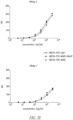
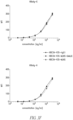
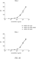
- neutralization assays animal viruses are typically propagated in cells and/or cell lines.
- a neutralization assay wherein cultured cells are incubated with a fixed amount of HBV or HDV in the presence (or absence) of the antibody (or antigen-binding fragment or fusion protein) to be tested may be used.
- the levels of hepatitis B surface antigen (HBsAg) or hepatitis B e antigen (HBeAg) secreted into the cell culture supernatant may be used and/or HBcAg staining may be assessed to provide a readout.
- HBsAg hepatitis B surface antigen
- HBeAg hepatitis B e antigen secreted into the cell culture supernatant
- HBcAg staining may be assessed to provide a readout.
- delta antigen immunofluorescence staining may be assessed.
- cultured cells for example HepaRG cells, such as differentiated HepaRG cells
- incubation may be carried out, for example, for 16 hours at 37°C. That incubation may be performed in a medium (e.g. supplemented with 4% PEG 8000). After incubation, cells may be washed and further cultivated.
- HBsAg hepatitis B surface antigen
- HBeAg hepatitis B e antigen
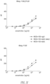
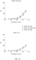
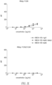
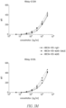
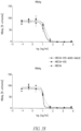
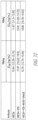
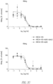
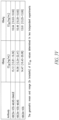
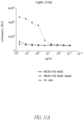
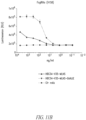
- the concentration of a binding protein as described herein required for 50% neutralization of HBV and HDV is 500 ng/ml or less.
- the concentration of abinding protein as described herein required for 50% neutralization of HBV and HDV may be selected from 450 ng/ml or less, 400 ng/ml or less, 350 ng/ml or less, 300 ng/ml or less, 250 ng/ml or less, 200 ng/ml or less, 175 ng/ml or less, 150 ng/ml or less, 125 ng/ml or less, 100 ng/ml or less, 90 ng/ml or less, 80 ng/ml or less, 70 ng/ml or less, 60 ng/ml or less or 50 ng/ml or less.
- Antibodies or antigen binding fragments according to the present disclosure which can neutralize both HBV and HDV, are useful in the prevention and treatment of hepatitis B and hepatitis D.
- Infection with HDV typically occurs simultaneously with or subsequent to infection by HBV (e.g ., inoculation with HDV in the absence of HBV does not cause hepatitis D since HDV requires the support of HBV for its own replication) and hepatitis D is typically observed in chronic HBV carriers.
- Embodiments of the disclosed binding proteins promote clearance of HBsAg and HBV.
- binding proteins promote clearance of both HBV and subviral particles of hepatitis B virus (SVPs). Clearance of HBsAg or of subviral particles may be assessed by measuring the level of HBsAg for example in a blood sample, e.g. from a hepatitis B patient. Similarly, clearance of HBV may be assessed by measuring the level of HBV for example in a blood sample, e.g. from a hepatitis B patient.
- SVPs hepatitis B virus
- hepatitis B surface antigen (HBsAg) loss is considered in some instances to be an ideal endpoint of treatment and the closest outcome to cure chronic hepatitis B (CHB).
- Embodiments of binding proteins of the present disclosure may promote clearance of HbsAg.
- the binding proteins may promote clearance of subviral particles of hepatitis B virus.
- the binding proteins may be used to treat chronic hepatitis B.
- a binding protein of the present disclosure is capable of binding an HBsAg of a genotype selected from the HBsAg genotypes A, B, C, D, E, F, G, H, I, and J, or any combination thereof.
- binding proteins of the present disclosure are capable of binding to 1, 2, 3, 4, 5, 6, 7, 8, 9 or 10 of the HBsAg genotypes A, B, C, D, E, F, G, H, I, and J.
- HBsAg genotypes include the following: GenBank accession number J02203 (HBV-D, ayw3); GenBank accession number FJ899792.1 (HBV-D, adw2); GenBank accession number AM282986 (HBV-A); GenBank accession number D23678 (HBV-B1 Japan); GenBank accession number AB117758 (HBV-C1 Cambodia); GenBank accession number AB205192 (HBV-E Ghana); GenBank accession number X69798 (HBV-F4 Brazil); GenBank accession number AF160501 (HBV-G USA); GenBank accession number AY090454 (HBV-H Portugal); GenBank accession number AF241409 (HBV-I Vietnam); and GenBank accession
- a binding protein is capable of binding to one or more, and in some cases at least 6 of the 10 HBsAg genotypes A, B, C, D, E, F, G, H, I, and J. In certain embodiments, a binding protein is capable of binding to at least 8 of the 10 HBsAg genotypes A, B, C, D, E, F, G, H, I, and J. In some embodiments, a binding protein is capable of binding to all 10 of the 10 HBsAg genotypes A, B, C, D, E, F, G, H, I, and J. HBV is differentiated into several genotypes, according to genome sequence.
- genotypes of the HBV genome have been defined. Moreover, two other genotypes, I and J, have also been identified ( Sunbul M., 2014, World J Gastroenterol 20(18): 5427-5434 ). The genotype is known to affect the progression of the disease and differences between genotypes in response to antiviral treatment have been determined.
- a binding protein according to the present disclosure is capable of binding to 1, 2, 3, 4, 5, 6, 7, 8, 9, 10, 11, 12, 13, 14, 15, 16, 17 or 18 of the HBsAg mutants having mutations in the antigenic loop region, with such mutant(s) being selected from one ore more of HBsAg Y100C/P120T, HBsAg P120T, HBsAg P120T/S143L, HBsAg C121S, HBsAg R122D, HBsAg R122I, HBsAg T123N, HBsAg Q129H, HBsAg Q129L, HBsAg M133H, HBsAg M133L, HBsAg M133T, HBsAg K141E, HBsAg P142S, HBsAg S143K, HBsAg D144A, HBsAg G145R and HBsAg N146A.
- mutants are naturally occurring mutants based on the S domain of HBsAg Genotype D, Genbank accession no. FJ899792 (SEQ ID NO: 4).
- the mutated amino acid residue(s) in each of the mutants noted herein are indicated in the name.
- SEQ ID NO: 4: (the antigenic loop region, i.e. amino acids 101 - 172, is shown underlined).
- Amino acid sequences of the antigenic loop region of the S domain of HBsAg of different mutants are shown in SEQ ID NOs: 16 - 33.
- a binding protein as disclosed herein is capable of binding to at one or more, and in some cases at least 12 infectious HBsAg mutants selected from HBsAg Y100C/P120T, HBsAg P120T, HBsAg P120T/S143L, HBsAg C121S, HBsAg R122D, HBsAg R122I, HBsAg T123N, HBsAg Q129H, HBsAg Q129L, HBsAg M133H, HBsAg M133L, HBsAg M133T, HBsAg K141E, HBsAg P142S, HBsAg S143K, HBsAg D144A, HBsAg G145R and HBsAg N146A.
- a binding protein is capable of binding to at least 15 infectious HBsAg mutants selected from HBsAg Y100C/P120T, HBsAg P120T, HBsAg P120T/S143L, HBsAg C121S, HBsAg R122D, HBsAg R122I, HBsAg T123N, HBsAg Q129H, HBsAg Q129L, HBsAg M133H, HBsAg M133L, HBsAg M133T, HBsAg K141E, HBsAg P142S, HBsAg S143K, HBsAg D144A, HBsAg G145R and HBsAg N146A.
- a binding protein is capable of binding to each of the following infectious HBsAg mutants: HBsAg Y100C/P120T; HBsAg P120T; HBsAg P120T/S143L; HBsAg C121S; HBsAg R122D; HBsAg R122I; HBsAg T123N; HBsAg Q129H; HBsAg Q129L; HBsAg M133H; HBsAg M133L; HBsAg M133T; HBsAg K141E; HBsAg P142S; HBsAg S143K; HBsAg D144A; HBsAg G145R; and HBsAg N146A.
- the binding protein (e.g., including an antibody or antigen binding fragment thereof) is capable of reducing a serum concentration of HBV DNA in a mammal having an HBV infection. In certain embodiments, the binding protein is capable of reducing a serum concentration of HBsAg in a mammal having an HBV infection. In certain embodiments, binding protein is capable of reducing a serum concentration of HBeAg in a mammal having an HBV infection. In certain embodiments, the binding protein is capable of reducing a serum concentration of HBcrAg in a mammal having an HBV infection.
- the binding protein is capable of reducing the serum concentration of HBV DNA, HBsAg, HBeAg, and/or HBcrAg in the mammal for about 10, 11, 12, 13, 14, 15, 16, 17, 18, 19, 20, or more days following a single administration of the binding protein.
- epitope includes any molecule, structure, amino acid sequence, or protein determinant that is recognized and specifically bound by a cognate binding molecule, such as an immunoglobulin, chimeric antigen receptor, or other binding molecule, domain or protein.
- Epitopic determinants generally contain chemically active surface groupings of molecules, such as amino acids or sugar side chains, and can have specific three dimensional structural characteristics, as well as specific charge characteristics.
- a binding protein is capable of binding to an epitope comprising at least one, at least two, at least three, or at least four amino acids of the antigenic loop region of HbsAg. In certain embodiments, a binding protein is capable of binding at least two amino acids selected from amino acids 115 - 133 of the S domain of HbsAg, amino acids 120 - 133 of the S domain of HbsAg, or amino acids 120 - 130 of the S domain of HbsAg.
- a binding protein is capable of binding at least three amino acids selected from amino acids 115 - 133 of the S domain of HbsAg, amino acids 120 - 133 of the S domain of HbsAg, or amino acids 120 - 130 of the S domain of HbsAg. In some embodiments, a binding protein is capable of binding at least four amino acids selected from amino acids 115 - 133 of the S domain of HbsAg, amino acids 120 - 133 of the S domain of HbsAg, or amino acids 120 - 130 of the S domain of HbsAg. As used herein, the position of the amino acids (e.g.
- 115 - 133, 120 - 133, 120 - 130 refers to the S domain of HBsAg as described above, which is present in all three HBV envelope proteins S-HBsAg, M-HBsAg, and L-HBsAg, whereby S-HBsAg typically corresponds to the S domain of HBsAg.
- the epitope is a linear epitope and comprises more than one amino acid located at positions selected from amino acid positions 115 -133 or from amino acid positions 120 -133 of the S domain of HBsAg
- the amino acids comprised by the epitope may be located in adjacent positions of the primary structure (e.g., are consecutive amino acids in the amino acid sequence).
- the amino acid sequence typically forms a 3D structure as epitope and, thus, the amino acids forming the epitope may be or may be not located in adjacent positions of the primary structure (i.e. maybe or may be not consecutive amino acids in the amino acid sequence).
- an epitope to which a binding protein binds to a conformational epitope binds to an epitope comprising at least two amino acids of the antigenic loop region of HBsAg, wherein the at least two amino acids are selected from amino acids 120 - 133 or from from amino acids 120 - 130, of the S domain of HbsAg, and wherein the at least two amino acids are not located in adjacent positions (of the primary structure).
- a binding protein binds to an epitope comprising at least three amino acids of the antigenic loop region of HBsAg, wherein the at least three amino acids are selected from amino acids 120 - 133 or from from amino acids 120 - 130, of the S domain of HbsAg, and wherein at least two of the three amino acids are not located in adjacent positions (of the primary structure).
- a binding protein binds to an epitope comprising at least four amino acids of the antigenic loop region of HBsAg, wherein the at least four amino acids are selected from amino acids 120 - 133 or from from amino acids 120 - 130, of the S domain of HbsAg, and wherein at least two of the four amino acids are not located in adjacent positions (of the primary structure).
- Amino acids to which a presently disclosed antibody, antigen binding fragment, or fusion protein binds i.e. the amino acids forming the epitope
- Amino acids to which a presently disclosed antibody, antigen binding fragment, or fusion protein binds are in some cases spaced apart by one or more amino acids, to which the antibody, antigen binding fragment, or fusion protein does not bind.
- at least one, at least two, at least three, at least four, or at least five amino acids may be located between two of the amino acids not located in adjacent positions comprised by the epitope.
- a binding protein binds to an epitope comprising at least amino acids P120, C121, R122 and C124 of the S domain of HBsAg.
- a binding protein of the present disclosure binds to an epitope comprising an amino acid sequence according to SEQ ID NO: 88: PCRXC wherein X is any amino acid or no amino acid; X is any amino acid; X is T, Y, R, S, or F; X is T, Y or R; or X is TorR.
- a binding protein of the present disclosure binds to an epitope comprising an amino acid sequence according to SEQ ID NO: 80: TGPCRTC or to an amino acid sequence sharing at least 80%, at least 90%, or at least 95% sequence identity with SEQ ID NO: 80.
- a binding protein of the present disclosure binds to an epitope comprising an amino acid sequence according to SEQ ID NO: 85: STTSTGPCRTC or to an amino acid sequence sharing at least 80%, at least 90% or at least 95% sequence identity with SEQ ID NO: 85.
- a binding protein of the present disclosure binds to an epitope comprising an amino acid sequence according to SEQ ID NO: 85 and/or an amino acid sequence according to SEQ ID NO: 87.
- a binding protein of the present disclosure binds to an epitope in the antigenic loop of HBsAg formed by an amino acid sequence according to SEQ ID NO: 1: X 1 X 2 X 3 TC X 4 X 5 X 6 A X 7 G wherein X 1 , X 2 , X 3 , X 4 , X 5 , X 6 and X 7 may be any amino acid (SEQ ID NO: 1).
- X 1 , X 2 , X 3 , X 4 , X 5 , X 6 and X 7 are amino acids, which are conservatively substituted in comparison to amino acids 120 - 130 of SEQ ID NO: 3.
- X 1 , X 2 , X 3 , X 4 , X 5 , X 6 and X 7 are amino acids, which are conservatively substituted in comparison to amino acids 20 - 30 of any of SEQ ID NOs 5 - 33.
- X 2 of SEQ ID NO: 1 X 2 is a small amino acid.
- X 2 may be selected from cystein or threonine.
- X 5 of SEQ ID NO: 1 X 5 is a small amino acid and/or a hydrophobic amino acid. In certain embodiments, X 5 is selected from threonine, alanine or isoleucine.
- X 7 of SEQ ID NO: 1 is a polar amino acid or an aliphatic amino acid.
- a "polar" amino acid refers to any amino acid selected from the group consisting of aspartic acid, asparagine, arginine, glutamic acid, histidine, lysine, glutamine, tryptophan, tyrosine, serine, and threonine.
- X 7 is glutamine, histidine or leucine.
- abinding protein according to the present disclosure binds to an epitope in the antigenic loop of HBsAg formed by an amino acid sequence according to SEQ ID NO: 2: X 1 X 2 X 3 TC X 4 X 5 X 6 A X 7 G wherein
- a binding protein may bind only to some of the amino acids of SEQ ID NO: 1 or 2, whereby other amino acid residues may act as "spacers".
- the present disclosure provides an isolated antibody, or an antigen binding fragment thereof, comprising: (i) a heavy chain variable region (V H ) comprising at least 90% ( i.e ., 90%, 91%, 92%, 93%, 94%, 95%, 96%, 97%, 98%, 99%, or more) identity to the amino acid sequence according to SEQ ID NO:41 or 67; and (ii) a light chain variable region (V L ) comprising at least 90% identity to the amino acid sequence according to any one of SEQ ID NOs:42; 59; 65; 89, 90, or 111-120, provided that the amino acid at position 40 of the VL according to IMGT numbering is not a cysteine, wherein the antibody or antigen binding fragment thereof binds to the antigenic loop region of HBsAg and neutralizes infection with hepatitis B virus and hepatitis delta virus.
- V H heavy chain variable region
- V L light chain variable region
- the V H comprises at least 95% identity to the amino acid sequence according to SEQ ID NO:41 or 67; and/or (ii) the V L comprises at least 95% identity to the amino acid sequence according to any one of SEQ ID NOs:42, 59, 65, 89, 90, or 111-120.
- the amino acid at position 40 of the V L is alanine. In other embodiments, the amino acid at position 40 of the V L is serine. In still other embodiments, the amino acid at position 40 of the V L is glycine.
- the V L of the antibody or antigen binding fragment comprises or consists of the amino acid sequence according to SEQ ID NO:89. In some embodiments, the V L of the antibody or antigen binding fragment comprises or consists of the amino acid sequence according to SEQ ID NO:90. In other embodiments, the V L of the antibody or antigen binding fragment comprises or consists of the amino acid sequence according to any one of SEQ ID NOs: 111-120. In certain embodiments, the V H comprises or consists of the amino acid sequence according to SEQ ID NO:41. In other embodiments, the V H comprises or consists of the amino acid sequence according to SEQ ID NO: 67.
- the V H comprises or consists of the amino acid sequence according to SEQ ID NO:95; and the V L comprises or consists of the amino acid sequence according to SEQ ID NO:96.
- an Fc moiety refers to a sequence comprising, consisting, consisting essentially of, or derived from a portion of an immunoglobulin heavy chain beginning in the hinge region just upstream of the papain cleavage site (e.g., residue 216 in native IgG, taking the first residue of heavy chain constant region to be 114) and ending at the C-terminus of the immunoglobulin heavy chain.
- an Fc moiety may be a complete Fc moiety or a portion ( e.g., a domain) thereof.
- a complete Fc moiety comprises a hinge domain, a CH2 domain, and a CH3 domain (e.g., EU amino acid positions 216-446).
- Amino acid positions within an Fc moiety have been numbered according to the EU numbering system of Kabat, see e.g., Kabat et al., "Sequences of Proteins of Immunological Interest", U.S. Dept. Health and Human Services, 1983 and 1987 . Amino acid positions of an Fc moiety can also be numbered according to the IMGT numbering system (including unique numbering for the C-domain and exon numbering) and the Kabat numbering system.
- an Fc moiety may comprise or consist of: (i) hinge domain (or a portion thereof) fused to a CH2 domain (or a portion thereof), (ii) a hinge domain (or a portion thereof) fused to a CH3 domain (or a portion thereof), (iii) a CH2 domain (or a portion thereof) fused to a CH3 domain (or a portion thereof), (iv) a hinge domain (or a portion thereof), (v) a CH2 domain (or a portion thereof), or (vi) a CH3 domain or a portion thereof.
- An Fc moiety of the present disclosure may be modified such that it varies in amino acid sequence from the complete Fc moiety of a naturally occurring immunoglobulin molecule, while retaining or enhancing at least one desirable function conferred by the naturally occurring Fc moiety, and/or reducing an undesired function of a naturally occurring Fc moiety.
- Such functions include, for example, Fc receptor (FcR) binding, antibody half-life modulation (e.g., by binding to FcRn), ADCC function, protein A binding, protein G binding, and complement binding. Portions of naturally occurring Fc moieties which are involved with such functions have been described in the art.
- the C1q protein complex can bind to at least two molecules of IgG1 or one molecule of IgM when the immunoglobulin molecule(s) is attached to the antigenic target ( Ward, E. S., and Ghetie, V., Ther. Immunol. 2 (1995) 77-94 ).
- Burton, D. R. described (Mol. Immunol. 22 (1985) 161-206 ) that the heavy chain region comprising amino acid residues 318 to 337 is involved in complement fixation.
- FcR binding can be mediated by the interaction of the Fc moiety (of an antibody) with Fc receptors (FcRs), which are specialized cell surface receptors on cells including hematopoietic cells.
- Fc receptors belong to the immunoglobulin superfamily, and shown to mediate both the removal of antibody-coated pathogens by phagocytosis of immune complexes, and the lysis of erythrocytes and various other cellular targets (e.g. tumor cells) coated with the corresponding antibody, via antibody dependent cell mediated cytotoxicity (ADCC; Van de Winkel, J. G, and Anderson, C. L., J. Leukoc. Biol. 49 (1991) 511-524 ).
- ADCC antibody dependent cell mediated cytotoxicity
- FcRs are defined by their specificity for immunoglobulin classes; Fc receptors for IgG antibodies are referred to as Fc ⁇ R, for IgE as Fc ⁇ R, for IgA as Fc ⁇ R and so on and neonatal Fc receptors are referred to as FcRn.
- Fc receptor binding is described for example in Ravetch, J. V., and Kinet, J. P., Annu. Rev. Immunol. 9 (1991) 457-492 ; Capel, P. J., et al., Immunomethods 4 (1994) 25-34 ; de Haas, M., et al., J Lab. Clin. Med. 126 (1995) 330-341 ; and Gessner, J. E., et al., Ann. Hematol. 76 (1998) 231-248 .
- Fc ⁇ R Cross-linking of receptors by the Fc domain of native IgG antibodies
- Fc ⁇ R Fc domain of native IgG antibodies
- effector functions including phagocytosis, antibody-dependent cellular cytotoxicity, and release of inflammatory mediators, as well as immune complex clearance and regulation of antibody production.
- Fc moieties providing cross-linking of receptors e.g., FcyR are contemplated herein.
- Fc ⁇ R In humans, three classes of Fc ⁇ R have been characterized to-date, which are: (i) Fc ⁇ RI (CD64), which binds monomeric IgG with high affinity and is expressed on macrophages, monocytes, neutrophils and eosinophils; (ii) FcyRII (CD32), which binds complexed IgG with medium to low affinity, is widely expressed, in particular on leukocytes, is believed to be a central player in antibody-mediated immunity, and which can be divided into FcyRIIA, Fc ⁇ RIIB and Fc ⁇ RIIC, which perform different functions in the immune system, but bind with similar low affinity to the IgG-Fc, and the ectodomains of these receptors are highly homologuous; and (iii) FcyRIII (CD16), which binds IgG with medium to low affinity and has been found in two forms: Fc ⁇ RIIIA, which has been found on NK cells, macrophages,
- FcyRIIA is found on many cells involved in killing (e.g. macrophages, monocytes, neutrophils) and seems able to activate the killing process.
- Fc ⁇ RIIB seems to play a role in inhibitory processes and is found on B-cells, macrophages and on mast cells and eosinophils. Importantly, it has been shown that 75% of all Fc ⁇ RIIB is found in the liver ( Ganesan, L. P. et al., 2012: “FcyRIIb on liver sinusoidal endothelium clears small immune complexes," Journal of Immunology 189: 4981-4988 ).
- Fc ⁇ RIIB is abundantly expressed on Liver Sinusoidal Endothelium, called LSEC, and in Kupffer cells in the liver and LSEC are the major site of small immune complexes clearance ( Ganesan, L. P. et al., 2012: FcyRIIb on liver sinusoidal endothelium clears small immune complexes. Journal of Immunology 189: 4981-4988 ).
- the clearance of immune complexes can be enhanced ( Chu, S., et al., 2014: Accelerated Clearance of IgE In Chimpanzees Is Mediated By Xmab7195, An Fc-Engineered Antibody With Enhanced Affinity For Inhibitory Receptor Fc ⁇ RIIb. Am J Respir Crit, American Thoracic Society International Conference Abstracts ).
- the antibodies of the present disclosure, or the antigen binding fragments thereof comprise an engineered Fc moiety with the mutations S267E and L328F, in particular as described by Chu, S. Y. et al., 2008: Inhibition of B cell receptor-mediated activation of primary human B cells by coengagement of CD19 and FcgammaRIIb with Fc-engineered antibodies. Molecular Immunology 45, 3926-3933 .
- modification in native IgG of at least one of E233-G236, P238, D265, N297, A327 and P329 reduces binding to Fc ⁇ RI.
- FcyRIIA reduced binding for FcyRIIA is found, e.g., for IgG mutation of at least one of E233-G236, P238, D265, N297, A327, P329, D270, Q295, A327, R292 and K414.
- Fc ⁇ RIII binding reduced binding to Fc ⁇ RIIIA is found, e.g., for mutation of at least one of E233-G236, P238, D265, N297, A327, P329, D270, Q295, A327, S239, E269, E293, Y296, V303, A327, K338 and D376. Mapping of the binding sites on human IgG1 for Fc receptors, the above-mentioned mutation sites, and methods for measuring binding to FcyRI and FcyRIIA, are described in Shields, R. L., et al., J. Biol. Chem. 276 (2001) 6591-6604 .
- F158 Two allelic forms of human Fc ⁇ RIIIA are the "F158" variant, which binds to IgG1 Fc with low affinity, and the "V158” variant, which binds to IgG1 Fc with high affinity. See, e.g., Bruhns et al., Blood 113:3716-3725 (2009 ).
- two regions of native IgG Fc appear to be involved in interactions between FcyRIIs and IgGs, namely (i) the lower hinge site of IgG Fc, in particular amino acid residues L, L, G, G (234 - 237, EU numbering), and (ii) the adjacent region of the CH2 domain of IgG Fc, in particular a loop and strands in the upper CH2 domain adjacent to the lower hinge region, e.g. in a region of P331 ( Wines, B.D., et al., J. Immunol. 2000; 164: 5313 - 5318 ).
- FcyRI appears to bind to the same site on IgG Fc
- FcRn and Protein A bind to a different site on IgG Fc, which appears to be at the CH2-CH3 interface
- mutations that increase binding affinity of an Fc moiety of the present disclosure to a (i.e ., one or more) Fc ⁇ receptor (e.g., as compared to a reference Fc moiety or antibody containing the same that does not comprise the mutation(s)). See, e.g., Delillo and Ravetch, Cell 161(5):1035-1045 (2015 ) and Ahmed et al., J. Struc. Biol. 194(1):78 (2016 ), the Fc mutations and techniques of which are incorporated herein by reference.
- a binding protein can comprise a Fc moiety comprising a mutation selected from G236A; S239D; A330L; and I332E; or a combination comprising any two or more of the same; e.g., S239D/I332E; S239D/A330L/I332E; G236A/S239D/I332E; G236A/A330L/I332E (also referred to herein as "GAALIE"); or G236A/S239D/A330L/I332E.
- the Fc moiety does not comprise S239D.
- the Fc moiety comprises or is derived from a IgG Fc and a half-life-extending mutation comprises any one or more of: M428L; N434S; N434H; N434A; N434S; M252Y; S254T; T256E; T250Q; P257I Q311I; D376V; T307A; E380A (EU numbering).
- a half-life-extending mutation comprises M428L/N434S (also referred to herein as "MLNS").
- a half-life-extending mutation comprises M252Y/S254T/T256E.
- a half-life-extending mutation comprises T250Q/M428L. In certain embodiments, a half-life-extending mutation comprises P257I/Q311I. In certain embodiments, a half-life-extending mutation comprises P257I/N434H. In certain embodiments, a half-life-extending mutation comprises D376V/N434H. In certain embodiments, a half-life-extending mutation comprises T307A/E380A/N434A.
- a binding protein includes a Fc moiety that comprises the substitution mtuations M428L/N434S. In some embodiments, a binding protein includes a Fc moiety that comprises the substitution mtuations G236A/A330L/I332E. In certain embodiments, a binding protein includes a (e.g., IgG) Fc moiety that comprises a G236A mutation, an A330L mutation, and a I332E mutation (GAALIE), and does not comprise a S239D mutation (e.g., comprises a native S at position 239).
- a binding protein includes a (e.g., IgG) Fc moiety that comprises a G236A mutation, an A330L mutation, and a I332E mutation (GAALIE), and does not comprise a S239D mutation (e.g., comprises a native S at position 239).
- a binding protein includes an Fc moiety that comprises the substitution mutations: M428L/N434S and G236A/A330L/I332E, and optionally does not comprise S239D. In certain embodiments, a binding protein includes a Fc moiety that comprises the substitution mutations: M428L/N434S and G236A/S239D/A330L/I332E.
- a binding protein of the present disclosure comprises: CDRs and/or a variable domain and/or a heavy chain and/or a light chain according to any one of the exemplary anti-HBV antibodies disclosed herein and/or in PCT Publication No.
- WO 2017/060504 (including antibodies HBC34, HBC34-V7, HBC34-V23, HBC34-V31, HBC34-V32, HBC34-V33, HBC34-V34, HBC34-V35, (including herein disclosed variants of HBC antibodies which comprise a substitution mutation at position 40 in the light chain (e.g., a substitution of a native cysteine with an alanine, a serine, or the like) and HBC24); and a Fc moiety comprising a G236A mutation, an A330L mutation, and a I332E (GAALIE) mutation, wherein the Fc moiety optionally further comprises a M428L/N434S (MLNS) mutation. In certain embodiments, the Fc moiety does not comprise S239D.
- a substitution mutation at position 40 in the light chain e.g., a substitution of a native cysteine with an alanine, a serine, or the like
- a binding protein comprises: a CDRH1 amino acid sequence according to SEQ ID NO:34, a CDRH2 amino acid sequence according to SEQ ID NO:35 or 66, a CDRH3 amino acid sequence according to SEQ ID NO:36, a CDRL1 acid sequence according to SEQ ID NO:37, a CDRL2 acid sequence according to SEQ ID NO:38 or 39, and CDRL3 amino acid sequence according to SEQ ID NO:58 or 40; and a Fc moiety comprising a GAALIE mutation.
- the Fc moiety does not comprise a S239D mutation.
- the Fc moiety further comprises a MLNS mutation.
- a binding protein comprises: a heavy chain variable domain (VH) amino acid sequence according to any one of SEQ ID NOs:41 or 67 and a light chain variable domain (VL) amino acid sequence according to any one of SEQ ID NOs:42, 59, 65, 89, 90, and 111-120; and a Fc moiety comprising a GAALIE mutation.
- the Fc moiety further comprises a MLNS mutation.
- a binding protein comprises a heavy chain amino acid sequence according to SEQ ID NO: 92 and a light chain amino acid sequences according to SEQ ID NOs:93. In certain embodiments, a binding protein comprises a heavy chain amino acid sequence according to SEQ ID NO: 92 and a light chain amino acid sequences according to SEQ ID NOs:94.
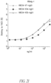
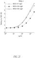
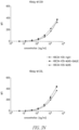
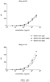
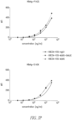
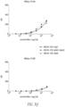
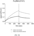
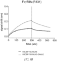
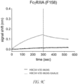
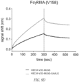
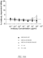
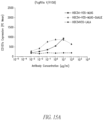
- a biolayer interferometry (BLI) assay can be performed using an Octet ® RED96 (ForteBio, Fremont, California USA) instrument according to manufacturer's instructions to determine real-time association and dissociation between a first polypeptide of interest (e.g ., HBC34v35 comprising a GAALIE mutation) and a second polypeptide of interest (e.g ., a Fc ⁇ RIIA (H131), a FcyRIIA (R131), a Fc ⁇ RIIIA (F158), a Fc ⁇ RIIIA (V158), or a Fc ⁇ RIIb) that is captured on a sensor substrate.
- a first polypeptide of interest e.g ., HBC34v35 comprising a GAALIE mutation
- a second polypeptide of interest e.g ., a Fc ⁇ RIIA (H131), a FcyRIIA (R131), a Fc ⁇ RIIIA (F158), a
- the BLI assay comprises use of Octet (R) RED96 (ForteBio, Fremont, California USA) instrument.
- the BLI assay comprises a tagged human Fc ⁇ R captured onto an anti-penta-tag sensor and exposed to the binding protein.
- the binding protein comprises a IgG Fab and the BLI assay further comprises exposing the captured human Fc ⁇ R to the binding protein in the presence of an anti-IgG Fab binding fragment to cross-link the binding proteins through the Fab fragment.
- a binding protein includes a Fc moiety comprising a GAALIE mutation and has enhanced binding to a human FcyRIIA (H131), a human Fc ⁇ RIIA (R131), a human Fc ⁇ RIIIA (F158), and a human Fc ⁇ RIIIA (V158), as compared to a reference polypeptide.
- a binding protein includes a Fc moiety comprising a GAALIE mutation and activates a human FcyRIIA, a human Fc ⁇ RIIIA, or both, to a greater degree than does a reference polypeptide.
- a polypeptide which may be a HbsAg-specific binding protein, that includes a Fc moiety that does not comprise the GAALIE mutation.
- the reference polypeptide includes a Fc moiety that is a wild-type Fc moiety or that comprises one or more substitution mutation, provided that the substitution mutation is not GAALIE.
- Activation of a human Fc ⁇ R can be determined or detected using methods known in the art.
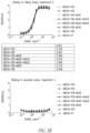
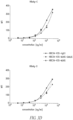
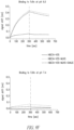
- a well-validated, commercially available bioreporter assay involves incubating a HBsAg-specific binding protein with a recombinant HBsAg (Engerix B, GlaxoSmithKline) in the presence of Jurkat effector cells (Promega; Cat. no: G9798) stably expressing (i) a Fc ⁇ R of interest and (ii) firefly luciferase reporter under the control of a NFAT response element. Binding of Fc to cell surface-expressed Fc ⁇ R drives NFAT-mediated expression of luciferase reporter gene.
- Luminescence is then measured with a luminometer (e.g., Bio-Tek) using the Bio-Glo- TM Luciferase Assay Reagent (Promega) according to the manufacturer's instructions.
- Activation is expressed as the average of relative luminescence units (RLU) over the background by applying the following formula: (RLU at concentration [x] of binding protein ( e.g., mAbs) - RLU of background).
- a binding protein includes a Fc moiety comprising a GAALIE mutation activates a human FcyRIIA (H131), a human FcyRIIA (R131), a human Fc ⁇ RIIIA (F158), and/or a human Fc ⁇ RIIIA (V158) to a greater degree than does a reference polypeptide.
- a greater degree of activation refers to a higher peak luminescence and/or a greater luminescence area under the curve, as determined using a luminescence bioreporter assay as described herein.
- a binding protein includes a Fc moiety comprising a GAALIE mutation does not activate a human FcyRIIB, as determined by the absence of a statistically significant and/or measurable RLU in a luminescence bioreporter assay as described above.
- a binding protein includes a Fc moiety comprising a GAALIE mutation and activates a human natural killer (NK) cell in the presence of HBsAg to a greater degree than does a reference polypeptide.
- activation of a NK cell is determined by CD107a expression ( e.g., by flow cytometry).
- the NK cell comprises a cell that comprises V158/V158 homozygous, a F158/F158 homozygous, or a V158/F158 heterozygous Fc ⁇ RIIIa genotype.
- a binding protein of the present disclosure includes a Fc moiety comprising a GAALIE mutation and: (i) has enhanced binding to a human FcyRIIA, a human FcyRIIIA, or both, as compared to a reference polypeptide that includes a Fc moiety that does not comprise G236A/A330L/I332E, wherein the human FcyRIIA is optionally H131 or R131, and/or the human Fc ⁇ RIIIA is optionally F158 or V158; (ii) has reduced binding to a human Fc ⁇ RIIB, as compared to a reference polypeptide that includes a Fc moiety that does not comprise G236A/A330L/I332E; (iii) does not bind to a human FcyRIIB; (iv) has reduced binding to a human C1q, as compared to a reference polypeptide that includes a Fc moiety that does not comprise G236A/A330L/
- a binding protein according to the present disclosure comprises an Fc region.
- Fc region refers to the portion of an immunoglobulin formed by two or more Fc moieties of antibody heavy chains.
- an Fc region may be monomeric or "single-chain" Fc region (i.e., a scFc region).
- Single chain Fc regions are comprised of Fc moieties linked within a single polypeptide chain (e.g., encoded in a single contiguous nucleic acid sequence).
- Exemplary scFc regions are disclosed in WO 2008/143954 A2 , and are incorporated by reference herein.
- the Fc region can be or comprise a dimeric Fc region.
- an antibody or antigen binding fragment comprises a heavy chain according to SEQ ID NO:91 and a light chain according to SEQ ID NO:93.
- an antibody or antigen binding fragment comprises a heavy chain according to SEQ ID NO:92 and a light chain according to SEQ ID NO:94.
- an antibody or antigen binding fragment comprises a heavy chain according to SEQ ID NO:91 and a light chain according to SEQ ID NO:94. In other embodiments, an antibody or antigen binding fragment comprises a heavy chain according to SEQ ID NO:92 and a light chain according to SEQ ID NO:93. In some embodiments, an antibody or antigen binding fragment comprises or consists of a heavy chain according to SEQ ID NO: 129. In some embodiments, an antibody or antigen binding fragment comprises or consists of a heavy chain according to SEQ ID NO:138, and optionally a light chain according to any one of SEQ ID NOs:93 or 94. These sequences are provided in the Sequence Listing.
- Fc moieties may comprise Fc sequences or regions of the same or different class and/or subclass.
- Fc moieties may be derived from an immunoglobulin (e.g., a human immunoglobulin) of an IgG1, IgG2, IgG3 or IgG4 subclass, or from any combination thereof.
- the Fc moieties of Fc region are of the same class and subclass.
- the Fc region (or one or more Fc moieties of an Fc region) may also be chimeric, whereby a chimeric Fc region may comprise Fc moieties derived from different immunoglobulin classes and/or subclasses.
- a dimeric Fc region can comprise sequences from two or more different isotypes or subclasses; e.g., a SEEDbody ("strand-exchange engineered domains"), see Davis el al., Protein Eng. Des. Sel, 23(4):195 (2010 ).
- the chimeric Fc region may comprise an Fc moiety (e.g., a complete Fc moiety) from an immunoglobulin for a first subclass (e.g., an IgG4 subclass) and an Fc moiety from an immunoglobulin of a second subclass (e.g., an IgG1, IgG2 or IgG3 subclass).
- the Fc region or moiety may comprise a CH2 domain from an IgG4 immunoglobulin and a CH3 domain from an IgG1 immunoglobulin.
- the Fc region or moiety may comprise a CH1 domain and a CH2 domain from an IgG4 molecule and a CH3 domain from an IgG1 molecule.
- the disclosure provides a nucleic acid molecule comprising a polynucleotide encoding an antibody, antigen binding fragment, or fusion protein according to the present disclosure
- Table 4 shows exemplary V H -, V L -, CH-, CL-, HC-, and LC-encoding nucleotide sequences according to the present disclosure:
- Antibody nucleotide acid sequence description SEQ ID NO: nuclecotide sequence V H of HBC34-V7, HBC34-V35, and HBC34-V34 (codon optimized) 103 HBC34-V34 V L (codon optimized) 104 HBC34-V35 V L (codon optimized) 105 HBC34-V7 V L (codon optimized) 110 HBC24 V H (wild type) 106 HBC24 V L (wild type) 107 HBC24 V H (codon optimized) 108 HBC24 V L (codon optimized) 109 HBC34-V7, HBC34-V34, HBC34-V35 CH1-
- the present disclosure also comprises sequence variants of these nucleic acid sequences and in particular such sequence variants, which encode the same amino acid sequences.
- a polynucleotide or nucleic acid molecule comprises a nucleotide sequence sharing at least 80% identity to the nucleotide sequence according to any one of SEQ ID NOs: 103-110 and 130-136, wherein the nucleotide sequence is codon optimized for expression by a host cell.
- a nucleic acid molecule according to the present disclosure comprises or consists of a nucleic acid sequence according to any one of SEQ ID NOs: 103-110 and 130-136.
- a polynucleotide comprises a V H -encoding nucleotide sequence according to SEQ ID NO:103 and a V L -encoding nucleotide sequence according to SEQ ID NO:105. In other embodiments, a polynucleotide comprises a V H -encoding nucleotide sequence according to SEQ ID NO:103, and a V L -encoding nucleotide sequence according to SEQ ID NO:104. In other embodiments, a polynucleotide comprises a V H -encoding nucleotide sequence according to SEQ ID NO: 108, and a V L -encoding nucleotide sequence according to SEQ ID NO: 109.
- polynucleotides that encode an antibody or antigen binding fragment
- the polynucleotide comprises or consists of a V H -encoding nucleotide sequence according to SEQ ID NO:103 and a V L -encoding nucleotide sequence according to SEQ ID NO:110, wherein the encoded antibody or antigen binding fragment binds to the antigenic loop region of HBsAg and neutralizes infection with hepatitis B virus and hepatitis delta virus.
- a polynucleotide can comprise a CH1-hinge-CH2-CH3-encoding nucleotide sequence according to SEQ ID NO:130, and/or comprises a HC (VH-CH1-hinge-CH3-CH3)-encoding nucleotide sequence according to SEQ ID NO:131.
- a polynucleotide comprises a CL-encoding nucleotide sequence according to SEQ ID NO:132 and/or comprises a LC (VL-CL)-encoding nucleotide sequence according to SEQ ID NO:133.
- a polynucleotide comprises a CL-encoding nucleotide sequence according to SEQ ID NO:134 and/or comprises a LC (VL-CL)-encoding nucleotide sequence according to SEQ ID NO:135 or SEQ ID NO:136.
- vector refers to a construct comprising a nucleic acid molecule.
- a vector in the context of the present disclosure is suitable for incorporating or harboring a desired nucleic acid sequence.
- Such vectors may be storage vectors, expression vectors, cloning vectors, transfer vectors etc.
- a storage vector is a vector which allows the convenient storage of a nucleic acid molecule.
- the vector may comprise a sequence corresponding, e.g., to a desired antibody or antibody fragment thereof according to the present description.
- the vector may replicate and function independently of the host genome, or may, in some instances, integrate into the genome itself.
- plasmid "expression plasmid,” “virus” and “vector” are often used interchangeably.
- a cloning vector is typically a vector that contains a cloning site, which may be used to incorporate nucleic acid sequences into the vector.
- a cloning vector may be, e.g., a plasmid vector or a bacteriophage vector.
- Hepa RG cells myeloma cells or hybridoma cells.
- mammalian host cell lines include mouse sertoli cells (e.g., TM4 cells); monkey kidney CV1 line transformed by SV40 (COS-7); baby hamster kidney cells (BHK); African green monkey kidney cells (VERO-76); monkey kidney cells (CV1); human cervical carcinoma cells (HELA); human lung cells (W138); human liver cells (Hep G2); canine kidney cells (MDCK; buffalo rat liver cells (BRL 3A), mouse mammary tumor (MMT 060562); TRI cells; MRC 5 cells; and FS4 cells.
- mouse sertoli cells e.g., TM4 cells
- COS-7 monkey kidney CV1 line transformed by SV40
- BHK baby hamster kidney cells
- VERO-76 African green monkey kidney cells
- CV1 monkey kidney cells
- HELA human cervical carcinoma cells
- W138 human lung cells
- Hep G2 human liver cells
- canine kidney cells MDCK; buffalo
- Insect cells useful expressing a binding protein of the present disclosure are known in the art and include, for example, Spodoptera frugipera Sf9 cells, Trichoplusia ni BTI-TN5B1-4 cells, and Spodoptera frugipera SfSWT01 "Mimic TM " cells. See, e.g., Palmberger et al., J. Biotechnol. 153(3-4):160-166 (2011 ). Numerous baculoviral strains have been identified which may be used in conjunction with insect cells, particularly for transfection of Spodoptera frugiperda cells.
- Plant cells can also be utilized as hosts for expressing a binding protein of the present disclosure.
- PLANTIBODIES TM technology (described in, for example, U.S. Pat. Nos. 5,959,177 ; 6,040,498 ; 6,420,548 ; 7,125,978 ; and 6,417,429 ) employs transgenic plants to produce antibodies.
- the cell may be transfected with a vector according to the present description with an expression vector.
- transfection refers to the introduction of nucleic acid molecules, such as DNA or RNA (e.g. mRNA) molecules, into cells, such as into eukaryotic cells.
- RNA e.g. mRNA
- transfection encompasses any method known to the skilled person for introducing nucleic acid molecules into cells, such as into eukaryotic cells, including into mammalian cells.
- Such methods encompass, for example, electroporation, lipofection, e.g., based on cationic lipids and/or liposomes, calcium phosphate precipitation, nanoparticle based transfection, virus based transfection, or transfection based on cationic polymers, such as DEAE-dextran or polyethylenimine etc.
- the introduction is non-viral.
- Suitable detection means include the use of labels such as radionuclides, enzymes, coenzymes, fluorescers, chemiluminescers, chromogens, enzyme substrates or co-factors, enzyme inhibitors, prosthetic group complexes, free radicals, particles, dyes, and the like.
- labels such as radionuclides, enzymes, coenzymes, fluorescers, chemiluminescers, chromogens, enzyme substrates or co-factors, enzyme inhibitors, prosthetic group complexes, free radicals, particles, dyes, and the like.
- an antibody, antibody fragment, or fusion protein can be conjugated to a second antibody, or antibody fragment thereof, (or second fusion protein) to form a heteroconjugate as described in US 4,676,980 .
- linkers may be used between the labels and the antibodies of the description, e.g., as described in US 4,831,175 .
- Antibodies, antigen-binding fragments, and fusion proteins may be directly labeled with radioactive iodine, indium, yttrium, or other radioactive particle known in the art, e.g., as described in US 5,595,721 .
- antibodies are produced using a method described in WO 2004/076677 .
- B cells producing the antibody are transformed with EBV and a polyclonal B cell activator. Additional stimulants of cellular growth and differentiation may optionally be added during the transformation step to further enhance the efficiency. These stimulants may be cytokines such as IL-2 and IL-15. In one aspect, IL-2 is added during the immortalization step to further improve the efficiency of immortalization, but its use is not essential.
- the immortalized B cells produced using these methods can then be cultured using methods known in the art and antibodies isolated therefrom.
- WO 2010/046775 Another method for producing antibodies is described in WO 2010/046775 .
- plasma cells are cultured in limited numbers, or as single plasma cells in microwell culture plates.
- Antibodies can be isolated from the plasma cell cultures. Further, from the plasma cell cultures, RNA can be extracted and PCR can be performed using methods known in the art.
- the VH and VL regions of the antibodies can be amplified by RT-PCR (reverse transcriptase PCR), sequenced and cloned into an expression vector that is then transfected into HEK293T cells or other host cells.
- RT-PCR reverse transcriptase PCR
- the cloning of nucleic acid in expression vectors, the transfection of host cells, the culture of the transfected host cells and the isolation of the produced antibody can be done using any methods known to one of skill in the art.
- compositions may also contain a pharmaceutically acceptable carrier, diluent and/or excipient.
- a pharmaceutically acceptable carrier may facilitate administration, it should not itself induce the production of antibodies harmful to the individual receiving the composition. Nor should it be toxic.
- Suitable carriers may be large, slowly metabolized macromolecules such as proteins, polypeptides, liposomes, polysaccharides, polylactic acids, polyglycolic acids, polymeric amino acids, amino acid copolymers and inactive virus particles.
- pharmaceutically acceptable carriers in a pharmaceutical composition according to the present disclosure may be active components or inactive components.
- compositions of the disclosure may be prepared in various forms.
- the compositions may be prepared as injectables, either as liquid solutions or suspensions.
- Solid forms suitable for solution in, or suspension in, liquid vehicles prior to injection can also be prepared (e.g., a lyophilized composition, similar to Synagis TM and Herceptin TM , for reconstitution with sterile water containing a preservative).
- the composition may be prepared for topical administration e.g., as an ointment, cream or powder.
- the composition may be prepared for oral administration e.g., as a tablet or capsule, as a spray, or as a syrup (optionally flavored).
- compositions present in several forms of administration include, but are not limited to, those forms suitable for parenteral administration, e.g., by injection or infusion, for example by bolus injection or continuous infusion.
- parenteral administration e.g., by injection or infusion, for example by bolus injection or continuous infusion.
- the product may take the form of a suspension, solution or emulsion in an oily or aqueous vehicle and it may contain formulatory agents, such as suspending, preservative, stabilizing and/or dispersing agents.
- the antibody molecule may be in dry form, for reconstitution before use with an appropriate sterile liquid.
- a vehicle is typically understood to be a material that is suitable for storing, transporting, and/or administering a compound, such as a pharmaceutically active compound, in particular the antibodies according to the present description.
- the vehicle may be a physiologically acceptable liquid, which is suitable for storing, transporting, and/or administering a pharmaceutically active compound, in particular the antibodies according to the present description.
- compositions according to the present description may also be administered topically, especially when the target of treatment includes areas or organs readily accessible by topical application, e.g., including diseases of the skin or of any other accessible epithelial tissue. Suitable topical formulations are readily prepared for each of these areas or organs.
- the pharmaceutical composition may be formulated in a suitable ointment, containing the inventive pharmaceutical composition, particularly its components as defined above, suspended or dissolved in one or more carriers.
- Carriers for topical administration include, but are not limited to, mineral oil, liquid petrolatum, white petrolatum, propylene glycol, polyoxyethylene, polyoxypropylene compound, emulsifying wax and water.
- the pharmaceutical composition can be formulated in a suitable lotion or cream.
- the amount of the antibody, or the antigen binding fragment thereof, in the pharmaceutical compositiondoes not exceed 1 g.
- the single dose does not exceed a dose selected from 500 mg, 250 mg, 100 mg, and 50 mg.
- an a composition of the present disclosure is used in combination with a LAG3 inhibitor, such as LAG525, IMP321, IMP701, 9H12, BMS-986016, or any combination thereof.
- a LAG3 inhibitor such as LAG525, IMP321, IMP701, 9H12, BMS-986016, or any combination thereof.
- composition of the present disclosure is used in combination with an inhibitor of CD244.
- composition of the present disclosure is used in combination with an inhibitor of BLTA, HVEM, CD160, or any combination thereof.
- Anti CD-160 antibodies are described in, for example, PCT Publication No. WO 2010/084158 .
- composition of the present disclosure is used in combination with an inhibitor of TIM3.
- composition of the present disclosure is used in combination with an inhibitor of Gal9.
- composition of the present disclosure is used in combination with an inhibitor of adenosine signaling, such as a decoy adenosine receptor.
- composition of the present disclosure is used in combination with an inhibitor of A2aR.
- composition of the present disclosure is used in combination with an inhibitor of KIR, such as lirilumab (BMS-986015).
- composition of the present disclosure is used in combination with an inhibitor of an inhibitory cytokine (typically, a cytokine other than TGF ⁇ ) or Treg development or activity.
- an inhibitor of an inhibitory cytokine typically, a cytokine other than TGF ⁇
- Treg development or activity typically, a cytokine other than TGF ⁇
- a composition of the present disclosure is used in combination with an arginase inhibitor, such as N(omega)-Nitro-L-arginine methyl ester (L-NAME), N-omega-hydroxy-nor-l-arginine (nor-NOHA), L-NOHA, 2(S)-amino-6-boronohexanoic acid (ABH), S-(2-boronoethyl)-L-cysteine (BEC), or any combination thereof.
- an arginase inhibitor such as N(omega)-Nitro-L-arginine methyl ester (L-NAME), N-omega-hydroxy-nor-l-arginine (nor-NOHA), L-NOHA, 2(S)-amino-6-boronohexanoic acid (ABH), S-(2-boronoethyl)-L-cysteine (BEC), or any combination thereof.
- composition of the present disclosure is used in combination with an inhibitor of VISTA, such as CA-170 (Curis, Lexington, Mass.).
- composition of the present disclosure is used in combination with an inhibitor of TIGIT such as, for example, COM902 (Compugen, Toronto, Ontario Canada), an inhibitor of CD155, such as, for example, COM701 (Compugen), or both.
- an inhibitor of TIGIT such as, for example, COM902 (Compugen, Toronto, Ontario Canada)
- an inhibitor of CD155 such as, for example, COM701 (Compugen)
- COM701 Compugen
- composition of the present disclosure is used in combination with an inhibitor of PVRIG, PVRL2, or both.
- Anti-PVRIG antibodies are described in, for example, PCT Publication No. WO 2016/134333 .
- Anti-PVRL2 antibodies are described in, for example, PCT Publication No. WO 2017/021526 .
- composition of the present disclosure is used in combination with a LAIR1 inhibitor.
- composition of the present disclosure is used in combination with an inhibitor of CEACAM-1, CEACAM-3, CEACAM-5, or any combination thereof.
- a composition of the present disclosure is used in combination with an agent that increases the activity (i.e ., is an agonist) of a stimulatory immune checkpoint molecule.
- a composition of the present disclosure can be used in combination with a CD137 (4-1BB) agonist (such as, for example, urelumab), a CD134 (OX-40) agonist (such as, for example, MEDI6469, MEDI6383, or MEDI0562), lenalidomide, pomalidomide, a CD27 agonist (such as, for example, CDX-1127), a CD28 agonist (such as, for example, TGN1412, CD80, or CD86), a CD40 agonist (such as, for example, CP-870,893, rhuCD40L, or SGN-40), a CD122 agonist (such as, for example, IL-2) an agonist of GITR (such as, for example, humanized monoclonal antibodies described in PCT Patent Publication
- a method may comprise administering a composition of the present disclosure with one or more agonist of a stimulatory immune checkpoint molecule, including any of the foregoing, singly or in any combination.
- An antibody, antigen binding fragment, or fusion protein according to the present disclosure can be present either in the same pharmaceutical composition as the additional active component or, the antibody, antigen binding fragment, or fusion protein according to the present disclosure may be included in a first pharmaceutical composition and the additional active component may be included in a second pharmaceutical composition different from the first pharmaceutical composition.
- the present disclosure provides methods for the use of an antibody, an antigen binding fragment, a fusion protein, a nucleic acid, a vector, a cell or a pharmaceutical composition, or kit according to the present disclosure in the (i) prophylaxis, treatment or attenuation of hepatitis B and/or hepatitis D; or in (ii) diagnosis of hepatitis B and/or hepatitis D (e.g., in a human subject).
- Methods of diagnosis may include contacting an antibody, antibody fragment (e.g., antigen binding fragment), or fusion protein with a sample.
- samples may be isolated from a subject, for example an isolated tissue sample taken from, for example, nasal passages, sinus cavities, salivary glands, lung, liver, pancreas, kidney, ear, eye, placenta, alimentary tract, heart, ovaries, pituitary, adrenals, thyroid, brain, skin or blood.
- the methods of diagnosis may also include the detection of an antigen/antibody or antigen/fusion protein complex, in particular following the contacting of an antibody, antibody fragment, or fusion protein with a sample.
- Such a detection step is typically performed at the bench, i.e. without any contact to the human or animal body.
- detection methods are well-known to the person skilled in the art and include, e.g., ELISA (enzyme-linked immunosorbent assay).
- the disclosure also provides the use of (i) an antibody, an antibody fragment, fusion protein, or variants and derivatives thereof according to the disclosure, (ii) host cell (which can be an immortalized B cell) according to the disclosure, (iii) a nucleic acid or a vector according to the present disclosure or (iv) a pharmaceutical composition of the disclosure in (a) the manufacture of a medicament for the prevention, treatment or attenuation of hepatitis B and/or hepatitis D or for (b) diagnosis of hepatitis B and/or hepatitis D.
- the disclosure also provides an antibody, antigen binding fragment, or fusion protein according to the present disclosure, a nucleic acid according to the present disclosure, a vector according to the present disclosure, a cell according to the present disclosure or the pharmaceutical composition according to the present disclosure for use as a medicament for the prevention or treatment of hepatitis B and/or hepatitis D. It also provides the use of an antibody, antigen binding fragment, or fusion protein of the disclosure in the manufacture of a medicament for treatment of a subject and/or diagnosis in a subject. It also provides a method for treating a subject (e.g., a human subject), comprising administering to the subject an effective amount of a composition as described herein. In some embodiments, the subject may be a human.
- One way of checking efficacy of therapeutic treatment involves monitoring disease symptoms after administration of the composition. Treatment can be a single dose schedule or a multiple dose schedule.
- an antibody, antibody fragment, fusion protein, host cell e.g., immortalized B cell clone, or T cell, NK-T cell, or NK cell that expresses a fusion protein
- a subject in need of such treatment.
- a subject includes, but is not limited to, one who is particularly at risk of or susceptible to hepatitis B and/or hepatitis D.
- kits for the prevention, treatment, attenuation, and/or diagnosis of hepatitis B and/or hepatitis D.
- a kit further comprises instructions for using the component to prevent, treat, attenuate, and/or diagnose a hepatitis B infection and/or a hepatitis D infection.
- the epitope in the antigenic loop region of HBsAg which is capable of binding an antibody, antigen binding fragment, or fusion protein of the disclosure as described herein may be used in a kit for monitoring the efficacy of application procedures by detecting the presence or determining the titer of protective anti-HBV antibodies.
- a composition or a kit of this disclosure further comprises: a polymerase inhibitor, wherein the polymerase inhibitor optionally comprises Lamivudine, Adefovir, Entecavir, Telbivudine, Tenofovir, or any combination thereof; (ii) an interferon, wherein the interferon optionally comprises IFNbeta and/or IFNalpha; (iii) a checkpoint inhibitor, wherein the checkpoint inhibitor optionally comprises an anti-PD-1 antibody or antigen binding fragment thereof, an anti-PD-L1 antibody or antigen binding fragment thereof, and/or an anti-CTLA4 antibody or antigen binding fragment thereof; (iv) an agonist of a stimulatory immune checkpoint molecule; or (v) any combination of (viii)-(xii).
- a polymerase inhibitor optionally comprises Lamivudine, Adefovir, Entecavir, Telbivudine, Tenofovir, or any combination thereof
- an interferon wherein the interfer
- an antibody, an antigen binding fragment, or fusion protein according to the present disclosure, a nucleic acid according to the present disclosure, the vector according to the present disclosure, the cell according to the present disclosure or the pharmaceutical composition according to the present disclosure is used in treatment or attenuation of chronic hepatitis B infection.
- an antibody, antigen binding fragment, or fusion protein according to the present disclosure (i) neutralizes HBV infection, (ii) binds to L-HBsAg (the large HBV envelope protein, which is present in infectious HBV particles), thereby preventing spreading of HBV, (iii) binds to S-HBsAg, thereby promoting clearance of subviral particles (SVP) and/or (iv) can induce seroconversion, i.e. an active immune response to the virus.
- L-HBsAg the large HBV envelope protein, which is present in infectious HBV particles
- antibody, antigen binding fragment, or fusion protein according to the present disclosure, a nucleic acid according to the present disclosure, a vector according to the present disclosure, a cell according to the present disclosure, or a pharmaceutical composition according to the present disclosure may be used in prevention of hepatitis B (re-)infection after liver transplantation in particular for hepatitis B induced liver failure.
- an antibody, antigen binding fragment, or fusion protein according to the present disclosure, the nucleic acid according to the present disclosure, a vector according to the present disclosure, a cell according to the present disclosure, or a pharmaceutical composition according to the present disclosure is used in prophylaxis of hepatitis B in haemodialysed patients.
- the present disclosure also provides the use of an antibody, antigen binding fragment, or fusion protein according to the present disclosure, a nucleic acid according to the present disclosure, a vector according to the present disclosure, a cell according to the present disclosure or a pharmaceutical composition according to the present disclosure in the diagnosis ( e.g. in vitro, ex vivo, or in vivo ) of hepatitis B and/or hepatitis D.
- an antibody, antigen binding fragment, or fusion protein according to the present disclosure a nucleic acid according to the present disclosure, a vector according to the present disclosure, a cell according to the present disclosure or a pharmaceutical composition according to the present disclosure in determining whether an isolated blood sample is infected with hepatitis B virus and/or hepatitis delta virus is provided.
- methods of diagnosis may include contacting an antibody, antibody fragment, or fusion protein with a sample.
- samples may be isolated from a subject, for example an isolated tissue sample taken from, for example, nasal passages, sinus cavities, salivary glands, lung, liver, pancreas, kidney, ear, eye, placenta, alimentary tract, heart, ovaries, pituitary, adrenals, thyroid, brain, skin or blood.
- the methods of diagnosis may also include the detection of an antigen/antibody complex, in particular following the contacting of an antibody or an antibody fragment with a sample.
- a detection step is typically performed at the bench, i.e. without any contact to the human or animal body. Examples of detection methods are well-known to the person skilled in the art and include, e.g., ELISA (enzyme-linked immunosorbent assay).
- the present disclosure also provides a method of treating, preventing and/or attenuating hepatitis B and/or hepatitis D in a subject, wherein the method comprises administering to the subject an antibody, antigen binding fragment, or fusion protein according to the present disclosure, a nucleic acid according to the present disclosure, a vector according to the present disclosure, a cell according to the present disclosure or a pharmaceutical composition according to the present disclosure.
- a method further comprises administering to the subject one or more of: (vii) a polymerase inhibitor, wherein the polymerase inhibitor optionally comprises Lamivudine, Adefovir, Entecavir, Telbivudine, Tenofovir, or any combination thereof; (viii) an interferon, wherein the interferon optionally comprises IFNbeta and/or IFNalpha; (ix) a checkpoint inhibitor, wherein the checkpoint inhibitor optionally comprises an anti-PD-1 antibody or antigen binding fragment thereof, an anti-PD-L1 antibody or antigen binding fragment thereof, and/or an anti-CTLA4 antibody or antigen binding fragment thereof; (x) an agonist of a stimulatory immune checkpoint molecule; or (xi) any combination of (vii)-(x).
- a polymerase inhibitor optionally comprises Lamivudine, Adefovir, Entecavir, Telbivudine, Tenofovir, or any combination thereof
- an interferon wherein the interfer
- the hepatitis B infection is a chronic hepatitis B infection.
- the subject has received a liver transplant.
- the subject is non-immunized against hepatitis B.
- the subject is a newborn.
- the subject is undergoing or has undergone hemodialysis.
- the present disclosure also provides a method of treating a subject who has received a liver transplant comprising administering to the subject who has received the liver transplant an effective amount of an antibody, an antigen binding fragment, or fusion protein according to the present disclosure, a nucleic acid according to the present disclosure, a vector according to the present disclosure, a cell according to the present disclosure or a pharmaceutical composition according to the present disclosure.
- Also provided herein are methods for detecting the presence or absence of an epitope in a correct conformation in an anti-hepatitis-B and/or an anti-hepatitis-D vaccine wherein the methods comprise: (i) contacting the vaccine with an antibody, antigen-binding fragment, or fusion protein of any one of the present disclosure; and (ii) determining whether a complex comprising an antigen and the antibody, or comprising an antigen and the antigen binding fragment, or comprising an antigen and the fusion protein, has been formed.
- the term "vaccine” as used herein is typically understood to be a prophylactic or therapeutic material providing at least one antigen, such as an immunogen.
- the antigen or immunogen may be derived from any material that is suitable for vaccination.
- the antigen or immunogen may be derived from a pathogen, such as from bacteria particles, virus particles, a tumor (including a solid or liquid tumor), or other cancerous tissue.
- the antigen or immunogen stimulates the body's adaptive immune system to provide an adaptive immune response.
- an "antigen" or an “immunogen” refers to a substance which may be recognized by the immune system, e.g. by the adaptive immune system, and which is capable of triggering an antigen-specific immune response, e.g.
- an antigen may be or may comprise a peptide or protein which may be presented by an MHC complex (e.g., MHC class I; MHC class II) to T cells.
- the antigen comprises a HBV and/or HBD antigen; e.g., an HBsAg antigen.
- HBC34-V7 ( WO 2017/060504 ) were engineered in which the cysteine amino acid at position 40 of the light chain variable region was substituted with a serine (thereby generating "HBC34-V34") or with an alanine (thereby generating "HBC34-V35").
- nucleotide sequences encoding these additional variant antibodies were codon-optimized, and antibodies were expressed as IgG1 (g1m17, 1 allotype) in ExpiCHO TM cells (ThermoFisher). Codon-optimized nucleotide sequences encoding the VH and VL domains of HBC34-V35 are provided in SEQ ID NOS: 103 and 104, respectively.
- HBC34-V34 and HBC34-V35 were investigated using a direct antigen-binding ELISA.
- HBC34-V7 was used as a comparator.
- both HBC34-V34 and HBC34-V35 bound effectively to two recombinant HBsAg antigens ("adw”, top panel; "adr", bottom panel), and HBC34-V35 had very similar binding as the parent HBC34-V7.
- the variant antibodies were examined for binding to all known HBsAg genotypes ((A)-(J)). Briefly, human epithelial cells (Hep2 cells) were transfected with plasmids expressing the HBsAg of each of the 10 HBV genotypes A, B, C, D, E, F, G, H, I, and J. All antibodies were tested at multiple concentrations for staining of transiently transfected permeabilized cells. Two days after transfection, Hep2 cells were collected, fixed and permeabilized with saponin for immunostaining with HBC34 and the five selected variants. HBC34-V7 was included as a comparator.
- HBC34-V34 and HBC34-V35 recognized all 10 HBV HBsAg genotypes. HBC34-V35 showed somewhat stronger staining than HBC34-V34.
- HBC34-V35 was expressed as IgG1 with wild-type Fc, or with Fc containing a "MLNS" mutation (M428L/N434S) or with Fc containing MLNS in combination with a "GAALIE” mutation (G239A/A330L/I332E).
- Each construct was tested for binding to recombinant HBsAg (adw) in two separate antigen-binding ELISA experiments.
- Three (3) lots of HBC34-v35 wild-type Fc were tested.
- Two (2) lots of HBC34-V35-MLNS and two (2) lots of HBC34-V35-MLNS- GAALIE were tested.
- HBC34v7 one lot was tested as a comparator.
- HBC34-V35 with MLNS or MLNS and GAALIE mutations to EXPI293 cells expressing HBsAg genotypes (A)-(J) or HBsAg variants was assessed.
- HBC34-V35 with wild-type IgG1 Fc was used as a comparator.
- Data showing binding to HBsAg genotypes are shown in Figures 3C-3H .
- Data showing binding to HBsAg variants are shown in Figures 3I-3R .
- HBC34, HBC34-V35, HBC34-V35-MLNS, and HBC34-V35-MLNS-GAALIE was compared by measuring the levels of HBsAg (A) and HBeAg (B) in the cell culture supernatant of HBV infected HepG2 cells expressing NTCP. Data are shown in Figures 3S-3V , and represent the means ⁇ SD from one of two independent experiments.
- HBC34-V35-MLNS-GAALIE neutralization assays were conducted with HBC34-V35-MLNS-GAALIE against eight prevalent human HBV genotypes.
- An in vitro system that takes advantage of hepatitis D virus (HDV) engineered to express HBV HBsAg representing different genotypes was employed. Briefly, as both HBV and HDV share the same envelope proteins, their viral entry pathway via heparan sulfate proteoglycans and NTCP is identical, and HDV can be used as a model system to study HBsAg-mediated viral entry (Tu 2018; Lempp 2016). Further, HDV can be enveloped/pseudotyped with HBsAg of different HBV genotypes and subsequently be used in infection studies (Freitas 2014).
- HDV hepatitis D virus
- HBC34-V35-MLNS-GAALIE showed neutralizing capacity for all tested genotypes with similar EC 50 values, ranging from 0.92 ng/mL (genotype C) to 2.34 ng/mL (genotype A). These results show that HBC34-V35-MLNS-GAALIE is able to neutralize infectious virus carrying HBsAg from all eight HBV genotypes tested, supporting in vivo pan-genotypic neutralization activity of HBC34-V35-MLNS-GAALIE ( Figure 4 ).
- Example 5 Clearance of HB Antigens and Viral Entry Inhibition in an In Vivo Model
- mice An immune-deficient mouse having transplanted human hepatocytes was used to test the effectiveness of anti-HBV antibodies of the present disclosure in clearing HBsAg. Briefly, primary human hepatocytes were transplanted into SCID mice for which mouse hepatocytes had previously been destroyed enzymatically. The mice were T- and B-cell deficient. This model is useful for studying HBV infection including entry, spreading, cccDNA regulation, hepatocyte-intrinsic immune responses, and viral integration into host genome.
- Plasma and serum samples were collected periodically throughout the study, and viral loads, HBV DNA (by PCR), and HB Ag (HBsAg, HBeAg, HBcrAg) were measured. Mice were sacrificed at week 6.
- treatment with the highest tested dose of HBC34-V35 reduced viral load and viral entry into hepatocytes.
- GLP Good Laboratory Practice
- ADCC Antibody-dependent cellular cytotoxicity
- ADCP Antibody-dependent cellular phagocytosis
- Fc Fragment crystallizable
- HBsAg Hepatitis B surface Antigen
- mAb Monoclonal antibody
- PBS Phosphate-buffered saline
- UHPL-SEC Ultra-high performance liquid size-exclusion chromatography
- ATCC American Type Culture Collection
- FcyRs Fc gamma receptor(s)
- CHO cells Chinese hamster ovary cells
- RLU Relative luminescence units
- BLI Bio-layer interferometry. Table 5. Test Articles.
- Test Article HBC34-V35-MLNS Isotype • IgG 1 ⁇ • Relative molecular weight • ⁇ 150 kDa • Concentration • 3.47 mg/ml • Source • In-house • Handling and storage conditions • 4°C short term, -80°C long term storage • Formulation buffer • PBS, pH 7.2 Test Article HBC34-V35-MLNS-GAALIE • Isotype • IgG1 ⁇ • Relative molecular weight • ⁇ 150 kDa • Concentration • 2.1 mg/ml / 0.86 mg/ml • Source • In-house • Handling and storage conditions • 4°C short term, -80°C long term storage • Formulation buffer • PBS, pH 7.2 Test Article HBC34-V35-LALA • Isotype • IgG1 ⁇ • Relative molecular weight • ⁇ 150 kDa • Concentration • 1.2 mg/ml • Source • In-house • Handling and storage conditions • 4°C short term, -80
- HBC34-V35-MLNS and HBC34-V35-MLNS-GAALIE Binding of HBC34-V35-MLNS and HBC34-V35-MLNS-GAALIE to human FcyRs was measured on an Octet ® instrument (BLI, biolayer interferometry; FortéBio) Briefly, His-tagged human FcyRs (FcyRIIa allele H131, FcyRIIa allele R131, Fc ⁇ RIIAa allele F158, FcyRIIIa allele V158 and FcyRIIb) at 2 ⁇ g/ml were captured onto anti-penta-His sensors for 6 minutes.
- FcyR-loaded sensors were then exposed for 4 minutes to a solution of kinetics buffer (pH 7.1) containing 2 ⁇ g/ml of each mAb in the presence 1 ⁇ g/ml of affiniPure F(ab') 2 Fragment Goat Anti-Human IgG, F(ab') 2 fragment-specific (to cross-link human mAbs through the Fab fragment), followed by a dissociation step in the same buffer for 4 additional minutes (right part of the plot). Association and dissociation profiles were measured in real time as change in the interference pattern using an Octet ® RED96 (FortéBio).
- HBC34-V35-MLNS and HBC34-V35-MLNS-GAALIE Binding of HBC34-V35-MLNS and HBC34-V35-MLNS-GAALIE to human complement was measured on an Octet ® instrument (BLI, biolayer interferometry; ForteBio). Briefly, anti-human Fab (CH1-specific) sensors were used to capture, through the Fab fragment, the full IgG1 of HBC34-V35-MLNS and HBC34-V35-MLNS-GAALIE mAbs at 10 ⁇ g/ml for 10 minutes.
- IgG-loaded sensors were then exposed for 4 minutes to a solution of kinetics buffer (pH 7.1) containing 3 ⁇ g/ml of purified human C1q (left part of the plot), followed by a dissociation step in the same buffer for 4 additional minutes (right part of the plot).
- Association and dissociation profiles were measured in real time as change in the interference pattern using an Octet ® RED96 (FortéBio).
- NK cells were freshly isolated from whole EDTA blood using the MACSxpress ® NK isolation Kit following the manufacturer's instruction. Briefly, anticoagulated blood was mixed in a 50 ml tube with 15 ml of the NK isolation cocktail and incubated for 5 minutes at room temperature using a rotator at approximately 12 rounds per minute. The tube was then placed in the magnetic field of the MACSxpress ® Separator for 15 minutes. The magnetically labeled cells adhere to the wall of the tube while the aggregated erythrocytes sediment to the bottom. The target NK cells were then collected from the supernatant while the tube was still inside the MACSxpress ® Separator. NK cells were centrifuged, treated with distilled water to remove residual erythrocytes, centrifuged again and finally resuspended in AIM-V medium.
- MAbs were serially diluted 10-fold in AIM-V medium from 100 ⁇ g/ml to 0.001 ⁇ g/ml.
- Target cells PLC/PRF/5; MacNab, et al., British Journal of Cancer, 34(5), 1976 ) were added in a round bottom 384-well plate at 7.5 x 10 3 cells/well in 23 ⁇ l, then serially diluted antibodies were added to each well (23 ⁇ l per well), and the antibody/cell mixture was incubated for 10 minutes at room temperature. Following incubation, human NK cells were added at a cell density of 7.5 x 10 4 /well in 23 ⁇ l, yielding an effector to target ratio of 10:1.
- Control wells were also included that were used to measure maximal lysis (containing target cells with 23 ⁇ l of 3% Triton x-100) and spontaneous lysis (containing target cells and effector cells without antibody). Plates were incubated for 4 hours at 37°C with 5% CO 2 . Cell death was determined by measuring lactate dehydrogenase (LDH) release using a LDH detection kit according to the manufacturer's instructions. In brief, plates were centrifuged for 4 minutes at 400 x g, and 35 ⁇ l of supernatant was transferred to a flat 384-well plate. LDH reagent was prepared and 35 ⁇ l were added to each well.
- LDH lactate dehydrogenase
- Activation of primary NK cells was tested using freshly isolated cells from two donors that had been previously genotyped for expressing homozygous high (V158 allele) or low (F158 allele) affinity FcyRIIIa.
- Serial dilutions of mAbs (serially diluted 10-fold in AIM-V medium from 100 ⁇ g/ml to 0.0001 ⁇ g/ml) were incubated with NK cells for 4 hours.
- Activation of NK cell was measured by flow cytometry by staining NK cells with anti-CD107a mAb (anti-CD107 PE, BioLegend ® , used diluted 1/35) as a functional marker for NK cell activity.
- HBC34-V35-MLNS and HBC34-V35-MLNS-GAALIE were serially diluted 4-fold in ADCC Assay buffer from 5 ⁇ g/ml to 0.076 ⁇ g/ml.
- Target antigen HBsAg from Engerix B, Glaxo SmithKline
- HBsAg from Engerix B, Glaxo SmithKline
- Effector cells for the ADCC Bioassay were thawed and added at a cell density of 7.5 x 10 4 /well in 25 ⁇ l (final HBsAg concentration was 0.2 ⁇ g/ml). Control wells were also included that were used to measure antibody-independent activation (containing HBsAg and effector cells but no antibody) and spontaneous luminescence of the plate (wells containing the ADCC Assay buffer only). Plates were incubated for 24 hours at 37°C with 5% CO 2 . Activation of human FcyRIIIa (V158 or F158 variants) in this bioassay results in NFAT-mediated expression of the luciferase reporter gene.
- Luminescence was measured with a luminometer using the Bio-Gio- TM Luciferase Assay Reagent according to the manufacturer's instructions.
- the data i.e ., specific FcyRIIIa activation
- RLU relative luminescence units
- HBC34-V35-MLNS and HBC34-V35-MLNS-GAALIE were serially diluted 5-fold in ADCP Assay buffer from 50 ⁇ g/ml to 0.00013 ⁇ g/ml.
- Target antigen (HBsAg from Engerix B) was added in a white flat-bottom 96-well plate at 0.6 or 6 ⁇ g/ml in 25 ⁇ l, then serially diluted antibodies were added to each well (25 ⁇ l per well), and the antigen/antibody was incubated for 25 minutes at room temperature.
- Effector cells for the FcyRIIa activation bioassay were thawed and added at a cell density of 50.0 x 10 4 /well in 25 ⁇ l (final HBsAg concentration was 0.2 or 2 ⁇ g/ml, respectively).
- Control wells were also included that were used to measure antibody-independent activation (containing HBsAg and effector cells but no antibody) and spontaneous luminescence of the plate (wells containing the ADCP Assay buffer only). Plates were incubated for 23 hours at 37°C with 5% COz.
- Activation of human Fc ⁇ RIIa (H131 variants) in this bioassay results in NFAT-mediated expression of the luciferase reporter gene.
- Luminescence was measured with a luminometer using the Bio-Gio- TM Luciferase Assay Reagent according to the manufacturer's instructions.
- the data i.e., specific FcyRIIa activation
- RLU relative luminescence units
- HBC34-V35-MLNS and HBC34-V35-MLNS-GAALIE were serially diluted 5-fold in ADCP Assay buffer from 100 ⁇ g/ml to 0.00026 ⁇ g/ml.
- Target antigen (HBsAg from Engerix B) was added in a white flat bottom 96-well plate at 3 ⁇ g/ml in 25 ⁇ l, then serially diluted antibodies were added to each well (25 ⁇ l per well), and the antigen/antibody was incubated for 15 minutes at room temperature.
- Effector cells for the FcyRIIb activation bioassay were thawed and added at a cell density of 75.0 x 10 4 /well in 25 ⁇ l (the final HBsAg concentration was 1 ⁇ g/ml). Control wells were also included that were used to measure antibody-independent activation (containing HBsAg and effector cells but no antibody) and spontaneous luminescence of the plate (wells containing the ADCP Assay buffer only). Plates were incubated for 20 hours at 37°C with 5% CO 2 . Activation of human FcyRIIb in this bioassay results in NFAT-mediated expression of the luciferase reporter gene.
- Luminescence was measured with a luminometer using the Bio-Gio- TM Luciferase Assay Reagent according to the manufacturer's instructions.
- the data i.e ., specific FcyRIIb activation
- RLU relative luminescence units
- PLC/PRF/5 cells were trypsinized for 5 min at 37°C, transferred in 7 ml growing medium, centrifugated at 400 x g, 4 min, 4°C, and extensively washed at 4°C in PBS. Some cells were fixed with 4% formaldehyde (20 minutes at 4°C); others were fixed and then permeabilized with permeabilization buffer (20 minutes at 4°C). The cellular pellet was resuspended in 2.64 ml of wash buffer (fixed cells) or permeabilization buffer (fix&perm cells) (Table 7) and dispensed at 200 ⁇ l/well into 96-well round bottom plates (corresponding to 100'000 cells/well). The plate was centrifugated at 400g , 4 min, 4°C.
- Direct antiviral mechanisms are important for neutralizing HBV in vivo.
- Fc-dependent mechanisms of action mediated by the interaction of the Fc region with Fc gamma receptors (FcyRs) on immune cells may also have important contributions to in vivo efficacy and to mediate endogenous immune responses.
- Fc ⁇ R-dependent mechanisms can be assessed in vitro by measuring binding to FcyRs as well as in antibody-dependent activation of human FcyRs ( Hsieh, Y.-T., et al., Journal of Immunological Methods, 441(C), 56-66. doi.org/10.1016/j.jim.2016.12.002 ). The ability of the antibody to bind to FcRn and to complement are other factors of interest.
- HBC34-V35-MLNS and HBC34-V35-MLNS-GAALIE were compared side-by-side for their ability to bind to the full set of human FcyRs (FcyRIIIa V158 and F158 alleles, FcyRIIa H131 and R131 alleles, and FcyRIIb) using biolayer interferometry (BLI Octet ® System, ForteBio).
- Fc bearing MLNS-GAALIE mutations have altered interactions with FcyRs; specifically, Fc bearing both of these mutations, as opposed to Fc bearing MLNS alone, have enhanced binding to Fc ⁇ RIIIa and FcyRIIa, and reduced binding to FcyRIIb.
- antibodies bearing the MLNS Fc mutation have increased binding to FcRn at pH 6.0 relative to antibody with wild-type Fc, but little to no measurable binding to FcRn at pH 7.4, which is comparable to antibody with wild-type Fc.
- HBC34-V35-MLNS and HBC34-V35-MLNS-GAALIE were also tested for their ability to activate human FcyRIIIa and FcyRIIa using cell-based reporter bioassays. These assays utilize Jurkat cells engineered with a NFAT-mediated luciferase reporter to reflect activation of human FcyRs. While HBC34-V35-MLNS poorly activated or did not activate human Fc ⁇ RIIIa and FcyRIIa in the presence of HBsAg, HBC34-V35-MLNS-GAALIE showed dose-dependent activation of all tested FcyRs ( Figures 11A , 11B , 12A-12B ). HBC34-V35-MLNS-GAALIE did not activate FcyRIIb, even when tested at 100 ⁇ g/ml ( Figures 13A-13B ).
- ADCC activity was also measured using natural killer cells (NK) isolated from human peripheral blood mononuclear cells of one donor who was previously genotyped for expressing heterozygous high (V158) and low (F158) affinity FcyRIIIa (F/V). Isolated NK cells were used to measure the killing of the hepatoma cell line PLC/PR/5 upon exposure to HBC34-V35; HBC34-V35-MLNS; HBC34-V35-MLNS-GAALIE; or another mAb (17.1.41, targeting another epitope on the antigenic loop of the HBsAg; see Eren, R., et al., Hepatology, doi.org/10.1053/jhep.2000.9632 ; Galun, E., et al., Hepatology, doi.org/10.1053/jhep.2002.31867 ).
- NK natural killer cells
- HBV-specific binding proteins of the present disclosure bearing the GAALIE Fc mutation bind to and activate low affinity activating FcyRIIa and Fc ⁇ RIIIa more effectively than the non-GAALIE Fc parental antibody.
- GAALIE-bearing binding proteins also do not bind to and or activate low affinity inhibitory Fc ⁇ RIIb.
- GAALIE-bearing binding proteins also do not bind to C1q.
- GAALIE-bearing binding proteins do not promote ADCC on hepatoma cells, but activate human NK cells in the presence of soluble HBsAg.
- Introduction of MLNS mutation increases binding to FcRn at pH 6.0.
- Example 7 In Vitro Study of Drug-Drug Interaction between HBC34-V35-MLNS-GAALIE and a Pol/RT Inhibitor
- HBC34-V35-MLNS-GAALIE HBV pol/RT inhibitor Entecavir
- ETV HBV pol/RT inhibitor
- combination effects were determined using HBC34-V35-MLNS-GAALIE and ETV in HepG2.2.15 cells in a checkerboard format.
- HBsAg and HBV DNA levels were used as read-outs and data were analyzed for combination effects using MacSynergy II (uab.edu/medicine/peds/macsynergy).
- MacSynergy II uab.edu/medicine/peds/macsynergy
- values obtained from untreated HepG2.2.15 cells were used as positive controls, while tissue culture medium was used as negative control. Synergy plots at 99% confidence were used for reporting. Data are shown in Figure 16 .
- HBC24 is an IgG1-type fully human monoclonal antibody having the CDR, V H and V L sequences as shown above in Table 3. Exemplary nucleotide sequences encoding the V H and V L of HBC24 are provided in Table 4.
- HBC24 is analyzed for the presence of somatic mutations in the variable regions relative to germline sequence. Identified somatic mutations are reverted to germline sequence to produce HBC24 variants. HBC24 and variants are tested for binding (in vitro ) and neutralization ( in vitro, in vivo ) of HBV and HBD serotypes using assays as described herein.
- HBC24 variants are produced that contain the MLNS and GAALIE mutations in both Fc monomers.
- the HC amino acid sequences of selected variants are provided in SEQ ID NOs: 121 and 122. Variants are examined for: (1) in vitro binding to antigen; (2) in vitro neutralization of HBV serotypes using assays as described herein.
Landscapes
- Health & Medical Sciences (AREA)
- Life Sciences & Earth Sciences (AREA)
- Chemical & Material Sciences (AREA)
- Virology (AREA)
- Medicinal Chemistry (AREA)
- Immunology (AREA)
- General Health & Medical Sciences (AREA)
- Organic Chemistry (AREA)
- Molecular Biology (AREA)
- Veterinary Medicine (AREA)
- Animal Behavior & Ethology (AREA)
- Public Health (AREA)
- Pharmacology & Pharmacy (AREA)
- Engineering & Computer Science (AREA)
- Biochemistry (AREA)
- Proteomics, Peptides & Aminoacids (AREA)
- Communicable Diseases (AREA)
- Epidemiology (AREA)
- Genetics & Genomics (AREA)
- Biophysics (AREA)
- Cell Biology (AREA)
- Microbiology (AREA)
- Biomedical Technology (AREA)
- Nuclear Medicine, Radiotherapy & Molecular Imaging (AREA)
- General Chemical & Material Sciences (AREA)
- Chemical Kinetics & Catalysis (AREA)
- Oncology (AREA)
- Mycology (AREA)
- Bioinformatics & Cheminformatics (AREA)
- Biotechnology (AREA)
- Hematology (AREA)
- Zoology (AREA)
- Urology & Nephrology (AREA)
- Gastroenterology & Hepatology (AREA)
- Food Science & Technology (AREA)
- Physics & Mathematics (AREA)
- Analytical Chemistry (AREA)
- General Physics & Mathematics (AREA)
- Pathology (AREA)
- Tropical Medicine & Parasitology (AREA)
Applications Claiming Priority (4)
| Application Number | Priority Date | Filing Date | Title |
|---|---|---|---|
| US201862782274P | 2018-12-19 | 2018-12-19 | |
| US201962860085P | 2019-06-11 | 2019-06-11 | |
| PCT/US2019/067216 WO2020132091A2 (en) | 2018-12-19 | 2019-12-18 | Antibodies that neutralize hepatitis b virus and uses thereof |
| EP19839558.4A EP3898668B1 (de) | 2018-12-19 | 2019-12-18 | Antikörper zur neutralisierung von hepatitis-b-viren und verwendungen davon |
Related Parent Applications (1)
| Application Number | Title | Priority Date | Filing Date |
|---|---|---|---|
| EP19839558.4A Division EP3898668B1 (de) | 2018-12-19 | 2019-12-18 | Antikörper zur neutralisierung von hepatitis-b-viren und verwendungen davon |
Publications (2)
| Publication Number | Publication Date |
|---|---|
| EP4292659A2 true EP4292659A2 (de) | 2023-12-20 |
| EP4292659A3 EP4292659A3 (de) | 2024-03-20 |
Family
ID=69182688
Family Applications (2)
| Application Number | Title | Priority Date | Filing Date |
|---|---|---|---|
| EP19839558.4A Active EP3898668B1 (de) | 2018-12-19 | 2019-12-18 | Antikörper zur neutralisierung von hepatitis-b-viren und verwendungen davon |
| EP23193597.4A Pending EP4292659A3 (de) | 2018-12-19 | 2019-12-18 | Antikörper zur neutralisierung von hepatitis-b-viren und verwendungen davon |
Family Applications Before (1)
| Application Number | Title | Priority Date | Filing Date |
|---|---|---|---|
| EP19839558.4A Active EP3898668B1 (de) | 2018-12-19 | 2019-12-18 | Antikörper zur neutralisierung von hepatitis-b-viren und verwendungen davon |
Country Status (25)
| Country | Link |
|---|---|
| US (2) | US12304946B2 (de) |
| EP (2) | EP3898668B1 (de) |
| JP (1) | JP7498714B2 (de) |
| KR (1) | KR102880569B1 (de) |
| CN (2) | CN118108836A (de) |
| AU (1) | AU2019403245B2 (de) |
| BR (1) | BR112021012094A2 (de) |
| CA (1) | CA3121004A1 (de) |
| DK (1) | DK3898668T5 (de) |
| ES (1) | ES2961791T3 (de) |
| FI (1) | FI3898668T3 (de) |
| HR (1) | HRP20231440T1 (de) |
| HU (1) | HUE063732T2 (de) |
| IL (2) | IL284044B1 (de) |
| LT (1) | LT3898668T (de) |
| MX (1) | MX2021006756A (de) |
| MY (1) | MY202685A (de) |
| PH (1) | PH12021551458A1 (de) |
| PL (1) | PL3898668T3 (de) |
| PT (1) | PT3898668T (de) |
| SG (1) | SG11202106483WA (de) |
| SI (1) | SI3898668T1 (de) |
| TW (1) | TWI892974B (de) |
| WO (1) | WO2020132091A2 (de) |
| ZA (1) | ZA202104213B (de) |
Families Citing this family (12)
| Publication number | Priority date | Publication date | Assignee | Title |
|---|---|---|---|---|
| SG11202106483WA (en) | 2018-12-19 | 2021-07-29 | Humabs Biomed Sa | Antibodies that neutralize hepatitis b virus and uses thereof |
| SI3897672T1 (sl) | 2018-12-20 | 2024-02-29 | Humabs Biomed Sa | Kombinirana terapija HBV |
| WO2021042000A1 (en) * | 2019-08-29 | 2021-03-04 | Vir Biotechnology, Inc. | Antibody compositions and methods for treating hepatitis b virus infection |
| TW202208422A (zh) | 2020-06-24 | 2022-03-01 | 美商維爾生物科技股份有限公司 | 經工程化之b型肝炎病毒中和抗體及其用途 |
| CA3195590A1 (en) | 2020-10-14 | 2022-04-21 | Vahe Bedian | Compositions and methods for treatment of thyroid eye disease |
| JP2024504167A (ja) * | 2021-01-26 | 2024-01-30 | ヴィア・バイオテクノロジー・インコーポレイテッド | B型肝炎ウイルス感染を処置するための抗体組成物および方法 |
| TW202320850A (zh) | 2021-08-10 | 2023-06-01 | 美商維里迪恩醫療股份有限公司 | 用於治療甲狀腺眼病之組合物、劑量及方法 |
| KR102821393B1 (ko) * | 2021-10-18 | 2025-06-18 | 고려대학교 산학협력단 | pH-의존 FcRn 결합력과 FcγRⅢa 결합 선택성이 향상된 Fc 변이체들 |
| EP4526339A2 (de) * | 2022-05-19 | 2025-03-26 | VIR Biotechnology, Inc. | Zusammensetzungen und verfahren zur behandlung von hepatitis-d-virus (hdv)-infektionen und damit assoziierten erkrankungen |
| US20250320497A1 (en) * | 2022-05-19 | 2025-10-16 | Vir Biotechnology, Inc. | Compositions and methods for treating hepatitis b virus (hbv) infection and hbv-associated diseases |
| AR129399A1 (es) | 2022-05-23 | 2024-08-21 | Vir Biotechnology Inc | Anticuerpos diseñados neutralizantes del virus de la hepatitis b y usos de los mismos |
| WO2024148263A1 (en) * | 2023-01-06 | 2024-07-11 | Victorian Infectious Diseases Reference Laboratory | Methods and compositions for antibody treatment of hepatitis b infection |
Citations (32)
| Publication number | Priority date | Publication date | Assignee | Title |
|---|---|---|---|---|
| US3766162A (en) | 1971-08-24 | 1973-10-16 | Hoffmann La Roche | Barbituric acid antigens and antibodies specific therefor |
| US3791932A (en) | 1971-02-10 | 1974-02-12 | Akzona Inc | Process for the demonstration and determination of reaction components having specific binding affinity for each other |
| US3817837A (en) | 1971-05-14 | 1974-06-18 | Syva Corp | Enzyme amplification assay |
| US4179337A (en) | 1973-07-20 | 1979-12-18 | Davis Frank F | Non-immunogenic polypeptides |
| US4233402A (en) | 1978-04-05 | 1980-11-11 | Syva Company | Reagents and method employing channeling |
| US4495285A (en) | 1981-10-30 | 1985-01-22 | Kimihiro Shimizu | Plasminogen activator derivatives |
| US4609546A (en) | 1982-06-24 | 1986-09-02 | Japan Chemical Research Co., Ltd. | Long-acting composition |
| US4676980A (en) | 1985-09-23 | 1987-06-30 | The United States Of America As Represented By The Secretary Of The Department Of Health And Human Services | Target specific cross-linked heteroantibodies |
| US4766106A (en) | 1985-06-26 | 1988-08-23 | Cetus Corporation | Solubilization of proteins for pharmaceutical compositions using polymer conjugation |
| US4831175A (en) | 1986-09-05 | 1989-05-16 | The United States Of America As Represented By The Secretary Of The Department Of Health And Human Services | Backbone polysubstituted chelates for forming a metal chelate-protein conjugate |
| US5595721A (en) | 1993-09-16 | 1997-01-21 | Coulter Pharmaceutical, Inc. | Radioimmunotherapy of lymphoma using anti-CD20 |
| US5648237A (en) | 1991-09-19 | 1997-07-15 | Genentech, Inc. | Expression of functional antibody fragments |
| US5789199A (en) | 1994-11-03 | 1998-08-04 | Genentech, Inc. | Process for bacterial production of polypeptides |
| US5840523A (en) | 1995-03-01 | 1998-11-24 | Genetech, Inc. | Methods and compositions for secretion of heterologous polypeptides |
| US5959177A (en) | 1989-10-27 | 1999-09-28 | The Scripps Research Institute | Transgenic plants expressing assembled secretory antibodies |
| US6040498A (en) | 1998-08-11 | 2000-03-21 | North Caroline State University | Genetically engineered duckweed |
| WO2000052473A2 (en) | 1999-03-01 | 2000-09-08 | Idec Pharmaceuticals Corporation | Radiolabeling kit and binding assay |
| WO2000052031A2 (en) | 1999-03-01 | 2000-09-08 | Idec Pharmaceuticals Corporation | Kit for radiolabeling proteins with yttrium-90 |
| US6420548B1 (en) | 1999-10-04 | 2002-07-16 | Medicago Inc. | Method for regulating transcription of foreign genes |
| WO2004076677A2 (en) | 2003-02-26 | 2004-09-10 | Institute For Research In Biomedicine | Monoclonal antibody production by ebv transformation of b cells |
| US20050163783A1 (en) | 2003-06-27 | 2005-07-28 | Biogen Idec Ma Inc. | Purification and preferential synthesis of binding molecules |
| US7125978B1 (en) | 1999-10-04 | 2006-10-24 | Medicago Inc. | Promoter for regulating expression of foreign genes |
| WO2008143954A2 (en) | 2007-05-14 | 2008-11-27 | Biogen Idec Ma Inc. | Single-chain fc (scfc) regions, binding polypeptides comprising same, and methods related thereto |
| WO2010046775A2 (en) | 2008-10-22 | 2010-04-29 | Institute For Research In Biomedicine | Methods for producing antibodies from plasma cells |
| WO2010084158A1 (en) | 2009-01-21 | 2010-07-29 | Monoclonal Antibodies Therapeutics | Anti-cd 160 monoclonal antibodies and uses thereof |
| WO2013025779A1 (en) | 2011-08-15 | 2013-02-21 | Amplimmune, Inc. | Anti-b7-h4 antibodies and their uses |
| WO2016040724A1 (en) | 2014-09-12 | 2016-03-17 | Genentech, Inc. | Anti-b7-h4 antibodies and immunoconjugates |
| WO2016054638A1 (en) | 2014-10-03 | 2016-04-07 | Dana-Farber Cancer Institute, Inc. | Glucocorticoid-induced tumor necrosis factor receptor (gitr) antibodies and methods of use thereof |
| WO2016134333A1 (en) | 2015-02-19 | 2016-08-25 | Compugen Ltd. | Anti-pvrig antibodies and methods of use |
| WO2017021526A1 (en) | 2015-08-05 | 2017-02-09 | Amgen Research (Munich) Gmbh | Immune-checkpoint inhibitors for use in the treatment of blood-borne cancers |
| US9574000B2 (en) | 2012-12-19 | 2017-02-21 | Medimmune, Llc | Anti-human B7-H4 antibodies and their uses |
| WO2017060504A1 (en) | 2015-10-07 | 2017-04-13 | Humabs Biomed Sa | Antibodies that potently neutralize hepatitis b virus and uses thereof |
Family Cites Families (22)
| Publication number | Priority date | Publication date | Assignee | Title |
|---|---|---|---|---|
| CA2251904A1 (en) | 1996-04-18 | 1997-10-23 | Abbott Laboratories | An antigenic epitope of the a determinant of hepatitis b surface antigen and uses thereof |
| GB9608626D0 (en) | 1996-04-25 | 1996-07-03 | Univ College Of London | Hepatitis b monoclonal antibodies |
| AU736032B2 (en) | 1996-12-30 | 2001-07-26 | Innogenetics N.V. | Annexin V-binding polypeptides derived from HBsAg and their uses |
| WO2000005266A1 (en) * | 1998-07-21 | 2000-02-03 | Connex Gesellschaft Zur Optimierung Von Forschung Und Entwicklung Mbh | Anti hepatitis c virus antibody and uses thereof |
| US8802820B2 (en) * | 2004-11-12 | 2014-08-12 | Xencor, Inc. | Fc variants with altered binding to FcRn |
| WO2006076640A1 (en) | 2005-01-14 | 2006-07-20 | The Government Of The United States Of America, As Represented By The Secretary, Department Of Health And Human Services | Monoclonal antibodies that bind or neutralize hepatitis b virus |
| KR20090056537A (ko) | 2007-11-30 | 2009-06-03 | 주식회사 녹십자 | B형 간염 바이러스 중화능을 갖는 항체를 유효성분으로포함하는 b형 간염 바이러스 감염의 예방 또는 치료용조성물 |
| JP5894912B2 (ja) | 2009-05-13 | 2016-03-30 | ジェンザイム・コーポレーション | 抗ヒトcd52免疫グロブリン |
| KR20160092042A (ko) | 2012-07-10 | 2016-08-03 | 주식회사 녹십자 | B형 간염 돌연변이 바이러스 감염의 예방 또는 치료용 항체 조성물 |
| BR112015003985A2 (pt) | 2012-08-30 | 2017-08-08 | Replicor Inc | métodos para tratamento de infecções de hepatite b e hepatite d |
| GB201216649D0 (en) | 2012-09-18 | 2012-10-31 | Univ Birmingham | Agents and methods |
| JP6445447B2 (ja) | 2012-11-12 | 2018-12-26 | ルプレヒト−カールス−ウニヴェルジテート ハイデルベルクRuprecht−Karls−Universitaet Heidelberg | Hbv及び/又はhdv感受性の細胞、細胞株及び非ヒト動物の開発 |
| US20140294812A1 (en) | 2013-03-15 | 2014-10-02 | Xencor, Inc. | Fc variants that improve fcrn binding and/or increase antibody half-life |
| ES2747760T3 (es) | 2014-01-16 | 2020-03-11 | Mario Umberto Francesco Mondelli | Neutralización de anticuerpos monoclonales humanos contra el antígeno de superficie del virus de la hepatitis B |
| TWI681969B (zh) | 2014-01-23 | 2020-01-11 | 美商再生元醫藥公司 | 針對pd-1的人類抗體 |
| WO2016138160A1 (en) * | 2015-02-24 | 2016-09-01 | The United States Of America, As Represented By The Secretary, Department Of Health And Human Services | Middle east respiratory syndrome coronavirus immunogens, antibodies, and their use |
| CR20180321A (es) * | 2015-12-15 | 2018-08-21 | Gilead Sciences Inc | Anticuerpos neutralizadores del virus de inmunodeficiencia humana |
| EP3621649A2 (de) * | 2017-05-10 | 2020-03-18 | Albajuna Therapeutics, S.L. | Fc-fusionsproteinderivate mit hoher dualer hiv-antiviraler und immunmodulatorischer aktivität |
| CR20200313A (es) | 2017-12-19 | 2020-12-01 | Univ Rockefeller | VARIANTES DE DOMINIO DE Fc DE IgG HUMANA CON FUNCIÓN EFECTIVA MEJORADA |
| SG11202106483WA (en) | 2018-12-19 | 2021-07-29 | Humabs Biomed Sa | Antibodies that neutralize hepatitis b virus and uses thereof |
| SI3897672T1 (sl) | 2018-12-20 | 2024-02-29 | Humabs Biomed Sa | Kombinirana terapija HBV |
| WO2021042000A1 (en) | 2019-08-29 | 2021-03-04 | Vir Biotechnology, Inc. | Antibody compositions and methods for treating hepatitis b virus infection |
-
2019
- 2019-12-18 SG SG11202106483WA patent/SG11202106483WA/en unknown
- 2019-12-18 BR BR112021012094-7A patent/BR112021012094A2/pt unknown
- 2019-12-18 MY MYPI2021003176A patent/MY202685A/en unknown
- 2019-12-18 CN CN202410307928.8A patent/CN118108836A/zh active Pending
- 2019-12-18 CA CA3121004A patent/CA3121004A1/en active Pending
- 2019-12-18 PT PT198395584T patent/PT3898668T/pt unknown
- 2019-12-18 ES ES19839558T patent/ES2961791T3/es active Active
- 2019-12-18 DK DK19839558.4T patent/DK3898668T5/da active
- 2019-12-18 HU HUE19839558A patent/HUE063732T2/hu unknown
- 2019-12-18 FI FIEP19839558.4T patent/FI3898668T3/fi active
- 2019-12-18 WO PCT/US2019/067216 patent/WO2020132091A2/en not_active Ceased
- 2019-12-18 PL PL19839558.4T patent/PL3898668T3/pl unknown
- 2019-12-18 LT LTEPPCT/US2019/067216T patent/LT3898668T/lt unknown
- 2019-12-18 EP EP19839558.4A patent/EP3898668B1/de active Active
- 2019-12-18 MX MX2021006756A patent/MX2021006756A/es unknown
- 2019-12-18 PH PH1/2021/551458A patent/PH12021551458A1/en unknown
- 2019-12-18 SI SI201930652T patent/SI3898668T1/sl unknown
- 2019-12-18 HR HRP20231440TT patent/HRP20231440T1/hr unknown
- 2019-12-18 US US17/413,696 patent/US12304946B2/en active Active
- 2019-12-18 CN CN201980092549.9A patent/CN113544148B/zh active Active
- 2019-12-18 EP EP23193597.4A patent/EP4292659A3/de active Pending
- 2019-12-18 KR KR1020217022590A patent/KR102880569B1/ko active Active
- 2019-12-18 TW TW108146488A patent/TWI892974B/zh active
- 2019-12-18 JP JP2021536337A patent/JP7498714B2/ja active Active
- 2019-12-18 AU AU2019403245A patent/AU2019403245B2/en active Active
-
2021
- 2021-06-15 IL IL284044A patent/IL284044B1/en unknown
- 2021-06-18 ZA ZA2021/04213A patent/ZA202104213B/en unknown
-
2025
- 2025-04-21 US US19/184,932 patent/US20250353900A1/en active Pending
- 2025-09-15 IL IL323376A patent/IL323376A/en unknown
Patent Citations (34)
| Publication number | Priority date | Publication date | Assignee | Title |
|---|---|---|---|---|
| US3791932A (en) | 1971-02-10 | 1974-02-12 | Akzona Inc | Process for the demonstration and determination of reaction components having specific binding affinity for each other |
| US3817837A (en) | 1971-05-14 | 1974-06-18 | Syva Corp | Enzyme amplification assay |
| US3766162A (en) | 1971-08-24 | 1973-10-16 | Hoffmann La Roche | Barbituric acid antigens and antibodies specific therefor |
| US4179337A (en) | 1973-07-20 | 1979-12-18 | Davis Frank F | Non-immunogenic polypeptides |
| US4233402A (en) | 1978-04-05 | 1980-11-11 | Syva Company | Reagents and method employing channeling |
| US4495285A (en) | 1981-10-30 | 1985-01-22 | Kimihiro Shimizu | Plasminogen activator derivatives |
| US4495285B1 (de) | 1981-10-30 | 1986-09-23 | Nippon Chemiphar Co | |
| US4609546A (en) | 1982-06-24 | 1986-09-02 | Japan Chemical Research Co., Ltd. | Long-acting composition |
| US4766106A (en) | 1985-06-26 | 1988-08-23 | Cetus Corporation | Solubilization of proteins for pharmaceutical compositions using polymer conjugation |
| US4676980A (en) | 1985-09-23 | 1987-06-30 | The United States Of America As Represented By The Secretary Of The Department Of Health And Human Services | Target specific cross-linked heteroantibodies |
| US4831175A (en) | 1986-09-05 | 1989-05-16 | The United States Of America As Represented By The Secretary Of The Department Of Health And Human Services | Backbone polysubstituted chelates for forming a metal chelate-protein conjugate |
| US5959177A (en) | 1989-10-27 | 1999-09-28 | The Scripps Research Institute | Transgenic plants expressing assembled secretory antibodies |
| US6417429B1 (en) | 1989-10-27 | 2002-07-09 | The Scripps Research Institute | Transgenic plants expressing assembled secretory antibodies |
| US5648237A (en) | 1991-09-19 | 1997-07-15 | Genentech, Inc. | Expression of functional antibody fragments |
| US5595721A (en) | 1993-09-16 | 1997-01-21 | Coulter Pharmaceutical, Inc. | Radioimmunotherapy of lymphoma using anti-CD20 |
| US5789199A (en) | 1994-11-03 | 1998-08-04 | Genentech, Inc. | Process for bacterial production of polypeptides |
| US5840523A (en) | 1995-03-01 | 1998-11-24 | Genetech, Inc. | Methods and compositions for secretion of heterologous polypeptides |
| US6040498A (en) | 1998-08-11 | 2000-03-21 | North Caroline State University | Genetically engineered duckweed |
| WO2000052031A2 (en) | 1999-03-01 | 2000-09-08 | Idec Pharmaceuticals Corporation | Kit for radiolabeling proteins with yttrium-90 |
| WO2000052473A2 (en) | 1999-03-01 | 2000-09-08 | Idec Pharmaceuticals Corporation | Radiolabeling kit and binding assay |
| US6420548B1 (en) | 1999-10-04 | 2002-07-16 | Medicago Inc. | Method for regulating transcription of foreign genes |
| US7125978B1 (en) | 1999-10-04 | 2006-10-24 | Medicago Inc. | Promoter for regulating expression of foreign genes |
| WO2004076677A2 (en) | 2003-02-26 | 2004-09-10 | Institute For Research In Biomedicine | Monoclonal antibody production by ebv transformation of b cells |
| US20050163783A1 (en) | 2003-06-27 | 2005-07-28 | Biogen Idec Ma Inc. | Purification and preferential synthesis of binding molecules |
| WO2008143954A2 (en) | 2007-05-14 | 2008-11-27 | Biogen Idec Ma Inc. | Single-chain fc (scfc) regions, binding polypeptides comprising same, and methods related thereto |
| WO2010046775A2 (en) | 2008-10-22 | 2010-04-29 | Institute For Research In Biomedicine | Methods for producing antibodies from plasma cells |
| WO2010084158A1 (en) | 2009-01-21 | 2010-07-29 | Monoclonal Antibodies Therapeutics | Anti-cd 160 monoclonal antibodies and uses thereof |
| WO2013025779A1 (en) | 2011-08-15 | 2013-02-21 | Amplimmune, Inc. | Anti-b7-h4 antibodies and their uses |
| US9574000B2 (en) | 2012-12-19 | 2017-02-21 | Medimmune, Llc | Anti-human B7-H4 antibodies and their uses |
| WO2016040724A1 (en) | 2014-09-12 | 2016-03-17 | Genentech, Inc. | Anti-b7-h4 antibodies and immunoconjugates |
| WO2016054638A1 (en) | 2014-10-03 | 2016-04-07 | Dana-Farber Cancer Institute, Inc. | Glucocorticoid-induced tumor necrosis factor receptor (gitr) antibodies and methods of use thereof |
| WO2016134333A1 (en) | 2015-02-19 | 2016-08-25 | Compugen Ltd. | Anti-pvrig antibodies and methods of use |
| WO2017021526A1 (en) | 2015-08-05 | 2017-02-09 | Amgen Research (Munich) Gmbh | Immune-checkpoint inhibitors for use in the treatment of blood-borne cancers |
| WO2017060504A1 (en) | 2015-10-07 | 2017-04-13 | Humabs Biomed Sa | Antibodies that potently neutralize hepatitis b virus and uses thereof |
Non-Patent Citations (74)
| Title |
|---|
| "Antigen receptor Numbering And Receptor Classification (ANARCI) software tool", BIOINFORMATICS, vol. 15, 2016, pages 298 - 300 |
| "Genbank", Database accession no. FJ899792 |
| "GenBank", Database accession no. FJ899792.1 |
| "Gene Expression Systems", 1999, ACADEMIC PRESS |
| "Monoclonal Antibodies for Cancer Detection and Therapy", 1985, ACADEMIC PRESS, article "Analysis, Results, and Future Prospective of the Therapeutic Use of Radiolabeled Antibody in Cancer Therapy", pages: 303 - 316 |
| AHMED ET AL., J. STRUC. BIOL., vol. 194, no. 1, 2016, pages 78 |
| ALTSCHUL ET AL., NUCLEIC ACIDS RES., vol. 25, 1997, pages 3389 - 3402 |
| ANDROULLA ET AL., CURR. PHARM. BIOTECHNOL., vol. 19, April 2018 (2018-04-01) |
| ARMOUR, K. L. ET AL., EUR. J. IMMUNOL., vol. 29, 1999, pages 2613 - 2624 |
| ARNON ET AL.: "Monoclonal Antibodies and Cancer Therapy", 1985, ALAN R. LISS, INC., article "Monoclonal Antibodies for Immunotargeting of Drugs in Cancer Therapy", pages: 243 - 256 |
| BOERNER, P. ET AL., J. IMMUNOL., vol. 147, 1991, pages 86 - 95 |
| BRINKMANNKONTERMANN, MABS, vol. 9, no. 2, 2017, pages 182 - 212 |
| BRUGGEMANN, M. ET AL., YEAR IMMUNOL., vol. 7, 1993, pages 3340 |
| BRUHNS ET AL., BLOOD, vol. 113, 2009, pages 3716 - 3725 |
| BRUNS M. ET AL., J VIROL, vol. 72, no. 2, 1998, pages 1462 - 1468 |
| BURTON, D. R., A1OL. IMMUNOL, vol. 22, 1985, pages 161 - 206 |
| CAPEL, P. J. ET AL., IMMUNOMETHODS, vol. 4, 1994, pages 25 - 34 |
| CHISARI FVISOGAWA MWIELAND SF, PATHOLOGIC BIOLOGIE, vol. 58, 2010, pages 258 - 66 |
| CHU, S. ET AL.: "Accelerated Clearance of IgE In Chimpanzees Is Mediated By Xmab7195, An Fc-Engineered Antibody With Enhanced Affinity For Inhibitory Receptor FcγRIIb", AM J RESPIR CRIT, AMERICAN THORACIC SOCIETY INTERNATIONAL CONFERENCE ABSTRACTS, 2014 |
| CHU, S. Y. ET AL.: "Inhibition of B cell receptor-mediated activation of primary human B cells by coengagement of CD19 and FcgammaRIIb with Fc-engineered antibodies", MOLECULAR IMMUNOLOGY, vol. 45, 2008, pages 3926 - 3933, XP002498116, DOI: 10.1016/j.molimm.2008.06.027 |
| DANGAJ ET AL., CANCER RES., vol. 73, 2013, pages 4820 |
| DAVIS ET AL., PROTEIN ENG. DES. SEL., vol. 23, no. 4, 2010, pages 195 |
| DELILLORAVETCH, CELL, vol. 161, no. 5, 2015, pages 1035 - 1045 |
| DUNCAN, A. R.WINTER, G., NΑTZCRE, vol. 332, 1988, pages 738 - 740 |
| FATTOVICH GGIUSTINA GCHRISTENSEN EPANTALENA MZAGNI IREALDI GSCHALM SW: "Influence of hepatitis delta virus infection on morbidity and mortality in compensated cirrhosis type B", GUT, vol. 46, no. 3, March 2000 (2000-03-01), pages 420 - 6 |
| GALUN, E. ET AL., HEPATOLOGY |
| GANESAN, L. P. ET AL.: "FcyRIIb on liver sinusoidal endothelium clears small immune complexes", JOURNAL OF IMMUNOLOGY, vol. 189, 2012, pages 4981 - 4988, XP002724347, DOI: 10.4049/jimmunol.1202017 |
| GENNARO, REMINGTON: THE SCIENCE AND PRACTICE OF PHARMACY, 2000 |
| GERNGROSS, NAT. BIOTECH., vol. 22, 2004, pages 1409 - 1414 |
| GESSNER, J. E. ET AL., ANN. HEMATOL., vol. 76, 1998, pages 231 - 248 |
| HAAS, M. ET AL., J LAB. CLIN. MED., vol. 126, 1995, pages 330 - 341 |
| HARRISKRANZ, TRENDS PHARMACOL. SCI., vol. 37, no. 3, 2016, pages 220 |
| HELLSTROM ET AL.: "Controlled Drug Delivery", 1987, MARCEL DEKKER, INC., article "Antibodies for Drug Delivery", pages: 623 - 653 |
| HOLLIGERHUDSON, NATURE BIOTECHNOLOGY, vol. 9, 2005, pages 1126 - 1136 |
| HOOGENBOOM, H. R.WINTER, G., J. MOL. BIOL., vol. 227, 1992, pages 381 - 388 |
| HSIEH, Y.-T. ET AL., JOURNAL OF IMMUNOLOGICAL METHODS, vol. 441, pages 56 - 66 |
| JAKOBOVITS, A. ET AL., NATURE, vol. 362, 1993, pages 255 - 258 |
| JAKOBOVITS, A. ET AL., PROC. NATL. ACAD. SCI. USA, vol. 90, 1993, pages 2551 - 2555 |
| JAOUDE GASUREAU C, JOURNAL OF VIROLOGY, vol. 79, 2005, pages 10460 - 6 |
| JEFFERISLEFRANC, MABS, vol. 1, no. 4, 2009, pages 1 - 7 |
| KABAT ET AL.: "Post-translational Covalent Modifications of Proteins", 1983, U.S. DEPT. HEALTH AND HUMAN SERVICES |
| LI ET AL., NAT. BIOTECH., vol. 24, 2006, pages 210 - 215 |
| LIANG TJBLOCK TMMCMAHON BJGHANY MGURBAN SGUO JTLOCARNINI SZOULIM FCHANG KMLOK AS: "Present and future therapies of hepatitis B: From discovery to cure", HEPATOLOGY, 3 August 2015 (2015-08-03) |
| LIU ET AL., BLOOD, vol. 775, 2010, pages 3520 - 30 |
| MACNAB ET AL., BRITISH JOURNAL OF CANCER, vol. 34, no. 5, 1976 |
| MARKS, J. D. ET AL., J. MOL. BIOL., vol. 222, 1991, pages 581 - 597 |
| MAUTINO ET AL., AMERICAN ASSOCIATION FOR CANCER RESEARCH 104TH ANNUAL MEETING 2013, 6 April 2013 (2013-04-06) |
| MIMOTO ET AL., PROT ENG DES SEL., vol. 26, no. 10, 2013, pages 589 - 598 |
| PALMBERGER ET AL., J. BIOTECHNOL, vol. 753, 2011, pages 160 - 166 |
| PLUCKTHUN, A., BIO `TECHNOLOGY, vol. 9, 1991, pages 545 - 551 |
| RAVETCH, J. V.KINET, J. P, ANNU. REV. IMMUNOL., vol. 9, 1991, pages 457 - 492 |
| REN ET AL., PROTEIN CELL, vol. 8, 2017, pages 634 |
| SADELAIN ET AL., CANCER DISCOV., vol. 3, no. 4, 2013, pages 388 |
| SALISSE JSUREAU C., JOURNAL OF VIROLOGY, vol. 83, 2009, pages 9321 - 9328 |
| SCHOLTEN ET AL., CLIN. IMMUNOL., vol. 119, 2006, pages 135 - 145 |
| SEIFTER ET AL.: "Analysis for protein modifications and nonprotein cofactors", METH. ENZYMOL, vol. 182, 1990, pages 626 - 646, XP009082492, DOI: 10.1016/0076-6879(90)82049-8 |
| SHIELDS, R. L. ET AL., J. BIOL. CHEM., vol. 276, 2001, pages 6591 - 6604 |
| SPIESS ET AL., MOL. IMMUNOL, vol. 67, no. 2, 2015, pages 95 |
| STONE ET AL., CANCER IMMUNOL. IMMUNOTHER., vol. 63, no. 11, 2014, pages 1163 |
| SUNBUL M., WORLD J GASTROENTEROL, vol. 20, no. 18, 2014, pages 5427 - 5434 |
| TERENTIS ET AL., BIOCHEM., vol. 49, 2010, pages 591 - 600 |
| THORPE ET AL., IMMUNOL. REV., vol. 62, 1982, pages 119 - 158 |
| THORPE ET AL.: "Monoclonal Antibodies '84: Biological and Clinical Applications", 1985, EDITRICE KURTIS, article "Antibody Carriers of Cytotoxic Agents in Cancer Therapy: A Review", pages: 475 - 506 |
| TRAGGIAI E. ET AL., NAT MED, vol. 10, no. 8, 2004, pages 871 - 5 |
| TRAGGIAI EBECKER SSUBBARAO KKOLESNIKOVA LUEMATSU YGISMONDO MRMURPHY BRRAPPUOLI RLANZAVECCHIA A: "An efficient method to make human monoclonal antibodies from memory B cells: potent neutralization of SARS coronavirus", NAT MED., vol. 10, no. 8, 2004, pages 871 - 5, XP037065944, DOI: 10.1038/nm1080 |
| URLAUB ET AL., PNAS, vol. 77, 1980, pages 4216 |
| VAN DE WINKEL, J. GANDERSON, C. L., J. LEUKOC. BIOL., vol. 49, 1991, pages 511 - 524 |
| VAN DIJK, M. A.VAN DE WINKEL, J. G., CURR. OPIN. CHEM. BIOL., vol. 5, 2001, pages 368 - 374 |
| WARD, E. S.GHETIE, V., THER. IMMUNOL., vol. 2, 1995, pages 77 - 94 |
| WINES, B.D. ET AL., J. IMMUNOL., vol. 164, 2000, pages 5313 - 5318 |
| WU ET AL., EXPERT OPIN. BIOL. THER, vol. 16, 2016, pages 1469 |
| XU ET AL., ONCOTARGET, vol. 9, 2018, pages 13991 |
| YAZAKIWU: "Methods in Molecular Biology", vol. 248, 2003, HUMANA PRESS, pages: 255 - 268 |
| ZAIGHAM ABBAS: "Minaam Abbas Management of hepatitis delta: Need for novel therapeutic Options", WORLD J GASTROENTEROL, vol. 21, no. 32, 28 August 2015 (2015-08-28), pages 9461 - 9465 |
Also Published As
Similar Documents
| Publication | Publication Date | Title |
|---|---|---|
| US20250353900A1 (en) | Antibodies that neutralize hepatitis b virus and uses thereof | |
| JP2024097078A (ja) | B型肝炎ウイルスを強力に中和する抗体及びその使用 | |
| RS64645B1 (sr) | Antitela protiv sars-cov-2 | |
| AU2020336980B2 (en) | Antibody compositions and methods for treating hepatitis B virus infection | |
| US20240092872A1 (en) | Compositions and methods for treating hepatitis b virus infection | |
| OA20238A (en) | Antibodies that neutralize hepatitis B virus and uses thereof. | |
| EA046709B1 (ru) | Антитела, нейтрализирующие вирус гепатита в, и их применение | |
| HK40056051B (zh) | 中和乙型肝炎病毒的抗体和其用途 | |
| HK40056051A (en) | Antibodies that neutralize hepatitis b virus and uses thereof | |
| TW202411246A (zh) | 經工程化之b型肝炎病毒中和抗體及其用途 | |
| WO2025015321A1 (en) | Broadly neutralizing antibodies against rsv and mpv paramyxoviruses | |
| HK40073510B (zh) | 用於治疗乙型肝炎病毒感染的抗体组合物和方法 | |
| CN117042797A (zh) | 用于治疗乙型肝炎病毒感染的组合物和方法 |
Legal Events
| Date | Code | Title | Description |
|---|---|---|---|
| PUAI | Public reference made under article 153(3) epc to a published international application that has entered the european phase |
Free format text: ORIGINAL CODE: 0009012 |
|
| STAA | Information on the status of an ep patent application or granted ep patent |
Free format text: STATUS: REQUEST FOR EXAMINATION WAS MADE |
|
| 17P | Request for examination filed |
Effective date: 20230825 |
|
| AC | Divisional application: reference to earlier application |
Ref document number: 3898668 Country of ref document: EP Kind code of ref document: P |
|
| AK | Designated contracting states |
Kind code of ref document: A2 Designated state(s): AL AT BE BG CH CY CZ DE DK EE ES FI FR GB GR HR HU IE IS IT LI LT LU LV MC MK MT NL NO PL PT RO RS SE SI SK SM TR |
|
| REG | Reference to a national code |
Ref country code: DE Ref legal event code: R079 Free format text: PREVIOUS MAIN CLASS: A61P0031200000 Ipc: C07K0016080000 |
|
| PUAL | Search report despatched |
Free format text: ORIGINAL CODE: 0009013 |
|
| AK | Designated contracting states |
Kind code of ref document: A3 Designated state(s): AL AT BE BG CH CY CZ DE DK EE ES FI FR GB GR HR HU IE IS IT LI LT LU LV MC MK MT NL NO PL PT RO RS SE SI SK SM TR |
|
| RIC1 | Information provided on ipc code assigned before grant |
Ipc: A61P 31/20 20060101ALI20240209BHEP Ipc: A61P 31/14 20060101ALI20240209BHEP Ipc: A61K 39/395 20060101ALI20240209BHEP Ipc: A61K 31/522 20060101ALI20240209BHEP Ipc: C07K 16/10 20060101ALI20240209BHEP Ipc: C07K 16/08 20060101AFI20240209BHEP |