EP4144834B1 - Verfahren zur trennung plazentaler trophoblastzellen von exfolierten zervikalzellen schwangeren frauen - Google Patents
Verfahren zur trennung plazentaler trophoblastzellen von exfolierten zervikalzellen schwangeren frauen Download PDFInfo
- Publication number
- EP4144834B1 EP4144834B1 EP22803614.1A EP22803614A EP4144834B1 EP 4144834 B1 EP4144834 B1 EP 4144834B1 EP 22803614 A EP22803614 A EP 22803614A EP 4144834 B1 EP4144834 B1 EP 4144834B1
- Authority
- EP
- European Patent Office
- Prior art keywords
- hla
- cells
- outlet
- inlet
- cell
- Prior art date
- Legal status (The legal status is an assumption and is not a legal conclusion. Google has not performed a legal analysis and makes no representation as to the accuracy of the status listed.)
- Active
Links
Images







Classifications
-
- G—PHYSICS
- G01—MEASURING; TESTING
- G01N—INVESTIGATING OR ANALYSING MATERIALS BY DETERMINING THEIR CHEMICAL OR PHYSICAL PROPERTIES
- G01N15/00—Investigating characteristics of particles; Investigating permeability, pore-volume or surface-area of porous materials
- G01N15/10—Investigating individual particles
- G01N15/14—Optical investigation techniques, e.g. flow cytometry
- G01N15/1456—Optical investigation techniques, e.g. flow cytometry without spatial resolution of the texture or inner structure of the particle, e.g. processing of pulse signals
- G01N15/1459—Optical investigation techniques, e.g. flow cytometry without spatial resolution of the texture or inner structure of the particle, e.g. processing of pulse signals the analysis being performed on a sample stream
-
- G—PHYSICS
- G01—MEASURING; TESTING
- G01N—INVESTIGATING OR ANALYSING MATERIALS BY DETERMINING THEIR CHEMICAL OR PHYSICAL PROPERTIES
- G01N33/00—Investigating or analysing materials by specific methods not covered by groups G01N1/00 - G01N31/00
- G01N33/48—Biological material, e.g. blood, urine; Haemocytometers
- G01N33/50—Chemical analysis of biological material, e.g. blood, urine; Testing involving biospecific ligand binding methods; Immunological testing
- G01N33/53—Immunoassay; Biospecific binding assay; Materials therefor
- G01N33/531—Production of immunochemical test materials
- G01N33/532—Production of labelled immunochemicals
- G01N33/533—Production of labelled immunochemicals with fluorescent label
-
- C—CHEMISTRY; METALLURGY
- C07—ORGANIC CHEMISTRY
- C07K—PEPTIDES
- C07K16/00—Immunoglobulins [IG], e.g. monoclonal or polyclonal antibodies
- C07K16/18—Immunoglobulins [IG], e.g. monoclonal or polyclonal antibodies against material from animals or humans
-
- C—CHEMISTRY; METALLURGY
- C07—ORGANIC CHEMISTRY
- C07K—PEPTIDES
- C07K16/00—Immunoglobulins [IG], e.g. monoclonal or polyclonal antibodies
- C07K16/18—Immunoglobulins [IG], e.g. monoclonal or polyclonal antibodies against material from animals or humans
- C07K16/26—Immunoglobulins [IG], e.g. monoclonal or polyclonal antibodies against material from animals or humans against hormones ; against hormone releasing or inhibiting factors
-
- C—CHEMISTRY; METALLURGY
- C07—ORGANIC CHEMISTRY
- C07K—PEPTIDES
- C07K16/00—Immunoglobulins [IG], e.g. monoclonal or polyclonal antibodies
- C07K16/18—Immunoglobulins [IG], e.g. monoclonal or polyclonal antibodies against material from animals or humans
- C07K16/28—Immunoglobulins [IG], e.g. monoclonal or polyclonal antibodies against material from animals or humans against receptors, cell surface antigens or cell surface determinants
-
- C—CHEMISTRY; METALLURGY
- C07—ORGANIC CHEMISTRY
- C07K—PEPTIDES
- C07K16/00—Immunoglobulins [IG], e.g. monoclonal or polyclonal antibodies
- C07K16/18—Immunoglobulins [IG], e.g. monoclonal or polyclonal antibodies against material from animals or humans
- C07K16/28—Immunoglobulins [IG], e.g. monoclonal or polyclonal antibodies against material from animals or humans against receptors, cell surface antigens or cell surface determinants
- C07K16/2803—Immunoglobulins [IG], e.g. monoclonal or polyclonal antibodies against material from animals or humans against receptors, cell surface antigens or cell surface determinants against the immunoglobulin superfamily
-
- C—CHEMISTRY; METALLURGY
- C07—ORGANIC CHEMISTRY
- C07K—PEPTIDES
- C07K16/00—Immunoglobulins [IG], e.g. monoclonal or polyclonal antibodies
- C07K16/40—Immunoglobulins [IG], e.g. monoclonal or polyclonal antibodies against enzymes
-
- C—CHEMISTRY; METALLURGY
- C12—BIOCHEMISTRY; BEER; SPIRITS; WINE; VINEGAR; MICROBIOLOGY; ENZYMOLOGY; MUTATION OR GENETIC ENGINEERING
- C12N—MICROORGANISMS OR ENZYMES; COMPOSITIONS THEREOF; PROPAGATING, PRESERVING, OR MAINTAINING MICROORGANISMS; MUTATION OR GENETIC ENGINEERING; CULTURE MEDIA
- C12N5/00—Undifferentiated human, animal or plant cells, e.g. cell lines; Tissues; Cultivation or maintenance thereof; Culture media therefor
- C12N5/0081—Purging biological preparations of unwanted cells
-
- C—CHEMISTRY; METALLURGY
- C12—BIOCHEMISTRY; BEER; SPIRITS; WINE; VINEGAR; MICROBIOLOGY; ENZYMOLOGY; MUTATION OR GENETIC ENGINEERING
- C12N—MICROORGANISMS OR ENZYMES; COMPOSITIONS THEREOF; PROPAGATING, PRESERVING, OR MAINTAINING MICROORGANISMS; MUTATION OR GENETIC ENGINEERING; CULTURE MEDIA
- C12N5/00—Undifferentiated human, animal or plant cells, e.g. cell lines; Tissues; Cultivation or maintenance thereof; Culture media therefor
- C12N5/06—Animal cells or tissues; Human cells or tissues
- C12N5/0602—Vertebrate cells
- C12N5/0603—Embryonic cells ; Embryoid bodies
- C12N5/0605—Cells from extra-embryonic tissues, e.g. placenta, amnion, yolk sac, Wharton's jelly
-
- C—CHEMISTRY; METALLURGY
- C12—BIOCHEMISTRY; BEER; SPIRITS; WINE; VINEGAR; MICROBIOLOGY; ENZYMOLOGY; MUTATION OR GENETIC ENGINEERING
- C12Q—MEASURING OR TESTING PROCESSES INVOLVING ENZYMES, NUCLEIC ACIDS OR MICROORGANISMS; COMPOSITIONS OR TEST PAPERS THEREFOR; PROCESSES OF PREPARING SUCH COMPOSITIONS; CONDITION-RESPONSIVE CONTROL IN MICROBIOLOGICAL OR ENZYMOLOGICAL PROCESSES
- C12Q1/00—Measuring or testing processes involving enzymes, nucleic acids or microorganisms; Compositions therefor; Processes of preparing such compositions
- C12Q1/68—Measuring or testing processes involving enzymes, nucleic acids or microorganisms; Compositions therefor; Processes of preparing such compositions involving nucleic acids
-
- G—PHYSICS
- G01—MEASURING; TESTING
- G01N—INVESTIGATING OR ANALYSING MATERIALS BY DETERMINING THEIR CHEMICAL OR PHYSICAL PROPERTIES
- G01N15/00—Investigating characteristics of particles; Investigating permeability, pore-volume or surface-area of porous materials
- G01N15/10—Investigating individual particles
- G01N15/14—Optical investigation techniques, e.g. flow cytometry
-
- G—PHYSICS
- G01—MEASURING; TESTING
- G01N—INVESTIGATING OR ANALYSING MATERIALS BY DETERMINING THEIR CHEMICAL OR PHYSICAL PROPERTIES
- G01N33/00—Investigating or analysing materials by specific methods not covered by groups G01N1/00 - G01N31/00
- G01N33/48—Biological material, e.g. blood, urine; Haemocytometers
- G01N33/50—Chemical analysis of biological material, e.g. blood, urine; Testing involving biospecific ligand binding methods; Immunological testing
- G01N33/5005—Chemical analysis of biological material, e.g. blood, urine; Testing involving biospecific ligand binding methods; Immunological testing involving human or animal cells
-
- C—CHEMISTRY; METALLURGY
- C12—BIOCHEMISTRY; BEER; SPIRITS; WINE; VINEGAR; MICROBIOLOGY; ENZYMOLOGY; MUTATION OR GENETIC ENGINEERING
- C12N—MICROORGANISMS OR ENZYMES; COMPOSITIONS THEREOF; PROPAGATING, PRESERVING, OR MAINTAINING MICROORGANISMS; MUTATION OR GENETIC ENGINEERING; CULTURE MEDIA
- C12N2509/00—Methods for the dissociation of cells, e.g. specific use of enzymes
- C12N2509/10—Mechanical dissociation
-
- G—PHYSICS
- G01—MEASURING; TESTING
- G01N—INVESTIGATING OR ANALYSING MATERIALS BY DETERMINING THEIR CHEMICAL OR PHYSICAL PROPERTIES
- G01N15/00—Investigating characteristics of particles; Investigating permeability, pore-volume or surface-area of porous materials
- G01N15/10—Investigating individual particles
- G01N15/14—Optical investigation techniques, e.g. flow cytometry
- G01N15/149—Optical investigation techniques, e.g. flow cytometry specially adapted for sorting particles, e.g. by their size or optical properties
-
- G—PHYSICS
- G01—MEASURING; TESTING
- G01N—INVESTIGATING OR ANALYSING MATERIALS BY DETERMINING THEIR CHEMICAL OR PHYSICAL PROPERTIES
- G01N15/00—Investigating characteristics of particles; Investigating permeability, pore-volume or surface-area of porous materials
- G01N15/10—Investigating individual particles
- G01N2015/1006—Investigating individual particles for cytology
-
- G—PHYSICS
- G01—MEASURING; TESTING
- G01N—INVESTIGATING OR ANALYSING MATERIALS BY DETERMINING THEIR CHEMICAL OR PHYSICAL PROPERTIES
- G01N33/00—Investigating or analysing materials by specific methods not covered by groups G01N1/00 - G01N31/00
- G01N33/48—Biological material, e.g. blood, urine; Haemocytometers
- G01N33/50—Chemical analysis of biological material, e.g. blood, urine; Testing involving biospecific ligand binding methods; Immunological testing
- G01N33/53—Immunoassay; Biospecific binding assay; Materials therefor
- G01N33/569—Immunoassay; Biospecific binding assay; Materials therefor for microorganisms, e.g. protozoa, bacteria, viruses
- G01N33/56966—Animal cells
Definitions
- the present invention belongs to the technical field of cell sorting. More specifically, the present invention relates to a method for isolating placental trophoblast cells from cervical exfoliated cells of a pregnant woman.
- the three-level (pre-pregnancy, antenatal and newborn) prevention and control system is one of the important means to reduce birth defects and to improve population quality in China, among which prenatal screening and diagnosis is the most complex and most difficult part.
- amniocentesis or cordocentesis various disadvantages exist in clinically used prenatal diagnosic technologies such as, amniocentesis or cordocentesis, and non-invasive prenatal screening methods like fetal free nucleic acid sequencing.
- the amniocentesis and cordocentesis have high risks of infection during sampling and abortion, long analysis period, limited detection items and range.
- the diagnosis time is limited within the middle and advanced stages of pregnancy, pregnant women have low acceptability of these two technologies, and the clinical treatment is difficult.
- the fetal free nucleic acid sequencing technology has extremely limited test range, and can only detect 3-5 designated chromosome aneuploid, and it's hard to avoid false positive and false negative cases using this method.
- the technology has insufficient capacity in the detection of common mutations and the individual differences exist in the content of fetal free nucleic acids in maternal blood.
- the applicant discloses a method for isolating trophoblast cells in patent application CN111304153A .
- this method by determining a specific antigen expressed on the surface of trophoblast cells and by using immunomagnetic beads carrying the corresponding specific antibody, the placental trophoblast cells are isolated from a cell suspension of a placental trophoblast sample and purified.
- the technology Compared with the conventional amniocentesis and chorionic villus sampling, the technology has the advantages of non-invasive, earlier sampling time, low risk of infection and abortion, and similar reliability of test results.
- the technology still has space for the improvement of the applicable antibodies whose disclosed combinations are still limited, accuracy and specificity.
- the applicant team has carried out continuous research and development to the project.
- the objective of the present invention is to provide a method for isolating placental trophoblast cells from cervical exfoliated cells of a pregnant woman based on flow cytometry separation or microfluidics.
- the present invention not only overcomes the problems and defects of conventional methods, but also can achieve the synchronous labeling of a plurality of antigens as well as identification and sorting of characteristic fluorescence signals simultaneously; and the method has greatly improved accuracy and specificity superior to the previous immunomagnetic beads separation solution.
- the method has advantages in the improvement of both cell quantity and quality. The number of cells obtained is larger than that of conventional methods, with good specificity and sensitivity.
- a method for isolating trophoblast cells including the following steps:
- the microfluidic cell sorting chip in the step (3) has a structure: including a substrate and a cover plate fitted therewith; the chip is made via the injection molding technology from base material, including but not limited to acrylic.
- One side of the substrate is provided with a main channel, a side channel A and a side channel B, and the two side channels are close to left and right end portions of the main channel, correspondingly.
- Another side of the substrate is provided with an inlet C, an inlet S, an outlet N and an outlet T; all the two inlets and the two outlets penetrate the substrate to communicate with the channels on the other side; and a position of the inlet C corresponds to the left end portion of the main channel; a position of the inlet S corresponds to the end portion of the side channel A; a position of the outlet N corresponds to the right end portion of the main channel; and a position of the outlet T corresponds to the end portion of the side channel B.
- a deflection electrode device is further disposed in the main channel and at a convergence site of the outlet N and the outlet T.
- the main channel, the side channel A and the side channel B have a width not greater than 1000 ⁇ m and a depth not greater than 500 ⁇ m.
- the main channel, the side channel A and the side channel B have a width of 500-1000 ⁇ m.
- the main channel, the side channel A and the side channel B have a width of 1000 ⁇ m.
- the inlet C is used for feeding the mixed cell sample to be sorted; the inlet S is used for feeding a buffer solution; the outlet T is used for collecting target cells, and the outlet N is used for collecting non-target cells.
- the mixed sample containing target cells flows into the main channel of the chip from the inlet C, and the buffer solution flows into the side channel from the inlet S; the two are mixed at the intersection of the channels and then continuously flow along the same direction of the main channel.
- the channel into which the cells are about to flow is selected by controlling the on/off of the electrode; the target cells are sorted to reach the outlet T, while the non-target cells continue to flow along the main channel to reach the outlet N, thus completing the sorting.
- a primary antibody in the step (2) is incubated in the following conditions: reacting for 30 - 90 min at 4°C, preferably, reacting for 60 min at 4°C; and a second antibody-fluorescent labeling complex is incubated in the following conditions: reacting for 20 min at 2°C - 8°C.
- the step (2) specifically includes: successively and specifically binding the primary antibody and the second antibody-fluorescent labeling complex to a target antigen step by step via incubation, wherein a washing and centrifugal separation technology is used to avoid cross contamination during the binding process.
- the step (3) specifically includes: feeding the incubated cell resuspension into the inlet C of the microfluidic cell sorting chip, feeding the buffer solution into the inlet S, then placing the microfluidic cell sorting chip in a cell sorter to carry out the sorting program, and collecting specimens at the outlet T at the end of the sorting program to obtain sorted trophoblast cells.
- a liquid-phase cell sorting system in the step (3) is 0.2% - 0.4% Triton-X-100, preferably, 0.3% Triton-X-100. More preferably, PBS is used for preparation.
- the sorting conditions of the flow cytometer in the step (3) are as follows: a sample loading rate is adjusted within 1000 - 2000 events/s, and a collecting rate is 5.0.
- an optimal system of the cell suspension in the step (1) is 1xPBS containing 0.2% - 0.4% FBS, preferably, 1xPBS containing 0.3% FBS.
- the prevent invention has the following beneficial effects: Compared with the conventional amniocentesis and chorion villus sampling, the method for isolating placental trophoblast cells based on flow cytometry separation or microfluidics provided by the present invention has the advantages of non-invasively obtaining specimens, earlier sampling time, low risk of infection and abortion, and the test result has higher reliability and broader coverage area.
- Whole genome nucleic acid samples of fetus can be obtained by such kind of specimen, which makes the detection and analysis of all the genetic diseases possible, and basically achieves the coverage of genetic diseases detection (chromosome ploidy, chromosome structure variation CNV, mitochondria, microdeletion/duplicate, single-gene mutation, SNP/STR genetic characteristics detection, and the like).
- genetic diseases detection chromosome ploidy, chromosome structure variation CNV, mitochondria, microdeletion/duplicate, single-gene mutation, SNP/STR genetic characteristics detection, and the like.
- the method of the present invention can obtain considerable cells (thousands of cells, the designed minimum quality control standard is greater than 2000 positive cells), and can achieve the specimen detection by the conventional molecular assay technique without special operations. Moreover, the method of the present invention has no strict requirement for technicians and laboratory equipment, which reduces the technical thresholds and use costs and can be carried out in more medical institutions, capable of being popularized in wide range.
- the method provided by the present invention is a multi-labeling screening solution, which can achieve the synchronous labeling of a more plurality of antigens as well as identification and sorting of characteristic fluorescence signals simultaneously, and the accuracy has been greatly improved.
- the method of the present invention can both have the advantages of cell quantity and quality.
- the cells obtained are not only numerous, but also have minor injury, and the cells obtained have good quality, good detection specificity and sensitivity.
- the present invention will be further described in combination with the detailed embodiments of the description, and the embodiments are not used to limit the present invention in any form.
- the reagent, method and equipment used in the present invention are conventional reagent, method and equipment in the art.
- Example 1 Design of a microfluidic cell sorting chip
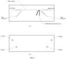
- FIG. 1 The schematic diagram of the microfluidic cell sorting chip for isolating trophoblast cells from cervical exfoliated cells of a pregnant woman is shown in FIG. 1 .
- the structure of the microfluidic cell sorting chip is designed and described below: the chip is prepared by including but not limited to acrylic as a base material; a pipe shape of FIG. 1 was formed on one side of the base material via the injection molding technology, with the pipe width not greater than 1000 ⁇ m, and depth not greater than 500 ⁇ m, and another side of the base material is used for fitting to form a complete chip.
- the microfluidic cell sorting chip includes a substrate and a cover plate fitted therewith.
- one side of the substrate is provided with a main channel, a side channel A and a side channel B, and the two side channels are respectively close to left and right end portions of the main channel; and all the channels have a width of 1000 ⁇ m.
- Another side of the substrate is provided with an inlet C (a liquid inlet for cell samples), an inlet S (a liquid inlet for buffer solution), an outlet N (a liquid storage hole for non-target cells) and an outlet T (a liquid storage hole for target cells); all the two inlets and the two outlets penetrate the substrate to communicate with the channels; and a position of the inlet C corresponds to the left end portion of the main channel; a position of the inlet S corresponds to the end portion of the side channel A; a position of the outlet N corresponds to the right end portion of the main channel; and a position of the outlet T corresponds to the end portion of the side channel B.
- inlet C a liquid inlet for cell samples
- an inlet S a liquid inlet for buffer solution
- an outlet N a liquid storage hole for non-target cells
- an outlet T a liquid storage hole for target cells
- the inlet C is used for feeding the mixed cell samples to be sorted; the inlet S is used for feeding the buffer solution; the outlet T is used for collecting the target cells, and the outlet N is used for collecting the non-target cells.
- the main channel is further provided with a deflection electrode device for cell sorting; the deflection electrode device is specifically located at the intersection of the outlet N and the outlet T; the on/off of the electrode may be controlled according to the presence of a flow cell signal.
- Negatively-charged cells are subjected to deflection in the electromagnetic field formed by electrodes to flow into a designated pipe leading to the outlet N or the outlet T.
- the mixed sample containing target cells flows into the main channel of the chip from the inlet C, and the buffer solution flows into the side channel from the inlet S; the two are mixed at the intersection of the channels and then continuously flow along the same direction of the main channel.
- the channel into which the cells are about to flow is selected by controlling the on/off of the electrode; the target cells are sorted to reach the outlet T, while the non-target cells continue to reach the outlet N along with the main channel, thus completing the sorting.
- the chip is immediately discarded, and a new chip needs to be exchanged before each sorting to ensure a clean, controllable and cross contamination-free sorting environment.
- Example 2 Isolation of trophoblast cells from cervical exfoliated cells of a pregnant woman based on the microfluidic cell sorting chip
- Table 1 Combinations of antigens and antibodies expressed on the trophoblast cells Antibody combinations HLA-G+CK7, HLA-G+CK18, HLA-G+P-HCG, CD31+HPL, MMP9+CD31, HLA-G+HPL, HLA-G+MMP9, HLA-G+CD31, HLA-G+P, CD31+P, HLA-G+CDH5, CD31+CDH5, CD31+CK7+HLA-G, HLA-G+CK18+CD31, HLA-G+P-HCG+CD31, CD31+HPL+HLA-G, MMP9+CD31+HLA-G, CD31+P+HLA-G, HLA-G+CDH5+CD31
- the information of the corresponding antigens is shown below: Specific antigens on the surface of trophoblast cells Corresponding receptors Available monoclonal antibodies HLA-G (human leucocyte antigen G) LIR
- the sorting method of the trophoblast cells includes the following steps of:
- Example 3 Method for isolating trophoblast cells from cervical exfoliated cells of a pregnant woman based on a flow cytometer
- a method for sorting trophoblast cells includes the following steps:
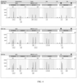
- FIG. 2 shows a sorting result of mono-fluorescence labeling
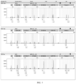
- FIG. 3 shows a sorting result of bifluorescence labeling.
- Trophoblast cells were sorted and isolated from a sample (cervical exfoliated cells) according to the method of the above Example 2. 2. The isolated trophoblast cells and cervical exfoliated cells of a pregnant woman were subjected to DNA extraction:
- Example 2 for Medical Device of the People's Republic of China: 20153401698) according to the method of Example 2; specifically, 9 mutation sites (mtDNA1494, mtDNA1555, SLC26A4-IVS7(-2), SLC26A4-2168, GJB2-35, GJB2-176, GJB2-235, GJB2-299 and GJB3-538) of the epicophosis-related genes (GJB2, GJB3, SLC26A4 and mtDNA) were subjected to testing.
- 9 mutation sites mtDNA1494, mtDNA1555, SLC26A4-IVS7(-2), SLC26A4-2168, GJB2-35, GJB2-176, GJB2-235, GJB2-299 and GJB3-538
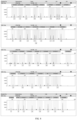
- the DNA extraction method of the cervical exfoliated cells of a pregnant woman in Example 4 was used, and then a commercial kit (epicophosis-susceptible gene kit (PCR+flow-through hybridization), Chaozhou Hybribio Biochemistry Co., Ltd., Registration Certificate No. for Medical Device of the People's Republic of China: 20153401698) was used for subsequent testing. Specific operations are specifically shown in the instructions. Raw data are shown in FIG. 9 ; and result analysis are shown in Table 3.
- Cervical exfoliated cells of a pregnant woman were subjected to epicophosis-susceptible gene testing with a commercial kit ( ⁇ - and ⁇ -thalassemia gene kit (PCR+ membrane hybridization method), Chaozhou Hybribio Biochemistry Co., Ltd., SFDA Certified No.: 3400399, 2012) according to the method of Example 2; specifically, 3 common ⁇ -thalassemia deletion types (-- SEA , - ⁇ 3.7 , - ⁇ 4.2 ), 2 ⁇ -thalassemia mutant types (CS and QS) and 11 ⁇ -thalassemia mutant types (CD14-15, CD17, CD27-28, CD41-42, CD43, CD71-72, -28, -29, IVS-I-1, IVS-II-654 and ⁇ EN) were subjected to testing.
- ⁇ - and ⁇ -thalassemia gene kit PCR+ membrane hybridization method
- CS and QS 2 ⁇ -thalassemia mutant types
- the DNA extraction method of the cervical exfoliated cells of a pregnant woman in Example 4 was used, and then a commercial kit ( ⁇ - and ⁇ -thalassemia gene kit (PCR+ membrane hybridization method), Chaozhou Hybribio Biochemistry Co., Ltd., SFDA Certified No.: 3400399, 2012) was used for subsequent testing. Specific operations are specifically shown in the instructions. Raw data are shown in FIG. 10 ; and result analysis is shown in Table 4.
- One abnormity is detected in the sample, namely, 22q11.21 deletion; the start-end positions [UCSC hg19] of the abnormal fragment are arr22q11.21 (19006943_21461068)x1 with a size of 2.454 Mb.
- Related disease area is Pathogenic (pathogenicity, ACMG classification); and such abnormal fragment covers 106 ISCA genes, such as TBX1, CRKL, GP1BB, SLC25A1, DGCR10, TSSK1A, GSC2 and CLTCL1.
- the regional anomaly is located at 22q11.2 recurrent (DGS/VCFS) region (includes TBX1).
- the deletion of 22q11.2 proximal (A-D) region is related to the DiGeorge / Velocardioffacial (DGS/VCFS) syndrome which is generally clinically featured by congenital heart disease, heart abnormity, characteristic facial features, DD/ID, behavior disorders, immune deficiency and hypocalcemia (PMID 25217958).
- the regional anomaly is located at 22q11.2 recurrent region (central, B/C-D) (includes CRKL).
- the clinical phenotypes possibly caused by the region deletion includes: deformed facial features, growth restriction/short stature, central nervous system abnormity/attack, developmental retardation, dysgnosia, skeletal anomalies, cardiovascular defects, urogenital system anomalies, immune deficiency/repeated infection (PMID 25123976).
- the results indicate that the sorted cell specimen can be effectively subjected to chromosome structure variation detection and corresponding mutations can be detected.
- Example 9 Comparison between the method for isolating trophoblast cells from cervical exfoliated cells of a pregnant woman based on the microfluidic cell sorting chip and the method of immunomagnetic beads
- the method of the present invention was the same as that in Example 2.
- the process for sorting trophoblast cells by the immunomagnetic beads includes the following steps:
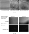
- FIG. 14 is a picture showing positive cells sorted by the method of the present invention
- FIG. 15 is a picture showing comparison of positive cells and negative cells before and after being sorted by the method of the present invention
- FIG. 16 is a picture showing positive cells sorted by the immunomagnetic beads. It can be obviously seen from the comparison that there is an obvious difference in the order of magnitudes of the cell population after being sorted by the two methods; the method of the present invention is significantly superior to the immunomagnetic beads.
- Statistics show that the number of positive cells sorted by the method of the present invention may be up to about 3000-13,000.
Landscapes
- Health & Medical Sciences (AREA)
- Life Sciences & Earth Sciences (AREA)
- Chemical & Material Sciences (AREA)
- Engineering & Computer Science (AREA)
- Organic Chemistry (AREA)
- Immunology (AREA)
- Biomedical Technology (AREA)
- General Health & Medical Sciences (AREA)
- Biochemistry (AREA)
- Genetics & Genomics (AREA)
- Biotechnology (AREA)
- Wood Science & Technology (AREA)
- Zoology (AREA)
- Molecular Biology (AREA)
- Bioinformatics & Cheminformatics (AREA)
- Microbiology (AREA)
- Reproductive Health (AREA)
- Developmental Biology & Embryology (AREA)
- Gynecology & Obstetrics (AREA)
- Cell Biology (AREA)
- Proteomics, Peptides & Aminoacids (AREA)
- Medicinal Chemistry (AREA)
- Physics & Mathematics (AREA)
- Analytical Chemistry (AREA)
- General Engineering & Computer Science (AREA)
- Biophysics (AREA)
- Pathology (AREA)
- General Physics & Mathematics (AREA)
- Hematology (AREA)
- Urology & Nephrology (AREA)
- Pregnancy & Childbirth (AREA)
- Dispersion Chemistry (AREA)
- Food Science & Technology (AREA)
- Tropical Medicine & Parasitology (AREA)
- Endocrinology (AREA)
- Measuring Or Testing Involving Enzymes Or Micro-Organisms (AREA)
- Micro-Organisms Or Cultivation Processes Thereof (AREA)
- Medicines Containing Material From Animals Or Micro-Organisms (AREA)
Claims (3)
- Verfahren zum Isolieren von Trophoblastenzellen, dadurch gekennzeichnet, dass das Verfahren die folgenden Schritte umfasst:Schritt (1) Vorbereiten einer Probenzellsuspension aus einer Lösung, die zervikale exfolierte Zellen enthält, wobei die Zellen in 1xPBS suspendiert sind, das 0,2% - 0,4% FBS enthält;Schritt (2) Zugeben eines spezifischen Antikörpers zu der Probenzellsuspension zur Inkubation; ein primärer Antikörper wird unter den folgenden Bedingungen inkubiert: Reagieren für 30-90 min bei 4°C; und ein zweiter Antikörper-Fluoreszenz-Markierungskomplex wird unter den folgenden Bedingungen inkubiert: Reagieren für 20 min bei 2°C - 8°C;wobei der spezifische Antikörper eine Antikörperkombination ist, die spezifisch ein Antigen erkennt, das auf der Oberfläche oder im Inneren von entsprechenden Trophoblastenzellen exprimiert wird, d.h. eine spezifische Antikörperkombination ist HLA-G+CK7, HLA-G+CK18, HLA-G+β-HCG, CD31+HPL, MMP9+CD31, HLA-G+HPL, HLA-G+MMP9, HLA-G+CD31, HLA-G+P, CD31+P, HLA-G+CDH5, CD31+CDH5, CD31+CK7+HLA-G, HLA-G+CK18+CD31, HLA-G+ß-HCG+CD31, CD31+HPL+HLA-G, MMP9+CD31+HLA-G, CD31+P+HLA-G oder HLA-G+CDH5+CD31; undSchritt (3) Durchführen von Fluoreszenzmarkierung und mikrofluidischer Zellsortierung einer Zellresuspension, die in Schritt (2) inkubiert wurde, durch einen mikrofluidischen Zellsortierungschip, um isolierte und gereinigte plazentare Trophoblastenzellen zu erhalten;wobei ein Flüssigphasen-Zellsortierungssystem 0,2% - 0,4% Triton-X-100 ist;der mikrofluidische Zellsortierungschip ein Substrat und eine damit ausgestattete Abdeckplatte umfasst;wobei eine Seite des Substrats mit einem Hauptkanal, einem Nebenkanal A und einem Nebenkanal B versehen ist und die beiden Nebenkanäle jeweils nahe linken und rechten Endabschnitten des Hauptkanals sind;und wobei eine andere Seite des Substrats mit einem Einlass C, einem Einlass S, einem Auslass N und einem Auslass T versehen ist; alle der beiden Einlässe und der beiden Auslässe das Substrat durchdringen, um mit den Kanälen auf der anderen Seite in Verbindung zu stehen; und eine Position des Einlasses C dem linken Endabschnitt des Hauptkanals entspricht; eine Position des Einlasses S dem Endabschnitt des Nebenkanals A entspricht; eine Position des Auslasses N dem rechten Endabschnitt des Hauptkanals entspricht; und eine Position des Auslasses T dem Endabschnitt des Nebenkanals B entspricht;und eine Ablenkelektrodenvorrichtung ferner in dem Hauptkanal und an einer Konvergenzstelle des Auslasses N und des Auslasses T angeordnet ist; undjeder des Hauptkanals, des Nebenkanals A und des Nebenkanals B eine Breite von nicht mehr als 1000 µm und eine Tiefe von nicht mehr als 500 µm aufweist.
- Verfahren nach Anspruch 1, wobei der Schritt (2) insbesondere umfasst: sukzessives und spezifisches Binden des primären Antikörpers und des zweiten Antikörper-Fluoreszenz-Markierungskomplexes an ein Zielantigen Schritt für Schritt durch Inkubation, wobei eine Wasch- und Zentrifugaltrenntechnologie verwendet wird, um Kreuzkontamination während des Bindungsprozesses zu vermeiden.
- Verfahren nach Anspruch 1, wobei der Schritt (3) insbesondere umfasst: Einspeisen der inkubierten Zellresuspension in den Einlass C des mikrofluidischen Zellsortierungschips, Einspeisen einer Pufferlösung in den Einlass S, dann Platzieren des mikrofluidischen Zellsortierungschips in einem Zellsortierer, um das Sortierungsprogramm durchzuführen, und Sammeln von Proben am Auslass T am Ende des Sortierungsprogramms, um sortierte Trophoblastenzellen zu erhalten.
Applications Claiming Priority (2)
| Application Number | Priority Date | Filing Date | Title |
|---|---|---|---|
| CN202110549947.8A CN112980779B (zh) | 2021-05-20 | 2021-05-20 | 一种从孕妇宫颈脱落细胞中分离胎盘滋养层细胞的方法 |
| PCT/CN2022/081234 WO2022242285A1 (zh) | 2021-05-20 | 2022-03-16 | 一种从孕妇宫颈脱落细胞中分离胎盘滋养层细胞的方法 |
Publications (4)
| Publication Number | Publication Date |
|---|---|
| EP4144834A1 EP4144834A1 (de) | 2023-03-08 |
| EP4144834A4 EP4144834A4 (de) | 2023-09-27 |
| EP4144834B1 true EP4144834B1 (de) | 2024-10-23 |
| EP4144834B9 EP4144834B9 (de) | 2024-12-18 |
Family
ID=76337075
Family Applications (1)
| Application Number | Title | Priority Date | Filing Date |
|---|---|---|---|
| EP22803614.1A Active EP4144834B9 (de) | 2021-05-20 | 2022-03-16 | Verfahren zur trennung plazentaler trophoblastzellen von exfolierten zervikalzellen schwangeren frauen |
Country Status (11)
| Country | Link |
|---|---|
| US (1) | US11796443B2 (de) |
| EP (1) | EP4144834B9 (de) |
| JP (1) | JP7368642B2 (de) |
| KR (1) | KR102626620B1 (de) |
| CN (1) | CN112980779B (de) |
| AU (1) | AU2022277079B2 (de) |
| BR (1) | BR112022024743B1 (de) |
| ES (1) | ES3007607T3 (de) |
| PL (1) | PL4144834T3 (de) |
| PT (1) | PT4144834T (de) |
| WO (1) | WO2022242285A1 (de) |
Families Citing this family (2)
| Publication number | Priority date | Publication date | Assignee | Title |
|---|---|---|---|---|
| CN112980779B (zh) * | 2021-05-20 | 2021-08-24 | 广州凯普医药科技有限公司 | 一种从孕妇宫颈脱落细胞中分离胎盘滋养层细胞的方法 |
| CN118957027A (zh) * | 2024-10-17 | 2024-11-15 | 广州市刑事科学技术研究所 | 基于流式细胞分选实现男女混合生物检材的分型拆分方法 |
Family Cites Families (31)
| Publication number | Priority date | Publication date | Assignee | Title |
|---|---|---|---|---|
| ATE161960T1 (de) * | 1988-12-06 | 1998-01-15 | Flinders Technologies Pty Ltd | Isolierung von fetalen zellen aus mütterlichem blut zur ausführung von pränataler diagnostik |
| JPH07509136A (ja) * | 1992-07-17 | 1995-10-12 | バイシス,インク. | インサイチュ(in situ)・ハイブリダイゼーションのための母体血液中の胎児細胞の富化と同定 |
| US5714325A (en) * | 1993-09-24 | 1998-02-03 | New England Medical Center Hospitals | Prenatal diagnosis by isolation of fetal granulocytes from maternal blood |
| CA2213620A1 (en) * | 1995-04-07 | 1996-10-10 | The Regents Of The University Of California | Antibodies for the detection of hla-g |
| US20020045196A1 (en) | 2000-05-12 | 2002-04-18 | Walt Mahoney | Methods of isolating trophoblast cells from maternal blood |
| AUPR749901A0 (en) | 2001-09-06 | 2001-09-27 | Monash University | Method of identifying chromosomal abnormalities and prenatal diagnosis |
| US20050181429A1 (en) * | 2003-04-03 | 2005-08-18 | Monaliza Medical Ltd. | Non-invasive prenatal genetic diagnosis using transcervical cells |
| US20040197832A1 (en) | 2003-04-03 | 2004-10-07 | Mor Research Applications Ltd. | Non-invasive prenatal genetic diagnosis using transcervical cells |
| WO2005047532A1 (en) | 2003-11-17 | 2005-05-26 | Gribbles Molecular Science Pty Ltd | Improved method of performing genetic analyses on reproductive tract cell samples |
| US20070224597A1 (en) * | 2006-03-23 | 2007-09-27 | Biocept, Inc. | Isolating fetal trophoblasts |
| DE102007019017A1 (de) | 2007-04-19 | 2009-01-22 | Ldt Laser Display Technology Gmbh | Verfahren und Vorrichtung zum Projizieren eines Bildes auf eine Projektionsfläche |
| ITTO20070307A1 (it) * | 2007-05-04 | 2008-11-05 | Silicon Biosystems Spa | Metodo e dispositivo per la diagnosi prenatale non-invasiva |
| US8674172B2 (en) | 2009-04-14 | 2014-03-18 | Institut Gustave Roussy | Prostate cancer cell lines and their use in screening method |
| US9447467B2 (en) | 2009-04-21 | 2016-09-20 | Genetic Technologies Limited | Methods for obtaining fetal genetic material |
| CN101726585A (zh) * | 2009-11-30 | 2010-06-09 | 宁波普赛微流科技有限公司 | 一种基于微流控芯片的流式细胞仪 |
| CA2874048C (en) | 2012-05-24 | 2021-05-11 | Rarecells | Process for multi-analyses of rare cells extracted or isolated from biological samples through filtration |
| EP2909315B1 (de) | 2012-10-19 | 2019-05-01 | Wayne State University | Erkennung und analyse von fötalen trophoblastenzellen in der gebärmutterhalsschleimhaut zur pränatalen diagnose |
| CN103983794B (zh) * | 2013-02-12 | 2016-06-15 | 李木 | 一种微流控芯片及一种微流控方法 |
| WO2014201138A1 (en) * | 2013-06-11 | 2014-12-18 | Stelling James R | Method for detection of fetal abnormalities |
| AU2017246480B2 (en) | 2016-04-06 | 2023-06-01 | Wayne State University | Isolation and analysis of fetal DNA from extravillous trophoblast cells retrieved from the endocervical canal |
| CN107312747A (zh) * | 2016-12-20 | 2017-11-03 | 华东医药(杭州)基因科技有限公司 | 一种免疫磁珠法分离胎盘滋养层细胞的方法 |
| CN108265051B (zh) * | 2016-12-30 | 2020-08-25 | 张婧 | 用于从母体捕获和分离胎儿细胞的无创方法和试剂盒 |
| WO2019153032A1 (en) | 2018-02-07 | 2019-08-15 | University Of South Australia | Pre-natal cell isolation |
| CN108535228B (zh) * | 2018-04-03 | 2020-12-25 | 德运康明(厦门)生物科技有限公司 | 一种从孕妇外周血中分离游离胎儿细胞的方法 |
| TWI668423B (zh) * | 2018-10-02 | 2019-08-11 | 吳宏偉 | 細胞分選方法及系統 |
| CN110117571B (zh) * | 2019-05-08 | 2023-01-31 | 南方医科大学南方医院 | 无创获取胎儿稀有细胞的试剂盒及其方法 |
| UA128273C2 (uk) * | 2019-06-07 | 2024-05-22 | Арседі Байотек Апс | Спосіб пренатальної діагностики (варанти) |
| CN114430805B (zh) | 2019-07-15 | 2025-10-10 | A.美纳里尼生物标记新加坡有限公司 | 分离、检测和分析胎儿细胞的组合物和方法 |
| CN111304153A (zh) * | 2019-12-17 | 2020-06-19 | 广东凯普生物科技股份有限公司 | 一种分离滋养层细胞的方法 |
| CN111440696B (zh) * | 2020-02-26 | 2023-02-24 | 德运康明(厦门)生物科技有限公司 | 胎儿细胞捕获模块、用于胎儿细胞捕获的微流控芯片,及它们的使用方法 |
| CN112980779B (zh) * | 2021-05-20 | 2021-08-24 | 广州凯普医药科技有限公司 | 一种从孕妇宫颈脱落细胞中分离胎盘滋养层细胞的方法 |
-
2021
- 2021-05-20 CN CN202110549947.8A patent/CN112980779B/zh active Active
-
2022
- 2022-03-16 JP JP2022573784A patent/JP7368642B2/ja active Active
- 2022-03-16 WO PCT/CN2022/081234 patent/WO2022242285A1/zh not_active Ceased
- 2022-03-16 US US18/009,740 patent/US11796443B2/en active Active
- 2022-03-16 BR BR112022024743-5A patent/BR112022024743B1/pt active IP Right Grant
- 2022-03-16 EP EP22803614.1A patent/EP4144834B9/de active Active
- 2022-03-16 PT PT228036141T patent/PT4144834T/pt unknown
- 2022-03-16 PL PL22803614.1T patent/PL4144834T3/pl unknown
- 2022-03-16 ES ES22803614T patent/ES3007607T3/es active Active
- 2022-03-16 KR KR1020227043777A patent/KR102626620B1/ko active Active
- 2022-03-16 AU AU2022277079A patent/AU2022277079B2/en active Active
Also Published As
| Publication number | Publication date |
|---|---|
| BR112022024743A2 (de) | 2023-06-27 |
| EP4144834A1 (de) | 2023-03-08 |
| ES3007607T3 (en) | 2025-03-20 |
| PL4144834T3 (pl) | 2025-02-24 |
| US20230228667A1 (en) | 2023-07-20 |
| AU2022277079A1 (en) | 2023-01-19 |
| KR102626620B1 (ko) | 2024-01-17 |
| WO2022242285A1 (zh) | 2022-11-24 |
| JP7368642B2 (ja) | 2023-10-24 |
| CN112980779A (zh) | 2021-06-18 |
| PT4144834T (pt) | 2024-12-26 |
| BR112022024743B1 (pt) | 2024-03-05 |
| EP4144834B9 (de) | 2024-12-18 |
| KR20230004915A (ko) | 2023-01-06 |
| JP2023527246A (ja) | 2023-06-27 |
| EP4144834A4 (de) | 2023-09-27 |
| US11796443B2 (en) | 2023-10-24 |
| AU2022277079B2 (en) | 2024-07-04 |
| CN112980779B (zh) | 2021-08-24 |
Similar Documents
| Publication | Publication Date | Title |
|---|---|---|
| Bianchi | Fetal cells in the maternal circulation: feasibility for prenatal diagnosis. | |
| US7732143B2 (en) | Method for the diagnosis of genetic diseases by molecular combing and diagnostic kit | |
| Vona et al. | Enrichment, immunomorphological, and genetic characterization of fetal cells circulating in maternal blood | |
| US20230092810A1 (en) | Fetal cell capture module and microfluidic chip for fetal cell capture and methods for using the same | |
| Hahn et al. | Prenatal diagnosis using fetal cells and cell-free fetal DNA in maternal blood: what is currently feasible? | |
| CA2459725A1 (en) | Method of isolating cells and uses thereof | |
| EP4144834B9 (de) | Verfahren zur trennung plazentaler trophoblastzellen von exfolierten zervikalzellen schwangeren frauen | |
| Hyland et al. | Non-invasive prenatal testing for management of haemolytic disease of the fetus and newborn induced by maternal alloimmunisation | |
| EP2152905B1 (de) | Verfahren und kits zum nachweis fötaler zellen in mütterlichem blut | |
| EP2207879A2 (de) | Identifizierung und isolierung fötaler zellen und nukleinsäure | |
| CN107236809A (zh) | 一种基于母体的胎儿外膜滋养层细胞的胎儿遗传检测方法 | |
| Sekizawa et al. | Fetal cell recycling: diagnosis of gender and RhD genotype in the same fetal cell retrieved from maternal blood | |
| WO2005047532A1 (en) | Improved method of performing genetic analyses on reproductive tract cell samples | |
| CN111781360A (zh) | 游离细胞捕获探针及其相关产品和用途 | |
| KR20150127917A (ko) | 자성입자를 이용한 핵산의 추출 및 증폭 방법 | |
| HK40090275B (en) | Method for isolating placental trophoblast cells from cervical exfoliated cells of pregnant woman | |
| HK40090275A (en) | Method for isolating placental trophoblast cells from cervical exfoliated cells of pregnant woman | |
| Lipinski et al. | Phenotypic characterization of Ewing sarcoma cell lines with monoclonal antibodies | |
| RU2808260C1 (ru) | Способ отделения клеток плацентарного трофобласта от отслоившихся клеток шейки матки беременных женщин | |
| US8206935B2 (en) | Method of rapidly quantifying hydroxymethylated DNA | |
| Bianchi | Progress in the genetic analysis of fetal cells circulating in maternal blood | |
| Sekizawa et al. | Prenatal screening of single-gene disorders from maternal blood | |
| Chiu et al. | Non-invasive prenatal diagnosis: on the horizon? | |
| US20160090631A1 (en) | Method for detection of fetal abnormalities | |
| CN118562949A (zh) | 一种用于辅助诊断mrkh综合征的标记物及试剂盒 |
Legal Events
| Date | Code | Title | Description |
|---|---|---|---|
| STAA | Information on the status of an ep patent application or granted ep patent |
Free format text: STATUS: THE INTERNATIONAL PUBLICATION HAS BEEN MADE |
|
| PUAI | Public reference made under article 153(3) epc to a published international application that has entered the european phase |
Free format text: ORIGINAL CODE: 0009012 |
|
| STAA | Information on the status of an ep patent application or granted ep patent |
Free format text: STATUS: REQUEST FOR EXAMINATION WAS MADE |
|
| 17P | Request for examination filed |
Effective date: 20221128 |
|
| AK | Designated contracting states |
Kind code of ref document: A1 Designated state(s): AL AT BE BG CH CY CZ DE DK EE ES FI FR GB GR HR HU IE IS IT LI LT LU LV MC MK MT NL NO PL PT RO RS SE SI SK SM TR |
|
| A4 | Supplementary search report drawn up and despatched |
Effective date: 20230825 |
|
| RIC1 | Information provided on ipc code assigned before grant |
Ipc: G01N 15/14 20060101ALI20230821BHEP Ipc: C12N 5/073 20100101AFI20230821BHEP |
|
| REG | Reference to a national code |
Ref country code: HK Ref legal event code: DE Ref document number: 40090275 Country of ref document: HK |
|
| STAA | Information on the status of an ep patent application or granted ep patent |
Free format text: STATUS: EXAMINATION IS IN PROGRESS |
|
| 17Q | First examination report despatched |
Effective date: 20240212 |
|
| GRAP | Despatch of communication of intention to grant a patent |
Free format text: ORIGINAL CODE: EPIDOSNIGR1 |
|
| STAA | Information on the status of an ep patent application or granted ep patent |
Free format text: STATUS: GRANT OF PATENT IS INTENDED |
|
| INTG | Intention to grant announced |
Effective date: 20240627 |
|
| DAV | Request for validation of the european patent (deleted) | ||
| DAX | Request for extension of the european patent (deleted) | ||
| GRAS | Grant fee paid |
Free format text: ORIGINAL CODE: EPIDOSNIGR3 |
|
| GRAA | (expected) grant |
Free format text: ORIGINAL CODE: 0009210 |
|
| STAA | Information on the status of an ep patent application or granted ep patent |
Free format text: STATUS: THE PATENT HAS BEEN GRANTED |
|
| AK | Designated contracting states |
Kind code of ref document: B1 Designated state(s): AL AT BE BG CH CY CZ DE DK EE ES FI FR GB GR HR HU IE IS IT LI LT LU LV MC MK MT NL NO PL PT RO RS SE SI SK SM TR |
|
| REG | Reference to a national code |
Ref country code: GB Ref legal event code: FG4D |
|
| REG | Reference to a national code |
Ref country code: CH Ref legal event code: EP |
|
| REG | Reference to a national code |
Ref country code: DE Ref legal event code: R096 Ref document number: 602022007107 Country of ref document: DE |
|
| REG | Reference to a national code |
Ref country code: IE Ref legal event code: FG4D |
|
| REG | Reference to a national code |
Ref country code: CH Ref legal event code: PK Free format text: BERICHTIGUNG B9 |
|
| REG | Reference to a national code |
Ref country code: PT Ref legal event code: SC4A Ref document number: 4144834 Country of ref document: PT Date of ref document: 20241226 Kind code of ref document: T Free format text: AVAILABILITY OF NATIONAL TRANSLATION Effective date: 20241218 |
|
| P01 | Opt-out of the competence of the unified patent court (upc) registered |
Free format text: CASE NUMBER: APP_63238/2024 Effective date: 20241128 |
|
| REG | Reference to a national code |
Ref country code: NL Ref legal event code: FP |
|
| REG | Reference to a national code |
Ref country code: SE Ref legal event code: TRGR |
|
| REG | Reference to a national code |
Ref country code: LT Ref legal event code: MG9D |
|
| REG | Reference to a national code |
Ref country code: AT Ref legal event code: MK05 Ref document number: 1734854 Country of ref document: AT Kind code of ref document: T Effective date: 20241023 |
|
| REG | Reference to a national code |
Ref country code: ES Ref legal event code: FG2A Ref document number: 3007607 Country of ref document: ES Kind code of ref document: T3 Effective date: 20250320 |
|
| PG25 | Lapsed in a contracting state [announced via postgrant information from national office to epo] |
Ref country code: HR Free format text: LAPSE BECAUSE OF FAILURE TO SUBMIT A TRANSLATION OF THE DESCRIPTION OR TO PAY THE FEE WITHIN THE PRESCRIBED TIME-LIMIT Effective date: 20241023 Ref country code: IS Free format text: LAPSE BECAUSE OF FAILURE TO SUBMIT A TRANSLATION OF THE DESCRIPTION OR TO PAY THE FEE WITHIN THE PRESCRIBED TIME-LIMIT Effective date: 20250223 |
|
| PG25 | Lapsed in a contracting state [announced via postgrant information from national office to epo] |
Ref country code: FI Free format text: LAPSE BECAUSE OF FAILURE TO SUBMIT A TRANSLATION OF THE DESCRIPTION OR TO PAY THE FEE WITHIN THE PRESCRIBED TIME-LIMIT Effective date: 20241023 |
|
| PG25 | Lapsed in a contracting state [announced via postgrant information from national office to epo] |
Ref country code: BG Free format text: LAPSE BECAUSE OF FAILURE TO SUBMIT A TRANSLATION OF THE DESCRIPTION OR TO PAY THE FEE WITHIN THE PRESCRIBED TIME-LIMIT Effective date: 20241023 |
|
| PG25 | Lapsed in a contracting state [announced via postgrant information from national office to epo] |
Ref country code: NO Free format text: LAPSE BECAUSE OF FAILURE TO SUBMIT A TRANSLATION OF THE DESCRIPTION OR TO PAY THE FEE WITHIN THE PRESCRIBED TIME-LIMIT Effective date: 20250123 |
|
| PG25 | Lapsed in a contracting state [announced via postgrant information from national office to epo] |
Ref country code: LV Free format text: LAPSE BECAUSE OF FAILURE TO SUBMIT A TRANSLATION OF THE DESCRIPTION OR TO PAY THE FEE WITHIN THE PRESCRIBED TIME-LIMIT Effective date: 20241023 Ref country code: AT Free format text: LAPSE BECAUSE OF FAILURE TO SUBMIT A TRANSLATION OF THE DESCRIPTION OR TO PAY THE FEE WITHIN THE PRESCRIBED TIME-LIMIT Effective date: 20241023 Ref country code: GR Free format text: LAPSE BECAUSE OF FAILURE TO SUBMIT A TRANSLATION OF THE DESCRIPTION OR TO PAY THE FEE WITHIN THE PRESCRIBED TIME-LIMIT Effective date: 20250124 |
|
| PG25 | Lapsed in a contracting state [announced via postgrant information from national office to epo] |
Ref country code: RS Free format text: LAPSE BECAUSE OF FAILURE TO SUBMIT A TRANSLATION OF THE DESCRIPTION OR TO PAY THE FEE WITHIN THE PRESCRIBED TIME-LIMIT Effective date: 20250123 |
|
| PG25 | Lapsed in a contracting state [announced via postgrant information from national office to epo] |
Ref country code: SM Free format text: LAPSE BECAUSE OF FAILURE TO SUBMIT A TRANSLATION OF THE DESCRIPTION OR TO PAY THE FEE WITHIN THE PRESCRIBED TIME-LIMIT Effective date: 20241023 |
|
| PG25 | Lapsed in a contracting state [announced via postgrant information from national office to epo] |
Ref country code: DK Free format text: LAPSE BECAUSE OF FAILURE TO SUBMIT A TRANSLATION OF THE DESCRIPTION OR TO PAY THE FEE WITHIN THE PRESCRIBED TIME-LIMIT Effective date: 20241023 |
|
| PGFP | Annual fee paid to national office [announced via postgrant information from national office to epo] |
Ref country code: ES Payment date: 20250416 Year of fee payment: 4 |
|
| PGFP | Annual fee paid to national office [announced via postgrant information from national office to epo] |
Ref country code: IT Payment date: 20250331 Year of fee payment: 4 |
|
| PG25 | Lapsed in a contracting state [announced via postgrant information from national office to epo] |
Ref country code: EE Free format text: LAPSE BECAUSE OF FAILURE TO SUBMIT A TRANSLATION OF THE DESCRIPTION OR TO PAY THE FEE WITHIN THE PRESCRIBED TIME-LIMIT Effective date: 20241023 |
|
| PGFP | Annual fee paid to national office [announced via postgrant information from national office to epo] |
Ref country code: CH Payment date: 20250401 Year of fee payment: 4 |
|
| REG | Reference to a national code |
Ref country code: DE Ref legal event code: R097 Ref document number: 602022007107 Country of ref document: DE |
|
| PG25 | Lapsed in a contracting state [announced via postgrant information from national office to epo] |
Ref country code: SK Free format text: LAPSE BECAUSE OF FAILURE TO SUBMIT A TRANSLATION OF THE DESCRIPTION OR TO PAY THE FEE WITHIN THE PRESCRIBED TIME-LIMIT Effective date: 20241023 |
|
| PG25 | Lapsed in a contracting state [announced via postgrant information from national office to epo] |
Ref country code: CZ Free format text: LAPSE BECAUSE OF FAILURE TO SUBMIT A TRANSLATION OF THE DESCRIPTION OR TO PAY THE FEE WITHIN THE PRESCRIBED TIME-LIMIT Effective date: 20241023 |
|
| PLBE | No opposition filed within time limit |
Free format text: ORIGINAL CODE: 0009261 |
|
| STAA | Information on the status of an ep patent application or granted ep patent |
Free format text: STATUS: NO OPPOSITION FILED WITHIN TIME LIMIT |
|
| 26N | No opposition filed |
Effective date: 20250724 |
|
| REG | Reference to a national code |
Ref country code: BE Ref legal event code: MM Effective date: 20250331 |
|
| PG25 | Lapsed in a contracting state [announced via postgrant information from national office to epo] |
Ref country code: PT Free format text: LAPSE BECAUSE OF NON-PAYMENT OF DUE FEES Effective date: 20251216 |
|
| PG25 | Lapsed in a contracting state [announced via postgrant information from national office to epo] |
Ref country code: BE Free format text: LAPSE BECAUSE OF NON-PAYMENT OF DUE FEES Effective date: 20250331 |
|
| REG | Reference to a national code |
Ref country code: CH Ref legal event code: U11 Free format text: ST27 STATUS EVENT CODE: U-0-0-U10-U11 (AS PROVIDED BY THE NATIONAL OFFICE) Effective date: 20260401 |
|
| PGFP | Annual fee paid to national office [announced via postgrant information from national office to epo] |
Ref country code: SE Payment date: 20260323 Year of fee payment: 5 |
|
| PGFP | Annual fee paid to national office [announced via postgrant information from national office to epo] |
Ref country code: GB Payment date: 20260324 Year of fee payment: 5 |
|
| PGFP | Annual fee paid to national office [announced via postgrant information from national office to epo] |
Ref country code: MC Payment date: 20260320 Year of fee payment: 5 |
|
| PGFP | Annual fee paid to national office [announced via postgrant information from national office to epo] |
Ref country code: IE Payment date: 20260323 Year of fee payment: 5 Ref country code: DE Payment date: 20260320 Year of fee payment: 5 |
|
| PGFP | Annual fee paid to national office [announced via postgrant information from national office to epo] |
Ref country code: LU Payment date: 20260320 Year of fee payment: 5 Ref country code: RO Payment date: 20260310 Year of fee payment: 5 |
|
| PGFP | Annual fee paid to national office [announced via postgrant information from national office to epo] |
Ref country code: NL Payment date: 20260323 Year of fee payment: 5 |
|
| PGFP | Annual fee paid to national office [announced via postgrant information from national office to epo] |
Ref country code: FR Payment date: 20260324 Year of fee payment: 5 |
|
| PGFP | Annual fee paid to national office [announced via postgrant information from national office to epo] |
Ref country code: PT Payment date: 20260302 Year of fee payment: 5 |
|
| PGFP | Annual fee paid to national office [announced via postgrant information from national office to epo] |
Ref country code: PL Payment date: 20260309 Year of fee payment: 5 |