EP4088744A1 - Détection et traitement de caries et de microcavités avec des nanoparticules - Google Patents
Détection et traitement de caries et de microcavités avec des nanoparticules Download PDFInfo
- Publication number
- EP4088744A1 EP4088744A1 EP22183087.0A EP22183087A EP4088744A1 EP 4088744 A1 EP4088744 A1 EP 4088744A1 EP 22183087 A EP22183087 A EP 22183087A EP 4088744 A1 EP4088744 A1 EP 4088744A1
- Authority
- EP
- European Patent Office
- Prior art keywords
- nanoparticle
- nanoparticles
- equal
- cationic
- composition
- Prior art date
- Legal status (The legal status is an assumption and is not a legal conclusion. Google has not performed a legal analysis and makes no representation as to the accuracy of the status listed.)
- Pending
Links
Images


Classifications
-
- A—HUMAN NECESSITIES
- A61—MEDICAL OR VETERINARY SCIENCE; HYGIENE
- A61K—PREPARATIONS FOR MEDICAL, DENTAL OR TOILETRY PURPOSES
- A61K49/00—Preparations for testing in vivo
- A61K49/06—Nuclear magnetic resonance [NMR] contrast preparations; Magnetic resonance imaging [MRI] contrast preparations
- A61K49/18—Nuclear magnetic resonance [NMR] contrast preparations; Magnetic resonance imaging [MRI] contrast preparations characterised by a special physical form, e.g. emulsions, microcapsules, liposomes
- A61K49/1818—Nuclear magnetic resonance [NMR] contrast preparations; Magnetic resonance imaging [MRI] contrast preparations characterised by a special physical form, e.g. emulsions, microcapsules, liposomes particles, e.g. uncoated or non-functionalised microparticles or nanoparticles
-
- A—HUMAN NECESSITIES
- A61—MEDICAL OR VETERINARY SCIENCE; HYGIENE
- A61K—PREPARATIONS FOR MEDICAL, DENTAL OR TOILETRY PURPOSES
- A61K49/00—Preparations for testing in vivo
- A61K49/001—Preparation for luminescence or biological staining
- A61K49/0013—Luminescence
- A61K49/0017—Fluorescence in vivo
- A61K49/005—Fluorescence in vivo characterised by the carrier molecule carrying the fluorescent agent
- A61K49/0054—Macromolecular compounds, i.e. oligomers, polymers, dendrimers
-
- A—HUMAN NECESSITIES
- A61—MEDICAL OR VETERINARY SCIENCE; HYGIENE
- A61B—DIAGNOSIS; SURGERY; IDENTIFICATION
- A61B5/00—Measuring for diagnostic purposes; Identification of persons
- A61B5/0059—Measuring for diagnostic purposes; Identification of persons using light, e.g. diagnosis by transillumination, diascopy, fluorescence
- A61B5/0082—Measuring for diagnostic purposes; Identification of persons using light, e.g. diagnosis by transillumination, diascopy, fluorescence adapted for particular medical purposes
- A61B5/0088—Measuring for diagnostic purposes; Identification of persons using light, e.g. diagnosis by transillumination, diascopy, fluorescence adapted for particular medical purposes for oral or dental tissue
-
- A—HUMAN NECESSITIES
- A61—MEDICAL OR VETERINARY SCIENCE; HYGIENE
- A61K—PREPARATIONS FOR MEDICAL, DENTAL OR TOILETRY PURPOSES
- A61K49/00—Preparations for testing in vivo
- A61K49/001—Preparation for luminescence or biological staining
- A61K49/0013—Luminescence
- A61K49/0017—Fluorescence in vivo
- A61K49/0019—Fluorescence in vivo characterised by the fluorescent group, e.g. oligomeric, polymeric or dendritic molecules
- A61K49/0021—Fluorescence in vivo characterised by the fluorescent group, e.g. oligomeric, polymeric or dendritic molecules the fluorescent group being a small organic molecule
- A61K49/0041—Xanthene dyes, used in vivo, e.g. administered to a mice, e.g. rhodamines, rose Bengal
- A61K49/0043—Fluorescein, used in vivo
-
- A—HUMAN NECESSITIES
- A61—MEDICAL OR VETERINARY SCIENCE; HYGIENE
- A61K—PREPARATIONS FOR MEDICAL, DENTAL OR TOILETRY PURPOSES
- A61K49/00—Preparations for testing in vivo
- A61K49/001—Preparation for luminescence or biological staining
- A61K49/0063—Preparation for luminescence or biological staining characterised by a special physical or galenical form, e.g. emulsions, microspheres
- A61K49/0069—Preparation for luminescence or biological staining characterised by a special physical or galenical form, e.g. emulsions, microspheres the agent being in a particular physical galenical form
- A61K49/0089—Particulate, powder, adsorbate, bead, sphere
- A61K49/0091—Microparticle, microcapsule, microbubble, microsphere, microbead, i.e. having a size or diameter higher or equal to 1 micrometer
- A61K49/0093—Nanoparticle, nanocapsule, nanobubble, nanosphere, nanobead, i.e. having a size or diameter smaller than 1 micrometer, e.g. polymeric nanoparticle
-
- A—HUMAN NECESSITIES
- A61—MEDICAL OR VETERINARY SCIENCE; HYGIENE
- A61K—PREPARATIONS FOR MEDICAL, DENTAL OR TOILETRY PURPOSES
- A61K49/00—Preparations for testing in vivo
- A61K49/06—Nuclear magnetic resonance [NMR] contrast preparations; Magnetic resonance imaging [MRI] contrast preparations
- A61K49/18—Nuclear magnetic resonance [NMR] contrast preparations; Magnetic resonance imaging [MRI] contrast preparations characterised by a special physical form, e.g. emulsions, microcapsules, liposomes
- A61K49/1818—Nuclear magnetic resonance [NMR] contrast preparations; Magnetic resonance imaging [MRI] contrast preparations characterised by a special physical form, e.g. emulsions, microcapsules, liposomes particles, e.g. uncoated or non-functionalised microparticles or nanoparticles
- A61K49/1821—Nuclear magnetic resonance [NMR] contrast preparations; Magnetic resonance imaging [MRI] contrast preparations characterised by a special physical form, e.g. emulsions, microcapsules, liposomes particles, e.g. uncoated or non-functionalised microparticles or nanoparticles coated or functionalised microparticles or nanoparticles
- A61K49/1824—Nuclear magnetic resonance [NMR] contrast preparations; Magnetic resonance imaging [MRI] contrast preparations characterised by a special physical form, e.g. emulsions, microcapsules, liposomes particles, e.g. uncoated or non-functionalised microparticles or nanoparticles coated or functionalised microparticles or nanoparticles coated or functionalised nanoparticles
- A61K49/1827—Nuclear magnetic resonance [NMR] contrast preparations; Magnetic resonance imaging [MRI] contrast preparations characterised by a special physical form, e.g. emulsions, microcapsules, liposomes particles, e.g. uncoated or non-functionalised microparticles or nanoparticles coated or functionalised microparticles or nanoparticles coated or functionalised nanoparticles having a (super)(para)magnetic core, being a solid MRI-active material, e.g. magnetite, or composed of a plurality of MRI-active, organic agents, e.g. Gd-chelates, or nuclei, e.g. Eu3+, encapsulated or entrapped in the core of the coated or functionalised nanoparticle
- A61K49/1851—Nuclear magnetic resonance [NMR] contrast preparations; Magnetic resonance imaging [MRI] contrast preparations characterised by a special physical form, e.g. emulsions, microcapsules, liposomes particles, e.g. uncoated or non-functionalised microparticles or nanoparticles coated or functionalised microparticles or nanoparticles coated or functionalised nanoparticles having a (super)(para)magnetic core, being a solid MRI-active material, e.g. magnetite, or composed of a plurality of MRI-active, organic agents, e.g. Gd-chelates, or nuclei, e.g. Eu3+, encapsulated or entrapped in the core of the coated or functionalised nanoparticle having a (super)(para)magnetic core coated or functionalised with an organic macromolecular compound, i.e. oligomeric, polymeric, dendrimeric organic molecule
-
- A—HUMAN NECESSITIES
- A61—MEDICAL OR VETERINARY SCIENCE; HYGIENE
- A61K—PREPARATIONS FOR MEDICAL, DENTAL OR TOILETRY PURPOSES
- A61K8/00—Cosmetics or similar toiletry preparations
- A61K8/02—Cosmetics or similar toiletry preparations characterised by special physical form
- A61K8/0241—Containing particulates characterized by their shape and/or structure
-
- A—HUMAN NECESSITIES
- A61—MEDICAL OR VETERINARY SCIENCE; HYGIENE
- A61K—PREPARATIONS FOR MEDICAL, DENTAL OR TOILETRY PURPOSES
- A61K8/00—Cosmetics or similar toiletry preparations
- A61K8/18—Cosmetics or similar toiletry preparations characterised by the composition
- A61K8/19—Cosmetics or similar toiletry preparations characterised by the composition containing inorganic ingredients
- A61K8/20—Halogens; Compounds thereof
- A61K8/21—Fluorides; Derivatives thereof
-
- A—HUMAN NECESSITIES
- A61—MEDICAL OR VETERINARY SCIENCE; HYGIENE
- A61K—PREPARATIONS FOR MEDICAL, DENTAL OR TOILETRY PURPOSES
- A61K8/00—Cosmetics or similar toiletry preparations
- A61K8/18—Cosmetics or similar toiletry preparations characterised by the composition
- A61K8/19—Cosmetics or similar toiletry preparations characterised by the composition containing inorganic ingredients
- A61K8/24—Phosphorous; Compounds thereof
-
- A—HUMAN NECESSITIES
- A61—MEDICAL OR VETERINARY SCIENCE; HYGIENE
- A61K—PREPARATIONS FOR MEDICAL, DENTAL OR TOILETRY PURPOSES
- A61K8/00—Cosmetics or similar toiletry preparations
- A61K8/18—Cosmetics or similar toiletry preparations characterised by the composition
- A61K8/19—Cosmetics or similar toiletry preparations characterised by the composition containing inorganic ingredients
- A61K8/25—Silicon; Compounds thereof
-
- A—HUMAN NECESSITIES
- A61—MEDICAL OR VETERINARY SCIENCE; HYGIENE
- A61K—PREPARATIONS FOR MEDICAL, DENTAL OR TOILETRY PURPOSES
- A61K8/00—Cosmetics or similar toiletry preparations
- A61K8/18—Cosmetics or similar toiletry preparations characterised by the composition
- A61K8/30—Cosmetics or similar toiletry preparations characterised by the composition containing organic compounds
- A61K8/55—Phosphorus compounds
-
- A—HUMAN NECESSITIES
- A61—MEDICAL OR VETERINARY SCIENCE; HYGIENE
- A61K—PREPARATIONS FOR MEDICAL, DENTAL OR TOILETRY PURPOSES
- A61K8/00—Cosmetics or similar toiletry preparations
- A61K8/18—Cosmetics or similar toiletry preparations characterised by the composition
- A61K8/30—Cosmetics or similar toiletry preparations characterised by the composition containing organic compounds
- A61K8/64—Proteins; Peptides; Derivatives or degradation products thereof
-
- A—HUMAN NECESSITIES
- A61—MEDICAL OR VETERINARY SCIENCE; HYGIENE
- A61K—PREPARATIONS FOR MEDICAL, DENTAL OR TOILETRY PURPOSES
- A61K8/00—Cosmetics or similar toiletry preparations
- A61K8/18—Cosmetics or similar toiletry preparations characterised by the composition
- A61K8/72—Cosmetics or similar toiletry preparations characterised by the composition containing organic macromolecular compounds
- A61K8/73—Polysaccharides
- A61K8/732—Starch; Amylose; Amylopectin; Derivatives thereof
-
- A—HUMAN NECESSITIES
- A61—MEDICAL OR VETERINARY SCIENCE; HYGIENE
- A61K—PREPARATIONS FOR MEDICAL, DENTAL OR TOILETRY PURPOSES
- A61K8/00—Cosmetics or similar toiletry preparations
- A61K8/18—Cosmetics or similar toiletry preparations characterised by the composition
- A61K8/72—Cosmetics or similar toiletry preparations characterised by the composition containing organic macromolecular compounds
- A61K8/73—Polysaccharides
- A61K8/736—Chitin; Chitosan; Derivatives thereof
-
- A—HUMAN NECESSITIES
- A61—MEDICAL OR VETERINARY SCIENCE; HYGIENE
- A61P—SPECIFIC THERAPEUTIC ACTIVITY OF CHEMICAL COMPOUNDS OR MEDICINAL PREPARATIONS
- A61P1/00—Drugs for disorders of the alimentary tract or the digestive system
- A61P1/02—Stomatological preparations, e.g. drugs for caries, aphtae, periodontitis
-
- A—HUMAN NECESSITIES
- A61—MEDICAL OR VETERINARY SCIENCE; HYGIENE
- A61Q—SPECIFIC USE OF COSMETICS OR SIMILAR TOILETRY PREPARATIONS
- A61Q11/00—Preparations for care of the teeth, of the oral cavity or of dentures; Dentifrices, e.g. toothpastes; Mouth rinses
-
- A—HUMAN NECESSITIES
- A61—MEDICAL OR VETERINARY SCIENCE; HYGIENE
- A61K—PREPARATIONS FOR MEDICAL, DENTAL OR TOILETRY PURPOSES
- A61K2800/00—Properties of cosmetic compositions or active ingredients thereof or formulation aids used therein and process related aspects
- A61K2800/40—Chemical, physico-chemical or functional or structural properties of particular ingredients
- A61K2800/41—Particular ingredients further characterized by their size
- A61K2800/413—Nanosized, i.e. having sizes below 100 nm
-
- A—HUMAN NECESSITIES
- A61—MEDICAL OR VETERINARY SCIENCE; HYGIENE
- A61K—PREPARATIONS FOR MEDICAL, DENTAL OR TOILETRY PURPOSES
- A61K2800/00—Properties of cosmetic compositions or active ingredients thereof or formulation aids used therein and process related aspects
- A61K2800/40—Chemical, physico-chemical or functional or structural properties of particular ingredients
- A61K2800/42—Colour properties
- A61K2800/43—Pigments; Dyes
- A61K2800/434—Luminescent, Fluorescent; Optical brighteners; Photosensitizers
-
- A—HUMAN NECESSITIES
- A61—MEDICAL OR VETERINARY SCIENCE; HYGIENE
- A61K—PREPARATIONS FOR MEDICAL, DENTAL OR TOILETRY PURPOSES
- A61K2800/00—Properties of cosmetic compositions or active ingredients thereof or formulation aids used therein and process related aspects
- A61K2800/40—Chemical, physico-chemical or functional or structural properties of particular ingredients
- A61K2800/54—Polymers characterized by specific structures/properties
- A61K2800/542—Polymers characterized by specific structures/properties characterized by the charge
- A61K2800/5426—Polymers characterized by specific structures/properties characterized by the charge cationic
-
- A—HUMAN NECESSITIES
- A61—MEDICAL OR VETERINARY SCIENCE; HYGIENE
- A61K—PREPARATIONS FOR MEDICAL, DENTAL OR TOILETRY PURPOSES
- A61K2800/00—Properties of cosmetic compositions or active ingredients thereof or formulation aids used therein and process related aspects
- A61K2800/40—Chemical, physico-chemical or functional or structural properties of particular ingredients
- A61K2800/57—Compounds covalently linked to a(n inert) carrier molecule, e.g. conjugates, pro-fragrances
-
- A—HUMAN NECESSITIES
- A61—MEDICAL OR VETERINARY SCIENCE; HYGIENE
- A61K—PREPARATIONS FOR MEDICAL, DENTAL OR TOILETRY PURPOSES
- A61K2800/00—Properties of cosmetic compositions or active ingredients thereof or formulation aids used therein and process related aspects
- A61K2800/40—Chemical, physico-chemical or functional or structural properties of particular ingredients
- A61K2800/60—Particulates further characterized by their structure or composition
- A61K2800/65—Characterized by the composition of the particulate/core
- A61K2800/654—The particulate/core comprising macromolecular material
-
- A—HUMAN NECESSITIES
- A61—MEDICAL OR VETERINARY SCIENCE; HYGIENE
- A61K—PREPARATIONS FOR MEDICAL, DENTAL OR TOILETRY PURPOSES
- A61K2800/00—Properties of cosmetic compositions or active ingredients thereof or formulation aids used therein and process related aspects
- A61K2800/80—Process related aspects concerning the preparation of the cosmetic composition or the storage or application thereof
- A61K2800/81—Preparation or application process involves irradiation
-
- A—HUMAN NECESSITIES
- A61—MEDICAL OR VETERINARY SCIENCE; HYGIENE
- A61K—PREPARATIONS FOR MEDICAL, DENTAL OR TOILETRY PURPOSES
- A61K2800/00—Properties of cosmetic compositions or active ingredients thereof or formulation aids used therein and process related aspects
- A61K2800/80—Process related aspects concerning the preparation of the cosmetic composition or the storage or application thereof
- A61K2800/92—Oral administration
Definitions
- the present disclosure relates to nanoparticles having diagnostic agents that can be delivered to an oral cavity of a subject to provide detection of caries and/or provide therapeutic treatment of caries, especially active (progressing) carious lesions.
- Dental caries is the most common disease in dental health worldwide. In the United States, more than 90% of adults have experienced dental caries in their permanent teeth. Approximately 36% of the world's population has active caries. Furthermore, with developing countries gaining access to high sugar diets, the incidence of dental caries is likely to increase.
- Dental cavities form when bacteria in the dental biofilm on the surface of teeth ferment sugars and produce acids which demineralize dentin and/or enamel.
- Early decalcification is indicated by white spot lesions forming on the surface enamel. These lesions, also called “microcavities” or incipient carious lesions, are reversible by a process called remineralizaton, which uses calcium and phosphorous in the saliva and is aided by the presence of fluoride in drinking water and toothpaste.
- remineralizaton which uses calcium and phosphorous in the saliva and is aided by the presence of fluoride in drinking water and toothpaste.
- irreversible cavitation occurs requiring a dental procedure to stop the decalcification.
- X-ray images of the teeth are routinely taken to identify cavities, particularly for regions in between teeth (interproximal caries). This method can clearly identify advanced cavity progression for dental treatment, but suffers from several limitations.
- X-ray images are unable to identify early forming lesions, which can still be repaired by an improved oral hygiene regimen and application of an active ingredient (e . g ., fluoride application).
- an active ingredient e . g ., fluoride application
- X-rays are expensive for both the dentist and the patient.
- radiation exposure from X-rays has been linked to cancer risk, thus providing an impetus to minimize the need for radiation based diagnostics.
- the present disclosure contemplates a composition for oral administration.
- the composition may comprise a component or nanoparticle.
- the component or nanoparticle comprises an imaging agent, an active ingredient (e.g., a therapeutic agent), or combinations thereof.
- the composition has a cationic moiety or a net positive charge.
- the composition is thus capable of indicating the presence of one or more carious lesions when the component or nanoparticle is associated therewith.
- the component or nanoparticle may comprise a biocompatible, bio-based, and/or biodegradable polymer.
- the biocompatible, bio-based, and/or biodegradable polymer may bear at least one cationic region or having a net positive charge that is capable of associating with one or more caries or carious lesions on a tooth in an oral cavity of a subject.
- an imaging agent may be optionally bonded to the biocompatible, bio-based and/or biodegradable polymer.
- the component or nanoparticle is capable of treating one or more carious lesions when the component or nanoparticle is associated therewith.
- the component or nanoparticle is capable of indicating the presence of one or more carious lesions and treating the one or more carious lesions when the component or nanoparticle is associated therewith.
- the oral care composition optionally further comprises an orally acceptable carrier.
- the present disclosure contemplates a composition for oral administration.
- the composition may comprise a nanoparticle.
- the nanoparticle may comprise a biocompatible and biodegradable polymer.
- the biocompatible and biodegradable polymer optionally bears at least one cationic region capable of associating with one or more caries or carious lesions on a tooth in an oral cavity of a subject.
- the biocompatible and biodegradable polymer bears a net positive charge.
- the nanoparticle also comprises an imaging agent associated with (e.g., bonded to) the biocompatible and biodegradable polymer. The nanoparticle is thus capable of indicating the presence of one or more carious lesions when the nanoparticle is associated therewith.
- the present disclosure contemplates an oral care composition for oral administration in an oral cavity of a subject.
- the oral care composition may comprise a plurality of nanoparticles.
- Each nanoparticle may comprise a biocompatible and biodegradable polymer bearing at least one cationic region having a positive charge capable of associating with one or more caries or carious lesions on a tooth in the oral cavity of the subject.
- the oral care composition may also comprise an imaging agent bonded to the biocompatible and biodegradable polymer.
- the plurality of nanoparticles is capable of indicating the presence of one or more carious lesions when the nanoparticles are associated therewith.
- the oral care composition further comprises an orally acceptable carrier.
- a method may comprise functionalizing a biocompatible and biodegradable polymer with a reactive group capable of reacting with an imaging agent.
- the biocompatible and biodegradable polymer comprises at least one cationic region capable of associating with one or more carious lesion on a tooth in an oral cavity of a subject.
- the method may thus further include reacting the reactive group on the biocompatible and biodegradable polymer with the imaging agent, so that the nanoparticle bears the imaging agent that is capable of indicating the presence of one or more carious lesions when the nanoparticle is associated therewith.
- a method of making a composition for oral administration comprises functionalizing a polymer with a reactive group capable of reacting with an imaging agent, wherein the polymer comprises at least one cationic region. The method further comprises reacting the reactive group on the polymer with the imaging agent, wherein the composition has a net positive charge.
- the present disclosure further provides a method of detecting caries in yet other variations.
- the method comprises introducing a positively-charged fluorescent component to an oral cavity of a subject. Light is directed into the oral cavity and then identifying any fluorescence in the oral cavity that corresponds to a location of one or more caries in the oral cavity.
- the present disclosure provides a method of treating caries comprising introducing a positively-charged nanoparticle comprising a remineralizing agent to an oral cavity of a subject, wherein the positively-charged nanoparticle is capable of associating with one or more carious lesions in the oral cavity of the subject.
- the present disclosure contemplates use of a positively-charged fluorescent composition to determine a location of caries on teeth.
- the present disclosure contemplates use of a positively-charged comprising a component or nanoparticle comprising a remineralizing agent in a medicament for treatment of caries.
- Example embodiments are provided so that this disclosure will be thorough, and will fully convey the scope to those who are skilled in the art. Numerous specific details are set forth such as examples of specific compositions, components, devices, and methods, to provide a thorough understanding of embodiments of the present disclosure. It will be apparent to those skilled in the art that specific details need not be employed, that example embodiments may be embodied in many different forms and that neither should be construed to limit the scope of the disclosure. In some example embodiments, well-known processes, well-known device structures, and well-known technologies are not described in detail.
- compositions, materials, components, elements, features, integers, operations, and/or process steps are also specifically includes embodiments consisting of, or consisting essentially of, such recited compositions, materials, components, elements, features, integers, operations, and/or process steps.
- the alternative embodiment excludes any additional compositions, materials, components, elements, features, integers, operations, and/or process steps, while in the case of “consisting essentially of,” any additional compositions, materials, components, elements, features, integers, operations, and/or process steps that materially affect the basic and novel characteristics are excluded from such an embodiment, but any compositions, materials, components, elements, features, integers, operations, and/or process steps that do not materially affect the basic and novel characteristics can be included in the embodiment.
- first, second, third, etc. may be used herein to describe various steps, elements, components, regions, layers and/or sections, these steps, elements, components, regions, layers and/or sections should not be limited by these terms, unless otherwise indicated. These terms may be only used to distinguish one step, element, component, region, layer or section from another step, element, component, region, layer or section. Terms such as “first,” “second,” and other numerical terms when used herein do not imply a sequence or order unless clearly indicated by the context. Thus, a first step, element, component, region, layer or section discussed below could be termed a second step, element, component, region, layer or section without departing from the teachings of the example embodiments.
- Spatially or temporally relative terms such as “before,” “after,” “inner,” “outer,” “beneath,” “below,” “lower,” “above,” “upper,” and the like, may be used herein for ease of description to describe one element or feature's relationship to another element(s) or feature(s) as illustrated in the figures.
- Spatially or temporally relative terms may be intended to encompass different orientations of the device or system in use or operation in addition to the orientation depicted in the figures.
- disclosure of ranges includes disclosure of all values and further divided ranges within the entire range, including endpoints and sub-ranges given for the ranges.
- Caries as referred to herein includes various stages of enamel demineralization and bacterial decay of a tooth, including initial microscopic increases in pore size in the tooth enamel (e . g ., microcavity development) to extensive decay and cavities leading to severe loss of tooth structure and eventually loss of the tooth.
- the characteristic feature of active carious lesions in enamel with decalcification, when dry, is a white and rough surface. This indicates an increase in microscopic pore size of the enamel.
- An active lesion is one that is progressing toward cavitation (demineralizing) and may be considered to have a slightly decalcified (approximately 5% compared to normal enamel) microporous surface, overlying a subsurface lesion that may have porosity of 30-40% (see B.
- the present disclosure provides an oral care composition for detecting one or more carious lesions in teeth in the oral cavity of a subject.
- the oral care composition may be administered to the subject for detection and diagnosis of the presence of one or more carious lesions.
- the subject may be a human, companion animal, such as a cat, dog, or horse, and the like.
- the oral care composition may comprise a component or a nanoparticle.
- the composition has at least one cationic region or a net positive charge that is capable wherein the composition has a cationic moiety or a net positive charge, which is capable of indicating the presence of one or more carious lesions when component or nanoparticles are associated therewith.
- the component or nanoparticle may comprise a biocompatible, bio-based, and/or biodegradable polymer bearing at least one cationic region or having a net positive charge that is capable of associating with one or more caries or carious lesions on a tooth in an oral cavity of a subject.
- the component or nanoparticle also comprises an imaging agent, an active ingredient (e.g., a therapeutic agent), or combinations thereof.
- An imaging agent may be optionally bonded to the biocompatible, bio-based and/or biodegradable polymer.
- the oral care composition may further comprise an oral care carrier or delivery vehicle.
- the component or nanoparticle can indicate the presence of one or more carious lesions when the component or nanoparticle is associated with the one or more carious lesions on the subject's tooth. In other aspects, the component or nanoparticle is capable of treating one or more carious lesions when the component or nanoparticle is associated therewith on the subject's tooth. In certain other aspects, the component or nanoparticle is thus capable of indicating the presence of one or more carious lesions and treating the one or more carious lesions when the component or nanoparticle is associated therewith.
- the oral care composition further comprises an orally acceptable carrier.
- the present disclosure provides nanoparticles for oral administration that may be diagnostic components for detecting one or more carious lesions in teeth in the oral cavity of a subject.
- An oral care composition comprising the nanoparticles may be administered to the subject for detection and diagnosis of the presence of one or more carious lesions.
- the nanoparticle may be incorporated into an oral care composition that further comprises an oral care carrier or delivery vehicle.
- the nanoparticle comprises a biocompatible and biodegradable polymer bearing at least one cationic region (e.g., having a positive charge). In certain other aspects, the nanoparticle may exhibit an overall net positive charge.
- the nanoparticle also comprises an imaging agent associated with (for example, by bonding) the biocompatible and biodegradable polymer.
- the ability to differentiate between inactive and active carious lesions can be very helpful in monitoring caries progression following treatment using remineralization, in order to enable the dentist and dental patient to implement conservative treatment strategies, avoiding more invasive and expensive restorative procedures, such as "drill and fill” or other invasive procedures, as well as ability to reduce patient exposure to harmful radiation from taking X-ray images, which may especially be undesirable in children.
- the characteristic feature of active carious lesions in the enamel is a white and rough surface (also known as a "white spot lesion"), combined with surface porosity that provides access to the active lesion.
- the internal surfaces of active carious lesions have a negative surface charge. Consequently, the overall net charge or cationic regions of the nanoparticles according to the present teachings provide the ability to target one or more of these lesions of the tooth by electrostatic attraction and adsorption onto the exposed surfaces of these lesions.
- a biocompatible, biodegradable, and/or bio-based polymer has at least one cationic region (e . g ., having a positive charge) capable of selectively associating with one or more carious lesions that have a negative surface charge on a tooth in an oral cavity of a subject.
- the nanoparticle comprises at least one imaging agent that enables detection of the particle after being administered and after the nanoparticle associates with the one or more carious lesions regions on the tooth.
- biocompatible it is meant that a material or combination of materials can be in contact with cells, tissue in vitro or in vivo, or used with a subject (such as mammals or other organisms) and has acceptable toxicological properties for contact and/or beneficial use with such cells, tissue, and/or animals.
- a biocompatible material may be one that is suitable for administration in a subject without adverse consequences, for example, without substantial toxicity or acute or chronic inflammatory response and/or acute rejection of the material by the immune system, for instance, via a T-cell response.
- biocompatibility is a relative term, and some degree of inflammatory and/or immune response is to be expected even for materials that are highly compatible with living tissue.
- non-biocompatible materials are typically those materials that are highly toxic, inflammatory and/or are acutely rejected by the immune system, e.g., a non-biocompatible material implanted into a subject may provoke an immune response in the subject that is severe enough such that the rejection of the material by the immune system cannot be adequately controlled, in some cases even with the use of immunosuppressant drugs, and often can be of a degree such that the material must be removed from the subject.
- biocompatible materials are those that are approved for use in humans by an appropriate regulatory agency, such as the Federal Drug Administration (FDA) in the United States; the European Commission (EC)/ European Medicines Agency (EMEA) in Europe; or Health Products and Food Branch (HPFB) in Canada.
- FDA Federal Drug Administration
- EMEA European Commission
- HPFB Health Products and Food Branch
- the material dissolves or disintegrates at different rates ex vivo or in vivo.
- Dissolving refers to physical disintegration, erosion, disruption and/or dissolution of a material and may include the resorption of a material by a living organism.
- the polymeric material forming the nanoparticle may dissolve or disintegrate at different rates or have different solubility (e.g., aqueous solubility) that impacts the rate of dissolution.
- the materials can dissolve or erode upon exposure to a solvent comprising a high concentration of water, such as saliva, serum, growth or culture media, blood, or bodily fluids.
- Disintegration may also include the material breaking into small pieces, which may collectively form a colloid or gel.
- the nanoparticle or component degrades in a time period of greater than or equal to about 30 minutes. In other variations, the nanoparticle or component may degrade in less than or equal to about 30 days after introduction into the oral cavity and exposure to saliva.
- An oral composition may have a degradation time of greater than or equal to about 30 minutes to less than or equal to about 30 days after introduction into the oral cavity.
- saliva which contains the natural enzyme amylase may act upon starch to cleave the glycosidic linkages and reduce its high molecular weight (MW) structure to result in low MW water-soluble mono- and oligo-saccharides and sugars.
- MW molecular weight
- the material may be "bio-based,” meaning that at least a substantial portion, for example 50% or more, of a material is made from one or more substances derived from living or once-living organisms.
- a bio-based material may comprise biopolymers, which are polymers produced by living organisms or derived from polymers produced by living organisms.
- the nanoparticle thus comprises a polymer, such as a biocompatible, biodegradable, and/or bio-based polymer, bearing at least one cationic region (e.g., having a positive charge).
- the at least one cationic surface region on the nanoparticle has a zeta potential value at the pH of saliva (a pH of about 7) that is greater than or equal to about 0 to less than or equal to about +50 mV, optionally greater than or equal to about +2 mV to less than or equal to about +30 mV, and in other variations greater than or equal to about +5 mV to less than or equal to about +20 mV.
- the nanoparticle having such ranges of zeta potential is thus capable of associating with one or more carious lesions on the surface of a tooth having a negative charge.
- the nanoparticles or components may have a net positive charge of less than or equal to about 30 mV or optionally less than or equal to about 20 mV.
- the nanoparticle or component may have a net positive charge and/or a zeta potential corresponding to any of the values specified above.
- the nanoparticle or component may have a net positive charge and/or a zeta potential at greater than or equal to about +2 mV at a pH of 7.
- nanoparticles used in accordance with certain aspects of the present disclosure have only a moderate positive charge, for example, less than or equal to about 30 mV or optionally less than or equal to about 20 mV.
- the components or nanoparticles are optionally water-soluble or dispersible.
- the composition has a cationic moiety or a net positive charge.
- a component may be a material comprising a compound having cationic moieties or a net positive charge, like a functionalized starch-based compound by way of example, which is used for oral administration and is capable of associating with one or more carious lesions on a tooth.
- a nanoparticle is provided, which may comprise the same compound (e.g., a functionalized starch-based compound) so that the nanoparticle thereby also has cationic moieties or a net positive charge so that it is capable of associating with one or more carious lesions on a tooth.
- a nanoparticle may comprise one or more of a polymer, an imaging agent, and an active ingredient (e . g ., a therapeutic agent), where one or more of these constituents has a negative charge, but the nanoparticle still exhibits a net positive charge or cationic moieties capable of associating with an active carious lesion on a tooth.
- an active ingredient e . g ., a therapeutic agent
- the biocompatible and biodegradable polymer is itself cationic or may be a copolymer comprising cationic domains.
- the biocompatible and biodegradable polymer is reacted with a cationic moiety to create the one or more cationic regions on the surface of the nanoparticle.
- Such a bond may be a covalent bond or an ionic bond.
- the bond is a covalent bond between the cationic moiety and the biocompatible and biodegradable polymer.
- the nanoparticle may be zwitterionic and include both positively charged regions and negatively charged regions.
- the biocompatible and biodegradable polymer may be a polymer selected from the group consisting of: a mono-, oligo-, or polysaccharide, carboxymethylcellulose, a polymeric starch, dextrin, dextran, cellulose, chitosan, gelatin, polyethyleneimine (PEI), poly(L-lysine) (PLL), poly(L-arginine), poly(amidoamine) (PAA), poly (amino-co-ester) (PAE), poly(2-N,N-dimethylaminoethylmethacrylate) PDMAEMA, poly(4-vinylpyridine) (P4VP), polyesters, poly(acrylic acid), poly(methacrylic acid), a polyalkylene glycol, a methyl vinyl ether/maleic anhydride copolymer, combinations and equivalents thereof.
- Exemplary starches include amylose or amylopectin.
- biocompatible and biodegradable polymer is itself a cationic polymer
- it may be selected from the group consisting of: a cationic or cationically modified mono-, oligo-, or polysaccharide, carboxymethylcellulose, starch, dextrin, dextran, chitosan, cellulose, gelatin, polyethyleneimine (PEI), poly(L-lysine) (PLL), poly(L-arginine), poly(amidoamine) (PAA), poly (amino-co-ester) (PAE), poly(2-N,N-dimethylaminoethylmethacrylate) PDMAEMA, poly(4-vinylpyridine) (P4VP), combinations and equivalents thereof.
- PEI polyethyleneimine
- PLA poly(L-lysine)
- PAA poly(L-arginine)
- PAA poly(amidoamine)
- PAE poly (amino-co-ester)
- PDMAEMA poly(4
- the biocompatible polymer may comprise an amine, such as a tertiary amine or a quaternary amine.
- the biocompatible and biodegradable polymer comprises a reaction product of glycidyl trimethyl ammonium chloride bonded to the biocompatible and biodegradable polymer.
- the biocompatible polymer may be zwitterionic.
- the cationic region on the nanoparticle comprises a cationic moiety bonded with the biocompatible and biodegradable polymer.
- the cationic moiety may comprise an amine, such as a tertiary amine or a quaternary amine, or a zwitterionic group.
- the cationic moiety is a reaction product of glycidyl trimethyl ammonium chloride bonded to the biocompatible and biodegradable polymer.
- a nanoparticle is fabricated that comprises starch nanoparticles having a particle size ranging of greater than or equal to about 20 nm to less than or equal to about 250 nm. As will be discussed further below, the starch nanoparticle is then modified to include a cationic moiety comprising a reaction product of glycidyl trimethyl ammonium chloride.
- nano-sized or “nanometer-sized” as used herein is generally understood to be less than or equal to about 1 micrometer ( i.e., 1, 000 nanometers).
- the nanoparticle has at least one spatial dimension that is less than about 1 ⁇ m, optionally less than or equal to about 750 nm, optionally less than about 500 nm, and in certain aspects, less than about 200 nm. In certain aspects, all spatial dimensions of the nanoparticle component are less than or equal to about 1 ⁇ m (1,000 nm).
- the nanoparticles of the present disclosure have an average particle size or diameter of less than or equal to about 1,000 nm.
- the average diameter of the nanoparticle may be greater than or equal to about 1 nm to less than or equal to about 1,000 nm, optionally greater than or equal to about 10 nm to less than or equal to about 1,000 nm, optionally greater than or equal to about 20 nm to less than or equal to about 1,000 nm, optionally greater than or equal to about 30 nm to less than or equal to about 1,000 nm, optionally greater than or equal to about 50 nm to less than or equal to about 1,000 nm, optionally greater than or equal to about 100 nm to less than or equal to about 1,000 nm, optionally greater than or equal to about 10 nm to less than or equal to about 900 nm, optionally greater than or equal to about 100 nm to less than or equal to about 900 nm, optionally greater than or equal to about 10 nm to less than or equal to less than or equal
- the nanoparticles may have an average size that is less than the size of the pores on the surface of an active lesion.
- the size of such pores may vary, but are generally in the range of 500 nm. Accordingly, an average particle size of the nanoparticle may be less than or equal to about 500 nm.
- the nanoparticles may be made by a process comprising a) preparation of a first phase comprising a dispersion of starch in water, b) preparation of a dispersion or emulsion of the first phase in a second liquid phase, c) cross-linking of the starch present in the first phase; and d) separation the starch particles thus formed, or another process, such as that described in U.S. Patent No. 6,755,915 .
- nanoparticles produced by such methods are estimated to have a particle size of less than 600 nm.
- Nanoparticles include particles optionally made up either partially or entirely of organic materials.
- nanoparticles may partially or entirely comprise cross-linked polymers, which might, in some cases, be a single molecule.
- the nanoparticle may have a round shape (e.g. a sphere or spheroid shape) or may have a variety of other shapes, such as discs, platelets, rods, and the like.
- waxy starch-based nanoparticles might, in some cases, be made up entirely or partially of one or more non-crosslinked molecules given that amylopectin is a highly branched and high molecular weight polymer, and that certain crosslinkers may be reversible in water, especially at low concentrations.
- the nanoparticle may be a regenerated starch particle.
- regenerated it is meant that the particle is formed by partially or completely destroying the crystalline structure of native starch granules, for example by heating and/or thermomechanical processing, and re-combining the products by physical aggregation and/or crosslinking.
- nanoparticles may be made by plasticizing starch using shear forces, optionally in an extruder, and optionally adding a crosslinker during the processing. Examples of such processes are described in U.S. Patent No. 6,677,386 , U.S. Pub. No. 2011/0042841 ; Delong Song et al., Carbohydrate Polymers 85 (2011) 208-214 ; PCT International Publication No.
- fragmented particles may be used.
- GB 1420392 describes a method of producing fragmented starch particles by cross-linking starch prior to extrusion.
- the nanoparticles are made according to a process described in U.S. Pat. No. 6,677,386 and U.S. Pub. No. 2011/0042841 .
- a biopolymer such as starch
- plasticizers such as polyethylene glycol
- a crosslinking agent is added to the extruder, while mixing continues, to form crosslinked starch-based nanoparticles.
- the nanoparticles exit the extruder as a strand of extrudate, which is ground to a fine dry powder.
- the nanoparticles are present in the powder in an agglomerated form, and can be dispersed in an aqueous medium.
- Particular examples of nanoparticles made by this process include commercially available ECOSPHERE TM nanoparticles available from EcoSynthetix Inc. of Burlington, Ontario, Canada.
- a cationic moiety and an imaging or therapeutic agent are chemically bonded to the same polymer, so that a compound may be used that is not in the form of a nanoparticle, but rather may be considered to be a component in the composition.
- a soluble form of starch such as cooked starch, cold soluble starch, pre-gelatinized starch, or a lower molecular weight starch derivative such as dextrin or dextran, other oligomeric or polymeric carbohydrates or other polymers having units.
- the polymer may comprise glucose repeat units.
- the polymer comprises hydroxy groups.
- an embodiment employing a soluble compound may be more difficult to provide in a stable aqueous composition; may have limited ability to carry an active agent (e.g., on a mass of active agent per mass of compound basis); or may not preferentially associate with more porous active lesions (as opposed to less porous inactive lesions) to the same extent as a nanoparticle does.
- a nanoparticle is used in an aqueous dispersion or solution.
- a component (e.g., a compound) used in an aqueous dispersion or solution may have an average size of greater than or equal to about 1 nm or optionally greater than or equal to about 10 nm
- the imaging agent may be a variety of diagnostic or imaging agents that permit detection when delivered to an oral cavity of a subject.
- one or more imaging agents may be used.
- the imaging agent on the nanoparticle or component may be capable of detection by visual inspection of the oral cavity with the human eye using a dental curing lamp, for example, by a dental clinician, dental assistant or hygienist.
- the visual inspection comprises use of an optical filter.
- the optical filter may be a pair of UV-filtering glasses worn by the clinician, assistant, or hygienist.
- the imaging agent may be capable of detection by visual inspection or digital photography of the oral cavity while shining a dental curing lamp (exposing the oral cavity and tooth to electromagnetic from the lamp) on the imaging agent.
- the visual inspection or digital photography comprises use of an optical filter or filtering of the digital image.
- the imaging agent is associated with the biocompatible and biodegradable polymer.
- the imaging agent is bonded to the polymer.
- the imaging agent may comprise a fluorophore that fluoresces in response to electromagnetic radiation from a commercially available standard dental curing lamp that typically emits blue light.
- Such dental curing lamps typically emit electromagnetic radiation having a wavelength of greater than or equal to about 350 nm to less than or equal to about 600 nm, optionally greater than or equal to about 400 nm to less than or equal to about 500 nm, and in certain variations, greater than or equal to about 430 nm to less than or equal to about 480 nm.
- the fluorophore comprises a fluorescein molecule that is covalently conjugated to the biocompatible and biodegradable polymer.
- Fluorescein is a suitable fluorophore due to its understood safety and low toxicity.
- fluorescent tags may be used for diagnostic purposes, including rhodamine, and Alexa FLUO ® fluorodyes sold by Molecular Probes, Inc.
- imaging agents are also contemplated as being bonded to or included ( e.g., dispersed within) the biocompatible and biodegradable polymer.
- the imaging agent comprises at least one biocompatible dye or colorant.
- one or more biocompatible dyes may be used.
- colorants, pigments, or dyes are optionally selected which are approved for incorporation into a food, drug, or cosmetic by a regulatory agency, such as FD&C or D&C pigments and dyes approved by the Federal Drug Administration (FDA) for use in the United States.
- FDA Federal Drug Administration
- Biocompatible dyes and pigments include natural colors, including by way of non-limiting example, caramel coloring (E150), annatto (E160b), green dye from chlorella algae (E140), cochineal (E120), betanin extracted from beets, turmeric (E100), saffron (E160a), paprika (E160c), iron oxides (E172) and Elderberry juice.
- Other biocompatible dyes and pigments include biocompatible synthetic colors and lakes and dyes.
- Such food-safe and/or cosmetically acceptable colorants among those useful herein include FD&C Red No. 3 (sodium salt of tetraiodofluorescein), FD&C Red No.
- the colorant comprises a cosmetically and/or pharmaceutically acceptable water insoluble inorganic pigment, such as titanium dioxide, chromium oxide green, phthalocyanine green, ultramarine blue, ferric oxide, or a water insoluble dye lake, including but not limited to aluminum lakes.
- a cosmetically and/or pharmaceutically acceptable water insoluble inorganic pigment such as titanium dioxide, chromium oxide green, phthalocyanine green, ultramarine blue, ferric oxide, or a water insoluble dye lake, including but not limited to aluminum lakes.
- dye lakes include calcium or aluminum salts of an FD&C dye such as FD&C Green #1 lake, FD&C Blue #2 lake, D&C Red #30 lake and FD&C Yellow #15 lake.
- a white colorant is used, for example titanium dioxide (TiO 2 ), titanium dioxide coated mica, a mineral, or a clay. Combinations of any of these colorants or dyes may also be used.
- the present disclosure contemplates a starch-based fluorescently-labeled nanoparticle that targets white lesion spot enamel lesions that may correspond to early stage caries.
- the starch-based chemistry makes the nanoparticles non-toxic and biodegradable by salivary amylase.
- fluorescein as the fluorophore imaging agent on the nanoparticle, carious lesions can be illuminated and identified using a dental curing lamp, commonly used in most dental offices, which emits a blue light.
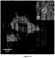
- Targeted fluorescent imaging nanoparticles can specifically illuminate carious lesions, improving visual contrast, and aiding with diagnosis.
- the nanoparticles according to certain aspects of the present disclosure are inexpensive, biodegradable in the mouth/oral cavity, non-toxic, and fit in-line with current dental practices (e.g., only requiring visual inspection and/or a dental curing lamp for imaging).
- nanoparticle comprises a biopolymer with a cationic charge and an imaging agent.
- the nanoparticle comprises an imaging agent at about 0.1 % to less than or equal to about 10% by weight of the nanoparticle.
- the nanoparticles of the present disclosure may be diagnostic, meaning that they include an imaging agent that reveals the presence of one or more possible caries on or in a tooth in the oral cavity of a subject.
- the nanoparticles of the present disclosure may be therapeutic or diagnostic and therapeutic.
- Therapeutic nanoparticles may comprise at least one oral care active ingredient.
- a composition may comprise a first plurality of diagnostic nanoparticles and a second plurality of therapeutic nanoparticles comprising an oral care active ingredient.
- the nanoparticle desirably degrades in a time period of greater than or equal to about 30 minutes and optionally less than or equal to a few hours, for example, to provide the ability for a dental clinician, assistant, or hygienist, to complete the diagnostic procedure and evaluation.
- the nanoparticle may be designed to have a much longer period for degradation, for example, degrading in greater than or equal to about 24 hours to less than or equal to about 30 days or even longer after introduction into the oral cavity and exposure to saliva. This permits the active ingredient to be delivered to the oral cavity (e.g., to a region within or adjacent to carious lesions in a tooth) over a longer time frame to provide therapeutic benefits.
- An oral care active ingredient may be used for the prevention or treatment of a condition or disorder of hard or soft tissue of the oral cavity, including but not limited to oral cancer and dry mouth, the prevention or treatment of a physiological disorder or condition, or may provide a cosmetic benefit.
- Optional oral care active ingredients include an anticaries agent, a remineralizing agent, an antibacterial agent, an anticalculus agent, a tartar control agent, a tooth desensitizer, and combinations thereof, by way of non-limiting example. While general attributes and properties of each of the above categories of actives may differ, there may some common attributes and any given material may serve multiple purposes within two or more of such categories of actives.
- the oral care active ingredient comprises an anticaries agent, a remineralizing agent, an antibacterial agent, an anticalculus agent, and combinations thereof.
- the nanoparticles may be used to identify dental caries, microcavities, or enamel lesions; reduce or inhibit early enamel lesions or microcavities; reduce or inhibit formation of dental caries or cavities; reduce or inhibit demineralization and promote remineralization of the tooth; protect teeth from cariogenic bacteria; inhibit microbial biofilm formation on the tooth or in the oral cavity; and/or reduce levels of acid-producing bacteria in the oral cavity.
- Oral care actives that are useful herein are optionally present in the compositions of the present invention in safe and effective amounts.
- the nanoparticle comprises an oral care active ingredient of about 0.1 % to less than or equal to about 50% by weight after incorporation into the nanoparticle, and optionally about 0.1 % to less than or equal to about 15% by weight of the oral care active ingredient after incorporation into the nanoparticle.
- the oral care active ingredient comprises an anti-caries agent, such as an anti-caries fluoride-containing component that provides fluorine ions in the oral cavity.
- the fluoride-containing active ingredient may be present at greater than or equal to about 0.02% to less than or equal to about 2.2% by weight after incorporation into the nanoparticle.
- the fluoride-containing component may be selected from the group consisting of: fluorohydroxyapatite, stannous fluoride, sodium fluoride, calcium fluoride, silver fluoride dehydrate, sodium monofluorophosphate, difluorosilane, combinations and equivalents thereof.
- the oral care active ingredient comprises a calcium-containing component that provides calcium ions in the oral cavity for remineralizing the tooth.
- the calcium-containing active ingredient component may be present at greater than or equal to about 1% to less than or equal to about 5% by weight after incorporation into the nanoparticle.
- the calcium-containing active ingredient may be calcium lactate.
- the oral care active ingredient comprises a calcium and phosphate-containing component for remineralizing the tooth.
- the calcium and phosphate-containing component optionally comprises calcium glycerophosphate, dicalcium phosphate, tricalcium phosphate, calcium sodium phosphosilicate, or combinations and equivalents thereof.
- calcium glycerophosphate may be present in the nanoparticle at greater than or equal to about 0.1% to less than or equal to about 1% by weight after incorporation into the nanoparticle.
- dicalcium phosphate may be present in the nanoparticle at greater than or equal to about 2% to less than or equal to about 50% by weight after incorporation into the nanoparticle, optionally greater than or equal to about 2% to less than or equal to about 10% by weight after incorporation into the nanoparticle.
- tricalcium phosphate may be present in the nanoparticle at greater than or equal to about 1% to less than or equal to about 5% by weight after incorporation into the nanoparticle.
- calcium sodium phosphosilicate may be present in the nanoparticle at greater than or equal to about 1% to less than or equal to about 10% by weight after incorporation into the nanoparticle.
- the nanoparticle may comprise an oral care active ingredient selected from the group consisting of: amine fluoride, casein phosphopeptide, phosphoprotein, and equivalents and combinations thereof.
- the nanoparticle may comprise an amine fluoride present in the nanoparticle at greater than or equal to about 0.2% to less than or equal to about 2.2% by weight after incorporation into the nanoparticle.
- casein phosphopeptide may be present in the nanoparticle at greater than or equal to about 1% to less than or equal to about 5% by weight after incorporation into the nanoparticle.
- phosphoprotein may be present in the nanoparticle at greater than or equal to about 0.001% to less than or equal to about 0.01% by weight after incorporation into the nanoparticle.
- the nanoparticle may be a multiphasic nanoparticle that comprises multiple compositionally distinct compartments. Each compartment may thus comprise distinct material compositions. Multiphasic nanoparticles may have a variety of shapes and may comprise two, three, or more distinct compartments.
- a first compartment may include the imaging agent (e.g., bonded to a polymer in the first compartment), while the second compartment may have one or more oral care active ingredients.
- Such multiphasic nano-components may be formed by electrified jetting of materials that comprise one or more polymers, such as that disclosed by Roh et al., "Biphasic Janus Particles With Nanoscale Anisotropy", Nature Materials, Vol. 4, pp.
- the present disclosure contemplates an oral care composition for oral administration in an oral cavity of a subject.
- the oral care composition includes any of the nanoparticles discussed above.
- the nanoparticle also includes an orally acceptable carrier, meaning a material or combination of materials that are relatively safe for use with in a subject while considering the risks versus benefits (e.g., that the benefits outweigh the risks).
- An orally acceptable carrier may thus be any carrier toxicologically suitable for use in the oral cavity. Selection of specific components of the orally acceptable carrier depend upon the form of the oral care composition, for example, whether the oral care composition is a mouth rinse, dentifrice, gel, paint, or the like.
- Such orally acceptable carriers include the usual components of dentifrices ( e .
- the oral care composition is a mouth rinse that facilitates extensive and comprehensive coverage of surfaces of teeth, including interproximal/interdental surfaces where caries often tends to develop.
- the orally acceptable carrier used to prepare an oral composition may comprise a water-based phase, which may include alcohols and other components.
- the oral compositions may include other conventional oral care composition materials, including by way of non-limiting example, surface active agents, such as surfactants, emulsifiers, and foam modulators, abrasives, humectants, mouth feel agents, viscosity modifiers, diluents, pH modifying agents, sweetening agents, flavor agents, colorants, preservatives and combinations thereof.
- the oral care composition comprising the nanoparticles may be administered to the subject and thus introduced into the oral cavity of the subject.
- the plurality of particles selectively accumulates adjacent to caries on the surface of a tooth and/or within cavities in the tooth corresponding to the one or more carious lesions.
- the oral care composition may include multiple distinct types of nanoparticles.
- the plurality of nanoparticles is optionally a first plurality of diagnostic nanoparticles comprising the imaging agent.
- the oral care composition may thus further comprise a second plurality of therapeutic nanoparticles comprising an oral care active ingredient.
- the present disclosure contemplates use of nanoparticles comprising biopolymers to target caries, including microcavities, on a tooth within the oral cavity of a subject.
- the present disclosure provides nanoparticle compositions and metabolites that are non-toxic and resorbable in contrast to certain synthetic polymers that can potentially cause side-effects and toxicity when used in medical diagnostic applications.
- the nanoparticles have an advantageous size for microcavity diagnosis being of a particle size that permits entry into cavities and lesions in tooth enamel.
- the nanoparticles according to certain variations of the present disclosure are easy to functionalize, allowing for the attachment of various fluorescent or optical dyes or imaging agents, protective coatings, and control over particle charge.
- the present technology provides the ability to work in-line with current dental technology without requiring additional equipment purchase or training, for example, using an existing standard dental curing lamp for detection.
- certain variations of the present disclosure provide nanoparticles that can be manufactured on an industrial scale with high production rates for relatively low cost, compared by many micro and nanoparticle systems that are limited by the ability to scale up production.
- the present disclosure provides use of fluorescent biopolymers in the form of nanoparticles used to aid in diagnosis of microcavities.
- Starch-based nanoparticles offer a new way to assist dentists in the diagnosis of active carious lesions. These particles are biodegradable, inexpensive, non-toxic and use the currently available technology in the dentist's office.
- image processing to extract only green light and improve contrast is also a new and unusual finding provided by the present teachings, as discussed further below.
- the nanoparticles and oral composition contemplated by the present teachings can be used for one or more of the following applications: administration for diagnosis of cavities in a dental office by a clinician (e . g ., dentist, dental assistant, or hygienist), home diagnosis of dental cavities, monitoring a level of tooth degradation in clinical trials, and sustained and highly targeted delivery of therapeutics to one or more regions of a tooth having caries or dental cavities.
- a clinician e . g ., dentist, dental assistant, or hygienist
- home diagnosis of dental cavities e.g ., monitoring a level of tooth degradation in clinical trials
- sustained and highly targeted delivery of therapeutics e.g a tooth having caries or dental cavities.
- the present disclosure provides methods of making a nanoparticle for oral administration.
- the method may include functionalizing a biocompatible and biodegradable polymer with a reactive group capable of reacting with an imaging agent.
- a reactive functional group may be a carboxyl group on the polymer that reacts with an amine on an imaging particle (e.g., an amine-functionalized imaging agent).
- Another variation may include reacting an alkyne functional group on the polymer by copper-click chemistry on the corresponding imaging agent.
- Other variations include use of carbodiimides (EDC) that cause direct conjugation of carboxyls (-COOH) to primary amines (-NH 2 ) without becoming part of the final crosslink (amide bond) between target molecules.
- EDC carbodiimides
- N-hydroxysuccinimide or its water-soluble analog (Sulfo-NHS) can be included in EDC coupling processes to enhance bonding.
- conjugation chemistries include the reaction of azides with phosphines, thiols with maleimide or vinyl groups, or photoinduced cross-linking of photoreactive groups, such as benzophenone.
- the biocompatible and biodegradable polymer comprises at least one cationic region capable of associating with one or more carious lesions on or inside a tooth in the oral cavity of a subject.
- the method may thus include reacting the reactive group on the biocompatible and biodegradable polymer with the imaging agent, so that the nanoparticle bears the imaging agent, which is capable of indicating the presence of one or more carious lesions when the nanoparticle is associated therewith.
- the imaging agent which is capable of indicating the presence of one or more carious lesions when the nanoparticle is associated therewith.
- the method may include first functionalizing the biocompatible and biodegradable polymer to have at least one first reactive group capable of reacting with a cationic moiety.
- a reactive functional group may be a hydroxyl group on the polymer that reacts with an epoxide group on a cationic moiety (e.g., an epoxide-functionalized cationic moiety).
- the biocompatible and biodegradable polymer is reacted with the cationic moiety so that the biocompatible and biodegradable polymer has at least one cationic region with a positive charge capable of associating with one or more carious lesions on a tooth in an oral cavity of a subject. Any of the cationic moieties (or precursors thereof) discussed previously above may be used in such a method.
- particles are made using starch nanoparticles having a particle size ranging from greater than or equal to about 20 nm to less than or equal to about 250 nm measured using dynamic light scattering (DLS) and nanoparticle tracking analysis (NTA) methods. These particles are then chemically modified to increase functionality.
- starch is cationized with a cationic epoxide group moiety (glycidyl trimethylammonium chloride) to imbue positive charge to the polymer.
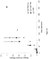
- Cationic starch nanoparticles have a moderately positive zeta-potential of +30 mV. This positive charge is believed to help the particles to target active early carious lesions, as discussed previously above.
- nanoparticles may be starch nanoparticles having an average particle size ranging from greater than or equal to about 10 nm to less than or equal to about 250 nm, measured using DLS and NTA methods.
- Cationic particles may then be oxidized to create carboxyl functional groups.
- a process using the water-soluble catalyst TEMPO (2,2,6,6-tetramethylpiperidine-1-oxyl) is preferably used to modify the C6 hydroxyl of the glucopyranose starch polymer unit to a carboxyl, which minimizes the molecular weight reduction of the polysaccharide polymer that is common to other oxidative processes.
- Oxidized cationic nanoparticles are measured to have a slight positive zeta-potential of approximately +8.5 mV.
- the carboxyl functionality is intended to allow attachment of fluorescent molecules, however, it also adds some slight negative charge to the particles. Without being limited or bound to any particular theory, this zwitterionic property is believed to enhance the possible fluorescence signal by allowing self-aggregation of fluorescent particles in active carious lesions.
- a fluorescent molecule or marker can be attached to the nanoparticle surface.
- Some examples include fluorescein, Alexa FLUO ® dyes, rhodamine, and the like discussed previously above.
- fluorescein isomer 1 is chosen for its ability to illuminate under the blue light emitted from a standard dental curing lamp and its low toxicity and subsequent use in other dental applications
- the fluorescein isomer 1 is modified to have an amine functionality which allowed for EDC/NHS coupling to oxidized cationic biopolymers.
- the number of functional groups on the surface of the nanoparticle and the fluorescent tag concentration relative to the particle concentration may determine the fluorescence intensity of the particles. In one embodiment, this is approximately 20 times less fluorescent than a commercially available fluorescein isothiocyanate (FITC)-dextran (10k MW) from Sigma Aldrich.
- FITC-dextran solution has an average size of approximately 100 nm and a zeta potential of approximately +6 mV.
- Bio-based nanoparticles are provided by EcoSynthetix Inc. All chemicals are lab grade and purchased from Sigma Aldrich, unless otherwise noted. These include 2,2,6,6-tetramethylpiperidinyloxy radical (TEMPO), sodium bromide, sodium hypochlorite, isopropyl alcohol, glycidyl trimethyl ammonium chloride (ETA), sodium hydroxide, ethanol, fluoresceinamine isomer 1, EDC, NHS, FITC-dextran (10k), and fluorescein sodium salt.
- TEMPO 2,2,6,6-tetramethylpiperidinyloxy radical
- ETA glycidyl trimethyl ammonium chloride
- EDC EDC
- NHS FITC-dextran (10k)
- fluorescein sodium salt fluorescein sodium salt.
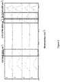
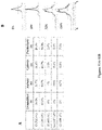
- Starch nanoparticles shown at 1 in Figure 1 are modified to be cationic according to a procedure shown in Huanga Y. et al. "Ultra-small and innocuous cationic starch nanospheres: Preparation, characterization and drug delivery study.” International Journal of Biological Macromolecules, 58: pp. 231-239 (2013 ).
- Starch nanoparticles are dispersed at 10 % solids into 100 mL of 1% sodium hydroxide in DI water. To this solution, 3 mL of isopropyl alcohol and 4.3 g of glycidyl trimethyl ammonium chloride are added, and allowed to mix for one hour. The mixture is then heated to 75°C overnight, before precipitation in ethanol and centrifugation, followed by lyophilization, yielding cationic starch nanoparticles (shown at 2 in Figure 1 ).
- Starch nanoparticles are oxidized according to a procedure shown in Kato, Y. et al., "Oxidation process of water-soluble starch in TEMPO-mediated system," Carbohydrate Polymers 51, pp. 69-75 (2003 ). Briefly, a 100 mL 5% solution of cationic starch nanoparticles dispersed in DI water is mixed with a 100 mL aqueous solution containing 0.048 g of TEMPO and 0.635 g of sodium bromide. The mixture is put on ice, and the pH is adjusted using a 10% sodium hydroxide solution to bring the pH above 10.
- EDC/NHS linkage of FITC-amine to -COOH functionality Carbodiimide (EDC) and other similar carbodiimides are cross-linkers that directly conjugate carboxyls (-COOH) to primary amines (-NH 2 ) without becoming part of the final crosslink (amide bond) between target molecules.
- N-hydroxysuccinimide (NHS) or its water-soluble analog (Sulfo-NHS) is often included in EDC coupling processes to improve efficiency or to create a more stable, amine-reactive intermediate.
- FTIR Fourier Transform Infrared
- H'-NMR analysis is completed using a Varian MR400 instrument. Samples are dispersed in D 2 O at approximately 5% solids and peak analysis is compared to results as previously shown in Kato et al.
- X-ray Photon Spectroscopy is run on dry powder starch nanoparticle samples using an Axis Ultra X-ray photoelectron spectrometer (Kratos Analyticals, UK) outfitted with a monochromatized A1 K ⁇ X-ray source at a power of 150 kW.
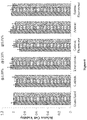
- Starch nanoparticle samples 1-6 are dispersed at 0.025% solids and analyzed by various particle characterization techniques, including zeta potential and DLS analysis using a Malvern ZetaSizer, and Nanoparticle Tracking Analysis (NTA) using a NanoSight NS300.
- particle characterization techniques including zeta potential and DLS analysis using a Malvern ZetaSizer, and Nanoparticle Tracking Analysis (NTA) using a NanoSight NS300.
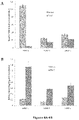
- Particle fluorescence of fluorescein-labeled anionic and fluorescein-labeled cationic StNPs is measured using a BioTek Neo 1 fluorescence plate reader and compared at multiple concentrations against a commercially available FITC-dextran (10k MW).
- Example 2 Fluorescein loaded cationic starch nanoparticles.
- Cationic starch nanoparticles are loaded with fluorescein sodium salt by dispersing a mixture of 5% by mass starch particles and 0.25% fluorescein salt, and lyophilizing the solution.
- the fluorescein anion interacts with the cationic groups on the starch nanoparticle, holding the fluorescent dye within the particle.
- Particles are approximately 30 nm in size.
- Example 3 EHD jetting of chitosan.
- Chitosan powder (degree of deacetylation, 75-85%; molecular weight 190-310 kDa) is dissolved at 1% by weight in trifluoroacetic acid containing 0.05% by mass fluorescein to form a homogeneous solution.
- Chitosan nanoparticles are prepared by electrohydrodynamic (EHD) jetting using a setup involving a 1 mL plastic syringe, a stainless-steel needle (22 G; inner diameter, 0.413 mm), a syringe pump, a high-voltage power supply, and an aluminum plate placed directly below the needle as the grounded counter electrode (collector).
- EHD electrohydrodynamic
- the aforementioned solution can be drawn into the syringe and extruded through the needle at a constant flow rate using the syringe pump.
- a high voltage of approximately 10kV the liquid forms a Taylor cone at the tip of the needle which breaks into droplets which are collected 15 cm from the tip of the needle on the grounded collection plate.
- the nanoparticles are allowed to dry for 24 hours under vacuum to remove any trace solvent, before being collected by scraping from the aluminum plate and dispersed in water, followed by filtration using a 0.4 micron filter, yielding fluorescently-labeled chitosan nanoparticles, approximately 200-300 nm in size.
- Example 4 EHD jetting of cationic starch nanoparticles.
- Cationic starch nanoparticles are dispersed at 5% by mass in an 80:20 v:v solution of water:ethanol with 0.25% fluorescein sodium salt. This solution is jetted using an electrohydrodynamic jetting setup as described previously in Example 3, however using a voltage of approximately 16 kV, yields nanoparticles which are approximately 300-500 nm in size.
- Example 5 Bicompartmental jetted particles for combined fluoride delivery and fluorescent diagnosis.
- Bicompartmental starch nanoparticles are prepared by using the same jetting procedure described above by using a setup involving needles placed adjacent to one another.
- One needle is used for jetting a solution of cationic starch nanoparticles dispersed at 5% by mass in an 80:20 v:v solution of water: ethanol with 0.25% FITC-dextran.
- the other needle is used for jetting a solution of cationic starch nanoparticles dispersed at 5% by mass in an 80:20 v:v solution of water:ethanol with 1.3% by mass of a 10k M.W. PEG-diglycidyl ether, and 0.25% by mass of sodium fluoride.
- Particles are then left for 72 hours at 37°C to allow the PEG-diglycidyl ether to cross-link the fluoride containing component of the starch to extend release.
- the final particles are bicompartmental with a fast-degrading fluorescent compartment for caries diagnosis, and a slow degrading fluoride-loaded compartment for remineralization. Particles are approximately 300-500 nm in size.
- Example 6 Click-functionalized cationic starch nanoparticles.
- Cationic starch nanoparticles are chemically modified by dispersion at 5% by mass solids in dimethyl sulfoxide (DMSO) (Sigma), and addition of 1% by mass DMAP (Sigma) to the solution. Once fully dispersed, 1% by mass glycidyl propargyl ether (Sigma) is added and allowed to react at room temperature for 48 hours, before finishing the reaction by quenching with hydrochloric acid (Sigma). Addition of 50% volume ethanol, allows for precipitation of the starch nanoparticles and removal of the supernatant, and an additional cleaning step and re-precipitation from water:ethanol purifies the particles, which are then dried by lyophilization. The final particles are approximately 20 nm in size, cationic with a zeta potential of approximately +25 mV, and have an alkyne functional group with can be modified by copper-click chemistry.
- the reaction mixture is kept at room temperature overnight, before precipitation using ethanol and centrifugation, followed by lyophilization.
- Final particles are approximately 20-30 nm in size, with a cationic zeta potential (20-35 mV), and fluorescently labeled with Cy5 fluorophores.
- Starch particles are dispersed at 1% solids. Half of the particle dispersions are set aside as an initial solution and diluted 1:1 v:v with DI-water. The remaining half of the particle dispersions are taken and added to an equal volume of saliva, and placed in a 37°C incubator for 30 minutes, as a final dispersion. Saliva is collected and used immediately to minimize potential denaturation of salivary enzymes. Both the initial and final dispersions are tested with iodine and Benedict's reagent.
- Extracted teeth are obtained from School of Dentistry, University of Michigan (human subjects exempt) and stored in 1 % sodium azide before use.
- the teeth are painted with an acid resistant varnish leaving a 1 mm 2 enamel window on the buccal surface of the crowns of the teeth.
- the teeth are then immersed in a pH 5.0 demineralization gel containing 0.1 M lactic acid, 4.1 mM CaCl 2 ⁇ 2H 2 O, 8 mM KH 2 PO 4 , and 1% w/v CMC (carboxymethylcellulose sodium) at 37°C for 8 days (ref: Lippert et al., Caries Res. 46: pp. 23-30 (2012 )).
- the teeth are rinsed with distilled H 2 O before subjected to caries activity test. Residual varnish is removed by washing in acetone.
- FITC-dextran 1% solutions of FITC-dextran, FITC-anionic StNP, and FITC-cationic StNP, and a 0.001% solution of fluorescein sodium salt are prepared. Teeth are exposed to 20 microliters of sample for three minutes prior to rinsing in DI water. Rinsing is done initially for 10 seconds, and teeth are examined and photographed while illuminated with a standard dental curing lamp. Rinsing is continued for an additional 10 seconds, followed by imaging, and this is repeated for up to 5 minutes to determine "residence times" for each sample to optimize the exposure procedure.
- Digital images are taken with a Nikon ABC camera and analyzed using ImageJ image analysis software.
- the "mean gray value” measurement tool is used to compare grayscale brightness between the carious lesion and the background tooth as a measurement of contrast.
- the "split channels” option is used as a second method to extract green pixels from the images, to isolate the fluorescein color.
- Treated teeth are examined using a Leica TCS SP8 2-Photon Confocal with FLIM & FCS using a 40x oil-immersion objective. Samples are immersed in oil and placed on a glass-bottomed petri dish. The illumination wavelength is set to 810 nm, and z-stack images are collected for a variety of tooth samples.
- particle analysis is also performed on these samples, including size analysis by NTA and DLS, and zeta potential measurements, as shown in Figures 3A and 12 .
- This grade of unmodified StNPs shows a small particle size and neutral charge.
- TEMPO oxidation particle charge decreased and average particle size decreased significantly.
- cationization particle size decreased and charge increased.
- both reactions are combined to make zwitterionic particles, average particle size increased and particles showed a moderate cationic charge. It is expected that the combination of both charges results in slight aggregation due to electrostatic interactions.
- fluorescein by EDC-NHS ligandization slightly neutralized the particle charge, and appeared to have no significant impact on particle size.
- Starch particle degradation is confirmed using the iodine and Benedict's reagent tests, with results shown in Figures 3B and 4A-4B .
- the iodine test showed a decrease in starch staining after 30 minutes of exposure to saliva, while the Benedict's test showed an increase in the presence of reducing sugars.
- Particle fluorescence of the cationic and anionic starch nanoparticles are measured relative to FITC-dextran. It is found that the cationic starch nanoparticles are approximately 20x less fluorescent than FITC dextran, and the anionic starch nanoparticles are approximately 80x less fluorescent than FITC-dextran on a per gram basis.
- Teeth are exposed to the fluorescent cationic starch nanoparticles and imaged to provide baseline fluorescence.
- Three different washing conditions 1% sodium chloride solution, 1% magnesium sulfate solution, artificial saliva solution
- Figure 5A shows fluorescence over 2 hours.
- Figure 5B shows fluorescence over 20 minutes. Image analysis of the contrast is tracked to determine overall reduction in fluorescence from the lesions as a function of washing time and type of rinse fluid.
- Teeth are exposed for 3 minutes to 10 ⁇ L of 10 -2 g/mL concentrations of fluorescently labeled dextran and anionic StNPs, the 10 -5 g/mL solution of fluorescein sodium salt (controls), as well as a 10 -5 g/mL dispersion of fluorescently labeled cationic StNPs, and then rinsed. Images are taken under a dental curing light and lesion versus background tooth intensity is compared. Figure 8 shows % Intensity Difference for lesions minus background intensity for each, as well unexposed blank controls. "Normal” stands for % Intensity Difference obtained for normal images using ImageJ software, and "Green” stands for the extracted green pixel data from those same images.
- Photographs are taken of the teeth as shown in Figures 7A-7B . There are visible changes in the color of teeth with microcavities (as shown in Figure 7B ) as compared to teeth without any microcavities ( Figure 7A ) after exposure to a suspension of cationic starch nanoparticles prepared in accordance with certain aspects of the present disclosure.
- the photographs are modified from full-scale lighting to extract the green pixels, as this is found to significantly increase the contrast between lesion and background.
- the carious lesions are initially slightly darker than the rest of the tooth, and this result is consistent across all controls.
- the same teeth, treated with the fluorescent cationic StNPs showed fluorescence in the carious lesion region, which is visibly brighter than the background.
- the teeth treated with fluorescent cationic StNPs yielded highly significant positive percent intensity differences (p ⁇ 10 -5 ), indicating that these particles improved the contrast of carious lesions relative to the background, even to the naked eye.
- analyzing extracted green-pixel images instead of standard RGD images further improved the contrast. It is interesting to note that comparing the non-illuminated images to the illuminated images highlights differences between the delineated shape of the lesion depending on the optical method used (e.g., image of carious lesions treated with fluorescent anionic particles and corresponding image of lesions treated with fluorescent cationic particles).
- the prepared fluorescent cationic starch nanoparticles can specifically highlight active carious lesions when illuminated with a standard dental curing lamp in vitro, and therefore offer a simple method to assist dentists in diagnosis of white spot carious lesions in vivo
- the FITC-dextran polymer solution has an average size of approximately 100 nm and a zeta potential of approximately +6 mV, and is thus mildly cationic, as a control it did not successfully associate and illuminate the carious lesions.
- the cationic starch nanoparticles with a similar zeta potential did successfully associate with and illuminate the carious lesions.
- the cationic dextrin polymer may be held in solution by stronger forces, on an absolute basis or at least relative to the mass of the molecules in solution, as compared to the cationic starch nanoparticles.
- the cationic starch nanoparticles are a dispersion rather than a solution.
- the nanoparticle dispersion is stable, probably due to steric stabilization, yet only a mildly cationic charge is required for the nanoparticles to come out of the dispersion and associate with the carious lesions.
- the cationic starch nanoparticles may aggregate in or around an active carious lesion, perhaps aided by their zwitterionic nature, and become somewhat more resistant to rinsing, whereas a polymer in solution does not.
- the lesser charge required by the nanoparticle is advantageous, in that less reactants are required to produce the mild charge and there may be a trend for toxicity with increases in cationic charge.
- Nanoparticle targeting to remineralize lesions
- a specific fluorescent nanoparticle probe can identify caries activity by virtue of surface porosity, which, in active carious lesions allows for diffusion to the subsurface pores, but prevents access to the fluorescent probe in the case of inactive lesions.
- a dentist does not need to treat inactive lesions, and treatments such as fluoride varnishes, gels, washes, or sealants, will have no beneficial effect.
- active lesions are progressing, and appropriate treatment can halt and reverse demineralization.
- Two-Photon micrographs of the remineralized lesions, both before and after exposure to fluorescent cationic StNPs, further support these results ( Figure 14 ).
- a fluoride release study tests the loading of fluoride into cationic starch nanoparticles using a freeze-drying method. This is intended to be a baseline measurement as there is potential interaction of the anionic fluoride salt with the cationic starch polymer of the particles.
- PBS phosphate-buffered-saline
- AS artificial saliva
- Aliquots of a half milliliter in size are taken at consecutive time points extending from 30 seconds to 72 hours. The aliquots are then tested using a fluoride electric probe to determine the fluoride concentration, and these values are normalized using a calibration to samples of known concentration.
- a reference solution of free fluoride salt is used as a comparison to see the diffusion-limited release of the fluoride salt through the dialysis membrane. Results of the release study are shown in Figure 15 .
- the present disclosure provides cationic nanoparticles that target and illuminate early forming active carious lesions.
- a nanoparticle may be a starch-based fluorescent cationic nanoparticle.
- the particles are biodegradable and are enzymatically broken down in saliva. These particles can be illuminated using a standard dental curing lamp, comporting with current dental practices and workflow.
- the present technology thus improves detection of microcavities while they are still reversible and treatable by improved dental hygiene and treatment with a dental remineralizing agent, for example, fluoride and/or phosphate.
- a dental remineralizing agent for example, fluoride and/or phosphate.
- image analysis and in particular by analyzing the green colors in images, these particles significantly improve the contrast of carious lesions.
- nanoparticles can be used for early diagnosis of caries in dental clinics, for home use, or to monitor tooth remineralization and conservative restoration strategies in dental practices and in clinical trials and the like.
- Such nanoparticles can be used to deliver oral care active ingredients, such as fluoride ions, other remineralization aides, or antibacterial therapeutics to improve dental health.
- the present technology provides the ability to differentiate between inactive and active carious lesions to help monitor progression following treatment using remineralization, in order to enable dentist and dental patient to implement conservative treatment strategies, avoiding more invasive and expensive restorative procedures, such as "drill and fill” or other invasive procedures, as well as ability to reduce patient exposure to harmful radiation from taking X-Ray images, which may especially be undesirable in children.
- This specification also provides diagnostic methods of one or more carious lesions, imaging methods of one or more carious legions, methods of caries treatment, methods of remineralization, methods of monitoring caries treatment, uses of a component ( e . g ., compound) or a nanoparticle as described above as a medicament, or uses of a component ( e.g., compound) or nanoparticle as a diagnostic agent.
- the present disclosure also describes a fluorescent, optionally starch-based, component ( e . g ., compound) or nanoparticle having a net positive charge, and the use of such component ( e . g ., compound) or nanoparticles to aid in the diagnosis, imaging and/or monitoring of caries.
- the present disclosure also describes an optionally starch-based component or nanoparticle comprising a re-mineralizing agent, wherein the nanoparticle has a net positive charge, and such components or nanoparticles used in the treatment of caries.
- the present disclosure contemplates a composition for oral administration comprising a component that comprises a biocompatible polymer and an imaging agent, an oral care active ingredient, or both the imaging agent and the active ingredient.
- the composition has a cationic moiety or a net positive charge. Also specifically disclosed are the combinations including this composition with any one or any combination of more than one of the enumerated features (1)-(23) below.
- the composition optionally has any one or any combination of more than one of the following features: (1) the cationic moiety is bonded with the biocompatible polymer; (2) the biocompatible polymer comprises a tertiary amine or a quaternary amine; (3) the biocompatible polymer comprises a reaction product of glycidyl trimethyl ammonium chloride bonded to the biocompatible and biodegradable polymer; (4) the biocompatible polymer is selected from the group consisting of: a mono-, oligo- or polysaccharide, carboxymethylcellulose, polymeric starch, dextrin, dextran, chitosan, cellulose, gelatin, polyethyleneimine (PEI), poly(L-lysine) (PLL), poly(L-arginine), poly(amidoamine) (PAA), poly (amino-co-ester) (PAE), poly(2-N,N-dimethylaminoethylmethacrylate) PDMAEMA, poly(4-vinyl
- the present disclosure contemplates a nanoparticle for oral administration comprising a biocompatible and biodegradable polymer bearing at least one cationic region capable of associating with one or more carious lesions on a tooth in an oral cavity of a subject; and an imaging agent bonded to the biocompatible and biodegradable polymer, so that the nanoparticle is capable of indicating the presence of one or more carious lesions when the nanoparticle is associated therewith. Also specifically disclosed are the combinations including this composition with any one or any combination of more than one of the enumerated features (24)-(43) below.
- the nanoparticle of this embodiment optionally has any one or any combination of more than one of the following features: (24) the at least one cationic region comprises a cationic moiety bonded with the biocompatible and biodegradable polymer; (25) the cationic moiety comprises a tertiary amine or a quaternary amine; (26) the cationic moiety is a reaction product of glycidyl trimethyl ammonium chloride bonded to the biocompatible and biodegradable polymer; (27) the biocompatible and biodegradable polymer is a cationic polymer selected from the group consisting of: a cationic or cationically modified mono-, oligo-, or polysaccharide, carboxymethylcellulose, starch, dextrin, dextran, chitosan, cellulose, gelatin, polyethyleneimine (PEI), poly(L-lysine) (PLL), poly(L-arginine), poly(amidoamine) (PAA), poly (amino-co
- the present disclosure contemplates an oral care composition for oral administration in an oral cavity of a subject comprising a plurality of nanoparticles, wherein each nanoparticle comprises a biocompatible and biodegradable polymer bearing at least one cationic region having a positive charge capable of associating with one or more carious lesions on a tooth in the oral cavity of the subject; and an imaging agent bonded to the biocompatible and biodegradable polymer, so that the plurality of nanoparticles is capable of indicating the presence of one or more carious lesions when the nanoparticles are associated therewith; and an orally acceptable carrier.
- the combinations including this composition with any one or any combination of more than one of the enumerated features (44)-(46) below.
- the oral care composition of this embodiment optionally has any one or any combination of more than one of the following features: (44) the oral care composition is selected from the group consisting of: mouth rinse, paint, gel, and dentifrice; (45) the plurality of nanoparticles selectively accumulate within cavities in the tooth corresponding to the one or more carious lesions; and/or (46) the plurality of nanoparticles is a first plurality of diagnostic nanoparticles and the oral care composition further comprises a second plurality of therapeutic nanoparticles comprising an oral care active ingredient.
- the present disclosure contemplates a method of making a nanoparticle for oral administration comprising functionalizing a biocompatible and biodegradable polymer with a reactive group capable of reacting with an imaging agent, wherein the biocompatible and biodegradable polymer comprises at least one cationic region capable of associating with one or more carious lesions on a tooth in an oral cavity of a subject.
- the method also includes reacting the reactive group on the biocompatible and biodegradable polymer with the imaging agent, so that the nanoparticle bears the imaging agent that is capable of indicating the presence of one or more carious lesions when the nanoparticle is associated therewith.
- the method of making a nanoparticle for oral administration optionally has any one or any combination of more than one of the following steps or features: (47) further comprising reacting the biocompatible and biodegradable polymer with a cationic moiety before the functionalizing to form the at least one cationic region; and/or (48) further comprising functionalizing the biocompatible and biodegradable polymer to have at least one first reactive group capable of reacting with the cationic moiety before the reacting with the cationic moiety.
- the present disclosure contemplates a method of making a composition for oral administration comprising functionalizing a polymer with a reactive group capable of reacting with an imaging agent, wherein the polymer comprises at least one cationic region.
- the method also comprises reacting the reactive group on the polymer with the imaging agent, wherein the composition has a net positive charge.
- compositions for oral administration optionally has any one or any combination of more than one of the following steps or features: (49) further comprising reacting the polymer with a cationic moiety before the functionalizing to form the at least one cationic region; (50) further comprising functionalizing the polymer to have at least one first reactive group capable of reacting with the cationic moiety before the reacting with the cationic moiety; (51) wherein the polymer comprises hydroxy groups; and/or (52) wherein the polymer comprises glucose repeat units.
- the present disclosure contemplates a method of detecting caries comprising introducing a positively-charged fluorescent component to an oral cavity of a subject. Light is directed into the oral cavity. The method further includes identifying a location of any fluorescence in the oral cavity, which may correspond to a location of one or more caries in the oral cavity.
- the method of detecting caries optionally has any one or any combination of more than one of the following steps or features: (53) the light is generated by a dental curing lamp; (54) further comprising rinsing the oral cavity after introducing the positively-charged fluorescent component and before the directing of the light into the oral cavity; (55) further comprising producing a digital image of the fluorescence; (56) further comprising filtering the digital image; (57) further comprising treating to the location with a remineralizing agent; (58) where the treating is repeated in multiple steps; and/or (59) further comprising subsequently repeating the introducing, the directing, and the identifying to determine if the fluorescence has increased, decreased, or remained unchanged.
- the present disclosure contemplates a method of treating caries comprising introducing a positively-charged nanoparticle comprising a remineralizing agent to an oral cavity of a subject.
- the positively-charged nanoparticle is capable of associating with one or more carious lesions in the oral cavity of the subject.
- the present disclosure contemplates use of a positively-charged fluorescent composition according to any of the variations previously described above to determine a location of caries on at least one tooth in a subject.
- the present disclosure contemplates use of a positively-charged component or nanoparticle comprising a re-mineralizing agent in a medicament for treatment of caries.
- the positively-charged component or nanoparticle may be any of the variations previously described above.
Landscapes
- Health & Medical Sciences (AREA)
- Life Sciences & Earth Sciences (AREA)
- Animal Behavior & Ethology (AREA)
- Veterinary Medicine (AREA)
- Public Health (AREA)
- General Health & Medical Sciences (AREA)
- Epidemiology (AREA)
- Birds (AREA)
- Chemical & Material Sciences (AREA)
- Engineering & Computer Science (AREA)
- Inorganic Chemistry (AREA)
- Biomedical Technology (AREA)
- Nanotechnology (AREA)
- Nuclear Medicine, Radiotherapy & Molecular Imaging (AREA)
- Oral & Maxillofacial Surgery (AREA)
- Radiology & Medical Imaging (AREA)
- Molecular Biology (AREA)
- Pharmacology & Pharmacy (AREA)
- Organic Chemistry (AREA)
- Medicinal Chemistry (AREA)
- General Chemical & Material Sciences (AREA)
- Chemical Kinetics & Catalysis (AREA)
- Bioinformatics & Cheminformatics (AREA)
- Immunology (AREA)
- Physics & Mathematics (AREA)
- Dentistry (AREA)
- Audiology, Speech & Language Pathology (AREA)
- Biophysics (AREA)
- Pathology (AREA)
- Heart & Thoracic Surgery (AREA)
- Medical Informatics (AREA)
- Surgery (AREA)
- Cosmetics (AREA)
- Medicines Containing Antibodies Or Antigens For Use As Internal Diagnostic Agents (AREA)
- Medicinal Preparation (AREA)
- Pharmaceuticals Containing Other Organic And Inorganic Compounds (AREA)
- Medicines That Contain Protein Lipid Enzymes And Other Medicines (AREA)
Applications Claiming Priority (3)
| Application Number | Priority Date | Filing Date | Title |
|---|---|---|---|
| US201562244512P | 2015-10-21 | 2015-10-21 | |
| PCT/US2016/058274 WO2017070578A1 (fr) | 2015-10-21 | 2016-10-21 | Détection et traitement de caries et de microcavités avec des nanoparticules |
| EP16858371.4A EP3365028B1 (fr) | 2015-10-21 | 2016-10-21 | Détection et traitement de caries et de microcavités avec des nanoparticules |
Related Parent Applications (2)
| Application Number | Title | Priority Date | Filing Date |
|---|---|---|---|
| EP16858371.4A Division-Into EP3365028B1 (fr) | 2015-10-21 | 2016-10-21 | Détection et traitement de caries et de microcavités avec des nanoparticules |
| EP16858371.4A Division EP3365028B1 (fr) | 2015-10-21 | 2016-10-21 | Détection et traitement de caries et de microcavités avec des nanoparticules |
Publications (1)
| Publication Number | Publication Date |
|---|---|
| EP4088744A1 true EP4088744A1 (fr) | 2022-11-16 |
Family
ID=58558161
Family Applications (2)
| Application Number | Title | Priority Date | Filing Date |
|---|---|---|---|
| EP22183087.0A Pending EP4088744A1 (fr) | 2015-10-21 | 2016-10-21 | Détection et traitement de caries et de microcavités avec des nanoparticules |
| EP16858371.4A Active EP3365028B1 (fr) | 2015-10-21 | 2016-10-21 | Détection et traitement de caries et de microcavités avec des nanoparticules |
Family Applications After (1)
| Application Number | Title | Priority Date | Filing Date |
|---|---|---|---|
| EP16858371.4A Active EP3365028B1 (fr) | 2015-10-21 | 2016-10-21 | Détection et traitement de caries et de microcavités avec des nanoparticules |
Country Status (7)
| Country | Link |
|---|---|
| US (2) | US10987434B2 (fr) |
| EP (2) | EP4088744A1 (fr) |
| JP (2) | JP7465494B2 (fr) |
| KR (1) | KR20180075565A (fr) |
| CN (2) | CN113876600B (fr) |
| CA (1) | CA3039906A1 (fr) |
| WO (1) | WO2017070578A1 (fr) |
Families Citing this family (12)
| Publication number | Priority date | Publication date | Assignee | Title |
|---|---|---|---|---|
| EP2645991B1 (fr) | 2010-12-02 | 2020-02-12 | GreenMark Biomedical Inc. | Dispositif d'administration de médicament de type bioconjugué à base d'un aptamère |
| WO2017070578A1 (fr) | 2015-10-21 | 2017-04-27 | The Regents Of The University Of Michigan | Détection et traitement de caries et de microcavités avec des nanoparticules |
| CN107343960A (zh) * | 2017-06-01 | 2017-11-14 | 点铂医疗科技(常州)有限公司 | 生物技术齿科脱敏剂及其制备方法 |
| WO2019191456A1 (fr) * | 2018-03-28 | 2019-10-03 | Greenmark Biomedical Inc. | Nanoparticule d'amidon réticulé au phosphate et traitements dentaires |
| US12484770B2 (en) | 2018-09-06 | 2025-12-02 | Greenmark Biomedical Inc. | Dental imaging and/or curing system |
| CN109875903A (zh) * | 2018-12-17 | 2019-06-14 | 点铂医疗科技(常州)有限公司 | 一种亲水改性硅橡胶印模材料及其制备方法 |
| US20220273530A1 (en) * | 2019-08-29 | 2022-09-01 | Skinosive | Bioadhesive particles comprising active agents, and uses thereof |
| KR20220088489A (ko) * | 2019-10-29 | 2022-06-27 | 더 트러스티스 오브 더 유니버시티 오브 펜실베니아 | 치태를 검출, 관찰 및 제거하기 위한 자동화된 정밀 장치 |
| CN113197693B (zh) * | 2020-09-14 | 2026-03-31 | 无锡时代天使医疗器械科技有限公司 | 防龋膜、防龋溶液、具有防龋膜的牙科器械以及在牙科器械上形成防龋膜的方法 |
| WO2022169936A1 (fr) * | 2021-02-03 | 2022-08-11 | Modern Ideas LLC | Formulations de fluorure d'argent, procédés et dispositifs pour la gestion des caries |
| EP4433037A4 (fr) * | 2021-11-19 | 2025-10-15 | Univ Michigan Regents | Nanoparticules ciblant un biofilm pour augmenter l'effet anti-caries d'un fluorure |
| CN114795558B (zh) * | 2022-05-12 | 2022-12-27 | 浙江大学 | 用于口腔pH值检测和给药的柔性智能牙齿贴片装置 |
Citations (20)
| Publication number | Priority date | Publication date | Assignee | Title |
|---|---|---|---|---|
| GB1420392A (en) | 1973-06-29 | 1976-01-07 | Scholten Honig Research Nv | Aqueous starch adhesive |
| JP2000063290A (ja) * | 1998-08-17 | 2000-02-29 | Kuraray Co Ltd | 抗菌性齲蝕検知液 |
| US6677386B1 (en) | 1999-01-25 | 2004-01-13 | Ato B.V. | Biopolymer nanoparticles |
| US6755915B1 (en) | 1998-12-30 | 2004-06-29 | Ecosynthetix Inc. | Method for the preparation of starch particles |
| WO2006137936A2 (fr) | 2004-11-10 | 2006-12-28 | The Regents Of The University Of Michigan | Nanoparticules multiphasiques |
| WO2010012473A2 (fr) * | 2008-07-30 | 2010-02-04 | Charite - Universitätsmedizin Berlin | Nanocapteurs spécifiques de l’hydrolase, leurs méthodes de production et leurs utilisations en imagerie moléculaire |
| WO2010065750A1 (fr) | 2008-12-03 | 2010-06-10 | Ecosynthetix Inc. | Procédé de fabrication de compositions de biolatex à base de nanoparticules de biopolymère présentant des performances améliorées et compositions fondées sur celui-ci |
| WO2010084088A2 (fr) | 2009-01-21 | 2010-07-29 | Basf Se | Nanoparticules d'amidon destinées à des systèmes d'administration de médicaments |
| US20100204448A1 (en) * | 2003-05-01 | 2010-08-12 | Szeto Hazel H | Method and carrier complexes for delivering molecules to cells |
| US20100272639A1 (en) * | 2007-12-21 | 2010-10-28 | John Robert Dutcher | Polysaccharide nanoparticles |
| US20110042841A1 (en) | 2006-08-15 | 2011-02-24 | Wildi Robert H | Process for producing biopolymer nanoparticles |
| WO2011071742A2 (fr) | 2009-12-10 | 2011-06-16 | Dow Global Technologies Llc | Procédé de préparation de dispersions stables d'amidon |
| WO2011128931A1 (fr) * | 2010-04-12 | 2011-10-20 | 財団法人ヒューマンサイエンス振興財団 | Marqueur diagnostique de pulpite et système diagnostique de pulpite |
| US8043480B2 (en) | 2004-11-10 | 2011-10-25 | The Regents Of The University Of Michigan | Methods for forming biodegradable nanocomponents with controlled shapes and sizes via electrified jetting |
| US20120045487A1 (en) | 2009-04-29 | 2012-02-23 | The Regents Of The University Of Michigan | Multiphasic microfibers for spatially guided cell growth |
| US8187708B2 (en) | 2004-11-10 | 2012-05-29 | The Regents Of The University Of Michigan | Microphasic micro-components and methods for controlling morphology via electrified jetting |
| US20120269729A1 (en) * | 2011-04-20 | 2012-10-25 | Swadeshmukul Santra | Stabilized chitosan-based nanoparticles and methods for making the same |
| US20140161745A1 (en) * | 2011-06-15 | 2014-06-12 | Rls Global Ab | Detection and removal of carious dentin tissue |
| US20140182797A1 (en) * | 2009-02-13 | 2014-07-03 | Upm-Kymmene Oyj | Method for producing modified cellulose |
| WO2015183995A2 (fr) * | 2014-05-28 | 2015-12-03 | Stealth Peptides International, Inc. | Compositions thérapeutiques comprenant de la frataxine, de la lactoferrine et des enzymes mitochondriales génératrices d'énergie et leurs utilisations |
Family Cites Families (36)
| Publication number | Priority date | Publication date | Assignee | Title |
|---|---|---|---|---|
| FR2647012B1 (fr) * | 1989-05-18 | 1991-07-26 | Oreal | Nouvelles compositions dentifrices a action anti-caries, contenant un fluorure de polymere polycationique |
| ATE252894T1 (de) | 1995-01-05 | 2003-11-15 | Univ Michigan | Oberflächen-modifizierte nanopartikel und verfahren für ihre herstellung und verwendung |
| EP0736544B1 (fr) | 1995-04-05 | 2001-10-24 | Unilever Plc | Compositions poursoins buccaux |
| JP3479822B2 (ja) * | 1996-06-26 | 2003-12-15 | ライオン株式会社 | 口腔用組成物 |
| US6428814B1 (en) * | 1999-10-08 | 2002-08-06 | Elan Pharma International Ltd. | Bioadhesive nanoparticulate compositions having cationic surface stabilizers |
| EP1176254A1 (fr) | 2000-07-24 | 2002-01-30 | The Dow Chemical Company | Utilisation de dispersions d'amidon reticulé dans la fabrication du papier |
| US6589562B1 (en) | 2000-10-25 | 2003-07-08 | Salvona L.L.C. | Multicomponent biodegradable bioadhesive controlled release system for oral care products |
| JP2002187857A (ja) | 2000-12-20 | 2002-07-05 | Gc Corp | 初期齲蝕検知材 |
| US20050191359A1 (en) * | 2001-09-28 | 2005-09-01 | Solubest Ltd. | Water soluble nanoparticles and method for their production |
| US7947772B2 (en) | 2004-11-10 | 2011-05-24 | The Regents Of The University Of Michigan | Multiphasic nano-components comprising colorants |
| US8048404B1 (en) * | 2005-01-04 | 2011-11-01 | Gp Medical, Inc. | Pharmaceutical composition of nanoparticles for protein drug delivery |
| US7629027B2 (en) | 2005-10-14 | 2009-12-08 | 3M Innovative Properties Company | Method for making chromonic nanoparticles |
| AU2011202746B2 (en) * | 2006-04-05 | 2011-10-13 | Intercontinental Great Brands Llc | Calcium phosphate complex and salts in oral delivery systems |
| US20080038686A1 (en) * | 2006-04-18 | 2008-02-14 | Shigemi Nagai | Methods and kits for early stage caries detection |
| CN101426448A (zh) * | 2006-04-18 | 2009-05-06 | 哈佛学院董事会 | 早期龋齿检测的方法和试剂盒 |
| EP2024421A1 (fr) * | 2006-06-02 | 2009-02-18 | Ciba Holding Inc. | Sels et acides antimicrobiens |
| WO2007149310A2 (fr) | 2006-06-16 | 2007-12-27 | The Regents Of The University Of Michigan | Nano-composants multiphasiques biofonctionnels et procédés d'utilisation desdits nano-composants |
| CN104069013A (zh) * | 2006-10-02 | 2014-10-01 | 宝洁公司 | 亚锡口腔护理组合物 |
| US8224045B2 (en) | 2007-01-17 | 2012-07-17 | Carestream Health, Inc. | System for early detection of dental caries |
| WO2009055693A2 (fr) | 2007-10-24 | 2009-04-30 | The Regents Of The University Of Michigan | Procédés pour former par jet électrifié des nanocomposants biodégradables de formes et de tailles réglées |
| WO2009103651A2 (fr) * | 2008-02-21 | 2009-08-27 | Basf Se | Préparation de nanoparticules cationiques et compositions de soins personnels comprenant lesdites nanoparticules |
| US20100092548A1 (en) * | 2008-08-28 | 2010-04-15 | Viva Pharmaceuticals Inc. | Chewable softgel capsules |
| DK3223013T3 (en) | 2009-07-02 | 2019-04-15 | Sloan Kettering Inst Cancer Res | FLUORESCING SILICA-BASED NANOPARTICLES |
| EP2475325B1 (fr) * | 2009-09-09 | 2018-01-10 | Board of Regents of the University of Nebraska | Compositions et procédés pour détecter et traiter le descellement d'un implant et l'ostéolyse |
| US20110110575A1 (en) | 2009-11-11 | 2011-05-12 | Thiagarajar College Of Engineering | Dental caries detector |
| US20130115169A1 (en) | 2009-12-04 | 2013-05-09 | The Regents Of The University Of California | Red blood cell-mimetic particles and methods for making use thereof |
| US20140147510A1 (en) | 2010-10-28 | 2014-05-29 | The Regents Of The University Of Michigan | Multiphasic polymeric particles capable of shape-shifting via environmental stimulation |
| EP2645991B1 (fr) * | 2010-12-02 | 2020-02-12 | GreenMark Biomedical Inc. | Dispositif d'administration de médicament de type bioconjugué à base d'un aptamère |
| CN103501821A (zh) * | 2011-03-08 | 2014-01-08 | 艾克塞斯制药公司 | 用于递送活性剂穿过生物膜的靶向纳米载体系统 |
| AU2012318273B2 (en) | 2011-12-02 | 2016-05-19 | Greenmark Biomedical Inc. | Aptamer bioconjugate drug delivery device |
| US9993439B2 (en) * | 2012-06-20 | 2018-06-12 | University Of Waterloo | Mucoadhesive nanoparticle delivery system |
| US20150152197A1 (en) * | 2012-08-03 | 2015-06-04 | Celluforce Inc. | Surface modified nanocrystalline cellulose and uses thereof |
| WO2014130994A1 (fr) * | 2013-02-25 | 2014-08-28 | University Of Rochester | Nanoparticules pour la libération contrôlée d'agents anti-film biologique |
| CN103421195B (zh) * | 2013-08-19 | 2015-09-23 | 苏州大学 | 酸敏感阳离子型嵌段共聚物及其制备方法与应用 |
| FI20145289A7 (fi) * | 2014-03-28 | 2015-09-29 | Teknologian Tutkimuskeskus Vtt Oy | Menetelmä polysakkaridien kationoimiseksi, eutektiset seokset ja niiden käyttö polysakkaridien kationoimiseen |
| WO2017070578A1 (fr) | 2015-10-21 | 2017-04-27 | The Regents Of The University Of Michigan | Détection et traitement de caries et de microcavités avec des nanoparticules |
-
2016
- 2016-10-21 WO PCT/US2016/058274 patent/WO2017070578A1/fr not_active Ceased
- 2016-10-21 JP JP2018540696A patent/JP7465494B2/ja active Active
- 2016-10-21 KR KR1020187014314A patent/KR20180075565A/ko not_active Ceased
- 2016-10-21 CN CN202111237630.7A patent/CN113876600B/zh active Active
- 2016-10-21 EP EP22183087.0A patent/EP4088744A1/fr active Pending
- 2016-10-21 CN CN201680075282.9A patent/CN108601851B/zh active Active
- 2016-10-21 EP EP16858371.4A patent/EP3365028B1/fr active Active
- 2016-10-21 US US15/331,408 patent/US10987434B2/en active Active
- 2016-10-21 CA CA3039906A patent/CA3039906A1/fr active Pending
-
2021
- 2021-04-12 US US17/228,039 patent/US12023388B2/en active Active
-
2022
- 2022-01-14 JP JP2022004069A patent/JP7565958B2/ja active Active
Patent Citations (21)
| Publication number | Priority date | Publication date | Assignee | Title |
|---|---|---|---|---|
| GB1420392A (en) | 1973-06-29 | 1976-01-07 | Scholten Honig Research Nv | Aqueous starch adhesive |
| JP2000063290A (ja) * | 1998-08-17 | 2000-02-29 | Kuraray Co Ltd | 抗菌性齲蝕検知液 |
| US6755915B1 (en) | 1998-12-30 | 2004-06-29 | Ecosynthetix Inc. | Method for the preparation of starch particles |
| US6677386B1 (en) | 1999-01-25 | 2004-01-13 | Ato B.V. | Biopolymer nanoparticles |
| US20100204448A1 (en) * | 2003-05-01 | 2010-08-12 | Szeto Hazel H | Method and carrier complexes for delivering molecules to cells |
| WO2006137936A2 (fr) | 2004-11-10 | 2006-12-28 | The Regents Of The University Of Michigan | Nanoparticules multiphasiques |
| US8187708B2 (en) | 2004-11-10 | 2012-05-29 | The Regents Of The University Of Michigan | Microphasic micro-components and methods for controlling morphology via electrified jetting |
| US8043480B2 (en) | 2004-11-10 | 2011-10-25 | The Regents Of The University Of Michigan | Methods for forming biodegradable nanocomponents with controlled shapes and sizes via electrified jetting |
| US7767017B2 (en) | 2004-11-10 | 2010-08-03 | The Regents Of The University Of Michigan | Multi-phasic nanoparticles |
| US20110042841A1 (en) | 2006-08-15 | 2011-02-24 | Wildi Robert H | Process for producing biopolymer nanoparticles |
| US20100272639A1 (en) * | 2007-12-21 | 2010-10-28 | John Robert Dutcher | Polysaccharide nanoparticles |
| WO2010012473A2 (fr) * | 2008-07-30 | 2010-02-04 | Charite - Universitätsmedizin Berlin | Nanocapteurs spécifiques de l’hydrolase, leurs méthodes de production et leurs utilisations en imagerie moléculaire |
| WO2010065750A1 (fr) | 2008-12-03 | 2010-06-10 | Ecosynthetix Inc. | Procédé de fabrication de compositions de biolatex à base de nanoparticules de biopolymère présentant des performances améliorées et compositions fondées sur celui-ci |
| WO2010084088A2 (fr) | 2009-01-21 | 2010-07-29 | Basf Se | Nanoparticules d'amidon destinées à des systèmes d'administration de médicaments |
| US20140182797A1 (en) * | 2009-02-13 | 2014-07-03 | Upm-Kymmene Oyj | Method for producing modified cellulose |
| US20120045487A1 (en) | 2009-04-29 | 2012-02-23 | The Regents Of The University Of Michigan | Multiphasic microfibers for spatially guided cell growth |
| WO2011071742A2 (fr) | 2009-12-10 | 2011-06-16 | Dow Global Technologies Llc | Procédé de préparation de dispersions stables d'amidon |
| WO2011128931A1 (fr) * | 2010-04-12 | 2011-10-20 | 財団法人ヒューマンサイエンス振興財団 | Marqueur diagnostique de pulpite et système diagnostique de pulpite |
| US20120269729A1 (en) * | 2011-04-20 | 2012-10-25 | Swadeshmukul Santra | Stabilized chitosan-based nanoparticles and methods for making the same |
| US20140161745A1 (en) * | 2011-06-15 | 2014-06-12 | Rls Global Ab | Detection and removal of carious dentin tissue |
| WO2015183995A2 (fr) * | 2014-05-28 | 2015-12-03 | Stealth Peptides International, Inc. | Compositions thérapeutiques comprenant de la frataxine, de la lactoferrine et des enzymes mitochondriales génératrices d'énergie et leurs utilisations |
Non-Patent Citations (7)
| Title |
|---|
| ARAÚJO CÍNTIA T P ET AL: "Influence of fluorescent dye on mechanical properties of adhesive systems", INTERNATIONAL JOURNAL OF ADHESION AND ADHESIVES, ELSEVIER, AMSTERDAM, NL, vol. 47, 30 August 2013 (2013-08-30), pages 129 - 133, XP028771096, ISSN: 0143-7496, DOI: 10.1016/J.IJADHADH.2013.08.007 * |
| B. CLARKSON ET AL.: "Redistribution of enamel fluoride during white spot lesion formation: an in vitro study on human dental enamel", CARIES RES, 1981, pages 15 |
| DELONG SONG ET AL., CARBOHYDRATE POLYMERS, vol. 85, 2011, pages 208 - 214 |
| HUANGA Y. ET AL.: "Ultra-small and innocuous cationic starch nanospheres: Preparation, characterization and drug delivery study", INTERNATIONAL JOURNAL OF BIOLOGICAL MACROMOLECULES, vol. 58, 2013, pages 231 - 239, XP028555868, DOI: 10.1016/j.ijbiomac.2013.04.006 |
| KATO, Y. ET AL.: "Oxidation process of water-soluble starch in TEMPO-mediated system", CARBOHYDRATE POLYMERS, vol. 51, 2003, pages 69 - 75, XP004386130, DOI: 10.1016/S0144-8617(02)00159-5 |
| LIPPERT ET AL., CARIES RES, vol. 46, 2012, pages 23 - 30 |
| ROH: "Biphasic Janus Particles With Nanoscale Anisotropy", NATURE MATERIALS, vol. 4, October 2005 (2005-10-01), pages 759 - 763, XP055010513, DOI: 10.1038/nmat1486 |
Also Published As
| Publication number | Publication date |
|---|---|
| CA3039906A1 (fr) | 2017-04-27 |
| JP7465494B2 (ja) | 2024-04-11 |
| CN113876600A (zh) | 2022-01-04 |
| US10987434B2 (en) | 2021-04-27 |
| JP2022051758A (ja) | 2022-04-01 |
| CN108601851A (zh) | 2018-09-28 |
| CN108601851B (zh) | 2021-11-16 |
| JP7565958B2 (ja) | 2024-10-11 |
| EP3365028B1 (fr) | 2022-08-10 |
| EP3365028A1 (fr) | 2018-08-29 |
| EP3365028A4 (fr) | 2019-07-17 |
| US20170112949A1 (en) | 2017-04-27 |
| CN113876600B (zh) | 2024-05-17 |
| KR20180075565A (ko) | 2018-07-04 |
| WO2017070578A1 (fr) | 2017-04-27 |
| US20210228744A1 (en) | 2021-07-29 |
| JP2018536021A (ja) | 2018-12-06 |
| BR112018007986A2 (pt) | 2020-02-18 |
| US12023388B2 (en) | 2024-07-02 |
Similar Documents
| Publication | Publication Date | Title |
|---|---|---|
| US12023388B2 (en) | Detection and treatment of caries and microcavities with nanoparticles | |
| Xu et al. | Dental plaque-inspired versatile nanosystem for caries prevention and tooth restoration | |
| Park et al. | Effect of polymer molecular weight on the tumor targeting characteristics of self-assembled glycol chitosan nanoparticles | |
| US20250134782A1 (en) | Oral cavity cleaning composition, method, and apparatus | |
| CA2876057A1 (fr) | Composition, en milieu aqueux, comprenant au moins un acide hyaluronique et au moins un sel hydrosoluble de sucrose octasulfate | |
| Saveleva et al. | CaCO 3-based carriers with prolonged release properties for antifungal drug delivery to hair follicles | |
| JP3557044B2 (ja) | 皮膚柔軟化剤 | |
| Curylofo-Zotti et al. | Effect of green tea-loaded chitosan nanoparticles on leathery dentin microhardness | |
| Lopes et al. | Preparation and characterization of NaF/Chitosan supramolecular complex and their effects on prevention of enamel demineralization | |
| Ren et al. | Stereocomplex crystallized nanomedical system for enhanced type i pdt and synergistic chemo-phototherapy | |
| Soliman et al. | Topographic evaluation of the remineralizing potential of biomimetic scaffolds on enamel white spot lesions: an in vitro study | |
| CN118490561B (zh) | 一种基于缔合网络的载体、组合物、用途和化妆品 | |
| BR122019021930B1 (pt) | Detecção e tratamento de cáries e microcavidades com nanopartículas | |
| EP3479846A1 (fr) | Nanoémulsion à base d'hydrogel pour le marquage sélectif de lésions cancéreuses et son procédé de préparation | |
| BR112018007986B1 (pt) | Detecção e tratamento de cáries e microcavidades com nanopartículas | |
| CA3213966A1 (fr) | Composition, procede et appareil de nettoyage de cavite buccale | |
| AU2017245246A1 (en) | Polyelectrolyte dental adhesives for whitening teeth and teeth components | |
| US20250235394A1 (en) | Biofilm-targeting nanoparticles to increase the anticaries effect of fluoride | |
| Korolev et al. | Fluorescent nanoagents for biomedical applications | |
| Gong et al. | Stimuli‐Responsive Nanosystems for Theranostic Applications in Oral Diseases | |
| JP3574719B2 (ja) | キトサン誘導体と、そのキトサン誘導体を含有する化粧料 | |
| BR112022004332B1 (pt) | Composições dentifrícias para o tratamento de biofilme dental | |
| CN117396183A (zh) | 口腔清洁组合物、方法和装置 | |
| JP2018145281A (ja) | ブロック共重合体、及びポリマーミセル、並びに薬粧品組成物、及び経皮吸収用製剤 |
Legal Events
| Date | Code | Title | Description |
|---|---|---|---|
| PUAI | Public reference made under article 153(3) epc to a published international application that has entered the european phase |
Free format text: ORIGINAL CODE: 0009012 |
|
| STAA | Information on the status of an ep patent application or granted ep patent |
Free format text: STATUS: THE APPLICATION HAS BEEN PUBLISHED |
|
| AC | Divisional application: reference to earlier application |
Ref document number: 3365028 Country of ref document: EP Kind code of ref document: P |
|
| AK | Designated contracting states |
Kind code of ref document: A1 Designated state(s): AL AT BE BG CH CY CZ DE DK EE ES FI FR GB GR HR HU IE IS IT LI LT LU LV MC MK MT NL NO PL PT RO RS SE SI SK SM TR |
|
| STAA | Information on the status of an ep patent application or granted ep patent |
Free format text: STATUS: REQUEST FOR EXAMINATION WAS MADE |
|
| 17P | Request for examination filed |
Effective date: 20230515 |
|
| RBV | Designated contracting states (corrected) |
Designated state(s): AL AT BE BG CH CY CZ DE DK EE ES FI FR GB GR HR HU IE IS IT LI LT LU LV MC MK MT NL NO PL PT RO RS SE SI SK SM TR |
|
| STAA | Information on the status of an ep patent application or granted ep patent |
Free format text: STATUS: EXAMINATION IS IN PROGRESS |
|
| 17Q | First examination report despatched |
Effective date: 20230907 |