EP3699275A1 - Organoïde d'organe et procédé de production associé - Google Patents
Organoïde d'organe et procédé de production associé Download PDFInfo
- Publication number
- EP3699275A1 EP3699275A1 EP18860752.7A EP18860752A EP3699275A1 EP 3699275 A1 EP3699275 A1 EP 3699275A1 EP 18860752 A EP18860752 A EP 18860752A EP 3699275 A1 EP3699275 A1 EP 3699275A1
- Authority
- EP
- European Patent Office
- Prior art keywords
- heart
- organoid
- organoids
- lung
- protein
- Prior art date
- Legal status (The legal status is an assumption and is not a legal conclusion. Google has not performed a legal analysis and makes no representation as to the accuracy of the status listed.)
- Pending
Links
- 210000002220 organoid Anatomy 0.000 title claims abstract description 447
- 238000004519 manufacturing process Methods 0.000 title claims abstract description 33
- 210000000056 organ Anatomy 0.000 title description 24
- 210000002216 heart Anatomy 0.000 claims abstract description 424
- 210000002242 embryoid body Anatomy 0.000 claims abstract description 116
- 210000004072 lung Anatomy 0.000 claims abstract description 111
- 108090000623 proteins and genes Proteins 0.000 claims abstract description 91
- 102000004169 proteins and genes Human genes 0.000 claims abstract description 56
- 238000012258 culturing Methods 0.000 claims abstract description 47
- 102000010834 Extracellular Matrix Proteins Human genes 0.000 claims abstract description 45
- 108010037362 Extracellular Matrix Proteins Proteins 0.000 claims abstract description 45
- 210000002744 extracellular matrix Anatomy 0.000 claims abstract description 45
- 239000000470 constituent Substances 0.000 claims abstract description 34
- 210000004027 cell Anatomy 0.000 claims description 184
- 108010085895 Laminin Proteins 0.000 claims description 79
- 150000001875 compounds Chemical class 0.000 claims description 67
- 238000000034 method Methods 0.000 claims description 65
- 210000001778 pluripotent stem cell Anatomy 0.000 claims description 33
- 238000012360 testing method Methods 0.000 claims description 31
- 239000012634 fragment Substances 0.000 claims description 29
- 102100037369 Nidogen-1 Human genes 0.000 claims description 25
- 108010008217 nidogen Proteins 0.000 claims description 25
- 230000001225 therapeutic effect Effects 0.000 claims description 25
- 208000019622 heart disease Diseases 0.000 claims description 21
- 230000001988 toxicity Effects 0.000 claims description 19
- 231100000419 toxicity Toxicity 0.000 claims description 19
- 208000019693 Lung disease Diseases 0.000 claims description 18
- 230000001747 exhibiting effect Effects 0.000 claims description 12
- 239000003112 inhibitor Substances 0.000 claims description 11
- 238000004114 suspension culture Methods 0.000 claims description 11
- 101800001318 Capsid protein VP4 Proteins 0.000 claims description 10
- 229940043355 kinase inhibitor Drugs 0.000 claims description 9
- 239000003757 phosphotransferase inhibitor Substances 0.000 claims description 9
- 108010035532 Collagen Proteins 0.000 claims description 8
- 102000008186 Collagen Human genes 0.000 claims description 8
- 229920001436 collagen Polymers 0.000 claims description 8
- 102000002254 Glycogen Synthase Kinase 3 Human genes 0.000 claims description 7
- 108010014905 Glycogen Synthase Kinase 3 Proteins 0.000 claims description 7
- 230000006866 deterioration Effects 0.000 claims description 7
- 238000002054 transplantation Methods 0.000 claims description 3
- 230000001131 transforming effect Effects 0.000 claims description 2
- 230000014509 gene expression Effects 0.000 description 133
- 241000699666 Mus <mouse, genus> Species 0.000 description 77
- 239000002609 medium Substances 0.000 description 72
- 230000000747 cardiac effect Effects 0.000 description 69
- 238000003125 immunofluorescent labeling Methods 0.000 description 45
- 102000018233 Fibroblast Growth Factor Human genes 0.000 description 36
- 108050007372 Fibroblast Growth Factor Proteins 0.000 description 36
- 230000004069 differentiation Effects 0.000 description 36
- 239000000499 gel Substances 0.000 description 33
- 102000003969 Fibroblast growth factor 4 Human genes 0.000 description 28
- 108090000381 Fibroblast growth factor 4 Proteins 0.000 description 28
- 230000015572 biosynthetic process Effects 0.000 description 28
- 210000004413 cardiac myocyte Anatomy 0.000 description 25
- 102100024616 Platelet endothelial cell adhesion molecule Human genes 0.000 description 24
- 101001116302 Homo sapiens Platelet endothelial cell adhesion molecule Proteins 0.000 description 23
- 101150115978 tbx5 gene Proteins 0.000 description 21
- FWBHETKCLVMNFS-UHFFFAOYSA-N 4',6-Diamino-2-phenylindol Chemical compound C1=CC(C(=N)N)=CC=C1C1=CC2=CC=C(C(N)=N)C=C2N1 FWBHETKCLVMNFS-UHFFFAOYSA-N 0.000 description 20
- 238000004458 analytical method Methods 0.000 description 20
- 230000006870 function Effects 0.000 description 19
- 230000001939 inductive effect Effects 0.000 description 19
- 230000008569 process Effects 0.000 description 18
- 239000003550 marker Substances 0.000 description 17
- 210000005242 cardiac chamber Anatomy 0.000 description 16
- 102100036859 Troponin I, cardiac muscle Human genes 0.000 description 15
- 101710128251 Troponin I, cardiac muscle Proteins 0.000 description 15
- 239000003814 drug Substances 0.000 description 15
- 230000008602 contraction Effects 0.000 description 13
- 201000010099 disease Diseases 0.000 description 13
- 208000037265 diseases, disorders, signs and symptoms Diseases 0.000 description 13
- 238000013507 mapping Methods 0.000 description 13
- 230000003287 optical effect Effects 0.000 description 13
- 101100347633 Drosophila melanogaster Mhc gene Proteins 0.000 description 12
- 238000010586 diagram Methods 0.000 description 12
- 238000011156 evaluation Methods 0.000 description 12
- BHPQYMZQTOCNFJ-UHFFFAOYSA-N Calcium cation Chemical compound [Ca+2] BHPQYMZQTOCNFJ-UHFFFAOYSA-N 0.000 description 11
- 101150114527 Nkx2-5 gene Proteins 0.000 description 11
- 238000010009 beating Methods 0.000 description 11
- 229910001424 calcium ion Inorganic materials 0.000 description 11
- 238000000338 in vitro Methods 0.000 description 11
- 238000001727 in vivo Methods 0.000 description 11
- 230000006698 induction Effects 0.000 description 11
- 101000844504 Homo sapiens Transient receptor potential cation channel subfamily M member 4 Proteins 0.000 description 10
- 102000003618 TRPM4 Human genes 0.000 description 10
- 239000011575 calcium Substances 0.000 description 10
- 229940079593 drug Drugs 0.000 description 10
- 238000011161 development Methods 0.000 description 9
- 230000018109 developmental process Effects 0.000 description 9
- 239000000203 mixture Substances 0.000 description 9
- 239000000243 solution Substances 0.000 description 9
- OYPRJOBELJOOCE-UHFFFAOYSA-N Calcium Chemical compound [Ca] OYPRJOBELJOOCE-UHFFFAOYSA-N 0.000 description 8
- 102000004987 Troponin T Human genes 0.000 description 8
- 108090001108 Troponin T Proteins 0.000 description 8
- 229910052791 calcium Inorganic materials 0.000 description 8
- 210000002837 heart atrium Anatomy 0.000 description 8
- 210000002966 serum Anatomy 0.000 description 8
- 210000001519 tissue Anatomy 0.000 description 8
- 102000008730 Nestin Human genes 0.000 description 7
- 108010088225 Nestin Proteins 0.000 description 7
- 239000007853 buffer solution Substances 0.000 description 7
- 239000000306 component Substances 0.000 description 7
- 210000004165 myocardium Anatomy 0.000 description 7
- 210000005055 nestin Anatomy 0.000 description 7
- 230000002861 ventricular Effects 0.000 description 7
- 108090000379 Fibroblast growth factor 2 Proteins 0.000 description 6
- 102100023972 Keratin, type II cytoskeletal 8 Human genes 0.000 description 6
- 108010070511 Keratin-8 Proteins 0.000 description 6
- 230000001746 atrial effect Effects 0.000 description 6
- 238000002474 experimental method Methods 0.000 description 6
- 230000000877 morphologic effect Effects 0.000 description 6
- 230000004660 morphological change Effects 0.000 description 6
- 230000002107 myocardial effect Effects 0.000 description 6
- 210000001908 sarcoplasmic reticulum Anatomy 0.000 description 6
- 210000000329 smooth muscle myocyte Anatomy 0.000 description 6
- 210000000130 stem cell Anatomy 0.000 description 6
- UCSJYZPVAKXKNQ-HZYVHMACSA-N streptomycin Chemical compound CN[C@H]1[C@H](O)[C@@H](O)[C@H](CO)O[C@H]1O[C@@H]1[C@](C=O)(O)[C@H](C)O[C@H]1O[C@@H]1[C@@H](NC(N)=N)[C@H](O)[C@@H](NC(N)=N)[C@H](O)[C@H]1O UCSJYZPVAKXKNQ-HZYVHMACSA-N 0.000 description 6
- 102100024505 Bone morphogenetic protein 4 Human genes 0.000 description 5
- 101000762379 Homo sapiens Bone morphogenetic protein 4 Proteins 0.000 description 5
- 208000029523 Interstitial Lung disease Diseases 0.000 description 5
- 241001465754 Metazoa Species 0.000 description 5
- 238000003559 RNA-seq method Methods 0.000 description 5
- 230000019552 anatomical structure morphogenesis Effects 0.000 description 5
- -1 and ITS) Substances 0.000 description 5
- 238000004113 cell culture Methods 0.000 description 5
- 230000008859 change Effects 0.000 description 5
- 230000005284 excitation Effects 0.000 description 5
- 229940126864 fibroblast growth factor Drugs 0.000 description 5
- 230000009067 heart development Effects 0.000 description 5
- 210000005240 left ventricle Anatomy 0.000 description 5
- 238000005259 measurement Methods 0.000 description 5
- 230000036961 partial effect Effects 0.000 description 5
- 238000000513 principal component analysis Methods 0.000 description 5
- 230000001172 regenerating effect Effects 0.000 description 5
- 230000001105 regulatory effect Effects 0.000 description 5
- 210000005241 right ventricle Anatomy 0.000 description 5
- 230000002269 spontaneous effect Effects 0.000 description 5
- 210000003556 vascular endothelial cell Anatomy 0.000 description 5
- DGVVWUTYPXICAM-UHFFFAOYSA-N β‐Mercaptoethanol Chemical compound OCCS DGVVWUTYPXICAM-UHFFFAOYSA-N 0.000 description 5
- JWZZKOKVBUJMES-UHFFFAOYSA-N (+-)-Isoprenaline Chemical compound CC(C)NCC(O)C1=CC=C(O)C(O)=C1 JWZZKOKVBUJMES-UHFFFAOYSA-N 0.000 description 4
- VOXZDWNPVJITMN-ZBRFXRBCSA-N 17β-estradiol Chemical compound OC1=CC=C2[C@H]3CC[C@](C)([C@H](CC4)O)[C@@H]4[C@@H]3CCC2=C1 VOXZDWNPVJITMN-ZBRFXRBCSA-N 0.000 description 4
- 102100024785 Fibroblast growth factor 2 Human genes 0.000 description 4
- 102100035423 POU domain, class 5, transcription factor 1 Human genes 0.000 description 4
- 101710126211 POU domain, class 5, transcription factor 1 Proteins 0.000 description 4
- RJKFOVLPORLFTN-LEKSSAKUSA-N Progesterone Chemical compound C1CC2=CC(=O)CC[C@]2(C)[C@@H]2[C@@H]1[C@@H]1CC[C@H](C(=O)C)[C@@]1(C)CC2 RJKFOVLPORLFTN-LEKSSAKUSA-N 0.000 description 4
- 101150086694 SLC22A3 gene Proteins 0.000 description 4
- 108091023040 Transcription factor Proteins 0.000 description 4
- 102000040945 Transcription factor Human genes 0.000 description 4
- 102100026893 Troponin T, cardiac muscle Human genes 0.000 description 4
- 101710165323 Troponin T, cardiac muscle Proteins 0.000 description 4
- 150000001413 amino acids Chemical class 0.000 description 4
- 206010003119 arrhythmia Diseases 0.000 description 4
- 239000007640 basal medium Substances 0.000 description 4
- 230000005540 biological transmission Effects 0.000 description 4
- 238000001514 detection method Methods 0.000 description 4
- 230000013020 embryo development Effects 0.000 description 4
- 210000002889 endothelial cell Anatomy 0.000 description 4
- 239000003102 growth factor Substances 0.000 description 4
- 210000002064 heart cell Anatomy 0.000 description 4
- NOESYZHRGYRDHS-UHFFFAOYSA-N insulin Chemical compound N1C(=O)C(NC(=O)C(CCC(N)=O)NC(=O)C(CCC(O)=O)NC(=O)C(C(C)C)NC(=O)C(NC(=O)CN)C(C)CC)CSSCC(C(NC(CO)C(=O)NC(CC(C)C)C(=O)NC(CC=2C=CC(O)=CC=2)C(=O)NC(CCC(N)=O)C(=O)NC(CC(C)C)C(=O)NC(CCC(O)=O)C(=O)NC(CC(N)=O)C(=O)NC(CC=2C=CC(O)=CC=2)C(=O)NC(CSSCC(NC(=O)C(C(C)C)NC(=O)C(CC(C)C)NC(=O)C(CC=2C=CC(O)=CC=2)NC(=O)C(CC(C)C)NC(=O)C(C)NC(=O)C(CCC(O)=O)NC(=O)C(C(C)C)NC(=O)C(CC(C)C)NC(=O)C(CC=2NC=NC=2)NC(=O)C(CO)NC(=O)CNC2=O)C(=O)NCC(=O)NC(CCC(O)=O)C(=O)NC(CCCNC(N)=N)C(=O)NCC(=O)NC(CC=3C=CC=CC=3)C(=O)NC(CC=3C=CC=CC=3)C(=O)NC(CC=3C=CC(O)=CC=3)C(=O)NC(C(C)O)C(=O)N3C(CCC3)C(=O)NC(CCCCN)C(=O)NC(C)C(O)=O)C(=O)NC(CC(N)=O)C(O)=O)=O)NC(=O)C(C(C)CC)NC(=O)C(CO)NC(=O)C(C(C)O)NC(=O)C1CSSCC2NC(=O)C(CC(C)C)NC(=O)C(NC(=O)C(CCC(N)=O)NC(=O)C(CC(N)=O)NC(=O)C(NC(=O)C(N)CC=1C=CC=CC=1)C(C)C)CC1=CN=CN1 NOESYZHRGYRDHS-UHFFFAOYSA-N 0.000 description 4
- 229940039009 isoproterenol Drugs 0.000 description 4
- 150000002632 lipids Chemical class 0.000 description 4
- 230000007040 lung development Effects 0.000 description 4
- 210000003742 purkinje fiber Anatomy 0.000 description 4
- 230000001020 rhythmical effect Effects 0.000 description 4
- DAEPDZWVDSPTHF-UHFFFAOYSA-M sodium pyruvate Chemical compound [Na+].CC(=O)C([O-])=O DAEPDZWVDSPTHF-UHFFFAOYSA-M 0.000 description 4
- 108091003079 Bovine Serum Albumin Proteins 0.000 description 3
- 239000006144 Dulbecco’s modified Eagle's medium Substances 0.000 description 3
- 241000282412 Homo Species 0.000 description 3
- ZDXPYRJPNDTMRX-VKHMYHEASA-N L-glutamine Chemical compound OC(=O)[C@@H](N)CCC(N)=O ZDXPYRJPNDTMRX-VKHMYHEASA-N 0.000 description 3
- 229930182816 L-glutamine Natural products 0.000 description 3
- 102000004058 Leukemia inhibitory factor Human genes 0.000 description 3
- 108090000581 Leukemia inhibitory factor Proteins 0.000 description 3
- 241000699670 Mus sp. Species 0.000 description 3
- 206010028980 Neoplasm Diseases 0.000 description 3
- 229930182555 Penicillin Natural products 0.000 description 3
- JGSARLDLIJGVTE-MBNYWOFBSA-N Penicillin G Chemical compound N([C@H]1[C@H]2SC([C@@H](N2C1=O)C(O)=O)(C)C)C(=O)CC1=CC=CC=C1 JGSARLDLIJGVTE-MBNYWOFBSA-N 0.000 description 3
- 102000004257 Potassium Channel Human genes 0.000 description 3
- 102100032122 Ryanodine receptor 1 Human genes 0.000 description 3
- HEMHJVSKTPXQMS-UHFFFAOYSA-M Sodium hydroxide Chemical compound [OH-].[Na+] HEMHJVSKTPXQMS-UHFFFAOYSA-M 0.000 description 3
- 229920004890 Triton X-100 Polymers 0.000 description 3
- 102000013814 Wnt Human genes 0.000 description 3
- 108050003627 Wnt Proteins 0.000 description 3
- 239000012190 activator Substances 0.000 description 3
- 230000006793 arrhythmia Effects 0.000 description 3
- 229940098773 bovine serum albumin Drugs 0.000 description 3
- 239000000872 buffer Substances 0.000 description 3
- 239000003795 chemical substances by application Substances 0.000 description 3
- 238000012790 confirmation Methods 0.000 description 3
- 210000002808 connective tissue Anatomy 0.000 description 3
- 210000002919 epithelial cell Anatomy 0.000 description 3
- 230000001605 fetal effect Effects 0.000 description 3
- 210000003754 fetus Anatomy 0.000 description 3
- 238000000684 flow cytometry Methods 0.000 description 3
- 229940088597 hormone Drugs 0.000 description 3
- 239000005556 hormone Substances 0.000 description 3
- 230000006872 improvement Effects 0.000 description 3
- 230000001965 increasing effect Effects 0.000 description 3
- 230000003834 intracellular effect Effects 0.000 description 3
- 210000005246 left atrium Anatomy 0.000 description 3
- 239000012528 membrane Substances 0.000 description 3
- 230000003278 mimic effect Effects 0.000 description 3
- 230000004118 muscle contraction Effects 0.000 description 3
- 230000010355 oscillation Effects 0.000 description 3
- 238000000059 patterning Methods 0.000 description 3
- 229940049954 penicillin Drugs 0.000 description 3
- 108020001213 potassium channel Proteins 0.000 description 3
- 239000000047 product Substances 0.000 description 3
- 230000004043 responsiveness Effects 0.000 description 3
- 210000005245 right atrium Anatomy 0.000 description 3
- 239000011435 rock Substances 0.000 description 3
- 238000009781 safety test method Methods 0.000 description 3
- 150000003839 salts Chemical class 0.000 description 3
- 210000002235 sarcomere Anatomy 0.000 description 3
- 239000012679 serum free medium Substances 0.000 description 3
- 210000002460 smooth muscle Anatomy 0.000 description 3
- 238000010186 staining Methods 0.000 description 3
- 229960005322 streptomycin Drugs 0.000 description 3
- 231100000041 toxicology testing Toxicity 0.000 description 3
- 238000005406 washing Methods 0.000 description 3
- GUAHPAJOXVYFON-ZETCQYMHSA-N (8S)-8-amino-7-oxononanoic acid zwitterion Chemical compound C[C@H](N)C(=O)CCCCCC(O)=O GUAHPAJOXVYFON-ZETCQYMHSA-N 0.000 description 2
- DELRXTMEZKHWMU-UHFFFAOYSA-N (Z)-6-Brom-1H,1'H-[2,3']biindolyliden-3,2'-dion Natural products O=C1NC2=CC=CC=C2C1=C1C(=O)C2=CC=C(Br)C=C2N1 DELRXTMEZKHWMU-UHFFFAOYSA-N 0.000 description 2
- JKMHFZQWWAIEOD-UHFFFAOYSA-N 2-[4-(2-hydroxyethyl)piperazin-1-yl]ethanesulfonic acid Chemical compound OCC[NH+]1CCN(CCS([O-])(=O)=O)CC1 JKMHFZQWWAIEOD-UHFFFAOYSA-N 0.000 description 2
- UZOVYGYOLBIAJR-UHFFFAOYSA-N 4-isocyanato-4'-methyldiphenylmethane Chemical compound C1=CC(C)=CC=C1CC1=CC=C(N=C=O)C=C1 UZOVYGYOLBIAJR-UHFFFAOYSA-N 0.000 description 2
- 102000007469 Actins Human genes 0.000 description 2
- 108010085238 Actins Proteins 0.000 description 2
- IJGRMHOSHXDMSA-UHFFFAOYSA-N Atomic nitrogen Chemical compound N#N IJGRMHOSHXDMSA-UHFFFAOYSA-N 0.000 description 2
- 206010048610 Cardiotoxicity Diseases 0.000 description 2
- 108091006146 Channels Proteins 0.000 description 2
- 102000004127 Cytokines Human genes 0.000 description 2
- 108090000695 Cytokines Proteins 0.000 description 2
- 108020004414 DNA Proteins 0.000 description 2
- 241000196324 Embryophyta Species 0.000 description 2
- 102000003974 Fibroblast growth factor 2 Human genes 0.000 description 2
- 239000007995 HEPES buffer Substances 0.000 description 2
- 102000004877 Insulin Human genes 0.000 description 2
- 108090001061 Insulin Proteins 0.000 description 2
- TWRXJAOTZQYOKJ-UHFFFAOYSA-L Magnesium chloride Chemical compound [Mg+2].[Cl-].[Cl-] TWRXJAOTZQYOKJ-UHFFFAOYSA-L 0.000 description 2
- 102100026925 Myosin regulatory light chain 2, ventricular/cardiac muscle isoform Human genes 0.000 description 2
- LCTONWCANYUPML-UHFFFAOYSA-N Pyruvic acid Chemical compound CC(=O)C(O)=O LCTONWCANYUPML-UHFFFAOYSA-N 0.000 description 2
- 239000012980 RPMI-1640 medium Substances 0.000 description 2
- FAPWRFPIFSIZLT-UHFFFAOYSA-M Sodium chloride Chemical compound [Na+].[Cl-] FAPWRFPIFSIZLT-UHFFFAOYSA-M 0.000 description 2
- 102000004338 Transferrin Human genes 0.000 description 2
- 108090000901 Transferrin Proteins 0.000 description 2
- HCHKCACWOHOZIP-UHFFFAOYSA-N Zinc Chemical compound [Zn] HCHKCACWOHOZIP-UHFFFAOYSA-N 0.000 description 2
- 230000036982 action potential Effects 0.000 description 2
- 210000004102 animal cell Anatomy 0.000 description 2
- 239000003242 anti bacterial agent Substances 0.000 description 2
- 229940088710 antibiotic agent Drugs 0.000 description 2
- 239000003963 antioxidant agent Substances 0.000 description 2
- QVGXLLKOCUKJST-UHFFFAOYSA-N atomic oxygen Chemical compound [O] QVGXLLKOCUKJST-UHFFFAOYSA-N 0.000 description 2
- 210000004369 blood Anatomy 0.000 description 2
- 239000008280 blood Substances 0.000 description 2
- 150000001720 carbohydrates Chemical class 0.000 description 2
- 231100000259 cardiotoxicity Toxicity 0.000 description 2
- 201000000015 catecholaminergic polymorphic ventricular tachycardia Diseases 0.000 description 2
- 230000012292 cell migration Effects 0.000 description 2
- WPLPQAPDKVSKJQ-UHFFFAOYSA-N chembl176904 Chemical compound C1=CC=C2C(=O)C(C=3C4=CC=C(Br)C=C4NC=3O)=NC2=C1 WPLPQAPDKVSKJQ-UHFFFAOYSA-N 0.000 description 2
- 239000003153 chemical reaction reagent Substances 0.000 description 2
- 239000003638 chemical reducing agent Substances 0.000 description 2
- 230000004087 circulation Effects 0.000 description 2
- 238000012136 culture method Methods 0.000 description 2
- 235000014113 dietary fatty acids Nutrition 0.000 description 2
- 238000007876 drug discovery Methods 0.000 description 2
- 230000000694 effects Effects 0.000 description 2
- 239000003797 essential amino acid Substances 0.000 description 2
- 235000020776 essential amino acid Nutrition 0.000 description 2
- 229960005309 estradiol Drugs 0.000 description 2
- 239000000194 fatty acid Substances 0.000 description 2
- 229930195729 fatty acid Natural products 0.000 description 2
- 150000004665 fatty acids Chemical class 0.000 description 2
- 239000007789 gas Substances 0.000 description 2
- 238000012239 gene modification Methods 0.000 description 2
- 230000005017 genetic modification Effects 0.000 description 2
- 235000013617 genetically modified food Nutrition 0.000 description 2
- 239000011521 glass Substances 0.000 description 2
- 230000002401 inhibitory effect Effects 0.000 description 2
- 229940125396 insulin Drugs 0.000 description 2
- 108010044426 integrins Proteins 0.000 description 2
- 102000006495 integrins Human genes 0.000 description 2
- JVTAAEKCZFNVCJ-UHFFFAOYSA-N lactic acid Chemical compound CC(O)C(O)=O JVTAAEKCZFNVCJ-UHFFFAOYSA-N 0.000 description 2
- 239000010410 layer Substances 0.000 description 2
- 230000000670 limiting effect Effects 0.000 description 2
- 239000007788 liquid Substances 0.000 description 2
- 230000007774 longterm Effects 0.000 description 2
- 239000000463 material Substances 0.000 description 2
- 238000002493 microarray Methods 0.000 description 2
- 238000004264 monolayer culture Methods 0.000 description 2
- 230000001002 morphogenetic effect Effects 0.000 description 2
- 210000003205 muscle Anatomy 0.000 description 2
- 208000010125 myocardial infarction Diseases 0.000 description 2
- 108010065781 myosin light chain 2 Proteins 0.000 description 2
- 210000001982 neural crest cell Anatomy 0.000 description 2
- 150000007524 organic acids Chemical class 0.000 description 2
- 235000005985 organic acids Nutrition 0.000 description 2
- 230000008520 organization Effects 0.000 description 2
- 150000002923 oximes Chemical class 0.000 description 2
- 239000001301 oxygen Substances 0.000 description 2
- 229910052760 oxygen Inorganic materials 0.000 description 2
- 239000002243 precursor Substances 0.000 description 2
- 108090000765 processed proteins & peptides Proteins 0.000 description 2
- 239000000186 progesterone Substances 0.000 description 2
- 229960003387 progesterone Drugs 0.000 description 2
- 208000005069 pulmonary fibrosis Diseases 0.000 description 2
- 239000012264 purified product Substances 0.000 description 2
- 210000000449 purkinje cell Anatomy 0.000 description 2
- 238000012950 reanalysis Methods 0.000 description 2
- 230000002829 reductive effect Effects 0.000 description 2
- 238000011160 research Methods 0.000 description 2
- 230000004044 response Effects 0.000 description 2
- 239000000523 sample Substances 0.000 description 2
- 238000012216 screening Methods 0.000 description 2
- 229940082569 selenite Drugs 0.000 description 2
- MCAHWIHFGHIESP-UHFFFAOYSA-L selenite(2-) Chemical compound [O-][Se]([O-])=O MCAHWIHFGHIESP-UHFFFAOYSA-L 0.000 description 2
- 238000000926 separation method Methods 0.000 description 2
- 208000002131 short QT syndrome Diseases 0.000 description 2
- 229940054269 sodium pyruvate Drugs 0.000 description 2
- 210000001082 somatic cell Anatomy 0.000 description 2
- 210000003699 striated muscle Anatomy 0.000 description 2
- 239000000126 substance Substances 0.000 description 2
- 230000000946 synaptic effect Effects 0.000 description 2
- 210000003437 trachea Anatomy 0.000 description 2
- 239000012581 transferrin Substances 0.000 description 2
- 239000011782 vitamin Substances 0.000 description 2
- 235000013343 vitamin Nutrition 0.000 description 2
- 229940088594 vitamin Drugs 0.000 description 2
- 229930003231 vitamin Natural products 0.000 description 2
- 239000011701 zinc Substances 0.000 description 2
- 229910052725 zinc Inorganic materials 0.000 description 2
- HAPJROQJVSPKCJ-UHFFFAOYSA-N 3-[4-[2-[6-(dibutylamino)naphthalen-2-yl]ethenyl]pyridin-1-ium-1-yl]propane-1-sulfonate Chemical compound C1=CC2=CC(N(CCCC)CCCC)=CC=C2C=C1C=CC1=CC=[N+](CCCS([O-])(=O)=O)C=C1 HAPJROQJVSPKCJ-UHFFFAOYSA-N 0.000 description 1
- HJCMDXDYPOUFDY-WHFBIAKZSA-N Ala-Gln Chemical compound C[C@H](N)C(=O)N[C@H](C(O)=O)CCC(N)=O HJCMDXDYPOUFDY-WHFBIAKZSA-N 0.000 description 1
- 206010001557 Albinism Diseases 0.000 description 1
- 206010003671 Atrioventricular Block Diseases 0.000 description 1
- 241000271566 Aves Species 0.000 description 1
- 102100024506 Bone morphogenetic protein 2 Human genes 0.000 description 1
- 102100022525 Bone morphogenetic protein 6 Human genes 0.000 description 1
- 102100022544 Bone morphogenetic protein 7 Human genes 0.000 description 1
- 241000283690 Bos taurus Species 0.000 description 1
- 206010059027 Brugada syndrome Diseases 0.000 description 1
- 238000011740 C57BL/6 mouse Methods 0.000 description 1
- 102000000905 Cadherin Human genes 0.000 description 1
- 108050007957 Cadherin Proteins 0.000 description 1
- UXVMQQNJUSDDNG-UHFFFAOYSA-L Calcium chloride Chemical compound [Cl-].[Cl-].[Ca+2] UXVMQQNJUSDDNG-UHFFFAOYSA-L 0.000 description 1
- 241000282472 Canis lupus familiaris Species 0.000 description 1
- 241000283707 Capra Species 0.000 description 1
- ZEOWTGPWHLSLOG-UHFFFAOYSA-N Cc1ccc(cc1-c1ccc2c(n[nH]c2c1)-c1cnn(c1)C1CC1)C(=O)Nc1cccc(c1)C(F)(F)F Chemical compound Cc1ccc(cc1-c1ccc2c(n[nH]c2c1)-c1cnn(c1)C1CC1)C(=O)Nc1cccc(c1)C(F)(F)F ZEOWTGPWHLSLOG-UHFFFAOYSA-N 0.000 description 1
- 241000282693 Cercopithecidae Species 0.000 description 1
- 102000029816 Collagenase Human genes 0.000 description 1
- 108060005980 Collagenase Proteins 0.000 description 1
- 208000002330 Congenital Heart Defects Diseases 0.000 description 1
- 206010010356 Congenital anomaly Diseases 0.000 description 1
- 206010056370 Congestive cardiomyopathy Diseases 0.000 description 1
- 102100022317 Dihydropteridine reductase Human genes 0.000 description 1
- 201000010046 Dilated cardiomyopathy Diseases 0.000 description 1
- 102000016942 Elastin Human genes 0.000 description 1
- 108010014258 Elastin Proteins 0.000 description 1
- 201000009051 Embryonal Carcinoma Diseases 0.000 description 1
- 102000004190 Enzymes Human genes 0.000 description 1
- 108090000790 Enzymes Proteins 0.000 description 1
- 241000283086 Equidae Species 0.000 description 1
- 108091008794 FGF receptors Proteins 0.000 description 1
- 101150099234 FGF10 gene Proteins 0.000 description 1
- 208000023281 Fallot tetralogy Diseases 0.000 description 1
- 241000282326 Felis catus Species 0.000 description 1
- 102100023593 Fibroblast growth factor receptor 1 Human genes 0.000 description 1
- 101710182386 Fibroblast growth factor receptor 1 Proteins 0.000 description 1
- 102100023600 Fibroblast growth factor receptor 2 Human genes 0.000 description 1
- 101710182389 Fibroblast growth factor receptor 2 Proteins 0.000 description 1
- 108010067306 Fibronectins Proteins 0.000 description 1
- 102000016359 Fibronectins Human genes 0.000 description 1
- 206010016654 Fibrosis Diseases 0.000 description 1
- 241000287828 Gallus gallus Species 0.000 description 1
- 108010010803 Gelatin Proteins 0.000 description 1
- 206010064571 Gene mutation Diseases 0.000 description 1
- WQZGKKKJIJFFOK-GASJEMHNSA-N Glucose Natural products OC[C@H]1OC(O)[C@H](O)[C@@H](O)[C@@H]1O WQZGKKKJIJFFOK-GASJEMHNSA-N 0.000 description 1
- 229920002527 Glycogen Polymers 0.000 description 1
- 108010043121 Green Fluorescent Proteins Proteins 0.000 description 1
- 102000003693 Hedgehog Proteins Human genes 0.000 description 1
- 108090000031 Hedgehog Proteins Proteins 0.000 description 1
- 108010054147 Hemoglobins Proteins 0.000 description 1
- 102000001554 Hemoglobins Human genes 0.000 description 1
- 102000009331 Homeodomain Proteins Human genes 0.000 description 1
- 108010048671 Homeodomain Proteins Proteins 0.000 description 1
- 101000762366 Homo sapiens Bone morphogenetic protein 2 Proteins 0.000 description 1
- 101000899390 Homo sapiens Bone morphogenetic protein 6 Proteins 0.000 description 1
- 101000899361 Homo sapiens Bone morphogenetic protein 7 Proteins 0.000 description 1
- 101001026236 Homo sapiens Intermediate conductance calcium-activated potassium channel protein 4 Proteins 0.000 description 1
- 101000612671 Homo sapiens Pulmonary surfactant-associated protein C Proteins 0.000 description 1
- 206010020772 Hypertension Diseases 0.000 description 1
- 201000003838 Idiopathic interstitial pneumonia Diseases 0.000 description 1
- 108010030465 Integrin alpha6beta1 Proteins 0.000 description 1
- 208000035478 Interatrial communication Diseases 0.000 description 1
- 102100037441 Intermediate conductance calcium-activated potassium channel protein 4 Human genes 0.000 description 1
- 101150088608 Kdr gene Proteins 0.000 description 1
- 102000004016 L-Type Calcium Channels Human genes 0.000 description 1
- 108090000420 L-Type Calcium Channels Proteins 0.000 description 1
- 241000124008 Mammalia Species 0.000 description 1
- 101100450227 Mus musculus Hbb-y gene Proteins 0.000 description 1
- 101100016440 Mus musculus Hbz gene Proteins 0.000 description 1
- 101100127662 Mus musculus Lama1 gene Proteins 0.000 description 1
- 101100398650 Mus musculus Lamb1 gene Proteins 0.000 description 1
- 101100020591 Mus musculus Lamc1 gene Proteins 0.000 description 1
- 102000005604 Myosin Heavy Chains Human genes 0.000 description 1
- 108010084498 Myosin Heavy Chains Proteins 0.000 description 1
- 101150090929 NID1 gene Proteins 0.000 description 1
- 208000012868 Overgrowth Diseases 0.000 description 1
- 101150038994 PDGFRA gene Proteins 0.000 description 1
- 241001494479 Pecora Species 0.000 description 1
- 108010067902 Peptide Library Proteins 0.000 description 1
- 108091000080 Phosphotransferase Proteins 0.000 description 1
- 206010035664 Pneumonia Diseases 0.000 description 1
- 102000016611 Proteoglycans Human genes 0.000 description 1
- 108010067787 Proteoglycans Proteins 0.000 description 1
- 102100040971 Pulmonary surfactant-associated protein C Human genes 0.000 description 1
- 206010061924 Pulmonary toxicity Diseases 0.000 description 1
- 241000700159 Rattus Species 0.000 description 1
- 241000283984 Rodentia Species 0.000 description 1
- 102000001424 Ryanodine receptors Human genes 0.000 description 1
- 206010040639 Sick sinus syndrome Diseases 0.000 description 1
- 101710172711 Structural protein Proteins 0.000 description 1
- 206010042434 Sudden death Diseases 0.000 description 1
- 241000282887 Suidae Species 0.000 description 1
- 238000010459 TALEN Methods 0.000 description 1
- 208000001871 Tachycardia Diseases 0.000 description 1
- 102000007000 Tenascin Human genes 0.000 description 1
- 108010008125 Tenascin Proteins 0.000 description 1
- 201000003005 Tetralogy of Fallot Diseases 0.000 description 1
- 108010043645 Transcription Activator-Like Effector Nucleases Proteins 0.000 description 1
- 239000007983 Tris buffer Substances 0.000 description 1
- 102000004903 Troponin Human genes 0.000 description 1
- 108090001027 Troponin Proteins 0.000 description 1
- 102000004142 Trypsin Human genes 0.000 description 1
- 108090000631 Trypsin Proteins 0.000 description 1
- COQLPRJCUIATTQ-UHFFFAOYSA-N Uranyl acetate Chemical compound O.O.O=[U]=O.CC(O)=O.CC(O)=O COQLPRJCUIATTQ-UHFFFAOYSA-N 0.000 description 1
- 102000016549 Vascular Endothelial Growth Factor Receptor-2 Human genes 0.000 description 1
- 208000001910 Ventricular Heart Septal Defects Diseases 0.000 description 1
- 108010031318 Vitronectin Proteins 0.000 description 1
- 102100035140 Vitronectin Human genes 0.000 description 1
- 101710088929 Voltage-dependent L-type calcium channel subunit alpha-1S Proteins 0.000 description 1
- 101150010310 WNT-4 gene Proteins 0.000 description 1
- 102000052547 Wnt-1 Human genes 0.000 description 1
- 108700020987 Wnt-1 Proteins 0.000 description 1
- 102000052549 Wnt-3 Human genes 0.000 description 1
- 108700020985 Wnt-3 Proteins 0.000 description 1
- 102000052548 Wnt-4 Human genes 0.000 description 1
- 108700020984 Wnt-4 Proteins 0.000 description 1
- 108010017070 Zinc Finger Nucleases Proteins 0.000 description 1
- 108091007916 Zinc finger transcription factors Proteins 0.000 description 1
- 102000038627 Zinc finger transcription factors Human genes 0.000 description 1
- 230000005856 abnormality Effects 0.000 description 1
- 230000009471 action Effects 0.000 description 1
- 239000000654 additive Substances 0.000 description 1
- 230000000996 additive effect Effects 0.000 description 1
- 239000000853 adhesive Substances 0.000 description 1
- 230000001070 adhesive effect Effects 0.000 description 1
- 239000000808 adrenergic beta-agonist Substances 0.000 description 1
- 210000004504 adult stem cell Anatomy 0.000 description 1
- 238000005273 aeration Methods 0.000 description 1
- 230000000692 anti-sense effect Effects 0.000 description 1
- 239000002246 antineoplastic agent Substances 0.000 description 1
- 229940041181 antineoplastic drug Drugs 0.000 description 1
- 238000003556 assay Methods 0.000 description 1
- 208000013914 atrial heart septal defect Diseases 0.000 description 1
- 206010003664 atrial septal defect Diseases 0.000 description 1
- 210000003403 autonomic nervous system Anatomy 0.000 description 1
- 230000001580 bacterial effect Effects 0.000 description 1
- HOQPTLCRWVZIQZ-UHFFFAOYSA-H bis[[2-(5-hydroxy-4,7-dioxo-1,3,2$l^{2}-dioxaplumbepan-5-yl)acetyl]oxy]lead Chemical compound [Pb+2].[Pb+2].[Pb+2].[O-]C(=O)CC(O)(CC([O-])=O)C([O-])=O.[O-]C(=O)CC(O)(CC([O-])=O)C([O-])=O HOQPTLCRWVZIQZ-UHFFFAOYSA-H 0.000 description 1
- LZAXPYOBKSJSEX-UHFFFAOYSA-N blebbistatin Chemical compound C1CC2(O)C(=O)C3=CC(C)=CC=C3N=C2N1C1=CC=CC=C1 LZAXPYOBKSJSEX-UHFFFAOYSA-N 0.000 description 1
- 230000000903 blocking effect Effects 0.000 description 1
- 230000017531 blood circulation Effects 0.000 description 1
- 239000012503 blood component Substances 0.000 description 1
- 230000036471 bradycardia Effects 0.000 description 1
- 208000006218 bradycardia Diseases 0.000 description 1
- 210000004556 brain Anatomy 0.000 description 1
- 210000005013 brain tissue Anatomy 0.000 description 1
- 239000001110 calcium chloride Substances 0.000 description 1
- 229910001628 calcium chloride Inorganic materials 0.000 description 1
- 230000009460 calcium influx Effects 0.000 description 1
- 201000011510 cancer Diseases 0.000 description 1
- 230000004956 cell adhesive effect Effects 0.000 description 1
- 230000024245 cell differentiation Effects 0.000 description 1
- 239000002771 cell marker Substances 0.000 description 1
- 230000004663 cell proliferation Effects 0.000 description 1
- 238000005119 centrifugation Methods 0.000 description 1
- 239000002962 chemical mutagen Substances 0.000 description 1
- 235000013330 chicken meat Nutrition 0.000 description 1
- 230000015271 coagulation Effects 0.000 description 1
- 238000005345 coagulation Methods 0.000 description 1
- 239000011248 coating agent Substances 0.000 description 1
- 238000000576 coating method Methods 0.000 description 1
- 229960002424 collagenase Drugs 0.000 description 1
- 239000012141 concentrate Substances 0.000 description 1
- 230000001595 contractor effect Effects 0.000 description 1
- 208000029078 coronary artery disease Diseases 0.000 description 1
- 239000012228 culture supernatant Substances 0.000 description 1
- 230000007812 deficiency Effects 0.000 description 1
- 230000003111 delayed effect Effects 0.000 description 1
- 238000012217 deletion Methods 0.000 description 1
- 230000037430 deletion Effects 0.000 description 1
- 230000001419 dependent effect Effects 0.000 description 1
- 210000001047 desmosome Anatomy 0.000 description 1
- 238000010790 dilution Methods 0.000 description 1
- 239000012895 dilution Substances 0.000 description 1
- 239000000539 dimer Substances 0.000 description 1
- 238000010494 dissociation reaction Methods 0.000 description 1
- 230000005593 dissociations Effects 0.000 description 1
- 238000012137 double-staining Methods 0.000 description 1
- 230000004064 dysfunction Effects 0.000 description 1
- 229920002549 elastin Polymers 0.000 description 1
- 238000000635 electron micrograph Methods 0.000 description 1
- 238000001493 electron microscopy Methods 0.000 description 1
- 210000001671 embryonic stem cell Anatomy 0.000 description 1
- 229940088598 enzyme Drugs 0.000 description 1
- 210000002304 esc Anatomy 0.000 description 1
- 238000005562 fading Methods 0.000 description 1
- 208000015700 familial long QT syndrome Diseases 0.000 description 1
- 230000008175 fetal development Effects 0.000 description 1
- 108060002895 fibrillin Proteins 0.000 description 1
- 102000013370 fibrillin Human genes 0.000 description 1
- 102000052178 fibroblast growth factor receptor activity proteins Human genes 0.000 description 1
- 230000004761 fibrosis Effects 0.000 description 1
- 238000002073 fluorescence micrograph Methods 0.000 description 1
- 238000001943 fluorescence-activated cell sorting Methods 0.000 description 1
- 230000008014 freezing Effects 0.000 description 1
- 238000007710 freezing Methods 0.000 description 1
- 239000012520 frozen sample Substances 0.000 description 1
- 239000008273 gelatin Substances 0.000 description 1
- 229920000159 gelatin Polymers 0.000 description 1
- 235000019322 gelatine Nutrition 0.000 description 1
- 235000011852 gelatine desserts Nutrition 0.000 description 1
- 238000001879 gelation Methods 0.000 description 1
- 238000010362 genome editing Methods 0.000 description 1
- 210000004602 germ cell Anatomy 0.000 description 1
- 210000001654 germ layer Anatomy 0.000 description 1
- 210000001368 germline stem cell Anatomy 0.000 description 1
- 229940096919 glycogen Drugs 0.000 description 1
- 239000001963 growth medium Substances 0.000 description 1
- 230000004217 heart function Effects 0.000 description 1
- 230000004398 heart morphogenesis Effects 0.000 description 1
- 125000005842 heteroatom Chemical class 0.000 description 1
- 210000005260 human cell Anatomy 0.000 description 1
- 150000002433 hydrophilic molecules Chemical class 0.000 description 1
- 206010020871 hypertrophic cardiomyopathy Diseases 0.000 description 1
- 238000010191 image analysis Methods 0.000 description 1
- 238000012744 immunostaining Methods 0.000 description 1
- 210000004263 induced pluripotent stem cell Anatomy 0.000 description 1
- 239000000411 inducer Substances 0.000 description 1
- 230000030214 innervation Effects 0.000 description 1
- 230000003993 interaction Effects 0.000 description 1
- 230000000968 intestinal effect Effects 0.000 description 1
- 238000010884 ion-beam technique Methods 0.000 description 1
- 230000001788 irregular Effects 0.000 description 1
- 108010028309 kalinin Proteins 0.000 description 1
- 238000012933 kinetic analysis Methods 0.000 description 1
- 239000004310 lactic acid Substances 0.000 description 1
- 235000014655 lactic acid Nutrition 0.000 description 1
- 229960000448 lactic acid Drugs 0.000 description 1
- 108010038862 laminin 10 Proteins 0.000 description 1
- 108010042502 laminin A Proteins 0.000 description 1
- 208000032839 leukemia Diseases 0.000 description 1
- 210000000982 limb bud Anatomy 0.000 description 1
- 230000004807 localization Effects 0.000 description 1
- 230000033001 locomotion Effects 0.000 description 1
- 208000004731 long QT syndrome Diseases 0.000 description 1
- 229910001629 magnesium chloride Inorganic materials 0.000 description 1
- 238000012423 maintenance Methods 0.000 description 1
- 230000035800 maturation Effects 0.000 description 1
- 210000003716 mesoderm Anatomy 0.000 description 1
- 244000005700 microbiome Species 0.000 description 1
- 210000003470 mitochondria Anatomy 0.000 description 1
- 230000024799 morphogenesis of a branching structure Effects 0.000 description 1
- 229940028444 muse Drugs 0.000 description 1
- 210000001178 neural stem cell Anatomy 0.000 description 1
- 239000002547 new drug Substances 0.000 description 1
- 229910052757 nitrogen Inorganic materials 0.000 description 1
- 239000002777 nucleoside Substances 0.000 description 1
- 125000003835 nucleoside group Chemical group 0.000 description 1
- 235000015097 nutrients Nutrition 0.000 description 1
- 210000004681 ovum Anatomy 0.000 description 1
- NRNCYVBFPDDJNE-UHFFFAOYSA-N pemoline Chemical compound O1C(N)=NC(=O)C1C1=CC=CC=C1 NRNCYVBFPDDJNE-UHFFFAOYSA-N 0.000 description 1
- 238000011170 pharmaceutical development Methods 0.000 description 1
- 239000008055 phosphate buffer solution Substances 0.000 description 1
- 102000020233 phosphotransferase Human genes 0.000 description 1
- 229920003023 plastic Polymers 0.000 description 1
- 239000004033 plastic Substances 0.000 description 1
- 231100000374 pneumotoxicity Toxicity 0.000 description 1
- 229920000729 poly(L-lysine) polymer Polymers 0.000 description 1
- 229920001184 polypeptide Polymers 0.000 description 1
- 102000004196 processed proteins & peptides Human genes 0.000 description 1
- 230000002035 prolonged effect Effects 0.000 description 1
- 210000003456 pulmonary alveoli Anatomy 0.000 description 1
- 230000002685 pulmonary effect Effects 0.000 description 1
- 230000007047 pulmonary toxicity Effects 0.000 description 1
- 229940107700 pyruvic acid Drugs 0.000 description 1
- 238000011002 quantification Methods 0.000 description 1
- 239000001397 quillaja saponaria molina bark Substances 0.000 description 1
- 102000005962 receptors Human genes 0.000 description 1
- 108020003175 receptors Proteins 0.000 description 1
- 230000008672 reprogramming Effects 0.000 description 1
- 239000011347 resin Substances 0.000 description 1
- 229920005989 resin Polymers 0.000 description 1
- 230000033764 rhythmic process Effects 0.000 description 1
- 210000003935 rough endoplasmic reticulum Anatomy 0.000 description 1
- 108091052345 ryanodine receptor (TC 1.A.3.1) family Proteins 0.000 description 1
- 238000005070 sampling Methods 0.000 description 1
- 229930182490 saponin Natural products 0.000 description 1
- 150000007949 saponins Chemical class 0.000 description 1
- 230000003248 secreting effect Effects 0.000 description 1
- 238000005204 segregation Methods 0.000 description 1
- 238000012163 sequencing technique Methods 0.000 description 1
- 230000001568 sexual effect Effects 0.000 description 1
- 230000019491 signal transduction Effects 0.000 description 1
- 239000011780 sodium chloride Substances 0.000 description 1
- AJPJDKMHJJGVTQ-UHFFFAOYSA-M sodium dihydrogen phosphate Chemical compound [Na+].OP(O)([O-])=O AJPJDKMHJJGVTQ-UHFFFAOYSA-M 0.000 description 1
- 229910000162 sodium phosphate Inorganic materials 0.000 description 1
- 210000004872 soft tissue Anatomy 0.000 description 1
- 239000002689 soil Substances 0.000 description 1
- 238000001179 sorption measurement Methods 0.000 description 1
- 230000023895 stem cell maintenance Effects 0.000 description 1
- 230000004936 stimulating effect Effects 0.000 description 1
- 230000000638 stimulation Effects 0.000 description 1
- 210000002784 stomach Anatomy 0.000 description 1
- 210000003537 structural cell Anatomy 0.000 description 1
- 239000000725 suspension Substances 0.000 description 1
- 208000011580 syndromic disease Diseases 0.000 description 1
- 230000008685 targeting Effects 0.000 description 1
- 230000002123 temporal effect Effects 0.000 description 1
- 229950003937 tolonium Drugs 0.000 description 1
- HNONEKILPDHFOL-UHFFFAOYSA-M tolonium chloride Chemical compound [Cl-].C1=C(C)C(N)=CC2=[S+]C3=CC(N(C)C)=CC=C3N=C21 HNONEKILPDHFOL-UHFFFAOYSA-M 0.000 description 1
- 230000002588 toxic effect Effects 0.000 description 1
- 230000001052 transient effect Effects 0.000 description 1
- LENZDBCJOHFCAS-UHFFFAOYSA-N tris Chemical compound OCC(N)(CO)CO LENZDBCJOHFCAS-UHFFFAOYSA-N 0.000 description 1
- 239000012588 trypsin Substances 0.000 description 1
- 210000005239 tubule Anatomy 0.000 description 1
- 201000003130 ventricular septal defect Diseases 0.000 description 1
Images


Classifications
-
- G—PHYSICS
- G01—MEASURING; TESTING
- G01N—INVESTIGATING OR ANALYSING MATERIALS BY DETERMINING THEIR CHEMICAL OR PHYSICAL PROPERTIES
- G01N33/00—Investigating or analysing materials by specific methods not covered by groups G01N1/00 - G01N31/00
- G01N33/48—Biological material, e.g. blood, urine; Haemocytometers
- G01N33/50—Chemical analysis of biological material, e.g. blood, urine; Testing involving biospecific ligand binding methods; Immunological testing
- G01N33/5005—Chemical analysis of biological material, e.g. blood, urine; Testing involving biospecific ligand binding methods; Immunological testing involving human or animal cells
- G01N33/5008—Chemical analysis of biological material, e.g. blood, urine; Testing involving biospecific ligand binding methods; Immunological testing involving human or animal cells for testing or evaluating the effect of chemical or biological compounds, e.g. drugs, cosmetics
- G01N33/5082—Supracellular entities, e.g. tissue, organisms
-
- A—HUMAN NECESSITIES
- A61—MEDICAL OR VETERINARY SCIENCE; HYGIENE
- A61K—PREPARATIONS FOR MEDICAL, DENTAL OR TOILETRY PURPOSES
- A61K35/00—Medicinal preparations containing materials or reaction products thereof with undetermined constitution
- A61K35/12—Materials from mammals; Compositions comprising non-specified tissues or cells; Compositions comprising non-embryonic stem cells; Genetically modified cells
- A61K35/48—Reproductive organs
- A61K35/54—Ovaries; Ova; Ovules; Embryos; Foetal cells; Germ cells
- A61K35/545—Embryonic stem cells; Pluripotent stem cells; Induced pluripotent stem cells; Uncharacterised stem cells
-
- C—CHEMISTRY; METALLURGY
- C12—BIOCHEMISTRY; BEER; SPIRITS; WINE; VINEGAR; MICROBIOLOGY; ENZYMOLOGY; MUTATION OR GENETIC ENGINEERING
- C12N—MICROORGANISMS OR ENZYMES; COMPOSITIONS THEREOF; PROPAGATING, PRESERVING, OR MAINTAINING MICROORGANISMS; MUTATION OR GENETIC ENGINEERING; CULTURE MEDIA
- C12N5/00—Undifferentiated human, animal or plant cells, e.g. cell lines; Tissues; Cultivation or maintenance thereof; Culture media therefor
- C12N5/06—Animal cells or tissues; Human cells or tissues
- C12N5/0602—Vertebrate cells
- C12N5/0618—Cells of the nervous system
- C12N5/0619—Neurons
-
- C—CHEMISTRY; METALLURGY
- C12—BIOCHEMISTRY; BEER; SPIRITS; WINE; VINEGAR; MICROBIOLOGY; ENZYMOLOGY; MUTATION OR GENETIC ENGINEERING
- C12N—MICROORGANISMS OR ENZYMES; COMPOSITIONS THEREOF; PROPAGATING, PRESERVING, OR MAINTAINING MICROORGANISMS; MUTATION OR GENETIC ENGINEERING; CULTURE MEDIA
- C12N5/00—Undifferentiated human, animal or plant cells, e.g. cell lines; Tissues; Cultivation or maintenance thereof; Culture media therefor
- C12N5/06—Animal cells or tissues; Human cells or tissues
- C12N5/0602—Vertebrate cells
- C12N5/0652—Cells of skeletal and connective tissues; Mesenchyme
- C12N5/0657—Cardiomyocytes; Heart cells
-
- C—CHEMISTRY; METALLURGY
- C12—BIOCHEMISTRY; BEER; SPIRITS; WINE; VINEGAR; MICROBIOLOGY; ENZYMOLOGY; MUTATION OR GENETIC ENGINEERING
- C12N—MICROORGANISMS OR ENZYMES; COMPOSITIONS THEREOF; PROPAGATING, PRESERVING, OR MAINTAINING MICROORGANISMS; MUTATION OR GENETIC ENGINEERING; CULTURE MEDIA
- C12N5/00—Undifferentiated human, animal or plant cells, e.g. cell lines; Tissues; Cultivation or maintenance thereof; Culture media therefor
- C12N5/06—Animal cells or tissues; Human cells or tissues
- C12N5/0602—Vertebrate cells
- C12N5/0652—Cells of skeletal and connective tissues; Mesenchyme
- C12N5/0661—Smooth muscle cells
-
- C—CHEMISTRY; METALLURGY
- C12—BIOCHEMISTRY; BEER; SPIRITS; WINE; VINEGAR; MICROBIOLOGY; ENZYMOLOGY; MUTATION OR GENETIC ENGINEERING
- C12N—MICROORGANISMS OR ENZYMES; COMPOSITIONS THEREOF; PROPAGATING, PRESERVING, OR MAINTAINING MICROORGANISMS; MUTATION OR GENETIC ENGINEERING; CULTURE MEDIA
- C12N5/00—Undifferentiated human, animal or plant cells, e.g. cell lines; Tissues; Cultivation or maintenance thereof; Culture media therefor
- C12N5/06—Animal cells or tissues; Human cells or tissues
- C12N5/0602—Vertebrate cells
- C12N5/0688—Cells from the lungs or the respiratory tract
-
- C—CHEMISTRY; METALLURGY
- C12—BIOCHEMISTRY; BEER; SPIRITS; WINE; VINEGAR; MICROBIOLOGY; ENZYMOLOGY; MUTATION OR GENETIC ENGINEERING
- C12N—MICROORGANISMS OR ENZYMES; COMPOSITIONS THEREOF; PROPAGATING, PRESERVING, OR MAINTAINING MICROORGANISMS; MUTATION OR GENETIC ENGINEERING; CULTURE MEDIA
- C12N5/00—Undifferentiated human, animal or plant cells, e.g. cell lines; Tissues; Cultivation or maintenance thereof; Culture media therefor
- C12N5/06—Animal cells or tissues; Human cells or tissues
- C12N5/0602—Vertebrate cells
- C12N5/069—Vascular Endothelial cells
-
- C—CHEMISTRY; METALLURGY
- C12—BIOCHEMISTRY; BEER; SPIRITS; WINE; VINEGAR; MICROBIOLOGY; ENZYMOLOGY; MUTATION OR GENETIC ENGINEERING
- C12N—MICROORGANISMS OR ENZYMES; COMPOSITIONS THEREOF; PROPAGATING, PRESERVING, OR MAINTAINING MICROORGANISMS; MUTATION OR GENETIC ENGINEERING; CULTURE MEDIA
- C12N5/00—Undifferentiated human, animal or plant cells, e.g. cell lines; Tissues; Cultivation or maintenance thereof; Culture media therefor
- C12N5/06—Animal cells or tissues; Human cells or tissues
- C12N5/0697—Artificial constructs associating cells of different lineages, e.g. tissue equivalents
-
- G—PHYSICS
- G01—MEASURING; TESTING
- G01N—INVESTIGATING OR ANALYSING MATERIALS BY DETERMINING THEIR CHEMICAL OR PHYSICAL PROPERTIES
- G01N33/00—Investigating or analysing materials by specific methods not covered by groups G01N1/00 - G01N31/00
- G01N33/48—Biological material, e.g. blood, urine; Haemocytometers
- G01N33/50—Chemical analysis of biological material, e.g. blood, urine; Testing involving biospecific ligand binding methods; Immunological testing
- G01N33/5005—Chemical analysis of biological material, e.g. blood, urine; Testing involving biospecific ligand binding methods; Immunological testing involving human or animal cells
- G01N33/5008—Chemical analysis of biological material, e.g. blood, urine; Testing involving biospecific ligand binding methods; Immunological testing involving human or animal cells for testing or evaluating the effect of chemical or biological compounds, e.g. drugs, cosmetics
- G01N33/5014—Chemical analysis of biological material, e.g. blood, urine; Testing involving biospecific ligand binding methods; Immunological testing involving human or animal cells for testing or evaluating the effect of chemical or biological compounds, e.g. drugs, cosmetics for testing toxicity
-
- C—CHEMISTRY; METALLURGY
- C12—BIOCHEMISTRY; BEER; SPIRITS; WINE; VINEGAR; MICROBIOLOGY; ENZYMOLOGY; MUTATION OR GENETIC ENGINEERING
- C12N—MICROORGANISMS OR ENZYMES; COMPOSITIONS THEREOF; PROPAGATING, PRESERVING, OR MAINTAINING MICROORGANISMS; MUTATION OR GENETIC ENGINEERING; CULTURE MEDIA
- C12N2500/00—Specific components of cell culture medium
- C12N2500/05—Inorganic components
- C12N2500/10—Metals; Metal chelators
- C12N2500/20—Transition metals
- C12N2500/24—Iron; Fe chelators; Transferrin
- C12N2500/25—Insulin-transferrin; Insulin-transferrin-selenium
-
- C—CHEMISTRY; METALLURGY
- C12—BIOCHEMISTRY; BEER; SPIRITS; WINE; VINEGAR; MICROBIOLOGY; ENZYMOLOGY; MUTATION OR GENETIC ENGINEERING
- C12N—MICROORGANISMS OR ENZYMES; COMPOSITIONS THEREOF; PROPAGATING, PRESERVING, OR MAINTAINING MICROORGANISMS; MUTATION OR GENETIC ENGINEERING; CULTURE MEDIA
- C12N2501/00—Active agents used in cell culture processes, e.g. differentation
- C12N2501/10—Growth factors
- C12N2501/113—Acidic fibroblast growth factor (aFGF, FGF-1)
-
- C—CHEMISTRY; METALLURGY
- C12—BIOCHEMISTRY; BEER; SPIRITS; WINE; VINEGAR; MICROBIOLOGY; ENZYMOLOGY; MUTATION OR GENETIC ENGINEERING
- C12N—MICROORGANISMS OR ENZYMES; COMPOSITIONS THEREOF; PROPAGATING, PRESERVING, OR MAINTAINING MICROORGANISMS; MUTATION OR GENETIC ENGINEERING; CULTURE MEDIA
- C12N2501/00—Active agents used in cell culture processes, e.g. differentation
- C12N2501/10—Growth factors
- C12N2501/115—Basic fibroblast growth factor (bFGF, FGF-2)
-
- C—CHEMISTRY; METALLURGY
- C12—BIOCHEMISTRY; BEER; SPIRITS; WINE; VINEGAR; MICROBIOLOGY; ENZYMOLOGY; MUTATION OR GENETIC ENGINEERING
- C12N—MICROORGANISMS OR ENZYMES; COMPOSITIONS THEREOF; PROPAGATING, PRESERVING, OR MAINTAINING MICROORGANISMS; MUTATION OR GENETIC ENGINEERING; CULTURE MEDIA
- C12N2501/00—Active agents used in cell culture processes, e.g. differentation
- C12N2501/10—Growth factors
- C12N2501/119—Other fibroblast growth factors, e.g. FGF-4, FGF-8, FGF-10
-
- C—CHEMISTRY; METALLURGY
- C12—BIOCHEMISTRY; BEER; SPIRITS; WINE; VINEGAR; MICROBIOLOGY; ENZYMOLOGY; MUTATION OR GENETIC ENGINEERING
- C12N—MICROORGANISMS OR ENZYMES; COMPOSITIONS THEREOF; PROPAGATING, PRESERVING, OR MAINTAINING MICROORGANISMS; MUTATION OR GENETIC ENGINEERING; CULTURE MEDIA
- C12N2501/00—Active agents used in cell culture processes, e.g. differentation
- C12N2501/10—Growth factors
- C12N2501/155—Bone morphogenic proteins [BMP]; Osteogenins; Osteogenic factor; Bone inducing factor
-
- C—CHEMISTRY; METALLURGY
- C12—BIOCHEMISTRY; BEER; SPIRITS; WINE; VINEGAR; MICROBIOLOGY; ENZYMOLOGY; MUTATION OR GENETIC ENGINEERING
- C12N—MICROORGANISMS OR ENZYMES; COMPOSITIONS THEREOF; PROPAGATING, PRESERVING, OR MAINTAINING MICROORGANISMS; MUTATION OR GENETIC ENGINEERING; CULTURE MEDIA
- C12N2501/00—Active agents used in cell culture processes, e.g. differentation
- C12N2501/20—Cytokines; Chemokines
- C12N2501/23—Interleukins [IL]
- C12N2501/235—Leukemia inhibitory factor [LIF]
-
- C—CHEMISTRY; METALLURGY
- C12—BIOCHEMISTRY; BEER; SPIRITS; WINE; VINEGAR; MICROBIOLOGY; ENZYMOLOGY; MUTATION OR GENETIC ENGINEERING
- C12N—MICROORGANISMS OR ENZYMES; COMPOSITIONS THEREOF; PROPAGATING, PRESERVING, OR MAINTAINING MICROORGANISMS; MUTATION OR GENETIC ENGINEERING; CULTURE MEDIA
- C12N2501/00—Active agents used in cell culture processes, e.g. differentation
- C12N2501/30—Hormones
- C12N2501/38—Hormones with nuclear receptors
- C12N2501/39—Steroid hormones
- C12N2501/392—Sexual steroids
-
- C—CHEMISTRY; METALLURGY
- C12—BIOCHEMISTRY; BEER; SPIRITS; WINE; VINEGAR; MICROBIOLOGY; ENZYMOLOGY; MUTATION OR GENETIC ENGINEERING
- C12N—MICROORGANISMS OR ENZYMES; COMPOSITIONS THEREOF; PROPAGATING, PRESERVING, OR MAINTAINING MICROORGANISMS; MUTATION OR GENETIC ENGINEERING; CULTURE MEDIA
- C12N2501/00—Active agents used in cell culture processes, e.g. differentation
- C12N2501/40—Regulators of development
- C12N2501/415—Wnt; Frizzeled
-
- C—CHEMISTRY; METALLURGY
- C12—BIOCHEMISTRY; BEER; SPIRITS; WINE; VINEGAR; MICROBIOLOGY; ENZYMOLOGY; MUTATION OR GENETIC ENGINEERING
- C12N—MICROORGANISMS OR ENZYMES; COMPOSITIONS THEREOF; PROPAGATING, PRESERVING, OR MAINTAINING MICROORGANISMS; MUTATION OR GENETIC ENGINEERING; CULTURE MEDIA
- C12N2501/00—Active agents used in cell culture processes, e.g. differentation
- C12N2501/70—Enzymes
- C12N2501/72—Transferases (EC 2.)
- C12N2501/727—Kinases (EC 2.7.)
-
- C—CHEMISTRY; METALLURGY
- C12—BIOCHEMISTRY; BEER; SPIRITS; WINE; VINEGAR; MICROBIOLOGY; ENZYMOLOGY; MUTATION OR GENETIC ENGINEERING
- C12N—MICROORGANISMS OR ENZYMES; COMPOSITIONS THEREOF; PROPAGATING, PRESERVING, OR MAINTAINING MICROORGANISMS; MUTATION OR GENETIC ENGINEERING; CULTURE MEDIA
- C12N2506/00—Differentiation of animal cells from one lineage to another; Differentiation of pluripotent cells
- C12N2506/02—Differentiation of animal cells from one lineage to another; Differentiation of pluripotent cells from embryonic cells
-
- C—CHEMISTRY; METALLURGY
- C12—BIOCHEMISTRY; BEER; SPIRITS; WINE; VINEGAR; MICROBIOLOGY; ENZYMOLOGY; MUTATION OR GENETIC ENGINEERING
- C12N—MICROORGANISMS OR ENZYMES; COMPOSITIONS THEREOF; PROPAGATING, PRESERVING, OR MAINTAINING MICROORGANISMS; MUTATION OR GENETIC ENGINEERING; CULTURE MEDIA
- C12N2506/00—Differentiation of animal cells from one lineage to another; Differentiation of pluripotent cells
- C12N2506/03—Differentiation of animal cells from one lineage to another; Differentiation of pluripotent cells from non-embryonic pluripotent stem cells
-
- C—CHEMISTRY; METALLURGY
- C12—BIOCHEMISTRY; BEER; SPIRITS; WINE; VINEGAR; MICROBIOLOGY; ENZYMOLOGY; MUTATION OR GENETIC ENGINEERING
- C12N—MICROORGANISMS OR ENZYMES; COMPOSITIONS THEREOF; PROPAGATING, PRESERVING, OR MAINTAINING MICROORGANISMS; MUTATION OR GENETIC ENGINEERING; CULTURE MEDIA
- C12N2506/00—Differentiation of animal cells from one lineage to another; Differentiation of pluripotent cells
- C12N2506/45—Differentiation of animal cells from one lineage to another; Differentiation of pluripotent cells from artificially induced pluripotent stem cells
-
- C—CHEMISTRY; METALLURGY
- C12—BIOCHEMISTRY; BEER; SPIRITS; WINE; VINEGAR; MICROBIOLOGY; ENZYMOLOGY; MUTATION OR GENETIC ENGINEERING
- C12N—MICROORGANISMS OR ENZYMES; COMPOSITIONS THEREOF; PROPAGATING, PRESERVING, OR MAINTAINING MICROORGANISMS; MUTATION OR GENETIC ENGINEERING; CULTURE MEDIA
- C12N2513/00—3D culture
-
- C—CHEMISTRY; METALLURGY
- C12—BIOCHEMISTRY; BEER; SPIRITS; WINE; VINEGAR; MICROBIOLOGY; ENZYMOLOGY; MUTATION OR GENETIC ENGINEERING
- C12N—MICROORGANISMS OR ENZYMES; COMPOSITIONS THEREOF; PROPAGATING, PRESERVING, OR MAINTAINING MICROORGANISMS; MUTATION OR GENETIC ENGINEERING; CULTURE MEDIA
- C12N2533/00—Supports or coatings for cell culture, characterised by material
- C12N2533/50—Proteins
- C12N2533/52—Fibronectin; Laminin
-
- C—CHEMISTRY; METALLURGY
- C12—BIOCHEMISTRY; BEER; SPIRITS; WINE; VINEGAR; MICROBIOLOGY; ENZYMOLOGY; MUTATION OR GENETIC ENGINEERING
- C12N—MICROORGANISMS OR ENZYMES; COMPOSITIONS THEREOF; PROPAGATING, PRESERVING, OR MAINTAINING MICROORGANISMS; MUTATION OR GENETIC ENGINEERING; CULTURE MEDIA
- C12N2533/00—Supports or coatings for cell culture, characterised by material
- C12N2533/90—Substrates of biological origin, e.g. extracellular matrix, decellularised tissue
-
- G—PHYSICS
- G01—MEASURING; TESTING
- G01N—INVESTIGATING OR ANALYSING MATERIALS BY DETERMINING THEIR CHEMICAL OR PHYSICAL PROPERTIES
- G01N2800/00—Detection or diagnosis of diseases
- G01N2800/52—Predicting or monitoring the response to treatment, e.g. for selection of therapy based on assay results in personalised medicine; Prognosis
Definitions
- the present invention relates to an organoid and a method for producing the same.
- the present invention particularly relates to a method for producing heart organoids and/or lung organoids, and to heart organoids, lung organoids, or fragments or cells of these organoids produced by the method.
- the present invention also relates to a kit used for the production method.
- the present invention relates to a method for evaluating the toxicity of a compound to the organ using the heart organoids or the lung organoids, and a method for evaluating a therapeutic activity of a compound on a disease relating to the organ.
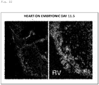
- the heart is a vital organ which is responsible for blood circulation by rhythmic contraction, sustaining the life of an animal. Since this organ is composed of various types of cells such as endothelial cells, cardiomyocytes, and smooth muscle cells, and enables rhythmic contraction, these groups of cells having different functions and morphologies are delicately and elaborately arranged in the heart during their development process.
- a cardiac crescent including a first heart field and a second heart field is formed. Then, the cells on both the left and right sides are fused to form a heart tube having a tubular shape. Then, this heart tube loops to form a looping heart tube, and further, an atrial chamber and a ventricular chamber are generated.
- the heart is completed as a cooperative and functional organ while having a complicated three-dimensional structure.
- iPS cells pluripotent stem cells
- ES cells pluripotent stem cells
- EB embryoid body
- organs having such a functional three-dimensional structure are useful in various applications such as regenerative medicine, drug discovery research, and safety testing.
- organoids such as so-called organoids
- drug discovery research analysis and screening of an organ that has been induced to differentiate from patient-derived disease-specific pluripotent stem cells and the like greatly contribute to the development of a new drug for the disease.
- the safety testing is usually performed on animal cells. Since the response of a living body to a drug is caused by the interaction of various cells, there is a possibility that the safety of a drug can be analyzed with high accuracy by targeting the organ rather than evaluating the cell alone.
- the method for three dimensionally culturing pluripotent stem cells or adult stem cells has been energetically advanced recently, making it possible to induce in-vitro differentiation into endodermal and ectodermal tissues such as the brain, the stomach, and the intestinal tissues by mimicking organ formation in a living body (NPLs 10 and 11).
- the present invention has been made in view of the above-described problems of the related art, and aims to provide a functional organoid such as a heart organoid having a three-dimensional structure, and a method for producing the same.
- a heart organoid having a three-dimensional structure is formed by culturing an embryoid body derived from mouse ES cells in the presence of fibroblast growth factor (FGF) on the surface of a complex composed of laminin or laminin and entactin constituting an extracellular matrix.
- FGF fibroblast growth factor
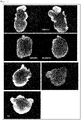
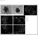
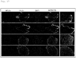
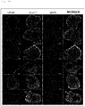
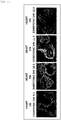
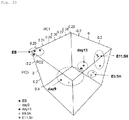
- the present inventors have also made it clear that that the obtained heart organoid exhibits various types of cell markers in a region-specific manner, such as platelet/endothelial cell adhesion molecule-1 (PECAM)/CD31 which is a marker of vascular endothelial cells, ⁇ -smooth muscle actin ( ⁇ SMA) and smooth muscle myosin heavy chain (SM-MHC) which are markers of smooth muscle, and Nkx2-5 and cardiac troponin I which are markers of cardiac muscle.
- PECAM platelet/endothelial cell adhesion molecule-1
- ⁇ SMA ⁇ -smooth muscle actin
- SM-MHC smooth muscle myosin heavy chain
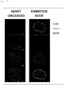
- the present inventors have further clarified that the above method makes it possible to produce heart organoids from human iPS cells via embryoid body formation. What is more, the present inventors have found that the method makes it possible to produce not only heart organoids but also lung organoids. Thus, the present invention has been completed.
- the present invention relates to a method for producing heart organoids and/or lung organoids, and to heart organoids, lung organoids, or fragments or cells of these organoids produced by the method.
- the present invention also relates to a kit used for the production method.
- the present invention relates to a method for evaluating the toxicity of a compound to the organ using the heart organoids or the lung organoids, and a method for evaluating a therapeutic activity of a compound on a disease relating to the organ. More specifically, the present invention provides the following.
- the present invention makes it possible to provide a functional heart organoid and lung organoid having a three-dimensional structure.
- a heart organoid and a lung organoid can be produced without requiring a complicated differentiation induction method adapted to each of the various types of cells constituting these organs (in the heart, endothelial cells, cardiomyocytes, smooth muscle cells, and the like, and in the lung, alveoli cells and the like).
- the regionality of various cells faithfully reproduces that in a living body, and also exerts functions such as myocardial contraction.
- the lung organoid of the present invention also has alveoli, which are an important structure for exerting its function. Therefore, the use of the organoids of the present invention makes it possible to evaluate the toxicity of a compound to the corresponding organs and the therapeutic activity of a compound on diseases related to the organs.
- the method for producing heart organoids and/or lung organoids of the present invention (hereinafter also referred to as "cardiac and other organoids”) is characterized in that it comprises culturing an embryoid body in the presence of an FGF protein on a surface of a gel containing an extracellular matrix constituent protein.
- the " organoid” means an organ-like structure produced in vitro. More specifically, the "heart organoid” means a heart-like structure produced in vitro, having at least one ventricle and one atrium, and capable of performing rhythmic contractions.
- the "lung organoid” means a lung-like structure having an alveolar structure produced in vitro.
- the gel for culturing the later-described embryoid body on the surface thereof may be any gel containing an extracellular matrix constituent protein, and examples thereof include a gel obtained by dissolving an extracellular matrix constituent protein in a buffer solution.
- concentration of extracellular matrix constituent protein in the buffer solution is not particularly limited as long as a gel can be formed.
- the adhesiveness and elasticity of the gel can hinder cell migration, proper patterning, and self-organization that occur during the process of differentiation from embryoid bodies to cardiac and other organoids.
- the concentration of extracellular matrix constituent protein in the buffer solution is preferably 0.5 to 20 mg/mL, more preferably 1 to 10 mg/mL, further preferably 2 to 6 mg/mL, and particularly preferably 3 to 4 mg/mL.
- the buffer solution is not particularly limited, and a buffer solution having a pH of 6 to 8 (preferably a buffer solution having a pH of 7 to 7.5) is usually used.
- examples of the pH-adjusted buffer solution include a phosphate buffer solution, a Tris buffer solution, and a HEPES buffer solution.
- the protein contained in the gel of the present invention may be any protein constituting the extracellular matrix, and its origin is not particularly limited.
- the protein may be derived from human cells, or may be derived from cells of a non-human animal (such as rodents such as mice and rats, mammals such as cattle, horses, pigs, sheep, monkeys, dogs, and cats, and birds such as chickens).
- a secretory product from a mouse Engelbreth-Holm-Swarm (EHS) tumor is preferably used.
- EHS Engelbreth-Holm-Swarm
- extracellular matrix constituent protein examples include laminin, entactin (nidogen 1), fibronectin, collagen, proteoglycan, vitronectin, elastin, fibrillin, and tenascin.
- the gel of the present invention may contain at least one kind of such protein, but may contain two or more kinds thereof. Moreover, when two or more kinds are contained, these different extracellular matrix constituent proteins may be contained in the gel in the form of a hetero complex. In addition, the gel of the present invention may be in a form containing no collagen or proteoglycan.
- a protein containing laminin and/or entactin is preferable, and a complex composed of laminin and entactin is more preferable.
- the content ratio of laminin and entactin in the gel is not particularly limited, but in a molar ratio, laminin : entactin is preferably 30 to 70 : 70 to 30, more preferably 40 to 60 : 60 to 40, and particularly preferably 50 : 50.
- Laminin is a heterotrimeric molecule having three subunit chains ( ⁇ -chain, ⁇ -chain, and ⁇ -chain) associated with one another, and moreover, the ⁇ -chain is classified into ⁇ 1- to 5-subunits, the ⁇ -chain is classified into ⁇ 1- to 3-subunits, and the ⁇ -chain is classified into ⁇ 1- to 3-subunits.
- the "laminin” of the present invention is not particularly limited as long as it is a heterotrimeric molecule composed of the subunits or a partial fragment thereof, and examples thereof include laminin 111 ( ⁇ 1-chain, ⁇ 1-chain, and ⁇ 1-chain), laminin 211 ( ⁇ 2-chain, ⁇ 1-chain, and ⁇ 1-chain), laminin 121 ( ⁇ 1-chain, ⁇ 2-chain, and ⁇ 1-chain), laminin 221 ( ⁇ 2-chain, ⁇ 2-chain, and ⁇ 1-chain), laminin 332 ( ⁇ 3-chain, ⁇ 3-chain, and ⁇ 2-chain), laminin 311 ( ⁇ 3-chain, ⁇ 1-chain, and ⁇ 1-chain), laminin 321 ( ⁇ 3-chain, ⁇ 2-chain, and ⁇ 1-chain), laminin 411 ( ⁇ 4-chain, ⁇ 1-chain, and ⁇ 1-chain), laminin 421 ( ⁇ 4-chain, ⁇ 1-chain, and ⁇ 1-chain), la
- the laminin according to the present invention may be a mixture of these heterotrimeric molecules (for example, a mixture of laminin 411 and laminin 221).
- the gel of the present invention preferably contains laminin 111, laminin 411, or a partial fragment thereof from the viewpoint that the efficiency of differentiation induction into cardiac and other organoids is more likely to increase, and more preferably contains laminin 411 or a partial fragment thereof and further preferably the E8 fragment of laminin 411 (the binding site of laminin 411 to integrin ⁇ 6 ⁇ 1 protein) from the viewpoint that self-organization with normal regionality acquired is more likely to occur in heart organoid formation.
- the embryoid body is cultured on the surface of the above-described gel. Specifically, in the present invention, the embryoid body is cultured while a part thereof is brought into contact with the surface of the gel without being completely embedded (enclosed) or dispersed in the gel.
- the culturing is usually achieved by mounting and culturing the embryoid body on the surface of an incubator covered with the gel of the present invention.
- the incubator coated with the gel of the present invention is not particularly limited as long as it is a cell culturing incubator capable of culturing embryoid bodies, and can be appropriately selected depending on the purpose. Examples thereof include slides, multiplates, microwell plates, Petri dishes, flasks, and dishes.
- the concentration of the gel of the present invention on the surface of the incubator may be appropriately adjusted depending on the type of the gel used, and is not particularly limited, but is preferably 0.1 to 100 ⁇ g/cm 2 .
- the "FGF protein” in the present invention may be any FGF protein that can bind to FGFR1 (IIIc) and FGFR2 (IIIc), and examples thereof include fibroblast growth factor 4 (FGF4) and fibroblast growth factor 2 (FGF2, bFGF). Since FGF4 (206 amino acids) is larger than bFGF (164 amino acids), FGF4 is preferable from the viewpoint that the dimer formation of FGFR is easily induced, and further that the downstream signal transduction is more easily activated.
- the origin of the FGF protein is not particularly limited, and may be derived from humans or non-human animals. Moreover, a partial fragment thereof may be used as long as the embryoid body can be induced to differentiate into cardiac and other organoids.
- the culture in the presence of an FGF protein is usually achieved by adding an FGF protein to the medium.
- the concentration of the FGF protein added to the medium may be any concentration that can induce differentiation of embryoid bodies into cardiac and other organoids, and the lower limit thereof is preferably 1 ng/mL or more, more preferably 3 ng/mL or more, and further preferably 10 ng/mL or more (for example, 20 ng/mL or more, 30 ng/mL or more, 40 ng/mL or more, 50 ng/mL or more, and 60 ng/mL or more).
- the upper limit thereof is preferably 300 ng/mL or less, more preferably 100 ng/mL or less, and further preferably 80 ng/mL or less (for example, 70 ng/mL or less, 60 ng/mL or less, 50 ng/mL or less, and 40 ng/mL or less).
- Preferred examples of the range of concentration of the FGF protein added to the medium include 1 to 100 ng/mL, more preferably 10 to 80 ng/mL, and further preferably 20 to 70 ng/mL.
- the concentration of the FGF protein added to the medium is preferably 10 to 50 ng/mL, and more preferably 20 to 40 ng/mL.
- the culture method for inducing differentiation of embryoid bodies into cardiac and other organoids in the present invention may be culture on the surface of a gel containing an extracellular matrix constituent protein in the presence of an FGF protein, and other culture conditions can be used by those skilled in the art by appropriately selecting a known embryoid body culture method.
- the medium used in culture for inducing differentiation of embryoid bodies into cardiac and other organoids can be prepared based on a known basal medium for culturing embryoid bodies.
- the known basal medium include DMEM medium, Ham's F12 medium, KSOM medium, Eagle's MEM medium, Glasgow MEM medium, ⁇ MEM medium, Ham medium, RPMI 1640 medium, Fisher's medium, BME medium, BGJb medium, CMRL 1066 medium, MEM Zinc optional improvement medium, IMDM medium, Medium 199 medium, and any mixed medium thereof.
- the medium for inducing differentiation into cardiac and other organoids may be a serum-containing medium or a serum-free medium.
- the serum-free medium refers to a medium that does not contain unprepared or unpurified serum, and includes a medium containing purified blood-derived components and animal tissue-derived components (for example, growth factors).
- the medium for inducing differentiation into cardiac and other organoids may contain a serum replacement.
- the serum replacement include commercially available products such as KnockOut Serum Replacement (KSR manufactured by Invitrogen), Chemically Defined Lipid Concentrate (manufactured by Gibco), and GlutaMAX (manufactured by Gibco).
- the medium for inducing differentiation into cardiac and other organoids may contain the FGF protein described above as well as amino acids (such as L-glutamine and non-essential amino acids), hormones (such as progesterone and ⁇ -estradiol), growth factors (such as insulin, transferrin, selenite, and ITS), reducing agents (such as ⁇ -mercaptoethanol), antibiotics (such as penicillin and streptomycin), organic acids (such as pyruvic acid, sodium pyruvate, and lactic acid), fatty acids or lipids, saccharides, vitamins, cytokines, antioxidants, buffer agents, inorganic salts, and the like.
- amino acids such as L-glutamine and non-essential amino acids
- hormones such as progesterone and ⁇ -estradiol
- growth factors such as insulin, transferrin, selenite, and ITS
- reducing agents such as ⁇ -mercaptoethanol
- antibiotics such as penicillin and streptomycin
- organic acids such as
- the medium for inducing differentiation into cardiac and other organoids may be further added with at least one substance selected from the group consisting of growth factors (GSK-3 inhibitors such as BIO (6' bromoindirubin 3' oxime) and Wnt activators such as Wnt1, Wnt3, Wnt3a, Wnt4, Wnt7a, BMP4, BMP2, BMP6, and BMP7), ROCK (Rho-binding kinase) inhibitors (Y-27632), and leukemia inhibitory factors (LIF).
- growth factors GSK-3 inhibitors such as BIO (6' bromoindirubin 3' oxime) and Wnt activators such as Wnt1, Wnt3, Wnt3a, Wnt4, Wnt7a, BMP4, BMP2, BMP6, and BMP7
- ROCK Rho-binding kinase inhibitors
- LIF leukemia inhibitory factors
- Rho-binding kinase inhibitor Y-27632
- ⁇ G protein for example, FGF4
- Y27632 Rho-binding kinase inhibitor
- 1 to 100 ng/mL and preferably 20 to 40 ng/mL of FGF protein (for example, FGF4) or 3 to 300 ng/mL and preferably 40 to 80 ng/mL of FGF protein (for example, FGF4) and 1 to 100 ⁇ M and preferably 5 to 15 ⁇ M (for example, 10 ⁇ M) of Y27632 (Rho-binding kinase inhibitor) can be contained in the medium for inducing differentiation into cardiac and other organoids.
- 3 to 300 ng/mL and preferably 20 to 70 ng/mL (for example, 30 ng/mL) of FGF protein (for example, FGF4 or bFGF), 5 to 500 ng/mL and preferably 10 to 100 ng/mL (for example, 50 ng/mL) of BMP protein (for example, BMP4), and 100 to 10000 u/mL and preferably 500 to 5000 u/mL (for example, 1000 u/mL) of LIF can be contained in the medium for inducing differentiation into cardiac and other organoids.
- the medium may contain 0.25 to 25 ⁇ M, and preferably 1 to 5 ⁇ M (for example, 2.5 ⁇ M) of GSK-3 inhibitor (for example, BIO (Wnt activator)).
- the culture period for inducing differentiation of embryoid bodies into cardiac and other organoids is not particularly limited, and can be appropriately adjusted by those skilled in the art according to the origin of the embryoid body used and the like.
- the period is preferably 5 to 30 days, more preferably 7 to 20 days, and further preferably 9 to 15 days.
- the embryoid bodies are cultured on a gel containing laminin 111 and entactin for 8 to 12 days, and thereafter cultured on a gel containing laminin 411 from the viewpoint that production and long-term culture of mature heart organoids become easier, as presented in Examples described later..
- the culture temperature is not particularly limited, but is usually about 30 to 40°C, and preferably about 37°C.
- concentration of CO 2 is usually about 1 to 10%, and preferably about 2 to 5%.
- oxygen concentration is usually 1 to 10%.
- the "embryoid body" subjected to the above-described culture means an aggregate formed of differentiated cells and undifferentiated cells obtained by suspension culture of pluripotent stem cells or overgrowth in monolayer culture.
- the "pluripotent stem cells” used for forming embryoid bodies may be any cells having a pluripotent differentiation self-renewal ability, and examples thereof include cells that can be collected from a living body, such as embryonic stem cells (ES cells), epiblast stem cells (EpiS cells), embryonal carcinoma cells (EC cells), embryonic germ cells (EG cells), multipotent germline stem cells (mGS cells), and MUSE cells (see Kuroda Y. et al., Proc. Natl. Acad. Sci. U.S.A., 2010, Volume 107, Issue 19, pages 8639 to 8643 ).
- the "pluripotent stem cells” also include cells artificially induced to have pluripotency and the like from somatic cells collected from a living body, such as induced pluripotent stem cells (iPS cells and the like) .
- the origin of these cells is not particularly limited, and includes humans and non-human animals.
- the origin of the cells is a patient suffering from a heart disease
- the present invention makes it possible to obtain a heart organoid useful as a heart disease model.
- the heart disease there is a hereditary heart disease, and examples thereof include congenital long QT syndrome (LQTS), Brugada syndrome, familial bradycardia syndrome (sick sinus syndrome, atrioventricular block), catecholaminergic polymorphic ventricular tachycardia (CPVT), short QT syndrome (SQTS), hypertrophic cardiomyopathy, and dilated cardiomyopathy.
- LQTS congenital long QT syndrome
- CPVT catecholaminergic polymorphic ventricular tachycardia
- SQLTS short QT syndrome
- hypertrophic cardiomyopathy and dilated cardiomyopathy.
- the present invention also makes it possible to obtain a heart organoid useful as a non-hereditary heart disease model.
- the non-hereditary heart disease include coronary artery diseases (myocardial infarction), congenital heart diseases (ventricular septal defect, atrial septal defect, tetralogy of Fallot), and hypertension.
- coronary artery diseases myocardial infarction
- congenital heart diseases ventricular septal defect, atrial septal defect, tetralogy of Fallot
- hypertension Those skilled in the art can reproduce these non-hereditary heart diseases by appropriately preparing (adding or excluding) components to be added to the culture system (medium) of heart organoids (compounds such as myocardial infarction inducers, inhibitors, hormones, and nutrients), or by changing the physical conditions of the culture system (such as temperature and salt concentration).
- the present invention makes it possible to obtain a lung organoid useful as a lung disease model by defining the origin of the cells as a patient suffering from a lung disease.
- the lung disease include hereditary interstitial lung disease such as familial interstitial pneumonia, congenital pulmonary alveolar albinism, and childhood-onset interstitial pneumonia.
- non-hereditary lung diseases include idiopathic interstitial pneumonia (Designated Intractable Disease 85). Interstitial pneumonia is characterized by the fact that the walls of the originally thin alveolar wall become thick and hard (fibrosis) and gas exchange is not successful. Therefore, it is considered that addition of an agent that or the like induces pulmonary fibrosis to the lung organoid in the present invention enables the in-vitro reproduction of interstitial pneumonia.
- the "pluripotent stem cells" of the present invention may be cells that have been genetically modified (genetically modified cells), such as ⁇ MHC-GFP ES cells used in Examples to be described later.
- the genetically modified cells may be cells exogenously introduced with a gene encoding a protein to be expressed such as GFP in the ⁇ MHC-GFP ES cells, may be cells in which the function of a specific gene is suppressed by genome editing, knockout method, RNA method, antisense method, or the like, or may be cells in which the function of a gene is randomly suppressed or activated.
- Examples of the cells in which the function of a gene is randomly suppressed or activated include cells treated with a chemical mutagen such as EMU, EMS, NMU, and NTG, cells introduced with a DNA-cleaving enzyme such as zinc finger nuclease or TALEN, cells irradiated with fast neutrons, gamma rays, or ion beams, and cells in which transposons are randomly inserted into genomic DNA.
- a genetic modification may be performed after the cells are established, or may be performed before establishment, for example, at the individual stage of collecting the unfertilized egg or the sperm.
- a gene mutation causing the disease may be introduced by genetic modification.
- the number of pluripotent stem cells provided for forming an embryoid body is not particularly limited as long as the embryoid body can be formed, and can be appropriately adjusted by those skilled in the art according to the type and origin of the pluripotent stem cells, and usually 50 to 50000.
- the number of pluripotent stem cells for forming one embryoid body is preferably 100 to 10000, more preferably 500 to 7000, and more preferably 1000 to 5000.
- the method for culturing pluripotent stem cells is not particularly limited as long as embryoid bodies can be formed, and may be adhesion culture or non-adhesion culture (for example, suspension culture such as coagulation suspension culture and suspension culture on a carrier).
- suspension culture such as coagulation suspension culture and suspension culture on a carrier.
- the medium used in such culture can be prepared based on a known basal medium for culturing pluripotent stem cells.
- the known basal medium include DMEM medium, KSOM medium, Eagle's MEM medium, Glasgow MEM medium, ⁇ MEM medium, Ham medium, RPMI 1640 medium, Fisher's medium, BME medium, BGJb medium, CMRL 1066 medium, MEM Zinc optional improvement medium, IMDM medium, Medium 199 medium, and any mixed medium thereof.
- a commercially available culture medium for culturing ES cells or iPS cells is also preferably used.
- the medium for forming an embryoid body may be a serum-containing medium or a serum-free medium, or may contain a serum replacement.
- LIF is usually added to the above-described medium during the maintenance culture of pluripotent stem cells, it is preferable not to add LIF in the present invention from the viewpoint that culture for forming embryoid bodies requires differentiation induction, not maintenance of pluripotency.
- the incubator used for culture to form embryoid bodies known ones used for culturing pluripotent stem cells and the like can be appropriately used, and in order to perform suspension cells, a non-adhesive (low-adsorption) incubator is preferable.
- the incubator include multiplates, microwell plates, petri dishes, flasks, and dishes coated with a hydrophilic compound or a water-soluble resin.
- the culture period for forming embryoid bodies is not particularly limited, and can be appropriately adjusted by those skilled in the art according to the type and origin of the pluripotent stem cells to be used, and is usually 1 to 14 days, preferably 2 to 7 days, and more preferably 3 to 5 days.
- the culture temperature is not particularly limited, but is usually about 30 to 40°C, and preferably about 37°C.
- the concentration of CO 2 is usually about 1 to 10%, and preferably about 2 to 5%.
- the oxygen concentration is usually 5 to 21%.
- the present invention also provides a kit used for producing the above-described cardiac and other organoids.
- the kit of the present invention comprises at least the above-described gel containing an extracellular matrix constituent protein, the above-described medium for culturing an embryoid body (medium for inducing differentiation into cardiac and other organoids), and an FGF protein to be added to the medium.
- the kit of the present invention may include a solution containing an extracellular matrix constituent protein before gelation instead of or in combination with the gel.
- kit of the present invention may further include LIF, BMP, and the like to be added to the medium, and may further include the embryoid body described above.
- the kit of the present invention may include the above-described medium for forming an embryoid body from pluripotent stem cells, and the pluripotent stem cells.
- the kit of the present invention can also include materials for producing the cardiac and other organoids as well as a reagent for confirming that the produced structures are the cardiac and other organoids.
- the reagent include an antibody that recognizes a cell-specific marker constituting an organ, and a labeled secondary antibody that recognizes the antibody.
- an intracellular calcium indicator that emits green fluorescence may be used, as in Examples to be described later.
- the kit of the present invention can include instructions for using the kit.
- the heart organoid and the lung organoid produced by the above-described method reproduce the structure responsible for their functions in vivo.
- the regionality of various cells faithfully reproduces that in a living body, and also exerts functions such as myocardial contraction.
- use of that organoid enables screening of a compound for treating, ameliorating, or preventing a disease relating to the corresponding organ or a compound having toxicity to the organ, and further enables regenerative medicine for diseases and the like related to the organ.
- the present invention also provides heart organoids, lung organoids, or fragments or cells of these organoids obtained by culturing an embryoid body on the surface of a gel containing an extracellular matrix constituent protein at least in the presence of an FGF protein.
- the organoids, fragments, or cells thereof may be for use in transplantation into a living body.
- the "organoid fragments" may be any part of the organoid.
- examples thereof include ventricles and atria
- examples thereof include alveoli.
- the " organoid cells” may be any cells that constitute an organoid.
- examples thereof include vascular endothelial cells, cardiomyocytes, and smooth muscle cells
- examples thereof include alveoli cells.
- the heart organoid of the present invention is a structure in which the blood inlet and outlet are closed.
- the size of the heart organoid of the present invention is not particularly limited and can be appropriately adjusted according to the size of the embryoid body subjected to the production method of the present invention, the number of pluripotent stem cells subjected to the formation of the embryoid body, and the like. Therefore, in the present invention, it is possible to provide a heart organoid having a size of 400 to 1200 ⁇ m (for example, 600 to 1000 pm, 700 to 900 ⁇ m), for example. In the present invention, the size of the heart organoid means the longest diameter.
- the lung organoid of the present invention has a structure similar to an alveolus or an alveolar duct formed by connecting two or more alveoli.
- a lung organoid having a size of 1 mm or more for example, 1 to 1.6 mm, for example.
- the size of the lung organoid is not particularly limited as in the case of the heart organoid described above, and can be appropriately adjusted.
- the present invention makes it possible to obtain a heart organoid and a lung organoid in a connected form. Therefore, the present invention also provides a conjugate of a heart organoid and a lung organoid.
- the heart organoid of the present invention faithfully reproduces the structure and function of the heart in a living body.
- the lung organoid of the present invention has alveoli, which are an important structure for exerting its function. Therefore, these organoids are useful for evaluating the toxic activity of a compound on the corresponding organ. Particularly with respect to the heart, these organoids may be useful because prolonged QT by drugs is also associated with sudden death.
- lung organoid it is also effective in evaluating whether a newly developed drug (particularly anticancer drug) has side effects of drug-induced pneumonia or pulmonary fibrosis (evaluation of pulmonary toxicity).
- the present invention provides a method for evaluating toxicity of a compound to a heart, comprising the steps of: bringing the heart organoid of the present invention into contact with a test compound to detect a condition of the heart organoid; and judging that the test compound is a compound having toxicity to the heart if deterioration is observed in the condition detected in the previous step.
- the present invention provides a method for evaluating toxicity of a compound to a lung, comprising the steps of: bringing the lung organoid of the present invention into contact with a test compound to detect a condition of the lung organoid; and judging that the test compound is a compound having toxicity to the lung if deterioration is observed in the condition detected in the previous step.
- the origin of the cardiac and other organoids of the present invention is a patient suffering from diseases relating to the corresponding organs, and further if the content components and/or physical conditions of the culture system of the cardiac and other organoids are adjusted, it is possible to use the organoids as a model for the diseases.
- the present invention provides a method for evaluating therapeutic activity of a compound on a heart disease, comprising the steps of: bringing the heart organoid of the present invention exhibiting a heart disease into contact with a test compound to detect a condition of the heart organoid; and judging that the test compound is a compound having therapeutic activity on the heart disease if a therapeutic effect on the heart disease is observed in the condition detected in the previous step.
- the present invention provides a method for evaluating therapeutic activity of a compound on a lung disease, comprising the steps of: bringing the lung organoid of the present invention exhibiting a lung disease into contact with a test compound to detect a condition of the lung organoid; and judging that the test compound is a compound having therapeutic activity on the lung disease if a therapeutic effect on the lung disease is observed in the condition detected in the previous step.
- organoid and lung organoid used in the evaluation method of the present invention are as described above, but the entirety of these organoids may be subjected to the evaluation method, or a part (fragment) of these organoids or a cell isolated from the organoids may be used.
- test compound to be brought into contact with the cardiac and other organoids of the present invention is not particularly limited, and a compound having toxicity to the corresponding organ or a compound having a therapeutic activity on a disease relating to the organ can be screened by using a synthetic low molecular compound library, an expression product of a gene library, a peptide library, an antibody, a bacterial release substance, a cell (microorganism, plant cell, animal cell) extract liquid and culture supernatant, a purified or partially purified polypeptide, an extract from marine organisms, plants, or animals, soil, or a random phage peptide display library.
- a synthetic low molecular compound library an expression product of a gene library, a peptide library, an antibody, a bacterial release substance, a cell (microorganism, plant cell, animal cell) extract liquid and culture supernatant, a purified or partially purified polypeptide, an extract from marine organisms, plants, or animals, soil, or a random phage peptid
- the "contact" with cardiac and other organoids can be usually performed by adding a test compound to a medium for culturing (maintaining) the cardiac and other organoids.
- a medium for culturing (maintaining) the cardiac and other organoids preferably used as the medium.
- the "condition" of the cardiac and other organoids detected in the evaluation method of the present invention is not particularly limited, and examples thereof include the form (appearance and internal form) and functions (such as contraction force, contraction rhythm, and the rate of beats for the heart, and gas exchange for the lung) of the heart and the like.
- the test compound can be judged as a compound having toxicity to the heart and the like.
- therapeutic activity if an improvement in the form or functions of the heart and the like (therapeutic effect) is observed, the test compound can be judged as a compound having a therapeutic activity on diseases of the heart and the like.
- embryoid bodies For the purpose of forming embryoid bodies, 1000 to 5000 ES cells were seeded in one well of a 96-well U-bottom plate (SUMILON Prime Surface (registered trademark) 96 Well U), and cultured in LIF-free FBS-ES medium at 37°C for 4 days. In addition, for the purpose of examining the minimum number of ES cells required for generating a heart organoid, embryoid bodies were formed using 500 ES cells.
- the embryoid bodies were transferred onto a chamber slide (manufactured by Falcon, catalog number: 35418) coated with 85.7 ⁇ g/cm 2 LN/ET gel (manufactured by BD, catalog number: 354259) together with 200 ⁇ L of a heart organoid differentiation induction medium.
- the heart organoid differentiation induction medium was DMEM/F12 (manufactured by Gibco, catalog number: 11320033) containing 30 ng/mL or 60 ng/mL FGF4, 50 ⁇ g/mL penicillin/streptomycin, 20% KSR, 1 mM sodium pyruvate, 100 ⁇ M ⁇ -mercaptoethanol, 2 mM L-glutamine, 60 ng/mL progesterone, 30 ng/mL ⁇ -estradiol, 5 ⁇ g/mL insulin, 20 ⁇ g/mL transferrin, and 30 nM selenite. Note that the concentration of FGF4 added to the medium was 30 ng/mL unless otherwise specified.
- the embryoid bodies were cultured at 37°C and 5% CO 2 for 10 to 15 days (medium exchange was performed on days 3, 5, 7, 9, 11, 13, 14, and 15).
- the embryoid bodies were cultured at 37°C and 5% CO 2 for 10 to 15 days (medium exchange was performed on days 3, 5, 7, 9, 11, 13, 14, and 15).
- 50 ng/ml BMP4, 2.5 ⁇ M BIO (6' bromoindirubin 3' oxime), and 1,000 units/mL LIF were added to the heart organoid differentiation induction medium and cultured.
- the heart organoids were collected for further analysis such as immunofluorescent staining and calcium ion measurement.
- the heart organoids were collected after 10 to 15 days of culture, subjected to Tissue-Tek (registered trademark) OCT Compound embedment in a plastic tissue mold (CRYO DISH manufactured by SHOEI WORK'S CO., LTD.), and frozen. Frozen sections were prepared to a thickness of 5 to 7 ⁇ m using a cryostat at -16°C, and transferred onto a MAS-coated glass slide (manufactured by Matsunami Glass Ind., Ltd.) or a poly-L-lysine-coated slide (manufactured by Sigma-Aldrich). Embryoid bodies that were not subjected to induction of differentiation into heart organoids were also collected, cryo-embedded in OCT, and prepared into sections in the same manner as described above.
- mice C57BL/6, 9.5 to 13.5 days after sexual intercourse
- the fetuses at various stages were dissected.
- the embryonic hearts Prior to freezing in OCT, the embryonic hearts were immersed in sucrose-PBS solutions (4%, 10%, 15% and 20%).
- the embryonic hearts frozen in OCT were prepared in cryostats into 7 ⁇ m thick sections.
- the frozen sections were immersed in 4% paraformaldehyde-PBS at room temperature for 15 minutes and immobilized. The immobilized sections were washed three times in PBS for 5 minutes. Next, the sections were immersed in a blocking buffer (5% normal goat serum and 0.3% Triton-X100, or 5% bovine serum albumin (BSA) and 0.3% Triton-X100) for 1 hour at room temperature. The sections were incubated overnight at 4°C with the primary antibody diluted in PBS containing 1% BSA and 0.3% Triton-X100. After washing with PBS, the sections were incubated with the secondary antibody for 1 to 2 hours at room temperature.
- a blocking buffer 5% normal goat serum and 0.3% Triton-X100, or 5% bovine serum albumin (BSA) and 0.3% Triton-X100
- the slides were counterstained with DAPI (dilution ratio: 1 : 1000, manufactured by Dojindo Laboratories) and mounted on VECTASHIELD HardSet Anti-Fading Kit (manufactured by Vector Laboratories).
- Tbx5 manufactured by Abcam, ab137833
- cardiac troponin I manufactured by Abcam, ab47003
- Nkx2-5 manufactured by Abcam, ab91196
- nestin manufactured by Abcam, ab105389
- Oct3/4 manufactured by Santa Cruz Biotech
- PECAM manufactured by BD
- Mlc2a manufactured by Synaptic System, #311 011)
- Mlc2v manufactured by Synaptic System, #310 003
- SM-MHC manufactured by Abcam and manufactured by R&D Systems
- ⁇ SMA ab5694
- the heart organoids were washed twice with PBS and immersed in a 4 ⁇ M green fluorescent intracellular calcium indicator Fluo8 AM (acetoxymethyl) or Fluo8 AM/F127, freshly diluted with Tyrode's solution containing 1.8 mM Ca 2+ , at 37°C for 15 to 30 minutes. Thereafter, the heart organoids were washed twice with PBS, treated with 200 ⁇ L of Tyrode's solution, mounted, and observed with a fluorescence microscope (manufactured by Keyence Corporation).
- a heart shows a characteristic morphological change accompanied by differentiation into a wide variety of cells such as vascular endothelial cells, cardiomyocytes, and smooth muscle cells.
- a cardiac crescent including a first heart field and a second heart field is characterized by the expression of cardiac mesoderm genes (Mesp1, Flk1, Pdgfra) induced by the expression of brachyury.
- a linear beating heart tube is formed.
- the heart tube in a tubular shape having vascular endothelial cells in the inner layer and cardiomyocytes in the outer layer loops by beating power or outflow tract (OFT).
- OFT beating power or outflow tract
- mouse ES cells were cultured in a low-binding U-bottom 96-well plate in the absence of leukemia inhibitory factor (LIF) to obtain complete embryoid bodies (EB) (1000 to 5000 cells/embryoid body). Then, the resulting embryoid bodies were cultured in the presence of an exogenous FGF signal (FGF4) on the surface of a gelated LN/ET complex containing components of the extracellular matrix (ECM) in the connective tissue.
- LIF leukemia inhibitory factor
- the embryoid bodies were cultured in the presence of FGF4 for 8 days throughout, and from day 9, added with BIO (GSK-3 inhibitor, Wnt activator), BMP4, and LIF and cultured for 10 days or more.
- BIO GSK-3 inhibitor, Wnt activator
- BMP4 BMP4 and LIF and cultured for 10 days or more.
- Fig. 2 a beating heart organoid having more than one cardiac chamber and resembling an embryonic heart was successfully produced.
- Fig. 3 it was also confirmed that the mouse ES cell-derived heart organoid produced in this manner had a morphologically two-atrial two-ventricular structure, not a spherical structure exhibiting a mere contractive action.
- the cardiac crescent-like (CCL) structure, the heart tube, and the looping heart tube generated together with the formation of the cardiac chamber were formed in the same order as in the process of developing the embryonic heart. The only exception is the appearance of spheres with buds (Sb) showing some polarity. This may be due to the difference between the precursor structure of the cardiac crescent or heart tube in vivo and the artificial structure due to in-vitro embryoid body culture.
- Sb spheres with buds
- cardiac progenitor cells move from the first heart field to the midline to form a beating linear heart tube having myocardium.
- This process, as shown in Fig. 5 is consistent with the detection of naturally occurring beats primarily in heart organoids containing a cardiac crescent-like structure (52.4% on day 3 of culture, 72.6% on day 6 of culture).
- morphogenesis the first morphological change was observed on day 3 of culture in the embryoid bodies in a spherical shape (89.3%) as shown in Fig. 6 , which mainly included cardiac crescent-like structures derived from the outgrowths of hatched embryoid bodies, resembling the outgrowth and patterning during limb bud formation by Fgf signals (see Powers, C. J. et al., Endocrine-Related Cancer 7, 165-197 (2000 )). Further, by day 6 of culture, this change was complete in most embryoid bodies (96.4%).
- Fig. 7 most of the heart organoids were cardiac crescent-like structures on day 3 of culture. These organoids shifted to the formation of a heart tube, a looping heart tube, and a cardiac chamber, and finally reached the formation of a cardiac chamber by days 10 to 15 of culture. This process is also similar to that of heart morphogenesis in vivo.
- heart organoids in this culture system can be said as a summary of the morphological change in embryonic heart development.
- the reason why such morphological change could be reproduced is not clear, but is assumed as follows.
- the extracellular matrix was able to induce efficiently morphogenesis without inhibiting cell migration during the process of forming heart organoids and by reducing mechanical hindrance preventing proper patterning and self-organization.
- the size and beating ability of the obtained heart organoids were significantly reduced, and the number of heart organoids in the later morphogenetic steps (the stages of forming a looping heart tube and a cardiac chamber) was also reduced. From this, it is conceivable that a decrease in the number of cardiomyocytes caused a deficiency in morphogenesis, reducing beating.
- Tbx5 T-box protein
- Tbx5 functions in coordination with zinc finger transcription factors and the GATA family to activate cardiac marker genes.
- Tbx5 is at an increased level on embryonic day 10.5 as compared with that of the heart on embryonic day 9.5, and continues to increase until embryonic day 12.5. Meanwhile, as shown in Fig. 14 , an increase in Tbx5 expression was also observed in, for example, heart organoids on days 10 to 15 of culture.
- the specific pattern of Tbx5 expression in heart organoids resembled that of the embryonic heart in vivo after cardiac chamber formation.
- the expression of Tbx5 was positive in the left atrium, right atrium, and left ventricle derived from the first heart field, and was negative in the right ventricle derived from the second heart field.
- Fig. 14 the segregated expression of platelet/endothelial cell adhesion molecule-1 (PECAM) was detected in most heart organoids. Specifically, expression of a representative EC marker was observed in inner membranes in primitive heart tubes and in vascular endothelial cells in multi-chambered heart organoids.
- PECAM platelet/endothelial cell adhesion molecule-1
- the expression of the marker revealed that the heart organoids cultured in vitro had cardiac structural cells essential in a three-dimensional structure similar to the heart in vivo, as shown in Figs. 15 and 16 .
- cardiomyocytes in heart organoids were mature and acquired regionality in terms of cardiomyocyte subtypes (for example, atrial chamber and ventricular chamber)
- the expression of the sarcomeric proteins myosin light chain 2 atrial (Mlc-2a) and myosin light chain 2 ventricular (Mlc-2v) in heart organoids was examined.
- Mlc-2v was dominantly expressed in most of the heart organoids, and weak expression of Mlc-2a was observed in only a few heart organoids.
- Figs. 18 and 19 expression of Mlc-2a was observed in the entire heart on embryonic day 9.5 (looping heart tube), but was observed in the atria on embryonic day 10.5 after cardiac chamber formation. Meanwhile, expression of Mlc-2v was observed in the atria on embryonic day 10.5, and very strong expression of Mlc-2v was observed in the left ventricle on embryonic day 12.5.
- CM marker expressed in striated muscle
- PECAM EC marker
- SM-MHC SM marker
- heart organoids can mimic the structural characteristics of the embryonic heart.
- ⁇ -smooth muscle actin which is a marker of smooth muscle and Nkx2-5 (homeodomain transcription factor indicating cardiac lineage)
- ⁇ SMA smooth muscle actin
- Nkx2-5 homeodomain transcription factor indicating cardiac lineage
- an immunostaining assay was performed on Oct3/4, a marker of pluripotent stem cells.
- nestin expressed as a marker for neural stem cells
- NCC neural crest cells
- heart organoids were produced using ES cells expressing GFP- ⁇ MHC only in the atria in a restricted manner ( ⁇ MHC-GFP ESCs, see Nemer, M. et al., Cardiovasc Pathol 17, 48-54, doi: 10.1016/j.carpath.2007.06.005 (2008 )).
- heart organoids having multi-chambered heart organoids by the above differentiation induction protocol, similarly to wild-type ES cells.
- these heart organoids shoed ⁇ MHC-GFP expression in a region-restricted manner, indicating that the organoids possessed the capacity for atria formation.
- An evaluation was made as to whether a particular cardiac function, such as calcium oscillation, was obtained when spontaneous beating was detected in heart organoids. Specifically, an intracellular calcium indicator was used to measure Ca 2+ levels, thereby analyzing complete 3D heart organoids without dissociation (n 10).
- TEM transmission electron microscope
- the microstructure analysis using a transmission electron microscope revealed that the heart organoid of the present invention contained a sarcomere structure including a Z band, and an intercalated disc (ID) which is a characteristic structure of cardiomyocytes (see the two photographs on the left of Fig. 28 ).
- ID intercalated disc
- these microstructures were similar to the microstructures of the embryonic heart (see the two photographs on the right of Fig. 28 ).
- an embryoid body and heart organoids on days 8 and 12 after culture were each treated with collagenase (1 mg/ml) at 37°C for 15 minutes, and then treated with TrpLE (manufactured by Gibco) at 37°C for 10 to 15 minutes. Then, the embryoid body, the heart organoids, and the heart of a living body were each subjected to a pipetting treatment so as to be separated into individual cells. The cells thus prepared were treated with 4% PFA/PBS at 4°C for 10 minutes and immobilized.
- PCA Principal Component Analysis
- RNA-seq analysis was prepared using KAPA Standard mRNA-Seq Kit (manufactured by KAPA Biosystems).
- the obtained library was subjected to sequencing by HiSeq 1500 (single end 50bp reads with HiSeq SR Rapid Cluster Kit v2 and HiSeq Rapid SBS Kit v2, manufactured by Illumina).
- HiSeq 1500 single end 50bp reads with HiSeq SR Rapid Cluster Kit v2 and HiSeq Rapid SBS Kit v2, manufactured by Illumina.
- Bowtie2 was used to perform mapping to a mouse reference genome sequence (GRCm38/mm10).
- Bioconductor package DEGseq was used to determine the read number and RPKM value of each gene.
- genes having a difference in expression between the heart organoids and the embryoid body were developed on an MA plot based on the above RNA-seq data.
- 39 genes highly expressed in heart organoids (regulated P-value ⁇ 0.01, log2FoldChange > 5) were extracted, and 55 genes lowly expressed in heart organoids (regulated P-value ⁇ 0.01, log2FoldChange ⁇ -5) were extracted.
- GO gene ontology analysis was performed on these genes, and as a result, among the 39 highly expressed genes, 26 out of the top 30 GO terms were heart-related (see Fig. 33 ).
- the terms of the top 30 GO did not include heart-related ones (see Fig. 34 ). As described above, it was confirmed that a large number of heart-related genes were included in the group of genes highly expressed in heart organoids.
- Fig. 35 the presence of Purkinje cells (TRPM4 positive) was observed in the heart organoid. Since these cells are stimulation conduction system cells responsible for ventricular contraction, it was confirmed that a conduction system was formed also in the heart organoid as in the living heart.
- TRPM4 positive Purkinje cells
- the expression of potassium channel iK1 was detected by immunofluorescent staining.
- the potassium channel iK1 is necessary for increasing the rate of rise of the action potential and the rate of conduction, and is detected only in mature cardiomyocytes.
- the immunofluorescent staining was performed in the same manner as the above (immunofluorescent staining) except that KCNN4 (iK1, GTX54786) was used as the primary antibody.
- T-tubule Transverse tubule
- a dihydropyridine receptor (DHPR, voltage-dependent L-type Ca 2+ channel) present in the T-tubule of the cells is activated.
- the activated receptor causes a small amount of Ca 2+ to flow into cells.
- the ryanodine receptor (RYR, Ca 2+ release channel) in the sarcoplasmic reticulum (SR) releases a large amount of Ca 2+ in the sarcoplasmic reticulum, causing muscle contraction (see Steven, O. M et al., Cell, 101, 365 to 376 (2000 )).
- optical mapping was performed by the following method. Note that, as another electrophysiological evaluation method, there is an extracellular potential recording method using a multipoint planar electrode (MEA), but this method cannot predict arrhythmia. On the other hand, use of optical mapping makes it possible to show that not only spontaneous excitation and membrane potential but also the interval between waveforms changes irregularly, so that arrhythmia can also be predicted.
- MEA multipoint planar electrode
- Optical mapping was performed using a high-speed CMOS camera system (MiCAM Ultima, Brainvision, Tokyo, Japan) according to the method described by Ihara et al. (J Vis Exp. 2018; (132): 56478 ) (See Fig. 38 ).
- the heart organoid was subjected to a staining treatment for 15 minutes using 15 ⁇ M di-4-ANEPPS (voltage-sensitive dye, Wako, Tokyo, Japan). After washing with PBS, the organoid was incubated for 15 minutes with 30 ⁇ M blebbistatin (excitation/contraction uncoupler, Sigma-Aldrich, St. Louis, MO) to suppress motion artifacts.
- the heart organoid was transferred to a glass bottom dish filled with Tyrode's solution (135 mM NaCl, 5.4 mM KCl, 1.8 mM CaCl 2 , 0.53 mM MgCl 2 , 0.33 mM NaH 2 PO 4 , 5.5 mM D-glucose, and 5.0 mM HEPES (pH 7.40, adjusted with aeration of NaOH and 100% O 2 )), and subjected to optical mapping. The temperature was maintained at 37°C during the optical mapping process.
- the heart organoid of the present invention has a high degree of maturity. For this reason, it can be an effective material for cardiotoxicity tests and the like for evaluating drug safety. Therefore, for the purpose of confirming its effectiveness, a drug causing cardiac dysfunction was used to evaluate the responsiveness of a drug to heart organoids.
- Ca 2+ measurement was performed by the method described above (Ca 2+ Measurement) in the presence and absence (control) of 1 ⁇ M isoproterenol, which is an adrenergic ⁇ -receptor agonist.
- a 15-second recording (movie) was taken, and image analysis was performed to detect the change in the intensity (Y-axis) of the fluorescent signal with time (X-axis).
- Fig. 40 it was confirmed that the addition of isoproterenol made it possible to shorten the Ca signal cycle (tachycardia-like) in heart organoids. Specifically, it was confirmed that the use of the heart organoid of the present invention made it possible to evaluate drug responsiveness in which muscle contraction is controlled by calcium transients.
- mouse ES cells were cultured in a low-binding U-bottom 96-well plate in the absence of leukemia inhibitory factor (LIF) to obtain complete embryoid bodies. Then, the resulting embryoid bodies were cultured in the presence of an exogenous Fgf signal (FGF4) on the surface of laminin 411 ((manufactured by Nippi Inc., iMatrix-411; purified product of integrin binding site (E8 section) of human laminin 411 protein)), laminin 221 (manufactured by Nippi Inc., iMatrix-221; purified product of integrin binding site (E8 section) of human laminin 221 protein), or a mixture of laminins 221 and 411 in equal amounts, containing components of the extracellular matrix (ECM) in the connective tissue.
- FGF4 exogenous Fgf signal
- each laminin was performed at 10.7 ⁇ g/cm 2 .
- the culture period of the embryoid bodies and medium exchange were also performed as described above (Cell Culture), and after the culture, the heart organoids were collected for further analysis such as immunofluorescent staining.
- the heart organoids obtained above were analyzed by the method described above (immunofluorescent staining).
- the expression of Mlc2a and Mlc2v was lower in the organoids cultured on the surface of laminin 221 than in the heart organoids cultured on the surface of laminin 411.
- the state was conspicuous where atrial type cardiomyocytes were not separated but scattered.
- the organoids cultured on the surface of laminin 221 had low expression of cardiac troponin T and TRPM4.
- expression of cardiac troponin T and TRPM4 equivalent to that in the mouse heart was observed.
- an appropriate positional relationship between normal cardiomyocytes and Purkinje cells was also confirmed, suggesting the formation of a conduction system.
- laminin 411 contributes to self-organization that has acquired normal regionality in cardiac development.
- mouse ES cells were cultured in a low-binding U-bottom 96-well plate in the absence of leukemia inhibitory factor (LIF) to obtain complete embryoid bodies. Then, the resulting embryoid bodies were cultured for 10 days in the presence of an exogenous Fgf signal (FGF4) on the surface of laminin 111-entactin (LN/ET complex) containing components of the extracellular matrix (ECM) in the connective tissue.
- FGF4 exogenous Fgf signal
- LN/ET complex laminin 111-entactin
- the heart organoids were transferred onto a chamber slide coated with laminin 411 (0.5 ⁇ g/cm 2 ) or a mixture of laminin 411 and laminin 111, and the culture was continued until day 21 to observe their morphology under a microscope. Note that exchange of the medium of the embryoid bodies was performed as described above (Cell Culture) except for adding BMP4 (10 ng/mL), FGF4 (30 ng/mL), and LIF (1000 units/mL) from day 9 of culture.
- human iPS cells (253G1 (Riken BRC #HPS0002) were cultured in a low-binding U-bottom 96-well plate in the absence of LIF to obtain embryoid bodies. Then, the resultant embryoid bodies were cultured on the surface of a gelated LN/ET complex under the conditions presented in the following table, that is, in the presence of FGF4 (conditions I, II, and IV) or in the presence of FGF4 and Y27632 (ROCK inhibitor) (condition III). On day 11 of culture, the cells were transferred to a new culture device and the culture was continued.
- FGF4 condition I, II, and IV
- ROCK inhibitor FGF4 and Y27632
- lung development is known to begin with trachea formation on embryonic days 9.5 to 10 (fetal period) and end on embryonic day 12.5.
- Important points for lung development are branching morphogenesis and epithelial organization. Structural changes taking place during lung development are controlled by the expression of genes different at the proximal and terminal.
- the heart on embryonic day 10.5 has completed the formation of two atria and two ventricles, but the lungs are in close contact with the heart while keeping the form of small tissues connected to the trachea, and their development is delayed in initial timing as compared with the development of the heart.
- Figs 51 to 53 show the obtained results.
- Fig. 51 in the human iPS cell-derived heart organoids, the expression of cardiac factors Tbx5 and Nkx2-5 was observed.
- Fig. 52 the expression of Mlc-2v, which is a mature ventricular marker, was also observed.
- the expression of cardiac troponin I was observed in all human iPS cell-derived heart organoids examined, and the expression of PECAM was observed in some heart organoids (see Fig. 53 ).
- histological analysis was performed as in the case of the heart organoids described above to determine whether the genes expressed in epithelial cells constituting the lung were to be properly expressed in the human iPS cell-derived lung organoids obtained above. Note that the analysis was performed in the same manner as described above (immunofluorescent staining) except that ECAD (ab11512) and CK8 (ab53280) were used as primary antibodies. Fig. 54 shows the obtained results.
- alveoli which are an important structure of the lung, are surrounded by an alveoli wall composed of the type I alveoli cells and type II alveoli cells being epithelial cells.
- ECAD E cadherin
- SFTPC SFTPC
- CK8 Cytokeratin 8
- the alveoli walls are organized in the alveoli by epithelial cells expressing ECAD and CK8.
- epithelial organization of alveolus-like portions was observed. That is, in the lung organoid of the present invention, an alveolar structure showing morphological similarity was observed.
- the method of the present invention makes it possible to produce heart organoids regardless of the type of pluripotent stem cells (ES cells, iPS cells, and the like) and their origins (mouse, human, and the like).
- ES cells pluripotent stem cells
- iPS cells pluripotent stem cells
- their origins mouse, human, and the like.
- the method of the present invention makes it possible to produce a lung organoid having an alveolar structure.
- the present invention has made it possible to produce heart organoids containing at least cardiomyocytes, endothelial cells, and smooth muscle cells.
- the heart organoids thus obtained exhibited within a short culture period the same structural and functional characteristics as the in vivo counterpart, such as expression of transcription factors and structural proteins in the heart, region-specific expression of various types of cardiac cells, functional calcium oscillations, and electrophysiological properties.
- the present invention makes it possible to produce lung organoids.
- the lung organoids thus obtained have alveoli, which are an important structure responsible for the function of the lung.
- the heart organoid and lung organoid of the present invention reproduce their structures and functions in vivo, and thus are useful in regenerative medicine, pharmaceutical development, safety testing, and the like.
Applications Claiming Priority (2)
| Application Number | Priority Date | Filing Date | Title |
|---|---|---|---|
| JP2017190950 | 2017-09-29 | ||
| PCT/JP2018/036538 WO2019066059A1 (fr) | 2017-09-29 | 2018-09-28 | Organoïde d'organe et procédé de production associé |
Publications (2)
| Publication Number | Publication Date |
|---|---|
| EP3699275A1 true EP3699275A1 (fr) | 2020-08-26 |
| EP3699275A4 EP3699275A4 (fr) | 2021-07-07 |
Family
ID=65902492
Family Applications (1)
| Application Number | Title | Priority Date | Filing Date |
|---|---|---|---|
| EP18860752.7A Pending EP3699275A4 (fr) | 2017-09-29 | 2018-09-28 | Organoïde d'organe et procédé de production associé |
Country Status (5)
| Country | Link |
|---|---|
| US (1) | US20200283735A1 (fr) |
| EP (1) | EP3699275A4 (fr) |
| JP (1) | JP7315923B2 (fr) |
| MA (1) | MA50831A (fr) |
| WO (1) | WO2019066059A1 (fr) |
Families Citing this family (8)
| Publication number | Priority date | Publication date | Assignee | Title |
|---|---|---|---|---|
| US20220162559A1 (en) * | 2018-07-11 | 2022-05-26 | Clemson University Research Foundation | Myocardial organoids and methods of making and uses thereof |
| KR102291265B1 (ko) * | 2020-03-19 | 2021-08-18 | 연세대학교 산학협력단 | 심장 오가노이드, 이의 제조 방법 및 이를 이용한 약물 독성 평가 방법 |
| WO2021225794A1 (fr) * | 2020-05-04 | 2021-11-11 | City Of Hope | Procédés pour la production d'organoïdes |
| WO2022005023A1 (fr) * | 2020-06-29 | 2022-01-06 | 주식회사 오간팩토리 | Organoïde de mésencéphale, son procédé de production rapide en grandes quantités, et procédé de criblage de neurotoxines et procédé de criblage de médicaments pour une maladie associée aux neurones dopaminergiques l'utilisant |
| KR102346243B1 (ko) * | 2020-06-29 | 2022-01-04 | 주식회사 오간팩토리 | 중뇌 오가노이드, 이의 고속 및 대량 제조 방법, 이를 이용한 신경독성물질 스크리닝 방법 및 도파민성 신경세포 관련 질환 치료제 스크리닝 방법 |
| US20230101335A1 (en) | 2021-05-06 | 2023-03-30 | Massachusetts Institute Of Technology | Ex vivo tissue explant and graft platform and uses thereof |
| KR20220153170A (ko) | 2021-05-10 | 2022-11-18 | (주) 넥셀 | 인간 전분화능 줄기세포 유래 심장 오가노이드 제조 방법 및 이를 통해 제조된 인간 전분화능 줄기세포 유래 심장 오가노이드 |
| CN115975907A (zh) * | 2022-12-16 | 2023-04-18 | 北京镁伽机器人科技有限公司 | 培养基及其应用以及培养心脏类器官的方法 |
Family Cites Families (6)
| Publication number | Priority date | Publication date | Assignee | Title |
|---|---|---|---|---|
| WO2005017131A2 (fr) | 2003-08-14 | 2005-02-24 | THE GOUVERNMENT OF THE UNITED STATES OF AMERICA as represented by THE SECRETARY OF THE DEPARTMENT F HEALTH AND HUMAN SERVICES | Methodes pour la differenciation de cellules souches humaines |
| EP3196297B1 (fr) | 2014-09-16 | 2020-11-04 | Osaka University | Procédé de production d'une population de cardiomyocytes à partir de cellules souches pluripotentes |
| GB201421092D0 (en) * | 2014-11-27 | 2015-01-14 | Koninklijke Nederlandse Akademie Van Wetenschappen | Culture medium |
| WO2016141137A1 (fr) * | 2015-03-03 | 2016-09-09 | President And Fellows Of Harvard College | Procédés de génération de tissu humain fonctionnel |
| WO2016162747A2 (fr) * | 2015-04-09 | 2016-10-13 | Biolamina Ab | Procédés et compositions de production de cellules dopaminergiques dérivées de cellules souches pour leur utilisation dans le traitement de maladies neurodégénératives |
| US11299714B2 (en) | 2015-05-11 | 2022-04-12 | The Trustees Of Columbia University In The City Of New York | Engineered adult-like human heart tissue |
-
2018
- 2018-09-28 MA MA050831A patent/MA50831A/fr unknown
- 2018-09-28 US US16/644,677 patent/US20200283735A1/en active Pending
- 2018-09-28 EP EP18860752.7A patent/EP3699275A4/fr active Pending
- 2018-09-28 WO PCT/JP2018/036538 patent/WO2019066059A1/fr unknown
- 2018-09-28 JP JP2019545191A patent/JP7315923B2/ja active Active
Also Published As
| Publication number | Publication date |
|---|---|
| MA50831A (fr) | 2020-08-26 |
| JP7315923B2 (ja) | 2023-07-27 |
| EP3699275A4 (fr) | 2021-07-07 |
| US20200283735A1 (en) | 2020-09-10 |
| WO2019066059A1 (fr) | 2019-04-04 |
| JPWO2019066059A1 (ja) | 2021-01-28 |
Similar Documents
| Publication | Publication Date | Title |
|---|---|---|
| EP3699275A1 (fr) | Organoïde d'organe et procédé de production associé | |
| US10787640B2 (en) | Producing mesodermal cell types and methods of using the same | |
| US9151744B2 (en) | Lung tissue model | |
| CA2760768C (fr) | Modele de tissu pulmonaire | |
| JP5924750B2 (ja) | Cd82陽性心筋前駆細胞 | |
| US20200268803A1 (en) | Use of lifr or fgfr3 as a cell surface marker for isolating human cardiac ventricular progenitor cells | |
| JP2020162608A (ja) | 心外膜細胞を形成するための方法及び組成物 | |
| CN108884441B (zh) | 集落形成培养基及其用途 | |
| WO2009116893A1 (fr) | Procédé de fabrication de cellules endothéliales (et variantes) | |
| CA2464088A1 (fr) | Cellules souches se transformant en cardiomyocytes a battements spontanes | |
| EP3882341A1 (fr) | Modèle de tissu cardiaque | |
| EP3524673A1 (fr) | Utilisation de jagged 1/frizzled 4 en tant que marqueur de surface cellulaire pour isoler des cellules progénitrices cardiaques humaines ventriculaires | |
| Xu et al. | Human embryonic stem cell-derived cardiomyocytes can be maintained in defined medium without serum | |
| EP4032971A1 (fr) | Production de lbm, cpc, opc, et procédés de contrôle de qualité correspondants, kit, matériau de greffe et modèle de maladie | |
| US10612002B2 (en) | Human pluripotent stem cell derived endocardial endothelium | |
| US20230374462A1 (en) | Method for producing myocardial stem/progenitor cell and method for suppressing myocardial fibrosis | |
| EP4249585A1 (fr) | Modèles de tissu cardiaque, procédé de production de modèles de tissu cardiaque et utilisations de modèles de tissu cardiaque | |
| JP2008141973A (ja) | 心筋細胞の培養増殖法 | |
| Fu et al. | Tumorigenesis of nuclear transfer-derived embryonic stem cells is reduced through differentiation and enrichment following transplantation in the infarcted rat heart | |
| Aghami et al. | ESC cardiac differentiation and applications | |
| Guddati | Derivation of cardiomyocytes from embryonic stem cells and development of techniques to study cardiac lineage | |
| SACHINIDIS | HEINRICH SAUER, PhD, MARIA WARTENBERG, PhD, AGAPIOS SACHINIDIS, PhD, AND JÜRGEN HESCHELER, PhD | |
| Wong et al. | Myocardial Repair and Restoration |
Legal Events
| Date | Code | Title | Description |
|---|---|---|---|
| STAA | Information on the status of an ep patent application or granted ep patent |
Free format text: STATUS: THE INTERNATIONAL PUBLICATION HAS BEEN MADE |
|
| PUAI | Public reference made under article 153(3) epc to a published international application that has entered the european phase |
Free format text: ORIGINAL CODE: 0009012 |
|
| STAA | Information on the status of an ep patent application or granted ep patent |
Free format text: STATUS: REQUEST FOR EXAMINATION WAS MADE |
|
| 17P | Request for examination filed |
Effective date: 20200424 |
|
| AK | Designated contracting states |
Kind code of ref document: A1 Designated state(s): AL AT BE BG CH CY CZ DE DK EE ES FI FR GB GR HR HU IE IS IT LI LT LU LV MC MK MT NL NO PL PT RO RS SE SI SK SM TR |
|
| AX | Request for extension of the european patent |
Extension state: BA ME |
|
| A4 | Supplementary search report drawn up and despatched |
Effective date: 20210604 |
|
| RIC1 | Information provided on ipc code assigned before grant |
Ipc: C12N 15/09 20060101AFI20210528BHEP Ipc: C12N 5/071 20100101ALI20210528BHEP Ipc: C12Q 1/02 20060101ALI20210528BHEP |
|
| RAP1 | Party data changed (applicant data changed or rights of an application transferred) |
Owner name: LEE, JIYOUNG Owner name: ISHINO, FUMITOSHI |
|
| RIN1 | Information on inventor provided before grant (corrected) |
Inventor name: LEE, JIYOUNG Inventor name: ISHINO, FUMITOSHI |
|
| STAA | Information on the status of an ep patent application or granted ep patent |
Free format text: STATUS: EXAMINATION IS IN PROGRESS |