EP3673910B1 - Sugar chain-related gene and use thereof - Google Patents
Sugar chain-related gene and use thereof Download PDFInfo
- Publication number
- EP3673910B1 EP3673910B1 EP20151386.8A EP20151386A EP3673910B1 EP 3673910 B1 EP3673910 B1 EP 3673910B1 EP 20151386 A EP20151386 A EP 20151386A EP 3673910 B1 EP3673910 B1 EP 3673910B1
- Authority
- EP
- European Patent Office
- Prior art keywords
- sirna
- mouse
- expression
- model
- galnac4s
- Prior art date
- Legal status (The legal status is an assumption and is not a legal conclusion. Google has not performed a legal analysis and makes no representation as to the accuracy of the status listed.)
- Active
Links
Images


Classifications
-
- C—CHEMISTRY; METALLURGY
- C12—BIOCHEMISTRY; BEER; SPIRITS; WINE; VINEGAR; MICROBIOLOGY; ENZYMOLOGY; MUTATION OR GENETIC ENGINEERING
- C12N—MICROORGANISMS OR ENZYMES; COMPOSITIONS THEREOF; PROPAGATING, PRESERVING, OR MAINTAINING MICROORGANISMS; MUTATION OR GENETIC ENGINEERING; CULTURE MEDIA
- C12N15/00—Mutation or genetic engineering; DNA or RNA concerning genetic engineering, vectors, e.g. plasmids, or their isolation, preparation or purification; Use of hosts therefor
- C12N15/09—Recombinant DNA-technology
- C12N15/11—DNA or RNA fragments; Modified forms thereof; Non-coding nucleic acids having a biological activity
- C12N15/113—Non-coding nucleic acids modulating the expression of genes, e.g. antisense oligonucleotides; Antisense DNA or RNA; Triplex- forming oligonucleotides; Catalytic nucleic acids, e.g. ribozymes; Nucleic acids used in co-suppression or gene silencing
- C12N15/1137—Non-coding nucleic acids modulating the expression of genes, e.g. antisense oligonucleotides; Antisense DNA or RNA; Triplex- forming oligonucleotides; Catalytic nucleic acids, e.g. ribozymes; Nucleic acids used in co-suppression or gene silencing against enzymes
-
- A—HUMAN NECESSITIES
- A61—MEDICAL OR VETERINARY SCIENCE; HYGIENE
- A61K—PREPARATIONS FOR MEDICAL, DENTAL OR TOILETRY PURPOSES
- A61K31/00—Medicinal preparations containing organic active ingredients
- A61K31/70—Carbohydrates; Sugars; Derivatives thereof
- A61K31/7088—Compounds having three or more nucleosides or nucleotides
- A61K31/7105—Natural ribonucleic acids, i.e. containing only riboses attached to adenine, guanine, cytosine or uracil and having 3'-5' phosphodiester links
-
- A—HUMAN NECESSITIES
- A61—MEDICAL OR VETERINARY SCIENCE; HYGIENE
- A61K—PREPARATIONS FOR MEDICAL, DENTAL OR TOILETRY PURPOSES
- A61K38/00—Medicinal preparations containing peptides
- A61K38/16—Peptides having more than 20 amino acids; Gastrins; Somatostatins; Melanotropins; Derivatives thereof
- A61K38/43—Enzymes; Proenzymes; Derivatives thereof
-
- A—HUMAN NECESSITIES
- A61—MEDICAL OR VETERINARY SCIENCE; HYGIENE
- A61P—SPECIFIC THERAPEUTIC ACTIVITY OF CHEMICAL COMPOUNDS OR MEDICINAL PREPARATIONS
- A61P1/00—Drugs for disorders of the alimentary tract or the digestive system
-
- A—HUMAN NECESSITIES
- A61—MEDICAL OR VETERINARY SCIENCE; HYGIENE
- A61P—SPECIFIC THERAPEUTIC ACTIVITY OF CHEMICAL COMPOUNDS OR MEDICINAL PREPARATIONS
- A61P1/00—Drugs for disorders of the alimentary tract or the digestive system
- A61P1/04—Drugs for disorders of the alimentary tract or the digestive system for ulcers, gastritis or reflux esophagitis, e.g. antacids, inhibitors of acid secretion, mucosal protectants
-
- A—HUMAN NECESSITIES
- A61—MEDICAL OR VETERINARY SCIENCE; HYGIENE
- A61P—SPECIFIC THERAPEUTIC ACTIVITY OF CHEMICAL COMPOUNDS OR MEDICINAL PREPARATIONS
- A61P1/00—Drugs for disorders of the alimentary tract or the digestive system
- A61P1/16—Drugs for disorders of the alimentary tract or the digestive system for liver or gallbladder disorders, e.g. hepatoprotective agents, cholagogues, litholytics
-
- A—HUMAN NECESSITIES
- A61—MEDICAL OR VETERINARY SCIENCE; HYGIENE
- A61P—SPECIFIC THERAPEUTIC ACTIVITY OF CHEMICAL COMPOUNDS OR MEDICINAL PREPARATIONS
- A61P1/00—Drugs for disorders of the alimentary tract or the digestive system
- A61P1/18—Drugs for disorders of the alimentary tract or the digestive system for pancreatic disorders, e.g. pancreatic enzymes
-
- A—HUMAN NECESSITIES
- A61—MEDICAL OR VETERINARY SCIENCE; HYGIENE
- A61P—SPECIFIC THERAPEUTIC ACTIVITY OF CHEMICAL COMPOUNDS OR MEDICINAL PREPARATIONS
- A61P11/00—Drugs for disorders of the respiratory system
-
- A—HUMAN NECESSITIES
- A61—MEDICAL OR VETERINARY SCIENCE; HYGIENE
- A61P—SPECIFIC THERAPEUTIC ACTIVITY OF CHEMICAL COMPOUNDS OR MEDICINAL PREPARATIONS
- A61P13/00—Drugs for disorders of the urinary system
- A61P13/08—Drugs for disorders of the urinary system of the prostate
-
- A—HUMAN NECESSITIES
- A61—MEDICAL OR VETERINARY SCIENCE; HYGIENE
- A61P—SPECIFIC THERAPEUTIC ACTIVITY OF CHEMICAL COMPOUNDS OR MEDICINAL PREPARATIONS
- A61P13/00—Drugs for disorders of the urinary system
- A61P13/10—Drugs for disorders of the urinary system of the bladder
-
- A—HUMAN NECESSITIES
- A61—MEDICAL OR VETERINARY SCIENCE; HYGIENE
- A61P—SPECIFIC THERAPEUTIC ACTIVITY OF CHEMICAL COMPOUNDS OR MEDICINAL PREPARATIONS
- A61P13/00—Drugs for disorders of the urinary system
- A61P13/12—Drugs for disorders of the urinary system of the kidneys
-
- A—HUMAN NECESSITIES
- A61—MEDICAL OR VETERINARY SCIENCE; HYGIENE
- A61P—SPECIFIC THERAPEUTIC ACTIVITY OF CHEMICAL COMPOUNDS OR MEDICINAL PREPARATIONS
- A61P15/00—Drugs for genital or sexual disorders; Contraceptives
- A61P15/10—Drugs for genital or sexual disorders; Contraceptives for impotence
-
- A—HUMAN NECESSITIES
- A61—MEDICAL OR VETERINARY SCIENCE; HYGIENE
- A61P—SPECIFIC THERAPEUTIC ACTIVITY OF CHEMICAL COMPOUNDS OR MEDICINAL PREPARATIONS
- A61P17/00—Drugs for dermatological disorders
-
- A—HUMAN NECESSITIES
- A61—MEDICAL OR VETERINARY SCIENCE; HYGIENE
- A61P—SPECIFIC THERAPEUTIC ACTIVITY OF CHEMICAL COMPOUNDS OR MEDICINAL PREPARATIONS
- A61P19/00—Drugs for skeletal disorders
- A61P19/02—Drugs for skeletal disorders for joint disorders, e.g. arthritis, arthrosis
-
- A—HUMAN NECESSITIES
- A61—MEDICAL OR VETERINARY SCIENCE; HYGIENE
- A61P—SPECIFIC THERAPEUTIC ACTIVITY OF CHEMICAL COMPOUNDS OR MEDICINAL PREPARATIONS
- A61P19/00—Drugs for skeletal disorders
- A61P19/04—Drugs for skeletal disorders for non-specific disorders of the connective tissue
-
- A—HUMAN NECESSITIES
- A61—MEDICAL OR VETERINARY SCIENCE; HYGIENE
- A61P—SPECIFIC THERAPEUTIC ACTIVITY OF CHEMICAL COMPOUNDS OR MEDICINAL PREPARATIONS
- A61P21/00—Drugs for disorders of the muscular or neuromuscular system
-
- A—HUMAN NECESSITIES
- A61—MEDICAL OR VETERINARY SCIENCE; HYGIENE
- A61P—SPECIFIC THERAPEUTIC ACTIVITY OF CHEMICAL COMPOUNDS OR MEDICINAL PREPARATIONS
- A61P21/00—Drugs for disorders of the muscular or neuromuscular system
- A61P21/04—Drugs for disorders of the muscular or neuromuscular system for myasthenia gravis
-
- A—HUMAN NECESSITIES
- A61—MEDICAL OR VETERINARY SCIENCE; HYGIENE
- A61P—SPECIFIC THERAPEUTIC ACTIVITY OF CHEMICAL COMPOUNDS OR MEDICINAL PREPARATIONS
- A61P25/00—Drugs for disorders of the nervous system
-
- A—HUMAN NECESSITIES
- A61—MEDICAL OR VETERINARY SCIENCE; HYGIENE
- A61P—SPECIFIC THERAPEUTIC ACTIVITY OF CHEMICAL COMPOUNDS OR MEDICINAL PREPARATIONS
- A61P25/00—Drugs for disorders of the nervous system
- A61P25/02—Drugs for disorders of the nervous system for peripheral neuropathies
-
- A—HUMAN NECESSITIES
- A61—MEDICAL OR VETERINARY SCIENCE; HYGIENE
- A61P—SPECIFIC THERAPEUTIC ACTIVITY OF CHEMICAL COMPOUNDS OR MEDICINAL PREPARATIONS
- A61P25/00—Drugs for disorders of the nervous system
- A61P25/14—Drugs for disorders of the nervous system for treating abnormal movements, e.g. chorea, dyskinesia
- A61P25/16—Anti-Parkinson drugs
-
- A—HUMAN NECESSITIES
- A61—MEDICAL OR VETERINARY SCIENCE; HYGIENE
- A61P—SPECIFIC THERAPEUTIC ACTIVITY OF CHEMICAL COMPOUNDS OR MEDICINAL PREPARATIONS
- A61P25/00—Drugs for disorders of the nervous system
- A61P25/18—Antipsychotics, i.e. neuroleptics; Drugs for mania or schizophrenia
-
- A—HUMAN NECESSITIES
- A61—MEDICAL OR VETERINARY SCIENCE; HYGIENE
- A61P—SPECIFIC THERAPEUTIC ACTIVITY OF CHEMICAL COMPOUNDS OR MEDICINAL PREPARATIONS
- A61P25/00—Drugs for disorders of the nervous system
- A61P25/28—Drugs for disorders of the nervous system for treating neurodegenerative disorders of the central nervous system, e.g. nootropic agents, cognition enhancers, drugs for treating Alzheimer's disease or other forms of dementia
-
- A—HUMAN NECESSITIES
- A61—MEDICAL OR VETERINARY SCIENCE; HYGIENE
- A61P—SPECIFIC THERAPEUTIC ACTIVITY OF CHEMICAL COMPOUNDS OR MEDICINAL PREPARATIONS
- A61P27/00—Drugs for disorders of the senses
- A61P27/02—Ophthalmic agents
-
- A—HUMAN NECESSITIES
- A61—MEDICAL OR VETERINARY SCIENCE; HYGIENE
- A61P—SPECIFIC THERAPEUTIC ACTIVITY OF CHEMICAL COMPOUNDS OR MEDICINAL PREPARATIONS
- A61P29/00—Non-central analgesic, antipyretic or antiinflammatory agents, e.g. antirheumatic agents; Non-steroidal antiinflammatory drugs [NSAID]
-
- A—HUMAN NECESSITIES
- A61—MEDICAL OR VETERINARY SCIENCE; HYGIENE
- A61P—SPECIFIC THERAPEUTIC ACTIVITY OF CHEMICAL COMPOUNDS OR MEDICINAL PREPARATIONS
- A61P3/00—Drugs for disorders of the metabolism
-
- A—HUMAN NECESSITIES
- A61—MEDICAL OR VETERINARY SCIENCE; HYGIENE
- A61P—SPECIFIC THERAPEUTIC ACTIVITY OF CHEMICAL COMPOUNDS OR MEDICINAL PREPARATIONS
- A61P3/00—Drugs for disorders of the metabolism
- A61P3/08—Drugs for disorders of the metabolism for glucose homeostasis
- A61P3/10—Drugs for disorders of the metabolism for glucose homeostasis for hyperglycaemia, e.g. antidiabetics
-
- A—HUMAN NECESSITIES
- A61—MEDICAL OR VETERINARY SCIENCE; HYGIENE
- A61P—SPECIFIC THERAPEUTIC ACTIVITY OF CHEMICAL COMPOUNDS OR MEDICINAL PREPARATIONS
- A61P31/00—Antiinfectives, i.e. antibiotics, antiseptics, chemotherapeutics
- A61P31/12—Antivirals
- A61P31/14—Antivirals for RNA viruses
-
- A—HUMAN NECESSITIES
- A61—MEDICAL OR VETERINARY SCIENCE; HYGIENE
- A61P—SPECIFIC THERAPEUTIC ACTIVITY OF CHEMICAL COMPOUNDS OR MEDICINAL PREPARATIONS
- A61P31/00—Antiinfectives, i.e. antibiotics, antiseptics, chemotherapeutics
- A61P31/12—Antivirals
- A61P31/20—Antivirals for DNA viruses
-
- A—HUMAN NECESSITIES
- A61—MEDICAL OR VETERINARY SCIENCE; HYGIENE
- A61P—SPECIFIC THERAPEUTIC ACTIVITY OF CHEMICAL COMPOUNDS OR MEDICINAL PREPARATIONS
- A61P37/00—Drugs for immunological or allergic disorders
- A61P37/02—Immunomodulators
-
- A—HUMAN NECESSITIES
- A61—MEDICAL OR VETERINARY SCIENCE; HYGIENE
- A61P—SPECIFIC THERAPEUTIC ACTIVITY OF CHEMICAL COMPOUNDS OR MEDICINAL PREPARATIONS
- A61P37/00—Drugs for immunological or allergic disorders
- A61P37/08—Antiallergic agents
-
- A—HUMAN NECESSITIES
- A61—MEDICAL OR VETERINARY SCIENCE; HYGIENE
- A61P—SPECIFIC THERAPEUTIC ACTIVITY OF CHEMICAL COMPOUNDS OR MEDICINAL PREPARATIONS
- A61P43/00—Drugs for specific purposes, not provided for in groups A61P1/00-A61P41/00
-
- A—HUMAN NECESSITIES
- A61—MEDICAL OR VETERINARY SCIENCE; HYGIENE
- A61P—SPECIFIC THERAPEUTIC ACTIVITY OF CHEMICAL COMPOUNDS OR MEDICINAL PREPARATIONS
- A61P7/00—Drugs for disorders of the blood or the extracellular fluid
-
- A—HUMAN NECESSITIES
- A61—MEDICAL OR VETERINARY SCIENCE; HYGIENE
- A61P—SPECIFIC THERAPEUTIC ACTIVITY OF CHEMICAL COMPOUNDS OR MEDICINAL PREPARATIONS
- A61P7/00—Drugs for disorders of the blood or the extracellular fluid
- A61P7/02—Antithrombotic agents; Anticoagulants; Platelet aggregation inhibitors
-
- A—HUMAN NECESSITIES
- A61—MEDICAL OR VETERINARY SCIENCE; HYGIENE
- A61P—SPECIFIC THERAPEUTIC ACTIVITY OF CHEMICAL COMPOUNDS OR MEDICINAL PREPARATIONS
- A61P9/00—Drugs for disorders of the cardiovascular system
-
- A—HUMAN NECESSITIES
- A61—MEDICAL OR VETERINARY SCIENCE; HYGIENE
- A61P—SPECIFIC THERAPEUTIC ACTIVITY OF CHEMICAL COMPOUNDS OR MEDICINAL PREPARATIONS
- A61P9/00—Drugs for disorders of the cardiovascular system
- A61P9/04—Inotropic agents, i.e. stimulants of cardiac contraction; Drugs for heart failure
-
- A—HUMAN NECESSITIES
- A61—MEDICAL OR VETERINARY SCIENCE; HYGIENE
- A61P—SPECIFIC THERAPEUTIC ACTIVITY OF CHEMICAL COMPOUNDS OR MEDICINAL PREPARATIONS
- A61P9/00—Drugs for disorders of the cardiovascular system
- A61P9/06—Antiarrhythmics
-
- A—HUMAN NECESSITIES
- A61—MEDICAL OR VETERINARY SCIENCE; HYGIENE
- A61P—SPECIFIC THERAPEUTIC ACTIVITY OF CHEMICAL COMPOUNDS OR MEDICINAL PREPARATIONS
- A61P9/00—Drugs for disorders of the cardiovascular system
- A61P9/10—Drugs for disorders of the cardiovascular system for treating ischaemic or atherosclerotic diseases, e.g. antianginal drugs, coronary vasodilators, drugs for myocardial infarction, retinopathy, cerebrovascula insufficiency, renal arteriosclerosis
-
- C—CHEMISTRY; METALLURGY
- C12—BIOCHEMISTRY; BEER; SPIRITS; WINE; VINEGAR; MICROBIOLOGY; ENZYMOLOGY; MUTATION OR GENETIC ENGINEERING
- C12N—MICROORGANISMS OR ENZYMES; COMPOSITIONS THEREOF; PROPAGATING, PRESERVING, OR MAINTAINING MICROORGANISMS; MUTATION OR GENETIC ENGINEERING; CULTURE MEDIA
- C12N9/00—Enzymes; Proenzymes; Compositions thereof; Processes for preparing, activating, inhibiting, separating or purifying enzymes
- C12N9/10—Transferases (2.)
- C12N9/13—Transferases (2.) transferring sulfur containing groups (2.8)
-
- C—CHEMISTRY; METALLURGY
- C12—BIOCHEMISTRY; BEER; SPIRITS; WINE; VINEGAR; MICROBIOLOGY; ENZYMOLOGY; MUTATION OR GENETIC ENGINEERING
- C12Q—MEASURING OR TESTING PROCESSES INVOLVING ENZYMES, NUCLEIC ACIDS OR MICROORGANISMS; COMPOSITIONS OR TEST PAPERS THEREFOR; PROCESSES OF PREPARING SUCH COMPOSITIONS; CONDITION-RESPONSIVE CONTROL IN MICROBIOLOGICAL OR ENZYMOLOGICAL PROCESSES
- C12Q1/00—Measuring or testing processes involving enzymes, nucleic acids or microorganisms; Compositions therefor; Processes of preparing such compositions
- C12Q1/48—Measuring or testing processes involving enzymes, nucleic acids or microorganisms; Compositions therefor; Processes of preparing such compositions involving transferase
- C12Q1/485—Measuring or testing processes involving enzymes, nucleic acids or microorganisms; Compositions therefor; Processes of preparing such compositions involving transferase involving kinase
-
- G—PHYSICS
- G01—MEASURING; TESTING
- G01N—INVESTIGATING OR ANALYSING MATERIALS BY DETERMINING THEIR CHEMICAL OR PHYSICAL PROPERTIES
- G01N33/00—Investigating or analysing materials by specific methods not covered by groups G01N1/00 - G01N31/00
- G01N33/48—Biological material, e.g. blood, urine; Haemocytometers
- G01N33/50—Chemical analysis of biological material, e.g. blood, urine; Testing involving biospecific ligand binding methods; Immunological testing
- G01N33/5005—Chemical analysis of biological material, e.g. blood, urine; Testing involving biospecific ligand binding methods; Immunological testing involving human or animal cells
- G01N33/5008—Chemical analysis of biological material, e.g. blood, urine; Testing involving biospecific ligand binding methods; Immunological testing involving human or animal cells for testing or evaluating the effect of chemical or biological compounds, e.g. drugs, cosmetics
- G01N33/502—Chemical analysis of biological material, e.g. blood, urine; Testing involving biospecific ligand binding methods; Immunological testing involving human or animal cells for testing or evaluating the effect of chemical or biological compounds, e.g. drugs, cosmetics for testing non-proliferative effects
- G01N33/5023—Chemical analysis of biological material, e.g. blood, urine; Testing involving biospecific ligand binding methods; Immunological testing involving human or animal cells for testing or evaluating the effect of chemical or biological compounds, e.g. drugs, cosmetics for testing non-proliferative effects on expression patterns
-
- C—CHEMISTRY; METALLURGY
- C12—BIOCHEMISTRY; BEER; SPIRITS; WINE; VINEGAR; MICROBIOLOGY; ENZYMOLOGY; MUTATION OR GENETIC ENGINEERING
- C12N—MICROORGANISMS OR ENZYMES; COMPOSITIONS THEREOF; PROPAGATING, PRESERVING, OR MAINTAINING MICROORGANISMS; MUTATION OR GENETIC ENGINEERING; CULTURE MEDIA
- C12N2310/00—Structure or type of the nucleic acid
- C12N2310/10—Type of nucleic acid
- C12N2310/14—Type of nucleic acid interfering nucleic acids [NA]
-
- G—PHYSICS
- G01—MEASURING; TESTING
- G01N—INVESTIGATING OR ANALYSING MATERIALS BY DETERMINING THEIR CHEMICAL OR PHYSICAL PROPERTIES
- G01N2333/00—Assays involving biological materials from specific organisms or of a specific nature
- G01N2333/90—Enzymes; Proenzymes
- G01N2333/91—Transferases (2.)
- G01N2333/91194—Transferases (2.) transferring sulfur containing groups (2.8)
-
- Y—GENERAL TAGGING OF NEW TECHNOLOGICAL DEVELOPMENTS; GENERAL TAGGING OF CROSS-SECTIONAL TECHNOLOGIES SPANNING OVER SEVERAL SECTIONS OF THE IPC; TECHNICAL SUBJECTS COVERED BY FORMER USPC CROSS-REFERENCE ART COLLECTIONS [XRACs] AND DIGESTS
- Y10—TECHNICAL SUBJECTS COVERED BY FORMER USPC
- Y10T—TECHNICAL SUBJECTS COVERED BY FORMER US CLASSIFICATION
- Y10T436/00—Chemistry: analytical and immunological testing
- Y10T436/14—Heterocyclic carbon compound [i.e., O, S, N, Se, Te, as only ring hetero atom]
- Y10T436/142222—Hetero-O [e.g., ascorbic acid, etc.]
- Y10T436/143333—Saccharide [e.g., DNA, etc.]
Definitions
- the present invention relates to inhibitors of fibrogenesis at the physiological tissue level by inhibiting sugar chain-related genes.
- sugars that constitute sugar chains.
- Such known sugars include, for example, glucose (Glc), galactose (Gal), mannose (Man), glucuronic acid (GlcUA), iduronic acid (IdoA), fucose (Fuc), glucosamine (GlcN), N-acetylglucosamine (GlcNAc), N-acetylgalactosamine (GalNAc), xylose (Xyl), and sialic acid (SA).
- sugars constituting sugar chains are subject to a variety of chemical modifications.
- Such chemical modifications include, for example, methylation, acetylation, formylation, myristoylation, amidation, ubiquitination, acylation, phosphorylation, epimerization, and sulfation.
- Examples of chemical modifications also include sialylation, asialylation, fucosylation, glycosylation, galactosylation, lactosylation, and mannosylation.
- GlcNAc can be chemically-modified in any of carbons at positions 1 to 6. It is reported that other sugars are also chemically-modified at various sites.
- WO 2007/049424 discloses that hepatic fibrosis could be suppressed by suppressing the production or accumulation of chondroitin sulfate proteoglycans (CSPG).
- CSPG chondroitin sulfate proteoglycans
- the present invention is based on a new finding obtained through studies on sugar chain-related genes.
- An objective of the present invention is to provide novel uses of sugar chain-related genes. Specifically, the objective is to provide agents that suppress fibrogenesis at the physiological tissue level by inhibiting the function of sugar chain-related genes.
- sugars constituting sugar chains are known.
- Sugars are variously chemically-modified at multiple sites, and such modifications are considered to assume important physiological effects in vivo.
- the present inventors conducted dedicated studies, and as a result succeeded for the first time in discovering that tissue fibrogenesis can be suppressed at the physiological level by inhibiting sulfation at position 4 or 6 of GalNAc, a sugar that constitutes sugar chains. Furthermore, by studies using various animal disease models, the present inventors demonstrated that inhibitors of sulfation at position 4 or 6 of GaINAc produce therapeutic effects against diseases caused by tissue fibrogenesis (tissue fibrogenic disorders).
- the present invention relates to agents that suppress fibrogenesis at the physiological tissue level by inhibiting the functions of sugar chain-related genes as further described in the claims. Specifically, the present invention provides:
- the present invention relates to tissue fibrogenesis inhibitors based on the mechanism of inhibiting sulfation at position 4 or 6 of N-acetylgalactosamine, a sugar constituting sugar chains.
- tissue fibrogenesis inhibitors comprising as an ingredient an inhibitor of sulfation at position 4 or 6 of N-acetylgalactosamine (herein sometimes also referred to as “inhibitors of the present invention” or simply as “inhibitors”).
- N-acetylgalactosamine (GalNAc) refers to the N-acetylated form of galactosamine, which is a hexosamine.
- N-acetylgalactosamine can be chemically-modified at positions 1 to 6.
- the present invention is characterized by inhibiting sulfation at position 4 or 6 of N-acetylgalactosamine.
- the sites where the sulfation is inhibited by the inhibitors of the present invention are indicated by arrow in the following formula of GalNAc.
- the sites where the sulfation is inhibited by the inhibitors of the present invention are positions 4 or 6 of GalNAc.
- the sulfation of GalNAc may be inhibited at both positions 4 and 6.
- preferred GalNAc is a sugar in chondroitin sulfate proteoglycan (CSPG).
- the inhibition of sulfation refers to inhibition of transfer of a sulfate group to position 4 or 6 in GalNAc, elimination of a sulfate group from a site where GalNAc has been already sulfated, or substitution of a sulfate group with other chemically-modified group.
- agents for suppressing tissue fibrogenesis of the present invention preferably have an in vivo fibrogenesis-suppressing effect.
- Tissues where fibrogenesis is suppressed by the agents of the present invention are not particularly limited.
- Such tissues include, for example, cardiac tissues, gastrointestinal tissues, lung tissues, pancreatic tissues, kidney tissues, ocular tissues, liver tissues, cranial nerve tissues, and skin tissues.
- fibrogenesis may be referred to as “fibrosis”.
- fibrogenesis may be synonymous with other phrases such as “fibrogenic lesion in tissues”, “fibrogenic tissue alteration”, and “neofibrogenesis”.
- the inhibitors of the present invention are substances having the activity of inhibiting the sulfation at position 4 or 6 of N-acetylgalactosamine as defined in the claims.
- inhibitors of the present invention includes, for example, substances having the activity of inhibiting the function of sulfotransferase that transfers a sulfate to position 4 or 6 of N-acetylgalactosamine.
- Such embodiments of the above-described substances include, for example, nucleic acids with the activity of using RNAi effect to inhibit the expression of genes encoding the sulfotransferases that transfer a sulfate to position 4 or 6 of N-acetylgalactosamine (siRNAs that suppress the expression of sulfotransferase genes) as defined in the claims.
- siRNAs that suppress the expression of sulfotransferase genes
- compositions with the activity of inhibiting sulfation also include, for example, compounds selected from the group consisting of:
- substances having the activity of desulfating a sulfate group at position 4 or 6 of N-acetylgalactosamine include, for example, enzymes that desulfate the sulfate group (desulfating enzymes) at position 4 of 6 of N-acetylgalactosamine.
- the "desulfating" sulfate group at position 4 or 6 of N-acetylgalactosamine means that a sulfate group at position 4 or 6 is eliminated from N-acetylgalactosamine.
- Such desulfating enzymes include, for example, chondroitin-4-sulfatase (C4-sulfatase) and chondroitin-6-sulfatase.
- Sulfotransferases of the present invention are not particularly limited as long as enzymes have an activity of transferring a sulfate to position 4 or 6 of GalNAc, but include, for example:
- both chondroitin-4-sulfatase and chondroitin-6-sulfatase can be retrieved from the public gene database GenBank as sequences referred to by multiple accession numbers (for example, GenBank accession Nos: NT_039500 (a portion thereof is shown under accession No: CAAA01098429 (SEQ ID NO: 1)), NT_078575, NT_039353, NW_001030904, NW_001030811, NW_001030796, and NW_000349).
- proteins listed above include those exhibiting high homology (typically 70% or higher, preferably 80% or higher, more preferably 90% or higher, and most preferably 95% or higher) to sequences shown in the Sequence Listing and having a function of the proteins listed above (for example, the function of binding to intracellular components).
- the proteins listed above are, for example, proteins comprising an amino acid sequence with an addition, deletion, substitution, or insertion of one or more amino acids in any of the amino acid sequences of SEQ ID NOs: 3, 5, 7, 9, 11, 13, 15, and 17, in which the number of altered amino acids is typically 30 amino acids or less, preferably ten amino acids or less, more preferably five amino acids or less, and most preferably three amino acids or less.
- genes include, for example, endogenous genes of other organisms which correspond to DNAs comprising any of the nucleotide sequences of SEQ ID NOs: 2, 4, 6, 8, 10,12, 14, and 16 (homologues to the human genes described above, or the like).
- Each of the endogenous DNAs of other organisms which correspond to DNAs comprising any of the nucleotide sequences of SEQ ID NOs: 2, 4, 6, 8, 10, 12, 14, and 16 are generally highly homologous to a DNA of any of SEQ ID NOs: 2, 4, 6, 8, 10, 12, 14, and 16.
- High homology means 50% or higher homology, preferably 70% of higher homology, more preferably 80% or higher homology, and still more preferably 90% or higher homology (for example, 95% or higher, or 96%, 97%, 98%, or 99% or higher).
- Homology can be determined using the mBLAST algorithm ( Altschul, et al. Proc. Natl. Acad. Sci. USA, 1990, 87, 2264-8 ; Karlin and Altschul, Proc.
- stringent conditions include, for example, “2x SSC, 0.1% SDS, 50°C”, “2x SSC, 0.1% SDS, 42°C", and “1x SSC, 0.1% SDS, 37°C”; more stringent conditions include “2x SSC, 0.1% SDS, 65°C", “0.5x SSC, 0.1% SDS, 42°C", and "0.2x SSC, 0.1% SDS, 65°C”.
- proteins functionally equivalent to the above-described proteins from the above-described highly homologous proteins by using methods for assaying the activity of desulfating or inhibiting the sulfation at position 4 or 6 of N-acetylgalactosamine.
- the proteins can be prepared not only as natural proteins but also as recombinant proteins using genetic recombination techniques.
- the natural proteins can be prepared by, for example, methods of subjecting cell extracts (tissue extracts) that may express the above-described proteins to affinity chromatography using antibodies against the above-described proteins.
- the recombinant proteins can be prepared, for example, by culturing cells transformed with DNAs encoding the proteins described above.
- the above-described proteins can be suitably used, for example, in the screening methods described herein below.
- nucleic acids refer to both RNAs and DNAs.
- Chemically synthesized nucleic acid analogs such as so-called “PNAs” (peptide nucleic acids), are also included in the nucleic acids of the present invention.
- PNAs are nucleic acids in which the fundamental backbone structure of nucleic acids, the pentose-phosphate backbone, is replaced by a polyamide backbone with glycine units, PNAs have a three-dimensional structure quite similar to that of nucleic acids.
- the antisense nucleic acids may inhibit the expression and/or function of genes encoding any of the sulfotransferases described above, based on any of the actions described above.
- antisense sequences designed to be complementary to an untranslated region adjacent to the 5' end of an mRNA for a gene encoding an above-described sulfotransferase may be effective for inhibiting translation of the gene.
- Sequences complementary to a coding region or 3'-untranslated region can also be used.
- the antisense nucleic acids include not only nucleic acids comprising sequences antisense to the coding regions, but also nucleic acids comprising sequences antisense to untranslated regions of genes encoding the above-described sulfotransferases.
- Such antisense nucleic acids to be used are linked downstream of adequate promoters and are preferably linked with transcription termination signals on the 3' side.
- Nucleic acids thus prepared can be introduced into desired animals (cells) using known methods.
- the sequences of the antisense nucleic acids are preferably complementary to a gene or portion thereof encoding a sulfotransferase that is endogenous to the animals (cells) to be transformed with them.
- the sequences need not be perfectly complementary, as long as the antisense nucleic acids can effectively suppress expression of a gene.
- the transcribed RNAs preferably have 90% or higher, and most preferably 95% or higher complementarity to target gene transcripts.
- the antisense nucleic acids are preferably at least 15 nucleotides long, and less than 25 nucleotides long.
- the lengths of the antisense nucleic acids may be 1 00 nucleotides or more, or 500 nucleotides or more.
- the antisense nucleic acids can be prepared, for example, based on the nucleotide sequence of C4ST-1 (GenBank Accession No: NM_021439; SEQ ID NO: 6), C4ST-2 (GenBank Accession No; NM_021528; SEQ ID NO: 8), C4ST-3 (GenBank Accession No: XM_355798; SEQ ID NO: 10), or such.
- Ribozymes refer to RNA molecules with catalytic activity. There are various ribozymes with different activities. Among others, studies that focused on ribozymes functioning as RNA-cleaving enzymes have enabled the design of ribozymes that cleave RNAs in a site-specific manner.
- Some ribozymes have 400 or more nucleotides, such as group I intron type ribozymes and M1 RNA, which is comprised by RNase P, but others, called hammerhead and hairpin ribozymes, have a catalytic domain of about 40 nucleotides ( Koizumi, M. and Otsuka E., Tanpakushitsu Kakusan Koso (Protein, Nucleic Acid, and Enzyme), 1990,35,2191 ).
- the autocatalytic domain of a hammerhead ribozyme cleaves the sequence G13U14C15 at the 3' side of C15.
- Base pairing between U14 and A9 has been shown to be essential for this activity, and the sequence can be cleaved when C15 is substituted with A15 or U15 ( Koizumi, M. et al., FEBS Lett., 1988, 228, 228 ).
- Restriction enzyme-like RNA-cleaving ribozymes that recognize the sequence UC, UU, or UA in target RNAs can be created by designing their substrate-binding sites to be complementary to an RNA sequence adjacent to a target site ( Koizumi, M.
- hairpin ribozymes are also useful Such ribozymes are found in, for example, the minus strand of satellite RNAs of tobacco ring spot viruses ( Buzayan, J.M., Nature, 1986, 323, 349 ). It has been shown that target-specific RNA-cleaving ribozymes can also be created from hairpin ribozymes ( Kikuchi, Y. and Sasaki, N., Nucl Acids Res., 1991, 19, 6751 ; and Kikuchi, Y. Kagaku to Seibutsu (Chemistry and Biology), 1992, 30, 112 ). Thus, the expression of the above-described genes encoding sulfotransferases can be inhibited by using ribozymes to specifically cleave the gene transcripts.
- RNA interference RNA interference
- RNAs small interfering RNAs
- RNAi is a phenomenon discovered by Fire et al. in 1998 ( Fire, A., Nature (1998) 391: 806-811 ), where double strand RNA strongly suppresses expression of homologous target genes.
- RNAi has been drawing attention recently as a method applicable in gene therapy, because it is simpler than conventional gene transfer methods using vectors or such, and its target specificity is high.
- RNAi can be induced using short dsRNAs (siRNAs) and has many advantages: compared to knockout mice, RNAi has a stable effect, is easy to experiment with, has a low cost, and so on.
- RNAi is a phenomenon in which, when cells or such are introduced with short double-stranded RNAs (hereinafter abbreviated as "dsRNAs") comprising sense RNAs that have sequences homologous to the mRNAs of a target gene, and antisense RNAs that comprise sequences homologous a sequence complementary thereto, the dsRNAs bind specifically and selectively to the target gene mRNAs, induce their disruption, and cleave the target gene, thereby effectively inhibiting (suppressing) target gene expression.
- dsRNAs short double-stranded RNAs
- RNAi can suppress the expression of target genes, and is thus drawing attention as a method applicable to gene therapy, or as a simple gene knockout method replacing conventional methods of gene disruption, which are based on complicated and inefficient homologous recombination.
- the RNAs to be used in RNAi are not necessarily perfectly identical to the genes or portions thereof that encode an above-described sulfotransferase; however, the RNAs are preferably perfectly homologous to the genes or portions thereof. Furthermore, the terminal portion may include an overhang of about two bases.
- the targets of the siRNAs to be designed are not particularly limited, as long as they are genes encoding an above-described sulfotransferase. Any region of the gene can be a candidate for a target.
- siRNAs may be prepared based on a nucleotide sequence of C4ST-1 gene (SEQ ID NO: 6), C4ST-2 gene (SEQ ID NO: 8), C4ST-3 gene (SEQ ID NO: 10), and such. More specifically, partial regions of such sequences may be used as candidates for the targets.
- siRNAs may be prepared based on portions of the nucleotide sequences of C4ST-1 gene (SEQ ID NO: 20), C4ST-2 gene (SEQ ID NO: 2 1), C4ST-3 gene (SEQ ID NO: 22), C6ST-1 gene (SEQ ID NO: 23), C6ST-2 gene (SEQ ID NO: 24), or such. More specifically, examples of the siRNAs also include those targeted to the DNA sequences (SEQ ID NOs: 25, 26,) specifically shown herein.
- the siRNAs can be introduced into cells by adopting methods of introducing cells with plasmid DNAs linked with siRNAs synthesized in vitro or methods that comprise annealing two RNA strands.
- shRNAs refer to short hairpin RNAs, which are RNA molecules with a stem-loop structure, since a portion of the single strand constitutes a strand complementary to another portion.
- siRNAs of the present invention molecules capable of forming an intramolecular RNA duplex structure are also included in the siRNAs of the present invention.
- the siRNAs of the present invention include, an siRNA of SEQ ID NOs: 25, 26) shown herein and which can suppress the expression of C4ST-1, C4ST-2, C4ST-3, or such via RNAi effect, as long as the double-stranded RNAs have the function of suppressing the expression of a gene encoding an above-described sulfotransferase.
- RNAi RNAi RNAs
- siRNAs RNAi RNAs
- the RNAs do not need to be perfectly identical (homologous) to the genes encoding the above proteins or portions thereof; however, the RNAs are preferably perfectly identical (homologous).
- RNAi mechanism an enzyme called "DICER” (a member of the RNase III nuclease family) is contacted with a double-stranded RNA and degrades it in to small fragments, called “small interfering RNAs" or "siRNAs".
- the double-stranded RNAs of the present invention that have RNAi effect include such double-stranded RNAs prior to being degraded by DICER.
- the length of the double-stranded RNAs of the present invention is not particularly limited.
- RNAs covering the full-length or near full-length mRNA of a gene encoding an above-described sulfotransferase can be pre-digested, for example, by DICER, and then the degradation products can be used as agents of the present invention.
- These degradation products are expected to contain double-stranded RNA (siRNA) molecules with an RNAi effect
- siRNA double-stranded RNA
- double-stranded RNAs capable of suppression via RNAi effect can be suitably prepared by those skilled in the art based on nucleotide sequences of the above-described sulfotransferases, which are targeted by the double-stranded RNAs.
- the double-stranded RNAs can be prepared based on the nucleotide sequence of SEQ ID NO: 25. In other words, it is within the range of ordinary experimentation for those skilled in the art to select an arbitrary consecutive RNA region in an mRNA that is a transcript of the nucleotide sequence of SEQ ID NO: 25, and prepare double-stranded RNA corresponding to the region.
- RNA sequences with stronger RNAi effect from the mRNA sequence, which is the transcript of the nucleotide sequence of SEQ ID NO: 25.
- mRNA sequence which is the transcript of the nucleotide sequence of SEQ ID NO: 25.
- those skilled in the art can readily determine the nucleotide sequence of the other strand (complementary strand).
- Those skilled in the art can appropriately prepare siRNAs using a commercially available nucleic acid synthesizer. Alternatively, general custom synthesis services may be used to synthesize desired RNAs.
- the siRNAs of the present invention are not necessarily single pairs of double-stranded RNAs directed to target sequences, but may be mixtures of multiple double-stranded RNAs directed to regions that cover the target sequence.
- those skilled in the art can appropriately prepare the siRNAs as nucleic acid mixtures matched to a target sequence by using a commercially available nucleic acid synthesizer or DICER enzyme. Meanwhile, general custom synthesis services may be used to synthesize desired RNAs.
- the siRNAs of the present invention include so-called "siRNA cocktails".
- RNAs ribonucleotides
- one or more of the ribonucleotides constituting the siRNAs of the present invention may be replaced with corresponding deoxyribonucleotides.
- corresponding means that although the sugar moieties are structurally differently, the nucleotide residues (adenine, guanine, cytosine, or thymine (uracil)) are the same.
- deoxyribonucleotides corresponding to ribonucleotides with adenine refer to deoxyribonucleotides with adenine.
- the term "or more" described above is not particularly limited, but preferably refers to a small number of about two to five ribonucleotides.
- DNAs (vectors) capable of expressing the RNAs of the present invention are also compounds capable of suppressing the expression of the genes encoding the above-described proteins of the present invention.
- the DNAs (vectors) capable of expressing the double-stranded RNAs are, for example, DNAs structured such that a DNA encoding one strand of a double-stranded RNA and a DNA encoding the other strand of the double-stranded RNA are linked with promoters so that each DNA can be expressed.
- the above DNAs can be appropriately prepared by those skilled in the art using standard genetic engineering techniques. More specifically, the expression vectors can be prepared by adequately inserting DNAs encoding the RNAs of the present invention into various known expression vectors.
- the expression-inhibiting substances also include compounds that inhibit the expression of the above-described sulfotransferases by binding to an expression regulatory region of a gene encoding the above-described sulfotransferases (for example, a promoter region).
- an expression regulatory region of a gene encoding the above-described sulfotransferases for example, a promoter region.
- Such compounds can be obtained, for example, using a fragment of a promoter DNA of the gene encoding an above-described sulfotransferase to perform screening methods using as an indicator the activity of binding to the DNA fragment.
- Those skilled in the art can appropriately determine whether compounds of interest inhibit the expression of the above-described genes encoding sulfotransferases by using known methods, for example, reporter assays and such.
- DNAs (vectors) capable of expressing the above-described RNAs are also capable of inhibiting the expression of a gene encoding an above-described sulfotransferase of the present invention.
- DNAs (vectors) capable of expressing the above-described double-stranded RNAs are structured such that a DNA encoding one strand of a double-stranded RNA and a DNA encoding the other strand of the double-stranded RNA are linked to promoters so that both can be expressed.
- Those skilled in the art can appropriately prepare the above-described DNAs using standard genetic engineering techniques. More specifically, the expression vectors can be prepared by appropriately inserting DNAs encoding the RNAs of the present invention into various known expression vectors.
- RNAs RNAs that can suppress the expression of C4ST-1, C4ST-2, C4ST-3, or the like by the RNAi effect.
- Antibodies that bind to the above-described sulfotransferases can be prepared by methods known to those skilled in the art.
- Polyclonal antibodies can be obtained, for example, by the following procedure: small animals such as rabbits are immunized with an above-described natural protein or a recombinant protein expressed in microorganisms as a fusion protein with GST, or a partial peptide thereof.
- Sera are obtained from these animals and purified by, for example, ammonium sulfate precipitation, Protein A or G column, DEAE ion exchange chromatography, affinity column coupled with the sulfotransferase described above, synthetic peptide, or such, to prepare antibodies.
- Monoclonal antibodies can be obtained by the following procedure: small animals such as mice are immunized with an above-described sulfotransferase, or a partial peptide thereof. Spleens are removed from the mice and crushed to isolate cells. The cells are fused with mouse myeloma cells using a reagent such as polyethylene glycol. Clones producing antibodies that bind to an above-described sulfotransferase are selected from among the resulting fused cells (hybridomas). The obtained hybridomas are then transplanted into the peritoneal cavities of mice, and ascites collected.
- the obtained monoclonal antibodies can be purified by, for example, ammonium sulfate precipitation, Protein A or G columns, DEAE ion exchange chromatography, affinity columns coupled with an above-described sulfotransferase, synthetic peptides, or such.
- the antibodies bind to an above-described sulfotransferase.
- the antibodies may be human antibodies, humanized antibodies created by gene recombination, fragments or modified products of such antibodies, in addition to the polyclonal and monoclonal antibodies described above.
- the proteins used as sensitizing antigens to prepare antibodies are not limited in terms of the animal species from which the proteins are derived. However, the proteins are preferably derived from mammals, for example, mice and humans. Human-derived proteins are particularly preferred. The human-derived proteins can be appropriately obtained by those skilled in the art using the gene or amino acid sequences disclosed herein.
- Proteins to be used as sensitizing antigens may be whole proteins or partial peptides thereof.
- Such partial peptides of the proteins include, for example, amino-terminal (N) fragments and carboxyl-terminal (C) fragments of the proteins.
- antibodies refer to antibodies that react with a full-length protein or fragment thereof.
- human lymphocytes for example, EB virus-infected human lymphocytes
- the sensitized lymphocytes can be fused with human-derived myeloma cells with the ability to divide permanently, for example, U266, to obtain hybridomas that produce desired human antibodies with binding activity to the proteins.
- antibodies against the above-described sulfotransferases of the present invention exhibit the effect of inhibiting protein expression or function by binding to the proteins.
- the antibodies are preferably human or humanized antibodies in order to reduce immunogenicity.
- low-molecular-weight substances that bind to the above-described sulfotransferases are also included in the substances capable of inhibiting the function of the above-described sulfotransferases.
- Such low-molecular-weight substances may be natural or artificial compounds.
- the compounds can be produced or obtained by methods known to those skilled in the art. The compounds can also be obtained by the screening methods described below.
- the substances capable of inhibiting the expression or function of the above-described sulfotransferases include dominant-negative mutants (dominant-negative proteins) for the above-described sulfotransferases.
- the "dominant-negative protein mutants for the above sulfotransferases that transfer a sulfate to position 4 or 6 of N-acetylgalactosamine” refer to proteins with the function of reducing or abolishing the activity of endogenous wild-type proteins by expressing the genes encoding the sulfotransferases that transfer a sulfate to position 4 or 6 of N-acetylgalactosamine.
- the inhibitors of the present invention that inhibit sulfation at position 4 or 6 of N-acetylgalactosamine have therapeutic or preventive effect for fibrogenic disorders. Therefore, in a preferred embodiment, the agents of the present invention are therapeutic or preventive agents for fibrogenic disorders.
- therapeutic or preventive does not necessarily refer to a perfect therapeutic or preventive effect on organs or tissues with tissue fibrogenesis, and may refer to a partial effect
- tissue fibrogenesis-suppressing agents of the present invention have the activity of suppressing fibrogenesis through inhibiting the sulfation at position 4 or 6 of N-acetyl galactosamine, which is a cause of fibrogenesis.
- preferred embodiments of the present invention provide, for example, therapeutic or preventive agents for tissue fibrogenic disorders which comprise as an active ingredient a tissue fibrogenesis-suppressing agent of the present invention.
- the "therapeutic agents for tissue fibrogenic disorders” can also be referred to as “improving agents for tissue fibrogenic disorders", “anti-tissue fibrogenesis agents”, or the like.
- the agents of the present invention can also be referred to as “pharmaceutical agents”, “pharmaceutical compositions”, “therapeutic medicines”, or the like.
- treatments also comprise preventive effects that can suppress the onset of fibrogenesis in advance.
- the treatments are not limited to those producing a complete therapeutic effect on fibrogenic organs (tissues), and the effects may be partial.
- the agents of the present invention can be combined with physiologically acceptable carriers, excipients, diluents and such, and orally or parenterally administered as pharmaceutical compositions.
- Oral agents may be in the form of solutions, emulsions, suspensions, or the like.
- the dosage forms of parenteral agents can be selected from injections, infusions, external preparations, inhalants (nebulizers), suppositories, and the like.
- Injections include preparations for subcutaneous, intramuscular, intraperitoneal, intracranial, and intranasal injections, and the like.
- the external preparations include nasal preparations, ointments, and such, Techniques for formulating the above-described dosage forms that contain the agents of the present invention as primary ingredients are known.
- injections can be prepared by dissolving the agents of the present invention, which are chief ingredients, together with an appropriate dispersing agent, or dissolving or dispersing the agents in a dispersion medium.
- an appropriate dispersing agent or dissolving or dispersing the agents in a dispersion medium.
- the dispersing agent is distilled water, physiological saline, Ringer's solution or such.
- any of the various vegetable oils, propylene glycols, or such is used as a dispersing agent.
- a preservative such as paraben may be added at this time.
- Known isotonizing agents such as sodium chloride and glucose can also be added to the injections.
- soothing agents such as benzalkonium chloride and procaine hydrochloride can be added.
- the agents of the present invention are used as gene therapy agents, the agents may be directly administered by injection.
- the agents of the present invention can be encapsulated into phospholipid vesicles such as liposomes, and then the vesicles can be administered.
- Vesicles carrying siRNAs or shRNAs are introduced into given cells by lipofection.
- the resulting cells are then systemically administered, for example, intravenously or intra-arterially.
- the cells can also be locally administered into tissues or such with fibrogenesis.
- siRNAs exhibit a quite superior and specific post-transcriptional suppression effect in vitro ; however, in vivo they are rapidly degraded due to serum nuclease activity, and thus, their time was limited. There is therefore demand for the development of optimized and effective delivery systems. As one example, Ochiya et al.
- atelocollagen a bio-affinity material
- siRNA carrier because it has the activity of protecting nucleic acids from nucleases in the body when mixed with the nucleic acids to form a complex ( Ochiya, T. et al., Nat. Med., 1999, 5, 707-710 ; Ochiya, T. et al., Curr. Gene Ther., 2001, 1, 31-52 ); however, the methods for introducing drugs of the present invention are not limited thereto.
- the agents of the present invention are administered to mammals including humans at required (effective) doses, within a dose range considered to be safe.
- the doses of the agents of the present invention can be appropriately determined by medical practitioners or veterinarians after considering the dosage form and administration method, and the patient's age and weight, symptoms, and the like.
- adenoviruses are administered once a day at a dose of about 10 6 to 10 13 viruses every one to eight weeks, although the doses vary depending on the age, sex, symptoms, administration route, administration frequency, and dosage form.
- Gene transfer kits for example: AdenoExpress TM , Clontech
- siRNAs or shRNAs may be used to introduce siRNAs or shRNAs into target tissues or organs.
- tissue fibrogenesis such as cardiac disorders, intestinal diseases, liver diseases, hepatic disorders, kidney disorders, cranial nerve diseases, eye disorders, pancreas disorders.
- the "diseases caused by fibrogenesis” specifically include, for example, elastosis, scleroderma, chronic peritonitis, and
- the present disclosure exemplifies methods of screening for agents for suppressing tissue fibrogenesis (herein sometimes referred to as "methods of the present invention"), which use as an indicator the degree of sulfation at position 4 or 6 of N-acetylgalactosamine.
- An exemplary method is a method comprising the step of selecting compounds that inhibit the sulfation at position 4 or 6 of N-acetylgalactosamine that constitute sugar chains.
- tissue fibrogenesis-suppressing agents or candidate compounds for agents for treating or preventing fibrogenic disorders can be efficiently acquired.
- Exemplary screening methods are methods of screening for tissue fibrogenesis-suppressing agents that comprise the steps of (a) to (c):
- N-acetylgalactosamines include those derived from humans, mice, rats, and others, but are not particularly limited thereto.
- test compounds to be used are not particularly limited, but include, for example, single compounds, such as natural compounds, organic compounds, inorganic compounds, proteins, and peptides, as well as compound libraries, expression products of gene libraries, cell extracts, cell culture supernatants, products of fermenting microorganisms, extracts of marine organisms, and plant extracts.
- the "contact” with test compounds is typically achieved by mixing the test compounds with N-acetylgalactosamines, sulfotransferases, , or desulfatases, but the "contact” is not limited to this methods.
- the "contact” can also be achieved by contacting test compounds with cells expressing these proteins or portions thereof.
- the "cells” include those derived from humans, mice, rats, and such, but are not limited thereto.
- Cells of microorganisms, such as Escherichia coli and yeasts, which are transformed to express the proteins used in each embodiment, can also be used.
- the "cells that express genes encoding sulfotransferases that transfer a sulfate to position 4 or 6 of N-acetylgalactosamine” include cells that express endogenous genes encoding sulfotransferases that transfer a sulfate to position 4 or 6 of N-acetylgalactosamine, or cells that express introduced foreign genes encoding sulfotransferases that transfer a sulfate to position 4 or 6 of N-acetylgalactosamine.
- Such cells that express foreign genes encoding sulfotransferases that transfer a sulfate to position 4 or 6 of N-acetylgalactosamine can typically be prepared by introducing host cells with expression vectors carrying a gene encoding a sulfotransferase that transfers a sulfate to position 4 or 6 of N-acetylgalactosamine as an insert.
- the expression vectors can be prepared using standard genetic engineering techniques.
- the degree of sulfation in the methods can be determined by methods known to those skilled in the art.
- the degree of sulfation can be determined by measuring the amount of label using a labeled compound, antibody, or such that binds to a sulfated structure at position 4 or 6 of N-acetylgalactosamine, or a portion thereof.
- the degree of sulfation can be detected by chromatography, mass spectrometry, or the like.
- a test compound is contacted with a sulfotransferase that transfers a sulfate to position 4 or 6 of N-acetylgalactosamine.
- a compound that reduces the activity as compared to without contact with the test compound is selected.
- Such a compound that reduces the activity can be used as a fibrogenesis inhibitor or a therapeutic agent for fibrogenic disorders.
- Methods that enable evaluation (determination) of whether a test compound has the above-described sulfotransferase activity include, for example, the methods described below.
- test compounds are mixed during a set period of culture of cells or cell lines that promote the sulfation at position 4 or 6 of N-acetylgalactosamine, and the degree of sulfation before and after the culture can be easily determined by, for example, using an antibody that recognizes sulfation at position 4 (clone: LY111, 2H6) or an antibody that recognizes sulfation at position 6 (clone: MC21C, MO225, and CS-56) (all from Seikagaku Co.). Fluorescence values may be compared between before and after the culture by using fluorescently labeled antibodies. Alternatively, the same detection method can be conducted using 2-B-6 or 3-B-3 antibodies before and after culture.
- cell lines that constitutively express sulfotransferase genes such as C4ST-1 and C6ST-1 can be prepared by introducing the genes into CHO cells, L cells, or such by well-known methods.
- the use of such cell lines that constitutively add sulfate groups allows a more clear determination of candidates for therapeutic compounds.
- Another screening methods for a tissue fibrogenesis inhibitor includes the step of selecting compounds that reduce the expression of N-acetylgalactosamine sulfotransferase genes
- the above methods comprise, for example, the steps of:
- test compounds are first contacted with cells expressing a gene encoding a sulfotransferase that transfers a sulfate to position 4 or 6 of N-acetylgalactosamine.
- the expression level of the gene encoding the sulfotransferases that transfer a sulfate to position 4 or 6 of N-acetylgalactosamine is measured.
- expression of the gene includes both transcription and translation. Gene expression level can be measured by methods known to those skilled in the art.
- mRNAs are extracted from cells expressing any one of the above-described proteins by conventional methods, and these mRNAs can be used as templates in Northern hybridization, RT-PCR, DNA arrays, or such to measure the transcription level of the gene.
- protein fractions are collected from cells expressing a gene encoding any of the above-described proteins, and expression of the protein can be detected by electrophoresis such as SDS-PAGE to measure the level of gene translation.
- the level of gene translation can be measured by detecting the expression of any of the above-described proteins by Western blotting using an antibody against the proteins.
- Such antibodies for use in detecting the proteins are not particularly limited, as long as they are detectable. For example, both monoclonal and polyclonal antibodies can be used,
- the expression level is compared with that in the absence of the test compounds (the control).
- the compounds that reduce (suppress) the expression level of the gene as compared to when the test compounds are absent are selected.
- the compounds resulting in a reduction (suppression) can be agents for suppressing tissue fibrogenesis or candidate compounds for treating fibrogenic disorders.
- screening methods includes selecting compounds that reduce the expression level of a gene encoding a sulfotransferase that transfers a sulfate to position 4 or 6 of N-acetylgalactosamine, using as an indicator, the amount (level) of reporter gene expression.
- the above-described methods comprise, for example, the steps of:
- test compounds are first contacted with cells or cell extracts containing DNAs structured such that a reporter gene is operably linked with a transcriptional regulatory region of a gene encoding a sulfotransferase that transfers a sulfate to position 4 or 6 of N-acetylgalactosamine.
- operably linked means that a reporter gene is linked with a transcriptional regulatory region of a gene encoding a sulfotransferase that transfers a sulfate to position 4 or 6 of N-acetylgalactosamine, such that expression of the reporter gene is induced upon binding of transcriptional factors to the transcriptional regulatory region.
- operably linked also includes cases where a reporter gene is linked with a different gene and produces a fusion protein with a different gene product, as long as expression of the fusion protein is induced upon the binding of transcriptional factors to the transcriptional regulatory region of the gene encoding the sulfotransferase that transfers a sulfate to position 4 or 6 of N-acetylgalactosamine.
- Those skilled in the art can obtain the transcriptional regulatory regions of genes encoding sulfotransferases that transfer a sulfate to position 4 or 6 of N-acetylgalactosamine that are present in the genome, based on the cDNA nucleotide sequences of the genes encoding the sulfotransferases that transfer a sulfate to position 4 or 6 of N-acetylgalactosamine.
- the reporter genes for use in these methods are not particularly limited, as long as their expression is detectable.
- the reporter genes include, for example, the CAT gene, the lacZ gene, the luciferase gene, and the GFP gene.
- the "cells containing a DNA structured such that a reporter gene is operably linked with a transcriptional regulatory region of a gene encoding a sulfotransferase that transfers a sulfate to position 4 or 6 of N-acetylgalactosamine” include, for example, cells introduced with vectors carrying such structures as inserts. Such vectors can be prepared by methods well known to those skilled in the art.
- the vectors can be introduced into cells by standard methods, for example, calcium phosphate precipitation, electroporation, lipofection, and microinjection.
- the "cells containing a DNA structured such that a reporter gene is operably linked with a transcriptional regulatory region of a gene encoding a sulfotransferase that transfers a sulfate to position 4 or 6 of N-acetylgalactosamine” include cells in which the structure has been integrated into the chromosomes.
- a DNA structure can be integrated into chromosomes by methods generally used by those skilled in the art, for example, gene transfer methods using homologous recombination.
- cell extracts containing a DNA structured such that a reporter gene is operably linked with a transcriptional regulatory region of a gene encoding a sulfotransferase that transfers a sulfate to position 4 or 6 of N-acetylgalactosamine include, for example, mixtures of cell extracts included in commercially available in vitro transcription-translation kits and DNAs structured such that a reporter gene is operably linked with the transcriptional regulatory region of the gene encoding a sulfotransferase that transfers a sulfate to position 4 or 6 of N-acetylgalactosamine.
- Contact can be achieved by adding test compounds to a culture medium of "cells containing a DNA structured such that a reporter gene is operably linked with a transcriptional regulatory region of a gene encoding a sulfotransferase that transfers a sulfate to position 4 or 6 of N-acetylgalactosamine", or by adding test compounds to the above-described commercially available cell extracts containing the DNAs.
- the test compound is a protein
- contact may also be achieved, for example, by introducing a DNA vector expressing the protein into the cells.
- the expression level of the reporter gene is then measured.
- the expression level of the reporter gene can be measured by methods known to those skilled in the art, depending on the type of the reporter gene.
- the reporter gene is the CAT gene
- its expression can be determined, for example, by detecting the acetylation of chloramphenicol by the gene product.
- the reporter gene is the lacZ gene
- its expression level can be determined by detecting the color development of chromogenic compounds due to the catalytic action of the gene expression product.
- the reporter gene is the luciferase gene
- its expression level can be determined by detecting the fluorescence of fluorogenic compounds due to the catalytic action of the gene expression product.
- the reporter gene is the GFP gene
- its expression level can be determined by detecting the fluorescence of the GFP protein.
- the expression amount (level) of the reporter gene is then compared with that in the absence of the test compounds (the control).
- Compounds that reduce (suppress) the expression level of the reporter gene as compared with a control are then selected, where the reporter gene is operably linked with a gene encoding a sulfotransferase that transfers a sulfate to position 4 or 6 of N-acetylgalactosamine.
- Compounds resulting in a reduction (suppression) can be agents for suppressing tissue fibrogenesis or candidate compounds for treating fibrogenic disorders.
- tissue fibrogenesis inhibitors that are found by the screening methods are preferably therapeutic or preventive agents for fibrogenic disorders.
- the present disclosure provides methods of producing pharmaceutical compositions for treating or preventing fibrogenic disorders.
- the above-described production methods comprise, for example, the steps of:
- tissue fibrogenesis inhibitor is selected from test samples by the above-described methods of screening for tissue fibrogenesis inhibitors.
- the selected agent is combined with a pharmaceutically acceptable carrier.
- the pharmaceutically acceptable carrier includes, for example, those described above.
- kits comprising various agents, reagents, and the like, which are used to conduct the screening methods of the present invention.
- kits can be prepared, for example, by selecting adequate reagents from the above-described various reagents, depending on the screening method to be conducted.
- the kits may contain, for example, the sulfotransferases that transfer a sulfate to position 4 or 6 of N-acetyl galactosamine of the present invention.
- the kits may further contain various reagents, vessels, and the like to be used in the methods of the present invention.
- the kits may appropriately contain, for example, antibodies, probes, various reaction reagents, cells, culture media, control samples, buffers, and instruction manuals containing a description of how to use the kits.
- the present disclosure exemplifies therapeutic or preventive methods for fibrogenic disorders, which comprise the step of administering the agents of the present invention to individuals (for example, to patients and such).
- the individuals subjected to the therapeutic or preventive methods are organisms that can develop a fibrogenic disorder; however, humans are preferred.
- administration to individuals can be achieved, for example, by methods known to those skilled in the art, such as intraarterial injections, intravenous injections, and subcutaneous injections.
- the administered dose varies depending on the patient's weight and age, and the administration method or such; however, those skilled in the art (medical practitioners, veterinarians, pharmacists, and the like) can appropriately select a suitable dose.
- siRNA structure (sequence) is presented by showing a DNA region of a target gene.
- sequence a sequence of a target gene.
- This mouse model is classical, but highly reproducible and simple.
- the model has been widely used as a cardiomyopathy model for elucidating pathological conditions, experimenting new therapeutics, or such ( Longhu Li, Circulation (2006) 113: 535-543 ; Xiaoming Yi, Am J Physiol Heart Circ Physiol (2006) 290: H1098-H 1102 ; Kang YJ, J Biol Chem. 2000 May 5; 275(18): 13690-8 ; Nazaki N, Circulation (2004) 110: 2869-2874 ; Fisher PW, Circulation (2005) 111: 1601-1610 ).
- the model mouse histologically develops fibrogenesis of the myocardial interstitium.
- This pathological findings is commonly observed in dilated cardiomyopathy, restrictive cardiomyopathy, hypertrophic cardiomyopathy, and arrhythmogenic right ventricular cardiomyopathy (ARVC), as well as left ventricular remodeling after acute myocardial infarction, stable angina pectoris, unstable angina pectoris, myocarditis, valvular heart disease, arrhythmia, or hypertension.
- the fibrogenesis is a pathological feature responsible for the myocardial dysfunction in chronic heart failure caused by the above-listed diseases (Jugdutt BI, Circulation. 108: 1395-1403,2003).
- DOX (15 mg/kg; Kyowa Hakko) is administered to the peritoneal cavities of C57BL6/J mice (male, eight weeks old, CLEA Japan Inc.). The mice were reared for one week after administration, and then heart tissues were collected from them. As a control group, similar mice were also purchased and reared around the same time without DOX administration.
- the GalNac4S-6ST siRNA agent was administered by the following procedure: 1 ⁇ g of GalNac4S-6ST siRNA (Hokkaido System Science, Co., Ltd.) was combined with 200 ⁇ l of 1% atelocollagen (Koken Co.) as a vehicle, and the mixture was intraperitoneally administered to each mouse 24 hours before DOX administration.
- the nucleotide sequence of the GalNac4S-6ST siRNA agent used in this Example is shown below, but the sequence is not limited to this Example.
- RNA iso 1 ml of RNA iso (TAKARA BIO INC.) was added to 50 mg each of organs (heart) excised from cardiomyopathy model mouse. The organs were crushed using an electrical homogenizer (DIGITAL HOMOGENIZER; AS ONE), then, 200 ⁇ l of chloroform (Sigma-Aldrich Japan) was added to the resulting suspension. The mixture was gently mixed and then cooled on ice for about five minutes, and centrifuged in a centrifuge (Centrifuge 5417R; Eppendorf) at 12,000 rpm and 4°C for 15 minutes.
- DIGITAL HOMOGENIZER an electrical homogenizer
- chloroform Sigma-Aldrich Japan
- RNA precipitate obtained after washing three times with 1,000 ⁇ l of 75% ethanol (Sigma-Aldrich Japan) was air-dried for 30 minutes to one hour, and then dissolved in Otsuka distilled water (Otsuka Pharmaceutical Co., Ltd). The solution was 100 times diluted with Otsuka distilled water.
- the RNA concentrations of extracted samples in UV plates were determined using a plate reader (POWER Wave XS; BIO-TEK).
- RNA samples were adjusted to 500 ng/20 ⁇ l.
- the samples were heated at 68°C for three minutes in a BLOCK INCUBATOR (ASTEC), and cooled on ice for ten minutes.
- RT PreMix solution composition: 18.64 ⁇ l of 25 mM MgCl 2 (Invitrogen), 20 ⁇ l of 5x Buffer (Invitrogen), 6.6 ⁇ l of 0.1 M DTT (Invitrogen), 10 ⁇ l of 10 mM dNTP mix (Invitrogen), 2 ⁇ l of RNase Inhibitor (Invitrogen), 1.2 ⁇ l of MMLV Reverse Transcriptase (Invitrogen), 2 ⁇ l of Random primer (Invitrogen), and 19.56 ⁇ l of sterile distilled water (Otsuka distilled water; Otsuka Pharmaceutical Co., Ltd.)), which had been prepared in advance, was added to the samples.
- RT PreMix solution composition: 18.64 ⁇ l of 25 mM MgCl 2 (Invitrogen), 20 ⁇ l of 5x Buffer (Invitrogen), 6.6 ⁇ l of 0.1 M DTT (Invitrogen), 10 ⁇ l of 10 mM
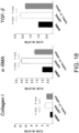
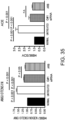
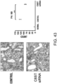
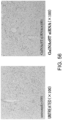
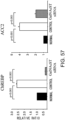
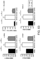
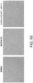
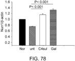
- the expressions of GalNAc4S-6ST, type I collagen, and ⁇ -SMA genes were determined, and the result showed that the expression of GalNac4S-6ST was significantly suppressed in the GalNAc4S-6ST siRNA-treated group as compared to the untreated group (P ⁇ 0.001; when compared to the untreated group). Furthermore, the expressions of ⁇ -SMA and type I collagen genes were measured as indicators for fibrogenesis, which is an important pathological condition of cardiomyopathy. As a result, the significant reduction of expression were confirmed in the GalNAc4S-6ST siRNA-treated group as compared to the untreated group (P ⁇ 0.001; when compared to the untreated group). This result demonstrates that the target knockdown effect of the GalNAc4S-6ST siRNA results in suppression of the progression of myocardial fibrogenesis at the gene expression level.
- agents of the present invention are thus useful, for example, as myocardial fibrogenesis inhibitors.
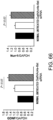
- the heart weights (mg) and body weights (g) of cardiomyopathy model mice were measured to calculate the heart/body weight ratio which is an indicator for cardiac hypertrophy.
- the cardiac hypertrophy-suppressing effect of the GalNAc4S-6ST (GalNac) siRNA was evaluated. Cardiac hypertrophy also serves as an indicator for tissue fibrotic change.
- the result showed that the ratio was 6.376 ⁇ 0.484 and 5.442 ⁇ 0.203 in the untreated and siRNA-treated groups, respectively.
- the significant reduction of the ratio was found in the siRNA-treated group as compared to the untreated group (p ⁇ 0.05; t-test). This suggests that GalNac4S-6ST siRNA has the effect of suppressing pathological cardiac hypertrophy.
- the agents of the present invention are thus useful, for example, as cardiac hypertrophy-suppressing agents (therapeutic agents for cardiac hypertrophy).
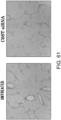
- the type I collagen deposition (an indicator of fibrogenesis)-suppressing effect of GalNac4S-6ST siRNA was assessed using heart samples of cardiomyopathy model mice. Cardiac tissue samples were collected from the same mice as described in Example 1, and embedded in OCT compound (Miles), an embedding medium for cryosectioning. The samples were sliced into thin sections using Cryostat (Carl Zeiss). The resulting sections were fixed with acetone (Sigma Aldrich Japan) for ten minutes, and then washed with phosphate buffer. A rabbit antiserum anti-type I collagen (rabbit polyclonal antibody, 1:2,000 dilution; LSL) was added as the primary antibody, and the sections were incubated at room temperature for one hour.
- Fig. 3 The histological findings were shown in Fig. 3 . Very intense positive signals for type I collagen were observed between myocardial fibers in the untreated group. Meanwhile, in the siRNA-treated group, the type I collagen-positive signals were considerably weaker than those of the untreated group.
- the above-described type I collagen immunostaining result demonstrates that the GalNac4S-6ST siRNA has the effect of suppressing the excessive deposition of type I collagen in myocardial tissues. This result correlates with the result of quantitative PCR described in Example 1.
- the agents of the present invention are thus useful, for example, as agents for suppressing type I collagen deposition in myocardial tissues.
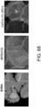
- the type III collagen deposition (an indicator of fibrogenesis activity) - suppressing effect of GalNac4S-6ST siRNA was assessed using heart samples of cardiomyopathy model mice.
- Tissue sections obtained by the same method as described in Example 3 were fixed with acetone (Sigma Aldrich Japan) for ten minutes, and then washed with phosphate buffer.
- a rabbit antiserum anti-type III collagen (rabbit polyclonal antibody, 1:2000 dilution; LSL) was added as the primary antibody, and the sections were incubated at room temperature for one hour.
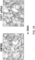
- Fig. 4 The histological findings are shown in Fig. 4 . Moderately strong positive signals for type III collagen were observed between myocardial fibers in the untreated group. Meanwhile, in the siRNA-treated group, the type III collagen-positive signals were comparable to those of the control group.
- the above-described result of type III collagen immunostaining demonstrates that the GalNac4S-6ST siRNA has the effect of suppressing the type III collagen deposition in heart tissues, implying that the siRNA is also effective in suppressing active collagen deposition.
- the agents of the present invention are thus useful, for example, as agents for suppressing type III collagen deposition in myocardial tissues.
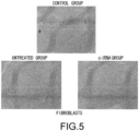
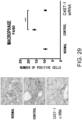
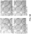
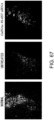
- This Example assesses the pharmacological effect of GalNAc4S-6ST siRNA on the kinetics of fibroblasts that infiltrate into cardiac tissues of cardiomyopathy model mice due to DOX administration.
- Tissue sections obtained by the same method as described in Example 3 were fixed with acetone (Sigma Aldrich Japan) for ten minutes, and then washed with phosphate buffer.
- An anti-mouse fibroblast antibody (ER-TR7, rat monoclonal antibody, 1:400 dilution; BMA Biomedicals Ltd.) was added as the primary antibody, and the sections were incubated at room temperature for one hour.
- a peroxidase-labeled goat anti-rat immunoglobulin antibody (1:200 dilution; Biosource International, Inc.) was added as the secondary antibody, and the sections were incubated at room temperature for 30 minutes. After incubation, DAB substrate (Nichirei Biosciences) was added to the samples. The samples were observed under a light microscope (Leica Microsystems).
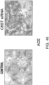
- the histological findings were shown in Fig. 5 .
- the photograph focuses on the ventricular septum. Infiltration of numerous fibroblasts was observed in the untreated group as compared to the control group. In contrast, the degree of fibroblast infiltration in the siRNA-treated group was less as compared to the untreated group.
- the above-described result shows that GalNac4S-6ST siRNA has the pharmacological effect of suppressing the fibroblast infiltration into myocardial tissues and this activity contributes to the anti-fibrogenic effect.
- agents of the present invention are thus useful, for example, as agents for suppressing fibroblast infiltration into myocardial tissues.
- the colitis model mice were prepared by allowing C57BL/6J mice (female, six weeks old; CLEA Japan Inc.) to freely drink high-concentration chlorine water containing 3% dextran sulfate sodium (DSS; Wako Pure Chemical Industries Ltd.) for eight days.
- the DSS-induced colitis model has excellent reproducibility, and is thus widely used as a typical experimental mouse model for inflammatory bowel diseases such as ulcerative colitis or Crohn's disease, as well as a model with full-thickness inflammation and fibrotic changes, and muscle layer thickening, which are histological characteristics of the narrowing of colon lumen ( Sasaki N, J Inflamm. 2005 2: 13 , Review: Pucilowska JB et al.
- GalNAc4S-6ST siRNA (1 ⁇ g/head) as described in Example 1 was combined with atelocollagen (Koken Co.) prediluted 10-fold with PBS and 200 ⁇ l of the mixture was injected to the peritoneal cavities of the mice.
- the group of mice treated as described above was named "GalNAc4S-6ST siRNA group", while a group treated with atelocollagen alone without combining GalNAc4S-6ST siRNA was named the "control group”.
- the body weight and the disease activity index (DAI) score were recorded during seven days of 3% DSS water feeding ( Kihara M., Gut. 2003, 52, 713-9 ). The evaluation criteria for DAI are shown below.
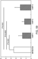
- mice were sacrificed and their colon lengths were measured on the fifth day.
- the colon shortening was significantly suppressed in the GalNAc4S-6ST siRNA-administered group (p ⁇ 0.005; t-test) ( Fig. 6 ).
- the colon length is a definitive indicator that reflects intestinal fibrogenesis or stenosis.
- agents of the present invention are thus useful, for example, as agents for suppressing fibrotic changes of the intestine.
- the intestinal fibrogenesis model was prepared by the same method as described in Example 6. The mice were sacrificed on day 7. A part of the collected colon was placed in 1.5-ml tubes, and frozen with liquid nitrogen. cDNA was synthesized by the same method as that described in Example 1, and quantitative PCR was carried out. The primer sequences and the number of PCR cycle conditions were the same as those described in Example 1.
- Fig. 7 The result is shown in Fig. 7 .
- agents of the present invention are thus useful, for example, as agents for suppressing the fibrotic changes of the colon.
- the intestinal fibrogenesis model was prepared by the same method as described in Example 6. The mice were sacrificed on day 7. Cryoblocks and tissue sections were prepared from the collected colon by the same method as described in Example 3. Masson-stained images of colonic tissue sections are shown in Fig. 8 . Masson staining serves as an indicator to assess fibrotic change of tissues by visualizing collagen fibers. In the GalNAc4S-6ST siRNA-administered group, the full-thickness (lamina intestinal mucosae, submucosa, and muscle layer) collagen fiber deposition was significantly suppressed as compared to the control group.
- the agents of the present invention are thus useful, for example, as agents for suppressing the full-thickness (lamina limbal mucosae, submucosa, and muscle layer) collagen fiber deposition.
- the intestinal fibrogenesis model was prepared by the same method as described in Example 6. The mice were sacrificed on day 7. Cryoblocks and tissue sections were prepared from the collected colon by the same method as described in Example 3. The resulting sections were fixed with acetone (Wako Pure Chemical Industries) for ten minutes, and then washed with phosphate buffer. An anti-ER-TR7 antibody (rat monoclonal antibody, 1 ⁇ g/ml; BMA) was added as the primary antibody, and the sections were incubated at room temperature for one hour. Then, the secondary antibody reaction was carried out by adding a peroxidase-labeled anti-rat IgG antibody (1:200 dilution), which was followed by color development by adding DAB substrate (Nichirei Biosciences). Then, the nucleus was stained by Lillie- Mayer hematoxylin (Muto Pure Chemicals Co.). The samples were observed under a light microscope (Leica Microsystems). The antibody binding was detected by visualizing it as brown signals.
- acetone
- agents of the present invention are thus useful, for example, as agents for suppressing the infiltration or retention of fibroblasts.
- the intestinal fibrogenesis model was prepared by the same method as described in Example 6. The mice were sacrificed on day 7. Cryoblocks and tissue sections were prepared from the collected colon by the same method as described in Example 3. The resulting sections were fixed with acetone (Wako Pure Chemical Industries) for ten minutes, and then washed with phosphate buffer. An anti-F4/80 antibody (clone A3-1, rat monoclonal antibody, 2 ⁇ g/ml: CALTAG LABORATORIES) was added as the primary antibody, and the sections were incubated at room temperature for one hour.
- the secondary antibody reaction was carried out by adding a peroxidase-labeled anti-rat IgG antibody (1:200 dilution), which was followed by color development by adding DAB substrate (Nichirei Biosciences). Then, the nucleus was stained by Lillie- Mayer hematoxylin (Muto Pure Chemicals Co.). The samples were observed under a light microscope (Leica Microsystems). The antibody binding was detected by visualizing it as brown signals.
- agents of the present invention are thus useful, for example, as agents for suppressing the infiltration of macrophages or fibroblasts.
- GalNAc4S-6ST is an enzyme that transfers a sulfate group to position 6 in N-acetylgalactosamine sulfated at position 4.
- the intestinal fibrogenesis model was prepared by the same method as described in Example 6. The mice were sacrificed on day 7.
- GalNAc4S-6ST siRNA, GalNAc4ST-1, and GalNAc4ST-2 (GeneWorld) were combined together; 1 ⁇ g of the mixture was combined with 200 ⁇ l of 1% atelocollagen (Koken Co.), which is a vehicle, and administered intraperitoneally to each mouse.
- GalNAc ST siRNA-administered group A group administered with the siRNA is referred to as "GalNAc ST siRNA-administered group".
- the control group is the same as described in Example 6.
- the siRNA nucleotide sequences of GalNAc4S-6ST, GalNAc4ST-1, and GalNAc4ST-2 used in this Example are shown below, but the sequence are not limited the Example.
- the tissue fibrotic changes in the mouse intestinal fibrogenesis model can be assessed representatively by the colon length. Accordingly, the colon length on day 7 is shown as an essential evaluation item in this Example.
- the shortening of colon was significantly suppressed as compared to the control group (p ⁇ 0.01; t-test) ( Fig. 11 ). It was thus demonstrated that the intestinal fibrogenesis was inhibited by suppressing the expression of the GaINAc4ST-1 and GalNAc4ST-2 genes.
- agents of the present invention are thus useful, for example, as intestinal fibrogenesis inhibitors.
- a basic mouse pulmonary emphysema model which is prepared by intratracheal administration of porcine pancreatic elastase (PPE), is used in this Example.
- PPE porcine pancreatic elastase
- This mouse model is classical, but highly reproducible and simple. Thus, this mouse model has been used commonly as a pulmonary emphysema model. Inflammatory cell infiltration to the pulmonary alveolar interstitium is observed as a histological feature.
- COPD chronic obstructive pulmonary disease
- IIPs idiopathic interstitial pneumonias
- coniosis coniosis
- mice were prepared.
- PPE Calbiochem-Novabiochem
- C57BL6/J mice female, 5- to 6-weeks old; CLEA Japan.
- the mice were grown for three weeks after administration, and then lung tissues were collected from them. Mice that did not have PPE administration were used as the control group.
- the C6ST-1 siRNA was administered by the same procedure as shown in Example 1: 1 ⁇ g of C6ST-1 siRNA (Ambion) was combined with 1% atelocollagen (Koken Co.), which is an siRNA vehicle, and administered to the peritoneal cavities once a week after PPE administration. The dose was 200 ⁇ l/head.
- the collected right lung tissues were embedded in the OCT compound (Miles), an embedding medium for cryosectioning, and cryoblocks were prepared using liquid nitrogen.
- the cryoblocks were sliced into 6- ⁇ m sections using cryostat (Microm).
- the resulting sections were fixed with 1% glutaraldehyde (Nacalai Tesque) for 10 minutes, and further fixed with formol-calcium solution for 10 minutes.
- the sections were washed with phosphate buffer, and then stained with Lillie- Mayer's hematoxylin solution (Sigma Aldrich Japan) at room temperature for 5 minutes.
- the sections were washed gently with a decolorizing solution (70% ethanol containing 0.5% HCl; prepared using reagents from Nacalai Tesque), and then washed with water for 10 minutes.
- the sections were stained with eosin-alcohol at room temperature for 5 minutes, and then washed with water for 10 minutes.
- the sections were washed gently with 100% ethanol, and then allowed to stand for 3 minutes.
- the sections were further washed gently with xylene (Nacalai Tesque), and allowed to stand for 10 minutes. This sample was histologically observed using a light microscope (Leica Microsystems).
- the obtained histological images are shown in Fig. 12 .
- the histological features of a normal lung parenchyma with characteristic bootsolate alveolar septal walls are found in the control group (PPE non-administered mice). Meanwhile, emphysematous lesions due to destruction and abolishment of alveolar septal walls and enlargement of air space (characteristics of pulmonary emphysema) can be observed in the untreated group (administered with PPE but not with C6ST-1 siRNA) shown in the middle photograph.
- the enzyme-treated group administered with PPE and C6ST-1 siRNA
- slight abolishment of alveolar septal walls and emphysematous lesions can be observed; however, their levels are significantly improved.
- the COPD model was prepared by the same method as described in Example 12. A part of the collected lung tissue were placed in 1.5-ml tubes, and frozen with liquid nitrogen. cDNA synthesis was carried out by the same method as described in Example 1, and quantitative PCR was conducted. The primer sequences for type I collagen and ⁇ -SMA and PCR conditions were also the same as described in Example 1. The sequences of C6ST-1 primers are shown below.
- C6ST-1 gene is enhanced in this model. Significant knockdown of the C6ST-1 gene was confirmed by C6ST-1 siRNA treatment (p ⁇ 0.01; t-test). Furthermore, the enhanced expression of type I collagen and ⁇ -SMA as indicators for fibrogenesis were significantly suppressed by C6ST-1 siRNA (for both genes, p ⁇ 0.01; t-test). This result suggests that the enhanced fibrotic changes of pulmonary interstitium can be effectively suppressed by inhibiting the expression of C6ST-1.
- agents of the present invention are thus useful, for example, as agents for suppressing fibrotic changes of the pulmonary interstitium.
- the intestinal fibrogenesis model was prepared by the same method as described in Example 12. Cryoblocks and tissue sections were prepared from the collected lung tissues by the same method as described in Example 3. The resulting sections were fixed with acetone (Wako Pure Chemical Industries) for ten minutes, and then washed with phosphate buffer. An anti-ER-TR7 antibody (rat monoclonal antibody, 1 ⁇ g/ml; BMA) was added as the primary antibody, and the sections were incubated at room temperature for one hour. Then, the secondary antibody reaction was carried out by adding a peroxidase-labeled anti-rat IgG antibody (1:200 dilution), which was followed by color development by adding DAB substrate (Nichirei Biosciences). Then, the nucleus was stained by Lillie- Mayer hematoxylin (Muto Pure Chemicals Co.). The samples were observed under a light microscope (Leica Microsystems). The antibody binding was detected by visualizing it as brown signals.
- agents of the present invention are thus useful, for example, as agents for suppressing fibroblast infiltration and retention in the interstitium of alveolar tissue.
- the COPD model was prepared by the same method as described in Example 12.
- the collected lung tissues were processed by the same method as described in Example 3 to prepare tissue cryoblocks and sections.
- the resulting sections were fixed with acetone (Wako Pure Chemical Industries) for ten minutes, and then washed with phosphate buffer.
- An anti-F4/80 antibody (clone A3-1, rat monoclonal antibody, 2 ⁇ g/ml; CALTAG LABORATORIES) was added as the primary antibody, and the sections were incubated at room temperature for one hour.
- the secondary antibody reaction was carried out by adding a peroxidase-labeled anti-rat IgG antibody (1:200 dilution), which was followed by color development by adding DAB substrate (Nichirei Biosciences). Then, the nucleus was stained by Lillie- Mayer hematoxylin (Muto Pure Chemicals Co.). The samples were observed under a light microscope (Leica Microsystems). The antibody binding was detected by visualizing it as brown signals.
- agents of the present invention are thus useful, for example, as agents for suppressing the infiltration of macrophages to the pulmonary alveolar interstitium.
- C6ST-I siRNA was assessed for its influence on respiratory function using static lung compliance static compliance (Cst)) as an indicator to evaluate the clinical effect of C6ST-1 siRNA in pulmonary emphysema model mice.
- Cst represents a measure of lung tissue flexibility.
- Cst is increased in pulmonary emphysema, which is a disease with alveolar tissue damage.
- Pulmonary emphysema model mice were prepared by the same procedure described in Example 12, and then treated with C6ST-I siRNA. An anesthetic agent was given to the mice to stop spontaneous respiration, and then their Cst was monitored using FlexiVent (SCIREQ) respiratory function analyzer in the PV loop mode. Mice were connected to the FlexiVent by the following procedure: a median incision was performed after stopping spontaneous respiration, and then a special cannula was inserted into the trachea, which was followed by peribronchial ligation.
- SCIREQ FlexiVent
- C6ST-1 siRNA-administered group significantly suppressed the increase in Cst caused by pulmonary emphysema induced by intratracheal administration of PPE. Furthermore, the result described in this Example suggests that the suppression of C6ST- I gene expression not only suppresses the alveolar damage caused by fibrotic changes of the interstitium of lung tissue but also improves the actual clinical symptoms (respiratory conditions).
- the agents of the present invention are thus useful, for example, as agents for suppressing alveolar damage.
- the pulmonary tissues used in this Example were the right lungs of the same lung tissues as used in Example 12.
- the lung tissues isolated from mice were gently washed with phosphate buffer and then immersed into phosphate buffer saturated in a glass container.
- the glass container filled with phosphate buffer was weighed in advance. After the lung tissues were added to the container, lung capacity was calculated by converting the increased weight into a liquid volume.
- the result of this Example is shown in Fig. 17 .
- a statistically significant increase of lung capacity was observed in the untreated group.
- agents of the present invention are thus useful, for example, as agents for suppressing the increase of lung capacity caused by pulmonary emphysema.
- Example 11 The importance of sulfation at position 4 and 6 is demonstrated with an additional Example.
- the siRNA sequences are the same as shown in Example 11.
- the pulmonary emphysema model was prepared by the same method as described in Example 12. A part of the collected lung tissues were placed into 1.5-ml tubes, and frozen with liquid nitrogen. cDNA synthesis was carried out by the same method as described in Example 1, and assessed by quantitative PCR method. The primer sequences and of PCR conditions were the same as described in Examples I and 13. The primer sequences for TGF- ⁇ are shown below.
- Fig. 18 The result is shown in Fig. 18 .
- the enhanced expression of type I collagen, ⁇ -SMA, and TGF- ⁇ as indicators for fibrogenesis were also significantly suppressed by GalNAcST siRNA (for all genes, p ⁇ 0.01; t-test) in this Example.
- This result demonstrates that the suppression of GAINAc4ST-1, GAlNAc4ST-2, and GAlNAc4S-6ST expression can effectively inhibit the enhanced fibrotic changes of pulmonary interstitium.
- agents of the present invention are thus useful, for example, as agents for suppressing fibrotic changes in lung interstitium.
- GAlNAcST siRNA was assessed for its influence on respiratory function using static lung compliance (static compliance (Cst)) as an indicator to evaluate the clinical effect of GAlNAcST siRNA in pulmonary emphysema model mice.
- static lung compliance static compliance (Cst)
- Pulmonary emphysema model mice were prepared by the same procedure described in Example 12, and then treated with GAlNAcST siRNA. The spontaneous respiration of mice was ceased by an anesthetic agent, and then their Cst was monitored using FlexiVent (SCIREQ) respiratory function analyzer in the PV loop mode. Mice were connected to the FlexiVent by the following procedure: a median incision was performed after stopping spontaneous respiration, and then a special cannula was inserted into the trachea, which was followed by peribronchial ligation.
- SCIREQ FlexiVent
- the agents of the present invention are thus useful, for example, as agents for suppressing alveolar damage or as an agent for improving respiratory conditions.
- the objective of this Example is to prove that the therapeutic effect of GalNAcST siRNA is not only the suppression at the cell level but also the effect of morphological maintenance and preservation of the organ.
- the pulmonary tissues used in this Example were the right lungs of the same lung tissues as used in Example 12.
- the lung tissues isolated from mice were gently washed with phosphate buffer and then immersed in phosphate buffer saturated in a glass container.
- the glass container filled with phosphate buffer was weighed in advance. After the lung tissues were added to the container, lung capacity was calculated by converting the increased weight into a liquid volume.
- the result of this Example is shown in Fig. 20 .
- a statistically significant increase of lung capacity was observed in the untreated group.
- agents of the present invention are thus useful, for example, as agents for maintaining or preserving lung morphology.
- sample preparation was conducted as described below.
- mice Gestational Day 14 C57BL/6JcL mice (CLEA Japan Inc.) were reared and allowed to deliver. 10 mg/ml Streptozocin (SIGMA) was subcutaneously administered at 20 ⁇ l/head to Day 2 postnatal female C57BL/6JcL mice. The mice were reared with sterile water and CE-2 Diet (CLEA Japan Inc.) until they were four weeks old, and then on sterile water and a High Fat Diet (CLEA Japan Inc.) for the next two weeks.
- SIGMA Streptozocin
- mice On Day 14 of this experiment, 100 ⁇ l of 5 mg/ml BrdU (ZyMED Laboratory Inc.) was administered into the tail vein, and one hour after administration the mice were dissected and the liver, pancreas, kidney, testis, ovary, and muscle were isolated to obtain samples for immunostaining and gene expression analysis. The body weight change was monitored over time for 14 days of this Example.
- BrdU Zinc Laboratory Inc.
- Fig. 21 The result is shown in Fig. 21 .
- the vertical axis indicates the body weight (g), while the horizontal axis indicates the monitoring days.
- Fig. 21 shows that the weight increase tended to be suppressed 10 days after treatment in the C4ST-1 siRNA-treated group, C4ST-2 siRNA-treated group, and C4ST-3 siRNA-treated group as compared to the control group.
- the weight increase was significantly suppressed on day 18 in the C4ST-2 siRNA-treated group and C4ST-3 siRNA-treated group (both groups; p ⁇ 0.05). This result suggests that obesity associated with type 2 diabetes can be suppressed by inhibiting the expression of C4ST-1, C4ST-2, and C4ST-3.
- the agents of the present invention are thus useful, for example, as agents for suppressing body weight increase, or as agents for suppressing obesity associated with type 2 diabetes.
- a blood glucose monitor As an insulin-tolerance test, human crystalline insulin (0.75 U/kg) was administered into peritoneal cavities the day before dissection, and 0, 15, and 60 minutes after administration, the blood glucose levels were measured and evaluated using a blood glucose monitor, Glu-Test Ace (BOMBYX Medicine co.). The blood glucose level changes monitored at 0, 15, and 60 minutes after siRNA treatment are shown in Fig. 22 .
- the vertical axis indicates the blood glucose level (mg/dl), while the horizontal axis indicates 0, 15, and 60 minutes after insulin-tolerance test.
- Fig. 22 shows that the blood glucose level was not significantly decreased 0 and 15 minutes after treatment in any of the C4ST-1 siRNA-treated group.
- C4ST-2 siRNA-treated group, and C4ST-3 siRNA-treated group as compared to the control group, while it significantly decreased 60 minutes after treatment in each of the C4ST-1 siRNA-treated group, C4ST-2 siRNA-treated group, and C4ST-3 siRNA-treated group.
- insulin resistance which is an essential functional disorder in type 2 diabetes, can be effectively improved by suppressing the expression of C4ST-1 , C4ST-2, and C4ST-3.
- the agents of the present invention are thus useful, for example, as agents for improving insulin resistance in type 2 diabetes.
- cDNA was prepared by the same method as described in Example 1 from 50 mg of each organ (pancreas) isolated from Streptozocin-induced female C57BL/6JcL mice. PCR was conducted in the following composition.
- PCR products were combined with 2 ⁇ l of Loading Dye (Invitrogen).
- 1.5% agarose gel was prepared using UltraPure Agarose (Invitrogen), and the samples were electrophoresed in a Mupid-2 plus (ADVANCE) at 100 V for 20 minutes.
- the gel was shaken for 20-30 minutes in a stain solution prepared by 10,000 times diluting Ethidium Bromide (Invitrogen) with 1x LoTE (composition: 3 mM Tris-HCl (pH 7.5) (Invitrogen), 0.2 mM EDTA (pH 7.5) (Sigma Aldrich Japan)).
- the gel was photographed with EXILIM (CASIO) positioned on I-Scope WD (ADVANCE) and the gene expression was confirmed.
- Result is shown in Fig. 23 .
- GAPDH expression which is used as a positive control, was confirmed by RT-PCR in the control group, C4ST1 (Chondroitin D-N-acetylgalactosamine-4- O- sulfotransferase 1), C4ST2 (Chondroitin D-N-acetylgalactosamine-4- O -sulfotransferase 2), and C4ST3 (Chondroitin D-N-acetylgalactosamine-4- O -sulfotransferase 3).
- C4ST1 Chodroitin D-N-acetylgalactosamine-4- O- sulfotransferase 1 siRNA-, C4ST2 (Chondroitin D-N-acetylgalactosamine-4- O -sulfotransferase 2) siRNA-, C4ST3 (Chondroitin D-N-acetylgalactosamine-4- O -sulfotransferase 3) siRNA-treated groups, and C4ST1 (Chondroitin D-N-acetylgalactosamine-4- O -sulfotransferase 1), C4ST2 (Chondroitin D-N-acetylgalactosamine-4- O -sulfotransferase 2), C4ST3 (Chondroitin D-N-acetylgalactosamine-4- O -sulfotransferase 3) gene knockdown
- amyloid precursor protein (APP) deposition-suppressing effect of C4ST-1, C4ST-2, and C4ST-3 siRNAs was assessed using pancreatic tissue samples of type 2 diabetes model mice.
- the deposition of APP and amyloid fibers in islets including ⁇ cells has long been known to be an important histopathological feature of type 2 diabetes.
- APP has been demonstrated to be involved in Alzheimer's disease ( Johnson KH et al. N Eng J Med 321: 513, 1989 ; Rhodes CJ. Science 307: 380, 2005 ; Haan MN. Nat Clin Pract Neurol 3: 159, 2006 ; Prentki M et al. J Clin Invest 116: 1802,2006 ).
- Fig. 24 shows histological images of the normal mouse group, control group, and C4ST-2 siRNA-treated group.
- the APP deposition is enhanced in the islets of type 2 diabetes model mice. The deposition was demonstrated to be clearly suppressed in the C4ST-2 siRNA-treated group as compared to the control group.
- the agents of the present invention are thus useful, for example, as agents for suppressing amyloid fiber deposition.
- Cryoblocks and tissue sections were prepared from the collected pancreatic tissues by the same method as described in Example 3. The resulting sections were fixed with acetone (Wako Pure Chemical Industries) for ten minutes, and then washed with phosphate buffer. An anti-ER-TR7 antibody (rat monoclonal antibody, 1 ⁇ g/ml; BMA) was added as the primary antibody, and the sections were incubated at room temperature for one hour. Then, the secondary antibody reaction was carried out by adding a peroxidase-labeled anti-rat IgG antibody (1:200 dilution), and color development was performed by adding DAB substrate (Nichirei Biosciences). Then, the nucleus was stained by Lillie- Mayer hematoxylin (Muto Pure Chemicals Co.). The samples were observed under a light microscope (Leica Microsystems). The antibody binding was detected by visualizing it as brown signals.
- BMA anti-ER-TR7 antibody
- agents of the present invention are thus useful, for example, as agents for suppressing fibroblast infiltration to islets.
- Cryoblocks and tissue sections were prepared from the collected pancreatic tissues by the same method as described in Example 3. The resulting sections were fixed with acetone (Wako Pure Chemical Industries) for ten minutes, and then washed with phosphate buffer. An anti-F4/80 antibody (clone A3-1, rat monoclonal antibody, 2 ⁇ g/ml; CALTAG LABORATORIES) was added as the primary antibody, and the sections were incubated at room temperature for one hour. Then, the secondary antibody reaction was carried out by adding a peroxidase-labeled anti-rat IgG antibody (1:200 dilution), and color development was performed by adding DAB substrate (Nichirei). Then, the nucleus was stained by Lillie- Mayer hematoxylin (Muto Pure Chemicals Co.). The samples were observed under a light microscope (Leica Microsystems). The antibody binding was detected by visualizing it as brown signals.
- the agents of the present invention are thus useful for example, as agents for suppressing the infiltration of macrophages to the panceratic islets.
- GalNAcST siRNA administration exhibited a good antihyperglycemic effect after insulin loading. This result suggests that the insulin resistance arising from fibrotic changes in islets of pancreatic tissues can be effectively improved by suppressing the expression of GalNAc4ST-1, GalNAc4ST-2, and GalNAc4S-6ST.
- the agents of the present invention are thus useful, for example, as hypoglycemic agents or as agents for improving the insulin resistance of pancreatic tissue.
- tubulointerstitial disease is understood as a representative disease caused by renal fibrogenesis. This disease includes Sjogren's syndrome, transplant rejection (chronic allograft nephropathy, etc.), and graft-versus-host reaction (graft-versus-host disease, etc.), in addition to drug-induced, infective, radiation-induced, and heavy metal-induced interstitial renal disorders.
- Renal fibrogenesis also includes renovascular disease. Renovascular disease includes nephrosclerosis associated with hypertension. In recent years, this disease also includes fibrogenesis associated with arteriosclerosis and metabolic syndrome.
- renal fibrotic changes can also be developed in: (3) primary glomerular disease and (4) secondary glomerular diseases.
- the diseases of (3) include IgA nephropathy, minimal lesion nephrotic syndrome, membranous nephropathy, membranoproliferative glomerulonephritis, and focal segmental glomerulosclerosis (FGFS).
- the diseases of (4) include diabetic nephropathy, lupus nephritis associated with systemic lupus erythematosus (SLE), nephropathy associated with chronic rheumatoid arthritis, amyloid nephropathy, and nephropathy associated with type B or C hepatitis.
- SLE systemic lupus erythematosus
- nephropathy associated with chronic rheumatoid arthritis amyloid nephropathy
- nephropathy associated with type B or C hepatitis (5)
- renal fibrogenesis also includes interstitial kidney diseases caused by urinary obstruction, including urinary lithiasis, tumors, and neurogenic bladder dysfunctions.
- CKD chronic kidney disease
- ESRD end-stage renal disease
- a type 2 diabetes model was prepared by the same method as described in Example 21 Gestational Day 14 of C57BL/6JcL mice (CLEA Japan Inc.) were reared and allowed to deliver. Streptozotocin (STZ; Sigma) was administered to Day 2 postnatal C57BL/6JcL mice to prepare a STZ-induced diabetes model. The mice were subcutaneously injected with 20 ⁇ l of STZ (10 mg/ml) three times for two days. Thus, a total of 60 ⁇ l was administered to the mice. Together with their mothers, the mice were fed with a normal diet until they are four weeks old. After weanling at the age of four completed weeks, the mice were fed with a High Fat Diet (CLEA Japan Inc.) for two weeks.
- STZ Steptozotocin
- C4ST-1 siRNA chondroitin D-N-acetylgalactosamine-4- O -suIfotransferase 1 (C4ST-1) siRNA (GeneWorld) was combined with 0.1% atelocollagen (Koken Co.). which is a vehicle for siRNA, and 200 ⁇ l of the resulting mixture was administered into peritoneal cavities once a week (one shot/week) twice in total (for two weeks). On day 14 of this experiment, the mice were dissected and their kidneys were isolated to obtain samples for immunostaining. Gene expression, body weight, and effect on insulin resistance are described in Examples 21, 22, and 23.
- the prepared sections of kidney tissue samples were fixed with acetone (Wako Pure Chemical Industries) for ten minutes, and then washed with phosphate buffer.
- An anti-ER-TR7 antibody (rat monoclonal antibody, 1 ⁇ g/ml; BMA) was added as the primary antibody, and the sections were incubated at room temperature for one hour.
- the secondary antibody reaction was carried out by adding a peroxidase-labeled anti-rat IgG antibody (1:200 dilution), and color development was performed by adding DAB substrate (Nichirei Biosciences). Then, the nucleus was stained by Lillie- Mayer hematoxylin (Muto Pure Chemicals Co.). The samples were observed under a light microscope (Leica Microsystems). The antibody binding was detected by visualizing it as brown signals.
- the result showed that the infiltration of fibroblasts (ER-TR7-positive cells) in the renal cortex and medulla was less in the C4ST-1 siRNA-treated group as compared to that in the control group ( Fig. 28 ; the original images are in color).
- the ER-TR7-positive cells were counted to quantify the fibrogenesis in inflammatory cells.
- Each sample was observed with ten microscopic optical fields under a microscope at a magnification of 400 fold, and the number of positive cell were counted and compared between the control group and C4ST-1 siRNA-treated group.
- the result showed that the ER-TR7 positivity ratio was significantly reduced in the C4ST-1 siRNA-treated group as compared to the control group (p ⁇ 0.001).
- the prepared sections of kidney tissue samples were fixed with acetone (Wako Pure Chemical Industries) for ten minutes, and then washed with phosphate buffer.
- An anti-ER-TR7 antibody (rat monoclonal antibody, 1 ⁇ g/ml; BMA) was added as the primary antibody, and the sections were incubated at room temperature for one hour.
- the secondary antibody reaction was carried out by adding a peroxidase-labeled anti-rat IgG antibody (1:200 dilution), and color development was performed by adding DAB substrate (Nichirei Biosciences). Then, the nucleus was stained by Lillie- Mayer hematoxylin (Muto Pure Chemicals Co.). The samples were observed under a light microscope (Leica Microsystems). The antibody binding was detected by visualizing it as brown signals.
- kidney tissue samples were fixed with acetone (Wako Pure Chemical Industries) for ten minutes, and washed with phosphate buffer.
- An anti-human smooth muscle actin antibody ( ⁇ SMA: mouse monoclonal antibody, 1:100; DACO) was added as the primary antibody, and the sections were incubated at room temperature for one hour.
- the secondary antibody reaction was carried out using Histofine Mouse Stain kit (Nichirei), and color development was performed by adding DAB substrate (Nichirei Biosciences).
- the nucleus was stained by Lillie-Mayer hematoxylin (Muto Pure Chemicals Co.).
- the samples were observed under a light microscope (Leica Microsystems). The antibody binding was detected by visualizing it as brown signals.
- ⁇ SMA-positive cells retained in juxtaglomerular and interstitial areas were significantly reduced in the C4ST-1 siRNA-treated group as compared to the control group ( Fig. 30 ; the original images are in color).
- ⁇ SMA serves as a functional marker for fibroblast activation. This result demonstrates that inhibition of C4ST-1 gene expression suppress activation of fibroblasts infiltrating in the tissue.
- agents of the present invention are thus useful, for example, as fibroblast activation inhibitors.
- the diabetic nephropathy model was prepared by the same method as described in Example 28 to assess the effect of GalNAc4S-6ST siRNA.
- the follow-up examination was carried out over a longer period of time.
- the sequence of GalNAc4S-6ST siRNAs was the same as shown in Example 1.
- the siRNAs were administered into peritoneal cavities once at the age of eight weeks and once again at age of nine weeks.
- Valsartan an angiotensin II receptor blocker (ARB) was orally administered at a dose of 30 mg/kg on the same schedule.
- Kidney tissues were collected at the age often weeks to conduct immunohistochemical and gene expression tests. To assess the gene expression in the kidney tissues, quantitative PCR was carried out by the same method as described in Example 1. In this Example, 36B4 was used as an internal control. The sequence of 36B4 is shown below.
- GalNAc4S-6ST is abbreviated as G#1 in this figure.
- G#1 the expression of GalNAc4S-6ST in kidney tissues is enhanced.
- the administration of GalNAc4S-6ST siRNA significantly suppressed not only the expression of GalNAc4S-6ST gene in kidney tissues but also the enhanced expression of aSMA and TGF ⁇ , which are fibrosis markers.
- the therapeutic effect was evaluated by comparing with that of ARB, and the result showed that the TGF ⁇ -suppressing effect was observed in both groups and there was no significant difference between the two while the ⁇ SMA-suppressing effect was significant in the GalNAc4S-6ST siRNA-administered group as compared to the ARB-administered group.
- This result suggests that the markers for fibrotic changes in kidney tissues can be suppressed by inhibiting the expression of GalNAc4S-6ST gene.
- the result also demonstrates that GalNAc4S-6ST siRNA showed a markedly superior effect in terms of fibroblast activation (enhancement of ⁇ SMA).
- the degree of fibroblast infiltration into kidney tissues was quantitatively evaluated by conducting an immunohistochemical study using the same method as described in Example 28. The result is shown in Fig. 32 .
- Fibroblast infiltration to kidney tissues was significantly suppressed by GalNAc4S-6ST siRNA administration.
- a quantitative evaluation over the entire interstitium did not show any significant difference between GalNAc4S-6ST siRNA administration and ARB treatment.
- the evaluation was restricted to fibroblasts infiltrating in juxtaglomerular areas, a significant suppressing effect was observed in the GalNAc4S-6ST siRNA-administered group, as shown in the figure.
- agents of the present invention are thus useful, for example, as agents for suppressing fibroblast infiltration into renal interstitium.
- the degree of fibroblast infiltration into kidney tissues was quantitatively evaluated by conducting an immunohistochemical study using the same method as described in Example 29. The result is shown in Fig. 33 . Macrophage infiltration into kidney tissues was significantly suppressed by GalNAc4S-6ST administration. A quantitative evaluation over the entire interstitium did not show any significant difference between GalNAc4S-6ST siRNA administration and ARB treatment. Meanwhile, when the evaluation was restricted to macrophages infiltrating in juxtaglomerular areas, a significant suppressing effect was observed in the GalNAc4S-6ST siRNA-administered group, as shown in the figure.
- the agents of the present invention are thus useful, for example, as agents for suppressing macrophage infiltration to renal interstitium.
- Type IV collagen was immunostained by the same method as described in Examples 32 and 33.
- a rabbit anti-mouse type IV collagen antiserum (LSL) was added as the primary antibody, and the sections were incubated at room temperature for one hour.
- the secondary antibody reaction was carried out by adding a peroxidase-labeled anti-rabbit IgG antibody (1:200 dilution), and color development was performed by adding DAB substrate (Nichirei Biosciences).
- the nucleus was stained by Lillie- Mayer hematoxylin (Muto Pure Chemicals Co.). The samples were observed under a light microscope (Leica Microsystems). The antibody binding was detected by visualizing it as brown signals.
- the thickness of collagen visualized as brown signals surrounding glomeruli was measured to quantify the accumulation of type IV collagen. 15 to 20 glomeruli were assessed for each sample. The thickness of the thickest portion around a glomerulus was measured on a display monitor using vernier calipers. The result showed that glomerular basement membrane (GBM) thickening was significantly suppressed in the GalNAc4S-6ST siRNA-administered group as compared to the control group ( Fig. 34 ). The thickening tended to be suppressed more markedly in the GalNAc4S-6ST siRNA-administered group as compared to ARB.
- GBM glomerular basement membrane
- agents of the present invention are thus useful, for example, as agents for suppressing glomerular basement membrane thickening.
- Angiotensin II has been reported to be involved in fibrogenesis in diabetic nephropathy.
- the angiotensin pathway in kidney tissues was assessed by quantitative PCR.
- the expression of angiotensinogen and angiotensin converting enzyme (ACE) is enhanced in this model ( Fig. 35 ).
- the enhanced expression was speculated to be a factor responsible for the renal enhancement of angiotensin II.
- the effect on suppressing angiotensinogen and ACE was observed in the GalNAc4S-6ST siRNA-administered group ( Fig. 35 ).
- the result demonstrates that GalNAc4S-6ST siRNA also produces an improving effect on the angiotensin pathway through suppression of fibroblast infiltration and activation by suppressing the GaINAc4S-6ST gene.
- agents of the present invention are thus useful, for example, as agents for suppressing the expression of angiotensinogen or as an angiotensin converting enzyme inhibitor.
- Serum creatinine is a most commonly used clinical marker for renal function.
- the serum creatinine concentration is elevated due to impaired renal function during the process from diabetic nephropathy to ESRD.
- functional nephrons are already functionally impaired by 50% when the serum creatinine level is elevated.
- Protecting renal function before the creatinine level increases is a clinically important challenge.
- agents of the present invention are thus useful, for example, as agents for suppressing the deterioration of renal function, or as a renal protective agent
- GalNAc4ST-1 As shown in Fig. 37 , the expression of GalNAc4ST-1, GalNAc4ST-2, and GalNAc4S-6ST was enhanced in kidney tissues in the diabetic nephropathy model. The expression of all the genes was significantly suppressed by administering GalNAcST siRNA.
- ⁇ SMA, TGF ⁇ , and CTGF which are fibrogenesis markers for kidney tissue fibrogenesis
- the enhanced expression of ACE was also significantly suppressed in the GalNAcST siRNA-administered group.
- This result demonstrates that inhibition of GalNAc4ST-1, GalNAc4ST-2, and GaINAc4S-6ST expression results in suppression of kidney tissue fibrogenesis and renal protection.
- the fact that ACE was markedly reduced by suppressing any of the genes demonstrates that they have a hypotensive effect.
- agents of the present invention are thus useful, for example, as antihypertensive agents.
- This Example assesses the effect of GalNAc4S-6ST siRNA on a typical drug-induced interstitial nephritis.
- the mouse model is prepared as described below.
- Adriamycin (15 mg/kg; Kyowa Hakko) was administered to the peritoneal cavities of C57BL6/J mice (male, eight weeks old, CLEA Japan Inc.). The mice were reared for one week after administration, and then kidney tissues were collected from them.
- As a control group mice of the same lineage and age were also purchased and reared in the same period, but Adriamycin was not given to them.
- GalNac 4S-6ST siRNA was administered by the same method as described in Example 1: 1 ⁇ g of GalNac 4S-6ST siRNA (Hokkaido System Science Co.) was combined with 200 ⁇ l of 1% atelocollagen (Koken Co.), which is a vehicle, and the resulting mixture was administered intraperitoneally to each mouse 24 hours before Adriamycin administration.
- the expression of GalNac4S-6ST is also enhanced in kidney tissues in the typical drug-induced interstitial nephritis model described in this Example ( Fig. 39 ). The expression was significantly suppressed by administering GalNac4S-6ST siRNA.
- Fig. 40 Diffuse deposition of type 1 collagen was observed over juxtaglomerular and interstitial areas in the control group, while the deposition was markedly suppressed in the GalNac4S-6ST-administered group. This result suggests that fibrotic changes in kidney tissues can also be suppressed by suppressing the expression of GalNac4S-6ST in the drug-induced interstitial nephritis.
- agents of the present invention are thus useful, for example, as agents for suppressing fibrotic changes in kidney tissues.
- Renal fibrosis model mouse was prepared by conducting unilateral ureteral obstruction (UUO) to C57BL/6JcL mice (female, eight weeks old; CLEA Japan Inc.). This model has excellent reproducibility, and is thus widely used as an experimental mouse renal fibrosis model ( American Journal of Pathology 2003 163 (4): 1261-1273 ). Mice were subjected to laparotomy under Ketalar/xylazine anesthesia. The ureters were exposed and the right ureter was ligated at two sites with 4-0 surgical suture. The peritoneum and skin were closed with 1-0 surgical suture.
- UUO unilateral ureteral obstruction
- the effect of inhibiting C6ST expression by C6ST siRNA administration was checked by PCR method using renal fibrosis model mice prepared by unilateral ureteral obstruction (UUO), as a typical example of renal fibrosis model mice.
- the renal fibrosis model was prepared by conducting UUO to C57BL/6JcL mice (female, eight weeks old; CLEA Japan Inc.).
- a mixture of C6ST-1 and C6ST-2 siRNAs (1 ⁇ g/head; Gene World) or PBS was combined with 0.1 % atelocollagen (Koken Co.), which is a vehicle for siRNA, and 200 ⁇ l of the resulting mixture was injected into the peritoneal cavity of each mouse.
- mice treated as described above were named C6ST siRNA group and control group.
- the mice were dissected to excise the UUO-treated kidney.
- samples for immunostaining and gene expression analysis were obtained from the mice.
- Quantitative PCR was carried out by the same method as described in Example 1.
- C6ST-1 siRNA, C6ST-2 primers (Forward, Reverse) used herein are shown below.
- C6ST-2 (G#10) was enhanced in the kidney ( Fig. 41 ; C6ST-2 is abbreviated as G# 10).
- the expression level was decreased in the C6ST siRNA-treated group.
- the C6ST-2 gene knockdown was confirmed in the atelocollagenmediated C6ST siRNA administration ( Fig. 41 ).
- the C6ST siRNA administration also significantly suppressed the enhanced expression of fibrogenesis markers: TGF ⁇ , ⁇ SMA, type I collagen, and CTGF.
- the isolated tissues were embedded in OCT compound (Sakura Finetechnical Co.), an embedding medium for cryosectioning.
- Cryoblocks were prepared using liquid nitrogen, and sliced into 6- ⁇ m thick sections using Cryostat (Micro-edge Instruments Co.). The resulting sections were immunostained by the same method as described in Example 28 using an anti-ER-TR7 antibody. The samples were observed under a light microscope (Leica Microsystems). The antibody binding was detected by visualizing it as brown signals and quantified.
- agents of the present invention are thus useful, for example, as agents for suppressing fibroblast accumulation in kidney interstitium.
- the agents of the present invention are thus useful, for example, as agents for suppressing macrophage accumulation in kidney interstitium.
- agents of the present invention are thus useful, for example, as agents for suppressing the infiltration of inflammatory cells.
- Immunostaining was carried out by the same method as described in Example 30 using an ⁇ SMA antibody.
- the result showed that ⁇ SMA-positive cells were clearly decreased in the interstitium, juxtaglomerular areas in particular, of the UUO-treated kidneys in the C6ST siRNA-treated group as compared to the control group ( Fig. 45 ).
- This result suggests that the activation of fibroblasts accumulating in the renal interstitium is suppressed by inhibiting the expression of C6ST-1 and C6ST-2 genes.
- agents of the present invention are thus useful, for example, as agents for suppressing the activation of fibroblasts in the renal interstitium.
- agents of the present invention are thus useful, for example, as arteriosclerosis-suppressing agents.
- fibrotic changes in ocular tissues occur as a result of invasion due to various causes. This leads to impairment and/or loss of vision.
- Such major diseases include diabetic retinopathy, retinal vein occlusion, retinopathy of prematurity, age-related macular degeneration, and retinitis pigmentosa.
- the diseases also include fibrogenesis associated with corneal inflammation, glaucoma, or cataract (reviews: Fiedlander M. J Clin Invest 117: 576-586, 2007 ; Harada T et al. Genes and Dev. 21: 367-378, 2007 ). From the histopathological viewpoint, damage/decrease of photoreceptor cells caused by fibrogenesis is the major cause of visual loss.
- suppressing fibrogenesis in ocular tissues has been expected as a novel therapeutic strategy to prevent visual loss in all ocular diseases.
- mice Gestational Day 14 C57BL6J/JcL mice (CLEA Japan Inc.) were reared and allowed to deliver. 10 mg/ml Streptozocin (SIGMA) was subcutaneously administered at 20 ⁇ l/head to Day 2 postnatal female C57BL6J/JcL mice. The mice were reared with sterile water and CE-2 Diet (CLEA Japan Inc.) until they were four weeks old, and then with sterile water and a High Fat Diet (CLEA Japan Inc.) for subsequent two weeks.
- SIGMA Streptozocin
- GalNac4S-6ST (G#1) siRNA Hokkaido System Science Co.
- atelocollagen Keren Co.
- eye balls were excised from mice of the two groups and immunohistochemically examined to assess the effect of GaINac4S-6ST (G#1) siRNA.
- Cryoblocks and sections were prepared from the excised eye balls.
- the sections were fixed with acetone (Sigma Aldrich Japan) for ten minutes, and then washed with phosphate buffer.
- a rabbit anti-type IV collagen antiserum (1 :2000 dilution; LSL) was added as the primary antibody, and the sections were incubated at room temperature for one hour.
- a peroxidase-labeled anti-rabbit IgG antibody (1:25 dilution; Cappel) was added as the secondary antibody, and the samples were incubated at room temperature for 30 minutes.
- an enzyme-mediated chromogenic reaction was conducted by adding DAB substrate (Nichirei Biosciences). The samples were observed under a light microscope (Leica Microsystems).
- Fig. 47 Obtained histology of the retina is shown in Fig. 47 .
- the deposition of type IV collagen was increased over the region from ganglion cell layer (GCL) to inner nuclear layer (INL), which is essential for vision.
- the increase of collagen in GCL in particular was significantly suppressed in the GalNac4S-6ST (G#1) siRNA-administered group.
- agents of the present invention are thus useful, for example, as agents for suppressing collagen increase in the ganglion cell layer.
- the retina was immunohistochemically assessed using the same method as described in Example 47.
- An anti-chondroitin sulfate proteoglycan (CSPG) antibody (clone CS56, mouse monoclonal antibody, 1:100; Seikagaku Co.) was added as the primary antibody, and the sections were incubated at room temperature for one hour. Then, the secondary antibody reaction was carried out using Histofine Mouse Stain kit (Nichirei; used for mouse monoclonal antibody).
- CS56-positive signals were observed in GCL, and the segment from outer nuclear layer (ONI.) to pigmented cell layer of retina in the control group.
- the CS56 signal intensity was markedly reduced in the GalNac4S-6ST (G#1) siRNA-administered group.
- GalNac4S-6ST G#1 siRNA-administered group.
- GaINac4S-6ST G# I siRNA significantly suppresses the induced CSPG deposition in retinal tissues in this model mouse. Together with collagen, CSPG is considered to be essential for the formation of fibrogenic lesions. Furthermore, CSPG has been reported to inhibit the process of axon extension of gangliocyte ( Brittis PA et al., Science 255: 733, 1992 ). Previously published reports only describe results of in vitro experiments and developmental process. Thus, the role in in vivo pathological lesions still remains unknown. However, the result described herein for the first time suggests the role of CSPG in lesional tissues.
- Optic nerve regeneration has been reported to serve as a biological defense mechanism after retinal damage. Meanwhile, it is reported that the optic nerve progenitor cells responsible for such regeneration after injury are glial cells ( Fischer AJ et al., Nature neuroscience 4: 247, 2001 ; Ooto S et al., PNAS 101: 13645, 2004 ). In this Example, immunostaining was carried out using a goat anti-GFAP antibody (Santa Cruz) as a glial cell marker by the same method as described in Example 47.
- Fig. 49 The result is shown in Fig. 49 .
- the number of GFAP-positive glial cells was not altered in both normal and control groups.
- the cell count was markedly increased in the area from INL to GCL in the GalNac4S-6ST (G#1) siRNA-administered group.
- Optic nerve regeneration has been reported to occur from INL toward GCL.
- the result described above suggests the process of active optic nerve regeneration induced by GalNac4S-6ST (G#1) siRNA.
- agents of the present invention are thus useful, for example, as agents for regenerating the optic nerve.
- Gangliocytes were quantified using samples prepared in Example 47. The result revealed that GCL gangliocytes were reduced in the diabetic retinopathy model but the loss was significantly recovered by GalNac4S-6ST (G#1) siRNA administration ( Fig. 50 ).
- OS glutamate synthetase
- Fig. 51 The result is shown in Fig. 51 .
- Administration of GaINac4S-6ST (G#1) siRNA could significantly inhibit the enhanced expression of GaINAc4S-6ST in ocular tissues.
- the expression inhibition resulted in a significant increase in the expression of GS in ocular tissues i the GalNac4S-6ST (G#1) siRNA-administered group.
- the result described above shows that GalNac4S-6ST (G# 1) siRNA administration results in Muller cell regeneration, i.e., optic nerve restoration.
- This Example revealed that fibrotic changes of retinal tissues could be suppressed by inhibiting the expression of GaINAc4S-6ST gene, which led to the prevention of photoreceptor cell loss through the regeneration of gangliocytes.
- This histological feature is commonly observed in a wide variety of ocular diseases with fibrosis including glaucoma and diabetic retinopathy.
- the agents of the present invention are thus useful, for example, as agents for regenerating Muller cells or gangliocytes.
- Fibrotic changes in liver tissues are the progressive or terminal stage of various liver diseases. Liver fibrogenesis results from various liver diseases, including viral hepatitis (type A, B, C, D, E, and G viral hepatitis), alcohol liver disease, nonalcoholic fatty liver diseases (NAFLD and NASH), metabolic liver disease, drug-induced liver disease, idiopathic portal hypertension, Budd-Chiari's syndrome, autoimmune hepatitis, primary biliary cirrhosis, primary sclerosing cholangitis, biliary disorders (including biliary atresia and biliary dilation), biliary atresia caused by pancreatic diseases such as tumor, graft-versus-host reaction, and chronic rejection ( Bataller R et al., J Clin Invest 115: 209, 2005 ; Iredale JP. J Clin Invest 117: 539, 2007 ). Fibrotic changes of liver at the tissue level were assessed in the next Examples.
- mice Gestational day 14 C57BL/6JcL mice (CLEA Japan Inc.) were reared and allowed to deliver. Streptozocin (STZ; SIGMA) was administered to Day 2 postnatal C57BL/6JcL mice. STZ(10 mg/ml) was subcutaneously administered at 20 ⁇ l/head three times for two days. Thus, a total of 60 ⁇ l was administered to the mice. Together with their mothers, the mice were fed with a normal diet until they were four weeks old. After weanling at the age of four weeks, the mice were fed with a High Fat Diet (CLEA Japan Inc.) for two weeks.
- STZ Streptozocin
- fibrogenesis markers in liver tissues was assessed by the same method as described in Example 52.
- the expression of type I collagen and ⁇ SMA was significantly enhanced in this model ( Fig. 53 ), suggesting enhanced fibrotic changes in liver tissues. Meanwhile, the enhanced expression of the fibrogenesis markers was significantly suppressed by administering GalNAcST siRNA.
- liver tissues were immunostained by the same method as described in Example 3.
- a rat anti-mouse fibroblast antibody (clone ER-TR7, 1:500 dilution; BMA) was added as the primary antibody, and the sections were incubated at room temperature for one hour.
- a peroxidase-labeled anti-rat IgG antibody (1:200 dilution; Biosource International, Inc.) was added as the secondary antibody, and the sections were incubated at room temperature for 30 minutes.
- DAB substrate Naichirei Biosciences
- the antibody binding was detected by visualizing it as brown signals.
- Fig. 53 Examples of obtained images of immunostained liver tissues are shown in Fig. 53 .
- the accumulation of fibroblasts was clearly observed and a bridge formation was confirmed in the histological picture in the control group.
- fibrogenesis scores were defined according to the following criteria: 0, normal; 1, few collagen fibrils extend from the central vein; 2, extension of collagen fibrils is apparent but collagen fibrils have not yet encompassed the whole liver; 3, extension of collagen fibrils is apparent and collagen fibrils have encompassed the whole liver; 4, diffuse extension of collagen fibrils is observed in the whole liver and pseudolobules are formed.
- fibrogenesis was statistically significantly reduced in the GalNAcST siRNA-administered group as compared to the control group (p ⁇ 0.01; t-test). This finding suggests that GalNAcST siRNA administration also clinically produces a superior liver fibrogenesis-suppressing effect.
- liver tissues were immunostained by the same method as described in Example 54.
- a rat anti-mouse F4/80 antibody (clone A3-1, 2 ⁇ g/ml; CALTAG LABORATORIES) was added as the primary antibody, and the sections were incubated at room temperature for one hour.
- a peroxidase-labeled anti-rat IgG antibody (1:200 dilution; Biosource International, Inc.) was added as the secondary antibody, and the sections were incubated at room temperature for 30 minutes.
- DAB substrate Naichirei Biosciences
- the antibody binding was detected by visualizing it as brown signals.
- the expression of lipid metabolism-related genes in the liver was assessed by the same method as described in Example 52.
- the enhanced expression of carbohydrate response element-binding protein (ChREBP) and acetyl-CoA carxylase-2 (ACC2) was observed. Meanwhile, the enhanced expression was significantly suppressed in the GalNAcST siRNA-administered group ( Fig. 57 ). This result shows that the glycolipid metabolism can be improved via suppression of fibrotic changes in liver tissues by inhibiting the expression of GAlNAc4ST-1, GalNAc4ST-2, and GalNAc4S-6ST genes.
- the agents of the present invention are thus useful, for example, as agents for improving glycolipid metabolism.
- a fatty liver disease model was prepared by the same method as described in Example 52.
- C4ST-1 siRNA, C4ST-2 siRNA, and C4ST-3 siRNA described in Example 21 were administered according to the same administration protocol. Liver tissues were collected from the mice.
- liver tissues were immunostained by the same method as described in Example 3.
- a rat anti-mouse fibroblast antibody (clone ER-TR7, 1:500 dilution; BMA) was added as the primary antibody, and the sections were incubated at room temperature for one hour.
- a peroxidase-labeled anti-rat IgG antibody (1:200 dilution; Biosource International, Inc.) was added as the secondary antibody, and the sections were incubated at room temperature for 30 minutes.
- DAB substrate Naichirei Biosciences
- the antibody binding was detected by visualizing it as brown signals.
- Fig. 58 Examples of obtained images of immunostained liver tissues are shown in Fig. 58 .
- Each sample was assessed for the degree of liver fibrogenesis using the fibrogenesis scores based on the immunohistochemical staining carried out in Example 58. The result is shown in Fig. 59 . Each bar indicates mean ⁇ standard deviation of the fibrogenesis score in each group. The fibrogenesis was statistically significantly reduced in all of the C4ST-1 siRNA-, C4ST-2 siRNA-, C4ST-3 siRNA-administered groups as compared to the control group (p ⁇ 0.001; t-test). This finding suggests that the administration of siRNAs against C4ST-1, C4ST-2, or C4ST-3 also clinically produces a superior liver fibrogenesis-suppressing effect.
- Example 60 Assessment of C4ST-1 siRNA, C4ST-2 siRNA. and C4ST-3 siRNA in hepatocyte disorders in a mouse fatty liver disease model
- mice When mice were sacrificed, blood was collected from them according to the protocol as described in Example 58.
- the blood samples were custom-assayed for the serum alanine transferase (ALT) levels through SRL Inc. The result is shown in Fig. 60 .
- the serum ALT level is the most widely used clinical indicator for hepatocyte destruction.
- the serum ALT level was elevated in the control group.
- the mean value was decreased to 50% or less in each of the C4ST-1 siRNA-, C4ST-2 siRNA-, and C4ST-3 siRNA-administered groups as compared to the control group ( Fig. 60 ). This result demonstrates that hepatocyte damages can be reduced via suppression of fibrotic changes by inhibiting the expression of C4ST-1, C4ST-2, or C4ST-3 gene in liver tissues.
- agents of the present invention are thus useful, for example, as agents for reducing hepatocyte damage.
- mice were injected into peritoneal cavities of C57BL6/J mice (female, five or six weeks old; CLEA Japan Inc.) twice a week for four weeks (eight times) to induce hepatic fibrosis. Then, carbon tetrachloride was additionally administered twice a week for two weeks (a total of 12 times) to induce cirrhosis.
- mice with induced cirrhosis were sacrificed, and their livers were collected (cirrhotic livers). Meanwhile, in the control experiment, livers were collected from C57BL6/J mice (female, CLEA Japan Inc.) of the same age without carbon tetrachloride administration (normal livers).
- mice in the both groups were sacrificed. Liver tissue sections were prepared from the mice and immunohistochemically assessed. A rat anti-mouse fibroblast antibody (clone ER-TR7, 1:500 dilution; BMA) was added as the primary antibody, and the sections were incubated at room temperature for one hour.
- BMA rat anti-mouse fibroblast antibody
- a peroxidase-labeled anti-rat IgG antibody (1 :200 dilution; Biosource international, Inc.) was added as the secondary antibody, and the sections were incubated at room temperature for 30 minutes. After incubation, DAB substrate (Nichirei Biosciences) was added, and the samples were observed under a light microscope (Leica Microsystems). The antibody binding was detected by visualizing it as brown signals.
- Fig. 61 Examples of obtained images of immunostained liver tissues are shown in Fig. 61 .
- the degree of liver fibrogenesis in each sample was assessed using the fibrogenesis scores based on the immunohistochemical staining carried out in Example 61. The result is shown in Fig. 62 . Each bar indicates mean ⁇ standard deviation of the fibrogenesis score in each group. The fibrogenesis was statistically significantly reduced in the C6ST siRNA-administered group as compared to the control group (p ⁇ 0.05; t-test). This finding suggests that the administration of C4ST-1 siRNA, C4ST-2 siRNA, or C4ST-3 siRNA also produces a clinically beneficial effect of suppressing liver fibrogenesis.
- the expression of ⁇ SMA, type I collagen, CTGF, and TGF ⁇ as fibrogenesis markers was enhanced in the control group. Meanwhile, the expression was significantly suppressed in the C6ST siRNA-administered group. This result suggests that fibrotic changes in liver tissues can be suppressed by inhibiting the expression of C6ST-1 and C6ST-2 genes.
- an MPTP-induced Parkinson's disease model was used as a fundamental mouse model for Parkinson's disease.
- This model is a classical but highly reproducible and simple model, and thus has been widely used as a Parkinson's disease model.
- the histological features are: infiltration of inflammatory cells into brain parenchymal tissue and reduction in the number of dopamine-producing neurons.
- neurofibrillary tangles were thought to be a cause of the reduction in the number of neurons in such pathological conditions.
- diseases include pathological conditions with neuronal disorders, specifically, not only representative neurodegenerative diseases such as Parkinson's disease, progressive supranuclear palsy, corticobasal degeneration, Alzheimer's disease, polyglutamine disease, amyotrophic lateral sclerosis (ALS), spinal progressive muscular atrophy, spinobulbar muscular atrophy, Huntington's disease, and multiple sclerosis, but also other diseases such as multiple system atrophy (striatonigral degeneration, olivopontocerebellar atrophy, and Shy-Drager syndrome), adrenoleukodystrophy, Guillain-Barre syndrome, myasthenia gravis, Fisher syndrome, chronic inflammatory demyelinating polyneuropathy, Lewis-Sumner syndrome, Crow-Fukase syndrome, normal pressure hydrocephalus, syringomyelia, prion disease (Creutzfeldt-Jakob disease, Gerstmann-Straussler-Scheinker disease, and fatal familial insomnia), subacute s
- Such diseases also include posttraumatic cerebral sequelae, sequelae of cerebrovascular disease (cerebral infarction and hemorrhage), sequelae of viral encephalitis, sequelae of bacterial meningitis, sequelae of spinal cord injury, and neurofibrillary tangle in the spinal nerve, peripheral nerve, auditory nerve, and optical nerve, etc.
- sequelae of cerebrovascular disease Cerebral infarction and hemorrhage
- sequelae of viral encephalitis sequelae of bacterial meningitis
- sequelae of spinal cord injury sequelae of spinal cord injury
- neurofibrillary tangle in the spinal nerve, peripheral nerve, auditory nerve, and optical nerve, etc.
- mice Parkinson's disease model was prepared by selectively degenerating dopamine neurons using 1-methyl-4-phenyl-1,2,3,6 tetrahydropyridine (MPTP) ( Amende et al., (2005) Journal of NeuroEngineering and Rehabilitation 2(20): 1-13 ).
- the mice were administered with GalNAc4S-6ST siRNA, and the gene expression and histological features after treatment was assessed.
- C57BL/6JcL mice (CLEA Japan Inc.) were reared and allowed to deliver.
- 1 ⁇ g of the same GalNAc4S-6ST siRNA (GeneWorld) as described in Example 1 was combined with 1% atelocollagen (Koken Co.), which is a vehicle for siRNA, and the mixture was administered at 200 ⁇ l/head to the peritoneal cavities of C57BL/6JcL mice (female, eight weeks old; CLEA Japan Inc.).
- MPTP Sigma Aldrich Japan
- mice were reared, and on day 8 of the experiment 100 ⁇ l of 5 mg/ml BrdU (ZyMED Laboratory Inc.) was administered into the tail vein. After one hour, the mice were dissected and their brains were isolated to prepare samples for immunostaining and gene expression analysis.
- BrdU ZyMED Laboratory Inc.
- the gene expression was assessed quantitatively by the same method as described in Example 1. The result is shown in Fig. 64 .
- the expressions of GalNAc4S-6ST, and TGF ⁇ , type I collagen, and ⁇ SMA, which are fibrogenesis markers, are enhanced in brain tissues in the Parkinson's disease model. Meanwhile, the expression was significantly suppressed by GalNAc4S-6ST siRNA administration. This demonstrates that fibrotic changes in brain tissues can be suppressed by inhibiting the expression of GalNAc4S-6ST.
- agents of the present invention are thus useful, for example, as agents for suppressing fibrotic changes in brain tissues.
- Example 65 Assessment of GalNAc4S-6ST siRNA treatment for fibroblast accumulation in a C57BL/6JcL mouse Parkinson's disease model induced by MPTP
- Brain tissue samples were treated by the same method as described in Example 3 to compare histological features after in vivo administration of GalNAc4S-6ST against fibrogenesis of neurons in the brain.
- the resulting sections were fixed with 4% PFA-phosphate buffer (Nacalai Tesque) for ten minutes, and then washed with deionized water.
- An anti-fibroblast antibody (ER-TR7, 1:100 dilution; BMA) was added as the primary antibody, and the sections were incubated at 4°C overnight.
- an Alexa488-labeled goat anti-rat IgG antibody (1:200 dilution; Invitrogen) was added as the secondary antibody, and the sections were incubated at room temperature for 30 minutes.
- Fig. 65 Images of tissues obtained by the method described above are shown in Fig. 65 .
- the strong positive signals in the untreated group suggest intracranial infiltration of fibroblasts around the granular cortex in the splenium of posterior corpus callosum as compared to the control group.
- the positive signals of fibroblasts were drastically decreased in the GalNAc4S-6ST siRNA-treated group.
- the result described above demonstrates that the feature represented by ER-TR7-positive signals in brain tissues induced in the mouse Parkinson's disease model was significantly suppressed by in vivo administration of GalNAc4S-6ST siRNA.
- the expression of nerve regeneration-related genes in brain tissues was quantified by the same method as described in Example 64.
- the administration of GalNAc4S-6ST siRNA resulted in enhanced expression of GDNF, which is a factor that regulates the survival and differentiation of dopamine neurons as well as enhances the regeneration of the neurons, and Nurr1, which is a factor for forming dopamine neurons ( Fig. 66 ).
- This result suggests that the regeneration of dopamine neurons in brain tissues can be stimulated by inhibiting the expression of GalNAc4S-6ST.
- the agents of the present invention are thus useful, for example, as agents for stimulating the regeneration of dopamine neurons in brain tissues.
- tyrosine hydroxylase is an enzyme that converts the dopamine precursor into dopamine.
- the sections prepared by the same method as described in Example 64 were fixed with 4% PFA-phosphate buffer (Nacalai Tesque) for ten minutes, and then washed with deionized water.
- a rabbit anti-tyrosine hydroxylase polyclonal antibody (1:50 dilution; Calbiochem) was added as the primary antibody, and the sections were incubated at room temperature for one hour.
- An Alexa488-labeled donkey anti-rabbit antibody (1:200 dilution; Invitrogen) was added as the secondary antibody, and the sections were incubated at room temperature for 30 minutes.
- Fig. 67 Histological images for the control, untreated, and GalNAc4S-6ST siRNA-treated groups are shown in Fig. 67 (the original images are in color). Normal expression of tyrosine hydroxylase was confirmed in the superior colliculus of midbrain in the control group. Meanwhile, the signal for the expression was negative in the untreated group. This finding suggests that MPTP selectively destroyed dopamine neurons. On the other hand, a stronger signal was confirmed in the GalNAc4S-6ST siRNA-treated group as compared to the untreated group. In sum, it was concluded that functional recovery of dopamine neurons can be achieved via suppression of fibrotic changes by in vivo administration of GalNAc4S-6ST siRNA.
- the agents of the present invention are thus useful, for example, as agents for recovering the function of dopamine neuron.
- MPTP Sigma Aldrich Japan
- BrdU ZyMED Laboratory Inc.
- Example 67 The resulting sections were immunostained with an anti-TH antibody by the same method as described in Example 67. The result is shown in Fig. 68 . The result demonstrated that the reduction of TH-positive dopamine neurons was suppressed in the GalNAcST siRNA-treated group. Specifically, like Example 67, it was concluded that function recovery of dopamine neurons can be achieved via suppression of neurofibrillary tangle by inhibiting the expression of GalNAc4ST-1 , GaINAc4ST-2, and GalNAc4S-6ST.
- agents of the present invention are thus useful, for example, as agents for suppressing neurofibrillary tangle.
- mice Gestational day 14 C57BL/6JcL mice (CLEA Japan Inc.) were reared and allowed to deliver. 10 mg/ml Streptozocin (SIGMA) was subcutaneously administered at 20 ⁇ l/head to postnatal Day 2 female C57BL/6JcL mice. The mice were reared with sterile water and CE-2 Diet (CLEA Japan Inc.) until they were four weeks old, and then with sterile water and a High Fat Diet (CLEA Japan Inc.) for subsequent two weeks.
- SIGMA Streptozocin
- C4-sulfatase chondro-4-desulfating enzyme (20 units/ml; Seikagaku Co.) was administered at 4 units/head or medium (phosphate buffer) was administered into the peritoneal cavities twice a week four times (two weeks).
- medium phosphate buffer
- eye balls were isolated from mice of both groups and immunohistochemically assayed to assess the effect of C4-sulfatase.
- Cryoblocks and sections were prepared from the isolated eye balls.
- the sections were fixed with acetone (Sigma Aldrich Japan) for ten minutes, and then washed with phosphate buffer.
- an anti-chondroitin sulfate proteoglycan (CSPG) antibody (clone CS56, mouse monoclonal antibody, 10 ⁇ g/ml; Seikagaku Co.) was added as the primary antibody, and the sections were incubated at room temperature for one hour.
- CSPG chondroitin sulfate proteoglycan
- the secondary antibody reaction was carried out using a Histofine Mouse Stain kit (Nichirei Biosciences; used for mouse monoclonal antibody), and color development was performed by adding DAB substrate (Nichirei Biosciences). The samples were observed under a light microscope (Leica Microsystems).
- CS56-positive signals are newly found in the vitreous side of the retina.
- the intensity of CS56 is reduced in the enzyme-treated group.
- CS56 is an antibody that recognizes a sulfate group, and the reduction is suggested to reflect a decrease of sulfate groups at position 4.
- C4-sulfatase is an enzyme that desulfates GalNAc at position 4.
- tissue fibrogenesis at the biological level could be suppressed by inhibiting the sulfation of GalNAc at position 4.
- desulfating enzymes for the sulfate group of GalNAc at position 4 are useful as tissue fibrogenesis inhibitors.
- the sections prepared by the same method as described in Example 69 were fixed with acetone (Sigma Aldrich Japan) for ten minutes, and then washed with phosphate buffer.
- a rat anti-vascular endothelial cell antibody (CD31; 1:200 dilution; Phamingen) was added as the primary antibody, and the sections were incubated at room temperature for one hour.
- a donkey peroxidase-labeled anti-rat IgG antibody (1:200 dilution; Biosource International, Inc.
- DAB substrate Naichirei Biosciences
- Fig. 70 The obtained histological pictures are shown in Fig. 70 (the original images are in color).
- the images show that the number of CD31-positive cells was increased in the vitreous side of the retina and some of them were protruded into the vitreum in the untreated group. This indicates that the model reflects the stage of preproliferative retinopathy in diabetic retinopathy and shows the effectiveness of this model. Meanwhile, the number of CD31-positive cells was significantly reduced in the corresponding region in the enzyme-treated group. In sum, it is suggested that the number of vascular endothelial cells is increased in the vitreous side of the retina in type 2 diabetes model, and that such proliferation of blood vessels can be suppressed by administering C4-sulfatase.
- desulfating enzymes for the sulfate group at position 4 of GalNAc are useful as an agent for suppressing proliferation of blood vessels.
- the sections prepared by the same method as described in Example 69 were fixed with acetone (Sigma Aldrich Japan) for ten minutes, and then washed with phosphate buffer.
- a rabbit anti-type IV collagen antibody (1:250 dilution; Sigma) was added as the primary antibody, and the sections were incubated at room temperature for one hour.
- a peroxidase-labeled anti-rabbit IgG antibody (1:200 dilution; Jackson Immuno Research) was added as the secondary antibody, and the sections were incubated at room temperature for 30 minutes. After incubation, DAB substrate (Nichirei) was added to the samples. The samples were observed under a light microscope (Leica Microsystems).
- Fig. 71 The obtained histological pictures are shown in Fig. 71 (the original images are in color).
- type IV collagen-positive signals were increased in the vitreous side of the retina and arranged parallel to the internal limiting membrane of the retina. This suggests morphological aberration of vein and collagen proliferation, i.e., fibrotic changes.
- type IV collagen proliferation was markedly suppressed in the corresponding region in the enzyme-treated group. This demonstrated that retinal collagen proliferation is observed in the type 2 diabetes model but the collagen proliferation can be suppressed by administering C4-sulfatase.
- desulfating enzymes for the sulfate group at position 4 of GalNAc are useful as agents for treating type 2 diabetic retinopathy.
- the anti-fibrogenic effect (using tissue infiltration of fibroblasts as an indicator) of C4-sulfatase on the liver was assessed in Examples 72 using the livers collected from the above-described type 2 diabetes model mice (Examples 69-71). Cryoblock preparation, immunostaining, and such were all achieved by the methods described above. As shown in Fig. 72 , infiltration of many fibroblasts was observed in the untreated group. Meanwhile, the degree of fibroblast infiltration was reduced in the C4-sulfatase-treated group as compared to the untreated group. This suggests that C4-sulfatase has the pharmacological effect of suppressing fibroblast infiltration and this effect contributes to the anti-fibrogenic effect
- desulfating enzymes for the sulfate group at position 4 of GalNAc are useful as agents for suppressing fibroblast infiltration.
- the anti-inflammatory effect (using tissue infiltration of macrophages as an indicator) of C4-sulfatase on the liver was assessed using the livers collected from the above-described type 2 diabetes model mice (Examples 69-71). Cryoblock preparation, immunostaining, and such were all achieved by the methods described above. As shown in Fig. 73 , infiltration of many macrophages accompanying spot formation was observed in the untreated group. Meanwhile, the degree of macrophage infiltration was reduced in the C4-sulfatase-treated group as compared to the untreated group. This result suggests that C4-sulfatase has the pharmacological effect of suppressing macrophage infiltration and this effect contributes to the anti-inflammatory effect
- desulfating enzymes for the sulfate group at position 4 of GalNAc are useful as agents for suppressing macrophage infiltration or as anti-inflammatory agents.
- Example 74 An additional analysis was carried out in Example 74 to supplement the results of Examples 72 and 73.
- This Example assayed aspartate aminotransferase (AST) and alanine aminotransferase (ALT) as indicators for liver function, and triacylglycerol (TG) as an indicator for lipid metabolism, using sera collected from the above-described type 2 diabetes model mice (Example 69-71). The result is shown in Fig. 74 . Biochemical tests were outsourced. AST, ALT, and TG values all tended to be increased in the untreated group (unt) as compared to the control group (nor).
- desulfating enzymes for the sulfate group at position 4 of GalNAc are useful as agents for treating liver function disorders.
- Example 75 Comparison of CSPG expression in brain tissues in a C57BL/6JcL mouse model for Parkinson's disease induced by MPDP
- RNA-Bee TEL-TEST
- mice were dissected and their brains were isolated to prepare samples for immunostaining and gene expression analysis.
- 1 ml of RNA-Bee TEL-TEST was added to 50 mg each of excised organs (brains). The organs were crushed using an electrical homogenizer (DIGITAL HOMOGENIZER; AS ONE), then, 200 ⁇ l of chloroform (Sigma-Aldrich Japan) was added to the resulting suspension.
- the mixture was gently mixed and then cooled art ice for about five minutes, and centrifuged in a centrifuge (Centrifuge 5417R; Eppendorf) at 12,000 rpm and 4°C for 15 minutes. After centrifugation, 500 ⁇ l of the supernatant was transferred to a fresh eppendorf tube, and an equal volume of isopropanol (500 ⁇ l; Sigma-Aldrich Japan) was added thereto. The solution was mixed, and then 1 ⁇ l of glycogen (Invitrogen) was added thereto. The mixture was cooled on ice for 15 minutes, and then centrifuged at 12,000 rpm and 4°C for 15 minutes.
- RNA precipitate obtained after washing three times with 1,000 ⁇ l of 75% ethanol was air-dried for 30 minutes to one hour, and then dissolved in Otsuka distilled water (Otsuka Pharmaceutical Co., Ltd). The solution was 100 times diluted with Otsuka distilled water.
- the RNA concentrations of extracted samples in UV plates were determined using a plate reader (POWER Wave XS; BIO-TEK).
- RNA samples were adjusted to 500 ng120 ⁇ l.
- the samples were heated at 68°C for three minutes in a BLOCK INCUBATOR (ASTEC), and cooled on ice for ten minutes.
- RT PreMix solution composition: 18.64 ⁇ l of 25 mM MgCl 2 (Invitrogen), 20 ⁇ l of 5x Buffer (Invitrogen), 6.6 ⁇ l of 0.1 M DTT (Invitrogen), 10 ⁇ l of 10 mM dNTP mix (Invitrogen), 2 ⁇ l of RNase Inhibitor (Invitrogen), 1.2 ⁇ l of MMLV Reverse Transcriptase (Invitrogen), 2 ⁇ l of Random primer (Invitrogen), and 19.56 ⁇ l of sterile distilled water (Otsuka distilled water; Otsuka Pharmaceutical Co., Ltd.)), which had been prepared in advance, was added to the samples.
- RT PreMix solution composition: 18.64 ⁇ l of 25 mM MgCl 2 (Invitrogen), 20 ⁇ l of 5x Buffer (Invitrogen), 6.6 ⁇ l of 0.1 M DTT (Invitrogen), 10 ⁇ l of 10 mM
- PCR Buffer [composition: 166 mM (NH 4 ) 2 SO 4 (Sigma Aldrich Japan), 670 mM Tris pH8.8 (Invitrogen), 67 mM MgCl 2 •6H 2 O (Sigma Aldrich Japan), 100 mM 2-mercaptoethanol (WAKO)], 0.8 ⁇ l of 25 mM dNTP mix (Invitrogen), 0.6 ⁇ l of DMSO (Sigma Aldrich Japan), 0.2 ⁇ l of Primer Forward (GeneWorld), 0.2 ⁇ l of Primer Reverse (GeneWorld), 15.7 ⁇ l of Otsuka distilled water (Otsuka Pharmaceuticals, Inc.), 0.1 ⁇ l of Taq polymerase (Perkin Elmer), and 1 ⁇ l of cDNA obtained as described above were combined, and reacted using Authorized Thermal Cycler (eppendorf) at 30 cycles of 94°C for 45 seconds, 55°C for 45 seconds, 72°C for 60 seconds.
- PCR products were combined with 2 ⁇ l of Loading Dye (Invitrogen).
- 1.5% agarose gel was prepared using UltraPure Agarose (Invitrogen), and the samples were electrophoresed in a Mupid-2 plus (ADVANCE) at 100 V for 20 minutes.
- the gel was shaken for 20 minutes in a stain solution prepared by 10,000 times diluting Ethidium Bromide (Invitrogen) with 1x LoTE (composition: 3 mM Tris-HCl (pH 7.5) (Invitrogen), 0.2 mM EDTA (pH 7.5) (Sigma Aldrich Japan)).
- the gel was photographed with EXILIM (CASIO) positioned on I-Scope WD (ADVANCE) and confirmed the gene expression. Cryoblock preparation, immunostaining, and such were all achieved by the methods described above.
- CSPG CSPG in the MPDP-induced Parkinson's disease model was assessed by immunostaining using antibody against CS-56 (an anti-CSPG antibody, 1:100 dilution; Seikagaku Co.). The result is shown in Fig. 75 . Strong CSPG-positive signals were observed in the untreated group, as shown in Fig. 75 . Meanwhile, the positive signals were reduced in the C4-sulfatase-treated group and gene therapy group.
- Example 76 Localization of dopaminergic neurons in brain tissues in a C57BL/6JcL mouse model for Parkinson's disease induced by MPDP
- inhibitors that act on GalNAc at position 4 or 6 are useful as agents or protecting dopaminergic neurons or as agents for promoting the regeneration/repair of dopaminergic neurons.
- Example 77 Analysis of inflammation-related gene expression in brain tissues in a C57BL/6JcL mouse Parkinson's disease model induced by MPDP
- RNAs were extracted by the method described above from the same samples used to prepare tissue sections described in Examples 75 and 76, and the expression of TNF- ⁇ was analyzed by quantitative PCR.
- SYBR Premix Kit (TAKARA BIO INC.) and Real-time PCR thermal cycler DICE (TAKARA BIO INC.) were used.
- Conditions of PCR reaction was: 95°C for 10 seconds, 40 cycles of 95°C for 5 seconds and 60°C for 30 seconds, finally, melting curve analysis was conducted. Nucleotide sequences of primers used in the quantitative PCR were described below.
- desulfating enzymes for the sulfate group at position 4 of GalNAc are useful as anti-inflammatory agents.
- Example 78 Analysis of inflammation-related acnes for their expression in brain tissues in a C57BL/6JcL mouse Parkinson's disease model induced by MPDP
- the expression level of Nurrl was significantly elevated in the C4-sulfatase-treated group and gene therapy group as compared to the untreated group (P ⁇ 0.001). This result supports the result described in Example 76 and shows the dopaminergic neuron-protecting effect or dopaminergic neuron regeneration/repair-promoting effect by administration of C4-sulfatase or the siRNA.
- inhibitors that act on GalNAc at position 4 or 6 are useful as agents for protecting dopaminergic neurons or as agents for promoting the regeneration/repair of dopaminergic neurons.
- the present invention provides agents for suppressing fibrogenesis at the physiological tissue level through inhibiting the functions of sugar chain-related genes.
- the agents are useful for treating or preventing tissue fibrogenic disorders.
- Chronic tissue fibrogenesis (fibrogenic tissue alterations) can develop in any organ in the body, and is a general term for a group of diseases that cause organ dysfunctions, leading to death (review: Wynn TA, J. Clin. Invest. 117: 524-529, 2007 ).
- Fibrogenic disorders are thought to be a terminal stage of chronic inflammation and can develop in any organ of the body.
- Fibrogenic disorders is a general term for a group of diseases that cause organ dysfunctions, leading to death. Fibrogenesis and the resulting dysfunctions are suspected to be the root of diseases with a high mortality rate, such as cardiovascular and cerebrovascular diseases. There is a view that 45% of deaths are caused by fibrogenesis in Western countries. Under this view, causes of individual death from disease are summarized into three groups: cancer, infections, and fibrogenesis.
- fibrogenic disorders such as cirrhosis, pulmonary fibrosis, and nephrosclerosis
- a wide variety of diseases mainly chronic diseases and excluding cancer and infections
- the present invention provides techniques for suppressing fibrogenic disorders (fibrogenic tissue lesions and resulting dysfunctions) based on a completely new method that targets the recently-identified sugar chain-related genes, functions of which were unknown.
- Fibrogenic disorders that lead to clinically intractable dysfunctions are not only a deciding factor of death, but are also disorders that severely impair the daily quality of life (QOL). Thus, it is very important to establish tissue fibrogenesis-suppressing agents. However, no such agents are commercially available to date. Dedicated experimental studies are being conducted to assess TGF- ⁇ inhibitors, angiotensin inhibitors, inflammatory cytokine inhibitors, TLR inhibitors, MMP inhibitors, and the like. Still, however, their efficacy has not yet been established (review: Wynn TA, J. Clin. Invest. 117: 524-529, 2007 ).
- the present inventors administered inhibitors that target sugar chain-related genes to subjects, which were model animals for fibrogenic disorders of various organs, in order to relieve fibrogenic lesions and to restore organ function.
- the fibrogenic disorders were definitively confirmed based on histopathological features (including immunostaining for fibroblasts or collagen, and Masson staining).
- the therapeutic effect was determined based on clinical symptoms and the expression level of collagen in each organ, in addition to the histopathological features.
- the therapeutic effect was proven by using as a technical method direct gene knockdown with nucleic acid pharmaceuticals (siRNA) against sugar chain-related genes.
- siRNA nucleic acid pharmaceuticals
Landscapes
- Health & Medical Sciences (AREA)
- Life Sciences & Earth Sciences (AREA)
- Chemical & Material Sciences (AREA)
- Engineering & Computer Science (AREA)
- Organic Chemistry (AREA)
- Bioinformatics & Cheminformatics (AREA)
- General Health & Medical Sciences (AREA)
- Medicinal Chemistry (AREA)
- Veterinary Medicine (AREA)
- Public Health (AREA)
- Pharmacology & Pharmacy (AREA)
- Animal Behavior & Ethology (AREA)
- Nuclear Medicine, Radiotherapy & Molecular Imaging (AREA)
- General Chemical & Material Sciences (AREA)
- Chemical Kinetics & Catalysis (AREA)
- Biomedical Technology (AREA)
- Genetics & Genomics (AREA)
- Molecular Biology (AREA)
- Zoology (AREA)
- Wood Science & Technology (AREA)
- Biotechnology (AREA)
- Biochemistry (AREA)
- Immunology (AREA)
- General Engineering & Computer Science (AREA)
- Microbiology (AREA)
- Proteomics, Peptides & Aminoacids (AREA)
- Physics & Mathematics (AREA)
- Biophysics (AREA)
- Neurology (AREA)
- Analytical Chemistry (AREA)
- Urology & Nephrology (AREA)
- Hematology (AREA)
- Virology (AREA)
- Neurosurgery (AREA)
- Diabetes (AREA)
- Cardiology (AREA)
- Epidemiology (AREA)
- Physical Education & Sports Medicine (AREA)
- Heart & Thoracic Surgery (AREA)
- Plant Pathology (AREA)
Applications Claiming Priority (5)
| Application Number | Priority Date | Filing Date | Title |
|---|---|---|---|
| JP2007336518 | 2007-12-27 | ||
| PCT/JP2008/004025 WO2009084232A1 (ja) | 2007-12-27 | 2008-12-26 | 糖鎖関連遺伝子、およびその利用 |
| EP15194114.3A EP3045178A1 (en) | 2007-12-27 | 2008-12-26 | Sugar chain-related gene and use thereof |
| EP18151424.1A EP3360556B1 (en) | 2007-12-27 | 2008-12-26 | Sugar chain-related gene and use thereof |
| EP08866254A EP2238987A4 (en) | 2007-12-27 | 2008-12-26 | SUGAR CHAIN RELATED GENE AND ITS USE |
Related Parent Applications (4)
| Application Number | Title | Priority Date | Filing Date |
|---|---|---|---|
| EP15194114.3A Division EP3045178A1 (en) | 2007-12-27 | 2008-12-26 | Sugar chain-related gene and use thereof |
| EP18151424.1A Division EP3360556B1 (en) | 2007-12-27 | 2008-12-26 | Sugar chain-related gene and use thereof |
| EP18151424.1A Division-Into EP3360556B1 (en) | 2007-12-27 | 2008-12-26 | Sugar chain-related gene and use thereof |
| EP08866254A Division EP2238987A4 (en) | 2007-12-27 | 2008-12-26 | SUGAR CHAIN RELATED GENE AND ITS USE |
Publications (2)
| Publication Number | Publication Date |
|---|---|
| EP3673910A1 EP3673910A1 (en) | 2020-07-01 |
| EP3673910B1 true EP3673910B1 (en) | 2023-11-22 |
Family
ID=40823968
Family Applications (4)
| Application Number | Title | Priority Date | Filing Date |
|---|---|---|---|
| EP20151386.8A Active EP3673910B1 (en) | 2007-12-27 | 2008-12-26 | Sugar chain-related gene and use thereof |
| EP18151424.1A Active EP3360556B1 (en) | 2007-12-27 | 2008-12-26 | Sugar chain-related gene and use thereof |
| EP08866254A Withdrawn EP2238987A4 (en) | 2007-12-27 | 2008-12-26 | SUGAR CHAIN RELATED GENE AND ITS USE |
| EP15194114.3A Withdrawn EP3045178A1 (en) | 2007-12-27 | 2008-12-26 | Sugar chain-related gene and use thereof |
Family Applications After (3)
| Application Number | Title | Priority Date | Filing Date |
|---|---|---|---|
| EP18151424.1A Active EP3360556B1 (en) | 2007-12-27 | 2008-12-26 | Sugar chain-related gene and use thereof |
| EP08866254A Withdrawn EP2238987A4 (en) | 2007-12-27 | 2008-12-26 | SUGAR CHAIN RELATED GENE AND ITS USE |
| EP15194114.3A Withdrawn EP3045178A1 (en) | 2007-12-27 | 2008-12-26 | Sugar chain-related gene and use thereof |
Country Status (7)
| Country | Link |
|---|---|
| US (6) | US20110027248A1 (enExample) |
| EP (4) | EP3673910B1 (enExample) |
| JP (3) | JP4585611B2 (enExample) |
| KR (6) | KR20160103554A (enExample) |
| CN (2) | CN103656644A (enExample) |
| SG (2) | SG10201402793PA (enExample) |
| WO (1) | WO2009084232A1 (enExample) |
Families Citing this family (16)
| Publication number | Priority date | Publication date | Assignee | Title |
|---|---|---|---|---|
| WO2009004995A1 (ja) * | 2007-06-29 | 2009-01-08 | Stelic Institute Of Regenerative Medicine, Stelic Institute & Co. | 生理活性物質を定着および発現させる方法 |
| US20120117671A1 (en) | 2009-07-31 | 2012-05-10 | Hiroyuki Yoneyama | Steatohepatitis-liver cancer model animal |
| RU2637372C2 (ru) | 2011-11-18 | 2017-12-04 | Нитто Денко Корпорейшн | Средство для лечения фиброза кишечника |
| EP2876162A4 (en) * | 2012-07-17 | 2016-03-16 | Stelic Institute&Co | MUCOSA CURE PROMOTER |
| AU2014259756B2 (en) * | 2013-05-01 | 2017-02-23 | Ionis Pharmaceuticals, Inc. | Compositions and methods for modulating apolipoprotein C-III expression |
| JP6302183B2 (ja) * | 2013-07-12 | 2018-03-28 | 株式会社エンザミン研究所 | アディポサイトカイン産生バランス調整剤及び脂肪組織の炎症・酸化ストレス抑制剤並びに脂肪組織のマクロファージ浸潤抑制剤 |
| EP3265567B1 (en) | 2015-03-02 | 2020-07-29 | Conagen Inc. | Regulatory elements from labyrinthulomycetes microorganisms |
| WO2017078054A1 (ja) | 2015-11-04 | 2017-05-11 | 株式会社ステリック再生医科学研究所 | RNAi分子とN-アセチル化キトサンとを含む複合体 |
| US10633454B2 (en) | 2016-11-01 | 2020-04-28 | Conagen Inc. | Expression of modified glycoproteins and glycopeptides |
| WO2018106959A1 (en) | 2016-12-07 | 2018-06-14 | Progenity Inc. | Gastrointestinal tract detection methods, devices and systems |
| US20200093850A1 (en) | 2016-12-07 | 2020-03-26 | Stelic Institute & Co., Inc. | Pharmaceutical composition for treatment and prevention of chronic disease |
| CA3054159A1 (en) | 2017-03-30 | 2018-10-04 | Progenity, Inc. | Treatment of a disease of the gastrointestinal tract with a chst15 inhibitor |
| WO2019173226A1 (en) * | 2018-03-05 | 2019-09-12 | Synthetic Genomics, Inc. | Organisms and methods for producing glycomolecules with low sulfation |
| CN109897812B (zh) * | 2019-03-19 | 2021-09-03 | 江南大学 | 一种表达软骨素4-硫酸转移酶基因的重组菌及其应用 |
| CN113811313A (zh) * | 2019-05-08 | 2021-12-17 | Tme治疗医学有限公司 | 食管狭窄抑制剂 |
| CN116254296A (zh) * | 2023-03-27 | 2023-06-13 | 中国科学院微生物研究所 | 利用昆虫细胞杆状病毒表达系统表达硫酸软骨素修饰酶的方法及应用 |
Family Cites Families (11)
| Publication number | Priority date | Publication date | Assignee | Title |
|---|---|---|---|---|
| JP4571242B2 (ja) * | 1996-07-24 | 2010-10-27 | 生化学工業株式会社 | ヒト由来のグリコサミノグリカンスルホトランスフェラーゼのポリペプチド及びそれをコードするdna |
| CA2285940A1 (en) * | 1997-04-11 | 1998-10-22 | K. Ramakrishnan Bhaskar | Use of chondroitinase in the manufacture of a medicament in the treatment and prevention of mucoid secretions |
| EP1839671A3 (en) * | 1999-12-02 | 2007-10-31 | BioMarin Pharmaceutical Inc. | Attenuation of fibroblast proliferation |
| CA2393186A1 (en) * | 1999-12-02 | 2001-06-07 | Ibex Technologies, Inc. | Attenuation of fibroblast proliferation |
| US6713274B2 (en) * | 2002-05-13 | 2004-03-30 | The Regents Of The University Of California | Method for identifying modulators of sulfotransferase activity |
| EP1541580B1 (en) * | 2002-07-10 | 2013-02-27 | Seikagaku Corporation | Sulfotransferase inhibitors |
| EP3175855B1 (en) * | 2004-03-12 | 2020-07-15 | Intercept Pharmaceuticals, Inc. | Treatment of fibrosis using fxr ligands |
| WO2007049361A1 (ja) * | 2005-10-27 | 2007-05-03 | Stelic Corp. | 肝線維化抑制剤 |
| JP2009286695A (ja) * | 2006-09-08 | 2009-12-10 | Stelic Institute Of Regenerative Medicine | 眼線維性血管新生抑制剤 |
| US20090202515A1 (en) * | 2006-09-08 | 2009-08-13 | Stelic Institute Of Regenerative Medicine, Stelic Institute & Co. | Agents for suppressing neural fibrotic degeneration |
| WO2009004995A1 (ja) * | 2007-06-29 | 2009-01-08 | Stelic Institute Of Regenerative Medicine, Stelic Institute & Co. | 生理活性物質を定着および発現させる方法 |
-
2008
- 2008-12-26 KR KR1020167022756A patent/KR20160103554A/ko not_active Ceased
- 2008-12-26 KR KR1020157037089A patent/KR20160007671A/ko not_active Withdrawn
- 2008-12-26 JP JP2009547912A patent/JP4585611B2/ja active Active
- 2008-12-26 KR KR1020197037818A patent/KR102244872B1/ko active Active
- 2008-12-26 EP EP20151386.8A patent/EP3673910B1/en active Active
- 2008-12-26 SG SG10201402793PA patent/SG10201402793PA/en unknown
- 2008-12-26 EP EP18151424.1A patent/EP3360556B1/en active Active
- 2008-12-26 US US12/809,969 patent/US20110027248A1/en not_active Abandoned
- 2008-12-26 SG SG10202001748VA patent/SG10202001748VA/en unknown
- 2008-12-26 WO PCT/JP2008/004025 patent/WO2009084232A1/ja not_active Ceased
- 2008-12-26 KR KR1020187034630A patent/KR20180132162A/ko not_active Ceased
- 2008-12-26 KR KR1020177023558A patent/KR101926292B1/ko active Active
- 2008-12-26 CN CN201310495268.2A patent/CN103656644A/zh active Pending
- 2008-12-26 CN CN200880127570XA patent/CN101965196A/zh active Pending
- 2008-12-26 EP EP08866254A patent/EP2238987A4/en not_active Withdrawn
- 2008-12-26 KR KR1020107016694A patent/KR20100111282A/ko not_active Ceased
- 2008-12-26 EP EP15194114.3A patent/EP3045178A1/en not_active Withdrawn
-
2010
- 2010-08-02 JP JP2010173610A patent/JP4751956B2/ja active Active
-
2011
- 2011-05-17 JP JP2011110047A patent/JP5481426B2/ja active Active
-
2013
- 2013-11-08 US US14/075,919 patent/US20140128452A1/en not_active Abandoned
-
2015
- 2015-05-05 US US14/704,333 patent/US20150290238A1/en not_active Abandoned
-
2016
- 2016-07-21 US US15/216,231 patent/US20170067058A1/en not_active Abandoned
-
2019
- 2019-01-02 US US16/237,875 patent/US20190330637A1/en not_active Abandoned
-
2022
- 2022-06-03 US US17/805,290 patent/US20220340910A1/en active Pending
Also Published As
| Publication number | Publication date |
|---|---|
| SG10202001748VA (en) | 2020-04-29 |
| CN103656644A (zh) | 2014-03-26 |
| KR20180132162A (ko) | 2018-12-11 |
| KR102244872B1 (ko) | 2021-04-27 |
| JP2011037847A (ja) | 2011-02-24 |
| EP3360556A1 (en) | 2018-08-15 |
| JP5481426B2 (ja) | 2014-04-23 |
| SG10201402793PA (en) | 2014-10-30 |
| JP4751956B2 (ja) | 2011-08-17 |
| US20110027248A1 (en) | 2011-02-03 |
| JP2011162558A (ja) | 2011-08-25 |
| KR20100111282A (ko) | 2010-10-14 |
| KR20160103554A (ko) | 2016-09-01 |
| WO2009084232A1 (ja) | 2009-07-09 |
| US20150290238A1 (en) | 2015-10-15 |
| JPWO2009084232A1 (ja) | 2011-05-19 |
| US20190330637A1 (en) | 2019-10-31 |
| EP3360556B1 (en) | 2020-04-08 |
| US20170067058A1 (en) | 2017-03-09 |
| KR20160007671A (ko) | 2016-01-20 |
| EP3673910A1 (en) | 2020-07-01 |
| EP2238987A1 (en) | 2010-10-13 |
| JP4585611B2 (ja) | 2010-11-24 |
| US20220340910A1 (en) | 2022-10-27 |
| EP2238987A4 (en) | 2011-11-02 |
| KR101926292B1 (ko) | 2018-12-06 |
| US20140128452A1 (en) | 2014-05-08 |
| EP3045178A1 (en) | 2016-07-20 |
| CN101965196A (zh) | 2011-02-02 |
| KR20170100064A (ko) | 2017-09-01 |
| KR20190143485A (ko) | 2019-12-30 |
Similar Documents
| Publication | Publication Date | Title |
|---|---|---|
| US20220340910A1 (en) | Sugar chain-related gene and use thereof | |
| Tang et al. | Loss of a candidate biliary atresia susceptibility gene, add3a, causes biliary developmental defects in zebrafish | |
| Mahadevan | Myotonic dystrophy: is a narrow focus obscuring the rest of the field? | |
| Li et al. | Sufu limits sepsis-induced lung inflammation via regulating phase separation of TRAF6 | |
| JP2008189667A (ja) | 慢性閉塞性肺疾患改善剤 | |
| Geiger et al. | Incomplete nonsense-mediated decay of mutant lamin A/C mRNA provokes dilated cardiomyopathy and ventricular tachycardia | |
| JP2008273955A (ja) | 炎症性腸疾患改善剤 | |
| US20090202515A1 (en) | Agents for suppressing neural fibrotic degeneration | |
| US20090060892A1 (en) | Agents for suppressing hepatic fibrosis | |
| JP2006213621A (ja) | Adoplinタンパク質、およびその利用 | |
| US20230312728A1 (en) | Methods and Compositions for Diabetes Treatment and Beta-Cell Regeneration | |
| HK1195501A (en) | Sugar chain-related gene and use thereof | |
| JP2009292725A (ja) | 腎疾患改善剤 | |
| WO2016201246A1 (en) | Compositions and methods for maintaining splicing fidelity | |
| WO2015174441A1 (ja) | 糖尿病の予防または治療剤のスクリーニング方法 |
Legal Events
| Date | Code | Title | Description |
|---|---|---|---|
| PUAI | Public reference made under article 153(3) epc to a published international application that has entered the european phase |
Free format text: ORIGINAL CODE: 0009012 |
|
| STAA | Information on the status of an ep patent application or granted ep patent |
Free format text: STATUS: REQUEST FOR EXAMINATION WAS MADE |
|
| 17P | Request for examination filed |
Effective date: 20200113 |
|
| AC | Divisional application: reference to earlier application |
Ref document number: 3045178 Country of ref document: EP Kind code of ref document: P Ref document number: 3360556 Country of ref document: EP Kind code of ref document: P Ref document number: 2238987 Country of ref document: EP Kind code of ref document: P |
|
| AK | Designated contracting states |
Kind code of ref document: A1 Designated state(s): AT BE BG CH CY CZ DE DK EE ES FI FR GB GR HR HU IE IS IT LI LT LU LV MC MT NL NO PL PT RO SE SI SK TR |
|
| STAA | Information on the status of an ep patent application or granted ep patent |
Free format text: STATUS: EXAMINATION IS IN PROGRESS |
|
| 17Q | First examination report despatched |
Effective date: 20220729 |
|
| GRAP | Despatch of communication of intention to grant a patent |
Free format text: ORIGINAL CODE: EPIDOSNIGR1 |
|
| STAA | Information on the status of an ep patent application or granted ep patent |
Free format text: STATUS: GRANT OF PATENT IS INTENDED |
|
| INTG | Intention to grant announced |
Effective date: 20230605 |
|
| RIN1 | Information on inventor provided before grant (corrected) |
Inventor name: FUJII, MASATO Inventor name: KOYAMA, JUN Inventor name: YONEYAMA, HIROYUKI |
|
| GRAS | Grant fee paid |
Free format text: ORIGINAL CODE: EPIDOSNIGR3 |
|
| GRAA | (expected) grant |
Free format text: ORIGINAL CODE: 0009210 |
|
| STAA | Information on the status of an ep patent application or granted ep patent |
Free format text: STATUS: THE PATENT HAS BEEN GRANTED |
|
| RAP3 | Party data changed (applicant data changed or rights of an application transferred) |
Owner name: STELIC INSTITUTE & CO., INC. |
|
| AC | Divisional application: reference to earlier application |
Ref document number: 2238987 Country of ref document: EP Kind code of ref document: P Ref document number: 3045178 Country of ref document: EP Kind code of ref document: P Ref document number: 3360556 Country of ref document: EP Kind code of ref document: P |
|
| AK | Designated contracting states |
Kind code of ref document: B1 Designated state(s): AT BE BG CH CY CZ DE DK EE ES FI FR GB GR HR HU IE IS IT LI LT LU LV MC MT NL NO PL PT RO SE SI SK TR |
|
| REG | Reference to a national code |
Ref country code: GB Ref legal event code: FG4D |
|
| P01 | Opt-out of the competence of the unified patent court (upc) registered |
Effective date: 20231025 |
|
| REG | Reference to a national code |
Ref country code: CH Ref legal event code: EP Ref country code: DE Ref legal event code: R096 Ref document number: 602008064911 Country of ref document: DE |
|
| REG | Reference to a national code |
Ref country code: IE Ref legal event code: FG4D |
|
| REG | Reference to a national code |
Ref country code: LT Ref legal event code: MG9D |
|
| REG | Reference to a national code |
Ref country code: NL Ref legal event code: MP Effective date: 20231122 |
|
| PG25 | Lapsed in a contracting state [announced via postgrant information from national office to epo] |
Ref country code: GR Free format text: LAPSE BECAUSE OF FAILURE TO SUBMIT A TRANSLATION OF THE DESCRIPTION OR TO PAY THE FEE WITHIN THE PRESCRIBED TIME-LIMIT Effective date: 20240223 |
|
| PG25 | Lapsed in a contracting state [announced via postgrant information from national office to epo] |
Ref country code: IS Free format text: LAPSE BECAUSE OF FAILURE TO SUBMIT A TRANSLATION OF THE DESCRIPTION OR TO PAY THE FEE WITHIN THE PRESCRIBED TIME-LIMIT Effective date: 20240322 |
|
| PG25 | Lapsed in a contracting state [announced via postgrant information from national office to epo] |
Ref country code: LT Free format text: LAPSE BECAUSE OF FAILURE TO SUBMIT A TRANSLATION OF THE DESCRIPTION OR TO PAY THE FEE WITHIN THE PRESCRIBED TIME-LIMIT Effective date: 20231122 |
|
| REG | Reference to a national code |
Ref country code: AT Ref legal event code: MK05 Ref document number: 1633239 Country of ref document: AT Kind code of ref document: T Effective date: 20231122 |
|
| PG25 | Lapsed in a contracting state [announced via postgrant information from national office to epo] |
Ref country code: NL Free format text: LAPSE BECAUSE OF FAILURE TO SUBMIT A TRANSLATION OF THE DESCRIPTION OR TO PAY THE FEE WITHIN THE PRESCRIBED TIME-LIMIT Effective date: 20231122 |
|
| PG25 | Lapsed in a contracting state [announced via postgrant information from national office to epo] |
Ref country code: AT Free format text: LAPSE BECAUSE OF FAILURE TO SUBMIT A TRANSLATION OF THE DESCRIPTION OR TO PAY THE FEE WITHIN THE PRESCRIBED TIME-LIMIT Effective date: 20231122 |
|
| PG25 | Lapsed in a contracting state [announced via postgrant information from national office to epo] |
Ref country code: ES Free format text: LAPSE BECAUSE OF FAILURE TO SUBMIT A TRANSLATION OF THE DESCRIPTION OR TO PAY THE FEE WITHIN THE PRESCRIBED TIME-LIMIT Effective date: 20231122 |
|
| PG25 | Lapsed in a contracting state [announced via postgrant information from national office to epo] |
Ref country code: NL Free format text: LAPSE BECAUSE OF FAILURE TO SUBMIT A TRANSLATION OF THE DESCRIPTION OR TO PAY THE FEE WITHIN THE PRESCRIBED TIME-LIMIT Effective date: 20231122 Ref country code: LT Free format text: LAPSE BECAUSE OF FAILURE TO SUBMIT A TRANSLATION OF THE DESCRIPTION OR TO PAY THE FEE WITHIN THE PRESCRIBED TIME-LIMIT Effective date: 20231122 Ref country code: IS Free format text: LAPSE BECAUSE OF FAILURE TO SUBMIT A TRANSLATION OF THE DESCRIPTION OR TO PAY THE FEE WITHIN THE PRESCRIBED TIME-LIMIT Effective date: 20240322 Ref country code: GR Free format text: LAPSE BECAUSE OF FAILURE TO SUBMIT A TRANSLATION OF THE DESCRIPTION OR TO PAY THE FEE WITHIN THE PRESCRIBED TIME-LIMIT Effective date: 20240223 Ref country code: ES Free format text: LAPSE BECAUSE OF FAILURE TO SUBMIT A TRANSLATION OF THE DESCRIPTION OR TO PAY THE FEE WITHIN THE PRESCRIBED TIME-LIMIT Effective date: 20231122 Ref country code: BG Free format text: LAPSE BECAUSE OF FAILURE TO SUBMIT A TRANSLATION OF THE DESCRIPTION OR TO PAY THE FEE WITHIN THE PRESCRIBED TIME-LIMIT Effective date: 20240222 Ref country code: AT Free format text: LAPSE BECAUSE OF FAILURE TO SUBMIT A TRANSLATION OF THE DESCRIPTION OR TO PAY THE FEE WITHIN THE PRESCRIBED TIME-LIMIT Effective date: 20231122 Ref country code: PT Free format text: LAPSE BECAUSE OF FAILURE TO SUBMIT A TRANSLATION OF THE DESCRIPTION OR TO PAY THE FEE WITHIN THE PRESCRIBED TIME-LIMIT Effective date: 20240322 |
|
| PG25 | Lapsed in a contracting state [announced via postgrant information from national office to epo] |
Ref country code: SE Free format text: LAPSE BECAUSE OF FAILURE TO SUBMIT A TRANSLATION OF THE DESCRIPTION OR TO PAY THE FEE WITHIN THE PRESCRIBED TIME-LIMIT Effective date: 20231122 Ref country code: PL Free format text: LAPSE BECAUSE OF FAILURE TO SUBMIT A TRANSLATION OF THE DESCRIPTION OR TO PAY THE FEE WITHIN THE PRESCRIBED TIME-LIMIT Effective date: 20231122 Ref country code: NO Free format text: LAPSE BECAUSE OF FAILURE TO SUBMIT A TRANSLATION OF THE DESCRIPTION OR TO PAY THE FEE WITHIN THE PRESCRIBED TIME-LIMIT Effective date: 20240222 Ref country code: LV Free format text: LAPSE BECAUSE OF FAILURE TO SUBMIT A TRANSLATION OF THE DESCRIPTION OR TO PAY THE FEE WITHIN THE PRESCRIBED TIME-LIMIT Effective date: 20231122 Ref country code: HR Free format text: LAPSE BECAUSE OF FAILURE TO SUBMIT A TRANSLATION OF THE DESCRIPTION OR TO PAY THE FEE WITHIN THE PRESCRIBED TIME-LIMIT Effective date: 20231122 |
|
| PG25 | Lapsed in a contracting state [announced via postgrant information from national office to epo] |
Ref country code: DK Free format text: LAPSE BECAUSE OF FAILURE TO SUBMIT A TRANSLATION OF THE DESCRIPTION OR TO PAY THE FEE WITHIN THE PRESCRIBED TIME-LIMIT Effective date: 20231122 |
|
| PG25 | Lapsed in a contracting state [announced via postgrant information from national office to epo] |
Ref country code: CZ Free format text: LAPSE BECAUSE OF FAILURE TO SUBMIT A TRANSLATION OF THE DESCRIPTION OR TO PAY THE FEE WITHIN THE PRESCRIBED TIME-LIMIT Effective date: 20231122 |
|
| PG25 | Lapsed in a contracting state [announced via postgrant information from national office to epo] |
Ref country code: SK Free format text: LAPSE BECAUSE OF FAILURE TO SUBMIT A TRANSLATION OF THE DESCRIPTION OR TO PAY THE FEE WITHIN THE PRESCRIBED TIME-LIMIT Effective date: 20231122 |
|
| PG25 | Lapsed in a contracting state [announced via postgrant information from national office to epo] |
Ref country code: SK Free format text: LAPSE BECAUSE OF FAILURE TO SUBMIT A TRANSLATION OF THE DESCRIPTION OR TO PAY THE FEE WITHIN THE PRESCRIBED TIME-LIMIT Effective date: 20231122 Ref country code: RO Free format text: LAPSE BECAUSE OF FAILURE TO SUBMIT A TRANSLATION OF THE DESCRIPTION OR TO PAY THE FEE WITHIN THE PRESCRIBED TIME-LIMIT Effective date: 20231122 Ref country code: IT Free format text: LAPSE BECAUSE OF FAILURE TO SUBMIT A TRANSLATION OF THE DESCRIPTION OR TO PAY THE FEE WITHIN THE PRESCRIBED TIME-LIMIT Effective date: 20231122 Ref country code: EE Free format text: LAPSE BECAUSE OF FAILURE TO SUBMIT A TRANSLATION OF THE DESCRIPTION OR TO PAY THE FEE WITHIN THE PRESCRIBED TIME-LIMIT Effective date: 20231122 Ref country code: DK Free format text: LAPSE BECAUSE OF FAILURE TO SUBMIT A TRANSLATION OF THE DESCRIPTION OR TO PAY THE FEE WITHIN THE PRESCRIBED TIME-LIMIT Effective date: 20231122 Ref country code: CZ Free format text: LAPSE BECAUSE OF FAILURE TO SUBMIT A TRANSLATION OF THE DESCRIPTION OR TO PAY THE FEE WITHIN THE PRESCRIBED TIME-LIMIT Effective date: 20231122 |
|
| REG | Reference to a national code |
Ref country code: CH Ref legal event code: PL |
|
| PG25 | Lapsed in a contracting state [announced via postgrant information from national office to epo] |
Ref country code: LU Free format text: LAPSE BECAUSE OF NON-PAYMENT OF DUE FEES Effective date: 20231226 |
|
| REG | Reference to a national code |
Ref country code: DE Ref legal event code: R097 Ref document number: 602008064911 Country of ref document: DE |
|
| PG25 | Lapsed in a contracting state [announced via postgrant information from national office to epo] |
Ref country code: MC Free format text: LAPSE BECAUSE OF FAILURE TO SUBMIT A TRANSLATION OF THE DESCRIPTION OR TO PAY THE FEE WITHIN THE PRESCRIBED TIME-LIMIT Effective date: 20231122 |
|
| REG | Reference to a national code |
Ref country code: BE Ref legal event code: MM Effective date: 20231231 |
|
| PG25 | Lapsed in a contracting state [announced via postgrant information from national office to epo] |
Ref country code: MC Free format text: LAPSE BECAUSE OF FAILURE TO SUBMIT A TRANSLATION OF THE DESCRIPTION OR TO PAY THE FEE WITHIN THE PRESCRIBED TIME-LIMIT Effective date: 20231122 Ref country code: LU Free format text: LAPSE BECAUSE OF NON-PAYMENT OF DUE FEES Effective date: 20231226 |
|
| PLBE | No opposition filed within time limit |
Free format text: ORIGINAL CODE: 0009261 |
|
| STAA | Information on the status of an ep patent application or granted ep patent |
Free format text: STATUS: NO OPPOSITION FILED WITHIN TIME LIMIT |
|
| REG | Reference to a national code |
Ref country code: IE Ref legal event code: MM4A |
|
| PG25 | Lapsed in a contracting state [announced via postgrant information from national office to epo] |
Ref country code: IE Free format text: LAPSE BECAUSE OF NON-PAYMENT OF DUE FEES Effective date: 20231226 |
|
| PG25 | Lapsed in a contracting state [announced via postgrant information from national office to epo] |
Ref country code: BE Free format text: LAPSE BECAUSE OF NON-PAYMENT OF DUE FEES Effective date: 20231231 |
|
| PG25 | Lapsed in a contracting state [announced via postgrant information from national office to epo] |
Ref country code: CH Free format text: LAPSE BECAUSE OF NON-PAYMENT OF DUE FEES Effective date: 20231231 |
|
| PG25 | Lapsed in a contracting state [announced via postgrant information from national office to epo] |
Ref country code: SI Free format text: LAPSE BECAUSE OF FAILURE TO SUBMIT A TRANSLATION OF THE DESCRIPTION OR TO PAY THE FEE WITHIN THE PRESCRIBED TIME-LIMIT Effective date: 20231122 |
|
| 26N | No opposition filed |
Effective date: 20240823 |
|
| PG25 | Lapsed in a contracting state [announced via postgrant information from national office to epo] |
Ref country code: SI Free format text: LAPSE BECAUSE OF FAILURE TO SUBMIT A TRANSLATION OF THE DESCRIPTION OR TO PAY THE FEE WITHIN THE PRESCRIBED TIME-LIMIT Effective date: 20231122 Ref country code: IE Free format text: LAPSE BECAUSE OF NON-PAYMENT OF DUE FEES Effective date: 20231226 Ref country code: CH Free format text: LAPSE BECAUSE OF NON-PAYMENT OF DUE FEES Effective date: 20231231 Ref country code: BE Free format text: LAPSE BECAUSE OF NON-PAYMENT OF DUE FEES Effective date: 20231231 |
|
| PGFP | Annual fee paid to national office [announced via postgrant information from national office to epo] |
Ref country code: DE Payment date: 20241218 Year of fee payment: 17 |
|
| PG25 | Lapsed in a contracting state [announced via postgrant information from national office to epo] |
Ref country code: FI Free format text: LAPSE BECAUSE OF FAILURE TO SUBMIT A TRANSLATION OF THE DESCRIPTION OR TO PAY THE FEE WITHIN THE PRESCRIBED TIME-LIMIT Effective date: 20231122 |
|
| PG25 | Lapsed in a contracting state [announced via postgrant information from national office to epo] |
Ref country code: CY Free format text: LAPSE BECAUSE OF FAILURE TO SUBMIT A TRANSLATION OF THE DESCRIPTION OR TO PAY THE FEE WITHIN THE PRESCRIBED TIME-LIMIT; INVALID AB INITIO Effective date: 20081226 |
|
| PG25 | Lapsed in a contracting state [announced via postgrant information from national office to epo] |
Ref country code: HU Free format text: LAPSE BECAUSE OF FAILURE TO SUBMIT A TRANSLATION OF THE DESCRIPTION OR TO PAY THE FEE WITHIN THE PRESCRIBED TIME-LIMIT; INVALID AB INITIO Effective date: 20081226 |
|
| PG25 | Lapsed in a contracting state [announced via postgrant information from national office to epo] |
Ref country code: TR Free format text: LAPSE BECAUSE OF FAILURE TO SUBMIT A TRANSLATION OF THE DESCRIPTION OR TO PAY THE FEE WITHIN THE PRESCRIBED TIME-LIMIT Effective date: 20231122 |
|
| PGFP | Annual fee paid to national office [announced via postgrant information from national office to epo] |
Ref country code: GB Payment date: 20251229 Year of fee payment: 18 |
|
| PGFP | Annual fee paid to national office [announced via postgrant information from national office to epo] |
Ref country code: FR Payment date: 20251223 Year of fee payment: 18 |